The Split-Brain Phenomenon Revisited: A Single Conscious ...
Upload
independentCategory
view
0download
0
Sensors 2009, 9, 2511-2523; doi:10.3390/s90402511
sensors ISSN 1424-8220
www.mdpi.com/journal/sensors
Article
Biotelemetric Monitoring of Brain Neurochemistry in Conscious Rats Using Microsensors and Biosensors
Giammario Calia 1, Gaia Rocchitta 1, Rossana Migheli 1, Giulia Puggioni 1, Ylenia Spissu 1,
Gianfranco Bazzu 1, Vittorio Mazzarello 2, John P. Lowry 3, Robert D. O’Neill 4,
Maria S. Desole 1 and Pier A. Serra 1,*
1 Department of Neuroscience, Medical School, University of Sassari, Viale S. Pietro 43/b, 07100
Sassari, Italy; E-Mails: [email protected] (G.C.); [email protected] (G.R.);
[email protected] (R.M.); [email protected] (G.P.); [email protected] (Y.S.);
[email protected] (G.B.); [email protected] (M.-S.D.) 2 Department of Biomedical Sciences, Medical School, University of Sassari, Viale S. Pietro 43/b,
07100 Sassari, Italy; E-Mails: [email protected] (V.M.); 3 Department of Chemistry, National University of Ireland, Maynooth, Co. Kildare, Ireland;
E-Mail: [email protected] (J.-P.L.); 4 UCD School of Chemistry and Chemical Biology, University College Dublin, Belfield, Dublin 4,
Ireland; E-Mail: [email protected] (R.-D.O.)
* Author to whom correspondence should be addressed; E-Mail: [email protected];
Tel. +39-079-228558; Fax: +39-079-228525
Received: 3 March 2009; in revised form: 8 April 2009 / Accepted: 14 April 2009 /
Published: 14 April 2009
Abstract: In this study we present the real-time monitoring of three key brain
neurochemical species in conscious rats using implantable amperometric electrodes
interfaced to a biotelemetric device. The new system, derived from a previous design, was
coupled with carbon-based microsensors and a platinum-based biosensor for the detection
of ascorbic acid (AA), O2 and glucose in the striatum of untethered, freely-moving rats.
The miniaturized device consisted of a single-supply sensor driver, a current-to-voltage
converter, a microcontroller and a miniaturized data transmitter. The redox currents were
digitized to digital values by means of an analog-to-digital converter integrated in a
peripheral interface controller (PIC), and sent to a personal computer by means of a
miniaturized AM transmitter. The electronics were calibrated and tested in vitro under
different experimental conditions and exhibited high stability, low power consumption and
good linear response in the nanoampere current range. The in-vivo results confirmed
OPEN ACCESS
Sensors 2009, 9
2512
previously published observations on striatal AA, oxygen and glucose dynamics recorded
in tethered rats. This approach, based on simple and inexpensive components, could be
used as a rapid and reliable model for studying the effects of different drugs on brain
neurochemical systems.
Keywords: Biotelemetry; microsensor; biosensor; glucose; oxygen; ascorbic acid.
1. Introduction
Details of the links between neurochemical and brain physiological functions or neurodegenerative
diseases are mostly unknown. Because of its high energy metabolism, related to anatomical
characteristics and physiology, the central nervous system (CNS) is assumed to be particularly
sensitive to reactive oxygen species (ROS). Oxidative stress (OS) is crucial for the modulation of
fundamental cellular functions such as apoptosis, calcium mobilization, and ion transport, all of which
are involved in excitotoxicity. [1]. OS results from a disparity involving the physiological antioxidant
capability and free radical synthesis [2]. Ascorbic acid (AA) is a water soluble vitamin that possesses
radical scavenger properties against ROS [3], and represents the most important low molecular weight
antioxidant in the brain. Even if not synthesized in humans, AA is an essential component of a healthy
diet and the presence of a specific transporter (SVCT2) allows its internalization in neurons reaching a
concentration 200-fold greater than in blood [4]. AA is readily oxidized to dehydroascorbate (DHAA)
that can undergo irreversible hydrolysis to 2,3-diketo-L-gulonic acid, but because of its crucial role in
CNS, DHAA is readily reconverted to AA to prevent vitamin C depletion. AA is also implicated in the
protection against the excitotoxicity associated with high glutamate extracellular concentration through
ascorbate/glutamate hetero-exchange [5,6]. Brain AA levels can be monitored amperometrically, using
a carbon electrode poised at a mild anodic applied potential [5]:
L-Ascorbic Acid → DHAA + 2e– + 2H+ (1)
Oxygen, an essential molecule for life, is utilized not only for cellular respiration but also for
biosynthesis and metabolism of various important biomolecules such steroids, eicosanoids, and
neuroactive substances [7]. Oxygen is also implicated in several biochemical reactions involving for
instance ATP in the brain [8]. Monitoring oxygen concentration dynamics could give important
information about brain energy metabolism related to glucose [9] or lactate consumption [10]. The
two-step electrochemical reduction of oxygen can achieved through amperometrically at a carbon-
epoxy sensor surface as follows [8]:
O2 + 2H+ + 2e– → H2O2 (2)
H2O2 + 2H+ + 2e– → 2H2O (3)
Glucose is actively involved in ATP synthesis and its concentration in extracellular spaces is the
most important factor for energy metabolism [9,11,12]. Glucose detection is possible by means of a
glucose oxidase (GOx)-based biosensor. GOx is covalently linked with flavin adenine dinucleotide
(FAD) [13] and is extremely reliable because of its good sensitivity to the enzyme substrate and high
Sensors 2009, 9
2513
stability when immobilized on Pt electrodes by means of poly-orthophenylenediamine (pOPD) [11,14].
Reactions occur as follows:
β-D-glucose + FAD+-oxidase → D-glucono-δ- lactone + FADH2-oxidase (4)
FADH2-oxidase + O2→ FAD-oxidase + H2O2 (5)
By applying a positive potential of 700 mV to the Pt working electrode, versus a Ag/AgCl reference
electrode, the electrochemical oxidation of hydrogen peroxide occurs as follows:
H2O2 → O2 + 2e– + 2H+ (6)
where the current produced by (6) is proportional to the concentration of glucose transformed by
the enzyme.
Nowadays the most frequent use for biotelemetry is in medicine, in cardiac care units or step-down
units in hospitals [15,16,17]. In this study, we present a wireless device connected to microsensors or
biosensors capable of detecting rapid changes of AA, O2 and Glucose concentrations in the striatum of
untethered freely-moving rats. The intrinsic chemical characteristics of these molecules allow their
detection using specific telemetric devices able to work in oxidation [18,19] or in reduction [8] mode.
2. Results and Discussion
2.1. Biotelemetric device test and calibration
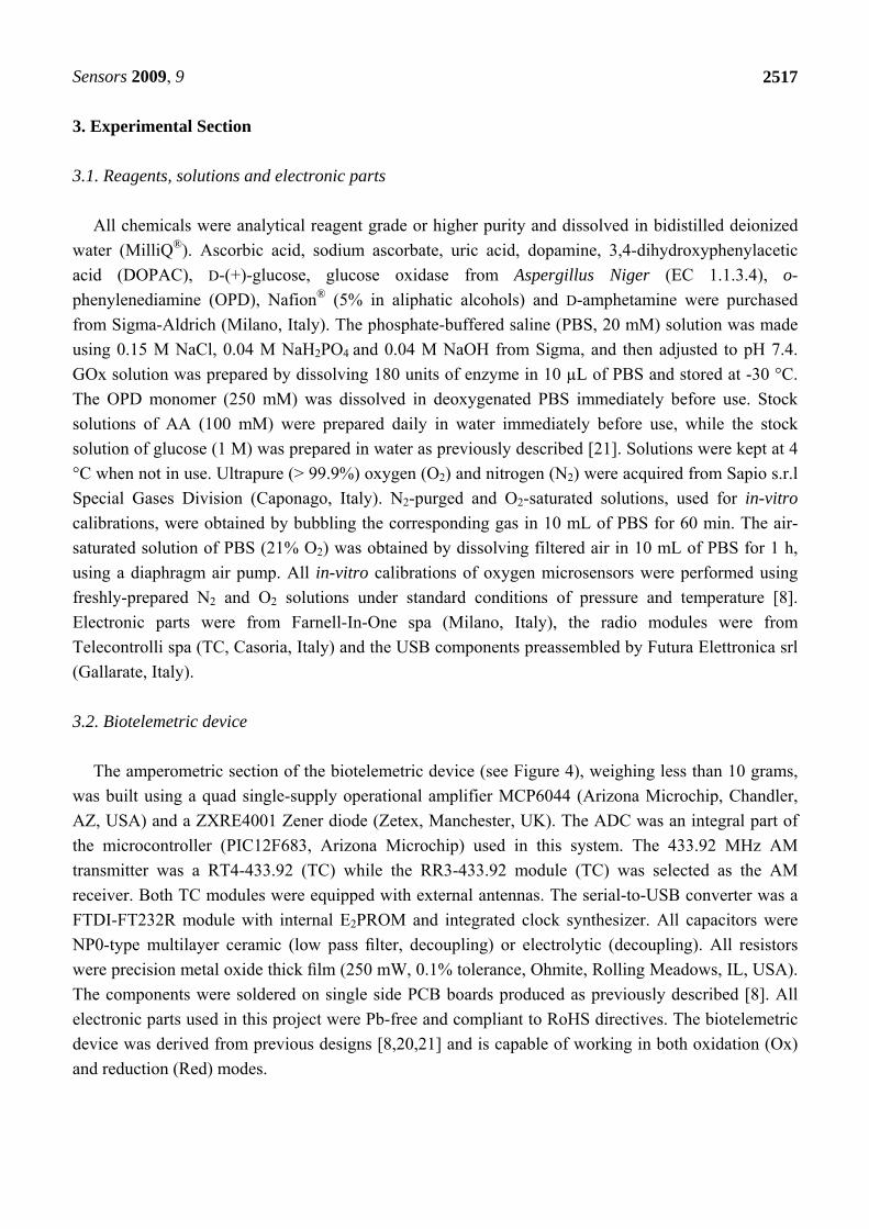
The electronic circuit of the miniaturized biotelemetric device was composed by three different
parts: the amperometric module, the microcontroller and the transmitter. As described in Section 3.2
below, the amperometric module was made using three “rail-to-rail” operational amplifiers working as
potentiostat (OPA1), voltage follower (OPA2) and current-to-voltage (I/V) converter (OPA3). The
Zener diode (Z) plays a pivotal role in the amperometric circuitry generating a fixed voltage of 1.22 V
useful for the fine regulation of the potential applied (VApp) to the working electrode by means of a
miniaturized potentiometer (P). The non-inverting input of OPA1 can be alternatively grounded or
connected to Z for working in oxidation or reduction mode respectively. The transfer function of the
I/V converter is:
VOut = -(Iredox · Rf) + VApp (7)
in which Iredox is the current flowing through the WE, Rf is the feedback resistor and VApp is the
potential applied to the WE. Rf has a capacitor in parallel (Cf) to complete a low pass filter with a cut-
off frequency (Fcut-off) of 25 Hz. The value of Cf was calculated in farads according to the equation:
Cf = 1 / (Fcut-off · 2π · Rf) (8)
An automated dummy cell was made based on a previously published design [8,20] for testing the
amperometric module of the biotelemetric device. The calibration of the electronics was made indoors
with a linear distance between the TX and RX units of about 3 m confirming previously-published
results [8]. The averaged power consumption necessary to drive the biotelemetric device was
experimentally determined [8,20,21] as 375 µW (125 µA). This means that a 3 V lithium coin battery
Sensors 2009, 9
2514
(Maxell CR1216), having a capacity of 25 mA h–1, can power the unit for more than one week of
continuous operation (sample rate: 1 Hz). The current necessary to drive the receiver unit was equal to
45 mA (225 mW). The biotelemetric device is characterized by gain precision, stability and an
excellent linear response. The system can operate both in oxidation and reduction modes and it is
particularly suited to work with direct-oxidation sensors (AA) or biosensors based on oxidase enzymes
(glucose) and direct-reduction sensors (O2) or O2-consuming biosensors [22]. The weight of the
biotelemetry unit is compatible with similar commercial devices [23], represents ~ 3% of the rat body
weight and it is well tolerated by the animals in agreement with other studies [24].
2.2. In-vitro calibration of ascorbic acid microsensor and in-vivo results
In-vitro calibrations of AA microsensors were carried out in fresh PBS at room temperature (25 °C)
before and after implantation. A constant potential of +120 mV vs Ag/AgCl was applied and, after a
stable baseline was reached, known amount of AA stock solution were added to the PBS in order to
obtain concentrations ranging from 0 to 1 mM.
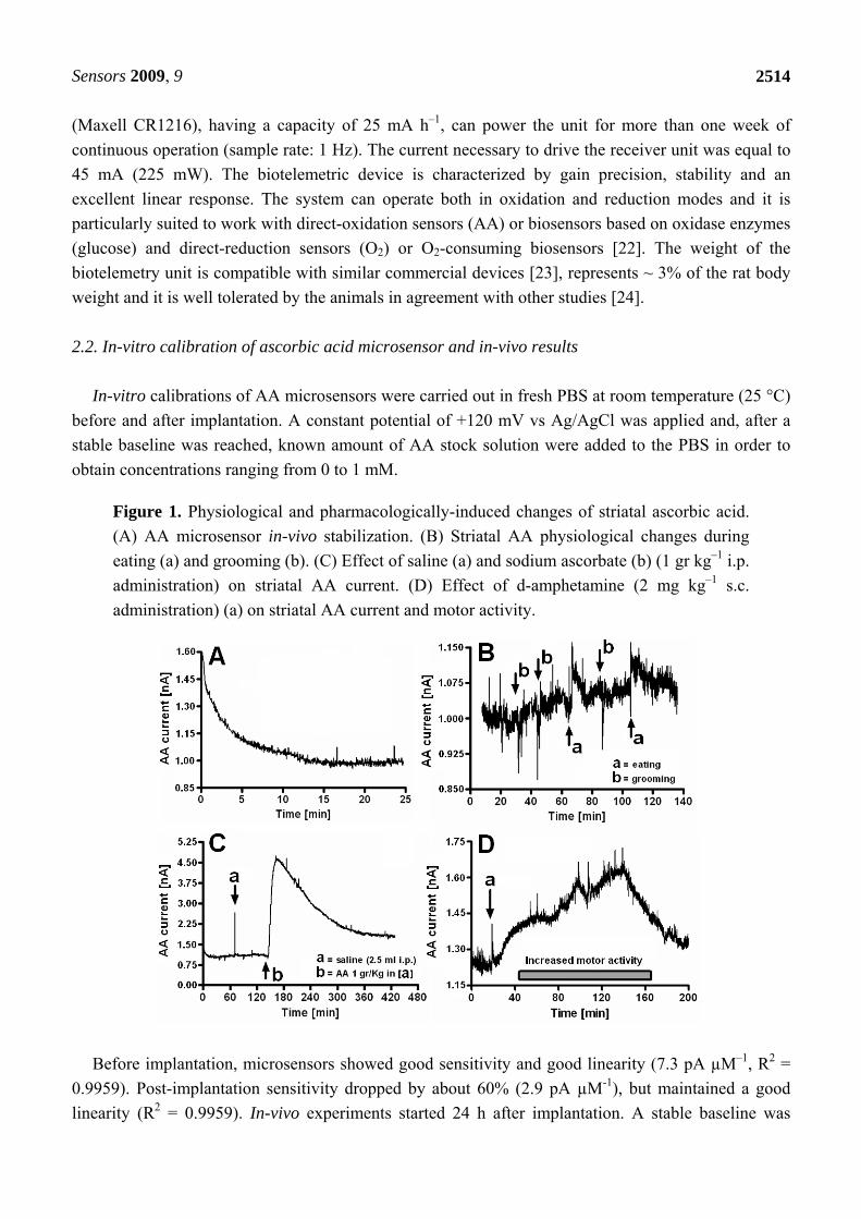
Figure 1. Physiological and pharmacologically-induced changes of striatal ascorbic acid.
(A) AA microsensor in-vivo stabilization. (B) Striatal AA physiological changes during
eating (a) and grooming (b). (C) Effect of saline (a) and sodium ascorbate (b) (1 gr kg–1 i.p.
administration) on striatal AA current. (D) Effect of d-amphetamine (2 mg kg–1 s.c.
administration) (a) on striatal AA current and motor activity.
Before implantation, microsensors showed good sensitivity and good linearity (7.3 pA µM–1, R2 =
0.9959). Post-implantation sensitivity dropped by about 60% (2.9 pA µM-1), but maintained a good
linearity (R2 = 0.9959). In-vivo experiments started 24 h after implantation. A stable baseline was
Sensors 2009, 9
2515
reached after a period of about 20 min (see Figure 1A). The calculated AA baseline corresponded to a
concentration of ~ 350 µM, in agreement with previous findings [25]. Physiological fluctuations of AA
current were observed in concomitance with stereotyped behaviors (see Figure 1B). Pharmacological
treatments were performed by administering sodium ascorbate (1 gr kg–1 i.p.) and d-amphetamine (2
mg kg–1 s.c.). Sodium ascorbate was administered intraperitoneally in order to verify sensor response
and resulted in a 4-fold increase in striatal AA current (see Figure 1C). Subcutaneous d-amphetamine
(see Figure 1D) induced an increase in AA current (+0.40 nA corresponding to +138 µM) and motor
activity in accord with previous studies [26]. D-Amphetamine has also been shown to decrease
glutamate striatal concentrations [26]. These findings are consistent with the functioning of an
AA/glutamate heteroexchange system [6,27] in which AA release is linked to impulse traffic,
transmitter release and glutamate uptake [26].
2.3. In-vitro calibration of oxygen microsensor and in-vivo results
All in-vitro calibrations of oxygen microsensors were carried out 24 h after manufacture,
immediately before implantation and then repeated after in-vivo experiments, using a previously-
described electrochemical cell [20,21], appropriately set for oxygen [8].
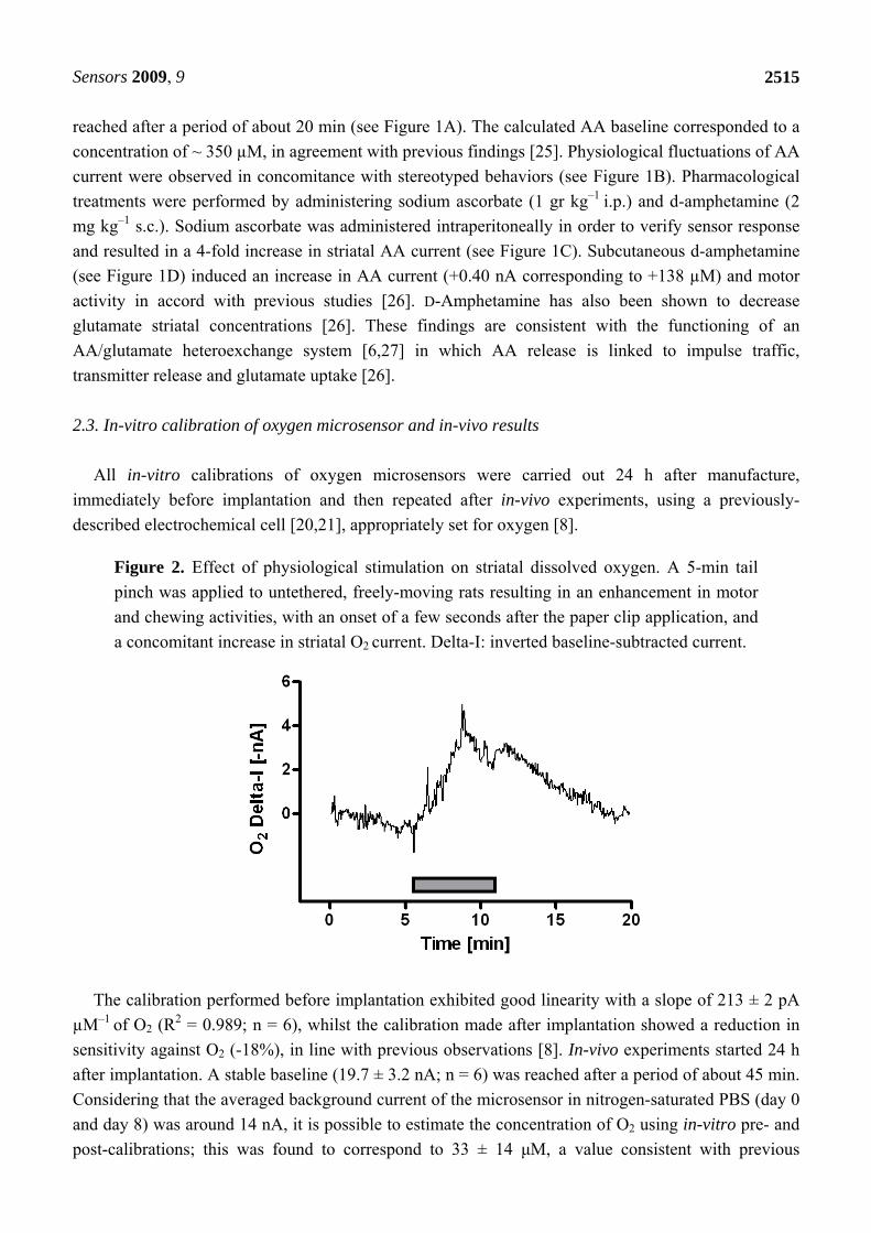
Figure 2. Effect of physiological stimulation on striatal dissolved oxygen. A 5-min tail
pinch was applied to untethered, freely-moving rats resulting in an enhancement in motor
and chewing activities, with an onset of a few seconds after the paper clip application, and
a concomitant increase in striatal O2 current. Delta-I: inverted baseline-subtracted current.
The calibration performed before implantation exhibited good linearity with a slope of 213 ± 2 pA
µM–1 of O2 (R2 = 0.989; n = 6), whilst the calibration made after implantation showed a reduction in
sensitivity against O2 (-18%), in line with previous observations [8]. In-vivo experiments started 24 h
after implantation. A stable baseline (19.7 ± 3.2 nA; n = 6) was reached after a period of about 45 min.
Considering that the averaged background current of the microsensor in nitrogen-saturated PBS (day 0
and day 8) was around 14 nA, it is possible to estimate the concentration of O2 using in-vitro pre- and
post-calibrations; this was found to correspond to 33 ± 14 μM, a value consistent with previous
Sensors 2009, 9
2516
estimates. [28,29,30-32]. Physiological stimulation, a 5 min-tail pinch (see Figure 2), administered in
order to increase neural activity and to promote regional cerebral blood flow (rCBF), led to increased
motor activity and striatal O2 current of +4.8 nA, corresponding to +27 µM. Striatal oxygen dynamics,
following physiological stimulation, results in a rise in the local O2 signal [8], mainly related to an
increase of rCBF during neural activation in agreement with previous reports on wired rats [8,28,29].
2.4. In-vitro calibration of glucose biosensor and in-vivo results
The in-vitro response of the glucose biosensor was determined just before implantation by adding
known amounts of glucose in the electrochemical cell giving concentrations ranging between 0 and
140 mM. Calibrations showed classical Michaelis-Menten kinetics (R2 = 0.989, n = 6) with Vmax and
KM equal respectively to 89 ± 4 nA and 4.8 ± 0.6 mM. The linear region was evaluated at low
concentrations (0 – 2 mM), which showed good linearity (R2 = 0.987, n = 6) with a slope of 15.2 ± 1.1
nA mM–1. The in-vivo experiments were carried out using the same procedures as oxygen studies. A
stable baseline was observed 30 – 35 min after sensor polarization and corresponded to 7.5 ± 0.5 nA
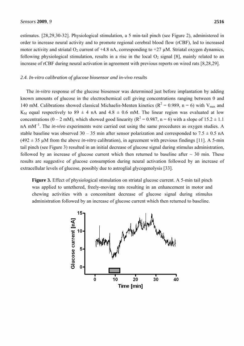
(492 ± 35 µM from the above in-vitro calibration), in agreement with previous findings [11]. A 5-min
tail pinch (see Figure 3) resulted in an initial decrease of glucose signal during stimulus administration,
followed by an increase of glucose current which then returned to baseline after ~ 30 min. These
results are suggestive of glucose consumption during neural activation followed by an increase of
extracellular levels of glucose, possibly due to astroglial glycogenolysis [33].
Figure 3. Effect of physiological stimulation on striatal glucose current. A 5-min tail pinch
was applied to untethered, freely-moving rats resulting in an enhancement in motor and
chewing activities with a concomitant decrease of glucose signal during stimulus
administration followed by an increase of glucose current which then returned to baseline.
Sensors 2009, 9
2517
3. Experimental Section
3.1. Reagents, solutions and electronic parts
All chemicals were analytical reagent grade or higher purity and dissolved in bidistilled deionized
water (MilliQ®). Ascorbic acid, sodium ascorbate, uric acid, dopamine, 3,4-dihydroxyphenylacetic
acid (DOPAC), D-(+)-glucose, glucose oxidase from Aspergillus Niger (EC 1.1.3.4), o-
phenylenediamine (OPD), Nafion® (5% in aliphatic alcohols) and D-amphetamine were purchased
from Sigma-Aldrich (Milano, Italy). The phosphate-buffered saline (PBS, 20 mM) solution was made
using 0.15 M NaCl, 0.04 M NaH2PO4 and 0.04 M NaOH from Sigma, and then adjusted to pH 7.4.
GOx solution was prepared by dissolving 180 units of enzyme in 10 µL of PBS and stored at -30 °C.
The OPD monomer (250 mM) was dissolved in deoxygenated PBS immediately before use. Stock
solutions of AA (100 mM) were prepared daily in water immediately before use, while the stock
solution of glucose (1 M) was prepared in water as previously described [21]. Solutions were kept at 4
°C when not in use. Ultrapure (> 99.9%) oxygen (O2) and nitrogen (N2) were acquired from Sapio s.r.l
Special Gases Division (Caponago, Italy). N2-purged and O2-saturated solutions, used for in-vitro
calibrations, were obtained by bubbling the corresponding gas in 10 mL of PBS for 60 min. The air-
saturated solution of PBS (21% O2) was obtained by dissolving filtered air in 10 mL of PBS for 1 h,
using a diaphragm air pump. All in-vitro calibrations of oxygen microsensors were performed using
freshly-prepared N2 and O2 solutions under standard conditions of pressure and temperature [8].
Electronic parts were from Farnell-In-One spa (Milano, Italy), the radio modules were from
Telecontrolli spa (TC, Casoria, Italy) and the USB components preassembled by Futura Elettronica srl
(Gallarate, Italy).
3.2. Biotelemetric device
The amperometric section of the biotelemetric device (see Figure 4), weighing less than 10 grams,
was built using a quad single-supply operational amplifier MCP6044 (Arizona Microchip, Chandler,
AZ, USA) and a ZXRE4001 Zener diode (Zetex, Manchester, UK). The ADC was an integral part of
the microcontroller (PIC12F683, Arizona Microchip) used in this system. The 433.92 MHz AM
transmitter was a RT4-433.92 (TC) while the RR3-433.92 module (TC) was selected as the AM
receiver. Both TC modules were equipped with external antennas. The serial-to-USB converter was a
FTDI-FT232R module with internal E2PROM and integrated clock synthesizer. All capacitors were
NP0-type multilayer ceramic (low pass filter, decoupling) or electrolytic (decoupling). All resistors
were precision metal oxide thick film (250 mW, 0.1% tolerance, Ohmite, Rolling Meadows, IL, USA).
The components were soldered on single side PCB boards produced as previously described [8]. All
electronic parts used in this project were Pb-free and compliant to RoHS directives. The biotelemetric
device was derived from previous designs [8,20,21] and is capable of working in both oxidation (Ox)
and reduction (Red) modes.
Sensors 2009, 9
2518
Figure 4. Amperometric section of the biotelemetric device. Ox: Oxidation; Red:
Reduction; Z: Zener diode; P: Potentiometer; OPA: Operational Amplifier; Rf: Feedback
Resistor; Cf: Feedback Capacitor; VApp: Applied Potential; VOut: Output Voltage; WE:
Working Electrode; RE: Reference Electrode; AE: Auxiliary Electrode.
3.3. Preparation and calibration of microsensors and biosensors
The AA microsensors were made using Teflon™-insulated silver wires (30 mm in length; Ø = 125
µm, Advent Research Materials, Suffolk, UK) modifying a previously-described procedure [34].
Approximately 1 mm of the wire was exposed and inserted into a silica capillary tube (10 mm in
length; I.D. Ø = 180 µm, Polymicro Technologies, Phoenix, AZ, USA) partly filled with graphite-
loaded (55% w/w) epoxy resin (Araldite-M®, Sigma-Aldrich, Milan, Italy). A preliminary 180 µm
diameter carbon-composite disc electrode (area: 2.5 10–4 cm2) was fabricated by mixing 850 mg of
graphite with 500 mg of Araldite-M and 200 mg of hardener and filling the silica capillary tubing with
the mixture. The silver wire guaranteed a good electrical contact. After 24 h at 40 °C, the shape of the
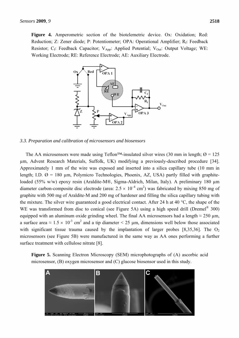
WE was transformed from disc to conical (see Figure 5A) using a high speed drill (Dremel® 300)
equipped with an aluminum oxide grinding wheel. The final AA microsensors had a length ≈ 250 µm,
a surface area ≈ 1.5 10-3 cm2 and a tip diameter < 25 µm, dimensions well below those associated
with significant tissue trauma caused by the implantation of larger probes [8,35,36]. The O2
microsensors (see Figure 5B) were manufactured in the same way as AA ones performing a further
surface treatment with cellulose nitrate [8].
Figure 5. Scanning Electron Microscopy (SEM) microphotographs of (A) ascorbic acid
microsensor, (B) oxygen microsensor and (C) glucose biosensor used in this study.
Sensors 2009, 9
2519
AA oxidation and O2 reduction potentials were experimentally established using cyclic
voltammetry and were found to be +120 mV [19] and -400 mV [8], respectively vs Ag/AgCl (NaCl 3
M; RE4 Bioanalytical Systems, Inc., Lafayette, TX, USA) reference electrode. The fabrication of the
glucose biosensors (see Figure 5C) has been previously described in detail [21]. Briefly, 1 mm Pt
cylinder, obtained by cutting Teflon-insulated Pt wire (Ø = 125 µm, Advent Research Materials,
Suffolk, UK), was immersed 3 times into a solution of GOx and let it dry for 5 min after each dip. The
biosensor was then placed in the cell filled with 5 mL of N2-purged PBS containing the o-
phenylenediamine monomer (250 mM). The electrosynthesis of p-OPD was carried out at +700 mV
vs. Ag/AgCl for 15 min. H2O2 electro-oxidation was carried out at +700 mV [21] vs Ag/AgCl
reference electrode. Constant potential amperometry (CPA) was used for in-vitro and in-vivo
experiments; all in-vitro calibrations were performed in fresh PBS 24 h after sensors’ fabrication as
previously described in detail [8,19,21]. No significant interference signals were observed on exposing
AA, O2 microsensors and glucose biosensors to other electroactive molecules present in the striatal
extracellular fluid (ECF), even at pharmacologically relevant concentrations [37] (Table 1).
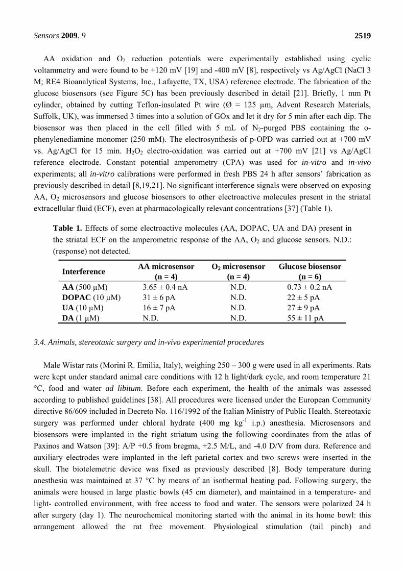
Table 1. Effects of some electroactive molecules (AA, DOPAC, UA and DA) present in
the striatal ECF on the amperometric response of the AA, O2 and glucose sensors. N.D.:
(response) not detected.
Interference AA microsensor
(n = 4) O2 microsensor
(n = 4) Glucose biosensor
(n = 6) AA (500 µM) 3.65 ± 0.4 nA N.D. 0.73 ± 0.2 nA DOPAC (10 µM) 31 ± 6 pA N.D. 22 ± 5 pA UA (10 µM) 16 ± 7 pA N.D. 27 ± 9 pA DA (1 µM) N.D. N.D. 55 ± 11 pA
3.4. Animals, stereotaxic surgery and in-vivo experimental procedures
Male Wistar rats (Morini R. Emilia, Italy), weighing 250 – 300 g were used in all experiments. Rats
were kept under standard animal care conditions with 12 h light/dark cycle, and room temperature 21
°C, food and water ad libitum. Before each experiment, the health of the animals was assessed
according to published guidelines [38]. All procedures were licensed under the European Community
directive 86/609 included in Decreto No. 116/1992 of the Italian Ministry of Public Health. Stereotaxic
surgery was performed under chloral hydrate (400 mg kg-1 i.p.) anesthesia. Microsensors and
biosensors were implanted in the right striatum using the following coordinates from the atlas of
Paxinos and Watson [39]: A/P +0.5 from bregma, +2.5 M/L, and -4.0 D/V from dura. Reference and
auxiliary electrodes were implanted in the left parietal cortex and two screws were inserted in the
skull. The biotelemetric device was fixed as previously described [8]. Body temperature during
anesthesia was maintained at 37 °C by means of an isothermal heating pad. Following surgery, the
animals were housed in large plastic bowls (45 cm diameter), and maintained in a temperature- and
light- controlled environment, with free access to food and water. The sensors were polarized 24 h
after surgery (day 1). The neurochemical monitoring started with the animal in its home bowl: this
arrangement allowed the rat free movement. Physiological stimulation (tail pinch) and
Sensors 2009, 9
2520
pharmacological treatments (sodium ascorbate and D-amphetamine) were carried out within the first
week after stereotaxic surgery.
3.5. Hystology
After each set of experiments (day 8), rats were sacrificed with an injection of chloral hydrate (800
mg kg-1 i.p.). The location of each microsensor and biosensor in the striatum was confirmed by post-
mortem histology. Brains were fixed in formal saline and 50 µm coronal sections were made with a
cryostat. The slices were stained with cresyl violet and examined under a microscope.
3.6. Statistical analysis
Concentrations of AA, O2 and glucose were expressed in µM. AA and glucose (H2O2) anodic
signals were given as absolute current values (nA) while oxygen cathodic current was expressed in nA
and given as baseline-subtracted (Delta-I) raw data. The sign of the oxygen currents was inverted to
give a positive correlation of the plotted data with the concentration of analyte. The in-vitro response
of AA and oxygen microsensors was evaluated before and after in in-vivo experiments while the
glucose biosensors parameters were calculated only before implantation because of the damage during
explant can lead to inaccurate calibration [40]. The changes of brain tissue neurochemicals were
calculated as absolute variations versus the corresponding baselines and their striatal concentrations
were estimated using pre-implantation (glucose) or post-implantation (AA and O2) in-vitro
calibrations.
4. Conclusions
In this study we present the real-time monitoring of three brain neurochemical species (AA, O2 and
glucose) in untethered, freely-moving rats using a biotelemetric device coupled with implantable
sensors. The transmitter and the receiver units have been used for accurate transduction of the redox
currents generated on the surface of these microsensors and biosensors, both in vitro and in vivo. The
miniaturized biotelemetric device, composed by an amperometric module, a microcontroller and a
transmitter, polarizes the sensor and sends sensor data to a receiving unit connected to a PC. The
system electronics have been tested under different experimental conditions exhibiting low power
consumption, high stability and good linear response. The in-vivo results confirmed previously-
published observations on striatal AA, oxygen and glucose dynamics. This approach, based on simple
and inexpensive components, could be used as a rapid and reliable model for studying the effects of
different drugs on brain neurochemical systems.
Acknowledgements
The authors acknowledge the Italian distributor of Arizona Microchip© Corporation for free
samples of integrated circuits. The research was supported by the Ministero dell’Istruzione,
Sensors 2009, 9
2521
dell’Università e della Ricerca (PRIN 2007 fund), University of Sassari (ex 60% fund) and Fondazione
Banco di Sardegna.
References
1. Emerit, J.; Edeas, M. Neurodegenerative diseases and oxidative stress. Eur.
Neuropsychopharmacol. 2005, 15, S100-S101.
2. Andersen, J.K. Oxidative stress in neurodegeneration: cause or consequence? Nat. Med. 2004, 10,
S18-S25.
3. Serra, P.A.; Sciola, L.; Delogu, M.R.; Spano, A.; Monaco, G.; Miele, E.; Rocchitta, G.; Miele, M.;
Migheli, R.; Desole, M.S. The neurotoxin 1-methyl-4-phenyl-1,2,3,6-tetrahydropyridine induces
apoptosis in mouse nigrostriatal glia. Relevance to nigral neuronal death and striatal
neurochemical changes. J Biol Chem. 2002, 277, 34451-34461.
4. Hediger, M.A. New view at C. Nat. Med. 2002, 8, 445-446.
5. O'Neill, R.D.; Fillenz, M.; Sundstrom, L.; Rawlins, J.N.P. Voltammetrically monitored brain
ascorbate as an index of excitatory amino acid release in the unrestrained rat. Neurosci. Lett.
1984, 52, 227-233.
6. Rice, M.E. Ascorbate regulation and its neuroprotective role in the brain. Trends Neurosci. 2000,
23, 209-216.
7. Watanabe, Y.; Hyllbrant, B.B.; Langstrom, B. Tracing oxygen metabolism by use of positron
emitter Oxygen-15. Biochem. Biophys. Res. Commun. 1997, 231, 131-134.
8. Bazzu, G.; Puggioni, G.G.; Dedola, S.; Calia, G.; Rocchitta, G.; Migheli, R.; Desole, M.S.; Lowry,
J.P.; O'Neill, R.D.; Serra, P.A. Real-time monitoring of brain tissue oxygen using a miniaturized
biotelemetric device implanted in freely-moving rats. Anal. Chem. 2009, doi: 10.1021/ac802390f,
in press.
9. Fillenz, M. The role of lactate in brain metabolism. Neurochem. Int. 2005, 47, 413-417.
10. Aubert, A.; Costalat, R.; Magistretti, P.J.; Pellerin, L. Brain lactate kinetics: modeling evidence
for neuronal lactate uptake upon activation. Proc. Natl. Acad. Sci. USA 2005, 102, 16448-16453.
11. Lowry, J.P.; Miele, M.; O’Neill, R.D.; Boutelle, M.G.; Fillenz, M. An amperometric glucose
oxidase/poly(o-phenylenediamine) biosensor for monitoring brain extracellular glucose: In vivo
characterisation in the striatum striatum of freely-moving rats. J. Neurosci. Methods 1998, 79,
65-74.
12. Magistretti, P.J.; Pellerin, L.; Rothman, D.L.; Shulman, R.G. Energy on demand. Science 1999,
283, 496-497.
13. Pfeiffer, D.; Schubert, F.; Wollenberger, U.; Scheller, F.W. Electrochemical sensors: Enzyme
electrodes and field effect transistors. In Handbook of Chemical and Biological Sensors; Taylor,
R.F., Schultz, J.S., Eds.; IOP Publishing Ltd: Bristol, UK, 1996; pp. 435-458.
14. Wilson, R.; Turner, A.P.F. Glucose oxidase: an ideal enzyme. Biosens. Bioelectron. 1992, 7,
165-185.
15. Zhou, R.J; Hao, Z.Q. The present status and development of biotelemetry. Zhongguo Yi Liao Qi
Xie Za Zhi 2002, 26, 212-214.
16. FCC (Federal Communication Commission). Commission’s Rules to Create a Wireless Medical
Telemetry Service; FCC : Washington, DC, USA, 2000. No. FCC 00-211, pp. 1-24.
Sensors 2009, 9
2522
17. Leuher, D.C. Overview of biomedical telemetry techniques. Eng. Med. Biol. 1983, 3, 17-24.
18. Serra, P.; Hebel, M.; Rocchitta, G.; Tate, R. Biotelemetry NET for neurochemical biosensor and
microsensor applications: design, construction and validation. In Telemetry: Research,
Technology and Applications; Barculo, D., Daniels, J., Eds.; Nova Science Publishers Inc:
Hauppauge, NY, USA, 2009.
19. Hebel, M.; Serra, P.A. Development of a parallel-computing embedded telemetry system for
voltammetric microsensor and biosensor applications. In Sensors for Environment, Health and
Security: Advanced Materials and Technologies; Baraton, M.I., Ed.; Springer: Dordrecht, The
Netherlands, 2008; pp. 229-238.
20. Rocchitta, G.; Migheli, R.; Dedola, S.; Calia, G.; Desole, M.S.; Miele, E.; Lowry, J.P.; O’Neill,
R.D.;.Serra, P.A. Development of a distributed, fully automated, bidirectional telemetry system
for amperometric microsensor and biosensor applications. Sens. Actuat. B 2007, 126, 700-709.
21. Serra, P.A.; Rocchitta, G.; Bazzu, G.; Manca, A.; Puggioni, G.M.; Lowry, J.P.; O’Neill, R.D.
Design and construction of a low cost single-supply embedded telemetry system for amperometric
biosensor applications. Sens. Actuat B 2007, 122, 118-126.
22. Wu, L.; Zhang, X.; Ju, H. Amperometric glucose sensor based on catalytic reduction of dissolved
oxygen at soluble carbon nanofiber. Biosens. Bioelectron. 2007, 23, 479-484.
23. Morita, H.; Abe, C.; Awazu, C.; Tanaka, K. Long-term hypergravity induces plastic alterations in
vestibulo-cardiovascular reflex in conscious rats. Neurosc. Lett. 2007, 412, 201-205.
24. Leon, L.R.; Walker, L.D.; DuBose, D.A.; Stephenson, L.A. Biotelemetry transmitter implantation
in rodents: impact on growth and circadian rhythms. Am. J. Physiol. Regul. Integr. Comp. Physiol.
2004, 286, R967-R974.
25. Miele, M.; Fillenz, M. In vivo determination of extracellular brain ascorbate. J. Neurosci. Methods
1996, 70, 15-19.
26. Miele, M.; Mura, M.A.; Enrico, P.; Esposito, G.; Serra, P.A.; Migheli, R.; Zangani, D.; Miele, E.;
Desole, M.S. On the mechanism of d-amphetamine-induced changes in glutamate, ascorbic acid
and uric acid release in the striatum of freely-moving rats. Br. J. Pharmacol. 2000, 129, 582-588.
27. Hediger, M.A. Transporters for vitamin C keep vitamin concentrations optimal in the body. A
new mouse knockout of one transporter reveals previously unknown requirements for the vitamin.
Nat. Med. 2002, 8, 514-517.
28. Lowry, J.P.; Boutelle, M.G.; Fillenz, M. Measurement of brain tissue oxygen at a carbon paste
electrode can serve as an index of increases in regional cerebral blood flow. J. Neurosci. Methods.
1997, 71, 177-182.
29. Bolger, F.B.; Lowry, J.P. Brain tissue oxygen: In vivo monitoring with carbon paste electrodes.
Sensors 2005, 5, 473-487.
30. Lowry, J.P.; Fillenz M. Real-time monitoring of brain energy metabolism in vivo using
microelectrochemical sensors: the effects of anesthesia. Bioelectrochemistry 2001, 54, 39-47.
31. Lowry, J.P.; Fillenz M. Evidence for uncoupling of oxygen and glucose utilization during
neuronal activation in rat striatum. J. Physiol. 1997, 498, 497-501.
32. Dixon, B.M.; Lowry, J.P.; O'Neill, R.D. Characterization in vitro and in vivo of the oxygen
dependence of an enzyme/polymer biosensor for monitoring brain glucose. J. Neurosci. Methods
2002, 119, 135-142.
Sensors 2009, 9
2523
33. Fillenz, M.; Lowry, J.P.; Boutelle, M.G.; Fray, A.E. The role of astrocytes and noradrenaline in
neuronal glucose metabolism. Acta Physiol. Scand. 1999, 167, 275-284.
34. Migheli, R.; Puggioni, G.; Dedola, S.; Rocchitta, G.; Calia, G.; Bazzu, G.; Esposito, G.; Lowry,
J.P.; O’Neill, R.D.; Desole, M.S.; Miele, E.; Serra P.A. Novel integrated microdialysis–
amperometric system for in vitro detection of dopamine secreted from PC12 cells: design,
construction, and validation. Anal. Biochem. 2008, 380, 323-330.
35. Duff, A.; O'Neill, R.D. Effect of probe size on the concentration of brain extracellular uric acid
monitored with carbon paste electrodes. J. Neurochem. 1994, 62, 1496-1502.
36. Fumero, B.; Guadalupe, T.; Valladares, F.; Mora, F.; O'Neill, R.D.; Mas, M.; Gonzalez-Mora, J.L.
Fixed versus removable microdialysis probes for in vivo neurochemical analysis: implications for
behavioral studies. J. Neurochem. 1994, 63, 1407-1415.
37. Ryan, M.R.; Lowry, J.P.; O'Neill, R.D. Biosensor for neurotransmitter L-glutamic acid designed
for efficient use of L-glutamate oxidase and effective rejection of interference. Analyst 1997, 122,
1419-1424.
38. Wolfensohn, S.; Lloyd, M. Handbook of Laboratory Animal Management and Welfare, 3rd Ed.;
Blackwell Publishing: Cornwall, ON, Canada, 2003.
39. Paxinos, G.; Watson, C. The Rat Brain in Stereotaxic Coordinates, 6th Ed.; Academic Press: San
Diego, CA, USA, 2007.
40. Lowry, J.P.; Ryan, M.R.; O'Neill, R.D. Behaviourally induced changes in extracellular levels of
brain glutamate monitored at 1 s resolution with an implanted biosensor. Anal. Commun. 1998,
35, 87-89.
© 2009 by the authors; licensee Molecular Diversity Preservation International, Basel, Switzerland.
This article is an open-access article distributed under the terms and conditions of the Creative
Commons Attribution license (http://creativecommons.org/licenses/by/3.0/).
Copyright © 2022 FDOKUMEN