Topics of Biotechnological Tools for Agricultural Development
Upload
itmexicaliCategory
view
3download
0
This article was downloaded by: [148.231.90.67]On: 20 June 2014, At: 19:04Publisher: Taylor & FrancisInforma Ltd Registered in England and Wales Registered Number: 1072954 Registered office: Mortimer House,37-41 Mortimer Street, London W1T 3JH, UK
Biotechnology & Biotechnological EquipmentPublication details, including instructions for authors and subscription information:http://www.tandfonline.com/loi/tbeq20
Comparison of the performances of four hydrophilicpolymers as supports for lipase immobilisationLydia Toscanoab, Gisela Monteroa, Margarita Stoytchevaa, Lourdes Cervantesc & VelizarGochevd
a Institute of Engineering, Autonomous University of Baja California, Mexicali, Mexicob Technological Institute of Mexicali, Mexicali, Mexicoc Institute of Agricultural Science, Autonomous University of Baja California, Mexicali,Mexicod Department of Biochemistry and Microbiology, Plovdiv University, Plovdiv, BulgariaPublished online: 30 Apr 2014.
To cite this article: Lydia Toscano, Gisela Montero, Margarita Stoytcheva, Lourdes Cervantes & Velizar Gochev (2014)Comparison of the performances of four hydrophilic polymers as supports for lipase immobilisation, Biotechnology &Biotechnological Equipment, 28:1, 52-60, DOI: 10.1080/13102818.2014.901684
To link to this article: http://dx.doi.org/10.1080/13102818.2014.901684
PLEASE SCROLL DOWN FOR ARTICLE
Taylor & Francis makes every effort to ensure the accuracy of all the information (the “Content”) contained inthe publications on our platform. Taylor & Francis, our agents, and our licensors make no representations orwarranties whatsoever as to the accuracy, completeness, or suitability for any purpose of the Content. Versionsof published Taylor & Francis and Routledge Open articles and Taylor & Francis and Routledge Open Selectarticles posted to institutional or subject repositories or any other third-party website are without warrantyfrom Taylor & Francis of any kind, either expressed or implied, including, but not limited to, warranties ofmerchantability, fitness for a particular purpose, or non-infringement. Any opinions and views expressed in thisarticle are the opinions and views of the authors, and are not the views of or endorsed by Taylor & Francis. Theaccuracy of the Content should not be relied upon and should be independently verified with primary sourcesof information. Taylor & Francis shall not be liable for any losses, actions, claims, proceedings, demands,costs, expenses, damages, and other liabilities whatsoever or howsoever caused arising directly or indirectly inconnection with, in relation to or arising out of the use of the Content. This article may be used for research, teaching, and private study purposes. Terms & Conditions of access anduse can be found at http://www.tandfonline.com/page/terms-and-conditions It is essential that you check the license status of any given Open and Open Select article to confirmconditions of access and use.
ARTICLE; AGRICULTURE AND ENVIRONMENTAL BIOTECHNOLOGY
Comparison of the performances of four hydrophilic polymers as supports
for lipase immobilisation
Lydia Toscanoa,b, Gisela Monteroa, Margarita Stoytchevaa, Lourdes Cervantesc and Velizar Gochevd*
aInstitute of Engineering, Autonomous University of Baja California, Mexicali, Mexico; bTechnological Institute of Mexicali, Mexicali,Mexico; cInstitute of Agricultural Science, Autonomous University of Baja California, Mexicali, Mexico; dDepartment of Biochemistryand Microbiology, Plovdiv University, Plovdiv, Bulgaria
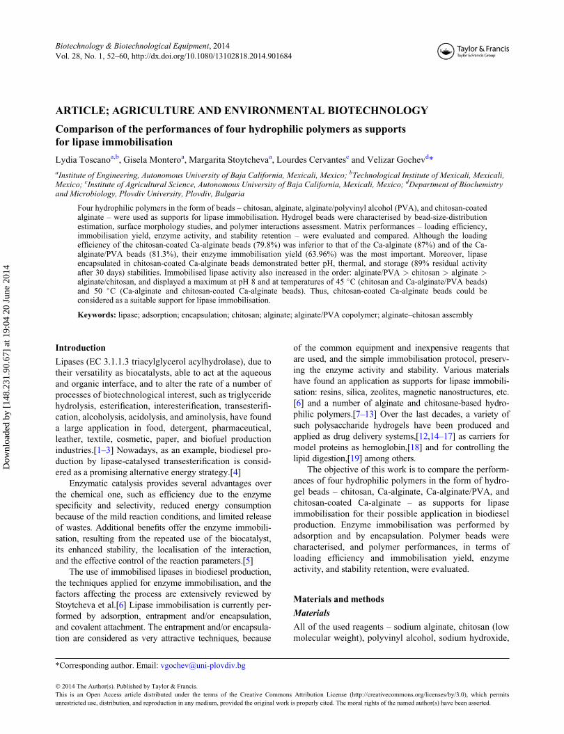
Four hydrophilic polymers in the form of beads – chitosan, alginate, alginate/polyvinyl alcohol (PVA), and chitosan-coatedalginate – were used as supports for lipase immobilisation. Hydrogel beads were characterised by bead-size-distributionestimation, surface morphology studies, and polymer interactions assessment. Matrix performances – loading efficiency,immobilisation yield, enzyme activity, and stability retention – were evaluated and compared. Although the loadingefficiency of the chitosan-coated Ca-alginate beads (79.8%) was inferior to that of the Ca-alginate (87%) and of the Ca-alginate/PVA beads (81.3%), their enzyme immobilisation yield (63.96%) was the most important. Moreover, lipaseencapsulated in chitosan-coated Ca-alginate beads demonstrated better pH, thermal, and storage (89% residual activityafter 30 days) stabilities. Immobilised lipase activity also increased in the order: alginate/PVA > chitosan > alginate >alginate/chitosan, and displayed a maximum at pH 8 and at temperatures of 45 �C (chitosan and Ca-alginate/PVA beads)and 50 �C (Ca-alginate and chitosan-coated Ca-alginate beads). Thus, chitosan-coated Ca-alginate beads could beconsidered as a suitable support for lipase immobilisation.
Keywords: lipase; adsorption; encapsulation; chitosan; alginate; alginate/PVA copolymer; alginate–chitosan assembly
Introduction
Lipases (EC 3.1.1.3 triacylglycerol acylhydrolase), due to
their versatility as biocatalysts, able to act at the aqueous
and organic interface, and to alter the rate of a number of
processes of biotechnological interest, such as triglyceride
hydrolysis, esterification, interesterification, transesterifi-
cation, alcoholysis, acidolysis, and aminolysis, have found
a large application in food, detergent, pharmaceutical,
leather, textile, cosmetic, paper, and biofuel production
industries.[1–3] Nowadays, as an example, biodiesel pro-
duction by lipase-catalysed transesterification is consid-
ered as a promising alternative energy strategy.[4]
Enzymatic catalysis provides several advantages over
the chemical one, such as efficiency due to the enzyme
specificity and selectivity, reduced energy consumption
because of the mild reaction conditions, and limited release
of wastes. Additional benefits offer the enzyme immobili-
sation, resulting from the repeated use of the biocatalyst,
its enhanced stability, the localisation of the interaction,
and the effective control of the reaction parameters.[5]
The use of immobilised lipases in biodiesel production,
the techniques applied for enzyme immobilisation, and the
factors affecting the process are extensively reviewed by
Stoytcheva et al.[6] Lipase immobilisation is currently per-
formed by adsorption, entrapment and/or encapsulation,
and covalent attachment. The entrapment and/or encapsula-
tion are considered as very attractive techniques, because
of the common equipment and inexpensive reagents that
are used, and the simple immobilisation protocol, preserv-
ing the enzyme activity and stability. Various materials
have found an application as supports for lipase immobili-
sation: resins, silica, zeolites, magnetic nanostructures, etc.
[6] and a number of alginate and chitosane-based hydro-
philic polymers.[7–13] Over the last decades, a variety of
such polysaccharide hydrogels have been produced and
applied as drug delivery systems,[12,14–17] as carriers for
model proteins as hemoglobin,[18] and for controlling the
lipid digestion,[19] among others.
The objective of this work is to compare the perform-
ances of four hydrophilic polymers in the form of hydro-
gel beads – chitosan, Ca-alginate, Ca-alginate/PVA, and
chitosan-coated Ca-alginate – as supports for lipase
immobilisation for their possible application in biodiesel
production. Enzyme immobilisation was performed by
adsorption and by encapsulation. Polymer beads were
characterised, and polymer performances, in terms of
loading efficiency and immobilisation yield, enzyme
activity, and stability retention, were evaluated.
Materials and methods
Materials
All of the used reagents – sodium alginate, chitosan (low
molecular weight), polyvinyl alcohol, sodium hydroxide,
*Corresponding author. Email: [email protected]
� 2014 The Author(s). Published by Taylor & Francis.
This is an Open Access article distributed under the terms of the Creative Commons Attribution License (http://creativecommons.org/licenses/by/3.0), which permits
unrestricted use, distribution, and reproduction in any medium, provided the original work is properly cited. The moral rights of the named author(s) have been asserted.
Biotechnology & Biotechnological Equipment, 2014
Vol. 28, No. 1, 52–60, http://dx.doi.org/10.1080/13102818.2014.901684
Dow
nloa
ded
by [
148.
231.
90.6
7] a
t 19:
04 2
0 Ju
ne 2
014

calcium chloride, boric acid, acetic acid, and so on – were
of analytical reagent grade.
Lipase (EC 3.1.1.3) was produced in solid-state fer-
mentation by Trichoderma harzianum, as described in a
previous work.[20]
Lipase immobilisation
Immobilisation in chitosan beads
Chitosan solution (4% (w/v)) was prepared by dissolving
the appropriate amount of chitosan powder in 1% acetic
acid. Chitosan beads of various shapes and sizes (3 �5 mm average) were obtained applying the “syringe meth-
od”, i.e. the viscous chitosan solution was extruded (nee-
dle diameter 1 mm) dropwise in a coagulant bath
containing NaOH (1 mol L�1) and ethanol (26% (v/v))
under gentle stirring. The mixture was allowed to remain
overnight. Then, the beads were removed by decantation,
washed with deionised water until reaching pH 8.0, and
soaked in hexane for 1 h under agitation (120 rpm). After
hexane excess removal, lipase immobilisation was per-
formed by physical absorption, according to the method-
ology reported in the literature [8,21] by chitosan beads
immersing into a lipase solution for 3 h under stirring at
ambient temperature, followed by 24-h static adsorption
at 4 �C. The enzyme solution was prepared by dissolving
10 mg of lyophilised lipase into 40 mL of double-distilled
water. The obtained derivatives were removed by decanta-
tion and filtration, and rinsed with hexane. The decanted
solutions and filtrates were collected for estimation of the
efficiency of lipase immobilisation. A similar method was
used to prepare chitosan beads placebo (without lipase).
Immobilisation in Ca-alginate beads
Lipase encapsulation in alginate beads was carried out
using established protocols.[13] First, sodium alginate
(1.5% (w/v)) solution and CaCl2 (0.1 mol L�1) were pre-
pared by dissolving suitable amounts of the corresponding
reagent in Tris-HCl buffer (0.05 mol L�1), pH 8.0. After
that, 11.2 mg of lyophilised lipase dissolved in 2 mL of
the same buffer was added to 8 mL of the alginate solution
and the mixture was gently stirred for 30 min. The
obtained lipase containing homogeneous alginate gel was
extruded dropwise through a syringe (needle diameter
1 mm) to the gelling solution of CaCl2 under stirring. The
beads were allowed to polymerise for 40 min. The deriva-
tives were washed twice with 3 mL aliquots of Tris-HCl
buffer and the beads were stored at 4 �C for later studies.
A similar approach was followed for the preparation of
alginate beads placebo. The residual solutions were col-
lected to determine the efficiency of the immobilisation.
Immobilisation in chitosan-coated Ca-alginate beads
Lipase encapsulation in alginate was accomplished as
described above. Nevertheless, the lipase containing
alginate gel was extruded dropwise (needle diameter
1 mm) into a gelling bath obtained by mixing 35 mL of
CaCl2 aqueous solution (1.5% (w/v)) and 65 mL of chito-
san solution (2% (w/v)) in acetic acid. This procedure led
to the lipase immobilisation in chitosan-coated alginate
beads.[22] The beads were allowed to harden in the bath
for 45 min under stirring. The formed derivatives were
washed with double-distilled water and stored wet at 4 �C.The residual solutions were analysed for protein content in
order to evaluate the enzyme immobilisation efficiency.
Immobilisation in hybrid Ca-alginate/PVA beads
The immobilisation was achieved according to the PVA-
alginate-boric acid method.[9,10] A homogeneous lipase–
copolymer gel was obtained by mixing 10 mL of the algi-
nate solution (2% (w/v)) with 20 mL of the PVA solution
(12% (w/v)), and 2 mL of the buffered lipase solution
(5.6 mg mL�1) under stirring for 30 min. The mixture was
extruded (needle diameter 1 mm) as drops into a saturated
boric acid solution containing CaCl2 (2% (w/v)). The
beads were stirred for 45 min to complete the solidifica-
tion, rinsed with double-distilled water, and finally stored
wet at 4 �C. The same procedure was used for placebo
preparation. The residual solutions were collected for fur-
ther analysis.
Polymer bead characterisation
Bead characterisation implicated: counting of the number
of beads obtained in each batch, bead-size-distribution
estimation, surface morphology studies, and polymer
interaction assessment. The studies were performed using
an optical microscope VistaVision and a Fourier trans-
form infrared spectroscopy (FTIR) spectrophotometer
WQF FT-IR 520-A.
Enzyme immobilisation performances of the selected
hydrophilic polymers
The polymer loading efficiency and the enzyme immobili-
sation yield were the two parameters selected to evaluate
the enzyme immobilisation performances of the used
hydrophilic polymers.
The loading efficiency was defined as the per cent of
the immobilised enzyme, and was calculated using the fol-
lowing expression:
Loading efficiency ð%Þ ¼ ðC1V1 � C2V2Þ=C1V1; ð1Þ
where C1 and C2 are the initial and the residual protein
concentrations, respectively, and V1 and V2 are the initial
volume of the enzyme solution and the total volume of
the residual solutions, respectively. The initial protein
concentration C1 of the enzyme solutions and the concen-
tration C2 of the protein remaining in the decanted and
Biotechnology & Biotechnological Equipment 53
Dow
nloa
ded
by [
148.
231.
90.6
7] a
t 19:
04 2
0 Ju
ne 2
014

filtered solutions and washings were determined accord-
ing to the Bradford method.[23] Bovine serum albumin
was used as a standard.
The immobilisation yield, i.e. the specific activity
ratio of immobilised lipase to free lipase, was calculated
as follows:
Immobilisation yield ð%Þ ¼ AIm
AF
� 100; ð2Þ
where AIm is the specific activity of the immobilised
enzyme and AF is the specific activity of the free enzyme.
Enzyme activity and stability assessment
The activity of the free and the equivalent amounts of
immobilised lipase was determined spectrophotometri-
cally,[13] using p-nitrophenyl butyrate (p-NPB) as a
lipase substrate. The reaction mixture consisted of 3.9 mL
of 0.05 mol L�1 Tris-HCl buffer and 0.1 mL of 0.05 mol
L�1 p-NPB. The hydrolysis was achieved for 25 min
under stirring (120 rpm). Samples with a volume of
100 mL were withdrawn every 5 min, diluted up to
4 mL, and the released p-nitrophenol by the reaction was
monitored at 410 nm. Control tests were carried out using
the placebo preparations. The enzyme activity was calcu-
lated by the initial rate of the enzyme reaction (the slope
of the absorbance plot) and the p-nitrophenol molar
absorption coefficient. The lipase unit was defined as the
amount of enzyme that liberates 1 mmol of p-nitrophenol
per minute under the assay conditions. Enzyme activity
was assessed under various experimental conditions,
namely pH and temperature.
The thermal and pH enzyme stabilities were estimated
by measuring the enzyme activity after thermal treatment,
and pH change. The thermal treatment involved sample
buffered solution (0.1 mol L�1 Tris-HCl, pH 8.0) incu-
bated at 60 �C for 2 h followed by rapid cooling in ice
water to reach 25 �C. The pH stability was evaluated by
measuring the enzyme activity at pH 8 and 25 �C after
sample incubation for 1 h in acetate or 50 mmol L�1 Tris-
HCl buffers, pH 5.0, 6.0, and 7.0 for 1 h at the same
temperature.
The storage stability was determined by residual activ-
ity measurements every 10 days (40 �C, pH 8) during a
month. Between the measurements, the samples were
stored wet at 4 �C. The residual activity was expressed as
a per cent of the initial activity.
Results and discussion
Polymer bead characterisation
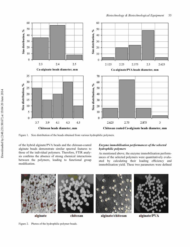
The number of the obtained polymer beads by apply-
ing various methods was approximately 250 per batch.
Out of each batch, 25 beads were used for bead-size-
distribution estimation. The diameter of each bead was
measured in different angles employing an optical
microscope (magnification 4x), and the average bead
size was calculated. The obtained results are presented
in Figure 1. As shown, the size of the Ca-alginate, and
of the Ca-alginate/PVA beads, does not differ consid-
erably, varying in the range from 2.125 to 2.625 mm.
A total of 56% from the Ca-alginate beads have an
average diameter of 2.4 mm, and 48% of the Ca-algi-
nate/PVA beads display an average diameter of
2.5 mm. The diameter of the chitosan-coated Ca-algi-
nate beads is increased to some extent (2.625–3 mm),
64% of the beads having an average diameter of
2.75 mm. As reported in the literature, chitosan bind-
ing is affected by the size of the Ca-alginate beads:
smaller beads bound more chitosan, due to their higher
surface-to-volume ratio.[11] On the other hand, the
formation of the alginate–chitosan polyelectrolyte com-
plex involves the carboxyl groups of alginate and the
amino groups of chitosan. As demonstrated by Douglas
and Tabrizian,[24] the availability of the functional
groups in stoichiometric proportion leads to the forma-
tion of smaller particles. Nevertheless, the increased
relative amount of chitosan provokes an increase in
bead size. The pure chitosan beads demonstrate notice-
ably greater sizes (3.7–4.5 mm) with an average diam-
eter of 3.7 mm for 25% of the beads and an average
diameter of 4.3 for 30% of the beads. The average of
all the measurements for each of the produced poly-
mers is 2.35, 2.45, 2.75, and 4.35 mm for the Ca-algi-
nate, the Ca-alginate/PVA, the chitosan-coated Ca-
alginate, and the chitosan beads, respectively.
The surface morphology of the beads was studied
using optical microscopy (magnification 10x). The obser-
vations demonstrated that the Ca-alginate beads, the
hybrid Ca-alginate/PVA beads, and the chitosan-coated
Ca-alginate beads have a spherical shape, slightly rough
surface, compact structure, and elastic properties. In con-
trast, the obtained chitosan beads were irregularly shaped,
with ovoid form and soft deformable texture. The corre-
sponding photos are presented in Figure 2.
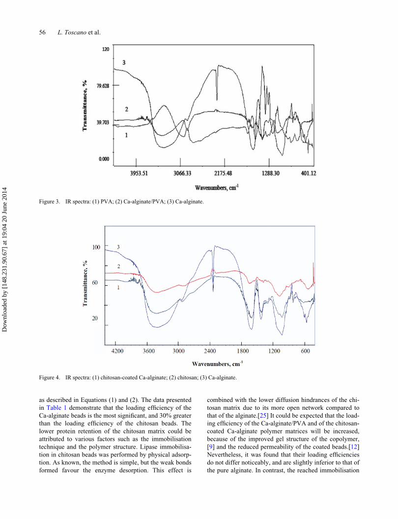
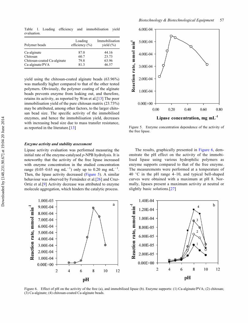
The possible interactions between the polymers form-
ing the hybrid alginate/PVA beads and the chitosan-coated
alginate beads were investigated by FTIR spectrometry. The
recorded spectra are shown in Figures 3 and 4. The bands
displayed in the Na-alginate infrared (IR) spectrum at
around 1050 and 1640 cm�1 are characteristics for the C–
O–C stretching attributed to the saccharide alginate struc-
ture, and for the asymmetrical stretching of the –COO–
groups. The chitosan spectrum shows the characteristic
absorption bands of C ¼ O and NH groups at 1633 and
1028 cm�1, respectively. The infrared (IR) spectrum of
PVA evidences the adsorption bands at 1086 and
1423 cm�1, assigned to the C–O stretch. The FTIR spectra
54 L. Toscano et al.
Dow
nloa
ded
by [
148.
231.
90.6
7] a
t 19:
04 2
0 Ju
ne 2
014
of the hybrid alginate/PVA beads and the chitosan-coated
alginate beads demonstrate similar spectral features to
those of the individual polymers. Therefore, FTIR analy-
sis confirms the absence of strong chemical interactions
between the polymers, leading to functional group
modification.
Enzyme immobilisation performances of the selected
hydrophilic polymers
As mentioned above, the enzyme immobilisation perform-
ances of the selected polymers were quantitatively evalu-
ated by calculating their loading efficiency and
immobilisation yield. These two parameters were defined
Figure 1. Size distribution of the beads obtained from various hydrophilic polymers.
Figure 2. Photos of the hydrophilic polymer beads.
Biotechnology & Biotechnological Equipment 55
Dow
nloa
ded
by [
148.
231.
90.6
7] a
t 19:
04 2
0 Ju
ne 2
014
as described in Equations (1) and (2). The data presented
in Table 1 demonstrate that the loading efficiency of the
Ca-alginate beads is the most significant, and 30% greater
than the loading efficiency of the chitosan beads. The
lower protein retention of the chitosan matrix could be
attributed to various factors such as the immobilisation
technique and the polymer structure. Lipase immobilisa-
tion in chitosan beads was performed by physical adsorp-
tion. As known, the method is simple, but the weak bonds
formed favour the enzyme desorption. This effect is
combined with the lower diffusion hindrances of the chi-
tosan matrix due to its more open network compared to
that of the alginate.[25] It could be expected that the load-
ing efficiency of the Ca-alginate/PVA and of the chitosan-
coated Ca-alginate polymer matrices will be increased,
because of the improved gel structure of the copolymer,
[9] and the reduced permeability of the coated beads.[12]
Nevertheless, it was found that their loading efficiencies
do not differ noticeably, and are slightly inferior to that of
the pure alginate. In contrast, the reached immobilisation
Figure 4. IR spectra: (1) chitosan-coated Ca-alginate; (2) chitosan; (3) Ca-alginate.
Figure 3. IR spectra: (1) PVA; (2) Ca-alginate/PVA; (3) Ca-alginate.
56 L. Toscano et al.
Dow
nloa
ded
by [
148.
231.
90.6
7] a
t 19:
04 2
0 Ju
ne 2
014
yield using the chitosan-coated alginate beads (63.96%)
was markedly higher compared to that of the other tested
polymers. Obviously, the polymer coating of the alginate
beads prevents enzyme from leaking out, and therefore,
retains its activity, as reported by Won et al.[13] The poor
immobilisation yield of the pure chitosan matrix (23.73%)
may be attributed, among other factors, to the larger chito-
san bead size. The specific activity of the immobilised
enzymes, and hence the immobilisation yield, decreases
with increasing bead size due to mass transfer resistance,
as reported in the literature.[13]
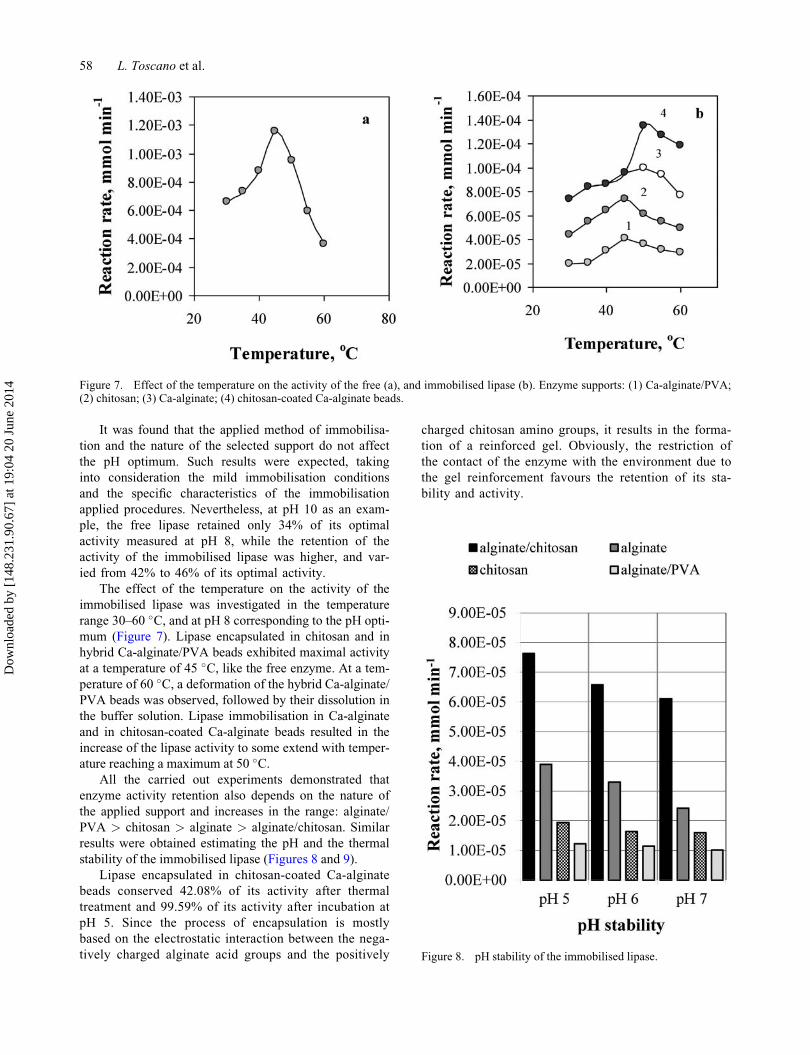
Enzyme activity and stability assessment
Lipase activity evaluation was performed measuring the
initial rate of the enzyme-catalysed p-NPB hydrolysis. It is
noteworthy that the activity of the free lipase increased
with enzyme concentration in the studied concentration
range (0.05–0.65 mg mL�1) only up to 0.20 mg mL�1.
Then, the lipase activity decreased (Figure 5). A similar
behaviour was observed by Fern�andez et al.[26] and Cruz-
Ortiz et al.[9] Activity decrease was attributed to enzyme
molecule aggregation, which hinders the catalytic process.
The results, graphically presented in Figure 6, dem-
onstrate the pH effect on the activity of the immobi-
lised lipase using various hydrophilic polymers as
enzyme supports compared to that of the free enzyme.
The measurements were performed at a temperature of
40 �C in the pH range 4–10, and typical bell-shaped
curves were obtained with a maximum at pH 8. Nor-
mally, lipases present a maximum activity at neutral or
slightly basic solutions.[27]
Table 1. Loading efficiency and immobilisation yieldevaluation.
Polymer beadsLoading
efficiency (%)Immobilisation
yield (%)
Ca-alginate 87.0 44.16Chitosan 60.7 23.73Chitosan-coated Ca-alginate 79.8 63.96Ca-alginate/PVA 81.3 46.57
Figure 5. Enzyme concentration dependence of the activity ofthe free lipase.
Figure 6. Effect of pH on the activity of the free (a), and immobilised lipase (b). Enzyme supports: (1) Ca-alginate/PVA; (2) chitosan;(3) Ca-alginate; (4) chitosan-coated Ca-alginate beads.
Biotechnology & Biotechnological Equipment 57
Dow
nloa
ded
by [
148.
231.
90.6
7] a
t 19:
04 2
0 Ju
ne 2
014
It was found that the applied method of immobilisa-
tion and the nature of the selected support do not affect
the pH optimum. Such results were expected, taking
into consideration the mild immobilisation conditions
and the specific characteristics of the immobilisation
applied procedures. Nevertheless, at pH 10 as an exam-
ple, the free lipase retained only 34% of its optimal
activity measured at pH 8, while the retention of the
activity of the immobilised lipase was higher, and var-
ied from 42% to 46% of its optimal activity.
The effect of the temperature on the activity of the
immobilised lipase was investigated in the temperature
range 30–60 �C, and at pH 8 corresponding to the pH opti-
mum (Figure 7). Lipase encapsulated in chitosan and in
hybrid Ca-alginate/PVA beads exhibited maximal activity
at a temperature of 45 �C, like the free enzyme. At a tem-
perature of 60 �C, a deformation of the hybrid Ca-alginate/
PVA beads was observed, followed by their dissolution in
the buffer solution. Lipase immobilisation in Ca-alginate
and in chitosan-coated Ca-alginate beads resulted in the
increase of the lipase activity to some extend with temper-
ature reaching a maximum at 50 �C.All the carried out experiments demonstrated that
enzyme activity retention also depends on the nature of
the applied support and increases in the range: alginate/
PVA > chitosan > alginate > alginate/chitosan. Similar
results were obtained estimating the pH and the thermal
stability of the immobilised lipase (Figures 8 and 9).
Lipase encapsulated in chitosan-coated Ca-alginate
beads conserved 42.08% of its activity after thermal
treatment and 99.59% of its activity after incubation at
pH 5. Since the process of encapsulation is mostly
based on the electrostatic interaction between the nega-
tively charged alginate acid groups and the positively
charged chitosan amino groups, it results in the forma-
tion of a reinforced gel. Obviously, the restriction of
the contact of the enzyme with the environment due to
the gel reinforcement favours the retention of its sta-
bility and activity.
Figure 7. Effect of the temperature on the activity of the free (a), and immobilised lipase (b). Enzyme supports: (1) Ca-alginate/PVA;(2) chitosan; (3) Ca-alginate; (4) chitosan-coated Ca-alginate beads.
Figure 8. pH stability of the immobilised lipase.
58 L. Toscano et al.
Dow
nloa
ded
by [
148.
231.
90.6
7] a
t 19:
04 2
0 Ju
ne 2
014

The evaluation of the storage stability of the immobi-
lised lipase demonstrated that it increases in the range:
alginate/PVA > chitosan > alginate > alginate/chitosan
(Figure 10), such as the temperature and pH stabilities.
While the free lipase lost 60% of its activity after 10 days
of storage, the immobilised lipase preserved more than
80% of its activity. It is noteworthy that the lipase encap-
sulated into chitosan-coated Ca-alginate beads conserved
89% of its activity after 30 days of storage, while the free
lipase was almost completely inactivated. Nevertheless, a
rapid drop in lipase activity was observed, using the
hybrid Ca-alginate/PVA beads as enzyme supports. This
fact could be attributed to enzyme leakage, due to PVA
dissolution.
Conclusions
The comparison of the performances of the four hydro-
philic polymers – Ca-alginate/PVA, chitosan, Ca-alginate,
and chitosan-coated Ca-alginate – demonstrated that the
chitosan-coated Ca-alginate beads could be considered as
a promising matrix for lipase immobilisation. Even if the
loading efficiency of the chitosan-coated Ca-alginate
beads was inferior to this of the Ca-alginate beads, they
demonstrated better performance characteristics in terms
of enzyme immobilisation yield, pH, thermal, and storage
stability, and enzyme activity retention compared to the
other selected polymers. The presented studies may be of
potential value for the production of esters in the reactions
of transesterification using immobilised lipases.
References
[1] Hasan F, Shah A, Hameed A. Enzyme Microb Technol.2006;39:235–251. Available from: http://dx.doi.org/10.1016/j.enzmictec.2005.10.016
[2] Houde A, Kademi A, Leblanc D. Appl Biochem Biotech-nol. 2004;118:155–170. Available from: http://dx.doi.org/10.1385/ABAB:118:1-3:155
[3] Seitz E. J Am Oil Chem Soc. 1973;51:12–16. Availablefrom: http://dx.doi.org/10.1007/BF02545206
[4] Fjerbaek L, Christensen V, Norddahl B. Biotechnol Bio-eng. 2009;102:1298–1315. Available from: http://dx.doi.org/10.1002/bit.22256
[5] D’Souza SF. Indian J Microbiol. 1989;29:83–117.[6] Stoytcheva M, Montero G, Toscano L, Gochev V, Valdez
B. In: Stoytcheva M, Montero G, editors. Biodiesel: feed-stocks and processing technologies. Croatia: InTech; 2011.p. 397–410. Available from: http://dx.doi.org/10.5772/25246
[7] Betigeri SS, Neau SH. Biomaterials. 2002;23:3627–3636.Available from: http://dx.doi.org/10.1016/S0142-9612(02)00095-9
[8] Brabcova J, Blazek J, Janska L, Krecmerova M, ZarevuckaM. Molecules. 2012;17:13813–13824. Available from:http://dx.doi.org/10.3390/molecules171213813
[9] Cruz-Ortiz BR, Rios-Gonzalez LJ, Garcia YG, Rodriguezde la Garza JA, Rodriguez-Martinez J. J Mex Chem Soc.2011;55:176–180.
[10] Dave R, Madamwar D. Indian J Biotechnol. 2006;5(Suppl):368–372.
[11] Gaserod O, Smidsrod O, Gudmund SB. Biomaterials.1998;19:1815–1825. Available from: http://dx.doi.org/10.1016/S0142-9612(98)00073-8
[12] Sun J, Tan H. Materials. 2013;6:1285–1309. Availablefrom: http://dx.doi.org/10.3390/ma6041285
[13] Won K, Ki S, Kim K-J, Park HW, Moon S-J. Process Bio-chem. 2005;40:2149–2154. Available from: http://dx.doi.org/10.1016/j.procbio.2004.08.014
[14] Anal AK, Stevens WF. Int J Pharm. 2005;290:45–54.Available from: http://dx.doi.org/10.1016/j.ijpharm.2004.11.015
[15] De S, Robinson D. J Control Release. 2003;89:101–112.Available from: http://dx.doi.org/10.1016/S0168-3659(03)00098-1
[16] George M, Abraham TE. J Control Release. 2006;114:1–14. Available from: http://dx.doi.org/10.1016/j.jconrel.2006.04.017
Figure 9. Thermal stability of the immobilised lipase.
Figure 10. Storage stability of the free and immobilised lipaseusing various supports.
Biotechnology & Biotechnological Equipment 59
Dow
nloa
ded
by [
148.
231.
90.6
7] a
t 19:
04 2
0 Ju
ne 2
014

[17] Yu CY, Zhang XC, Zhou FZ, Zhang XZ, Cheng SX, ZhuoRX. Int J Pharm. 2008;357:15–21. Available from: http://dx.doi.org/10.1016/j.ijpharm.2008.01.030
[18] Silva CM, Ribeiro AJ, Figueiredo M, Ferreira D, Veiga F.AAPS J. 2006;7:E903–E913. Available from: http://dx.doi.org/10.1208/aapsj070488
[19] Li Y, McClements DJ. Food Hydrocolloids. 2011;25:1025–1033. Available from: http://dx.doi.org/10.1016/j.foodhyd.2010.09.024
[20] Toscano L, Montero G, Cervantes L, Stoytcheva M,Gochev V, Beltran M. Biotechnol Biotechnol Equip.2013;27:3776–3781. Available from: http://dx.doi.org/10.5504/BBEQ.2012.0140
[21] Carneiro da Cunha MG, Rocha JM, Garcia FA, Gil MH.Biotechnol Tech. 1999;13:403–409. Available from: http://dx.doi.org/10.1023/A:1008913824571
[22] Huguet ML, Dellacherie E. Process Biochem.1996;31:745–751. Available from: http://dx.doi.org/10.1016/S0032-9592(96)00032-5
[23] Bradford MM. Anal Biochem. 1976;2:248–254. Avail-able from: http://dx.doi.org/10.1016/0003-2697(76)90527-3
[24] Douglas KL, Tabriziani M. J Biomater Sci Polym Ed.2005;16:43–56. Available from: http://dx.doi.org/10.1163/1568562052843339
[25] Takka S, Gurel A. AAPS PharmSciTech. 2010;11:460–466. Available from: http://dx.doi.org/10.1208/s12249-010-9406-z
[26] Fernandes M.LM, Krieger N, Baron AM, Zamora PP,Ramos LP, Mitchell DA. J Mol Catal B Enzym.2004;30:43–49. Available from: http://dx.doi.org/10.1016/j.molcatb.2004.03.004
[27] Castro-Ochoa LD, Rodriguez-Gomez C, Valerio-Alfaro G,Oliart-Ros R. Enzym Microb Technol. 2005;37:648–654.Available from: http://dx.doi.org/10.1016/j.enzmictec.2005.06.003
60 L. Toscano et al.
Dow
nloa
ded
by [
148.
231.
90.6
7] a
t 19:
04 2
0 Ju
ne 2
014
Copyright © 2022 FDOKUMEN