Biospecific fractionation matrices for sequence specific endonucleases
10
volume 5 Number 7 July 1978 Nucleic Acids Research Biospecific fractionation matrices for sequence specific endonucleases Jay George and Jack G.Chirikjian Department of Biochemistry, Georgetown University Medical Center, Georgetown University, Washington, DC 20007, USA Received 22 May 1978 ABSTRACT Fractionation of several type II specific restriction endonucleases was achieved by separation on two novel biospecific matrices. The matrices are pyran, a copolymer of divinyl ether of maleic anhydride, and Cibacron Blue F3GA, a blue dye commonly used for the calibration of molecular sieves. Both compounds are insolubllized by coupling to sepharose through a cyanogen bromide linkage and in their soluble form inhibit the restriction endo- nucleases which we have tested. These affinity matrices can be used to obtain restriction endonucleases from crude extracts after removal of nucleic acids. They have also proven to have a high capacity when used as subsequent steps in enzyme purification. Their additional advantage is the rapid development time and reusability of columns packed with the two matrices. INTRODUCTION Site specific DNA endonucleases are used in several aspects of molecular biology. Since their discovery (1), a variety of enzymes have been purified from numerous strains (2). The major task in the purification of these enzymes is to remove contamin- ating enzymes and proteins which also use DNA as a substrate. In general, these enzymes appear to be stable; however, a number of fractionation steps are routinely required to obtain enzymes at a purity level useful for such structural studies as DNA sequencing. Our recent interest has been to purify type II restriction endonucleases to homogeneity in order to study details of their interactions with naturally occuring DNAs and synthetic oligo- nucleotides containing the catalytic palindromic sites. We explored the possibility of developing matrices which could be used to purify these enzymes by biospecific affinity chroma- tography in conjunction with commonly used fractionation pro- cedures. Specifically, we considered using two matrices, pyran- 2223 © Information Retrieval Limited 1 Falconberg Court London W1V5FG England by guest on September 13, 2016 http://nar.oxfordjournals.org/ Downloaded from
-
Upload
georgetown -
Category
Documents
-
view
1 -
download
0
Transcript of Biospecific fractionation matrices for sequence specific endonucleases
volume 5 Number 7 July 1978 Nucleic Acids Research
Biospecific fractionation matrices for sequence specific endonucleases
Jay George and Jack G.Chirikjian
Department of Biochemistry, Georgetown University Medical Center, Georgetown University,Washington, DC 20007, USA
Received 22 May 1978
ABSTRACT
Fractionation of several type II specific restrictionendonucleases was achieved by separation on two novel biospecificmatrices. The matrices are pyran, a copolymer of divinyl etherof maleic anhydride, and Cibacron Blue F3GA, a blue dye commonlyused for the calibration of molecular sieves. Both compounds areinsolubllized by coupling to sepharose through a cyanogen bromidelinkage and in their soluble form inhibit the restriction endo-nucleases which we have tested. These affinity matrices can beused to obtain restriction endonucleases from crude extractsafter removal of nucleic acids. They have also proven to have ahigh capacity when used as subsequent steps in enzyme purification.Their additional advantage is the rapid development time andreusability of columns packed with the two matrices.
INTRODUCTION
Site specific DNA endonucleases are used in several aspects
of molecular biology. Since their discovery (1), a variety of
enzymes have been purified from numerous strains (2). The major
task in the purification of these enzymes is to remove contamin-
ating enzymes and proteins which also use DNA as a substrate. In
general, these enzymes appear to be stable; however, a number of
fractionation steps are routinely required to obtain enzymes at a
purity level useful for such structural studies as DNA sequencing.
Our recent interest has been to purify type II restriction
endonucleases to homogeneity in order to study details of their
interactions with naturally occuring DNAs and synthetic oligo-
nucleotides containing the catalytic palindromic sites. We
explored the possibility of developing matrices which could be
used to purify these enzymes by biospecific affinity chroma-
tography in conjunction with commonly used fractionation pro-
cedures. Specifically, we considered using two matrices, pyran-
2223© Information Retrieval Limited 1 Falconberg Court London W1V5FG England
by guest on September 13, 2016
http://nar.oxfordjournals.org/D
ownloaded from
Nucleic Acids Research
sepharose, which we had previously used to purify DNA polymerases
(3), and blue CNBr-sepharose as methods of purification. The
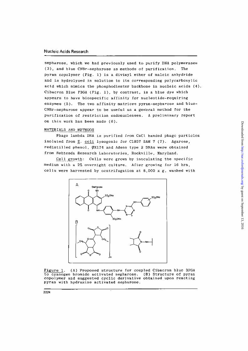
pyran copolymer (Fig. 1) is a dlvinyl ether of maleic anhydride
and is hydrolyzed in solution to its corresponding polycarboxylic
acid which mimics the phosphodiester backbone in nucleic acids (4).
Cibacron Blue F3GA (Fig. 1), by contrast, is a blue dye which
appears to have biospecific affinity fpr nucleotide-requiring
enzymes (5). The two affinity matrices pyran-sepharose and blue-
CNBr-sepharose appear to be useful as a general method for the
purification of restriction endonucleases. A preliminary report
on this work has been made (6).
MATERIALS AND METHODS
Phage lambda DNA is purified from CsCl banded phage particles
isolated from E. coli lysogenic for C1857 SAM 7 (7). Agarose,
redistilled phenol, 0X174 and Adeno type 2 DNAs were obtained
from Behtesda Research Laboratories, Rockville, Maryland.
Cell growth: Cells were grown by inoculating the specific
medium with a 2% overnight culture. After growing for 16 hrs,
cells were harvested by centrifugation at 8,000 x g, washed with
A Sepharose
0 NH
S020Na
Figure 1. (A) Proposed structure for coupled Cibacron blue 3FGAto cyanogen bromide activated sepharose. (B) Structure of pyrancopolymer and suggested cyclic derivative obtained upon reactingpyran with hydrazine activated sepharose.
2224
by guest on September 13, 2016
http://nar.oxfordjournals.org/D
ownloaded from

Nucleic Acids Research
0.9% saline and frozen at -90°C. Growth media and temperature
conditions for the different strains were as follows: Bacillus
amyloliquefaciens and Bacillus globiggi were grown on brain heart
infusion (Difco), 37 g/1 at 37°; Haemophilus influenzae Rf
was also grown on brain heart infusion supplemented with 2 ug/ml
of NAD and 5 yg/ml of Hemin; Providencia stuartii 164 was
grown on tryptone (15 g/1), yeast extract (10 g/1) and NaCl
(5 g/1) at 37°; Xanthomonas badrii was grown on nutrient broth
(8 g/1) at 32°.
Analysis of DNA endonuclease activities and digestion
products by gel electrophoresis: Assays were performed in 25 yl
volumes in either optimal buffers based on our findings or values
published by other laboratories (details for each specific
enzyme are indicated in the legends). Selection of the specific
DNAs was based on obtaining a characteristic and recognizable
fragmentation pattern. Incubations to locate activity were at
37 C for 1 hr or for 16 hrs to determine absence of contaminants.
Reactions were stopped by the addition of 5 yl of reaction stop
solution. For routine assays, gels at a concentration of 1.0-
1.4% agarose were prepared and electrophoresed in Tris (48.4 g/1),
sodium acetate (16.4 g/1), and EDTA (7.4 g/1); adjusted to
pH 7.8 with acetic acid; and run for 3 hrs at 6 volts/cm. Tube
polyacrylamide (4%) gels were run for 2 hrs at 4 mamp/gel in
Tris-borate EDTA buffer (8). Gels were stained with ethidium
bromide, and DNA visualized with short wavelength UV light
source. Photographs were obtained by the use of a red filter
(Kodak 23A) and Polaroid type 57 film.
Unit determination for type II restriction endonuclease:
Unit determination was performed with serial dilution of enzyme
using a fixed amount of substrate. A unit of enzyme is defined
as the minimum amount of enzyme which will totally degrade 1 vig
of lambda or other DNA in 60 min at 37°. Definitions such as
pmoles of phosphodiester bonds cleaved under specified conditions
and activity based on synthetic oligonucleotides containing
specific palindromes were considered as substrates. The
deciding factors in our selection of the definition parameters
were the difficulty of obtaining synthetic oligonucleotides as
well as the widely current usage of the enzymes to generate end
2225
by guest on September 13, 2016
http://nar.oxfordjournals.org/D
ownloaded from

Nucleic Acids Research
point products with DNA substrates.
Preparation of pyran-sepharose and blue CNBr-sepharose:
(a) CNBr-activated Sepharose In order to activate the
sepharose 4B (Pharmacia), 100 g was suspended in 100 ml of
distilled water and 100 mg of finely divided solid cyanogen bromide
(CNBr) was added over 5 min. The pH was adjusted and maintained
at 11 by addition of 8 N NaOH with stirring. After 10 min at
20°C, 200 ml of cold distilled water was added. The suspension
was washed and the activated sepharose was used routinely within
a few hours.
(b) Pyran-sepharose Pyran was coupled to activated sepharose
as described previously (3). Specifically, the pH was adjusted
to 10 and hydrazine was added at a concentration of 2 mM/ml
sepharose. The reaction was allowed to proceed overnight at 4
for 16 hrs. Hydrazine-activated sepharose was washed with 0.1 M
sodium acetate pH 5.0 containing 10 mM MgClg- Pyran was added,
at 3 mg/ml of hydrazine sepharose and reacted at 45 for 16 hrs.
Coupled pyran-sepharose with a characteristic off-white color was
washed with 2 M NaCl to remove free polymer. To verify that the
chemical reactions are successful, the matrix is first evaluated
by its ability to bind avian myeloblastosis virus reverse
transcriptase (3).
(c) Blue-CNBr-sepharose Cyanogen bromide-activated sepharose
was washed with excess 1 mM HC1, and Cibacron Blue F3GA was coupled
at pH 8.3 in 0.1 M NaHCOg and 0.5 M NaCl at 4° for 16 hrs. If
necessary, pH was maintained at 8.3 with 0.1 N NaOH. The ratio
of starting blue dye to swelled activated sepharose was 1 g to 10
ml. To block the remaining active groups,1 M Tris HC1 pH 9.0 was
added and incubated for 1 hr at 22°. Excess dye was removed by
several washes of acetate (Na ) pH 4.0 buffer containing 0.5 M
NaCl and a final 7 M urea wash to remove traces of unreacted blue
dye.
Storage of matrices Both affinity matrices were stored at
4 in the presence of high NaCl and 2 mM azide and will remain
stable for several years.
RESULTS
Preparation of bacterial crude extracts prior to affinity
chromatography fractionation A typical preparation utilizes 15 g
2226
by guest on September 13, 2016
http://nar.oxfordjournals.org/D
ownloaded from

Nucleic Acids Research
of prewashed bacterial cells suspended in 35 ml of Buffer A
(20 mM Tris-HCl pH 7.2, 1 mM Na2 EDTA, 7 mM 6-mercaptoethanol and
10% glycerol). Cells were at least 80% lysed by sonication using
30 second pulses for 8-10 min with intermittent off cycles. The
temperature of the slurry was maintained at 10° through the
lysing, and all procedures to follow were carried out at 4°
unless otherwise specified. The cell extract was then separated
by two sequential centrifugation steps: the first at 10,000 x g
for 15 min and the second for 100,000 x g for 2 hrs. The super-
natant (approx. 40 ml), referred to as crude extract, is re-
covered and pellets discarded. Nucleic acids are then removed by
one of several procedures, e.g., batch elution from DEAE-cellulose
under conditions where the enzyme was recovered, or by precipi-
tation with streptomycin sulfate (9) and PEG-6000 (10). Removal
of nucleic acid was found to be a prerequisite to enhancing the
capacity of the matrix to bind enzyme. The supernatant after the
removal of nucleic acid is submitted to fractionation by affinity
matrices as described in the legends.
Purification of restriction endonucleases from unfractionated
bacterial extracts Pyran-sepharose and blue-CNBr-sepharose are
effective in the separation of restriction endonucleases from
nucleic acid free bacterial extracts. We have chosen three
enzymes, Hinf 1, Pst 1, and Xba 1, obtained from three different
genera. For the initial experiments, a 1.5 ml column each of
blue-CNBr-sepharose and pyran-sepharose was equilibrated with
Buffer A. Columns were developed with a 20 ml salt gradient 0-
0.8 M in Buffer A. As shown in Figure 2A, the restriction endo-
nuclease Hinf 1 can be purified by a single pass on a blue-CNBr-
sepharose. On visual examination of digestion patterns using
excess enzyme in overnight assays, Hinf 1 obtained appeared to
be free of interfering nucleases. In the case of Pst 1 and Xba 1,
sequential chromatography over blue-CNBr-sepharose and pyran-
sepharose was needed to yield contaminant-free enzyme as shown
in Figures 2B and 2C. Salt concentration at the peak of enzyme
activity for these and other enzymes tested is summarized in
Table 1.
Usage of pyran-sepharose and Blue-CNBr-Sepharose in further
fractionating bacterial extracts In contrast to obtaining enzymes
2227
by guest on September 13, 2016
http://nar.oxfordjournals.org/D
ownloaded from

Nucleic Acids Research
B
Figure 2. Analysis of restriction endonuclease activity after fractionation on blue CNBr-sepharose and pyran-sepharose. (A) Hinf I activity from crude extracts after fractionation on blue-CNBr-sepharose. Reactions werecarried out in 7 mM Tris-HCI, pH 7.5, 10 mM MgCl2, 50 mM NaCI, 7 mM 2-mercaptoethanol, 1 fig of 0X174 DNAand enzyme. Reaction products were displayed on 4% polyacrylamide gels using Tris-borate buffer (8). (B) Pst Iactivity from extracts after sequential fractionation on blue-CNBr-sepharose and pyran-sepharose. Reactions werecarried out and displayed as described above using 1 JKJ of X DNA. Polyacrylamide 4% was employed to separatethe small DNA fragments. (C) Xba I activity after sequential fractionation by blue-CNBr-sepharose and pyran-sepharose. Reactions were carried out in 6 mM Tris-HCI pH 7.4, 6 mM MgCl2, 100 mM NaCI and 5 mMMercaptoethanol using Adeno 2 DNA. Reaction products were displayed on 1% agarose using Tris-acetate buffer.Select fractions were free of exonuclease activities. Those containing contaminating activities displayed a lightsmear. All assays were carried out at 37° for 16 hours to determine levels of exonuclease contamination. Recoveryof enzyme units could not be calculated due to interfering activities in the starting crude extracts.
2228
by guest on September 13, 2016
http://nar.oxfordjournals.org/D
ownloaded from

Nucleic Acids Research
TABLE If
Salt Concentration at Peak of Endonuclease Activity
Organism
Bacillus amyloliquefaciens
Bacillus globigii
Haemophilus influenzae R.
Providencia stuartii 164
Xanthomanus badrii
Enzy
Bam
Bgl
Hinf
Pst
Xba
me
HI
1
1
1
1
Peak of(Molar
Pyran-Sepharose
0.20
0.25
—
0.50
0.20
elutionKC1)
Blue-CNBr-Sepharose
0.45
—
0.30
0.30
0.25
free from contaminating nucleases, purification of restriction
endonucleases to homogeneity has been a relatively difficult
task. As part of our effort in pursuing structural studies on
Bam HI (11) and Bgl 1 (12), we have been involved in purifying these
two enzymes to homogeniety. We have investigated the feasibility
of the two matrices in such purification schemes. Both Bam HI
and Bgl 1 bound to pyran-sepharose (Figure 3) and to blue-CNBr-
sepharose (data not shown). In both cases, 2,000-6,000 units
were bound by 1 ml swelled pyran-sepharose and blue-CNBr-sepharose.
DISCUSSION
A variety of biological materials have been fractionated by
affinity chromatography. This approach takes advantage of
biospecific interactions between molecules not offered by con-
ventional fractionation methods. A major use of affinity
chromatography has been in the purification of enzymes. With
substrates, cofactors or biospecific macromolecules known to have
an inhibitory effect, enzymes can be concentrated selectively and
then eluted by gradients containing substrates or salt. The
advantages offered by such a procedure are speed of purification
and potential protection against denaturation during fraction-
ation procedures.
The affinity matrices we investigated in some detail are
blue-CNBr-sepharose and pyran-aepharose. The capacity for both
2229
by guest on September 13, 2016
http://nar.oxfordjournals.org/D
ownloaded from

Nucleic Acids Research
Figure 3. Fractional;ion of purified Bam HI and Bgl I on pyran-sepharose. Pooled samples of Bam HI and Bgl I which had previouslybeen fractionated by phosphocellulose and hydroxylapatite weresubmitted to fractionation on pyran-sepharose. (A) Bam HI(pH 7.5), 7 mM MgCl2, 7 mM 2-mercaptoethanol and 1 yg of DNA in atotal incubation mixture of 50 yl. (B) Bgl I digest of X DNA.Reaction mixture contained 20 mM Tris-HCl (pH 7.4), 7 mM MgCl2,7 mM 2-mercaptoethanol, 100 yg/ml gelatin. All assays were carriedout at 37°C for 15 minutes. DNA digestion products for bothenzymes were displayed on 1.4% agarose gels as described in thetext.
matrices is satisfactory although they vary ten-fold when
partially purified enzyme is further fractionated on the matrices.
Thus, 2,000-6,000 units of enzyme binding per gram of swelled
affinity matrix is the obtained capacity as opposed to 200-600
units per gram for the crude starting material. Cibacron Blue
2230
by guest on September 13, 2016
http://nar.oxfordjournals.org/D
ownloaded from

Nucleic Acids Research
F3GA has also been covalently coupled by the triazine method (14)
and has been used extensively to purify a variety of nucleotide-
binding enzymes. Several reports (13,14) show kinases and
dehydrogenases to bind to this matrix and to be eluted with
substrates. These observations strongly suggest the binding to
be affinity in nature (13). The blue-CNBr-sepharose we describe
in this paper, unlike that coupled by the triazine method (14),
is coupled through the amino group of the blue chromaphore
(Fig. 1A). Currently available commerical preparations were not
as effective for restriction endonuclease purification. It is not
clear at this time whether the difference is due to coupling site
or potential difference in the amount of coupled dye to sepharose.
The exact linkage between pyran and sepharose has not been
investigated, however, the proposed linkage (3) is shown in Fig.
IB. We previously have reported pyran-sepharose to be a bio-
specific affinity matrix for the purification of DNA polymerases
(3). Since pyran mimics the phosphodiester backbone of nucleic
acid, it is not surprising that it can be used to fractionate
restriction endonucleases. A recent report has also described an
affinity fractionation of restriction endonuclease using heparin-
sepharose (15). In our hands heparin-sepharose proved to be
ineffective when relatively pure samples of Bam HI preparations
were submitted to it. The method we have described appears to be
readily applicable to a variety of restriction endonucleases
(Table I). The stability and rapid development time clearly
offer distinct advantages to be exploited in purifying restric-
tion endonucleases.
ACKNOWLEDGMENTS
This investigation was supported by Grant No. CA 16914,
Awarded by the National Cancer Institute, DHEW. J.G.C. is a
Scholar of the Leukemia Society of America, Inc.
REFERENCES
1. Kelley, T.J. and Smith, H.O. (1970) J. Mol. Biol. 51, 393-409.2. Roberts, R.J. (1976) CRC Critical Reviews in Biochemistry 4,
123-164.3. Chirikjian, J.G., Rye, L. and Papas, T.S. (1975) Proc. Natl.
Acad. Sci. 72, 1142-1146.4. Papas, T.S., Pry, T. and Chirigos, M. (1974) Proc. Natl.
Acad. 71, 367-370.
2231
by guest on September 13, 2016
http://nar.oxfordjournals.org/D
ownloaded from

Nucleic Acids Research
5. Easterday, R.L. and Easterday, I.M., _in: ImmobilizedBiochemicals and Affinity Chromatography, pp. 123-133(Dunlap, R.B., ed.) Plenum Publishing Corporation, New York(1974).
6. Chirikjian, J.G., George, J. and Smith, L.A. (1978) Fed. Proc.37, 1415.
7. Schrenk, W.J. and Weisberg, R.A. (1975) Molec. Gen. Genet.137, 101-107.
8. Blakesley, R.W., Dodgson, J.B., Nes, I.F. and Wells, R.D.(1977) J. Biol. Chem. 252, 7350-7306.
9. Modrich, P., Zabel, D. (1976) J. Biol. Chem. 251, 5866-5876.10. Alberts, B., Herrick, G. (1971) Methods in Enzymology pp. 148-
217, (Grossman and Moldavck, Eds.) Vol XXI.11. Smith, L.A. and Chirikjian, J.G. (manuscript in Preparation).12. Lee, Y., Blakesley, R.W., Smith, L.A. and Chirikjian, J.G.
(1978) Nucleic Acid Research 65, 674-689.13. Deibel, R. and Ives, H.D. (1977) J. Biol. Chem. 252, 8234-
8239.14. Stellwagen, E. (1977) Ace. of Chem. Res. 10, 92-98.15. Bickle, T.A., Pirrotta, V. and Imber T. (1977) Nucleic Acid
Research 4, 2561-2572.
2232
by guest on September 13, 2016
http://nar.oxfordjournals.org/D
ownloaded from