Surface-enhanced Raman spectra of magnetic nanoparticles adsorbed on a silver electrode
Upload
independentCategory
view
0download
0
J. Chitin Chitosan 16(1), 7-14 (2011)
7
Http://www.chitosan.or.kr
Biomedical and Environmental Applications of Chitosan-based Nanomaterials
Lam Dai Tran1,*, Hoang Vinh Tran
2, Trang Thu Mai
1, Thu Phuong Ha
1, Binh Hai Nguyen
1, Hoang Thai
3,
Hoang Dinh Vu2, Dien Gia Pham
4, Phuc Xuan Nguyen
1, and Jae Kweon Park
5,**
1Institute of Materials Science, Vietnam Academy of Science and Technology, 18 Hoang Quoc Viet, Hanoi, Viet Nam
2Faculty of Chemical Technology, Hanoi University of Technology, 1 Dai Co Viet, Hanoi, Vietnam
3Institute of Tropical Technology, Vietnam Academy of Science and Technology, 18 Hoang Quoc Viet, Hanoi, Viet Nam
4Institute of Chemistry, Vietnam Academy of Science and Technology, 18 Hoang Quoc Viet, Hanoi, Viet Nam
5Department of Pharmaceutical Science, Gachon University of Medicine and Science, Yeonsu-dong, Yeonsu-gu, InCheon, Korea
ABSTRACT
Being naturally abundant resources and having many interesting physicochemical and biological properties, chitin/chitosan
have been found useful in many fields. This paper describes the strategy to design a multifunctional, chitosan based
nanomaterials and their biomedical and environmental applications. Different physicochemical methods including FESEM/
TEM, PPMS were used to characterize the obtained nanomaterials. For each application, a series of specific characterizing
methods were used for evaluating the applicability/capacity of materials.
Keywords: biomedical, environment, chitosan, nanmaterials
INTRODUCTION
Chitin, a natural polysaccharide and first identified in 1884,
is the most abundant polymer after cellulose. The most
important derivative of chitin is chitosan (CS), obtained by
deacetylation of chitin in the solid state under alkaline
conditions, resulting in a heterogeneous distribution of acetyl
groups along its chains, or by enzymatic hydrolysis in the
presence of chitin deacetylase (Fig. 1). A wide variety of
biomedical applications in tissue engineering (1, 2), wound
healing (3), drug and gene delivery (1, 4-6) was based on
biocompatible, biodegradable and non-toxic CS. This paper
demonstrates synthetic strategies, characterizations and the
most important biomedical and environmental applications of
chitosan-based nanoparticles and nanocomposites.
MATERIALS AND METHODS
Preparation of glucosamine sulphate
All the chemicals were of reagent grade used without
further purification. Ferric chloride hexa-hydrate (FeCl36H2O),
ferrous chloride tetra-hydrate (FeCl24H2O) and sodium
hydroxide (NaOH) were purchased from Aldrich. Aluminium
sulfate octadecahydrate (Al2(SO4)3.18H2O), copper(II) sulfate
pentahydrate (CuSO4.5H2O), cobalt(II) chloride hexahydrate
(CoCl2.6H2O), cadmium chloride hemipentahydrate (CdCl2.
2.5H2O), lead(II) nitrate (Pb(NO3)2), Nickel(II) sulfate
hexahydrate (NiSO4.6H2O),4-(2-Pyridylazo)resorcinol (PAR)
were purchased from Acros.
NaOH, NH4OH (26% of ammonia), oleic acid (C17H33
COOH) were purchased from Aldrich. Chitin, chitosan was
purchased from Nha Trang Aquatic Institute (Vietnam),
OMSCS was from Aldrich. Glucosamine hydrochloride was
obtained by acidic hydrolysis of chitin (with concentrated
HCl) at 60~70oC. Glucosamine hydrochloride could convert
into glucosamine sulphate with the help of Na2SO4 under
room temperature (4).
Biocompatibility of the materials
In vitro biocompatibility of the materials was tested in
simulated body fluid (SBF) at 37oC, for period of 0 to 7 days.
SBF solutions have ionic concentrations similar to those of
human blood plasma. SBF solutions were prepared according
to the detailed protocol given elsewhere and denoted as 1x
SBF and 5x SBF, corresponding to 1-fold and 5- fold
concentrations respectively. pH of solution were buffered at
the value of 7.4 using 0.01 M of tris-(hydroxymethyl)-
aminomethane (CH2OH)3CNH2). Curcumin (1,7-bis(4-hydroxy-
3-methoxyphenyl)-1,6-heptadiene-3,5-dione) was from Institute
of Chemistry (Vietnam). Cells were cultured in RPMI 1640
(Roswell Park Memorial Institute) (Gibco) medium. This
medium was supplemented with 10% Fetal Bovine Serum
(Invitrogen), 100 IU/ml penicillin-streptomycine (Invitrogen),
2 mM – Glutamine (Invitrogen). Cells were grown in a
humidified chamber in the presence of 5% CO2, at 37oC.
*To whom all correspondence should be addressed
Tel: +84-4-3756-4129; Fax: +84-4-3836-0705; E-mail: [email protected]
**Co-corresponding author. Tel. +82-32-820-4781; E-mail : [email protected]

8 Biomedical and Environmental Applications of Chitosan-based Nanomaterials
Evaluation of macrophage-colony stimulating factor
Human buffycoat was obtained from National Institute
of Hematology and Transfusion (Vietnam). Mononuclear
cells were isolated by density gradient centrifugation using
1.077 g/ml Ficoll. Cells were cultured in RPMI 1640 medium
with 1 µg/ml HGM- CSF (human granulocyte macrophage-
colony stimulating factor) (MP Biomedicals). 7 to 12 week
old Swiss mice were obtained from National Institute of
Hygiene and Epidemiology (Vietnam). Human monocytes or
mouse primary peritoneal macrophages were grown for 24 h
on glass coverslips. 106 cells were incubated with 0.05 mg
MNPs for 2-15 h, then treated with either anti-human CD14
antibody (Bio Legend) or actins antibody (Invitrogen) for
taking LSCM images.
FT-IR Spectrometer analysis
Infra red (IR) spectra were recorded with Nicolet 6700 FT-
IR Spectrometer, using KBr pellets, in the region of 400-
4000 cm-1, with resolution of 4 cm
-1. Field Emission Scanning
Electron Microscope (FE-SEM) and Transmission Electron
Microscope (TEM) images was analyzed by Hitachi S-4800
and JEM-1200EX (Voltage:100kV, magnification X200,000),
respectively. Dynamic light scattering (DLS) was analyzed
with Zetasizer 2000 instrument (Malvern, UK).
Confocal microscope analysis
Laser Scanning Confocal Microscope (LSCM) images with
excitation light of 488nm were collected with use of a ZEISS
510 LSCM with a 20x or 40x or 63x oil immersion objectives.
The magnetic properties were measured using Physical Properties
Measurement System (PPMS) from Quantum Design at fields
ranging from 20 to 20 kOe at 25oC, with accuracy of 10
-5emu.
RESULTS AND DISCUSSION
Synthesis of glucosamine sulfate sodium chloride
Glucosamine, an amino monosaccharide derived from
chitin, has been demonstrated to be useful and effective in
osteoarthritic therapy for nearly 40 years (7-10). Among its
different derivatives including glucosamine hydrochloride,
and N-acetyl-glucosamine, glucosamine sulfate (11), glucosamine
sulfate is considered to be one of the most effective one (12).
The synthesis of Glucosamine sulfate sodium chloride (Glu-
) from chitin was performed (Fig. 2). FTIR, 1H–NMR,
13C–NMR and HPLC spectra (shown in Fig. 3) confirmed that
synthesized (Glu- ) matched well with the standard
sample. Its purity, quality and other characteristics with
respect to United State Pharmacopoeia 26 were also fulfilled.
Synthesis of chitosan/hydroxyapatite for tissue engineering
The biomaterial Hydroxyapatite (HAp) has attracted many
scientists thanks to its excellent physicochemical (structure,
composition) and biological properties (bioactivity, biocompatibility,
biodegradability). However, its mechanical (micro hardness, tensile)
properties do not meet those of nature scaffolds. To overcome this
limitation, HAp was combined with CS to improve mechanical
properties of the resulting composites (13, 14). The HAp powder,
first synthesized by coprecipitation of Ca2+
and HPO4
2-, then
was immersed in chitosan solution at the weight ratio of 1 : 1,
and finally dried to obtain the composite. To investigate its
bioactivities, HAp/CS composites were submerged in simulated
body fluid (SBF) at different testing periods. XRD spectra in Fig.
4 show that the crystality of HAp/CS, manifested in its typical
peak of 211, increased gradually. FESEM images in Fig. 5 also
confirmed that the HAp/CS has needle shapes, uniform
dimensions and homogeneous dispersion after 5 and 7 days,
whereas it still agglomerated at the very few days.
SO4
2 –
SO4
2 –
Fig. 1. Structure of chitin and chitosan.

Lam Dai Tran et al. 9
Antibacterial and proliferative activities of silver/
chitosan nanoparticles
Silver nanoparticles (AgNPs), including ionic silver (Ag+)
or metallic silver (Ago), were shown to have good antibacterial
and proliferative activities. Numerous methods have been
proposed and reported in literature (15-19). In our study,
AgNPs were prepared by reducing silver from silver salt
solutions using chitosan as both reducing and stabilizing
agent. It can be observed clearly that core-shell structure of
AgNPs/CS was successfully obtained (composite diameter
was about 12 nm, whereas the structural coating shell of
Chitosan was about 2 nm). The investigation of AgNPs/CS
into antibacterial activities was conducted with six bacterial
lines, including Lactobacillus feremetum, Bacillius subtillis,
Staphylococcus aureus, Samonella enterica, Escherichia coli,
Pseudomonas aeruginosa, and one fungal line named Candida
albicans (Table 1) using the multimicrodilution assay. Figure
6 and Table 2 show the bactericidal effects of AgNPs/CS
samples (SK-W, SO-W and SA-W, corresponding to 60, 85,
900C of AgNPs synthesis respectively) against Bacillius
subtillis and Pseudomonas aeruginosa along with the testing
conditions (Table 2). It is obvious that the AgNPs have
significant effect on controlling the growth of Bacillius
subtillis and Pseudomonas aeruginosa. It can also be observed
in Table 1 that the half maximal inhibitory concentration
(IC50), the minimum inhibitory concentration (MIC) and the
minimum bactericidal concentration (MBC) values of SO-W,
SA-W and SK-W samples against bacteria and fungi lines
Fig. 2. Chemical synthesis of glucosamine-Cl- and glucosamine-SO4
2- from chitin.
Fig. 3. HPLC curves of synthesized (solid line) and standard
(dot line) glucosamine-SO4
2- .
Fig. 4. XRD patterns of CS/Hap in simulated body fluid after: 0,
1, 3, 5, 7 days.

10 Biomedical and Environmental Applications of Chitosan-based Nanomaterials
were achieved as promising ones. In our study, AgNPs/CS
exerted a bactericidal effect against all bacterial strains of
negative gram (S. enterica, E. coli and P. aeruginosa), positive
gram (L. fermentum, S. aureus and B. subtilis) and yeast (C.
albicans). To our best understanding so far, we have achieved
the surprisingly low number (1.5-6 g/ml, except for S.
enterica) with respect to IC50, in comparison to the best result,
recently reported in literature (20), which studied on 25 nm
AgNPs using Maltose as reducing agent.
Drug delivery chitosan-based system
Unique physicochemical and biological characteristics of
CS make it suitable for drug delivery systems. The strategy to
design a multifunctional, nanosized magnetofluorescent water-
dispersible Fe3O4-curcumin conjugates (those diameters was less
than 500 nm) and its multiple ability to label, target and treat
the tumor cells was developed. The conjugate possesses
magnetic nano Fe3O4 core, CS as outer shell and entrapped
curcumin (Cur, an anti-oxidant, anti-inflammatory and anti-
tumor substance), serving dual function of naturally
autofluorescent dye as well as antitumor model drug, delivered
to the cells with the help of macrophage whose role is to
phagocytise (engulf and then digest) cellular debris and
pathogens either as stationary or as mobile cells, and to
stimulate lymphocytes and other immune cells to respond to
the pathogen. Primary peritoneal macrophages isolation was
described in details elsewhere (21). Hence, it can be used as potential
vehicles for transport of MNPs into the core of tumor cells.
Fig. 5. FE-SEM images of CS/HAp in simulated body fluid after: 0, 1, 5, 7 days.
Fig. 6. Microplates (96-wells) image of testing samples against Bacilius Subtilis (a) and Psudomonas Aeugunosa (b).
Lam Dai Tran et al. 11
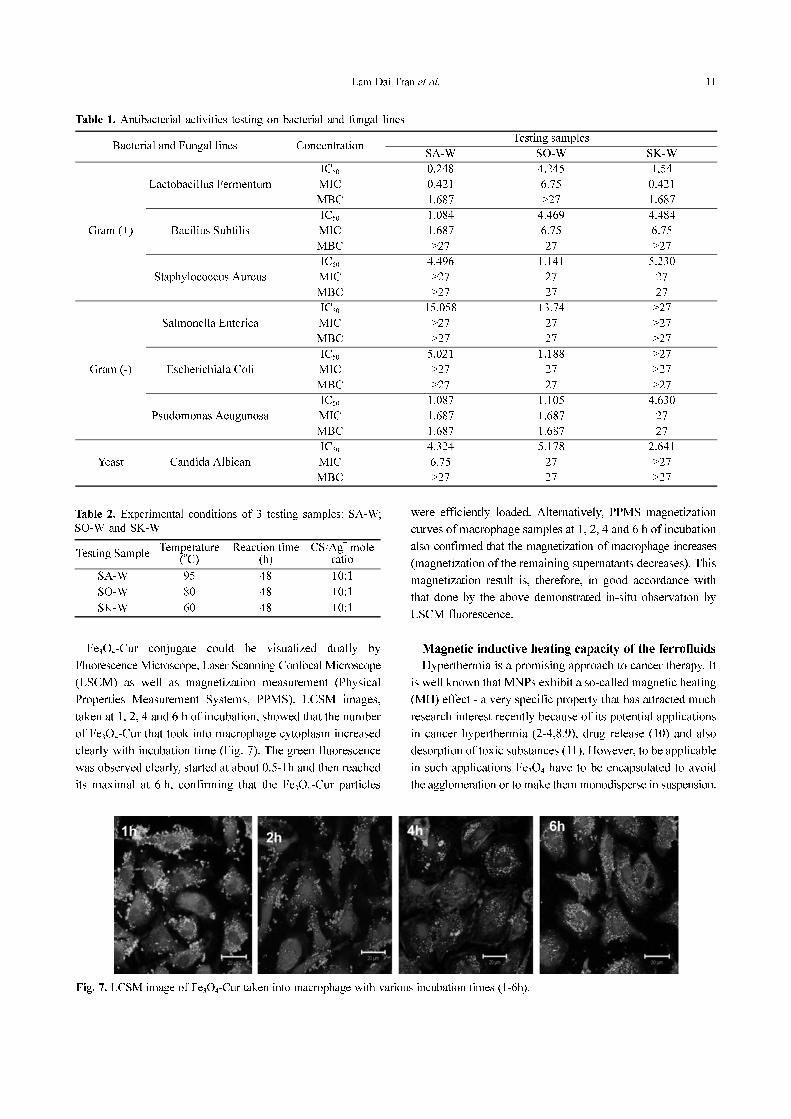
Fe3O4-Cur conjugate could be visualized dually by
Fluorescence Microscope, Laser Scanning Confocal Microscope
(LSCM) as well as magnetization measurement (Physical
Properties Measurement Systems, PPMS). LCSM images,
taken at 1, 2, 4 and 6 h of incubation, showed that the number
of Fe3O4-Cur that took into macrophage cytoplasm increased
clearly with incubation time (Fig. 7). The green fluorescence
was observed clearly, started at about 0.5-1h and then reached
its maximal at 6 h, confirming that the Fe3O4-Cur particles
were efficiently loaded. Alternatively, PPMS magnetization
curves of macrophage samples at 1, 2, 4 and 6 h of incubation
also confirmed that the magnetization of macrophage increases
(magnetization of the remaining supernatants decreases). This
magnetization result is, therefore, in good accordance with
that done by the above demonstrated in-situ observation by
LSCM fluorescence.
Magnetic inductive heating capacity of the ferrofluids
Hyperthermia is a promising approach to cancer therapy. It
is well known that MNPs exhibit a so-called magnetic heating
(MH) effect - a very specific property that has attracted much
research interest recently because of its potential applications
in cancer hyperthermia (2-4,8,9), drug release (10) and also
desorption of toxic substances (11). However, to be applicable
in such applications Fe3O4 have to be encapsulated to avoid
the agglomeration or to make them monodisperse in suspension.
Table 1. Antibacterial activities testing on bacterial and fungal lines
Bacterial and Fungal lines ConcentrationTesting samples
SA-W SO-W SK-W
Gram (+)
Lactobacillus Fermentum
IC50 0.248 4.245 1,54
MIC 0.421 6.75 0.421
MBC 1.687 >27 1.687
Bacilius Subtilis
IC50 1.084 4.469 4.484
MIC 1.687 6.75 6.75
MBC >27 27 >27
Staphylococcus Aureus
IC50 4.496 1.141 5.230
MIC >27 27 27
MBC >27 27 27
Gram (-)
Salmonella Enterica
IC50 15.058 13.74 >27
MIC >27 27 >27
MBC >27 27 >27
Escherichiala Coli
IC50 5.021 1.188 >27
MIC >27 27 >27
MBC >27 27 >27
Psudomonas Aeugunosa
IC50 1.087 1.105 4.630
MIC 1.687 1.687 27
MBC 1.687 1.687 27
Yeast Candida Albican
IC50 4.324 5.178 2.641
MIC 6.75 27 >27
MBC >27 27 >27
Table 2. Experimental conditions of 3 testing samples: SA-W;
SO-W and SK-W
Testing SampleTemperature
(oC)
Reaction time(h)
CS/Ag+ mole
ratio
SA-W 95 48 10:1
SO-W 80 48 10:1
SK-W 60 48 10:1
Fig. 7. LCSM image of Fe3O4-Cur taken into macrophage with various incubation times (1-6h).

12 Biomedical and Environmental Applications of Chitosan-based Nanomaterials
Therefore, it is essential to modify the surface of these particles
to increase the stability by surfactants, among which
biocompatible polymers like CS and its derivatives (eg, O-
carboxymethylchitosan, OCMCS) are the most potential
candidates owing to its biocompatibility and biodegradability.
Figure 8 presents FE-SEM images of CS/Fe3O4 and
OCMCS/Fe3O4 nanoparticles. Accordingly, for each samples,
the heating curve measurements was carried out under the
same AC field conditions (frequency of 236 kHz and
amplitude of 80Oe, Fig. 9). OCMCS has the backbone
structure similar to CS but hydroxyl group (-OH) is substituted
by carboxyl group (-COOH). This substitution helps increasing
the ability to disperse in neutral and alkaline solution of
OCMCS, and thus broadening their biomedical applications
(22, 23). The so called saturation temperature for suspensions
of the magnetite NPs modified by CS and OCMCS are
gathered in Table 3. Comparing heating curves for the Fe3O4-
CS and Fe3O4-OCMCS magnetic fluids with the same
concentration (10 mg/ml), it can be seen that the saturation
heating temperatures are different. In the concentration range
from 0.5 to 1 mg/ml, the heating capacity of Fe3O4/CS is
better than that of Fe3O4/OCMCS, when the concentration is
below 0.5 mg/ml the saturation temperature of Fe3O4-
OCMCS is higher. This might be explained as because of the
fact that CS is not dissolvable in aqueous solution. So when
reducing the concentration, CS will precipitate, the stability of
magnetic fluid decrease and this also lessen the saturation heating
temperature. And therefore, the applications of CS in biomedicine
fields are limited despite of its higher hyperthermia ability.
Heavy metal ions removal
Apart from the above mentioned applications, CS and its
derivatives can be used for Heavy metal ions removal. CS has
excellent properties for the adsorption of metal ions, principally
due to the presence of amino groups (–NH2) in the polymer
matrix, which can interact with metal ions in solution by ion
exchange and complexation reactions (11). The high content of
amino groups also makes possible many chemical modifications
in polymer with the purpose of improving selectivity and
adsorption capacity.
In our group, chitosan/magnetite nanocomposite beads
Fig. 8. FESEM images of unmodified (a), CS (b) and OCMCS (c) modified Fe3O4 particles.
Fig. 9. Magnetic heating curves measured for suspensions of OCMCS-modified (a) and CS-modified (b) Fe3O4 NPs.
Table 3. Magnetic heating parameters
NoConcentration
(mg/ml)
Saturation temperature (Ts, oC )
CS – Fe3O4 OCMCS – Fe3O4
1 1 > 100 98
2 0.7 > 98 90
3 0.5 90 78.5
4 0.3 59 72
5 0.2 53.3 60.7
Lam Dai Tran et al. 13
were prepared, characterized and used for removal of toxic
metal ions such as Pb(II), Ni(II) in the pH range from 4 to 6.
Langmuir isotherms were used to analyze the equilibrium
data at different pH. These nanocomposite beads can be
removed easily from water with the help of an external
magnet thanks to their exceptional magnetic properties (24).
Further, to increase the adsorption capacity, Fe3O4/Al(OH)3/
CS was designed by co-precipitation of Fe2+
, Fe3+
and Al3+
in
CS solution, using concentrated alkaline solution as a
precipitation agent. Al(OH)3 is expected to form intercapillary
structure and thus to synergize the adsorbability of Fe3O4/
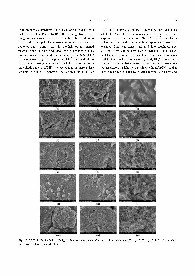
Al(OH)3/CS composite. Figure 10 shows the FESEM images
of Fe3O4/Al(OH)3/CS nanocomposites before and after
exposure to heavy metal ion (Ni2+
, Pb2+
, Cd2+
and Cu2+
)
solutions, clearly indicating that the morphology of materials
changed from smoothness and fold into roughness and
swelling. This change brings an evidence that that heavy
metal ions were efficiently adsorbed (as in metal complexes
with Chitosan) onto the surface of Fe3O4/Al(OH)3/CS composite.
It should be noted that saturation magnetization of nanocom-
posites decreases slightly, even with or without Al(OH)3, so that
they can be manipulated by external magnet to retrieve and
Fig. 10. FESEM of CS/MNPs/Al(OH)3 surface before (a-c) and after adsorption metals ions: Co2+
(d-f); Cu2+
(g-i); Pb2+
(j-l) and Cd2+
(m-o) with different magnification.
14 Biomedical and Environmental Applications of Chitosan-based Nanomaterials
reuse afterwards. In conclusion, some CS based nanomaterials
have been successfully synthesized and used in biomedical
and environmental applications.
ACKNOWLEDGEMENTS
The authors are grateful for the financial support for this
work by Korean-Vietnamese joint research project (2010-
2011, code 59/2615/2010/HD-NDT).
REFERENCES
1. Jayakumar, R., Deepthy Menon, K., Manzoor, S. V. Nair,
and Tamura, N.: Biomedical applications of chitin and
chitosan based nanomaterials-A short review. Carbohyd. Poly.,
2010, 82, 227-232
2. Madhumathi, K., Binulal, N.S., Nagahama, H., Tamura, H.,
Shalumon, K.T., Selvamurugan, N., Nair, S.V., and Jayakumar,
R.: Preparation and characterization of novel β-chitin-
hydroxyapatite composite membranes for tissue engineering
applications. Int.J. Biol.Macromol., 2009, 44, 1-5
3. Jayakumar, R., Prabaharan, M., Reis, R.L., and Mano, J.F.:
Graft copolymerized chitosan-present status and applications.
Carbohyd. Poly., 2005, 62, 142-158.
4. Jayakumar, R., Nwe, N., Tokura, S., and Tamura, H.: Sulfated
chitin and chitosan as novel biomaterials. Int.J. Biol.
Macromol., 2007, 40, 175-181
5. Borchard, G.: Chitosans for gene delivery, Adv. Drug
Delivery Reviews (Elsevier), 2001, 52, 145-150
6. Jayakumar, R., Chennazhi, K.P., Muzzarelli, R.A.A., Tamura,
H., Nair, S.V., and Selvamurugan, N.: Chitosan conjugated
DNA nanoparticles in gene therapy. Carbohyd. Poly., 2010,
79, 1-8
7. Bruyere, O., Pavelka, K., Rovati, L.C., Gatterova, J.,
Giacovelli, G., and Olejarova, M.: Total joint replacement
after glucosamine sulphate treatment in knee osteoarthritis:
results of a mean 8-year observation of patients from two
previous 3-year, randomised, placebo-controlled trials.
Osteoarth.Cartil., 2008, 16, 254-260
8. Pavelka, K., Gatterova, J., Olejarova, M., Machacek, S.,
Giacovelli, G., and Rovati, L.C.: Glucosamine sulfate use
and delay of progression of knee osteoarthritis: a 3-year,
randomized, placebo-controlled, double-blind study. Archiv.
Internal Med., 2002, 162, 2113-2123
9. Reginster, J.Y, Deroisy, R., Rovati, L.C., Lee, R.L., Lejeune,
E., and Bruyere, O.: Long-term effects of glucosamine
sulphate on osteoarthritis progression: a randomised, placebo-
controlled clinical trial. The Lancet, 2001, 357, 251-256.
10. Vangsness, C.T. Jr., Spiker, W., and Erickson, J.: A review of
evidence-based medicine for glucosamine and chondroitin
sulfate use in knee osteoarthritis, Arthroscopy: J. Arthroscop.
Related Surg., 2009, 25,86-94
11. Anderson, J.W., Nicolosi, R.J., and Borzelleca, J.F.: Glucosamine
eects in humans: a review of eects on glucose metabolism,
side eects, safety considerations and ecacy. Food Chem.
Toxicol., 2005, 43, 187-201
12. Persiani, S., Roda, E., Rovati, L.C., Locatelli, M., Giacovelli,
G., and Roda, A.: Glucosamine oral bioavailability and
plasma pharmacokinetics after increasing doses of crystalline
glucosamine sulfate in man. Osteoarth.Cartil., 2005, 13,
1041-1049
13. Rogers, K. D., Etok, S.E., and Scott, R.: Structural
characterisation of apatite coatings. J. Mat. Sci., 2004, 39,
5757-5754
14. Pang, X. and Igor Zhitomirsky, I.: Electrophoretic deposition
of composite hydroxyapatite-chitosan coatings. Mat. Character.,
2007, 58, 339-348
15. Wei, D., Sun, W., Qian, W., Ye, Y., and Ma, X.: The
synthesis of chitosan-based silver nanoparticles and their
antibacterial activity. Carbohyd. Res., 2009, 344, 2375-2382
16. Virender, K., Sharma, K., Yngard, R.A., and Lin, Y.: Silver
nanoparticles: Green synthesis and their antimicrobial activities.
Adv. Colloid. Interface Sci., 2009, 145, 83-96
17. Ghosh, S., Kaushik, R., Nagalakshmi, K., Hoti, Menezes,
G.A., Harish, B.N., and Vasan, H.N.: Antimicrobial activity
of highly stable silver nanoparticles embedded in agar-agar
matrix as a thin film. Carbohyd. Res., 2010, 345, 2220-2227
18. Seo, Y.I., Hong, K.H., Dae-Gun Kim, D.G., and Kim, Y.D.:
Ag/Al(OH)3 mesoporous nanocomposite film as antibacterial
agent, colloids and surfaces B. Biointerfaces, 2010, 81, 369-373
19. Virender, K., Sharma, K., Yngard, R.A., and Yekaterina Lin,
Y.: Silver nanoparticles: Green synthesis and their antimicrobial
activities. Adv. Colloid. Interface Sci., 2009, 145, 83-96
20. Tran, H.V., Tran, L.D., Ba, C.T., Vu, H.D., Nguyen, T.N.,
Pham, D.G., and Nguyen, P.X.: Synthesis, characterization,
antibacterial and antiproliferative activities of monodisperse
chitosan-based silver nano particles, colloids and surfaces A.
Physicochem. Engin. Aspects, 2010, 360, 32-40
21. Tran, L.D., Hoang, N.M.T., Mai, T.T., Tran, H.V., Nguyen,
N.T., Tran, T.D., Do, M.H., Nguyen, Q.T., Pham, D.G., Ha, T.P.,
Le, H.V., and Nguyen, P.X.: Nanosized magnetofluorescent
Fe3O4-curcumin conjugate for multimodal monitoring and
drug targeting, colloids and surfaces A. Physicochem. Engin.
Aspects, 2010, 371, 104-112
22. Zhu, A., Yuan, L., and Liao, T.: Suspension of Fe3O4 nanoparticles
stabilized by chitosan and o-carboxymethylchitosan. Int. J.
Pharmaceut., 2008, 350, 361-368
23. Elsabee, M.Z., Morsi, R.E., and Al-Sabagh, A.M.: Surface
active properties of chitosan and its derivatives, colloids and
surfaces B. Biointerfaces, 2009, 74, 1-16
24. Tran, H.V., Tran, L.D., and Nguyen, T.N.: Preparation of
chitosan/magnetite composite beads and their application for
removal of Pb(II) and Ni(II) from aqueous solution. Mat.
Sci. Engin. C, 2010, 30, 304-310
Received December 1, 2010
Revised December 25, 2010
Accepted December 30, 2010
Copyright © 2022 FDOKUMEN