Biomarker analysis of microbial diversity in sediments of a saline groundwater seep of Salt Basin,...
20
Biomarker analysis of microbial diversity in sediments of a saline groundwater seep of Salt Basin, Nebraska Jiasong Fang a, * , Olivia Chan a , R.M. Joeckel b , Yongsong Huang c , Yi Wang c , Dennis A. Bazylinski d , Thomas B. Moorman e , Barbara J. Ang Clement f a Department of Geological and Atmospheric Sciences, Iowa State University, Ames, IA 50011, United States b Conservation and Survey Division, School of Natural Resources, University of Nebraska, Lincoln, NE 68588-0517, United States c Department of Geological Sciences, Brown University, 324 Brook Street, Providence, RI 02912, United States d Department of Biochemistry, Biophysics, and Molecular Biology Iowa State University, Ames, IA 50011, United States e USDA/ARS National Soil Tilth Laboratory, 2150 Pammel Drive, Ames, IA 50011-4420, United States f Department of Biology, Doane College, Crete, NE 68333, United States Received 29 June 2005; received in revised form 13 March 2006; accepted 14 April 2006 Available online 22 June 2006 Abstract Lipids extracted from sediments in a saline seep in the Salt Basin of Lancaster County, Nebraska included alkanes, alk- enes, alkanols, phytol, C 27–30 sterols, C 30–32 hopanoids, tetrahymanol, glycolipid and phospholipid fatty acids, and lipo- polysaccharide hydroxyl fatty acids. Biomarker profiles suggest that the brine seeps of Salt Basin support a microbial ecosystem adapted to a relatively highly saline and sulfidic environment. The phospholipid fatty acid (PLFA) and lipopoly- saccharide hydroxyl fatty acid profiles are consistent with the presence of large numbers of sulfate-reducing bacteria (SRB) in black, sulfidic muds surrounding the seeps. In the context of field and laboratory observations, the presence of large amounts of glycolipid fatty acids is attributed to large populations of photosynthetic microorganisms (cyanobacteria, phy- toplankton, and purple sulfur bacteria) that likely play important roles in the local cycling of carbon and sulfur. The sterol profile and the detection of polyunsaturated alkenes (C 21:6 ,C 21:7 ,C 30:4 , and C 30:5 ) implicates microalgae as important con- tributors of organic matter at the site. Comparatively high concentrations of phytol (58.2 lgg 1 dry wt sediment) record the activity of photosynthetic organisms in the system. The d 13 C of phytol (37.1&) is compatible with a dominance of microalgae, cyanobacteria, or higher plants and a lesser contribution from phototrophic sulfur bacteria. The presence of various intermediate degradation products of phytol (phytenes and phytadienes) indicates that SRB likely mediate the chemical reduction of phytol in the anaerobic zone. The presence of C 30–32 hopanols can be attributed to cyanobacteria and methanotrophs in oxic regions of the water column, whereas bacterivorous ciliates and phototrophic sulfur bacteria living at the chemocline are likely sources of tetrahymanol. The carbon isotopic composition of individual fatty acids and neutral lipids helps to identify source organisms. These microorganisms and others constitute a unique and integrated eco- system prescribed by the geochemistry of the Salt Basin. Ó 2006 Elsevier Ltd. All rights reserved. 0146-6380/$ - see front matter Ó 2006 Elsevier Ltd. All rights reserved. doi:10.1016/j.orggeochem.2006.04.007 * Corresponding author. Tel.: +1 515 294 6583; fax: +1 515 294 6049. E-mail address: [email protected] (J. Fang). Organic Geochemistry 37 (2006) 912–931 www.elsevier.com/locate/orggeochem Organic Geochemistry
Transcript of Biomarker analysis of microbial diversity in sediments of a saline groundwater seep of Salt Basin,...

Organic Geochemistry 37 (2006) 912–931
www.elsevier.com/locate/orggeochem
OrganicGeochemistry
Biomarker analysis of microbial diversity in sediments of asaline groundwater seep of Salt Basin, Nebraska
Jiasong Fang a,*, Olivia Chan a, R.M. Joeckel b, Yongsong Huang c, Yi Wang c,Dennis A. Bazylinski d, Thomas B. Moorman e, Barbara J. Ang Clement f
a Department of Geological and Atmospheric Sciences, Iowa State University, Ames, IA 50011, United Statesb Conservation and Survey Division, School of Natural Resources, University of Nebraska, Lincoln, NE 68588-0517, United States
c Department of Geological Sciences, Brown University, 324 Brook Street, Providence, RI 02912, United Statesd Department of Biochemistry, Biophysics, and Molecular Biology Iowa State University, Ames, IA 50011, United States
e USDA/ARS National Soil Tilth Laboratory, 2150 Pammel Drive, Ames, IA 50011-4420, United Statesf Department of Biology, Doane College, Crete, NE 68333, United States
Received 29 June 2005; received in revised form 13 March 2006; accepted 14 April 2006Available online 22 June 2006
Abstract
Lipids extracted from sediments in a saline seep in the Salt Basin of Lancaster County, Nebraska included alkanes, alk-enes, alkanols, phytol, C27–30 sterols, C30–32 hopanoids, tetrahymanol, glycolipid and phospholipid fatty acids, and lipo-polysaccharide hydroxyl fatty acids. Biomarker profiles suggest that the brine seeps of Salt Basin support a microbialecosystem adapted to a relatively highly saline and sulfidic environment. The phospholipid fatty acid (PLFA) and lipopoly-saccharide hydroxyl fatty acid profiles are consistent with the presence of large numbers of sulfate-reducing bacteria (SRB)in black, sulfidic muds surrounding the seeps. In the context of field and laboratory observations, the presence of largeamounts of glycolipid fatty acids is attributed to large populations of photosynthetic microorganisms (cyanobacteria, phy-toplankton, and purple sulfur bacteria) that likely play important roles in the local cycling of carbon and sulfur. The sterolprofile and the detection of polyunsaturated alkenes (C21:6, C21:7, C30:4, and C30:5) implicates microalgae as important con-tributors of organic matter at the site. Comparatively high concentrations of phytol (58.2 lg g�1 dry wt sediment) recordthe activity of photosynthetic organisms in the system. The d13C of phytol (�37.1&) is compatible with a dominance ofmicroalgae, cyanobacteria, or higher plants and a lesser contribution from phototrophic sulfur bacteria. The presence ofvarious intermediate degradation products of phytol (phytenes and phytadienes) indicates that SRB likely mediate thechemical reduction of phytol in the anaerobic zone. The presence of C30–32 hopanols can be attributed to cyanobacteriaand methanotrophs in oxic regions of the water column, whereas bacterivorous ciliates and phototrophic sulfur bacterialiving at the chemocline are likely sources of tetrahymanol. The carbon isotopic composition of individual fatty acids andneutral lipids helps to identify source organisms. These microorganisms and others constitute a unique and integrated eco-system prescribed by the geochemistry of the Salt Basin.� 2006 Elsevier Ltd. All rights reserved.
0146-6380/$ - see front matter � 2006 Elsevier Ltd. All rights reserved.doi:10.1016/j.orggeochem.2006.04.007
* Corresponding author. Tel.: +1 515 294 6583; fax: +1 515 294 6049.E-mail address: [email protected] (J. Fang).
J. Fang et al. / Organic Geochemistry 37 (2006) 912–931 913
1. Introduction
Interest in the chemistry and biology of inlandalkaline salt basins is growing for several reasons.First, they are discrete ecosystems with well-definedphysical limits, contributing to the ease of studyingthe chemical and microbiological interactions inthem. Second, they are extreme environments, offer-ing the opportunity to study relatively uniqueaspects of biodiversity, microbial physiology andmetabolism. Finally, the last surface aquatic habi-tats for life on Mars were likely to have been highlysaline, making modern terrestrial salt basins goodanalogs for conditions under which life might haveevolved on both Mars and Earth (Moore and Bull-ock, 1999; Joeckel and Ang Clement, 2001, 2005;Schouten et al., 2001; Zettler et al., 2002). If so,bio-molecules found in organisms adapted to highsalt and high-pH environments on Earth may bereliable biomarkers for detecting life on Mars andfor studying microbial evolution in general.
Lipid biomarkers, the preserved structural skele-tons of biological molecules, can be used to identifypotential source organisms (e.g., Summons et al.,1999), to establish possible links between modernmicrobial communities and their ancient counter-parts (Brocks et al., 1999, 2003; Jahnke et al.,2004), and to illuminate the evolutionary historiesof organisms and their environments (e.g., Brockset al., 2003). Analysis of phospholipid fatty acids(PLFA), which are present in the form of lipid bilay-ers in microbial membranes, can provide a quantita-tive measure of the viable biomass and biologicaldiversity of microbial communities in environmen-tal samples (White et al., 1997). Glycolipids areunique membrane lipid components in photosyn-thetic organisms (Collins and Ferrier, 1995). There-fore, the analysis of glycolipids providesinformation on the activity of photosynthetic micro-organisms in the environment (Sinninghe Damsteet al., 2001). Other lipid biomarkers, including ster-ols and hopanoids, are better preserved in theextended geological record and therefore offerinsights into the presence of microorganisms inpaleoenvironments (see Jahnke et al., 2004 and ref-erences therein).
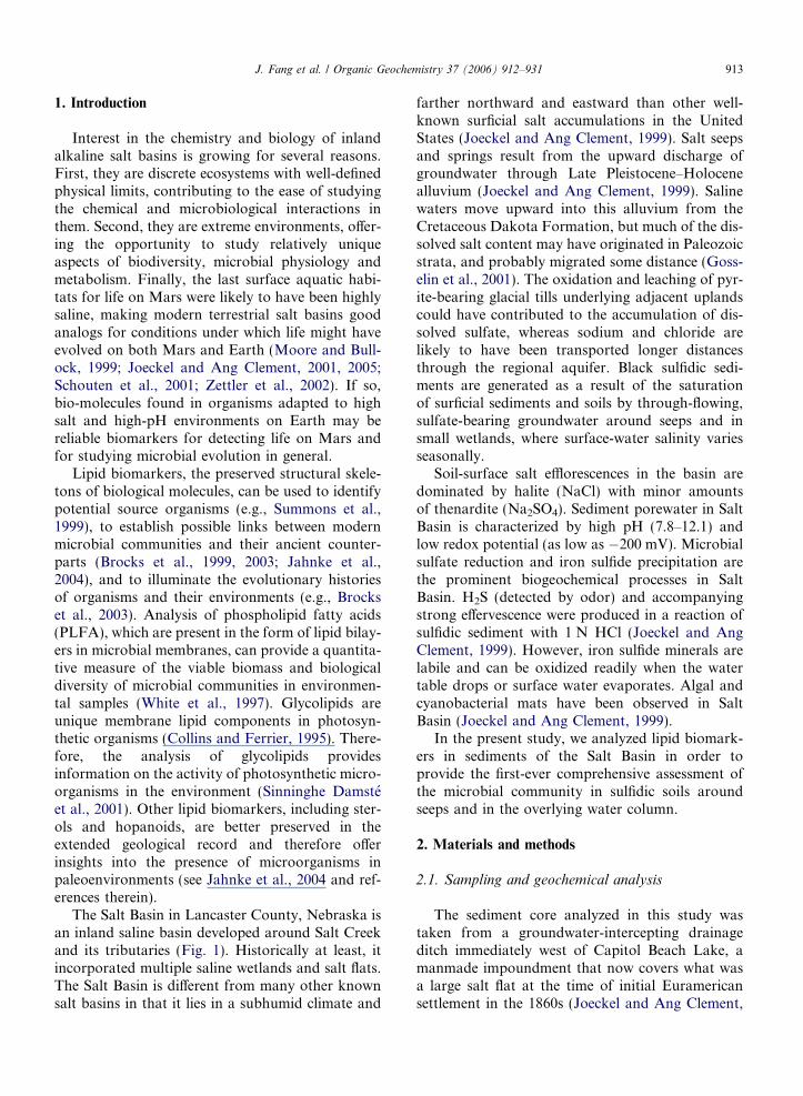
The Salt Basin in Lancaster County, Nebraska isan inland saline basin developed around Salt Creekand its tributaries (Fig. 1). Historically at least, itincorporated multiple saline wetlands and salt flats.The Salt Basin is different from many other knownsalt basins in that it lies in a subhumid climate and
farther northward and eastward than other well-known surficial salt accumulations in the UnitedStates (Joeckel and Ang Clement, 1999). Salt seepsand springs result from the upward discharge ofgroundwater through Late Pleistocene–Holocenealluvium (Joeckel and Ang Clement, 1999). Salinewaters move upward into this alluvium from theCretaceous Dakota Formation, but much of the dis-solved salt content may have originated in Paleozoicstrata, and probably migrated some distance (Goss-elin et al., 2001). The oxidation and leaching of pyr-ite-bearing glacial tills underlying adjacent uplandscould have contributed to the accumulation of dis-solved sulfate, whereas sodium and chloride arelikely to have been transported longer distancesthrough the regional aquifer. Black sulfidic sedi-ments are generated as a result of the saturationof surficial sediments and soils by through-flowing,sulfate-bearing groundwater around seeps and insmall wetlands, where surface-water salinity variesseasonally.
Soil-surface salt efflorescences in the basin aredominated by halite (NaCl) with minor amountsof thenardite (Na2SO4). Sediment porewater in SaltBasin is characterized by high pH (7.8–12.1) andlow redox potential (as low as �200 mV). Microbialsulfate reduction and iron sulfide precipitation arethe prominent biogeochemical processes in SaltBasin. H2S (detected by odor) and accompanyingstrong effervescence were produced in a reaction ofsulfidic sediment with 1 N HCl (Joeckel and AngClement, 1999). However, iron sulfide minerals arelabile and can be oxidized readily when the watertable drops or surface water evaporates. Algal andcyanobacterial mats have been observed in SaltBasin (Joeckel and Ang Clement, 1999).
In the present study, we analyzed lipid biomark-ers in sediments of the Salt Basin in order toprovide the first-ever comprehensive assessment ofthe microbial community in sulfidic soils aroundseeps and in the overlying water column.
2. Materials and methods
2.1. Sampling and geochemical analysis
The sediment core analyzed in this study wastaken from a groundwater-intercepting drainageditch immediately west of Capitol Beach Lake, amanmade impoundment that now covers what wasa large salt flat at the time of initial Euramericansettlement in the 1860s (Joeckel and Ang Clement,
Fig. 1. Study area in Salt Basin, Lancaster County, Nebraska. Shading along streams indicates areas of saline groundwater discharge.Multiple saline wetlands and salt flats existed in the area prior to Euramerican settlement in the 1850s.
914 J. Fang et al. / Organic Geochemistry 37 (2006) 912–931
1999). At the study site (Fig. 1), patches of blacksulfidic sediment under saline water, associated withseeps and small springs, were distributed along theditch. A fifteen-centimeter push core was collectedin clear polycarbonate tubing (100 ID) and sealedwith plastic caps. The core was placed on ice in acooler and transported to the laboratory within4 h, where it was sampled aseptically after firstdecanting the water covering the core. Then, thetop sections of the core, 0–3.3 cm (Section 1) and3.3–8 cm (Section 2), were removed for lipid analy-sis. Section 1 was black sulfidic material (BSM)(Joeckel and Ang Clement, 1999). Sediment ofSection 2 was grey sand and depleted in organicmatter.
Subsamples were immediately placed into testtubes with an extraction solvent mixture for lipidanalysis. Pore water was extracted by placingan aliquot of the sediment into 85-ml Nalgene
centrifuge tubes centrifuged at 10,000 rpm for15 min. About 3–5 ml of the collected pore waterwas poisoned with sodium azide for chlorideand sulfate measurements by ion chromatography.The remaining porewater was used for pH, redoxpotential (both by electrode probes), dissolvediron, sulfide, and dissolved oxygen analyses usingCheMatrics kit (CheMatrics, Inc., Calverton,VA) (Fang et al., 2000). Concentrations of sulfateand chloride were determined using a Dionex ionchromatography system 2000. A 4-mm AS18 ana-lytical column and a 4-mm AS18 anion guard col-umn with an ASRS Ultra II anion suppressoroperated at 96 milliamps were used for analyticalseparation. Potassium hydroxide (37 mM) wasthe mobile phase at a flow rate of 1 ml min�1.The column temperatures were 30 �C and the celltemperature was 35 �C. The injection volume was0.25 ll.

J. Fang et al. / Organic Geochemistry 37 (2006) 912–931 915
2.2. Lipid extraction and fractionation
Total sedimentary lipids were extracted at roomtemperature in test tubes containing a solventmixture of methanol:dichloromethane (DCM):phosphate buffer (potassium phosphate, dibasic,50 mM, pH 7.4) (2:1:0.8) (Fang and Findlay,1996). Crude lipids were collected after phase parti-tioning by adding dichloromethane and deionizedwater to the test tube to the final ratio of metha-nol:dichloromethane:water 1:1:0.9. The aqueousphase was saved for the isolation of lipopolysaccha-ride (LPS) fatty acids (FA). The total lipid extractwas dried under a gentle stream of nitrogen andthen dissolved in hexane:dichloromethane (70:30,v/v). Total lipids were separated into different lipidclasses using miniature champagne columns (Supe-lco Inc., Bellefonte, PA). Hydrocarbons, neutral lip-ids, glycolipids, and phospholipids were eluted with5 ml of hexane, chloroform, acetone, and methanol,respectively.
The upper aqueous phase was dried in a freeze-dryer. The residue was acidified by adding 30 mlof 1 N HCl and refluxed at l00 �C for 2 h. Aftercooling to room temperature, the contents weretransferred into a 250-ml separatory funnel withwashes of 2 · 10 ml of methanol and 2 · 25 ml ofDCM. The two phases were allowed to separateovernight. The organic phase containing the LPSfatty acids was collected.
2.3. Lipid analysis by gas chromatography–mass
spectrometry
The neutral lipid fraction was dried under astream of nitrogen and derivatized by adding 60 llof N,O-bis(trimethylsilyl)-trifloroacetamide to pro-duce TMS ethers of sterols and hopanols (heatedat 75 �C for 30 min). The phospholipid fractionwas subjected to a mild alkaline trans-methylationprocedure to produce fatty acid methyl esters (Fangand Findlay, 1996). The LPS fatty acids were firstmethylated use BF3/MeOH (heated at 65 �C for25 min). The hydroxyl fatty acids were convertedto their TMS ethers using the same procedures asdescribed above.
All lipids were analyzed on an Agilent 6890 GCinterfaced with an Agilent 5973N Mass SelectiveDetector. Analytical separation of the compoundswas accomplished using a 30 m · 0.25 mm i.d. DB-5 MS fused-silica capillary column (J&W Scientific,Folsom, CA). The column temperature was pro-
grammed from 50 to 120 �C at 10 �C/min, then to310 �C at 5 �C/min, and then held for 20 min. Indi-vidual fatty acids were identified from their massspectra. Individual sterols and triterpenoids wereidentified based on retention times and comparisonwith published mass spectra (Venkatesan, 1989; Shi-ojima et al., 1992). Response factors were obtainedfor fatty acids using duplicate injections of quantita-tive standards at five different concentration levels.Concentrations of individual compounds wereobtained based on the GC/MS response relative tothat of an internal standard (C18:0 fatty acid ethylester). The double-bond position and geometry ofmonounsaturated fatty acids were determined byusing methods described by Dunkleblum et al.(1985). The concentrations of sterols and hopanolswere determined based on the chromatographicresponse of these compounds relative to that of aninternal standard, 5a-cholestane. Method blankswere extracted with each set of samples and wereassumed to be free of contamination if chromato-grams contained no peaks. A standard containingknown concentrations of 26 fatty acids was ana-lyzed daily on the GC/MS to check analytical accu-racy (>90%). Replicate analyses (2·) of sampleswere done to ensure reproducibility (variation610%).
Fatty acids are designated by the total number ofcarbon atoms:number of double bonds (i.e., a 16carbon alkanoic acid is 16:0). The position of thedouble bond is indicated with a D number closestto the carboxyl end of the fatty acid molecule withthe geometry of either c (cis) or t (trans). The termi-nal methyl-branching is indicated with i (iso) or a
(anteiso) and the mid-branching is indicated as theposition of the methyl group from the carboxylgroup of the fatty acid (e.g., 10Me16:0). Cyclopro-pane fatty acids are indicated with cy.
2.4. Carbon isotope analysis of lipids
Carbon isotope analyses of lipids were performedat Brown University using a gas chromatograph–combustion–isotope ratio mass spectrometer (GC–C–IRMS, Thermo Finnigan) as described previ-ously (Wang et al., 2004). An HP 6890 GC,equipped with an AS 200 autosampler, was con-nected to a Finnigan MAT Delta plus XL MS viathe combustion interface. The temperature programand capillary column used were identical to thoseused for GC analysis. Helium (UHP 5.5 grade)was used as the carrier gas operating in constant
916 J. Fang et al. / Organic Geochemistry 37 (2006) 912–931
flow mode with a rate of 1.5 ml/min. Injection wasperformed in the splitless mode to deliver typicalsample amounts (�80 ng per compound) onto thecolumn. Compounds separated by the GC columnwere converted to CO2 and H2O through thecombustion furnace (0.5 mm i.d. · 1.5 mm o.d. ·34 cm) operated at 960 �C and loaded with CuOand Pt wires as the oxidant and catalyst, respec-tively. Six pulses of CO2 reference gas with knownd13C values (�46.96&) were injected via the inter-face to the IRMS, for the determination of d13C val-ues of sample compounds. Typically, the standarddeviation of duplicate analyses is smaller than±0.3&. Values are reported in the usual d13C nota-tion relative to the Vienna Pee Dee Belemnite stan-dard. Isotopic composition of individual fatty acidswas obtained after the correction for the additionalmethyl carbon from methanol using a mass balanceequation (Fang et al., 1993):
d13CFAME ¼ fFAd13CFA þ fMethanold13CMethanol
where d13CFAME, d13CFA, and d13Cmethanol are thecarbon isotopic composition of the FAME, theunderivatized fatty acid, and the methanol, respec-tively, and fFA and fMethanol are the fractions of car-bon in the FAME due to the underivatized fattyacid and methanol, respectively.
3. Results
3.1. Sedimentary geochemistry
The groundwater of Salt Basin contains high lev-els of Na+ (10.4–18.0 g/l) and Cl� (15.3–22.6 g/l). Asurface water sample taken near a saline seep con-tained 82 and 114 g/l of Na+ and Cl�, respectively.Other major ions included Ca2+ (0.22–0.32 g/l),SO2�
4 (0.66–0.84 g/l), and HCO�3 (0.4–0.77 g/l)(Joeckel and Ang Clement, 1999). Porewaterextracted from Section 1 of the sediment corecontained elevated concentrations of Cl� (20.9 g/l),SO2�
4 (1.7 g/l), S2� (0.024 g/l), and alkalinity(0.53 g/l). The sedimentary environment was highlyreducing (Eh = �254 mV) (see Fig. 6).
3.2. Hydrocarbons
Compounds in the hydrocarbon fraction of Sec-tion 1 include n-alkanes, pristane, pristenes, phy-tane, phytenes, phytadienes, sterenes, and hopanes.The n-alkane profile showed a bimodal distributionwith Cmax at C17 and C27 (Fig. 2a). The concentra-
tions of most lower-molecular weight alkanes wererelatively low except heptadecane (C17:0) and hepta-decenes (C17:1), which constituted 10.2% and 10.5%of the total hydrocarbons (Table 1). The higher-molecular weight n-alkanes exhibited an apparentodd-over-even carbon number predominance. Theconcentrations of pristane and phytane were lowrelative to isomers of pristene, phytene and phytadi-ene. The three isomeric phytadienes accounted for14.7% of the total hydrocarbons. A number of poly-unsaturated alkenes were identified including C21:6,C21:7, C30:4, and C30:5. Significantly, C21:7 is noveland its presence in sediment has never beenreported. It had the highest concentration amongall polyunsaturated alkenes (1.3 lg g�1 dry weightsediment; 13.3% of total hydrocarbons). Squalenewas not detected in the sediment. However, tetrahy-drosqualene, a hydrogenated derivative, was found.Although a number of sterols were detected (seebelow), only two sterenes were present (Table 1).These were 24-ethylcholesta-3,5-diene (5.7%) and24-ethylcholesta-3,5,22E-triene (5.4%). The m/z191 mass chromatogram shows that a number ofhomo-, bishomo, and trishomohopanes werepresent in the sediment (Fig. 2b). However, theirconcentrations were extremely low. The most abun-dant hopene was hop-22(29)-ene (diploptene) (Table1).
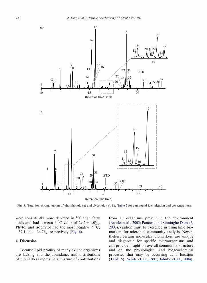
3.3. Phospholipid (PLFA), glycolipid (GLFA), and
lipopolysacchride (LPS) fatty acids
PLFA (Fig. 3a), GLFA (Fig. 3b), and LPS fattyacids (Fig. 4) were isolated from the sediment(Tables 2 and 3). PLFA with chain length fromC11–20 were detected in sediment from Salt Basin.The phospholipid fatty acid profiles were domi-nated by monounsaturated fatty acids which con-stituted 56% of the total fatty acids (Fig. 3a).The most abundant ones were 16:1D9 and18:1D11. Also detected in PLFA was br-17:1D9, amarker fatty acid commonly found in SRB. Thesaturated fatty acids accounted for 20% and 31%of the PLFA and GLFA, respectively. Terminal-branched fatty acids consisted of iso and anteiso
isomers of C15, C16, and C17 fatty acids. Thesefatty acids account for 12% and 16% of PLFAand GLFA, respectively. The only mid-methylbranched fatty acid detected was 10Me16:0 (0.8–0.9%). Polyunsaturated fatty acids (PUFA)detected in the sediment included C16:2, three iso-mers of C18:2D9,12, C20:3 and 20:4, constituting

a
b
cd
e
f
gh
i
j
k
lm
no
pq r
s
t
u v w
xy
z
aa
ab
ac
ad
ae af
ag
10 15 20 25 30 35Retention time (min)
11 12 13 14
(a)
ah
32 34 3633 35 37 38
Retention time (min)
(b)
ai
aj
ak
alam
an
ao
ap aqar
m/z 191
Fig. 2. Total ion chromatogram (a) and m/z 191 mass chromatogram (b) of the hydrocarbon fraction isolated from black sulfidicsediment, which shows macroscopic evidence for microbial sulfate reduction, around a saline seep. See Table 1 for compoundidentification and concentrations.
J. Fang et al. / Organic Geochemistry 37 (2006) 912–931 917
6.6% of the total fatty acids. The concentration oftotal PLFA was more than three times greater thanthat of the GLFA (209.4 vs. 62.2 lg g�1 dry weightsediment).
The total PLFA content of Section 2 (155 ng g�1)was three orders of magnitude lower than Section 1.The C16 and C18 fatty acids constituted 74% of thetotal fatty acid content in Section 2 (Table 2). ThePLFA abundances record the viable microbial bio-mass (White et al., 1997). Thus, microbial activityof Section 2 is limited. Our discussion will focuson lipids of Section 1 of the sediment core.
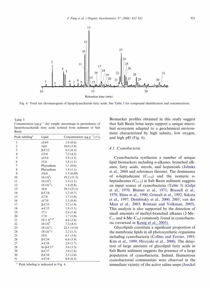
The lipid part (lipid A) of the lipopolysaccharidecomponent of Gram-negative bacteria contains avariety of hydroxy and non-hydroxy (normal andbranched) fatty acids (Table 3, Fig. 4). A total of10 hydroxy fatty acids, ranging from C12 to C18,were identified, including a- and b-hydroxy, andbranched b-hydroxy compounds. The most abun-dant LPS FA present in Salt Basin sediment wereb-hydroxy fatty acids. The concentration of individ-ual hydroxy fatty acids ranged from 0.3% to 3.9% ofthe total LPS fatty acids. A polyunsaturated fattyacid, 18:2D9,12, was also detected in the LPS lipids.
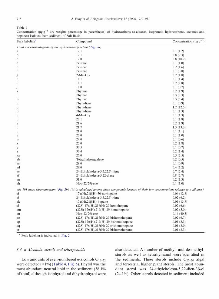
Table 1Concentration (lg g�1 dry weight; percentage in parentheses) of hydrocarbons (n-alkanes, isoprenoid hydrocarbons, steranes andhopanes) isolated from sediment of Salt Basin
Peak labelinga Compound Concentration (lg g�1)
Total ion chromatogram of the hydrocarbon fraction (Fig. 2a)
a 17:1 0.1 (1.2)b 17:1 0.8 (9.3)c 17:0 0.8 (10.2)d Pristane 0.1 (1.0)e Pristene 0.2 (1.6)f Pristene 0.1 (0.8)g 2-Me–C17 0.2 (1.8)h 18:1 0.1 (1.4)i 18:1 0.2 (2.8)j 18:0 0.1 (0.7)k Phytane 0.2 (1.9)l Phytene 0.3 (3.3)m Phytene 0.3 (3.4)n Phytadiene 0.1 (0.9)o Phytadiene 1.2 (12.5)p Phytadiene 0.1 (1.3)q 4-Me–C19 0.1 (1.3)r 20:1 0.1 (1.8)s 21:6 0.2 (1.9)t 21:7 1.3 (13.3)u 21:0 0.1 (1.1)v 23:0 0.1 (1.0)w 24:0 0.1 (0.6)x 25:0 0.2 (1.8)y 30:5 0.1 (0.7)z 30:4 0.2 (1.4)aa 27:0 0.5 (3.5)ab Tetrahydrosqualene 0.2 (0.5)ac 28:0 0.1 (0.9)ad 29:0 0.4 (3.2)ae 24-Ethylcholesta-3,5,22E-triene 0.7 (5.4)af 24-Ethylcholesta-5,22-diene 0.8 (5.7)ag 31:0 0.2 (1.3)ah Hop-22(29)-ene 0.1 (1.0)
m/z 191 mass chromatogram (Fig. 2b) (% is calculated among these compounds because of their low concentrations relative to n-alkanes)
ai 17a(H),21b(H)-30-norhopane 0.04 (12.8)aj 24-Ethylcholesta-3,5,22E-triene 0.02 (6.2)ak 17a(H),21b(H)-hopane 0.05 (13.7)al (22S)-17a(H),21b(H)-29-homohopane 0.02 (6.6)am (22R)-17a(H),21b(H)-29-homohopane 0.02 (5.0)an Hop-22(29)-ene 0.14 (40.5)ao (22S)-17a(H),21b(H)-29-bishomohopane 0.02 (6.7)ap (22R)-17a(H),21b(H)-29-bishomohopane 0.01 (3.3)aq (22S)-17a(H),21b(H)-29-trishomohopane 0.01 (3.0)ar (22S)-17a(H),21b(H)-29-bishomohopane 0.01 (2.3)
a Peak labeling is indicated in Fig. 2.
918 J. Fang et al. / Organic Geochemistry 37 (2006) 912–931
3.4. n-Alcohols, sterols and triterpenoids
Low amounts of even-numbered n-alcohols C16–22
were detected (<1%) (Table 4, Fig. 5). Phytol was themost abundant neutral lipid in the sediment (38.1%of total) although isophytol and dihydrophytol were
also detected. A number of methyl- and desmethyl-sterols as well as tetrahymanol were identified inthe sediments. These sterols include C27–30 algaland terrestrial higher plant sterols. The most abun-dant sterol was 24-ethylcholesta-5,22-dien-3b-ol(24.1%). Other sterols detected in sediment included
Table 2d13C and concentration (percentage in parentheses) of phospholipid (PLFA) and glycolipid (GLFA) fatty acids isolated from sediment ofSalt Basin
Peak labelinga Fatty acid PLFA GLFA
Section 1 Section 2 Section 1
Concentration (lg g�1) d13C (&) Concentration (ng g�1) Concentration (lg g�1) d13C (&)
1 12:0 0.2 (0.1) nd2 i14:0 3.0 (1.4) nd3 a14:0 3.3 (1.6) �30.0 1.3 (2.1) �28.74 14:0 6.1 (2.9) �29.4 3.2 (2.1) 3.2 (5.1) �27.85 15:1D7c 0.4 (0.2) 0.0 (0.0)6 15:1D7t 0.5 (0.2) 0.0 (0.0)7 i15:0 9.2 (4.4) �28.1 5.1 (3.3) 5.4 (8.7) �27.98 a15:0 7.2 (3.4) �29.8 9.3 (6.0) 2.1 (3.3) �29.19 15:1D9 0.4 (0.2) 0.0 (0.0)
10 15:0 1.7 (0.8) �27.6 1.4 (2.4) 0.7 (1.2) �28.611 i16:0 1.5 (0.7) �29.0 3.7 (2.4) 0.9 (1.4) �28.412 16:2 0.9 (0.4) 1.4 (2.2)13 16:1D9c 1.3 (0.6) 11.2 (7.2) 1.1 (1.8)14 16:1D9t 30.8 (14.7) �28.9 9.4 (15.0) �27.615 16:1D11c 2.4 (1.1) 0.8 (1.2)16 16:1D11t 2.1 (1.0) 0.7 (1.1)17 16:0 28.0 (13.3) �30.4 39.9 (25.7) 14.0 (22.3) �26.518 br17:1D9 0.9 (0.4) 7.7 (5.0) 0.0 (0.0)19 10Me16:0 1.6 (0.8) 6.4 (4.1) 0.2 (0.4) �28.820 i17:0 0.6 (0.3) 1.4 (0.9) 0.2 (0.3) �27.721 a17:0 1.0 (0.5) 0.0 (0.0)22 17:1D9 0.9 (0.4) 0.0 (0.0)23 cy17:0 4.0 (1.9) �26.8 0.7 (1.2) �28.724 17:1D11 0.3 (0.1) 0.0 (0.0)25 17:0 1.4 (0.7) 2.3 (1.5) 0.3 (0.5) �30.326 18:2 0.6 (0.3) 0.0 (0.0)27 18:2 1.0 (0.5) 0.0 (0.0)28 18:2 D9,12 6.6 (3.1) 4.5 (7.3)29 18:1D9 17.2 (8.2) 12.1 (7.8) 3.4 (5.5)30 18:1D11c 55.1 (26.2) �28.8 24.6 (15.9) 8.7 (14.0) �28.731 18:1D11t 5.1 (2.4) 6.8 (4.4) 0.0 (0.0)32 18:0 4.5 (2.1) �30.6 20.0 (12.9) 1.0 (1.7) �27.433 cy19:0 4.6 (2.2) �30.2 0.0 (0.0)34 20:4 1.8 (0.9) 0.4 (0.7)35 20:3 2.5 (1.2) 1.0 (1.6)36 20:1D13 0.4 (0.2) 0.4 (0.6)37 20:0 0.3 (0.1) 0.2 (0.3) �28.738 22:0 nd 1.0 (0.0) �28.739 24:0 nd 1.7 (0.1) �29.040 26:0 nd 1.5 (0.1) �31.0
a Peak labeling is indicated in Fig. 3a (PLFA) and Fig. 3b (GLFA).
J. Fang et al. / Organic Geochemistry 37 (2006) 912–931 919
cholesterol, 24-methyl and 24-ethylsterols. Smallamounts of tetrahymanol, (22R)-17b(H),21b(H)hopan-29-ol, (22R)-17b(H),21b(H)homohopan-31-ol and (22R)-17b(H),21b(H)bishomohopan-32-olwere also detected.
3.5. Carbon isotopic composition of lipid biomarkers
The d13C values of phospholipid fatty acidsvaried from �26.8& to �30.6& with a mean of
�28.9 ± 1.2&. The ubiquitous fatty acids C16:0
and C18:0 had more negative d13C values thanother fatty acids. More positive values wereobserved for monounsaturated, odd-numbered, ter-minal-branched and cyclopropane fatty acids (i15:0,n-15:0, i16:0, 16:1D9, 18:1D11, cy17:0). Glycolipidfatty acids had slightly more positive d13C values(28.3 ± 0.9&) than PLFA. Cy19:0 in both thePLFA and GLFA fractions had more negatived13C values than other fatty acids. The neutral lipids
(a)
1
2 3
4
56
78
910 11
12
14
1516
17
13
26
18
1920 21 22
23
24
25
27 28
29
30
31
3233
34 35 3637
ISTD
10 15 20
17
Retention time (min)
3
4
7
8
10 28
2931
32 3637
30
19
2021
23
25
(b)
11
12
13
14
15
16
17
ISTD
38
39 40
20 25
15
Retention time (min)
15
Fig. 3. Total ion chromatogram of phospholipid (a) and glycolipid (b). See Table 2 for compound identification and concentrations.
920 J. Fang et al. / Organic Geochemistry 37 (2006) 912–931
were consistently more depleted in 13C than fattyacids and had a mean d13C value of 29.2 ± 1.0&.Phytol and isophytol had the most negative d13C,�37.1 and �34.7&, respectively (Fig. 6).
4. Discussion
Because lipid profiles of many extant organismsare lacking and the abundance and distributionsof biomarkers represent a mixture of contributions
from all organisms present in the environment(Brocks et al., 2003; Pancost and Sinninghe Damste,2003), caution must be exercised in using lipid bio-markers for microbial community analysis. Never-theless, certain molecular biomarkers are uniqueand diagnostic for specific microorganisms andcan provide insight on overall community structureand on the physiological and biogeochemicalprocesses that may be occurring at a location(Table 5) (White et al., 1997; Jahnke et al., 2004).
1
2
3
45
6 78 9
10
11
1415
16
1718
1920 21
22
23
24
2526
2728
29 30
31
12
13
10 15 20
Retention time (min)
Fig. 4. Total ion chromatogram of lipopolysaccharide fatty acids. See Table 3 for compound identification and concentrations.
Table 3Concentration (lg g�1 dry weight; percentage in parenthesis) oflipopolysaccharide fatty acids isolated from sediment of SaltBasin
Peak labelinga Lipid Concentration (lg g�1) (%)
1 a14:0 1.0 (0.6)2 14:0 10.0 (5.9)3 b-C12 0.5 (0.3)4 i15:0 7.2 (4.2)5 a15:0 5.9 (3.5)6 15:0 1.8 (1.1)7 Phytadiene 1.1 (0.6)8 Phytadiene 1.9 (1.1)9 i16:0 1.5 (0.88)
10 16:1D9c 19.2 (11.3)11 16:1D9t 1.9 (1.1)12 16:1D11c 1.4 (0.8)13 16:0 39.2 (23.1)14 b-C14 1.2 (0.7)15 i17:0 1.7 (1.0)16 a17:0 1.3 (0.8)17 b-C15 3.2 (1.9)18 a-C15 1.8 (1.1)19 17:1 2.4 (1.4)20 17:0 1.7 (1.0)21 18:2 D9,12 4.4 (2.6)22 18:1D9 7.3 (4.3)23 18:1D11c 22.1 (13.0)24 18:1D11t 2.2 (1.3)25 18:0 6.1 (3.6)26 b-C16 6.6 (3.9)27 a-C16 2.8 (1.7)28 br-b-C17 5.6 (3.3)29 b-C17 3.0 (1.8)30 b-C18 3.3 (2.0)31 a-C18 0.8 (0.5)
a Peak labeling is indicated in Fig. 4.
J. Fang et al. / Organic Geochemistry 37 (2006) 912–931 921
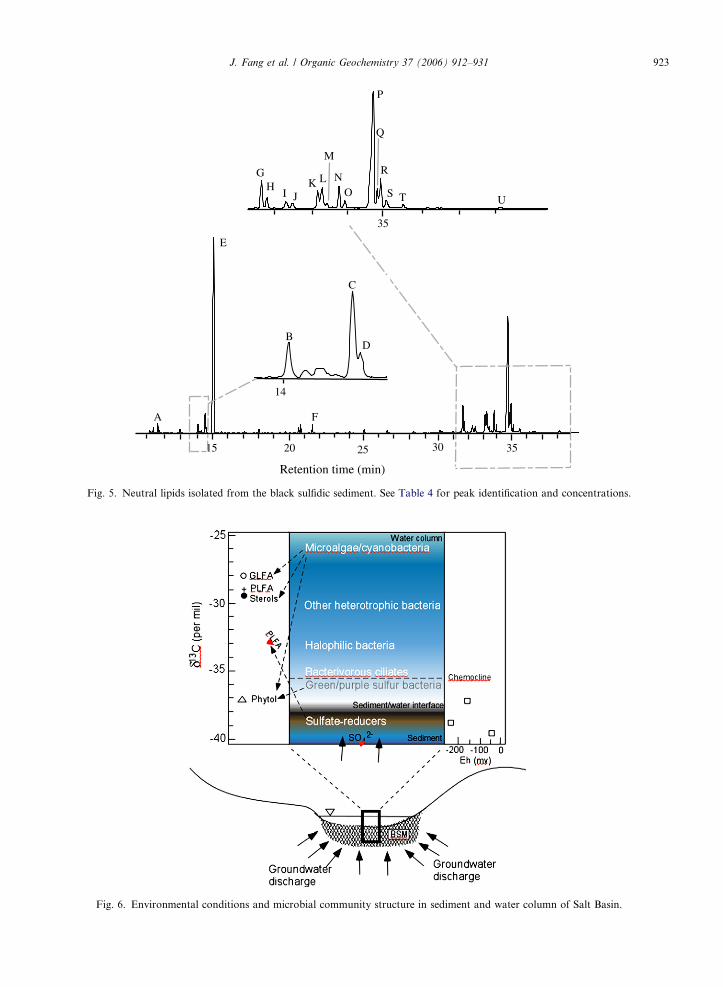
Biomarker profiles obtained in this study suggestthat Salt Basin brine seeps support a unique micro-bial ecosystem adapted to a geochemical environ-ment characterized by high salinity, low oxygen,and high pH (Fig. 6).
4.1. Cyanobacteria
Cyanobacteria synthesize a number of uniquelipid biomarkers including n-alkanes, branched alk-anes, fatty acids, sterols, and hopanoids (Jahnkeet al., 2004 and references therein). The dominanceof n-heptadecane (C17:0) and the isomeric n-heptadecenes (C17:1) in Salt Basin sediment suggestsan input source of cyanobacteria (Table 5) (Gelpiet al., 1970; Blumer et al., 1971; Brassell et al.,1978; Shiea et al., 1990; Grimalt et al., 1992; Sakataet al., 1997; Dembitsky et al., 2000, 2001; van derMeer et al., 2003; Rontani and Volkman, 2005).This analysis is also supported by the detection ofsmall amounts of methyl-branched alkanes (2-Me–C17 and 4-Me–C19) commonly found in cyanobacte-ria (reviewed in Kenig et al., 2005).
Glycolipids constitute a significant proportion ofthe membrane lipids in all photosynthetic organismsincluding cyanobacteria (Collins and Ferrier, 1995;Kim et al., 1999; Hiroyuki et al., 2000). The detec-tion of large amounts of glycolipid fatty acids inSalt Basin sediment suggests the presence of a largepopulation of cyanobacteria. Indeed, filamentouscyanobacterial communities were observed in theimmediate vicinity of the active saline seeps (Joeckel
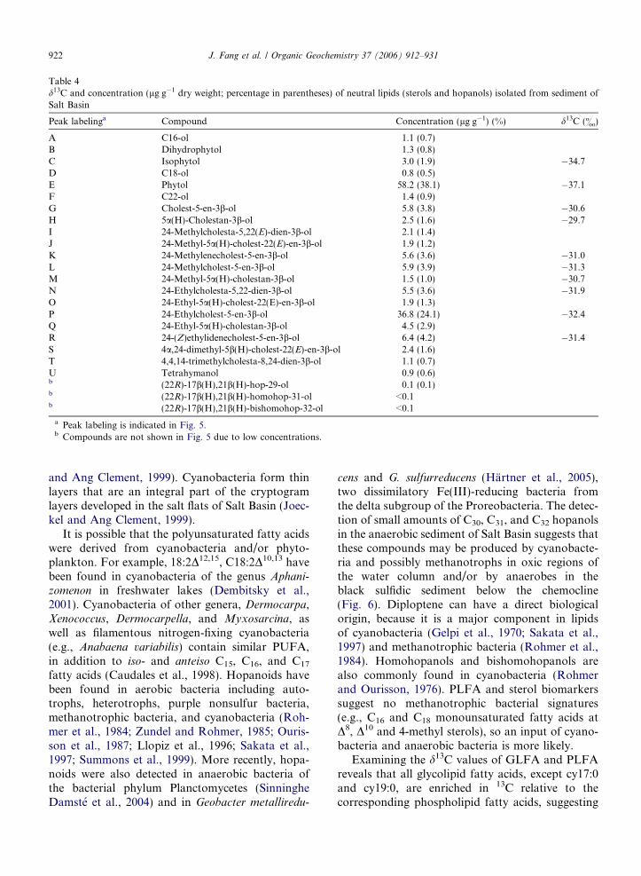
Table 4d13C and concentration (lg g�1 dry weight; percentage in parentheses) of neutral lipids (sterols and hopanols) isolated from sediment ofSalt Basin
Peak labelinga Compound Concentration (lg g�1) (%) d13C (&)
A C16-ol 1.1 (0.7)B Dihydrophytol 1.3 (0.8)C Isophytol 3.0 (1.9) �34.7D C18-ol 0.8 (0.5)E Phytol 58.2 (38.1) �37.1F C22-ol 1.4 (0.9)G Cholest-5-en-3b-ol 5.8 (3.8) �30.6H 5a(H)-Cholestan-3b-ol 2.5 (1.6) �29.7I 24-Methylcholesta-5,22(E)-dien-3b-ol 2.1 (1.4)J 24-Methyl-5a(H)-cholest-22(E)-en-3b-ol 1.9 (1.2)K 24-Methylenecholest-5-en-3b-ol 5.6 (3.6) �31.0L 24-Methylcholest-5-en-3b-ol 5.9 (3.9) �31.3M 24-Methyl-5a(H)-cholestan-3b-ol 1.5 (1.0) �30.7N 24-Ethylcholesta-5,22-dien-3b-ol 5.5 (3.6) �31.9O 24-Ethyl-5a(H)-cholest-22(E)-en-3b-ol 1.9 (1.3)P 24-Ethylcholest-5-en-3b-ol 36.8 (24.1) �32.4Q 24-Ethyl-5a(H)-cholestan-3b-ol 4.5 (2.9)R 24-(Z)ethylidenecholest-5-en-3b-ol 6.4 (4.2) �31.4S 4a,24-dimethyl-5b(H)-cholest-22(E)-en-3b-ol 2.4 (1.6)T 4,4,14-trimethylcholesta-8,24-dien-3b-ol 1.1 (0.7)U Tetrahymanol 0.9 (0.6)b (22R)-17b(H),21b(H)-hop-29-ol 0.1 (0.1)b (22R)-17b(H),21b(H)-homohop-31-ol <0.1b (22R)-17b(H),21b(H)-bishomohop-32-ol <0.1
a Peak labeling is indicated in Fig. 5.b Compounds are not shown in Fig. 5 due to low concentrations.
922 J. Fang et al. / Organic Geochemistry 37 (2006) 912–931
and Ang Clement, 1999). Cyanobacteria form thinlayers that are an integral part of the cryptogramlayers developed in the salt flats of Salt Basin (Joec-kel and Ang Clement, 1999).
It is possible that the polyunsaturated fatty acidswere derived from cyanobacteria and/or phyto-plankton. For example, 18:2D12,15, C18:2D10,13 havebeen found in cyanobacteria of the genus Aphani-zomenon in freshwater lakes (Dembitsky et al.,2001). Cyanobacteria of other genera, Dermocarpa,Xenococcus, Dermocarpella, and Myxosarcina, aswell as filamentous nitrogen-fixing cyanobacteria(e.g., Anabaena variabilis) contain similar PUFA,in addition to iso- and anteiso C15, C16, and C17
fatty acids (Caudales et al., 1998). Hopanoids havebeen found in aerobic bacteria including auto-trophs, heterotrophs, purple nonsulfur bacteria,methanotrophic bacteria, and cyanobacteria (Roh-mer et al., 1984; Zundel and Rohmer, 1985; Ouris-son et al., 1987; Llopiz et al., 1996; Sakata et al.,1997; Summons et al., 1999). More recently, hopa-noids were also detected in anaerobic bacteria ofthe bacterial phylum Planctomycetes (SinningheDamste et al., 2004) and in Geobacter metalliredu-
cens and G. sulfurreducens (Hartner et al., 2005),two dissimilatory Fe(III)-reducing bacteria fromthe delta subgroup of the Proreobacteria. The detec-tion of small amounts of C30, C31, and C32 hopanolsin the anaerobic sediment of Salt Basin suggests thatthese compounds may be produced by cyanobacte-ria and possibly methanotrophs in oxic regions ofthe water column and/or by anaerobes in theblack sulfidic sediment below the chemocline(Fig. 6). Diploptene can have a direct biologicalorigin, because it is a major component in lipidsof cyanobacteria (Gelpi et al., 1970; Sakata et al.,1997) and methanotrophic bacteria (Rohmer et al.,1984). Homohopanols and bishomohopanols arealso commonly found in cyanobacteria (Rohmerand Ourisson, 1976). PLFA and sterol biomarkerssuggest no methanotrophic bacterial signatures(e.g., C16 and C18 monounsaturated fatty acids atD8, D10 and 4-methyl sterols), so an input of cyano-bacteria and anaerobic bacteria is more likely.
Examining the d13C values of GLFA and PLFAreveals that all glycolipid fatty acids, except cy17:0and cy19:0, are enriched in 13C relative to thecorresponding phospholipid fatty acids, suggesting
A
E
B
C
D
F
15 20 25 30 35
Retention time (min)
14
35
GH
I JK L
M
NO
P
Q
R
S T U
Fig. 5. Neutral lipids isolated from the black sulfidic sediment. See Table 4 for peak identification and concentrations.
Fig. 6. Environmental conditions and microbial community structure in sediment and water column of Salt Basin.
J. Fang et al. / Organic Geochemistry 37 (2006) 912–931 923

Table 5Possible origin of lipid biomarkers isolated from sediment of SaltBasin
Biomarkers Origin
Hydrocarbons
C17:0, C17:1, 2-Me–C17,4-Me–C19
Cyanobacteria
C21:6, C21:7, C30:4, C30:5 AlgaePristane Oxidation products of phytolPhytane (phytenes) and
phytadienesProducts of anaerobic reduction ofphytol; methanogens
Tetrahydrosqualene Methanogens; halobacteria
Hopanoids
Diploptene Cyanobacteria methanotrophsHomo- bis-, andtrishomohopanes
Transformation products ofbacteriohopanepolyols
Fatty acids
10Me16:0, cy17:0, cy19:0 Desulfobacter
cy17:0 Green sulfur bacteriacy19:0 Purple sulfur bacteria17:1D9, 15:1 Desulfobulbus
i15:0, a15:0 Desulfotomaculum
16:1D9, cy17:0 and LPSb-OH fatty acids
Desulfomonile tiedjei
10Me16:0, 17:1D11,16:1D9
Desulfobacterium
i15:0, a15:0, 16:1D9,17:1D9, 17:1D11
Desulfococcus
17:1D9, 17:1D11 Desulfovibrio
18:2D9,12 Cyanobacteria
16:2, 20:3, 20:4 Algae
Alcohols and sterols
C16, C18, C20 alkanol Photosynthetic sulfur bacteriaPhytol Phototrophs (algae, plants,
cyanobacteria, and phototrophicsulfur-bacteria)
Isophytol,dihydrophytol
Halobacteria; methanogens; productof anaerobic transformation ofphytol
Tetrahymanol Bacterivorous ciliates (e.g.,Tetrahymena); purple non-sulfurbacteriumRhodopseudomonas palustris
C27D5, C27D
5,22, C28D5,22 Diatoms
C28D5,22, C29D
5,22 HaptophytesSitosterol Higher plants
924 J. Fang et al. / Organic Geochemistry 37 (2006) 912–931
that most of the glycolipid fatty acids are likelyfrom cyanobacteria, with additional input fromhigher plants (e.g., C21–26 fatty acids). This is inaccord with the notion that cyanobacterialribulose-1,5-bisphosphate carboxylase/oxygenase(RuBisCO) discriminates less against 13C duringinorganic carbon fixation than C3 plants (Sakataet al., 1997). Thus, the isotopic composition of thefatty acids points to an origin from autotrophic
organisms in the oxic surface water and from SRBin the anoxic zone (Fig. 6).
4.2. Sulfate-reducing bacteria and halophilic bacteria
The bacterial community of Salt Basin sedimentappears to be dominated by Gram-negative bacte-ria, indicated by the presence of high concentrationsof monounsaturated fatty acids in the PLFA andthe LPS FA fractions (Ratledge and Wilkinson,1988; Findlay et al., 1990). The detection of terminalbranched fatty acids suggests that Gram-positivebacteria are present as well as Gram-negative anaer-obic bacteria (Parkes and Taylor, 1983; Ratledgeand Wilkinson, 1988; Kaneda, 1991; White et al.,1996; Caudales et al., 1998).
The black sulfidic mud (BSM) is a distinguishingfeature of Salt Basin (Joeckel and Ang Clement,1999) compared to other freshwater sediments inthe area. The BSM are waterlogged, are sandy tomuddy in coarseness and are in direct associationwith standing water, or with continuous saturationby through-flowing groundwater from saline seepsand springs. BSM exhibited high pH (7.8–12.1),low redox potential and high sulfide content. Bacte-rial sulfate reduction in Salt Basin sediment hasbeen verified in the laboratory (Joeckel and AngClement, 1999). Laboratory culturing of SRB fromSalt Basin sediment in Medium E containing ironand sodium sulfate resulted in growth of black bac-terial colonies that produced sulfide (Joeckel andAng Clement, 1999). Thus, sulfate reduction islikely an important process in the sediment of SaltBasin (Fig. 6).
The PLFA profiles of the black sulfidic materialsin Salt Basin suggest the presence of high numbersof SRB (Table 5). The high levels of cis-vaccenicacid (18:1D11), known to be formed through theanaerobic fatty acid desaturase pathway, point tothe presence of anaerobic bacteria (White et al.,1997). The branched C15 and C17 fatty acids canbe attributed to Desulfococcus multivorans (Rutterset al., 2002), whereas the MBFA, 10Me16:0 is char-acteristic of Desulfobacter (Edlund et al., 1985;Dowling et al., 1986; Konneke and Widdel, 2003)although it has also been recognized as a biomarkerfor Actinomycetes (O’Leary and Wilkinson, 1988;Vestal and White, 1989; Sundh et al., 1997). How-ever, SRB do not contain 10Me18:0, whereas Acti-
nomycetes contain significant amounts of this fattyacid (Dowling et al., 1986; Vainshtein et al., 1992).Because 10Me18:0 was not detected in Salt Basin

J. Fang et al. / Organic Geochemistry 37 (2006) 912–931 925
sediments, the 10Me16:0 fatty acid could be derivedfrom SRB. The detection of 16:1D11t also suggeststhe presence of Desulfobacter as well as Desulfo-
monas acetoxidans (Dowling et al., 1986). The pres-ence of cy17:0 and cy19:0 suggest the presence ofDesulfobacter and Desulfobacterium (Konneke andWiddel, 2003; Londry and Des Marais, 2004). TheC17 monounsaturated fatty acids (e.g., 17:1D9;17:1D10, 17:1D11) are commonly associated withSRB such as Desulfovibrio (Taylor and Parkes,1983; Edlund et al., 1985; Vainshtein et al., 1992;Kohring et al., 1994; Londry and Des Marais,2004), Desulfobulbus (Parkes et al., 1993; Oude-Elferink et al., 1998), Desulfococcus, Desulfuro-
monas, Desulfosarcina (Kohring et al., 1994),Desulfomicrobium (Vainshtein et al., 1992) as wellas Desulfobacter (Konneke and Widdel, 2003). Theoccurrence of 15:1D9 and 17:1D9 is indicative ofthe presence of Desulfobulbus in the sediment (Tay-lor and Parkes, 1983; Parkes et al., 1993) and Des-ulfovibrio (Londry and Des Marais, 2004),corroborating the interpretation of C17 monounsat-urated fatty acids. SRB are also characterized by apredominance of iso over anteiso C15 and C17 fattyacids (Table 2; Dowling et al., 1986).
Hydroxy fatty acids, recovered from acid hydro-lysis of the solvent extracted sediment residue, rep-resent those linked to disaccharides by an amidelinkage in the outer membranes of Gram-negativebacteria (Edlund et al., 1985; Goossens et al.,1986). Although hydroxy fatty acids have beendetected in algae (Matsumoto et al., 1984) and cya-nobacteria (Schmidt et al., 1980; Matsumoto et al.,1984; Wakeham et al., 2003), the detection of abun-dant LPS b-hydroxy fatty acids with chain-lengthC12–17 in Salt Basin provides further evidence forthe presence of SRB in the sediments (Parkeret al., 1982; Edlund et al., 1985). Since LPS can berapidly degraded in the environment (Saddler andWardlaw, 1980), the detection of LPS hydroxylfatty acids is indicative of viable SRB in Salt Basinsediment (e.g., Parker et al., 1982).
Phytane in sediments is derived from the reduc-tion of phytol via isomeric phytadienes as interme-diates under anoxic conditions (Didyk et al., 1978;Grossi et al., 1998; Schulze et al., 2001; Rontaniand Volkman, 2003). The anaerobic degradationof phytol under sulfate-reducing conditions hasbeen studied by Grossi et al. (1998). Phytol is readilydegraded by SRB to a number of isomeric phytenesand phytadienes (Grossi et al., 1998). The profile ofthese reduction products in Salt Basin is similar to
those from in vitro sulfate reduction experiments(Grossi et al., 1998), suggesting that an active phytolreduction process mediated possibly by SRB wastaking place in the sediment.
Tetrahydrosqualene was identified in Salt Basinsediment based on comparison with published massspectrum and retention time (e.g., Kramer et al.,1972; Rontani and Volkman, 2003). The source ofthe hydrogenated squalene derivatives can be frommethanogens (Tornabene et al., 1979; Ferranteet al., 1986) as well as halophilic bacteria such asHalobacterium cutirubrum and Sarcina marina(Tornabene et al., 1969; Hunter et al., 1981). Dihyd-rophytol can be derived from reduction of free phy-tol (Brooks et al., 1978; Schulze et al., 2001) or frommethanogens (Tornabene et al., 1979) or halophilicbacteria (Kushner, 1966; Nissenbaum et al., 1972;Volkman and Maxwell, 1986).
All of the fatty acids have relatively low d13C val-ues. This could reflect an origin from terrestrialhigher plants (Rieley et al., 1991; Huang et al.,1996; Fang et al., 2002). However, given the appar-ent bacterial source of certain biomarkers (e.g., mid-and terminal-branched and cyclopropane fattyacids), these 13C-depleted lipids could derive fromrecycling of organic matter by anaerobic bacteria(e.g., SRB). This has been demonstrated by Teeceet al. (1999) who showed that anaerobically synthe-sized fatty acids were depleted by up to 11& relativeto aerobically produced fatty acids.
4.3. Tetrahymena and anaerobic phototrophic sulfur
bacteria
Tetrahymanol has been found in bacteriovorousciliates such as Tetrahymena (Mallory et al., 1963;Ourisson et al., 1987; Harvey and McManus,1991) and the anaerobic phototrophic purple bacte-rium Rhodopseudomonas palustris (Kleemann et al.,1994) as well as in an anaerobic fungus (Kemp et al.,1984) and a fern (Zander et al., 1969). Rhodopseudo-
monas palustris is a purple, non-sulfur, phototrophicbacterium (Kleemann et al., 1990;Kleemann et al.,1994). This species is a metabolically versatileGram-negative bacterium belonging to the alphasubgroup of the Proteobacteria commonly foundin soil and sediment (Larimer et al., 2003).
Tetrahymena pyriformitis is widely distributed infreshwater ponds, lakes, and stream and evapora-tive environments (Hill, 1972; Elliot, 1973; Venkate-san, 1989). It is believed that tetrahymanol plays thesame role as sterols as membrane inserts in lower
926 J. Fang et al. / Organic Geochemistry 37 (2006) 912–931
eukaryotes and some bacteria (Ourisson et al.,1987). Ciliates biosynthesize tetrahymanol onlywhen their dietary supplies do not contain sterolswhich would otherwise be used as cell membraneconstituents (Harvey and McManus, 1991). Thus,tetrahymanol is a biomarker for stratified environ-ments where the bacterivorous ciliates thrive at thechemocline (oxic–anoxic interface) feeding on theanaerobic phototrophs (green and purple sulfurbacteria) (Zubkov et al., 1992; Schoell et al., 1994;Sinninghe Damste et al., 1995; Thiel et al., 1997).In Salt Basin, chemical stratification of the watercolumn can occur due to freshwater runoff overthe dense, saline groundwater where dense popula-tions of microaerophilic and perhaps anaerobic bac-teria are expected to develop at the oxic–anoxicinterface. In fact, in certain sites where chemicalstratification seems to have occurred, large popula-tions of magnetotactic bacteria, known to be bothmicroaerophilic and anaerobic (Bazylinski andFrankel, 2004), were present (D.A. Bazylinski andJ. Fang, unpublished). These organisms thrive atthe oxic–anoxic interface and their presence is indic-ative of chemical stratification with oxygen concen-tration and redox gradients (Bazylinski andFrankel, 2004). It is therefore possible that ciliatesand anaerobic phototrophic bacteria are the majorsources of tetrahymanol in Salt Basin.
The presence of C16, C18 and C20 n-alkanols andthe absence of high-molecular weight counterpartsin the sediment is consistent with the presence ofphotosynthetic, green non-sulfur and sulfur bacteriasuch as Chloroflexus aurantiacus and Chlorobium sp.(green non-sulfur and sulfur, respectively) (Shieaet al., 1991; van der Meer et al., 1998; Rontaniand Volkman, 2005). Furthermore, the LPS b-14:0fatty acid and phytol may also derive from photo-trophic sulfur bacteria such as Chromatium, Thio-
capsa, and Thiocystis (Meissner et al., 1988;Imhoff and Bias-Imhoff, 1995).
Cyclopropane fatty acids (cy17:0 and cy19:0)have commonly been used as biomarkers for SRBsuch as Desulfobacterium autotrophicum and Desul-
fobacter hydrogenophilus (Parkes and Taylor, 1983;Parkes et al., 1993; Londry and Des Marais,2004). Cy17:0 is a major component in lipids ofgreen sulfur bacteria (GSB) of the Chlorobiaceae(e.g., Kenyon and Gray, 1974). Cy19:0 was detectedin purple sulfur phototrophic bacteria (PSB)Ectothiorhodospira (Grimalt et al., 1992). The greensulfur bacteria utilize a reverse or reductive tricar-boxylic acid (TCA) cycle (for autotrophy) and the
purple sulfur bacteria use a normal or forwardTCA cycle (for processing of carbonaceous com-pounds) (Holo and Sirevag, 1986; Madigan et al.,1989; van der Meer et al., 1998). Examining thed13C values of cy17:0 and cy19:0 suggest thatcy17:0 in both PLFA and GLFA fractions isenriched in 13C relative to cy19:0 by 3.4–4.2&
(Table 2). It is therefore possible that part of thecy17:0 was from GSB and part of the cy19:0 wasfrom PSB.
The d13C of phytol can provide further insightinto the distribution of phototrophs in Salt Basin.Phytol can be contributed from plants, algae, cya-nobacteria, and phototrophic sulfur bacteria. Puts-chew et al. (1996) measured the d13C value ofphytol from green algae (�33.9&), grass(�33.9&) and purple sulfur bacteria (�47.9&) inLake Cadagno in the Swiss Alps. This lake is sim-ilar to Salt Basin in many ways, e.g., receiving con-stant inflow of groundwater enriched in sulfate andhaving a permanent layer of anoxic bottom water.Phytol isolated in Salt Basin sediment has a d13Cvalue of �37.1& (Table 5). Apparently, microalgaeand vegetation cannot be the sole source of phytoldetected in Salt Basin sediments, as phytol wouldhave similar d13C values as or be enriched in 13Crelative to the sterols (Bidigare et al., 1997) andfatty acids (Bach, 1995; Schouten et al., 1998).Thus, an isotopically depleted source, i.e., photo-trophic purple sulfur bacteria living at the chemo-cline, may have contributed phytol to the sediment(Fig. 6).
4.4. Microalgae and higher plants
The distribution patterns of fatty acids andsterols in the sediment suggest that diatoms andhaptophytes may be two of the main contributingsources of organic matter to Salt Basin sediment.Polyunsaturated fatty acids 16:2, 20:3 and 20:4are typically found in diatoms (Schouten et al.,1998). Diatoms are characterized by C27D
5,C27D
5,22, C28D5,22 (diatomsterol), whereas hapto-
phytes by C28D5,22 and C29D
5,22 (Volkman, 1986;Conte et al., 1994). C29D
5,22 has also been foundin Chrysophyceae (Volkman, 1986). The C29
sterols (24-ethylcholesterol, 24-ethylcholesta-5,22E-dien-3b-ol) are also known as major sterolsin diatoms (Volkman, 1986; Volkman et al.,1998), although these and other C29 sterols (Table4) are commonly associated with higher plants(Volkman, 1986).
J. Fang et al. / Organic Geochemistry 37 (2006) 912–931 927
Marine planktonic algae (particularly diatoms)are known to contain various polyunsaturated alk-enes in the C21–37 range with up to seven doublebonds (Lee and Loeblich, 1971; Volkman et al.,1998; Sinninghe Damste et al., 2000). The mostcommonly found polyunsaturated alkene is n-C21:6
and to a lesser extent n-C21:5. However, these com-pounds are rarely found in sediments because theycan be degraded rapidly. Thus, detecting these poly-unsaturated alkenes in Salt Basin sediments furthersuggests the presence of algal input, perhaps fromintact algal cells (Volkman et al., 1998). To the bestof our knowledge, this is the first report of C21:7 insediment.
The contributions of organic matter from thesurrounding vegetation can be inferred from steroland high-molecular weight n-alkane compositions.The strong odd-number carbon preference of n-alkanes in the C21–31 range indicates an origin fromhigher plants (Eglinton and Hamilton, 1967; Lock-heart et al., 1997). The dominance of sitosterol(24-ethylcholest-5-en-3b-ol) could also suggest inputfrom higher plants (Huang and Meinschein, 1976),and the most likely source is the vegetation growingaround the basin.
5. Conclusions
Biomarkers of cyanobacteria, phototrophic sul-fur bacteria, SRB, sulfide-oxidizing bacteria, andbacterivorous ciliates were identified in sedimentsnear saline seeps in the Salt Basin of LancasterCounty, Nebraska. The identification of these com-pounds verifies and greatly amplifies the resultsachieved by more conventional means (Joeckeland Ang Clement, 1999). Our results also demon-strate that distinct and comparatively unique micro-bial assemblages exist in shallow geologicalenvironments and in the even shallower overlyingwater column within a microenvironment character-ized by relatively extreme chemical conditions(Fig. 6). The biomarker distributions also providestrong evidence for the type of biogeochemical pro-cesses taking place in the sediment. Microbial sul-fate reduction occurs primarily in the top fewcentimeters of the sediment (Section 1 of the core)and is coupled with seeping saline groundwaterfrom below and driven by production of organicmatter in the water column. In contrast, the lowerlayer (Section 2) has equally abundant sulfate, butthe concentration of PLFA in this layer is threeorders of magnitude lower than that of the top
layer, suggesting a drastically reduced microbialbiomass and the detachment of the carbon and sul-fur cycles in the lower layer. It is clear that carbonand sulfur cycles, mediated by microbes with diversephysiologies, are actively at work within mere centi-meters of sediment, as long as fluxes of salinegroundwater are maintained.
Acknowledgements
We thank Kelly Crowley, Carrie Carlson, andSeth Chamberlain for their assistance with labora-tory analyses. Comments from Associate EditorRichard Pancost and Helen Talbot greatly im-proved the manuscript. D.A.B. is supported byUS National Science Foundation grant EAR-0311950.
Associate Editor—R.D. Pancost
References
Bach, T.J., 1995. Some new aspects of isoprenoid biosynthesis – areview. Lipids 30, 191–202.
Bazylinski, D.A., Frankel, R.B., 2004. Magnetosome formationin prokaryotes. Nature Reviews in Microbiology 2, 217–230.
Bidigare, R.R., Fluegge, A., Freeman, K.H., Hanson, K.L.,Hayes, J.M., Hollander, D., Jasper, J.P., King, L.L., Laws,E.A., Millero, F.J., Pancost, R., Popp, B.N., Steinberg, P.A.,Wakeham, S.G., 1997. Consistent fractionation of 13C innature and in the laboratory: growth-rate effects in somehaptophyte algae. Global Biogeochemical Cycle 11, 279–292.
Blumer, M., Guillard, R.R.L., Chase, T., 1971. Hydrocarbons ofmarine phytoplankton. Marine Biology 8, 183–186.
Brassell, S.C., Eglinton, G., Maxwell, J.R., Philip, R.P., 1978.Natural background of alkenes in the aquatic environment.In: Hutzinger, O., van Lelyveld, L.H., Zoeteman, B.C.J.(Eds.), Aquatic Pollutants: Transformation and BiologicalEffects. Pergamon, Oxford, pp. 69–86.
Brocks, J.J., Logan, G.A., Buick, R., Summons, R.E., 1999.Archean molecular fossils and the early rise of eukaryotes.Science 285, 1033–1036.
Brocks, J.J., Buick, R., Summons, R.E., Logan, G.A., 2003. Areconstruction of Archean biological diversity based onmolecular fossils from the 2.78–2.45 billion year old MountBruce Supergroup, Hamersley Basin, Western Australia.Geochimica et Cosmochimica Acta 67, 4321–4335.
Brooks, P.W., Maxwell, J.R., Patience, R.L., 1978. Stereochem-ical relationships between phytol and phytanic acid, dihydro-phytol and C18 ketone in recent sediments. Geochimica etCosmochimica Acta 42, 1175–1180.
Caudales, R., Forni, C., Wells, J.M., 1998. Cellular fatty acidcomposition of rod and coccus forms of Arthrobacter glob-
iformis, A. crystallopoietes and A. nicotianae isolated from thewater from Azolla. Journal of Applied Microbiology 84, 784–790.
Collins, P., Ferrier, R., 1995. Monosaccharides. Their Chemistryand Their Roles in Natural Products. Wiley, Chichester.

928 J. Fang et al. / Organic Geochemistry 37 (2006) 912–931
Conte, M.H., Volkman, J.K., Eglinton, G., 1994. Lipid bio-markers of the Haptophyta. In: Green, J.C., Leadbetter,B.S.C. (Eds.), The Haptophyte Algae. Clarendon Press, pp.351–377.
Dembitsky, V.M., Dor, I., Shkrob, I., 2000. Variability of lipidconstituents of the soil cyanobacterium Microcoleus vaginatus
from the Dea Sea Basin and Negev desert. Biochemistry-Biokhimiia 65, 1403–1408.
Dembitsky, V.M., Dor, I., Shkrob, I., Aki, M., 2001. Branchedand other apolar compounds produced by the cyanobacte-rium Micrcocoleus vaginatus from the Negev Desert. RussianJournal of Bioorganic Chemistry 27, 110–119.
Didyk, B.M., Simoneit, B.R.T., Brassell, S.C., Eglinton, G., 1978.Organic geochemical indicators of palaeoenvironmental con-ditions of sedimentation. Nature 272, 216–222.
Dowling, N.J.E., Widdel, F., White, D.C., 1986. Phospholipidester-linked fatty acid biomarkers of acetate-oxidizing sulfate-reducing bacteria and other sulfide-forming bacteria. Journalof General Microbiology 132, 1815–1825.
Dunkleblum, E.S., Tan, E., Silk, P.J., 1985. Double-bondlocation in monounsaturated fatty acids by dimethyl disulfidederivatization and mass spectrometry: application to analysisof fatty acids in pheromone glands of four lepidoptera.Journal of Chemical Ecology 11, 265–277.
Edlund, A., Nichols, P.D., Roffey, P.D., White, D.C., 1985.Extractable and lipopolysaccharide fatty acid and hydroxylacid profiles from Desulfovibrio species. Journal of LipidResearch 26, 982–988.
Eglinton, G., Hamilton, R.J., 1967. Leaf epicuticular waxes.Science 156, 1322–1335.
Elliot, A.M., 1973. Life cycle and distribution of Tetrahymena.In: Elliot, A.M. (Ed.), Biology of Tetrahymena. Dowden,Hitchinson & Ross, Stroudsberg, pp. 259–286.
Fang, J., Abrajano, T.A., Comet, P.A., Brooks, J.M., Sassen, R.,1993. Gulf of Mexico hydrocarbon seep communities: IX.Isotope fractionation during fatty acid biosynthesis of seepmytilids and vestimentiferans: implications for symbioticprocesses. Chemical Geology 109, 271–279.
Fang, J., Findlay, R.H., 1996. The use of a classic lipid extractionmethod for simultaneous recovery of organic pollutants andmicrobial lipids from sediments. Journal of MicrobiologicalMethods 27, 63–71.
Fang, J., Barcelona, M.J., Krishnamurthy, R.V., Atekwana,E.A., 2000. Stable carbon isotope biogeochemistry of anaquifer contaminated with fuel hydrocarbons. Applied Geo-chemistry 15, 157–169.
Fang, J., Kawamura, K., Ishimura, Y., Matsumoto, K., 2002.Carbon isotopic composition of fatty acids in the marineaerosols from the western North Pacific: Implication for thesource and atmospheric transport. Environmental Scienceand Technology 36, 2598–2604.
Ferrante, G., Ekiel, I., Sprott, G.D., 1986. Structural character-ization of the lipids of Methanococcus voltae, including anovel N-acetylglucosamine 1-phosphate diether. Journal ofBiological Chemistry 261, 17062–17066.
Findlay, R.H., Trexler, M.B., Guckert, J.B., White, D.C., 1990.Laboratory study of disturbance in marine sediments:response of a microbial community. Marine Ecology ProgressSeries 62, 121–133.
Gelpi, P., Schneider, H., Mann, J., Oro, J., 1970. Hydrocarbonsof geochemical significance in microscopic algae. Phytochem-istry 9, 603–612.
Goossens, H., Rijpstra, W.I.C., Duren, R.R., de Leeuw, J.W.,Schenck, P.A., 1986. Bacterial contribution to sedimentaryorganic matter; a comparative study of lipid moieties inbacteria and recent sediments. In: Leythaeuser, D., Rullkot-ter, J. (Eds.), Advances in Organic Geochemistry 1985.Pergamon Press, Oxford, pp. 683–696.
Gosselin, D.C., Harvey, F.E., Frost, C.D., 2001. Geochemicalevolution of ground water in the Great Plains (Dakota)aquifer of Nebraska; implications for the management of aregional aquifer system. Ground Water 39, 98–108.
Grimalt, J.O., Wit, R.D., Teixidor, P., Albaiges, J., 1992. Lipidbiogeochemistry of Phormidium and Microcoleus mats.Organic Geochemistry 19, 509–530.
Grossi, V., Hirschler, A., Raphel, D., Rontani, J.-F., De Leeuw,J.W., Bertrand, J.-C., 1998. Biotransformation pathways ofphytol in recent anoxic sediments. Organic Geochemistry 29,845–861.
Hartner, T., Straub, K.L., Kannenberg, E., 2005. Occurrence ofhopanoid lipids in anaerobic Geobacter species. FEMSMicrobiology Letters 243, 59–64.
Harvey, H.R., McManus, G.B., 1991. Marine ciliates as awidespread source of tetrahymanol and hopan-3-ol in sedi-ments. Geochimica et Cosmochimica Acta 55, 3387–3390.
Hill, D.L., 1972. The Biochemistry and Physiology of Tetrahy-
mena. Academic Press, New York.Hiroyuki, O., Koichiro, A., Ken-ichiro, T., 2000. Glycolipids of
photosynthetic organisms – their biosynthesis and evolution-ary origin. Trends in Glycoscience and Glycotechnology 12,241–253.
Holo, H., Sirevag, R., 1986. Autotrophic growth and CO2
fixation of Chloroflexus aurantiacus. Archives of Microbiol-ogy 145, 173–180.
Huang, W.Y., Meinschein, W.G., 1976. Sterols as sourceindicators of organic materials in sediments. Geochimica etCosmochimica Acta 40, 323–330.
Huang, Y., Lockheart, M.J., Logan, G.A., Eglinton, G., 1996.Isotope and molecular evidence for the diverse origins ofcarboxylic acids in leaf fossils and sediments from theMiocene Lake Clarkia deposit, Idaho, U.S.A. Organic Geo-chemistry 24, 289–299.
Hunter, M.I.S., Olawoye, T.L., Saynor, D.A., 1981. The effect oftemperature on the growth and lipid composition of theextremely halophilic coccus, Sarcina marina. Antonie vanLeeuwenhoek 47, 25–40.
Imhoff, J.F., Bias-Imhoff, U., 1995. Lipids, quinones and fattyacids of anoxygenic phototrophic bacteria. In: Blankenship,R.E., Madigan, M.T., Bauer, C.E. (Eds.), Anoxygenic Pho-tosynthetic Bacteria. Kluwer, Dordrecht, pp. 179–205.
Jahnke, L.L., Embaye, T., Hope, J., Turk, K.A., Zuilen, M.V.,Des Marais, D.J., Farmer, J.D., Summons, R.E., 2004. Lipidbiomarker and carbon isotopic signatures for stromatolite-forming, microbial mat communities and Phormidium cul-tures from Yellowstone National Park. Geobiology 2, 31–47.
Joeckel, R.M., Ang Clement, B.J., 1999. Surface features of theSalt Basin of Lancaster County, Nebraska. Catena 34, 243–275.
Joeckel, R.M., Ang Clement, B.J., 2001. Geology and microbiol-ogy of sulfate-reducing alkaline wetlands, western Nebraska,USA. In: GSA Annual Meeting, November 5–8, 2001.
Joeckel, R.M., Ang Clement, B.J., 2005. Soils, surficial geology,and geomicrobiology of saline-sodic wetlands, North PlatteRiver Valley, Nebraska, USA. Catena 61, 63–101.

J. Fang et al. / Organic Geochemistry 37 (2006) 912–931 929
Kaneda, T., 1991. Iso- and anteiso-fatty acids in bacteria:biosynthesis, function, and taxonomic significance. Microbi-ological Reviews 55, 288–302.
Kemp, P., Lander, D.J., Orpin, C.G., 1984. The lipids of therumen fungus Piromonas communis. Journal of GeneralMicrobiology 130, 27–37.
Kenig, F., Simons, D-J.H., Crich, D., Cowen, J.P., Ventura,G.T., Rehbein-Khalily, T., 2005. Structure and distributionof branched aliphatic alkanes with quaternary carbon atomsin Cenomanian and Turonian black shales of Pasquia Hills(Saskatchewan, Canada). Organic Geochemistry 36, 117–138.
Kenyon, C.N., Gray, A.M., 1974. Preliminary analysis of fattyacids of green bacteria and Chloroflexus aurantiacus. Journalof Bacteriology 120, 131–138.
Kim, Y.H., Choi, J.S., Park, Y.M., Kim, M.S., 1999. Structuralidentification of glycolipid molecular species isolated fromcyanobacterium Synechocystis sp. PCC 6803 using fast atomicbombardment tandem mass spectrometry. Analytical Bio-chemistry 267, 260–270.
Kleemann, G., Poralla, K., Englert, G., Kjosen, H., liaaen-Jansen, N., Neunlist, S., Rohmer, M., 1990. Tetrahymanolfrom the autotrophic bacterium Rhodopseudomonas palustris:first report of a gammacerane triterpane from a prokaryote.Journal of General Microbiology 136, 2551–2553.
Kleemann, G., Kellne, R., Poralla, K., 1994. Purification andproperties of the squalene-hopene cyclase from Rhodopseudo-
monas palustris, a purple non-sulfur bacterium producinghopanoids and tetrahymanol. Biochimica et Biophysica Acta1210, 317–320.
Kohring, L.L., Ringelberg, D.B., Devereux, R., Stahl, D.A.,Mittelman, M.W., White, D.C., 1994. Comparison of phylo-genetic relationships based on phospholipid fatty acid profilesand ribosomal RNA sequence similarities among dissimila-tory sulfate-reducing bacteria. FEMS Microbiology Letters119, 303–308.
Konneke, M., Widdel, F., 2003. Effect of growth temperature oncellular fatty acids in sulphate-reducing bacteria. Environ-mental Microbiology 5, 1063–1070.
Kramer, J.K.G., Kushwaha, S.C., Kates, M., 1972. Structuredetermination of the squalene, dihydrosqualene and tetrahy-drosqualene in Halobacterium cutirubrum. Biochimica etBiophysica Acta 270, 103–110.
Kushner, D.J., 1966. Mass culture of red halophilic bacteria.Biotechnology and Bioengineering 8, 237–245.
Larimer, F.W., Chain, P., Hauser, L., Lamerdin, J., Malfatti, S.,Do, L., Land, M., Pelletier, D.A., Beatty, J.T., Lang, A.S.,Tabita, F.R., Gibson, J.L., Hanson, T.E., Bobst, C., Torres,J., Torres, Y., Peres, C., Harrison, F., Gibson, J., Harwood,C.S., 2003. Complete genome sequence of the metabolicallyversatile photosynthetic bacterium Rhodopseudomonas palus-
tris. Nature Biotechnology 22, 55–61 (published online).Lee, R.F., Loeblich III, A.R., 1971. Distribution of 21:6
hydrocarbon and its relationship to 22:6 fatty acid in algae.Phytochemistry 10, 593–602.
Llopiz, P., Jurgens, U.J., Rohmer, M., 1996. Prokaryotictriterpenoids: Bacteriohopanetetrol glycuronosides from thethermophilic cyanobacterium Synechococcus PCC 6907.FEMS Microbiology Letters 140, 199–202.
Lockheart, M.J., van Bergen, P.F., Evershed, R.P., 1997.Variations in the stable carbon isotope compositions ofindividual lipids from the leaves of modern angiosperms:
Implications for the study of sedimentary organic matter.Organic Geochemistry 26, 137–153.
Londry, K.L., Des Marais, D.J., 2004. Stable carbon isotopefractionation by sulfate-reducing bacteria. Applied and Envi-ronmental Microbiology 69, 2942–2949.
Mallory, F.B., Gordon, J.T., Conner, R.L., 1963. The isolation ofa pentacyclic triterpenoid alcohol from a protozoan. Journalof the American Chemical Society 85, 1362–1363.
Matsumoto, G.I., Shioya, M., Nagashima, H., 1984. Occurrenceof 2-hydroxyacids in microalgae. Phytochemistry 23, 1421–1423.
Meissner, J., Pfennig, N., Krauss, J.H., Mayer, H., Weckesser, J.,1988. Lipopolysaccharides of Thiocystis violacea Thiocapsa
pfennigii and Chromatium tepidum species of the familyChromatiaceae. Journal of Bacteriology 170, 3217–3222.
Madigan, M.T., Takigiku, R., Lee, R.G., Gest, H., Hayes, J.M.,1989. Carbon isotopic fractionation by thermophilic photo-trophic sulfur bacteria: evidence for autotrophic growth innatural populations. Applied and Environmental Microbiol-ogy 55, 639–644.
Moore, J.M., Bullock, M.A., 1999. Experimental studies of Marsanalog brines. Journal of Geophysical Research 104, 21925–21934.
Nissenbaum, A., Baedecker, M.J., Kaplan, I.R., 1972. Organicgeochemistry of Dead Sea sediments. Geochimica et Cosmo-chimica Acta 36, 709–727.
O’Leary, W.M., Wilkinson, S.G., 1988. Gram-positive bacteria.In: Ratledge, C., Wilkinson, S.G. (Eds.), Microbial Lipids,vol. 1. Academic Press, London, England, pp. 117–202.
Oude-Elferink, S.J.W.H., Boschker, H.T.S., Stams, A.J.M., 1998.Identification of sulfate reducers and Syntrophobacter sp. inanaerobic granular sludge by fatty-acid biomarkers and 16SrRNA probing. Geomicrobiology Journal 15, 3–17.
Ourisson, G., Rohmer, M., Poralla, K., 1987. Prokaryotichopanoids and other polyterpenoid sterol surrogates. AnnualReview of Microbiology 41, 301–333.
Pancost, R.D., Sinninghe Damste, J.S., 2003. Carbon isotopiccompositions of prokaryotic lipids as tracers of carboncycling in diverse settings. Chemical Geology 195, 29–59.
Parker, J.H., Smith, G.A., Fredrickson, H.L., Vestal, J.R.,White, D.C., 1982. Sensitive assay, based on hydroxy fattyacids from lipopolysaccharide lipid A, for Gram-negativebacteria in sediments. Applied and Environmental Microbi-ology 44, 1170–1177.
Parkes, R.J., Dowling, N.J.E., White, D.C., Herbert, R.A.,Gibson, G.R., 1993. Characterization of sulphate-reducingbacterial populations within marine and estuarine sedimentswith different rates of sulphate reduction. FEMS Microbiol-ogy Ecology 102, 235–250.
Parkes, R.J., Taylor, J., 1983. The relationship between fatty aciddistribution and bacterial respiratory types in contemporarymarine sediments. Estuarine Coastal Shelf Science 16, 173–189.
Putschew, A., Scholz-Bottcher, B.M., Rullkotter, J., 1996. Earlydiagenesis of organic matter and related sulphur incorpora-tion in surface sediments of meromictic Lake Cadagno in theSwiss Alps. Organic Geochemistry 25, 379–390.
Ratledge, C., Wilkinson, S.G., 1988. Microbial Lipids. AcademicPress, London.
Rieley, G., Collier, R.J., Jones, D.M., Eglinton, G., 1991.Organic geochemistry of Ellesmere Lake, UK: I. Sourcecorrelation of leaf wax inputs to the sedimentary lipid record.Organic Geochemistry 17, 901–912.
930 J. Fang et al. / Organic Geochemistry 37 (2006) 912–931
Rohmer, M., Ourisson, G., 1976. Derives du bacteriohopane:variations structurales et repartition. Tetrahedron Letters 17,3637–3640.
Rohmer, M., Bouvier-Nave, P., Ourrison, G., 1984. Distributionof hopanoid triterpenes in prokaryotes. Journal of GeneralMicrobiology 130, 1137–1150.
Rontani, J.-F., Volkman, J.K., 2003. Phytol degradation prod-ucts as biogeochemical tracers in aquatic environments.Organic Geochemistry 34, 1–35.
Rontani, J.-F., Volkman, J.K., 2005. Lipid characterization ofcoastal hypersaline cyanobacterial mats from the Camargue(France). Organic Geochemistry 36, 251–272.
Rutters, H., Sass, H., Cypionka, H., Rullkotter, J., 2002.Phospholipid analysis as a tool to study complex microbialcommunities in marine sediments. Journal of MicrobiologicalMethods 48, 149–160.
Saddler, J.N., Wardlaw, A.C., 1980. Extraction, distribution, andbiodegradation of bacterial lipopolysaccharides in estuarinesediments. Antonie van Leeuwenhoek Journal of Microbiol-ogy and Serology 46, 27–39.
Sakata, S., Hayes, J.M., McTaggart, A.R., Evans, R.A., Leck-rone, K.J., Togasaki, R.K., 1997. Carbon isotopic fraction-ation associated with lipid biosynthesis by a cyanobacterium:Relevance for interpretation of biomarker records. Geochi-mica Cosmochimica Acta 61, 5379–5389.
Schmidt, W., Drews, G., Weckesser, J., Fromme, I., Borowiak,D., 1980. Characterization of the lipopolysaccharides fromeight strains of the cyanobacterium Synechococcus. Archivesof Microbiology 127, 209–215.
Schoell, M., Hwang, R.J., Carlson, R.M.K., Welton, J.E.,1994. Carbon isotopic composition of individual biomark-ers in gilsonites (Utah). Organic Geochemistry 21, 673–683.
Schouten, S., Breteler, W.C.M.K., Blokker, P., Schogt, N.,Rijpstra, W.I.C., Grice, K., Bass, M., Sinninghe Damste, J.S.,1998. Biosynthetic effects on the stable carbon isotopiccompositions of algal lipids: Implications for decipheringthe carbon isotopic biomarker record. Geochimica et Cos-mochimica Acta 62, 1397–1406.
Schouten, S., Rijpstra, W.I.C., Kok, M., Hopmans, E.C.,Summons, R.E., Volkman, J.K., Sinninghe Damste, J.S.,2001. Molecular organic tracers of biogeochemical processesin a saline meromictic lake (Ace Lake). Geochimica etCosmochimica Acta 65, 1629–1640.
Schulze, M., Schaeffer, P., Bernasconi, S., Albrecht, P., 2001.Investigation of the fate of 13C-labelled phytol in sulfur-richanaerobic lacustrine sediments. Extended abstract. In: Pro-ceedings of the 20th International Meeting on OrganicGeochemistry, Nancy, Abstracts, vol. 1, pp. 136–137.
Shiea, J., Brassell, S.C., Ward, D.M., 1990. Mid-chain branchedmono- and dimethyl alkanes in hot spring cyanobacterialmats: a direct biogenic source for branched alkanes in ancientsediments? Organic Geochemistry 15, 223–231.
Shiea, J., Brassell, S., Ward, D.M., 1991. Comparative analysis ofextractable lipids in hot spring microbial mats and theircomponent photosynthetic bacteria. Organic Geochemistry17, 309–319.
Shiojima, K., Arai, Y., Masuda, K., Takase, Y., Ageta, T.,Ageta, H., 1992. Mass spectra of pentacyclic triterpenoids.Chemical and Pharmaceutical Bulletin 40, 1683–1690.
Sinninghe Damste, J.S., Kenig, F., Koopmans, M.P., Koster, J.,Schouten, S., Hayes, J.M., de Leeuw, J.W., 1995. Evidence for
gammacerane as an indicator of water column stratification.Geochimica et Cosmochimica Acta 59, 1895–1900.
Sinninghe Damste, J.S., Schouten, S., Rijpstra, W.I.C., Hop-mans, E.C., Peletier, H., Gieskes, W.W.C., Geenevasen,J.A.J., 2000. Novel polyunsaturated n-alkenes in the marinediatom Rhizosolenia setigera. European Journal of Biochem-istry 267, 5727–5732.
Sinninghe Damste, J.S., van Dongen, B.E., Rijpstra, W.I.C.,Schouten, S., Volkman, J.K., Greenevasen, J.A.J., 2001.Novel intact glycolipids in sediments from an Antarctic lake(Ace Lake). Organic Geochemistry 32, 321–332.
Sinninghe Damste, J.S., Rijpstra, W.I.C., Schouten, S., Fuerst,J.A., Jetten, M.S.M., Strous, M., 2004. The occurrence ofhopanoids in planctomycetes: implications for the sedimen-tary biomarker record. Organic Geochemistry 35, 561–566.
Summons, R.E., Jahnke, L.L., Hope, J.M., Logan, G.A., 1999. 2-Methylhopanoids as biomarkers for cyanobacterial oxygenicphotosynthesis. Nature 400, 554–557.
Sundh, I., Nilsson, M., Borga, P., 1997. Variation in microbialcommunity structure in two boreal peatlands as determinedby analysis of phospholipid fatty acid profiles. Applied andEnvironmental Microbiology 32, 1476–1482.
Taylor, J., Parkes, J., 1983. The cellular fatty acids of the sulfate-reducing bacteria Desulfobacter sp., Desulfobulbus sp. andDesulfovibrio desulfuricans. Journal of General Microbiology129, 3303–3309.
Teece, M.A., Fogel, M.L., Dollhopf, M.E., Nealson, K.H., 1999.Isotopic fractionation associated with biosynthesis of fattyacids by a marine bacterium under oxic and anoxic condi-tions. Organic Geochemistry 30, 1571–1579.
Thiel, V., Jenisch, A., Landmann, G., Reimer, A., Michaelis, W.,1997. Unusual distributions of long-chain alkenones andtetrahymanol from the highly alkaline Lake Van, Turkey.Geochimica et Cosmochimica Acta 61, 2053–2064.
Tornabene, T.G., Kates, M., Gelpi, E., Oro, J., 1969. Occurrenceof squalene, di- and tetrahydrosqualenes, and vitamin MK8in an extremely halophilic bacterium, Halobacterium cutiru-
brum. Journal of Lipid Research 10, 294–303.Tornabene, T.G., Langworthy, T.A., Holzer, G., Oro, J., 1979.
Squalenes, phytanes and other isoprenoids as major neutrallipids of methanogenic and thermoacidophilic archaebacteria.Journal of Molecular Evolution 13, 73–83.
Vainshtein, M., Hippe, H., Kroppenstedt, R.M., 1992. Cellularfatty acid composition of Desulfovibrio species and its use inclassification of sulfate-reducing bacteria. Systematic andApplied Microbiology 15, 554–566.
van der Meer, M.T.J., Schouten, S., Sinninghe Damste, J.S.,1998. The effects of the reverse tricarboxylic acid cycle on the13C contents of bacterial lipids. Organic Geochemistry 28,527–533.
van der Meer, M.T.J., Schouten, S., Sinninghe Damste, J.S., deLeeuw, J.W., Ward, D.M., 2003. Compound-specific isotopicfractionation patterns suggest different carbon metabolismsamong Chloroflexus-like bacteria in hot-spring microbialmats. Applied Environmental Microbiology 69, 6000–6006.
Vestal, J.R., White, D.C., 1989. Lipid analysis in microbialecology. BioScience 39, 535–541.
Venkatesan, M.I., 1989. Tetrahymanol: its widespread occur-rence and geochemical significance. Geochimica et Cosmo-chimica Acta 53, 3095–3101.
Volkman, J.K., 1986. A review of sterol markers for marine andterrigenous organic matter. Organic Geochemistry 9, 83–99.

J. Fang et al. / Organic Geochemistry 37 (2006) 912–931 931
Volkman, J.K., Maxwell, J.R., 1986. Acyclic isoprenoids asbiological markers. In: Johns, R.B. (Ed.), Biological Markersin the Sedimentary Record. Elsevier, Amsterdam, pp. 1–46.
Volkman, J.K., Barrett, S.M., Blackburn, S.I., Mansour, M.P.,Sikes, E.L., Gelin, F., 1998. Microalgal biomarkers: a reviewof recent research developments. Organic Geochemistry 29,1163–1179.
Wang, Y., Huang, Y., Huckins, J.N., Petty, J.D., 2004.Compound-specific carbon and hydrogen isotope analysis ofsub-ppb level waterborne petroleum hydrocarbons. Environ-mental Science and Technology 38, 3689–3697.
Wakeham, S.G., Pease, T.K., Benner, R., 2003. Hydroxy fattyacids in marine dissolved organic matter as indicators ofbacterial membrane material. Organic Geochemistry 34, 857–868.
White, D.C., Star, J.O., Ringelberg, D.B., 1996. Quantitativecomparisons of in situ microbial biodiversity by signaturebiomarker analysis. Journal of Industrial Microbiology andBiotechnology 17, 185–196.
White, D.C., Pinkart, H.C., Ringelberg, A.B., 1997. Biomassmeasurements: biochemical approaches. In: Hurst, C.J.,Knudson, G.R., McInerney, M.J., Stetzenbach, L.D., Walter,M.V. (Eds.), Manual of Environmental Microbiology. ASMPress, Washington, DC, pp. 91–101.
Zander, J.M., Caspi, E., Pandey, G.N., Mitra, C.R., 1969. Thepresence of tetrahymanol in Oleandra wallichii. Phytochem-istry 8, 2265–2267.
Zettler, L.A., Laatsch, A., Keenan, B., Sogin, M., 2002. Amolecular survey of eukaryotes from Nebraska’s alkalinelakes. In: Second Astrobiology Science Conference, NASAAMES Research Center, April 7–11, 2002.
Zubkov, M.V., Sazhin, A.F., Flint, M.V., 1992. The microplank-ton organisms at the oxic–anoxic interface in the pelagial ofthe Black-Sea. FEMS Microbiology Letters 101, 245–250.
Zundel, M., Rohmer, M., 1985. Hopanoids of the methylotrophicbacteria Methylococcus capsulatus and Methylomonas sp. aspossible precursors of C29 and C30 hopanoid chemical fossils.FEMS Microbiology Letters 28, 61–64.