The cytokine IL-22 promotes pathogen colonization by suppressing related commensal bacteria
Upload
fieldmuseumCategory
view
0download
0
MALACOLOGIA, 1992, 34(1-2): 1-24
BIOLOGY AND COMPARATIVE ANATOMY OF THREE NEW SPECIES OF COMMENSAL GALEOMMATIDAE, WITH A POSSIBLE CASE OF MATING
BEHAVIOR IN BIVALVES
Paula M. Mikkelsen 1 & Rudiger Bieler2
ABSTRACT
Three new galeommatid bivalves, Divariscintilla octotentaculata, D. luteocrinita, and D. cordiformis, are described as commensals occupying the burrows of the mantis shrimp Lysiosquilla scabricauda from central eastern Florida. Morphological comparisons are made with all other known members of the genus, comprising two previously described species from the same burrow system (D. yoyo, D. troglodytes) and the type species, D. maoria, from New Zealand. Key characters defining this genus (hinge morphology, flower-like organs, "hanging foot" structure) are discussed, especially with regard to their presence in other galeommatoidean genera. lntraspecific interaction resembling mating behavior is noted and discussed as one of the few possible examples in the Bivalvia.
Key words: Divariscintilla, Galeommatoidea, Bivalvia, systematics, anatomy, mating behavior, commensalism, Stomatopoda.
INTRODUCTION
Investigation of the organisms associated with the· · sand-burrowing mantis shrimp Lysiosquil/a scabricauda (Lamarck, 1818) (Crustacea: Stomatopoda: Lysiosquillidae) in shallow waters of eastern Florida has revealed a community of seven molluscan species that appear highly dependent on this specialized habitat. Remarkably, all of these species were found to be either poorly known or undescribed. Accounts of the two species of vitrinellid gastropods-Cyclostremiscus beauii (Fischer, 1857) and Circulus texanus (Moore, 1965); Bieler & Mikkelsen, 1988-and two of the five species of bivalves-Divariscintilla yoyo Mikkelsen & Bieler, 1989, and D. troglodytes Mikkelsen & Bieler, 1989-have appeared elsewhere. This report deals with the remaining three species of galeommatoidean bivalves. Although superficially different from the two species previously described, and preliminarily treated as members of another genus (as Scintilla spp.; Mikkelsen & Bieler, 1989: 192; Eckelbarger et al., 1990), detailed study has revealed their proper placement in Divariscintilla, and has shown them to be in fact more similar to the New Zealand type species, D. maoria Powell, 1932, than were the two species previously described from Florida stomatopod burrows.
MATERIAL AND METHODS
Lysiosquilla burrows in shallow-water sand flats at several locations on the central eastern Florida coast were sampled using a stainless steel bait pump ("yabby pump") and sieves of 1-2 mm mesh. Sampling depths during extreme low water ranged from less than 0.5 m to supratidal, when the water level lay several centimeters below the level of the sand.
Living clams were maintained in finger bowls of seawater at ambient laboratory conditions (22-25°C), with variable lighting. Water was changed every 1-2 days, and an irregularly-supplied, unmeasured diet of mixed unicellular algae (e.g., /sochrysis, Ch/ore/la, Chaetoceras) was provided. Behavioral studies were aided by video recordings taken of the living animals in aquaria using a standard commercial 1/2-inch-format video camera equipped with a macro lens.
Transfer of specimens between laboratory bowls was best accomplished using small spoons. The spoon could be applied against the glass from below the specimen to gently break the byssus threads while also cradling the clam. Handling these kinds of animals with forceps is awkward and frequently causes damage to fragile shells and tissue.
Relaxation prior to dissection or preservation was most effectively accomplished with
1Harbor Branch Oceanographic Museum, Harbor Branch Oceanographic Institution, 5600 Old Dixie Highway, Ft. Pierce, Florida 34946 U.S.A .. and Department of Biological Sciences, Florida Institute of Technology, Melbourne, Florida 32901 U.S.A. 2Department of Zoology, Field Museum of Natural History, Roosevelt Road at Lake Shore Drive, Chicago, Illinois 60605 U.S.A.
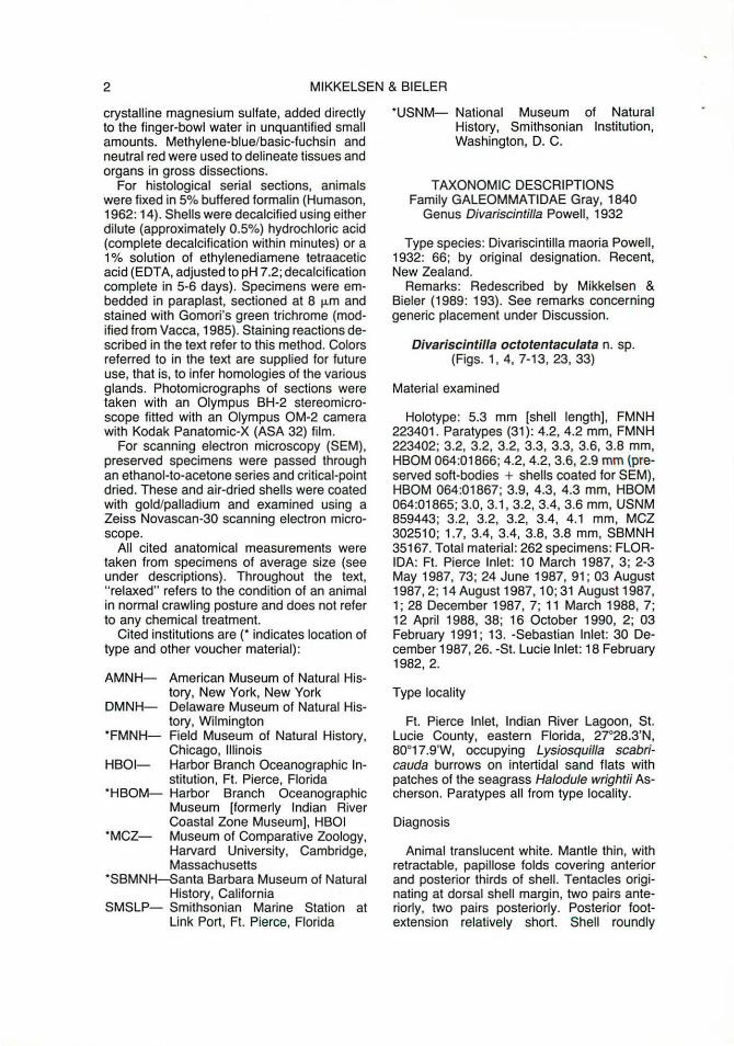
2 MIKKELSEN & BIELER
crystalline magnesium sulfate, added directly to the finger-bowl water in unquantified small amounts. Methylene-blue/basic-fuchsin and neutral red were used to delineate tissues and organs in gross dissections.
For histological serial sections, animals were fixed in 5% buffered formalin (Humason, 1962: 14). Shells were decalcified using either dilute (approximately 0.5%) hydrochloric acid (complete decalcification within minutes) or a 1 % solution of ethylenediamene tetraacetic acid (EDTA, adjusted to pH 7.2; decalcification complete in 5-6 days). Specimens were embedded in paraplast, sectioned at 8 µm and stained with Gomori's green trichrome (modified from Vacca, 1985). Staining reactions described in the text refer to this method. Colors referred to in the text are supplied for future use, that is, to infer homologies of the various glands. Photomicrographs of sections were taken with an Olympus BH-2 stereomicroscope fitted with an Olympus OM-2 camera with Kodak Panatomic-X (ASA 32) film.
For scanning electron microscopy (SEM), preserved specimens were passed through an ethanol-to-acetone series and critical-point dried. These and air-dried shells were coated with gold/palladium and examined using a Zeiss Novascan-30 scanning electron microscope.
All cited anatomical measurements were taken from specimens of average size (see under descriptions) . Throughout the text, "relaxed" refers to the condition of an animal in normal crawling posture and does not refer to any chemical treatment.
Cited institutions are (* indicates location of type and other voucher material):
AMNH- American Museum of Natural History, New York, New York
DMNH- Delaware Museum of Natural History, Wilmington
*FMNH- Field Museum of Natural History, Chicago, Illinois
HBOI- Harbor Branch Oceanographic In-stitution, Ft. Pierce, Florida
*HBOM- Harbor Branch Oceanographic Museum [formerly Indian River Coastal Zone Museum], HBOI
*MCZ- Museum of Comparative Zoology, Harvard University, Cambridge, Massachusetts
*SBMNH--Santa Barbara Museum of Natural History, California
SMSLP- Smithsonian Marine Station at Link Port , Ft. Pierce, Florida
*USNM- National Museum of Natural History, Smithsonian Institution, Washington, D. C.
TAXONOMIC DESCRIPTIONS Family GALEOMMATIDAE Gray, 1840
Genus Divariscintilla Powell, 1932
Type species: Divariscintilla maoria Powell, 1932: 66; by original designation. Recent, New Zealand.
Remarks: Redescribed by Mikkelsen & Bieler (1989: 193). See remarks concerning generic placement under Discussion.
Divariscintilla octotentaculata n. sp. (Figs. 1, 4, 7-13, 23, 33)
Material examined
Holotype: 5.3 mm [shell length), FMNH 223401 . Paratypes (31 ): 4.2, 4.2 mm, FMNH 223402; 3.2, 3.2, 3.2, 3.3, 3.3, 3.6, 3.8 mm, HBOM 064:01866; 4.2, 4.2, 3.6, 2.9 mm (preserved soft-bodies + shells coated for SEM), HBOM 064:01867; 3.9, 4.3, 4.3 mm, HBOM 064:01865; 3.0, 3.1, 3.2, 3.4, 3.6 mm, USNM 859443; 3.2, 3.2, 3.2, 3.4, 4.1 mm, MCZ 302510; 1.7, 3.4, 3.4, 3.8, 3.8 mm, SBMNH 35167. Total material: 262 specimens: FLORIDA: Ft. Pierce Inlet: 10 March 1987, 3; 2-3 May 1987, 73; 24 June 1987, 91; 03 August 1987, 2; 14August 1987, 10; 31August1987, 1; 28 December 1987, 7; 11 March 1988, 7; 12 April 1988, 38; 16 October 1990, 2; 03 February 1991; 13. -Sebastian Inlet: 30 December 1987, 26. -St. Lucie Inlet: 18 February 1982, 2.
Type locality
Ft. Pierce Inlet, Indian River Lagoon, St. Lucie County, eastern Florida, 27°28.3'N, 80°17.9'W, occupying Lysiosquilla scabricauda burrows on intertidal sand flats with patches of the seagrass Halodule wrightii Ascherson. Paratypes all from type locality.
Diagnosis
Animal translucent white. Mantle thin, with retractable, papillose folds covering anterior and posterior thirds of shell. Tentacles originating at dorsal shell margin, two pairs anteriorly, two pairs posteriorly. Posterior footextension relatively short. Shell roundly
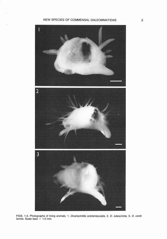
NEW SPECIES OF COMMENSAL GALEOMMATIDAE 3
FIGS. 1-3. Photographs of living animals. 1. Divariscintilla octotentaculata. 2. D. luteocrinita. 3. D. cordiformis. Scale bars = 1.0 mm.
4 MIKKELSEN & BIELER
ft
ft
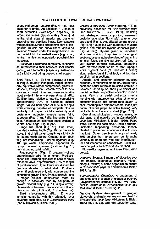
7 aapr ppr aam P"m ppm
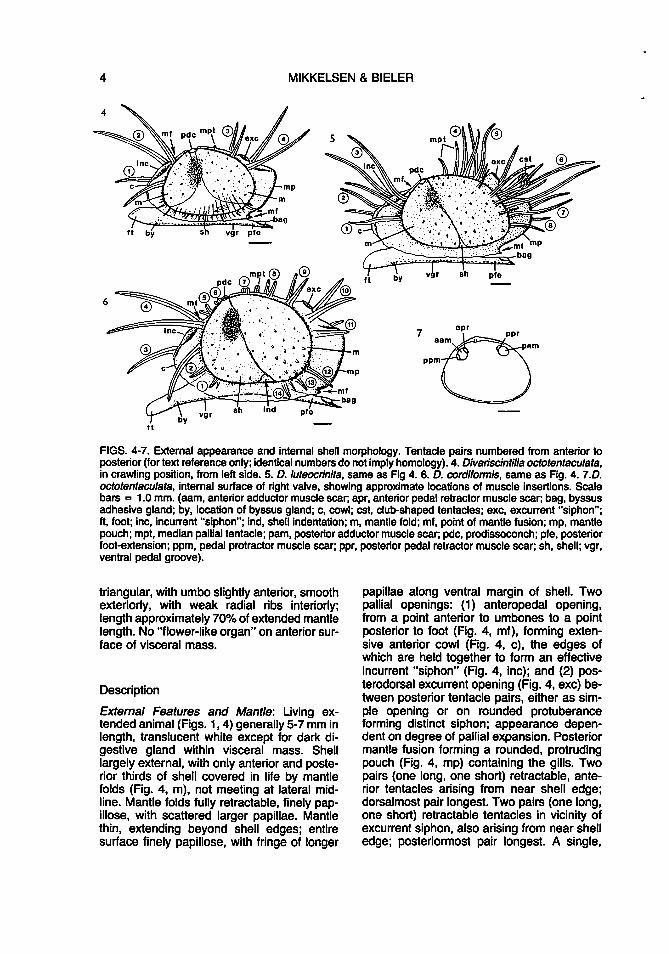
FIGS. 4-7. External appearance and internal shell morphology. Tentacle pairs numbered from anterior to posterior (for text reference only; identical numbers do not imply homology). 4. Divariscintilla octotentaculata, in crawling position, from left side. 5. D. luteocrinita, same as Fig 4. 6. D. cordiformis, same as Fig. 4. 7.D. octotentaculata, internal surface of right valve, showing approximate locations of muscle insertions. Scale bars = 1.0 mm. (aam, anterior adductor muscle scar; apr, anterior pedal retractor muscle scar; bag, byssus adhesive gland; by, location of byssus gland; c, cowl; est, club-shaped tentacles; exc, excurrent "siphon"; ft, foot; inc, incurrent "siphon"; ind, shell indentation; m, mantle fold; mf, point of mantle fusion; mp, mantle pouch; mpt, median pallial tentacle; pam, posterior adductor muscle scar; pdc, prodissoconch; pfe, posterior foot-extension; ppm, pedal protractor muscle scar; ppr, posterior pedal retractor muscle scar; sh, shell; vgr, ventral pedal groove).
triangular, with umbo slightly anterior, smooth exteriorly, with weak radial ribs interiorly; length approximately 70% of extended mantle length. No "flower-like organ" on anterior surface of visceral mass.
Description
External Features and Mantle: Living extended animal (Figs. 1, 4) generally 5-7 mm in length, translucent white except for dark digestive gland within visceral mass. Shell largely external, with only anterior and posterior thirds of shell covered in life by mantle folds (Fig. 4, m), not meeting at lateral midline. Mantle folds fully retractable, finely papillose, with scattered larger papillae. Mantle thin, extending beyond shell edges; entire surface finely papillose, with fringe of longer
papillae along ventral margin of shell. Two pallial openings: (1) anteropedal opening, from a point anterior to umbones to a point posterior to foot (Fig. 4, mf ), forming extensive anterior cowl (Fig. 4, c), the edges of which are held together to form an effective incurrent "siphon" (Fig. 4, inc); and (2) posterodorsal excurrent opening (Fig. 4, exc) between posterior tentacle pairs, either as simple opening or on rounded protuberance forming distinct siphon; appearance dependent on degree of pallial expansion. Posterior mantle fusion forming a rounded, protruding pouch (Fig. 4, mp) containing the gills. Two pairs (one long, one short) retractable, anterior tentacles arising from near shell edge; dorsalmost pair longest. Two pairs (one long, one short) retractable tentacles in vicinity of excurrent siphon, also arising from near shell edge; posteriormost pair longest. A single,
NEW SPECIES OF COMMENSAL GALEOMMATIDAE 5
short, mid-dorsal tentacle (Fig. 4, mpt), just posterior to umbo. An additional 1-2 pairs of short tentacles (=enlarged papillae?) in larger specimens (approximately 5 mm) at ventral shell edge at anterior and posterior ends of ventral papillose fringe. Each tentacle with papillose surface and central core of longitudinal muscle and nerve fibers, visible as an inner "thread" under low magnification. Inner pallial fold of non-shell areas (e.g., cowl, ventral mantle margin, posterior pouch) highly muscular.
Preserved specimens completely (or nearly so) retracted into shell, however, shell usually gaping, with tentacles (especially anterodorsal) slightly protruding beyond shell edges.
Shell (Figs. 7-11, 13): Shell generally 3-5 mm in length, roundly triangular, longer posteriorly, equivalve, rather compressed, glossy, iridescent, transparent, smooth except for fine concentric growth lines and weak radial ribs most evident interiorly at ventral margin (Fig. 9). Size large relative to mantle, comprising approximately 70% of extended mantle length. Valves held open at a 50-60x angle while crawling, capable of complete closure ventrally but gaping slightly anteriorly and posteriorly. Adductor muscle scars faint, subequal (Figs. 7, 9). Pallial line entire, indistinct. Periostracum colorless, most evident at ventral shell edge (Fig. 9, per).
Hinge line short (Fig. 10). One small, rounded cardinal tooth (Fig. 10, car) in each valve, that of left valve sometimes slightly bifid; lateral teeth absent. Cardinal teeth abutting, not interlocking. External ligament (Fig. 10, lig) weak, amphidetic, supported by nymph. Internal ligament (resilium; Fig. 10, res) stronger, opisthodetic.
Prodissoconch (Fig. 11) brownish-yellow, approximately 360 fm in length. Prodissoconch I corresponding in size to shell of newly released larva, approximately 32% of length of prodissoconch II; sculpture not discernible (surface abraded in adult shell). Prodissoconch II sculptured only with coarse and fine concentric growth lines. Prodissoconch I and II stages distinct, demarcated more by change in convexity than by sculpture or growth discontinuity (Fig. 11, single arrow). Demarcation between prodissoconch 11 and dissoconch abrupt (Figs. 8; 11, double arrow).
Shell microstructure (Fig. 13) crosslamellar centrally, with thin prismatic layer covering each side, as in Divariscintilla yoyo (see Mikkelsen & Bieler, 1989).
Organs of the Pallial Cavity. Foot (Fig. 4, ft) as previously described for Divariscintilla yoyo (see Mikkelsen & Bieler, 1989), including hatchet-shaped anterior portion, narrowed posterior extension (Fig. 4, pfe), anterior byssus gland (Fig. 4, by), ciliated ventral groove (Fig. 4, vgr) supplied with numerous mucous glands, and terminal byssus adhesive gland (Fig. 4, bag). Byssus gland of undefined structure, staining turquoise in histological sections; byssus adhesive gland of branching lamellar folds, staining purplish-red. One to four byssus threads produced, emanating from extreme posterior tip of foot. Opaque white pigment band (of unknown function) along anterodorsal tip of foot, staining dark purplish-red in sections.
Anterior and posterior adductor muscles subequal, of moderate diameter. Anterior and posterior pedal retractor muscles smaller in diameter, inserting on shell just dorsal and medial to their respective adductor muscle scars. Very small pedal protractor muscle merging with anteroventral edge of anterior adductor muscle just before both attach to shell; inserting into anterior visceral mass just dorsal of labial palps. Muscles leaving very faint attachment scars on shell (Figs. 7, 9).
Overall morphologies of visceral mass, labial palps and ctenidia as in Divariscintilla yoyo (see Mikkelsen & Bieler, 1989). Palps with 6-8 lamellae each side. Ctenidia smooth, unpleated (appearing posteriorly loosely pleated in preserved specimens due to contraction). Outer demibranch approximately 50% smaller than inner; both demibranchs ventrally rounded and with both interfilamental and interlamellar connectives. Cilial currents on palps and ctenidia not verified.
Flower-like organ absent (see Discussion below).
Digestive System: Structure of digestive system (mouth, esophagus, stomach, midgut, hindgut, rectum) of same organization as that in Divariscintilla yoyo (see Mikkelsen & Bieler, 1989: fig. 28).
Suprabranchial Chamber. Arrangement of openings and presence of glandular patches (?hypobranchial glands; Fig. 23, hyp) adjacent to rectum as in Divariscintilla yoyo (see Mikkelsen & Bieler, 1989: fig. 29).
Nervous System: Arrangement of ganglia, statocysts, and major nerves as described for Divariscintilla yoyo (see Mikkelsen & Bieler, 1989: fig. 31). Left and right posterior tenta-
6 MIKKELSEN & BIELER
cles innervated by branches from the pallial nerve, adjacent to its junction with the visceral ganglion. Anterior tentacles similarly innervated but both from a common branch of the pallial nerve, adjacent to its junction with the cerebro-pleural ganglion.
Reproductive System: Simultaneous hermaphrodite. Ovotestis white, encompassing most of volume of visceral mass, as in Divariscintilla yoyo (see Mikkelsen & Bieler. 1989).
Mature spermatozoa morphologically indistinguishable from that of D. yoyo (see Eckelbarger et al., 1990: figs. 28-29, table 1; D. octotentaculata as Scintilla sp.). except smaller in relative size. Spermatogenesis fully described by Eckelbarger et al. (1990; as Scintilla sp.).
Brooding large number of small larvae for variable period; brooding time for individuals collected with larvae, 9-15 days (n = 4); total brooding time from set to release in laboratory. 9, 10, and 12 days (n = 3). Larvae held within both demibranchs and in suprabranchial chamber, where they are circulated via pallial expansions and contractions. During brooding, excurrent siphonal opening constricted by sphincter-like muscle, sometimes noted around free end of rectum, allowing digestive processes to continue. Larvae initially white, turning pink with shell development on day 5-7 (n = 2); released as straight-hinged "D" larvae with apical flagella, 115-123 µ.m in shell length ( X: = 119 µ.m, n = 40; Fig. 12). Larvae expelled through excurrent siphon via strong contractions of shell and pallial muscles. Adults brooding larvae collected in May, June, December 1987, and March, April 1988; additional adults setting larvae in laboratory in February, May, June 1982 and November 1990; one specimen setting two broods in laboratory, four months apart, with second brood approximately 20% quantity of first. No apparent seasonality.
Circulatory and Excretory Systems: As described for Divariscintilla yoyo (see Mikkelsen & Bieler, 1989).
Distribution and Abundance
Known from three locations, all on intertidal and shallow subtidal sand flats within the Indian River Lagoon, eastern Florida: the type locality, Ft. Pierce Inlet (St. Lucie County, 27°28.3'N, 80°17.9'W), just north of St. Lucie Inlet (Martin County, 27°11.4'N, 80°11.1 'W),
and Sebastian Inlet (Brevard County, 27°51.6'N, 80°27.0'W). May be quite numerous; largest number per burrow sample = 7 4.
Etymology
An adjective, octotentaculatus, -a. -um, from the Latin octo (eight) and the late Latin tentaculum (a "feeler"), referring to the eight long mantle tentacles, a diagnostic feature.
Remarks
This is the most common of the five Divariscintilla species in the Lysiosquilla burrows (see Ecology and Behavior).
Prior to the beginning of this study in March 1987, two individuals of this species were encountered, and are presently the only known specimens of any of the Floridian burrow galeommatoideans from previous collections. These were collected in a shovel-and-sieve sample from a sand bar in the Indian River Lagoon, north of St. Lucie Inlet, Martin County, Florida, 27°11.4'N, 80°11.1 'W, on 18 February 1982. These individuals were maintained in the laboratory for approximately four months, providing material for notes and photographs on behavior, reproduction, and development.
Dlvarlsclntllla luteocrlnlta n. sp. (Figs. 2, 5, 14-22, 24-25)
Material examined
Holotype: 5.0 mm (shell length). FMNH 223403. Paratypes (12): 4.6 mm, FMNH 223407; 4.4 mm (shell only), FMNH 223404; 3.2, 3.9 mm, HBOM 064:01863; 4.9, 4.5, 4.1, 2.9 mm (shells only, coated for SEM). HBOM 064:01864; 4.8 mm, USNM 860195; 5.5 mm (shell only), USNM 859444; 3.4 mm, MCZ 302517; 3.5 mm, SBMNH 35168. Total material: 16 specimens: FLORIDA: Ft. Pierce Inlet: 10 March 1987, 1; 2-3 May 1987, 5; 24 June 1987, 1; 03 August 1987, 1; 14 August 1987, 2; 31 August 1987, 1; 12 April 1988, 1; 03 February 1990; 4.
Type locality
Ft. Pierce Inlet, Indian River Lagoon, St. Lucie County, eastern Florida, 27°28.3'N, 80°17.9'W, occupying Lysiosquilla scabricauda burrows on intertidal sand flats with

NEW SPECIES OF COMMENSAL GALEOMMATIDAE 7
Figs. 8-13. Shell of Divariscintilla octotentaculata (SEM). 8. Left valve, external view, 2.9 mm length, paratype, HBOM 064:01867. 9. Right valve, internal view, 3.6 mm length, paratype, HBOM 064:01867. 10.Hinge, anterior to left, paratype, HBOM 064:01867. Scale bar= 100 µm. 11 . Prodissoconch, 360 µm length. Single arrow = prodissoconch 1-11 boundary. Double arrow = prodissoconch 11-dissoconch boundary. 12. Newly released larval shell, 119 µm length. 13. Microstructure, with internal surface at top. Scale bar = 5 µm. (car, cardinal tooth; lig, external ligament; nym, nymph; per, periostracum; res, resilium).
8 MIKKELSEN & BIELER
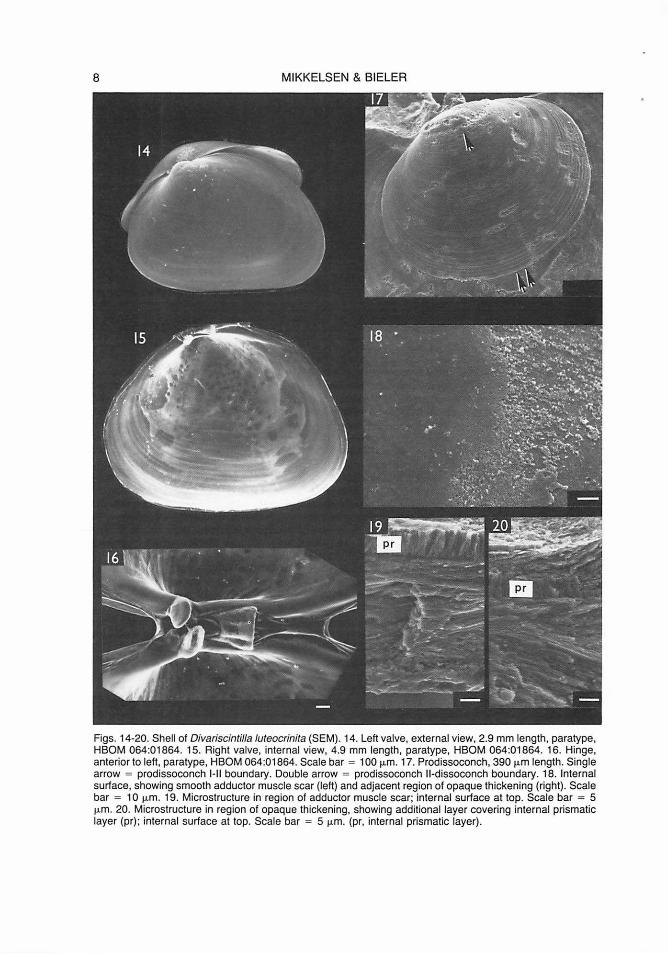
Figs. 14-20. Shell of Divariscintilla Juteocrinita (SEM). 14. Left valve, external view, 2.9 mm length, paratype, HBOM 064:01864. 15. Right valve, internal view, 4.9 mm length, paratype, HBOM 064:01864. 16. Hinge, anterior to left, paratype, HBOM 064:01864. Scale bar = 100 µm. 17. Prodissoconch, 390 µm length. Single arrow = prodissoconch 1-11 boundary. Double arrow = prodissoconch 11-dissoconch boundary. 18. Internal surface, showing smooth adductor muscle scar (left) and adjacent region of opaque thickening (right). Scale bar = 1 O µm. 19. Microstructure in region of adductor muscle scar; internal surface at top. Scale bar = 5 µm. 20. Microstructure in region of opaque thickening, showing additional layer covering internal prismatic layer (pr); internal surface at top. Scale bar = 5 µm. (pr, internal prismatic layer).
NEW SPECIES OF COMMENSAL GALEOMMATIDAE 9
patches of the seagrass Halodule wrightii Ascherson. Paratypes all from type locality.
Diagnosis
Animal translucent yellow. Mantle thin, with extensive, retractable, papillose folds completely covering shell, meeting at mid-line. Tentacles originating at shell edge, three pairs anteriorly, two singles and 9-11 pairs posteriorly, plus one pair club-shaped tentacles adjacent to excurrent opening. Posterior foot-extension relatively short. Shell roundly triangular, with umbo slightly anterior, smooth exteriorly, with opaque thickenings interiorly; length approximately 70% of extended mantle length. Single "flower-like organ" on anterior surface of visceral mass.
Description
External Features and Mantle: Living extended animal (Figs. 2, 5) generally 6-7 mm in length. Mantle and tentacles translucent pale yellow; foot white. Upper portion of digestive gland showing through mantle and shell as dark elongate-oval spot. Shell entirely covered in life by anterior and posterior mantle folds (Fig. 5, m), meeting at lateral dorso-ventral midline on each side. Mantle folds thin, incompletely retractable, entirely finely papillose, with scattered, elongated papillae especially posteroventrally. Mantle edge extending widely beyond shell edges; entire surface finely papillose. Pallial openings, musculature, and posterior pouch as in Divariscintilla octotentaculata. Numerous, long, retractile tentacles originating at shell edge: three pairs anterior to umbo; two singles plus five pairs posterior to umbo ( = two singles on midline + two pairs + [excurrent siphon] + three pairs). Shorter accessory pairs posterior to umbo originating from mantle fold near, but ventrad of, shell edge: one to three pairs anterior, two to three pairs posterior to excurrent siphon. Structure of these tentacles as in D. octotentaculata; those on shell edge adjacent to cowl and posterior pouch (third and sixth, see numbers, Fig. 5) longest. One prominent pair of thicker, whitish, club-shaped tentacles (Fig. 5, est) immediately posterior to excurrent siphon originating just inside shell edge; internal structure differing slightly from that of other tentacles (see Remarks).
Preserved specimens incompletely retracted into gaping shell; mantle folds contracted to narrow rim along shell edge, expos-
ing most of shell surface; tentacles, cowl, posterior pouch, and foot contracted but still usually extending beyond shell edges.
Shell (Figs. 14-20): Shell generally 4-5 mm in length, roundly triangular to oval, longer posteriorly, equivalve, rather inflated, glossy, transparent to translucent white, smooth except for fine concentric growth lines and irregular opaque thickening interiorly, imparting a white-blotched pattern (Fig. 15). Size large relative to mantle, comprising approximately 70% of extended mantle length. Valves held open at approximately 40° angle while crawling, incapable of complete closure. Adductor muscle scars subequal, distinct due to presence of surrounding shell thickening (Fig. 15). Periostracum as in Divariscintilla octotentaculata.
Hinge line short (Fig. 16), similar to that of Divariscintilla octotentaculata. Both cardinal teeth rounded.
Prodissoconch (Fig. 17) brownish-yellow, approximately 390 µm in length. Prodissoconch I approximately 145 µm in length, approximately 37% of length of prodissoconch II; sculpture not discernible (surface abraded in adult shell). Prodissoconch II sculptured with coarse concentric growth lines. Demarcation between prodissoconch I and II stages (Fig. 17, single arrow), and between prodissoconch II and dissoconch (Figs. 14; 17, double arrow) as in Divariscintilla octotentaculata.
Shell microstructure as in Divariscintilla octotentaculata, except with additional layer of parallel crystals covering internal perpendicular prismatic layer, forming regions of opaque thickening (Figs. 18-20).
Organs of the Palfial Cavity. Foot (Fig. 5, ft) as in Divariscintilla octotentaculata. Adductor, pedal retractor, and pedal protractor muscles (including relative positions) as in D. octotentaculata. Muscles leaving distinct attachment scars on shell (Fig. 15).
Visceral mass, labial palps and ctenidia as in Divariscintilla octotentaculata. Palps with approximately 8 lamellae each side. Outer demibranch approximately 40% smaller than inner. Cilial currents on palps and ctenidia unknown.
A single "flower-like organ" (Figs. 21-22; see Mikkelsen & Bieler, 1989, for explanation of term) on anterior surface of visceral mass just ventral to labial palps. Size variable, not correlated with shell length. Digestive System: As in Divariscintilla octotentaculata.

10 MIKKELSEN & BIELER
Suprabranchial Chamber. As in Divariscintilla octotentaculata, except that the whitish glandular patches (?hypobranchial glands) are much more extensive (Fig. 24).
Nervous System: Arrangement of ganglia, statocysts, and major nerves as described for Divariscintilla yoyo (see Mikkelsen & Bieler, 1989: fig. 31 ). Additional tentacular nerves arising (independently) from pallial nerve.
Reproductive System: Overall gross morphology as in Divariscintilla octotentaculata. Reproductive mode could not be determined from sectioned specimen, which showed no recognizable developed gametes. Adults brooding larvae have not been collected.
Circulatory and Excretory Systems: As in Divariscintilla octotentaculata.
Distribution and Abundance
Known only from the type locality, Ft. Pierce Inlet, St. Lucie County, Florida, 27°28.3'N, 80°17.9'W, on intertidal and shallow subtidal sand flats. Uncommon; only 16 specimens known.
Etymology
An adjective, luteocrinitus, -a, -um, from the Latin luteus (yellow) and the Latin crinitus (hairy), referring to the numerous, long, yellowish tentacles, imparting an overall "hairy" appearance.
Remarks
The single posterior pair of club-shaped tentacles is distinct and consistent in both living and preserved specimens. Normal tentacles (Fig. 25, nt; similar morphology in all five Floridian Divariscintilla spp.) show usually four haemocoelic compartments in cross section, each of these supplied with a longitudinal muscle bundle and nerve fiber, and separated by connective tissue septa. The muscle and nerve fibers form a more-or-less concentrated central core, appearing as a central thread under low magnification. Judd (1971: fig. 6) figured a similar internal morphology for tentacles of Divariscintilla maoria, but the individual compartmental muscle-plus-nerve bundles are not as concentrated at the core of the tentacle; this difference may not be real, but rather implied by Judd's interpretation of the histological sections. An additional difference
is found in the outer surfaces of the tentacles of D. luteocrinita, which are highly papillose and convoluted rather than smooth as in D. maoria as shown by Judd (1971 : fig. 6). Internally, the club-shaped tentacles of D. luteocrinita (Figs. 5, 25, est) are identical in structure to normal tentacles except that the muscle fibers (Fig. 25, arrow) are dispersed over the surfaces of the septa instead of being concentrated at the core of the tentacle. This difference may allow this particular pair of tentacles to undergo stronger contraction, resulting in the club-like shape. Under full extension, the club shape and whitish coloration of these tentacles disappears, indicating that these features are products of the normally contracted state.
The function of the club-shaped tentacles is unknown. They are probably not homologous to the posterior "defensive appendages" of D. maoria (see Judd, 1971 : fig. 7) or Galeomma takii (Kuroda, 1945) (see B. Morton, 1973a: fig. 5), which can be autotomized and possess only a single, central haemocoelic tube. No tentacles in D. luteocrinita have this morphology nor were they ever seen to autotomize. However, these tentacles were often observed to hyperelongate when the animals were disturbed, for example during specimen transfer between laboratory dishes. While most other tentacles contracted, the clubshaped tentacles elongated immediately to 2-3 times the shell length just as the transfer spoon made contact. This bears resemblance to the dymantic ("threatening") display of tentacles in Galeomma polita Deshayes, 1856 (see B. Morton, 1975), and Ephippodonta oedipus Morton, 1976 (see B. Morton, 1976), when disturbed, and suggests defensive function. Unlike the dymantic tentacles of the latter two species, however, the club-shaped tentacles of D. luteocrinita do not fully retract into the mantle at rest.
The large "?hypobranchial glands" of this species are located at sites similar to those of the smaller glands of other Divariscintilla species, i.e., adjacent to the rectum and the branchial nerves as they join the visceral ganglia. In histological sections, they bear striking resemblance to "seminal receptacles" described and figured for Aligena elevata (Stimpson, 1851) by Fox (1979: 101, 103, fig. 32), although in the single sectioned specimen of D. luteocrinita, they contained no sperm. The montacutid Aligena elevata is a protandrous hermaphrodite, and its seminal receptacles are present only during the fe-

NEW SPECIES OF COMMENSAL GALEOMMATIDAE 11
Figs. 21-22. Flower-like organ of Divariscintilla luteocrinita (SEM). Anterior tip of foot has been severed to enhance visibility. 21. Scale bar = 100 µm. 22. Scale bar = 50 µm. (ct, ctenidium; fl, flower-l ike organ; ft, foot ; Ip, labial palp; m, mantle).
male stage. The occurrence of such structures in Galeommatoidea was summarized by Fox (1979: table 8, as Leptonacea, 15 species), who noted that sperm storage organs were unknown outside of Montacutidae. However, protandry is known in members of other families (e.g. Lasaeidae: Arthritica crassiformis Powell, 1933 (see B. Morton, 1973b); see also Fox, 1979: table 11 ). The reproductive mode of 0 . luteocrinita is presently unknown, but seminal receptacles and/or protandry would be no surprise within the context of this reproductively complex superfamily.
Divariscintilla cordiformis n. sp. (Figs. 3, 6, 26-32)
Material Examined
Holotype: 5.6 mm (shell length), FMNH 223405. Paratypes (4): 6.4 mm (sectioned on 29 microslides), FMNH 223406; 5.4 mm, HBOM 064:1861; 4.9 mm (partially dissected; prodissoconch coated for SEM), HBOM 064: 01862; 6.4 mm (shell only, coated for SEM), USNM 859445. Total material: 6 specimens: FLORIDA: Ft. Pierce Inlet: 24 June 1987, 2; -Peanut Island: 10 August 1987, 4.
Type locality
Peanut Island, near Lake Worth Inlet, Palm Beach County, eastern Florida, 26°46.6'N, 80°02. 7'W, occupying Lysiosquil/a scabri-
cauda burrows on intertidal sand flats with patches of the seagrass Halodule wrightii Ascherson. Paratypes from type locality (FMNH, USNM, HBOM 064:01861) or Ft. Pierce Inlet (HBOM 064:01862).
Diagnosis
Animal translucent white. Mantle thin , with extensive, retractable papillose folds covering shell completely, meeting at midline. Tentacles originating at shell edge, six pairs anteriorly, eight pairs posteriorly. Posterior footextension relatively short. Shell oval, with umbo slightly anterior; length approximately 65% of extended mantle length. Small, ventral, anteriorly directed indentation in each valve. Coarse growth lines and slightly beaded radial ribs restricted to edges of otherwise-smooth shell. Single "flower-like organ" on anterior surface of visceral mass.
Description
External Features and Mantle: Living extended animal (Figs. 3, 6) generally 7-10 mm in length, translucent white except for dark digestive gland within visceral mass. Shell entirely covered in life by anterior and posterior mantle folds (Fig. 6, m) meeting at lateral dorso-ventral mid-line. Mantle folds thin, incompletely retractable, entirely finely papillose, with scattered larger papillae, which are longest at posteroventral section. Mantle edge extending widely beyond shell edges;

12 MIKKELSEN & BIELER
23
sh
n-
. . ; mus 1::1
25 est ,.,£.
·~
Figs. 23-25. Histological sections. 23. ?Hypobranchial gland adjacent to rectum in 0. octotentaculata. 24. ?Hypobranchial gland adjacent to rectum in 0 . luteocrinita. 25. Cross-sections of normal and club-shaped tentacles in O. luteocrinita. Arrow = muscularized septum. Scale bars = 100 µm. (est, club-shaped tentacle; ct, ctenidium; hyp, ?hypobranchial gland; mus, muscle fibers; n, nerve fiber; nt, normal tentacle; pam, posterior adductor muscle; r, rectum; sbc, suprabranchial chamber; sep, connective tissue septum; sh, shell; ten, tentacle).
entire surface finely papillose, with additional enlarged papillae near shell edge. Ventral mantle edge entire, not cleft in vicinity of ventral shell indentation. Pallial openings, musculature, and posterior pouch as in Divariscintilla octotentaculata. Paired, retractable, pallial tentacles numerous, originating near shell edge. Six anterior pairs, those adjacent to cowl area (third and fourth, see numbers, Fig. 6) longest. Eight posterior pairs, those adjacent to excurrent siphon (tenth) longest, but shorter than longest anterior tentacles. A single median tentacle (Fig. 6, mpt) between first and second posterior pairs (Fig. 6, nos. 7, 8). Structure of individual tentacles as in 0 .
octotentaculata. Excurrent opening (Fig. 6, exc) between ninth and tenth tentacle pairs.
Preserved animals not fully retracted into gaping shell; mantle folds contracted to narrow rim along shell edge, exposing most of shell surface; tentacles, cowl, and posterior pouch contracted but still usually extending beyond shell edges; foot usually completely withdrawn into pallial cavity.
Shell (Figs. 26-30, 32): Shell generally 5-7 mm in length, nearly evenly oval, longer posteriorly, equivalve, compressed, glossy, transparent to translucent white. Small indentation (Figs. 6, ind; 27, arrow; 28) at mid-ventral
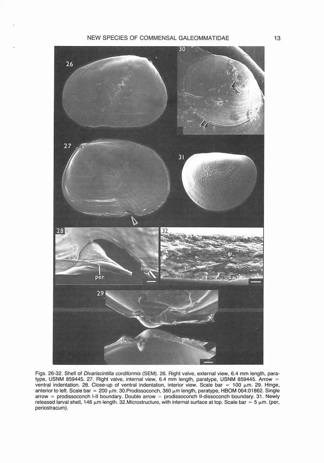
NEW SPECIES OF COMMENSAL GALEOMMATIDAE 13
Figs. 26-32. Shell of Divariscintilla cordiformis (SEM). 26. Right valve, external view, 6.4 mm length, paratype, USNM 859445. 27. Right valve, internal view, 6.4 mm length, paratype, USNM 859445. Arrow = ventral indentation. 28. Close-up of ventral indentation, interior view. Scale bar = 100 µm. 29. Hinge, anterior to left. Scale bar = 200 µm. 30.Prodissoconch, 360 µm length, paratype, HBOM 064:01862. Single arrow = prodissoconch 1- 11 boundary. Double arrow = prodissoconch 11-dissoconch boundary. 31. Newly released larval shell, 146 µm length. 32.Microstructure, with internal surface at top. Scale bar = 5 µm. (per, periostracum).
14 MIKKELSEN & BIELER
margin, slanting anteriorly toward umbo, evident only on distal 1 mm or so of shell growth (as evidenced by growth lines). Exterior sculpture smooth except for fine concentric growth lines, heavier at anteroventral margin, anterior to indentation. Beaded radial ribs restricted to distal 1 mm or so of shell edge, most prevalent interiorly at posteroventral margin (Fig. 27) and exteriorly at antero- and posterodorsal margins (Fig. 26), forming distinct, fine crenulation at shell edge, absent only at ventral margin immediately anterior to indentation. Size large relative to mantle, comprising approximately 65% of extended mantle length. Valves held open at 20-30° angle while crawling, incapable of complete closure. Adductor muscle scars subequal, faint. Periostracum as in Divariscintilla octotentaculata; also evident covering ventral indentation (Fig. 28, per).
Hinge line (Fig. 29) short, as in Divariscintilla octotentacu/ata, with two small rounded cardinal teeth.
Prodissoconch (Fig. 30) brownish-yellow, approximately 360 µ.m in length. Prodissoconch I corresponding in size to shell of newly released larva, approximately 45% of length of prodissoconch II; sculpture not discernible (surface abraded in adult shell). Prodissoconch II sculptured with coarse concentric growth lines. Demarcation between prodissoconch I and II stages (Fig. 30, single arrow), and between prodissoconch II and dissoconch (Fig. 30, double arrow) as in Divariscintilla octotentaculata.
Organs of the Pallial Cavity. Foot and shell muscles (adductors, pedal retractors, pedal protractors, including relative positions) as in Divariscintilla octotentaculata. Muscles leaving very faint attachment scars on shell (Fig. 27).
Visceral mass, labial palps and ctenidia as in Divariscintilla octotentaculata. Palps with approximately seven lamellae each side. Outer demibranch approximately 35% smaller than inner. Cilial currents on palps and ctenidia unknown.
Single "flower-like organ" (see Mikkelsen & Bieler, 1989) on anterior surface of visceral mass just ventral to labial palps.
Digestive System: Similar to that of Divariscintilla octotentaculata (relative positions of gastric shield, style sac, digestive diverticula, midgut, etc.), based on histological sections. Limited number of specimens did not permit
confirmation of structure through gross dissection.
Suprabranchial Chamber. As in Divariscintilla octotentacu/ata.
Nervous System: Arrangement of ganglia, statocysts, and major nerves as described for Divariscintilla yoyo (see Mikkelsen & Bieler, 1989: fig. 31). Additional tentacular nerves arising (independently) from pallial nerve.
Reproductive System: Simultaneous hermaphrodite. Overall gross morphology as in Divariscintilla octotentaculata.
One specimen brooding larvae collected in August 1987. Larvae released one day after collection. Newly-released larval shells 136-148 µ.m in length (x = 143 µm, n = 40; Fig. 31 ). Adult preserved and subsequently sectioned; apparently intact larvae found in suprabranchial chamber and throughout digestive system, including intestine and rectum (therefore not being digested).
Circulatory and Excretory Systems: As in Divariscintilla octotentaculata.
Distribution and Abundance
Known from the type locality at Peanut Island, Palm Beach County, and from sand flats at Ft. Pierce Inlet, St. Lucie County, Florida, 27°28.3'N, 80°17.9'W. Rare; only 6 specimens known.
Etymology
An adjective, cordiformis, -e, from the Latin cordis (heart) and the Latin forms (shape), referring to the ventrally indented shell outline.
Remarks
As in Divariscintilla maoria (see Judd, 1971 ), the ventral indentation in the shell of this species does not seem to be functionally important. In both species, it is present in both valves, anteriorly inclined, not developed in juveniles, and not reflected by soft anatomy.
ECOLOGY AND BEHAVIOR
As in the two previously described commensal galeommatids (Mikkelsen & Bieler, 1989), no specimens of the newly described species were ever found physically attached
NEW SPECIES OF COMMENSAL GALEOMMATIDAE 15
to a mantis shrimp, either in the field or in museum specimens (HBOM). They are assumed to be free-living within the vertical portions of the LI-shaped burrow, although specimens were never visible at the opening prior to pumping. Again as with the previous species (Mikkelsen & Bieler, 1989), these clams were never found free-living outside of the burrows or associated with any other burrowing invertebrate in the area (e.g. other mantis shrimps, callianassid shrimps, polychaetes, sipunculans), nor were any empty shells located in dry collections (AMNH, DMNH, FMNH, HBOM, LISNM), probably because of their fragile nature.
Oivariscintilla octotentaculata was the most frequently encountered commensal mollusk in the Lysiosquilla burrows; of the 35 burrows containing mollusks, 31 contained 0. octotentaculata, 20 0. yoyo, 19 0. troglodytes, 8 0. /uteocrinita, 6 Cyclostremiscus beauii, 7 Circulus texanus, and only 2 0. cordiformis. Oivariscintilla octotentaculata was usually collected with other commensals, occurring alone in only 6 of the 31 samples. Oivariscintilla luteocrinita was always collected with other commensals. Oivariscintilla cordiformis was collected once with 0. octotentaculata, and once alone. Densities of 0. octotentaculata varied greatly, ranging from 1-74 per sample (x = 8.3, n = 31 ). Oivariscintilla luteocrinita was usually present in numbers of only one or two specimens per burrow; one sample contained four specimens. Oivariscintilla cordiformis was encountered only twice, once as two specimens, once as four. Only a small number of burrows sampled contained commensals. And, as previously reported (Mikkelsen & Bieler, 1989), it must be emphasized that in no case could an entire burrow be sampled using the yabby pump, which only effectively samples its own length (0.5-1.0 m) of the vertical parts of the LI-shaped burrow. Estimates of occurrence and/or density of any clams living in the deeper horizontal section of the burrow was thus not possible.
Animals of all three species spent most of their time in the laboratory attached to the glass surface of laboratory bowls by up to four byssus threads, and it is assumed that this is also their habit on the smooth walls of the Lysiosquilla burrow. When dislodged, they actively crawl about using an even, gliding motion produced by ciliary action on the central surface of the foot. This was equally effective on the underside of the water surface as on glass. They made no attempts to bur-
row when offered a substrate of loose sand in the laboratory.
All three Oivariscintilla species previously described (0. maoria, Judd, 1971: fig. 4; 0. yoyo and 0. troglodytes, Mikkelsen & Bieler, 1989: fig. 32) are known to "hang" from a vertical substrate by the posterior foot-extension, which in these species is extremely long and elastic; byssus threads secreted by the anteriorly located byssus gland are laid down within the ventral groove of the foot and emerge from the terminus to attach to the substrate. The threads are secured within the groove by secretions of the posteriorly located byssus adhesive gland. Byssus and byssus adhesive glands of similar morphologies are present in each of the three new species described here and are assumed to function in the same way. This has been confirmed for 0. octotentaculata and 0. luteocrinita, in which secretion of byssus threads, accompanied by distinct pulsing of the byssus gland area, was observed as described for 0. yoyo and 0. troglodytes (Mikkelsen & Bieler, 1989). Following this activity, the clam hangs from the posterior foot-extension, with the byssus threads emerging from the posterior terminus of the foot in the vicinity of the byssus adhesive gland. The posterior foot-extension of these species, as well as of 0. cordiformis, is not as elongated and extensible as in those previously described, therefore the distinctive "hanging" posture, wherein the clam "dangles" from an elongated foot, is not as pronounced. 3
A peculiar interaction between pairs of Oivariscintilla species was observed in the laboratory on three occasions. In two instances involving 0. octotentaculata, one animal of a pair was noted reaching its foot into the mantle cavity of the second specimen, either from in front of or behind its partner. On one of these occasions, the two individuals performed this activity simultaneously, "facing" one another, with each one reaching around the visceral mass of the other to contact the posterior surface with the tip of its foot (Fig. 33). Two specimens of 0. yoyo have been
3Careful notes were not recorded on the behavior of D. cordiformis in the early phase of the study, and the lnavailability of additional living specimens prevented confirmation of the presence or absence of "hanging" behavior. Morphology suggests that this behavior does occur, however, the shorter posterior extension and the absence of any mention in our preliminary written observations Indicates that the "hanging" posture was Inconspicuous, as in D. octotentaculata and D. luteocrlnita.

16 MIKKELSEN & BIELER
Fig. 33. Mating (?) behavior between two specimens of Divariscintilla octotentaculata, as observed in laboratory.
noted performing this same type of activity. In all three instances, the interaction was initiated by the smaller of the two specimens of the pair, and the activity was sustained for 3-7 minutes. Although there is no direct evidence, it seems likely that this observed interaction is part of some sort of reproductive activity, perhaps stimulation of sperm transfer (external gonadal openings are located on the posterodorsal surface of the visceral mass). Each of the two specimens involved in the simultaneous behavior described above had larvae in their suprabranchial chambers within three weeks of the noted activity. (If this behavior is in fact copulatory in function, then this confirms simultaneous hermaphroditism in these species.)
Observed animals of these Divariscintilla species do not respond to changes in light intensity (e.g. photographic strobes) and did not seek darkness when offered "artificial burrows" (in the form of black plastic tubes) in the laboratory.
DISCUSSION
Generic Placement
The genus Divariscintilla was redescribed in a previous part of this study on Lysiosquillaassociated mollusks (Mikkelsen & Bieler, 1989). Major emphasis was placed on two features, the presence of flower-like organs and a bipartite foot with both byssus and byssus-adhesive glands, shared between these species and the type species, D. maoria Powell, 1932 (the latter described by Judd, 1971 ).
We regarded the presence of an apparently functionless (Judd, 1971) shell notch in D. maoria, as well as the difference in degree of shell coverage by the mantle, as specieslevel rather than generic characters. At the time, we also considered placement of D. yoyo and D. troglodytes in the monotypic genus Ph/yctaenachlamys Popham, 1939, based on P. /ysiosquillina Popham, 1939. However, there are a number of anatomical characters that distinguish the Divariscintilla species (flower organs, hindgut typhlosole, interlamellar ctenidial junctions, relative size of adductor muscles, hinge teeth; compare data of Popham, 1939, and Mikkelsen & Bieler, 1989).
Divariscintilla luteocrinita and D. cordiformis fit well within Divariscintilla, as redescribed. In fact, these new species actually bridge several morphological gaps between D. maoria and D. yoyo/D. troglodytes, that is shell notch, posterior shell prolongation, less shell internalization, and more numerous posterior tentacles. Divariscintilla octotentaculata does not possess the flower-like organs; however, in all other taxonomic characters (hinge, mantle, ctenidia, etc.), it does conform to the redefined genus. The hinge teeth of this species, comprised (as in other Divariscintilla species) of only one small cardinal tooth in each valve, was an important element in deciding generic placement. Hinge structure is presently considered taxonomically important at the generic level in Galeommatoidea (P. H. Scott, in litt., October 1990). Most members of the superfamily possess more than one cardinal tooth in at least one valve; some show distinct lateral teeth as well. The genus Sein-

NEW SPECIES OF COMMENSAL GALEOMMATIDAE 17
TABLE 1. Distinguishing characteristics of all described species of Divariscintilla: an expansion of table 1 from Mikkelsen & Bieler (1989).
D. maoria (from Judd, D. D. D. D.
1971) D. yoyo troglodytes octotentaculata luteocrinita cordiformis
Shell: General shape oval elongate- oval roundly roundly oval
pointed triangular triangular Ventral indentation present absent absent absent absent present Prolongation posterior anterior anterior posterior posterior posterior Internal sculpture unribbed unribbed radially radially unribbed, with radially ribbed
ribbed ribbed internal (marginally) (marginally) (marginally) thickening
Length relative to extended mantle length 68% 40% 50% 70% 70% 65%
Prodissoconch length (µm) "small" 360 380 360 390 360
Mean newly-released larval shell
length (µm) unknown 132 126 119 unknown 143 Mantle:
Color, thickness (not whitish, yellowish, whitish, thin yellowish, whitish, thin given) thick thin thin
Extent covering margins entire, entire, anterior and entire, entire, shell only umbonal anterior slit posterior thirds mid line midline
foramen overlap overlap Papillae very sparse, numerous, numerous, numerous, numerous,
small very small small, small, small, small, evenly- longer at evenly- evenly-distributed ventral distributed distributed
edge Anterior tentacles 2 pairs 1 pair 2 pairs 2 pairs 3 pairs 6 pairs Posterior tentacles 1 single 1 single 1 single, 1 single, 2 singles, 1 single,
1 pair 2-3 pairs 5 pairs, 8 pairs + 3-6 pairs accessory, + 1 pair club-shaped
Defensive appendages 6-8 absent absent absent absent absent
present Pedal protractor
muscle insertion relative to anterior adductor muscle (not dorsal dorsal ventral ventral ventral
given) Flower-like organs:
number 3-7 0 (usually 5)
Labial palps: Lamellae per palp approx. 9 10-14 14-20 6-8 approx. 8 approx. 7
Ctenidia: smooth pleated pleated smooth smooth smooth? Geographical range: New eastern eastern eastern eastern eastern
Zealand Florida Florida Florida Florida Florida
ti/Iona Finlay, 1927, is the only other genus bers of the latter genus are all attached ecto-known to us in which members have a single commensals on echinoderms, and in addi-cardinal tooth in each valve. However, mem- tion, are distinguished by a highly specialized,
18 MIKKELSEN & BIELER
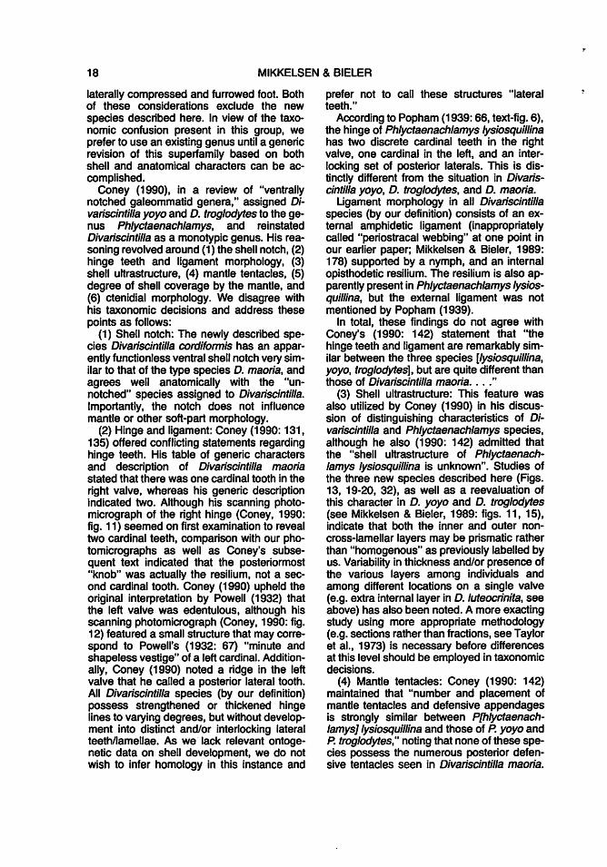
laterally compressed and furrowed foot. Both of these considerations exclude the new species described here. In view of the taxonomic confusion present in this group, we prefer to use an existing genus until a generic revision of this superfamily based on both shell and anatomical characters can be accomplished.
Coney (1990), in a review of "ventrally notched galeommatid genera," assigned Divariscintilla yoyo and D. troglodytes to the genus Phlyctaenachlamys, and reinstated Divariscintilla as a monotypic genus. His reasoning revolved around (1) the shell notch, (2) hinge teeth and ligament morphology, (3) shell ultrastructure, (4) mantle tentacles, (5) degree of shell coverage by the mantle, and (6) ctenidial morphology. We disagree with his taxonomic decisions and address these points as follows:
(1) Shell notch: The newly described species Divariscintilla cordiformis has an apparently functionless ventral shell notch very similar to that of the type species D. maoria, and agrees well anatomically with the "unnotched" species assigned to Divariscintilla. Importantly, the notch does not influence mantle or other soft-part morphology.
(2) Hinge and ligament: Coney (1990: 131, 135) offered conflicting statements regarding hinge teeth. His table of generic characters and description of Divariscintilla maoria stated that there was one cardinal tooth in the right valve, whereas his generic description indicated two. Although his scanning photomicrograph of the right hinge (Coney, 1990: fig. 11) seemed on first examination to reveal two cardinal teeth, comparison with our photomicrographs as well as Coney's subsequent text indicated that the posteriormost "knob" was actually the resilium, not a second cardinal tooth. Coney (1990) upheld the original interpretation by Powell (1932) that the left valve was edentulous, although his scanning photomicrograph (Coney, 1990: fig. 12) featured a small structure that may correspond to Powell's (1932: 67) "minute and shapeless vestige" of a left cardinal. Additionally, Coney (1990) noted a ridge in the left valve that he called a posterior lateral tooth. All Divariscintilla species (by our definition) possess strengthened or thickened hinge lines to varying degrees, but without development into distinct and/or interlocking lateral teeth/lamellae. As we· lack relevant ontogenetic data on shell development, we do not wish to infer homology in this instance and
prefer not to call these structures "lateral teeth."
According to Popham (1939: 66, text-fig. 6), the hinge of Phlyctaenachlamys lysiosquillina has two discrete cardinal teeth in the right valve, one cardinal in the left, and an interlocking set of posterior laterals. This is distinctly different from the situation in Divariscintilla yoyo, D. troglodytes, and D. maoria.
Ligament morphology in all Divariscintilla species (by our definition) consists of an external amphidetic ligament (inappropriately called "periostracal webbing" at one point in our earlier paper; Mikkelsen & Bieler, 1989: 178) supported by a nymph, and an internal opisthodetic resilium. The resilium is also apparently present in Phlyctaenachlamys lysiosquillina, but the external ligament was not mentioned by Popham (1939).
In total, these findings do not agree with Coney's (1990: 142) statement that "the hinge teeth and ligament are remarkably similar between the three species [lysiosquillina, yoyo, troglodytes], but are quite different than those of Divariscintilla maoria . .. .''
(3) Shell ultrastructure: This feature was also utilized by Coney (1990) in his discussion of distinguishing characteristics of Divariscintilla and Phlyctaenachlamys species, although he also (1990: 142) admitted that the "shell ultrastructure of Phlyctaenachlamys lysiosquillina is unknown". Studies of the three new species described here (Figs. 13, 19-20, 32), as well as a reevaluation of this character in D. yoyo and D. troglodytes (see Mikkelsen & Bieler, 1989: figs. 11, 15), indicate that both the inner and outer noncross-lamellar layers may be prismatic rather than "homogenous" as previously labelled by us. Variability in thickness and/or presence of the various layers among individuals and among different locations on a single valve (e.g. extra internal layer in D. luteocrinita, see above) has also been noted. A more exacting study using more appropriate methodology (e.g. sections rather than fractions, see Taylor et al., 1973) is necessary before differences at this level should be employed in taxonomic decisions.
(4) Mantle tentacles: Coney (1990: 142) maintained that "number and placement of mantle tentacles and defensive appendages is strongly similar between P[hlyctaenachlamys] lysiosquillina and those of P. yoyo and P. troglodytes," noting that none of these species possess the numerous posterior defensive tentacles seen in Divariscintilla maoria.

NEW SPECIES OF COMMENSAL GALEOMMATIDAE 19
The three new species described here in Divariscintilla all possess a number of posterior tentacles, albeit none "defensive... The two primary anterior tentacles, also mentioned specifically by Coney (1990: 142) to combine P. lysiosquillina, D. yoyo, and D. troglodytes, are in fact also present in Divariscintilla maoria.
(5) Shell coverage: Although there is a distinct difference in the degree of shell coverage by the mantle between the type species and Divariscintilla yoyo and D. troglodytes, the three newly described species show intermediate conditions. All Divariscintilla species (by our definition) show at least some degree of shell exposure, thus differing from the condition in Phlyctaenachlamys lysiosquillina ("the shell is completely embedded11
; Popham, 1939: 65). A similar range of variability was described for the genus Ephippodonta Tate, 1889, by Arakawa (1960: 57), and can also be found in Entovalva Voltzkow, 1890, sensu Jato, wherein the genus Devonia was distinguished by Winckworth (1930: 14) for a species with incomplete shell coverage.
(6) Ctenidial morphology: Coney (1990: 142) noted that the ctenidia in Divariscintilla maoria are smooth, whereas those of Phlyctaenach/amys lysiosquillina, D. yoyo and D. troglodytes have been described as pleated. However, unlike pleated ( = plicate) gills in other bivalve groups (e.g. Pecten; see Rice, 1897), pleating in these species is not based on structural differences in the filaments (pars. obs.; Popham, 1939: 71 ). Some degree of "pleating11 caused by contraction in preserved specimens has been noted during the present study. An additional ctenidial character separates P. /ysiosquillina and the five Floridian species of Divariscintilla, in that the latter have interlamellar junctions (unknown for New Zealand D. maoria).
Comparative characteristics for all six known species of Divariscintilla are presented in Table 1. The three new species described here more closely resemble the type species, D. maoria (see Judd, 1971 : fig. 1 ), in general morphology than do the other species previously described during this study (D. yoyo and D. troglodytes, see Mikkelsen & Bieler, 1989: figs. 1, 2). Like D. maoria, the three new species have posteriorly prolonged, relatively large shells and smooth ctenidia (differences in percent reduction of the outer demibranch as cited in the text may not be reliable, as they were taken from preserved specimens and were affected by contraction). One species,
D. octotentaculata, has a shell that is similarly incompletely covered by the mantle. Another species, D. cordiformis, shows a similar ventral indentation that is likewise apparently functionless. These similarities effectively remove most of the dissimilarity between the New Zealand type species and the included eastern Florida species that existed at the completion of the previous paper (Mikkelsen & Bieler, 1989). Divariscintilla maoria is the only species in the genus for which detachable defensive papillae have been described.
Distribution
The peculiar pattern of geographic distribution of Divariscintilla species, with now five members in the western Atlantic and one in New Zealand, is most likely a result of insufficient sampling of burrow fauna. No ecological niche separation between the five sympatric species was recognized, leaving interesting questions for future research.
Comparison with Other Genera
Divariscintilla was formerly treated as a subgenus of Vasconiella Dall, 1899, by Chavan (1969), apparently on the basis of notched shells. However, members of V. jeffreysiana (P. Fischer, 1873), type and sole species of the genus, possess a deep indentation only in the comparatively smaller right valve, and importantly, there are modifications in the right mantle and ctenidial tissues corresponding to the notch (Cornet, 1982). This species is also the only other galeommatoidean with a published account of a "flowerlike organ11 (Table 2). Unfortunately, although recognizably illustrated by Cornet (1982: fig. 5), no details on structure or possible function were provided for the briefly mentioned "rounded tubercle just under the labial palps" (Cornet, 1982: 39). Vasconiel/a jeffreysiana is probably also commensally associated with a mantis shrimp, Lysiosquilla eusebia (Risso, 1816) (Table 2; Cornet, 1982). Differences of Vasconiella from Divariscintilla (i.e. two cardinal teeth in the left valve, lack of a posterior foot-extension, ?lack of ctenidial interlamellar junctions) prevent synonymy of the two genera as currently defined, but clearly, their relationship should be investigated further.
Mention has been made several times above to the relationships of galeommatoideans with certain phyla of host invertebrates (e.g. echinoderms versus stomatopods). Evi-
20 MIKKELSEN & BIELER
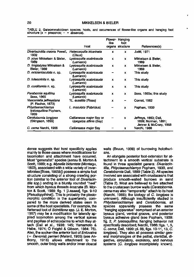
TABLE 2. Galeommatoidean species, hosts, and occurrences of flower-like organs and hanging foot structure (x = presence; - = absence).
Flower- Hanging like foot
Host organs structure References(s)
Divariscintilla maoria Powell, Heterosquilla tricarinata x x Judd, 1971 1932 (Claus}
D. yoyo Mikkelsen & Bieler, Lysiosquilla scabricauda x x Mikkelsen & Bieler, 1989 (Lamarck} 1989
D. troglodytes Mikkelsen & Lysiosquilla scabricauda x x Mikkelsen & Bieler, Bieler, 1989 (Lamarck} 1989
D. octotentaculata n. sp. Lysiosquilla scabricauda x This study (Lamarck)
D. luteocrinita n. sp. Lysiosquilla scabricauda x x This study (Lamarck)
D. cordiformis n. sp. Lysiosquilla scabricauda x x This study (Lamarck)
Parabornia squillina Lysiosquilla scabricauda x x Boss, 1965a; this study Boss, 1965 (Lamarck)
Vasconiella jeffreysiana ?L. eusebia (Risso) x Cornet, 1982 (P. Fischer, 1873)
Phlyctaenachlamys L. maculata (Fabricius) x Popham, 1939 lysiosquillina Popham, 1939
Ceratobornia longipes Callianassa major Say or x Jeffreys, 1863; Dall, (Stimpson, 1855) Upogebia afflnis (Say)
C. cema Narchi, 1966 Callianassa major Say
dence suggests that host specificity applies mainly to those cases where modifications for locomotion and attachment have occurred. Most "generalist" species (sensu B. Morton & Scott, 1989; e.g. Mysel/a bidentata (Montagu, 1803), associated with a wide variety of invertebrates [Boss, 1965b]) possess a simple foot structure consisting of a strong crawling portion (similar to the anterior foot of Divariscintilla spp.) ending in a bluntly rounded "heel" from which byssus threads emanate (B. Morton & Scott, 1989: fig. 1 [Lasaea], figs. 9-10 [Pseudopythina]). This is probably the plesiomorphic condition in the superfamily, compared to the more derived states seen in some of the host specialists. For example, the flattened foot of Scintillona spp. (J. E. Morton, 1957) may be a modification for laterally applied locomotion among the vertical spines and papillae of echinoderms to which they attach (Dall et al., 1938: 145; Yamamoto & Habe, 1974; 6 Foighil & Gibson, 1984: 75). Also, the sucker-like anterior foot of Entovalva ( = Devonia) pe"ieri (Malard, 1903) (see Anthony, 1916) allows attachment to the smooth, outer body walls and/or inner cloacal
1899; Norman, 1891; Jenner & McCrary, 1968
x Narchi, 1966
walls (Bruun, 1938) of burrowing holothurians.
An elongate posterior foot-extension for attachment to a smooth vertical substrate is found in three specialist genera: Divariscintil/a, Phlyctaenach/amys Popham, 1939, and Ceratobornia Dall, 1899 (Table 2). All species involved are associated with crustaceans that produce smooth-walled burrows in sand (Table 2). Most are believed to live attached to the crustacean burrow walls ( Ceratobornia. cema may also "temporarily" attach to its host (Narchi, 1966); the biology of C. longipes is unknown). Although insufficiently studied in Phlyctaenachlamys and Ceratobornia, all species apparently possess the same "hanging apparatus" comprised of an anterior byssus gland, ventral groove, and posterior byssus adhesive gland (see Popham, 1939: fig. 2, P. lysiosquillina, hanging behavior not specifically described; Narchi, 1966: figs. 1, 5, C. cema; Dall, 1899: pl. 88, figs. 10-11, 13, C. longipes). They also all possess similar general morphologies of the pallial, ctenidial, digestive, circulatory, excretory, and nervous systems (C. /ongipes incompletely known).

NEW SPECIES OF COMMENSAL GALEOMMATIDAE 21
TABLE 3. Distinguishing characteristics for the three galeommatoidean genera possessing the "hanging foot." See text for included species and sources of data. [L, left; R, right]
Divariscintilla
Shell internalization incomplete Hinge:
Cardinal teeth 1 R, 1 L Lateral teeth absent
Retraction into shell yes (1 sp.) no (5 spp.)
Adductor muscles subequal Flower-like organs present (5 spp.)
absent (1 sp.) lnterlamellar ctenidial present
junctions: Hindgut typhlosole present Hypobranchial gland present? Supportive chondroid absent
edge in foot
The anatomical differences among the three genera that presently prevent their synonymy are listed in Table 3. Until the importance of each of these characters can be reassessed, it is uncertain whether possession of a "hanging foot" reflects convergence or phylogenetic relationship, and the three genera are best treated separately.
Another possible difference between Divariscintilla on one hand and Ph/yctaenachlamys and Ceratobornia on the other lies in the mode of reproduction. Members of Divariscintilla for which such data are available (D. yoyo, D. troglodytes, D. octotentaculata, D. cordiformis) are known to be simultaneous hermophrodites. From literature data, it seems that Phlyctaenachlamys lysiosquillina and Ceratobornia cema are forms with separate sexes (Popham, 1939, p. 80: "specimen sectioned was a male"; and Narchi, 1966, p. 521: "sectioned specimen was a female"). However, while simultaneous hermaphroditism can be documented from individuals without observations over extended periods of time, "males" or "females" could belong to forms with consecutive hermaphroditism. Protandrous and protogynous hermaphroditism have both been reported for the superfamily (summarized by Fox, 1979: tab. 11, as Leptonacea).
Rhamphidonta Bernard, 1975, represented by the single species R. retifera (Dall, 1899), also possesses a bipartite foot but one which is different from that discussed above, both morphologically and functionally. According to Bernard (1975), the foot of R. retifera is
Phlyctaenachlamys Ceratobornia
complete incomplete
2 R, 1 L 2 R, 2 L 1 posterior, 1 posterior,
reduced reduced no yes
posterior reduced subequal absent absent
absent absent
absent absent absent absent absent present
anteriorly elongated, with the main enlarged crawling portion located posteriorly. Members of this species are not known to hang; they burrow into sand to avoid illumination, and are apparently free-living. The hinge of R. retifera (see Bernard, 1975: fig. 1) is distinctly "montacutid." Close relationship with the three genera discussed above is unlikely.
The mantis shrimp Lysiosquilla scabricauda also serves as host to Parabornia squillina Boss, 1965, a galeommatoidean that attaches to the inner surface of the abdominal sclera of the shrimp (Table 2). Parabornia squillina has been collected in the region of this study (Sebastian Inlet, Peanut Island), but has not been collected in burrows containing Divariscintilla species. Interestingly, the animal of P. squillina also possesses a single "flower-like organ" (pers. obs.), although this was not mentioned in the original species description based on preserved material. (Boss, 1965a: fig. 3 shows an unexplained five-part zig-zag outline for the visceral mass below the gills and labial palps.) Its foot (Boss, 1965a: fig. 3) appears morphologically similar to the "hanging type" described above; a deep ventral groove ends with an opaque white area at the end of a very short posterior extension (pers. obs.). Boss (1965a: 4) interpreted the white area at the end of the foot as the byssus gland (as did Judd (1971] for D. maoria, and Narchi [1966] for Ceratobornia cema). In view of the byssus adhesive gland described here and previously (Mikkelsen & Bieler, 1989), this organ (which was depicted with internal lamellae by Boss, 1965a: fig. 3)
22 MIKKELSEN & BIELER
warrants reinvestigation. If this structure is the primary byssus gland, what is the function of the ventral groove? (Histological study of the structure of the entire foot, and observations of living animals producing byssus threads would resolve this question.) Boss (1965a) placed Parabornia in the Erycinidae (Erycininae), in part, because its hinge has two cardinal teeth in each valve and a central resilium. If hinge structure is eventually confirmed as a reliable taxonomic character based on phylogeny of the group, the "hanging foot" and "flower-like organs" would need to be explained as results of evolutionary convergence and/or symplesiomorphies.
Mating Behavior in Bivalves
The observed "mating" behavior (Fig. 33) between individuals of Divariscintilla octotentaculata as well as of D. yoyo is of special interest. Mating behavior has not been confirmed for bivalves (Mackie, 1984: 362). Mackie (1984: 363) summarized the reported cases of associations between females and dwarf males in bivalves and concluded that those interactions would qualify as mating behavior if it can be shown that the associations are confined to a breeding period.
True copulatory behavior is absent in bivalves because they lack copulatory organs. However, several authors (Clapp, 1951: 7; Townsley et al., 1966: 49) have observed male teredinids (Bankia spp.) inserting their exhalent siphons into the inhalant siphons of females during sperm release. Purchon (19n: 295), and subsequently Mackie (1984: 362), interpreted this as the use of an intromittant organ, the development of which would be of "great survival value to species ... living in isolated pieces of drift wood (Purchon, 19n: 295). Similarly, quasi-copulatory behavior in Divariscintilla would be advantageous for these small bivalves with limited gamete production, living in restricted, relatively isolated populations within the burrows of their hosts. Selective mating in burrows that contain several other congeneric species would guarantee successful fertilization and make conservative use of male gametes. In our previous discussion of flower-like organs, now known from five species of Divariscintilla, we (Mikkelsen & Bieler, 1989: 191) speculated that these organs might emit a pheromone for attracting reproductive partners (another possibility, if they are indeed phero-
mane-emitting organs, would be the attraction of larvae to settlement sites).
ACKNOWLEDGMENTS
The following are gratefully acknowledged: Dr. Raymond B. Manning (USNM), for bringing this interesting fauna to our attention; William D. ("Woody") Lee (SMSLP), for invaluable field help; Paul H. Scott (SBMNH), for comments on generic placement; Patricia A. Linley and Pamela Blades-Eckelbarger (both HBOI) for SEM assistance; Tom Smoyer (HBOI) for photographic work; Dr. Kerry B. Clark (Florida Institute of Technology, Melbourne) and John E. Miller (formerly HBOI) for the use of and help with video recording equipment; Paul S. Mikkelsen (formerly HBOI), for behavioral and developmental notes and the photograph in Fig. 1 ; Chris Bauman (Vero Beach, Florida) for the illustration in Fig. 33; Paul H. Scott (SBMNH), Dr. Kenneth J. Boss (MCZ), and two anonymous reviewers for valuable comments on the manuscript.
This research was supported in part by the Smithsonian Marine Station at Link Port; the cooperation of Dr. Mary E. Rice and her staff is gratefully acknowledged. Funding for this project was derived in part from a National Capital Shell Club scholarship to P. M., and a NATO Postdoctoral Research Fellowship at SMSLP to R. B. This is Harbor Branch Oceanographic Institution Contribution no. 862 and Smithsonian Marine Station Contribution no. 273.
LITERATURE CITED
ANTHONY, R., 1916, Contribution a l'etude de l'Entovalva (Synapticola) perrieri Malard, mollusque acephale commensaJ des synaptes. Archives de Zoologie Experimentale et Generate, 55: 375-391, pis. 6-7.
ARAKAWA, K. Y., 1960, Ecological observations on an aberrant lamellibranch, Ephippodonta murakamii Kuroda. Venus, Japanese Journal of Malacology, 21(1): 50-60, pis. 7-8.
BERNARD, F. A., 1975, Rhamphidonta gen. n. from the northeastern Pacific (Bivalvia, Leptonacea). Journal de Conchyliologie, 112(3-4): 105-115.
BIELER, A. & P. M. MIKKELSEN, 1988, Anatomy and reproductive biology of two western Atlantic species of Vitrinellidae, with a case of protan-
1'

NEW SPECIES OF COMMENSAL GALEOMMATIDAE 23
drous hermaphroditism in the Rissoacea. The Nautilus, 102(1 ): 1-29.
BOSS, K. J., 1965a, A new mollusk (Bivalvia, Erycinidae) commensal on the stomatopod crustacean Lysiosquilla. American Museum Novitates, no. 2215: 11 pp.
BOSS, K.J., 1965b, Symbiotic erycinacean bivalves. Malacologia, 3(2): 183-195.
BRUUN, A. F., 1938, A new entocommensalistic bivalve, Entovalva major n.sp., from the Red Sea. Videnskabelige Meddelelser fra Dansk naturhistorisk Forening i Kebenhavn, 102: 163-167.
CHAVAN, A., 1969, Superfamily Leptonacea Gray, 1847. Pp 518-537, in: R. c. MOORE, ed., Treatise on Invertebrate Paleontology, Part N. Mollusca 6. Bivalvia 2. Geological Society of America (Boulder, Colorado) & University of Kansas (Lawrence), ii + N491-N952 pp.
CLAPP, W. F., 1951, Observations on living Teredinidae. Fourth Progress Report on Marine Borer Activity in Test Boards Operated during 1950. Report no. 7550, W. F. Clapp Laboratories, Inc., Duxbury, Massachusetts, 9 pp.
CONEY, C. C., 1990, Bellascintilla parmaleeana new genus and species from the tropical eastern Pacific, with a review of the other, ventrally notched galeommatid genera (Bivalvia: Galeommatacea). The Nautilus, 104(4): 130-144.
CORNET, M., 1982, Anatomical description of Vasconiella jeffreysiana (P. Fischer, 1873) (Mollusca, Bivalvia, Leptonacea). Journal of Molluscan Studies, 48(1): 36-43.
DALL, W. H., 1899, Synopsis of the Recent and Tertiary Leptonacea of North America and the West Indies. Proceedings of the United States National Museum, 21(1177):873-897, pis. 87-88.
DALL, W. H., P. BARTSCH & H. A. REHDER, 1938, A manual of the Recent and fossil marine pelecypod mollusks of the Hawaiian Islands. Bernice P. Bishop Museum Bulletin, 153: iv + 233 pp., 58 pis.
ECKELBARGER, K. J., R. BIELER & P. M. MIKKELSEN, 1990, Ultrastructure of sperm development and mature sperm morphology in three species of commensal bivalves (Mollusca: Galeommatoidea). Journal of Morphology, 205: 63-75.
FOX, T. H., 1979, Reproductive adaptations and life histories of the commensal leptonacean bivalves. Ph.D. dissertation, University of North Carolina, Chapel Hill, ix + 207 pp.
HUMASON, G. L., 1962, Animal Tissue Techniques. W. H. Freeman & Co., San Francisco & London, xv + 468 pp.
JEFFREYS, J. G., 1863, British Conchology, or an account of the Mollusca which now inhabit the British Isles and the surrounding seas. Vol. II. Marine Shells, comprising the Brachiopoda, and Conchifera from the family of Anomiidae to that of Mactridae. London, John Van Voorst, xiv + 465 pp., 8 pis.
JENNER, C. E. & A. B. McCRARY, 1968, A "gas-
tropod" bivalve, commensal on Squilla empusa (Abstract). Annual Report for 1968, American Malacological Union: pp. 20-21.
JUDO, W., 1971, The structure and habits of Divariscintilla maoria Powell (Bivalvia: Galeommatidae). Proceedings of the Ma/acological Society of London, 39: 343-354.
MACKIE, G. L., 1984, Bivalves. Pp. 351-418, in: A. S. TOMPA, et al., The Mollusca, Vol. 7, Reproduction. Academic Press, Orlando, xix + 486 pp.
MIKKELSEN, P. M. & R. BIELER, 1989, Biology and comparative anatomy of Divariscintilla yoyo and D. troglodytes, two new species of Galeommatidae (Bivalvia) from stomatopod burrows in eastern Florida. Malacologia, 31 (1): 1-21.
MORTON, B., 1973a, The biology and functional morphology of Galeomma (Paralepida) takii (Bivalvia: Leptonacea). Journal of Zoology, 169(2): 133-150.
MORTON, B., 1973b, Some factors affecting the location of Arthritics crassiformis (Bivalvia: Leptonacea) commensal upon Anchomasa similis (Bivalvia: Pholadidae). Journal of Zoology, 170: 463-473.
MORTON, B., 1975, Oymantic display in Galeomma polita Deshayes (Bivalvia: Leptonacea). Journal of Conchology, 28: 365-369.
MORTON, B., 1976, Secondary brooding of temporary dwarf males in Ephippodonta (Ephippodontina) oedipus sp. nov. (Bivalvia: Leptonacea). Journal of Conchology, 29: 31-39.
MORTON, B. & P. H. SCOTT, 1989, The Hong Kong Galeommatacea (Mollusca: Bivalvia) and their hosts, with descriptions of new species. Asian Marine Biology, 6: 129-160.
MORTON, J.E., 1957, The habits of Scintillona zelandica (Odhner) 1924 (Lamellibranchia: Galeommatidae). Proceedings of the Malacological Society of London, 32(5): 185-188.
NARCHI, W., 1966, The functional morphology of Ceratobornia cema, new species of the Erycinacea (Mollusca, Eulamellibranchiata). Anais da Academia Brasileira de Ciencias, 38(3-4): 513-524.
NORMAN, A. M., 1891, Lepton squamosum (Montagu), a commensal. Annals and Magazine of Natural History, (6) 7(39): 276-278.
6 FOIGHIL, 0. & A. GIBSON, 1984, The morphology, reproduction and ecology of the commensal bivalve Scintillona bellerophon spec. nov. (Galeommatacea). The Veliger, 27(1 ): 72-80.
POPHAM, M. L., 1939, On Phlyctaenachlamys lysiosquillina gen. and sp. nov ., a lamellibranch commensal in the burrows of Lysiosquilla maculata. British Museum (Natural History), Great Barrier Reef Expedition 1928-29, Scientific Reports, 6(2): 549-587.
PURCHON, R. D., 1977, The Biology of the Mollusca, 2nd ed. Pergamon Press, Oxford, xxv + 560 pp.
RICE, E. L., 1897, Die systematische Verwertbarkeit der Kiemen bei den Lamellibranchiaten.

24 MIKKELSEN & BIELER
Jenaische Zeitschrift tar Naturwissenschaft, 31 (N.F. 24): 29-89, pis. 3-4. .
TAYLOR, J. 0., W. J. KENNEDY & A. HALL, 1973, The shell structure and mineralogy of the Bivalvia. II. Lucinacea--Clavagellacea, conclusions. Bulletin of the British Museum (Natural History), 22(9): 253-294, pis. 1-15.
TOWNSLEY, P. M., R. A. RICHY & P. C. TRUSSELL, 1966, The laboratory rearing of the shipworm, Bankia setacea (Tryon). Proceedings of the National Shel/fisheries Association, 56: 49-52.
VACCA, L. L., 1985, Laboratory manual of histochemistry. Raven Press, New York, xvii + 578 pp.
WINCKWORTH, R., 1930, Notes on nomenclature. Proceedings of the Malacological Society of London, 19(1): 14-16.
YAMAMOTO, T. & T. HABE, 1974, Scintillona stigmatics (Pilsbry) new to Japan. Venus, Japanese Journal of Malacology, 33(3): 116.
Revised Ms accepted 1 August 1991
,
Copyright © 2022 FDOKUMEN