Under Duress: Suppressing and Recovering Memories of the Indonesian Sixties
Upload
independentCategory
view
3download
0
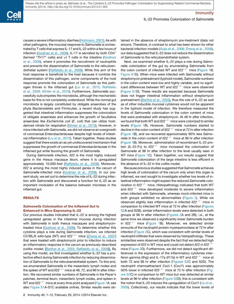
Please cite this article in press as: Behnsen et al., The Cytokine IL-22 Promotes Pathogen Colonization by Suppressing Related Commensal Bacteria,Immunity (2014), http://dx.doi.org/10.1016/j.immuni.2014.01.003
Immunity
Article
The Cytokine IL-22 Promotes Pathogen Colonizationby Suppressing Related Commensal BacteriaJudith Behnsen,1,2 Stefan Jellbauer,1,2 Christina P. Wong,1,2 Robert A. Edwards,2,3 Michael D. George,4 Wenjun Ouyang,5
and Manuela Raffatellu1,2,*1Department of Microbiology, University of California, Irvine, Irvine, CA 92697, USA2Institute for Immunology, University of California, Irvine, Irvine, CA 92697, USA3Department of Pathology and Laboratory Medicine, University of California, Irvine, Irvine, CA 92697, USA4Department of Medical Microbiology and Immunology, School of Medicine, University of California, Davis, CA 95616, USA5Department of Immunology, Genentech, South San Francisco, CA 94080, USA
*Correspondence: [email protected]://dx.doi.org/10.1016/j.immuni.2014.01.003
SUMMARY
Interleukin-22 (IL-22) is highly induced in response toinfections with a variety of pathogens, and its mainfunctions are considered to be tissue repair andhost defense at mucosal surfaces. Here we showedthat IL-22 has a unique role during infection in thatits expression suppressed the intestinal microbiotaand enhanced the colonization of a pathogen. IL-22induced the expression of antimicrobial proteins,including lipocalin-2 and calprotectin, whichsequester essential metal ions from microbes.Because Salmonella enterica ser. Typhimurium canovercome metal ion starvation mediated by lipoca-lin-2 and calprotectin via alternative pathways, IL-22 boosted its colonization of the inflamed intestineby suppressing commensal Enterobacteriaceae,which are susceptible to the antimicrobial proteins.Thus, IL-22 tipped the balance between pathogenicand commensal bacteria in favor of a pathogen.Taken together, IL-22 induction can be exploited bypathogens to suppress the growth of their closestcompetitors, thereby enhancing pathogen coloniza-tion of mucosal surfaces.
INTRODUCTION
The cytokine interleukin-22 (IL-22) has important functions in tis-
sue regeneration and in the maintenance of the skin and the
mucosal barrier (reviewed in Rutz et al., 2013). In the mucosa,
IL-22 expression is primarily triggered in response to the interac-
tion of microorganisms with antigen-presenting cells (APCs).
APCs in turn release many cytokines including IL-23, which en-
gages the intraepithelial and lamina propria lymphocytes to pro-
duce cytokines like IL-17 and IL-22 (reviewed in Blaschitz and
Raffatellu, 2010; Khader and Gopal, 2010; Liu et al., 2009). The
major function of IL-17 is to orchestrate the recruitment of
neutrophils to the site of infection via the induction of CXC che-
mokines, like CXCL1 and CXCL8, and via the enhancement of
granulopoiesis, which explains its protective role during infection
with a variety of pathogens (reviewed in Blaschitz and Raffatellu,
2010). Similar to IL-17, IL-22 is rapidly induced in the intestinal
mucosa in response to IL-23, but also through activation of the
aryl hydrocarbon receptor (reviewed in Esser et al., 2009).
IL-22 shares sequence homology with IL-10 and it signals
through a receptor complex, consisting of both the IL-22 recep-
tor 1 and the IL-10 receptor 2 subunits, that is expressed almost
exclusively in nonhematopoietic cells (reviewed in Sonnenberg
et al., 2011). Several sources of IL-22 have been identified in
the gut, including innate lymphoid cells (ILCs) and T helper 17
(Th17) cells (reviewed in Sonnenberg et al., 2011). Akin to and
often in synergy with IL-17, IL-22 has been shown to play a pro-
tective role during infection with some pathogens, including
Klebsiella pneumoniae (Aujla et al., 2008), Citrobacter rodentium
(Zheng et al., 2008), vancomycin-resistant Enterococcus (Kinne-
brew et al., 2010), and Plasmodium chabaudi (Mastelic et al.,
2012).
One of the mechanisms by which IL-22 is thought to enhance
mucosal barrier function is through the induction of antimicrobial
proteins in the mucosa (Aujla et al., 2008; Zheng et al., 2007;
Zheng et al., 2008), a function that partly explains its role in the
containment of commensals to the intestinal niche (Sonnenberg
et al., 2012). Antimicrobial proteins that are upregulated by IL-22
in epithelial cells include: lipocalin-2, which binds to the sidero-
phore enterochelin and limits iron availability in the gut (Fisch-
bach et al., 2006; Raffatellu et al., 2009); the C-type lectins
regenerating islet-derivative protein 3 beta and 3 gamma
(Reg3b and Reg3g), which control some components of the
microbiota (Stelter et al., 2011); and S100A8 and S100A9, two
antimicrobial peptides that heterodimerize to form the antimicro-
bial protein calprotectin, which sequesters zinc and manganese
from microbes (Corbin et al., 2008; Damo et al., 2013; Hayden
et al., 2013; Liu et al., 2012). And yet, despite these epithelial anti-
microbial defenses, many pathogens colonize mucosal surfaces
and establish an infection; for example, IL-22 failed to reduce
oral infection with Candida albicans and genital infection with
Neisseria gonorrhoeae (Feinen and Russell, 2012; Kagami
et al., 2010), although the basis for this was not investigated.
At present, themechanism(s) by which IL-22 provides protection
against a subset of microorganisms is not well understood.
One of the most prolific examples of a successful mucosal
pathogen is Salmonella enterica serovar Typhimurium (Salmo-
nella), which colonizes the human gastrointestinal tract and
Immunity 40, 1–12, February 20, 2014 ª2014 Elsevier Inc. 1
Immunity
IL-22 Promotes Colonization of Salmonella
Please cite this article in press as: Behnsen et al., The Cytokine IL-22 Promotes Pathogen Colonization by Suppressing Related Commensal Bacteria,Immunity (2014), http://dx.doi.org/10.1016/j.immuni.2014.01.003
causes a severe inflammatory diarrhea (Hohmann, 2001). Aswith
other pathogens, the mucosal response to Salmonella is orches-
trated by T cells that express IL-17 and IL-22within a few hours of
infection (Godinez et al., 2008). IL-17 is secreted by both CD4+
(termed ‘‘Th17’’ cells) and gd T cells in the intestine (Godinez
et al., 2009), where it promotes the recruitment of neutrophils
and prevents the dissemination of Salmonella to the reticuloen-
dothelial system (Raffatellu et al., 2008). While this arm of the
host response is beneficial to the host because it controls the
dissemination of this pathogen, some components of the host
response promote the colonization of Salmonella as this path-
ogen thrives in the inflamed gut (Liu et al., 2012; Raffatellu
et al., 2009; Winter et al., 2010). Furthermore, Salmonella suc-
cessfully outcompetes the microbiota, although the mechanistic
basis for this is not completely understood. While the normal gut
microbiota is largely constituted by obligate anaerobes of the
phyla Bacteroidetes and Firmicutes, a recent study has sug-
gested that intestinal inflammation suppresses the colonization
of obligate anaerobes and enhances the growth of facultative
anaerobes like Escherichia coli (E. coli) that can utilize host-
derived nitrate for respiration (Winter et al., 2013). However, in
mice infectedwithSalmonella, we did not observe an overgrowth
of commensal Enterobacteriaceae despite high levels of intesti-
nal inflammation (Liu et al., 2012). Taken together, these studies
suggest that there exists an as yet undiscoveredmechanism that
suppresses the growth of commensal Enterobacteriaceae in the
inflamed gut while favoring the growth of Salmonella.
During Salmonella infection, Il22 is the most highly induced
gene in the rhesus macaque ileum, where it is upregulated
approximately 10,000-fold (Raffatellu et al., 2008). Moreover,
Il22 is among the most highly induced genes in the cecum of
Salmonella-infected mice (Godinez et al., 2008). In our pre-
sent study, we set out to determine the role of IL-22 during infec-
tion with Salmonella and discovered a function for IL-22 as an
important modulator of the balance between microbes in the
inflamed gut.
RESULTS
Salmonella Colonization of the Inflamed Gut IsEnhanced in Mice Expressing IL-22Our previous studies indicated that IL-22 is among the highest
upregulated genes in the intestinal mucosa during infection
with Salmonella in both rhesus macaques and streptomycin-
treated mice (Godinez et al., 2008). To determine whether this
cytokine plays a role during Salmonella infection, we infected
C57BL/6 wild-type (WT) and Il22�/� mice (Zheng et al., 2007)
that were treated with streptomycin prior to infection to induce
an inflammatory response in the cecum as previously described
(colitis model; [Barthel et al., 2003; Raffatellu et al., 2009]). By
using this model, we first investigated whether IL-22 has a pro-
tective effect duringSalmonella infection by reducing dissemina-
tion of Salmonella to the reticuloendothelial system. To this end,
we enumerated Salmonella in the mesenteric lymph nodes and
the spleen ofWT and Il22�/�mice at 48, 72, and 96 hr after infec-
tion. We recovered similar numbers of Salmonella in the Peyer’s
patches, terminal ileum, mesenteric lymph nodes, and spleen in
WT and Il22�/� mice at every time point analyzed (Figure 1A; see
also Figure S1A-S1C available online). Similar results were ob-
2 Immunity 40, 1–12, February 20, 2014 ª2014 Elsevier Inc.
tained in the absence of streptomycin pre-treatment (data not
shown). Therefore, in contrast to what has been shown for other
bacterial infection models (Aujla et al., 2008; Zheng et al., 2008),
our data suggested that IL-22 does not reduce the dissemination
of Salmonella to the reticuloendothelial system.
Next, we examined whether IL-22 plays a role during Salmo-
nella colonization of the gut by enumerating Salmonella from
the colon content of infected WT and Il22�/� mice (Figure 1B;
Figure S1D). When mice were infected with Salmonella without
streptomycin pretreatment (typhoidmodel),Salmonella numbers
in the colon content were low and highly variable, and no signif-
icant differences between WT and Il22�/� mice were observed
(Figure S1D). These results are expected because Salmonella
does not trigger intestinal inflammation without streptomycin
pretreatment (Barthel et al., 2003), thus the role of IL-22 as well
as of other inducible mucosal cytokines would not be apparent
in the typhoid model of infection. We therefore measured the
levels of Salmonella colonization in the colon content of mice
that were pretreated with streptomycin. At 48 hr after infection,
we found that bothWT and Il22�/�micewere colonized to similar
levels (Figure 1B). However, Salmonella numbers started to
decline in the colon content of Il22�/�mice at 72 hr after infection
(Figure 1B), and we recovered approximately 90% less Salmo-
nella in the colon content of Il22�/� mice at 96 hr after infection
(Figure 1B). Moreover, administration of recombinant IL-22 pro-
tein (IL-22-Fc) to Il22�/� mice increased the colonization of
Salmonella at 96 hr after infection to the level usually seen in
WT mice (Figure 1C). Taken together, our results suggest that
Salmonella colonization of the large intestine is less efficient in
the absence of IL-22 in the colitis model.
Because previous studies suggested thatSalmonella achieves
high levels of colonization of the cecum only when this organ is
inflamed, we next sought to investigate whether low levels of in-
testinal inflammationmight explain the reduced Salmonella colo-
nization in Il22�/� mice. Histopathology indicated that both WT
and Il22�/� mice developed moderate to severe inflammation
when infected with Salmonella, whereas mock-infected mice in
both groups exhibited no abnormalities (Figure 2). While we
observed slightly less inflammation in infected Il22�/� mice in
comparison to infected WT mice at 72 hr after infection (Figures
S2A and S2B), similar inflammation levels were detected in both
groups at 96 hr after infection (Figures 2A and 2B), i.e., at the
same time we observed a significantly lower Salmonella burden
in Il22�/� mice (Figure 1B). Moreover, we detected similar
amounts of the neutrophil protein myeloperoxidase at 72 hr after
infection (Figure 2C), which was consistent with similar levels of
neutrophil infiltrate that were observed by histopathology. These
similarities were observed despite the fact that we detected high
expression of Il22 in WT mice and could not detect Il22 in Il22�/�
mice (Figure 2D). Furthermore, we did not detect significant dif-
ferences in the expression of the inflammatory cytokines inter-
feron gamma (Ifng) and IL-17a (Il17a) in WT and Il22�/� mice at
both 72 and 96 hr after infection (Figures S2C and S2D). The
neutrophil chemoattractant Cxcl-1 (Cxcl1) was approximately
50% lower in infected Il22�/� mice at 72 hr after infection (Fig-
ure S2C) in comparison to WT mice but was detected at similar
levels at 96 hr after infection (Figure S2D); this is consistent with
the notion that IL-22 induces the upregulation of Cxcl1 (Liu et al.,
2009). Collectively, our results indicate that the lower levels of

WT Il22-/-103
104
105
106
107
CFU
/mg
n.s.
WT Il22-/-103
104
105
106
107
CFU
/mg
p=0.056
WT Il22-/-103
104
105
106
107
CFU
/mg
**
96h p.i.72h p.i.48h p.i.
A
B
WT Il22-/-102
103
104
105
106
107
CFU
/org
an
n.s.
WT Il22-/-102
103
104
105
106
107
CFU
/org
an
n.s.
WT Il22-/-100
101
102
103
104
105
CFU
/org
an
n.s.
spleen
coloncontent
IL22-Fc isotype control
103
104
105
106
107
108
CFU
/mg
*
Il22-/- mice
C
IL22-Fc isotype control
103
104
105
106
107
108
CFU
/mg
n.s.
Il22-/- mice
IL22-Fc isotype control
103
104
105
106
107
108
CFU
/mg
n.s.
Il22-/- mice
coloncontent
Figure 1. Colonization of Salmonella in WT and Il22–/– Mice
(A and B)WT and Il22�/�micewere infected with Salmonella, and cfu in spleen (A) and colon content (B) were determined at 48 hr (WT n = 10, Il22�/� n = 10), 72 hr
(WT n = 11, Il22�/� n = 9), and 96 hr (WT n = 11, Il22�/� n = 10) after infection.
(C) Il22�/� mice were administered IL-22-Fc or an isotype control antibody (n = 3 per group), and fecal samples were collected at 48 hr, 72 hr, and 96 hr after
infection. Data shown represent cfu per mg of fecal material for colon content and cfu per organ for spleen. Data represent the geometric mean ± SE. n.s. = not
significant. A significant decrease over WT control is indicated by ** (p value % 0.01). See also Figure S1 and Table S1.
Immunity
IL-22 Promotes Colonization of Salmonella
Please cite this article in press as: Behnsen et al., The Cytokine IL-22 Promotes Pathogen Colonization by Suppressing Related Commensal Bacteria,Immunity (2014), http://dx.doi.org/10.1016/j.immuni.2014.01.003
Salmonella colonization observed in the cecum at 96 hr after
infection in Il22�/� mice do not appear to be explained by differ-
ences in the levels of inflammation.
IL-22 Expression Changes the Relative Abundance ofProteobacteria in the Inflamed GutSeveral studies have established that competition with the
microbiota is essential for Salmonella to colonize the inflamed
gut (Barman et al., 2008; Lawley et al., 2008; Liu et al., 2012;
Lupp et al., 2007; Stecher et al., 2007;Winter et al., 2010). There-
fore, we hypothesized that differences in the microbiota might
explain the lower Salmonella burden in Il22�/� mice. To this
end, we analyzed the microbiota composition in the colon con-
tent of WT and Il22�/� littermate mice before streptomycin treat-
ment and after either mock infection or infection with Salmonella
(Figure 3, Figure S3; Table S3). The use of littermates for the anal-
ysis of the microbiota helped us to exclude any potential differ-
ences in the microbiota between WT and Il22�/� mice, as it
has been shown that the baseline microbiota of Il22�/� mice
might be more colitogenic and that it is transmissible to WT
mice (Zenewicz et al., 2013). Illumina MiSeq analysis of DNA ex-
tracted from fecal samples confirmed previous observations that
the major bacterial classes detected in animals before strepto-
mycin treatment and in mock-infected animals at 96 hr after
Immunity 40, 1–12, February 20, 2014 ª2014 Elsevier Inc. 3

A
B
C D
Figure 2. Histopathology of WT and Il22–/–
Mice after Infection with Salmonella
(A) Blinded histopathology score indicating the
score of individual mice 96 hr after either mock
infection (treated with streptomycin but not in-
fected) or infection with Salmonella. The gray re-
gion includes scores indicative of moderate to
severe inflammation.
(B) H&E stained cecal sections from representative
animals in each group. An image at lower magni-
fication (103) and one at higher magnification
(403) from the same section are shown. Abbrevi-
ations are as follows: L, lumen; M, mucosa; SM,
submucosa. Note marked edema in the submu-
cosa and inflammation in mice infected with
Salmonella.
(C) Myeloperoxidase (MPO) was detected 72 hr
after infection by immunoblot in protein samples
prepared from the cecum of mice that were mock
infected or infected with Salmonella.
(D) Il22 was detected by qRT-PCR in the cecum
of WT mice (n = 6) and Il22�/� mice (n = 6) 96 hr
after infection with WT Salmonella. Data represent
the geometric mean ± SE. A significant increase
over mock control is indicated by *** (p value %
0.001), n.d. = not detected. See also Figure S2 and
Table S2.
Immunity
IL-22 Promotes Colonization of Salmonella
Please cite this article in press as: Behnsen et al., The Cytokine IL-22 Promotes Pathogen Colonization by Suppressing Related Commensal Bacteria,Immunity (2014), http://dx.doi.org/10.1016/j.immuni.2014.01.003
infection were Clostridia and Bacteroidia, whereas Proteobacte-
ria were low or undetectable (Figure 3; Figure S3A) (Barman
et al., 2008; Lawley et al., 2008; Liu et al., 2012; Lupp et al.,
2007; Stecher et al., 2007; Winter et al., 2010). A comparison
of WT and Il22�/� mice showed no major differences in the
composition of the microbiota, either before streptomycin
treatment (baseline) or after mock infection. Five days after
streptomycin treatment, the microbiota of both WT and Il22�/�
mice was still constituted by Bacteroidia and Clostridia, although
the relative abundance of Bacteroides was increased (Figure 3;
Figure S3A). Groups of bacteria that are known to enhance
resistance to bacterial infection were equally represented
in both WT and Il22�/� mice. For instance, bacteria of the
Porphyromonadaceae family have been associated with in-
creased resistance to Salmonella infection (Ferreira et al.,
2011); however, in both WT and Il22�/� mice the relative abun-
dance of the genus Parabacteroides (family Porphyromonada-
4 Immunity 40, 1–12, February 20, 2014 ª2014 Elsevier Inc.
ceae) was similarly low before infection
(0.1%–0.7%) (Figure 3; Figure S3A). Like-
wise, our WT and Il22�/� mice were simi-
larly colonized with varying levels of
segmented filamentous bacteria (SFB)
(Figure S3B), Clostridia-related bacteria
known to enhance resistance to Citro-
bacter rodentium infection through the
induction of Th17 cells (Ivanov et al.,
2009). Although SFB colonization levels
were reduced by streptomycin treat-
ment, no significant differences in SFB
levels were observed between WT and
Il22�/� mice (Figure S3B). Altogether,
our results suggest that differences in
the microbiota prior to infection are
unlikely to account for the higher numbers of Salmonella coloni-
zation in WT mice.
The most striking differences in the microbial communities
were observed in mice that were infected with S. Typhimurium.
As expected from prior studies (Barman et al., 2008; Lawley
et al., 2008; Liu et al., 2012; Lupp et al., 2007; Stecher et al.,
2007; Winter et al., 2010), Bacteroidia and Clostridia were sub-
stantially reduced in Salmonella-infected mice compared to
mock-infectedmice (Figure 3; Figure S3A). Furthermore, Proteo-
bacteria bloomed in the inflamed gut of bothWT and Il22�/�mice
infected with Salmonella, constituting approximately 70% of the
microbiota. Indeed, the most prominent difference between WT
and Il22�/� littermatemice infectedwith Salmonellawas the rela-
tive abundance of the genera Escherichia and Salmonella.While
Salmonella constituted approximately 50% of the total bacteria
in infected WT mice, it comprised 15% on average in infected
Il22�/� mice. In contrast, Escherichia constituted the largest

Figure 3. Analysis of Colonic Microbiota in
WT and Il22–/– Mice by Sequencing
Fecal samples were collected from mice before
streptomycin treatment (n = 9 per group) and
mock-infected animals (n = 4 per group) or
Salmonella-infected animals (n = 5 per group) at
96 hr postinfection. The colonic microbiota was
analyzed by sequencing using an Illumina MiSeq
system. Graphed is the average relative abun-
dance of each bacterial genus. See also Figure S3
and Tables S3 and S4.
Immunity
IL-22 Promotes Colonization of Salmonella
Please cite this article in press as: Behnsen et al., The Cytokine IL-22 Promotes Pathogen Colonization by Suppressing Related Commensal Bacteria,Immunity (2014), http://dx.doi.org/10.1016/j.immuni.2014.01.003
bacterial fraction in infected Il22�/� mice (on average, 40%) and
a small fraction of bacteria in infected WT mice (on average, 9%)
(Figure 3; Figure S3A). These findings led us to hypothesize that,
in the absence of IL-22, commensal Enterobacteriaceae can
compete with Salmonella in the inflamed gut.
IL-22 Promotes Salmonella Competition withCommensal EnterobacteriaceaeTo test our hypothesis, we first sought to corroborate our micro-
biota sequencing data (Figure 3) by streaking mouse fecal
samples from WT and Il22�/� mice infected with Salmonella on
MacConkey-lactose agar, a selective and differential media
commonly used in diagnostic laboratories to detect Enterobac-
teriaceae; enterobacterial strains that ferment lactose (e.g.,
Escherichia spp) form pink colonies while strains that do not
Immunity 40, 1–1
ferment lactose (e.g., Salmonella spp)
form colorless colonies. In agreement
with our profiles, fecal samples from
most WT mice infected with Salmonella
yielded only colorless colonies, which
were determined to be Salmonella (Fig-
ure 4A). Also in line with our analysis,
feces from Il22�/� mice infected with Sal-
monella primarily yielded pink colonies,
which were determined to be E. coli,
although colorless Salmonella colonies
were also observed (Figure 4A). Subse-
quent enumeration of fecal bacteria on
MacConkey-lactose agar revealed that
in WT mice, Salmonella established high
levels of colon colonization, effectively
outcompeting the resident commensal
E. coli in most mice by approximately 4
logs (Figure 4B). In contrast, in Il22�/�
mice Salmonella was mostly outcom-
peted by the resident commensal E. coli
(Figure 4B), which explains the lower
levels of Salmonella colonization in the
colon (Figures 1B, 3, 4B). However,
higher levels of E. coli colonization in
Il22�/� mice were not due to differences
in E. coli colonization between WT and
Il22�/� mice prior to infection. Similar
low numbers (approximately 102 colony-
forming units [cfu]/mg) of E. coli were
measured in the feces from both WT
and Il22�/� mice prior to treatment with streptomycin and no
E. coli were detectable after 24 hr (Figures S4A and S4B). More-
over, analysis of multiple E. coli strains isolated from feces of WT
and Il22�/�mice, both before and after infection with Salmonella,
revealed that all mice from our colony are colonized with the
same E. coli strain; testing for serotype (O166, H6, or 41), colo-
nization factors, and antibiotic susceptibility yielded the same re-
sults for all analyzed isolates (Figures S4C and S4D). Therefore,
differences in E. coli colonization between WT and Il22�/� mice
infected with Salmonella are not due to differences in the
E. coli strains between groups.
Because our results suggested that E. coli spontaneously
grew to higher amounts in Il22�/� mice than in WT mice infected
with Salmonella, we next tested whether the E. coli growth
advantage was also seen when an equal high dose of the
2, February 20, 2014 ª2014 Elsevier Inc. 5

WTmice
Il22-/-
mice
100101102103104105106107108
CFU
/mg
*
Salmonella infection
SalmonellaE. coli (mouse)
WTmice
Il22-/-
mice
105
106
107
108
CFU
/mg
SalmonellaE. coli (mouse)
**
Salmonella + mouse E. coliinfection
Afe
cals
ampl
eson
Mac
Con
key
plat
es
WT mouse Il22-/- mouse
SalmonellaSalmonella
mouseE. coli
B C
before 24h 48h 72h 96h 120h 144h 168h100
101
102
103
104
105
106
E.coli
CFU
/mg
WT miceIl22-/- mice
DSS treatment
D
Figure 4. Competition of Salmonella with
E. coli
(A) Colon content samples collected from WT and
Il22�/� mice at 96 hr postinfection were re-
suspended in PBS and streaked on MacConkey
agar plates for single colonies. Magnification
shows the presence of lactose fermenting (pink)
and lactose nonfermenting colonies in a sample
from an Il22�/� mouse.
(B and C) WT and Il22�/� mice were either (B) in-
fected with Salmonella (WT n = 7, Il22�/� n = 6) or
(C) infected with Salmonella and 24 hr later given
109 cfu of mouse E. coli strain JB2 (WT n = 10,
Il22�/� n = 8). Four days after infection with
Salmonella, colon content samples were collected
and MacConkey plates were used to enumerate
lactose fermenting (E. coli) and lactose
nonfermenting (Salmonella) colonies. Horizontal
lines represent the geometric mean.
(D) Fecal samples were collected from WT and
Il22�/�mice before and every 24 hr after micewere
given DSS in water. Samples were resuspended in
PBS and plated on MacConkey agar plates to
enumerate cfu of E. coli (WT n = 4, Il22�/� n = 4).
Gray area (101 to 3 3 102) indicates basal E. coli
level in mice. See also Figure S4.
Immunity
IL-22 Promotes Colonization of Salmonella
Please cite this article in press as: Behnsen et al., The Cytokine IL-22 Promotes Pathogen Colonization by Suppressing Related Commensal Bacteria,Immunity (2014), http://dx.doi.org/10.1016/j.immuni.2014.01.003
E. coli strain isolated in Figure 4A (JB2) was administered to both
WT and Il22�/� mice 24 hr after infection with Salmonella. In this
setting, E. coli also grew to higher numbers in Il22�/�mice than in
WT mice (Figure 4C). To determine whether the colonization
advantage of E. coli in Il22�/� mice occurred independently of
Salmonella infection, we induced colonic inflammation by treat-
ing WTmice and Il22�/�mice with dextran sodium sulfate (DSS).
Before DSS treatment, mice were equally colonized with low
numbers of E. coli (approximately 102 cfu/mg) and colonization
levels did not increase up to 96 hr after DSS treatment (Fig-
ure 4D). However, after the onset of intestinal inflammation,
E. coli started to bloom in Il22�/� mice while remaining at base-
line levels in WTmice (Figure 4D). At later time points, E. coli also
proliferated in WT mice, although at lower levels than in Il22�/�
mice. Even though there was a trend toward higher E. coli colo-
nization in Il22�/� mice, the difference did not reach statistical
significance. This is probably due to differences in themagnitude
and nature of inflammation caused by DSS and by Salmonella.
6 Immunity 40, 1–12, February 20, 2014 ª2014 Elsevier Inc.
Altogether, these results demonstrate
that IL-22 expression reduces the coloni-
zation of commensal Enterobacteriaceae,
resulting in a colonization advantage for
Salmonella in the inflamed gut.
IL-22 Induces the Expression ofAntimicrobial Proteins in theIntestine of Mice Infected withSalmonella
We next set out to determine the mecha-
nism by which IL-22 provides a coloniza-
tion advantage to Salmonella during
infection, thereby enabling it to effectively
compete with commensal Enterobacter-
iaceae. Because IL-22 regulates antimicrobial responses, we
examined whether the expression of antimicrobial genes during
Salmonella infection was also dependent on IL-22. To this end,
we analyzed the expression of the following: Lcn2, the gene
that encodes for the antimicrobial peptide lipocalin-2, which se-
questers the siderophore enterochelin and inhibits the growth of
commensal Enterobacteriaceae (Flo et al., 2004); S100a8 and
S100a9, which encode for the two subunits of calprotectin, an
antimicrobial protein that sequesters zinc and manganese from
pathogens (Corbin et al., 2008; Damo et al., 2013; Hayden
et al., 2013; Liu et al., 2012); Nos2, which encodes for the induc-
ible nitric oxide synthase (iNOS) (Muhl et al., 2011), as well as
Duox2, which encodes for the dual oxidase 2 protein (Rada
and Leto, 2008), each playing a role in the generation of reactive
nitrogen species and reactive oxygen species, respectively;
Reg3g, which encodes for regenerating islet-derivative protein
3 gamma (RegIIIg), a C-type lectin which binds to peptidoglycan
and inhibits the growth of Gram-positive bacteria (Cash et al.,

WT Il22-/-0
50
100
150
200
Lcn2
mR
NA
(fold
chan
ge) *
WT Il22-/-0
2000
4000
6000
Nos2
mR
NA
(fold
chan
ge) **
WT Il22-/-0
20,000
40,000
60,000
S100a8m
RN
A(fo
ldch
ange
) **
WT Il22-/-0
50
100
150
Duox2
mR
NA
(fold
chan
ge) ***
WT Il22-/-0
20,000
40,000
60,000
S100a9
mR
NA
(fold
chan
ge) **
WT Il22-/-0
50
100
150
200
250
Ido1
mR
NA
(fold
chan
ge) *
WT Il22-/-0
200
400
600
800
1000
Reg3g
mR
NA
(fold
chan
ge) ***
Metal chelators
Oxidative stress
A
B
Tryptophan starvationC D
C-type lectin
Figure 5. Expression of Antimicrobial Peptide Genes
(A) Lcn2, S100a8, S100a9, (B) Duox2, Nos2, (C) Reg3g, and (D) Ido1 were detected by qRT-PCR in the cecum of WT mice and Il22�/� mice 72 hr after infection
with WT Salmonella. Infected WT n = 6, infected Il22�/� n = 6, mock n = 4. Data are expressed as fold increase over mock-infected WT mice. Data represent the
geometric mean ± SE. A significant increase over mock control is indicated by * (p value% 0.05), ** (p value% 0.01), and *** (p value% 0.001). See also Figure S5.
Immunity
IL-22 Promotes Colonization of Salmonella
Please cite this article in press as: Behnsen et al., The Cytokine IL-22 Promotes Pathogen Colonization by Suppressing Related Commensal Bacteria,Immunity (2014), http://dx.doi.org/10.1016/j.immuni.2014.01.003
2006); and Ido1, which encodes for indoleamine 2,3-dioxyge-
nase (IDO1), an antimicrobial protein that causes tryptophan
starvation in microbes (Zelante et al., 2009).
Basal transcript expression of all antimicrobial genes analyzed
was overall similar in mock-infected WT and Il22�/� mice,
although we observed lower (approximately 1/8) expression of
Reg3g and an 8- to 10-fold upregulation of Nos2 and Duox2 in
mock-infected Il22�/� mice compared to mock-infected WT
mice (Figure S5). As shown in Figure 5, the expression of genes
encoding for metal-binding proteins (Lcn2, S100a8, and S100a9)
(Figure 5A), and those encoding for proteins involved in the
generation of reactive oxygen and reactive nitrogen species
(Nos2 and Duox2) (Figure 5B), was significantly reduced in
Il22�/� mice. Strikingly, Reg3g transcripts were upregulated
Immunity 40, 1–12, February 20, 2014 ª2014 Elsevier Inc. 7

B
A Figure 6. Expression of Antimicrobial Pep-
tide Genes in Colonic Crypts
(A) Lcn2, S100a8, and S100a9 were detected by
qRT-PCR in isolated colonic crypts of WT mice
and Il22�/� mice 48 hr after infection with WT
Salmonella. Infected WT n = 6, infected Il22�/�
n = 6, mock n = 2. Data are expressed as fold
increase over mock-infected WT mice. Data
represent the geometric mean ± SE (for some
conditions error marks are not visible due to
small error). A significant increase over mock
control is indicated by ** (p value % 0.01) and
*** (p value % 0.001).
(B) Lcn-2, S100A8, S100A9, myeloperoxidase
(MPO), and tubulin were detected 48 hr after
infection by immunoblot in isolated crypts of WT
and Il22�/� mice that were mock-infected or in-
fected with Salmonella.
Immunity
IL-22 Promotes Colonization of Salmonella
Please cite this article in press as: Behnsen et al., The Cytokine IL-22 Promotes Pathogen Colonization by Suppressing Related Commensal Bacteria,Immunity (2014), http://dx.doi.org/10.1016/j.immuni.2014.01.003
approximately 400-fold in WT mice after Salmonella infection,
whereas they were nearly undetectable in Il22�/� mice (Fig-
ure 5C). In contrast, expression of the Ido1 gene was increased
in Il22�/�mice compared toWTmice (Figure 5D). Overall, our re-
sults suggest that IL-22 contributes to the induction of the
expression of antimicrobial host-defense genes during infection
with Salmonella.
Our gene-expression analysis was performed on RNA ex-
tracted from the whole cecum; however, receptors for IL-22
are almost exclusively found on nonhematopoietic cells like
colonocytes (Sonnenberg et al., 2011). Therefore, we set out to
determine the effect of IL-22 on the induction of antimicrobial
proteins expressed by colonocytes in vivo. To this end, we in-
fected mice with either Salmonella or mock and isolated crypt
cells from the large intestine and cecum (Figure 6). We found
that the expression of Lcn2, S100a8, and S100a9 was signifi-
cantly lower in colonocytes from Il22�/� mice compared to WT
mice (Figure 6A). Moreover, the corresponding proteins lipoca-
lin-2, S100A8, and S100A9 were also produced significantly
less in Il22�/�mice in comparison toWTmice (Figure 6B). Similar
differences in gene and protein expression were also observed
at both 48 hr and 72 hr after Salmonella infection (data not
shown). Together, our results show that IL-22 promotes the
expression of antimicrobial proteins in colonocytes during infec-
tion with Salmonella.
Salmonella Exploits IL-22-Dependent Host-DefenseMechanismsIL-22 induction of antimicrobial proteins is generally considered
a mechanism of host defense against microbial infection; how-
ever, this notion does not explain why Salmonella colonization
was increased when both IL-22 and antimicrobial proteins
were expressed at high levels (Figures 1, 3, 4, 5 and 6). One
possible explanation might come from previous studies showing
that Salmonella is resistant to certain antimicrobial proteins (Liu
8 Immunity 40, 1–12, February 20, 2014 ª2014 Elsevier Inc.
et al., 2012; Raffatellu et al., 2009; Stelter
et al., 2011). To test this hypothesis, we
assessed whether IL-22 enhances the
colonization of Salmonella over isogenic
mutant strains with known susceptibilities
to antimicrobial proteins whose induction is dependent on IL-22.
As shown in Figures 5 and 6, one of the antimicrobial proteins
whose expression was reduced in Il22�/� mice is lipocalin-2, a
peptide that sequesters the siderophore enterochelin and in-
hibits iron uptake by commensal Enterobacteriaceae including
E. coli (Berger et al., 2006; Flo et al., 2004). In contrast to non-
pathogenic commensals, Salmonella overcomes this response
by acquiring iron with the siderophore salmochelin (Crouch
et al., 2008; Fischbach et al., 2006; Hantke et al., 2003). Mutants
in the salmochelin receptor (iroN mutant), however, are not able
to take up salmochelin and are thus susceptible to iron seques-
tration by lipocalin-2 in the inflamed gut (Raffatellu et al., 2009).
A second antimicrobial protein whose induction was lower in
Il22�/� mice is calprotectin (Figures 5 and 6). This heterodimer
of the two EF-hand calcium-binding proteins S100A8 and
S100A9 (Teigelkamp et al., 1991) chelates metal ions including
zinc and manganese (Corbin et al., 2008), thereby exerting an
antimicrobial effect against many bacteria including gut com-
mensals (Kehl-Fie and Skaar, 2010; Sonnenberg et al., 2012).
In contrast, Salmonella overcomes calprotectin-mediated zinc
sequestration and outgrows the microbiota by transporting
zinc via the high affinity ZnuABC system (Liu et al., 2012). A
mutant in this zinc transporter (znuA mutant) is susceptible to
calprotectin-mediated zinc starvation and exhibits a growth
defect in the inflamed gut (Liu et al., 2012). Based on this, we em-
ployed both an iroN mutant (lipocalin-2 sensitive) and a znuA
mutant (calprotectin sensitive) to test whether IL-22 enhances
the colonization of Salmonella WT by inducing the expression
of lipocalin-2 and calprotectin.
To this end, we infected both WT and Il22�/� mice with an
equal mixture of Salmonella WT and either the iroN or the znuA
mutant (Figure 7); this experimental setting ensured that high
levels of inflammation were induced by infection with WT Salmo-
nella (data not shown). In WTmice, WT Salmonella outcompeted
the iroN mutant by 12 to 1 (Figure 7A), consistent with our

WT Il22-/-
1 / 1
10 / 1
100 / 1
1 / 10
CompetitiveIndex
in cecalcontents(72h p.i.)
*
wild type vs. iroN
WT Il22-/-1 / 1
10 / 1
100 / 1
1000 / 1
10000 / 1
CompetitiveIndex
in cecalcontents(96h p.i.)
*
wild type vs. znuA
WT Il22-/-1 / 1
100 / 1
10,000 / 1
1,000,000 / 1
10 / 1
1,000 / 1
100,000 / 1
CompetitiveIndex
in cecalcontents(72h p.i.)
*
Salmonellavs.E. coli
A
C
B
Figure 7. Competition in WT and Il22–/– Mice of Salmonella WT with
Strains of Known Sensitivity to Antimicrobial Proteins
Colon content samples were collected from mice 3 or 4 days after infection
with Salmonella. Competitive index was calculated by dividing the output cfu
ratio (WT divided by mutant or E. coli) by the input cfu ratio (WT divided by
mutant or E. coli). Competitive indices of Salmonella strains in the colon
contents of WT and Il22�/� mice (n = 5 per group) infected with (A) an equal
mixture ofWTSalmonella and the iroNmutant, (B)WTSalmonella and the znuA
mutant, or (C) WT Salmonella and E. coli. Data represent the geometric mean ±
SE. A significant decrease over the competitive index in WT mice is indicated
by * (p value % 0.05).
Immunity
IL-22 Promotes Colonization of Salmonella
Please cite this article in press as: Behnsen et al., The Cytokine IL-22 Promotes Pathogen Colonization by Suppressing Related Commensal Bacteria,Immunity (2014), http://dx.doi.org/10.1016/j.immuni.2014.01.003
previously published data (Raffatellu et al., 2009). In contrast,
the competitive advantage of WT Salmonella was abrogated in
Il22�/� mice, as both WT Salmonella and the iroN mutant were
recovered at similar levels (Figure 7A). Of note, this outcome is
comparable to what we had previously observed in Lcn2�/�
mice (Raffatellu et al., 2009), which lack lipocalin-2, and it is
consistent with lower levels of lipocalin-2 in Il22�/�mice (Figures
5 and 6). Similarly, we recovered approximately 1,000 times
more WT Salmonella than znuA mutant in WT mice (Figure 7B),
which is comparable to our previous results (Liu et al., 2012). In
contrast, the competitive advantage of WT Salmonella was
significantly diminished in Il22�/� mice and approximately 15-
fold lower than in WT mice (Figure 7B). To further examine the
advantage that antimicrobial upregulation by IL-22 provides for
Salmonella, we tested whether the observed lower amounts of
antimicrobials in Il22�/� mice affected the competition between
Salmonella and a commensal E. coli strain (MG1655) with known
susceptibility to lipocalin-2 (Berger et al., 2006; Flo et al., 2004).
While in WT mice we recovered approximately 30,000 fold more
Salmonella than commensal E. coli, the growth advantage ofSal-
monella was significantly reduced in Il22�/� mice, where we
recovered only 400-fold moreSalmonella than E. coli (Figure 7C).
Collectively, our findings indicate that Salmonella exploits IL-22-
mediated host antimicrobial defenses to colonize the inflamed
gut and to compete with the intestinal microbiota.
DISCUSSION
IL-22 is an important cytokine for maintaining the skin and
mucosal barrier, as well as for tissue repair. Because IL-22 is
highly upregulated during infection and because it orchestrates
antimicrobial host defenses, it is generally thought that this cyto-
kine has a broad protective function. However, although IL-22
expression has been shown to ameliorate and control infection
with some pathogens (Aujla et al., 2008; Kinnebrew et al.,
2010; Mastelic et al., 2012; Zheng et al., 2008), its induction
was found to play no protective role in other infection models
(Conti et al., 2009; Feinen and Russell, 2012; Graham et al.,
2011; Kagami et al., 2010; Poulsen et al., 2013; Wilson et al.,
2010). Therefore, these studies suggested that IL-22 induces
antimicrobial responses that are not equally effective against
all pathogens.
Here we investigated whether IL-22 plays a role during infec-
tion with Salmonella, a highly evolvedmucosal pathogen that es-
tablishes a successful intestinal infection despite high levels of
IL-22 expression (Godinez et al., 2009; Raffatellu et al., 2008).
Consistent with a previous report showing that Salmonella colo-
nization of the liver was comparable between WT and Il22�/�
mice at 72 hr postinfection (Awoniyi et al., 2012), we found that
expression of IL-22 did not result in a reducedSalmonella burden
in mesenteric lymph nodes or spleen. Therefore, in contrast to
what we observed for IL-17 with Salmonella (Raffatellu et al.,
2008), and similar to a subset of other pathogens, IL-22 did not
appear to play a protective role during Salmonella infection.
Strikingly, however, we found that IL-22 not only fell short in pro-
tecting the host against Salmonella dissemination, but its upre-
gulation was also actually beneficial to Salmonella growth.
While the microbiota is largely composed of anaerobes like
Bacteroidetes and Firmicutes (mainly Clostridia) in the normal,
Immunity 40, 1–12, February 20, 2014 ª2014 Elsevier Inc. 9

Immunity
IL-22 Promotes Colonization of Salmonella
Please cite this article in press as: Behnsen et al., The Cytokine IL-22 Promotes Pathogen Colonization by Suppressing Related Commensal Bacteria,Immunity (2014), http://dx.doi.org/10.1016/j.immuni.2014.01.003
noninflamed intestine, intestinal inflammation provides a more
favorable environment to facultative anaerobes like Enterobac-
teriaceae that can utilize nitrate respiration (Winter et al., 2013).
Consistent with this, we also observed a bloom of Enterobacter-
iaceae (e.g., E. coli and Salmonella) in the inflamed gut of both
WT and Il22�/�mice. But whereas inWTmice Salmonella consti-
tuted a large fraction of the intestinal bacteria and the growth of
commensal E. coli was largely suppressed, there was an over-
growth of commensal E. coli that outcompeted Salmonella in
the gut of mice lacking IL-22. Based on these results, we pro-
pose that IL-22 tips the balance in favor of Salmonella against
commensal Enterobacteriaceae, and in particular E. coli, which
are its closest competitors for a niche in the inflamed gut.
IL-22-mediated responses include the induction of several
antimicrobial proteins by epithelial cells, which include the
C-type lectins RegIIIg and RegIIIb, the psoriasin S100A7, the
two subunits of calprotectin S100A8 and S100A9, b-defensins
2 and 3, and lipocalin-2 (Aujla et al., 2008; Conti et al., 2009; Ka-
gami et al., 2010; Raffatellu et al., 2009; Zheng et al., 2008). In
some cases, these antimicrobial proteins were shown tomediate
the beneficial effects of IL-22 on mucosal barriers and skin by
protecting the host against potentially dangerous microbes.
This is, for instance, the case for vancomycin resistant Entero-
coccus, an opportunistic pathogen whose intestinal colonization
is controlled by IL-22 through the induction of RegIIIg (Kinnebrew
et al., 2010). In agreement with this study, we also observed
higher colonization of Enterococcus after infection with Salmo-
nella in the absence of IL-22 (Il22�/� mice) than in the presence
of IL-22 (WT mice). Nevertheless, these antimicrobial proteins
are not equally effective against all microorganisms, which might
contribute to explaining why IL-22 has a protective role in only
some infections. For example, RegIIIg is bactericidal only
against Gram-positive organisms while it has no direct effect
on Gram-negatives; lipocalin-2 inhibits the growth of bacteria
that rely on a specific subset of catecholate siderophores for
iron acquisition, but does not inhibit pathogens that have ac-
quired diverse additional iron transport systems; calprotectin
has little effect against Salmonella and likely other pathogens
that have high affinity zinc transporters. While we found that IL-
22 induced the expression of antimicrobial proteins during Sal-
monella infection, we demonstrated that these responses are
evaded by this pathogen with specific virulence mechanisms.
When iron and zinc availability are limited by lipocalin-2 and
calprotectin, respectively, iron acquisition through salmochelin
and zinc acquisition through the ZnuABC transporter greatly
enhanced the competitive advantage of Salmonella in the
intestine of WT mice (here and Liu et al., 2012; Raffatellu et al.,
2009). In Il22�/� mice, however, where the expression of both
lipocalin-2 and calprotectin is reduced, the competitive advan-
tage was diminished. Because both lipocalin-2 and calprotectin
are components of the nutritional immune response that starves
microorganisms from essential metal nutrients, our results also
suggest that IL-22 is one of the key regulators of nutritional
immunity. Furthermore, IL-22 might also benefit other mucosal
pathogens by similar mechanisms, i.e., by inducing antimicrobial
responses that suppress the growth of the microbiota, thereby
enhancing their colonization.
Several studies to date have proposed that intestinal inflam-
mation enhances the colonization of Salmonella and its compe-
10 Immunity 40, 1–12, February 20, 2014 ª2014 Elsevier Inc.
tition with the intestinal microbiota. Our study demonstrates that
IL-22 is an important arm of the host response that enhances
Salmonella competition with the microbiota, and in particular
with commensal Enterobacteriaceae, its closest relatives in the
intestine. Because other species of the Enterobacteriaceae
have adapted to colonize and thrive in the inflamed intestine
(Winter et al., 2013), Salmonella exploits IL-22 host defenses to
control their growth.
Although our findings demonstrate that IL-22 expression is
beneficial to Salmonella and suggest that other mucosal patho-
gens might compete for colonization via similar mechanisms,
blockade of IL-22 during the course of infection would be detri-
mental to the host because it would result in poor control
and dissemination of the microbiota, which is susceptible to
IL-22-mediated antimicrobial responses. In light of this, specific
targeting of virulence mechanisms that promote the evasion of
IL-22-mediated host defenses is a more promising therapeutic
strategy to reduce the intestinal colonization of mucosal patho-
gens resistant to the IL-22 response.
EXPERIMENTAL PROCEDURES
Bacterial Strains and Growth Conditions
IR715 is a fully virulent, nalidixic acid-resistant derivative of Salmonella
enterica serovar Typhimurium WT isolate ATCC 14028 (Stojiljkovic et al.,
1995). Mutant Salmonella strains used in this study were a znuA deletion
strain (Liu et al., 2012) and a strain deficient in iroN (Baumler et al., 1998).
Escherichia coli strains used in this study were E. coli MG1655 and JB2,
an E. coli mouse isolate from this study (Figure 4A). See Table S1 for a list
of strains used in this study. All strains were grown aerobically at 37�C in Lu-
ria-Bertani (LB) broth unless otherwise noted.
Mouse Experiments
C57BL/6 WT mice and Il22�/� mice were used. The construction of Il22�/�
mice is described in the Supplemental Experimental Procedures of Zheng
et al., 2007. Mice were treated with streptomycin and mock-infected or
infected with Salmonella as previously described (Barthel et al., 2003) (Raffa-
tellu et al., 2009). For some experiments, colitis was induced by dextran
sodium sulfate as described in (Winter et al., 2013; Wirtz et al., 2007). To re-
constitute IL-22 in Il22�/� mice, we administered mice recombinant IL-22
(IL22-Fc, Genentech PRO312045) every other day starting the day of strepto-
mycin treatment. Control mice were administered an isotype control antibody
to ragweed (Genentech 10D9.1E11.1F12). All animal experiments were re-
viewed and approved by the Institutional Animal Care and Use Committee at
the University of California, Irvine.
Isolation of Colon Crypts
Streptomycin-treated C57BL/6mice or Il22�/�mice were infected withSalmo-
nella or mock and sacrificed at 48 hr postinfection. Crypt isolation from colon
and cecum was performed as described (Whitehead et al., 1993).
Analysis of the Microbiota
DNA from the colon content was extracted with the QIAamp DNA stool kit
(QIAGEN) according to the manufacturer’s instructions with modifications
explained in Supplemental Experimental Procedures. Bacterial DNA was
amplified by a two-step PCR enrichment of the 16S rDNA (V4 region) encod-
ing sequences from each sample with primers 515F and 806R modified by
addition of barcodes for multiplexing. Libraries were sequenced using an
Illumina MiSeq system. Uncalled bases, incorrect primer sequence, and
runs ofR12 identical nucleotides sequences were removed. Following quality
filtering, the sequences were demultiplexed and trimmed before performing
sequence alignments, identification of operational taxonomic units (OTU),
clustering, and phylogenetic analysis using QIIME open-source software
(http://qiime.org).

Immunity
IL-22 Promotes Colonization of Salmonella
Please cite this article in press as: Behnsen et al., The Cytokine IL-22 Promotes Pathogen Colonization by Suppressing Related Commensal Bacteria,Immunity (2014), http://dx.doi.org/10.1016/j.immuni.2014.01.003
Immunoblot
Total protein was extracted from mouse cecum with Tri-Reagent (Molecular
Research Center), resolved by SDS-PAGE, and transferred to a PVDF mem-
brane. Detection of mouse tubulin was performed with primary rabbit poly-
clonal antibodies (Cell Signaling Technology) while detection of calprotectin
was performed with polyclonal goat anti-mouse S100A8 and polyclonal goat
anti-mouse S100A9 antibodies (R&D Systems). Lcn-2 was detected by poly-
clonal goat anti-mouse antibodies (R&D Systems) and myeloperoxidase was
detected with a primary polyclonal goat anti-human and mouse antibody
(R&D Systems). As secondary antibodies, goat anti-rabbit or rabbit anti-goat
conjugates to horseradish peroxidase (HRP) (Jackson) were used.
Quantitative Real-Time PCR
Total RNA was extracted frommouse cecal tissue with Tri-Reagent (Molecular
Research Center). Reverse transcription of 1 mg of total RNA was performed
with the Transcriptor First Strand cDNA Synthesis kit (Roche). Quantitative
real-time PCR (qRT-PCR) for the expression of Actb, Il17a, Il22, S100a8,
S100a9, Duox2, Nos2, Reg3g, Ido1, Cxcl1, and Ifng was performed with the
primers described in Supplemental Experimental Procedures.
Histopathology
Tissue samples were fixed in formalin, processed according to standard pro-
cedures for paraffin embedding, sectioned at 5 mm, and stained with hematox-
ylin and eosin. The pathology score of cecal samples was determined by
blinded examinations of cecal sections from a board certified pathologist by
using previously published methods (Barthel et al., 2003; Raffatellu et al.,
2009). Each section was evaluated for the presence of neutrophils, mononu-
clear infiltrate, submucosal edema, surface erosions, inflammatory exudates,
and cryptitis. Inflammatory changes were scored from 0 to 4 according to the
following scale: 0 = none; 1 = low; 2 = moderate; 3 = high; 4 = extreme. The
inflammation score was calculated by adding up all of the scores obtained
for each parameter and interpreted as follows: 0–2 = within normal limit;
3–5 = mild; 6–8 = moderate; 8+ = severe.
Statistical Analysis
Differences between treatment groups were analyzed by ANOVA followed by
Student’s t test. A p value equal to or below 0.05 was considered statistically
significant.
SUPPLEMENTAL INFORMATION
Supplemental Information includes five figures, four tables, and Supplemental
Experimental Procedures and can be found with this article online at http://dx.
doi.org/10.1016/j.immuni.2014.01.003.
ACKNOWLEDGMENTS
We would like to acknowledge Sean-Paul Nuccio for help with editing the
manuscript and Ellena Peterson for help with strain identification. Furthermore,
we would like to thank Nita Salzman for providing the SFB plasmid, Matthew
Rolston at the UC Davis School of Medicine Host-Microbe Systems Biology
Core for processing samples for Illumina MiSeq analysis, and Janet Z. Liu
for generating the artwork for our model. Work in the M.R. laboratory is
supported by Public Health Service Grant AI083663 and by funds from
the Pacific Southwest Regional Center of Excellence for Biodefense and
Emerging Infectious Disease (Award Number U54AI065359 from the National
Institute of Allergy and Infectious Diseases). J.B. was supported by an Amer-
ican Heart Postdoctoral Fellowship (11POST7090006). W.O. is an employee of
Genentech.
Received: March 19, 2013
Accepted: November 27, 2013
Published: February 6, 2014
REFERENCES
Aujla, S.J., Chan, Y.R., Zheng, M., Fei, M., Askew, D.J., Pociask, D.A.,
Reinhart, T.A., McAllister, F., Edeal, J., Gaus, K., et al. (2008). IL-22 mediates
mucosal host defense against Gram-negative bacterial pneumonia. Nat. Med.
14, 275–281.
Awoniyi, M., Miller, S.I., Wilson, C.B., Hajjar, A.M., and Smith, K.D. (2012).
Homeostatic regulation of Salmonella-induced mucosal inflammation and
injury by IL-23. PLoS ONE 7, e37311.
Barman,M., Unold, D., Shifley, K., Amir, E., Hung, K., Bos, N., and Salzman, N.
(2008). Enteric salmonellosis disrupts the microbial ecology of the murine
gastrointestinal tract. Infect. Immun. 76, 907–915.
Barthel, M., Hapfelmeier, S., Quintanilla-Martınez, L., Kremer, M., Rohde, M.,
Hogardt, M., Pfeffer, K., Russmann, H., and Hardt, W.D. (2003). Pretreatment
ofmicewith streptomycin provides aSalmonella enterica serovar Typhimurium
colitis model that allows analysis of both pathogen and host. Infect. Immun. 71,
2839–2858.
Baumler, A.J., Norris, T.L., Lasco, T., Voight, W., Reissbrodt, R., Rabsch, W.,
and Heffron, F. (1998). IroN, a novel outer membrane siderophore receptor
characteristic of Salmonella enterica. J. Bacteriol. 180, 1446–1453.
Berger, T., Togawa, A., Duncan, G.S., Elia, A.J., You-Ten, A., Wakeham, A.,
Fong, H.E., Cheung, C.C., and Mak, T.W. (2006). Lipocalin 2-deficient mice
exhibit increased sensitivity to Escherichia coli infection but not to ischemia-
reperfusion injury. Proc. Natl. Acad. Sci. USA 103, 1834–1839.
Blaschitz, C., and Raffatellu, M. (2010). Th17 cytokines and the gut mucosal
barrier. J. Clin. Immunol. 30, 196–203.
Cash, H.L., Whitham, C.V., Behrendt, C.L., and Hooper, L.V. (2006). Symbiotic
bacteria direct expression of an intestinal bactericidal lectin. Science 313,
1126–1130.
Conti, H.R., Shen, F., Nayyar, N., Stocum, E., Sun, J.N., Lindemann, M.J., Ho,
A.W., Hai, J.H., Yu, J.J., Jung, J.W., et al. (2009). Th17 cells and IL-17 receptor
signaling are essential for mucosal host defense against oral candidiasis.
J. Exp. Med. 206, 299–311.
Corbin, B.D., Seeley, E.H., Raab, A., Feldmann, J., Miller, M.R., Torres, V.J.,
Anderson, K.L., Dattilo, B.M., Dunman, P.M., Gerads, R., et al. (2008). Metal
chelation and inhibition of bacterial growth in tissue abscesses. Science
319, 962–965.
Crouch, M.L., Castor, M., Karlinsey, J.E., Kalhorn, T., and Fang, F.C. (2008).
Biosynthesis and IroC-dependent export of the siderophore salmochelin are
essential for virulence of Salmonella enterica serovar Typhimurium. Mol.
Microbiol. 67, 971–983.
Damo, S.M., Kehl-Fie, T.E., Sugitani, N., Holt, M.E., Rathi, S., Murphy, W.J.,
Zhang, Y., Betz, C., Hench, L., Fritz, G., et al. (2013). Molecular basis for man-
ganese sequestration by calprotectin and roles in the innate immune response
to invading bacterial pathogens. Proc. Natl. Acad. Sci. USA 110, 3841–3846.
Esser, C., Rannug, A., and Stockinger, B. (2009). The aryl hydrocarbon recep-
tor in immunity. Trends Immunol. 30, 447–454.
Feinen, B., and Russell, M.W. (2012). Contrasting Roles of IL-22 and IL-17 in
Murine Genital Tract Infection by Neisseria gonorrhoeae. Front Immunol 3, 11.
Ferreira, R.B., Gill, N., Willing, B.P., Antunes, L.C., Russell, S.L., Croxen, M.A.,
and Finlay, B.B. (2011). The intestinal microbiota plays a role in Salmonella-
induced colitis independent of pathogen colonization. PLoS ONE 6, e20338.
Fischbach, M.A., Lin, H., Zhou, L., Yu, Y., Abergel, R.J., Liu, D.R., Raymond,
K.N., Wanner, B.L., Strong, R.K., Walsh, C.T., et al. (2006). The pathogen-
associated iroA gene cluster mediates bacterial evasion of lipocalin 2. Proc.
Natl. Acad. Sci. USA 103, 16502–16507.
Flo, T.H., Smith, K.D., Sato, S., Rodriguez, D.J., Holmes, M.A., Strong, R.K.,
Akira, S., and Aderem, A. (2004). Lipocalin 2 mediates an innate immune
response to bacterial infection by sequestrating iron. Nature 432, 917–921.
Godinez, I., Haneda, T., Raffatellu, M., George,M.D., Paixao, T.A., Rolan, H.G.,
Santos, R.L., Dandekar, S., Tsolis, R.M., and Baumler, A.J. (2008). T cells help
to amplify inflammatory responses induced by Salmonella enterica serotype
Typhimurium in the intestinal mucosa. Infect. Immun. 76, 2008–2017.
Godinez, I., Raffatellu, M., Chu, H., Paixao, T.A., Haneda, T., Santos, R.L.,
Bevins, C.L., Tsolis, R.M., and Baumler, A.J. (2009). Interleukin-23 orches-
trates mucosal responses to Salmonella enterica serotype Typhimurium in
the intestine. Infect. Immun. 77, 387–398.
Immunity 40, 1–12, February 20, 2014 ª2014 Elsevier Inc. 11

Immunity
IL-22 Promotes Colonization of Salmonella
Please cite this article in press as: Behnsen et al., The Cytokine IL-22 Promotes Pathogen Colonization by Suppressing Related Commensal Bacteria,Immunity (2014), http://dx.doi.org/10.1016/j.immuni.2014.01.003
Graham, A.C., Carr, K.D., Sieve, A.N., Indramohan, M., Break, T.J., and Berg,
R.E. (2011). IL-22 production is regulated by IL-23 during Listeria monocyto-
genes infection but is not required for bacterial clearance or tissue protection.
PLoS ONE 6, e17171.
Hantke, K., Nicholson, G., Rabsch, W., and Winkelmann, G. (2003).
Salmochelins, siderophores of Salmonella enterica and uropathogenic
Escherichia coli strains, are recognized by the outer membrane receptor
IroN. Proc. Natl. Acad. Sci. USA 100, 3677–3682.
Hayden, J.A., Brophy, M.B., Cunden, L.S., and Nolan, E.M. (2013). High-affin-
ity manganese coordination by human calprotectin is calcium-dependent and
requires the histidine-rich site formed at the dimer interface. J. Am. Chem. Soc.
135, 775–787.
Hohmann, E.L. (2001). Nontyphoidal salmonellosis. Clin. Infect. Dis. 32,
263–269.
Ivanov, I.I., Atarashi, K., Manel, N., Brodie, E.L., Shima, T., Karaoz, U., Wei, D.,
Goldfarb, K.C., Santee, C.A., Lynch, S.V., et al. (2009). Induction of intestinal
Th17 cells by segmented filamentous bacteria. Cell 139, 485–498.
Kagami, S., Rizzo, H.L., Kurtz, S.E., Miller, L.S., and Blauvelt, A. (2010). IL-23
and IL-17A, but not IL-12 and IL-22, are required for optimal skin host defense
against Candida albicans. J. Immunol. 185, 5453–5462.
Kehl-Fie, T.E., and Skaar, E.P. (2010). Nutritional immunity beyond iron: a role
for manganese and zinc. Curr. Opin. Chem. Biol. 14, 218–224.
Khader, S.A., and Gopal, R. (2010). IL-17 in protective immunity to intracellular
pathogens. Virulence 1, 423–427.
Kinnebrew, M.A., Ubeda, C., Zenewicz, L.A., Smith, N., Flavell, R.A., and
Pamer, E.G. (2010). Bacterial flagellin stimulates Toll-like receptor 5-depen-
dent defense against vancomycin-resistant Enterococcus infection. J. Infect.
Dis. 201, 534–543.
Lawley, T.D., Bouley, D.M., Hoy, Y.E., Gerke, C., Relman, D.A., and Monack,
D.M. (2008). Host transmission of Salmonella enterica serovar Typhimurium is
controlled by virulence factors and indigenous intestinal microbiota. Infect.
Immun. 76, 403–416.
Liu, J.Z., Pezeshki, M., and Raffatellu, M. (2009). Th17 cytokines and host-
pathogen interactions at the mucosa: dichotomies of help and harm.
Cytokine 48, 156–160.
Liu, J.Z., Jellbauer, S., Poe, A.J., Ton, V., Pesciaroli, M., Kehl-Fie, T.E.,
Restrepo, N.A., Hosking, M.P., Edwards, R.A., Battistoni, A., et al. (2012).
Zinc sequestration by the neutrophil protein calprotectin enhances
Salmonella growth in the inflamed gut. Cell Host Microbe 11, 227–239.
Lupp, C., Robertson, M.L., Wickham, M.E., Sekirov, I., Champion, O.L.,
Gaynor, E.C., and Finlay, B.B. (2007). Host-mediated inflammation
disrupts the intestinal microbiota and promotes the overgrowth of
Enterobacteriaceae. Cell Host Microbe 2, 204.
Mastelic, B., do Rosario, A.P., Veldhoen, M., Renauld, J.C., Jarra, W.,
Sponaas, A.M., Roetynck, S., Stockinger, B., and Langhorne, J. (2012). IL-
22 Protects Against Liver Pathology and Lethality of an Experimental Blood-
Stage Malaria Infection. Front Immunol 3, 85.
Muhl, H., Bachmann, M., and Pfeilschifter, J. (2011). Inducible NO synthase
and antibacterial host defence in times of Th17/Th22/T22 immunity. Cell.
Microbiol. 13, 340–348.
Poulsen, K.P., Faith, N.G., Steinberg, H., and Czuprynski, C.J. (2013). Bacterial
load and inflammation in fetal tissues is not dependent on IL-17a or IL-22 in 10-
14 day pregnant mice infected with Listeria monocytogenes. Microb. Pathog.
56, 47–52.
Rada, B., and Leto, T.L. (2008). Oxidative innate immune defenses by Nox/
Duox family NADPH oxidases. Contrib. Microbiol. 15, 164–187.
Raffatellu, M., Santos, R.L., Verhoeven, D.E., George, M.D., Wilson, R.P.,
Winter, S.E., Godinez, I., Sankaran, S., Paixao, T.A., Gordon, M.A., et al.
(2008). Simian immunodeficiency virus-induced mucosal interleukin-17 defi-
ciency promotes Salmonella dissemination from the gut. Nat. Med. 14,
421–428.
12 Immunity 40, 1–12, February 20, 2014 ª2014 Elsevier Inc.
Raffatellu, M., George, M.D., Akiyama, Y., Hornsby, M.J., Nuccio, S.P.,
Paixao, T.A., Butler, B.P., Chu, H., Santos, R.L., Berger, T., et al. (2009).
Lipocalin-2 resistance confers an advantage to Salmonella enterica serotype
Typhimurium for growth and survival in the inflamed intestine. Cell Host
Microbe 5, 476–486.
Rutz, S., Eidenschenk, C., and Ouyang, W. (2013). IL-22, not simply a Th17
cytokine. Immunol. Rev. 252, 116–132.
Sonnenberg, G.F., Fouser, L.A., and Artis, D. (2011). Border patrol: regulation
of immunity, inflammation and tissue homeostasis at barrier surfaces by IL-22.
Nat. Immunol. 12, 383–390.
Sonnenberg, G.F., Monticelli, L.A., Alenghat, T., Fung, T.C., Hutnick, N.A.,
Kunisawa, J., Shibata, N., Grunberg, S., Sinha, R., Zahm, A.M., et al. (2012).
Innate lymphoid cells promote anatomical containment of lymphoid-resident
commensal bacteria. Science 336, 1321–1325.
Stecher, B., Robbiani, R., Walker, A.W., Westendorf, A.M., Barthel, M.,
Kremer, M., Chaffron, S., Macpherson, A.J., Buer, J., Parkhill, J., et al.
(2007). Salmonella enterica serovar typhimurium exploits inflammation to
compete with the intestinal microbiota. PLoS Biol. 5, 2177–2189.
Stelter, C., Kappeli, R., Konig, C., Krah, A., Hardt, W.D., Stecher, B., and
Bumann, D. (2011). Salmonella-inducedmucosal lectin RegIIIb kills competing
gut microbiota. PLoS ONE 6, e20749.
Stojiljkovic, I., Baumler, A.J., and Heffron, F. (1995). Ethanolamine utilization in
Salmonella typhimurium: nucleotide sequence, protein expression, and muta-
tional analysis of the cchA cchB eutE eutJ eutG eutH gene cluster. J. Bacteriol.
177, 1357–1366.
Teigelkamp, S., Bhardwaj, R.S., Roth, J., Meinardus-Hager, G., Karas, M., and
Sorg, C. (1991). Calcium-dependent complex assembly of the myeloic differ-
entiation proteins MRP-8 and MRP-14. J. Biol. Chem. 266, 13462–13467.
Whitehead, R.H., VanEeden, P.E., Noble, M.D., Ataliotis, P., and Jat, P.S.
(1993). Establishment of conditionally immortalized epithelial cell lines from
both colon and small intestine of adult H-2Kb-tsA58 transgenic mice. Proc.
Natl. Acad. Sci. USA 90, 587–591.
Wilson, M.S., Feng, C.G., Barber, D.L., Yarovinsky, F., Cheever, A.W., Sher, A.,
Grigg, M., Collins, M., Fouser, L., andWynn, T.A. (2010). Redundant and path-
ogenic roles for IL-22 in mycobacterial, protozoan, and helminth infections.
J. Immunol. 184, 4378–4390.
Winter, S.E., Thiennimitr, P., Winter, M.G., Butler, B.P., Huseby, D.L.,
Crawford, R.W., Russell, J.M., Bevins, C.L., Adams, L.G., Tsolis, R.M., et al.
(2010). Gut inflammation provides a respiratory electron acceptor for
Salmonella. Nature 467, 426–429.
Winter, S.E., Winter, M.G., Xavier, M.N., Thiennimitr, P., Poon, V., Keestra,
A.M., Laughlin, R.C., Gomez, G., Wu, J., Lawhon, S.D., et al. (2013). Host-
derived nitrate boosts growth of E. coli in the inflamed gut. Science 339,
708–711.
Wirtz, S., Neufert, C., Weigmann, B., and Neurath, M.F. (2007). Chemically
induced mouse models of intestinal inflammation. Nat. Protoc. 2, 541–546.
Zelante, T., Fallarino, F., Bistoni, F., Puccetti, P., and Romani, L. (2009).
Indoleamine 2,3-dioxygenase in infection: the paradox of an evasive strategy
that benefits the host. Microbes Infect. 11, 133–141.
Zenewicz, L.A., Yin, X., Wang, G., Elinav, E., Hao, L., Zhao, L., and Flavell, R.A.
(2013). IL-22 deficiency alters colonic microbiota to be transmissible and col-
itogenic. J. Immunol. 190, 5306–5312.
Zheng, Y., Danilenko, D.M., Valdez, P., Kasman, I., Eastham-Anderson, J.,Wu,
J., and Ouyang, W. (2007). Interleukin-22, a T(H)17 cytokine, mediates IL-23-
induced dermal inflammation and acanthosis. Nature 445, 648–651.
Zheng, Y., Valdez, P.A., Danilenko, D.M., Hu, Y., Sa, S.M., Gong, Q., Abbas,
A.R., Modrusan, Z., Ghilardi, N., de Sauvage, F.J., and Ouyang, W. (2008).
Interleukin-22mediates early host defense against attaching and effacing bac-
terial pathogens. Nat. Med. 14, 282–289.
Copyright © 2022 FDOKUMEN