Biologically effective doses in radiotherapy of cervical carcinoma

Biologically Inspired Evolutionary Computing tools for the Extraction of Fetal ElectroCardioGram
RAVI KUMAR JATOTH,SALADI S V K K ANOOP,CH MIDHUN PRABHU
DEPARTMENT OF ELECTRONICS AND COMMUNICATION ENGINEERING NATIONAL INSTITUTE OF TECHNOLOGY WARANGAL
WARANGAL INDIA
[email protected],[email protected],[email protected] http://www.nitw.ac.in
Abstract: - Signals recorded from the human body provide valuable information about the biological activities of body organs. The spectral properties of different organs help in medical diagnosis. Even small changes in functioning of organs is indicated by the changes in their spectra. Fetal heart rate extraction from the abdominal ECG is of great importance because the information carried by it is helpful in assessing appropriately the fetus well-being during pregnancy. Fetal ECG is always contaminated by a drift and interference caused by several bioelectric phenomena, or by various types of noise, such as intrinsic noise from the recorder and noise from electrode-skin contact. The low Signal to noise Ratio of fetal ECG makes it difficult to analyze it effectively. Accurate detection of QRS complex is a pre-requisite in the assessment of fetal heart beat.In this paper we utilize an intelligent Adaptive Filter for noise cancellation in the effective extraction and analysis of fetal ECG. The PSO based adaptive noise cancellation technique is shown to be superior to the conventional FIR adaptive filtering. Key-Words: - Fetal ECG, Adaptive Noise Cancellation, Least Mean Squares(LMS) ,Genetic Algorithm (GA), Particle Swarm Optimization (PSO).
1 Introduction
ECG signal is a measurement of the electrical activity from the heart muscles, i.e., myocardiac. Inter cardiac signals, generated by the action potentials of the different cardiac parts, pass through various body layers, and are finally picked up as the ECG signal by electrodes on the skin surface. That signal pass through various a complex system of human tissues and so certain amount of electrical noises are associated with that signal. Many external sources also account to this noise.
The fetal electrocardiogram (FECG) provides information about fetal maturity, position of the fetus, multiple pregnancies, as well as being a diagnostic tool which can monitor conditions such as arrhythmia, and assess the fetal acidosis, and may be of vital importance to both mother and child when risk factors are present during pregnancy. FECGs can be between 5 and 1000 times smaller in intensity of voltages than in the adult, because of the layers of tissue between the electrodes and the fetal heart and, therefore, it is generally obscured by noise. In addition, the amplitude of FECG signals
changes during pregnancy: it increases during the first 25 weeks, experiences a marked minimum toward the 32nd week, and increases again afterwards. The best and most accurate FECG is obtained when the electrodes are attached directly to the fetal scalp, but this is only achievable during delivery, and clearly cannot be used to monitor the state of the fetus throughout pregnancy, or for an early diagnosis. And this invasive method though necessary is not recommended during the time of pregnancy.
Figure 1. (a) Lead locations of ECG signal measurements in a pregnant woman. (b) Sample
WSEAS TRANSACTIONS on SIGNAL PROCESSING Ravi Kumar Jatoth, Saladi S. V. K. K. Anoop, Ch. Midhun Prabhu
ISSN: 1790-5052 106 Issue 3, Volume 5, March 2009

examples of the signals from the chest leads (top four) and an abdominal lead (bottom) [1].
Normally in a pregnant woman, the ECG signals are commonly measured at two locations; the chest and the abdomen.The FECG is commonly extracted from multiple leads information. One particular method involves making three chests (maternal) and five abdominal (composite) measurements. The abdominal leads pick up a composite signal, consisting of the contributions from both the MECG and the FECG. The energy of the latter has been estimated as less than one quarter of the total signal energy.
The fetal electrocardiogram (ECG) signal is a recording of the heart’s electrical activity and provides valuable clinical information as to the heart performance. During the last 20 years, QRS detection has been one of the most frequently addressed tasks in ECG signal processing . However, the reliability of the detection has been a question when the signal is accompanied by maternal signal and various sources of noise contamination such as baseline drifts, powerline interference, motion artifacts and muscular activity. Maternal ECG needs to be filtered out from abdominal ECG of pregnant woman, before that fetal ECG can be fed into QRS detection network.
Recent research have proved that artificial intelligence techniques can be widely applied for the modelling of non linear domain. Some approaches like fuzzy logic and moving averaged have been proposed to extract fetal ECG from abdominal ECG of pregnant woman (Azad, K.A.K., Darus, Z.M., Mohd Ali, M.A. 1998, Park, Y.C., Lee, K.Y., Youn, D.H., Kim, N.H., and Prk, S.H. 1992). Among different artificial intelligence tools, Evolutionary computing tools auch as GA and PSO are increasingly applied to detect and extract fetal ECG . These are chosen mainly because they are adaptive to the nonlinear and time-varying features of ECG signal. It can be trained to recognize the normal waveform and filter out the unnecessary artifacts.
We require the cancellation of the maternal ECG waveform in fetal electrocardiography . In this case, the primary signal is a low power high frequency fetal heart beat signal in the presence of large-amplitude lower-frequency maternal ECG. The signal is also usually corrupted by the presence of muscle noise and 60 Hz power line pickup. In the majority of cases, the weak, desired fetal signal is
not evident upon visual inspection of this signal. A reference signal set xr(k) is obtained by placing K ECG sensors on the mothers chest. The maternal heartbeat signal observed at these sensors is strongly correlated with that in the abdominal lead but usually has a substantially different time waveshape caused by the difference in transfer function between the source (heart) and the sensors. Due to their distance from the fetus, the fetal heartbeat signal is essentially absent in the chest lead signals. Noice cancellation is achieved in these systems by forming an adaptive estimate i(k) of the primary signal from linear combinations of the reference vector and then subtracting this estimate from the corrupted signal.
2 Problem Formulation
The ECG signal is characterized by six peaks and valleys labeled with successive letters of the alphabet P, Q, R, S, T, and U.
Figure 2 ECG signal depicting P,Q,R,S intervals
Accurate detection of the QRS complex is necessary for efficient extraction of beat-to-beat intervals (RR) from long electrocardiogram (ECG) recordings such as nighttime data or 24-hour Holter monitoring. Heart Rate variability(HRV) analysis ,which is mainly considered to provide noninvasive and quantitative assessment of cardiac-autonomic function in medical diagnosis, require exact RR series. Over the recent past, HRV analysis has been increasingly recognized as a useful tool for understanding autonomic regulation during sleep as well as patient screening in obstructive sleep apnea syndrome (OSAS), congestive heart failure and other disorders.
WSEAS TRANSACTIONS on SIGNAL PROCESSING Ravi Kumar Jatoth, Saladi S. V. K. K. Anoop, Ch. Midhun Prabhu
ISSN: 1790-5052 107 Issue 3, Volume 5, March 2009

A standard clinical ECG application has a bandwidth of 0.05Hz to I00Hz . The useful bandwidth of a standard ECG signal, depending on the application, can range from 0.5Hz to 50Hz-for a monitoring application in intensive care units-up to 1kHz for late-potential measurements (pacemaker detection).The front end of an ECG must be able to deal with extremely weak signals ranging from 0.5mV to 5.OmV, combined with a dc component of up to 300mV-resulting from the electrode skin contact plus a common-mode component of up to 1.5V, resulting from the potential between the electrodes and ground.’’ ‘’
Coming to FECG, it is generated from a very small heart, which is a very low voltage signal i.e. the amplitude of the signal is very low. Noise from any activity affects the signal due to its low voltage. Another interfering source is the maternal ECG (MECG) which can be 5-1000 times higher in its intensity than the FECG.So, when dealing with such low voltages, the signal gets easily contaminated.There is no possibility to obtain only FECG from the mother. In all cases where the FECG is observed, the MECG is higher in magnitude.
Besides MECG, there are many sources of noises which make the extraction of exact R-R series much difficult.
SOURCE EFFECT
power-line interference 50-60 Hz pickup and
harmonics from the
power mains.
electrode contact noise variable contact between
the electrode and the
skin, causing baseline
drift
motion artifacts shifts in the baseline
caused by changes in
the electrode-skin
impedance
muscle contraction electromyogram-type
signals (EMG) are
generated and mixed
with the ECG signals
respiration causing drift in the
baseline
electromagnetic
interference
----
For meaningful and accurate detection, steps have to be taken to filter out or discard all these noise sources. Power line interference is significant in electrocardiography. Often, a proper recording environment is not sufficient to avoid this interference. The amplitude of the power line interference should be less than 0.5% of the peak-to-peak QRS amplitude for a high quality analysis of the electrocardiogram (ECG),. For this signal-to-interference ratio (SIR) should be about 30 dB. The SIR is defined as the power ratio between the ECG signal and the power line interference.Whan contact electrodes are used, we get SIRs between 0 and 40 dB.High amounts source impedance associated with capacitive electrodes can result in large amounts of power line interference and the SIR may be much smaller than 0 dB. The power line interference can contain higher harmonics in addition to the fundamental component.For convenience of processing and analysis, the ECG signal is usually digitized. If the sampling frequency is sufficiently high, the resulting digital signal preserves all the information of the analog one. We represented all the signals in discrete domain with sufficient sampling frequency so as to cover all the necessary information to the maximum extent. The interference is commonly modelled as an additive signal. Therefore, the measured corrupted signal is the sum of the signal of interest, noise and the interference , i.e., . An ideal adaptive noise cancellation technique is adopted to eliminate the power line interference Motion artifacts, Baseline drift, ECG amplitude modulation with respiration and other composite noises beside maternal signal.
WSEAS TRANSACTIONS on SIGNAL PROCESSING Ravi Kumar Jatoth, Saladi S. V. K. K. Anoop, Ch. Midhun Prabhu
ISSN: 1790-5052 108 Issue 3, Volume 5, March 2009

added during extraction while preserving the ECG signal .
3 Adaptive Noise Cancellation
An Adaptive Noise Cancellation System is a self-optimised system to make itself suitable for our requirements. It is programmed by a training process and can extrapolate the model behavior to deal with new situations after training. It is a non-linear system that repairs itself with time.
Figure 3 Block Diagram of Adaptive Noise Canceller
Adaptive interference cancellers have a general structure as above fig.. x and x are the interference signal s the signal of interest, and d the corrupted signal, respectively. The interference can be represented as a known function of the interference parameter vector, i.e. If is a sinusoid the interference parameter vector may contain its amplitude and phase. An interference estimate is internally generated as a function of the estimated parameter vector , the error signal is the difference between the corrupted signal and the estimated interference , and it is produced by time varying coefficients of adaptive filters in order to find an estimate of accurate signal.
The subscheme behavior depends on , the adaptation constant vector. Our primary aim is to minimize the Mean square error(MSE) by using a proper adaptive system. This is referred to as least mean square (LMS) estimation. Afterconvergence, the error is an
estimate for the signal of interest, which, in our case, is the ECG signal.
In general, a maximum-likelihood estimator is desirable. This is the estimator that maximizes the probability , where is the dataset and is the parameter vector. This estimator has profitable bias and variance properties. A least-squares approach produces maximum-likelihood estimates if is additive, Gaussian, and white . If is not white, an approximate maximum-likelihood behavior can be obtained by applying an error filter
A key feature of adaptive cancellers is the possibility of a fine-tuning to the exact power line frequency. When the power line interference does not show harmonics, this tuning can be achieved by using an external power line interference reference signal and estimating the amplitude and phaseof the fundamental component . In this method, not only the amplitude and phase, but also the frequency is estimated.
Figure 4 Appication of Adaptive Noise Canceller to ECG problem
Applying the above adaptive system for the noise cancellation in our FECG extraction:
We have done this in two stages.
Stage 1:
Maternal ECG is considered as unwanted signal and filtered out using adaptive filtering.
• The reference signal is taken from the chest of the mother though thoracic electrode which consists of actual maternal heart beat with some additive noise.
WSEAS TRANSACTIONS on SIGNAL PROCESSING Ravi Kumar Jatoth, Saladi S. V. K. K. Anoop, Ch. Midhun Prabhu
ISSN: 1790-5052 109 Issue 3, Volume 5, March 2009
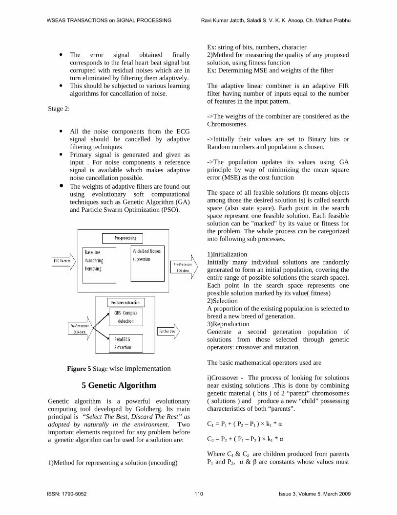
� The error signal obtained finally
corresponds to the fetal heart beat signal but corrupted with residual noises which are in turn eliminated by filtering them adaptively.
� This should be subjected to various learning algorithms for cancellation of noise.
Stage 2:
� All the noise components from the ECG
signal should be cancelled by adaptive filtering techniques
� Primary signal is generated and given as input . For noise components a reference signal is available which makes adaptive noise cancellation possible.
� The weights of adaptive filters are found out using evolutionary soft computational techniques such as Genetic Algorithm (GA) and Particle Swarm Optimization (PSO).
Figure 5 Stage wise implementation
5 Genetic Algorithm
Genetic algorithm is a powerful evolutionary computing tool developed by Goldberg. Its main principal is “Select The Best, Discard The Rest” as adopted by naturally in the environment. Two important elements required for any problem before a genetic algorithm can be used for a solution are:
1)Method for representing a solution (encoding)
Ex: string of bits, numbers, character 2)Method for measuring the quality of any proposed solution, using fitness function Ex: Determining MSE and weights of the filter The adaptive linear combiner is an adaptive FIR filter having number of inputs equal to the number of features in the input pattern.
->The weights of the combiner are considered as the Chromosomes.
->Initially their values are set to Binary bits or Random numbers and population is chosen.
->The population updates its values using GA principle by way of minimizing the mean square error (MSE) as the cost function
The space of all feasible solutions (it means objects among those the desired solution is) is called search space (also state space). Each point in the search space represent one feasible solution. Each feasible solution can be "marked" by its value or fitness for the problem. The whole process can be categorized into following sub processes. 1)Initialization Initially many individual solutions are randomly generated to form an initial population, covering the entire range of possible solutions (the search space). Each point in the search space represents one possible solution marked by its value( fitness) 2)Selection A proportion of the existing population is selected to bread a new breed of generation. 3)Reproduction Generate a second generation population of solutions from those selected through genetic operators: crossover and mutation. The basic mathematical operators used are
i)Crossover - The process of looking for solutions near existing solutions .This is done by combining genetic material ( bits ) of 2 “parent” chromosomes ( solutions ) and produce a new “child” possessing characteristics of both “parents”.
C1 = P1 + ( P2 – P1 ) × k1 * α
C2 = P2 + ( P1 – P2 ) × k1 * α
Where C1 & C2 are children produced from parents P1 and P2, α & β are constants whose values must
WSEAS TRANSACTIONS on SIGNAL PROCESSING Ravi Kumar Jatoth, Saladi S. V. K. K. Anoop, Ch. Midhun Prabhu
ISSN: 1790-5052 110 Issue 3, Volume 5, March 2009

be suitably chosen, k1 & K 2 are random numbers lying between -0.5 to + 0.5
ii)Mutation-The process of looking at completely new areas of search space .This is done mathematically by Random inversion of bits in solution to maintain diversity in population set .
m = P + k2 × β
and m is the mutated child produced from parent P.
4)Termination- A solution is found that satisfies pre-specified criteria
5)Fixed number of generations.-The highest ranking solution’s fitness has reached.The following flow chart pictorially represents Genetic algorithm in a brief and efficient manner.
Figure 6 Block digramof GA
� Computation complexity is high
� More training time is required
� Conversion from binary to decimal
� Chances of falling to local minima
6 Particle Swarm Optimisation
PSO is a robust stochastic optimization technique based on the movement and intelligence of swarms.PSO applies the concept of social interaction to problem solving.
• It uses a number of agents (particles) that
constitute a swarm moving around in the search space looking for the best solution.
• Each particle is treated as a point in a N-dimensional space R2 which adjusts its flying according to its own flying experience as well as the flying experience of other particles.
• The population is set of vectors and is called swarm.
• Each particle keeps track of its coordinates in the solution space which are associated with the best solution (fitness) that has achieved so far by that particle. This value is called personal best, pbest.
• Another best value that is tracked by the PSO is the best value obtained so far by any particle in the neighbourhood of that particle. This value is called gbest.
• The basic concept of PSO lies in accelerating each particle toward its pbest and the gbest locations, with a random weighted accelaration at each time step .
Search space D-Dimensional
X i = [xi1, …, xiD]T = ith particle of Swarm
V i = [vi1, …, viD]T = Velocity of ith particle
Pi = [pi1, …, piD]T = Best previous position of the ith particle
Each particle tries to modify its position using the following information: 1) The current positions. 2) The current velocities. 3) The distance between the current position and pbest. 4. The distance between the current position and the gbest.
WSEAS TRANSACTIONS on SIGNAL PROCESSING Ravi Kumar Jatoth, Saladi S. V. K. K. Anoop, Ch. Midhun Prabhu
ISSN: 1790-5052 111 Issue 3, Volume 5, March 2009
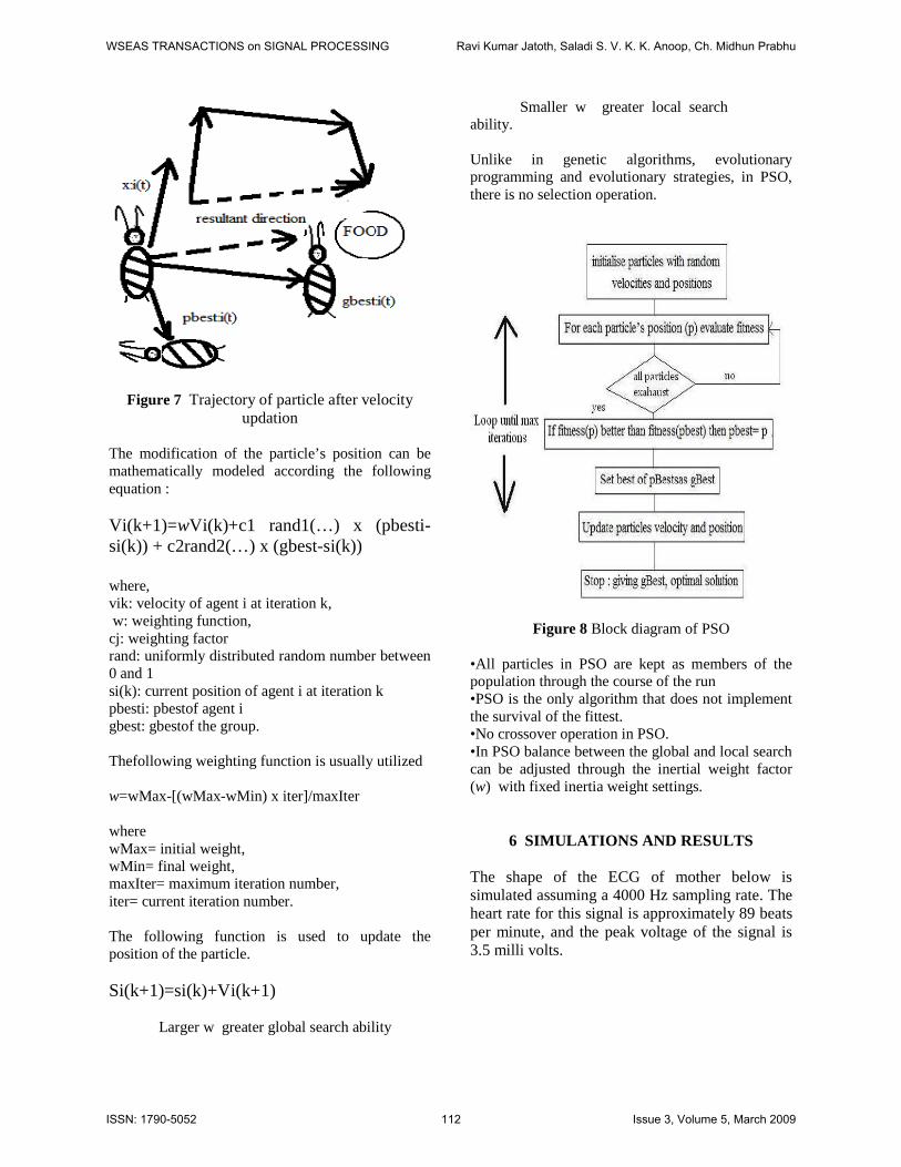
Figure 7 Trajectory of particle after velocity updation
The modification of the particle’s position can be mathematically modeled according the following equation : Vi(k+1)=wVi(k)+c1 rand1(…) x (pbesti-si(k)) + c2rand2(…) x (gbest-si(k)) where, vik: velocity of agent i at iteration k, w: weighting function, cj: weighting factor rand: uniformly distributed random number between 0 and 1 si(k): current position of agent i at iteration k pbesti: pbestof agent i gbest: gbestof the group. Thefollowing weighting function is usually utilized w=wMax-[(wMax-wMin) x iter]/maxIter where wMax= initial weight, wMin= final weight, maxIter= maximum iteration number, iter= current iteration number. The following function is used to update the position of the particle. Si(k+1)=si(k)+Vi(k+1) Larger w greater global search ability
Smaller w greater local search ability. Unlike in genetic algorithms, evolutionary programming and evolutionary strategies, in PSO, there is no selection operation.
Figure 8 Block diagram of PSO •All particles in PSO are kept as members of the population through the course of the run •PSO is the only algorithm that does not implement the survival of the fittest. •No crossover operation in PSO. •In PSO balance between the global and local search can be adjusted through the inertial weight factor (w) with fixed inertia weight settings.
6 SIMULATIONS AND RESULTS
The shape of the ECG of mother below is simulated assuming a 4000 Hz sampling rate. The heart rate for this signal is approximately 89 beats per minute, and the peak voltage of the signal is 3.5 milli volts.
WSEAS TRANSACTIONS on SIGNAL PROCESSING Ravi Kumar Jatoth, Saladi S. V. K. K. Anoop, Ch. Midhun Prabhu
ISSN: 1790-5052 112 Issue 3, Volume 5, March 2009

Figure 9 : Maternal Heartbeat Signal The heart of a fetus beats noticeably faster than that of its mother, with rates ranging from 120 to 160 beats per minute. The fetal ECG signal generated corresponds to a heart rate of 139 beats per minute and a peak voltage of 0.25 millivolts.
Figure 10 : Fetal Heartbeat Signal(targeted signal)
The signal obtained from the abdominal electrode is a noisy signal and is shown below. This is to be filtered and given as input to the adaptive noise canceller.
Figure 11 : Abdominal Electrode Signal
The signal obtained from the thoracic electrode is given as reference signal as shown below. This is given as reference input to the adaptive noise canceller.
Figure 12 : Thoracic Electrode Signal The filtered signal from the abdominal electrode signal is obtained .This contains much noise in addition to the fetal heartbeat which is to be filtered by adaptive noise canceller trained by various algorithms.
Figure 13 : Steady State Error Signal By training with LMS algorithm to the adaptive noise canceller the following signal is obtained. Here, order of the filter used is 3. The QRS complex detection is important so that RR-interval can be extracted for monitoring of fetal condition.This detection is poor in case of the second peak of the obtained signal by LMS algorithm.
WSEAS TRANSACTIONS on SIGNAL PROCESSING Ravi Kumar Jatoth, Saladi S. V. K. K. Anoop, Ch. Midhun Prabhu
ISSN: 1790-5052 113 Issue 3, Volume 5, March 2009

Figure 14 : Obtained FECG Signal by LMS The QRS complex detection which is the key to decide many abnormalities is better in case of the signal obtained by SGA compared to the LMS algorithm as shown in fig 14.
Figure 12 : Obtained FECG Signal by SGA
The signal obtained by PSO is very much similar to the desired FECG.QRS detection is also very good compared to that obtained by various other algorithms.
Figure 13 : Obtained FECG Signal by PSO
7 CONCLUSIONS
Here in this paper we compared biologically Inspired computational techniques with the Conventional Least Mean Square algorithm .From Simulations and results we can conclude that Particle Swarm Optimization based adaptive noise Cancellation technique is performing better and hence preferred over the other techniques . A better solution may be expected by replacing the adaptive linear combiner with a Functional Link Artificial Neural Network(FLANN).Further scope can be carried by the implementation of the above discussed technique on FPGA. It can also be carried by the implementation of above discussed technique in real time ,using DSP processor that does process of conversion and subtraction and the host PC doing the training process. REFERENCES [1] Amit Kumar, Lillie Dewan, Mukhtiar Singh. Real Time Monitoring System for ECG Signal Using Virtual Instrumentation,. WSEAS Transactions on Biology and Biomedicine Issue 11, Volume 3, 2006 [2] Ch. Renumadhavi, S. Madhava Kumar, A. G. Ananth, Nirupama Srinivasan Algorithms for Filtering and Finding SNR of ECG Signals with Powerline Interference, WSEAS Transactions on Signal Processing Issue 9, Volume 2, September 2006 pp 1320-1325 [3] B.W. Widrow, S.D. Stearns, “Adaptive signal processing”, 1985,Prentice-Hall, Inc., New Jearsy. [4] Levkov C, Michov G, Ivanov R , Daskalov IK . “Subtraction of 50 Hz interference from the elect rocardiogram” . Med & Biol, Eng & Comput 1984 ; [5] M B I Reaz,L S Weis “Adaptive linear network fi lter for fetal ECG extraction” 2004 [6] “Comparision between blind seperation and adaptive noise cancellation techniques for fetal ECG extraction” IEE Savoy place London 1999 [7] Haykin S . “Adaptive Filter Theory” . 3rd Edition . Prentice Hall 2002 .. [8] A. van Eck, “Abdominale foetale ECG opname” in Dutch), Twente Univ. Technol., Enschede, The Netherlands, Rep. 1224.0093, 1976. [9] T. Wheeler, A. Murrills, and T. Shelly, “M easurement of the fetal heart rate during pregnancy by a new electrocardiographic technique” Brit. J. Obstet. Gynecol., vol. 85, pp. 12-17, 1978 [10] J.H.van Bemmel, “Detection of weak fetal electrocardiograms by autocorrelation and crosscorrelation of envelopes” IEEE Trans. Biomed. Eng., vol. BME-15, no. 1, pp. 17-23, 1968.
WSEAS TRANSACTIONS on SIGNAL PROCESSING Ravi Kumar Jatoth, Saladi S. V. K. K. Anoop, Ch. Midhun Prabhu
ISSN: 1790-5052 114 Issue 3, Volume 5, March 2009

[11]Bobbie, P. O., Chaudhari, H., Arif, C.-Z., and Pujari, S., Electrocardiogram (EKG) Data Acquisition and Wireless Transmission, WSEAS Transactions on Computers, vol. 3, issue 8, 2004, pp 2665 – 2672 [12] L. El Khansa and A. Naït-Ali, Parametrical modelling of a premature ventricular contraction ECG beat: comparaison with the normal case, Computers in Biology and Medicine, vol. 37, 2007, pp. 1-7. [13] G. Mandellos, M. Koukias, G. Anastassopoulos, D. Lymperopoulos, A new SCP-ECG module for telemedicine services, WSEAS Transactions on Computers,, November 2004, vol. 3, Iss. 5, pp. 1258-1263 [14] P. Baldi and S. Brunak, “Bioinformatics,” The Machine Learning Approach, MIT Press, London, UK, 2000. [15]McSharry P E, Clifford G D, Tarassenko L , Smith L A., A dynamic model for generating synthetic electrocardiogram signals, IEEE Trans. on Biomedical Engineering vol.50, 2003, pp.289-294 [16] Zhao Z D and Chen Y Q., A new method for removal of baseline wander and power line interface in ECG signals, Proc. of the fifth Int. Conf. on Machine Learning and Cybernetics (Dalian) 13-16 aug. 2006, pp. 4342-4347 347, pp 12-15, 1996. [17] Selker, H. P., Griffith, J. L., Patil, S., Long, W. J., & D'Agostino, R. B. A comparison of performance of mathematical predictive methods for medical diagnosis: identifying acute cardiac ischemia among emergency department patients. J. Investig. Med., No 43, Vol. 5. pp 468-476, 1995. [18] Polak, M. J., Zhou, S. H., Rautaharju, P. M., Armstrong, W. W., & Chaitman, B. R. Using automated analysis of the resting twelve-lead ECG to identify patients at risk if developing transient myocardial ischemia-an application of an adaptive logic network. Physiology. Meas., No 18 Vol. 4, pp 317-32, 19975. [19] Henrik Haraldsson, Lars Edenbrandt, Mattias Ohlsson, Detecting acute myocardialinfarction in the 12-lead ECG using Hermite expansions and neural networks, Artificial Intelligence in Medicine 2004 pp 127-135
WSEAS TRANSACTIONS on SIGNAL PROCESSING Ravi Kumar Jatoth, Saladi S. V. K. K. Anoop, Ch. Midhun Prabhu
ISSN: 1790-5052 115 Issue 3, Volume 5, March 2009
Copyright © 2022 FDOKUMEN