Biological Investigation of Fruit of Spondias pinnata (Anacardiaceae ))
116
B B i i o o l l o o g g i i c c a a l l I I n n v v e e s s t t i i g g a a t t i i o o n n o o f f F F r r u u i i t t o o f f S S p p o o n n d d i i a a s s p p i i n n n n a a t t a a ( ( A A n n a a c c a a r r d d i i a a c c e e a a e e ) ) A dissertation submitted to the Department of Pharmacy, East West University in the partial fulfillment of the requirements for the degree of Bachelor of Pharmacy (B. Pharm.) Submitted by : Shawkat Md. Aminul Islam ID# 2008-3-70-085 Session : 2012-2013 June 2012 Department of Pharmacy East West University
-
Upload
khangminh22 -
Category
Documents
-
view
1 -
download
0
Transcript of Biological Investigation of Fruit of Spondias pinnata (Anacardiaceae ))
BBiioollooggiiccaall IInnvveessttiiggaattiioonn ooff FFrruuiitt ooff SSppoonnddiiaass ppiinnnnaattaa
((AAnnaaccaarrddiiaacceeaaee))
A dissertation submitted to the Department of Pharmacy, East West University in the partial fulfillment of the requirements for the degree of
Bachelor of Pharmacy (B. Pharm.)
Submitted by : Shawkat Md. Aminul Islam
ID# 2008-3-70-085
Session : 2012-2013
June 2012 Department of Pharmacy
East West University


AAcckknnoowwlleeddggmmeennttss
Acknowledgments
All praises are for the Almighty Allah for His mercy and blessings. I feel proud to express my heartiest regards and deep sense of gratitude to my reverend teacher
and supervisor Kh. Tanvir Ahmed, Lecturer, Department of Pharmacy, East West University,
for his day to day mastermind direction, constant supervision and support, constructive
suggestions, optimistic counseling, valuable criticism, keen interest and active encouragement to
carry out this research work.
It gives me immense pleasure to express sincere appreciation and gratefulness to Professor Dr.
Chowdhury Faiz Hossain, Dean, Faculty of Sciences and Engineering, East West University
and to Dr. Sufia Islam, Chairperson, Department of Pharmacy, East West University for
providing all possible help during the course of this research work.
It is also great pleasure for me to offer my deepest indebtedness to all of my respected teachers
of the Department of Pharmacy, East West University for extending their helping hands
whenever needed.
I also would like to extend my thanks to the office staffs and Lab officers of the Department of
Pharmacy, East West University. Special thanks goes to all the research students in the lab for
their help and assistance, friendly behavior and earnest cooperation, which enabled me to work
in a congenial and comfortable atmosphere.
Finally I wish to convey my thanks and heartiest regards to all my friends, especially Asif
Mahmud Tunan, Md. Arif Wahid and Anis Mahmud Khokon, for their untiring cooperation.
Special appreciation goes to my friend Mohammad Kawsar Manik for collection and supply of
plant materials. I would like to convey thanks to my friend Mahmudul Hasan Chowdhury for his
cooperation during extraction and antimicrobial screening. I wish to convey special thanks to my
friend Md. Nabil and Mohammad Ibrahim Khalil for their assistance during thrombolytic activity
screening. I feel that without their cooperation this research work would not have been
completed to my satisfaction.
Cordial thanks are also due to my parents, relatives and to all my well wishers for their
wholehearted inspiration throughout the period of the research work.
Shawkat Md. Aminul Islam
Author
CCeerrttiiffiiccaattee
Certificate
This is to certify that the research work on “Biological Investigation of Spondias pinnata
fruit” submitted to the Department of Pharmacy, East West University in partial
fulfillment of the requirements for the degree of Bachelor of Pharmacy was carried out by
Shawkat Md. Aminul Islam (ID: 2008-3-70-085) under or guidance and supervision and
that no part of the thesis has been submitted for any other degree. We further certify that
all the sources of information and facilities availed of this connection are duly
acknowledged.
………………………….. …………………………. Sufia Islam, PhD Chairperson Department of Pharmcay East West University
Kh. Tanvir Ahmed
Lecturer
Department of Pharmacy
East West University
Contents
CCoonntteennttss
Abstract IX
Chapter 1: Introduction
Topics Page no
1.1 Rationale and Objective of the Work 1
1.2 History of traditional herbal medicine in Bangladesh 3
1.3 Research of traditional drugs in Bangladesh 6
1.4 General Approaches to Drug Discovery from Natural Sources 7
1.5 Flow Chart of Bioactivity Guided Phytochemical Approach 9
1.6 The plant family: Anacardiaceae 10
1.6.1 Taxonomy 11
1.6.2 Botanical features of Anacardiaceae family 12
1.6.3 Distribution of Anacardiaceae family 15
1.6.4 Anacardiaceae Species Available in Bangladesh 15
1.7 Introduction to Spondias pinnata 19
1.7.1 Taxonomic hierarchy of the investigated plant 19
1.7.2 Common names 20
1.7.3 Botanical features 20
1.7.4 Images of various parts of Spondias pinnata 21
1.7.5 Distribution of Spondias pinnata 23
1.7.6 Growing conditions 23
1.7.7 Chemical constituents of Spondias pinnata 23
1.7.8 Ethnobotany of Spondias pinnata 23
1.7.9 Nutritive and mineral potential of ripe fruits of Spondias pinnata 24
1.7.10 Medicinal Potentials of Spondias pinnata 24
1.7.11 Reported Biological Works on Spondias pinnata 25
Biological Investigation of Spondias pinnata fruit I
Contents
Chapter 2: SSttuuddyy PPrroottooccooll
Topics Page no
2.1 Present Study Protocol 28
2.1.1 Phytochemical Screening 28
2.1.2 Evaluation of Antioxidant activity 29
2.1.3 Antimicrobial Screening 30
2.1.4 Evaluation of Thrombolytic Activity 30
Chapter 3: CCoolllleeccttiioonn,, PPrreeppaarraattiioonn aanndd PPhhyyttoocchheemmiiccaall SSccrreeeenniinngg
Topics Page no
3.1 Collection and preparation of the plant material 32
3.2 Extraction of the Plant material 32
3.3 Preliminary phytochemical investigations of crude extract 33
3.3.1 Apparatus 33
3.3.2 Reagents 34
3.3.3 Methods and results of preliminary phytochemical investigations
of crude extract
34
Chapter 4: AAnnttiimmiiccrroobbiiaall SSccrreeeenniinngg
Topics Page no
4.1 Introduction 37
4.2 Principle of Disc Diffusion Method 38
4.3 Experimental Design 39
4.3.1 Apparatus and Reagents 39
4.3.2 Test Organisms 39
4.3.3 Test Materials of Spondias pinnata 40
4.3.4 Culture Medium and its Composition 40
4.3.5 Preparation of the Medium 40
4.3.6 Sterilization Procedure 40
4.3.7 Preparation of Subculture 41
4.3.8 Preparation of the Test Plate 41
4.3.9 Preparation of Discs 41
4.3.10 Standard Discs 41
Biological Investigation of Spondias pinnata fruit II
Contents
4.3.11 Blank Discs 41
4.3.12 Preparation of Sample Discs with Test Sample 41
4.3.13 Application of the Test Samples 42
4.3.14 Diffusion and Incubation 42
4.3.15 Determination of Antimicrobial Activity by Measuring the Zone
of Inhibition
43
4.4 Determination of Minimum Inhibitory Concentration (MIC) and
Minimum Bactericidal Concentration (MBC)
43
4.4.1 Principle of MIC 44
4.5 Experimental Design for MIC & MBC 44
4.5.1 Apparatus and Reagents 44
4.5.2 Test Organisms 44
4.5.3 Test Materials of Spondias pinnata 45
4.5.4 Culture Medium and their Composition 45
4.5.5 Method of determination of MIC & MBC 45
Chapter 5 Evaluation of Antioxidant Activity
Topics Page no
5.1 Rationale and Objective 48
5.2 Mechanism of Antioxidant 49
5.3 Methods of evaluating antioxidant activity 49
5.3.1 Determination of DPPH radical scavenging assay 50
5.3.1 (1) Qualitative assay 50
5.3.1.1 Principle of Quantitative Assay 50
5.3.1.2 Materials & Reagents 51
5.3.1.3 Methods 51
5.3.2 Determination of total phenolic content 52
5.3.2.1 Principle 52
5.3.2.2 Materials & Reagents 52
5.3.2.3 Composition of Folin-Ciocalteu reagent 53
5.3.2.4 Methods 53
5.3.3 Determination of reducing power assay 54
5.3.3.1 Principle 54
Biological Investigation of Spondias pinnata fruit III

Contents
5.3.3.2 Materials & Reagents 54
5.3.3.3 Methods 54
5.3.4 Total Flavonoids Concentration 56
5.3.4.1 Principle 56
5.3.4.2 Materials & Reagents 56
5.3.4.3 Methods 56
5.3.5 Determination of total antioxidant capacity by
phosphomolybdenum method
57
5.3.5.1 Principle 57
5.3.5.2 Materials & Reagents 58
5.3.5.3 Methods 58
Chapter 6 EEvvaalluuaattiioonn ooff TThhrroommbboollyyttiicc AAccttiivviittyy
Topics Page no
6.1 Introduction 60
6.2 Principle 61
6.3 Materials & Reagents 61
6.4 Preparation of test sample 61
6.5 Methods 62
Chapter 7 Results and discussion
Topics Page no
7.1 Results and discussion of the test samples of Spondias pinnata
(fruit)
64
7.1.1 In vitro antimicrobial screenings 64
7.1.2 Minimum Inhibitory Concentration (MIC) and Minimum
Bactericidal Concentration (MBC)
69
7.1.3.1 Total phenolic content 70
7.1.3.2 DPPH radical scavenging assay (Quantitative analysis) and IC50 71
7.1.3.3 Reducing power assay 72
7.1.3.4 Total antioxidant capacity by phosphomolybdenum method 73
7.1.3.5 Total flavonoids content 75
7.1.4 In vitro thrombolytic activity 76
Biological Investigation of Spondias pinnata fruit IV
Contents
Conclusion 79
Reference 80
Biological Investigation of Spondias pinnata fruit V
Contents
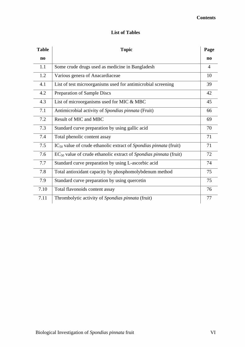
List of Tables
Table
no
Topic Page
no
1.1 Some crude drugs used as medicine in Bangladesh 4
1.2 Various genera of Anacardiaceae 10
4.1 List of test microorganisms used for antimicrobial screening 39
4.2 Preparation of Sample Discs 42
4.3 List of microorganisms used for MIC & MBC 45
7.1 Antimicrobial activity of Spondias pinnata (Fruit) 66
7.2 Result of MIC and MBC 69
7.3 Standard curve preparation by using gallic acid 70
7.4 Total phenolic content assay 71
7.5 IC50 value of crude ethanolic extract of Spondias pinnata (fruit) 71
7.6 EC50 value of crude ethanolic extract of Spondias pinnata (fruit) 72
7.7 Standard curve preparation by using L-ascorbic acid 74
7.8 Total antioxidant capacity by phosphomolybdenum method 75
7.9 Standard curve preparation by using quercetin 75
7.10 Total flavonoids content assay 76
7.11 Thrombolytic activity of Spondias pinnata (fruit) 77
Biological Investigation of Spondias pinnata fruit VI
Contents
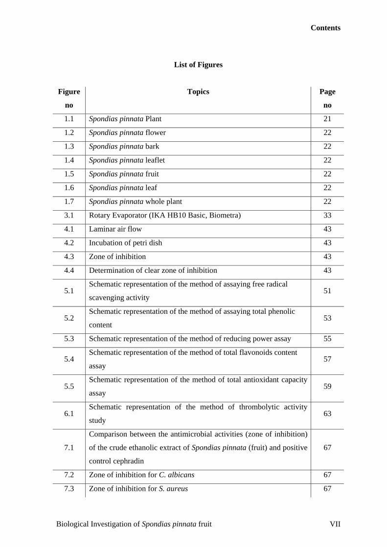
List of Figures
Figure
no
Topics Page
no
1.1 Spondias pinnata Plant 21
1.2 Spondias pinnata flower 22
1.3 Spondias pinnata bark 22
1.4 Spondias pinnata leaflet 22
1.5 Spondias pinnata fruit 22
1.6 Spondias pinnata leaf 22
1.7 Spondias pinnata whole plant 22
3.1 Rotary Evaporator (IKA HB10 Basic, Biometra) 33
4.1 Laminar air flow 43
4.2 Incubation of petri dish 43
4.3 Zone of inhibition 43
4.4 Determination of clear zone of inhibition 43
5.1 Schematic representation of the method of assaying free radical
scavenging activity 51
5.2 Schematic representation of the method of assaying total phenolic
content 53
5.3 Schematic representation of the method of reducing power assay 55
5.4 Schematic representation of the method of total flavonoids content
assay 57
5.5 Schematic representation of the method of total antioxidant capacity
assay 59
6.1 Schematic representation of the method of thrombolytic activity
study 63
7.1
Comparison between the antimicrobial activities (zone of inhibition)
of the crude ethanolic extract of Spondias pinnata (fruit) and positive
control cephradin
67
7.2 Zone of inhibition for C. albicans 67
7.3 Zone of inhibition for S. aureus 67
Biological Investigation of Spondias pinnata fruit VII
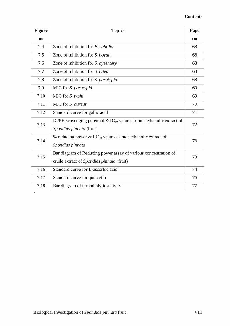
Contents
Figure
no
Topics Page
no
7.4 Zone of inhibition for B. subtilis 68
7.5 Zone of inhibition for S. boydii 68
7.6 Zone of inhibition for S. dysentery 68
7.7 Zone of inhibition for S. lutea 68
7.8 Zone of inhibition for S. paratyphi 68
7.9 MIC for S. paratyphi 69
7.10 MIC for S. typhi 69
7.11 MIC for S. aureus 70
7.12 Standard curve for gallic acid 71
7.13 DPPH scavenging potential & IC50 value of crude ethanolic extract of
Spondias pinnata (fruit) 72
7.14 % reducing power & EC50 value of crude ethanolic extract of
Spondias pinnata 73
7.15 Bar diagram of Reducing power assay of various concentration of
crude extract of Spondias pinnata (fruit) 73
7.16 Standard curve for L-ascorbic acid 74
7.17 Standard curve for quercetin 76
7.18 Bar diagram of thrombolytic activity 77
`
Biological Investigation of Spondias pinnata fruit VIII

AAbbssttrraacctt
Abstract
AAbbssttrraacctt
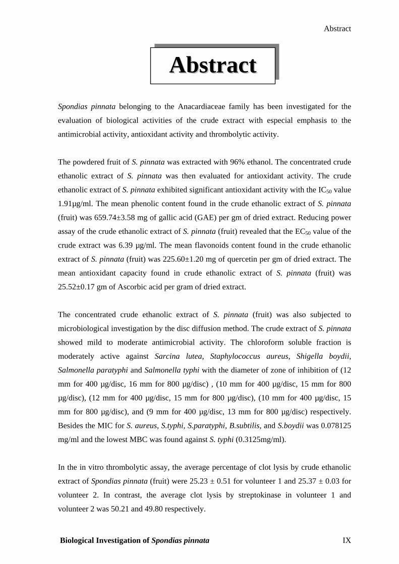
Spondias pinnata belonging to the Anacardiaceae family has been investigated for the
evaluation of biological activities of the crude extract with especial emphasis to the
antimicrobial activity, antioxidant activity and thrombolytic activity.
The powdered fruit of S. pinnata was extracted with 96% ethanol. The concentrated crude
ethanolic extract of S. pinnata was then evaluated for antioxidant activity. The crude
ethanolic extract of S. pinnata exhibited significant antioxidant activity with the IC50 value
1.91µg/ml. The mean phenolic content found in the crude ethanolic extract of S. pinnata
(fruit) was 659.74±3.58 mg of gallic acid (GAE) per gm of dried extract. Reducing power
assay of the crude ethanolic extract of S. pinnata (fruit) revealed that the EC50 value of the
crude extract was 6.39 µg/ml. The mean flavonoids content found in the crude ethanolic
extract of S. pinnata (fruit) was 225.60±1.20 mg of quercetin per gm of dried extract. The
mean antioxidant capacity found in crude ethanolic extract of S. pinnata (fruit) was
25.52±0.17 gm of Ascorbic acid per gram of dried extract.
The concentrated crude ethanolic extract of S. pinnata (fruit) was also subjected to
microbiological investigation by the disc diffusion method. The crude extract of S. pinnata
showed mild to moderate antimicrobial activity. The chloroform soluble fraction is
moderately active against Sarcina lutea, Staphylococcus aureus, Shigella boydii,
Salmonella paratyphi and Salmonella typhi with the diameter of zone of inhibition of (12
mm for 400 µg/disc, 16 mm for 800 µg/disc) , (10 mm for 400 µg/disc, 15 mm for 800
µg/disc), (12 mm for 400 µg/disc, 15 mm for 800 µg/disc), (10 mm for 400 µg/disc, 15
mm for 800 µg/disc), and (9 mm for 400 µg/disc, 13 mm for 800 µg/disc) respectively.
Besides the MIC for S. aureus, S.typhi, S.paratyphi, B.subtilis, and S.boydii was 0.078125
mg/ml and the lowest MBC was found against S. typhi (0.3125mg/ml).
In the in vitro thrombolytic assay, the average percentage of clot lysis by crude ethanolic
extract of Spondias pinnata (fruit) were 25.23 ± 0.51 for volunteer 1 and 25.37 ± 0.03 for
volunteer 2. In contrast, the average clot lysis by streptokinase in volunteer 1 and
volunteer 2 was 50.21 and 49.80 respectively.
Biological Investigation of Spondias pinnata IX
IInnttrroodduuccttiioonn
C H A P T E R
Chapter 1: Introduction
1.1 Rationa
Terrestrial
diseases (B
to the Midd
known spec
capsule late
tablets from
various dis
traditional
isolated fro
used. Hem
found in li
countries (B
Success in
criteria suc
random col
plants as th
Plants, esp
medicines
showed th
ethnopharm
on bioactiv
important
Camptothec
Biological I
CChhaapptteerr
11
nn
le and Objective of the
plants, especially higher
alunas MJ et al., 2006). F
le Paleolithic age some
ies, including licorice (G
x (Papaver somniferum)
Mesopotamia in 2600 B
eases as ingredients of
medicine. Furthermore,
m P. somniferum were
isuccinate carbenoxolon
corice, is prescribed fo
alunas MJ et al., 2006).
natural products research
h as chemotaxonomic d
lection (Kurt Hostettman
erapeutic tools have help
ecially those with ethno
for early drug discovery
at the uses of 80% of
acological purposes. Cur
ity-guided isolation met
anticancer agents, pac
a acuminata (Balunas M
nvestigation of Spondias
IInnttrroodduuccttiioo
Work
plants, have a long history of use in the treatment of human
ossil records date human use of plants as medicines at least
60,000 years ago (Solecki & Shanidar, 1975). Several well
lycyrrhiza glabra), myrrh (Commiphora species), and poppy
, were referred to by the first known written record on clay
C, and these plants are still in use today for the treatment of
official drugs or herbal preparations used in systems of
morphine, codeine, noscapine (narcotine), and papaverine
developed as single chemical drugs and are still clinically
e sodium, a semi-synthetic derivative of glycyrrhetic acid
r the treatment of gastric and duodenal ulcers in various
is conditioned by a careful plant selection, based on various
ata, ethnomedical information, field observations or even
n and Christian Terreaux, 2000). Historical experiences with
ed to introduce single chemical entities in modern medicine.
pharmacological uses, have been the primary sources of
. In fact, a recent analysis by Fabricant and Farnsworth
122 plant-derived drugs were related to their original
rent drug discovery from terrestrial plants has mainly relied
hods, which, for example, have led to discoveries of the
litaxel from Taxus brevifolia and camptothecin from
J et al., 2006).
pinnata fruit 1
Chapter 1: Introduction
The goals of using plants as sources of therapeutic agents are (Fabricant DS and Farnsworth NR,
2001)
a) to isolate bioactive compounds for direct use as drugs, e.g. digoxin, digitoxin, morphine,
reserpine, taxol, vinblastine, vincristine;
b) to produce bioactive compounds of novel or known structures as lead compounds for
semisynthesis to produce patentable entities of higher activity and/or lower toxicity, e.g.,
metformin, nabilone, oxycodon (and other narcotic analgesics), taxotere, teniposide,
verapamil, and miodarone, which are based, respectively, on galegine, ∆9-
tetrahydrocannabinol, morphine, taxol, podophyllotoxin, and khellin;
c) to use agents as pharmacologic tools, e.g., lysergic acid diethylamide (LSD), mescaline,
yohimbine; and
d) to use the whole plant or part of it as a herbal remedy, e.g., cranberry, echinacea,
feverfew, garlic, etc.
The number of higher plant species (angiosperms and gymnosperms) on this planet is estimated
at 250,000 (Ayensu & DeFilipps, 1978) with a lower level at 215,000 (Cronquist, 1981;
Cronquist, 1988) and an upper level as high as 500,000 (Tippo & Stern, 1977; Schultes, 1972).
Of these, only about 6% have been screened for biologic activity, and a reported 15% have been
evaluated phytochemically (Verpoorte, 2000). It was estimated that in 1991 in the United States,
for every 10,000 pure compounds (most likely those based on synthesis) that are biologically
evaluated (primarily in vitro), 20 would be tested in animal models, and 10 of these would be
clinically evaluated, and only one would reach U.S. Food and Drug Administration approval for
marketing. The time required for this process was estimated as 10 years at a cost of $231 million
(U.S.) (Vagelos, 1991).
The major drawback of this strategy is the frequent isolation of known metabolites. Therefore,
hyphenated techniques (LC-UV, LC-MS, LC-NMR) have been developed, in order to detect as
early as possible potential original structures. These compounds can then be tested in various
bioassays (Kurt Hostettmann and Christian Terreaux, 2000). More recently combinatorial
chemistry and high throughput robotic screening techniques have been employed as viable
strategies for drug discovery programs (Berhanu M. et al., 1999).
Chemical diversity of secondary plant metabolites that results from plant evolution is superior to
that found in synthetic combinatorial chemical libraries (Vagelos, 1991). Medicinal plants have
Biological Investigation of Spondias pinnata fruit 2
Chapter 1: Introduction
played an essential role in the development of human culture, for example religions and different
ceremonies. (E.g. Dutura has long been associated with the worship of Shiva, the Indian god).
Plants are directly used as medicines by a majority of cultures around the world, for example
Chinese medicine and Indian medicine. Many food crops have medicinal effects, for example
garlic. Medicinal plants are resources of new drugs. Studying medicinal plants helps to
understand the plant toxicity and protect human and animals from natural poisons. Cultivation
and preservation of medicinal plants protect biological diversity, for example metabolic
engineering of plants (Naik S.N and Panda V.S, 2008). The medicinal plants find application in
pharmaceutical, cosmetic, agricultural and food industry. With onset of scientific research in
herbals, it is becoming clearer that the medicinal herbs have a potential in today’s synthetic era,
as numbers of medicines are becoming resistant. According to one estimate only 20% of the
plant flora has been studied and 60%of synthetic medicines owe their origin to plants. Ancient
knowledge coupled with scientific principles can come to the forefront and provide us with
powerful remedies to eradicate the diseases. In real sense, coupling of ancient knowledge and
scientific principle is essential-
(1) To identify alternative and complementary medicine.
(2) To reduce the toxicity of drug therapy especially toxicity reduction of synthetic and semi
synthetic drugs.
(3) To find the lead compound diversification to treat various diseases (Ayuvedaherbs, 2005).
1.2 History of traditional herbal medicine in Bangladesh
Traditional Medicine is the medicine or treatment based on traditional uses of plants, animals or
their products, other natural substances (including some inorganic chemicals), religious verses,
cultural practices, and physical manipulations including torture. As this system of medicine has
been in use almost unchanged generation after generation throughout the ages for the treatment
of various physical and psychological diseases, it is called traditional. Most of the times, the
type, preparation, and uses of traditional medicines are largely influenced by folklore customs
and the cultural habits, social practices, religious beliefs and, in many cases, superstitions of the
people who prescribe or use them (Ghani A., and Pasha M.K, 2006).
The earliest mention of traditional medicine is found in ‘‘Rigveda’’, the oldest repository of
knowledge in this subcontinent. Later ‘‘Ayurveda’’, developed from the Vedic concept of life,
Biological Investigation of Spondias pinnata fruit 3
Chapter 1: Introduction
became the important source of all systems of medical sciences. In course of time it became a
part of culture and heritage of the people of the Indian subcontinent.
Traditional medicine involves the use of both material and non-material components. The
material components invariably comprise parts or organs of plants and their products. They also
consist of animal organs, minerals and other natural substances. The non-material components,
which constitute important items of religious and spiritual medicines, include torture, charms,
magic, incantations, religious verses, amulets and rituals like sacrifices, appeasement of evil
spirits, etc (Ghani A., and Pasha M.K, 2006).
Treatments in traditional medicine are carried out by internal and external application of
medicaments, physical manipulation of various parts of the body, performing rituals,
psychological treatment, and also by minor surgery. Ayurvedic medicinal preparations consist
mainly of plant materials in the form of powders, semi-solid preparations, decoctions, elixirs and
distillates. Many of them also contain inorganic chemical substances, minerals and animal
products. Alcoholic extracts and alcoholic solutions of the ingredients, tinctures and elixirs are
also frequently used in Ayurvedic medicine (Ghani A., and Pasha M.K, 2006).
Whole plants or their powders or pastes or products and their extracts, infusions, decoctions and
distillates constitute the major constituents of Unani medicine. Minerals, inorganic chemicals
and animal products are also frequently used in preparing these medicines.
For hundreds of years, the medical knowledge of the Indian subcontinent is termed as Ayurveda.
Ayurveda remains an important system of medicine and drug therapy in India and Bangladesh.
Plant alkaloids are the primary active ingredients of Ayurvedic drugs. Today the
pharmacologically active ingredients of many Ayurvedic medicines are being identified and their
usefulness in drug therapy being determined. As only a certain percentage of plants are used in
traditional medicines, it is roughly estimated that of the discovered 17,000 species, nearly 3,000
species are used in medicinal field (Samy RP et al., 2008). Some crude drugs used as medicine in
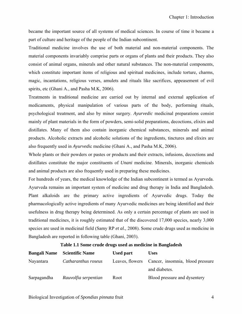
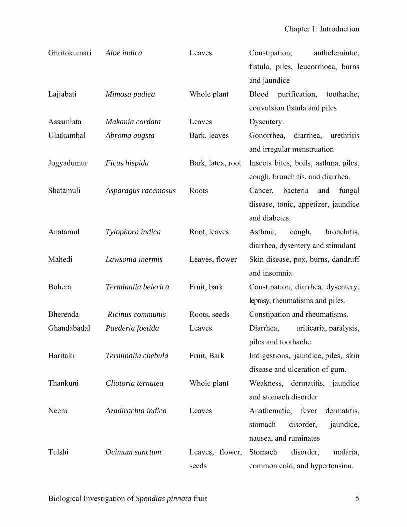
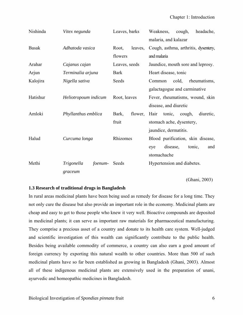
Bangladesh are reported in following table (Ghani, 2003).
Table 1.1 Some crude drugs used as medicine in Bangladesh
Bangali Name Scientific Name Used part Uses
Nayantara
Catharanthus roseus Leaves, flowers Cancer, insomnia, blood pressure
and diabetes.
Sarpagandha Rauvolfia serpentian Root Blood pressure and dysentery
Biological Investigation of Spondias pinnata fruit 4
Chapter 1: Introduction
Ghritokumari Aloe indica Leaves Constipation, anthelemintic,
fistula, piles, leucorrhoea, burns
and jaundice
Lajjabati Mimosa pudica Whole plant Blood purification, toothache,
convulsion fistula and piles
Assamlata Makania cordata Leaves Dysentery.
Ulatkambal Abroma augsta Bark, leaves Gonorrhea, diarrhea, urethritis
and irregular menstruation
Jogyadumur Ficus hispida Bark, latex, root Insects bites, boils, asthma, piles,
cough, bronchitis, and diarrhea.
Shatamuli Asparagus racemosus Roots Cancer, bacteria and fungal
disease, tonic, appetizer, jaundice
and diabetes.
Anatamul Tylophora indica Root, leaves Asthma, cough, bronchitis,
diarrhea, dysentery and stimulant
Mahedi Lawsonia inermis Leaves, flower Skin disease, pox, burns, dandruff
and insomnia.
Bohera Terminalia belerica Fruit, bark Constipation, diarrhea, dysentery,
leprosy, rheumatisms and piles.
Bherenda Ricinus communis Roots, seeds Constipation and rheumatisms.
Ghandabadal Paederia foetida Leaves Diarrhea, uriticaria, paralysis,
piles and toothache
Haritaki Terminalia chebula Fruit, Bark Indigestions, jaundice, piles, skin
disease and ulceration of gum.
Thankuni Cliotoria ternatea Whole plant Weakness, dermatitis, jaundice
and stomach disorder
Neem Azadirachta indica Leaves Anathematic, fever dermatitis,
stomach disorder, jaundice,
nausea, and ruminates
Tulshi Ocimum sanctum Leaves, flower,
seeds
Stomach disorder, malaria,
common cold, and hypertension.
Biological Investigation of Spondias pinnata fruit 5
Chapter 1: Introduction
Nishinda Vitex negunda Leaves, barks Weakness, cough, headache,
malaria, and kalazar
Basak Adhatoda vasica Root, leaves,
flowers
Cough, asthma, arthritis, dysentery,
and malaria
Arahar Cajanus cajan Leaves, seeds Jaundice, mouth sore and leprosy.
Arjun Terminalia arjuna Bark Heart disease, tonic
Kalojira Nigella sativa Seeds Common cold, rheumatisms,
galactagogue and carminative
Hatishur Heliotropoum indicum Root, leaves Fever, rheumatisms, wound, skin
disease, and diuretic
Amloki Phyllanthus emblica Bark, flower,
fruit
Hair tonic, cough, diuretic,
stomach ache, dysentery,
jaundice, dermatitis.
Halud Curcuma longa Rhizomes Blood purification, skin disease,
eye disease, tonic, and
stomachache
Methi Trigonella foenum-
graceum
Seeds Hypertension and diabetes.
(Ghani, 2003)
1.3 Research of traditional drugs in Bangladesh
In rural areas medicinal plants have been being used as remedy for disease for a long time. They
not only cure the disease but also provide an important role in the economy. Medicinal plants are
cheap and easy to get to those people who knew it very well. Bioactive compounds are deposited
in medicinal plants; it can serve as important raw materials for pharmaceutical manufacturing.
They comprise a precious asset of a country and donate to its health care system. Well-judged
and scientific investigation of this wealth can significantly contribute to the public health.
Besides being available commodity of commerce, a country can also earn a good amount of
foreign currency by exporting this natural wealth to other countries. More than 500 of such
medicinal plants have so far been established as growing in Bangladesh (Ghani, 2003). Almost
all of these indigenous medicinal plants are extensively used in the preparation of unani,
ayurvedic and homeopathic medicines in Bangladesh.
Biological Investigation of Spondias pinnata fruit 6
Chapter 1: Introduction
A survey conducted in 1990 in different villages of Bangladesh shows that on average of 14% of
people suffering from illness approach qualified allopathic doctors, 29% contact unqualified
village doctors, 10% contact mullahs, 29% contact quack and 19% contact homeopaths. The
survey indicates an extensive use of medicinal plants, most of which are served in crude and
substandard form, by our people (Haque MM et al., 1990). Traditional medicines are still
manufactured in our country by following the age-old unscientific, traditional methods.
Hundreds of indigenous medicinal plants are employed in different Ayurvedic and Unani
commercial preparations without proper standardization, quality control, evaluation and
determination of the chemical nature, pharmacological and toxicological studies of the active
components which are essential to utilize their therapeutic potential fully. Toxicity of the plants
or plant extracts is coming to light with the advancement of science. Since Bangladesh is a
country of low economic growth, a proper health care system can be established by supplying
low cost medicines to its population. This may be possible only by developing standard drugs
from our natural resources of medicinal plants. In order to achieve this goal research and
development of traditional medicines should be given the due priority (Ghani, 2003).
Besides, Bangladesh imports a large quantity of pharmaceutical raw materials including
medicinal plants and semi-processed plant products to manufacture drugs. Each year a great deal
of money is spent on this purpose.
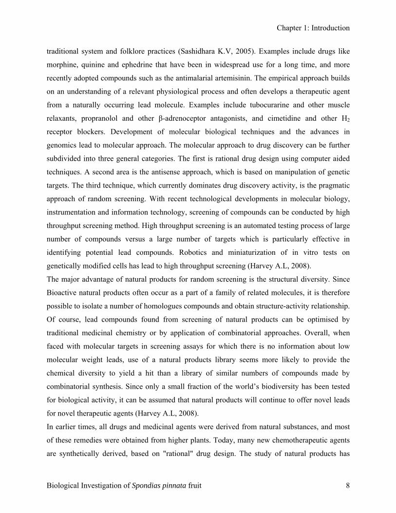
1.4 General Approaches to Drug Discovery from Natural Sources
In general, three different approaches have been, and continue to be used in the drug discovery
process from natural sources. These approaches are: traditional, empirical and molecular (Harvey
A.L, 2008). During the vedic period the ‘‘Susruta samhita’’ and the ‘‘charaka samhita’’ were
influential works on traditional medicine. Hundreds of medicinal plant were identified and have
been traditionally used since then. Over the following centuries, Ayurvedic practitioners
developed a number of medicinal preparations and surgical procedures for the treatment of
various ailments and diseases. WHO (World Health Organization) estimates that 80% of the
populations living in the developing countries rely exclusively on traditional medicine for their
primary health care needs. In almost all the traditional medicine, the traditional plants play a
major role and constitute the backbone of the traditional medicine. Indian Materia medica
includes about 1600 drugs of vegetable origin almost all of which are derived from different
Biological Investigation of Spondias pinnata fruit 7
Chapter 1: Introduction
traditional system and folklore practices (Sashidhara K.V, 2005). Examples include drugs like
morphine, quinine and ephedrine that have been in widespread use for a long time, and more
recently adopted compounds such as the antimalarial artemisinin. The empirical approach builds
on an understanding of a relevant physiological process and often develops a therapeutic agent
from a naturally occurring lead molecule. Examples include tubocurarine and other muscle
relaxants, propranolol and other β-adrenoceptor antagonists, and cimetidine and other H2
receptor blockers. Development of molecular biological techniques and the advances in
genomics lead to molecular approach. The molecular approach to drug discovery can be further
subdivided into three general categories. The first is rational drug design using computer aided
techniques. A second area is the antisense approach, which is based on manipulation of genetic
targets. The third technique, which currently dominates drug discovery activity, is the pragmatic
approach of random screening. With recent technological developments in molecular biology,
instrumentation and information technology, screening of compounds can be conducted by high
throughput screening method. High throughput screening is an automated testing process of large
number of compounds versus a large number of targets which is particularly effective in
identifying potential lead compounds. Robotics and miniaturization of in vitro tests on
genetically modified cells has lead to high throughput screening (Harvey A.L, 2008).
The major advantage of natural products for random screening is the structural diversity. Since
Bioactive natural products often occur as a part of a family of related molecules, it is therefore
possible to isolate a number of homologues compounds and obtain structure-activity relationship.
Of course, lead compounds found from screening of natural products can be optimised by
traditional medicinal chemistry or by application of combinatorial approaches. Overall, when
faced with molecular targets in screening assays for which there is no information about low
molecular weight leads, use of a natural products library seems more likely to provide the
chemical diversity to yield a hit than a library of similar numbers of compounds made by
combinatorial synthesis. Since only a small fraction of the world’s biodiversity has been tested
for biological activity, it can be assumed that natural products will continue to offer novel leads
for novel therapeutic agents (Harvey A.L, 2008).
In earlier times, all drugs and medicinal agents were derived from natural substances, and most
of these remedies were obtained from higher plants. Today, many new chemotherapeutic agents
are synthetically derived, based on "rational" drug design. The study of natural products has
Biological Investigation of Spondias pinnata fruit 8
Chapter 1: Introduction
advantages over synthetic drug design in that it leads optimally to materials having new
structural features with novel biological activity. Not only do plants continue to serve as
important sources of new drugs, but phytochemicals derived from them are also extremely useful
as lead structures for synthetic modification and optimization of bioactivity. The starting
materials for about one-half of the medicines we use today come from natural sources. Virtually
every pharmacological class of drugs includes a natural product prototype. The future of plants
as sources of medicinal agents for use in investigation, prevention, and treatment of diseases is
very promising (Setzer W.N, 1999).
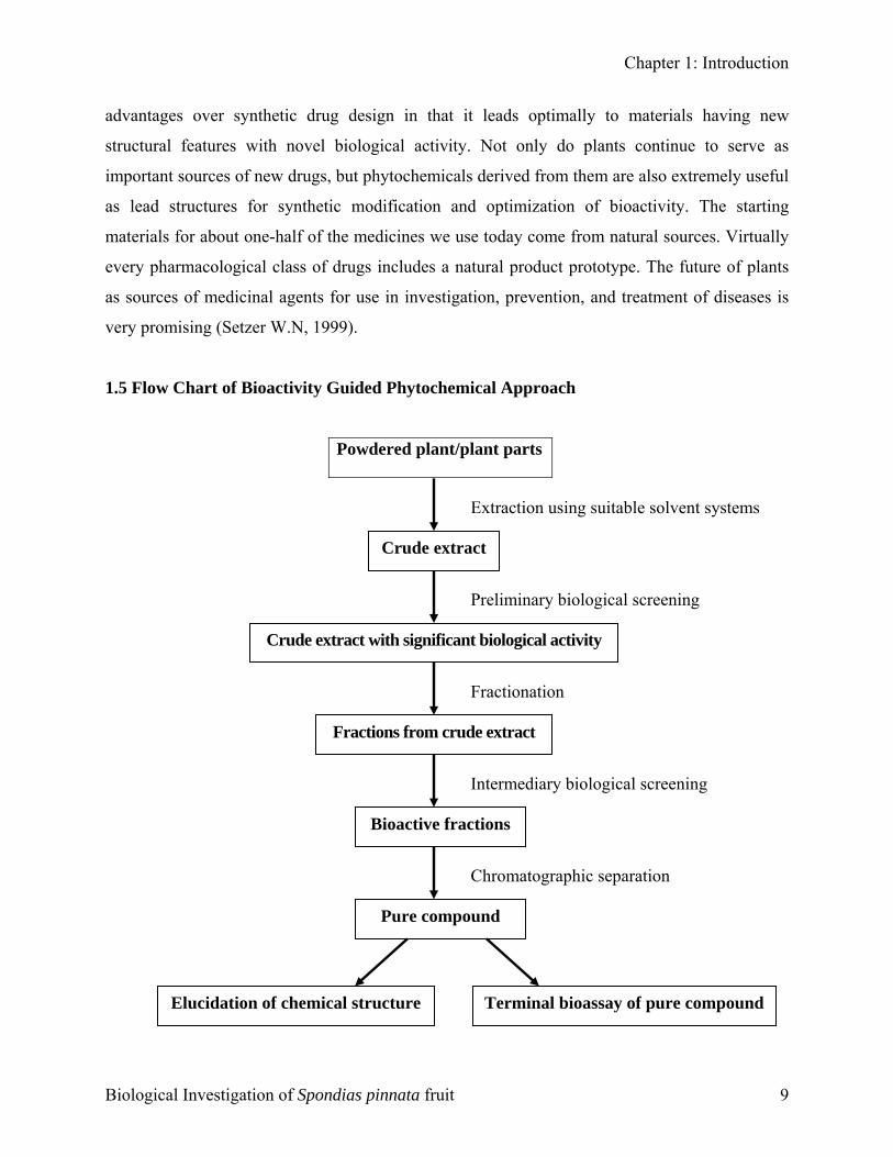
1.5 Flow Chart of Bioactivity Guided Phytochemical Approach
Powdered plant/plant parts
Crude extract
Extraction using suitable solvent systems
Crude extract with significant biological activity
Preliminary biological screening
Fractions from crude extract
Fractionation
Bioactive fractions
Intermediary biological screening
Chromatographic separation
Elucidation of chemical structure
Pure compound
Terminal bioassay of pure compound
Biological Investigation of Spondias pinnata fruit 9
Chapter 1: Introduction
The objective of this dissertation is to identify the biological activity of the fruit of an indigenous
medicinal plant, viz., Spondias pinnata (L.f.) Kurz (Family: Anacardiaceae) and to evaluate the
possible pharmacological and microbiological profiles of the crude extracts. So far some
chemical and biological investigations have been carried out on this plant mainly focusing on the
bark and root of the plant. That’s why the goal of this framework is to explore the potential
possibilities of developing new drug candidates from the fruit of this plant which could be
crucial for the treatment of various ailments.
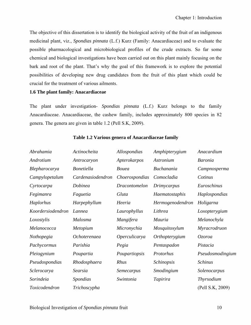
1.6 The plant family: Anacardiaceae
The plant under investigation- Spondias pinnata (L.f.) Kurz belongs to the family
Anacardiaceae. Anacardiaceae, the cashew family, includes approximately 800 species in 82
genera. The genera are given in table 1.2 (Pell S.K, 2009).
Table 1.2 Various genera of Anacardiaceae family
Abrahamia Actinocheita Allospondias Amphipterygium Anacardium
Androtium Antrocaryon Apterokarpos Astronium Baronia
Blepharocarya Bonetiella Bouea Buchanania Campnosperma
Campylopetalum Cardenasiodendron Choerospondias Comocladia Cotinus
Cyrtocarpa Dobinea Dracontomelon Drimycarpus Euroschinus
Fegimanra Faguetia Gluta Haematostaphis Haplospondias
Haplorhus Harpephyllum Heeria Hermogenodendron Holigarna
Koordersiodendron Lannea Laurophyllus Lithrea Loxopterygium
Loxostylis Malosma Mangifera Mauria Melanochyla
Melanococca Metopium Micronychia Mosquitoxylum Myracrodruon
Nothopegia Ochoterenaea Operculicarya Orthopterygium Ozoroa
Pachycormus Parishia Pegia Pentaspadon Pistacia
Pleiogynium Poupartia Poupartiopsis Protorhus Pseudosmodingium
Pseudospondias Rhodosphaera Rhus Schinopsis Schinus
Sclerocarya Searsia Semecarpus Smodingium Solenocarpus
Sorindeia Spondias Swintonia Tapirira Thyrsodium
Toxicodendron Trichoscypha (Pell S.K, 2009)
Biological Investigation of Spondias pinnata fruit 10

Chapter 1: Introduction
Members of the family are cultivated throughout the world for their edible fruits and seeds,
medicinal compounds, valuable timber, and landscape appeal. Some of the products of
Anacardiaceae, including mango (Mangifera indica), pistachio (Pistacia vera), cashew
(Anacardium occidentale), and pink peppercorn (Schinus terebinthifolia), are enjoyed worldwide
while other notables such as the pantropical Spondias fruits, the marula of Africa (Sclerocarya
birrea), and the Neotropical fruits of Antrocaryon are restricted to localized cultivation and
consumption and are not generally transported far distances to larger markets (Pell S.K, 2009).
1.6.1 Taxonomy
The exact definition of taxonomy varies slightly from source to source, but the core of the
discipline remains: the identification, naming, and classifying of organisms. As points of
reference, three recent textbook definitions are presented below:
1. Theory and practice of grouping individuals into species, arranging species into larger groups,
and giving those groups names, thus producing a classification (Judd et al., 2007)
2. A field of science (and major component of systematics) that encompasses description,
identification, nomenclature, and classification (Simpson, 2010).
3. The science of classification, in biology the arrangement of organisms into a classification
(Kirk et al., 2008).
In 1759, Bernard de Jussieu arranged the plants in the royal garden of the Trianon at Versailles,
according to his own scheme. That classification included a description of an order called
Terebintaceae which contained a suborder that included Cassuvium (Anacardium), Anacardium
(Semecarpus), Mangifera, Connarus, Rhus and Rourea. In 1789, Antoine Laurent de Jussieu,
nephew of Bernard de Jussieu, published that classification scheme (Bernard J, 1789).
Robert Brown described a subset of Terebintaceae called Cassuvlae or Anacardeae in 1818,
using the herbarium that was collected by Christen Smith during a fated expedition headed by
James Kingston Tuckey to explore the River Congo. The name and genera were based on the
order with the same name that had been described by Bernard de Jussieu in 1759. The herbarium
from that expedition contained only one genus from the family, Rhus (Brown R, 1818).
Biological Investigation of Spondias pinnata fruit 11
Chapter 1: Introduction
Augustin Pyramus de Candolle in 1824, used Robert Browns name Cassuvlae or Anacardeae,
wrote another description of the group and filled it with the genera Anacardium, Semecarpus,
Holigarna, Mangifera, Buchanania, Pistacia, Astronium, Comocladia and Picramnia (Candolle
AP, 1825).
John Lindley described the "Essential character" of Anacardiaceae, the "Cashew Tribe" in 1831,
adopting the order that was described by Jussieu but abandoning the name Terebintaceae. He
includes the genera which were found in de Candolle's Anacardieae and Sumachineae:
Anacardium, Holigarna, Mangifera, Rhus and Mauria (Lindley F.R.S., 1831).
The genus Pistacia has sometimes been separated into its own family, Pistaciaceae, based on the
reduced flower structure, differences in pollen, and the feathery style of the flowers. However,
the nature of the ovary does suggest it belongs in the Anacardiaceae, a position which is
supported by morphological and molecular studies, and recent classifications have included
Pistacia in the Anacardiaceae (Tingshuang Y, 2008).
1.6.2 Botanical features of Anacardiaceae family
Anacardiaceae, the sumac family of flowering plants includes primarily trees and shrubs (rarely
lianas or subshrubs), with resin canals and clear to milky exudates (Pell S.K, 2009).
Habit and leaf form: Members of Anacardiaceae family have resin ducts in the bark, leaves
usually composed of leaflets in various arrangements, inconspicuous flowers often with only
male or female parts, and usually fleshy fruits (Encyclopaedia britannica, 2012). Resin-canals
located in the inner fibrous bark of plants fibrovascular system found in the stems, roots and
leaves is characteristic of all members of this family; resin-canals located in the pith is a
characteristic of many of the cashew family species and several species have them located in the
primary cortex or the regular bark. Tannin sacs are also widespread among the family. The wood
of Anacardiaceae has the frequent occurrence of simple small holes in the vessels, occasionally
in some species side by side with scalariform holes (in Campnosperma, Micronychia and
Anaphrenium argenteum). The simple pits are located along the vessel wall and in contact with
the parenchyma (Solereder H, 1908).
The sumacs have either simple or pinnately compound leaves, depending on the species. It is
common for members of Anacardiaceae to have pinnately compound leaves (e.g. Toxicodendron,
Biological Investigation of Spondias pinnata fruit 12
Chapter 1: Introduction
Schinus). Many members of Anacardiaceae (eg - Rhus, Toxicondendron) produce compounds on
their leaves that cause rash (Hickman J.C, 1993).
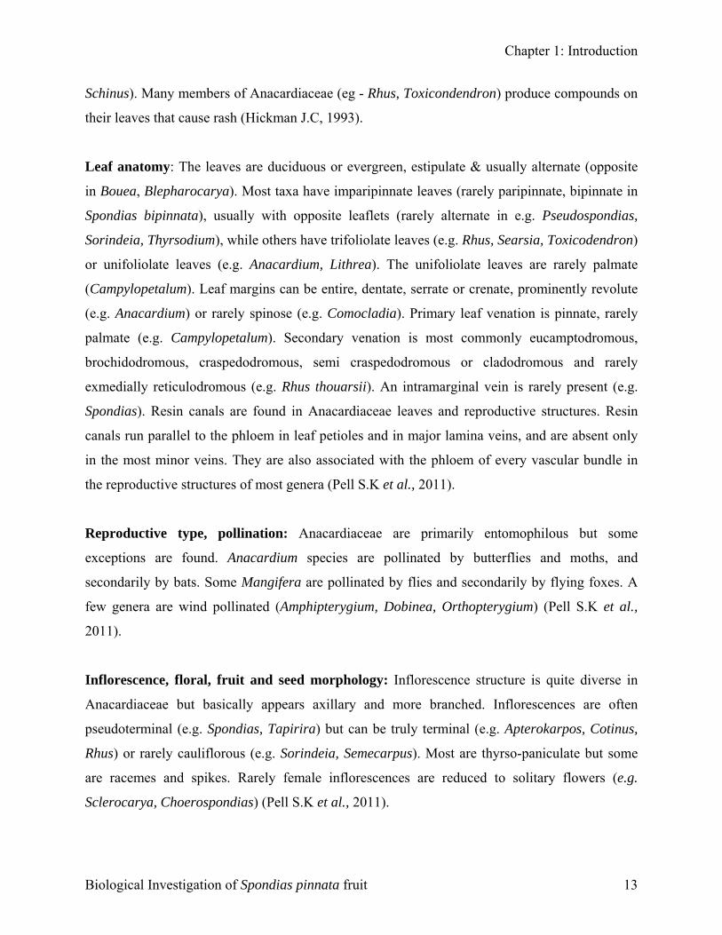
Leaf anatomy: The leaves are duciduous or evergreen, estipulate & usually alternate (opposite
in Bouea, Blepharocarya). Most taxa have imparipinnate leaves (rarely paripinnate, bipinnate in
Spondias bipinnata), usually with opposite leaflets (rarely alternate in e.g. Pseudospondias,
Sorindeia, Thyrsodium), while others have trifoliolate leaves (e.g. Rhus, Searsia, Toxicodendron)
or unifoliolate leaves (e.g. Anacardium, Lithrea). The unifoliolate leaves are rarely palmate
(Campylopetalum). Leaf margins can be entire, dentate, serrate or crenate, prominently revolute
(e.g. Anacardium) or rarely spinose (e.g. Comocladia). Primary leaf venation is pinnate, rarely
palmate (e.g. Campylopetalum). Secondary venation is most commonly eucamptodromous,
brochidodromous, craspedodromous, semi craspedodromous or cladodromous and rarely
exmedially reticulodromous (e.g. Rhus thouarsii). An intramarginal vein is rarely present (e.g.
Spondias). Resin canals are found in Anacardiaceae leaves and reproductive structures. Resin
canals run parallel to the phloem in leaf petioles and in major lamina veins, and are absent only
in the most minor veins. They are also associated with the phloem of every vascular bundle in
the reproductive structures of most genera (Pell S.K et al., 2011).
Reproductive type, pollination: Anacardiaceae are primarily entomophilous but some
exceptions are found. Anacardium species are pollinated by butterflies and moths, and
secondarily by bats. Some Mangifera are pollinated by flies and secondarily by flying foxes. A
few genera are wind pollinated (Amphipterygium, Dobinea, Orthopterygium) (Pell S.K et al.,
2011).
Inflorescence, floral, fruit and seed morphology: Inflorescence structure is quite diverse in
Anacardiaceae but basically appears axillary and more branched. Inflorescences are often
pseudoterminal (e.g. Spondias, Tapirira) but can be truly terminal (e.g. Apterokarpos, Cotinus,
Rhus) or rarely cauliflorous (e.g. Sorindeia, Semecarpus). Most are thyrso-paniculate but some
are racemes and spikes. Rarely female inflorescences are reduced to solitary flowers (e.g.
Sclerocarya, Choerospondias) (Pell S.K et al., 2011).
Biological Investigation of Spondias pinnata fruit 13
Chapter 1: Introduction
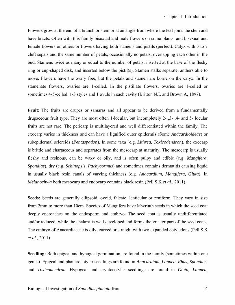
Flowers grow at the end of a branch or stem or at an angle from where the leaf joins the stem and
have bracts. Often with this family bisexual and male flowers on some plants, and bisexual and
female flowers on others or flowers having both stamens and pistils (perfect). Calyx with 3 to 7
cleft sepals and the same number of petals, occasionally no petals, overlapping each other in the
bud. Stamens twice as many or equal to the number of petals, inserted at the base of the fleshy
ring or cup-shaped disk, and inserted below the pistil(s). Stamen stalks separate, anthers able to
move. Flowers have the ovary free, but the petals and stamen are borne on the calyx. In the
stamenate flowers, ovaries are 1-celled. In the pistillate flowers, ovaries are 1-celled or
sometimes 4-5-celled. 1-3 styles and 1 ovule in each cavity (Britton N.L and Brown A, 1897).
Fruit: The fruits are drupes or samaras and all appear to be derived from a fundamentally
drupaceous fruit type. They are most often 1-locular, but incompletely 2- ,3- ,4- and 5- locular
fruits are not rare. The pericarp is multilayered and well differentiated within the family. The
exocarp varies in thickness and can have a lignified outer epidermis (Some Anacardioideae) or
subepidermal sclereids (Pentaspadon). In some taxa (e.g. Lithrea, Toxicodendron), the exocarp
is brittle and chartaceous and separates from the mesocarp at maturity. The mesocarp is usually
fleshy and resinous, can be waxy or oily, and is often pulpy and edible (e.g. Mangifera,
Spondias), dry (e.g. Schinopsis, Pachycormus) and sometimes contains dermatitis causing liquid
in usually black resin canals of varying thickness (e.g. Anacardium, Mangifera, Gluta). In
Melanochyla both mesocarp and endocarp contains black resin (Pell S.K et al., 2011).
Seeds: Seeds are generally ellipsoid, ovoid, falcate, lenticular or reniform. They vary in size
from 2mm to more than 10cm. Species of Mangifera have labyrinth seeds in which the seed coat
deeply encroaches on the endosperm and embryo. The seed coat is usually undifferentiated
and/or reduced, while the chalaza is well developed and forms the greater part of the seed coats.
The embryo of Anacardiaceae is oily, curved or straight with two expanded cotyledons (Pell S.K
et al., 2011).
Seedling: Both epigeal and hypogeal germination are found in the family (sometimes within one
genus). Epigeal and phanerocotylar seedlings are found in Anacardium, Lannea, Rhus, Spondias,
and Toxicodendron. Hypogeal and cryptocotylar seedlings are found in Gluta, Lannea,
Biological Investigation of Spondias pinnata fruit 14
Chapter 1: Introduction
Mangifera, Melanochyla and Semecarpus. Epigeal and cryptocotylar seedlings are found in
Swintonia, Astronium graveolens (Pell S.K et al., 2011).
1.6.3 Distribution of Anacardiaceae family
Anacardiaceae are found worldwide in dry to moist, mostly lowland habitats, primarily in the
tropics and subtropics but extending into the temperate zone. The family is native to the western
hemisphere (from southern Canada south to Patagonia), Africa, southern Europe, temperate and
tropical Asia, tropical and subtropical Australia, and most of the Pacific Islands. Anacardiaceae
are absent from northern Europe, temperate and arid Australia, New Zealand, the Galapagos
Islands, and extreme desert and high elevation habitats (although they can reach elevations as
high as 3,500 m) (Pell S.K, 2009).
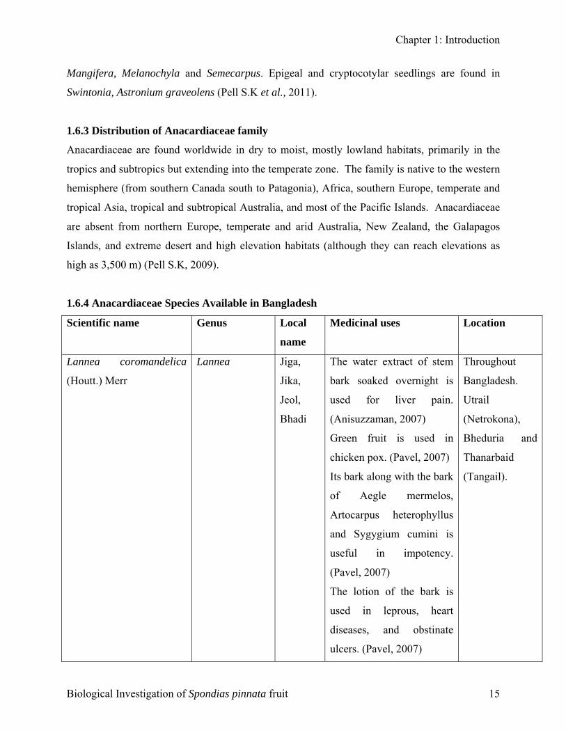
1.6.4 Anacardiaceae Species Available in Bangladesh
Scientific name Genus Local
name
Medicinal uses Location
Lannea coromandelica
(Houtt.) Merr
Lannea Jiga,
Jika,
Jeol,
Bhadi
The water extract of stem
bark soaked overnight is
used for liver pain.
(Anisuzzaman, 2007)
Green fruit is used in
chicken pox. (Pavel, 2007)
Its bark along with the bark
of Aegle mermelos,
Artocarpus heterophyllus
and Sygygium cumini is
useful in impotency.
(Pavel, 2007)
The lotion of the bark is
used in leprous, heart
diseases, and obstinate
ulcers. (Pavel, 2007)
Throughout
Bangladesh.
Utrail
(Netrokona),
Bheduria and
Thanarbaid
(Tangail).
Biological Investigation of Spondias pinnata fruit 15
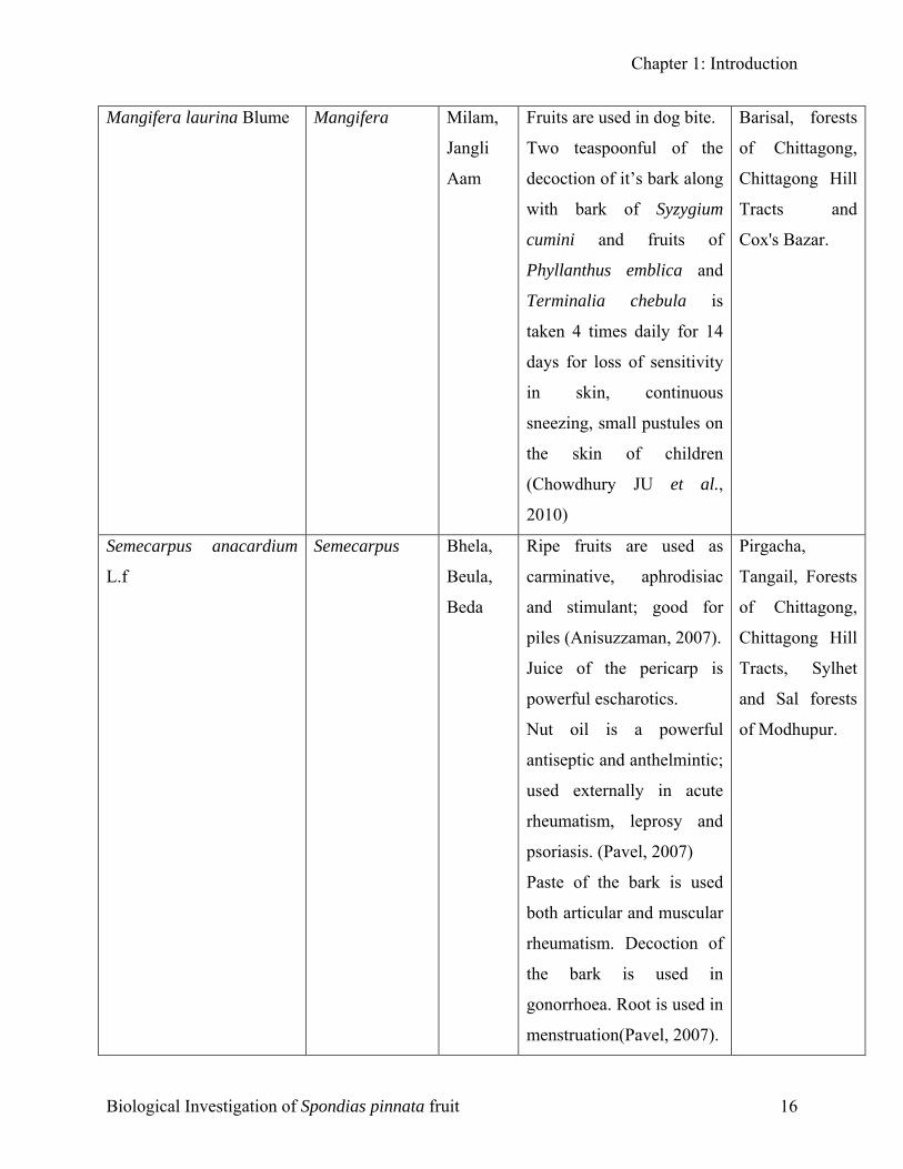
Chapter 1: Introduction
Mangifera laurina Blume Mangifera Milam,
Jangli
Aam
Fruits are used in dog bite.
Two teaspoonful of the
decoction of it’s bark along
with bark of Syzygium
cumini and fruits of
Phyllanthus emblica and
Terminalia chebula is
taken 4 times daily for 14
days for loss of sensitivity
in skin, continuous
sneezing, small pustules on
the skin of children
(Chowdhury JU et al.,
2010)
Barisal, forests
of Chittagong,
Chittagong Hill
Tracts and
Cox's Bazar.
Semecarpus anacardium
L.f
Semecarpus Bhela,
Beula,
Beda
Ripe fruits are used as
carminative, aphrodisiac
and stimulant; good for
piles (Anisuzzaman, 2007).
Juice of the pericarp is
powerful escharotics.
Nut oil is a powerful
antiseptic and anthelmintic;
used externally in acute
rheumatism, leprosy and
psoriasis. (Pavel, 2007)
Paste of the bark is used
both articular and muscular
rheumatism. Decoction of
the bark is used in
gonorrhoea. Root is used in
menstruation(Pavel, 2007).
Pirgacha,
Tangail, Forests
of Chittagong,
Chittagong Hill
Tracts, Sylhet
and Sal forests
of Modhupur.
Biological Investigation of Spondias pinnata fruit 16
Chapter 1: Introduction
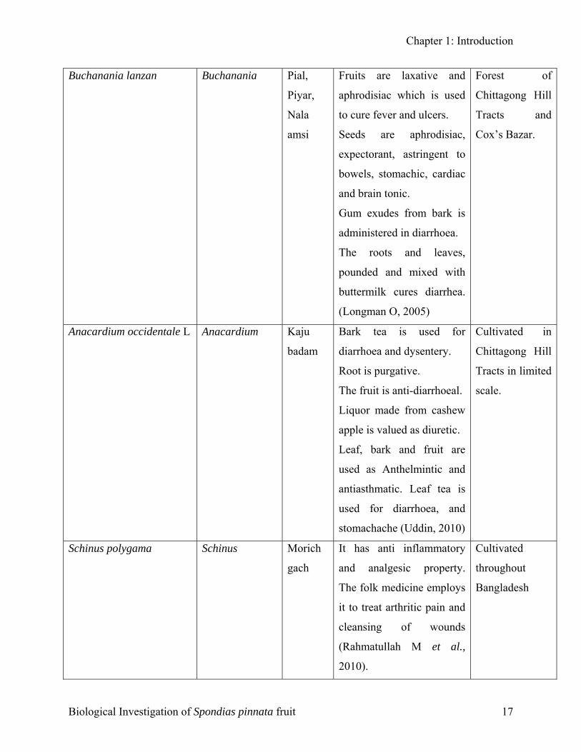
Buchanania lanzan Buchanania Pial,
Piyar,
Nala
amsi
Fruits are laxative and
aphrodisiac which is used
to cure fever and ulcers.
Seeds are aphrodisiac,
expectorant, astringent to
bowels, stomachic, cardiac
and brain tonic.
Gum exudes from bark is
administered in diarrhoea.
The roots and leaves,
pounded and mixed with
buttermilk cures diarrhea.
(Longman O, 2005)
Forest of
Chittagong Hill
Tracts and
Cox’s Bazar.
Anacardium occidentale L Anacardium Kaju
badam
Bark tea is used for
diarrhoea and dysentery.
Root is purgative.
The fruit is anti-diarrhoeal.
Liquor made from cashew
apple is valued as diuretic.
Leaf, bark and fruit are
used as Anthelmintic and
antiasthmatic. Leaf tea is
used for diarrhoea, and
stomachache (Uddin, 2010)
Cultivated in
Chittagong Hill
Tracts in limited
scale.
Schinus polygama Schinus Morich
gach
It has anti inflammatory
and analgesic property.
The folk medicine employs
it to treat arthritic pain and
cleansing of wounds
(Rahmatullah M et al.,
2010).
Cultivated
throughout
Bangladesh
Biological Investigation of Spondias pinnata fruit 17
Chapter 1: Introduction
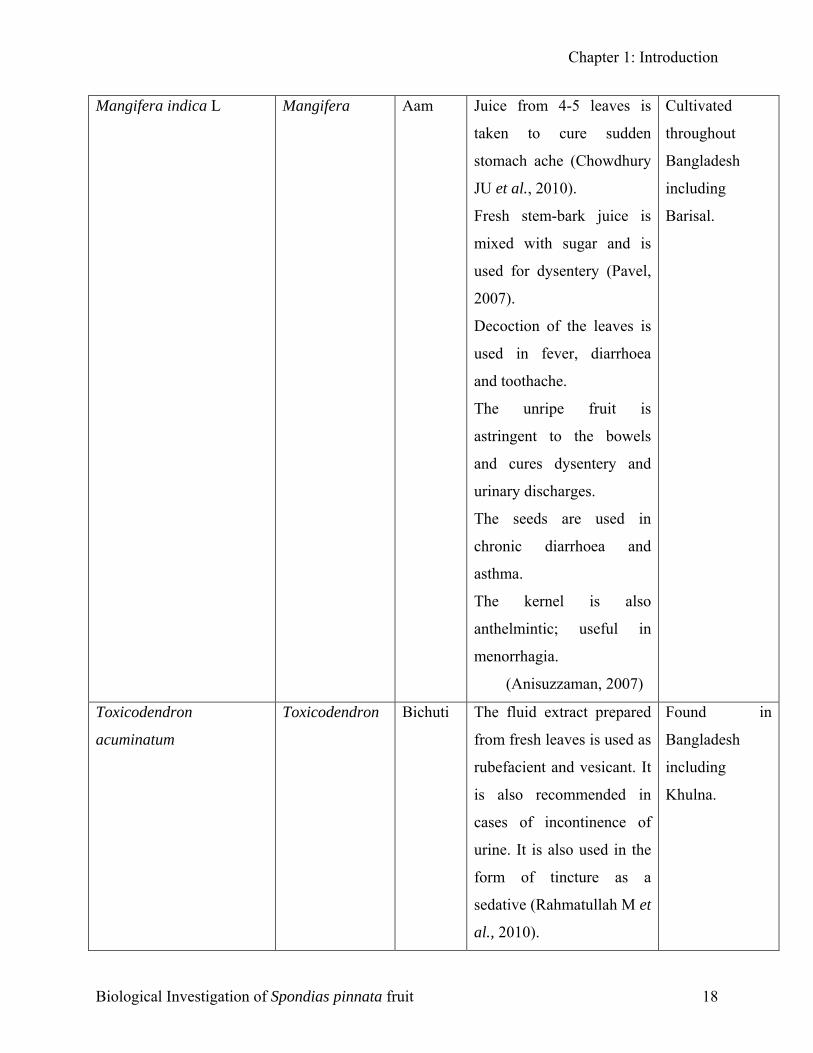
Mangifera indica L Mangifera Aam
Juice from 4-5 leaves is
taken to cure sudden
stomach ache (Chowdhury
JU et al., 2010).
Fresh stem-bark juice is
mixed with sugar and is
used for dysentery (Pavel,
2007).
Decoction of the leaves is
used in fever, diarrhoea
and toothache.
The unripe fruit is
astringent to the bowels
and cures dysentery and
urinary discharges.
The seeds are used in
chronic diarrhoea and
asthma.
The kernel is also
anthelmintic; useful in
menorrhagia.
(Anisuzzaman, 2007)
Cultivated
throughout
Bangladesh
including
Barisal.
Toxicodendron
acuminatum
Toxicodendron Bichuti The fluid extract prepared
from fresh leaves is used as
rubefacient and vesicant. It
is also recommended in
cases of incontinence of
urine. It is also used in the
form of tincture as a
sedative (Rahmatullah M et
al., 2010).
Found in
Bangladesh
including
Khulna.
Biological Investigation of Spondias pinnata fruit 18
Chapter 1: Introduction
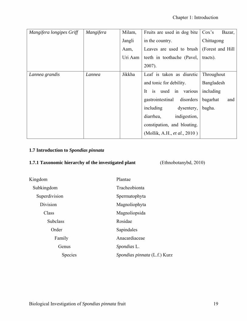
Mangifera longipes Griff Mangifera Milam,
Jangli
Aam,
Uri Aam
Fruits are used in dog bite
in the country.
Leaves are used to brush
teeth in toothache (Pavel,
2007).
Cox’s Bazar,
Chittagong
(Forest and Hill
tracts).
Lannea grandis Lannea Jikkha Leaf is taken as diuretic
and tonic for debility.
It is used in various
gastrointestinal disorders
including dysentery,
diarrhea, indigestion,
constipation, and bloating.
(Mollik, A.H., et al., 2010 )
Throughout
Bangladesh
including
bagarhat and
bagha.
1.7 Introduction to Spondias pinnata
1.7.1 Taxonomic hierarchy of the investigated plant (Ethnobotanybd, 2010)
Kingdom Plantae
Subkingdom Tracheobionta
Superdivision Spermatophyta
Division Magnoliophyta
Class Magnoliopsida
Subclass Rosidae
Order Sapindales
Family Anacardiaceae
Genus Spondias L.
Species Spondias pinnata (L.f.) Kurz
Biological Investigation of Spondias pinnata fruit 19
Chapter 1: Introduction
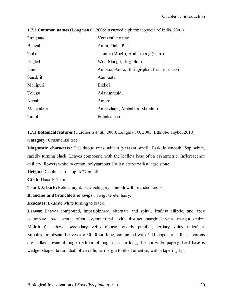
1.7.2 Common names (Longman O, 2005; Ayurvedic pharmacopoeia of India, 2001)
Language Vernacular name
Bengali Amra, Piala, Pial
Tribal Thoura (Mogh); Ambi-thong (Garo)
English Wild Mango, Hog-plum
Hindi Ambara, Amra, Bhringi-phal, Pashu-haritaki
Sanskrit Aamraata
Manipuri Eikhoi
Telugu Adavimamidi
Nepali Amaro
Malayalam Ambazham, Ambalam, Mambuli
Tamil Pulicha kaai
1.7.3 Botanical features (Gardner S et al., 2000; Longman O, 2005; Ethnobotanybd, 2010)
Category: Ornamental tree
Diagnostic characters: Deciduous trees with a pleasant smell. Bark is smooth. Sap white,
rapidly turning black. Leaves compound with the leaflets base often asymmetric. Inflorescence
axillary, flowers white to cream, polygamous. Fruit a drupe with a large stone.
Height: Deciduous tree up to 27 m tall.
Girth: Usually 2.5 m
Trunk & bark: Bole straight, bark pale grey, smooth with rounded knobs.
Branches and branchlets or twigs : Twigs terete, hairy.
Exudates: Exudate white turning to black.
Leaves: Leaves compound, imparipinnate, alternate and spiral, leaflets elliptic, and apex
acuminate, base acute, often asymmetrical, with distinct marginal vein, margin entire.
Midrib flat above, secondary veins obtuse, widely parallel, tertiary veins reticulate.
Stipules are absent. Leaves are 30-40 cm long, compound with 5-11 opposite leaflets. Leaflets
are stalked, ovate-oblong to elliptic-oblong, 7-12 cm long, 4-5 cm wide, papery. Leaf base is
wedge- shaped to rounded, often oblique, margin toothed or entire, with a tapering tip.
Biological Investigation of Spondias pinnata fruit 20
Chapter 1: Introduction
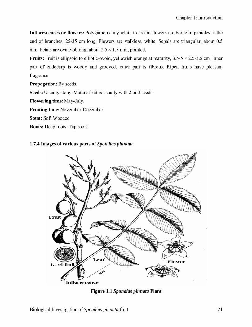
Inflorescences or flowers: Polygamous tiny white to cream flowers are borne in panicles at the
end of branches, 25-35 cm long. Flowers are stalkless, white. Sepals are triangular, about 0.5
mm. Petals are ovate-oblong, about 2.5 × 1.5 mm, pointed.
Fruits: Fruit is ellipsoid to elliptic-ovoid, yellowish orange at maturity, 3.5-5 × 2.5-3.5 cm. Inner
part of endocarp is woody and grooved, outer part is fibrous. Ripen fruits have pleasant
fragrance.
Propagation: By seeds.
Seeds: Usually stony. Mature fruit is usually with 2 or 3 seeds.
Flowering time: May-July.
Fruiting time: November-December.
Stem: Soft Wooded
Roots: Deep roots, Tap roots
1.7.4 Images of various parts of Spondias pinnata
Figure 1.1 Spondias pinnata Plant
Biological Investigation of Spondias pinnata fruit 21
Chapter 1: Introduction
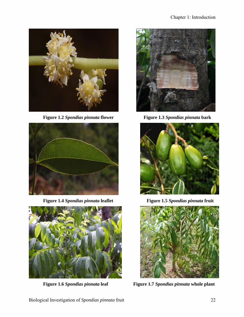
Figure 1.2 Spondias pinnata flower Figure 1.3 Spondias pinnata bark
Figure 1.4 Spondias pinnata leaflet Figure 1.5 Spondias pinnata fruit
Figure 1.6 Spondias pinnata leaf Figure 1.7 Spondias pinnata whole plant
Biological Investigation of Spondias pinnata fruit 22
Chapter 1: Introduction
1.7.5 Distribution of Spondias pinnata
Native to Southeast Asia, this slow growing fruit tree is seen everywhere in Bangladesh for it’s
fruits including wild forests of Chittagong, Chittagong Hill Tract, Cox’s Bazar, Tangail, Sylhet
and Dinajpur. It is also found in village shrubberies throughout the country (Ethnobotanybd,
2010). Besides it is found in India, Srilanka, Assam, Myanmar, China, Malaysia and Thailand
(Badoni, A. and Bisht, C, 2009).
1.7.6 Growing conditions
The plants are easily grown with adequate moisture preferring humus rich, moist but well
drained soil. Like the Mango, the tree thrives in humid tropical and subtropical areas growing up
to 2 metres in a single growing season. It grows on all types of soil, as long as they are well
drained. It has been noted that some trees can suffer from some nutritional disorders if the soil is
too alkaline. Trees are cold sensitive when small and should be protected from serious frost and
strong wind. Trees do best in full sun, but will produce some fruit in light shade. As a large and
vigorous tree, they prefer not be planted underneath other large trees and unlike some mango
varieties they are not too fussed on salt spray (Morton J, 1987).
1.7.7 Chemical constituents of Spondias pinnata
Plant Part Chemical Constituent
Aerial parts 24-methylene cycloartenone, stigmast-4-en-3-one, β -sitosterol, glycoside of β-
sitosterol and lignoceric acid (Ghani, 2003)
Fruits Water soluble polysaccharides, composed of mainly L-arabinose, D-galactose
and galacturonic acid (Ghani, 2003)
β-amyrin and oleanolic acid, glycine, cystine, serine, alanine and leucine
(Rastogi & Mehrotra, 1993)
1.7.8 Ethnobotany of Spondias pinnata
Timber is used for interior furniture. This plant is used to treat diarrhea, ear aches, and to make a
cancer home remedy. The fruit contains 40-70 mg Vitamin C per 100 g and is domesticated for
food production. Unripe fruits are used to make pickles. The young leaves and flowers are edible
too (Ethnobotanybd, 2010).
Biological Investigation of Spondias pinnata fruit 23
Chapter 1: Introduction
1.7.9 Nutritive and mineral potential of ripe fruits of Spondias pinnata (Purohit, V.K et al.,
2010)
Energy 189–203 kcal/g
Crude fat 12.23–12.54%
Crude fiber 3.13–4.03%
Total carbohydrate 16.30–23.54%
Sodium 0.96–1.38%
Calcium 0.15–0.93%
Iron 1.3–1.5%
Copper 0.9–1.23%
Protein 0.50-0.80%
Acid 0.47%
1.7.10 Medicinal Potentials of Spondias pinnata
Plant Part Medicinal properties
Leaf It is aromatic, acidic, appetizing and astringent, and is used in dysentery. The
juice of the leaves is recommended for local application in otalgia
(Ethnobotanybd, 2010). In Nigeria, a decoction of the mashed leaves is used
for washing a swollen face. A leaf infusion is a common cough remedy &
used as a laxative for fever with constipation. A leaf decoction is used for
gonorrhoea. All these leaves are used for leprosy. Crushed with lemon they
are effective for worms in children. A decoction of pounded leaves is used as
an eye lotion and the juice pressed from young, warm leaves is given to
children for stomach troubles. The young leaves are used as an infusion taken
internally or as a warm astringent lotion by women in confinement in Sierra
Leone. In Suriname's traditional medicine, the infusion of the leaves is used as
a treatment of eye inflammation, diarrhoea and venereal diseases (Faiz M,
2011).
Fruit (Unripe) It is astringent, sour, thermogenic, appetizer and aphrodisiac, and is good for
rheumatism and sore throat (Ethnobotanybd, 2010).
Root Regulation of menstruation. It is used in fever in Thailand (Faiz M, 2011).
Biological Investigation of Spondias pinnata fruit 24
Chapter 1: Introduction
Fruit (Ripe) It is sweet, astringent, cooling, emollient, tonic, constipating and
antiscorbutic, and is administered in bilious dyspepsia, diarrhea, and vitiated
conditions of tridosa. Ripe fruit is aphrodisiac & cures burning sensation. The
fruit-juice is used as a febrifuge and diuretic (Ethnobotanybd, 2010).
Bark It is aromatic, astringent and refrigerant, and infusion of the bark is
administered in dysentery, diarrhea, vomiting. Paste of the bark is used as an
embrocation for both articular and muscular rheumatism. Decoction of the
bark is given in gonorrhoea & severe cough. Gum of the bark is demulcent.
Bark is used as purgative and in local applications for leprosy (Faiz M, 2011).
1.7.11 Reported Biological Works on Spondias pinnata
Plant part Investigation & Result Reference
Whole plant The ethanolic extract was investigated for
total phenolic activity, total flavonoid & free
radical scavenging activity.
Maisuthisakul, P. et al (2007)
Fruit The methanolic extract was investigated for
total phenolic activity, total flavonoid &
DPPH radical scavenging activity.
Wetwitayaklung, P. et al
(2012)
Stem bark The 70% methanolic extract was
investigated for hydroxyl radical
scavenging, superoxide radical scavenging,
NO radical scavenging, Hydrogen peroxide
radical scavenging, Peroxynitrite radical
scavenging, Singlet oxygen scavenging,
Hypochlorous acid scavenging, reducing
power, ferrous chelation, total phenolic
activity & total flavonoid activity.
Hazra, B. et al (2008)
Fruit The extract was investigated for total
phenolic activity, total flavonoid & DPPH
radical scavenging & ferric reducing
activity.
Kubola, J. et al (2011)
Biological Investigation of Spondias pinnata fruit 25
Chapter 1: Introduction
Bark The chloroform & ethanolic extract was
investigated for cytotoxic, antioxidant,
antibacterial, & phytochemical screening.
Das, J. et al.(2011)
Fruit The 80% ethanolic extract was investigated
for cytotoxic and antibacterial activity
Muhammad, A. et al.(2011)
Fruit The extract was investigated for the
correlation analysis between total acid, total
phenolic and ascorbic acid contents and
their antioxidant activities
Samee, W. et al. (2006)
Stem heart wood The methanolic and ethyl acetate extract
was investigated for hepatoprotective
activity.
Rao, BG. et al (2010).
Fruit The hexane extract was investigated for Anti
HIV-1 reverse transcriptase activity.
Silprasit, K. et al. (2011)
Bark Hypoglycemic activity of the bark of
Spondias pinnata Linn. kurz
Mondal, S. et al. (2009)
Leaf Aqueous & methanolic extracts was
investigated for anti diarrheal activity
Panda, SK. et al. (2012)
Bark Petroleum ether, chloroform and methanolic
extracts was investigated for diuretic and
laxative activity
Mondal, S. et al. (2009)
Stem heart wood
and bark
Methanolic extracts was investigated for
antihelmintic activity
Jayaraju, N. et al. (2009)
Stem bark Ethanolic extracts was investigated for
analgesic activity
Panda, BK. et al. (2009)
Fruits pulp Polysaccharide has been identified from the
fruits pulp which has eliciting activity on
peritoneal macrophages
Iacomini et al. (2005)
Whole plant Isolation of 24-methylelle cydoartanone,
stigma-4en-3one, lignoceric acid, β-
sitosterol and its β- D-glucoside
Rastogi, R.P. et al (1976)
Biological Investigation of Spondias pinnata fruit 26
Chapter 1: Introduction
Fruit Isolation and characterization of active
compound an oleanolic acid (3α
hydroxyolea-12-en-28-oic acid)
Kandali, R. et al. (2011)
Pulp Isolation and characterization of 5-
hydroxymethylfurfural, 1,4-pentadiene, 3,5-
dihydroxy-2-methyl-5,6-dihydropyran-4-
one and furfural from the methanolic extract
Pholsongkram, K. et al (2009)
Pulp Isolation and characterization of 9,12,15-
octadecatrien-1-ol, hexadecanoic acid and
furfural from the essential oil of the pulp
Liawruangrath, B. et al (2009)
Stem bark Methanolic extracts was investigated for
antibacterial activity
Chetia, B., and Gogoi, S.
(2011)
Biological Investigation of Spondias pinnata fruit 27
SSttuuddyy pprroottooccooll
C H A P T E R
Chapter 2: Study Protocol
2.1 Presen
Our presen
extract of t
consisted o
Extra
Filtra
Doub
Phyto
Scree
Inves
of M
(MBC
Inves
2.1.1 Phyto
Freshly pre
chemical c
tannins, ant
Biological
CChhaapptteerr
22
ll
t Study Protocol
t study was designed
he plant Spondias pi
f the following steps:
ction at room tempera
tion of the crude et
le rings filter paper (9
chemical screening o
ning of in vitro antio
tigation of in vitro an
inimum inhibitory c
) of crude ethanolic
tigation of in vitro th
chemical Screening
pared crude extracts o
onstituents including
hraquinone, cardiac g
Investigation of Spond
SSttuuddyy PPrroottooccoo
to observe pharmacological activities of the crude ethanolic
nnata (L.f.) Kurz (Family: Anacardiaceae). The study protocol
ture of the fruit with 96% ethanol for 10 days.
hanolic extract by using cotton and subsequently through the
.0 cm) and solvent evaporation.
f the crude ethanolic extract of Spondias pinnata fruit.
xidant activity of crude ethanolic extract.
timicrobial activity of crude ethanolic extract and determination
oncentration (MIC) and Minimum Bactericidal Concentration
extract.
rombolytic activity of crude ethanolic extract.
f S. pinnata were qualitatively tested for the presence of various
alkaloids, flavonoids, steroids, terpenoids, reducing sugars,
lycoside, and saponins by following standard procedures.
ias pinnata fruit 28
Chapter 2: Study Protocol
2.1.2 Evaluation of Antioxidant activity
Antioxidant test was done to determine the antioxidant capacity of the crude ethanolic extract of
the S .pinnata fruit. Antioxidant effects play an important role in many human diseases,
including cancer, diabetic, complications, heart disease, liver damage, autism and Alzheimer's
disease, etc. Recently, free radicals (FR) and reactive oxygen species (ROS) have been
considered as one of the main causes of these diseases. The protective effects of antioxidants on
cell membrane lipid bilayers attacked by free radicals are attracting more interest (Lai SC et al.,
2010). Highly reactive free radicals and oxygen species are present in biological systems from a
wide variety of sources. These free radicals may oxidize nucleic acids, proteins, lipids or DNA
and can initiate degenerative disease (Miller, H.E. et al., 2000). Antioxidants are substances that
delay or prevent the oxidation of cellular oxidizable substrates. They exert their effects by
scavenging reactive oxygen species, activating a battery of detoxifying proteins, or preventing
the generation of reactive oxygen species (Halliwell, 1992). It has been reported that many
compounds such as phenol acids, flavonoids, saponins, tannins, alkaloids and polysaccharides
have antioxidant activity in vitro or in vivo. These compounds are abundant in herbs and food
additives. A number of synthetic antioxidants such as butylated hydroxyanisole (BHA) and
butylated hydroxytoluene (BHT) have been extensively added to foodstuffs, although their use
has begun to be questioned because of their toxicity (Ito N et al., 1985). Therefore more recently,
interest in the use of natural antioxidants obtained from botanical sources, especially herbal
plants for the prevention and treatment of cancer has increased greatly, and oxidative stress was
shown to influence treatment efficacy and survival of non-small cell lung cancer patients (Gupta
A et al., 2010)
Antioxidant property of the various fraction of the plant was determined by following methods-
1. Determination of total phenolic content
2. Determination of DPPH radical scavenging assay (Qualitative and Quantitative analysis)
3. Determination of reducing power ability
4. Determination of total antioxidant capacity by phosphomolybdenum method
5. Determination of total flavonoids content
Biological Investigation of Spondias pinnata fruit 29
Chapter 2: Study Protocol
2.1.3 Antimicrobial Screening
The in vitro antimicrobial study was designed to investigate the antibacterial as well as
antifungal spectrum of the crude ethanolic extract by observing the growth response. The
rationale for these experiments is based on the fact that bacteria and fungi are responsible for
many infectious diseases, and if the test material inhibits bacterial or fungal growth then it may
be used in those particular diseases. However, a number of factors viz. the extraction method
(Nadir et al., 1986), inocula volume, culture medium composition (Bauer et al., 1966), pH
(Levan et al., 1979), and incubation temperature (Lorian, 1991) can influence the results.
Antimicrobial activity was observed by using two methods. The methods are-
a. Kirby- Bauer disk diffusion method.
b. Determination of Minimum Inhibitory Conentration (MIC) & Minimum Bactericidal
Concentration (MBC).
2.1.4 Evaluation of Thrombolytic Activity
One of the major causes of blood circulation problem is the formation of blood clots. Thrombi or
emboli can lodge in a blood vessel and block the flow of blood in that location depriving tissues
of normal blood flow and oxygen. This can result in damage, destruction (infarction), or even
death of the tissues (necrosis) in that area (Vorvick LJ, 2010). A blood clot (thrombus) is formed
from fibrinogen by thrombin and is lysed by plasmin, which is activated from plasminogen by
tissue plasminogen activator (tPA). Fribrinolytic drugs has been used to dissolve thrombi in
acutely occluded coronary arteries there by to restore blood supply to ischaemic myocardium, to
limit necrosis and to improve prognosis (Laurence DR, 1992). Streptokinase is an antigenic
thrombolytic agent used for the treatment of acute myocardial infarction. It reduces mortality as
effectively as the nonantigenic altreplase in most infarct patients while having the advantages of
being much less expensive. Tissue Plasminogen activator (tPA) is generally preferred as being
effective and safer than either urokinase or streptokinase type activators. All available
thrombolytic agents still have significant shortcomings, including the need for large doses to be
maximally effective, limited fibrin specificity and a significant associated bleeding tendency.
Biological Investigation of Spondias pinnata fruit 30
Chapter 2: Study Protocol
Because of the shortcomings of the available thrombolytic drugs, attempts are underway to
develop improved recombinant variants of these drugs (Nicolini FA et al., 1992; Adams DS et
al., 1991; Lijnen HR et al., 1991).The plant kingdom represents an enormous reservoir of
biologically active compounds with various chemical structures and disease preventive
properties. Nearly 50% of drugs used in medicine are of plant origin, and only a small fraction of
plants with medicinal activity has been assayed. Therefore much current research devoted to the
phytochemical investigation of higher plants which have ethnobotanical information associated
with them. The phytochemicals isolated are then screened for different types of biological
activity like thrombolytic potentials (Harborne JB, 1998). Herbal preparations are used potential
source of medicine since ancient times to maintain health and regain healthy state of mind. Herbs
showing thrombolytic activity have been studied and some significant observations have been
reported (Basta G et al., 2004)
Biological Investigation of Spondias pinnata fruit 31

CCPPrreeppPPhhyy
SS
C H A P T E R
oolllleeccttiioonn,, aarraattiioonn aanndd ttoocchheemmiiccaall ccrreeeenniinngg
Chapter 3: Collection, preparation & Phytochemical screening
3.1 C
Spond
identi
Bangl
Unive
was a
gm fr
(Tong
3.2 Ex
The a
conta
by co
the co
follow
55°C
air dri
Biolo
CChhaapptteerr
CCoolllleeccttiioonn,, PPrreeppaarraattiioonnaanndd PPhhyyttoocchheemmiiccaallSSccrreeeenniinngg 33
ollection and preparation of the plant material
ias pinnata fruit was collected from Mirzapur (Tangail) in August 2011. The plant was
fied by Bangladesh National Herbarium. One voucher specimen has been deposited in
adesh National Herbarium (DACB accession no. 36703) and another one to East West
rsity. After removing the peel or rind & seed, the fruit was then cut into small pieces &
ir dried for several days. After drying, the net weight of the fruit was reduced to about 500
om 18 kg. The dried fruit pieces were then ground in coarse powder in Holy Chemical Lab
i.) using high capacity grinding machine.
traction of the Plant material
ir-dried and pulverized plant material (about 500gm) was taken in a separate clean
iner (2.5 L) and soaked in 2 liters of ethanol. The container with its content was then sealed
tton plug and aluminum foil and kept for a period of 10 days. Within this period of time,
ntainer was shaken occasionally. The whole mixture was then filtered through cotton
ed by Double rings filter paper (9.0 cm) and the filtrate thus obtained was concentrated at
with a rotary evaporator (IKA HB10 Basic, Biometra). The concentrated extract was then
ed to solid residue. The weight of the crude ethanolic extract obtained was 13.4 gm.
gical Investigation of Spondias pinnata fruit 32
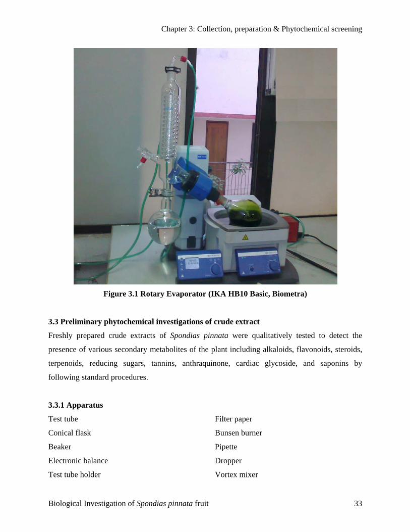
Chapter 3: Collection, preparation & Phytochemical screening
Figure 3.1 Rotary Evaporator (IKA HB10 Basic, Biometra)
3.3 Preliminary phytochemical investigations of crude extract
Freshly prepared crude extracts of Spondias pinnata were qualitatively tested to detect the
presence of various secondary metabolites of the plant including alkaloids, flavonoids, steroids,
terpenoids, reducing sugars, tannins, anthraquinone, cardiac glycoside, and saponins by
following standard procedures.
3.3.1 Apparatus
Test tube Filter paper
Conical flask Bunsen burner
Beaker Pipette
Electronic balance Dropper
Test tube holder Vortex mixer
Biological Investigation of Spondias pinnata fruit 33
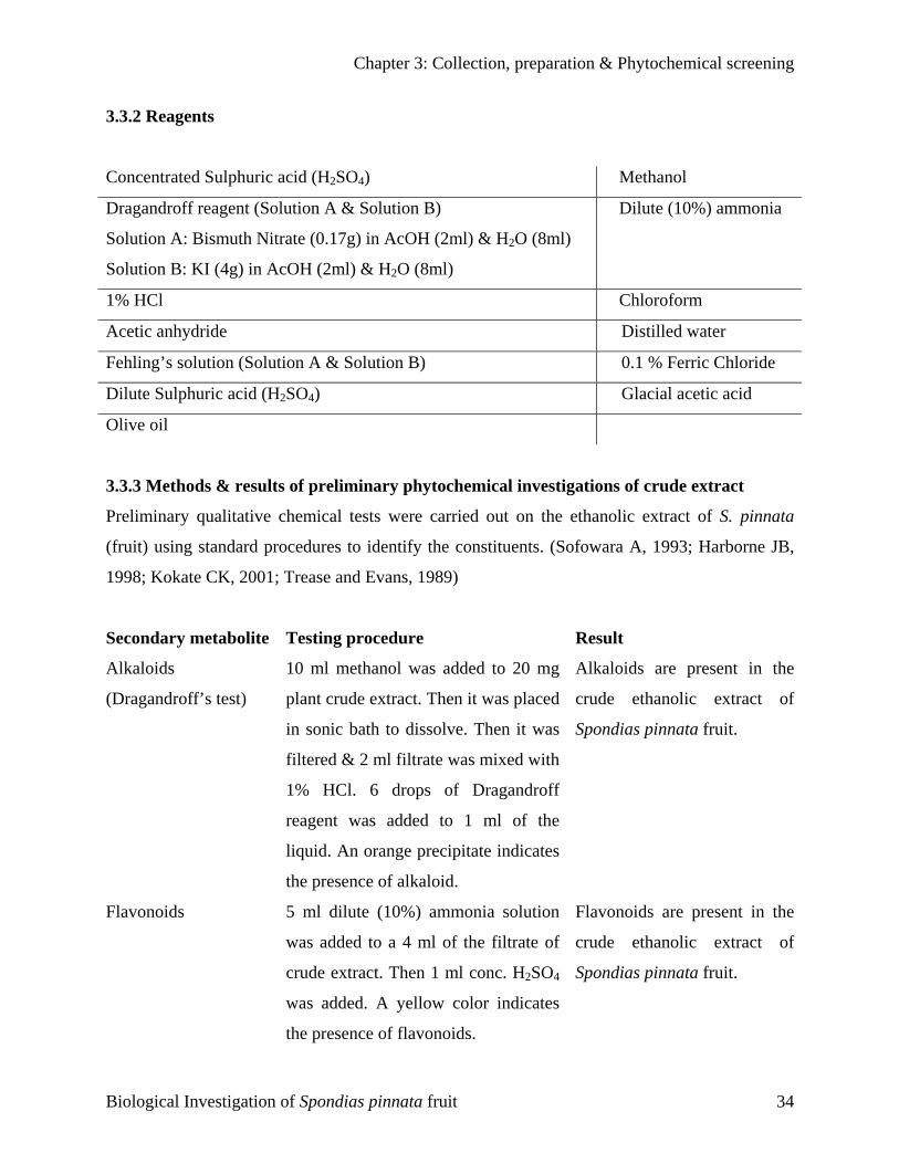
Chapter 3: Collection, preparation & Phytochemical screening
3.3.2 Reagents
Concentrated Sulphuric acid (H2SO4) Methanol
Dragandroff reagent (Solution A & Solution B)
ml) & H2O (8ml)
) ammonia
Solution A: Bismuth Nitrate (0.17g) in AcOH (2
Solution B: KI (4g) in AcOH (2ml) & H2O (8ml)
Dilute (10%
1% HCl Chloroform
Acetic anhydride Distilled water
Fehling’s solution (Solution A & Solution B) loride 0.1 % Ferric Ch
Dilute Sulphuric acid (H2SO4) Glacial acetic acid
Olive oil
3.3.3 Methods & results of preliminary phytochemical investigations of crude extract
nnata
econdary metabolite Testing procedure Result
ff’s test)
added to 20 mg s are present in the
Flavonoids nia solution Flavonoids are present in the
Preliminary qualitative chemical tests were carried out on the ethanolic extract of S. pi
(fruit) using standard procedures to identify the constituents. (Sofowara A, 1993; Harborne JB,
1998; Kokate CK, 2001; Trease and Evans, 1989)
S
Alkaloids
(Dragandro
10 ml methanol was
plant crude extract. Then it was placed
in sonic bath to dissolve. Then it was
filtered & 2 ml filtrate was mixed with
1% HCl. 6 drops of Dragandroff
reagent was added to 1 ml of the
liquid. An orange precipitate indicates
the presence of alkaloid.
5 ml dilute (10%) ammo
Alkaloid
crude ethanolic extract of
Spondias pinnata fruit.
was added to a 4 ml of the filtrate of
crude extract. Then 1 ml conc. H2SO4
was added. A yellow color indicates
the presence of flavonoids.
crude ethanolic extract of
Spondias pinnata fruit.
Biological Investigation of Spondias pinnata fruit 34
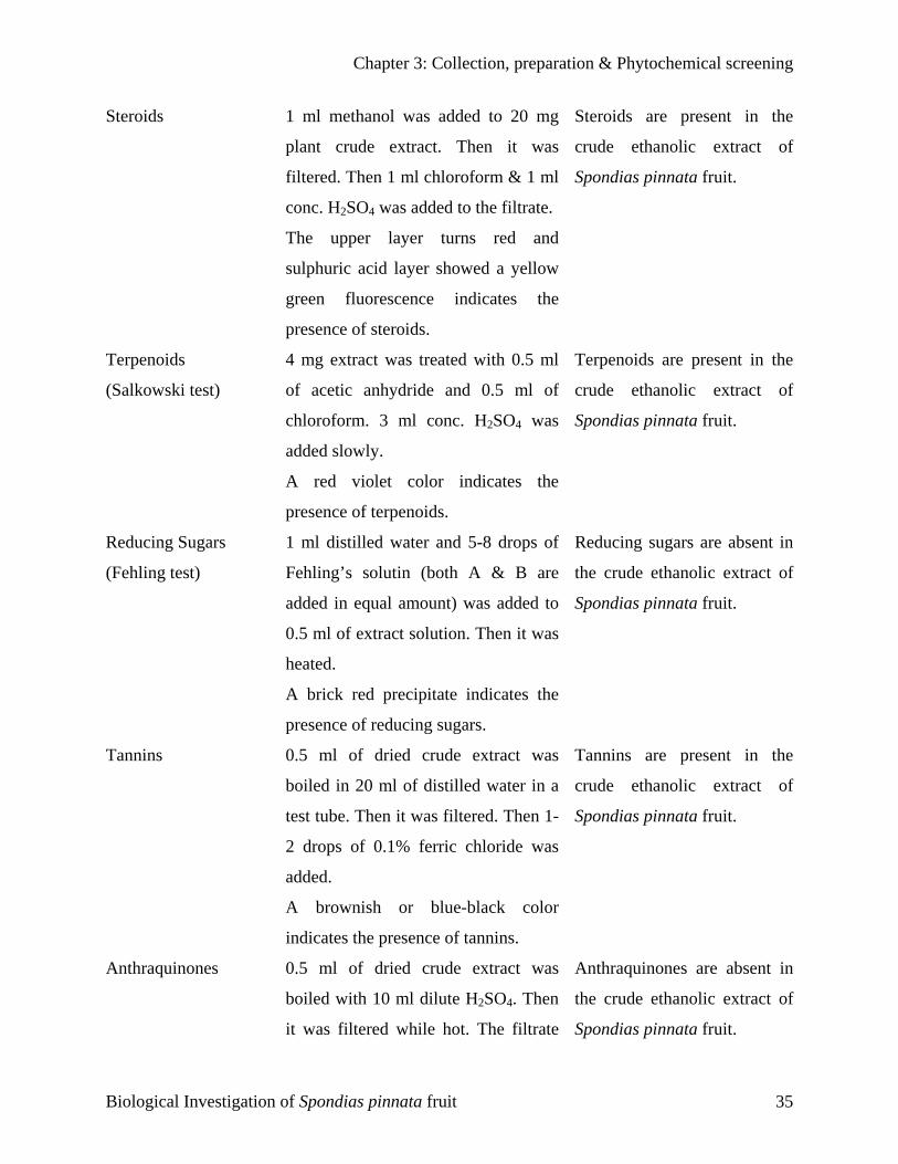
Chapter 3: Collection, preparation & Phytochemical screening
Steroids to 20 mg Steroids are present in the
Terpenoids
est)
ated with 0.5 ml
color indicates the
Terpenoids are present in the
Reducing Sugars 5-8 drops of
red precipitate indicates the
Reducing sugars are absent in
Tannins ract was
nish or blue-black color
Tannins are present in the
Anthraquinones was Anthraquinones are absent in
1 ml methanol was added
plant crude extract. Then it was
filtered. Then 1 ml chloroform & 1 ml
conc. H2SO4 was added to the filtrate.
The upper layer turns red and
sulphuric acid layer showed a yellow
green fluorescence indicates the
presence of steroids.
4 mg extract was tre
crude ethanolic extract of
Spondias pinnata fruit.
(Salkowski t of acetic anhydride and 0.5 ml of
chloroform. 3 ml conc. H2SO4 was
added slowly.
A red violet
presence of terpenoids.
1 ml distilled water and
crude ethanolic extract of
Spondias pinnata fruit.
(Fehling test) Fehling’s solutin (both A & B are
added in equal amount) was added to
0.5 ml of extract solution. Then it was
heated.
A brick
presence of reducing sugars.
0.5 ml of dried crude ext
the crude ethanolic extract of
Spondias pinnata fruit.
boiled in 20 ml of distilled water in a
test tube. Then it was filtered. Then 1-
2 drops of 0.1% ferric chloride was
added.
A brow
indicates the presence of tannins.
0.5 ml of dried crude extract
crude ethanolic extract of
Spondias pinnata fruit.
boiled with 10 ml dilute H2SO4. Then
it was filtered while hot. The filtrate
the crude ethanolic extract of
Spondias pinnata fruit.
Biological Investigation of Spondias pinnata fruit 35

Chapter 3: Collection, preparation & Phytochemical screening
was shaken with 5 ml of chloroform.
The chloroform layer was pipetted
into another test tube. 1 ml dilute
ammonia was then added to it.
A change in color of the resulting
Cardiac glycosides t was mixed with 2
at the interface indicates
Cardiac glycosides are present
Saponins was added to a
mulsion indicates
Saponins are present in the
solution indicates the presence of
anthraquinones.
5 ml crude extrac
(Keller-Killani test) ml glacial acetic acid & 1 drop of
ferric chloride. 1 ml conc. H2SO4 was
then added.
A brown ring
the deoxysugar characteristics of
cardenolides and the presence of
cardiac glycosides.
5 ml distilled water
in the crude ethanolic extract
of Spondias pinnata fruit.
test tube containing 0.5 g of crude
extract. Then the solution was shaken
vigorously & observed for a stable
persistent froth. The frothing was
mixed with 3 drops of olive oil &
shaken vigorously.
Formation of an e
the presence of saponins.
crude ethanolic extract of
Spondias pinnata fruit.
Biological Investigation of Spondias pinnata fruit 36
Antimicrobial Screening
C H A P T E R
Chapter 4: Antimicrobial screening
4.1 Introdu
Worldwide
half of all
developing
actually in
disease, ran
58% .It is e
occurring i
infectious
respiratory
antibiotic r
dramatic in
These nega
public heal
solution tha
The antimi
to ascertain
ability of e
estimated b
i) Dis
ii) Seri
Biological
CChhaapptteerr
44
AAnnttiimmiiccrroobbiiaall SSccrreeeenniinngg
ction
, infectious disease is one of main causes of death accounting for approximately one-
deaths in tropical countries. Perhaps it is not surprising to see these statistics in
nations, but what may be remarkable is that infectious disease mortality rates are
creasing in developed countries, such as the United States. Death from infectious
ked 5th in 1981, has become the 3rd leading cause of death in 1992; an increase of
stimated that infectious disease is the underlying cause of death in 8% of the deaths
n the US. This is alarming given that it was once believed that we would eliminate
disease by the end of the millenium. The increases are attributed to increases in
tract infections and HIV/AIDS. Other contributing factors are an increase in
esistance in nosicomial and community acquired infections. Furthermore, the most
creases are occurring in the 25–44 year old age group (Pinner et al., 1996).
tive health trends call for a renewed interest in infectious disease in the medical and
th communities and renewed strategies on treatment and prevention. It is this last
t would encompass the development of new antimicrobials (Fauci, 1998).
crobial screening which is the first stage of antimicrobial drug research is performed
the susceptibility of various fungi and bacteria to any agent. This test measures the
ach test sample to inhibit the in vitro fungal and bacterial growth. This ability may be
y any of the following three methods.
c diffusion method
al dilution method
Investigation of Spondias pinnata fruit 37
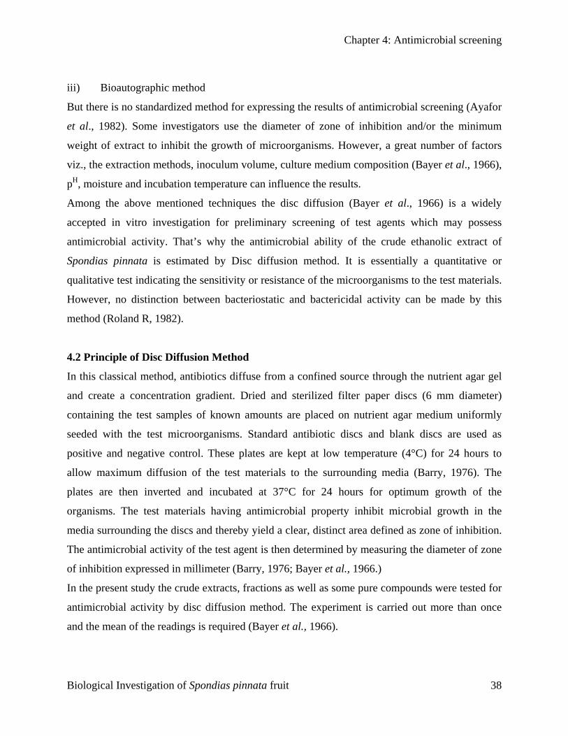
Chapter 4: Antimicrobial screening
iii) Bioautographic method
But there is no standardized method for expressing the results of antimicrobial screening (Ayafor
et al., 1982). Some investigators use the diameter of zone of inhibition and/or the minimum
weight of extract to inhibit the growth of microorganisms. However, a great number of factors
viz., the extraction methods, inoculum volume, culture medium composition (Bayer et al., 1966),
pH, moisture and incubation temperature can influence the results.
Among the above mentioned techniques the disc diffusion (Bayer et al., 1966) is a widely
accepted in vitro investigation for preliminary screening of test agents which may possess
antimicrobial activity. That’s why the antimicrobial ability of the crude ethanolic extract of
Spondias pinnata is estimated by Disc diffusion method. It is essentially a quantitative or
qualitative test indicating the sensitivity or resistance of the microorganisms to the test materials.
However, no distinction between bacteriostatic and bactericidal activity can be made by this
method (Roland R, 1982).
4.2 Principle of Disc Diffusion Method
In this classical method, antibiotics diffuse from a confined source through the nutrient agar gel
and create a concentration gradient. Dried and sterilized filter paper discs (6 mm diameter)
containing the test samples of known amounts are placed on nutrient agar medium uniformly
seeded with the test microorganisms. Standard antibiotic discs and blank discs are used as
positive and negative control. These plates are kept at low temperature (4°C) for 24 hours to
allow maximum diffusion of the test materials to the surrounding media (Barry, 1976). The
plates are then inverted and incubated at 37°C for 24 hours for optimum growth of the
organisms. The test materials having antimicrobial property inhibit microbial growth in the
media surrounding the discs and thereby yield a clear, distinct area defined as zone of inhibition.
The antimicrobial activity of the test agent is then determined by measuring the diameter of zone
of inhibition expressed in millimeter (Barry, 1976; Bayer et al., 1966.)
In the present study the crude extracts, fractions as well as some pure compounds were tested for
antimicrobial activity by disc diffusion method. The experiment is carried out more than once
and the mean of the readings is required (Bayer et al., 1966).
Biological Investigation of Spondias pinnata fruit 38
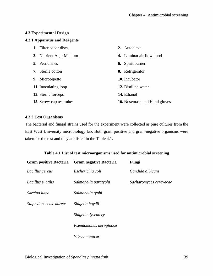
Chapter 4: Antimicrobial screening
4.3 Experimental Design
4.3.1 Apparatus and Reagents
1. Filter paper discs 2. Autoclave
3. Nutrient Agar Medium 4. Laminar air flow hood
5. Petridishes 6. Spirit burner
7. Sterile cotton 8. Refrigerator
9. Micropipette 10. Incubator
11. Inoculating loop 12. Distilled water
13. Sterile forceps 14. Ethanol
15. Screw cap test tubes 16. Nosemask and Hand gloves
4.3.2 Test Organisms
The bacterial and fungal strains used for the experiment were collected as pure cultures from the
East West University microbiology lab. Both gram positive and gram-negative organisms were
taken for the test and they are listed in the Table 4.1.
Table 4.1 List of test microorganisms used for antimicrobial screening
Gram positive Bacteria Gram negative Bacteria Fungi
Bacillus cereus Escherichia coli Candida albicans
Bacillus subtilis Salmonella paratyphi Sacharomyces cerevacae
Sarcina lutea Salmonella typhi
Staphylococcus aureus Shigella boydii
Shigella dysentery
Pseudomonas aeruginosa
Vibrio mimicus
Biological Investigation of Spondias pinnata fruit 39

Chapter 4: Antimicrobial screening
4.3.3 Test Materials of Spondias pinnata
Crude ethanolic extract
4.3.4 Culture Medium and its Composition
Nutrient agar medium is used normally to demonstrate the antimicrobial activity and to make
subculture of the test organisms. The agar medium was directly brought from the market. The
composition of the culture medium is stated below:
Nutrient agar medium
Ingredients Amount
Bacto peptone 0.5 gm
Sodium chloride 0.5 gm
Bacto yeast extract 1.0 gm
Bacto agar 2.0 gm
Distilled water q.s. 100 ml
pH 7.2 ± 0.1 at 250C
4.3.5 Preparation of the Medium
To prepare required volume of this medium, calculated amount of agar medium was taken in a
bottle with a cap and distilled water was added to it to make the required volume. The contents
were then autoclaved to make a clear solution.
4.3.6 Sterilization Procedure
In order to avoid any type of contamination and cross contamination by the test organisms the
antimicrobial screening was done in laminar hood and all types of precautions were highly
maintained. UV light was switched on one hour before working in the laminar hood. Petridishes
and other glassware were sterilized by autoclaving at a temperature of 1210C and a pressure of
15 lbs/sq. inch for 20 minutes. Micropipette tips, cotton, forceps, blank discs etc. were also
sterilized.
Biological Investigation of Spondias pinnata fruit 40
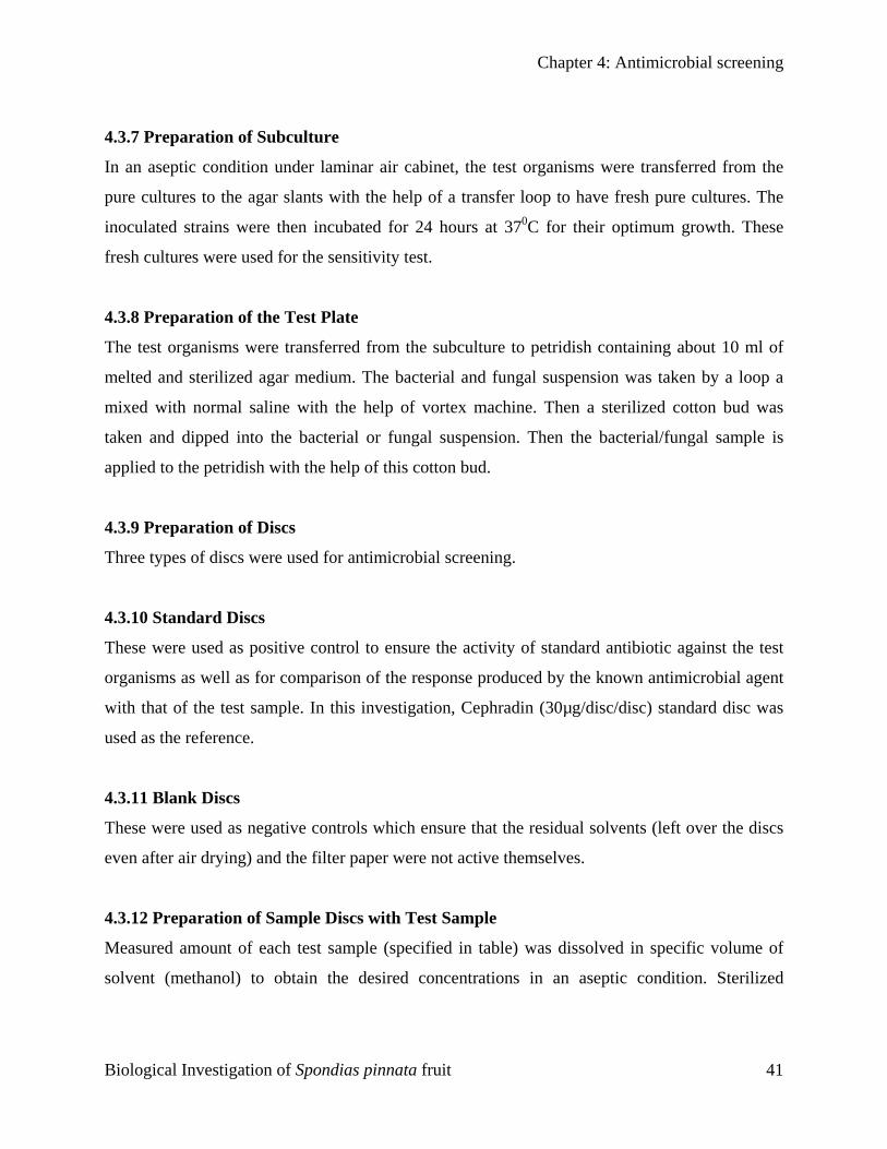
Chapter 4: Antimicrobial screening
4.3.7 Preparation of Subculture
In an aseptic condition under laminar air cabinet, the test organisms were transferred from the
pure cultures to the agar slants with the help of a transfer loop to have fresh pure cultures. The
inoculated strains were then incubated for 24 hours at 370C for their optimum growth. These
fresh cultures were used for the sensitivity test.
4.3.8 Preparation of the Test Plate
The test organisms were transferred from the subculture to petridish containing about 10 ml of
melted and sterilized agar medium. The bacterial and fungal suspension was taken by a loop a
mixed with normal saline with the help of vortex machine. Then a sterilized cotton bud was
taken and dipped into the bacterial or fungal suspension. Then the bacterial/fungal sample is
applied to the petridish with the help of this cotton bud.
4.3.9 Preparation of Discs
Three types of discs were used for antimicrobial screening.
4.3.10 Standard Discs
These were used as positive control to ensure the activity of standard antibiotic against the test
organisms as well as for comparison of the response produced by the known antimicrobial agent
with that of the test sample. In this investigation, Cephradin (30µg/disc/disc) standard disc was
used as the reference.
4.3.11 Blank Discs
These were used as negative controls which ensure that the residual solvents (left over the discs
even after air drying) and the filter paper were not active themselves.
4.3.12 Preparation of Sample Discs with Test Sample
Measured amount of each test sample (specified in table) was dissolved in specific volume of
solvent (methanol) to obtain the desired concentrations in an aseptic condition. Sterilized
Biological Investigation of Spondias pinnata fruit 41
Chapter 4: Antimicrobial screening
metrical (BBL, Cocksville, USA) filter paper discs were taken in a blank petridish under the
laminar hood. Then discs were soaked with solutions of test samples and dried.
Table 4.2 Preparation of Sample Discs
Test Sample Dose (µg/disc) Required amount for 20 disc (mg)
Crude ethanolic extract of
Spondias pinnata fruit
400 8.0
Crude ethanolic extract of
Spondias pinnata fruit
800 16
4.3.13 Application of the Test Samples
Standard Cephradin (30 µg/disc/disc) discs were used as positive control to ensure the activity of
standard antibiotic against the test organisms as well as for comparison of the response produced
by the known antimicrobial agent with that of produced by the test sample. Blank discs were
used as negative controls which ensure that the residual solvents (left over the discs even after
air-drying) and the filter paper were not active themselves.
4.3.14 Diffusion and Incubation
The sample discs, the standard antibiotic discs and the control discs were placed gently on the
previously marked zones in the agar plates pre-inoculated with test bacteria and fungi. The plates
were then kept in a refrigerator at 40C for about 24 hours upside down to allow sufficient
diffusion of the materials from the discs to the surrounding agar medium. The plates were then
inverted and kept in an incubator at 370C for 24 hours.
Biological Investigation of Spondias pinnata fruit 42
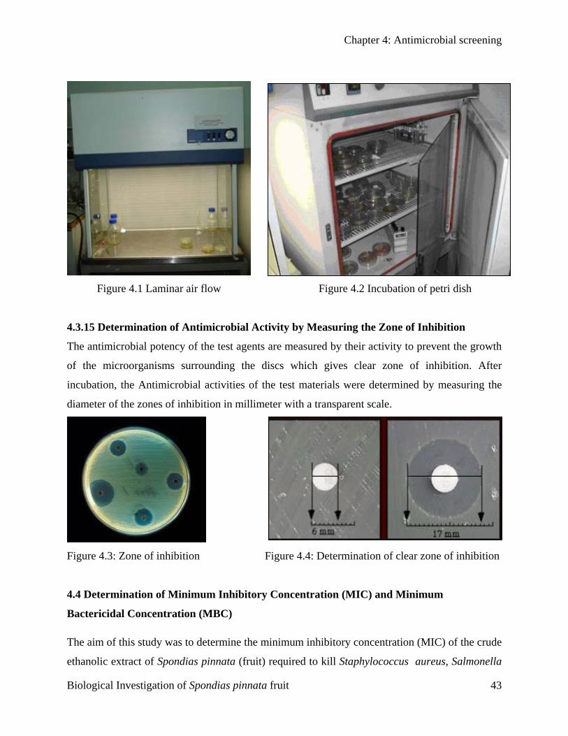
Chapter 4: Antimicrobial screening
Figure 4.1 Laminar air flow Figure 4.2 Incubation of petri dish
4.3.15 Determination of Antimicrobial Activity by Measuring the Zone of Inhibition
The antimicrobial potency of the test agents are measured by their activity to prevent the growth
of the microorganisms surrounding the discs which gives clear zone of inhibition. After
incubation, the Antimicrobial activities of the test materials were determined by measuring the
diameter of the zones of inhibition in millimeter with a transparent scale.
Figure 4.3: Zone of inhibition Figure 4.4: Determination of clear zone of inhibition
4.4 Determination of Minimum Inhibitory Concentration (MIC) and Minimum
Bactericidal Concentration (MBC)
The aim of this study was to determine the minimum inhibitory concentration (MIC) of the crude
ethanolic extract of Spondias pinnata (fruit) required to kill Staphylococcus aureus, Salmonella
Biological Investigation of Spondias pinnata fruit 43
Chapter 4: Antimicrobial screening
paratyphi, Salmonella typhi and Shigella boydii. In the experiment, medicaments were added to
bacterial species into eppendrof tube, in 10 different concentrations. The MIC is the lowest
concentration of the crude drug at which bacterial growth could not be observed.
4.4.1 Principle of MIC
The disc diffusion method is a semi quantitative method. Determination of MIC values provides
a quantitative measure for the level of resistance expressed by the test organism. Next to the agar
dilution method, the broth dilution method is one of the most frequently used methods to
determine minimum inhibitory concentration (MIC) of the crude extract. The method is based on
the inoculation of a standardized liquid culture of the test strain in a dilution series of the extract
for which the MIC is determined. The first concentration of the dilution series at which no visual
growth of microorganism can be determined is considered as the MIC (Huys G, 2002).
4.5 Experimental Design for MIC & MBC
4.5.1 Apparatus and Reagents
Nutrient Agar Medium
Nutrient Broth Medium
Autoclave
Eppendrof tube
Petridishes Laminar air flow hood
Sterile cotton Spirit burner
Micropipette Refrigerator
Inoculating loop Incubator
Sterile forceps Ethanol
Screw cap test tubes Nosemask and Hand gloves
4.5.2 Test Organisms
The bacterial strains used for the experiment were collected as pure cultures from the East West
University microbiology lab. Both gram positive and gram-negative organisms were taken for
the test and they are listed in the Table 4.3.
Biological Investigation of Spondias pinnata fruit 44
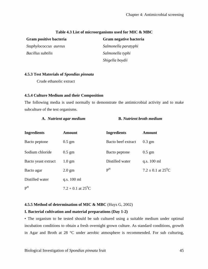
Chapter 4: Antimicrobial screening
Table 4.3 List of microorganisms used for MIC & MBC
Gram positive bacteria Gram negative bacteria
Staphylococcus aureus Salmonella paratyphi
Bacillus subtilis Salmonella typhi
Shigella boydii
4.5.3 Test Materials of Spondias pinnata
Crude ethanolic extract
4.5.4 Culture Medium and their Composition
The following media is used normally to demonstrate the antimicrobial activity and to make
subculture of the test organisms.
A. Nutrient agar medium
B. Nutrient broth medium
Ingredients Amount Ingredients Amount
Bacto peptone 0.5 gm Bacto beef extract 0.3 gm
Sodium chloride 0.5 gm Bacto peptone 0.5 gm
Bacto yeast extract 1.0 gm Distilled water q.s. 100 ml
Bacto agar 2.0 gm PH 7.2 ± 0.1 at 250C
Distilled water q.s. 100 ml
PH 7.2 + 0.1 at 250C
4.5.5 Method of determination of MIC & MBC (Huys G, 2002)
I. Bacterial cultivation and material preparations (Day 1-2)
• The organism to be tested should be sub cultured using a suitable medium under optimal
incubation conditions to obtain a fresh overnight grown culture. As standard conditions, growth
in Agar and Broth at 28 °C under aerobic atmosphere is recommended. For sub culturing,
Biological Investigation of Spondias pinnata fruit 45
Chapter 4: Antimicrobial screening
remove one bead from the cryovial and streak out onto a nutrient agar plate. Incubate at 28°C
overnight (or longer until clear visible growth is observed).
• After overnight incubation, the streak cultures are checked for purity. A number of pure
colonies (app. 5 or more if the isolate is a suspected fastidious organism) are introduced into a
glass culture tube containing 10 ml broth and incubated at 28°C overnight (or longer until clear
visible growth is observed). Ideally, the culture tubes should fit into a portable
spectrophotometer. In this way, the overnight grown culture can be easily adjusted to a
standardized cell density by dilution with sterile broth
• Control cultures should be included during each series of MIC determinations.
• A bottle with the necessary volume of double-distilled water should be prepared.
II. Preparation and inoculation of the dilution series (Day 3)
• The optical density of the overnight culture of the strain is determined spectrophotometrically
at 590 nm and is standardized at 0.1 ± 0.02 (i.e. app. 10E8 CFU/ml) by diluting with sterile
broth.
• For each batch of 4 strains, two 6 ml sterile stock solutions of extract should be prepared in the
suitable solvent. However, subsequent dilutions of these stock solutions can be made up in sterile
water. The use of two stock solutions is recommended to minimize quantitative errors in the low
concentration range of the serial dilution series.
• Extract concentrations 40, 20, 10, 5, 2.5, 1.25, 0.625, 0.3125, 0.15625, and 0.078125 mg/ml are
obtained by making serial dilutions from stock solution. It is absolutely crucial to thoroughly mix
every freshly prepared antibiotic dilution prior to using it to prepare the next dilution. It should
be noted that each antibiotic dilution undergoes a final 1:2 dilution when the broth culture is
added. If required, the tested MIC range can be extended with additional concentrations.
• Following the preparation of the serial dilutions of extract, 2 ml of freshly standardized broth
culture of the strain is inoculated in each tube of the dilution series. In this regard, it is important
to note that the standardized cultures should be processed within the hour after preparation.
Dilutions and broth cultures should be well homogenized prior to mixing.
• For each batch of MIC determinations, a blank tube (i.e. 2 ml non-inoculated broth mixed with
2 ml water) should be included. In addition, a positive control should be included for each strain.
Biological Investigation of Spondias pinnata fruit 46
Chapter 4: Antimicrobial screening
The positive control is made up by mixing 2 ml adjusted broth culture with 2 ml sterile double
distilled water.
• All MIC tubes and control tubes of the test isolates and the control strain as well as the blank
are incubated aerobically at 28°C for 24h.
• Each isolate should also be checked for purity by plating a drop of the adjusted culture onto
agar medium, and this plate should be incubated under the same conditions as the MIC test itself.
III. Reading of the MIC (Day 4)
• The purity of the broth culture is checked on agar on the basis of uniform colonial morphology.
If contamination is noted than all data generated from the involved strain should be rejected.
• Following a 24 hour incubation (or longer until clear visual growth can be determined in the
positive control tubes), growth is determined visually among the different tubes of the serial
dilution by comparing with the positive control and with the blank. Any series where
discontinuity in growth is observed (e.g. growth in tubes 5 and 7 but not in tube 6) should be
discarded. The end-point is defined as the lowest antibiotic concentration for which there is no
visual growth. This concentration should be reported as the MIC of that antibiotic for that
particular strain. If trailing end-points are observed, this should be reported as a remark and a
80% reduction in growth should be reported as end-point.
Then a sterilized cotton bud is dipped into the clear solution obtained then applied to an agar
plate.
IV. Reading of the MBC (Day 5)
The agar plates then checked for the growth.
Biological Investigation of Spondias pinnata fruit 47
Evaluation of Antioxidant
Activity
C H A P T E R
Chapter 5: Evaluation of antioxidant activity
5.1 Rat
There is
This da
several
which s
radical
derived
role in h
Pieroni
plant d
constitu
caroten
Lipid p
process
butylate
widely
by retar
only to
effects
especia
Rao, 20
Biologi
CChhaapptteerr
55
EEvvaalluuaattiioonn ooff AAnnttiiooxxiiddaanntt AAccttiivviittyy
ional and objective
considerable recent evidence that free radical induce oxidative damage to biomolecules.
mage causes cancer, aging, neurodegenerative diseases, atherosclerosis, malaria and
other pathological events in living organisms (Halliwell et al., 1992). Antioxidants
cavenge free radicals are known to posses an important role in preventing these free
induced-diseases. There is an increasing interest in the antioxidants effects of compounds
from plants, which could be relevant in relations to their nutritional incidence and their
ealth and diseases (Steinmetz and Potter, 1996; Aruoma, 1998; Bandoniene et al., 2000;
et al., 2002; Couladis et al., 2003). A number of reports on the isolation and testing of
erived antioxidants have been described during the past decade. Natural antioxidants
te a broad range of substances including phenolic or nitrogen containing compounds and
oids (Shahidi et al., 1992; Velioglu et al., 1998; Pietta et al., 1998)
eroxidation is one of the main reasons for deterioration of food products during
ing and storage. Synthetic antioxidant such as tert-butyl-1-hydroxytoluene (BHT),
d hydroxyanisole (BHA), propyl gallate (PG) and tert-butylhydroquinone (TBHQ) are
used as food additives to increase self life, especially lipid and lipid containing products
ding the process of lipid peroxidation. However, BHT and BHA are known to have not
xic and carcinogenic effects and humans (Ito et al.,1986; Wichi,1988), but abnormal
on enzyme systems (Inatani et al., 1983). Therefore, the interest in natural antioxidant,
lly of plant origin, has greatly increased in recent years (Jayaprakasha & Jaganmohan
00)
cal Investigation of Spondias pinnata fruit 48
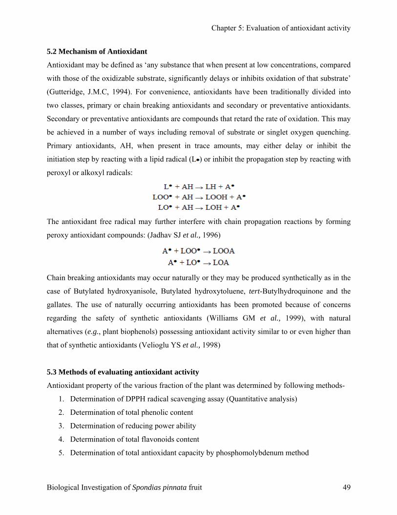
Chapter 5: Evaluation of antioxidant activity
5.2 Mechanism of Antioxidant
Antioxidant may be defined as ‘any substance that when present at low concentrations, compared
with those of the oxidizable substrate, significantly delays or inhibits oxidation of that substrate’
(Gutteridge, J.M.C, 1994). For convenience, antioxidants have been traditionally divided into
two classes, primary or chain breaking antioxidants and secondary or preventative antioxidants.
Secondary or preventative antioxidants are compounds that retard the rate of oxidation. This may
be achieved in a number of ways including removal of substrate or singlet oxygen quenching.
Primary antioxidants, AH, when present in trace amounts, may either delay or inhibit the
initiation step by reacting with a lipid radical (L ) or inhibit the propagation step by reacting with
peroxyl or alkoxyl radicals:
The antioxidant free radical may further interfere with chain propagation reactions by forming
peroxy antioxidant compounds: (Jadhav SJ et al., 1996)
Chain breaking antioxidants may occur naturally or they may be produced synthetically as in the
case of Butylated hydroxyanisole, Butylated hydroxytoluene, tert-Butylhydroquinone and the
gallates. The use of naturally occurring antioxidants has been promoted because of concerns
regarding the safety of synthetic antioxidants (Williams GM et al., 1999), with natural
alternatives (e.g., plant biophenols) possessing antioxidant activity similar to or even higher than
that of synthetic antioxidants (Velioglu YS et al., 1998)
5.3 Methods of evaluating antioxidant activity
Antioxidant property of the various fraction of the plant was determined by following methods-
1. Determination of DPPH radical scavenging assay (Quantitative analysis)
2. Determination of total phenolic content
3. Determination of reducing power ability
4. Determination of total flavonoids content
5. Determination of total antioxidant capacity by phosphomolybdenum method
Biological Investigation of Spondias pinnata fruit 49
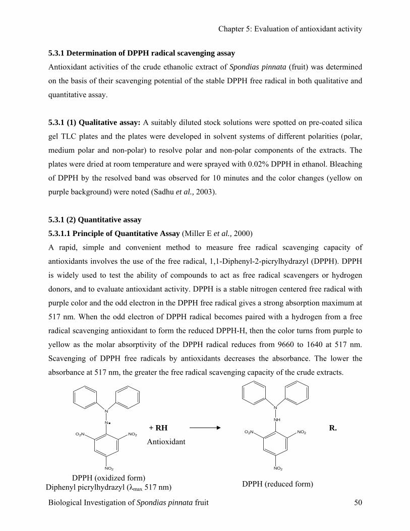
Chapter 5: Evaluation of antioxidant activity
5.3.1 Determination of DPPH radical scavenging assay
Antioxidant activities of the crude ethanolic extract of Spondias pinnata (fruit) was determined
on the basis of their scavenging potential of the stable DPPH free radical in both qualitative and
quantitative assay.
5.3.1 (1) Qualitative assay: A suitably diluted stock solutions were spotted on pre-coated silica
gel TLC plates and the plates were developed in solvent systems of different polarities (polar,
medium polar and non-polar) to resolve polar and non-polar components of the extracts. The
plates were dried at room temperature and were sprayed with 0.02% DPPH in ethanol. Bleaching
of DPPH by the resolved band was observed for 10 minutes and the color changes (yellow on
purple background) were noted (Sadhu et al., 2003).
5.3.1 (2) Quantitative assay
5.3.1.1 Principle of Quantitative Assay (Miller E et al., 2000)
A rapid, simple and convenient method to measure free radical scavenging capacity of
antioxidants involves the use of the free radical, 1,1-Diphenyl-2-picrylhydrazyl (DPPH). DPPH
is widely used to test the ability of compounds to act as free radical scavengers or hydrogen
donors, and to evaluate antioxidant activity. DPPH is a stable nitrogen centered free radical with
purple color and the odd electron in the DPPH free radical gives a strong absorption maximum at
517 nm. When the odd electron of DPPH radical becomes paired with a hydrogen from a free
radical scavenging antioxidant to form the reduced DPPH-H, then the color turns from purple to
yellow as the molar absorptivity of the DPPH radical reduces from 9660 to 1640 at 517 nm.
Scavenging of DPPH free radicals by antioxidants decreases the absorbance. The lower the
absorbance at 517 nm, the greater the free radical scavenging capacity of the crude extracts.
+ RH + R.
N
NH
Antioxidant
N
N.NO2O2N
NO
NO2O
2N
NO2
Diphen
Biolog
2
DPPH (oxidized form) yl picrylhydrazyl (λmax 517 nm)
ical Investigation of Spondias pinnata fruit
DPPH (reduced form)
50
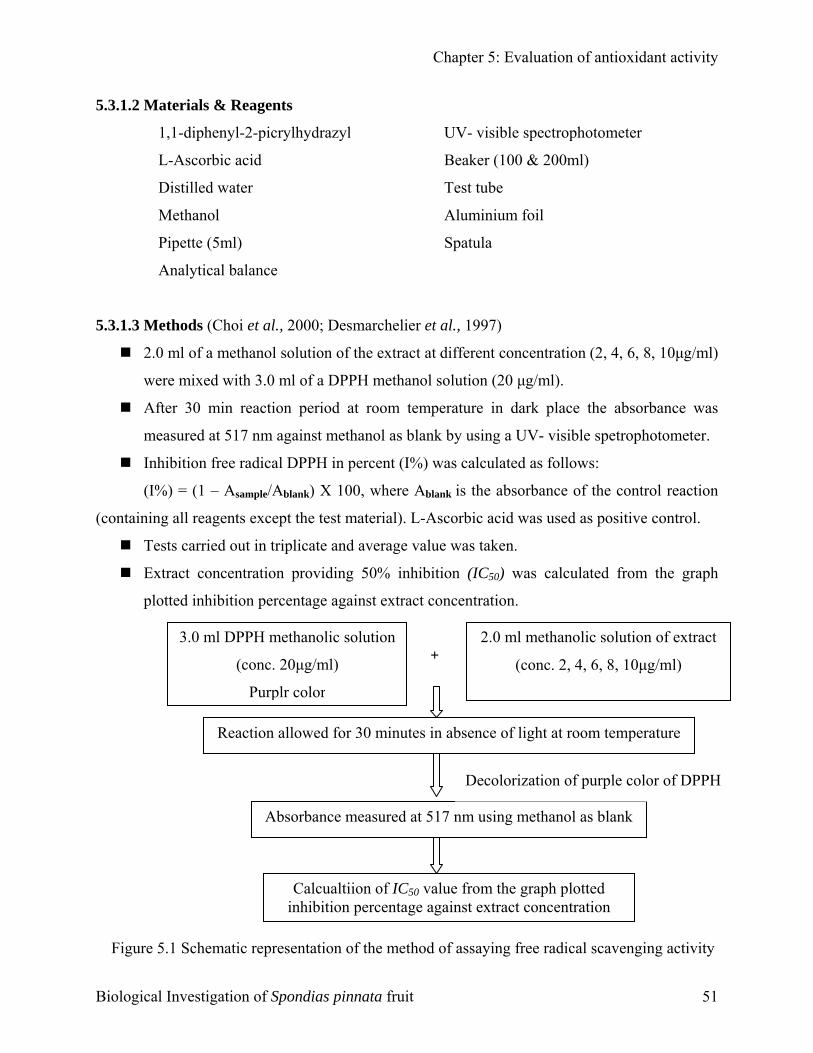
Chapter 5: Evaluation of antioxidant activity
5.3.1.2 Materials & Reagents
1,1-diphenyl-2-picrylhydrazyl UV- visible spectrophotometer
L-Ascorbic acid Beaker (100 & 200ml)
Distilled water Test tube
Methanol Aluminium foil
Pipette (5ml) Spatula
Analytical balance
5.3.1.3 Methods (Choi et al., 2000; Desmarchelier et al., 1997)
2.0 ml of a methanol solution of the extract at different concentration (2, 4, 6, 8, 10µg/ml)
were mixed with 3.0 ml of a DPPH methanol solution (20 µg/ml).
After 30 min reaction period at room temperature in dark place the absorbance was
measured at 517 nm against methanol as blank by using a UV- visible spetrophotometer.
Inhibition free radical DPPH in percent (I%) was calculated as follows:
(I%) = (1 – Asample/Ablank) X 100, where Ablank is the absorbance of the control reaction
(containing all reagents except the test material). L-Ascorbic acid was used as positive control.
Tests carried out in triplicate and average value was taken.
Extract concentration providing 50% inhibition (IC50) was calculated from the graph
plotted inhibition percentage against extract concentration.
+ 2.0 ml methanolic solution of extract
(conc. 2, 4, 6, 8, 10µg/ml)
Figure 5.1
Biological I
3.0 ml DPPH methanolic solution
(conc. 20µg/ml)
Purplr color
e
Sche
nvesti
Reaction allowed for 30 minutes in absence of light at room temperatur
Decolorization of purple color of DPPH
k
matic r
gation o
Absorbance measured at 517 nm using methanol as blan
Calcualtiion of IC50 value from the graph plotted inhibition percentage against extract concentration
epresentation of the method of assaying free radical scavenging activity
f Spondias pinnata fruit 51
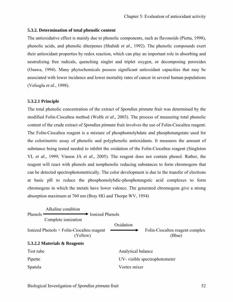
Chapter 5: Evaluation of antioxidant activity
5.3.2. Determination of total phenolic content
The antioxidative effect is mainly due to phenolic components, such as flavonoids (Pietta, 1998),
phenolic acids, and phenolic diterpenes (Shahidi et al., 1992). The phenolic compounds exert
their antioxidant properties by redox reaction, which can play an important role in absorbing and
neutralizing free radicals, quenching singlet and triplet oxygen, or decomposing peroxides
(Osawa, 1994). Many phytochemicals possess significant antioxidant capacities that may be
associated with lower incidence and lower mortality rates of cancer in several human populations
(Velioglu et al., 1998).
5.3.2.1 Principle
The total phenolic concentration of the extract of Spondias pinnata fruit was determined by the
modified Folin-Ciocalteu method (Wolfe et al., 2003). The process of measuring total phenolic
content of the crude extract of Spondias pinnata fruit involves the use of Folin-Ciocalteu reagent.
The Folin-Ciocalteu reagent is a mixture of phosphomolybdate and phosphotungstate used for
the colorimetric assay of phenolic and polyphenolic antioxidants. It measures the amount of
substance being tested needed to inhibit the oxidation of the Folin-Ciocalteu reagent (Singleton
VL et al., 1999; Vinson JA et al., 2005). The reagent does not contain phenol. Rather, the
reagent will react with phenols and nonphenolic reducing substances to form chromogens that
can be detected spectrophotometrically. The color development is due to the transfer of electrons
at basic pH to reduce the phosphomolybdic-phosphotungstic acid complexes to form
chromogens in which the metals have lower valence. The generated chromogens give a strong
absorption maximum at 760 nm (Bray HG and Thorpe WV, 1954)
Alkaline condition Phenols Ionized Phenols Complete ionization Oxidation Ionized Phenols + Folin-Ciocalteu reagent Folin-Ciocalteu reagent complex
(Yellow) (Blue)
5.3.2.2 Materials & Reagents
Test tube Analytical balance
Pipette UV- visible spectrophotometer
Spatula Vortex mixer
Biological Investigation of Spondias pinnata fruit 52
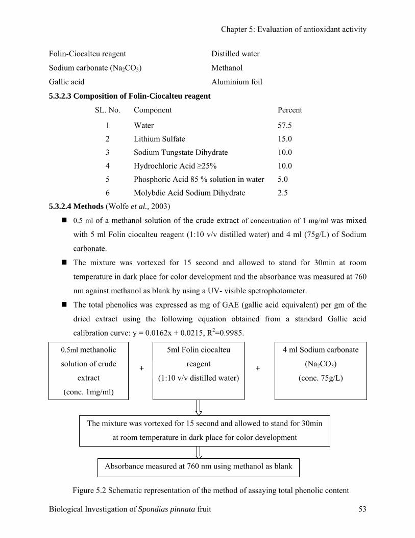
Chapter 5: Evaluation of antioxidant activity
Folin-Ciocalteu reagent Distilled water
Sodium carbonate (Na2CO3) Methanol
Gallic acid Aluminium foil
5.3.2.3 Composition of Folin-Ciocalteu reagent
SL. No. Component Percent
1 Water 57.5
2 Lithium Sulfate 15.0
3 Sodium Tungstate Dihydrate 10.0
4 Hydrochloric Acid ≥25% 10.0
5 Phosphoric Acid 85 % solution in water 5.0
6 Molybdic Acid Sodium Dihydrate 2.5
5.3.2.4 Methods (Wolfe et al., 2003)
0.5 ml of a methanol solution of the crude extract of concentration of 1 mg/ml was mixed
with 5 ml Folin ciocalteu reagent (1:10 v/v distilled water) and 4 ml (75g/L) of Sodium
carbonate.
The mixture was vortexed for 15 second and allowed to stand for 30min at room
temperature in dark place for color development and the absorbance was measured at 760
nm against methanol as blank by using a UV- visible spetrophotometer.
The total phenolics was expressed as mg of GAE (gallic acid equivalent) per gm of the
dried extract using the following equation obtained from a standard Gallic acid
calibration curve: y = 0.0162x + 0.0215, R2=0.9985.
5ml Folin ciocalteu
reagent
(1:10 v/v distilled water)+ +
4 ml Sodium carbonate
(Na2CO3)
(conc. 75g/L)
Bi
0.5ml methanolic
solution of crude
extract
(conc. 1mg/ml)
ological
The mixture was vortexed for 15 second and allowed to stand for 30min
at room temperature in dark place for color development
k
Inves
Absorbance measured at 760 nm using methanol as blan
Figure 5.2 Schematic representation of the method of assaying total phenolic contenttigation of Spondias pinnata fruit 53
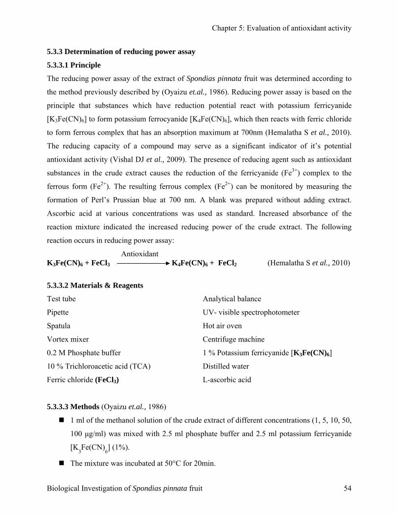
Chapter 5: Evaluation of antioxidant activity
5.3.3 Determination of reducing power assay
5.3.3.1 Principle
The reducing power assay of the extract of Spondias pinnata fruit was determined according to
the method previously described by (Oyaizu et.al., 1986). Reducing power assay is based on the
principle that substances which have reduction potential react with potassium ferricyanide
[K3Fe(CN)6] to form potassium ferrocyanide [K4Fe(CN)6], which then reacts with ferric chloride
to form ferrous complex that has an absorption maximum at 700nm (Hemalatha S et al., 2010).
The reducing capacity of a compound may serve as a significant indicator of it’s potential
antioxidant activity (Vishal DJ et al., 2009). The presence of reducing agent such as antioxidant
substances in the crude extract causes the reduction of the ferricyanide (Fe3+) complex to the
ferrous form (Fe2+). The resulting ferrous complex (Fe2+) can be monitored by measuring the
formation of Perl’s Prussian blue at 700 nm. A blank was prepared without adding extract.
Ascorbic acid at various concentrations was used as standard. Increased absorbance of the
reaction mixture indicated the increased reducing power of the crude extract. The following
reaction occurs in reducing power assay:
Antioxidant K3Fe(CN)6 + FeCl3 K4Fe(CN)6 + FeCl2 (Hemalatha S et al., 2010)
5.3.3.2 Materials & Reagents
Test tube Analytical balance
Pipette UV- visible spectrophotometer
Spatula Hot air oven
Vortex mixer Centrifuge machine
0.2 M Phosphate buffer 1 % Potassium ferricyanide [K3Fe(CN)6]
10 % Trichloroacetic acid (TCA) Distilled water
Ferric chloride (FeCl3) L-ascorbic acid
5.3.3.3 Methods (Oyaizu et.al., 1986)
1 ml of the methanol solution of the crude extract of different concentrations (1, 5, 10, 50,
100 µg/ml) was mixed with 2.5 ml phosphate buffer and 2.5 ml potassium ferricyanide
[K3Fe(CN)
6] (1%).
The mixture was incubated at 50°C for 20min.
Biological Investigation of Spondias pinnata fruit 54
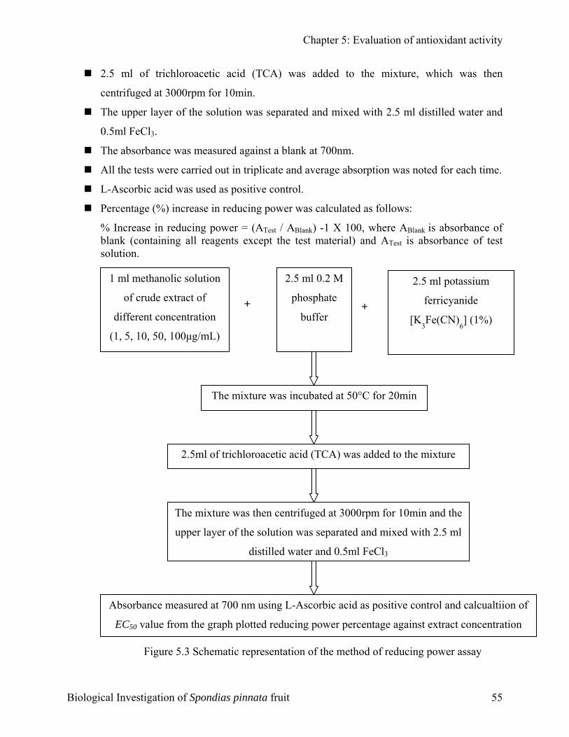
Chapter 5: Evaluation of antioxidant activity
2.5 ml of trichloroacetic acid (TCA) was added to the mixture, which was then
centrifuged at 3000rpm for 10min.
The upper layer of the solution was separated and mixed with 2.5 ml distilled water and
0.5ml FeCl3.
The absorbance was measured against a blank at 700nm.
All the tests were carried out in triplicate and average absorption was noted for each time.
L-Ascorbic acid was used as positive control.
Percentage (%) increase in reducing power was calculated as follows:
% Increase in reducing power = (ATest / ABlank) -1 X 100, where ABlank is absorbance of blank (containing all reagents except the test material) and ATest is absorbance of test solution.
2.5 ml 0.2 M
phosphate
buffer + +
2.5 ml potassium
ferricyanide
[K3Fe(CN)
6] (1%)
Biologic
1 ml methanolic solution
of crude extract of
different concentration
(1, 5, 10, 50, 100µg/mL)
Figure 5.3 Sch
al Investigation of Spo
The mixture was incubated at 50°C for 20min
2.5ml of trichloroacetic acid (TCA) was added to the mixtureThe mixture was then centrifuged at 3000rpm for 10min and the
upper layer of the solution was separated and mixed with 2.5 ml
distilled water and 0.5ml FeCl3
Absorbance measured at 700 nm using L-Ascorbic acid as positive control and calcualtiion of
EC50 value from the graph plotted reducing power percentage against extract concentration
ematic representation of the method of reducing power assay
ndias pinnata fruit 55
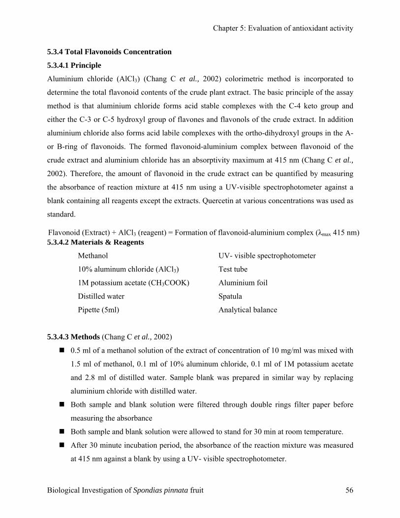
Chapter 5: Evaluation of antioxidant activity
5.3.4 Total Flavonoids Concentration
5.3.4.1 Principle
Aluminium chloride (AlCl3) (Chang C et al., 2002) colorimetric method is incorporated to
determine the total flavonoid contents of the crude plant extract. The basic principle of the assay
method is that aluminium chloride forms acid stable complexes with the C-4 keto group and
either the C-3 or C-5 hydroxyl group of flavones and flavonols of the crude extract. In addition
aluminium chloride also forms acid labile complexes with the ortho-dihydroxyl groups in the A-
or B-ring of flavonoids. The formed flavonoid-aluminium complex between flavonoid of the
crude extract and aluminium chloride has an absorptivity maximum at 415 nm (Chang C et al.,
2002). Therefore, the amount of flavonoid in the crude extract can be quantified by measuring
the absorbance of reaction mixture at 415 nm using a UV-visible spectrophotometer against a
blank containing all reagents except the extracts. Quercetin at various concentrations was used as
standard.
5.3.4.2 Materials & Reagents Flavonoid (Extract) + AlCl3 (reagent) = Formation of flavonoid-aluminium complex (λmax 415 nm)
Methanol UV- visible spectrophotometer
10% aluminum chloride (AlCl3) Test tube
1M potassium acetate (CH3COOK) Aluminium foil
Distilled water Spatula
Pipette (5ml) Analytical balance
5.3.4.3 Methods (Chang C et al., 2002)
0.5 ml of a methanol solution of the extract of concentration of 10 mg/ml was mixed with
1.5 ml of methanol, 0.1 ml of 10% aluminum chloride, 0.1 ml of 1M potassium acetate
and 2.8 ml of distilled water. Sample blank was prepared in similar way by replacing
aluminium chloride with distilled water.
Both sample and blank solution were filtered through double rings filter paper before
measuring the absorbance
Both sample and blank solution were allowed to stand for 30 min at room temperature.
After 30 minute incubation period, the absorbance of the reaction mixture was measured
at 415 nm against a blank by using a UV- visible spectrophotometer.
Biological Investigation of Spondias pinnata fruit 56
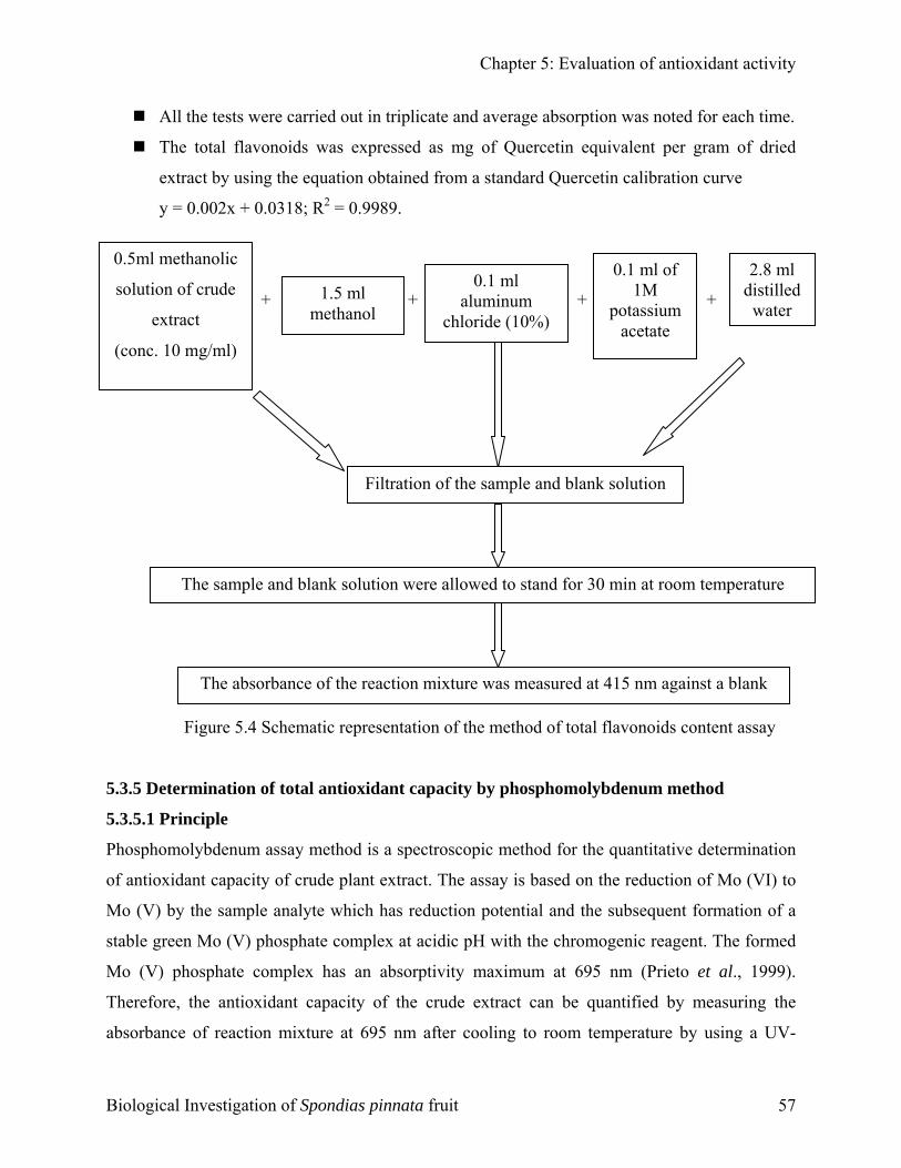
Chapter 5: Evaluation of antioxidant activity
All the tests were carried out in triplicate and average absorption was noted for each time.
The total flavonoids was expressed as mg of Quercetin equivalent per gram of dried
extract by using the equation obtained from a standard Quercetin calibration curve
y = 0.002x + 0.0318; R2 = 0.9989.
+ + + +
1.5 ml methanol
0.1 ml aluminum
chloride (10%)
Filtration of the sample and blank solution
2.8 ml distilled
0.1 ml of 1M
potassium acetate
0.5ml methanolic
solution of crude
extract
(conc. 10 mg/ml)
The absorbance of the reaction mixture was measured at 415 nm against a bl
The sample and blank solution were allowed to stand for 30 min at room tempe
F
5.3.5 Determination of total antioxidant capacity by phosphomolybdenum method
5.3.5.1 Principle
Phosphomolybdenum assay method is a spectroscopic method for the quantitative determ
of antioxidant capacity of crude plant extract. The assay is based on the reduction of Mo
Mo (V) by the sample analyte which has reduction potential and the subsequent formati
stable green Mo (V) phosphate complex at acidic pH with the chromogenic reagent. The
Mo (V) phosphate complex has an absorptivity maximum at 695 nm (Prieto et al.,
Therefore, the antioxidant capacity of the crude extract can be quantified by measur
absorbance of reaction mixture at 695 nm after cooling to room temperature by using
Biological Investigation of Spondias pinnata fruit
water
rature
ank
y
igure 5.4 Schematic representation of the method of total flavonoids content assaination
(VI) to
on of a
formed
1999).
ing the
a UV-
57
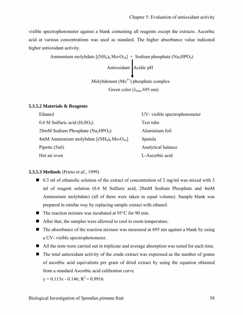
Chapter 5: Evaluation of antioxidant activity
visible spectrophotometer against a blank containing all reagents except the extracts. Ascorbic
acid at various concentrations was used as standard. The higher absorbance value indicated
higher antioxidant activity.
Ammonium molybdate [(NH4)6 Mo7O24] + Sodium phosphate (Na2HPO4)
Antioxidant Acidic pH
Molybdenum (Mo5+) phosphate complex
Green color (λmax 695 nm)
5.3.5.2 Materials & Reagents
Ethanol UV- visible spectrophotometer
0.6 M Sulfuric acid (H2SO4) Test tube
28mM Sodium Phosphate (Na2HPO4) Aluminium foil
4mM Ammonium molybdate [(NH4)6 Mo7O24] Spatula
Pipette (5ml) Analytical balance
Hot air oven L-Ascorbic acid
5.3.5.3 Methods (Prieto et al., 1999)
0.3 ml of ethanolic solution of the extract of concentration of 2 mg/ml was mixed with 3
ml of reagent solution (0.6 M Sulfuric acid, 28mM Sodium Phosphate and 4mM
Ammonium molybdate) (all of them were taken in equal volume). Sample blank was
prepared in similar way by replacing sample extract with ethanol.
The reaction mixture was incubated at 95°C for 90 min.
After that, the samples were allowed to cool to room temperature.
The absorbance of the reaction mixture was measured at 695 nm against a blank by using
a UV- visible spectrophotometer.
All the tests were carried out in triplicate and average absorption was noted for each time.
The total antioxidant activity of the crude extract was expressed as the number of grams
of ascorbic acid equivalents per gram of dried extract by using the equation obtained
from a standard Ascorbic acid calibration curve
y = 0.113x - 0.146; R2 = 0.9916.
Biological Investigation of Spondias pinnata fruit 58
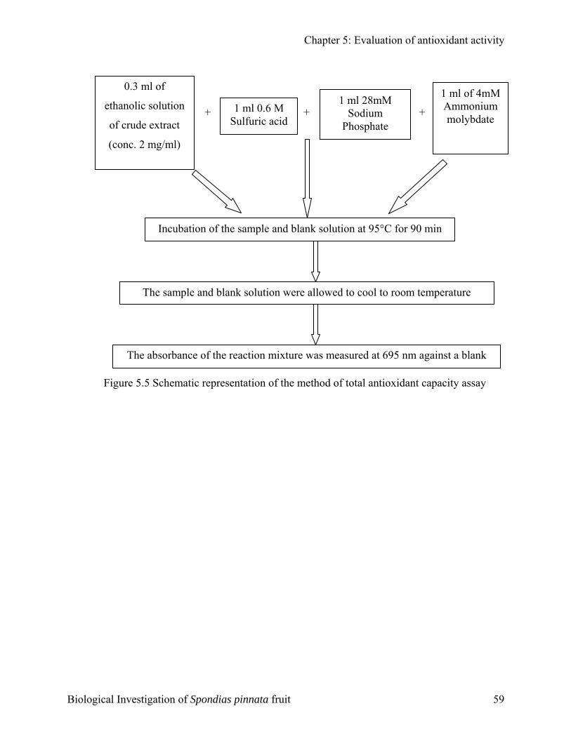
Chapter 5: Evaluation of antioxidant activity
+ + +
0.3 ml of
ethanolic solution
of crude extract
(conc. 2 mg/ml)
1 ml 0.6 M Sulfuric acid
1 ml 28mM Sodium
Phosphate
The a
Th rature
Incubation of the sample and blank solution at 95°C for 90 min
1 ml of 4mM Ammonium molybdate
Figure 5.5
Biological Investig
e sample and blank solution were allowed to cool to room tempe
bsorbance of the reaction mixture was measured at 695 nm against a blank
Schematic representation of the method of total antioxidant capacity assay
ation of Spondias pinnata fruit 59
EvTh
C H A P T E R
aluation of rombolytic Activity
Chapter 6: Evaluation of thrombolytic activity
6.1 Intr
Format
lodge in
blood f
localize
by thro
activato
linked f
thereby
1992).
Strepto
infarcti
patients
safer th
have si
al., 199
1. Larg
2. Limi
3. Sign
The pla
diversif
medicin
assayed
plants w
Biologi
CChhaapptteerr
66
EEvvaalluuaattiioonn ooff TThhrroommbboollyyttiicc AAccttiivviittyy
oduction
ion of blood clot (thrombus), one of the major causes of blood circulation problem can
a blood vessel and block the flow of blood in that location depriving tissues of normal
low and oxygen. This may result in damage, destruction (infarction), or even death of the
d tissues (necrosis) in that area (Vorvick LJ, 2010). Thrombus is formed from fibrinogen
mbin and is lysed by plasmin, which is activated from plasminogen by tissue plasminogen
r (tPA). All thrombolytic agents activate the enzyme plasminogen that clears the cross
ibrin mesh. Fribrinolytic drugs can dissolve thrombi in acutely occluded coronary arteries
can restore blood supply to ischaemic myocardium and can limit necrosis (Laurence DR,
kinase is an antigenic thrombolytic agent used for the treatment of acute myocardial
on. It reduces mortality as effectively as the nonantigenic altreplase in most infarct
. Tissue-type Plasminogen activator (tPA) is generally preferred as being effective and
an either urokinase or streptokinase type activators. All available thrombolytic agents still
gnificant shortcomings: (Nicolini FA et al., 1992; Adams DS et al., 1991; Lijnen HR et
1)
e doses are required to be maximally effective.
ted fibrin specificity.
ificant bleeding tendency.
nt kingdom represents an enormous reservoir of biologically active compounds with
ied structures having disease preventive properties. Nearly 50% of drugs used in
e are of plant origin, and only a small fraction of plants with medicinal activity has been
. Therefore much current research devoted to the phytochemical investigation of higher
hich have ethnobotanical information associated with them. The phytochemicals isolated
cal Investigation of Spondias pinnata fruit 60
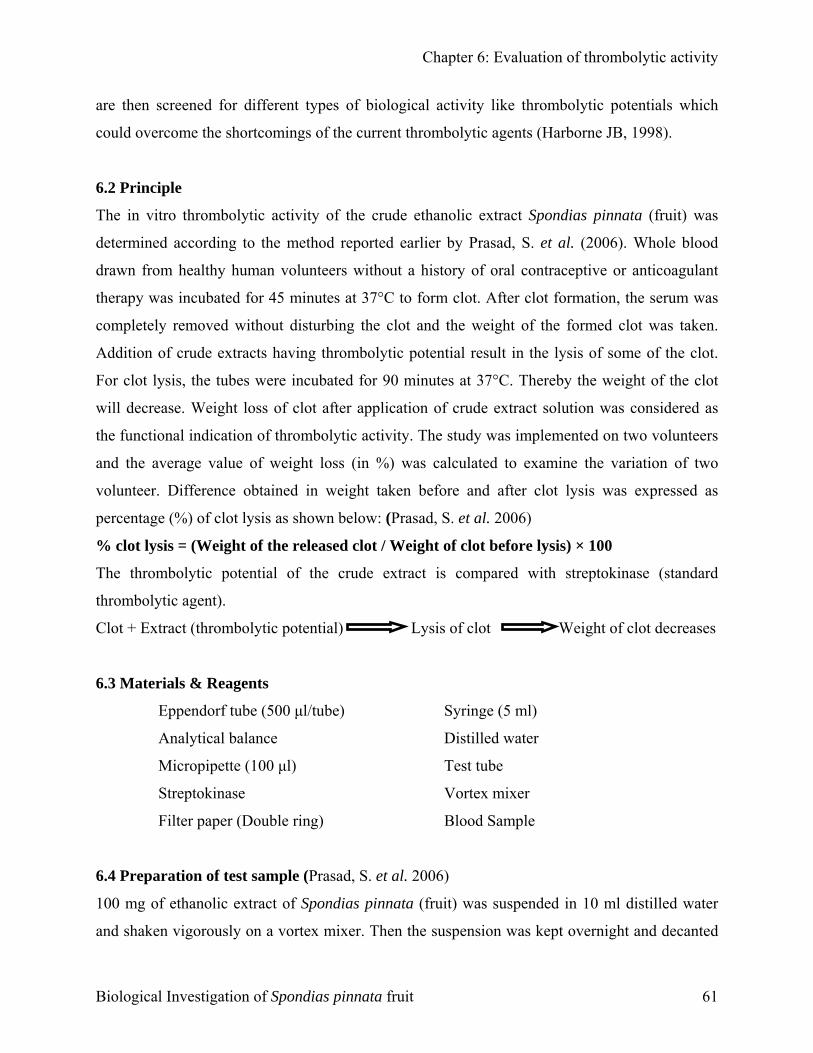
Chapter 6: Evaluation of thrombolytic activity
are then screened for different types of biological activity like thrombolytic potentials which
could overcome the shortcomings of the current thrombolytic agents (Harborne JB, 1998).
6.2 Principle
The in vitro thrombolytic activity of the crude ethanolic extract Spondias pinnata (fruit) was
determined according to the method reported earlier by Prasad, S. et al. (2006). Whole blood
drawn from healthy human volunteers without a history of oral contraceptive or anticoagulant
therapy was incubated for 45 minutes at 37°C to form clot. After clot formation, the serum was
completely removed without disturbing the clot and the weight of the formed clot was taken.
Addition of crude extracts having thrombolytic potential result in the lysis of some of the clot.
For clot lysis, the tubes were incubated for 90 minutes at 37°C. Thereby the weight of the clot
will decrease. Weight loss of clot after application of crude extract solution was considered as
the functional indication of thrombolytic activity. The study was implemented on two volunteers
and the average value of weight loss (in %) was calculated to examine the variation of two
volunteer. Difference obtained in weight taken before and after clot lysis was expressed as
percentage (%) of clot lysis as shown below: (Prasad, S. et al. 2006)
% clot lysis = (Weight of the released clot / Weight of clot before lysis) × 100
The thrombolytic potential of the crude extract is compared with streptokinase (standard
thrombolytic agent).
Clot + Extract (thrombolytic potential) Lysis of clot Weight of clot decreases
6.3 Materials & Reagents
Eppendorf tube (500 µl/tube) Syringe (5 ml)
Analytical balance Distilled water
Micropipette (100 µl) Test tube
Streptokinase Vortex mixer
Filter paper (Double ring) Blood Sample
6.4 Preparation of test sample (Prasad, S. et al. 2006)
100 mg of ethanolic extract of Spondias pinnata (fruit) was suspended in 10 ml distilled water
and shaken vigorously on a vortex mixer. Then the suspension was kept overnight and decanted
Biological Investigation of Spondias pinnata fruit 61
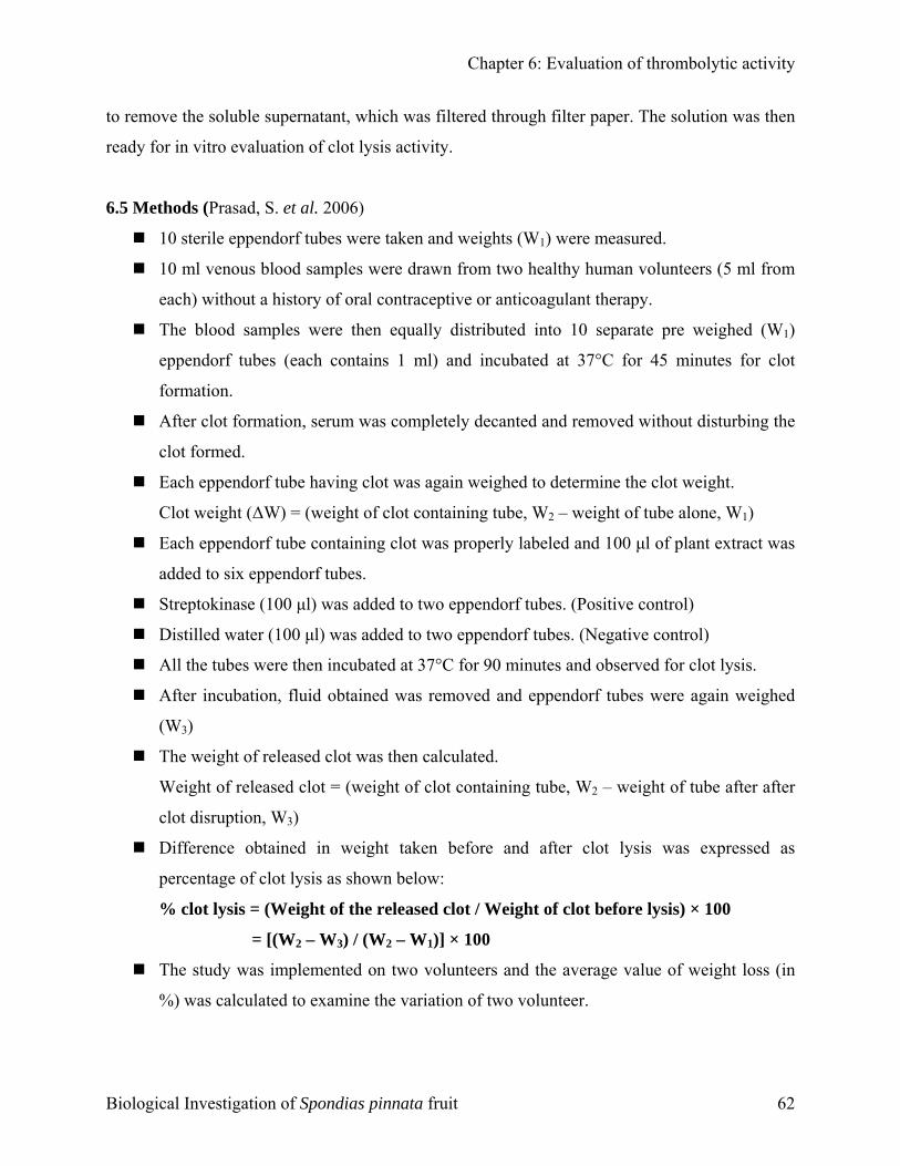
Chapter 6: Evaluation of thrombolytic activity
to remove the soluble supernatant, which was filtered through filter paper. The solution was then
ready for in vitro evaluation of clot lysis activity.
6.5 Methods (Prasad, S. et al. 2006)
10 sterile eppendorf tubes were taken and weights (W1) were measured.
10 ml venous blood samples were drawn from two healthy human volunteers (5 ml from
each) without a history of oral contraceptive or anticoagulant therapy.
The blood samples were then equally distributed into 10 separate pre weighed (W1)
eppendorf tubes (each contains 1 ml) and incubated at 37°C for 45 minutes for clot
formation.
After clot formation, serum was completely decanted and removed without disturbing the
clot formed.
Each eppendorf tube having clot was again weighed to determine the clot weight.
Clot weight (∆W) = (weight of clot containing tube, W2 – weight of tube alone, W1)
Each eppendorf tube containing clot was properly labeled and 100 µl of plant extract was
added to six eppendorf tubes.
Streptokinase (100 µl) was added to two eppendorf tubes. (Positive control)
Distilled water (100 µl) was added to two eppendorf tubes. (Negative control)
All the tubes were then incubated at 37°C for 90 minutes and observed for clot lysis.
After incubation, fluid obtained was removed and eppendorf tubes were again weighed
(W3)
The weight of released clot was then calculated.
Weight of released clot = (weight of clot containing tube, W2 – weight of tube after after
clot disruption, W3)
Difference obtained in weight taken before and after clot lysis was expressed as
percentage of clot lysis as shown below:
% clot lysis = (Weight of the released clot / Weight of clot before lysis) × 100
= [(W2 – W3) / (W2 – W1)] × 100
The study was implemented on two volunteers and the average value of weight loss (in
%) was calculated to examine the variation of two volunteer.
Biological Investigation of Spondias pinnata fruit 62
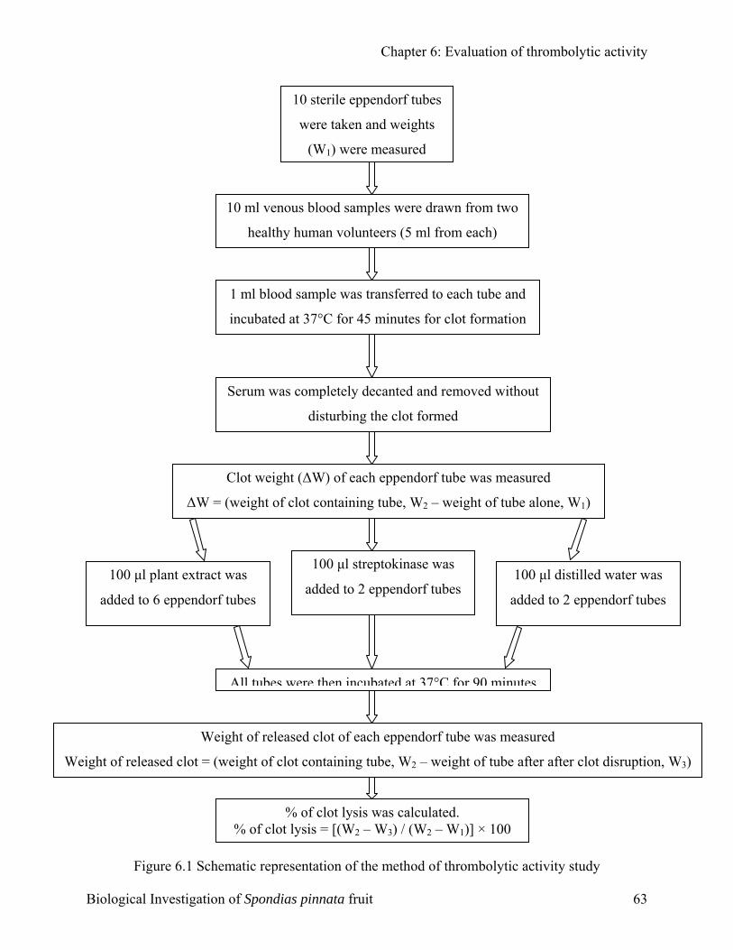
Chapter 6: Evaluation of thrombolytic activity
100 µl plant extract was
added to 6 eppendorf tubes
All tubes
% of clot
Weight of released clo
∆W =
Figure 6.1 Schematic repre
Biological Investigation of Spondi
10 sterile eppendorf tubes
were taken and weights
(W1) were measured
10 ml venous blood samples were drawn from twohealthy human volunteers (5 ml from each)
1 ml blood sample was transferred to each tube and
incubated at 37°C for 45 minutes for clot formation
100 µl streptokinase was
added to 2 eppendorf tubes 100 µl distilled water was
added to 2 eppendorf tubes
)
Serum was completely decanted and removed withoutdisturbing the clot formed
Clot weight (∆W) of each eppendorf tube was measured
(weight of clot containing tube, W2 – weight of tube alone, W1
were then incubated at 37°C for 90 minutes
Weight of released clot of each eppendorf tube was measured
t = (weight of clot containing tube, W2 – weight of tube after after clot disruption, W3)
% of clot lysis was calculated. lysis = [(W2 – W3) / (W2 – W1)] × 100
sentation of the method of thrombolytic activity study
as pinnata fruit 63
RReessuullttss aanndd DDiissccuussssiioonn
C H A P T E R
Chapter 7: Results & Discussion
7.1
The
Spo
test
1. I
2. M
3. I
4. I
Bes
met
7.1.
The
prod
7.1.
pinn
inhi
rang
Bio
CChhaapptteerr
77
RReessuullttss && DDiissccuussssiioonn
Results and discussion of the test samples of Spondias pinnata (fruit)
aim of this chapter is to illustrate the results and discussions of crude ethanolic extract of
ndias pinnata (fruit). This chapter will include the following results and discussions of the
samples of Spondias pinnata (fruit):
n vitro antimicrobial screening
inimum inhibitory concentration (MIC) and Minimum Bactericidal Concentration (MBC)
n vitro antioxidant activity
3.1. Total phenolic content
3.2. DPPH radical scavenging assay (Quantitative analysis) and IC50
3.3. Reducing power assay
3.4. Total antioxidant capacity by phosphomolybdenum method
3.5. Total flavonoids content
n vitro thrombolytic activity
ides the results of various tests are represented through suitable graphical representation
hods. All the data are expressed as mean ± standard deviation (n=3).
1 In vitro antimicrobial screening
antimicrobial activities of extracts were examined in the present study. The crude extract
uced strong activity against a number of the test organisms. The results are given in table
The zones of inhibitions produced by the crude ethanolic extract of the fruit of Spondias
ata were ranged from 0-12 mm at a concentration of 400µg/disc. While the zones of
bition produced by by the crude ethanolic extract of the fruit of Spondias pinnata were
ed from 0-16 mm at a concentration of 800µg/disc.
logical Investigation of Spondias pinnata fruit 64
Chapter 7: Results & Discussion
The ethanolic extract showed greatest activity against Sarcina lutea having the zone of inhibition
of 12 mm (400 µg/disc) and 16 mm (800 µg/disc). Besides the extract showed strong activity
against Staphylococcus aureus (15mm for 800µg/disc and 10 mm for 400µg/disc), Shigella
boydii (15 mm for 800µg/disc and 12 mm for 400µg/disc), Salmonella paratyphi (15 mm for
800µg/disc and 10 mm for 400µg/disc). Moderate activity was found against Salmonella typhi
(13 mm for 800µg/disc and 9 mm for 400µg/disc), Shigella dysentery (13 mm for 800µg/disc and
7 mm for 400µg/disc), Pseudomonas aeruginosa (13 mm for 800µg/disc and 7 mm for
400µg/disc), Candida albicans (13 mm for 800µg/disc and 7 mm for 400µg/disc), Bacillus
subtilis (12 mm for 800µg/disc and 7 mm for 400µg/disc), Escherichia coli (11 mm for
800µg/disc and 7 mm for 400µg/disc), Vibrio mimicus (10 mm for 800µg/disc and 6 mm for
400µg/disc), Bacillus cereus (9 mm for 800µg/disc and 5 mm for 400µg/disc). The crude
ethanolic extract of Spondias pinnata fruit showed no activity against Sacharomyces cerevacae.
Among the tested fungi, the extract showed greatest activity against Candida albicans (13 mm
for 800µg/disc and 7 mm for 400µg/disc). The crude extract failed to inhibit the growth of
Sacharomyces cerevacae having zone of inhibition of 0 mm for both 400µg/disc and 800µg/disc
respectively.
Among the tested Gram positive bacteria, the extract showed greatest activity against Sarcina
lutea having the zone of inhibition of 12 mm (400 µg/disc) and 16 mm (800 µg/disc). Besides,
the extract showed significant inhibitory activity against Staphylococcus aureus (15mm for
800µg/disc and 10 mm for 400µg/disc).
Among the tested Gram negative bacteria, the extract showed strong activity against Shigella
boydii (15 mm for 800µg/disc and 12 mm for 400µg/disc) and Salmonella paratyphi (15 mm for
800µg/disc and 10 mm for 400µg/disc). Besides the extract showed a similar significant extent of
inhibitory activity against Shigella dysentery and Pseudomonas aeruginosa having zone of
inhibition of 13 mm for 800µg/disc and 7 mm for 400µg/disc against both of them. The extract
showed a more potent inhibitory activity against Salmonella typhi (13 mm for 800µg/disc and 9
mm for 400µg/disc) than Shigella dysentery and Pseudomonas aeruginosa.
In brief, the ehanolic extract of Spondias pinnata fruit showed a potent inhibitory activity against
Sarcina lutea and Shigella boydii.
Biological Investigation of Spondias pinnata fruit 65
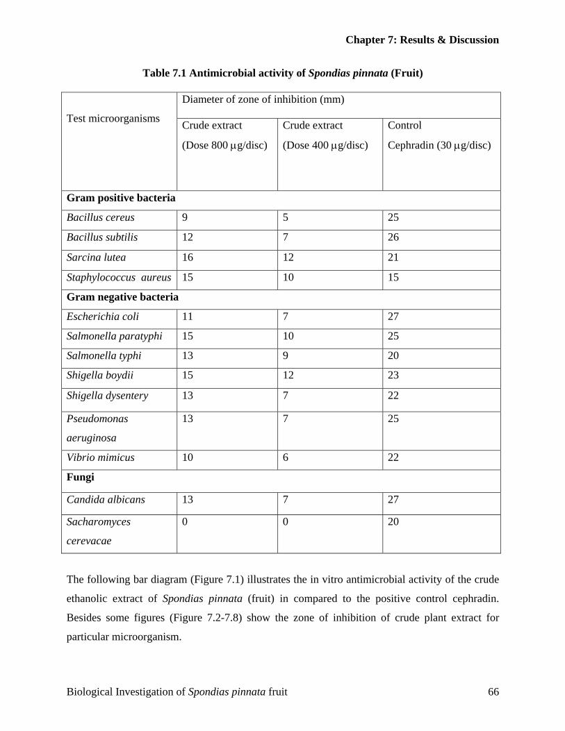
Chapter 7: Results & Discussion
Table 7.1 Antimicrobial activity of Spondias pinnata (Fruit)
Diameter of zone of inhibition (mm)
Test microorganisms Crude extract
(Dose 800 µg/disc)
Crude extract
(Dose 400 µg/disc)
Control
Cephradin (30 µg/disc)
Gram positive bacteria
Bacillus cereus 9 5 25
Bacillus subtilis 12 7 26
Sarcina lutea 16 12 21
Staphylococcus aureus 15 10 15
Gram negative bacteria
Escherichia coli 11 7 27
Salmonella paratyphi 15 10 25
Salmonella typhi 13 9 20
Shigella boydii 15 12 23
Shigella dysentery 13 7 22
Pseudomonas
aeruginosa
13 7 25
Vibrio mimicus 10 6 22
Fungi
Candida albicans 13 7 27
Sacharomyces
cerevacae
0 0 20
The following bar diagram (Figure 7.1) illustrates the in vitro antimicrobial activity of the crude
ethanolic extract of Spondias pinnata (fruit) in compared to the positive control cephradin.
Besides some figures (Figure 7.2-7.8) show the zone of inhibition of crude plant extract for
particular microorganism.
Biological Investigation of Spondias pinnata fruit 66
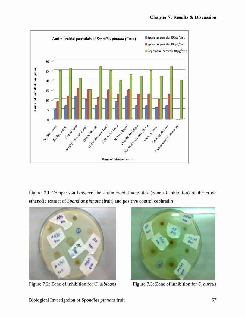
Chapter 7: Results & Discussion
0
5
10
15
20
25
30
Zon
e of
inhi
biti
on (
mm
)
Name of microorganism
Antimicrobial potentials of Spondias pinnata (Fruit) Spondias pinnata 400µg/disc
Spondias pinnata 800µg/disc
Cephradin (control) 30 µg/disc
Figure 7.1 Comparison between the antimicrobial activities (zone of inhibition) of the crude
ethanolic extract of Spondias pinnata (fruit) and positive control cephradin
Figure 7.2: Zone of inhibition for C. albicans Figure 7.3: Zone of inhibition for S. aureus
Biological Investigation of Spondias pinnata fruit 67

Chapter 7: Results & Discussion
Figure 7.4: Zone of inhibition for B. subtilis Figure 7.5: Zone of inhibition for S. boydii
Figure 7.6: Zone of inhibition for S. dysentery Figure 7.7: Zone of inhibition for S. lutea
Figure 7.8: Zone of inhibition for S. paratyphi
Biological Investigation of Spondias pinnata fruit 68
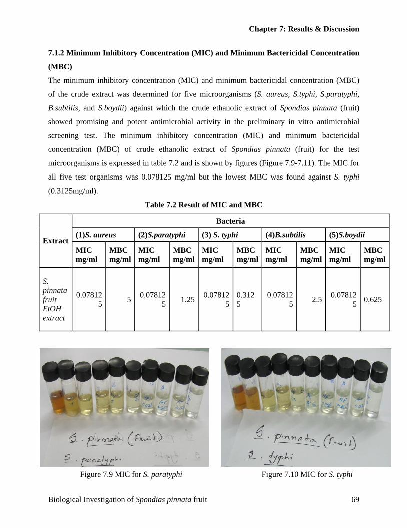
Chapter 7: Results & Discussion
7.1.2 Minimum Inhibitory Concentration (MIC) and Minimum Bactericidal Concentration
(MBC)
The minimum inhibitory concentration (MIC) and minimum bactericidal concentration (MBC)
of the crude extract was determined for five microorganisms (S. aureus, S.typhi, S.paratyphi,
B.subtilis, and S.boydii) against which the crude ethanolic extract of Spondias pinnata (fruit)
showed promising and potent antimicrobial activity in the preliminary in vitro antimicrobial
screening test. The minimum inhibitory concentration (MIC) and minimum bactericidal
concentration (MBC) of crude ethanolic extract of Spondias pinnata (fruit) for the test
microorganisms is expressed in table 7.2 and is shown by figures (Figure 7.9-7.11). The MIC for
all five test organisms was 0.078125 mg/ml but the lowest MBC was found against S. typhi
(0.3125mg/ml).
Table 7.2 Result of MIC and MBC
Bacteria
(1)S. aureus (2)S.paratyphi (3) S. typhi (4)B.subtilis (5)S.boydii Extract
MIC mg/ml
MBC mg/ml
MIC mg/ml
MBC mg/ml
MIC mg/ml
MBC mg/ml
MIC mg/ml
MBC mg/ml
MIC mg/ml
MBC mg/ml
S. pinnata fruit EtOH extract
0.078125 5 0.07812
5 1.25 0.078125
0.3125
0.078125 2.5 0.07812
5 0.625
Figure 7.9 MIC for S. paratyphi Figure 7.10 MIC for S. typhi
Biological Investigation of Spondias pinnata fruit 69
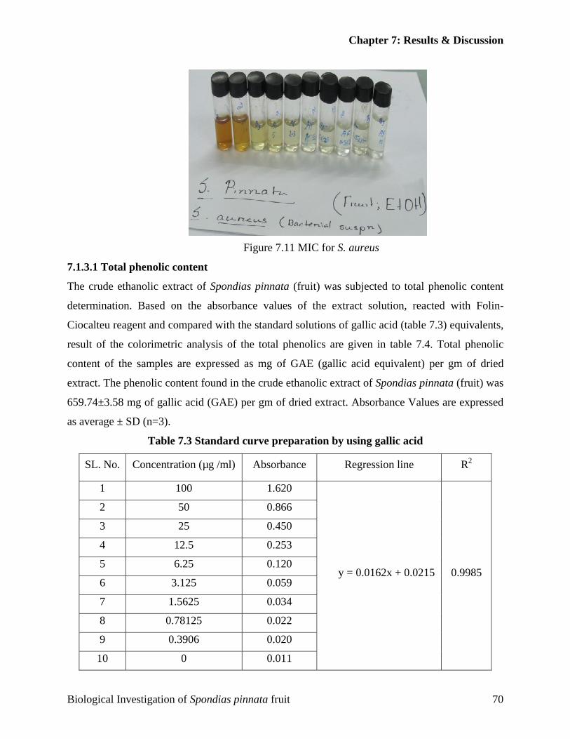
Chapter 7: Results & Discussion
Figure 7.11 MIC for S. aureus
7.1.3.1 Total phenolic content
The crude ethanolic extract of Spondias pinnata (fruit) was subjected to total phenolic content
determination. Based on the absorbance values of the extract solution, reacted with Folin-
Ciocalteu reagent and compared with the standard solutions of gallic acid (table 7.3) equivalents,
result of the colorimetric analysis of the total phenolics are given in table 7.4. Total phenolic
content of the samples are expressed as mg of GAE (gallic acid equivalent) per gm of dried
extract. The phenolic content found in the crude ethanolic extract of Spondias pinnata (fruit) was
659.74±3.58 mg of gallic acid (GAE) per gm of dried extract. Absorbance Values are expressed
as average ± SD (n=3).
Table 7.3 Standard curve preparation by using gallic acid
SL. No. Concentration (µg /ml) Absorbance Regression line R2
1 100 1.620
2 50 0.866
3 25 0.450
4 12.5 0.253
5 6.25 0.120
6 3.125 0.059
7 1.5625 0.034
8 0.78125 0.022
9 0.3906 0.020
10 0 0.011
y = 0.0162x + 0.0215 0.9985
Biological Investigation of Spondias pinnata fruit 70
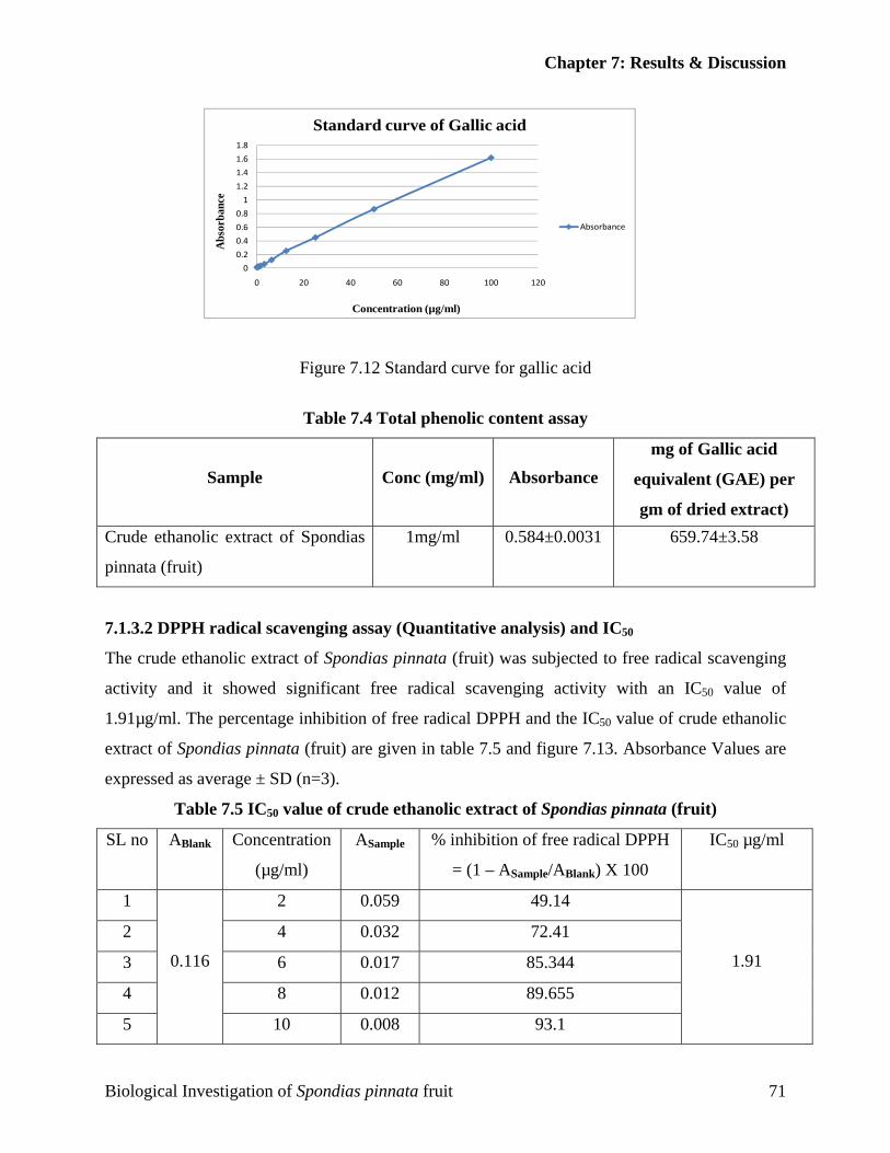
Chapter 7: Results & Discussion
0
0.2
0.4
0.6
0.8
1
1.2
1.4
1.6
1.8
0 20 40 60 80 100 120
Abs
orba
nce
Concentration (µg/ml)
Standard curve of Gallic acid
Absorbance
Figure 7.12 Standard curve for gallic acid
Table 7.4 Total phenolic content assay
Sample Conc (mg/ml) Absorbance mg of Gallic acid
equivalent (GAE) per
gm of dried extract) Crude ethanolic extract of Spondias
pinnata (fruit)
1mg/ml 0.584±0.0031 659.74±3.58
7.1.3.2 DPPH radical scavenging assay (Quantitative analysis) and IC50
The crude ethanolic extract of Spondias pinnata (fruit) was subjected to free radical scavenging
activity and it showed significant free radical scavenging activity with an IC50 value of
1.91µg/ml. The percentage inhibition of free radical DPPH and the IC50 value of crude ethanolic
extract of Spondias pinnata (fruit) are given in table 7.5 and figure 7.13. Absorbance Values are
expressed as average ± SD (n=3).
Table 7.5 IC50 value of crude ethanolic extract of Spondias pinnata (fruit)
SL no ABlank Concentration
(µg/ml)
ASample % inhibition of free radical DPPH
= (1 – ASample/ABlank) X 100
IC50 µg/ml
1 2 0.059 49.14
2 4 0.032 72.41
3 6 0.017 85.344
4 8 0.012 89.655
5
0.116
10 0.008 93.1
1.91
Biological Investigation of Spondias pinnata fruit 71
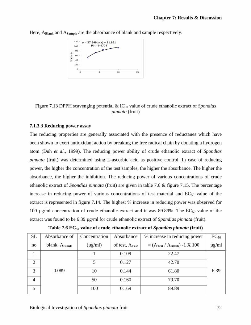
Chapter 7: Results & Discussion
Here, ABlank and ASample are the absorbance of blank and sample respectively.
y = 27.849ln(x) + 31.961R² = 0.9774
0
20
40
60
80
100
120
0 5 10 15
Figure 7.13 DPPH scavenging potential & IC50 value of crude ethanolic extract of Spondias
pinnata (fruit)
7.1.3.3 Reducing power assay
The reducing properties are generally associated with the presence of reductanes which have
been shown to exert antioxidant action by breaking the free radical chain by donating a hydrogen
atom (Duh et al., 1999). The reducing power ability of crude ethanolic extract of Spondias
pinnata (fruit) was determined using L-ascorbic acid as positive control. In case of reducing
power, the higher the concentration of the test samples, the higher the absorbance. The higher the
absorbance, the higher the inhibition. The reducing power of various concentrations of crude
ethanolic extract of Spondias pinnata (fruit) are given in table 7.6 & figure 7.15. The percentage
increase in reducing power of various concentrations of test material and EC50 value of the
extract is represented in figure 7.14. The highest % increase in reducing power was observed for
100 µg/ml concentration of crude ethanolic extract and it was 89.89%. The EC50 value of the
extract was found to be 6.39 µg/ml for crude ethanolic extract of Spondias pinnata (fruit).
Table 7.6 EC50 value of crude ethanolic extract of Spondias pinnata (fruit)
SL
no
Absorbance of
blank, ABlank
Concentration
(µg/ml)
Absorbance
of test, ATest
% increase in reducing power
= (ATest / ABlank) -1 X 100
EC50
µg/ml
1 1 0.109 22.47
2 5 0.127 42.70
3 10 0.144 61.80
4 50 0.160 79.70
5
0.089
100 0.169 89.89
6.39
Biological Investigation of Spondias pinnata fruit 72
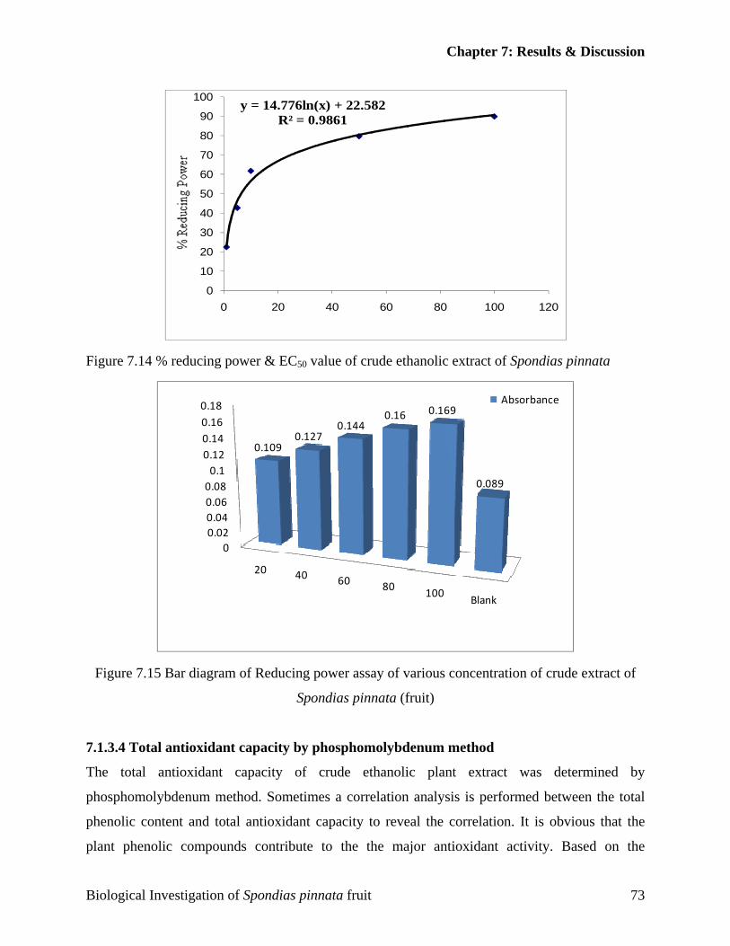
Chapter 7: Results & Discussion
y = 14.776ln(x) + 22.582R² = 0.9861
0
10
20
30
40
50
60
70
80
90
100
0 20 40 60 80 100 120
Figure 7.14 % reducing power & EC50 value of crude ethanolic extract of Spondias pinnata
00.020.040.060.080.1
0.120.140.160.18
20 40 60 80 100Blank
0.1090.127
0.1440.16 0.169
0.089
Absorbance
Figure 7.15 Bar diagram of Reducing power assay of various concentration of crude extract of
Spondias pinnata (fruit)
7.1.3.4 Total antioxidant capacity by phosphomolybdenum method
The total antioxidant capacity of crude ethanolic plant extract was determined by
phosphomolybdenum method. Sometimes a correlation analysis is performed between the total
phenolic content and total antioxidant capacity to reveal the correlation. It is obvious that the
plant phenolic compounds contribute to the the major antioxidant activity. Based on the
Biological Investigation of Spondias pinnata fruit 73
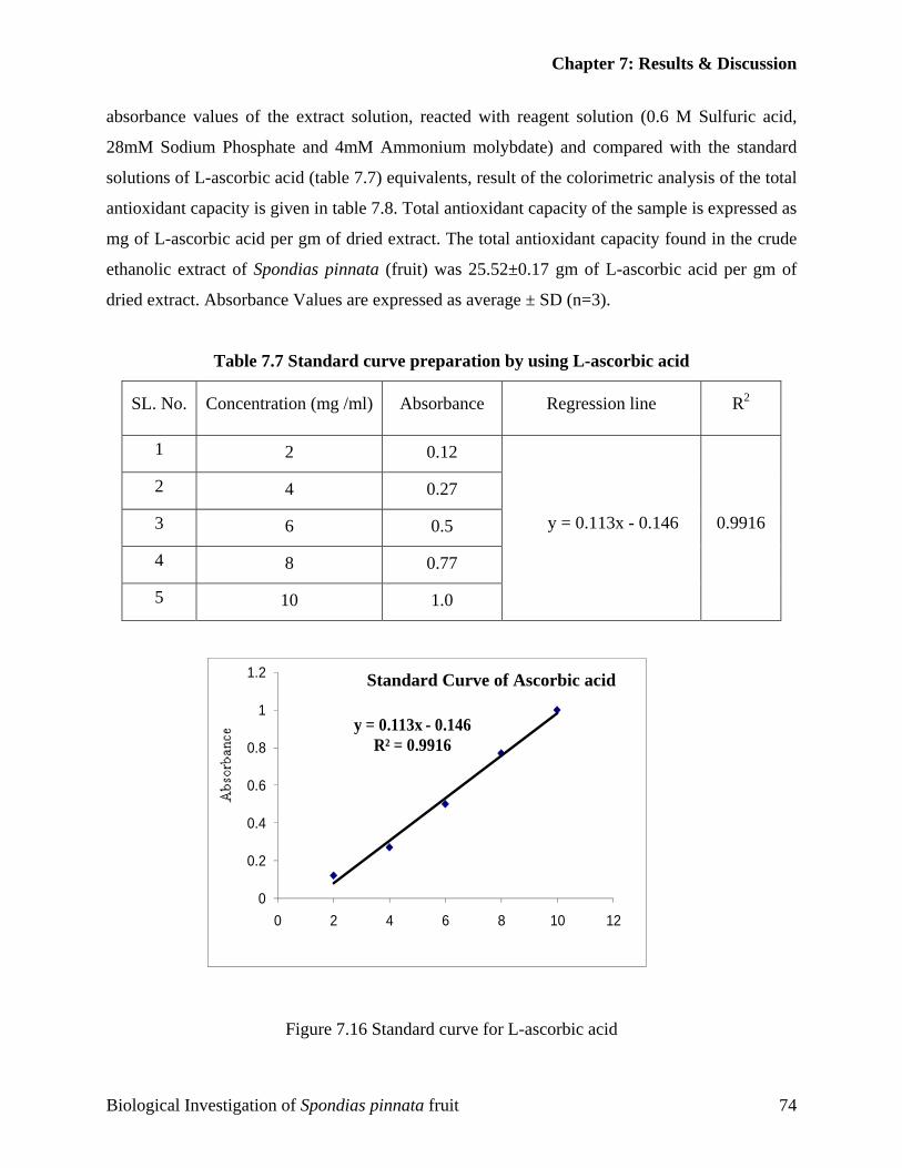
Chapter 7: Results & Discussion
absorbance values of the extract solution, reacted with reagent solution (0.6 M Sulfuric acid,
28mM Sodium Phosphate and 4mM Ammonium molybdate) and compared with the standard
solutions of L-ascorbic acid (table 7.7) equivalents, result of the colorimetric analysis of the total
antioxidant capacity is given in table 7.8. Total antioxidant capacity of the sample is expressed as
mg of L-ascorbic acid per gm of dried extract. The total antioxidant capacity found in the crude
ethanolic extract of Spondias pinnata (fruit) was 25.52±0.17 gm of L-ascorbic acid per gm of
dried extract. Absorbance Values are expressed as average ± SD (n=3).
Table 7.7 Standard curve preparation by using L-ascorbic acid
y = 0.113x - 0.146R² = 0.9916
0
0.2
0.4
0.6
0.8
1
1.2
0 2 4 6 8 10 12
SL. No. Concentration (mg /ml) Absorbance Regression line R2
1 2 0.12
2 4 0.27
3 6 0.5
4 8 0.77
5 10 1.0
y = 0.113x - 0.146 0.9916
Standard Curve of Ascorbic acid
Figure 7.16 Standard curve for L-ascorbic acid
Biological Investigation of Spondias pinnata fruit 74
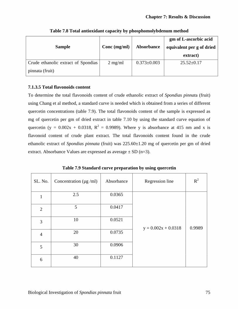
Chapter 7: Results & Discussion
Table 7.8 Total antioxidant capacity by phosphomolybdenum method
Sample Conc (mg/ml) Absorbance gm of L-ascorbic acid
equivalent per g of dried
extract) Crude ethanolic extract of Spondias
pinnata (fruit)
2 mg/ml 0.373±0.003 25.52±0.17
7.1.3.5 Total flavonoids content
To determine the total flavonoids content of crude ethanolic extract of Spondias pinnata (fruit)
using Chang et al method, a standard curve is needed which is obtained from a series of different
quercetin concentrations (table 7.9). The total flavonoids content of the sample is expressed as
mg of quercetin per gm of dried extract in table 7.10 by using the standard curve equation of
quercetin (y = 0.002x + 0.0318, R2 = 0.9989). Where y is absorbance at 415 nm and x is
flavonoid content of crude plant extract. The total flavonoids content found in the crude
ethanolic extract of Spondias pinnata (fruit) was 225.60±1.20 mg of quercetin per gm of dried
extract. Absorbance Values are expressed as average ± SD (n=3).
Table 7.9 Standard curve preparation by using quercetin
SL. No. Concentration (µg /ml) Absorbance Regression line R2
1 2.5 0.0365
2 5 0.0417
3 10 0.0521
4 20 0.0735
5 30 0.0906
6 40 0.1127
y = 0.002x + 0.0318 0.9989
Biological Investigation of Spondias pinnata fruit 75
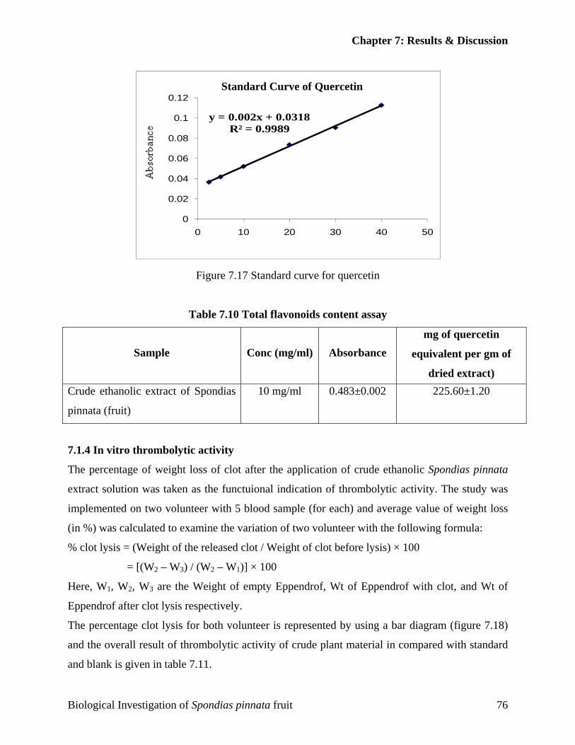
Chapter 7: Results & Discussion
y = 0.002x + 0.0318R² = 0.9989
0
0.02
0.04
0.06
0.08
0.1
0.12
0 10 20 30 40 50
Standard Curve of Quercetin
Figure 7.17 Standard curve for quercetin
Table 7.10 Total flavonoids content assay
Sample Conc (mg/ml) Absorbance mg of quercetin
equivalent per gm of
dried extract) Crude ethanolic extract of Spondias
pinnata (fruit)
10 mg/ml 0.483±0.002 225.60±1.20
7.1.4 In vitro thrombolytic activity
The percentage of weight loss of clot after the application of crude ethanolic Spondias pinnata
extract solution was taken as the functuional indication of thrombolytic activity. The study was
implemented on two volunteer with 5 blood sample (for each) and average value of weight loss
(in %) was calculated to examine the variation of two volunteer with the following formula:
% clot lysis = (Weight of the released clot / Weight of clot before lysis) × 100
= [(W2 – W3) / (W2 – W1)] × 100
Here, W1, W2, W3 are the Weight of empty Eppendrof, Wt of Eppendrof with clot, and Wt of
Eppendrof after clot lysis respectively.
The percentage clot lysis for both volunteer is represented by using a bar diagram (figure 7.18)
and the overall result of thrombolytic activity of crude plant material in compared with standard
and blank is given in table 7.11.
Biological Investigation of Spondias pinnata fruit 76
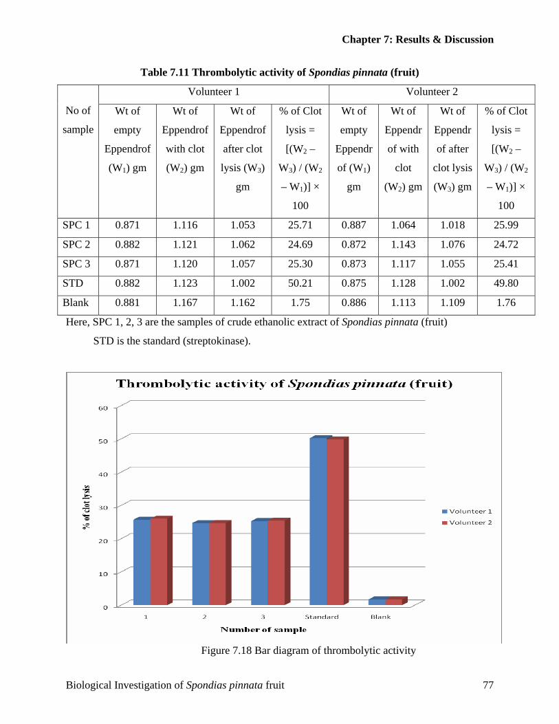
Chapter 7: Results & Discussion
Table 7.11 Thrombolytic activity of Spondias pinnata (fruit)
Volunteer 1 Volunteer 2
No of
sample
Wt of
empty
Eppendrof
(W1) gm
Wt of
Eppendrof
with clot
(W2) gm
Wt of
Eppendrof
after clot
lysis (W3)
gm
% of Clot
lysis =
[(W2 –
W3) / (W2
– W1)] ×
100
Wt of
empty
Eppendr
of (W1)
gm
Wt of
Eppendr
of with
clot
(W2) gm
Wt of
Eppendr
of after
clot lysis
(W3) gm
% of Clot
lysis =
[(W2 –
W3) / (W2
– W1)] ×
100
SPC 1 0.871 1.116 1.053 25.71 0.887 1.064 1.018 25.99
SPC 2 0.882 1.121 1.062 24.69 0.872 1.143 1.076 24.72
SPC 3 0.871 1.120 1.057 25.30 0.873 1.117 1.055 25.41
STD 0.882 1.123 1.002 50.21 0.875 1.128 1.002 49.80
Blank 0.881 1.167 1.162 1.75 0.886 1.113 1.109 1.76
Here, SPC 1, 2, 3 are the samples of crude ethanolic extract of Spondias pinnata (fruit)
STD is the standard (streptokinase).
Figure 7.18 Bar diagram of thrombolytic activity
Biological Investigation of Spondias pinnata fruit 77
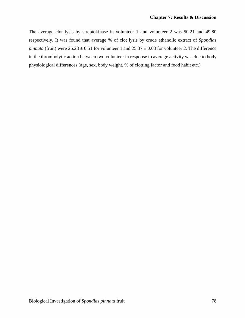
Chapter 7: Results & Discussion
The average clot lysis by streptokinase in volunteer 1 and volunteer 2 was 50.21 and 49.80
respectively. It was found that average % of clot lysis by crude ethanolic extract of Spondias
pinnata (fruit) were 25.23 ± 0.51 for volunteer 1 and 25.37 ± 0.03 for volunteer 2. The difference
in the thrombolytic action between two volunteer in response to average activity was due to body
physiological differences (age, sex, body weight, % of clotting factor and food habit etc.)
Biological Investigation of Spondias pinnata fruit 78

CCoonncclluussiioonn

Conclusion
CCoonncclluussiioonn
The crude ethanolic extract of Spondias pinnata showed significant antioxidant,
antimicrobial and thrombolytic activities, some of which supports the traditional use of
this plant in various diseases.
The plant can be further screened against various diseases in order to find out its
unexplored efficacy and can be a potential source of chemically interesting and
biologically important drug candidates. Very few compounds are isolated from the S.
pinnata. Therefore there is huge potential to find active principles which could be
beneficial for mankind for targeting various diseases.
Biological Investigation of Spondias pinnata 79

RReeffeerreenncceess
References
RReeffeerreenncceess
Adams, D.S, Griffin, L.A, Nachajko, W.R, Reddy, V.B and Wei, C.M. 1991. A synthetic
DNA encoding a modified human urokinase resistant to inhibition by serum plasminogen
activator inhibitor. Journal of Biological Chemistry, 266:8476–8482.
Anisuzzaman, M., Rahman, A. H., Rashid, M., Naderuzzaman, A.T.M., and Islam, A.K.M.
2007. An Ethnobotanical Study of Madhupur, Tangail. Journal of Applied Sciences
Research, 3(7): 519-530.
Aruoma, O.I., 1998. Free radicals, oxidative stress, and antioxidants in human health and
disease. Journal of the American Oil Chemists' Society, 75:199-212.
Ayafor, J.F. et al., 1972. Tetrahedron, 28:9343.
Ayensu, E.S, and DeFilipps, R.A., 1978. Endangered and Threatened Plants of the United
States. Smithsonian Institution Press and World Wildlife Fund, Washington, DC.
Ayuvedaherbs., 2005. Importance of Medicinal plants. The article was retrieved from"
http://ayuvedaherbs.wordpress.com/2005/12/"
Badoni, A. and Bisht, C. 2009. Importance and Problems in Natural Regeneration of
Spondias pinnata. Report and opinion, 1(5):12-13.
Bandoniene, D., Pukalskas, A., Venskutonis, P.R., and Gruzdiene, D., 2000. Preliminary
screnning of antioxidant activity of some plants extracts in repressed oil. Food Research
International, 33:786-791
Barry, A.L., 1976 .Principle & practice of Microbiology. 3rd Ed., Lea & Fabager,
Philadelphia.
Biological Investigation of Spondias pinnata 80
References
Basta, G, Lupi, C, Lazzerini, G, Chiarelli, P, L'Abbate, A and Rovai, D, 2004. Therapeutic
effect of diagnostic ultrasound on enzymatic thrombolysis. An in vitro study on blood of
normal subjects and patients with coronary artery disease. Journal of Thrombosis and
Haemostasis, 91:1078-1083.
Bauer, A.W., Kirby, W.M.M., Sherris, J.C. and Turck, M., 1966. American Journal of
Clinical Pathology, 45(4):493-496.
Berhanu M.A., Ngadjui, B.T., Bezabih, M. and Mdee, L.K. 1999. Novel natural products
from marketed plants of eastern and southern Africa. Pure Applied Chemistry, 71(6):919-
926.
Bernard J, 1789. Genera plantarum :secundum ordines naturales disposita, juxta methodum
in Horto regio parisiensi exaratam, anno M.DCC.LXXIV.
Bray, H.G., and Thorpe, W.V. 1954. Analysis of phenolic compounds of interest in
metabolism. Methods Biochemical analysis, 1:27-52.
Britton, N.L and Brown, A, 1897. An Illustrated Flora of the Northern United States. Atlantic
Ocean Westward, Volume 2.
Brown, R., Tuckey, Kingston, J., and Smith, C., 1818. Observations, Systematical and
Geographical, on Professor Christian Smith's Collection of Plants from the Vicinity of the
River Congo.
Candolle, A.P., Alphonse, C., 1825. Prodromus systematis naturalis regni vegetabilis: sive
enumeratio contracta ordinum generum specierumque plantarum huc usque cognitarum,
juxta methodi naturalis normas digesta.
Chang, C, Yang , M, Wen, H and Chern, J. 2002. Estimation of total flavonoid content in
propolis by two complementary colorimetric methods. Journal of Food and Drug Analaysis,
10:178-182.
Biological Investigation of Spondias pinnata 81
References
Chetia, B., and Gogoi, S. 2011. Antibacterial activity of the methanolic extract of stem bark
of Spondias pinnata, Moringa oleifera and Alstonia scholaris. Asian Journal of Traditional
Medicines, 6(4).
Chin, Y-W., Balunas, M.J., Chai, H.B. and Kinghorn, A.D. 2006. Drug Discovery From
Natural Sources. AAPS Journal; 8(2): E239-E253
Choi, H.Y., Jhun, E.J., Lim, B.O., and Chung M., Kyung, S.H., Park , D.K., 2000.
Application of flow injection-chemilumineacence to the study of radical scavenging activity
in plants. Phytotherapy Research, 14:250-253.
Chowdhury, J.U., Alam, M.K. and Hasan, M.A. 2010. Some traditional folk formularies
against diversity and diarrhoea in Bangladesh. Journal of economic and taxonomic botany,
Additional Series 12: 20-23.
Couladis, M., Tzakou, O., Verykokidou, E., and Harvala, C., 2003. Screening of some Greek
aromatic plants for antioxidant activity. Phytotherapy Research, 17: 194-195.
Cronquist, A., 1981.An Integrated System of Classification of Flowering Plants. Brottonia,
34 (2): 268-270.
Cronquist, A., 1988. The Evolution and Classification of Flowering Plants. Bronx, NY:New
York Botanical Garden.
Das, J., Mannan, A., Rahman, M.M., Dinar, M.A.N., Uddin, M.E., Khan, I.N., Habib, M.R.,
and Hasan, N. 2011. Chloroform and Ethanol Extract of Spondias Pinnata and its Different
Pharmacological activity Like- Antioxidant, Cytotoxic, Antibacterial Potential and
Phytochemical Screening through In-Vitro Method. International Journal of Research in
Pharmaceutical and Biomedical Sciences, 2(4): 1805-1812.
Desmarchelier, C., Repetto, M., Coussio, J., Liesuy, S., and Ciccia, G, 1997. Antioxidant and
prooxidant activities in aqueous extracts of Argentine plants. International Journal of
Pharmacognosy and phytochemical research, 35:116-120.
Biological Investigation of Spondias pinnata 82
References
Encyclopaedia britannica, 2012. Anacardiaceae. The article was retrieved from"
http://www.britannica.com/EBchecked/topic/22229/Anacardiaceae"
Ethnobotanical database of Bangladesh, 2010. The article was retrieved from"
http://www.ethnobotanybd.com/index.php?action=Taxonomy&key=sci#"
Fabricant, D.S and Farnsworth, N.R. 2001. The Value of Plants Used in Traditional Medicine
for Drug Discovery. Environmental Health Perspectives, 109, supplement 1.
Faiz, M., 2011. Amra medicinal uses, nutrition and food value. The article was retrieved
from" http://theindependentbd.com/stethoscope/77749-amra-medicinal-uses-nutrition-and-
food-value.html"
Fauci, A., 1998. New and reemerging diseases: The importance of biomedical research.
Emerging infectious diseases. The article was retrieved from
www.cdc.gov/ncidod/EID/vol4no3/fauci
Gardner, S., Sidisunthorn P. & Anusarnsunthorn, V. 2000. A field guide to Forest Trees of
Northern Thailand. Kobfai Publishing Project. Thailand.
Ghani, A., 2003. Medicinal Plants of Bangladesh, The Asiatic Society of Bangladesh, 2nd
Revised Edition., Dhaka, Bangladesh
Ghani ,A., and Pasha, M.K, 2006. Traditional medicine. The article was retrieved from"
http://www.banglapedia.org/httpdocs/HT/T_0207.HTM"
Government of India, 2001. Ayurvedic pharmacopoeia of India. Part 1, Volume 2, pp 5.
Gupta, A., Srivastava, S., Prasad, R., Natu, S.M., Mittal, B., Negi, M.P., and Srivastava, A.N.
2010. Oxidative stress in non-small cell lung cancer patients after chemotherapy: Association
with treatment response. Respirology, 15:349-356.
Biological Investigation of Spondias pinnata 83
References
Gutteridge, J.M.C, 1994. Biological origin of free radicals, and mechanisms of antioxidant
protection. Chemico-Biological Interactions, 91:133-140.
Halliwell, B, Gutteridge, J.M.C. and Cross, C.E. 1992. Free radicals, antiodidants and human
disease: where are we now?. Journal of Laboratory and Clinical Medicine, 119:598-620.
Harborne, J.B., 1998. Phytochemical methods: A guide to modern techniques of plant
analysis. 3rd Edn., Chapman and Hall Int. Ed., NY.
Harvey, A.L, 2008. Natural products in drug discovery. Elsevier, 13(19/20):894-901.
Hazra, B., Biswas, S., and Mandal, N. 2008. Antioxidant and free radical scavenging activity
of Spondias pinnata. BMC Complementary and Alternative Medicine, 8(63).
Hemalatha, S., Lalitha, P., and Arulpriya, P. 2010. Antioxidant activities of the extracts of
the aerial roots of Pothos aurea (Linden ex Andre). Der Pharma Chemica, 2(6):84-89.
Hickman, J.C, 1993. Anacardiaceae the Jepson Manual: Higher Plants of California.
University of California Press, Berkeley. pp 134-137.
Hostettmann, K and Terreaux, C. 2000. Search for New Lead Compounds from Higher
Plants. CHIMIA International Journal for Chemistry, 54: 652-657.
Hoque, M.M., Hassan, M.A. and Khan, M.R. 1990. Studies in the antibacterial activity of
plants available in Bangladesh. Ploigonum L. J. Asiatic society of Bangladesh (Sc), 12(1):77-
82.
Huys, G. 2002. Antibiotic susceptibility testing of aquaculture-associated bacteria with the
broth macrodilution method. Ledeganckstr: Universiteit Gent.
Iacomini, M, Serrato, R.V, Sassaki, G.L, Lopes, L, Buchi, and Gorin, P.A.J. 2005. Isolation
and partial characterization of a pectic polysaccharide from the fruit pulp of Spondias
pinnata and its effect on peritoneal macrophage activation. Fitoterapia, 76, 676-683.
Biological Investigation of Spondias pinnata 84
References
Inatani, R., Nakatani, N. & Fuwa, H., 1983. Antioxidative effects of the constituents of
rosemary (Rosmarinus officinalis) and their derivatives. Agricultural and Biological
Chemistry, 47:521-528.
Ito, N., Fukushina, S., and Tsuda, H. 1985. Carcinogenicity and modification of the
carcinogenic response by BHA, BHT and other antioxidants. Critical Reviews in Toxicology,
15:109–115.
Jadhav, S.J, Nimbalkar, S.S, Kulkarni, A.D, and Madhavi, D.L, 1996. Food Antioxidants:
Technological, Toxicological and Health Perspectives, NY, pp. 5–64.
Jayaprakasha, G.K., Jaganmohan, R.L., 2000. Phenolic constituents from lichen
Parmontrema stuppeum. Hale and antioxidant activity, 56:1018-1022.
Judd, W.S., Campbell, C.S., Kellog, E.A., Stevens, P.F. and Donoghue, M.J. 2007.
Taxonomy. Plant Systematics - A Phylogenetic Approach, 3rd edition. Sinauer Associates,
Sunderland.
Kandali, R, Konwar, B.K., 2011. Isolation and Characterization of Active Compound from
Fruits of Medicinal Plant Spondias pinnata Kurz. Indian Journal of Agricultural
Biochemistry, 24(1): 29-33.
Kirk, P.M., Cannon, P.F., Minter, D.W. and Stalpers, J.A, 2008. Taxonomy. Dictionary of
the Fungi, 10th edition. Netherlands.
Kokate, C.K., 2001. Pharmacohnosy. 16th Edn., Nirali Prakasham, India.
Kubola, J., and Siriamornpun, S. 2011. Phytochemicals, vitamin C and sugar content of Thai
wild fruits. Food Chemistry, 126(3): 972-981.
Lai, S.C., Ho, Y.L., Huang, S.C., Huang, T.H., Lai, Z.R., Wu, C.R., Lian, K.Y., and Chang,
Y.S, 2010. Antioxidant and antiproliferative activities of Desmodium triflorum (L.) DC.
American Journal of Chinese Medicine, 38: 329-333.
Biological Investigation of Spondias pinnata 85
References
Laurence, D.R., Bennett, P.N, 1992. Clinical Pharmacology: 7th Edition. Page 483
Liawruangrath, B., Pholsongkram, K, Kongtaweelert, S, and Keawsaard, S., 2009. Chemical
constituents of essential oil from the pulp of Spondias pinnata Kurz. 35th Congress on
Science and Technology of Thailand.
Lijnen, H.R, Vanhoef, B, DeCock, F, Okada, K, Ueshima, S, and Matsuo, O. 1991. On the
mechanism of fibrin-specific plasminogen activation by staphylokinase. Journal of
Biological Chemistry, 266:11826–11832.
Lindley, F.R.S., Torrey, M.D., 1831. An Introduction to the Natural System of Botany: or A
Systematic View of the Organization, Natural Affinities, and Geographical Distribution of
the Whole Vegetable Kingdom.
Longman, O., 2005. Indian medicinal plant (Vol 1). pp 309.
Maisuthisakul, P., Suttajit, M., Pongsawatmanit, R. 2007. Assessment of phenolic content
and free radical-scavenging capacity of some Thai indigenous plants. Food Chemistry,
100(4): 1409-1418.
Miller, H.E., Rigelhof, F., Marquart, L., Prakash, A., and Kanter, M., 2000. Cereal Foods
World, 45(2): 59-63.
Mollik, A.H., Hossan, M.S., Paul, A.K., Rahman, M.T., Jahan, R., and Rahmatullah, M.,
2010. A Comparative Analysis of Medicinal Plants Used by Folk Medicinal Healers in Three
Districts of Bangladesh and Inquiry as to Mode of Selection of Medicinal Plants.
Ethnobotany Research & Applications, 8:195-218.
Mondal, S., Dash G.K., 2009. Hypoglycemic activity of the bark of Spondias pinnata Linn.
kurz. Pharmacognosy magazine, 5(19): 42-45.
Mondal, S., Dash G.K., Acharyya, S., Brahma, D.K., and Bal, S. 2009. Studies on diuretic
and laxative activity of bark extracts of Spondias pinnata (Linn. f) Kurz. Pharmacognosy
magazine, 5(19): 28-31.
Biological Investigation of Spondias pinnata 86

References
Morton, J. 1987. Fruits of warm climates. Ambarella. pp. 240–242.
Muhammad, A., Muhammad, M.S., Kabir, A.N.M., Kabir, S., and Muhammad, M.K. 2011.
Antibacterial and cytotoxic activities of Spondias pinnata (Linn. f.) Kurz fruit extract. Indian
Journal of Natural Products and Resources, 2(2): 265-267.
Naik, S.R., Panda, V.S. 2008. Hepatoprotective effect of Ginkgoselect Phytosome in
rifampicin induced liver injury in rats. Evidence of antioxidant Activity. Fitoterapia,
79(6):439-445.
Nicolini, F.A, Nichols, W.W, Mehta, J.L, Saldeen, T.G, Schofield R, Ross M, Player, D.W,
Pohl G.B, and Mattsson C. 1992. Sustained reflow in dogs with coronary thrombosis with
K2P, a novel mutant of tissue plasminogen activator. Journal of the American College of
Cardiology, 20:228–235.
Panda, B.K., Patra, V.J., Mishra, U.S., Kar, S., Panda, B.R., Hati, M.R. 2009. Analgesic
activities of the stem bark extract of Spondias pinata (Linn.f) Kurz. Journal of Pharmacy
Research, 2(5): 825-827.
Panda, S.K., Patra, N., Dutta, S.K., Bastia, A.K., and Sahoo, G. 2012. Anti-diarrheal
activities of medicinal plants of Similipal Biosphere Re-serve, Odisha, India. International
Journal of Medicinal and Aromatic Plants, 2(1):123-134
Pavel, P., Enayet, A.B.M., 2007. Ethnobotanical Investigation into the Mandi Ethnic
Community in Bangladesh. Bangladesh Journal of Plant Taxonomy, 14(2): 129-145.
Pell, S.K. 2009. Anacardiaceae: Cashew family. The article was retrieved
fromhttp://tolweb.org/Anacardiaceae/21262
Pell, S.K., Mitchell, J.D., Miller, A.J., and Lobova, T.A., 2011. The families & Genera of
vascular plants (Volume 10).Chapter 3: Anacardiaceae. Springer- Heidelberg Dordrecht
NY. pp 7-9, 12, 13.
Pieroni, A., Janiak, V., Durr, C.M., Ludeke, S., Trachsel, E., and Heincrich, M., 2002. In
Biological Investigation of Spondias pinnata 87
References
vitro antioxidant activity of non-cultivated vegetables of ethnic Albanians in Southern Italy.
Phytotherapy Research, 16:467-473.
Pietta, A., Sionetti, P., and Mauri, P., 1998. Antioxidant activity of selected medicinal plants.
Journal of Agricultural and Food Chemistry, 46:4487-4490.
Pinner, R., Teutsch, S., Simonsen, L., Klug, L., Graber, J., Clarke, M. and Berkelman, R.,
1996. Trends in infectious diseases mortality in the United States. Journal of the American
Medical Association, 275:189–193.
Prasad, S., Daginawala, H.F., Kashyap, R.S., Deopujari, J.Y., Purohit, H.J. and Taori, G.M.,
2006. Development of an in vitro model to study clot lysis activity of thrombolytic
drugs. Thrombosis Journal, 4:14.
Prieto, P, Pineda, M, and Aguilar, M. 1999. Spectrophotometric quantitation of antioxidant
capacity through the formation of a Phosphomolybdenum Complex: Specific application to
the determination of vitamin E. Analytical Biochemistry, 269:337-341.
Purohit, V.K, Andola, H.C, 2010. Nutritive and mineral value of ripe fruits Evaluation of
nutritive and mineral value in ripe fruits of Spondias pinnata from two location of Western
Himalaya, India. International Journal of Phytomedicines and Related Industries, 2(3): 233-
236.
Rahmatullah, M., Mollik, A.H., Hossan, M.S., Paul, A.K., Rahman, M.T., and Jahan, R.,
2010. A Comparative Analysis of Medicinal Plants Used by Folk Medicinal Healers in
Villages Adjoining the Ghaghot, Bangali and Padma Rivers of Bangladesh. American-
Eurasian Journal of Sustainable Agriculture, 4(1): 70-85
Rao, B.G, Jayaraju, N., 2009. Anthelmintic activities of Glycosmis pentaphylla and Spondias
pinnata. IPRHC,1(1): 91-96.
Rao, B.G., Raju, N.J. 2010. Investigation of hepatoprotective activity of Spondias pinnata.
International Journal of Pharma Sciences and Research, 1(3): 193-198.
Biological Investigation of Spondias pinnata 88
References
Rastogi, R.P, Mehrotra, B.N, 1993. Compendium of Indian medicinal plants. pp. 643.
Rastogi, R.P, Tandon, S., 1976. Studies on the chemical constituents of Spondias pinnata.
Planta medica, 29: 190-192.
Ronald, R, 1982. Antbiotics-An Introduction, F. Hoffman La Roche & Co. Basle,
Switzerland: 70-71.
Sadhu, S.K., E. Okuyama, H, and Ishibashi, M., 2003. Separation of Leucas aspera, a
medicinal plant of Bangladesh, guided by prostaglandin inhibitory and antioxidant activities.
Chemical & Pharmaceutical Bulletin, 51:595-598.
Samee, W., Engkalohakul, M., Nebbua, N., Direkrojanavuti, P., Sornchaithawatwong, C.,
and Kamkaen, N. 2006. Correlation Analysis between Total Acid, Total Phenolic and
Ascorbic Acid Contents in Fruit Extracts and Their Antioxidant Activities. Thai
Pharmaceutical and Health Science Journal, 1(3):196-203.
Samy RP et al., 2008. A compilation of Bioactive Compounds from Ayurveda.
Bioinformation, 3(3):100–110.
Sashidhara, K.V, 2005. Natural products as drugs. The article was retrieved from"
http://www.vigyanprasar.gov.in"
Setzer, W.N, 1999. Natural Products Drug Discovery. The article was retrieved from"
http://chemistry.uah.edu/faculty/setzer/natprod.html"
Shahidi, F., Janitha, P.K., Wanasundara, P.D., 1992. Phenolic antioxidants. Critical Reviews
in Food Science and Nutrition, 32:67-103.
Silprasit, K., Seetaha, S., Pongsanarakul, P., Hannongbua, S. and Choowongkomon, K. 2011.
Anti-HIV-1 reverse transcriptase activities of hexane extracts from some Asian medicinal
plants. Journal of Medicinal Plants Research, 5(17), pp. 4194-4201.
Simpson, M.G., 2010. Chapter 1 Plant Systematics: an Overview. Plant Systematics, 2nd ed.
Biological Investigation of Spondias pinnata 89
References
Academic Press.
Singleton, V.L, Rudolf, O, Rosa M., 1999. Analysis of total phenols and other oxidation
substrates and antioxidants by means of folin-ciocalteu reagent. Methods in Enzymology,
299:152-178.
Sofowara, A., 1993. Medicinal plants and traditional medicine in Africa. Spectrum Books
Ltd, Ibadan, Nigeria. p. 289.
Solecki, R., Shanidar, I.V. 1975. A Neanderthal flower burial in northern Iraq. Science,
190:880–881.
Solereder H, 1908. Systematic Anatomy of the Dicotyledons, A Handbook for Laboratories
of Pure and Applied Botany. Volume I, Introduction, Polypetalae, Gamopetalae. Oxford:
Clarendon Press.
Steinmetz, K.A., Potter, J.D., 1996. Vegetables, fruit, and cancer prevetion. A review.
Journal of the American Dietetic Association, 96:1027-1039.
Tingshuang, Y., Wen, J., and Parfitt, D.E., 2008. Phylogenetics and reticulate evolution in
Pistacia (Anacardiaceae). American Journal of Botany, 95(2): 241–251.
Tippo, O., Stern, W.L, 1977. Humanistic Botany. New York:W.W. Norton.
Trease, G.E., and Evans, W.C., 1989. Pharmacognsy. 11th edn. Brailliar Tiridel Can.
Macmillian publishers.
Uddin, M.S., 2010. Ethnobotanical Studies of the Khumi Community in Bandarban District,
Bangladesh.
Vagelos, P.R., 1991.Are prescription drug prices high? Science, 252:1080–1084.
Velioglu, Y.S., Mazza, G., Gao, Y.L., and Oomah, B.D., 1998. Antioxidant activity and total
phenolics in selected fruits, vegetables and grain products. Journal of Agricultural and Food
Chemistry, 46:4113-4117.
Biological Investigation of Spondias pinnata 90

References
Verpoorte, R., 2000. Pharmacognosy in the new millennium: lead finding and biotechnology.
Journal of Pharmacy and Pharmacology, 52 (3):253–262.
Vinson, J., Zubik, L., Bose, P., Samman, N., and Proch, J. 2005. Dried fruits: excellent in
vitro and in vivo antioxidants. Journal of the American College of Nutrition, 24(1):44–50.
Vishal, D.J, Tekeshwar, R.V., and Prajwal, S.R. 2009. Antioxidant potential of Bauhinia
Purpurea Linn. Leaves. International Journal of Pharmaceutical Research, 1:51-55.
Vorvick, L.J., 2010. Thrombus. The article was retrieved from
http://adam.about.net/encyclopedia/Thrombus.htm
Wetwitayaklung, P., Charoenteeraboon, J., Limmatvapirat, C., and Phaechamud, T. 2012.
Antioxidant Activities of Some Thai and Exotic Fruits Cultivated in Thailand. Research
Journal of Pharmaceutical, Biological and Chemical Sciences, 3(1): 12-21.
Wichi, H.P., 1988. Enhanced tumor development by butylated hydroxyanisole (BHA) from
the prosrective of effects on forestomach and oesophageal squamous epithelium. Food and
Chemical Toxicology, 26:717-723.
Williams, G.M, Iatropoulos, M.J, and Whysner, J, 1999. Intake of Butylated Hydroxyanisole
and Butylated Hydroxytoluene and Stomach cancer risk. Food and Chemical Toxicology,
37:1027.
Biological Investigation of Spondias pinnata 91