Bioinorganic Chemistry of Hydrogen Sul de
22
Bioinorganic Chemistry of Hydrogen Sulde: Detection, Delivery, and Interactions with Metalloproteins Joshua J. Woods & Justin J. Wilson Cornell University, Ithaca, NY, USA 1 Introduction 1 2 Properties and Endogenous Production of H 2 S 1 3 Interactions with Metalloproteins 2 4 Metal Complexes for H 2 S Detection 6 5 Metal Complexes as Agents for H 2 S Delivery 12 6 Outlook and Conclusions 14 7 Acknowledgments 15 8 Related Articles 15 9 References 15 1 INTRODUCTION For the majority of the twentieth century, the gaseous small molecule hydrogen sulde (H 2 S) has been known for its toxicity and characteristic rotten egg smell. Only after a series of investigations on the metabolism of sulfur- containing amino acids was it discovered that H 2 S is produced endogenously in mammals. 1–8 Following this revelation, H 2 S is now recognized to be the third gasotrans- mitter, a small gaseous molecule that mediates cellular sig- naling processes, joining carbon monoxide (CO) and nitric oxide (NO; see Nitrogen Monoxide (Nitric Oxide): Bioinor- ganic Chemistry) in this role. 9 The biological activity of H 2 S and related polysuldes (H 2 S n ) is highly diverse, and within the last two decades, they have received signicant attention for their ability to regulate cell death signaling, 10,11 can- cer biology, 12 the cardiovascular system, 13,14 the nervous system, 15,16 and metabolism. 17,18 It has been hypothesized that a number of the observed biological actions of H 2 S stem from its interaction with biologically relevant metals and metalloproteins. 19–21 Thus, in-depth investigations of the coordination chemistry of H 2 S and HS − in simple tran- sition metal complexes have shed light on these interactions and highlighted the diverse reactivity of this gas and related sulfur species with metal ions (see Sulfur: Inorganic Chem- istry). 22–35 In addition to its roles in maintaining normal biological function, H 2 S has received signicant attention for its therapeutic potential for the treatment of patholog- ical conditions such as ischemic-reperfusion injury, stroke, and Alzheimer’s disease. 36–39 As such, there has been a signicant amount of research devoted to developing molecules to detect and deliver H 2 S in biological systems. 40 These compounds have been invaluable for studying the biological activity of this gas. This article will focus on the role of inorganic chemistry in the biology of H 2 S. Speci- cally, a description of the nature of chemical interactions of H 2 S with metalloproteins and the use of metal complexes for both the detection and delivery of this gasotransmitter will be provided. The body of work presented in this review highlights the signicance of bioinorganic H 2 S chemistry and will motivate future work in this area. 2 PROPERTIES AND ENDOGENOUS PRODUCTION OF H 2 S H 2 S is weakly acidic with pK a values of 7.02 and >17 at 25 C. 41,42 In pH 7.4 aqueous solution, approximately 72% of the total H 2 S exists in the form of HS − (aq) and 28% as H 2 S (aq) . Despite early speculation to the contrary, 43,44 the dianionic S 2− does not exist in aqueous solution. 45 H 2 S is slightly hydrophobic with a water-octanol parti- tion coefcient of 0.64 at pH 7.4, thereby enabling it to efciently permeate cell membranes. 46 The anion HS − is a potent nucleophile that readily reacts with reactive oxy- gen, nitrogen, and sulfur species (ROS, RNS, and RSS, respectively) in addition to biologically relevant metals and metalloproteins. 20,47 This high nucleophilicity has been implicated as a key feature of the biological chemistry of H 2 S and has also been leveraged in molecular strategies to detect this gasotransmitter in solution. In complex biologi- cal systems, sulfur can attain oxidation states ranging from −2 in H 2 S to +6 in SO 4 2− . With the sulfur atom of H 2 S in its lowest oxidation state, this molecule can be easily oxidized under biological conditions, giving rise to other biologically relevant RSS (see Sulfur: Organic Polysulfanes). 48 Its high nucleophilicity and ability to undergo a wide range of redox reactions make studying the biological chemistry of H 2 S particularly challenging. Enzymatic production of H 2 S relies primarily on the activity of cystathionine- -synthase (CBS), cystathionine- -lyase (CSE), and the combined action of cysteine aminotransferase (CAT) and 3-mercaptopyruvate sulfurtransferase (3MST). 49,50 H 2 S can also be produced nonenzymatically through metabolism of naturally occur- ring polysuldes, bacterial reduction of sulfur-containing species in the intestinal tract, and catalytic reduction of sulfur-containing amino acids in blood. 51–53 A summary of the major production pathways for enzymatic production of H 2 S is given in Figure 1. CBS is expressed primarily Encyclopedia of Inorganic and Bioinorganic Chemistry, Online © 2011–2021 John Wiley & Sons, Ltd. This article is © 2021 John Wiley & Sons, Ltd. This article was published in the Encyclopedia of Inorganic and Bioinorganic Chemistry in 2021 by John Wiley & Sons, Ltd. DOI: 10.1002/9781119951438.eibc2764 Printed by [University Of Arizona Library - 150.135.174.099 - /doi/epdf/10.1002/9781119951438.eibc2764] at [08/04/2021].
-
Upload
khangminh22 -
Category
Documents
-
view
0 -
download
0
Transcript of Bioinorganic Chemistry of Hydrogen Sul de

Bioinorganic Chemistry ofHydrogen Sulde:Detection, Delivery, andInteractions withMetalloproteins
Joshua J. Woods & Justin J. Wilson
Cornell University, Ithaca, NY, USA
1 Introduction 12 Properties and Endogenous Production of H2S 13 Interactions with Metalloproteins 24 Metal Complexes for H 2S Detection 65 Metal Complexes as Agents for H 2S Delivery 126 Outlook and Conclusions 147 Acknowledgments 158 Related Articles 159 References 15
1 INTRODUCTION
For the majority of the twentieth century, the gaseoussmall molecule hydrogen sulde (H2S) has been knownfor its toxicity and characteristic rotten egg smell. Onlyafter a series of investigations on the metabolism of sulfur-containing amino acids was it discovered that H2S isproduced endogenously in mammals.1–8 Following thisrevelation, H2S is now recognized to be the third gasotrans-mitter, a small gaseous molecule that mediates cellular sig-naling processes, joining carbon monoxide (CO) and nitricoxide (NO; seeNitrogen Monoxide (Nitric Oxide): Bioinor-
ganic Chemistry) in this role.9 The biological activity ofH2Sand related polysuldes (H2Sn) is highly diverse, and withinthe last two decades, they have received signicant attentionfor their ability to regulate cell death signaling,10,11 can-cer biology,12 the cardiovascular system,13,14 the nervoussystem,15,16 and metabolism.17,18 It has been hypothesizedthat a number of the observed biological actions of H2Sstem from its interaction with biologically relevant metalsand metalloproteins.19–21 Thus, in-depth investigations ofthe coordination chemistry of H2S and HS− in simple tran-sitionmetal complexes have shed light on these interactionsand highlighted the diverse reactivity of this gas and relatedsulfur species with metal ions (see Sulfur: Inorganic Chem-
istry).22–35 In addition to its roles in maintaining normal
biological function, H2S has received signicant attentionfor its therapeutic potential for the treatment of patholog-ical conditions such as ischemic-reperfusion injury, stroke,and Alzheimer’s disease.36–39 As such, there has beena signicant amount of research devoted to developingmolecules to detect and deliverH2S in biological systems.40
These compounds have been invaluable for studying thebiological activity of this gas. This article will focus on therole of inorganic chemistry in the biology of H2S. Speci-cally, a description of the nature of chemical interactions ofH2S with metalloproteins and the use of metal complexesfor both the detection and delivery of this gasotransmitterwill be provided. The body of work presented in this reviewhighlights the signicance of bioinorganic H2S chemistryand will motivate future work in this area.
2 PROPERTIES AND ENDOGENOUS
PRODUCTION OF H2S
H2S is weakly acidic with pKa values of 7.02 and >17at 25 C.41,42 In pH 7.4 aqueous solution, approximately72% of the total H2S exists in the form of HS−(aq) and 28%as H2S(aq). Despite early speculation to the contrary,43,44
the dianionic S2− does not exist in aqueous solution.45
H2S is slightly hydrophobic with a water-octanol parti-tion coefcient of 0.64 at pH 7.4, thereby enabling it toefciently permeate cell membranes.46 The anion HS− isa potent nucleophile that readily reacts with reactive oxy-gen, nitrogen, and sulfur species (ROS, RNS, and RSS,respectively) in addition to biologically relevant metalsandmetalloproteins.20,47 This high nucleophilicity has beenimplicated as a key feature of the biological chemistry ofH2S and has also been leveraged in molecular strategies todetect this gasotransmitter in solution. In complex biologi-cal systems, sulfur can attain oxidation states ranging from−2 inH2S to+6 in SO4
2− .With the sulfur atomofH2S in itslowest oxidation state, this molecule can be easily oxidizedunder biological conditions, giving rise to other biologicallyrelevant RSS (see Sulfur: Organic Polysulfanes).48 Its highnucleophilicity and ability to undergo awide range of redoxreactions make studying the biological chemistry of H2Sparticularly challenging.
Enzymatic production of H2S relies primarily on theactivity of cystathionine- -synthase (CBS), cystathionine-
-lyase (CSE), and the combined action of cysteineaminotransferase (CAT) and 3-mercaptopyruvatesulfurtransferase (3MST).49,50 H2S can also be producednonenzymatically through metabolism of naturally occur-ring polysuldes, bacterial reduction of sulfur-containingspecies in the intestinal tract, and catalytic reduction ofsulfur-containing amino acids in blood.51–53 A summary ofthe major production pathways for enzymatic productionof H2S is given in Figure 1. CBS is expressed primarily
Encyclopedia of Inorganic and Bioinorganic Chemistry, Online © 2011–2021 John Wiley & Sons, Ltd.This article is © 2021 John Wiley & Sons, Ltd.This article was published in the Encyclopedia of Inorganic and Bioinorganic Chemistry in 2021 by JohnWiley & Sons, Ltd.DOI: 10.1002/9781119951438.eibc2764
Printed by [University O
f Arizona Library - 150.135.174.099 - /doi/epdf/10.1002/9781119951438.eibc2764] at [08/04/2021].

2 BIOINORGANIC CHEMISTRY OF HYDROGEN SULFIDE
CSE
CBS
Cystathionine
Homocysteine
CBS
H2O
Serine
H2S
-ketobutyra
L-Cysteine
L-Cystine
H2 S
NH3
+
Pyruvate
[
hiocysteine
CSE
CAT
CSE
H2S
Serine
3-Mercaptopyruvate
3MST
+H2S
Pyruvate
+
O
SS
OHHO
H2
NH2
S
NH2
OHHS
O
OH
O
OH
HS
O
OH
OH
O
OH
HO
NH2
OHHS
NH2
HO
NH2
OH
O
S OHHO
H2
NH2
NH2
OHHS
Figure 1 Summary of the major H2S production pathways by cystathionine--synthase (CBS), cystathionine--lyase (CSE), and thecombined action of cysteine aminotransferase (CAT) and 3-mercaptopyruvate sulfurtransferase (3MST)
in the central nervous system and liver,54 whereas CSEis more broadly distributed throughout the rest of thebody. CSE knockout (CSE− −/ ) mice have decreased cir-culating H2S levels compared to the wild-type mice.55
Canonically, CBS catalyzes the pyridoxal 5-phosphate(PLP) dependent condensation of L-homocysteine andL-serine to generate -cystathionine,L 56 and CSE promotescleavage of L-cystathionine to yield L-cysteine, ammonia,and -ketobutyrate. Both enzymes are able to produceH2S through a number of different pathways starting fromcysteine or homocysteine.10,57–59 CBS is allosterically acti-vated by S-adenosyl-L-methionine (AdoMet), which bindsto the regulatory C-terminal domain.60 It has been sug-gested that CBS is further regulated by NO and CO, whichcan bind to the heme center located in the N-terminaldomain.61–66 In contrast to CBS, the biological regulationof CSE is poorly understood. It has been suggested thatH2S production by CSE increases in the presence of cal-cium and/or calmodulin.55 Other studies have found thatelevated Ca2+ levels decrease H2S production.67 UnlikeCBE, which is constitutively expressed within cells, theexpression of CSE is highly inducible; its expression intissues is modulated by a range of stimuli including endo-plasmic reticulum (ER) stress, oxidative stress, NO, andnutrient deprivation.68
CAT and 3MST generate H2S through a sequen-tial two-step reaction process. L-cysteine is converted to
3-mercaptopyruvate by CAT, which in turn is convertedto pyruvate by 3MST with the release of H 2S.
69 Thispathway was rst discovered in CBS− −/ mice, where thebrain levels of H2S were relatively unchanged compared tothose in wild-type mice.70 Rather little is understood of theactivity and regulation of H2S production via this pathway,although it has been shown that CAT is highly regulated bycellular Ca2+ levels.71 In the structure of 3MST, a centralcysteine residue (Cys248) is required for enzyme function.69
3 INTERACTIONSWITH METALLOPROTEINS
3.1 Hemeproteins and Synthetic Heme Systems
Heme-containing systems are interesting platforms thatcan bind to and activate a number of small moleculesincluding O2 , CO, and NO. The interactions of theseand related small molecules with both natural and syn-thetic heme systems have been studied in great detail (seeCytochrome Oxidase Iron: Heme Proteins & Dioxygen;Transport & Storage Iron: Heme Proteins, Mono- & Dioxy-;genases; Iron: Heme Proteins, Peroxidases, Catalases &
Catalase-Peroxidases).72–74 Consequently, the chemistryof H2S with hemeproteins, which has been recentlyreviewed,10,20,26,75 has received signicant attention com-pared to other metalloproteins. The nature of these
Encyclopedia of Inorganic and Bioinorganic Chemistry, Online © 2011–2021 John Wiley & Sons, Ltd.This article is © 2021 John Wiley & Sons, Ltd.This article was published in the Encyclopedia of Inorganic and Bioinorganic Chemistry in 2021 by JohnWiley & Sons, Ltd.DOI: 10.1002/9781119951438.eibc2764
Printed by [University O
f Arizona Library - 150.135.174.099 - /doi/epdf/10.1002/9781119951438.eibc2764] at [08/04/2021].

BIOINORGANIC CHEMISTRY OF HYDROGEN SULFIDE 3
FeIII Fe IIIH2SH 2S
FeIIIHSH+ + Fe IIHS
FeII
e–
+ S
– H2SFeIIH 2S H+ +
Oxidized sulfur and
polysulfide speciesFeIIHS
FeIIHS +
Fe IIIHS +Sulfheme
H2S
Figure 2 Possible pathways for H2S interactions with heme proteins
L. pectinata
Human
(a)
(b)
His98
Gln64
Figure 3 X-ray crystal structures of sulde-bound (a) HbI fromL. pectinata107 and (b) human hemoglobin103 (PDB: 1MOH,5UCU). [(a) Based on Rizzi, M., Wittenberg, J. B., Coda, A.,Ascenzi, P., & Bolognesi, M. (1996). Structural bases for sulderecognition in Lucina pectinata hemoglobin I. Journal of Molec-ular Biology, 258(1), 1–5. (b) Source: Based on V. Vitvitsky, P. K.Yadav, S. An, J. Seravalli, Structural andMechanistic Insights intoHemoglobin-catalyzed Hydrogen Sulde Oxidation and the Fateof Polysulde Products, U. S. Cho, R. Banerjee, J. Biol. Chem.2017, 292, 5584–5592]
reactions, summarized in Figure 2, is highly dependent onthe surface accessibility of the heme center, the polarityof the local protein environment, and the identity andposition of distal residues within the active site.76,77
A multitude of studies involving neuroglobins,78,79
hemoglobins,80–83 myoglobins,84–86 metalloenzymes,87–90
and synthetic heme systems91–101 have highlighted thediverse reactivity of H2S with heme centers. H2S or HS−
can bind ferric heme (FeIII ) and, in some cases, reduce theferric center to ferrous (FeII) iron to generate hydrothiyl(HS)–FeII type complexes.93,96 These compounds undergofurther reactions to generate oxidized sulfur and polysul-de species (Figure 2).83,84,86,88,99,102,103 Such polysuldeshave been implicated in the S-sulfhydration of variousproteins, which mediates many of the biological roles ofH2S.
103–106
Ferric heme can reversibly bind H2S/HS− for transportor storage to prevent sulde toxicity.84,107,108 For example,it has been speculated that neuroglobin plays a protectiverole by binding sulde under conditions of elevated brainsulde levels.79 In addition, H 2S has been shown to atten-uate lipid peroxidation in atherosclerotic lesions to preventa proinammatory response. It was hypothesized that thisresult was due to the ability of H2S to prevent formationof oxidized hemoglobin,109 illustrating how the cardiopro-tective effects of this gas may arise from its interactionwith metalloproteins. Ferrous heme may also bind suldebut with signicantly lower afnity than the correspondingFeIII species (Figure 2).75,95,108
One of the best-characterized examples of H2S/HS−
binding to naturally occurring hemeproteins can be foundin studies of the hemoglobin I (HbI), II (HbII), andIII (HbIII) proteins isolated from the mollusk Lucina
pectinata.110 These mollusks exist in a symbiotic relation-ship with autotrophic bacteria that oxidize H2S. HbI is asulde-reactive hemoglobin that is functionally devoted tosulde transport. Nucleophilic attack of H2S on oxygen-bound HbI displaces the bound superoxide anion. Theresulting ferric hemoglobin sulde transports the boundsulde to the autotrophic bacteria. By contrast, HbII andHbIII only bind H2S in the absence of O2. On the basisof these results, it was hypothesized that HbI is selectivelyused to transport H2S, whereas HbII and HbIII facilitatedelivery of O2 .
108X-ray crystallographic analysis of sulde-bound HbI revealed the importance of a glutamine residue(Gln64) at the distal ligand-binding site (Figure 3a). Itis hypothesized that this residue is important in facilitat-ing the relatively fast sulde association rate, in addition
Encyclopedia of Inorganic and Bioinorganic Chemistry, Online © 2011–2021 John Wiley & Sons, Ltd.This article is © 2021 John Wiley & Sons, Ltd.This article was published in the Encyclopedia of Inorganic and Bioinorganic Chemistry in 2021 by JohnWiley & Sons, Ltd.DOI: 10.1002/9781119951438.eibc2764
Printed by [University O
f Arizona Library - 150.135.174.099 - /doi/epdf/10.1002/9781119951438.eibc2764] at [08/04/2021].
-r
- -------- -- = l
- H2 --= -t -- ._. ·-- ------ . -
l I --
4 BIOINORGANIC CHEMISTRY OF HYDROGEN SULFIDE
CuB
heme a3
Tyr244
(a) (d) (e)
(c)
(b)
H2S
O2
HS–
Cull FellI Cu ll Fe lI
Cull FellI Cu l Fe llI
Cu ll Fe llI Cul FellI Cul FellI Cul Fe llI
Cul Fe llI Cull Fe llI
Cull Fe llI
HS–
HS– xs HS–
SH
SH SH
Inhibition Pt PtAu
Respiration
HS•
HS– HS•
•SH
•SOOH
O2
+
•SOOH
SH
+
HO
HN
O
N
O
O
C
N
N
e
H
N
S S S S SS
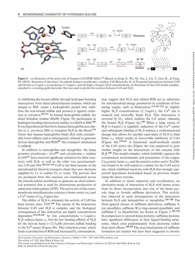
Figure 4 (a) Structure of the active site of human CcO (PDB 5Z62).117 [Based on Zong, S., Wu, M., Gu, J., Liu, T., Guo, R., & Yang,M. (2018). Structure of the intact 14-subunit human cytochrome c oxidase. Cell Research]. (b–d) Proposed interactions between CcOand (b) low ( 1 equiv.), (c) moderate (2–3 equiv.), and (d) high ( 4 equiv.) H > 2S concentrations. (e) Structure of the CcO model complex,attached to a rotating gold electrode, that was used to probe the reaction between CcO and H2S
to stabilizing the bound sulde through hydrogen-bondinginteractions. Four distal phenylalanine residues, which areunique to HbI, create a hydrophobic pocket that stabi-lizes the iron-bound sulde and protects it against oxida-tion or solvation.107,111 In human hemoglobin-sulde, thedistal histidine residue (His98; Figure 3b) participates inhydrogen-bonding interactions similar to Gln64 in HbI.103
It washypothesized that ferric humanhemoglobinacts sim-ilar to L. pectinata HbI to transport H2S in the blood.112
Given that human hemoglobin binds H2S with consider-ably lower afnity and is subsequently reduced to generateferrous hemoglobin and RSS,84 this transport mechanismis unlikely.
In addition to hemoglobin and myoglobin, the hemeproteins cytochrome c (Cc)102 and cytochrome c oxidase(CcO)113 have received signicant attention for their reac-tivity with H2S, as well as the other two gasotransmit-ters, CO and NO.102,114–116 CcO is the nal enzyme of themitochondrial electron transport chain that uses electronssupplied by Cc to reduce O2 to water. The protons thatare produced from this reaction are translocated acrossthe mitochondrial membrane to generate an electrochem-ical potential that is used for downstream production ofadenosine triphosphate (ATP). The active site of this trans-membrane metalloenzyme contains a heme center (a3) anda CuII center (CuB; Figure 4a).
The ability of H2S to attenuate the activity of CcO hasbeen known since 1929.118 The nature of the interactionbetween CcO and H2S is complex and the biologicalconsequences of this interaction are highly concentrationdependent.113,119,120 At low concentrations (1 equiv.),H2S reduces heme a3, but the low binding afnity of H2Sfor the ferrous heme ( M) prevents sulde binding12.5 to the FeII center (Figure 4b). This reduction event, whichleads to production of RSS and increasedO2 consumption,
may suggest that H2S and related RSS act as substratesfor mitochondrial energy production in conditions of lowenergy supply, such as hibernation.113,121–124 At slightlyhigher H2 S concentrations (2–3 equiv.), the Cu
II site isreduced and reversibly binds H2S. This interaction isreversed by O2 , which oxidizes the CuI center, releasingthe bound H2S (Figure 4c).120 When a large excess ofH2S (>4equiv.) is supplied, reduction of the Cu II centerand subsequent binding of H2S induces a conformationalchange that allows for another equivalent of H2S to bindheme a 3, which results in irreversible inhibition of CcO(Figure 4d).119,121 A functional small-molecule modelof the CcO active site (Figure 4e) was employed to gainfurther insight on the interactions of this enzyme withH2S.
120 This model complex, which faithfully captures thecoordination environments and proximities of the copperCuB center, heme a3, and the putative redox-active Tyr244,was found to be well suited as a mimic for the CcO activesite, which exhibited reactivity with H2S that strongly sup-ported hypotheses formulated based on previous studiesusing the intact enzyme.
In addition to metal reduction and coordination, analternative mode of interaction of H2S with hemes arisesfrom its direct incorporation into one of the heme pyr-role rings to furnish sulfheme derivatives. This activitywas observed in early investigations of the interactionbetween H2S and hemoglobin or myoglobin.125–128 Thethree general classes of sulfheme derivatives, sulfheme A(an episulde), sulfheme B (a ring-opened episulde), andsulfheme C (a thiochlorin) are shown in Figure 5.129–134
In comparison to natural heme proteins, sulfheme proteinshave signicant differences in their ligand-binding prop-erties, which arise predominately from electronic ratherthan steric effects.128,129 The exactmechanisms of sulfhemeformation are unclear but have been suggested to involve
Encyclopedia of Inorganic and Bioinorganic Chemistry, Online © 2011–2021 John Wiley & Sons, Ltd.This article is © 2021 John Wiley & Sons, Ltd.This article was published in the Encyclopedia of Inorganic and Bioinorganic Chemistry in 2021 by JohnWiley & Sons, Ltd.DOI: 10.1002/9781119951438.eibc2764
Printed by [University O
f Arizona Library - 150.135.174.099 - /doi/epdf/10.1002/9781119951438.eibc2764] at [08/04/2021].
□□ □□ uoo
00 V00 - 00V00
DO Ll 00 DO DD
'N~N ':D ~I N~N ~ -NA> NH /
, ,,______ ,,-=,
N I: \.y'o "\ IJ
\%\\\\\ '' ''' •

BIOINORGANIC CHEMISTRY OF HYDROGEN SULFIDE 5
SY
XS
S
Sulfheme A
Sulfheme B(a)
(b)
Sulfheme C
N
OHO
N
HO O
e
Figure 5 (a) Diagram of three sulfheme products formedthrough covalent addition of sulde to the porphyrin ring. (b)X-ray crystal structure of cyanide-bound myoglobin containingsulfhemeC (PDB: 1YMC).129 [(b) Based on Evans, S. V., Sishta, B.P., Mauk, A. G., &Brayer, G. D. (1994). Three-dimensional struc-ture of cyanomet-sulfmyoglobin C. Proceedings of the NationalAcademy of Sciences, 91(11), 4723–4726]
oxoferryl (FeIV=O) intermediates.10,89,135 The secondarycoordination sphere, specically a His residue in the distalposition of the enzyme active site, has been shown to beimportant for the production sulfheme species in somecases.77,135 For example, human hemoglobin, which con-tains this distal residue, forms sulfheme derivativeswhereasHbI from L. pectinata, which contains distal Gln instead,does not. Site-directed mutagenesis of this Gln residue to aHis residue allowed this protein to convert to the sulfhemeform upon exposure to H2S.
77 By contrast, CcO, which
contains His residues near the active site, does not formsulfheme in the presence of H2S. In this particular case,it has been hypothesized that these His residues do nothave the proper orientation and therefore cannot interactwith the heme center to promote sulfheme formation.76
Sulfheme derivatives of the metalloenzymes catalase andlactoperoxidase have also been observed.89,136 Given theirpoor afnity for O2 and inability to be converted back tonormal hemeproteins,125,137,138 it is unlikely that sulfhemespecies play a role in cellular signaling, but instead mediatethe toxic effects of this gas. For example, elevated sulfhemelevels induce a toxic condition known as sulfhemoglobine-mia due to their inability to adequately bind O2 .
139
3.2 Zinc-Containing Proteins
The interactions between spectroscopically silent Zn-containing proteins and H2S have received relatively littleattention compared to those for other metalloproteins.10
Proteins containing the zinc nger (ZF) motif are particu-larly important, as evidenced by their high abundance ineukaryotic cells. ZFs are small, folded domains that arestabilized by coordination to a structural ZnII ion with acombination His and Cys residues. This class of proteinsplays an important role in eukaryotic cells as transcriptionfactors that regulate gene transcription.140 Although ZnII
is not redox active, Cys-containing ZF domains undergoS-sulfhydration in response to H2S exposure.141–143 Themechanism of S-sulfhydration of the specic ZF proteintristetraprolin was investigated. A key discovery fromthis study was that this process occurs only in the pres-ence of O2 .
142 The ZnII center is thought to facilitate theS-sulfhydration by shifting the redox potential of theCys and enabling its oxidation by O2 and subsequentsulfhydration by H2S.
In addition to being a key structural requisite for ZFs,ZnII plays a catalytic role in several enzymes, which mayalso react with H2S. For example, the Zn-containingenzyme phosphodiesterase 5 is inhibited by nanomolarconcentrations ofH2S.
144 Themechanism of this inhibitionhas not been fully elucidated, but the direct coordinationof H2S to the catalytic ZnII center could not be ruled out.Similarly, the inhibition of carbonic anhydrase by H2Sarises from the direct coordination of this gasotransmitterto the His3Zn active site found in most isoforms of thisenzyme.145–147 A model complex of the dinuclear ZnII
enzyme CS2 hydrolase, which is found in archaebacte-ria, was shown to produce H2S and CO2 via hydrolysisof CS2 ,
148 suggesting that CS2 is a biologically relevantsource of H2S.
149
Lastly, H2S regulates proteins with alternative func-tions. For example, H2S is also known to interact withmetallothioneins, Cys-rich proteins that bind ZnII anddetoxify heavy metal ions. In this capacity, H2S preventsCdII-induced toxicity by stabilizing the ZnII-containing
Encyclopedia of Inorganic and Bioinorganic Chemistry, Online © 2011–2021 John Wiley & Sons, Ltd.This article is © 2021 John Wiley & Sons, Ltd.This article was published in the Encyclopedia of Inorganic and Bioinorganic Chemistry in 2021 by JohnWiley & Sons, Ltd.DOI: 10.1002/9781119951438.eibc2764
Printed by [University O
f Arizona Library - 150.135.174.099 - /doi/epdf/10.1002/9781119951438.eibc2764] at [08/04/2021].

6 BIOINORGANIC CHEMISTRY OF HYDROGEN SULFIDE
n
e
SH
N
Zn
H
n
HiPr
iPr
l
Cl
SH
RR
R
SH
R
R
R
R = Me, tBu, iPr, r, Ph, PyA R = Me, tBu, 2,6-dimethylphenyl
(a) (b) (c) (d) (e)
–
+
Figure 6 General structures of reported Zn–SH coordination compounds
isoform of metallothionein-1.150 Zn proteins also have apotential role for transport of H2S. The giant tubewormRiftia pachyptila, which inhabits sulde-rich environmentsand lives in a symbiotic relationship with sulde-oxidizingbacteria,151 uses a unique form of hemoglobin that con-tains 12 ZnII centers in a hollow cavity that bind and cap-ture H2S for transport through the bloodstream. 152
The reactivity of the ZF and catalytic ZnII proteins withH2S has prompted the evaluation of small-molecule com-plexes to model this chemistry.147 The tris-pyrazolylborateligand system has been used extensively to mimic theHis3 coordination environment of Zn-containing proteins(Figure 6a). Reactions of these model complexes with H2Shave conrmed thatSH− can coordinate directly to theZnII
center. These studies have demonstrated the importance ofbulky, nonpolar ligands to stabilize the resulting Zn–SHcomplex.153–157 Other Zn–SH complexes supported bythe tris(thioimidazole)hydroborate (Figure 6b),158,159
tris(2-pyridylmethyl)amine (Figure 6c),160,161 tris(2-pyridylseleno)methyl (Figure 6d),162 and a substitutedpyridinediimine ligand (Figure 6e)163 have also beenreported.
3.3 Cobalamin
In addition to the metalloprotein examples dis-cussed above, H2S also reacts with other redox-activebiomolecules. Of particular relevance to the eld ofbioinorganic chemistry, one of the more interesting reac-tion partners of H2S is vitamin B12, or cobalamin, aCo-containing molecule that is essential for DNA synthe-sis and metabolism (Figure 7a; see Cobalamin Biosynthesis
and Insertion).165 Only a handful of reports have exploredthe reactivity of H2S with cobalamin derivatives and assuch, relatively little is known about the biological impli-cations of the reactivity between these molecules. H2S candisplace strongly nucleophilic ligands, such as OH− orCN−, from the CoIII center of cobalamin to furnish stableCoII derivatives and RSS. 164,166,167 Under anaerobic con-ditions the generated CoII species readily react with methyliodide to generate methylcobalamin, which can be cleanly
converted back to Co III aquocobalamin in the presence oflight, demonstrating that the reaction between cobalaminand H2S is reversible.164,168 Kinetic studies have suggestedthat the reaction between H2S and cobalamin underanaerobic conditions proceeds via initial binding of H2Sto the CoIII center followed by reduction to Co II throughan inner-sphere electron transfer reaction. Addition of asecond equivalent of H2S to the CoII sulde complex pro-duces polysulde derivatives (Figure 7b). In the presenceof O2 , the reaction products are much more diverse andinclude [CoII–S–S–CoII ] type species and sulfur-modiedcorrinoids.164,167 Given the ability of cobalamin and itsderivatives to bind H2S, it has been demonstrated thatadministration of the vitamin B12 analogue cobinamidein rabbits prevents sulde poisoning in vivo by decreasingplasma H2S levels.169
4 METAL COMPLEXES FOR H2S DETECTION
4.1 Common H2S Detection Methods
Given the growing importance of H2S in biology andbioinorganic chemistry, methods for quantication anddetection of H2S in biological systems have become thefocus of intensive study. The spectrophotometricmethyleneblue (MB) assay is one of the oldest, albeit very reliable,method for sulde detection and quantication.170–172 Inthis assay, MB is generated through the FeCl3-catalyzedreaction between p-dimethylamino aniline and H2S. Thecharacteristic absorbance of MB at 670 nm allows fordirect quantication of H2S. The MB assay requiresthe use of acidic aqueous solutions. These conditions,however, can extract acid-labile sulfur from other bio-logical sources, thereby complicating analysis of H2Sin biological or pH-sensitive systems. Thus, the use ofthis assay is limited in measuring H2S production fromdonors that are activated under acidic conditions, suchas phosphinodithioates.173–175 An alternative procedureinvolving precipitation of H2S as zinc sulde prior toperforming the MB reaction can be used to circumvent
Encyclopedia of Inorganic and Bioinorganic Chemistry, Online © 2011–2021 John Wiley & Sons, Ltd.This article is © 2021 John Wiley & Sons, Ltd.This article was published in the Encyclopedia of Inorganic and Bioinorganic Chemistry in 2021 by JohnWiley & Sons, Ltd.DOI: 10.1002/9781119951438.eibc2764
Printed by [University O
f Arizona Library - 150.135.174.099 - /doi/epdf/10.1002/9781119951438.eibc2764] at [08/04/2021].
0 7 s
N,.1~,.., I ' _....- I ~ ~

BIOINORGANIC CHEMISTRY OF HYDROGEN SULFIDE 7
CoIII
OH
CoIII
SH
SH–
CoII
S
CoII
SS
SH–
Co III
OH2+ –
CoII SSH –+
2+
X = H 2O(a)
(b)
CONH2
CONH2
N
H2NOC
H2NOC
Ho
CONH2
NH
O
O
O–
N
Figure 7 (a) Chemical structure of aquocobalamin. (b) Proposed reaction pathway between cobalamin and H 2S under anaerobicconditions. For more details, see Ref. 164 [Based on Salnikov, D. S., Kucherenko, P. N., Dereven’kov, I. A., Makarov, S. V., & van Eldik,R. (2014). Kinetics and Mechanism of the Reaction of Hydrogen Sulde with Cobalamin in Aqueous Solution. European Journal ofInorganic Chemistry, 2014(5), 852–862]
these challenges.176 Despite its widespread use, the MBmethod is relatively insensitive with a limit of detection(LOD) of approximately 2M, thus making it insufcientfor detecting low concentrations of H2S that may be phys-iologically relevant in biological systems. 177 Another H2Sdetection method, called the monobromobimane (mBB)method, is generally more accurate and is signicantlymore sensitive (LOD 2 nM) than MB. This techniquerelies on the reaction of mBB with H2S to furnish suldedibimane, which can be detected either spectrophotomet-rically or by uorescence spectroscopy.178,179 The mBBmethod requires basic conditions and low oxygen con-centrations in order to obtain the highest sensitivity forH2S. Coupling this technique to high-performance liquidchromatography (HPLC) analysis provides information onthe speciation of H2S and H2Sn because mBB also reactswith biological thiols and poly- and persulde species.180
However, this additional reactivity can be problematicwhen trying to use the mBBmethod to measure H2 S undernormoxic, physiological conditions.
Amperometric electrodes have also been developedto complement spectrophotometric techniques for H2Sdetection.181,182 In general, these sensors consist of an ion-selective membrane and a polarizing voltage, which allows
H2S permeation into the electrode. The interior of theelectrode contains a strongly basic solution of Fe(CN)6
3− ,which is reduced selectively by H2S to Fe(CN)6
4−. Thegenerated ferrocyanide is re-oxidized by a platinum elec-trode, which generates a current relative to the amount ofH2S present.183,184 This technique is advantageous overother methods in that it can be used in unadulteratedbiological samples, such as tissue homogenates, culturedcells, and even circulating blood of living animals,185
allowing for time-resolved measurement of H2S dynamics.Sulde electrodes are limited in that they cannot provideinformation at sub-cellular resolution and are known tobe highly pressure and temperature sensitive, requiringfrequent recalibration.21
The past decade has seen rapid development and imple-mentation of uorescent and colorimetric reaction-basedH2S probes for use in biological applications. These sys-tems generally consist of a chromophore appended with asulde-sensitive protecting group. Attack by H2S releasesthe active species, resulting in a colorimetric or uores-cence response. Several recent reviews provide a compre-hensive background on advances in the development ofsuch sensors.186–191 Compared to the methods discussedabove, these probes allow for spatiotemporal monitoringof H2 S levels in cells and tissues that is minimally invasive.
Encyclopedia of Inorganic and Bioinorganic Chemistry, Online © 2011–2021 John Wiley & Sons, Ltd.This article is © 2021 John Wiley & Sons, Ltd.This article was published in the Encyclopedia of Inorganic and Bioinorganic Chemistry in 2021 by JohnWiley & Sons, Ltd.DOI: 10.1002/9781119951438.eibc2764
Printed by [University O
f Arizona Library - 150.135.174.099 - /doi/epdf/10.1002/9781119951438.eibc2764] at [08/04/2021].
7
7 · 7
□~D -0 -0 -0~□

8 BIOINORGANIC CHEMISTRY OF HYDROGEN SULFIDE
3+
2+
N
N
N
N
N
uN N
N
N
NH3H3N
NH3
H3
NH3H3N
21 3
Figure 8 Metal complexes that detect H2 S through reduction
This section will focus on the use of inorganic complexesfor H2S detection. Compared to sensors based on organicdyes, metal-based lumiphores generally have the advantageof high photostability, large Stokes shifts, which reducesself-quenching, and long-lived luminescence lifetimes (seeLuminescence Behavior & Photochemistry of Organotransi-
tion Metal Compounds), which permits time-gated lumines-cence imaging and minimizes interference from biologicalautouorescence.192–194
4.2 Metal-Based H2S Sensors
Although we note that metal–organic framework(MOF)-based sensing platforms have recently beenexplored for the detection of H2S,
195,196 this section willfocus solely on small-molecule probes. In a general sense,metal-based sensors take advantage of the strong reduc-ing power and nucleophilicity of H2S and HS−.186 Forexample, the reduction of the [Ru(NH3)6 ]
3+ (1, Figure 8)by sulde in pH 7.4 aqueous solution was employed todevelop a microchip-based system for continuous moni-toring of H2S levels in the central nervous system of guineapigs.197 Similarly, electron transfer betweenH2S and a RuII
polypyridyl complex (2, Figure 8) induces a phase shiftin the luminescence of the complex, which was leveragedto develop reversible sensors capable of monitoring H2Sdynamics over extended periods of time.198 In complex3 (Figure 8), the CoII center is reduced by H2 S to CoI,resulting in a shift in the electronic absorbance spectrum.The metal center can be cleanly oxidized back toCoII uponexposure to air, thereby conferring a degree of reversibilityto this system.199 In a different study which highlights theuse of metalloproteins for this application, reduction ofthe Cu II center of a uorescently tagged azurin proteinisolated from Pseudomonas aeruginosa by H2S resultedin an increase in uorescence.200 This response could bereversed back to the quenched state upon treatment withK3Fe(CN)6.
Luminescent Zn II (4),201Co II (5),202 RuII (6),198,203 andIr III ( )7–11 204–208 complexes have been reported as H2S sen-sors (Figure 9). With the exception of 5, which appearsto sense H2S through noncovalent interactions,202 thesescaffolds include H2S-reactive masking groups, such asan azides, nitros, dinitrophenyl sulfonyl, or dinitrophenylethers. These functional groups can quench the photoex-cited metal-to-ligand charge transfer state (MLCT) viaphotoinduced electron transfer (PET), thereby preventingphotoluminescent emissive decay pathways. Nucleophilicattack of H2 S on these groups modies them such thatPET to the excited state is no longer energetically viable,resulting in an increase in metal-based photoluminescenceintensity. Of these compounds, only have been used6 and 7in biological contexts. The high sensitivity of 6 toward H2S(LOD 45nM) enabled the monitoring of lysosomal H= 2Slevels in live cells, as well as endogenous H2S levels in liv-ing zebrash and mice.203 Despite the signicantly lowersensitivity of 7 6(LOD M) compared to= 4.35 , this com-pound was similarly able to detect H2S in vitro and invivo.208 Complex was also activated by hypoxic environ-6
ments, suggesting that its H 2S-detection capabilities underthese conditions is limited.Although compounds have8–11
not been studied in biological settings, they offer interest-ing opportunities for the development of electrochemilu-minescent (ECL) sensors, which rely on the generation ofluminescent excited states by redox chemistry rather thanphoton absorption.209
The narrow emission energies, long-lived luminescencelifetimes, and large Stokes shifts of the photoexcitedLaPorte forbidden f–f states of the lanthanides EuIII
and TbIII (see Lanthanides: Luminescence Applications;Lanthanides: Luminescence)210,211 have also been leveragedin the development of luminescent H 2S-responsive probes(12–16; Figure 10).212–217 These complexes contain anH2S-reactive functional group such as an azide or dinitro-phenyl ether, which quenches the complex photoexcitedstate in a similar manner to the transition metal complexesdiscussed above. The reaction of these groups with H2Srestores luminescence by converting these groups to forms
Encyclopedia of Inorganic and Bioinorganic Chemistry, Online © 2011–2021 John Wiley & Sons, Ltd.This article is © 2021 John Wiley & Sons, Ltd.This article was published in the Encyclopedia of Inorganic and Bioinorganic Chemistry in 2021 by JohnWiley & Sons, Ltd.DOI: 10.1002/9781119951438.eibc2764
Printed by [University O
f Arizona Library - 150.135.174.099 - /doi/epdf/10.1002/9781119951438.eibc2764] at [08/04/2021].
N 7 ......_ I ,,,...
Ru ,,.,, I '

BIOINORGANIC CHEMISTRY OF HYDROGEN SULFIDE 9
5
H
NNH
Co
HO
Cl
Cl
N
N
N
N
N
N
N3
Zn
N3
N3
4
Ru
NN
N
N
O
S
NO 2
O2N
2+
6 7
N
N
+O2
F
F
N
O
O
O
O
+
ON
O
O
O
O
O
O
O2
NO2
O
O
N
N
O
O
N 3
O
O
N3
ON
O
O
NO2
NO2
O2N
O
O2N
NO2
NO 2
8 9 10 11
Figure 9 Luminescent metal complexes for H 2S detection
NN
O
OO
OM
N
N
O
NO 2
NO2
Tb
N
COO
N
OOC
C
N
OO
N3
N
OO
N3
N
O
O
O
O
N 3
–
3–
OO
O
OO
O
NH
O
N3
O
O
O
O
2–
M = Eu, 12
M = Tb, 1315 1614
Figure 10 Luminescent lanthanide-based H2 S-sensing complexes
that cannot engage in PET. Compound was the rst12
example of a kinetically stable EuIII complex that couldbe used for detection of H2S. The biological compati-bility of was demonstrated by using it to detect this12
gasotransmitter in human serum. 212 More recently, aEuIII probe bearing a functionalized aminopolycarboxy-late ligand ( ) was reported. This probe can detect H14 2Sproduced by CSE in aqueous solution and was used in
Encyclopedia of Inorganic and Bioinorganic Chemistry, Online © 2011–2021 John Wiley & Sons, Ltd.This article is © 2021 John Wiley & Sons, Ltd.This article was published in the Encyclopedia of Inorganic and Bioinorganic Chemistry in 2021 by JohnWiley & Sons, Ltd.DOI: 10.1002/9781119951438.eibc2764
Printed by [University O
f Arizona Library - 150.135.174.099 - /doi/epdf/10.1002/9781119951438.eibc2764] at [08/04/2021].
l
(
7
' " '-./ cqb ciQ ..__,..., N ~ :' \ )

10 BIOINORGANIC CHEMISTRY OF HYDROGEN SULFIDE
Ru
N
NN
NN
uN
4+
18
RuN
N
N
4+HN
O
N
NN
u
19
O
O
HN
O N
N
Cu
OHHO
2+
17
Figure 11 Luminescent H2S sensors that detect H2S through metal displacement
a high-throughput screening assay to identify potentialinhibitors of this enzyme.215 Another lanthanide-basedH2S sensor, a TbIII complex bearing azide-substitutedpyridine carboxylate ligands (15), was able to detect H2 S atconcentrations as low as 10 nM in aqueous solution. Thiscompound was used in a paper-based assay to detect traceH2S exhaled by mice.213 Lastly, a TbIII complex containinga 2,6-dinitrophenol functionalized terpyridine type ligand( ) was used to detect H16 2S in living cells.216
4.3 Metal Displacement Sensors
The characteristic low solubility of metal sulde saltsoffers a possible strategy for the detection of H2S throughmetal displacement reactions. Since 1685,218,219 H2S hasbeen used to precipitate small concentrations of CuII ,SnII, Pd II, and HgII as a means of diagnosing their pres-ence. As such, compounds containing these metal ions, inaddition to ZnII , CdII, PbII, and AgI, have all been uti-lized for detection of this gas.220–227 Generally, this strat-egy relies on compounds that contain both a photolumi-nescent dye and a metal ion bound by a chelator, suchas 1,4,7,10-tetraazacyclododecane (cyclen) or di-(2-picolyl)amine (DPA).188,194 One of the rst sensors employing thisapproach combined the uorescent dye uoresceinwith theDPA ligand bound to CuII (17, Figure 11).228 The emis-sion of uorescein is quenched by the paramagnetic CuII
center,229 but is restored upon exposure to H2S, whichremoves theCuII in the formof insolubleCuS.220 Followingthis example, sensors containing organic uorescent dyessuch as rhodamine, anthracene, and 4,4-diuoro-4-bora-3a,4a-diaza-s-indacene (BODIPY) that act through metalion displacement have been reported.187,230,231 The follow-ing discussion focuses specically on inorganic lumiphoresthat detect H2 S through metal displacement reactions.
The heterobimetallicRu II–CuII complex [Ru(bpy)2(bpy-DPA)] 4+ (18; bpy-DPA= 4-methyl-4 -[N N, -bis(2-picolyl)amino-methylene]-2,2-bipyridine; Figure 11) was reportedto be a sensitive (LOD= 20 nM) turn-on luminescent H2S
sensor that is activated by precipitation of CuS. 232 Thiscomplexwas also used as an ECL sensor tomeasure suldeconcentration in the cortex of adult male rats.233 A similarruthenium complex containing phenanthroline appendedwith a cyclen chelator was also shown to sense H2S in thepresence of CuII ( , Figure 11). This complex is capable19
of detecting H2S over a broad pH range and can detectH2S in vitro with a rapid turn-on response time.234
A number of CuII displacement sensors have beenreported that capitalize on the long-lived luminescence ofthe lanthanides Eu III and TbIII (20–27, Figure 12).235–242
Although some of these compounds, such as 20, wereused to monitor H2S in petroleum plant waste streams,237
a few of these probes have found applications in bio-logical settings. Heterobimetallic EuIII–CuII compounds21 22and bearing -diketonate ligands and DPA CuII -chelating moieties were found to be relatively nontoxicand capable of selectively detecting intracellular H2Sin vitro.241,242 More recently, was reported as a23
luminescent probe with a rapid turn-on response, highsensitivity, and good selectivity for H2 S. Furthermore,this compound could be used to measure H2 S productionfrom both the slow-releasing H2S donor morpholin-4-ium 4-methoxyphenyl(morpholino)phosphinodithioate(GYY4137) and CSE, as well as to determine intracellularH2S levels in Na2S-stimulated cells.235
4.4 H2S Detection via Metal Coordination
Researchers have developed probes that can detectH2S via changes in uorescence or absorbance upondirect coordination of this gasotransmitter to the metalcenter. The Zn II tris(pyrazoyl)borate complex con-28
taining 7-mercapto-4-methylcoumarin was the earliestcoordination-based H2S sensor with a LOD of 1 M.243
Binding of H2S to the Zn center results in the release of thecoumarin ligand and a concomitant color change, making28 a colorimetric sensor. Pyridoxal (29), 244,245 porphyrin(30–35),246–248 phthalocyanine (36),199 and salen (37)249
Encyclopedia of Inorganic and Bioinorganic Chemistry, Online © 2011–2021 John Wiley & Sons, Ltd.This article is © 2021 John Wiley & Sons, Ltd.This article was published in the Encyclopedia of Inorganic and Bioinorganic Chemistry in 2021 by JohnWiley & Sons, Ltd.DOI: 10.1002/9781119951438.eibc2764
Printed by [University O
f Arizona Library - 150.135.174.099 - /doi/epdf/10.1002/9781119951438.eibc2764] at [08/04/2021].
7 ~ I
:::::,._ "' "' N/uD'-..~ ~ I # #
"' I ~

BIOINORGANIC CHEMISTRY OF HYDROGEN SULFIDE 11
N
O
O
N
2+
N N
O
NO
N
u
Cu
3+
N
O
O
N 2+
H
N
N
u
O
O
O
O
O
OH
2+
NHO
C3 F7
C 3F7
C
NH O
C3 F7
O
C3F7
O
3+
H
O
C3F 7 O C 3F 7O
N
N
C
N
N
3+
21
N
O
O
2+
H
N
23
N
O
O
2+
N
20 22
24 25 26 27
Figure 12 Luminescent lanthanide-based H2 S probes that rely on metal displacement for detection
complexes have also been shown to sense H2S via directmetal coordination (Figure 13). Compound 31 displaysdecent selectivity for H2S over a range of biologicallyrelevant anions, thiols, and oxidants.247 A second studyfound that 31 failed to bind sulde in organic solution, aresult that reveals the importance of solvent effects andproton availability for this class of sensors.248 The absorp-tion spectra of porphyrin compounds undergo34 and 35signicant changes in the presence of excess HS−. Themetal center, however, undergoes subsequent reduction togive unresolved reaction products,248 which might hindertheir use as colorimetric H2S sensors.
Researchers have also leveraged the ability of metallo-proteins and enzymes to bind H 2S for the developmentof coordination-based sensors. The general approachcombines an H2S-binding metalloprotein with auorescent tag. The metalloprotein chromophores of thesesystems exhibit signicant shifts in their absorbance wave-lengths upon binding to H2S. The change in absorbanceof the metalloprotein chromophore can modulate theemission intensity of uorescent tag by attenuating orenhancing the availability of photons from the excitation
source. This strategy relies on the use of uorescent tagswith excitation wavelengths near absorbance maxima ofthe protein or enzyme in the absence of H2S, which pre-vents uorescence of the tag. Coordination of H2S/HS−
to the metal center causes the protein absorbance to shift,unmasking the uorophore, which results in an increasein uorescence intensity. Fluorophore-tagged analogsof Horse skeletal muscle myoglobin isolated from horseskeletal muscle,250 cobalt peptide deformylase (PDF)from Escherichia coli,251 and HbI from L. pectinata252
have all been used as H2S sensors of this type with goodselectivity over other biological thiols and sensitivity inthe nanomolar range. The metal-sulde interaction inPDF was supported by X-ray crystallography, whichdemonstrated that both the Co and Ni-containing formsof this enzyme coordinate H2 S (Figure 14).251
The coordination-based approach for H2S detection isintriguing given the low stability and high reactivity ofmany metal H2S and hydrosuldo complexes.26,27,253–256 Incontrast to the reaction-based and metal displacement sen-sors, these systems can potentially give rise to reversibleH2S probes, an important property that could be used to
Encyclopedia of Inorganic and Bioinorganic Chemistry, Online © 2011–2021 John Wiley & Sons, Ltd.This article is © 2021 John Wiley & Sons, Ltd.This article was published in the Encyclopedia of Inorganic and Bioinorganic Chemistry in 2021 by JohnWiley & Sons, Ltd.DOI: 10.1002/9781119951438.eibc2764
Printed by [University O
f Arizona Library - 150.135.174.099 - /doi/epdf/10.1002/9781119951438.eibc2764] at [08/04/2021].

12 BIOINORGANIC CHEMISTRY OF HYDROGEN SULFIDE
B
N N
Zn
H
O
OH
ON
N
N
O
OH
Zn
N
NN
N+
N+
N+
N+
Zn
302928
N
N
N
N
N
N
36
NN
N
M
M = Cu, n = 0, 31M = Zn, n = 0, 32M = Cr, n = 0, 33
M = Mn, n = 0, 34M = Sn, n = 2, 35
O OH HO On+
R1
ON
N O
R1
Zn
37
NC
NC
R2
R2
R1 = H, OMe; R2 = H, EtN2, OH
Figure 13 Inorganic complexes that detect H2S upon metal coordination
(a) (b)
Figure 14 X-ray structures of the sulde-bound adducts of (a) Co-PDF and (b) Ni-PDF (PDB: 4AZ4, 4AL2). [Based on PDB: 4AZ4,4AL2, RCSB Protien Data Bank]
monitor H2S dynamics in biological or industrial settings.For example, treatment of the hydrosulde adduct of 33or with acetic acid or oxygen regenerates the parent36
complexes.199 Similarly, purging solutions of H2S-treatedHbI with argon produces the free protein, which can thendetect additional sulde.252 Together these studies highlightthe utility of the coordination-based approach for develop-ment of reversible sensors.
5 METAL COMPLEXES AS AGENTS FOR H2S
DELIVERY
5.1 Simple Inorganic Salts
Despite the numerous advances in understandingthe biological activity of H2S, it remains a challenge todeliver this gas to biological systems. The use of H2S in
its gaseous state is challenged by its toxicity, ammabil-ity, and volatility.257,258 Simple metal sulde salts suchas Na2S and NaSH are the most commonly employedsources of H2S in biological studies.258 Although thesesalts are generally easier to handle than gaseous H 2S,they are typically obtained commercially with very lowpurity levels,259 a limitation that makes it challenging todiscern the precise amount of H2S delivered by these salts.Furthermore, a 10 M aqueous solution of NaSH has a
half-life of only 0.5min due to volatilization of H2S,260
and the H2S generated from intravenously injected Na2Sis rapidly exhaled.261 In addition, toxic side effects fromthese salts may arise from their rapid release rate of H2Sin aqueous solution, which fails to mimic endogenousH2S dynamics. Motivated by these limitations, researchershave developed slow-release H2S donors that producethis gas on biologically relevant time scales. A number ofdifferent small-molecule sulde donors have been recentlyreported.257,258,262–264 These include simple molecules that
Encyclopedia of Inorganic and Bioinorganic Chemistry, Online © 2011–2021 John Wiley & Sons, Ltd.This article is © 2021 John Wiley & Sons, Ltd.This article was published in the Encyclopedia of Inorganic and Bioinorganic Chemistry in 2021 by JohnWiley & Sons, Ltd.DOI: 10.1002/9781119951438.eibc2764
Printed by [University O
f Arizona Library - 150.135.174.099 - /doi/epdf/10.1002/9781119951438.eibc2764] at [08/04/2021].
Ji·tb ~ / 7
~.:9 H .,. 1 ~ X :: ::. /J(I -- (~ Zn
&:to u "':~ #
'

BIOINORGANIC CHEMISTRY OF HYDROGEN SULFIDE 13
undergo uncontrolled hydrolysis,174,175,265–267 in additionto organic H2S-releasing compounds that are selec-tively activated by light,268–272 enzymatic activity,273–276
thiols, 277–284 and ROS. 285–288 In contrast to the preva-lence of inorganic CO- and NO-releasing molecules,289–294
an overwhelming majority of H 2S donors are organicmolecules. Herein, recent efforts towards the developmentof metal-based small-molecule H2S donors are described.
5.2 Tetrathiomolybdate
Tetrathiomolybdate ([MoS4]2− ; TM) is a widely avail-
able reagent that is used in organic chemistry for sulfurtransfer and reduction reactions.295,296 This ion is preparedby the reaction of the oxymolybdate anion ([MoO4 ]
2−)and H2 S in basic aqueous solution.297 Historically, TMhas received signicant attention for its ability to inhibitCuI -trafcking proteins through the formation of sulfur-bridged Cu–Mo clusters. 298 This orally available complexhas been widely used to treat disorders associated withimproper copper metabolism, such as Wilson’s disease.
Although TM has been long known to release H2Sunder acidic299 or high-temperature300,301 conditions,recent work has demonstrated that this compound, and toa lesser extent tetrathiotungstate ([WS4 ]
2− , TT), producethis gas via hydrolysis under biological conditions.302–305
TM generates H2S via hydrolysis over a period of hoursin buffered aqueous solution302 and interacts with biolog-ical thiols, such as glutathione, to produce other reactivepersulde and polysulde species.303 The H 2S generatedthrough TM hydrolysis was shown to prevent H2O 2-induced oxidative stress and preserve cellular functionin vitro. 302 When administered intravenously, TM sig-nicantly reduces infarct size in mice subjected to eithermyocardial or cerebral ischemia304 and prevents oxidativedamage and loss of functional activity in preclinical strokemodels.305 A recent report suggested that H2S producedby TM hinders its anticancer activity in A549 adenocarci-noma cells by enhancing cell growth at low concentrationsand upregulating H2S producing enzymes.306
Mechanistic studies suggest that the protective effectsof TM arise from its ability to decrease mitochondrialROS levels304 and improve antioxidant enzyme activity,305
which are consistent with the therapeutic effects reportedfor H2S.
307Furthermechanistic studies have suggested thatTM and TT are transported into cells by anion exchangeprotein-1 (AE-1).303 Despite the promise of TM as a bio-logically relevant H2S donor, it was reported that TMobtained from different commercial sources produced vari-able amounts H2S,
304 highlighting the difculty of obtain-ing these simple compounds in analytically pure forms. Inaddition, the H 2S release from TM is uncontrollable; oncethe complex is placed in aqueous solution, H2S is spon-taneously generated and the release cannot be targeted toa specic location in vivo. It would be advantageous to
develop agents that produce H2S selectively in response tospecic stimuli, allowing for selective and controlled H2Sproduction in complex biological systems.
5.3 Light-Activated Donors
Light-activated H2S-releasing agents have been recog-nized as promising tools for studying the biological andtherapeutic properties of this gas.262,263 Photoactivatabledonors allow for localized and noninvasive delivery ofH2S in vitro and offer exciting possibilities for deliveryin vivo. Upon irradiation with a specic wavelength oflight, the donor undergoes photoinduced decompositionto cleave a photoactivated protecting group and releasean H2S-producing moiety. The rst example of a pho-tocaged H2S donor consisted of a -orthonitrobenzylbis
protected geminal-dithiol (gem-dithiol).268 The nitrobenzylprotecting groups are removed upon reaction with 365 nmlight to produce an unstable -dithiol, which under-gem
goes rapid hydrolysis in aqueous solution to produce H2S.Other photoactivated H2S donors containing gem-dithiol,thiobenzaldehyde, and ketoprofenate moieties have alsobeen reported.257,268–270
The vast majority of these photoactivated compoundsrequire ultraviolet (UV) light ( 400 nm) for activation.This requirement limits the applicability of these donorsin vivo given that these wavelengths ineffectively penetratebiological tissue and can give rise to phototoxicity.308 Assuch, researchers have sought to develop light-activateddonors that function at lower energy wavelengths.
By virtue of their favorable photophysical properties(see Photochemistry of Transition Metal Complexes)309,310
metal compounds have been widely used for light-activateddelivery of CO and NO.310–314 By contrast, photoactivatedH2S donors based on inorganic systems have not beenreported until recently. An early example of a metal-basedH2S release platform used polyethylene glycol functional-ized LiYF4:Yb/Tm upconverting nanoparticles (UPNCs)conjugated to a caged gem-dithiol compound. Under near-infrared (NIR) irradiation, the UPNCs emit UV light,which then unmasks the protected gem-dithiol to produceH2S. This strategy was shown to deliver H2S both in livingcells and ex vivo in a pork skinmodel.315A complementarynanoparticle-based system consisting of a singlet oxygen(1O2) photosensitizer and 1,3-diphenylisobenzothiophene(DPBT) encapsulated in articial polymersomes has alsobeen reported. The photosensitizer, a PtII porphyrin com-plex or biscyclometalated IrIII compound, generates 1O2upon irradiation with visible light, which subsequentlyreacts with DPBT to produce H2S.
316 These early studieshave demonstrated how the photochemical properties ofmetal complexes can be leveraged to develop selective H2Sdonors that can be activated by light irradiation.
The characterization and biological activity of a redlight-activated H2S-releasing RuII complex were recently
Encyclopedia of Inorganic and Bioinorganic Chemistry, Online © 2011–2021 John Wiley & Sons, Ltd.This article is © 2021 John Wiley & Sons, Ltd.This article was published in the Encyclopedia of Inorganic and Bioinorganic Chemistry in 2021 by JohnWiley & Sons, Ltd.DOI: 10.1002/9781119951438.eibc2764
Printed by [University O
f Arizona Library - 150.135.174.099 - /doi/epdf/10.1002/9781119951438.eibc2764] at [08/04/2021].

14 BIOINORGANIC CHEMISTRY OF HYDROGEN SULFIDE
+
Ru
N
SS
N
O
38
Figure 15 Chemical structure of compound 38
reported.317 In this study, coordination of GYY4137173,174
to a ruthenium photocage (38, Figure 15) suppresses thespontaneous hydrolysis-driven H2S release from this com-pound. Compound produces H38 2S in living cells uponirradiation with red (631 nm) light and protects H9c2 car-diomyoblast cells against an in vitro model of ischemicreperfusion injury. Compound 38 is the rst example ofan inorganic small-molecule H2S donor that is activated byirradiation with red light; a BODIPY-based thiocarbamatecompound was reported as the rst NIR light-activatedorganic H2S donor in the same year.272 This work high-lights how transition metal complexes may serve as viablelight-activated H2S donors that can operate in biologicallyrelevant settings.
5.4 Reduction-Activated Donors
The redox chemistry of transition metal ions offersunique opportunities for redox-responsive delivery ofbiologically relevant molecules. The redox environment ofbiological tissue or cells can vary dramatically dependingon the specic cell type, organelle, or conditions. In thepresence of low O2 levels, a state known as hypoxia, thecellular environment becomes highly reducing as cells andtissue lose the ability to maintain redox balance. 318 In thiscontext, several inorganic complexes containing metalssuch as Ru, Os, Pt, Cu, and Co have been developed asredox-activated prodrugs,319–323 which release cytotoxicanticancer agents upon reduction that occurs in hypoxiccells. A recent report has shown that a dinuclear persulde(-S2
2− ) bridged ruthenium compound (39, Figure 16)produces H2S selectively upon reduction.324 The ability ofthis complex to produce H2S upon reduction was leveragedto deliver this gasotransmitter to hypoxic cells, making39 the rst example of a hypoxia-activated H2S donor.Notably, mechanistic studies revealed that the reductionprocess does not proceed through production of H2S2like many organic persulde compounds,280,325–329 but
Ru
NH3
H2O
H3
S
H3NS Ru
H3
OH2
H3N NH3
H 3
4+
39
MH
NN
S
S
S S
40
Figure 16 Redox-activemetal complexes that produceRSS uponreduction
rather directly produces H 2S. Lastly, this complex wasshown to preserve cell viability in H9c2 cardiomyoblastcells subjected to an in vitro model of ischemic reperfusioninjury. Given that the redox chemistry of transition metalions is highly dependent on the nature of the supportingligands, persulde-bridged metal complexes offer excitingopportunities for the development of tunable H2S donorsthat can deliver this gas under a wide span of biologicallyrelevant redox potentials.
Recently, a MoIV tetrasuldo complex (40, Figure 16)was reported to undergo a two-electron reduction to pro-duce HS2
− and a MoVI trisulde species.330 The reductiverelease of HS2
− by compound 40was only demonstrated inorganic solvent or aqueous organic mixtures, but this studydemonstrates the potential of this class of compounds forthe developmentof inorganic complexes for deliveryof H2Sand other RSS to biological systems.
6 OUTLOOK AND CONCLUSIONS
Studies on the biological activity of H2S and relatedRSS have highlighted diverse reactivity of these specieswith metal-containing biomolecules. This immense diver-sity and the complex nature of these reactions have madeit difcult to obtain intimate mechanistic understanding ofthese processes. In some cases, such as CcO, model com-pounds that mimic the active site of the enzyme or pro-teins have greatly increased understanding of the reactivitybetween H2S and these biomolecules. The biological tar-gets of H 2S and its derived RSS have only just begun tobe elucidated and a number of biologically relevant metal-containing biomolecules, such as ZF proteins, are still notwell studied. Investigation of the biological activity of H2Sis further complicated by its propensity to form reactivepolysuldes and persuldes, which possess biological activ-ity distinct from that of H2S.
331,332
The utility of inorganic complexes for H2S detectionand delivery in biological environments has only recentlybeen realized. Inorganic complexes display favorable pho-tophysical properties compared to organic uorophores
Encyclopedia of Inorganic and Bioinorganic Chemistry, Online © 2011–2021 John Wiley & Sons, Ltd.This article is © 2021 John Wiley & Sons, Ltd.This article was published in the Encyclopedia of Inorganic and Bioinorganic Chemistry in 2021 by JohnWiley & Sons, Ltd.DOI: 10.1002/9781119951438.eibc2764
Printed by [University O
f Arizona Library - 150.135.174.099 - /doi/epdf/10.1002/9781119951438.eibc2764] at [08/04/2021].
N N ' ~ .-"'
7 yy 7-
N- N I ' If
- B o- ~s ~--, /,= '-, I
N N -
~

BIOINORGANIC CHEMISTRY OF HYDROGEN SULFIDE 15
and offer exciting opportunities for ECL-based H2S sen-sors. Furthermore, reduction-activated and coordination-activated sensors have shown promise for the developmentof reversible H 2S sensors that could potentially be used tomonitor biological H2S dynamics over extended periodsof time.
Inorganic complexes have only recently begun to beinvestigated for their use as H2S donors for delivery of bio-logically relevant concentrations of this gas in vitro. Therich photochemical and redox properties of coordinationcompounds offer exciting opportunities for the develop-ment of novel H2S donors for use as tools for studying therole of H 2S and related RSS in biological processes anddisease.
7 ACKNOWLEDGMENTS
Research in the Wilson Lab related to this area is sup-ported by Cornell University, the American Heart Associ-ation (AHA Predoctoral Fellowship for J. J. Woods; awardno. 20PRE35120390), and the United States National Sci-ence Foundation (CHE-1750295).
8 RELATED ARTICLES
Sulfur: Organic Polysulfanes; Nitrogen Monoxide(Nitric Oxide): Bioinorganic Chemistry; Photochemistryof Transition Metal Complexes; Cytochrome Oxidase;Iron: Heme Proteins & Dioxygen Transport & Storage;Iron: Heme Proteins, Mono- & Dioxygenases; Iron: HemeProteins, Peroxidases, Catalases & Catalase-Peroxidases;Luminescence Behavior & Photochemistry of Organotran-sition Metal Compounds; Lanthanides: LuminescenceApplications; Lanthanides: Luminescence; CobalaminBiosynthesis and Insertion; Sulfur: Inorganic Chemistry
9 REFERENCES
1. M. Swaroop, K. Bradley, T. Ohura, T. Tahara, M. D.Roper, L. E. Rosenberg and J. P. Kraus, J. Biol. Chem.,1992, , 11455.267
2. A. D. Patrick, Biochem. J., 1962, , 248.83
3. M.H. Stipanukand P.W. Beck, Biochem. J., 1982, , 267.206
4. J. Loiselet and F. Chatagner, Biochim. Biophys. Acta, 1964,89, 330.
5. O. W. Grifth, Methods Enzymol., 1987, , 366.143
6. F. Binkley and V. du Vigneaud, J. Biol. Chem., 1942, 144,507.
7. V. du Vigneaud, H. S. Loring and H. A. Craft, J. Bi o l .
Chem., 1934, , 481.105
8. F. Chatagner and G. Sauret-Ignazi, Bull. Soc. Chim. Biol.,1956, , 415.38
9. B. D. Paul and S. H. Snyder, Trends Biochem. Sci., 2015,40, 687.
10. M. R. Filipovic, J. Zivanovic, B. Alvarez and R. Banerjee,Chem. Rev., 2018, , 1253.118
11. B. Olas, Clin. Chim. Acta, 2015, , 212.439
12. Z.-W. Lee and L.-W. Deng, Handb. Exp. Pharmacol., 2015,230, 243.
13. D.Wu, Q. Hu and D. Zhu, Oxid. Med. Cell. Longev., 2018,2018, 4579140.
14. Y. H. Liu, M. Lu, L. F. Hu, P. T. H. Wong, G. D.Webb andJ. S. Bian, Antiox. Redox Signal., 2012, , 141.17
15. H. Kimura, Neurochem. Int., 2013, , 492.63
16. H. Kimura, Neurochem. Int., 2019, , 118.126
17. N. V. Zaichko, A. V. Melnik, M. M. Yoltukhivskyy, A. S.Olhovskiy and I. V. Palamarchuk, Ukr. Biokhimichnyi
Zhurnal, 2014, 86, 5.
18. M. Fu, W. Zhang, L. Wu, G. Yang, H. Li and R. Wang,PNAS, 2012, , 2943.109
19. P. Haouzi and C. M. Klingerman, Respir. Physiol. Neuro-
biol., 2013, , 229.188
20. J. M. Fukuto, V. S. Vega, C. Works and J. Lin, Curr. Opin.
Chem. Biol., 2020, , 52.55
21. K. R. Olson, Antiox. Redox Signal., 2012, , 32.17
22. R. J. Pleus, H. Waden, W. Saak, D. Haase and S. Pohl,J. Chem. Soc. Dalt. Trans., 1999, 2601.
23. D. Sellmann and I. Barth, Inorg. Chim. Acta, 1989, 164,171.
24. B. R. James, Pure Appl. Chem., 1997, , 2213.69
25. J. S. Rebouças, B.O. Patrick and B. R. James, J. Am. Chem.
Soc., 2012, , 3555.134
26. M.D. Pluth and Z. J. Tonzetich, C he m. So c. Rev., 2020, 49,4070.
27. C. G. Kuehn and H. Taube, J. Am. Chem. Soc., 1976, 98,689.
28. C. R. Brulet, S. S. Isied and H. Taube, J. Am. Chem. Soc.,1973, , 4758.95
29. A. L. Eckermann, M. Wunder, D. Fenske, T. B. Rauchfussand S. R. Wilson, Inorg. Chem., 2002, , 2004.41
30. J. Darkwa, D. M. Giolando, C. J. Murphy, T. B. Rauchfussand A. Müller, Inorg. Synth., 1990, , 51.27
31. J. Amarasekera, T. B. Rauchfuss and A. L. Rheingold,Inorg. Chem., 1987, , 2017.26
32. J. Amarasekera and T. B. Rauchfuss, Inorg. Chem., 1989,28, 3875.
33. T. B. Rauchfuss, Inorg. Chem., 2004, , 14.43
34. J. Amarasekera, T. B. Rauchfuss and S. R. Wilson, Inorg.
Chem., 1987, , 3328.26
Encyclopedia of Inorganic and Bioinorganic Chemistry, Online © 2011–2021 John Wiley & Sons, Ltd.This article is © 2021 John Wiley & Sons, Ltd.This article was published in the Encyclopedia of Inorganic and Bioinorganic Chemistry in 2021 by JohnWiley & Sons, Ltd.DOI: 10.1002/9781119951438.eibc2764
Printed by [University O
f Arizona Library - 150.135.174.099 - /doi/epdf/10.1002/9781119951438.eibc2764] at [08/04/2021].

16 BIOINORGANIC CHEMISTRY OF HYDROGEN SULFIDE
35. E. J. Houser, S. Dev, A. E. Ogilvy, T. B. Rauchfuss andS. R. Wilson, Organometallics, 1993, , 4678.12
36. J. L. Wallace and R. Wang, Nat. Rev. Drug Discov., 2015,14, 329.
37. C. Szabó, Nat. Rev. Drug Discov., 2007, 6, 917.
38. S. J. Chan and P. T.-H. Wong, Neurochem. Int., 2017,105, 1.
39. J. Bełtowski, Pharmacol. Rep., 2015, , 647.67
40. H. Ibrahim, A. Serag and M. A. Farag, J. A d v. R e s., 2020,25, 243. DOI: 10.1016/j.jare.2020.05.018.
41. K. Y. Chen and J. C. Morris, Environ. Sci. Technol., 1972,6, 529.
42. B. Meyer,K.Ward,K.Koshlap and L. Peter, Inorg. Chem.,1983, , 2345.22
43. A. J. Ellis and R. M. Golding, J. C h e m . S o c., 1959, , 127.76
44. M. Widmer and G. Schwarzenbach, Helv. Chim. Acta,1964, , 266.47
45. P. M. May, D. Batka, G. Hefter, E. Königsberger andD. Rowland, Chem. Commun., 2018, , 1980.54
46. E. Cuevasanta, A. Denicola, B. Alvarez and M. N. Möller,PLoS One, 2012, , e34562.7
47. Q. Li and J. R. Lancaster, Nitric Oxide, 2013, , 21.35
48. O. Kabil andR. Banerjee, J. Biol. Chem., 2010, , 21903.285
49. P. Kamoun, Amino Acids, 2004, , 243.26
50. B. Murphy, R. Bhattacharya and P. Mukherjee, ,FAS E B J.
2019, , 13098.33
51. G. A. Benavides, G. L. Squadrito, R. W. Mills, H. D. Patel,T. S. Isbell, R. P. Patel, V. M. Darley-Usmar, J. E. Doellerand D. W. Kraus, PNAS, 2007, , 17977.104
52. J. Yang, P. Minkler, D. Grove, R. Wang, B. Willard,R. Dweik and C. Hine, Comm un. Biol., 2019, 2, 194.
53. X. Shen,M. Carlström, S. Borniquel, C. Jädert, C.G.Keviland J. O. Lundberg, Free Radic. Biol. Med., 2013, , 195.60
54. K. Abe and H. Kimura, J. Neurosci., 1996, , 1066.16
55. G. Yang, L.Wu, B. Jiang,W. Yang, J. Qi, K. Cao,Q.Meng,A. K. Mustafa, W. Mu, S. Zhang, S. H. Snyder andR. Wang, , 2008, , 587.Science 322
56. P. K. Yadav, P. Xie and R. Banerjee, J. Biol. Chem., 2012,287, 37611.
57. X. Chen, K. H. Jhee and W. D. Kruger, J. Biol. Chem.,2004, , 52082.279
58. S. Singh, D. Padovani, R. A. Leslie, T.Chiku andR. Baner-jee, J. Biol. Chem., 2009, , 22457.284
59. T. Chiku, D. Padovani, W. Zhu, S. Singh, V. Vitvitsky andR. Banerjee, J. Biol. Chem., 2009, , 11601.284
60. M.Koutmos, O.Kabil, J. L. Smith andR. Banerjee, PNAS,2010, , 20958.107
61. R. Banerjee and C. Zou, Arch. Biochem. Biophys., 2005,433, 144.
62. S. Singh, P. Madzelan, J. Stasser, C. L. Weeks, D. Becker,T. G. Spiro, J. Penner-Hahn and R. Banerjee, J. Inorg.
Biochem., 2009, , 689.103
63. O. Kabil, V. Yadav and R. Banerjee, J. Biol. Chem., 2016,291, 16418.
64. M. M. Cherney, S. Pazicni, N. Frank, K. A. Marvin, J. P.Kraus and J. N. Burstyn, Biochemistry, 2007, , 13199.46
65. E. W. Miles and J. P. Kraus, J. Biol. Chem., 2004, 279,29871.
66. S. Taoka and R.Banerjee, J. Inorg. Biochem., 2001, , 245.87
67. Y. Mikami, N. Shibuya, Y. Ogasawara and H. Kimura,Biochem. Biophys. Res. Commun., 2013, , 131.431
68. J. I. Sbodio, S. H. Snyder and B. D. Paul, Br. J. Pharmacol.,2019, , 583.176
69. P. K. Yadav, K. Yamada, T. Chiku, M. Koutmos andR. Banerjee, J. Biol. Chem., 2013, , 20002.288
70. N. Shibuya, M. Tanaka, M. Yoshida, Y. Ogasawara,T. Togawa, K. Ishii and H. Kimura, ,Antiox. Redox Signal.
2009, , 703.11
71. Y. Mikami, N. Shibuya, Y. Kimura, N. Nagahara,M. Yamada and H. Kimura, J. Biol. Chem., 2011, 286,39379.
72. M. P. Mims, A. G. Porras, J. S. Olson, R. W. Noble andJ. A. Peterson, J. Inorg. Biochem., 1983, , 14219.258
73. Y. W. Lin and J. Wang, J. Inorg. Biochem., 2013, , 162.129
74. T. G. Spiro, A. V. Soldatova and G. Balakrishnan, Coord.
Chem. Rev., 2013, , 511.257
75. F. M. Boubeta, S. A. Bieza, M. Bringas, J. C. Palermo,L. Boechi, D. A. Estrin and S. E. Bari, An t i ox . R e dox
Signal., 2020, , 247.32
76. R. Pietri, E. Román-Morales and J. López-Garriga,Antiox. Redox Signal., 2011, , 393.15
77. R. Pietri, A. Lewis, R. G. León, G. Casabona, L. Kiger,S. R. Yeh, S. Fernandez-Alberti, M. C. Marden, C. L.Cadilla and J. López-Garriga, Biochemistry, 2009, 48,4881.
78. M. Ruetz, J. Kumutima, B. E. Lewis, M. R. Filipovic,N. Lehnert, T. L. Stemmler andR. Banerjee, J. Biol. Chem.,2017, , 6512.292
79. T. Brittain, Y. Yosaatmadja and K. Henty, IUBMB Life,2008, , 135.60
80. F. P. Nicoletti, A. Comandini, A. Bonamore, L. Boechi,F.M.Boubeta, A.Feis, I. Smulevich andA. Bof, Biochem-
istry, 2010, , 2269.49
81. S. Fernandez-Alberti, D. E. Bacelo, R. C. Binning,J. Echave, M. Chergui and J. Lopez-Garriga, Biophys. J.,2006, , 1698.91
82. X. Bailly, R. Leroy, S. Carney, O. Collin, F. Zal, A. Toul-mond and D. Jollivet, PNAS, 2003, , 5885.100
83. V. Vitvitsky, P. K. Yadav, A. Kurthen and R. Banerjee, J.
Biol. Chem., 2015, , 8310.290
84. B. Jensen and A. Fago, J. Inorg. Biochem., 2018, , 133.182
Encyclopedia of Inorganic and Bioinorganic Chemistry, Online © 2011–2021 John Wiley & Sons, Ltd.This article is © 2021 John Wiley & Sons, Ltd.This article was published in the Encyclopedia of Inorganic and Bioinorganic Chemistry in 2021 by JohnWiley & Sons, Ltd.DOI: 10.1002/9781119951438.eibc2764
Printed by [University O
f Arizona Library - 150.135.174.099 - /doi/epdf/10.1002/9781119951438.eibc2764] at [08/04/2021].

BIOINORGANIC CHEMISTRY OF HYDROGEN SULFIDE 17
85. S. H. Libardi, H. Pindstrup, D. R. Cardoso and L. H.Skibsted, J. Agric. Food Chem., 2013, , 2883.61
86. T. Bostelaar, V. Vitvitsky, J. Kumutima, B. E. Lewis, P. K.Yadav, T. C. Brunold, M. Filipovic, N. Lehnert, T. L.Stemmler and R. Banerjee, J. Am. Chem. Soc., 2016, 138,8476.
87. K. R. Olson, Y. Gao, E. R. DeLeon, M. Arif, F. Arif,N. Arora and K. D. Straub, Redox Biol., 2017, , 325.12
88. D. Garai, B. B. Ríos-González, P. G. Furtmüller, J. M.Fukuto, M. Xian, J. López-Garriga, C. C. Obinger andP. Nagy, Free R adic. Biol. Med., 2017, , 551.113
89. S. Nakamura, M. Nakamura, I. Yamazaki and M. Morri-son, J. Biol. Chem., 1984, , 7080.259
90. T.Matsui,R. Sugiyama,K. Sakanashi, Y.Tamura,M. Iida,Y. Nambu, T. Higuchi, M. Suematsu and M. Ikeda-Saito,J. Biol. Chem., 2019, , 16931.293
91. F. M. Boubeta, S. A. Bieza, M. Bringas, D. A. Estrin,L. Boechi and S. E. Bari, Inorg. Chem., 2018, , 7591.57
92. M. D. Hartle, J. S. Prell and M. D. Pluth, Dalton Trans.,2016, , 4843.45
93. D. J. Meininger, H.D. Arman and Z. J. Tonzetich, J. Inorg.
Biochem., 2017, , 142.167
94. Z. Zhao,D. Wang, M. Wang, X. Sun, L. Wang, X. Huang,L. Ma and Z. Li, RSC Adv., 2016, , 78858.6
95. G. G. Martirosyan, A. A. Hovhannisyan, G. S. Hovhan-nisyan, A. V. Iretskii and T. S. Kurtikyan, Inorg. Chim.
Acta, 2018, , 894.482
96. J. W. Pavlik, B. C. Noll, A. G. Oliver, C. E. Schulz andW. Robert Scheldt, Inorg. Chem., 2010, , 1017.49
97. K.Watanabe, T. Suzuki, H. Kitagishi andK. Kano, Chem.
Commun., 2015, , 4059.51
98. J. A. Bennett, M. A. Neiswonger, C. D. Wheeler, J. E.Pander and S. E. McKinney, J. Electrochem. Soc., 2012,159, F119.
99. J. L. Miljkovic, I. Kenkel, I. Ivanovi c-Burmazovi c andM. R. Filipovic, Angew. Chem. Int. E d., 2013, , 12061.52
100. S. A.Bieza, F. Boubeta,A.Feis,G. Smulevich,D.A.Estrin,L. Boechi and S. E. Bari, Inorg. Chem., 2015, 54, 527.
101. D. R. English, D. N. Hendrickson, K. S. Suslick, C. W.Eigenbrot andW. R. Scheidt, J. Am. Chem. Soc., 1984, 106,7258.
102. V. Vitvitsky, J. L. Miljkovic, T. Bostelaar, B. Adhikari,P. K. Yadav, A. K. Steiger, R. Torregrossa, M. D. Pluth,M. Whiteman, R. Banerjee and M. R. Filipovic, AC S
Chem. Biol., 2018, , 2300.13
103. V. Vitvitsky, P. K. Yadav, S. An, J. Seravalli, U. S. Cho andR. Banerjee, J. Biol. Chem., 2017, , 5584.292
104. A. K. Mustafa, M. M. Gadalla and S. H. Snyder, Sci.
Signal., 2009, 2, re2.
105. T. V. Mishanina, M. Libiad and R. Banerjee, Nat. Chem.
Biol., 2015, , 457.11
106. H. Kimura, Handb. Exp. Pharmacol., 2015, , 61.230
107. M. Rizzi, J. B. Wittenberg, A. Coda, P. Ascenzi andM. Bolognesi, J. Mol. Biol., 1996, 258, 1.
108. D.W.Kraus and J. B.Wittenberg, J. Biol. Chem., 1990, 265,16043.
109. L. Potor, P. Nagy, G. Méhes, Z. Hendrik, V. Jeney,D. Petho, A. Vasas, Z. Pálinkás, E. Balogh, Á. Gyetvai,M. Whiteman, R. Torregrossa, M. E. Wood, S. Olvasztó,P. Nagy, G. Balla and J. Balla, Oxid. Med. Cell. Longev.,2018, , 3812568.2018
110. I. M. Montes-Rodríguez, L. E. Rivera, J. López-Garrigaand C. L. Cadilla, PLoS One, 2016, , e0147977.11
111. M.Rizzi, J. B. Wittenberg, A. Coda,M. Fasano, P. Ascenziand M. Bolognesi, J. Mol. Biol., 1994, , 86.244
112. B. Jensen and A. Fago, Antiox. Redox Signal., 2020, 32,258.
113. P. Nicholls, D. C. Marshall, C. E. Cooper and M. T.Wilson, Biochem. Soc. Trans., 2013, , 1312.41
114. R. O. Beauchamp, J. S. Bus, J. A. Popp,C. J. Boreiko, D. A.Andjelkovich and P. Leber, Crit. Rev. Toxicol., 1984, , 25.13
115. C. E. Cooper and G. C. Brown, J. Bioenerg. Biomembr.,2008, , 533.40
116. R. A. Decréau and J. P. Collman, Front. Physiol., 2015, 6,210.
117. S. Zong, M. Wu, J. Gu, T. Liu, R. Guo and M. Yang, Cell
Res., 2018, , 1026.28
118. D. Keilin, Proc. R. Soc. London B, 1929, , 206.104
119. B. C. Hill, T. C. Woon, P. Nicholls, J. Peterson, C. Green-wood and A. J. Thomson, Biochem. J., 1984, , 591.224
120. J. P. Collman, S. Ghosh,A.Dey andR. A.Decréau, PNAS,2009, , 22090.106
121. P. Nicholls and J.-K. Kim, Can. J. Biochem., 1982, , 613.60
122. B. C. Hill, T. Brittain, D. G. Eglinton, P. M. Gadsby,C. Greenwood, P. Nicholls, J. Peterson, A. J. Thomson andT. C. Woon, Biochem. J., 1983, , 57.215
123. E.Blackstone,M.Morrison andM. B.Roth, Science, 2005,308, 518.
124. G.P.Volpato,R. Searles, B.Yu,M. Scherrer-Crosbie, K.D.Bloch, F. Ichinose and W. M. Zapol, Anesthesiology, 2008,108, 659.
125. R. J.Carrico,W. E.Blumberg and J. Peisach, J. Biol. Chem.,1978, , 7212.253
126. D. Keilin, Proc. R. Soc. London B, 1933, , 393.113
127. H. O. Michel, J. Biol. Chem., 1938, , 323.126
128. J. A. Berzofsky, J. Peisach and W. E. Blumberg, J. Bi o l .
Chem., 1971, , 3367.246
129. S. V. Evans, B. P. Sishta, A. G. Mauk and G. D. Brayer,PNAS, 1994, , 4723.91
130. M. J. Chateld, G. N. La Mar, A. L. Balch and J. T. J.Lecomte, Biochem. Biophys. Res. Commun., 1986, , 309.135
131. M. J. Chateld, G.N. LaMar, J. T. J. Lecomte,A. L.Balch,K. M. Smith and K. C. Langry, J. Am. Chem. Soc., 1986,108, 7108.
Encyclopedia of Inorganic and Bioinorganic Chemistry, Online © 2011–2021 John Wiley & Sons, Ltd.This article is © 2021 John Wiley & Sons, Ltd.This article was published in the Encyclopedia of Inorganic and Bioinorganic Chemistry in 2021 by JohnWiley & Sons, Ltd.DOI: 10.1002/9781119951438.eibc2764
Printed by [University O
f Arizona Library - 150.135.174.099 - /doi/epdf/10.1002/9781119951438.eibc2764] at [08/04/2021].

18 BIOINORGANIC CHEMISTRY OF HYDROGEN SULFIDE
132. M. J. Chateld, G. N. La Mar, A. L. Balch, K. M. Smith,D. W. Parish and T. J. LePage, FEBS Lett., 1986, , 343.206
133. M. J. Chateld, G. N. La Mar and R. J. Kauten, Biochem-
istry, 1987, , 6939.26
134. M. J. Chateld, G. N. La Mar, K. M. Smith, H. K. Leungand R. K. Pandey, Biochemistry, 1988, , 1500.27
135. B. B. Ríos-González, E. M. Román-Morales, R. Pietri andJ. López-Garriga, J. Inorg. Biochem., 2014, , 78.133
136. P. Nicholls, Biochem. J., 1961, , 374.81
137. E. Román-Morales, E. López-Alfonzo, R. Pietri andJ. López-Garriga, Biochem. Biophys. Rep., 2016, , 386.7
138. J. A. Berzofsky, J. Peisach and J. O. Alben, J. Biol. Chem.,1972, , 3774.247
139. C. M. Park and R. L. Nagel, N. Engl. J. Med., 1984, 310,1579.
140. J. H. Laity, B.M. Lee and P. E. Wright, Curr. Opin. Struct.
Biol., 2001, , 39.11
141. K. Zhao, S. Li, L. Wu, C. Lai and G. Yang, J. Biol. Chem.,2014, , 20824.289
142. M. Lange, K. Ok, G. D. Shimberg, B. Bursac, L. Markó,I. Ivanovic-Burmazovic, S. L. J. Michel and M. R. Fil-ipovic, Angew. Chem. Int. Ed., 2019, , 7997.58
143. A. Dey, S. Prabhudesai, Y. Zhang, G. Rao, K. Thiru-gnanam, M. N. Hossen, S. K. D. Dwivedi, R. Ram-chandran, P. Mukherjee and R. Bhattacharya, Sci. Adv.,2020, , eaaz8534.6
144. M. Bucci, A. Papapetropoulos, V. Vellecco, Z. Zhou,A. Pyriochou, C. Roussos, F. Roviezzo, V. Brancaleoneand G. Cirino, Arterioscler. Thromb. Vasc. Biol., 2010, 30,1998.
145. D. Keilin and T. Mann, Biochem. J., 1940, , 1163.34
146. A. Innocenti, M. A. Firnges, J. Antel, M. Wurl, A. Scoz-zafava and C. T. Supuran, Bioorg. Med. Chem. Lett., 2004,14, 5769.
147. G. Parkin, Chem. Rev., 2004, 104, 699.
148. A. Saju, A.Mondal, T.Chattopadhyay, G.Kolliyedath andS. Kundu, Inorg. Chem., 2020, , 16154.59
149. A.W.Demartino, D. F. Zigler, J.M.Fukuto and P. C.Ford,Chem. Soc. Rev., 2017, , 21.46
150. Y. Zhang, A. Ali, Z. Jin, Y. Pei and G. Yang, Ecotoxicol.
Environ. Saf., 2019, , 222.179
151. C. R. Fisher, J. J. Childress and N. K. Sanders, Symbiosis,1988, , 229.5
152. J. F. Flores, C. R. Fisher, S. L. Carney, B. N. Green, J. K.Freytag, S.W. Schaeffer andW. E. Royer, PNAS, 2005, 102,2713.
153. M. Bräuer, E. Anders, S. Sinnecker, W. Koch, M. Rom-bach, H. Brombacher and H. Vahrenkamp, Chem. Com-
mun., 2000, 647.
154. M. Ruf and H. Vahrenkamp, Inorg. Chem., 1996, , 6571.35
155. A. Looney, R. Han, I. B. Gorrell, M. Cornebise, K. Yoon,G. Parkin, A. L. Rheingold, M. Comebise, K. Yoon,
G. Parkin and A. L. Rheingold, Organometallics, 1995, 14,274.
156. E. Galardon, A. Tomas, M. Selkti, P. Roussel andI. Artaud, Inorg. Chem., 2009, , 5921.48
157. M. Rombach and H. Vahrenkamp, Inorg. Chem., 2001, 40,6144.
158. J. G. Melnick, A. Docrat and G. Parkin, Chem. Commun.,2004, 2870.
159. M. M. Ibrahim, J. Seebacher, G. Steinfeld andH. Vahrenkamp, Inorg. Chem., 2005, , 8531.44
160. E. Galardon, A. Tomas, P. Roussel and I. Artaud, Eur. J.
Inorg. Chem., 2011, , 3797.25
161. N. G. Spiropulos, E. A. Standley, I. R. Shaw, B. L. Ingalls,B. Diebels, S. V. Krawczyk, B. F. Gherman, A. M.Arif andE. C. Brown, Inorg. Chim. Acta, 2012, , 83.386
162. Y. Rong and G. Parkin, Aust. J. Chem., 2013, , 1306.66
163. M. D. Hartle, M. Delgado, J. D. Gilbertson and M. D.Pluth, Chem. Commun., 2016, , 7680.52
164. D. S. Salnikov, P. N. Kucherenko, I. A. Dereven’Kov, S. V.Makarov and R. Van Eldik, Eur. J. Inorg. Chem., 2014,2014, 852.
165. B. Kräutler, Biochem. Soc. Trans., 2005, , 806.33
166. E. A. Kaczka, D. E. Wolf, F. A. Kuehl and K. Folkers, J.
Am. Chem. Soc., 1951, , 3569.73
167. J. I. Toohey, J. Inorg. Biochem., 1993, , 189.49
168. D. H.Dolphin andA. W. Johnson, Proc. Chem. Soc., 1963,311.
169. M. Brenner, S. Benavides, S. B. Mahon, J. Lee, D. Yoon,D. Mukai, M. Viseroi, A. Chan, J. Jiang, N. Narula, S. M.Azer, C. Alexander and G. R. Boss, Clin. Toxicol., 2014,52, 490.
170. Z. G. Li, Methods Enzymol., 2015, , 101.554
171. R. R. Moest, Anal. Chem., 1975, , 1204.47
172. M. S. Budd and H.A. Bewick, , 1952, , 1536.Anal. Chem. 24
173. M. Whiteman, A. Perry, Z. Zhou, M. Bucci, A. Papa-petropoulos, G.Cirino andM.E.Wood, Handb. Exp. Phar-
macol., 2015, , 337.230
174. L. Li, M. Whiteman, Y. Y. Guan, K. L. Neo, Y. Cheng,S. W. Lee, Y. Zhao, R. Baskar, C.-H. Tan and P. K.Moore,Circulation, 2008, , 2351.117
175. J. Kang, Z. Li, C. L. Organ, C.-M. Park, C. Yang,A. Pacheco, D. Wang, D. J. Lefer and M. Xian, J. A m .
Chem. Soc., 2016, , 6336.138
176. A.D.Ang, A.Konigstorfer, G. I. Giles andM. Bhatia, Adv.
Biol. Chem., 2012, , 360.02
177. X. Shen, E. A. Peter, S. Bir, R. Wang and C.G. Kevil, Free
Radic. Biol. Med., 2012, , 2276.52
178. X. Shen, C. B. Pattillo, S. Pardue, S. C. Bir, R. Wang andC. G. Kevil, Free Radic. Biol. Med., 2011, , 1021.50
179. X. Shen, G. K. Kolluru, S. Yuan and C. G. Kevil, Methods
Enzymol., 2015, , 31.554
Encyclopedia of Inorganic and Bioinorganic Chemistry, Online © 2011–2021 John Wiley & Sons, Ltd.This article is © 2021 John Wiley & Sons, Ltd.This article was published in the Encyclopedia of Inorganic and Bioinorganic Chemistry in 2021 by JohnWiley & Sons, Ltd.DOI: 10.1002/9781119951438.eibc2764
Printed by [University O
f Arizona Library - 150.135.174.099 - /doi/epdf/10.1002/9781119951438.eibc2764] at [08/04/2021].

BIOINORGANIC CHEMISTRY OF HYDROGEN SULFIDE 19
180. L. A. Montoya, X. Shen, J. J. McDermott, C. G. Kevil andM. D. Pluth, Chem. Sci., 2015, 6, 294.
181. X. Hu and B. Mutus, Rev. Anal. Chem., 2013, , 247.32
182. T. Xu, N. Scafa, L.-P. Xu, S. Zhou, K. Abdullah Al-Ghanem, S. Mahboob, B. Fugetsu and X. Zhang, ,Analyst
2016, , 1185.141
183. P. Jeroschewski, C. Steuckart and M. Kühl, ,Anal. Chem.
1996, , 4351.68
184. D.W.Kraus and J.E. Doeller, J. Exp. Biol., 2004, , 3667.207
185. N. L.Whiteld, E. L. Kreimier, F.C.Verdial,N. Skovgaardand K. R. Olson, Am. J. Physiol. Integr. Comp. Physiol.,2008, , R1930.294
186. B. J. Bezner, L. S. Ryan and A. R. Lippert, ,Anal. Chem.
2020, , 309.92
187. V. S. Lin, W. Chen, M. Xian and C. J. Chang, Chem. Soc.
Rev., 2015, , 4596.44
188. M. Strianese and C. Pellecchia, Coord. Chem. Rev., 2016,318, 16.
189. W. Feng and B. W. Dymock, Handb. Exp. Pharmacol.,2015, , 291.230
190. F. Yu, X. Han and L. Chen, , 2014,Chem. Commun. 50,12234.
191. M. D. Hartle and M. D. Pluth, Chem. Soc. Rev., 2016, 92,791.
192. M. C. Heffern, L. M. Matosziuk and T. J. Meade, Chem.
Rev., 2014, , 4496.114
193. K. K. W. Lo, A. W. T. Choi and W. H. T. Law, Dalton
Trans., 2012, , 6021.41
194. J. Alday,A.Mazzeo and S. Suarez, Inorg. Chim. Acta, 2020,510, 119696.
195. K. Vikrant, V. Kumar, Y. S. Ok, K. H. Kim and A. Deep,Trends Anal. Chem., 2018, , 263.105
196. Y.Ma,H. Su, X.Kuang,X. Li, T.Zhang and B. Tang, Anal.
Chem., 2014, , 11459.86
197. S. Wang, X. Liu and M. Zhang, Anal. Chem., 2017, 89,5382.
198. I. Urriza-Arsuaga, M. Bedoya and G. Orellana, Anal.
Chem., 2019, , 2231.91
199. M. D. Hartle, S. K. Sommer, S. R. Dietrich and M. D.Pluth, Inorg. Chem., 2014, , 7800.53
200. M. Strianese, G. J. Palm, D. Kohlhause, L. A. Ndamba,L. C. Tabares and C. Pellecchia, Eur. J. Inorg. Chem., 2019,2019, 885.
201. M. Albakour, M. Zeyrek Ongun, S. Z. Topal and A. G.Gürek, New J. Chem., 2020, , 6285.44
202. M. Strianese, S. Milione, V. Bertolasi, C. Pellecchia andA. Grassi, Inorg. Chem., 2011, , 900.50
203. Z. Du, B. Song, W. Zhang, C. Duan, Y.-L. Wang, C. Liu,R. Zhang and J. Yuan, Angew. Chem. Int. Ed., 2018, 57,3999.
204. J. Park, T. Kim, H. J. Kim and J. I. Hong, Dalton Trans.,2019, , 4565.48
205. S. Y. Kim, H. J. Kim and J. I. Hong, RSC Adv., 2017, 7,10865.
206. Y. Zhang, M. Zhao and D. Chao, Sensors Actuators, B
Chem., 2017, , 19.248
207. H. J. Kim, T. Kim and J.-I. Hong, Sensors Actuators B
Chem., 2020, , 127656.307
208. W. Wang, C. Wu, C. Yang, G. Li, Q.-B. Han, S. Li,S.M.-Y. Lee,C.-H.Leung andD.-L.Ma, Sensors Actuators
B Chem., 2018, , 1953.255
209. R. J. Forster, P. Bertoncello and T. E. Keyes, Annu. Rev.
Anal. Chem., 2009, , 359.2
210. J. C. G. Bünzli, Coord. Chem. Rev., 2015, , 19.293
211. E. G. Moore, A. P. S. Samuel and K. N. Raymond, Acc.
Chem. Res., 2009, , 542.42
212. M. Tropiano and S. Faulkner, ,Chem. Commun., 2014, 504696.
213. R. Zhang, S. Liu, J. Wang, G. Han, L. Yang, B. Liu, G.Guan and Z. Zhang, Analyst, 2016, , 4919.141
214. M. K. Thorson, P. Ung, F. M. Leaver, T. S. Corbin, K. L.Tuck, B. Graham and A. M. Barrios, Anal. Chim. Acta,2015, , 160.896
215. Y. Yao, C. Kong, L. Yin, A. D. Jain, K. Ratia, G. R. J.Thatcher, T. W. Moore, T. G. Driver and L. W. Miller,Chem. Eur. J., 2017, , 752.23
216. Z. Dai, L. Tian, B. Song, Z. Ye, X. Liu and J. Yuan, Anal.
Chem., 2014, , 11883.86
217. K. Zhang, W. Dou, X. Tang, L. Yang, Z. Ju, Y. Cui andW. Liu, Tetrahedron Lett., 2015, , 2707.56
218. F. Szabadváry, Talanta, 1959, 2, 156.
219. I.M. Kolthoff and E. A. Pearson, J. Phys. Chem., 1932, 36,549.
220. R. Kaushik, A. Ghosh and D. Amilan Jose, Coord. Chem.
Rev., 2017, , 141.347
221. J. F. d. S. Petruci and A. A. Cardoso, Microchem. J., 2013,106, 368.
222. J. F. D. S. Petruci and A.A. Cardoso, Anal. Methods, 2015,7, 2687.
223. F. Ma, M. Sun, K. Zhang, H. Yu, Z. Wang and S. Wang,Anal. Chim. Acta, 2015, , 104.879
224. A. P. Jarosz, T. Yep and B. Mutus, ,Anal. Chem., 2013, 853638.
225. E. T. Hayes, O. Y. Ataman, A. E. Karagözler, Y. L. Zhang,D. P. Hautman, R. T. Emerich, A. G. Ataman, H. Zimmerand H. B. Mark, Microchem. J., 1990, , 98.41
226. E. A. Delwiche, Anal. Biochem., 1960, 1, 397.
227. R. Kaushik, P. Kumar, A. Ghosh, N. Gupta, D. Kaur,S. Arora and D. A. Jose, RSC Adv., 2015, , 79309.5
228. M. G. Choi, S. Cha, H. Lee, H. L. Jeon and S. K. Chang,Chem. Comm un., 2009, 7390.
229. W. Yang, X. Chen, H. Su, W. Fang and Y. Zhang, Chem.
Commun., 2015, , 9616.51
Encyclopedia of Inorganic and Bioinorganic Chemistry, Online © 2011–2021 John Wiley & Sons, Ltd.This article is © 2021 John Wiley & Sons, Ltd.This article was published in the Encyclopedia of Inorganic and Bioinorganic Chemistry in 2021 by JohnWiley & Sons, Ltd.DOI: 10.1002/9781119951438.eibc2764
Printed by [University O
f Arizona Library - 150.135.174.099 - /doi/epdf/10.1002/9781119951438.eibc2764] at [08/04/2021].

20 BIOINORGANIC CHEMISTRY OF HYDROGEN SULFIDE
230. N. Kumar, V. Bhalla and M. Kumar, Coord. Chem. Rev.,2013, , 2335.257
231. X. Wang, L. An, Q. Tian and K. Cui, RSC Adv., 2019, 9,33578.
232. R. Zhang, X. Yu, Y. Yin, Z. Ye, G.Wang and J. Yuan, Anal.
Chim. Acta, 2011, , 83.691
233. X. Yue, Z. Zhu, M. Zhang and Z. Ye, Anal. Chem., 2015,87, 1839.
234. Z. Ye, X. An, B. Song, W. Zhang, Z. Dai and J. Yuan,Dalton Trans., 2014, , 13055.43
235. Y. Yao, L. Delgado-Rivera, H. Samareh Afsari, L. Yin,G. R. J. Thatcher, T. W. Moore and L. W. Miller, Inorg.
Chem., 2018, , 681.57
236. P. Mini,M. A. Springer, M.R. Grace, G.H. Dennison andK. L. Tuck, Chem. Comm un., 2020, , 5605.56
237. M. L. Aulsebrook, S. Biswas, F. M. Leaver, M. R. Grace,B. Graham, A. M. Barrios and K. L. Tuck, Chem. Com-
mun., 2017, , 4911.53
238. Z. Liang, T. H. Tsoi, C. F. Chan, L. Dai, Y. Wu, G. Du,L. Zhu, C. S. Lee, W. T. Wong, G. L. Law and K. L. Wong,Chem. Sci., 2016, , 2151.7
239. Y. W.Yip, G. L. Law andW. T. Wong, Dalton Trans., 2016,45, 928.
240. Y. Xiao, Y. Yang, G. Zhao, X. Fang, Y. Zhao, P. Guo,W. Yang and J. Xu, Inorg. Chem. Commun., 2014, , 31.50
241. Y. Wang, H. Wang, X. Zhao, Y. Jin, H. Xiong, J. Yuan andJ. Wu, New J. Chem., 2017, , 5981.41
242. Y. Wang, H. Wang, M. Yang, J. Yuan and J. Wu, Opt.
Mater., 2018, , 243.75
243. E. Galardon, A. Tomas, P. Roussel and I. Artaud, Dalt.
Trans., 2009, 9126.
244. M. Strianese, M. Lamberti and C. Pellecchia, Dalton
Trans., 2018, , 17392.47
245. M. Strianese, M. Lamberti, A. Persico and C. Pellecchia,Inorg. Chim. Acta, 2020, , 119235.501
246. M. Strianese, M. Lamberti and C. Pellecchia, Dalton
Trans., 2017, , 1872.46
247. S. Mirra, S. Milione, M. Strianese and C. Pellecchia, Eur.
J. Inorg. Chem., 2015, 2015, 2272.
248. M. D. Hartle, M. R. Tillotson, J. S. Prell and M. D. Pluth,J. Inorg. Biochem., 2017, , 152.173
249. M. Strianese, D. Guarnieri, M. Lamberti, A. Landi,A. Peluso andC. Pellecchia, Inorg. Chem., 2020, , 15977.59
250. M. Strianese, F. De Martino, C. Pellecchia, G. Ruggieroand S. D’Auria, Protein Pept. Lett., 2011, , 282.18
251. M. Strianese, G. J. Palm, S. Milione, O. Kühl, W. Hinrichsand C. Pellecchia, Inorg. Chem., 2012, , 11220.51
252. M. Dulac, A. Melet and E. Galardon, , 2018,ACS S e ns o rs
3, 2138.
253. D. Sellmann, P. Lechner, F. Knoch and M. Moll, J. Am .
Chem. Soc., 1992, , 922.114
254. D. Sellman, P. Lechner and F. Knoch, Angew. Chem. Int.
Ed., 1991, , 552.30
255. D. C. Mudalige, E. S. Ma, S. J. Rettig, B. R. James andW. R. Cullen, Inorg. Chem., 1997, , 5426.36
256. E. S. F. Ma, S. J. Rettig, B. O. Patrick and B. R. James,Inorg. Chem., 2012, , 5427.51
257. C. R. Powell, K. M. Dillon and J. B. Matson, Biochem.
Pharmacol., 2018, , 110.149
258. Y. Zheng, B. Yu, L. K. De La Cruz, M. R. Choudhury,A. Anifowose and B. Wang, Med . Res. Rev., 2018, , 57.38
259. P. Nagy, Z. Pálinkás, A. Nagy, B. Budai, I. Tóth andA. Vasas, Biochim. Biophys. Acta, 2014, , 876.1840
260. E. R. DeLeon,G. F. Stoy and K.R. Olson, Anal. Biochem.,2012, , 203.421
261. C. F. Toombs, M. A. Insko, E. A. Wintner, T. L. Deck-werth, H. Usansky, K. Jamil, B. Goldstein, M. Cooremanand C. Szabo, Br. J. Clin. Pharmacol., 2010, , 626.69
262. C. M. Levinn, M. M. Cerda and M. D. Pluth, Antiox.
Redox Signal., 2020, , 96.32
263. K.M.Dillon,R. J. Carrazzone, J. B.Matson andK.Kash,Biochem. Pharmacol., 2020, , 113931.176
264. C. R. Powell, J. C. Foster, B. Okyere, M.H. Theus and J. B.Matson, J. Am. Chem. Soc., 2016, , 13477.138
265. Z. Zhou,M. VonWantochRekowski,C.Coletta, C. Szabo,M. Bucci, G. Cirino, S. Topouzis, A. Papapetropoulos andA. Giannis, Bioorg. Med. Chem., 2012, , 2675.20
266. W. Feng, X. Y. Teo, W. Novera, P. M. Ramanujulu,D. Liang, D. Huang, P. K. Moore, L. W. Deng and B. W.Dymock, J. Med. Chem., 2015, , 6456.58
267. Y. Zhao, A. K. Steiger andM. D. Pluth, Angew. Chem. Int.
Ed., 2018, , 13101.57
268. N. O. Devarie-Baez, P. E. Bagdon, B. Peng, Y. Zhao, C.-M.Park and M. Xian, Org. Lett., 2013, , 2786.15
269. N. Fukushima, N. Ieda, K. Sasakura, T. Nagano,K. Hanaoka, T. Suzuki, N. Miyata and H. Nakagawa,Chem. Comm un., 2014, , 587.50
270. Z.Xiao,T.Bonnard,A. Shakouri-Motlagh,R. A.L.Wylie,J. Collins, J.White,D. E.Heath,C.E.Hagemeyer andL.A.Connal, Chem. Eur. J., 2017, , 11294.23
271. Y. Zhao, S. G. Bolton and M. D. Pluth, Org. Lett., 2017,19, 2278.
272. P. Štacko, L. Muchová, L. Vítek and P. Klán, Org. Lett.,2018, , 4907.20
273. Y. Zheng, B. Yu, K. Ji, Z. Pan, V. Chittavong and B. Wang,Angew. Chem. Int. Ed., 2016, , 4514.55
274. P. Chauhan, P. Bora, G. Ravikumar, S. Jos and H. Chakra-pani, Org. Lett., 2017, , 62.19
275. P. Shukla, V. S. Khodade, M. SharathChandra,P. Chauhan, S. Mishra, S. Siddaramappa, B. E. Pradeep,A. Singh and H. Chakrapani, Chem. Sci., 2017, 8, 4967.
276. A. K. Steiger, M. Marcatti, C. Szabo, B. Szczesny andM. D. Pluth, ACS Chem. Biol., 2017, , 2117.12
Encyclopedia of Inorganic and Bioinorganic Chemistry, Online © 2011–2021 John Wiley & Sons, Ltd.This article is © 2021 John Wiley & Sons, Ltd.This article was published in the Encyclopedia of Inorganic and Bioinorganic Chemistry in 2021 by JohnWiley & Sons, Ltd.DOI: 10.1002/9781119951438.eibc2764
Printed by [University O
f Arizona Library - 150.135.174.099 - /doi/epdf/10.1002/9781119951438.eibc2764] at [08/04/2021].

BIOINORGANIC CHEMISTRY OF HYDROGEN SULFIDE 21
277. Y. Zhao, H. Wang and M. Xian, J. Am. Chem. Soc., 2011,133, 15.
278. A. Martelli, L. Testai, V. Citi, A. Marino, I. Pugliesi,E. Barresi, G. Nesi, S. Rapposelli, S. Taliani, F. Da Set-timo, M. C. Breschi and V. Calderone, ACS Med. Chem.
Lett., 2013, , 904.4
279. Y. Zhao, S. Bhushan, C. Yang, H. Otsuka, J. D. Stein,A. Pacheco, B. Peng, N. O. Devarie-Baez, H. C. Aguilar,D. J. Lefer and M. Xian, ACS Chem. Biol., 2013, 8, 1283.
280. M.M. Cerda,M.D.Hammers,M. S.Earp, L.N.Zakharovand M. D. Pluth, Org. Lett., 2017, , 2314.19
281. M.M. Cerda, Y. Zhao andM.D. Pluth, J. Am. Chem. Soc.,2018, , 12574.140
282. Y. Zhao, A. K. Steiger and M. D. Pluth, ,Chem. Comm un.
2018, , 4951.54
283. Y. Zhao, M. M. Cerda and M. D. Pluth, Chem. Sci., 2019,10, 1873.
284. H. Yao, S. Luo, J. Liu, S. Xie, Y. Liu, J. Xu, Z. Zhu and S.Xu, Chem. Commun., 2019, , 6193.55
285. Y. Zhao andM. D. Pluth, Angew. Chem. Int. Ed., 2016, 55,14638.
286. Y. Zhao, H. A. Henthorn and M. D. Pluth, J. Am. Chem.
Soc., 2017, , 16365.139
287. P. Chauhan, S. Jos and H. Chakrapani, Org. Lett., 2018,20, 3766.
288. Y.Hu, X. Li, Y. Fang, W. Shi, X. Li, W. Chen, M. Xian andH. Ma, , 2019, , 7690.Chem. Sci. 10
289. I. Chakraborty, S. J. Carrington and P.K. Mascharak, Acc.
Chem. Res., 2014, , 2603.47
290. R. Alberto and R. Motterlini, Dalton Trans., 2007, 17,1651.
291. M. J. Rose and P. K. Mascharak, Coord. Chem. Rev., 2008,252, 2093.
292. H. J. Xiang, M. Guo and J. G. Liu, Eur. J. Inorg. Chem.,2017, , 1586.2017
293. A. E. Pierri, D. A. Muizzi, A. D. Ostrowski and P. C. Ford,Struct. Bond., 2015, 165, 1.
294. B. Pauwels, C. Boydens, L. Vanden Daele and J. Van DeVoorde, J. Pharm. Pharmacol., 2016, , 293.68
295. K. R. Prabhu, N. Devan and S. Chandrasekaran, ,Synlett
2002, , 1762.11
296. P. R. Sridhar, Synlett, 2004, , 744.4
297. A. Müller, E. Diemann, R. Jostes and H. Bögge, Angew.
Chem. Int. Ed., 1981, , 934.20
298. H. M. Alvarez, Y. Xue, C. D. Robinson, M. A. Canalizo-Hernández, R. G. Marvin, R. A. Kelly, A. Mondragón, J.E. Penner-Hahn and T. V. O’Halloran, Science, 2010, 327,331.
299. H.W. Wang, P. Skeldon,G. E. Thompson and G. C. Wood,J. Mater. Sci., 1997, , 497.32
300. R. I. Walton, A. J. Dent and S. J. Hibble, Chem. Mater.,1998, , 3737.10
301. T. P. Prasad, E. Diemann and A. Müller, J. Inorg. Nucl.
Chem., 1973, , 1895.35
302. S. Xu, C. T. Yang, F. H. Meng, A. Pacheco, L. Chen andM. Xian, Bioorg. Med. Chem. L ett., 2016, , 1585.26
303. T. Durham, D. Zander, N. Stomeo, M. Minnion, G. Hog-arth, M. Feelisch, M. Singer and A. Dyson, Br. J. Pharma-
col., 2020, , 745.177
304. A.Dyson,F. Dal-Pizzol,G. Sabbatini,A.B.Lach,F.Galfo,J. dos Santos Cardoso, B. Pescador Mendonça, I. Harg-reaves, B. Bollen Pinto, D. I. Bromage, J. F. Martin, K. P.Moore, M. Feelisch and M. Singer, , 2017,PLOS Med. 14,e1002310.
305. B. P.Mendonça, J. D. S. Cardoso,M. Michels,A. C.Vieira,D. Wendhausen, A. Manfredini, M. Singer, F. Dal-Pizzoland A. Dyson, Intensive Care Med. Exp., 2020, 8, 13.
306. X. Li, N. Li, L. Huang, S. Xu, X. Zheng, A. Hamsath,M. Zhang, L. Dai, H. Zhang, J. J.-L. Wong, M. Xian,C. Yang and J. Liu, Front. Oncol., 2020, , 234.10
307. Z. Li, D. J. Polhemus and D. J. Lefer, Circ. Res., 2018, 123,590.
308. S. L. Hopkins, B. Siewert, S. H. C. Askes, P. Veldhuizen,R. Zwier, M. Heger and S. Bonnet, Photochem. Photobiol.
Sci., 2016, , 644.15
309. S. Bonnet, Dalton Trans., 2018, , 10330.47
310. N. L. Fry and P. K.Mascharak, Acc. Chem. Res., 2011, 44,289.
311. P. C. Ford, Coord. Chem. Rev., 2018, , 548.376
312. R. D. Rimmer, A. E. Pierri and P. C. Ford, Coord. Chem.
Rev., 2012, , 1509.256
313. A. A. Eroy-Reveles, Y. Leung, C. M. Beavers, M. M. Olm-stead and P. K. Mascharak, J. Am. Chem. Soc., 2008, 130,4447.
314. B. Heilman and P. K. Mascharak, Philos. Trans. R. Soc. A
Math. Phys. Eng. Sci., 2013, , 20120368.371
315. W. Chen, M. Chen, Q. Zang, L. Wang, F. Tang, Y. Han,C. Yang, L. Deng and Y.-N. Liu, Chem. Commun., 2015,51, 9193.
316. S. Y. Yi, Y. K. Moon, S. Kim, S. Kim, G. Park, J. J. Kimand Y. You, Chem. Comm un., 2017, , 11830.53
317. J. J. Woods, J. Cao, A. R. Lippert and J. J. Wilson, J. Am .
Chem. Soc., 2018, , 12383.140
318. W. R. Wilson and M. P. Hay, Nat. Rev. Cancer, 2011, 11,393.
319. I. Romero-Canelón and P. J. Sadler, Inorg. Chem., 2013, 52,12276.
320. U. Jungwirth,C.R.Kowol, B.K.Keppler,C.G.Hartinger,W. Berger and P. Heffeter, Antiox. Redox Signal., 2011, 15,1085.
321. A. Sharma, J. F. Arambula, S. Koo, R. Kumar, H. Singh,J. L. Sessler and J. S. Kim, Ch em. Soc. Rev., 2019, , 771.48
322. E. Reisner, V. B. Arion, B. K. Keppler and A. J. L.Pombeiro, Inorg. Chim. Acta, 2008, , 1569.361
Encyclopedia of Inorganic and Bioinorganic Chemistry, Online © 2011–2021 John Wiley & Sons, Ltd.This article is © 2021 John Wiley & Sons, Ltd.This article was published in the Encyclopedia of Inorganic and Bioinorganic Chemistry in 2021 by JohnWiley & Sons, Ltd.DOI: 10.1002/9781119951438.eibc2764
Printed by [University O
f Arizona Library - 150.135.174.099 - /doi/epdf/10.1002/9781119951438.eibc2764] at [08/04/2021].

22 BIOINORGANIC CHEMISTRY OF HYDROGEN SULFIDE
323. N. Graf and S. J. Lippard, Adv. Drug Deliv. Rev., 2012, 64,993.
324. J. J. Woods and J. J. Wilson, Angew. Chem. Int. Ed., 2021,60, 1588.
325. S. Xu, Y. Wang, Z. Parent and M. Xian, Bioorg. Med.
Chem. Lett., 2020, , 126903.30
326. D. Liang, H. Wu, M. W. Wong and D. Huang, Org. Lett.,2015, , 4196.17
327. S. G. Bolton,M. M. Cerda, A.K. Gilbert and M.D. Pluth,Free R adic. Biol. Med., 2019, , 393.131
328. B. Yu, Y. Zheng, Z. Yuan, S. Li, H. Zhu, L.K. De La Cruz,J. Zhang, K. Ji, S. Wang and B. Wang, J. Am. Chem. Soc.,2018, , 30.140
329. A. Chaudhuri, Y. Venkatesh, B. C. Jena, K. K. Behara, M.Mandal and N. D. P. Singh, Org. Biomol. Chem., 2019, 17,8800.
330. T. J. Sherbow, L. N. Zakharov, D. W. Johnson and M. D.Pluth, Inorg. Chem., 2020, , 15574.59
331. N. Lau andM. D. Pluth, Curr. Opin. Chem. Biol., 2019, 49,1.
332. C. L. Bianco, T. A. Chavez, V. Sosa, S. S. Simran, Q. N. N.Nguyen, D. J. Tantillo, A. S. Ichimura, J. P. Toscano and J.M. Fukuto, Free Radic. Biol. Med., 2016, , 20.101
Encyclopedia of Inorganic and Bioinorganic Chemistry, Online © 2011–2021 John Wiley & Sons, Ltd.This article is © 2021 John Wiley & Sons, Ltd.This article was published in the Encyclopedia of Inorganic and Bioinorganic Chemistry in 2021 by JohnWiley & Sons, Ltd.DOI: 10.1002/9781119951438.eibc2764
Printed by [University O
f Arizona Library - 150.135.174.099 - /doi/epdf/10.1002/9781119951438.eibc2764] at [08/04/2021].