Bioactivity and protein attachment onto bioactive glasses containing silver nanoparticles
8
Bioactivity and protein attachment onto bioactive glasses containing silver nanoparticles A. Vulpoi, 1 C. Gruian, 1,2 E. Vanea, 1 L. Baia, 1 S. Simon, 1 H.-J. Steinhoff, 2 G. G€ oller, 3 V. Simon 1 1 Faculty of Physics and Institute for Interdisciplinary Research on Bio-Nano-Sciences, Babes-Bolyai University, 400084 Cluj-Napoca, Romania 2 Physics Department, University Osnabru ¨ ck, 49069 Osnabru ¨ ck, Germany 3 Metallurgical and Materials Engineering Department, Istanbul Technical University, 34469 Istanbul, Turkey Received 23 August 2011; revised 15 November 2011; accepted 5 December 2011 Published online in Wiley Online Library (wileyonlinelibrary.com). DOI: 10.1002/jbm.a.34060 Abstract: There is much interest in silver containing glasses for use in bone replacement owing to the demonstrated anti- bacterial effect. In this work, 2 and 8 mol % of silver was added during the sol-gel process to the composition of a bio- active glass belonging to CaO-SiO 2 -P 2 O 5 system. The sam- ples were characterized by means of ultraviolet-visible spectroscopy and X-ray photoelectron spectroscopy (XPS) techniques to demonstrate that the silver is embedded into the glass matrix as nanoparticles. Bioactivity test in simulated body fluid proved that the presence of silver in the bioactive glass composition, even in high amount, preserve or even improve the bioactivity of the starting glass, and conse- quently, leads to the carbonated apatite formation, which is the prerequisite for bioactive materials to bond with living bones. Complementary information proving these findings were delivered by performing X-ray diffraction, Fourier trans- form infrared spectroscopy, scanning electron microscopy, energy dispersive spectroscopy, and XPS measurements. The presence of silver also improves protein binding capability to the bioactive glass surface as demonstrated by cw-electron paramagnetic resonance experiments and XPS measure- ments. V C 2012 Wiley Periodicals, Inc. J Biomed Mater Res Part A: 00A: 000–000, 2012. Key Words: silver nanoparticles, bioactive glass, bioactivity, protein attachment How to cite this article: Vulpoi A, Gruian C, Vanea E, Baia L, Simon S, Steinhoff H.-J., G€ oller G, Simon V. 2012. Bioactivity and protein attachment onto bioactive glasses containing silver nanoparticles. J Biomed Mater Res Part A 2012:00A:000–000. INTRODUCTION Sol-gel derived glasses of variable CaO-SiO 2 -P 2 O 5 composi- tion have been widely investigated as biomaterials because, for a properly choose of precursors amount, the systems are likely to develop bioactivity. 1 These bioactive glasses undergo chemical reactions with some of the body fluids, and the reaction products are either hydroxyapatite (HA) and/or hydroxycarbonate apatite (HCA). 1 For many implants, a sustained and controlled release of antibacterial agents into the wound site is desirable to combat infection. Bioactive glasses containing Ag nanoparticles are interesting biomaterials because they combine antibacterial action of silver with the bioactivity of the glass. 2 Bellantone et al. 3 compared the in vitro antibacterial action of silver doped, in the system SiO 2 -CaO-P 2 O 5 -Ag 2 O, with those of its SiO 2 -CaO- P 2 O 5 (58S) counterpart. The incorporation of 3-wt % Ag 2 O in the glass provided antimicrobial properties without com- promising its bioactivity. 2,3 It should emphasize that no in- formation regarding the influence of the addition of a higher silver amount to a bioactive glass composition on the anti- microbial and/or bioactivity properties was previously reported. One of the aims of this study was to evaluate the nature of the silver species embedded into the bioactive glass net- work before and after immersion in simulated body fluid (SBF). The formation of Ag 3 PO 4 after immersion affords tuneable Ag release rates and enhances the antibacterial ac- tivity by ions release as shown Buckley et al. 4 Special atten- tion was also focused on determining how silver content influences the bioactivity of the investigated glasses and to investigate the protein binding capability of the Ag contain- ing bioactive glasses. To accomplish these purposes, samples belonging to the 56SiO 2 (40 x)CaO4P 2 O 5 xAg 2 O system, with x ¼ 0, 2, and 8 mol %, were prepared via sol-gel method and thermally treated at 580 C for 30 min. To assess the incorporation form of silver, the obtained samples were characterized by means of optical measurements: ultraviolet-visible spectroscopy Correspondence to: V. Simon; e-mail: [email protected] Contract grant sponsor: Romanian National University Research Council (CNCSIS); contract grant number: PNII Idei PCCE-312/2008 Contract grant sponsor: The Sectoral Operational Programme Human Resources Development; contract grant number: POSDRU 6/1.5/S/3— ‘‘Doctoral studies: through science towards society’’ and POSDRU/89/1.5/S/60189—‘‘Innovative postdoctoral programs for sustainable development in a knowledge based society’’ V C 2012 WILEY PERIODICALS, INC. 1
-
Upload
istanbultek -
Category
Documents
-
view
1 -
download
0
Transcript of Bioactivity and protein attachment onto bioactive glasses containing silver nanoparticles

Bioactivity and protein attachment onto bioactive glasses containingsilver nanoparticles
A. Vulpoi,1 C. Gruian,1,2 E. Vanea,1 L. Baia,1 S. Simon,1 H.-J. Steinhoff,2 G. G€oller,3 V. Simon1
1Faculty of Physics and Institute for Interdisciplinary Research on Bio-Nano-Sciences, Babes-Bolyai University,
400084 Cluj-Napoca, Romania2Physics Department, University Osnabruck, 49069 Osnabruck, Germany3Metallurgical and Materials Engineering Department, Istanbul Technical University, 34469 Istanbul, Turkey
Received 23 August 2011; revised 15 November 2011; accepted 5 December 2011
Published online in Wiley Online Library (wileyonlinelibrary.com). DOI: 10.1002/jbm.a.34060
Abstract: There is much interest in silver containing glasses
for use in bone replacement owing to the demonstrated anti-
bacterial effect. In this work, 2 and 8 mol % of silver was
added during the sol-gel process to the composition of a bio-
active glass belonging to CaO-SiO2-P2O5 system. The sam-
ples were characterized by means of ultraviolet-visible
spectroscopy and X-ray photoelectron spectroscopy (XPS)
techniques to demonstrate that the silver is embedded into
the glass matrix as nanoparticles. Bioactivity test in simulated
body fluid proved that the presence of silver in the bioactive
glass composition, even in high amount, preserve or even
improve the bioactivity of the starting glass, and conse-
quently, leads to the carbonated apatite formation, which is
the prerequisite for bioactive materials to bond with living
bones. Complementary information proving these findings
were delivered by performing X-ray diffraction, Fourier trans-
form infrared spectroscopy, scanning electron microscopy,
energy dispersive spectroscopy, and XPS measurements. The
presence of silver also improves protein binding capability to
the bioactive glass surface as demonstrated by cw-electron
paramagnetic resonance experiments and XPS measure-
ments. VC 2012 Wiley Periodicals, Inc. J Biomed Mater Res Part A:
00A: 000–000, 2012.
Key Words: silver nanoparticles, bioactive glass, bioactivity,
protein attachment
How to cite this article: Vulpoi A, Gruian C, Vanea E, Baia L, Simon S, Steinhoff H.-J., G€oller G, Simon V. 2012. Bioactivity andprotein attachment onto bioactive glasses containing silver nanoparticles. J Biomed Mater Res Part A 2012:00A:000–000.
INTRODUCTION
Sol-gel derived glasses of variable CaO-SiO2-P2O5 composi-tion have been widely investigated as biomaterials because,for a properly choose of precursors amount, the systemsare likely to develop bioactivity.1 These bioactive glassesundergo chemical reactions with some of the body fluids,and the reaction products are either hydroxyapatite (HA)and/or hydroxycarbonate apatite (HCA).1 For manyimplants, a sustained and controlled release of antibacterialagents into the wound site is desirable to combat infection.Bioactive glasses containing Ag nanoparticles are interestingbiomaterials because they combine antibacterial action ofsilver with the bioactivity of the glass.2 Bellantone et al.3
compared the in vitro antibacterial action of silver doped, inthe system SiO2-CaO-P2O5-Ag2O, with those of its SiO2-CaO-P2O5 (58S) counterpart. The incorporation of 3-wt % Ag2Oin the glass provided antimicrobial properties without com-promising its bioactivity.2,3 It should emphasize that no in-formation regarding the influence of the addition of a higher
silver amount to a bioactive glass composition on the anti-microbial and/or bioactivity properties was previouslyreported.
One of the aims of this study was to evaluate the natureof the silver species embedded into the bioactive glass net-work before and after immersion in simulated body fluid(SBF). The formation of Ag3PO4 after immersion affordstuneable Ag release rates and enhances the antibacterial ac-tivity by ions release as shown Buckley et al.4 Special atten-tion was also focused on determining how silver contentinfluences the bioactivity of the investigated glasses and toinvestigate the protein binding capability of the Ag contain-ing bioactive glasses.
To accomplish these purposes, samples belonging to the56SiO2�(40 � x)CaO�4P2O5�xAg2O system, with x ¼ 0, 2, and 8mol %, were prepared via sol-gel method and thermallytreated at 580�C for 30 min. To assess the incorporation formof silver, the obtained samples were characterized by meansof optical measurements: ultraviolet-visible spectroscopy
Correspondence to: V. Simon; e-mail: [email protected]
Contract grant sponsor: Romanian National University Research Council (CNCSIS); contract grant number: PNII Idei PCCE-312/2008
Contract grant sponsor: The Sectoral Operational Programme Human Resources Development; contract grant number: POSDRU 6/1.5/S/3—
‘‘Doctoral studies: through science towards society’’ and POSDRU/89/1.5/S/60189—‘‘Innovative postdoctoral programs for sustainable
development in a knowledge based society’’
VC 2012 WILEY PERIODICALS, INC. 1
(UV-vis) and X-ray photoelectron spectroscopy (XPS). Thebioactivity was investigated by soaking the materials in SBFat 37�C to study the HA/HCA formation by X-ray diffraction(XRD), Fourier transform infrared spectroscopy (FTIR), scan-ning electron microscopy (SEM) observation, energy disper-sive spectroscopy (EDS) analysis, and XPS. To investigate theprotein binding capability of the bioactive glass, spin labelingin combination with electron paramagnetic resonance (EPR)spectroscopy and XPS analysis was performed.
MATERIALS AND METHODS
Samples belonging to the system 56SiO2�(40 �x)CaO�4P2O5�xAg2O with x ¼ 0, 2, and 8 mol % were pre-pared via sol-gel method as reported elsewhere.5 Briefly, thesamples were obtained by using tetraethyl orthosilicate(TEOS), calcium nitrate tetrahydrate (Ca(NO3)2�4H2O), am-monium phosphate dibasic ((NH4)2HPO4), and silver nitrate(AgNO3). The molar ratio of EtOH:TEOS was of 1:1. Theother precursors were dissolved in distillated water. The pHof the solutions was adjusted around 2 by using HNO3. Theobtained gels were aged for 7 days at room temperature.The dried gels were heat treated at 580�C for 30 min. Thesesamples present a decreasing surface area with silver con-centration (118, 76, respectively, 16.5 m2 g�1).5 In vitrotests were performed by soaking the samples in SBF accord-ing with Kokubo composition up to 14 days.6
For protein attachment measurements, powder sampleswere incubated for 4 h at 37�C in a solution of 22.5 mgmL�1 (300 lM) horse methemoglobin in phosphate buffer(0.01M, pH 7.4) with low salt concentration (10 mM NaCl).After immersion, the samples were washed three times withbuffer solution, to remove the detachable protein moleculesfrom the surface. Before functionalization, horse methemo-globin was spin-labeled7 with (1-oxyl-2,2,5,5-tetramethyl-pyrroline-3-methyl) methanethiosulfonate spin label(MTSSL) in position b-93. A labeling efficiency of 50% wasdetermined as the ratio between spin label concentrationand protein concentration. Spin concentration was calcu-lated using the area of the EPR absorption spectra, that isdirectly proportional to the spin concentration in the sam-ple, compared with the integral area of a reference spinprobe, in this case 2,2,6,6-Tetramethyl-1-piperidinyloxy(TEMPO) of known concentration. The protein concentrationwas determined by UV-vis spectroscopy using the Lambert–Beer law.
UV-vis absorption measurements were performed withan UV-vis JASCO V-650 spectrometer. The spectral resolutionwas 2 nm.
The XRD patterns were recorded with a Shimadzu XRD-6000 diffractometer, using Cu Ka radiation (k ¼ 1.5418 Å),with Ni-filter. The diffractograms were recorded after thesamples were annealed at 580�C. The annealing tempera-ture was chosen according to differential thermal analysisresults.5
FTIR spectroscopic analyses were performed in reflec-tion configuration with a JASCO 6200 FTIR spectrometer inthe 4000 to 400 cm�1 spectral domain with a spectral reso-lution of 4 cm�1 using KBr pellet technique.
SEM imagines were recorded using FEI Quanta 3D FEG200/600 and JEOL JSM 7000F SEMs. The powders werecovered with metals to amplify the secondary electrons sig-nal. This cover was performed with Pt into Agar AutomaticSputter Coater, in air atmosphere.
XPS measurements were performed using a SPECSPHOIBOS 150 MCD system equipped with monochromaticAlKa source (250 W, hm ¼ 1486.6 eV), hemispherical ana-lyzer, and multichannel detector. The vacuum in the analysischamber during the measurements was in the range of10�9–10�10 mBar. Charge neutralization was used for allsamples. The binding energy scale was charge referenced tothe C 1s at 284.6 eV. Elemental compositions were deter-mined from survey spectra acquired at pass energy of 100eV. High-resolution spectra were obtained using analyzerpass energy of 30 eV, and Shirley background subtractionmethod was used for fitting procedure.
The cw-EPR experiments were performed using a home-made EPR spectrometer equipped with a Bruker dielectricresonator. The microwave power was set to 1.0 mW; theB-field modulation amplitude was 0.15 mT. For the X-band,cw EPR measurements sample volumes of 15 lL were filledinto EPR glass capillaries with 0.9 mm inner diameter.
RESULTS AND DISCUSSION
The optical absorption data of the sol-gel prepared glasseswith different silver concentration are plotted in Figure 1.The electronic absorption band located between 220 and300 nm is present in all investigated samples, but it isclearly evidenced in the glass matrix spectrum, where itsmaximum appears at 217 nm. Typically, the Agþ signaturecan be seen inside of the spectral region between 200 and250 nm, and, therefore, the analysis of this signal becomesdifficult.
However, as silver amount increases, one observes ashift of the maximum value of this band from 217 to 221nm, as well as the occurrence of an absorption signal
FIGURE 1. UV-vis absorption spectra of the 56SiO2�(40 �x)CaO�4P2O5�xAg2O bioactive glass samples.
2 VULPOI ET AL. PROTEIN ATTACHMENT ONTO BIOACTIVE GLASSES CONTAINING SILVER NANOPARTICLES

around 240 nm that increases in intensity. The shift couldbe caused by the convoluted signal given by the band situ-ated at lower wavelength (217 nm) and the electronicabsorption band located between 250 and 330 nm andassociated with the existence of a higher content of metallicsilver.8 The absorption around 420 nm in the UV-vis spectraof the sample with x ¼ 8 is attributed to the presence of sil-ver nanoparticles.9 The asymmetry of this broad bandcomes from the convolution of the signals given by individ-ual silver nanoparticles and silver clusters, the latter givingrise to bands at higher wavelengths.10,11 To get furtherinsight into the structural and morphological particularitiesof the composites, the spectral domains corresponding tohigher wavelengths (> 350 nm) are discussed. It was theo-
retically shown3,9 that above a certain particle size, about10 nm, the absorption band begins to shift to longer wave-lengths. Two factors can mainly contribute to the shift ofthe plasmon resonance absorption band to higher wave-lengths, namely the particles shape9 and size.9,12
It is justified to assume that the appearance of theabsorption signal at around 390 nm in the spectrum of thesample with 2 mol % Ag2O (Fig. 4) is mainly due to the ex-istence within the glass matrix of almost spherical silvernanoparticles of small dimension. What concerns the sam-ples with higher silver content, the absorption maximum at420–430 nm is caused by nonspherical silver particles or/and particles with higher sizes. The appearance of lessspherical nanoparticles, as silver particles become larger,was also previously observed.13 These spectral behaviorscould be associated with the raise of the number of silverions.
The presence of silver nanoparticles in the investigatedsamples is also evidenced by the XPS valence band (VB) spec-tra (Fig. 2). The positions of the VB maximum relative to theFermi level were evaluated by considering the onset of theVB, and were found to be 0.92 eV for x ¼ 0, �0.615 eV for x ¼2, and �1.81 eV for x ¼ 8. The band-gap decrease is causedby the silver addition, and the negative values indicate thepresence of metallic silver nanoparticles.14
XRD analyses performed after SBF immersion allow toverify how silver content influences the self-assembling pro-cess on samples surface induced by the ionic exchangebetween the glasses and the SBF solution. As reported in aprevious article,5 the XRD patterns of nonimmersed samplesalready shows a broad halo of the noncrystalline calciumphosphosilicate matrix (x ¼ 0), with a maximum centeredat 2y ¼ 32� and weak features of an apatite-like phase withnanosized crystallites. To clearly distinguish the apatitephase, a standard HA pattern15 was inserted in Figure 3.
FIGURE 2. XPS VB spectra of the 56SiO2�(40 � x)CaO�4P2O5�xAg2O
bioactive glass samples.
FIGURE 3. XRD pattern of the 56SiO2�(40 � x)CaO�4P2O5�xAg2O sam-
ples: (a) x ¼ 0, (c) x ¼ 2, and (e) x ¼ 8 before SBF soaking; (b) x ¼ 0,
(d) x ¼ 2, and (f) x ¼ 8 after SBF soaking (*: HA; #: Ag; ^: Ag3PO4).
FIGURE 4. FTIR spectra of the 56SiO2�(40 � x)CaO�4P2O5�xAg2O sam-
ples: (a) x ¼ 0, (c) x ¼ 2, and (e) x ¼ 8 before SBF soaking; (b) x ¼ 0,
(d) x ¼ 2, and (f) x ¼ 8 after SBF soaking.
ORIGINAL ARTICLE
JOURNAL OF BIOMEDICAL MATERIALS RESEARCH A | MONTH 2012 VOL 00A, ISSUE 00 3

For the sample without silver, only the strongest lines rela-tive to HA is evident, while after soaking in SBF (14 days)new peaks corresponding to crystallized HA phase appear.For the sample with x ¼ 2, one can observe small peaksthat can be attributed to metallic silver but also to silver ox-ide crystals. After SBF immersion, these signals grow in in-tensity, and some new peaks attributed to Ag3PO4 phaseappear.16 The sample with high silver concentration exhibitsthe same behavior as the one with x ¼ 2; but, in this case,the new crystalline phases formation is more evident.
The FTIR spectra of silver free and silver containingsamples before and after soaking in SBF for in vitro bioac-tivity tests are illustrated in Figure 4 together with thespectrum of pure HA.15 The presence of the absorptionbands at 569, 605, and 1040 cm�1 are attributed to the
[PO4] unit vibrations corresponding to the crystalline HA.These bands are clearly visible also for the nonimmersedsamples. This strengths our previous statement according tothat the samples contain an apatite phase even before SBFimmersion. Besides these peaks, one can observe in thespectra of nonimmersed samples the presence of the bandat 1080 cm�1 attributed to the stretching vibration of SiAObonds and of the shoulder around 950 cm�1, due to thevibration of SiAOASi groups.17 The strong band around 470cm�1 is attributed to the SiAOASi bond bending motion.The FTIR spectra of the samples incubated in SBF solutionshow, in addition to phosphate bands, a new band at 870cm�1 assigned to carbonate ions that indicate the formationof a carbonate apatite phase. By analyzing the IR signalgiven by the sample with x ¼ 8, after SBF incubation, a
FIGURE 5. SEM images of the bioactive glass samples with different silver content: (a) x ¼ 0, (d) x ¼ 2, and (g) x ¼ 8 before SBF soaking; (b) x
¼ 0, (e) x ¼ 2, and (h) x ¼ 8 after SBF soaking; (c) x ¼ 0, (f) x ¼ 2, and (i) x ¼ 8 back-scattered electron images after SBF soaking.
4 VULPOI ET AL. PROTEIN ATTACHMENT ONTO BIOACTIVE GLASSES CONTAINING SILVER NANOPARTICLES
decrease in the intensity of the bands situated at 569 and605 cm�1 and associated with the presence of [PO4] unitvibrations can be observed. These observations support theconclusion drawn by Shirkhanzadeh et al.16; the incorpora-tion of carbonate ions in the HA layer may occur by ionexchange mechanism, where CO2�
3 ions from the SBF par-tially replace the PO3�
4 ions, and these phosphate ions reactwith silver and form Ag3PO4 crystals that are visible in SEMimages. Figure 5 shows the SEM images of the three sam-ples before and after soaking in SBF. The HA/HCA layer isclearly visible on the surface of the SBF immersed samples[Fig. 5(b,e,h)], and the HA crystals size increases with theincreasing of silver concentration [Fig. 5(h)]. By analyzingthe SEM images, performed with back-scattered electrons,well-defined Ag3PO4 submicrocrystals were observed in the
silver containing sample.18 This crystalline phase was alsoobserved in the XRD pattern of SBF soaked silver containingsamples.
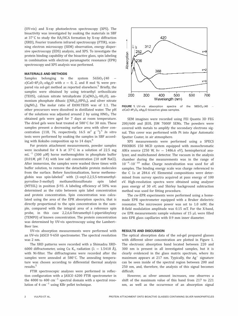
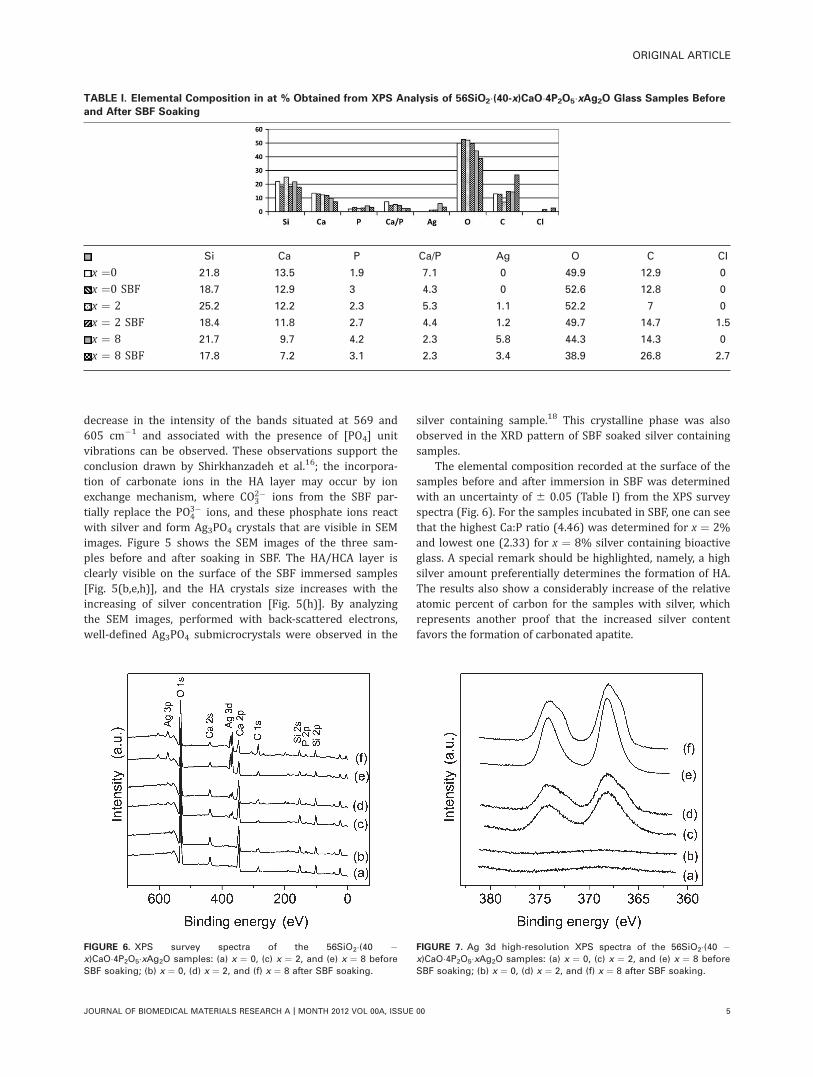
The elemental composition recorded at the surface of thesamples before and after immersion in SBF was determinedwith an uncertainty of 6 0.05 (Table I) from the XPS surveyspectra (Fig. 6). For the samples incubated in SBF, one can seethat the highest Ca:P ratio (4.46) was determined for x ¼ 2%and lowest one (2.33) for x ¼ 8% silver containing bioactiveglass. A special remark should be highlighted, namely, a highsilver amount preferentially determines the formation of HA.The results also show a considerably increase of the relativeatomic percent of carbon for the samples with silver, whichrepresents another proof that the increased silver contentfavors the formation of carbonated apatite.
TABLE I. Elemental Composition in at % Obtained from XPS Analysis of 56SiO2�(40-x)CaO�4P2O5�xAg2O Glass Samples Before
and After SBF Soaking
Si Ca P Ca/P Ag O C CI
x ¼0 21.8 13.5 1.9 7.1 0 49.9 12.9 0
x ¼0 SBF 18.7 12.9 3 4.3 0 52.6 12.8 0
x ¼ 2 25.2 12.2 2.3 5.3 1.1 52.2 7 0
x ¼ 2 SBF 18.4 11.8 2.7 4.4 1.2 49.7 14.7 1.5
x ¼ 8 21.7 9.7 4.2 2.3 5.8 44.3 14.3 0
x ¼ 8 SBF 17.8 7.2 3.1 2.3 3.4 38.9 26.8 2.7
FIGURE 6. XPS survey spectra of the 56SiO2�(40 �x)CaO�4P2O5�xAg2O samples: (a) x ¼ 0, (c) x ¼ 2, and (e) x ¼ 8 before
SBF soaking; (b) x ¼ 0, (d) x ¼ 2, and (f) x ¼ 8 after SBF soaking.
FIGURE 7. Ag 3d high-resolution XPS spectra of the 56SiO2�(40 �x)CaO�4P2O5�xAg2O samples: (a) x ¼ 0, (c) x ¼ 2, and (e) x ¼ 8 before
SBF soaking; (b) x ¼ 0, (d) x ¼ 2, and (f) x ¼ 8 after SBF soaking.
ORIGINAL ARTICLE
JOURNAL OF BIOMEDICAL MATERIALS RESEARCH A | MONTH 2012 VOL 00A, ISSUE 00 5

Furthermore, the XPS Ag 3d high-resolution spectraseem broader and asymmetric, with a shoulder at lowerbinding energy that signalizes the presence of new type ofbonds (Fig. 7).
The deconvolution of Ag 3d photoelectron peaks (Fig. 8)shows for Ag 3d5/2 line two components at the bindingenergies around 368.5 and 367 eV corresponding to metallicsilver and silver oxide, respectively (Fig. 8).19 After immer-sion in SBF solution, the Ag 3d5/2 line is well fitted withfour components. The new components occur at 367.9 and364.7 eV. The component recorded around 367.9 eV isattributed to Ag3PO4.
4 One should emphasize that the signa-ture of this structure was also evidenced in XRD patternsand SEM images. The component at lower binding energy,about 365 eV, may be associated to AgCl formation thatseems very likely, as a visible amount of chlorine was evi-denced after immersion in SBF solution (Table I).
X-band cw-EPR spectra recorded at room temperaturefor methemoglobin in solution and after adsorption ontobioactive glasses with different silver content are shown inFigure 9. In all EPR spectra were identified two compo-nents, which correspond to spin label populations with dif-ferent mobility. In solution, the mobile EPR component (a inFig. 9) arises from the surface exposed spin labels (whichpoint out of the protein, such that some flexibility isallowed), while the immobilized component (b in Fig. 9) isinterpreted as corresponding to the spin labels trapped in aprotein pocket (the so-called tyrosine pocket, due to the pe-nultimate residue being Tyr b145). This interpretation is inagreement with the results obtained by Moffat20 for horsemethemoglobin spin labeled with 4-(2-Iodoacetamido)-2,2,6,6-tetramethyl-1-piperidinyloxy) in position b-93.
On adsorption of protein on bioactive glass the equilib-rium between the two conformations is significantly shiftedtoward the component b (see Fig. 9). The spin labels of thisfraction interact with the surface of the bioactive glass orwith adjacent folded parts of protein and the observed shiftof the equilibrium between immobile and mobile states sug-gests that the environment of the b-93* residue is perturbedby the adsorption process. The increasing of silver content inthe bioactive glass leads to further immobilization of the
FIGURE 10. XPS survey spectra of the 56SiO2�(40 �x)CaO�4P2O5�xAg2O samples after protein attachment.
FIGURE 8. Deconvoluted XPS Ag 3d high-resolution spectra of the
56SiO2�(40 � x)CaO�4P2O5�xAg2O samples before and after SBF soak-
ing: (a) x ¼ 2 before SBF, (b) x ¼ 2 after SBF, (c) x ¼ 8 before SBF,
and (d) x ¼ 8 after SBF soaking.
FIGURE 9. Room temperature X-band cw-EPR spectra of horse meth-
emoglobin spin-labeled in position b-93* recorded in solution (green),
and in adsorbed state, immediately after immersion on bioactive
glass with 0% (black), 2% (red), and 8% (blue) of silver content,
respectively. The mobile and immobile components visible in the
lower field spectral lines are depicted with a and b, respectively (inset).
[Color figure can be viewed in the online issue, which is available at
wileyonlinelibrary.com.]
6 VULPOI ET AL. PROTEIN ATTACHMENT ONTO BIOACTIVE GLASSES CONTAINING SILVER NANOPARTICLES

protein. Methemoglobin becomes more rigid as a conse-quence of interaction with silver ions, which are disposed atthe surface of the bioactive glass. The explanation can bebased on the fact that Agþ reacts with thiol groups in pro-teins, due to the high affinity between the soft sulfide and thesoft metal.21,22 Although the protein was labeled with MTSspin label and, therefore, the cysteine sulfur is blocked by thespin label and thus not accessible for Ag, the labeling effi-ciency was 50%, so there are unlabeled cysteines in the sam-ple. We assume that these cysteines which do not carry aspin label interact with Ag and, consequently, unlabeled mol-ecules bind easier than spin labeled protein. This interactioncan induce a closer packing of methemoglobin on the surface,which would lead to enhanced interaction of the spin labelside chain with neighboring protein atoms. The closer pack-ing of methemoglobin around the Ag atoms leads to restric-tions in the spin label side-chain mobility, and thus the equi-librium between the two components of the EPR spectrum isshifted toward the immobile component. These assessmentsare supported by XPS elemental analysis based on XPS sur-vey spectra (Fig. 10) that indicate that the amount of the pro-tein bound at the surface increases with silver concentration(Table II), so a closer packing of the protein at the surface isevident. The effect of silver content on adsorption of methe-moglobin from solution is reflected by the evolution of thenew N 1s and S 2p photoelectron peaks, and by the greatamount of C 1s recorded after immersion in protein solution.The amounts of each of these elements were found toincrease significantly with the silver content (Table II), whilethe amounts of the main elements were reduced due to theprotein coverage of the surface. The nitrogen concentrationwas practically zero before immersion and considerablyincreased after immersion due to protein attachment.23,24
This result clearly shows a larger coverage with protein forthe sample with higher silver content. On the other hand, notonly the amount of the adsorbed protein but also the proteinconformation is important for further cell proliferation25 onthese materials. In this respect, both EPR and XPS measure-
ments show the highest agglomeration of protein moleculeson bioactive glass with 8% silver content and point out thatin this case the neighbor molecules constrain the protein tokeep its compact structure. Accordingly, increasing of silverconcentration up to 8% might prevent protein unfolding onbioactive glass surface. To characterize the conformationalchanges occurring in horse methemoglobin on binding to sil-ver nanoparticles, additional data are required, for example,pulse Double Electron Electron Resonance (DEER) measure-ments, to determine the distance between the two positionslabeled. We assume that protein conformation is affected bythis adsorption process, and the shift of the equilibrium to-ward the b component is a first clue with regard to thisassumption.
CONCLUSIONS
The influence of silver addition up to x � 8 mol % in56SiO2�(40 � x)CaO�4P2O5�xAg2O glasses on their bioactivityand protein interaction was investigated by a multitechni-que approach. UV-vis absorption and XPS VB results indicatethe presence of silver as Agþ and Ag0 distinct nanoparticlesand/or nanoparticle clusters. XRD, FTIR, SEM, and XPS datareveal that the silver addition to SiO2-CaO-P2O5 glass matrixfavors the formation of HA/HCA and also of Ag3PO4 crystalson the surface of the samples immersed in SBF. This mayensure a controllable and slower release of silver and im-plicitly an antibacterial effect of the bioactive glass for a lon-ger time. EPR and XPS results confirm that the relativelyhigh content of 8 mol % Ag2O incorporated in the bioactiveglass has an important effect in the improvement of the pro-tein affinity toward this kind of materials suggesting acloser packing of the protein at the sample surface.
REFERENCES1. Lusvardi G, Malavasi G, Menabue L, Aina V, Bertinetti L, Cerrato
G, Morterra C. Bioactive glasses containing Au nanoparticles.
Effect of calcination temperature on structure, morphology, and
surface properties. Langmuir 2010;26:10303–10314.
TABLE II. Elemental Composition in at % Obtained from XPS Analysis of 56SiO2�(40-x)CaO�4P2O5�xAg2O Glass Samples Before
and After Protein Binding
Si Ca P Ag O C N S Fe
x ¼ 0 21.8 13.5 1.9 – 49.9 12.9 – – –
x ¼ 0 Hgb 13.5 12.6 6.7 – 49.3 16 1.2 0.7 –
x ¼ 2 25.2 12.2 2.3 1.1 52.2 7 – – –
x ¼ 2 Hgb 12.3 9 4.8 1.1 43.4 26 2.6 0.8 –
x ¼ 8 Hgb 21.7 9.7 4.2 5.8 44.3 14.3 – – –
x ¼ 8 Hgb 15.5 4.3 2.3 3.1 37.8 27.9 5 1.4 2.6
ORIGINAL ARTICLE
JOURNAL OF BIOMEDICAL MATERIALS RESEARCH A | MONTH 2012 VOL 00A, ISSUE 00 7

2. Bellantone M, Coleman NJ, Hench LL. Bacteriostatic action of a
novel four-component bioactive glass. J Biomed Mater Res 2000;
51:484–490.
3. Bellantone M, Williams HD, Hench LL. Broad-spectrum bacteri-
cidal activity of Ag2O-doped bioactive glass. Antimicrob Agents
Chemother 2002;46:1940–1945.
4. Buckley JJ, Lee AF, Olivic L, Wilson K. Hydroxyapatite supported
antibacterial Ag3PO4 nanoparticles. J Mater Chem 2010;20:
8056–8063.
5. Vulpoi A, Baia L, Simon S, Simon V. Silver effect on the structure
of SiO2-CaO-P2O5 ternary system. Mat Sci Eng C 2011;32:178–183.
6. Kokubo T, Kushitani H, Sakka S, Kitsugi T, Yamamuro T. Solu-
tions able to reproduce in vivo surface-structure changes in bioac-
tive glass-ceramic A-W. J Biomed Mater Res 1990;24:721–734.
7. Klare JP, Steinhoff H-J. Spin labeling EPR. Photosynth Res 2009;
102:377–390.
8. Lu JQ, Bravo-Suarez JJ, Takahashi A, Haruta M, Oyama ST. Ag/SiO2:
A novel catalyst with high activity and selectivity for hydrogenation
of chloronitrobenzenes. Chem Commun 2005;24:5298–5300.
9. Chakraborty P. Metal nanoclusters in glasses as nonlinear pho-
tonic materials. J Mater Sci 1998;33:2235–2249.
10. Le Saout G, Simon P, Fayon F, Blin A, Vaills Y. Raman and infrared
study of (PbO)x(P2O5)1-x glasses. J Raman Spectrosc 2002;33:740–746.
11. Mie G. Beitr€age zur Optik truber Medien. Ann Phys 1908;25:
377–445.
12. Link S, El-Sayed MA. Optical properties and ultrafast dynamics in
metallic nanocrystals. Ann Rev Phys Chem 2003;54:331–366.
13. Baia L, Muresan D, Baia M, Popp J, Simon S. Structural proper-
ties of silver nanoclusters-phosphate glass composites. Vib Spec-
trosc 2007;2:313–318.
14. P�aszti Z, Pet€o G, Horv�ath ZE, Karacs A, Guczi L. Electronic struc-
ture of Ag nanoparticles deposited on Si. Solid State Commun
1998;107:329–333.
15. Downs RT (2006) The RRUFF Project: an integrated study of the
chemistry, crystallography, Raman and infrared spectroscopy of
minerals. Program and Abstracts of the 19th General Meeting of
the International Mineralogical Association in Kobe, Japan, O03-
13. RRUFF ID: R050512. Available at: http://rruff.info/hydroxylapa-
tite/display¼default/R050512.
16. Shirkhanzadeh M, Azadegan M. Formation of carbonate apatite
on calcium phosphate coatings containing silver ions. J Mater Sci
Mater Med 1998;9:385–391.
17. Srikumar T, Kityk IV, Srinivasa Rao Ch, Gandhi Y, Piasecki M, Bra-
giel P, Ravi Kumar V, Veeraiah N. Photostimulated optical effects
and some related features of CuO mixed Li2O–Nb2O5–ZrO2–SiO2
glass ceramics. Ceram Int 2011;37:2763–2779.
18. Bi Y, Ouyang S, Cao J, Ye J. Facile synthesis of rhombic dodeca-
hedral AgX/Ag3PO4 (X¼Cl, Br, I) hetero-crystals with enhanced
photocatalytic properties and stabilities. Phys Chem Chem Phys
2011;13:10071–10075.
19. Kumar PA, Reddy MP, Ju LK, Phil HH. Novel silver loaded hy-
droxyapatite catalyst for the selective catalytic reduction of NOx
by propene. Catal Lett 2008;126:78–83.
20. Moffat JK. Spin-labelled haemoglobins: A structural interpretation
of electron paramagnetic resonance spectra based on X-ray anal-
ysis. J Mol Biol 1971;55:135–146.
21. Simchi A, Tamjid E, Pishbin F, Boccaccini AR. Recent progress in
inorganic and composite coatings with bactericidal capability for
orthopaedic applications. Nanomed-Nanotechnol 2011;7:22–39.
22. Feng QL, Wu J, Chen GQ, Cui FZ, Kim TN, Kim JO. A mechanistic
study of the antibacterial effect of silver ions on Escherichia coli
and Staphylococcus aureus. J Biomed Mater Res 2000;52:
662–668.
23. Arvidsson A, Currie F, Kjellin P, Sul YT, Stenport V. Nucleation
and growth of calcium phosphates in the presence of fibrinogen
on titanium implants with four potentially bioactive surface prep-
arations. An in vitro study. J Mater Sci Mater Med 2009;20:
1869–1879.
24. Vanea E, Simon V. XPS study of protein adsorption onto nano-
crystalline aluminosilicate microparticles. Appl Surf Sci 2011;257:
2346–2352.
25. Gauckler LJ, Rezwan K. Adsorption of biomolecules on ceramic
particles and the impact on biomedical applications. Adv Sci Tech
2006;45:741–751.
8 VULPOI ET AL. PROTEIN ATTACHMENT ONTO BIOACTIVE GLASSES CONTAINING SILVER NANOPARTICLES