![A Family of Cyanide-Bridged Molecular Squares: Structural and Magnetic Properties of [{M II Cl 2 } 2 {Co II (triphos)(CN) 2 } 2 ]· x CH 2 Cl 2 , M = Mn, Fe, Co, Ni, Zn](https://static.fdokumen.com/doc/165x107/633d21a3494bd0957806efe3/a-family-of-cyanide-bridged-molecular-squares-structural-and-magnetic-properties.jpg)
Binding of the ferric uptake regulation repressor protein (Fur) to Mn(II), Fe(II), Co(II), and...
18
Binding of the Ferric Uptake Regulation Repressor Protein (Fur) to M&I), Fe@& Co(II), and C&I) Ions as Co-Repressors: Electronic Absorption, Equilibrium, and “Fe Massbauer Studies Mazen Y. Hamed,* J. B. Neilands, and V. Huynh+ Biochem&y Lkpartment, University of Califonria, Berkeley, Cakfomia, U.S. ABSTRACT The binding of the repressor protein (Fur) to Fe(H) as co-repressor was studied. Other transition metal ions such as M&I), Co(B), and Cu(I1) were also studied as models. From the equilibrium studies Kd values of 55,85,36, and 10 PM were obtained for the Fur complex with Fe(II), Mn(II), Co(II), and C&I), respectively. The ratio of metal to Fur monomer was 1:l in both the Fe(II) and M&I) complexes. Fur mutants were also studied. Electronic absorption spectra of the CofII) Fur complex gave evidence of a distorted tetrahedral Co(E) site bound to sulfur. Frozen solution 57Fe Miissbauer spectra of the Fe(B) Fur indicated the presence of Fe(II) in a high spin distorted octahedral environment. The role of the metal ion as co-repressor in the binding of Fur to DNA is discussed in view of the above results. INTRODUCTION Regulation of iron uptake in bacteria has been extensively studied because of its important medical sign@ance. It is established that one of the consequences of imbalance in iron regulation is the possible formation of poisonous free radicals in the presence of surplus iron [l]. Eschetichiu coli adjusts its iron uptake at the molecular level by regulating the synthesis and transport of iron chelators using Address reprints requests and correspondence to: Dr. Mazen Y. Hsmed, Chemistry Department, Birzeit University, West Bank, Via Israel. *A Fulbright visiting research associate in J. B. Neihmds’ Laboratory. ‘Address: Physics Department, Emory University, Atlanta, GA, U.S. Joumd of Inorganic Biochemistry, 15,193-210 (1993) 193 0 1993 Elsevier Science Publishing Co., Inc., 655 Avenue of the Americas, NY, NY 10010 0162-0134/93/$6.00
Transcript of Binding of the ferric uptake regulation repressor protein (Fur) to Mn(II), Fe(II), Co(II), and...
Binding of the Ferric Uptake Regulation Repressor Protein (Fur) to M&I), Fe@& Co(II), and C&I) Ions as Co-Repressors: Electronic Absorption, Equilibrium, and “Fe Massbauer Studies
Mazen Y. Hamed,* J. B. Neilands, and V. Huynh+
Biochem&y Lkpartment, University of Califonria, Berkeley, Cakfomia, U.S.
ABSTRACT
The binding of the repressor protein (Fur) to Fe(H) as co-repressor was studied. Other transition metal ions such as M&I), Co(B), and Cu(I1) were also studied as models. From the equilibrium studies Kd values of 55,85,36, and 10 PM were obtained for the Fur complex with Fe(II), Mn(II), Co(II), and C&I), respectively. The ratio of metal to Fur monomer was 1:l in both the Fe(II) and M&I) complexes. Fur mutants were also studied. Electronic absorption spectra of the CofII) Fur complex gave evidence of a distorted tetrahedral Co(E) site bound to sulfur. Frozen solution 57Fe Miissbauer spectra of the Fe(B) Fur indicated the presence of Fe(II) in a high spin distorted octahedral environment.
The role of the metal ion as co-repressor in the binding of Fur to DNA is discussed in view of the above results.
INTRODUCTION
Regulation of iron uptake in bacteria has been extensively studied because of its important medical sign@ance. It is established that one of the consequences of imbalance in iron regulation is the possible formation of poisonous free radicals in the presence of surplus iron [l]. Eschetichiu coli adjusts its iron uptake at the molecular level by regulating the synthesis and transport of iron chelators using
Address reprints requests and correspondence to: Dr. Mazen Y. Hsmed, Chemistry Department, Birzeit University, West Bank, Via Israel.
*A Fulbright visiting research associate in J. B. Neihmds’ Laboratory.
‘Address: Physics Department, Emory University, Atlanta, GA, U.S.
Joumd of Inorganic Biochemistry, 15,193-210 (1993) 193 0 1993 Elsevier Science Publishing Co., Inc., 655 Avenue of the Americas, NY, NY 10010 0162-0134/93/$6.00

194 M. Y. Hamed et al.
a 17 kDa molecular weight repressor, the ferric uptake regulation protein (Fur). This protein binds a 19 base pair segment of DNA in the “iron box” [2]. It was found that Fur, which is a negative regulator of expression of siderophore mediated iron assimilation, binds a divalent transition metal ion as co-repressor i.e., Fe(R) [l-3].
In vitro studies have shown that Mn(II), Fe(R), Co(II), or cd(R) were capable of activating Fur to bind the DNA [2,3,25]. Zn(I1) was partially effective and at higher concentrations, interfered with the activity of IkIn(II) [l]. In previous studies of the effect of CdUI), Fe(H), and M&I) on Fur binding to DNA, excess metal ion to Fur ratios were always used in the DNA-binding assays [3,25,26]. Neilands et al. [26] estimated a Fur concentration of 7 nM required to half saturate a DNA-fragment carrying the “iron box” in the presence of excess metal ion.
Neither the affinity nor the nature of the sites provided by Fur to bind the metal ions are known. Similarly, the exact nature of contacts between the Fur Fe(R) complex and the DNA operator [7], and the level of co-repressor needed, are points to be established.
A computer analysis of the Fur sequence revealed a potential for a substantial degree of alpha-helical structures [l], which was to be expected for DNA binding repressors. NMR data also revealed some structural aspects about the Fur sequence [51.
In addition to an iron uptake regulatory function of Fur, it participates in control of expression of manganese and iron containing superoxide dismutase of E. coii [4]. Thus, Fur is apparently part of a global regulatory circuit in E. coli.
The importance of Fe(R) ion as an activator and the Fur co-repressor activity of other divalent metal ions in vitro provided a basis for establishing the metal ion role by studying the coordination of M&II), Co(R), and C&II) to Fur as models for Fe(R). In the present work, the equilibrium dialysis, electronic absorption spectra, and “Fe Mijssbauer studies have provided information about the metal ion binding sites of Fur.
MATBRIALS AND METHODS
Materials
Fur protein was purified by the method of Wee et al. [7] from E. coli J R B 45 (p MON 2064). An oligonucleotide DNA “iron box” was prepared as described in Ref. [7]. CoC1,.6H,O, MnC1,.4H,O (99.99%), and CuC12.2H,0 (99.9%) were purchased from Chemical Dynamics, and Aldrich and Baker companies, respectively.
FeCl,.4H,O was freshly prepared, dried under nitrogen, and the crystals stored under nitrogen until needed. For atomic absorption analysis, standard dilutions were made from an iron solution (Sigma), 996 pg/ml, and from each of the following solutions: Co, Cu, Zn, and Mn (100 Fg/ml f l%, Fisher). The mutant Fur proteins were prepared and kindly supplied by C. Doyle and C. Besser in this laboratory.
Equilibrium Dialysis Studies
A 45 PM Fur solution in a constant volume of 3.0 ml of 20 mM MOPS buffer, pH 7.2, was dialyzed for 8 hr against 1.0 liter metal ion solution at a constant

FERRIC UPTAKE REGULATION REPRESSOR PROTEIN 195
ionic strength of 0.5 M NaCl at 20°C. The metal ion concentration range used was 30-350 PM. The metal concentration was measured by atomic absorption using as reference a standard metal ion solution in the recommended range (1.0-5.0 pg/ml), in 45 PM Fur solution containing 20 mM MOPS buffer and 0.5 M NaCl. For the experiments where iron(B) was involved the water was degassed, bubbled with nitrogen, and then a weighed amount of FeCl,. 4H,O was dissolved under nitrogen. The experiment was performed under a continu- ous flow of nitrogen [24]. At the end of each experiment the volume and pH of the Fur solution were measured but no significant change was observed. All data were computer fitted. A portion of the Fur/metal ion solution was stored in the deep freeze and later analyzed on a polyacryl-amide nondenaturing gel for comparison with fresh Fur and BSA. The Fur/metal ion complex gave a similar band to that of the untreated Fur dimer indicating no change in the protein, i.e., no denaturation took place in the presence of metal ions [26].
Visible Absorption Spectrophotometry
A 0.23 mM Fur solution was prepared in a volume of 0.4 ml in the appropriate buffer (see text), transferred to a cuvette, and titrated by adding 2.0 1.11 aliquotes of 10 mM metal ion solution. Spectra were recorded after each addition using a Shimadzu UV-160 recording spectrophotometer.
Pwpmtion of s7Fe(II) Fur M&sbauer samples. A weighed amount of 57Fe (95.3%), purchased from ISOTEC Inc., was dissolved in hot concentrated HCl to produce 0.1 mM concentration in a known volume. Exactly 0.5 ml portions of the iron solution were lyophilized to dryness.
The following were placed in a glove box: A 250 ~1 aliquot of 0.4 mM Fur or the double mutant (C92S C95S) solution in 40 mM MOPS, pH 7.5, degassed H,O, Miissbauer cells, and liquid nitrogen.
The iron was redissolved in 250 ~1 H,O, pH 7.0, and mixed with the Fur solution, the pH was adjusted to 7.2. The complex was transferred to the Miissbauer cells, frozen in liquid nitrogen, and stored in liquid nitrogen. The same process was repeated for the mutant Fur. The samples were shipped on dry ice. Preliminary Miissbauer measurements were performed by V. Huynh in the Department of Physics, Emory University (Atlanta, GA).
RESULTS AND DISCUSSION
Equilibrium Dialysis Studies
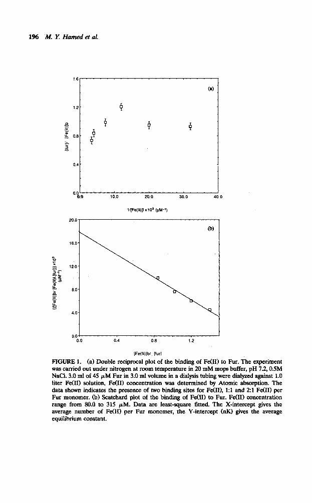
Iron(I.0 and h4nW) Complexes with Fur. From the scatchard plot in Figure 1 it is apparent that two Fe(B) ions associate with the Fur protein with an average dissociation constant of 55 PM as calculated from both the inverse of the slope and the ordinate (Fig. lb) [8]. The Benesi-Hildebrand plot 18, 91 showed the presence of two different Fe(II) sites (Fig. la). An attempt to analyze the data produced approximate values for Kl and K2 with a value for Kl indicating that the first site is approximately W-fold stronger than the site involved in K2, i.e., second Fe011 site (see Table 1).
While the interaction of Fe(B) with Fur showed the involvement of two different sites, M&I) gave a 1:l complex throughout the concentration range used in this work (Fig. 2). The site involved in this case is weaker (Kd = 85 PM)
1% .M. Y; Hamed et at!
l[Fe(ll)JfxlO' (PM-')
16.0
0.04 0.0 0.4 0.6 1.2
[Fe(li)jbl_ (furl
FIGURE 1. (a) Double reciprocal plot of the binding of Fe(H) to Fur. The experiment was carried out under nitrogen at room temperature in 20 mM mops butfer, pH 7.2, OSM NaCl. 3.0 ml of 45 PM Fur in 3.0 ml volume in a dialysis tubing were dialyzed against 1.0 liter Fe(H) solution, Fe(H) concentration was determined by Atomic absorption. The data shown indicates the presence of two binding sites for Fe(D), 1:l and 2:l Fe(H) per Fur monomer. (b) Scatchard plot of the bmclmg of Fe(H) to Fur. Fe(H) concentration range from 80.0 to 315 PM. Data are least-square fitted. The X-intercept gives the average number of Fe(H) per Fur monomer, the Y-intercept (nK1 gives the average equilibrium constant.

FERRIC UPTAKE REGULATION REPRESSOR PROTEIN 197
TABLE 1. Equih%rium Constants and Ratios of Different Metal Ions Studied with Fur and Fur Mutants
Metal Average Average Kd
{M(II)/Fur) ( pM) Site-Multiplicity
M&I) 1 85 Not observed Fe(II) 2 55 ObSXWd CdII) 6 36 Observed cu(II) 6.5 10 Not observed
Metal(II)/Fur mutant Average n value Average Kd *0.5 f 10%
Fe(II)/(CXS C92S) 2 MPM cdII)/(c9SS c92s) 6 1.3 mM cu(II)/Oz95c cxw 3.8 6.5 pM co(II)/C95S 0 3.3 lnM co(II)/c92s 1 70 pM
Dissociation constants analyzed (values f 15%). Fe(II)/Fur Kl = 30 pM, K2 = 280 pM, C&I)/ Fur, n = 2.5, K = 60 &I, n = 7, K = 600 pM.
5.0 10.0 15.0
l/[Mn(ll)]f x103 (fbl-‘)
FIGURE 2. Double reciprocal plot of the bin&g of M&I) to Fur. The Y-intercept (= l/n) where n is the number of Mn(I1) per Fur (monomer), and the X-intercept (== - l/Kd) gives the average dissociation constant. Other conditions are as for Figure 1.
198 M. X Humed et al.
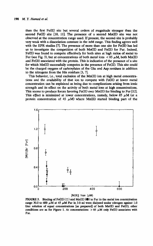
than the first Fe(R) site but several orders of magnitude stronger than the second Fe(R) site [lo, 111. The presence of a second M&I) site was not observed at the concentration range used. If present, the second site is probably very weak with a dissociation constant in the mM range. This finding agrees well with the EPR studies [7]. The presence of more than one site for Fe(I1) has led us to investigate the competition of both Mn(I1) and Fe(R) for Fur. Indeed, Fe(R) was found to compete effectively for both sites at high ratios of metal to Fur (see Fig. 3), but at concentrations of both metal ions < 85 PM, both M&I) and Fe(R) associated with the protein. This is indicative of the presence of a site for which Mn(I1) successfully competes in the presence of Fe(R). This site could be the charged oxygens of carboxylates of the Glu and Asp residues in addition to the nitrogens from the His residues [S, 71.
This behavior, i.e., total exclusion of the Mn(I1) ion at high metal concentra- tions and the availability of that ion to compete with Fe(R) at lower metal concentration can be explained as being due to complications arising from ionic strength and its effect on the activity of both metal ions at high concentrations. This seems to produce forces favoring Fe(R) over M&I) for binding to Fur [121. This effect is minimized at lower concentrations, namely, below 85 PM (at a protein concentration of 45 PM) where M&I) started binding part of the
1.6
1.2
i- I= 2 0.0 =;: e- z
0.4
0.0
P
4 P
f IL 200 400 600
FWI free (PM) FIGURE 3. Binding of Fe011 ( 0 1 and hfn(II) (W to Fur in the metal ion concentration range 30.0 to 600 pM at 45 pM Fur in 3.0 ml were dialyzed unde.r nitrogen against 1.0 liter solution of equal concentrations [as prepared] of both Mn(I1) and Fe(II), other conditions are as for Figure 1. At concentrations > 85 pIU only Fe00 associates with Fur.

FERRIC UPTAKE REGULATION REPRESSOR PROTEIN 199
protein sites. It should be kept in mind that metal ions in solution at high concentrations are more hydrated than at lower concentration. Also, the activity coefficients approach higher values, which affects the movement of hydrated ions. All this has a direct effect on the energies involved in binding of these ions to the protein since they do not bind as isolated ions but rather as hydrates. Dilution of the metal ions at constant ionic strength seems to have an affect on their relative availability towards the protein binding sites and hence the energy factor will become more pronounced in determining the affinity of the ligating sites towards the metal ion.
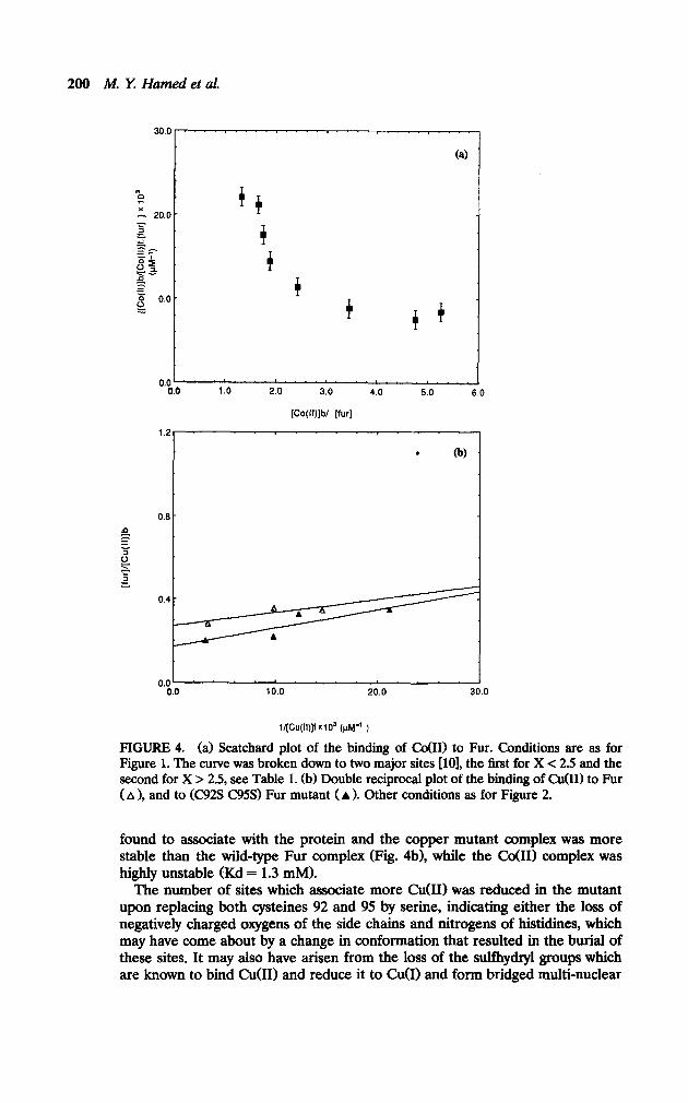
Co(ZZ) and Cu(ZZZ Complexes with Fur. Both co(B) and Cu(I1) ions showed higher affinity towards Fur than either Fe(B) and/or M&I) (Figs. 4a and 4b), this behavior is reminiscent of the Irving-Williams series, i.e., stability increases in the order M&I) < Fe(B) < co(B) < C&I), which implies the involvement of similar types of ligands from the Fur to all these ions (see Fig. 5 and Table 1). The interaction of both co(B) and Cu(I1) with Fur is characterized by the association of more metal ions per Fur, i.e., up to about six per Fur monomer. This finding agrees with the ability of both metal ions to form four-coordinate complexes, i.e., tetrahedral in the case of co(H) and square planer in the case of Cu(I1) (see electronic spectra). The smaller metal ion radius and the larger ligand field stabilization energy (LFSE), in these cases, play an important role in the formation of such complexes in addition to the presence of a total of 24 negative charges on the protein arising from Glu and Asp residues [13].
For Cobalt(B) an analysis of the data gave two different dissociation constants (Table 1) indicating the presence of at least two different sites; a weaker site (Kd=600 PM) h’ h w ic originates in the electrostatic attraction of the metal ion to the charged side chains and a stronger site (Kd = 60 PM) which is the main co(B) site (see electronic spectra). In the case of Cu(I1) the plot gave a straight line (Figure 4b) indicating that the binding constants could not differ by an amount of more than twofold (see UV-Visible studies below) [9, 111.
The association of more than two metal ions in the case of Co(B) and C&I) can be further explained as due to nonspecific binding which results from metal ion-ligand electrostatic attractions, in addition to the smaller metal ion radius and larger LFSE in the geometries involved. The protein tertiary structure must have available pockets into which these ions fit with a minimum energy require- ment.
Fur Mutants with Metal Zons. Since scatter and variations in the slope are common at low and high levels of binding [S, 111, the positive slope in the Scatchard plot at low saturation was ignored, except in the case of the cysteine mutant’s interaction with C&I). The positive slope forms a considerable part of the plot which cannot be ignored and was interpreted in similar studies [lo, 111 as due to the interaction between the binding sites on the protein. Since these mutants showed such behavior strongly in the NMR studies (Williams et al., unpublished), one can say this interpretation is very likely.
The studies on the mutant Fur (C92S C95S) produced more scattered data with a positive slope, probably due to aggregation, but values for stability constants and metal to protein ratios could be obtained (see Table 1). In case of Fe00 the double cysteine mutant gave higher stability and a 2:l metal to Fur ratio throughout the titration. In the case of co(B) and Cu(I1) less metal was
200 M. l-7 Hamed et al.
0.0 ‘. m.. e.. .“. c.. ‘. “.”
0.0 1.0 2.0 3.0 4.0 5.0 6.0
[Co(ll)jb/ [fur]
1.2 I
. 0
0.0’ 0.0 10.0 20.0 30.0
l/(Cu(ll)]f X103 (pt+ )
FIGURE 4. (a) Scatchard plot of the binding of CdIl> to Fur. Conditions are as for Figure 1. The curve was broken down to two major sites [lo], the first for X < 2.5 and the second for X > 2.5, see Table 1. (b) Double reciprocal plot of the binding of 0101) to Fur ( A 1, and to (C92S C95S) Fur mutant (A 1. Other conditions as for Figure 2.
found to associate with the protein and the copper mutant complex was more stable than the wild-type Fur complex (Fig. 4b1, while the Co011 complex was highly unstable (Kd = 1.3 mM).
The number of sites which associate more CuOI) was reduced in the mutant upon replacing both cysteines 92 and 95 by serine, indicating either the loss of negatively charged oxygens of the side chains and nitrogens of histidines, which may have come about by a change in conformation that resulted in the burial of these sites. It may also have arisen from the loss of the sulfhydryl groups which are known to bind C&I) and reduce it to Cu(I) and form bridged multi-nuclear
FERRIC UPTAKE REGULATION REPRESSOR PROTEIN 201
0.0’ . 0 10 20 30
l/[M(ll)]f x 103 (prvl-1)
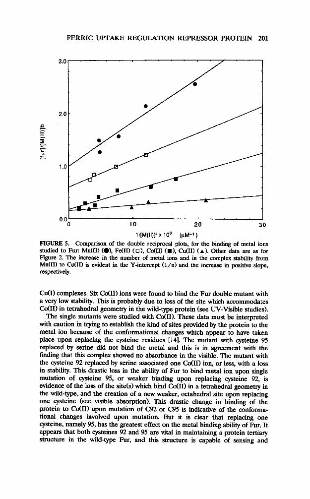
FIGURE 5. Comparison of the double reciprocal plots, for the binding of metal ions studied to Fur: Mn(I1) (01, Fe(U) (01, Co011 CD), C&I) (A ). Other data are as for Figure 2. The increase in the number of metal ions and in the complex stability from M&I) to CuUI) is evident in the Y-intercept (l/n) and the increase in positive slope, respectively.
MI) complexes. Six Co011 ions were found to bind the Fur double mutant with a very low stability. This is probably due to loss of the site which accommodates G$II) in tetrahedral geometry in the wild-type protein (see W-Visible studies).
The single mutants were studied with co(II). These data must be interpreted with caution in trying to establish the kind of sites provided by the protein to the metal ion because of the conformational changes which appear to have taken place upon replacing the cysteine residues 1141. The mutant with cysteine 95 replaced by serine did not bind the metal and this is in agreement with the finding that this complex showed no absorbance in the visible. The mutant with the cysteine 92 replaced by serine associated one co(II) ion, or less, with a loss in stability. This drastic loss in the ability of Fur to bind metal ion upon single mutation of cysteine 95, or weaker binding upon replacing cysteine 92, is evidence of the loss of the site(s) which bind Co(R) in a tetrahedral geometry in the wild-type, and the creation of a new weaker, octahedral site upon replacing one cysteine (see .visible absorption). This drastic change in binding of the protein to co(R) upon mutation of C92 or C95 is indicative of the conforma- tional changes involved upon mutation. But it is clear that replacing one cysteine, namely 95, has the greatest effect on the metal binding ability of Fur. It appears that both cysteines 92 and 95 are vital in maintaining a protein tertiary structure in the wild-type Fur, and this structure is capable of sensing and
202 M. Y Hamed et al.
binding (MI), and hence Fe(II), and other first-row transition metal ions. Indeed, it has been shown that chemical derivitixation of cysteine thiol groups destroys Fur DNA binding activity [27]. Doyle et al. (manuscript in preparation) confirmed this observation using site-directed mutagenesis which showed that Cys 92 and Cys 95 are necessary for repressor activity.
PfeZiminury Miissbauer Shtdy In a preliminary study on a froxen solution sample of Fe(U) Fur complex the isomer shift and quadrupole splitting indicated the presence of Fe(I1) in a high spin, highly distorted octahedral environment similar to FeC1,.9H,O [22] (6 = 1.3 mms-’ and A = 3.35 mms-‘). The spectra indicated the presence of one type of Fe(I1) sites, although the presence of two Fe(I1) atoms in two identical environments cannot be ruled out. The parameters are typical of high spin Fe(I1) bound to oxygen, nitrogen, and sulfur [231. No indication of pentacoordinate Fe(U) in a mostly sulfur environment, as observed for thiocarbamate and other thiol complexes giving an isomer shift value less than 1.0 nnns-’ and A value in the range 3.9-4.0 mms-’ [23, 241, and no evidence for the presence of FeUI) tetrahedrally bound in a sulfur environment was observed. The C92S C95S double mutant Fur with Fe(I1) gave a lower isomer shift value (0.92 mms-‘) and a A = 3.3 mms-’ which confhms the presence of Fe(I1) in a different environment from that in the wild-type. These parameters indicated a stronger ligand provided by the Fur mutant, which agrees with the larger stability as obtained from the equilibrium study.
Electronic Absorption Studies
Co@..) Fur Complex. Co(I1) was found to activate the Fur protein to bind the DNA operator [l, 31. co(I1) has long been used as a spectroscopic probe to substitute for other metal ions in metalloproteins because of its well-known ability for the formation of four-coordinate distorted tetrahedral complexes with proteins, although pentacoordinate was also observed [15]. It is also compatible with a Cu(I1) four-coordinate geometry [16], since Co(I1) has been successfully substituted for Cu(I1) in tetrahedrally distorted C&I) binding sites in proteins [16, 171. In spite of the fact that Fur is regarded as a metal binding protein rather than a metalloprotein [l], the use of CdII) is in line with its activity and the Fur affinity towards it.
The Fur protein was titrated with Co(I1) at pH 8.0 in either Tris or phosphate buffers. The titration data are shown in Figures 6a and 6b. During the titration there is little evidence for heterogeneity in spectral species. This indicates cooperativity, i.e., a 2:l complex is always formed by cooperative 2 Co00 binding (2:l major species), although there is a possibility that in the spectra recorded in phosphate buffer, an octahedral complex is more pronounced at lower Co(I1) ratios, i.e., up to 1.4:1 ratio (see Fig. 6b). Bands characteristic of the tetrahedral complex are observed in the titration performed in Tris buffer from the first equivalent of C&I) added. It is evident in the visible spectra that the formation of the tetrahedral complex was fully developed at a 2:l metal to Fur ratio in both buffers, but continued forming until the whole process was hampered by precipitation at 3.6 equivalents of CdII) per Fur. The visible spectrum of the Co01) complex (see insert of Fig. 6b) is characterized by absorbances which are typical of tetrahedrally bound CdII) in other proteins [19] (see Table 2). Specifically, the co(I1) Fur spectrum bears a resemblance to

FERRIC UPTAKE REGULATION REPRESSOR PROTEIN 203
0.08
0.04
o.oo 2 400 600 800 TOO 800
Wavelength
,
500 600 TOO 800
Wavelength
FIGURE 6. (a) Visible absorption spectra of the Fur C&I) complex in 20.0 mM Tris buffer at pH 8.0, recorded at different CdII) concentrations and 0.1 M NaCI. From bottom to top: Base line, 0.23, 0.38, 0.45, 0.52, 0.58, 0.65, 0.71 mM C&I), [Fur] = 0.23 mM. (b) Visible absorption spectra of Co(I1) Fur complex in 20 mM phosphate buffer, pH 8.CJ and ionic strength 0.1 M NaCl, recorded at varying CdII) concentrations. Bottom spectrum is for 0.23 mM Fur, then spectra were recorded after addition oE 0.16, 0.31, 0.45, 0.58, 0.71, and 0.83 mM C&I). Insert: Amplification of the spectrum recorded at 0.83 mM 0~01) concentration.
the visible spectra of Co(H) azurin [18], in which C&I) is bound to sulfur from cysteine and to three nitrogens from histidines. Further evidence for the co(H) tetrahedral site was seen in the EPR spectrum of Fur Co(R) complex (Hamed et al. submitted to BioMetuls).
The S + Co charge transfer band was also observed at around 340 nm, indicating a eysteine binding to the Cd10 in the tetrahedral site [19]. The ligation of the cysteine sulfur was confirmed by titrating the cysteine mutant Fur with C&I); the (C92S C95S) double mutant Fur did not give the visible absorbance seen in the wild-type Fur Co@) system (see Fig. 7); instead, an

204 hf. Y Hamed et al.
TABLE 2. W-Visible Absorption Data for Co(E), Cu(II), and Ni(I1) Fur Complexes as Compared to Other Proteins
Complex Reference
co(B) Fur co(H) azurin C&I) PFrPb Co@) SODC Co011 alkaline
phosphatase Cu(II) Fur 1:l
Ni(II) Fur 2:l Ni(I1) carbonic
anhydrase
Visible absorptiona (Ml)
715(180), 670(250), 530,340 (sh) 641wO), 531000), 380(1100), 33Oc2600) 596(700), 528(500), 398 (sh), 374WOO), 3330400) 615,575,533 640,605,555,510
600 (300) broad 630 broad 650 broad (231,410 (53) 630 (10)
Reference
18 18
7 19,2O
20
a Value in brackets represents c; b PFIT = pentaflourothiophenol; ’ SOD = superoxide dismutase.
absorbance typical of octahedrally bound Co@) sites was observed. Both single mutant Fur proteins did not show any considerable absorbance in the visible: C95S showed a band at 420 nm (E = 126 cm-’ M- ‘) which was not seen in either the double mutant (C92S C95S) or the mutant C92S. This absorbance can be interpreted as a charge transfer from the sulfur of cysteine 92 to Co@) [25]. In summary the following observations about the visible absorption of CoGI) Fur can be made:
1. No detectable effect of phosphate on the tetrahedrally bound co(U) site could be observed, i.e., absorbance and intensity were the same in both tris and phosphate buffers. This is contrary to other models studied [16, 171, which showed the participation of phosphate in Co(H) binding.
0.00 1 I I I I 400 600 600 700 800
Wavelength
FIGURE 7. Visible absorption spectra of the Fur mutant (c95S C92S) with various co(R) concentrations in 20 mM phosphate buffer at pH 8.0 spectra starting from bottom: 0.23 mM Fur mutant, 0.16,0.23, 0,31,0.38, 0.45,0.71, and 0.83 mM co(E) added to Fur. Insert is the spectrum at 0.71 mM Co(R) concentration.
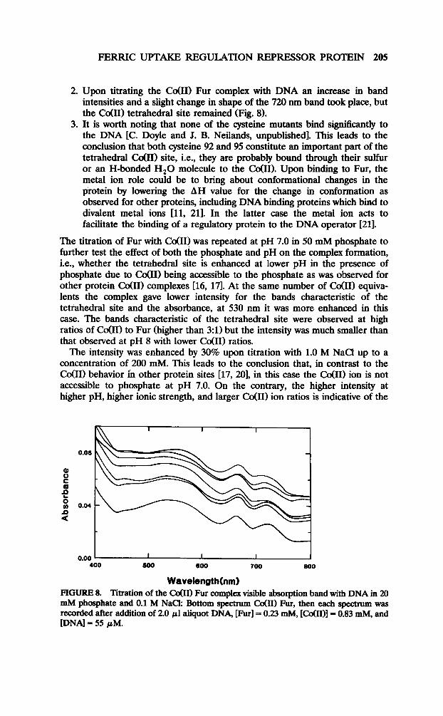
FERRIC UPTAKE REGULATION REPRESSOR PROTEIN 205
2. Upon titrating the Co(H) Fur complex with DNA an increase in band intensities and a slight change in shape of the 720 nm band took place, but the co(R) tetrahedral site remained (Fig. 8).
3. It is worth noting that none of the cysteine mutants bind significantly to the DNA [C. Doyle and J. B. Neilands, unpublished]. This leads to the conclusion that both cysteine 92 and 95 constitute an important part of the tetrahedral Co01) site, i.e., they are probably bound through their sulfur or an H-bonded H,O molecule to the Co(R). Upon binding to Fur, the metal ion role could be to bring about conformational changes in the protein by lowering the AH value for the change in conformation as observed for other proteins, including DNA binding proteins which bind to divalent metal ions [ll, 211. In the latter case the metal ion acts to facilitate the binding of a regulatory protein to the DNA operator [21].
The titration of Fur with co(R) was repeated at pH 7.0 in 50 mM phosphate to further test the effect of both the phosphate and pH on the complex formation, i.e., whether the tetrahedral site is enhanced at lower pH in the presence of phosphate due to C&II) being accessible to the phosphate as was observed for other protein co(R) complexes [16, 171. At the same number of Cd111 equiva- lents the complex gave lower intensity for the bands characteristic of the tetrahedral site and the absorbance, at 530 nm it was more enhanced in this case. The bands characteristic of the tetrahedral site were observed at high ratios of co(R) to Fur (higher than 3:l) but the intensity was much smaller than that observed at pH 8 with lower Co(R) ratios.
The intensity was enhanced by 30% upon titration with 1.0 M NaCl up to a concentration of 200 mM. This leads to the conclusion that, in contrast to the Co00 behavior in other protein sites [17, 201, in this case the Co(R) ion is not accessible to phosphate at pH 7.0. On the contrary, the higher intensity at higher pH, higher ionic strength, and larger Co(IIl ion ratios is indicative of the
Wavelength FIGURE 8. Titration of the CoOI) Fur complex visible absorption band with DNA in 20 mM phosphate and 0.1 M NaCk Bottom spectrum Cd111 Fur, then each spectrum was recorded after addition of 2.0 ~1 aliquot DNA, [Furl = 0.23 mM, [C&I)] = 0.83 mM, and [DNA] = 55 PM.

206 A4. Y. Hamed et al.
involvement of a functional group which needs to be deprotonated. The most likely group is the SH group of the cysteine residue. It is also likely that the higher ionic strength enhanced the availability of the negatively charged oxygens to bind the metal.
Co(ZZ~/iWa(ZZ~ Fur. No considerable effect dn’ the co(U) visible absorption spectra was recorded in the presence of one Mn(II) equivalent. The absorbance characteristic of the tetrahedral co(U) site was always seen in the presence of Mn(I1).
Cu(ZZ) Fur Complex. Titration of the Fur protein with cU(II) at pH 8.0 produced a broad band in the visible with a maximum at 600 nm. A shift in the maxima to 630 nm took place upon increasing the Cu(II)/Fur ratio (Fig. 9a). The hetero-
600 700
Wovelength (nm)
500 600 700
Wavelength (rim)
FIGURE 9. (a) Visible spectra ot the tnrauon of Fur protem with Cu(I1): Spectra (1) 0.26 mM Fur in the absence of CuUI). With increasing Cu (II) concentrations as foknvs: (2) 0.083, (3) 0.17, (4) 0.25, (5) 0.41, (6) 0.49, (7) 0.57 mM. (b) Titration of Fur Mn(I1) complex with Cu(II), in 20 mM Tris pH 8.0 and 500 mM NaC!k (1) Fur; (2) Fur with 0.26 mM Mn(II), increasing Cu(I1) concentrations; (3) 0.081; (4) 0.16; (6) 0.32; (7) 0.41; (8) 0.48; (9) 0.56; (10) 0.72; (11) 0.87 mM.
FERRIC UPTAKE REGULATION REPRESSOR PROTEIN 207
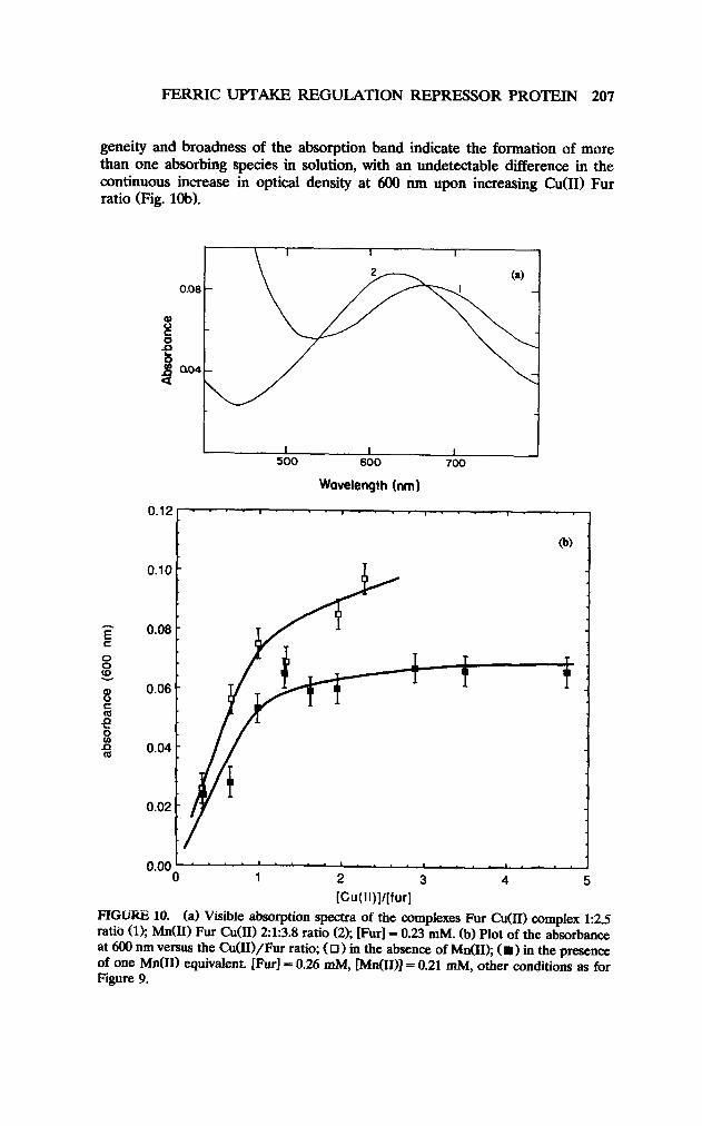
geneity and broadness of the absorption band indicate the formation of more than one absorbing species in solution, with an undetectable difference in the continuous increase in optical density at 600 nm upon increasing Cu(I1) Fur ratio (Fig. lob).
Wavelength (nm)
0.12 . . . . 1.. . 8.. . . I.. .., . . . .
(b)
0.10 -
0.00 . . ‘. ‘. . . . ‘. “. ‘. . . .‘a. “
0 1 2 3 4 5 [Cu(ll)]/[fur]
FIGi.+= 10. (a) Visible absorption spectra of the complexes Fur Cu(II) complex 1:2.5 ratio (1); M&I) Fur Cu(I1) 2:1:3.8 ratio (2); [Fur] = 0.23 mM. (b) Plot of the absorbance at 600 nm versus the Cu(II)/Fur ratio; ( 0) in the absence of M&I); ( l ) in the presence of one M&I) equivalent. [Furl = 0.26 mM, [M&III1 = 0.21 mM, other conditions as for Figure 9.
208 M. Y Hamed et aL
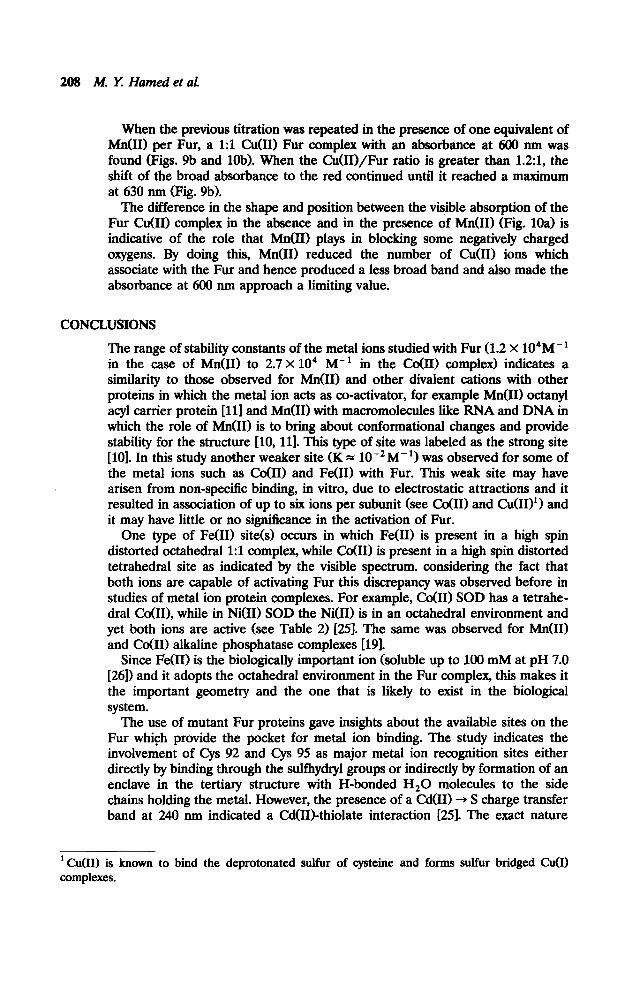
When the previous titration was repeated in the presence of one equivalent of M&I) per Fur, a 1:l C&I) Fur complex with an absorbance at 600 run was found (Figs. 9b and lob). When the Cu(I.I)/Fur ratio is greater than 1.2~1, the shift of the broad absorbance to the red continued until it reached a maximum at 630 run (Fig. 9b).
The difference in the shape and position between the visible absorption of the Fur Cu(I1) complex in the absence and in the presence of M&I) (Fig. 1Oa) is indicative of the role that M&I) plays in blocking some negatively charged oxygens. By doing this, Mn(I1) reduced the number of Cu(I1) ions which associate with the Fur and hence produced a less broad band and also made the absorbance at 600 nm approach a limiting value.
CONCLUSIONS
The range of stability constants of the metal ions studied with Fur (1.2 x 10⁴M- ’ in the case of M&I) to 2.7 X lo4 M- ’ in the CoUI) complex) indicates a similarity to those observed for MdII) and other divalent cations with other proteins in which the metal ion acts as co-activator, for example Mn(I1) octanyl acyl carrier protein 1111 and M&I) with macromolecules like RNA and DNA in which the role of M&I) is to bring about conformational changes and provide stability for the structure [lo, 111. This type of site was labeled as the strong site [lo]. In this study another weaker site (K = low2 M- ‘1 was observed for some of the metal ions such as co(II) and Fe(U) with Fur. This weak site may have arisen from non-specific binding, in vitro, due to electrostatic attractions and it resulted in association of up to six ions per subunit (see CoUI) and C&I)‘) and it may have little or no significance in the activation of Fur.
One type of Fe(U) site(s) occurs in which Fe(U) is present in a high spin distorted octahedral 1:l complex, while Cc4111 is present in a high spin distorted tetrahedral site as indicated by the visible spectrum. considering the fact that both ions are capable of activating Fur this discrepancy was observed before in studies of metal ion protein complexes. For example, Co@) SOD has a tetrahe- dral Co(H), while in NXII) SOD the Ni(I1) is in an octahedral environment and yet both ions are active (see Table 2) [WI. The same was observed for M&I) and co(U) alkaline phosphatase complexes [191.
Since Fe(H) is the biologically important ion (soluble up to 100 mM at pH 7.0 [26n and it adopts the octahedral environment in the Fur complex, this makes it the important geometry and the one that is likely to exist in the biological system.
The use of mutant Fur proteins gave insights about the available sites on the Fur which provide the pocket for metal ion binding. The study indicates the involvement of Cys 92 and Cys 95 as major metal ion recognition sites either directly by binding through the sulfhydryl groups or indirectly by formation of an enclave in the tertiary structure with H-bonded H,O molecules to the side chains holding the metal. However, the presence of a Cd(U) + S charge transfer band at 240 mn indicated a Cd(II)_thiolate interaction [25]. The exact nature
1 C&II) is known to bind the deprotonated sulfur of cysteine and forms sulfur bridged Cu(I) complexes.

FERRIC UPTAKE REGULATION REPRESSOR PROTEIN 209
and role of cd(R) and co(R) thiol interaction in DNA binding are discussed by Neilands et al. (manuscript in preparation). In a preliminary visible absorption study of the co(H) complexes with His mutants of Fur, other ligands involved in the major site could be suggested by comparing the spectra with that of co(R) Fur. They are nitrogens from H 86, H 117, and possibly H 131. The histidine residues in the N-terminus did not show evidence of metal binding as indicated by the visible absorption spectrum of the C&I) complex of H 31 L mutant which was identical to the Co(R) Fur spectrum. It is worth mentioning that an NMR study by Williams el al. (private communication) reported evidence of conformational changes in the mutant as compared to the wild-type Fur.
REFERENCES
1. A. Bags and J. B. Neilands, Microbial. Rev., 51(4), 509 (1987). 2. V. de Lore&, S. Wee, M. Herro, and J. B. Neilands, J. Bacteriol. 169(6), 2624
(1987). 3. A. Bagg and J. B. Neilands, Biochemistty 26,5471(1987). 4. E. C. Neiderhoffer, C. M. Maranjo, K. L. Bradley, and J. A. Fee, I. BacterioL 172(4),
1930 (1990). 5. T. Saito and R. J. P. Wiiams, Eur. J. Biochem. 197(l), 43 (1991). 6. T. Saito and R. J. P. Williams, Eur. J. Biochem. 197(l), 39 (1991). 7. M. Y. Hamed and J. B. Neilands (submitted to Biometals). 8. D. A. Deranleau, J. Am. Chem. Sot. 91(5), 4044 (1%9). 9. C. R. Cantor and P. R. Schimmel, in Biophysical Chemistty: The behavior of biological
molkcuks, A. C. Bartlet, P. C. Vapneck, and L. W. McCombs, Eds., W. H. Freeman, US, 1980, Vol. III, pp. 849-886.
10. A. Danchin, Eur. J. B&hem. 16,532 (1970). 11. D. M. Tener and K. N. Mayo, Eur. J. Biochem. 189,559 (1990). 12. H. G. Bray and K. White, Kinetics and Thermodynamics in Biochemiqv, Acad. Press,
New York, 1957, pp. 80-126. 13. S. Schaffer, K. Hat&e, and V. Braun, Mol. Gen. Genet. 201,204 (1985). 14. J. F. Reidhaar-Olson and R. T. Saver, Protein: Stmcture and Gen. 7,306 (1990). 15. A. Vedani and D. W. Hunta, J. Am. Chem. Sot. 112,4759 @90). 16. L. Calabrese, D. Cocco, A. Desider, and G. Rotilio, in Metalloptvteins: Shtctute and
Moleculnrfunction and chemical aspects, W. Uhich, Ed., G. Thieme Verlag, Stuttgart, New York, 1979, pp. 36-42.
17. L. J. Ming and J. S. Valentine, Z. Am. Chem. Sot. 112,4356 (1990). 18. M. L. Brader and M. F. Dunn, J. Am. Chem. Sot. 112,4585 (1990). 19. J. E. Coleman and J. F. Chlebowski, in Advanced Inorganic Biochemiwy, G. L.
Eichbom and L. G. Ma&Ii, Eds., Elsevier, New York, Gxford, Amsterdam, 1990, pp. l-22.
20. R. W. Hay, Bio-Inorganic Chemistry, Ellis-Horwood Ltd., (division of J. Wiley and Sons), New York, Chichester, Toronto, 1984, pp. 43-68.
21. S. Hu, P. Furst, and D. Hamer, The New Biokqist X6), 544 (19%). 22. R. C. Hider, A. R. Mohd-Nor, J. Silver, and I. E. Morrison, J. Chem. Sot. Dalton, 609
(1981). 23. M. Y. Hamed, J. Silver, and I. E. Morrison, Znorg. Chem. Acta 107,169 (1985).

210 hf. Y Hamed et al.
24. K. D. Karlin and S. L. Lippard, J. Am. Chem. Sot. 98,695l (1976). 25. M. Coy and J. B. Neilands, Biochernistty 30,8201(1991). 26. K. Nakamura, V. Delorenzo, and J. B. Neilands, ACS Symp. Series 402, Met-DNA
Chem., 146, (1989). 27. S. Del Cardayre and J. B. Neilands, Iron Biominerals, R. Frankel, Ed., Plenum Press,
New York, 1991.
Received Jub 7, 1992; accepted September 2, 1992

![Sunset Seed & Plant Co. (Sherwood Hall Nursery Co.) : [catalog]](https://static.fdokumen.com/doc/165x107/63266583e491bcb36c0ac5ab/sunset-seed-plant-co-sherwood-hall-nursery-co-catalog.jpg)