Beam-induced motion of vitrified specimen on holey carbon film
8
Beam-induced motion of vitrified specimen on holey carbon film Axel F. Brilot a , James Z. Chen a,b,1 , Anchi Cheng c , Junhua Pan d , Stephen C. Harrison d,f , Clinton S. Potter c , Bridget Carragher c , Richard Henderson e , Nikolaus Grigorieff a,b,⇑ a Department of Biochemistry, Rosenstiel Basic Medical Sciences Research Center, Brandeis University, MS029, 415 South Street, Waltham, MA 02454, USA b Howard Hughes Medical Institute, Brandeis University, MS029, 415 South Street, Waltham, MA 02454, USA c Department of Cell Biology, The Scripps Research Institute, La Jolla, CA 92037, USA d Harvard Medical School and Children’s Hospital, Boston, MA 02115, USA e MRC Laboratory of Molecular Biology, Hills Road, Cambridge CB2 0QH, UK f Department of Biological Chemistry and Molecular Pharmacology and Howard Hughes Medical Institute, Harvard Medical School, Boston, MA, USA article info Article history: Received 9 December 2011 Received in revised form 6 February 2012 Accepted 7 February 2012 Available online 16 February 2012 Keywords: Single-particle electron cryo-microscopy Beam-induced motion Radiation damage Dose rate High-resolution EM abstract The contrast observed in images of frozen-hydrated biological specimens prepared for electron cryo- microscopy falls significantly short of theoretical predictions. In addition to limits imposed by the current instrumentation, it is widely acknowledged that motion of the specimen during its exposure to the elec- tron beam leads to significant blurring in the recorded images. We have studied the amount and direction of motion of virus particles suspended in thin vitrified ice layers across holes in perforated carbon films using exposure series. Our data show that the particle motion is correlated within patches of 0.3–0.5 lm, indicating that the whole ice layer is moving in a drum-like motion, with accompanying particle rotations of up to a few degrees. Support films with smaller holes, as well as lower electron dose rates tend to reduce beam-induced specimen motion, consistent with a mechanical effect. Finally, analysis of movies showing changes in the specimen during beam exposure show that the specimen moves significantly more at the start of an exposure than towards its end. We show how alignment and averaging of movie frames can be used to restore high-resolution detail in images affected by beam-induced motion. Ó 2012 Elsevier Inc. All rights reserved. 1. Introduction Electron cryo-microscopy (cryo-EM) can be used to visualize the three-dimensional (3D) structure of a broad variety of specimens, including two-dimensional (2D) crystals (e.g., Gonen et al., 2004; Henderson et al., 1986), helical specimens (for example, Ge and Zhou, 2011; Miyazawa et al., 1999; Sachse et al., 2007; Yonekura et al., 2003) and isolated (single) particles. In recent years the application of the single particle approach has led to 3D reconstruc- tions of a number of highly symmetrical virus particles at near- atomic resolution (4 Å or better, see Grigorieff and Harrison, 2011) for a recent review). Despite this success, it is commonly acknowledged that contrast in images of vitrified specimens falls significantly short of predicted physical limits (Glaeser, 1999; Henderson, 1995). Physical limits are imposed by the radiolysis of biological molecules caused by the high-energy electron beam which limits the electron dose to 5–10 electrons/Å 2 (Baker et al., 2010; Henderson, 1992, 1995) if high-resolution features are to be preserved. Under idealized conditions, particle images are predicted to contain sufficient signal to obtain a 3D reconstruction at 3 Å resolution by averaging of a few thousand molecular images (Glaeser, 1999; Henderson, 1995). In practice however, the recent reconstructions of particles at near-atomic resolution have required averaging signal from several 100,000 to over 10 million images of subunits or asymmetric units (Grigorieff and Harrison, 2011). The contrast transfer function of the electron microscope, image detector noise and motion in the specimen induced by the incident electron beam all contribute to the loss of contrast in cryo-EM images (for a recent review, see Glaeser and Hall, 2011). The first two issues concern limitations of current instrumentation and are being addressed by technological improvements (Cambie et al., 2007; Danev and Nagayama, 2001; Majorovits et al., 2007; McMu- llan et al., 2009; Milazzo et al., 2011, 2005; Muller et al., 2010). Beam-induced specimen motion is thought to be caused by the reaction of the specimen to the high-energy electron beam, result- ing in a build-up of positive charge on the specimen (Brink et al., 1998) and radiolysis of the sample and vitrified embedding med- ium (Glaeser, 2008; Glaeser and Taylor, 1978). Charge build-up leads to a weak deflection of the electron beam that can blur the image, especially of tilted samples in which the component of the deflection perpendicular to the beam is not zero (Glaeser and 1047-8477/$ - see front matter Ó 2012 Elsevier Inc. All rights reserved. doi:10.1016/j.jsb.2012.02.003 ⇑ Corresponding author at: Department of Biochemistry, Rosenstiel Basic Medical Sciences Research Center, Brandeis University, MS029, 415 South Street, Waltham, MA 02454, USA. Fax: +1 781 736 2419. E-mail address: [email protected] (N. Grigorieff). 1 Present address: Department of Biology, Massachusetts Institute of Technology, 77 Massachusetts Avenue, Cambridge, MA 02139, USA. Journal of Structural Biology 177 (2012) 630–637 Contents lists available at SciVerse ScienceDirect Journal of Structural Biology journal homepage: www.elsevier.com/locate/yjsbi
-
Upload
independent -
Category
Documents
-
view
1 -
download
0
Transcript of Beam-induced motion of vitrified specimen on holey carbon film

Journal of Structural Biology 177 (2012) 630–637
Contents lists available at SciVerse ScienceDirect
Journal of Structural Biology
journal homepage: www.elsevier .com/ locate/y jsbi
Beam-induced motion of vitrified specimen on holey carbon film
Axel F. Brilot a, James Z. Chen a,b,1, Anchi Cheng c, Junhua Pan d, Stephen C. Harrison d,f, Clinton S. Potter c,Bridget Carragher c, Richard Henderson e, Nikolaus Grigorieff a,b,⇑a Department of Biochemistry, Rosenstiel Basic Medical Sciences Research Center, Brandeis University, MS029, 415 South Street, Waltham, MA 02454, USAb Howard Hughes Medical Institute, Brandeis University, MS029, 415 South Street, Waltham, MA 02454, USAc Department of Cell Biology, The Scripps Research Institute, La Jolla, CA 92037, USAd Harvard Medical School and Children’s Hospital, Boston, MA 02115, USAe MRC Laboratory of Molecular Biology, Hills Road, Cambridge CB2 0QH, UKf Department of Biological Chemistry and Molecular Pharmacology and Howard Hughes Medical Institute, Harvard Medical School, Boston, MA, USA
a r t i c l e i n f o
Article history:Received 9 December 2011Received in revised form 6 February 2012Accepted 7 February 2012Available online 16 February 2012
Keywords:Single-particle electron cryo-microscopyBeam-induced motionRadiation damageDose rateHigh-resolution EM
1047-8477/$ - see front matter � 2012 Elsevier Inc. Adoi:10.1016/j.jsb.2012.02.003
⇑ Corresponding author at: Department of BiochemiSciences Research Center, Brandeis University, MS029MA 02454, USA. Fax: +1 781 736 2419.
E-mail address: [email protected] (N. Grigorieff).1 Present address: Department of Biology, Massachu
77 Massachusetts Avenue, Cambridge, MA 02139, USA
a b s t r a c t
The contrast observed in images of frozen-hydrated biological specimens prepared for electron cryo-microscopy falls significantly short of theoretical predictions. In addition to limits imposed by the currentinstrumentation, it is widely acknowledged that motion of the specimen during its exposure to the elec-tron beam leads to significant blurring in the recorded images. We have studied the amount and directionof motion of virus particles suspended in thin vitrified ice layers across holes in perforated carbon filmsusing exposure series. Our data show that the particle motion is correlated within patches of 0.3–0.5 lm,indicating that the whole ice layer is moving in a drum-like motion, with accompanying particle rotationsof up to a few degrees. Support films with smaller holes, as well as lower electron dose rates tend toreduce beam-induced specimen motion, consistent with a mechanical effect. Finally, analysis of moviesshowing changes in the specimen during beam exposure show that the specimen moves significantlymore at the start of an exposure than towards its end. We show how alignment and averaging of movieframes can be used to restore high-resolution detail in images affected by beam-induced motion.
� 2012 Elsevier Inc. All rights reserved.
1. Introduction
Electron cryo-microscopy (cryo-EM) can be used to visualize thethree-dimensional (3D) structure of a broad variety of specimens,including two-dimensional (2D) crystals (e.g., Gonen et al., 2004;Henderson et al., 1986), helical specimens (for example, Ge andZhou, 2011; Miyazawa et al., 1999; Sachse et al., 2007; Yonekuraet al., 2003) and isolated (single) particles. In recent years theapplication of the single particle approach has led to 3D reconstruc-tions of a number of highly symmetrical virus particles at near-atomic resolution (4 Å or better, see Grigorieff and Harrison,2011) for a recent review). Despite this success, it is commonlyacknowledged that contrast in images of vitrified specimens fallssignificantly short of predicted physical limits (Glaeser, 1999;Henderson, 1995). Physical limits are imposed by the radiolysis ofbiological molecules caused by the high-energy electron beamwhich limits the electron dose to 5–10 electrons/Å2 (Baker et al.,
ll rights reserved.
stry, Rosenstiel Basic Medical, 415 South Street, Waltham,
setts Institute of Technology,.
2010; Henderson, 1992, 1995) if high-resolution features are tobe preserved. Under idealized conditions, particle images arepredicted to contain sufficient signal to obtain a 3D reconstructionat 3 Å resolution by averaging of a few thousand molecular images(Glaeser, 1999; Henderson, 1995). In practice however, the recentreconstructions of particles at near-atomic resolution have requiredaveraging signal from several 100,000 to over 10 million images ofsubunits or asymmetric units (Grigorieff and Harrison, 2011). Thecontrast transfer function of the electron microscope, imagedetector noise and motion in the specimen induced by the incidentelectron beam all contribute to the loss of contrast in cryo-EMimages (for a recent review, see Glaeser and Hall, 2011). The firsttwo issues concern limitations of current instrumentation and arebeing addressed by technological improvements (Cambie et al.,2007; Danev and Nagayama, 2001; Majorovits et al., 2007; McMu-llan et al., 2009; Milazzo et al., 2011, 2005; Muller et al., 2010).Beam-induced specimen motion is thought to be caused by thereaction of the specimen to the high-energy electron beam, result-ing in a build-up of positive charge on the specimen (Brink et al.,1998) and radiolysis of the sample and vitrified embedding med-ium (Glaeser, 2008; Glaeser and Taylor, 1978). Charge build-upleads to a weak deflection of the electron beam that can blur theimage, especially of tilted samples in which the component of thedeflection perpendicular to the beam is not zero (Glaeser and

A.F. Brilot et al. / Journal of Structural Biology 177 (2012) 630–637 631
Downing, 2004; Henderson, 1992). Radiolysis of the specimen isthought to lead to a build-up of internal pressure as the radiolysisproducts take up more space than the original molecules (Glaeser,2008). The mechanical stress is sufficiently high to cause specimendeformations (and ultimately breakdown of the entire fabric – so-called bubbling), again blurring the final image. In a recent study,Glaeser and Henderson (Glaeser et al., 2011) studied the beam-in-duced motion of paraffin 2D crystals supported by a continuous car-bon film and showed that film thicknesses greater than 35 nmsignificantly reduced the observed motion, thereby improving thefraction of images with strong high-resolution signal. These exper-iments further corroborate mechanical instability as one of theleading factors allowing beam-induced motion. Unfortunately, theuse of a continuous carbon film is often not ideal for non-crystallinesingle particles as it adds background to an image and can inducepreferred particle orientation.
We have recently investigated an imaging protocol in which theelectron dose rate was varied, to image single particles embeddedin ice over holes in a carbon support film (Chen et al., 2008). Theseexperiments suggested that a lower dose rate allows for a highertotal dose before bubbling occurs, but did not clearly demonstratethat beam-induced motion prior to bubbling was reduced. In thepresent study, we have investigated beam-induced motion bymonitoring positions and orientations of rotavirus double-layerparticles (DLPs). These particles are very regular and have a molec-ular mass of 70 MDa, allowing alignment with a reference struc-ture with accuracies of about 0.2 Å and 0.2�, for translational andorientational alignments, respectively (Zhang et al., 2008). We col-lected exposure series from holes inside perforated carbon filmscontaining rotavirus embedded in ice, varying dose rate and holesize. Changes in the particle orientations between exposures werethen taken as an indication for specimen motion. In a second set ofexperiments, we investigated the timing of the beam-induced mo-tion during exposures by recording movies using a new type ofcamera, a direct electron detector. Particles visible in individualframes or frame averages were analyzed in terms of their orienta-tional and translational changes during the exposure.
2. Materials and methods
2.1. Sample preparation
Rotavirus DLPs were prepared as described (Street et al., 1982).Three microliters of sample with a concentration of 2.5–4 mg/mlwas applied to Quantifoil� or C-flat™ grids and plunge-frozen usingeither a Gatan CP3 plunger (for all exposure series experiments) oran FEI Vitrobot Mark 2 (for all movie experiments), with a 4 or 6 s blottime and at relative humidity between 65% and 80%. The followinggrid types were used: Quantifoil� 1.2/1.3 Cu 400 mesh (measuredhole size = 1.6 lm; this difference between nominal and measuredhole size was observed for all grids in this batch), C-flat™ 0.6/2.0 Cu 400 mesh (measured hole size = 0.6 lm), C-flat™ 1.0/1.0 Cu400 mesh (measured hole size = 1.0 lm), and C-flat™ 1.2/1.3 Cu400 mesh (measured hole size = 1.2 lm). The Quantifoil� grids werecleaned prior to their use by immersion in a small amount of ethylacetate on top of filter paper in a glass Petri dish. Immediately beforeplunging, all grids were subject to glow discharge for 45 s at 20 mA.
2.2. Electron microscopy – exposure series
Images were collected on a Gatan US4000 4 k � 4 k CCD cameramounted on an FEI TF30 electron microscope operating at 300 kV,and using a side-entry Gatan 626 cryo holder. The calibrated mag-nification was 49,053, giving a pixel size on the specimen of 3.06 Å.Magnification calibration was performed using a DLP reference
structure (Zhang et al., 2008) and maximizing correlation coeffi-cients found by varying the pixel size during Frealign (Grigorieff,2007) runs (see below). We used an underfocus of 2.5–3.5 lm,and an electron dose per exposure of 8 electrons/Å2. Images in eachseries were taken about 60 s apart, and occasionally 600 s apart totest if charging was a factor in the observed particle motions (seeSection 3). The exposed area on the grid was centered on each holeand held approximately constant at 2.0 lm to ensure that the elec-tron beam made contact with the carbon support film everywherearound holes. Furthermore, an objective aperture was used for allexposures. The ice thickness was measured using holes in the icelayer produced by a focused electron beam with subsequent tiltingto 30� (Wright et al., 2006). The ice next to virus particles wasdetermined to be between 800 and 1000 Å thick while it was thin-ner in the center of holes, with the thinnest ice (500 Å) measured inthe center of 1.6 lm holes on Quantifoil� grids.
2.3. Electron microscopy – movies
Images were collected on a Direct Electron DE-12 4 k � 3 k di-rect electron detector mounted on an FEI TF20 electron microscopeoperating at 200 kV, and using a side-entry Gatan 626 cryo holder.The calibrated magnification was 17,858, giving a pixel size on thespecimen of 3.36 Å. The underfocus was set between 2.5 and3.5 lm, and the electron dose per frame was 0.5 electrons/Å2. Mov-ies were recorded at a rate of 40 frames/s. The exposed area wasadjusted as above and ice thickness measurements were essen-tially identical to those measured above.
2.4. Image processing – exposure series
Virus particles were semi-automatically selected from the firstimage in an exposure series using e2boxer from the EMAN2 process-ing package (Tang et al., 2007). Micrographs were cross-correlatedto each other using the Spider processing package (Frank et al.,1996) to determine translational offsets, and particles were selectedin subsequent exposures by coordinates updated with the offsets.The defocus for each micrograph was determined using CTFFIND3(Mindell and Grigorieff, 2003). Particle images were decimatedusing 2 � 2 pixel averaging and aligned in an exhaustive searchwith Frealign (Grigorieff, 2007) (Mode = 4 with DANG = 200 and IT-MAX = 200 using data between 18 and 300 Å resolution), followedby 10 rounds of refinement (Mode = 1), using a DLP reference struc-ture (Zhang et al., 2008). The rotation angles and axes of reorienta-tions experienced by particles between exposures were determinedusing the program Tiltdiff (Henderson et al., 2011). The rotation axisof most particles was within 0.5� in the image plane. About 6% of theparticles were measured to have tilt axes with larger out-of-planeangles. These were excluded from the analysis to eliminate caseswere the exhaustive parameter search failed. Histograms of themagnitude of the measured rotation angles were generated with abin size of 0.1�. The histograms show a clear positive skewness,indicating that a simple average and standard deviation might notbe appropriate to characterize the distribution of rotation angles.We used the computer program distfit from the Theseus package(Theobald and Wuttke, 2006) to test 20 different distributions andselected the Rayleigh distribution as the most appropriate accord-ing to the Bayesian information criterion. The Rayleigh distribution(Lalitha and Mishra, 1996) is characterized by a single parameter, kwhich indicates the maximum (mode) of the distribution:
Rðx; kÞ ¼ x
k2 e�x2=2k2: ð1Þ
The maximum likelihood estimate of k2 is:
k2 ¼ 12N
XN
i¼1
x2i ð2Þ

Table 1Rotation angles measured between the first and second exposures in exposure series of rotavirus particles in vitrified ice over holes in perforated carbon support films. The doseper exposure was 8 electrons/Å2. The dose rate was varied from 2 electrons/Å2/s – 160 electrons/Å2/s and support films with different hole sizes, varying from 0.6 to 1.6 lm wereused.
Dose Rate Hole = 0.6 lm Hole = 1.0 lm Hole = 1.2 lm Hole = 1.6 lm
2 e�/Å2/s 0.412 ± 0.015 (N = 187) 0.413 ± 0.014 (N = 211) 0.394 ± 0.008 (N = 599) 0.470 ± 0.008 (N = 783)20 e�/Å2/s 0.460 ± 0.013 (N = 307) 0.403 ± 0.013 (N = 257) 0.384 ± 0.006 (N = 879) 0.608 ± 0.010 (N = 919)80 e�/Å2/s 0.453 ± 0.013 (N = 302) 0.415 ± 0.013 (N = 261) 0.443 ± 0.007 (N = 889) 0.481 ± 0.009 (N = 694)160 e�/Å2/s 0.480 ± 0.017 (N = 210) 0.433 ± 0.014 (N = 255) 0.461 ± 0.010 (N = 538) 0.558 ± 0.011 (N = 678)
632 A.F. Brilot et al. / Journal of Structural Biology 177 (2012) 630–637
and standard error in k2 is:
SDðk2Þ ¼ k2
ffiffiffiffiffiffiffiffiffiffiffiffiffiN � 1p : ð3Þ
Therefore, standard error in k2 is:
SDðkÞ � SDðk2Þ2k
¼ k
2ffiffiffiffiffiffiffiffiffiffiffiffiffiN � 1p : ð4Þ
This error is reported together with k in Table 1 for the fitteddistributions.
2.5. Image processing – movies
Raw movie frames were corrected for dark current and gain-normalized using dark frames and flat fields recorded directly be-fore each movie and at the beginning of a session, respectively.Frame averages were calculated using the computer program labelfrom the MRC image processing package (Crowther et al., 1996).Particle rotations and translations were analyzed using Frealignas described above, except using data between 25 and 800 Å.
3. Results
3.1. Exposure series
In a first set of experiments we investigated the influence ofdose rate on beam-induced motion using exposure series. Fig. 1Ashows a field of rotavirus particles prepared using C-flat™ 1.2/
Fig.1. Exposure series of rotavirus DLPs in ice in a C-flat™ 1.2/1.3 grid (hole size = 1.2 lmto the sample is noted above each exposure. Panels E, F and G show vector plots correspoare indicated in the plots). Panel H plots the summed vectors from panels E, F and G.
1.3 Cu 400 mesh grids (hole size = 1.2 lm). The image was re-corded using a dose rate of 20 electrons/Å2/s and the exposurelasted 0.4 s, giving a total dose of 8 electrons/Å2. Subsequent expo-sures are shown in panels B–D, indicating no bubbling in the sam-ple even in the final exposure, after which the total dose was32 electrons/Å2. Virus particles are clearly visible in all exposuresand could readily be aligned using a reference structure (Zhanget al., 2008). Using projection matching implemented in the com-puter program Frealign (Grigorieff, 2007), the orientation of mostof the virus particles in all four exposures were determined suc-cessfully, as judged by their small angular differences and smallout-of-plane errors (see Materials and methods) determined bythe program Tiltdiff (Henderson et al., 2011). The angular differ-ences between exposures are plotted as vectors in panels E–G toshow both the magnitude and direction of the rotations. A strikingfeature of these plots is the local correlation in the rotation vectors,extending over a distance of 300–500 nm. This correlation alsoindicates that most particle orientations were determined cor-rectly within a degree or better. Furthermore, it shows that the ori-entation changes between exposures is not due to the motion ofindividual virus particles in the ice, but that it must be due to adeformation in the ice layer itself, moving the particles with it.The angular changes of up to 2� seen in panel E were also observedin many other exposure series. The direction of the rotation re-verses from one side of the hole to the other, indicating that theice undergoes a drum-like motion with a curvature radius of25 lm and translation of the central region perpendicular to thespecimen plane of about 150 Å (see Section 4). Subsequent expo-sures produce further particle rotations approximately in the same
). Four successive exposures are shown in panels A, B, C and D. The total dose appliednding to the changes in particle orientations between exposures (exposure numbers

A.F. Brilot et al. / Journal of Structural Biology 177 (2012) 630–637 633
direction but their magnitude decreases, suggesting that beam-in-duced motion is largest during the initial exposure, and that it isdose-dependent. The particle rotations observed in the ice also ap-ply to the few virus particles found on the carbon next to the holein Fig. 1, indicating that the motion in the ice extends into theneighboring carbon support film.
We observed the same pattern of correlated particle rotations inmany other exposure series, with vectors pointing either awayfrom or towards the center of the hole, indicating that the directionof the drum motion is random. Furthermore, the magnitude of theobserved rotations varied considerably from series to series. Togain a more quantitative understanding of the orientation changes,and to investigate the influence of dose rate and hole size, we re-corded exposure series from 438 holes on 11 grids, yielding a totalof 8089 DLPs for rotation measurements. The dose rates used were2, 20, 80 and 160 electrons/Å2/s. To vary the hole size, we usedQuantifoil� grids (1.6 lm holes), as well as C-flat™ grids (0.6, 1.0and 1.2 lm holes). Fig. 2 shows the histograms of measured rota-tions between the first and second exposures for all tested doserates and a hole size of 1.2 lm. To interpret the histograms, we cal-culated maximum-likelihood fits of the Rayleigh distribution (seeMaterials and methods). The fitted Rayleigh distributions aresuperimposed on each histogram in Fig. 2, and their distributionmaxima are given in Table 1 (for all conditions tested), togetherwith their standard errors and the number of particles analyzed.The results in Fig. 2 and Table 1 show that the magnitude of parti-cle motion is quite variable and that alterations in dose rate andhole size do not produce predictable changes in the particle mo-tions. Clear trends are discernible, however. Particles suspendedover larger holes show larger rotations than particles in smallerholes. Thus, the largest average rotations for all dose rates were ob-served with 1.6 lm holes (Quantifoil� grids) while the rotationaverages determined for 1.0 lm holes (C-flat™ grids) were equal(within error estimates) or smaller than those seen with 1.2 lmholes (C-flat™ grids). The results for 0.6 lm holes do not appearto follow this trend which may reflect irregular hole boundariesand/or a larger contribution of the carbon support to the overallmotion (see Section 4). There is also a clear trend with dose rate,showing larger rotations as the rate increases, with the highest rate(160 electrons/Å2/s) producing the largest average rotations inmost cases. We also varied the time between exposures between
Fig.2. Histograms for particle rotations measured between the first and second exposureThe dose applied in each exposure was 8 electrons/Å2. The dose rate varied between 2 awere fitted with a Rayleigh distribution and maximum (k) is indicated in each plot toge
60 and 600 s but did not detect noticeable changes in the magni-tude of beam-induced motion. Analysis of rotations in subsequentexposures generated similar histograms with rotations of some-what reduced but still significant magnitude, in agreement withthe observations shown in Fig. 1.
3.2. Movies
Particle rotations of a few degrees during beam exposure resultin movement of peripheral density features of the 700 Å virus par-ticles by 10 Å or more, thereby eliminating any signal at sub-nano-meter resolution in the direction of rotational movement. Sincecryo-EM data recorded under similar conditions led to a recon-struction of the peripheral rotavirus coat proteins at 3.8 Å resolu-tion (Chen et al., 2009; Settembre et al., 2011; Zhang et al., 2008)the observed particle rotations must occur in such a way that sig-nificant high-resolution signal is preserved. The rotation vectorssuggest that the rotation for any given particle occurs around anapproximately fixed axis. Subunits closer to the rotation axis willmove less than those further away from the axis (see Discussionand conclusions), thereby maintaining stronger high-resolutioncontrast in the images. Other particles may not move much atall, as indicated by the histograms in Fig. 2, and will exhibit evenstronger high-resolution contrast. It is also possible that the parti-cle rotations do not occur at a constant rate during the exposure.For example, if particles rotate more rapidly initially and then slowdown during an exposure, blurring in the final image would be lesssevere compared to blurring produced by particles that rotate at aconstant rate.
To investigate the timing of the motion, we recorded movies at40 frames/s of virus specimens prepared using Quantifoil� grids asthese showed the largest beam-induced motions (see Supplemen-tary material). At a dose rate of 20 electrons/Å2/s, the dose perframe is only 0.5 electrons/Å2, too low to produce sufficient con-trast for particle alignment (Movie S1). We therefore calculated10-frame averages to improve the contrast while still allowing atemporal resolution of 0.25 s/averaged frame (Movies S2 and S3).We also made sure that we captured the onset of the exposureby starting recording before the beam shutter (above the speci-men) was opened. Therefore, the first few frames of a movie con-tain dark frames. Fig. 3 shows results for a 1.5 s exposure with
s of rotavirus DLPs in ice on C-flat™ 1.2/1.3 Cu 400 mesh grids (hole size = 1.2 lm).nd 160 electrons/Å2/s, giving exposure times between 0.05 and 4 s. The histogramsther with the total number of measurements.

634 A.F. Brilot et al. / Journal of Structural Biology 177 (2012) 630–637
63 raw frames (Movie S1) of which the first three frames weredark. The vector plots in Fig. 3A–E indicate the incremental rota-tions between 10-frame averages (see corresponding Movie S2).The plots exhibit the same overall pattern as those produced fromthe exposure series, with local correlations and opposite directionson opposite sides of the hole. As the exposure progresses, rotationincrements become smaller until they are frequently within thedetection limit of our method (rotations smaller than about 0.2�).The rotation vectors suggest smaller rotations for particles closerto the center of the hole, consistent with the above-mentioneddrum-like motion, which predicts a larger rotational motion nearthe edge. The largest overall rotational motion during the 1.5 sexposure of the movie is about 4�, suggesting that the drum motionin the center of the hole could be up to 250 Å perpendicular to thespecimen plane, producing a curvature of the ice layer with a
Fig.3. Movie frame averages of rotavirus DLPs in ice on a Quantifoil� grid (holesize = 1.6 lm). The movie was recorded for 1.5 s at 40 frames/s using the DE-12direct electron detector (Direct Electron). 10-frame averages of the resulting 60frames were subjected to the same analysis as the exposure series. Vector plots inpanels A–E indicate particle rotations measured between frame averages. The totalaverage of the movie is shown in the background in A and the summed rotationsfrom panels A–E are shown in F. Panels G–K show vector plots indicating theparticle translations measured between frame averages. Panel L shows the summedtranslations from panels G–K. The square highlights the area shown as a close-up inFig. 5. The movie is further analyzed in Supplementary material (Movies S1–S3).
12 lm radius. The rotations seen in the movies are larger thanthose we observed with most of the exposure series. However, inthe exposure series we were not able to observe the full extentof the rotations at the beginning of an exposure, only the averageorientation over its entire duration. The larger rotations seen inthe movies therefore do not contradict the results from the expo-sure series.
We also analyzed the incremental shifts of each particle, shownin Fig. 3G–K (see corresponding Movie S3). The largest overalltranslational motion is about 70 Å (Fig. 3L), part of which may beattributable to the drum motion (see Discussion and conclusions).Unlike the rotations, the particle translations are not distributedroughly symmetrically around the center of the hole. Instead,translations in the upper right corner are larger than elsewherein the hole. Inspection of the movies (Supplementary material,Movies S2 and S3) shows that the edge of the carbon moves inthe same direction as the adjacent particles. The larger translationsare directed towards the center of the hole, suggesting that thehole contracts in the direction of the translations (along the diago-nal from the bottom left to the top right of the image) and that thedistances between particles in the image decrease. We found sim-ilar patterns of translation in many other movies. A reduction indistance was also observed previously in images of gold particlesin ice (Chen et al., 2008) and is most consistent with a drum mo-tion of the ice layer that is induced by a shrinking dimension ofthe hole, rather than an expansion of the ice. Hence, deformationof the carbon support film contributes to the overall particle mo-tion, consistent with the observed rotation of the virus particlesfound on the carbon next to the hole in Fig. 1.
4. Discussion and conclusions
4.1. Beam-induced motion is caused by changes in the ice layer andcarbon support film
Beam-induced specimen motion has long been recognized asone of the main factors attenuating high-resolution signal incryo-EM (Bottcher, 1995; Bullough and Henderson, 1987; Glaeseret al., 2011; Henderson, 1992). The main cause of this motion isbeam damage occurring to the specimen as it is exposed to thehigh-energy electron beam (Glaeser, 2008; Glaeser and Taylor,1978; Glaeser et al., 2011) although charging may also play a role(Glaeser and Downing, 2004; Henderson, 1992). Increasedmechanical stability of the continuous support film used with 2Dcrystals reduces or entirely avoids beam-induced motion, but anequivalent solution has not yet been found for single particles sus-pended in ice layers over holes in the perforated carbon film. In thepresent study we investigated the nature of beam-induced motionof particles in ice over holes. Our analysis of images obtained dur-ing an exposure series indicates that the ice inside the holes under-goes drum-like motion with a curvature radius of 12–25 lm andtranslation of the central region perpendicular to the specimenplane of 150–250 Å. In earlier studies, it was observed that theice layer spanning the holes in perforated carbon films assumes adome shape (Wright et al., 2006). This is consistent with the pres-ent study, which implies that the observed dome shape was atleast partially induced by a drum motion. It is likely that this drummotion is caused to some extent by an increase in internal pressurein the ice due to the molecular radicals generated by radiolysis,leading to an expansion of the ice layer as previously suggested(Glaeser, 2008). However, a significant amount of additional mo-tion is produced by changes in the carbon support film. This isclearly indicated by the particle translations during beam exposureshown in Fig. 3. The changes appear to be anisotropic, reducing thehole dimension predominantly in one direction. The hole analyzed

A.F. Brilot et al. / Journal of Structural Biology 177 (2012) 630–637 635
in Fig. 3 changes its dimension by about 50 Å (about 0.3%) in thedirection of the largest translations. In the case shown in Fig. 3,the main cause of the observed beam-induced motion must there-fore be due to changes in the carbon support. It is unlikely thatthese changes are the result of specimen heating by the electronbeam which are estimated for a dose rate of 20 electrons/Å2/s tobe between 0.2 K (Egerton and Rauf, 1999) and 0.5 K (Dietrichet al., 1978), leading to motions between 0.03 and 0.05 Å (assum-ing a thermal expansion coefficient of amorphous carbon of5 � 10�6/K, Booy and Pawley, 1993; Vonck, 2000) over a lengthof the beam diameter (2 lm). Instead, the changes may affect theatomic structure of the support film and explain why pre-irradia-tion of the support film (before application of the sample) withan electron beam may reduce beam-induced motion (Ge and Zhou,2011; Miyazawa et al., 1999) (see below). Additional experimentsare required to quantify how much beam-induced motion can bereduced by pre-irradiation of the carbon support, and to determineif a support film with higher mechanical stability (Glaeser et al.,2011) could also reduce the motion of the particles in the holes.
Fig.4. Schematic of beam-induced motion. (A) Particles are suspended in vitrifiedice over a hole. (B) When the electron beam illuminates the sample, the carbonsupport film deforms such that the size of the hole shrinks by a small amount. Atthe same time, radiolysis of the solvent and macromolecules produces radicals thatincrease the pressure inside the ice layer. Both changes cause a drum motion of theice layer that lead to particle rotations and translations. The direction of the drummotion is random. (C) After the beam is turned off, the changes induced in thespecimen remain (plastic deformation). This model does not consider specimencharging which could add to the blurring of images.
4.2. Deformations in the ice and carbon layers are plastic
The total dose used in the movie in Fig. 3 was 30 electrons/Å2.Our experiments with exposure series indicate that subsequentexposures further increase the particle rotations, and with it thedrum motion. The amount of motion therefore depends on the to-tal dose applied to the sample. On the other hand, variation of thetime between exposures did not significantly alter the amount ofmotion. This is inconsistent with a mechanism of beam-inducedmotion that depends on the build-up of charge on the specimen.Although we expect charging to occur as with any other cryo spec-imen, the charge drains after a few minutes (Brink et al., 1998). It istherefore likely that the deformation of the ice layer and carbonsupport is predominantly plastic, as one might expect for amor-phous media such as vitrified ice and amorphous carbon. Sincedose rate affects the magnitude of the motion, it appears that alower dose rate reduces the strain induced in the specimen some-what. The origin of the strain is not entirely clear. As previouslydiscussed (Glaeser, 2008), radiolysis of the water and macromole-cules likely leads to increased pressure inside the ice layer. A lowerdose rate may reduce the pressure by giving molecular radicalsmore time to diffuse to the ice surface where they are released intothe microscope vacuum. However, the carbon support is muchmore resistant to radiolysis and the induced strain in the carbonmust have a different origin. The effectiveness of pre-irradiationin reducing beam-induced motion suggests that the electron beamhas an ‘annealing effect’ on the carbon layer, allowing it to relaxinto a more stable configuration. The annealing will also dependon the energy input by the beam and hence the dose rate. Both pro-cesses (pressure increase due to radiolysis, relaxation by anneal-ing) are consistent with the initial larger motion at the beginningof the exposure, followed by gradual relaxation into a new statethat is stable under the beam. In this new state, the ice layer willhave undergone a drum-like motion, producing the observed par-ticle rotations and translations. When the exposure ends, the con-centration of radicals will drop again, presumably leading to somerelaxation of the ice layer. However, as the results from the expo-sure series show, this does not restore the latter to its original stateas the annealing of the carbon and the straining of the ice lead toplastic deformation. Renewed exposure to the electron beam re-starts the process and produces motion almost as large as the firstexposure. As the specimen is repeatedly exposed (or exposed for along time) to the electron beam, annealing of the carbon will pro-gress to a more stable state. The events occurring during beamexposure are summarized in the schematic in Fig. 4.
4.3. Loss of high-resolution contrast is caused by particle rotation andtranslation
There is considerable variability in the amount of motion ob-served between different experiments (see Fig. 2), on averageabout 0.3�. The high-resolution detail will therefore be better pre-served for some particles compared with others. The variability ispresumably due to small random differences between holes andthe ice in them. Further variability is due to the drum motion,which will lead to little or no particle motion in the center of thehole if it occurs perpendicular to the image plane, while rotationsand some translations will be noticeable for particles closer to theedge of the hole. If the specimen is tilted with respect to the imageplane, the drum motion will produce additional translations andhence blurring. For example, if the specimen is tilted by 30�, thetranslation in the image plane will be 75–130 Å for a drum motionof 150–250 Å, sufficient to obliterate all high-resolution contrast.Even at a small tilt angle of 3� which can occur unintentionally(Vonck, 2000), the translation in the image plane is still 7–13 Å,which will produce blurring corresponding to a temperature factor(B-factor) of about 4000–13,000 Å2 (Jensen, 2001) (B-factors are gi-ven in X-ray crystallographic notation). Additional blurring arisesdue to particle rotations. If we assume an average rotation angleof 2� we expect the signal of coat molecules within a radius ofabout 100 Å from the axis to be attenuated by a B-factor of about450 Å2 (Jensen, 2001), which is the average B-factor determinedfor the reference rotavirus reconstruction (Zhang et al., 2008).The surface area near the rotation axis included within this radiusis about 1/50 of the total area. Therefore, if rotation alone wereresponsible for the signal loss, we would expect to obtain a resolu-

636 A.F. Brilot et al. / Journal of Structural Biology 177 (2012) 630–637
tion of 3 Å from images of 50,000 to 500,000 molecules, still lessthan what has been observed in the recent virus reconstructionsat near-atomic resolution. The blurring factor due to translationcan easily account for the additional loss of signal to account forthe observed requirement of several 100,000 to over 10 millionmolecules (Grigorieff and Harrison, 2011).
4.4. Beam-induced motion depends on hole size and dose rate
We have previously shown that a reduced dose rate increasesthe total dose needed to observe bubbling in the specimen (Chenet al., 2008), prompting us to examine the effect that dose ratehas on beam-induced motion. Furthermore, the importance ofmechanical stability observed with 2D crystals led us to includein our study specimens with different hole sizes in the supportfilm. Our measurements suggest that neither dose rate nor holesize can be adjusted to avoid beam-induced motion entirely. How-ever, lower dose rates and smaller holes appear to reduce motionon average while the largest rotations are observed with 1.6 lmholes. Apart from their larger size, the ice layers in the 1.6 lm holesalso exhibit the greatest variation in thickness, 1000 Å near theedge and 500 Å in the middle. The thinner ice regions may be moresusceptible to deformation than the thicker regions, allowing lar-ger deformations in larger holes (Yoshioka et al., 2010).
Measurements made with the smallest hole size of 0.6 lm rep-resent an exception of the observed trend as the average rotationsare larger than those observed with 1.0 and 1.2 lm in most cases. Apossible reason might be the frequently observed irregular edges ofthe 0.6 lm holes (Figure S1) which may contribute to the mechan-ical instability of the specimen. Furthermore, as the size of the illu-minated area for all hole sizes was kept approximately constant(2 lm diameter), there was more carbon within the area. It istherefore possible that the instabilities of the carbon support filmdiscussed above and observed previously (Glaeser et al., 2011) con-tributed to the larger particle rotations inside the 0.6 lm holes.
4.5. Movie frame alignment and averaging can reduce blurring in theimages
There has been a common belief that beam-induced motion isworst at the beginning of the exposure. The movie in Fig. 3 showsthat this is indeed the case and that the first 10 electrons/Å2 causeabout twice as much motion (about 2.5�) as the following 20 elec-trons/Å2 (about 1�). The analysis of other movies yielded essen-tially identical results in terms of the temporal behavior ofbeam-induced motion. If beam-induced motion cannot be avoided,
Fig.5. Translational alignment of movie frames to reduce blurring in images affectedexperienced translations of about 60 Å (see Fig. 3L) Particles are significantly blurred andafter translational alignment of individual frames. The translations for the individual fraverages by linear interpolation. The frame average in panel B exhibits substantially im
the high-resolution signal might be boosted somewhat if 5–10 electrons/Å2 dose are accumulated on the specimen before thecamera shutter is opened. However, movies of the particle motionsmay make it possible to avoid image blurring entirely. If the direc-tion, magnitude and timing of the particle translations and rota-tions during the exposure can be determined, particle imagesfrom individual frames could be averaged with their correct orien-tations and positions, thus preserving the high-resolution signal.This will require low-noise image recording, a feature that isbecoming available with the latest generation of direct electrondetectors (McMullan et al., 2009; Milazzo et al., 2011, 2005). A fur-ther requirement will be sufficient signal in frame averages of themovies to determine the translations and rotations taking placeduring the exposure. For the 70 MDa virus particle used in ourstudies, images with a combined dose of 5 electrons/Å2 at 200 kV(10-frame averages) were sufficient. Using the translations deter-mined in Fig. 3, we performed frame alignment for a small areaof the hole with three virus particles (Fig. 5). Compared to the aver-age in Fig. 5A calculated without alignment, the average withalignment in Fig. 5B shows a substantial reduction of blurring.Blurring due to the particle rotations cannot be removed by simple2D alignment but could be corrected for by appropriate adjustmentof the Euler angles in a 3D reconstruction from individual movieframes.
For particles with smaller molecular mass, such as the 70S ribo-some (2.6 MDa), the signal in the images will be weaker and itmight be necessary to add larger particles, such as the rotavirusparticles used here, to enable tracking of the ice layer. However,since their radius is also smaller, which reduces the blurring ofperipheral regions compared to the virus particles examined here,the effect of particle rotation on the final 3D map will be less. Forexample, the 70S ribosome has a diameter of about 250 Å, suggest-ing that an angular accuracy of 0.6� would be sufficient to obtain areconstruction at 4 Å resolution. Therefore, although tracking ofthe ice deformation would still be necessary to reduce blurringby the expected beam-induced particle rotations, the requiredaccuracy is not as high as with the virus particles.
4.6. Other factors may play a role in beam-induced motion
We have investigated here only two factors with a possibleinfluence on beam-induced motion, namely dose rate and hole size(within a limited range of sizes from 0.6 to 1.6 lm). Much smallerholes (with more regular edges and illuminated with a beam withsmaller diameter) and dose rates may be required to achieve amore significant reduction of beam-induced motion than observed
by beam-induced motion. (A) Average of 60 frames of an area of Movie S1 thathigh-resolution information is lost. (B) Average of the same 60 frames as in panel Aames were calculated from the translations determined in Fig. 3 for the 10-frameproved contrast with details at higher resolution that were not visible in panel A.
A.F. Brilot et al. / Journal of Structural Biology 177 (2012) 630–637 637
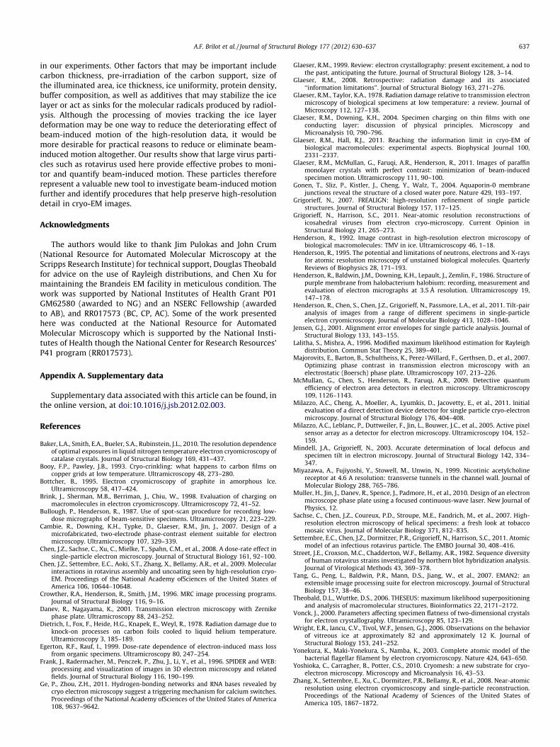
in our experiments. Other factors that may be important includecarbon thickness, pre-irradiation of the carbon support, size ofthe illuminated area, ice thickness, ice uniformity, protein density,buffer composition, as well as additives that may stabilize the icelayer or act as sinks for the molecular radicals produced by radiol-ysis. Although the processing of movies tracking the ice layerdeformation may be one way to reduce the deteriorating effect ofbeam-induced motion of the high-resolution data, it would bemore desirable for practical reasons to reduce or eliminate beam-induced motion altogether. Our results show that large virus parti-cles such as rotavirus used here provide effective probes to moni-tor and quantify beam-induced motion. These particles thereforerepresent a valuable new tool to investigate beam-induced motionfurther and identify procedures that help preserve high-resolutiondetail in cryo-EM images.
Acknowledgments
The authors would like to thank Jim Pulokas and John Crum(National Resource for Automated Molecular Microscopy at theScripps Research Institute) for technical support, Douglas Theobaldfor advice on the use of Rayleigh distributions, and Chen Xu formaintaining the Brandeis EM facility in meticulous condition. Thework was supported by National Institutes of Health Grant P01GM62580 (awarded to NG) and an NSERC Fellowship (awardedto AB), and RR017573 (BC, CP, AC). Some of the work presentedhere was conducted at the National Resource for AutomatedMolecular Microscopy which is supported by the National Insti-tutes of Health though the National Center for Research Resources’P41 program (RR017573).
Appendix A. Supplementary data
Supplementary data associated with this article can be found, inthe online version, at doi:10.1016/j.jsb.2012.02.003.
References
Baker, L.A., Smith, E.A., Bueler, S.A., Rubinstein, J.L., 2010. The resolution dependenceof optimal exposures in liquid nitrogen temperature electron cryomicroscopy ofcatalase crystals. Journal of Structural Biology 169, 431–437.
Booy, F.P., Pawley, J.B., 1993. Cryo-crinkling: what happens to carbon films oncopper grids at low temperature. Ultramicroscopy 48, 273–280.
Bottcher, B., 1995. Electron cryomicroscopy of graphite in amorphous Ice.Ultramicroscopy 58, 417–424.
Brink, J., Sherman, M.B., Berriman, J., Chiu, W., 1998. Evaluation of charging onmacromolecules in electron cryomicroscopy. Ultramicroscopy 72, 41–52.
Bullough, P., Henderson, R., 1987. Use of spot-scan procedure for recording low-dose micrographs of beam-sensitive specimens. Ultramicroscopy 21, 223–229.
Cambie, R., Downing, K.H., Typke, D., Glaeser, R.M., Jin, J., 2007. Design of amicrofabricated, two-electrode phase-contrast element suitable for electronmicroscopy. Ultramicroscopy 107, 329–339.
Chen, J.Z., Sachse, C., Xu, C., Mielke, T., Spahn, C.M., et al., 2008. A dose-rate effect insingle-particle electron microscopy. Journal of Structural Biology 161, 92–100.
Chen, J.Z., Settembre, E.C., Aoki, S.T., Zhang, X., Bellamy, A.R., et al., 2009. Molecularinteractions in rotavirus assembly and uncoating seen by high-resolution cryo-EM. Proceedings of the National Academy ofSciences of the United States ofAmerica 106, 10644–10648.
Crowther, R.A., Henderson, R., Smith, J.M., 1996. MRC image processing programs.Journal of Structural Biology 116, 9–16.
Danev, R., Nagayama, K., 2001. Transmission electron microscopy with Zernikephase plate. Ultramicroscopy 88, 243–252.
Dietrich, I., Fox, F., Heide, H.G., Knapek, E., Weyl, R., 1978. Radiation damage due toknock-on processes on carbon foils cooled to liquid helium temperature.Ultramicroscopy 3, 185–189.
Egerton, R.F., Rauf, I., 1999. Dose-rate dependence of electron-induced mass lossfrom organic specimens. Ultramicroscopy 80, 247–254.
Frank, J., Radermacher, M., Penczek, P., Zhu, J., Li, Y., et al., 1996. SPIDER and WEB:processing and visualization of images in 3D electron microscopy and relatedfields. Journal of Structural Biology 116, 190–199.
Ge, P., Zhou, Z.H., 2011. Hydrogen-bonding networks and RNA bases revealed bycryo electron microscopy suggest a triggering mechanism for calcium switches.Proceedings of the National Academy ofSciences of the United States of America108, 9637–9642.
Glaeser, R.M., 1999. Review: electron crystallography: present excitement, a nod tothe past, anticipating the future. Journal of Structural Biology 128, 3–14.
Glaeser, R.M., 2008. Retrospective: radiation damage and its associated‘‘information limitations’’. Journal of Structural Biology 163, 271–276.
Glaeser, R.M., Taylor, K.A., 1978. Radiation damage relative to transmission electronmicroscopy of biological specimens at low temperature: a review. Journal ofMicroscopy 112, 127–138.
Glaeser, R.M., Downing, K.H., 2004. Specimen charging on thin films with oneconducting layer: discussion of physical principles. Microscopy andMicroanalysis 10, 790–796.
Glaeser, R.M., Hall, R.J., 2011. Reaching the information limit in cryo-EM ofbiological macromolecules: experimental aspects. Biophysical Journal 100,2331–2337.
Glaeser, R.M., McMullan, G., Faruqi, A.R., Henderson, R., 2011. Images of paraffinmonolayer crystals with perfect contrast: minimization of beam-inducedspecimen motion. Ultramicroscopy 111, 90–100.
Gonen, T., Sliz, P., Kistler, J., Cheng, Y., Walz, T., 2004. Aquaporin-0 membranejunctions reveal the structure of a closed water pore. Nature 429, 193–197.
Grigorieff, N., 2007. FREALIGN: high-resolution refinement of single particlestructures. Journal of Structural Biology 157, 117–125.
Grigorieff, N., Harrison, S.C., 2011. Near-atomic resolution reconstructions oficosahedral viruses from electron cryo-microscopy. Current Opinion inStructural Biology 21, 265–273.
Henderson, R., 1992. Image contrast in high-resolution electron microscopy ofbiological macromolecules: TMV in ice. Ultramicroscopy 46, 1–18.
Henderson, R., 1995. The potential and limitations of neutrons, electrons and X-raysfor atomic resolution microscopy of unstained biological molecules. QuarterlyReviews of Biophysics 28, 171–193.
Henderson, R., Baldwin, J.M., Downing, K.H., Lepault, J., Zemlin, F., 1986. Structure ofpurple membrane from halobacterium halobium: recording, measurement andevaluation of electron micrographs at 3.5 Å resolution. Ultramicroscopy 19,147–178.
Henderson, R., Chen, S., Chen, J.Z., Grigorieff, N., Passmore, L.A., et al., 2011. Tilt-pairanalysis of images from a range of different specimens in single-particleelectron cryomicroscopy. Journal of Molecular Biology 413, 1028–1046.
Jensen, G.J., 2001. Alignment error envelopes for single particle analysis. Journal ofStructural Biology 133, 143–155.
Lalitha, S., Mishra, A., 1996. Modified maximum likelihood estimation for Rayleighdistribution. Commun Stat Theory 25, 389–401.
Majorovits, E., Barton, B., Schultheiss, K., Perez-Willard, F., Gerthsen, D., et al., 2007.Optimizing phase contrast in transmission electron microscopy with anelectrostatic (Boersch) phase plate. Ultramicroscopy 107, 213–226.
McMullan, G., Chen, S., Henderson, R., Faruqi, A.R., 2009. Detective quantumefficiency of electron area detectors in electron microscopy. Ultramicroscopy109, 1126–1143.
Milazzo, A.C., Cheng, A., Moeller, A., Lyumkis, D., Jacovetty, E., et al., 2011. Initialevaluation of a direct detection device detector for single particle cryo-electronmicroscopy. Journal of Structural Biology 176, 404–408.
Milazzo, A.C., Leblanc, P., Duttweiler, F., Jin, L., Bouwer, J.C., et al., 2005. Active pixelsensor array as a detector for electron microscopy. Ultramicroscopy 104, 152–159.
Mindell, J.A., Grigorieff, N., 2003. Accurate determination of local defocus andspecimen tilt in electron microscopy. Journal of Structural Biology 142, 334–347.
Miyazawa, A., Fujiyoshi, Y., Stowell, M., Unwin, N., 1999. Nicotinic acetylcholinereceptor at 4.6 A resolution: transverse tunnels in the channel wall. Journal ofMolecular Biology 288, 765–786.
Muller, H., Jin, J., Danev, R., Spence, J., Padmore, H., et al., 2010. Design of an electronmicroscope phase plate using a focused continuous-wave laser. New Journal ofPhysics, 12.
Sachse, C., Chen, J.Z., Coureux, P.D., Stroupe, M.E., Fandrich, M., et al., 2007. High-resolution electron microscopy of helical specimens: a fresh look at tobaccomosaic virus. Journal of Molecular Biology 371, 812–835.
Settembre, E.C., Chen, J.Z., Dormitzer, P.R., Grigorieff, N., Harrison, S.C., 2011. Atomicmodel of an infectious rotavirus particle. The EMBO Journal 30, 408–416.
Street, J.E., Croxson, M.C., Chadderton, W.F., Bellamy, A.R., 1982. Sequence diversityof human rotavirus strains investigated by northern blot hybridization analysis.Journal of Virological Methods 43, 369–378.
Tang, G., Peng, L., Baldwin, P.R., Mann, D.S., Jiang, W., et al., 2007. EMAN2: anextensible image processing suite for electron microscopy. Journal of StructuralBiology 157, 38–46.
Theobald, D.L., Wuttke, D.S., 2006. THESEUS: maximum likelihood superpositioningand analysis of macromolecular structures. Bioinformatics 22, 2171–2172.
Vonck, J., 2000. Parameters affecting specimen flatness of two-dimensional crystalsfor electron crystallography. Ultramicroscopy 85, 123–129.
Wright, E.R., Iancu, C.V., Tivol, W.F., Jensen, G.J., 2006. Observations on the behaviorof vitreous ice at approximately 82 and approximately 12 K. Journal ofStructural Biology 153, 241–252.
Yonekura, K., Maki-Yonekura, S., Namba, K., 2003. Complete atomic model of thebacterial flagellar filament by electron cryomicroscopy. Nature 424, 643–650.
Yoshioka, C., Carragher, B., Potter, C.S., 2010. Cryomesh: a new substrate for cryo-electron microscopy. Microscopy and Microanalysis 16, 43–53.
Zhang, X., Settembre, E., Xu, C., Dormitzer, P.R., Bellamy, R., et al., 2008. Near-atomicresolution using electron cryomicroscopy and single-particle reconstruction.Proceedings of the National Academy of Sciences of the United States ofAmerica 105, 1867–1872.