Bases génétiques du déterminisme du sexe chez le tilapia du ...
291
HAL Id: tel-03351827 https://tel.archives-ouvertes.fr/tel-03351827 Submitted on 22 Sep 2021 HAL is a multi-disciplinary open access archive for the deposit and dissemination of sci- entific research documents, whether they are pub- lished or not. The documents may come from teaching and research institutions in France or abroad, or from public or private research centers. L’archive ouverte pluridisciplinaire HAL, est destinée au dépôt et à la diffusion de documents scientifiques de niveau recherche, publiés ou non, émanant des établissements d’enseignement et de recherche français ou étrangers, des laboratoires publics ou privés. Bases génétiques du déterminisme du sexe chez le tilapia du Nil en populations naturelles Cecile Triay To cite this version: Cecile Triay. Bases génétiques du déterminisme du sexe chez le tilapia du Nil en populations naturelles. Sciences agricoles. Université Montpellier, 2021. Français. NNT : 2021MONTG025. tel-03351827
-
Upload
khangminh22 -
Category
Documents
-
view
3 -
download
0
Transcript of Bases génétiques du déterminisme du sexe chez le tilapia du ...

HAL Id: tel-03351827https://tel.archives-ouvertes.fr/tel-03351827
Submitted on 22 Sep 2021
HAL is a multi-disciplinary open accessarchive for the deposit and dissemination of sci-entific research documents, whether they are pub-lished or not. The documents may come fromteaching and research institutions in France orabroad, or from public or private research centers.
L’archive ouverte pluridisciplinaire HAL, estdestinée au dépôt et à la diffusion de documentsscientifiques de niveau recherche, publiés ou non,émanant des établissements d’enseignement et derecherche français ou étrangers, des laboratoirespublics ou privés.
Bases génétiques du déterminisme du sexe chez le tilapiadu Nil en populations naturelles
Cecile Triay
To cite this version:Cecile Triay. Bases génétiques du déterminisme du sexe chez le tilapia du Nil en populations naturelles.Sciences agricoles. Université Montpellier, 2021. Français. �NNT : 2021MONTG025�. �tel-03351827�

!"#$#%&'(')*+,$#%-,%.')$/0*(*#0$%-,%1$2$34$5% 6$ %7* 6"8*"%-,%9*6 %$(%:;8,6")*;(#%9"),/$6 6$#
%%%%%%%%%%%%%%%%%%%%%%%%%%%%%%%%%%%%%%%%%%%%%%%%%%%%%%%%%%%.$<"()%6$%=,/>%3;08;#'%-$
?"((%&@A&@B9C%.*/$3)$,/%-$%D$34$/34$C%E:&:%D$(($#C%A9DFB
9*3;6"#%:BDDA9C%:/;G$##$,/%H0'/*)$C%.BB%@(*<$/#*)'%-$%E",#"(($
:4*6*88$%IFD9BC%.*/$3)$,/%-$%D$34$/34$C%JBKB%L;()8$66*$/C%J9D1
&"M/*$6%LFDFA1C%.*/$3)$,/%-$%D$34$/34$C%E!!B%@!J%E>;(%NC%J9D1
F#)/*-%!O4($C%&/;,8%E$"-$/C%P;;6;Q*3"6%D$#$"/34%L,#$,0%F6$2"(-$/%R;$(*QC !;((
S'6T(" .UJV77FC%J"-/$%13*$()*G*+,$%-$#%B:AJC%A1BL%L;()8$66*$/C%JADF.
74;0"#%.W%RVJSBDC%:/;G$##;/C%@(*<$/#*)>%;G%L"/>6"(-
D"88;/)$,/
D"88;/)$,/
:/'#*-$()%-,%I,/>
B2"0*(")$,/
A(<*)'$
.*/$3)/*3$%-$%74T#$
J;X$(3"-/"()%-$%74T#$
:/'#$()'$%8"/%J'3*6$%7DAF?E$%YZ%I,*(%[Z[N
1;,#%6"%-*/$3)*;(%-$%S'6T("%.UJV77F
7S\1B%:V@D%V!7B9AD%EB%&DF.B%.B%.VJ7B@D%.B%EU@9A]BD1A7H%.B%LV97:BEEABD
B(%&'(')*+,$%$)%&'(;0*+,$
H3;6$%-;3);/"6$%&FAF
@(*)'%-$%/$34$/34$%@LD%^^^_%A1BL


À ma famille,
Papa, Maman, Lise et Mathieu, Mes grands-mères et Jinger.
J’ai une pensée émue pour mon grand-père, amoureux de la nature, qui n’aurait certainement eu d’autres mots que «!Bo bo bo… Rien que ca y nous manque!» s’il avait su que je ferai une thèse sur le déterminisme du sexe.
Je ne dédicace pas ces pages à Maxime… Il a tellement travaillé dessus… Elles sont les siennes ! Merci.


Remerciements
Je souhaiterais tout d’abord remercier les membres du jury qui ont accepté de relire ce
travail. J’espère que ce manuscrit donnera lieu à des discussions scientifiques enrichissantes
pour nous tous. Par avance, merci.
Evidemment, cette thèse n’aurait pas pu avoir lieu sans Helena et Jean-François. Merci
pour votre confiance ! Je pense qu’ensemble nous avons ajouté une petite pierre à l’édifice de
la compréhension du déterminisme du sexe chez le tilapia du Nil. Nous avons peut-être
soulevé encore plus de questions que nous n’avons réussi à fermer de portes… Mais n’est-ce
pas de bonne augure en sciences ? Je vous laisse avec pas mal de boulot sur les bras, alors
bon courage !
Je voudrais remercier tout le monde à l’ISEM et plus particulièrement l’équipe EPOISS
pour m’avoir si bien accueillie et soutenue dans mes travaux : Bruno, Christelle, Eric,
Frédérique, Nicolas, Jean-François A., Antoine, Erwan, Hala, Sélim, Madoka. Bonne
continuation !
Il y a de toute évidence les partenaires du projet qui ont permis de le rendre possible et
pour en parler, je vais passer à l’anglais pour quelques lignes.
David, Kerry and John, thank you for welcoming me so nicely at the University of
Stirling. We eventually managed to produce ddRAD libraries and it’s definitely thanks to
you! All the best for your futur project and/or retirement!
Thank you also to Astrid, Athimed and Nico for taking great care of me at the University
of Basel. I enjoyed a lot my time there and you were all of a precious help. Astrid, thank you
so much for the time you invested in this PhD.
I would like to thank the whole team at UMD I had the pleasure to join for a couple of
months! I cherish the time spent with you all (especially at Crazy Kebob ;)… what an
experience!). It was really awesome to be part of a great team with so much enthusiasm,
unity and kindness. I have a very emotional thoughts for Betsy and Matt: of course we
accomplished a lot of work together (with intense emotional rollercoasters), but more
importantly you welcomed me as if I’ve always been part of the team, thank you so much,
and thank you for your help all along. I wish you all the best for your forthcoming projects.

Karen, I thank you so much for welcoming me not only in your lab, but also in your home.
You are forever welcome in South of France!
Tom, you joined this thesis halfway to the end when accepting to endorse the role of Co-
supervisor. I’m really glad you did as you gave a whole new turn to this PhD project. I’m
sorry you had to listen to my French accent for so many hours on Zoom! Be sure that you
gave me two goals in life: First obtain my PhD; and then equip my basement as well as yours
for woodwork and metalwork (Though I’ll probably won’t aim for building a plane and
rather keep on with bench and shelves design!).
J’ai aussi eu l’opportunité de côtoyer des partenaires du Burkina Faso. Aboubacar et
Rokyatou, merci pour votre implication ! C’était un plaisir de travailler avec vous et j’espère
que nous parviendrons à terminer le travail que nous avons commencé. Rokyatou, je te
remercie tout particulièrement pour le soutient que tu as m’a apporté pendant ces quelques
années. Tu es une femme forte et pleine de sagesse !
Rencontrer les bonnes personnes au bon moment, c’est ce que m’a dit Pascal il y a près de
9 ans lorsque j’ai commencé l’Université. Alors merci Pascal, Pierrick, Mylène, Marion,
Christophe, Agnès et Céline d’avoir été des enseignants inspirants et de supers encadrants,
au bon moment.
D’aucun sait qu’une thèse est un parcours d’endurance et c’est plus agréable quand on est
bien entouré. Antoine, Célia, Yannis, vous avez été là depuis le jour 1 de cette thèse et jusqu’à
la fin. On a pleuré et on s’est pas mal énervé mais heureusement on a aussi su rire de la
situation parfois. Nous n’aurions peut-être pas su relativiser aussi bien sans Antoni, Maxime,
Roger le Bien Né, Etienne… Merci pour tout. On a pour sûr laissé des marques de notre
passage et pas uniquement à cause des traces de café sur le canapé.
J’ai la chance d’avoir pu compter sur les copains également. Merci aux Flo2, Alex, Loïs et
merci à tous les Copiloutres (Vincent, Morgane, Randy, Sarah, Elsa, Sebastien, Florian)
d’avoir été présents lorsque j’avais besoin de me vider la tête. Merci Florent de m’avoir
épaulé pendant un bon bout de ce parcours universitaire : depuis le BAC il y a eu un sacré
chemin. Il y a quelques années, qui aurait cru que nous parviendrions tous les deux à boucler
des thèses et que tu aurais un poste avant même que j’ai le temps de soutenir.

Enfin, je veux remercier ma famille. Elle déjà grande et belle et s’est agrandit encore avec
vous les Pouget & Courcelle. Je suis consciente de la chance que j’ai de tous vous avoir. Papa,
Maman, Lise, Mathieu, mes Maminous et Maxime, vous remercier juste pour la thèse
n’aurait pas de sens… Je ne serai pas la personne que je suis sans vous. Merci pour ça mais
surtout tout le reste, je vous aime.


TABLE DES MATIÈRES Introduction générale : Déterminisme du sexe chez les vertébrés 15 _____________
I. Historique et mythes des chromosomes sexuels 17
II. Diversité des déterminismes du sexe 19
III. Des transitions entre systèmes qui ne sont pas rares 32
IV. Les téléostéens, champions de la diversité 34
V. Le Tilapia du Nil, un modèle de choix 37
Contexte et objectifs de la thèse 55 ___________________________________________
Contexte 55
Objectifs 55
Context and objectives of the thesis project 59 _________________________________
Context 59
Objectives 59
Chapitre 1 : Déterminisme génétique du sexe de deux populations naturelles 63 __
Objectifs 65
Introduction et contexte 65
Matériels et méthodes 66
Principaux résultats et discussion 67
Conclusion 72
Publication associée 77
Analyses complémentaires 119
Chapitre 2 : Diversité et perte de l’haplotype Y ancestral en populations
naturelles 127 _______________________________________________________________
Objectifs 129
Introduction et contexte 129
Matériels et méthodes 130
Principaux résultats et discussion 132
Conclusion 134
Publication associée 139
Le challenge de l'assemblage des chromosomes sexuels 179
The challenge of sex chromosome assembly 188
Chapitre 3 : Différenciation du sexe chez une population qui ne présente pas le
chromosome Y ancestral 197 _________________________________________________
Objectifs 199
Introduction et contexte 199
Matériels et méthodes 201
Principaux résultats et discussion 202
Conclusion 205
Publication associée 213
Discussion générale et Perspectives 261 _______________________________________
Un système ancestral qui garde des zones d’ombres 263
La variabilité au niveau intra-spécifique, un atout pour comprendre l’évolution des chromosomes sexuels 265
Identifier le système de déterminisme du sexe de Hora 266
Les limites de nos études 268
Conclusion 276
Contributions scientifiques et enseignements 285______________________________


TABLE DES FIGURES
Introduction générale : Déterminisme du sexe chez les vertébrés 15 ___
Figure 1.1. Plaques équatoriales observées par Nettie Stevens sur le Tenebrio molitor permettant de mettre en évidence un chromosome plus petit uniquement chez les mâles et démontrant le lien entre chromosome et déterminisme du sexe. 17 .....................................................................................
Figure 1.2. Déterminisme et changement de sexe chez les vertébrés. GSD et ESD coexistent dans plusieurs clades de vertébrés pour différentes espèces. 20 ...................................................................
Figure 1.3. Possible patrons d’épistasie entre deux locus de déterminisme du sexe. 24 ................................
Figure 1.4. Turnovers de chromosomes sexuels parmi 28 espèces de grenouilles. 25 ..................................
Figure 1.5. Illustration schématique des trois patrons de TSD et d’espèces présentant ce type de de déterminisme. 29 .......................................................................................................................................
Figure 1.6. Méthodologie pour déterminer le type de déterminisme d’une espèce (ou une population) présentant ou non des chromosomes sexuels hétéromorphiques. 31 ....................................................
Figure 1.7. Évolution des systèmes de déterminisme du sexe chez les geckos. 32 .........................................
Table 1.1. Gènes du déterminisme du sexe formellement identifiés ou fortement suspectés chez les poissons téléostéens. 36 ...........................................................................................................................
Figure 1.8. Évolution des chromosomes sexuels chez les cichlidés africains. 37 ............................................
Figure 1.9. Représentation triangulaire schématique de la complexité du déterminisme du sexe chez le tilapia du Nil. 38 .......................................................................................................................................
Table 1.2. Bilan des origines, dénominations et déterminants génétiques du sexe chez les souches domestiques et commerciales de tilapia du Nil. 39 ...............................................................................
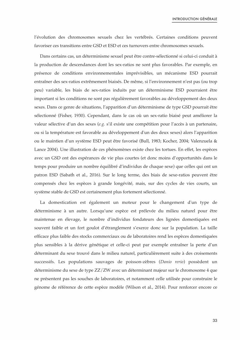
Figure 1.10. Localisation géographique des fourchettes de température des environnements d’où proviennent les populations sauvages étudiées dans ce projet de thèse. 42 .......................................
Contexte et objectifs de la thèse 55 __________________________________
Context and objectives of the thesis project 59 _______________________
Chapitre 1 : Déterminisme génétique du sexe de deux populations naturelles 63 _______________________________________________________
Figure 2.1. Illustration de la détection d’une duplication par l’étude de lectures courtes sur un génome complet, visionné dans IGV (Integrative Genome Viewer). 70 .............................................................
Figure 2.2. Relations entre les régions du chromosome B d'Astatotilapia latifasciata et les LGs de Metriaclima zebra. 71 ...............................................................................................................................
Figure 2.3. Manhattan plots des -log10(p-value) issues des Test Exacts de Fisher pour l’association des polymorphisme nucléotidique et sexe phénotypique. 121 .....................................................................
Figure 2.4. Emplacement des polymorphismes nucléotidiques simples (SNPs) significativement dimorphiques entre mâles et femelles sur LG19 d’après l’alignement sur le génome de référence d’O. niloticus. 121 ......................................................................................................................................
Figure 2.5. Génotypes de la descendance par rapport à leurs parents inférés à partir des sites significatifs du FET du ddRAD. 122 ..........................................................................................................

Figure 2.6. Diagramme en barres du nombre de segments et du nombre de locus identifiés comme appartenant au chromosome B dans le pool de femelles Koka et le pool de mâles Kpandu utilisés dans Triay et al., 2020. 124 .........................................................................................................
Chapitre 2 : Diversité et perte de l’haplotype Y ancestral en populations naturelles 127 __________________________________________
Figure 3.1. Intérêt du séquençage lectures longues (long reads) pour correctement assembler une région dupliquée. 131 ...............................................................................................................................
Figure 3.2. Problèmes rencontrés lors de la cartographie d'un individu hétérogamétique sur un génome de référence homogamétique. 181 ..........................................................................................................
Figure 3.3. Représentation schématique de graphe et de « bulles » de long reads. 184 ...............................
Figure 3.2. Problems encountered when mapping a heterogametic individual on a homogametic reference genome. 190 .............................................................................................................................
Figure 3.3. Schematic representation of graph and « bubbles » of long reads and hybrid assembly. 193 ..
Chapitre 3 : Différenciation du sexe chez une population qui ne présente pas le chromosome Y ancestral 197 ________________________
Figure 4.1. Modèle de réseaux de gènes antagonistes spécifiques à chaque sexe contrôlant la différenciation sexuelle chez les poissons. 200 ......................................................................................
Discussion générale et Perspectives 261 _____________________________
Figure 5.1. Aire de répartition géographique se chevauchant pour deux espèces d’Oreochromis. 274 .....
Figure 5.2. Proportion du génome introgressé en fonction du temps passé depuis l’hybridation. 274 .........
Contributions scientifiques et enseignements 285_____________________


INTRODUCTION GÉNÉRALE : DÉTERMINISME DU SEXE CHEZ
LES VERTÉBRÉS


INTRODUCTION GÉNÉRALE
I. Historique et mythes des chromosomes sexuels
On parle d’un déterminisme génétique du sexe lorsque le phénotype sexuel et donc le
type de gonades développées par un individu est défini par la présence d’un (ou plusieurs)
locus spécifique(s) dans son génome. Ces locus sont appelés déterminants du sexe (SD) et les
chromosomes qui les portent sont généralement qualifiés de chromosomes sexuels (en
opposition aux autosomes). L’ensemble des gènes portés par la paire de chromosomes
sexuels n’influe pas forcément sur le développement des gonades, mais peut parfois affecter
la valeur sélective des individus en étant plus favorable pour un sexe que pour l’autre.
Différents systèmes de déterminismes génétiques ont été décrits depuis les années 1900 et le
début de l’étude des chromosomes sexuels.
I.1. Les débuts de l’analyse des chromosomes sexuels
La première description empirique de chromosomes sexuels remonte à 1905 par Nettie
Steven lors de l’observation de caryotypes de ténébrion meunier (Tenebrio molitor), petits
coléoptères dont les larves sont communément appelées «!vers de farine!». Elle constate que
les individus possédant 19 gros chromosomes et un petit se développent en mâles alors que
les individus possédant 20 gros chromosomes se développent en femelles (Figure 1.1).
Figure 1.1. Plaques équatoriales observées par Nettie Stevens sur le Tenebrio molitor permettant de mettre en évidence un chromosome plus petit uniquement chez les mâles et démontrant le lien entre chromosome et déterminisme du sexe.Les figures 204, 205 et 206 montrent des plaques équatoriales de cellules somatiques mâles où un chromosome «!accessoire!» plus petit que les autres est identifié par un S. Les figures 207, 208 (a et b) correspondent des plaques équatoriales d'une cellule en division d'un follicule d'un jeune ovule. Dans les cellules femelles on observe 20 grands chromosomes et le petit chromosome (S) n’est pas présent. Issue de Stevens, 1905.
17

INTRODUCTION GÉNÉRALE
Le petit chromosome est appelé «! chromosome accessoire! » et correspond à ce qu'on
appelle aujourd’hui un chromosome Y. En effet, le ténébrion meunier est une espèce dont le
déterminisme génétique du sexe est de type XX/XY. Les individus peuvent soit porter une
paire de chromosomes identiques (deux chromosomes X), soit une paire de chromosomes
différents l’un de l’autre (chromosome X et chromosome Y). Les individus portant les
chromosomes différents représentent le sexe hétérogamétique, en opposition au sexe
homogamétique. Dans le cas du système XX/XY, les mâles sont les individus
hétérogamétiques et les femelles sont homogamétiques.
I.2. Des espèces modèles particulièrement étudiées
Certaines espèces ont particulièrement attiré l’attention des biologistes, soit parce qu’elles
ont un intérêt pour la société (médical ou économique par exemple), ou parce qu’elles sont
pratiques à étudier pour comprendre et illustrer des concepts théoriques. Ces espèces
couramment utilisées en laboratoire, appelées espèces «!modèles!», sont choisies pour leur
caractéristiques biologiques commodes pour le maintien d’élevages en conditions contrôlées.
Elles ont souvent des cycles de vie courts, des grandes tailles de descendances ou encore des
coûts faibles d’entretien (infrastructure ou soin nécessaire). C’est typiquement le cas des
drosophiles (mouches à fruits), des petits rongeurs (souris et rats) ou des nématodes.
L’homme fait de toute évidence partie des espèces qui ont souvent été placées au centre
des réflexions et notamment dans le cadre de l’étude du déterminisme du sexe. Chez
l’humain, comme chez le ténébrion meunier, le sexe est déterminé par des chromosomes
sexuels de type XX/XY. Le chromosome X porte de nombreux gènes qui ont été perdus sur le
chromosome Y. De fait, on observe une taille réduite du Y, qui est dit «!dégénéré!». Parmi les
gènes restant sur le Y, seule l’expression dans l’embryon du gène SRY (de l’anglais «!Sex-
determining Region of Y! ») suffit à initier la cascade de différenciation des gonades en
testicules (Sinclair et al., 1990). Les petits rongeurs étudiés en laboratoire, comme la souris, le
rat ou le cochon d’Inde voient également leur sexe déterminé par le gène Sry, comme la vaste
majorité des mammifères, même si toutes les espèces ne présentent pas systématiquement un
chromosome Y aussi dégénéré que celui de l’humain (Koopman et al., 1995). Dans le cas des
espèces modèles non-mammifères, telles que les mouches à fruits (Drosophilia melanogaster)
ou les petits nématodes Caenorhabditis elegans, les déterminismes du sexe sont comparables à
ceux des mammifères (XX/XY) même si Sry n’est pas le gène déterminant.
18

INTRODUCTION GÉNÉRALE
Cependant, l’étude des systèmes très stables de ces espèces de laboratoire et des
mammifères a mené à une représentation faussée de l’omniprésence des systèmes de type
XX/XY et de leur stabilité dans le règne animal. En réalité, d’autres types de systèmes de
déterminisme génétique ont été décrits peu de temps seulement après la première preuve
empirique de système XX/XY. Morgan montre en 1909 l’existence de système du type ZZ/
ZW chez des insectes. A l’inverse des chromosomes XX/XY, dans un système ZZ/ZW ce sont
les femelles qui représentent le sexe hétérogamétique (ZW) alors que les mâles sont
homogamétiques (ZZ). Ce système ZZ/ZW est aussi mis en évidence plus tard chez les
oiseaux (Ohno et al., 1966) et semble très stable au sein de ce groupe où le W n’est pas
toujours fortement dégénéré en comparaison de Z (Ellegren et al., 2010).
Dans les sections suivantes, nous nous intéresserons à la diversité des mécanismes du
déterminisme du sexe rencontrés dans une catégorie particulière du règne animal : les
vertébrés gonochoriques (espèces de vertébrés dont les individus ne portent qu’une seule
fonction reproductrice à la fois, mâle ou femelle). En effet, la plupart des vertébrés sont
gonochoriques (Devlin & Nagahama, 2001; Herpin & Schartl, 2015; Mei & Gui, 2015), même
si l’on retrouve dans de nombreux embranchements de l’arbre du vivant des espèces
hermaphrodites (dont chaque individu dispose des deux fonctions mâles et femelles
simultanément ou successivement) (Bachtrog et al., 2014; Shärer et al., 2017). Les mécanismes
intervenant dans l’hermaphrodisme ainsi que les pressions évolutives qui s’appliquent sur
ces systèmes peuvent être très différents de ceux des espèces avec un gonochorisme. Comme
dans ce projet de thèse, nous avons concentré notre attention sur le tilapia du Nil (un poisson
téléostéen gonochorique), nous ne nous attarderons pas ici sur les systèmes hermaphrodites.
Pour une revue très complète des déterminismes du sexe chez le vivant, incluant les modèles
hermaphrodites, la revue de Adolfi et al., (2019) est certainement un très bon point de départ.
II. Diversité des déterminismes du sexe
Bien que l’on connaisse des systèmes de déterminismes différents de celui des
mammifères ou des espèces modèles, ces mécanismes sont restés pendant longtemps dans
les consciences collectives comme des exceptions à la règle. Le développement du
séquençage et de la biologie moléculaire a permis au fil du temps de révéler la grande
diversité des systèmes déterminismes du sexe chez les vertébrés (voir revues de la littérature
19

INTRODUCTION GÉNÉRALE
de Valenzuela & Lance, 2004; Bachtrog et al., 2014; Capel et al., 2017; Smirnov & Trukhina,
2019). En effet, des espèces proches peuvent présenter des mécanismes de déterminisme du
sexe différents (Figure 1.2). C'est le cas par exemple chez les amphibiens où des systèmes
XX/XY et ZZ/ZW peuvent être observés dans différentes populations d’une même espèce
(Miura et al., 2017). De même, si les oiseaux présentent un système stable, les reptiles dans
leur ensemble présentent une grande variabilité de systèmes incluant des déterminismes
génétiques (GSD) et des déterminismes environnementaux du sexe (ESD) ou bien encore une
combinaison des deux (Sarre et al., 2011). Dans le cas d’une espèce à ESD, un même génome
peut développer deux phénotypes sexuels différents en fonction des conditions
environnementales dans lesquelles un individu se développe. De fait, les plus proches
parents des oiseaux, les crocodiliens, possèdent eux un déterminisme ESD où la température
du milieu contrôle le devenir du sexe des individus (Lang & Andrews 1994; Deeming, 2004).
Enfin, les tortues présentent des déterminismes du sexe de type XX/XY, ZZ/ZW et ESD
(Ewert et al., 2004). Les sections suivantes s’attèlent donc à donner une idée de la diversité à
la fois des mécanismes génétiques et environnementaux chez les vertébrés gonochoriques.
Figure 1.2. Déterminisme et changement de sexe chez les vertébrés. GSD et ESD coexistent dans plusieurs clades de vertébrés pour différentes espèces.
!"#$#%"&'$
()#%"&'$
*+)&$
,$)-$#$
./)$0$#$
*1"2
,&343-56'$
*4#'+37#"&58''
,%3+-&'4%#%5"2
,54632#30$#$
,"+3!"2393'4:$6"393'4:,
!"#;<=<>;=> <?@
!"#$%&"'%()*+,%+-
."/&0++,%+-
1)((2,%+-
345-%6%"(+
705$%&0+
8"44"&+
A"2#)-'+"2
$%& '%& '%(
')#
20

INTRODUCTION GÉNÉRALE
Suite de la légende : L’ESR pendant des fenêtres temporelles critiques affecte la détermination du sexe chez la plupart des clades de vertébrés, à l'exception des euthériens, et il y a un manque de preuves chez les chondrichtyens. Les poissons font preuve d'une plasticité sexuelle remarquable et peuvent subir un ESR suite à des changements dans les conditions extérieures ou même un changement complet de sexe à l'âge adulte. Les temps de divergence utilisés pour construire l'arbre ont été obtenus à partir de la base de données TimeTree. Abréviations : Ceno, Cénozoïque ; ESD, déterminisme environnemental du sexe par l'environnement ; ESR, inversion du sexe par l'environnement ; GSD, déterminisme génétique du sexe ; PC, Précambrien. Issu, traduit et modifié de Ortega-Recalde et al., (2020).
II.1. Les déterminismes génétiques du sexe
II.1.1. Une diversité de formes
Contrairement à l’image reçue du Y dégénéré, dont le contenu en gènes est drastiquement
réduit en comparaison du X, les chromosomes sexuels ne sont pas toujours fortement
différenciés. On retrouve en effet tous les cas possibles le long d’un gradient allant de la
stricte homomorphie à une complète hétéromorphie (Bachtrog et al., 2014). Dans le cas des
mammifères, un simple caryotype peut souvent permettre d’identifier la paire de
chromosomes sexuels du fait de la différence de taille importante entre le X et le Y et de leur
manque d’homologie moléculaire, en lien avec le manque de recombinaison notamment
(Skaletsky et al., 2003). Cependant, cette situation est loin d’être la règle et l’on retrouve des
poissons téléostéens, des amphibiens ou encore des reptiles dont les chromosomes sexuels
sont fortement homomorphes (Miura et al., 2017; Gammerdinger & Kocher, 2018). De plus,
dans un même clade on peut également trouver des chromosomes sexuels avec différents
degrés de différenciation. Ainsi, les Boidae et Pythonidae présentent des chromosomes
sexuels homomorphiques alors que les Viperidea portent des chromosomes sexuels
fortement hétéromorphiques et enfin les Colubridae eux possèdent des chromosomes sexuels
modérément différenciés (Gamble et al., 2017). C’est aussi le cas chez les oiseaux chez qui les
ratites, contrairement à l’ensemble de leurs congénères, présentent des chromosomes sexuels
grandement homomorphiques (Yazdi & Ellegren, 2014). Il existe également des cas extrêmes
de chromosomes sexuels homormophiques comme par exemple chez le Takifugu rubripes, un
poisson chez qui la région de déterminisme du sexe se résume au polymorphisme d’un
nucléotide entre le X et le Y (Kamiya et al., 2012).
Il existe donc une grande diversité de formes des paires de chromosomes sexuels.
21

INTRODUCTION GÉNÉRALE
II.1.2. Une diversité de systèmes
Si les deux types de déterminismes décrits précédemment (XX/XY et ZZ/ZW) sont les
plus connus, il en existe de nombreuses variantes.
On connait par exemple des cas où le chromosome Y n’est plus seulement fortement
dégénéré mais a complètement disparu. C’est le cas par exemple chez plusieurs rongeurs
fouisseurs du genre Ellobius (E. lutescens, E. tancrei et E. talpinus) qui ont perdu le
chromosome Y ainsi que le gène Sry (Mulugeta et al., 2016; Matveevsky et al., 2017). Chez E.
tancrei et E. talpinus la perte du Y a donné lieu à la duplication du chromosome X : mâles et
femelles sont donc XX. En revanche, chez E. lutescens tous les individus sont XO. On retrouve
également une perte du chromosome Y chez une espèce de rats épineux du genre Tokudaia (T.
osimensis), chez qui des fractions du Y ont été transloqué sur le X ou sur d’autres autosomes.
Ainsi, mâles et femelles sont également XO (Murata et al. 2016). Cependant, si la cascade de
différenciation du sexe est conservée, le ou les gènes remplaçant le Sry ne sont pas identifiés
(Otake & Kuroiwa, 2016).
A l’inverse, le Campagnol de l’Oregon (Microtus oregoni) a non pas subi une perte du Y
mais une perte d’un X chez les femelles. Ainsi, plusieurs génotypes ségrègent mais les
femelles de cette espèce peuvent notamment être XO alors que les mâles sont XY
(Charlesworth & Dempsey, 2001).
Il existe aussi des espèces pour lesquelles plusieurs paires de chromosomes peuvent
interagir pour déterminer le sexe d’un individu. Le Strabomantis biporcatus (un amphibien
endémique du Vénézuela, aussi nommé Eleutherodactylus maussi) possède deux paires de
chromosomes X distinctes. Un facteur masculinisant s’est transposé sur l’un des X d’une des
paires, agissant alors comme un Y et résultant en un déterminisme du type X1X1X2X2 chez
les femelles et X1Y1X2X2 ou X1X2Y2 chez les mâles (Schmid et al., 2003). Sur un autre
modèle, le Poisson-tigre (Hoplias malabaricus) possède lui deux chromosomes Y, ce qui induit
un déterminisme du type X1X1 (femelles) et X1Y1Y2 (mâles) (Cioffi and Bertollo, 2010). Un
exemple impliquant plus encore de paires de chromosomes sexuels est illustré par
l’ornithorynque (Ornithorhynchus anatinus) qui possède 5 paires de chromosomes sexuels de
type XX/XY (Grützner et al., 2004).
Le même type de variant existe pour les espèces dont les chromosomes sexuels suivent un
patron ZZ/ZW. Un système avec deux paires de chromosomes sexuels ZW détermine par
22

INTRODUCTION GÉNÉRALE
exemple le sexe chez un poisson du genre Ancistrus (Ancistrus sp.2 ‘’Barcelos’’) où les femelles
sont Z1Z2W1W2 tandis que les mâles sont Z1Z1Z2Z2 (De Oliveira et al., 2008). De même une
variante du système ZZ/ZW contrôle le sexe chez le poisson-lézard (Trachinocephalus myops)
et les femelles sont alors ZW1W2 tandis que les mâles sont ZZ (Ueno et al., 2001).
Des systèmes classiques peuvent aussi se cumuler même s'ils font intervenir plusieurs
locus qui ne suivent pas forcément le même patron. On retrouve ainsi des espèces pour
lesquelles le sexe dépend d’une combinaison de déterminants de type XX/XY et ZZ/ZW.
L’épistasie entre les locus définit alors le développement sexuel des individus (Figure 1.3). De
fait, on retrouve des espèces qui présentent un déterminisme XX/XY/ZZ/ZW avec
Y>W>Z>X ou encore W>Y>Z>X. Le sexe du platy (Xiphophorus maculatus) est ainsi
déterminé par la combinaison de trois chromosomes sexuels : W, Y, et X, avec W>Y>X. Les
femelles de cette espèces peuvent alors être XX, XW, ou YW, et les mâles peuvent être YY ou
XY (Schultheis et al., 2009). Xenopus tropicalis, un crapaud d’Afrique de l’ouest, possède
également trois chromosomes sexuels Y, W et Z avec les relations suivantes : Y>W>Z. Les
femelles sont alors WW ou ZW et les mâles sont YZ, YW ou ZZ (Roco et al., 2015).
Finalement on peut également retrouver des systèmes de déterminisme génétique où le
sexe est défini par un chromosome surnuméraire et non essentiel dans le caryotype et pour le
développement des individus. Ce chromosome est généralement appelé chromosome «!B!»,
en opposition au jeu de chromosomes originels de l’espèce («!A! »). Les chromosomes B
peuvent endosser le rôle de chromosomes sexuels s’ils portent un locus qui agit comme un
déterminant du sexe. Ainsi, le sexe des individus est déterminé en fonction de l’interaction
du chromosome B avec les systèmes existant avant son apparition dans le génome. C’est le
cas chez Metriaclima lombardii, un cichlidé du lac Tanganyika chez lequel le chromosome B
agit comme un chromosome W, en plus de la présence d'un système XX/XY ancestral (Clark
et al., 2017). Dans cette espèce, les relations de dominances entre les chromosomes sont les
suivantes : W>Y>X et les individus WXY se développent en femelles tout comme les
individus XX, alors que les individus XY se développent classiquement en mâles.
23

INTRODUCTION GÉNÉRALE
Figure 1.3. Possible patrons d’épistasie entre deux locus de déterminisme du sexe. Modèles possibles d'épistasie simple dans le cas de deux loci déterminant le sexe. Les relations épistatiques entre les allèles sont indiquées par les systèmes d'inégalités à côté de chaque tableau. Le nombre de systèmes possibles est limité par les modèles de dominance à chaque locus (Y > X et W > Z). Le sexe attendu de chaque génotype est indiqué par une couleur (bleu = mâles, rose = femelles). Les trois systèmes de gauche représentent un Y épistatiquement dominant, tandis que les trois systèmes de droite représentent un W épistatiquement dominant. Des différences dans les relations épistatiques des allèles récessifs (X et Z) peuvent inverser le phénotype attendu du locus épistatiquement dominant (*). Issu et traduit de Tao et al., (2021).
II.1.3. Les évènements entrainant des turnovers de chromosomes sexuels
La grande diversité des systèmes décrits précédemment montre une plasticité des
déterminismes génétiques du sexe chez les vertébrés et reflète les nombreux évènements de
changement de chromosomes sexuels (turnover) qui ont eu lieu dans l’histoire évolutive de
ces taxons. Les mécanismes qui permettent ces turnovers et les évènements qui les favorisent
sont introduits ci-après.
a) Les mécanismes de turnovers
Les turnovers de chromosomes sexuels sont classés en deux catégories : les turnovers
homologues et les turnovers non-homologues. Le premier cas correspond au déplacement du
déterminant sexuel sur une autre paire d’autosomes. Le déterminant reste donc le même
mais la paire de chromosomes sexuels change. C’est ce phénomène qui a été identifié chez le
saumon Atlantique où le gène du déterminisme du sexe (sdY) peut être retrouvé sur
différentes paires de chromosomes selon les souches (Lubieniecki et al., 2015).
24

INTRODUCTION GÉNÉRALE
Dans le cas d’un turnover non-homologue en revanche, il s’agit d’un nouveau locus sur
une autre paire de chromosomes qui prend la tête de la cascade du déterminisme du sexe.
Ainsi, chez les grenouilles, 13 turnovers de chromosomes sexuels ont été identifiés sur 28
espèces (en un laps de temps de 55 millions d’années) avec certains chromosomes recrutés
plus fréquemment que d’autres (Figure 1.4) (Jeffries et al., 2018).
Figure 1.4. Turnovers de chromosomes sexuels parmi 28 espèces de grenouilles. Le caryotype (en haut à gauche, chromosomes non à l'échelle) indique le nombre d'espèces utilisant chaque chromosome pour le déterminisme du sexe et les couleurs correspondent aux flèches, aux diagrammes circulaires des nœuds et aux extrémités. Les flèches de couleur montrent la branche sur laquelle les changements présumés se produisent sur la base des analyses de cartographie stochastique (figure supplémentaire 8) et les diagrammes circulaires aux nœuds représentent la proportion d'arbres simulés dans la cartographie stochastique avec chacun des états à ce nœud. Les extrémités bicolores représentent des événements de renouvellement intraspécifiques, la transition étant décrite par la flèche colorée après le nom de l'espèce. Le gris représente les identités inconnues des chromosomes sexuels dans les deux pointes et les flèches de rotation 1 et 2, et les points d'interrogation aux extrémités représentent un système d'hétérogamie inconnu. Issu et traduit de Jeffries et al., (2018), avec une simplification méthodologique de la légende.
!" "#" $"%" &"
'()*+,-(./*(,0*(+(12,345665/1+7
8'9:; 8'9:;
!"#$%&'#$#(')*+,#
-."+/01"#2($&*'+3#4)"#5),
-."+/01"#2(".,,+$#.
-."+/01"#2(,#0#'&4),
6#$#(4#5.,7.&#$#
6#$#(4"#3&5#$,
6#$#(5#'#0)3#'#.
6#$#(/&/&.$,
6#$#(40&'&4#0).$,&,
6#$#(3+$5.8)3#.
6#$#(,/0.$+4./0#"#
6#$#(1#9#/#&.$,&,
6#$#(7.'"#$%&.'&
6#$#(7"#&'&
6#$#(:#/+$&4#
6#$#().$+&
6#$#(+'$#5&9.$5'&,
6#$#(40.$,&$.$,&,
6#$#(;);)$+'&,
6#$#("#5#,5.&
6#$#(%#"3#5&$#
6#$#(3#4'+4$.3&,
6#$#(#'9#"&,
6#$#(5.3/+'#'&#
6#$#(&7.'&4#
6#$#(&5#"&4#
-."+/01"#2(/.'.8&
<
<
8'
8'
8'
8'
8'
8'
8'
8'
8'
8'
8'
8'
8'
8'
8':;
8'
<
8'
8'
8'
8'
8'
8'
8'
<
-."+/01"#2(/+'+,),8'
!
$
#
&
%
=
>
?
@
!"
!!
!$ !#
6<(#'9#"&,
6<(&5#"&4#
6<(:#/+$&4#
6<(3+$5.8)3#.
6<(5.3/+'#'&#
6<(7.'"#$%&.'&
6<(4"#3&5#$,
6<(,/0.$+4./0#"#
6<(%#"3#5&$#
6<(;);)$+'&,
6<(4#5.,7.&#$#
6<(/&/&.$,
-<(/+'+,),
!<(')*+,#(38'7
!<(')*+,#(3:;7
6<(7"#&'&
6<(/&/&.$,
6<(&7.'&4#
-<(".,,+$#.
-<($&*'+3#4)"#5),
-<(/+'+,),
6<(:#/+$&4#
=<(5'+/&4#"&,(A)*B/2B0(
! $ # & % = > ? @ !"
25

INTRODUCTION GÉNÉRALE
Ce scénario est également décrit chez les boas et les pythons qui possèdent un
déterminisme XX/XY mais qui ne font pas intervenir les mêmes chromosomes (Gamble et
al., 2017). Le passage d’un déterminisme ancestral certainement du type ZZ/ZW vers XX/
XY résulte d’une convergence évolutive entre ces deux lignées où de nouveaux déterminants
ont été fixés.
Les turnovers ne sont pas toujours instantanés et il peut y avoir une coexistence des deux
systèmes en parallèle notamment si le nouveau déterminant suit un patron différent de celui
déjà existant. Comme vu précédemment, le déterminisme du sexe du platy (Xiphophorus
maculatus) et du crapaud d’Afrique de l’ouest Xenopus tropicalis dépend de trois
chromosomes sexuels respectivement W, Y, et X, avec W>Y>X et Y, W et Z avec Y>W>Z
(Schultheis et al., 2009; Roco et al., 2015).
Ces turnovers de chromosomes sexuels peuvent avoir lieu si le déplacement ou
l’apparition de novo d’un nouveau déterminant est fixé par dérive, ou encore s’ils augmentent
la valeur sélective des individus.
Enfin, comme illustré précédemment avec le chromosome B chez le cichlidé Metriaclima
lombardii, un turnover non homologue de chromosomes sexuels peut avoir lieu si un élément
génétique égoïste vient endosser le rôle de déterminant du sexe (Clark et al., 2017).
b) Les conditions favorisant un turnover de chromosomes sexuels
Pour commencer peut-être la situation la plus évidente : un nouveau déterminant du sexe
peut être sélectionné s’il améliore la valeur sélective des individus.
Comme décrit chez les mammifères, les chromosomes sexuels peuvent parfois être
fortement hétéromorphes. Les causes pour ces différences de tailles sont souvent associées à
une perte de recombinaison entre les deux chromosomes sexuels (Wright et al., 2016; Furman
et al., 2020; Tao et al., 2021). On peut par exemple aisément imaginer qu'une diminution de la
recombinaison entre les chromosomes sexuels peut permettre l’accumulation de locus
bénéfiques aux mâles sur un chromosome Y, améliorant la valeur sélective du sexe mâle.
Dans le cas où il existe une sélection sexuelle forte et des conflits génétiques, la pression de
sélection peut-être encore plus forte pour le maintien des mutations sexuellement
antagonistes sur des chromosomes différents (X vs Y par exemple), et donc la diminution,
voire l’arrêt de la recombinaison (Rice, 1987). Cependant, un arrêt de la recombinaison
26

INTRODUCTION GÉNÉRALE
implique également un arrêt des balayages sélectifs dans ces régions chromosomiques,
laissant la possibilité à des allèles faiblement délétères de s’accumuler sur le chromosome
(Bachtrog, 2013). Ainsi, une translocation du déterminant du sexe sur une autre paire de
chromosome, ou bien l’apparition d’un nouveau déterminant sur une paire de chromosome
différente peut être sélectionnée si le Y devient particulièrement délétère chez les mâles
(revue dans Tao et al., 2021). Ce processus peut être imaginé exactement à l’inverse pour un
déterminisme de type ZZ/ZW. Cependant, comme nous l’avons déjà explicité
précédemment, tous les chromosomes sexuels ne sont pas hétéromorphes et tous ne
s’arrêtent pas systématiquement de recombiner. La dégénération du Y n’est donc pas le seul
moteur de turnovers de chromosomes sexuels.
Si une mutation confère à un gène existant une nouvelle fonction, (Kamiya et al., 2012;
Myosho et al., 2012) ou entraîne un changement de la régulation de la cascade du sexe
(Herpin et al., 2010), ces locus peuvent prendre le rôle de déterminant majeur.
Les duplications de gènes sont également un fort promoteur de l’apparition de nouveaux
déterminants du sexe car l’une des copies paralogues peut acquérir une nouvelle fonction et
prendre la tête de la cascade du déterminisme du sexe, et cela d’autant plus si la duplication
a eu lieu sur un gène faisant déjà partie de cette cascade (Mank & Avise, 2009; Ortega-
Recalde et al., 2020).
La dérive peut aussi faire disparaitre un déterminant ou au contraire aider la fixation d’un
déterminant qui ségrégerait par exemple en faible fréquence dans une population,
particulièrement si celle-ci subit un goulot d’étranglement, comme cela peut être le cas lors
de domestications (Wilson et al., 2014; Taslima et al., 2021).
Enfin, les éléments transposables ou les chromosomes B peuvent accélérer les turnovers
de chromosomes sexuels non pas parce qu’ils sont sélectionnés pour rétablir des sex-ratios
équilibrés, mais parce qu’ils agissent sous le modèle d’éléments égoïstes (Herpin et al., 2010;
Clark and Kocher, 2019). En contrepartie, un élément égoïste qui entrainerait de la dérive
méiotique pourrait promouvoir la sélection d’un déterminant génétique qui permette de
compenser les biais de l’élément génétique égoïste (Cocquet et al., 2012).
Les possibilités pour expliquer les turnovers de chromosomes sexuels sont donc
nombreuses et donnent une idée de la plasticité des systèmes de déterminisme du sexe
(Furman et al., 2020; Tao et al., 2021).
27

INTRODUCTION GÉNÉRALE
II.2. Quand l’environnement rentre en jeu
Les mécanismes de déterminismes environnementaux du sexe sont retrouvés dans
différents groupes taxonomiques (crocodiles, tortues, poissons…) mais les processus précis
par lesquels ils agissent sont encore mal compris (Shen & Wang, 2018). Les changements
environnementaux se traduisent par des modifications moléculaires qui peuvent impacter
des voies métaboliques. Ces variations peuvent alors altérer l’expression des gènes de la
différenciation du sexe. Les gènes intervenant dans les systèmes ESD sont donc certainement
ceux qui s’expriment aux stades embryonnaires précoces où les gonades sont indifférenciées
ou lorsqu'elles sont encore labiles. L’épigénétique, c’est à dire l’étude des mécanismes
réversibles et héritables modifiant l’expression et/ou la fonction des gènes sans modifier la
séquence ADN, est vue aujourd'hui comme l’une des clés pour comprendre les régulations
d’expressions induites par les changements environnementaux. De récentes avancées en
épigénétiques chez les reptiles et les poissons notamment permettent de mieux comprendre
l’interaction entre l’environnement et la différenciation du sexe (Navarro-Martín et al., 2011;
Shao et al., 2014; Deveson et al., 2017; Ge et al., 2018; Georges & Holleley, 2018; Weber et al.,
2020).
Différents types de déterminismes environnementaux ont été observés dans la nature et
parmi eux, le mieux décrit est certainement le déterminisme du sexe par la température
(TSD). Ce type de déterminisme est retrouvé chez des espèces qui peuvent être sensibles aux
conditions environnementales, et dans le cas de la température ce sont typiquement des
espèces ectothermes. La température a un impact sur la différenciation du sexe pendant une
période de temps définie (période thermosensible), durant laquelle les gonades sont encore
labiles et indifférenciées. Les mécanismes de TSD sont répartis en trois classes chez les
reptiles (Figure 1.5) (Valenzuela & Lance 2004). On qualifie de TSDIa, (ou patron mâle-
femelle) les espèces pour lesquelles à basse température les individus se développent en
mâles, et à hautes températures se développent en femelles. Il existe donc un seul seuil de
température pivot. C’est le mécanisme de déterminisme retrouvé notamment chez la tortue à
nez de cochon (Carettochelys insculpta) où l’incubation des œufs en deçà de 32°C donne lieu à
la différenciation en mâles, et au-delà de 32°C donne lieu au développement des voies
femelles (Georges, 1992; Young et al., 2004). Le TSD Ib (aussi appelé patron femelle-mâle) est
similaire au TSD Ia en ce sens qu’il n’y a qu’une seule température pivot, mais cette fois les
températures basses induisent la différenciation des femelles, alors que des températures
28

INTRODUCTION GÉNÉRALE
élevées donnent lieu à la différenciation des mâles. Le Sphénodon ponctué (Sphenodon
punctatus) présente ce type de déterminisme avec le développement majoritairement de
mâles au-dessus de la température pivot de 22°C (Mitchell et al., 2006). Enfin, le troisième
type est appelé TSD II ou encore patron femelle-mâle-femelle. Dans ce dernier cas, il existe
deux températures pivots. Les individus soumis à des températures extrêmes (hautes et
basses) se développent en femelles alors que les températures intermédiaires donnent lieu au
développement de mâles. Cette situation est retrouvée par exemple chez l’alligator
d’Amérique (Alligator mississippiensis) où 100% des œufs incubés entre 32,5 et 33,0°C se
développent en mâles, alors que les femelles se développent au-dessous et au-dessus de ces
températures pivots, avec 100% de femelles entre 29 et 31,5°C et au-delà de 34,5°C (Lang &
Andrews, 1994). Le gecko léopard (Eublepharis macularius) possède une fourchette de
température plus large qui conduit au développement des mâles (Viets et al., 1993). En effet,
le développement des mâles prédomine aux températures intermédiaires de 31 à 33°C. Puis,
entre 26 et 28°C et au-delà de 34°C, la différenciation du sexe aboutit à la formation de
femelles (90 à 100% de femelles).
Figure 1.5. Illustration schématique des trois patrons de TSD et d’espèces présentant ce type de de déterminisme.Les traits pointillés représentent les « températures pivots » à partir desquelles les tendances du pourcentage de mâles dans les descendances s’inversent. La tortue nez de cochon (Carettochelys insculpta) présente un déterminisme TSD Ia, où les mâles sont produits après incubation des œufs à température basse, alors que les femelles sont produites à température élevée. Le déterminisme du Sphénodon ponctué (Sphenodon punctatus) est de type TSD Ib, ce qui induit la formation de femelles à basses températures et de mâles à hautes températures. Enfin, le Gecko léopard (Eublepharis macularius) est une espèce arborant un déterminisme de type TSD II : les températures intermédiaires donnent lieu au développement de mâles, et de part et d’autre des températures pivots, des femelles se développent.
Karelj
https://commons.wikimedia.org/
w/index.php?curid=10180082
Sid Mosdell
https://commons.wikimedia.org/
w/index.php?curid=70735190
Matt Reinbold
https://commons.wikimedia.org/
w/index.php?curid=15137893
% d
e M
âle
s
°C
% d
e M
âle
s
°C
% d
e M
âle
s
°C
TSD Ia TSD Ib TSD II
Karelj
https://commons.wikimedia.org/
w/index.php?curid=10
29

INTRODUCTION GÉNÉRALE
II.3. La mince frontière entre GSD et facteurs environnementaux
Facteurs génétiques et environnementaux ne sont cependant pas toujours exclusifs et la
frontière entre GSD et ESD est parfois difficile à cerner, et ce particulièrement chez les
poissons. Ainsi, certaines espèces présentent un déterminisme du sexe génétique, avec des
chromosomes sexuels différenciés ou non, qui peut être altéré ou outrepassé par des effets
environnementaux. C’est le cas par exemple des fortes températures chez de nombreuses
espèces de poissons, comme entre-autres les Cichlidés, certains Carassins ou encore le
Pejerrey (Ospina-Álvarez & Piferrer, 2008; Baroiller et al., 2009). En dehors de la température,
d’autres facteurs environnementaux peuvent influencer la différenciation du sexe chez les
poissons. La photopériode peut avoir un effet sur les sexe-ratios de certaines espèces (Brown
et al., 2014) et une exposition continue à la lumière induit par exemple une masculinisation
chez les femelles de Chirostoma estor (Corona-Herrera et al., 2018). La couleur de l’aquarium
joue sur les sex-ratios chez le Cardeau de Floride (Paralichthys lethostigma) en augmentant la
proportion de mâles lorsque les individus sont élevés dans des aquariums bleus, en
comparaison d’aquariums noirs et gris (Mankiewicz et al., 2013). Les pH acides (5.5)
entraînent également des sex-ratios biaisés vers les mâles chez le cichlidé africain
Pelvicachromis pulcher (Reddon & Hurd, 2013). Enfin, une forte densité d’individus peut
entraîner une augmentation du développement des mâles chez le poisson-zèbre (Danio rerio),
chez différentes espèces d’anguilles (Anguilla), ainsi que chez le Pejerrey (Odontesthes
bonariensis) - également thermosensibles - malgré la présence de locus sexuels (Geffroy and
Bardonnet, 2016; Ribas et al., 2017; García-Cruz et al., 2020).
Même si les mécanismes précis par lesquels ces changements environnementaux
impactent la différenciation du sexe restent flous, ces variations peuvent générer du stress.
De fait, pour beaucoup d’espèces sensibles à l’environnement, des changements de
conditions sont associées à une augmentation du cortisol (l’hormone du stress chez les
vertébrés) (Fernandino et al., 2012; Geffroy & Bardonnet, 2016; Ribas et al., 2017; Goikoetxea
et al., 2017). De fortes températures ou de grandes densités d’individus chez le Pejerrey sont
associés à une augmentation des niveaux de cortisol qui favorise la production de
testostérone, 11-Ketotestosterone (11-KT) qui sont des signes moléculaires de la
masculinisation (Hattori et al., 2009 ; García-Cruz et al., 2020). Egalement d'une manière
dose-dépendante, le cortisol induit la masculinisation des femelles chez le Paralichthys
lethostigma chez qui des températures intermédiaires donnent des sex-ratios équilibrés, alors
30

INTRODUCTION GÉNÉRALE
qu’un peu à l’image d’un TSD II inversée, les températures élevés et basses donnent des
proportions plus importantes de mâles (Mankiewicz et al., 2013). Il en est de même chez le
Paralichthys olivaceus qui possèdent un déterminisme de type XX/XY mais dont l’exposition
des juvéniles à de fortes températures peut induire la masculinization des individus XX en
lien avec une l’augmentation du niveau de cortisol dans les gonades (Yamaguchi et al., 2010).
Chez certaines espèces, le cortisol semble donc être un élément clé pour comprendre le lien
entre masculinisation et effets environnementaux (et particulièrement des fortes
températures) au moins chez les téléostéens. Il est cependant important de noter que chez les
reptiles à ESD, la voie métabolique du stress ne semble pas être systématiquement impactée
par les fortes températures (Castelli et al., 2021). D’autres acteurs tels que la régulation
cellulaire du calcium et de l’oxydo-réduction ou l'action de facteurs d’épissage pourraient
être impliqués, et traduire un stress cellulaire (et non pas hormonal comme chez les poissons)
(Castelli et al., 2020, 2021; Weber et al., 2020).
Dans ces derniers cas le déterminisme n’est pas de type TSD mais plutôt GSD influencé
par des Effets Environnementaux (EE). La limite entre ces deux mécanismes est parfois floue,
et une confusion existe dans la littérature sur ces dénominations. Certains auteurs ont donc
suggérés des protocoles afin de déterminer si l’on est en présence d’un mécanisme GSD, ESD
ou GSD + EE (Valenzuela et al., 2003; Ospina-Álvarez & Piferrer, 2008; Shen & Wang, 2018).
Ainsi, sur un spectre des mécanismes de déterminismes du sexe, il y aurait à chacun des
extrêmes GSD et ESD, et une multitude de possibilités intermédiaires de systèmes GSD sur
lesquels les effets environnementaux ont plus ou moins d’impact (Figure 1.6).
Figure 1.6. Méthodologie pour déterminer le type de déterminisme d’une espèce (ou une population) présentant ou non des chromosomes sexuels hétéromorphiques.L’objectif principal est de différencier un système ESD strict, d’un système faisant interagir des effets génét iques et environnementaux pour déterminer le sexe. GSD : Déterminisme Génétique du sexe ; ESD : Déterminisme environmental du sexe ; GE : Effets Génétiques ; EE : Effets environnementaux. Cette figure est une synthèse des figures proposées dans Valenzuela et al., (2003); Ospina-Álvarez & Piferrer, (2008); Shen & Wang, (2018) dans l’objectif de généraliser le plus possible les protocoles proposés.
31
GSD
L’incubation à des conditions environnementales variables donne
des sex-ratios équilibrés(50:50)
Mortalité embryonnaire ou une fertilisation différentielle induite par les conditions environnementales
Le sex-ratio varie dans la gamme des conditions environnementales rencontrées normalement au cours
du développement
ESDGE + EE
Oui Non
Oui Non
Oui Non
INTRODUCTION GÉNÉRALE
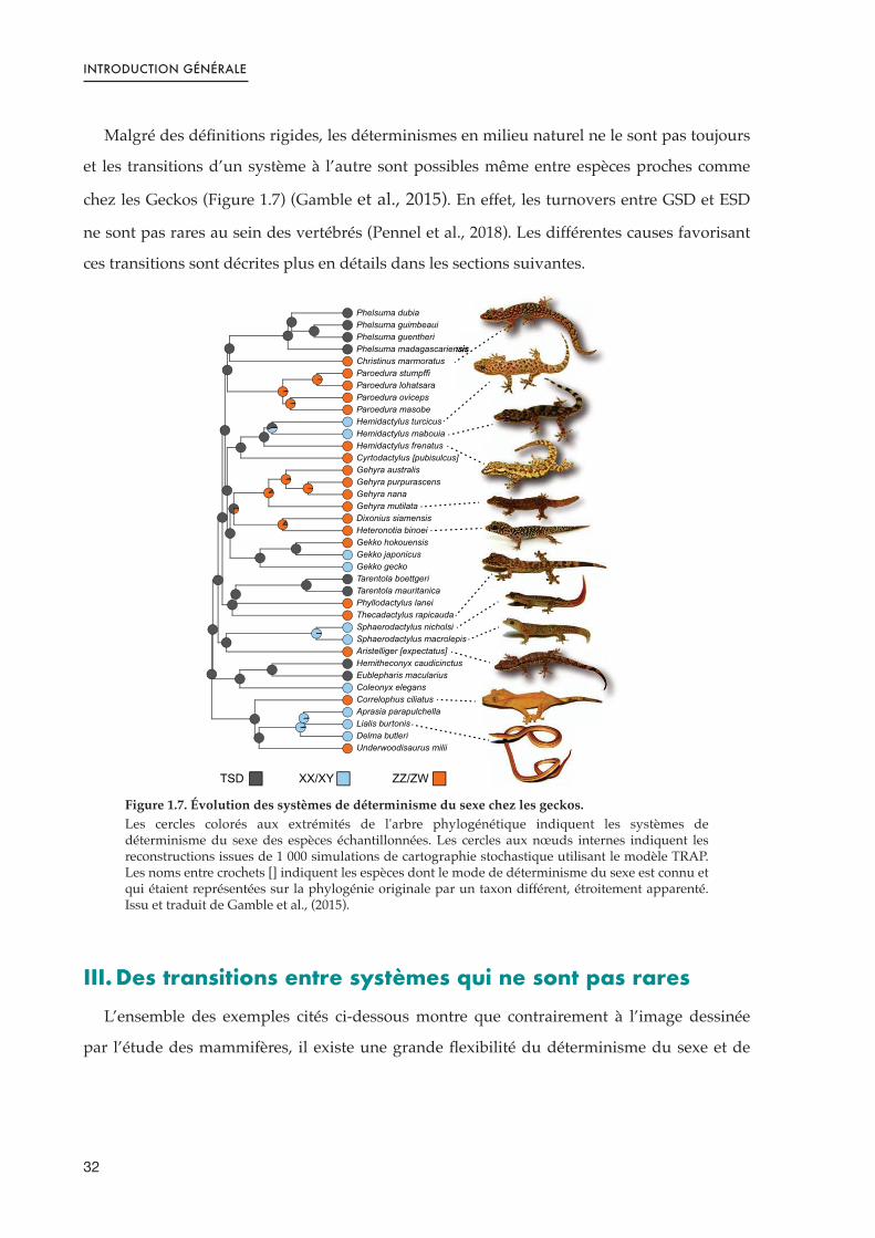
Malgré des définitions rigides, les déterminismes en milieu naturel ne le sont pas toujours
et les transitions d’un système à l’autre sont possibles même entre espèces proches comme
chez les Geckos (Figure 1.7) (Gamble et al., 2015). En effet, les turnovers entre GSD et ESD
ne sont pas rares au sein des vertébrés (Pennel et al., 2018). Les différentes causes favorisant
ces transitions sont décrites plus en détails dans les sections suivantes.
Figure 1.7. Évolution des systèmes de déterminisme du sexe chez les geckos.Les cercles colorés aux extrémités de l'arbre phylogénétique indiquent les systèmes de déterminisme du sexe des espèces échantillonnées. Les cercles aux nœuds internes indiquent les reconstructions issues de 1 000 simulations de cartographie stochastique utilisant le modèle TRAP. Les noms entre crochets [] indiquent les espèces dont le mode de déterminisme du sexe est connu et qui étaient représentées sur la phylogénie originale par un taxon différent, étroitement apparenté. Issu et traduit de Gamble et al., (2015).
III. Des transitions entre systèmes qui ne sont pas rares
L’ensemble des exemples cités ci-dessous montre que contrairement à l’image dessinée
par l’étude des mammifères, il existe une grande flexibilité du déterminisme du sexe et de
32
INTRODUCTION GÉNÉRALE
l’évolution des chromosomes sexuels chez les vertébrés. Certaines conditions peuvent
favoriser ces transitions entre GSD et ESD et ces turnovers entre chromosomes sexuels.
Dans certains cas, un déterminisme sexuel peut être contre-sélectionné si celui-ci conduit à
la production de descendances dont les sex-ratios ne sont plus favorables. Par exemple, en
présence de conditions environnementales imprévisibles, un mécanisme ESD pourrait
entraîner des sex-ratios extrêmement biaisés. De même, si l’environnement n’est pas (ou trop
peu) variable, les biais de sex-ratios induits par un déterminisme ESD pourraient être
important si les conditions ne sont pas régulièrement favorables au développement des deux
sexes. Dans ce genre de situations, l’apparition d’un déterminisme de type GSD pourrait être
sélectionné (Fisher, 1930). Cependant, dans le cas où un sex-ratio biaisé peut améliorer la
valeur sélective d’un des sexes (e.g. s’il existe une compétition pour l’accès à un partenaire,
ou si la température est favorable au développement d'un des deux sexes) alors l’apparition
ou le maintien d’un système ESD peut être favorisé (Bull, 1983; Kocher, 2004; Valenzuela &
Lance 2004). Une illustration de ces phénomènes existe chez les tortues. En effet, les espèces
avec un GSD ont des espérances de vie plus courtes (et donc moins d’opportunités dans le
temps pour produire un nombre équilibré d’individus de chaque sexe) que celles qui ont un
patron ESD (Sabath et al., 2016). Sur le long terme, des biais de sexe-ratios peuvent être
compensés chez les espèces à grande longévité, mais, sur des cycles de vies courts, un
système stable de GSD est certainement plus fortement sélectionné.
La domestication est également un moteur pour le changement d'un type de
déterminisme à un autre. Lorsqu’une espèce est prélevée du milieu naturel pour être
maintenue en élevage, le nombre d’individus fondateurs des lignées domestiquées est
souvent faible et un fort goulot d’étranglement s’exerce donc sur la population. La taille
efficace plus faible des stocks commerciaux ou de laboratoires rend les espèces domestiquées
plus sensibles à la dérive génétique et celle-ci peut par exemple entraîner la perte d’un
déterminant du sexe trouvé dans le milieu naturel, particulièrement suite à des croisements
successifs. Les populations sauvages de poisson-zèbres (Danio rerio) possèdent un
déterminisme du sexe de type ZZ/ZW avec un déterminant majeur sur le chromosome 4 que
ne présentent pas les souches de laboratoires, et notamment celle utilisée pour construire le
génome de référence de cette espèce modèle (Wilson et al., 2014). Pour renforcer encore ce
33

INTRODUCTION GÉNÉRALE
propos, le déterminisme sexuel de la souche domestiquée est complexe, polyfactoriel et
influencé par des effets environnementaux (Wilson et al., 2014; Ribas et al., 2017).
La thermosensibilité et son implication dans le déterminisme du sexe est certainement
l’une des clés pour comprendre les transitions entre différents modes de déterminismes qui
ont eu lieu à plusieurs reprises chez les amphibiens, les reptiles et les poissons (Sarre et al.,
2011). Enfin, il apparait que les transitions de déterminismes ESD vers GSD sont plus
courantes que la réciproque chez les poissons (et chez les squamates également) ce qui
suggère qu’un déterminisme GSD est plus stable (Gamble et al., 2015; Pokorna & Kratochvıl,
2009; Sabath, Itescu, et al., 2016; Pennel et al., 2018). Dans le contexte de changement
climatique actuel, les espèces à ESD (et particulièrement TSD) vont devoir rapidement
s’adapter pour limiter les biais de sex-ratios (qui sont déjà recensés), impliquant que les
espèces à ESD seront particulièrement plus enclines aux transitions vers un GSD sur le long
terme (Mitchell & Janzen, 2010; Jensen et al., 2018; Pennel et al., 2018). En revanche, les
transitions d’un système GSD vers ESD ne sont pas impossibles et peuvent se faire
rapidement en réponse à des conditions environnementales extrêmes particulièrement dans
une petite population et s’il existe une thermosensibilité, comme chez l’Agame barbu (Pogona
vitticeps) (Holleley et al., 2015).
IV. Les téléostéens, champions de la diversité
Les poissons téléostéens correspondent au groupe de vertébrés avec la plus grande
richesse spécifique (plus de 30,000 espèces) et à eux seuls représentent la moitié des espèces
de vertébrés (Helfman et al., 2009; Fricke et al., 2021). Ils sont présents dans tous les types
d’eaux du globe, adaptés à des conditions parfois extrêmes de températures et de salinités
notamment et arborent une grande diversité morphologique (Wootton et al., 2012). En plus
de cette richesse spécifique et de cette diversité écologique, les poissons sont également des
exemples de diversité et de plasticité du déterminisme et de la différentiation du sexe (Wang
et al., 2018). Pendant longtemps, le gène Sry, déterminant du sexe chez les mammifères
thériens, était le seul déterminant identifié chez les vertébrés. En dehors des poissons,
seulement deux autres déterminants du sexe ont été identifiés chez l’ensemble des vertébrés :
le gène Dmrt1 (Doublesex and mab-3 related transcription factor 1) qui se trouve sur le
chromosome Z chez les oiseaux (Smith et al., 2009); et le gène Dm-w chez le Xénope lisse (ou
34

INTRODUCTION GÉNÉRALE
Xénope du Cap, Xenopus laevis) qui est un paralogue du gène dmrt1 et se situe sur le
chromosome W (Yoshimoto et al., 2008). A contrario, chez les poissons, 8 déterminants du
sexes ont été identifiés : le gène dmy (aussi appelé dmrt1bY) chez le médaka (Oryzias latipes)
(Matsuda et al., 2002; Nanda et al., 2002) ; dmrt1 chez le poisson plat Cynoglossus semilaevis
(Cui et al., 2017) ; l’amhY chez le Pejerrey (Odontesthes hatchery) (Hattori et al., 2012) et
également chez le brochet (Esox lucius) (avec la dénomination amhbY) (Pan et al., 2019) ;
l’amhr2 chez le fugu (Takifugu rubripes) (Kamiya et al., 2012) ; gsdfY chez Oryzias luzonensis, un
proche parent du médaka (Myosho et al., 2012) ; sdY chez la truite arc-en-ciel (Oncorhynchus
mykiss) (Yano et al., 2012) ainsi que chez d’autres salmonidés (Guiguen et al., 2018); gdf6Y
chez Nothobranchius furzeri (Reichwald et al., 2015) ; et enfin sox3Y qui est un excellent
candidat pour le déterminisme du sexe chez une troisième espèce d’Oryzias (Oryzias dancena)
(Takehana et al., 2014) (Table 1). On peut noter que parmi les déterminants déjà identifiés, les
membres de la superfamille des facteurs de croissance transformant, ou TGF-β (de l'anglais
transforming growth factor), sont particulièrement représentés puisque les gènes de l’amh,
gsdf, gdf6 et de l’amhr2 en font partie. Les TGF-β pour beaucoup jouent un rôle de régulation
dans le développement embryonnaire et notamment dans le contrôle de la prolifération des
cellules germinales et des spermatogonies chez les poissons osseux comme chez le poisson-
zèbre ou la truite arc-en-ciel (Sawatari et al., 2007; Wang & Orban, 2007).
Sur l’ensemble de ces huit déterminants sexuels chez les poissons, cinq sont issus
d’évènement de duplications où l’un des paralogues a pris le rôle de la tête leader de la
cascade du déterminisme du sexe, et témoignent de la plasticité des génomes chez ce groupe
taxonomique (Table 1.1). En effet, les téléostéens ont globalement des taux de mutations et de
duplications supérieurs à ceux des autres vertébrés (Mank & Avise, 2009). Or, ces
duplications, lorsqu’elles interviennent sur gènes déjà impliqués dans la cascade de
déterminisme ou de la différenciation du sexe sont un phénomène efficace pour créer de
nouveaux déterminants car ceux-ci sont déjà intégrés à la cascade.
35

INTRODUCTION GÉNÉRALE
Table 1.1. Gènes du déterminisme du sexe formellement identifiés ou fortement suspectés chez les poissons téléostéens.Les déterminants qui ne sont pas issus d’évènement de duplication sont devenus déterminants majeurs suite à une diversification allélique. D’après Guiguen et al., (2018) et Li & Gui, (2018).
Les Cichlidés représentent peut-être la famille dont les radiations évolutives sont les plus
impressionnantes, et particulièrement dans les grands lacs de l’Est Africain, et l’on recense
plus de 3000 espèces de cichlidés entre l’Amérique du Sud, l’Afrique et l’Inde (Kocher, 2004;
Matschiner et al., 2020).
La vaste majorité des cichlidés est gonochorique et il existe une grande variabilité des
locus déterminants du sexe et des chromosomes/groupes de liaisons qui les portent. Ainsi,
on connait plus de 20 régions différentes associées au sexe sur 17 chromosomes/groupes de
liaisons, incluant les chromosomes B, chez les cichlidés (Figure 1.8) (Gammerdinger &
Kocher, 2018; Böhne et al., 2019; El Taher et al., 2020).
Les chromosomes sexuels identifiés jusqu’à présent sont systématiquement
homomorphiques (revue dans Gammerdinger & Kocher, 2018) mais présentent différents
degrés de différenciation génétique avec certains pour lesquels la région est étroite, et
d’autres pour lesquels elle est étendue sur la presque totalité d’un groupe de liaison (El Taher
et al., 2020). Enfin, certains chromosomes ont été plusieurs fois recrutés chez les cichlidés
pour endosser le rôle de chromosomes sexuels, comme les LGs 5, 7 et 19. Enfin, on retrouve
chez les cichlidés des patrons de GSD XX/XY et ZZ/ZW de déterminismes du sexe, mais
aussi des systèmes plus complexes comme XX/XY + ZZ/ZW ou encore XX/XY +
Chromosome B (spécifique aux femelles et agissant comme un W) (Clark & Kocher, 2018;
Gammerdinger & Kocher 2018; El Taher et al., 2020) et aussi une importance des facteurs
environnementaux et notamment de la température (Baroiller et al., 2009; Baroiller &
D’Cotta, 2016).
Déterminant
du sexeTaxon Système
Gène
ancestral
Issue d’une
Duplication
Famille
géniqueRéférences
dmy (dmrt1bY)
Oryzias latipes XY dmrt1 ✓ DM-domain Matsuda et al., 2002
dmrt1 Cynoglossus semilaevis ZW dmrt1 ✗ DM-domain Cui et al., 2017
amhY Odontesthes hatcheri XY amh ✓ TGF-β Hattori et al., 2012
amhbY Esox lucius XY amh ✓ TGF-β Pan et al., 2019
amhr2Y Takifugu rubripes XY amhr2 ✗ TGF-β Kamiya et al., 2012
gsdfY Oryzias luzonensis XY gsdf ✗ TGF-β Myosho et al., 2012
gdf6Y Nothobranchius furzeri XY gdf6 ✗ TGF-β Reichwald et al., 2015
sdy Oncorhynchus mykiss XY irf9 ✓ Irf Yano et al., 2012
sox3Y Oryzias dancena XY sox3 ✗ Sox Takehana et al., 2014
36

INTRODUCTION GÉNÉRALE
Figure 1.8. Évolution des chromosomes sexuels chez les cichlidés africains.Relations phylogénétiques et apparition des chromosomes sexuels par rapport au génome de référence du tilapia du Nil (O. niloticus) chez les cichlidés africains. Les lignées de cichlidés présentes dans le lac Tanganyika sont indiquées en noir, les cichlidés provenant d'autres lacs ou rivières en gris. Issue de El Taher et al., 2020 (version de bioRxiv).
V. Le Tilapia du Nil, un modèle de choix
V.1. Diversité du déterminisme du sexe en populations domestiques
Le tilapia du Nil (Oreochromis niloticus) est un cichlidé africain d’importance économique
majeure pour l’aquaculture puisqu’il représente l’une des espèces de poissons les plus
produites et consommées au monde avec 5,5 millions de tonnes produites en 2019 (FAO,
2021). Les mâles de cette espèce ont une croissance plus rapide, et disposent d’une masse
musculaire plus développée que les femelles. Malgré un fort intérêt économique pour la
production de mâles, le déterminisme du sexe de cette espèce est complexe et reste mal
compris. Depuis les années 1970, il a été établi que le déterminisme du sexe chez le tilapia du
Nil suit un sytème GSD XX/XY (Jalabert et al., 1974) mais des facteurs génétiques mineurs,
hérités des parents, peuvent impacter les sex-ratios des descendances (Baroiller & D’Cotta,
2001). De plus, ce déterminisme génétique peut également être supplanté par des effets
1094
37

INTRODUCTION GÉNÉRALE
environnementaux puisqu’une température élevée pendant les phases précoces du
développement peut affecter la différenciation du sexe et générer alors des mâles XX viables
et fertiles (Baroiller et al., 1995; Baroiller et al., 2009) (Figure 1.9). Cette sensibilité à la
température (thermosensibilité), définie comme la proportion de mâles XX obtenue, est un
trait variable et héritable qui peut être sélectionné en seulement quelques générations pour
fixer des souches très ou très peu thermosensibles (Baroiller and D’Cotta, 2001; Wessels et
Hörstgen-Schwark 2007).
Figure 1.9. Représentation triangulaire schématique de la complexité du déterminisme du sexe chez le tilapia du Nil.Modifié d’après Baroiller et al., (2009).
Jusqu’à présent, le déterminisme du sexe chez cette espèce a principalement été étudié sur
des souches domestiques et commerciales. Si la présence d’un déterminisme XX/XY est
acquise, la paire de chromosomes sexuels portant le déterminant majeur est encore débattue.
Des premières études, basées sur de la cytogénétique et l’observation de complexes
synaptonemaux chez O. niloticus ont montré un non-appariement des extrémités de la plus
grande paire de chromosome (LG3) chez les mâles XY mais pas chez les mâles XX, suggérant
que cette paire endosse le rôle de chromosomes sexuels (Carrasco et al., 1999). Des analyses
génétiques ont indiqué une association des marqueurs sexuels sur une paire de
chromosomes plus petite (LG1) (Lee et al., 2003; Cnaani et al., 2008; Lee et al., 2011).
Finalement, en utilisant des technologies plus récentes et des analyses génomiques, la région
Facteurs
génétiques mineursFacteurs
Environnementaux
Température
Facteurs
génétiques majeurs
XX/XY
Sexe
38

INTRODUCTION GÉNÉRALE
du déterminisme du sexe a été identifiée sur LG1 (Lee et al. 2003; Cnaani et al., 2008;
Palaiokostas et al. 2013; Gammerdinger et al. 2014; Palaiokostas et al. 2015) ou sur LG23
(Eshel et al. 2012, 2014; Li et al. 2015; Wessels et al., 2017; Cáceres et al. 2019; Taslima et al.
2020) ou bien sur les deux en même temps (Taslima et al., 2021) selon les souches étudiées.
Aucun gène précis n’a été proposé sur LG1. Sur LG23 en revanche, en étudiant des souches
domestiques Japonaises, la région du sexe a été identifiée : les mâles sont porteurs sur le Y
d’une duplication en tandem comprenant deux copies du gène de l’Hormone Anti-
Müllerienne (amh) dont une est intacte (amhY) et l’autre tronquée (amh∆Y) (Eshel et al., 2014;
Li et al., 2015). Une étude sur une population naturelle a également montré que le sexe était
associé au gène de l’amh sur LG23 chez des individus du lac Kou au Burkina-Faso (Sissao et
al., 2019).
Table 1.2. Bilan des origines, dénominations et déterminants génétiques du sexe chez les souches domestiques et commerciales de tilapia du Nil.
Laboratoire/Commercial Souche Origine du stock Dénomination LGLocus
du sexReference LG
Reference Origine
Université de Stirling (Royaume-Uni)
Manzala Manzala (Egypte) Manzala-Stirling
LG1 +
LG23
? + amh
Taslima et al., 2021
Taslima et al., 2021
Université de Liège (Tihange, Belgique) Manzala Manzala-Stirling
Manzala-Thiange LG23 amh
Sissao et al., 2019
Taslima et al., 2021
Université de Göttingen (Allemagne) Manzala Manzala-Stirling
Manzala-Göttingen LG23 amh
Wessels et al., 2017
Kronert et al., 1989
Agricultural Research Organization (Israël)
Manzala Manzala-Stirling-Swansea
Manzala-ARO LG23 amh Eshel et al., 2014
Taslima et al., 2021
Agricultural Research Organization (Israël)
Manzala (+ Ghana) Manzala-Stirling-Swansea
Manzala-ARO LG23 amh Curzon et al., 2019
Curzon et al., 2019
Laboratory of Reproductive Biology (Okazaki, Japon)
Egypte
Egypte (possible introgression par Manzala-Stirling par l’utilisation de mâles YY )
Japenese LG23 amh Li et al., 2015 Li et al., 2015
Souches commerciales d’Amérique du Sud
Malaysie (plus ou moins lié au GIFT)
AquaAmerica (Brésil) LG23 amh
Cáceres et al., 2019
Cáceres et al., 2019
Croisement d’individus d’Israel, de Singapore, de Taiwan et de Thaïlande présents aux Philippines (Carmen Aquafarm) dans la fin des années 1980
Acuacorporación Internacional (ACI, Costa Rica) (Obtenu du Brésil)
LG23 amh
GIFT + 2 souches originaires d’Afriques ayant servie à fonder la souche GIFT
Acuacorporación Internacional (ACI, Costa Rica)
LG23 amh
World Fish GIFT (*)Hybrid of several strains GIFT LG23 amh
Taslima et al., 2020
Eknath et al., 1998;
Ponzoni et al., 2011
39

INTRODUCTION GÉNÉRALE
(**) Hybride entre 4 populations de O. niloticus (Egypte, Ghana, Kenya et Sénégal), et 4 souches d’aquaculture (Israël, Singapour, Taïwan et Thaïlande).
(**) 3ème Génération du GIFT transmis en 1994 par l’ICLARM (International Center for Living Aquatic Resources Management (ancêtre du Worldfish).
Des marqueurs PCR permettant de génotyper le sexe des individus ont été développés
pour les souches présentant un déterminisme du sexe par le gène de l’amh (Eshel et al., 2014;
Li et al., 2015; Sissao et al., 2019). Ces marqueurs ne permettent cependant pas de génotyper
efficacement toutes les populations sauvages (Baroiller & D’Cotta, 2016), suggérant un
polymorphisme du chromosome Y dans les populations naturelles. En effet, ces marqueurs
PCR sont inutilisables sur les populations éthiopiennes (aucune amplifications de l’amh∆Y),
et des incongruences existent entre les marqueurs de l’amhY et de l’amh∆Y dans la
population sauvage de Kou (Burkina-Faso), alors que ces deux copies sont issues d'une
duplication en tandem et donc que la présence de l’un devrait systématiquement indiquer la
présence de l’autre (Sissao et al., 2019).
V.2. Une espèce à large aire de répartition et diversité écologique
L’influence de l’environnement et particulièrement de la température sur le déterminisme
du sexe des espèces GSD + EE n’a pas toujours été étudiée en tenant compte des conditions
pouvant réellement être rencontrés dans le milieu naturel. En l’occurence, le tilapia du Nil est
une espèce à l’aire de répartition très large à travers l’Afrique, des bassins du Nil au Sénégal,
aux plateaux des vallées du Grand Rift en Ethiopie, en passant par les provinces de Kivu et
du Nord du Tanganyika (Trewavas 1983; Bezault et al., 2011). Du fait de ces nombreuses
colonisations de différents cours d’eau et lac, cette espèce s’est adapté à une large gamme de
températures dans le milieu naturel et on la retrouve dans des eaux « froides » (17-26°C),
tempérées (28-34°C) et même des sources chaudes (36-40°C) (Bezault et al., 2007; Ndiwa et al.
2014). Or, une exposition à une température supérieure à 32°C pendant les phases précoces
Guangxi Fisheries Institute (Nanning, Guangxi, Chine)
Ancien GIFT (**)
2 programmes successifs de sélection mis en place par la Chine sur l’ancien GIFT (ICLARM non associé)
Chinese GIFT LG23 Zhao et al., 2019
USA Souche commerciale Amherst, Massachusetts USA
?USA
Commercial Stock
LG1 ? Gammerdinger et al., 2014
Gammerdinger et al., 2014
Université N. Boni, (Burkina Faso) Kou Lac Kou Kou-BF
LG23 + ?
amh + ?
Sissao et al., 2019
Sissao et al., 2019
Laboratoire/Commercial Souche Origine du stock Dénomination LGLocus
du sexReference LG
Reference Origine
40

INTRODUCTION GÉNÉRALE
du développement des gonades (10 à 20 jours après fécondation des œufs) peut induire une
réversion du sexe et donner lieu à la différenciation en mâles d’individus pourtant XX
(Baroiller et al., 1995; Baroiller et al., 2009). Cette thermosensibilité (plus ou moins forte selon
les familles ou les souches) chez le tilapia du Nil a aussi été démontrée pour l’ensemble des
populations étudiées issues du milieu naturel (Bezault et al. 2007; Baroiller et al., 2009).
En milieu naturel, après une période d’incubation buccale puis de garde, les femelles
relâchent les alevins pendant leur période thermosensible et ceux-ci rejoignent les zones
d’eau peu profondes où les températures sont aussi plus élevées (Philippart & Ruwet, 1982).
Les alevins peuvent donc être exposés à des températures qui dépassent la température de
masculinisation de 32°C. Une étude en conditions contrôlées sur les préférences en continuum
thermique a d’ailleurs montré que 20% des alevins avaient une préférences pour les eaux
plus chaudes (36°C) pendant les premiers jours puis dans l’ensemble montraient une
préférence pour les bassins à 32°C malgré la présence d’un bassin à 28°C accessible (Nivelle
et al., 2019).
Le tilapia du Nil est donc une espèce présentant un GSD+EE dont les effets
environnementaux pouvant influencer la différentiation du sexe sont rencontrés en milieu
naturel. Du fait de la diversité des mécanismes rencontrés dans les souches de laboratoire,
ainsi que de son déterminisme du sexe complexe dans lequel facteurs génétiques et
environnementaux interagissent, le tilapia du Nil représente un modèle idéal pour l’étude de
l’évolution du déterminisme du sexe à l’échelle intraspécifique. De plus, l’existence de
populations sauvages adaptées à une large gamme de température fait également du tilapia du
Nil un bon modèle pour étudier l’impact du réchauffement des eaux sur la dynamique des sex-
ratios des espèces de poissons thermosensibles dans un contexte de changement climatique.
Toutefois, il n’est pas évident de savoir en quelle mesure les différents mécanismes de
déterminisme du sexe observés chez le tilapia du Nil sont dues à une diversité propre à
l’espèce ou si elles sont dues aux évènements de domestications (avec des introgressions et
hybridations, accidentelles ou non).
Ainsi, dans ce projet de thèse l’objectif était, dans un premier temps, de comparer les bases
génétiques du déterminisme du sexe de populations d’O. niloticus du milieu naturel dont les
écologies thermiques sont variées (Figure 1.10) pour comprendre si la diversité observée dans
les stocks de laboratoires est le reflet d’une variabilité dans les populations sauvages ; dans un
41
INTRODUCTION GÉNÉRALE
second temps de pouvoir caractériser en détails la région du sexe et identifier des régions (si
possibles universelles) qui permettent le développement de marqueurs pour génotyper
sexuellement les individus sauvages ; enfin dans un troisième temps, il était souhaité
d’explorer les gènes précocement impliqués dans la différenciation du sexe et l’effet de la
température sur cette cascade.
Ce manuscrit se compose donc de trois chapitres au format article qui tentent de répondre à
ces problématiques ou du moins de démêler l’histoire complexe du déterminisme du sexe
chez le tilapia du Nil.
Figure 1.10. Localisation géographique des fourchettes de température des environnements d’où proviennent les populations sauvages étudiées dans ce projet de thèse.
!"#$%&$%!"'()*
+&,&
!"#$%&'()*'+,
-.#$#
!"#$%&'#"
()"*+,-
.$,
-.*/01'2)
!/0#1()'2+,
34.5/"5#
/0%&1
34%56
7*8)9&#:*
6/7#'')'8+,
-.*/01'2)
20%&1
!/&'8)*9+,
;&"0&"(
!"#$
%
'(")#
:&705$#;<#=/
42

INTRODUCTION GÉNÉRALE
Références
Adolfi, M.C., Nakajima, R.T., Nóbrega, R.H., and Schartl, M. (2019). Intersex, Hermaphroditism, and Gonadal Plasticity in Vertebrates: Evolution of the Müllerian Duct and Amh/Amhr2 Signaling. Annu. Rev. Anim. Biosci. 7, 149–172.
Bachtrog, D. (2013). Y-chromosome evolution: emerging insights into processes of Y-chromosome degeneration. Nature Reviews Genetics 14, 113–124.
Bachtrog, D., Mank, J.E., Peichel, C.L., Kirkpatrick, M., Otto, S.P., Ashman, T.-L., Hahn, M.W., Kitano, J., Mayrose, I., Ming, R., et al. (2014). Sex determination: why so many ways of doing it? PLOS Biology 12, e1001899.
Baroiller, J.F., and D’Cotta, H. (2001). Environment and sex determination in farmed fish. Comparative Biochemistry and Physiology Part C: Toxicology & Pharmacology 130, 399–409.
Baroiller, J.-F., and D’Cotta, H. (2016). The Reversible Sex of Gonochoristic Fish: Insights and Consequences. Sexual Development 10, 242–266.
Baroiller, J.F., Chourrout, D., Fostier, A., and Jalabert, B. (1995). Temperature and sex chromosomes govern sex ratios of the mouthbrooding Cichlid fish Oreochromis niloticus. J. Exp. Zool. 273, 216–223.
Baroiller, J.F., D’Cotta, H., and Saillant, E. (2009a). Environmental effects on fish sex determination and differentiation. SXD 3, 118–135.
Baroiller, J.F., D’Cotta, H., Bezault, E., Wessels, S., and Hoerstgen-Schwark, G. (2009b). Tilapia sex determination: where temperature and genetics meet. Comparative Biochemistry and Physiology Part A: Molecular & Integrative Physiology 153, 30–38.
Bezault, E., Clota, F., Derivaz, M., Chevassus, B., and Baroiller, J.-F. (2007). Sex determination and temperature-induced sex differentiation in three natural populations of Nile tilapia (Oreochromis niloticus) adapted to extreme temperature conditions. Aquaculture 272, S3–S16.
Bezault, E., Balaresque, P., Toguyeni, A., Fermon, Y., Araki, H., Baroiller, J.-F., and Rognon, X. (2011). Spatial and temporal variation in population genetic structure of wild Nile tilapia (Oreochromis niloticus) across Africa. BMC Genetics 12, 102.
Böhne, A., Weber, A.A.-T., Rajkov, J., Rechsteiner, M., Riss, A., Egger, B., and Salzburger, W. (2019). Repeated Evolution Versus Common Ancestry: Sex Chromosome Evolution in the Haplochromine Cichlid Pseudocrenilabrus philander. Genome Biology and Evolution 11, 439–458.
Brown, E.E., Baumann, H., and Conover, D.O. (2014). Temperature and photoperiod effects on sex determination in a fish. Journal of Experimental Marine Biology and Ecology 461, 39–43.
Bull, J.J. (1983). Evolution of sex determining mechanisms. Evolution of Sex Determining Mechanisms.
Cáceres, G., López, M.E., Cádiz, M.I., Yoshida, G.M., Jedlicki, A., Palma-Véjares, R., Travisany, D., Díaz-Domínguez, D., Maass, A., Lhorente, J.P., et al. (2019). Fine mapping
43

INTRODUCTION GÉNÉRALE
using whole-genome sequencing confirms anti-müllerian hormone as a major gene for sex determination in farmed Nile tilapia (Oreochromis niloticus L.). G3 (Bethesda) 9, 3213–3223.
Capel, B. (2017). Vertebrate sex determination: evolutionary plasticity of a fundamental switch. Nat. Rev. Genet. 18, 675–689.
Carrasco, L.A.P., Penman, D.J., and Bromage, N. (1999). Evidence for the presence of sex chromosomes in the Nile tilapia (Oreochromis niloticus) from synaptonemal complex analysis of XX, XY and YY genotypes. Aquaculture 173, 207–218.
Castelli, M., Georges, A., and Holleley, C.E. (2021). Corticosterone does not have a role in temperature sex reversal in the central bearded dragon ( Pogona vitticeps ). J Exp Zool A Ecol Integr Physiol 335, 301–310.
Castelli, M.A., Whiteley, S.L., Georges, A., and Holleley, C.E. (2020). Cellular calcium and redox regulation: the mediator of vertebrate environmental sex determination? Biological Reviews 95, 680–695.
Charlesworth, B., and Dempsey, N.D. (2001). A model of the evolution of the unusual sex chromosome system of Microtus oregoni. Heredity 86, 387–394.
Cioffi, M.B., and Bertollo, L. a. C. (2010). Initial steps in XY chromosome differentiation in Hoplias malabaricus and the origin of an X 1 X 2 Y sex chromosome system in this fish group. Heredity 105, 554–561.
Clark, F.E., and Kocher, T.D. (2019). Changing sex for selfish gain: B chromosomes of Lake Malawi cichlid fish. Scientific Reports 9, 1–10.
Clark, F.E., Conte, M.A., Ferreira-Bravo, I.A., Poletto, A.B., Martins, C., and Kocher, T.D. (2017). Dynamic sequence evolution of a sex-associated B chromosome in Lake Malawi cichlid fish. JHERED 108, 53–62.
Clark, F.E., Conte, M.A., and Kocher, T.D. (2018). Genomic characterization of a B chromosome in Lake Malawi cichlid fishes. Genes 9, 610.
Cnaani, A., Lee, B.-Y., Zilberman, N., Ozouf-Costaz, C., Hulata, G., Ron, M., D'Hont, A., Baroiller, J.-F., D'Cotta, H., Penman, D.J., et al. (2008). Genetics of sex determination in tilapiine species. Sexual Development 2, 43–54.
Cocquet, J., Ellis, P.J.I., Mahadevaiah, S.K., Affara, N.A., Vaiman, D., and Burgoyne, P.S. (2012). A Genetic Basis for a Postmeiotic X Versus Y Chromosome Intragenomic Conflict in the Mouse. PLOS Genetics 8, e1002900.
Corona-Herrera, G.A., Arranz, S.E., Martínez-Palacios, C.A., Navarrete-Ramírez, P., Toledo-Cuevas, E.M., Valdez-Alarcón, J.J., and Martínez-Chávez, C.C. (2018). Experimental evidence of masculinization by continuous illumination in a temperature sex determination teleost (Atherinopsidae) model: is oxidative stress involved? Journal of Fish Biology 93, 229–237.
Cui, Z., Liu, Y., Wang, W., Wang, Q., Zhang, N., Lin, F., Wang, N., Shao, C., Dong, Z., Li, Y., et al. (2017). Genome editing reveals dmrt1 as an essential male sex-determining gene in Chinese tongue sole ( Cynoglossus semilaevis ). Scientific Reports 7, 42213.
44

INTRODUCTION GÉNÉRALE
Curzon, A.Y., Seroussi, E., Benet-Perelberg, A., Zak, T., Dor, L., Shirak, A., and Ron, M. (2019). Hybrid Origin of the Thai-Chitralada Tilapia Strain Using DNA Barcoding and Microsatellite Analysis. Israeli Journal of Aquaculture - Bamidgeh.
Deeming, D.C. (2004). Chapter 4 - Prevalence of TSD in crocodilians. In Temperature-Dependent Sex Determination in Vertebrates, (Smithsonian), pp. 33–41.
Deveson, I.W., Holleley, C.E., Blackburn, J., Marshall Graves, J.A., Mattick, J.S., Waters, P.D., and Georges, A. (2017). Differential intron retention in Jumonji chromatin modifier genes is implicated in reptile temperature-dependent sex determination. Sci. Adv. 3, e1700731.
Devlin, R.H., and Nagahama, Y. (2002). Sex determination and sex differentiation in fish: an overview of genetic, physiological, and environmental influences. Aquaculture 208, 191–364.
Eknath, A., Dey, M., Rye, M., Gjerde, B., Abella, T.A., Sevilleja, R., Tayamen, M.M., Reyes, R.A., and Bentsen, H. (1998). Selective breeding of Nile tilapia for Asia.
El Taher, A., Ronco, F., Matschiner, M., Salzburger, W., and Böhne, A. (2020). Dynamics of sex chromosome evolution in a rapid radiation of cichlid fishes (Evolutionary Biology).
Ellegren, H. (2010). Evolutionary stasis: the stable chromosomes of birds. Trends in Ecology & Evolution 25, 283–291.
Eshel, O., Shirak, A., Weller, J.I., Hulata, G., and Ron, M. (2012). Linkage and physical mapping of sex Rregion on LG23 of Nile tilapia (Oreochromis niloticus). G3: Genes, Genomes, Genetics 2, 35–42.
Eshel, O., Shirak, A., Dor, L., Band, M., Zak, T., Markovich-Gordon, M., Chalifa-Caspi, V., Feldmesser, E., Weller, J.I., Seroussi, E., et al. (2014). Identification of male-specific amh duplication, sexually differentially expressed genes and microRNAs at early embryonic development of Nile tilapia (Oreochromis niloticus). BMC Genomics 15, 774.
Ewert, M.A., Etchberger, C.R., and Nelson, C.E. (2004). Chapter 3 - Turtle Sex-Determining Modes and TSD Patterns, and Some TSD Pattern Correlates. In Temperature-Dependent Sex Determination in Vertebrates, (Smithsonian), pp. 21–32.
FAO (2021). The State of World Fisheries and Aquaculture 2021 (FAO).
Fernandino, J.I., Hattori, R.S., Kishii, A., Strüssmann, C.A., and Somoza, G.M. (2012). The Cortisol and Androgen Pathways Cross Talk in High Temperature-Induced Masculinization: The 11β-Hydroxysteroid Dehydrogenase as a Key Enzyme. Endocrinology 153, 6003–6011.
Fisher, R.A. (1930). The genetical theory of natural selection. (Oxford: Clarendon Press).
Fricke, R., Eschmeyer, W.N., and Van der Laan, R. (2021). Eschmeyer’s Catalog of Fishes: Genera, Species, References.
Furman, B.L.S., Metzger, D.C.H., Darolti, I., Wright, A.E., Sandkam, B.A., Almeida, P., Shu, J.J., and Mank, J.E. (2020). Sex chromosome evolution: so many exceptions to the rules. Genome Biol Evol 12, 750–763.
Gamble, T., Coryell, J., Ezaz, T., Lynch, J., Scantlebury, D.P., and Zarkower, D. (2015). Restriction Site-Associated DNA Sequencing (RAD-seq) Reveals an Extraordinary
45
INTRODUCTION GÉNÉRALE
Number of Transitions among Gecko Sex-Determining Systems. Molecular Biology and Evolution 32, 1296–1309.
Gamble, T., Castoe, T.A., Nielsen, S.V., Banks, J.L., Card, D.C., Schield, D.R., Schuett, G.W., and Booth, W. (2017). The Discovery of XY Sex Chromosomes in a Boa and Python. Current Biology 27, 2148-2153.e4.
Gammerdinger, W.J., and Kocher, T.D. (2018). Unusual diversity of sex chromosomes in African cichlid fishes. Genes 9, 480.
Gammerdinger, W.J., Conte, M.A., Acquah, E.A., Roberts, R.B., and Kocher, T.D. (2014). Structure and decay of a proto-Y region in Tilapia, Oreochromis niloticus. BMC Genomics 15, 975.
García-Cruz, E.L., Yamamoto, Y., Hattori, R.S., de Vasconcelos, L.M., Yokota, M., and Strüssmann, C.A. (2020). Crowding stress during the period of sex determination causes masculinization in pejerrey Odontesthes bonariensis, a fish with temperature-dependent sex determination. Comparative Biochemistry and Physiology Part A: Molecular & Integrative Physiology 110701.
Ge, C., Ye, J., Weber, C., Sun, W., Zhang, H., Zhou, Y., Cai, C., Qian, G., and Capel, B. (2018). The histone demethylase KDM6B regulates temperature-dependent sex determination in a turtle species. Science.
Geffroy, B., and Bardonnet, A. (2016). Sex differentiation and sex determination in eels: consequences for management. Fish and Fisheries 17, 375–398.
Georges, A. (1992). Thermal-Characteristics and Sex Determination in Field Nests of the Pig-Nosed Turtle, Carettochelys-Insculpta (Chelonia, Carettochelydidae), From Northern Australia. Aust. J. Zool. 40, 511–521.
Georges, A., and Holleley, C.E. (2018). How does temperature determine sex? Science 360, 601–602.
Goikoetxea, A., Todd, E.V., and Gemmell, N.J. (2017). Stress and sex: does cortisol mediate sex change in fish? Reproduction 154, R149–R160.
Grützner, F., Rens, W., Tsend-Ayush, E., El-Mogharbel, N., O’Brien, P.C.M., Jones, R.C., Ferguson-Smith, M.A., and Marshall Graves, J.A. (2004). In the platypus a meiotic chain of ten sex chromosomes shares genes with the bird Z and mammal X chromosomes. Nature 432, 913–917.
Guiguen, Y., Fostier, A., and Herpin, A. (2018). Sex Determination and Differentiation in Fish: Genetic, Genomic, and Endocrine Aspects. In Sex Control in Aquaculture, (John Wiley & Sons), pp. 35–64.
Hattori, R.S., Fernandino, J.I., Kishii, A., Kimura, H., Kinno, T., Oura, M., Somoza, G.M., Yokota, M., Strüssmann, C.A., and Watanabe, S. (2009). Cortisol-Induced Masculinization: Does Thermal Stress Affect Gonadal Fate in Pejerrey, a Teleost Fish with Temperature-Dependent Sex Determination? PLOS ONE 4, e6548.
Hattori, R.S., Murai, Y., Oura, M., Masuda, S., Majhi, S.K., Sakamoto, T., Fernandino, J.I., Somoza, G.M., Yokota, M., and Strüssmann, C.A. (2012). A Y-linked anti-Müllerian hormone duplication takes over a critical role in sex determination. PNAS 109, 2955–2959.
46

INTRODUCTION GÉNÉRALE
Helfman, G.S., Colette, B.B., Facey, D.E., and Bowen, B.W. (2009). The diversity of fishes: biology, evolution, and ecology (Chichester, UK ; Hoboken, NJ: Blackwell).
Herpin, A., and Schartl, M. (2015). Plasticity of gene-regulatory networks controlling sex determination: of masters, slaves, usual suspects, newcomers, and usurpators. EMBO Reports 16, 1260–1274.
Herpin, A., Braasch, I., Kraeussling, M., Schmidt, C., Thoma, E.C., Nakamura, S., Tanaka, M., and Schartl, M. (2010). Transcriptional Rewiring of the Sex Determining dmrt1 Gene Duplicate by Transposable Elements. PLOS Genetics 6, e1000844.
Holleley, C.E., O’Meally, D., Sarre, S.D., Marshall Graves, J.A., Ezaz, T., Matsubara, K., Azad, B., Zhang, X., and Georges, A. (2015). Sex reversal triggers the rapid transition from genetic to temperature-dependent sex. Nature 523, 79–82.
Jalabert, B., Moreau, J., Planquette, P., and Billard, R. (1974). Déterminisme du sexe chez tilapia macrochir et tilapia nilotica : action de la méthyltestostérone dans l’alimentation des alevins sur la différenciation sexuelle; proportion des sexes dans la descendance des mâles «inversés». Annales de Biologie Animale Biochimie Biophysique Vol. 14, 729–739.
Jeffries, D.L., Lavanchy, G., Sermier, R., Sredl, M.J., Miura, I., Borzée, A., Barrow, L.N., Canestrelli, D., Crochet, P.-A., Dufresnes, C., et al. (2018). A rapid rate of sex-chromosome turnover and non-random transitions in true frogs. Nature Communications 9.
Jensen, M.P., Allen, C.D., Eguchi, T., Bell, I.P., LaCasella, E.L., Hilton, W.A., Hof, C.A.M., and Dutton, P.H. (2018). Environmental Warming and Feminization of One of the Largest Sea Turtle Populations in the World. Current Biology 28, 154-159.e4.
Kamiya, T., Kai, W., Tasumi, S., Oka, A., Matsunaga, T., Mizuno, N., Fujita, M., Suetake, H., Suzuki, S., Hosoya, S., et al. (2012). A trans-species missense SNP in Amhr2 is associated with sex determination in the tiger pufferfish, Takifugu rubripes (Fugu). PLoS Genet 8.
Kocher, T.D. (2004). Adaptive evolution and explosive speciation: the cichlid fish model. Nat Rev Genet 5, 288–298.
Koopman, P. (1995). The molecular biology of SRY and its role in sex determination in mammals. Reprod. Fertil. Dev. 7, 713–722.
Kronert, U., Hörstgen-Schwark, G., and Langholz, H.-J. (1989). Prospects of selecting for late maturity in tilapia (Oreochromis niloticus): I. Family studies under laboratory conditions. Aquaculture 77, 113–121.
Lang, J.W., and Andrews, H.V. (1994). Temperature-dependent sex determination in crocodilians. Journal of Experimental Zoology 270, 28–44.
Lee, B.-Y., Penman, D.J., and Kocher, T.D. (2003). Identification of a sex-determining region in Nile tilapia (Oreochromis niloticus) using bulked segregant analysis. Animal Genetics 34, 379–383.
Lee, B.-Y., Coutanceau, J.-P., Ozouf-Costaz, C., D’Cotta, H., Baroiller, J.-F., and Kocher, T.D. (2011). Genetic and physical mapping of sex-linked AFLP markers in Nile tilapia (Oreochromis niloticus). Mar Biotechnol (NY) 13, 557–562.
Li, X.-Y., and Gui, J.-F. (2018). Diverse and variable sex determination mechanisms in vertebrates. Sci. China Life Sci. 61, 1503–1514.
47
INTRODUCTION GÉNÉRALE
Li, M., Sun, Y., Zhao, J., Shi, H., Zeng, S., Ye, K., Jiang, D., Zhou, L., Sun, L., Tao, W., et al. (2015). A tandem duplicate of anti-müllerian hormone with a missense SNP on the Y chromosome is essential for male sex determination in Nile tilapia, Oreochromis niloticus. PLOS Genetics 11, e1005678.
Lubieniecki, K.P., Lin, S., Cabana, E.I., Li, J., Lai, Y.Y.Y., and Davidson, W.S. (2015). Genomic instability of the sex-determining locus in Atlantic salmon (Salmo salar). G3 Genes|Genomes|Genetics 5, 2513–2522.
Mank, J.E., and Avise, J.C. (2009). Evolutionary Diversity and Turn-Over of Sex Determination in Teleost Fishes. SXD 3, 60–67.
Mankiewicz, J.L., Godwin, J., Holler, B.L., Turner, P.M., Murashige, R., Shamey, R., Daniels, H.V., and Borski, R.J. (2013). Masculinizing effect of background color and cortisol in a flatfish with environmental sex-determination. Integr Comp Biol 53, 755–765.
Matschiner, M., Böhne, A., Ronco, F., and Salzburger, W. (2020). The genomic timeline of cichlid fish diversification across continents. Nat Commun 11, 5895.
Matsuda, M., Nagahama, Y., Shinomiya, A., Sato, T., Matsuda, C., Kobayashi, T., Morrey, C.E., Shibata, N., Asakawa, S., Shimizu, N., et al. (2002). DMY is a Y-specific DM-domain gene required for male development in the medaka fish. Nature 417, 559–563.
Matveevsky, S., Kolomiets, O., Bogdanov, A., Hakhverdyan, M., and Bakloushinskaya, I. (2017). Chromosomal Evolution in Mole Voles Ellobius (Cricetidae, Rodentia): Bizarre Sex Chromosomes, Variable Autosomes and Meiosis. Genes 8, 306.
Mei, J., and Gui, J.-F. (2015). Genetic basis and biotechnological manipulation of sexual dimorphism and sex determination in fish. Sci. China Life Sci. 58, 124–136.
Mitchell, N.J., and Janzen, F.J. (2010). Temperature-dependent sex determination and contemporary climate change. Sex Dev 4, 129–140.
Mitchell, N.J., Nelson, N.J., Cree, A., Pledger, S., Keall, S.N., and Daugherty, C.H. (2006). Support for a rare pattern of temperature-dependent sex determination in archaic reptiles: evidence from two species of tuatara (Sphenodon). Front Zool 3, 9.
Miura, I. (2017). Sex Determination and Sex Chromosomes in Amphibia. SXD 11, 298–306.
Morgan, T.H. (1909). Sex determination and parthenogenesis in phylloxerans and aphids. Science 29, 234–237.
Mulugeta, E., Wassenaar, E., Sleddens-Linkels, E., van IJcken, W.F.J., Heard, E., Grootegoed, J.A., Just, W., Gribnau, J., and Baarends, W.M. (2016). Genomes of Ellobius species provide insight into the evolutionary dynamics of mammalian sex chromosomes. Genome Res. 26, 1202–1210.
Murata, C., Kuroki, Y., Imoto, I., and Kuroiwa, A. (2016). Ancestral Y-linked genes were maintained by translocation to the X and Y chromosomes fused to an autosomal pair in the Okinawa spiny rat Tokudaia muenninki. Chromosome Res 24, 407–419.
Myosho, T., Otake, H., Masuyama, H., Matsuda, M., Kuroki, Y., Fujiyama, A., Naruse, K., Hamaguchi, S., and Sakaizumi, M. (2012). Tracing the emergence of a novel sex-determining gene in medaka, Oryzias luzonensis. Genetics 191, 163–170.
48

INTRODUCTION GÉNÉRALE
Nanda, I., Kondo, M., Hornung, U., Asakawa, S., Winkler, C., Shimizu, A., Shan, Z., Haaf, T., Shimizu, N., Shima, A., et al. (2002). A duplicated copy of DMRT1 in the sex-determining region of the Y chromosome of the medaka, Oryzias latipes. PNAS 99, 11778–11783.
Navarro-Martín, L., Viñas, J., Ribas, L., Díaz, N., Gutiérrez, A., Croce, L.D., and Piferrer, F. (2011). DNA Methylation of the Gonadal Aromatase (cyp19a) Promoter Is Involved in Temperature-Dependent Sex Ratio Shifts in the European Sea Bass. PLOS Genetics 7, e1002447.
Ndiwa, T.C., Nyingi, D.W., and Agnese, J.-F. (2014). An Important Natural Genetic Resource of Oreochromis niloticus (Linnaeus, 1758) Threatened by Aquaculture Activities in Loboi Drainage, Kenya. PLOS ONE 9, e106972.
Nivelle, R., Gennotte, V., Kalala, E.J.K., Ngoc, N.B., Muller, M., Mélard, C., and Rougeot, C. (2019). Temperature preference of Nile tilapia (Oreochromis niloticus) juveniles induces spontaneous sex reversal. PLOS ONE 14, e0212504.
Ohno, S. (1966). Conservation of the Original Z-Chromosome by Diverse Avian Species and Homology of the Z-linked Genes. In Sex Chromosomes and Sex-Linked Genes, (Berlin, Heidelberg: Springer Berlin Heidelberg), pp. 73–81.
De Oliveira, R.R., Feldberg, E., dos Anjos, M.B., and Zuanon, J. (2008). Occurrence of multiple sexual chromosomes (XX/XY1Y2 and Z1Z1Z2Z2/Z1Z2W1W2) in catfishes of the genus Ancistrus (Siluriformes: Loricariidae) from the Amazon basin. Genetica 134, 243–249.
Ortega-Recalde, O., Goikoetxea, A., Hore, T.A., Todd, E.V., and Gemmell, N.J. (2020). The Genetics and Epigenetics of Sex Change in Fish. Annual Review of Animal Biosciences 8, null.
Ospina-Álvarez, N., and Piferrer, F. (2008). Temperature-Dependent Sex Determination in Fish Revisited: Prevalence, a Single Sex Ratio Response Pattern, and Possible Effects of Climate Change. PLOS ONE 3, e2837.
Otake, T., and Kuroiwa, A. (2016). Molecular mechanism of male differentiation is conserved in the SRY -absent mammal, Tokudaia osimensis. Scientific Reports 6, 32874.
Palaiokostas, C., Bekaert, M., Khan, M.G.Q., Taggart, J.B., Gharbi, K., McAndrew, B.J., and Penman, D.J. (2013). Mapping and validation of the major sex-determining region in Nile tilapia (Oreochromis niloticus L.) using RAD sequencing. PLoS ONE 8, e68389.
Palaiokostas, C., Bekaert, M., Khan, M., Taggart, J.B., Gharbi, K., McAndrew, B.J., and Penman, D.J. (2015). A novel sex-determining QTL in Nile tilapia (Oreochromis niloticus). BMC Genomics 16, 171.
Pan, Q., Feron, R., Yano, A., Guyomard, R., Jouanno, E., Vigouroux, E., Wen, M., Busnel, J.-M., Bobe, J., Concordet, J.-P., et al. (2019). Identification of the master sex determining gene in Northern pike (Esox lucius) reveals restricted sex chromosome differentiation. PLOS Genetics 15, e1008013.
Pennell, M.W., Mank, J.E., and Peichel, C.L. (2018). Transitions in sex determination and sex chromosomes across vertebrate species. Mol Ecol 27, 3950–3963.
Philippart, J.-C., and Ruwet, J.-C. (1982). Ecology and distribution of tilapias (ICLARM-International Center for Living Aquatic Resources Management).
49

INTRODUCTION GÉNÉRALE
Pokorná, M., and Kratochvíl, L. (2009). Phylogeny of sex-determining mechanisms in squamate reptiles: are sex chromosomes an evolutionary trap? Zoological Journal of the Linnean Society 156, 168–183.
Ponzoni, R.W., Nguyen, N.H., Khaw, H.L., Hamzah, A., Bakar, K.R.A., and Yee, H.Y. (2011). Genetic improvement of Nile tilapia (Oreochromis niloticus) with special reference to the work conducted by the WorldFish Center with the GIFT strain. Reviews in Aquaculture 3, 27–41.
Reddon, A.R., and Hurd, P.L. (2013). Water pH during early development influences sex ratio and male morph in a West African cichlid fish, Pelvicachromis pulcher. Zoology 116, 139–143.
Reichwald, K., Petzold, A., Koch, P., Downie, B.R., Hartmann, N., Pietsch, S., Baumgart, M., Chalopin, D., Felder, M., Bens, M., et al. (2015). Insights into Sex Chromosome Evolution and Aging from the Genome of a Short-Lived Fish. Cell 163, 1527–1538.
Ribas, L., Valdivieso, A., Díaz, N., and Piferrer, F. (2017). Appropriate rearing density in domesticated zebrafish to avoid masculinization: links with the stress response. J Exp Biol 220, 1056–1064.
Rice, W.R. (1987). The Accumulation of Sexually Antagonistic Genes as a Selective Agent Promoting the Evolution of Reduced Recombination between Primitive Sex Chromosomes. Evolution 41, 911–914.
Roco, Á.S., Olmstead, A.W., Degitz, S.J., Amano, T., Zimmerman, L.B., and Bullejos, M. (2015). Coexistence of Y, W, and Z sex chromosomes in Xenopus tropicalis. PNAS 112, E4752–E4761.
Sabath, N., Itescu, Y., Feldman, A., Meiri, S., Mayrose, I., and Valenzuela, N. (2016). Sex determination, longevity, and the birth and death of reptilian species. Ecology and Evolution 6, 5207–5220.
Sarre, S.D., Ezaz, T., and Georges, A. (2011). Transitions Between Sex-Determining Systems in Reptiles and Amphibians. Annu. Rev. Genom. Hum. Genet. 12, 391–406.
Sawatari, E., Shikina, S., Takeuchi, T., and Yoshizaki, G. (2007). A novel transforming growth factor-β! superfamily member expressed in gonadal somatic cells enhances primordial germ cell and spermatogonial proliferation in rainbow trout (Oncorhynchus mykiss). Developmental Biology 301, 266–275.
Schärer, L. (2017). The varied ways of being male and female: T HE V ARIED W AYS OF B EING M ALE AND F EMALE. Mol. Reprod. Dev. 84, 94–104.
Schmid, M., Feichtinger, W., Steinlein, C., Visbal García, R., and Fernández Badillo, A. (2003). Chromosome banding in Amphibia. Cytogenet Genome Res 101, 62–73.
Schultheis, C., Böhne, A., Schartl, M., Volff, J.N., and Galiana-Arnoux, D. (2009). Sex Determination Diversity and Sex Chromosome Evolution in Poeciliid Fish. SXD 3, 68–77.
Shao, C., Li, Q., Chen, S., Zhang, P., Lian, J., Hu, Q., Sun, B., Jin, L., Liu, S., Wang, Z., et al. (2014). Epigenetic modification and inheritance in sexual reversal of fish. Genome Res 24, 604–615.
50

INTRODUCTION GÉNÉRALE
Shen, Z., and Wang, H. (2018). Environmental Sex Determination and Sex Differentiation in Teleosts – How Sex Is Established. In Sex Control in Aquaculture, (John Wiley & Sons), pp. 85–116.
Sinclair, A.H., Berta, P., Palmer, M.S., Hawkins, J.R., Griffiths, B.L., Smith, M.J., Foster, J.W., Frischauf, A.-M., Lovell-Badge, R., and Goodfellow, P.N. (1990). A gene from the human sex-determining region encodes a protein with homology to a conserved DNA-binding motif. Nature 346, 240–244.
Sissao, R., D’Cotta, H., Baroiller, J.-F., and Toguyeni, A. (2019). Mismatches between the genetic and phenotypic sex in the wild Kou population of Nile tilapia Oreochromis niloticus. PeerJ 7.
Skaletsky, H., Kuroda-Kawaguchi, T., Minx, P.J., Cordum, H.S., Hillier, L., Brown, L.G., Repping, S., Pyntikova, T., Ali, J., Bieri, T., et al. (2003). The male-specific region of the human Y chromosome is a mosaic of discrete sequence classes. Nature 423, 825–837.
Smirnov, A.F., and Trukhina, A.V. (2019). Comparison of Sex Determination in Vertebrates (Nonmammals). Sex-Determination System.
Smith, C.A., Roeszler, K.N., Ohnesorg, T., Cummins, D.M., Farlie, P.G., Doran, T.J., and Sinclair, A.H. (2009). The avian Z-linked gene DMRT1 is required for male sex determination in the chicken. Nature 461, 267–271.
Stevens, N.M. (1905). Studies in Spermatogenesis. Carnegie Institution of Washington Vol. 36.
Takehana, Y., Matsuda, M., Myosho, T., Suster, M.L., Kawakami, K., Shin-I, T., Kohara, Y., Kuroki, Y., Toyoda, A., Fujiyama, A., et al. (2014). Co-option of Sox3 as the male-determining factor on the Y chromosome in the fish Oryzias dancena. Nat Commun 5, 4157.
Tao, W., Conte, M.A., Wang, D., and Kocher, T.D. (2021). Network architecture and sex chromosome turnovers: Do epistatic interactions shape patterns of sex chromosome replacement? BioEssays 43, 2000161.
Taslima, K., Wehner, S., Taggart, J.B., de Verdal, H., Benzie, J.A.H., Bekaert, M., McAndrew, B.J., and Penman, D.J. (2020). Sex determination in the GIFT strain of tilapia is controlled by a locus in linkage group 23. BMC Genet 21, 49.
Taslima, K., Khan, M.G.Q., McAndrew, B.J., and Penman, D.J. (2021). Evidence of two XX/XY sex-determining loci in the Stirling stock of Nile tilapia (Oreochromis niloticus). Aquaculture 532, 735995.
Trewavas, E. (1983). Tilapiine fishes of the genera Sarotherodon, Oreochromis and Danakilia.
Ueno, K., Ota, K., and Kobayashi, T. (2001). Heteromorphic sex chromosomes of lizardfish (Synodontidae): focus on the ZZ-ZW1W2 system in Trachinocephalus myops. Genetica 111, 133–142.
Valenzuela, N., and Lance, V. (2004). Temperature-Dependent Sex Determination in Vertebrates (Smithsonian).
Valenzuela, N., Adams, D.C., and Janzen, F.J. (2003). Pattern Does Not Equal Process: Exactly When Is Sex Environmentally Determined? The American Naturalist 161, 676–683.
51

INTRODUCTION GÉNÉRALE
Viets, B.E., Tousignant, A., Ewert, M.A., Nelson, C.E., and Crews, D. (1993). Temperature-dependent sex determination in the leopard gecko, Eublepharis macularius. Journal of Experimental Zoology 265, 679–683.
Wang, X.G., and Orban, L. (2007). Anti-Müllerian hormone and 11 β-hydroxylase show reciprocal expression to that of aromatase in the transforming gonad of zebrafish males. Developmental Dynamics 236, 1329–1338.
Wang, H., Piferrer, F., and Chen, S. (2018). Sex Control in Aquaculture (John Wiley & Sons).
Weber, C., Zhou, Y., Lee, J.G., Looger, L.L., Qian, G., Ge, C., and Capel, B. (2020). Temperature-dependent sex determination is mediated by pSTAT3 repression of Kdm6b. Science 368, 303–306.
Wessels, S., and Hörstgen-Schwark, G. (2007). Selection experiments to increase the proportion of males in Nile tilapia (Oreochromis niloticus) by means of temperature treatment. Aquaculture 272, S80–S87.
Wessels, S., Krause, I., Floren, C., Schütz, E., Beck, J., and Knorr, C. (2017). ddRADseq reveals determinants for temperature-dependent sex reversal in Nile tilapia on LG23. BMC Genomics 18, 531.
Wilson, C.A., High, S.K., McCluskey, B.M., Amores, A., Yan, Y., Titus, T.A., Anderson, J.L., Batzel, P., Carvan, M.J., Schartl, M., et al. (2014). Wild sex in zebrafish: loss of the natural sex determinant in domesticated strains. Genetics 198, 1291–1308.
Wootton, R.J. (2012). Ecology of Teleost Fishes (Springer Science & Business Media).
Wright, A.E., Dean, R., Zimmer, F., and Mank, J.E. (2016). How to make a sex chromosome. Nat Commun 7, 12087.
Yamaguchi, T., Yoshinaga, N., Yazawa, T., Gen, K., and Kitano, T. (2010). Cortisol Is Involved in Temperature-Dependent Sex Determination in the Japanese Flounder. Endocrinology 151, 3900–3908.
Yano, A., Guyomard, R., Nicol, B., Jouanno, E., Quillet, E., Klopp, C., Cabau, C., Bouchez, O., Fostier, A., and Guiguen, Y. (2012). An immune-related gene evolved into the master sex-determining gene in rainbow trout, Oncorhynchus!mykiss. Current Biology 22, 1423–1428.
Yazdi, H.P., and Ellegren, H. (2014). Old but Not (So) Degenerated—Slow Evolution of Largely Homomorphic Sex Chromosomes in Ratites. Molecular Biology and Evolution 31, 1444–1453.
Yoshimoto, S., Okada, E., Umemoto, H., Tamura, K., Uno, Y., Nishida-Umehara, C., Matsuda, Y., Takamatsu, N., Shiba, T., and Ito, M. (2008). A W-linked DM-domain gene, DM-W, participates in primary ovary development in Xenopus laevis. Proceedings of the National Academy of Sciences 105, 2469–2474.
Young, J.E., Georges, A., Doody, J.S., West, P.B., and Alderman, R.L. (2004). Pivotal range and thermosensitive period of the pig-nosed turtle, Carettochelys insculpta (Testudines: Carettochelydidae), from northern Australia. Can. J. Zool. 82, 1251–1257.
Zhao, Y., Chen, H.J., Wang, Y.Y., Mei, Y., Huang, L.B., Wang, H., and Ji, X.S. (2019). Gonad development examination of high-temperature–treated genetically female Nile tilapia. Aquaculture 515, 734535.
52


CONTEXTE ET OBJECTIFS DE LA THÈSE
CONTEXTE ET OBJECTIFS DE LA THÈSE
Contexte
Nous avons vu dans l'introduction générale que le sexe chez le tilapia du Nil est un
processus complexe avec un déterminisme génétique du sexe de type XX/XY qui peut être
influencé par la température du milieu et par des facteurs génétiques mineurs (parentaux).
Les températures au delà de 32°C peuvent ainsi conduire des individus XX à se développer
en mâles fonctionnels et biaiser les sex-ratios. Ce phénomène a été observé dans les
populations sauvages (Bezault et al., 2007). Cependant, à part des analyses microsatellites sur
des individus sauvages qui ont donné des résultats peu clairs (Bezault, 2005), les études
précédentes sur le déterminisme génétique du sexe n'ont été réalisées que sur des souches
domestiquées (commerciales ou de laboratoire). Du fait de l’importance du tilapia du Nil en
aquaculture, ces souches ont fait l'objet de nombreux transferts de stocks, de consanguinité et
de goulots d'étranglement génétiques. Par conséquent, au début de cette thèse, nous ne
savions pas dans quelle mesure la complexité du déterminisme génétique du sexe, avec une
liaison sexuelle sur LG1 ou LG23, était due aux processus de domestication ou si elle reflétait
les différences dans le déterminisme du sexe en milieu naturel. En outre, en raison de
l'adaptation des populations de tilapia du Nil à différents régimes thermiques, parfois
extrêmes, nous nous sommes demandé quel était le système dominant de déterminisme
génétique du sexe, et de quelle manière il pouvait coexister avec l’influence de la
température au sein de ces populations sauvages. Des études préliminaires, avant mon
arrivée dans le groupe, réalisées sur des populations sauvages en utilisant des marqueurs
PCR de l’amh, ont suggéré que le sexe phénotypique n'était pas lié au LG23 dans les
populations éthiopiennes.
Objectifs
L'objectif de ma thèse était d'étudier les bases génétiques du déterminisme du sexe dans
les populations sauvages.
55

CONTEXTE ET OBJECTIFS DE LA THÈSE
La première partie de la thèse visait à étudier si les populations sauvages suivaient un
système XY hétérogamétique mâle similaire à celui détecté dans les souches domestiques, et
de trouver quel(s) groupe(s) de liaison ségrégeait(ent) dans différentes populations sauvages
pour définir le sexe phénotypique. Pour cela, nous avons choisi la population Kpandu du
Lac Volta en Afrique de l’Ouest appartenant à la vaste région soudano-sahélienne et la
population Koka, une population éthiopienne du Lac Koka en Afrique de l'Est comprenant le
bassin de l’Awash qui a été isolé pendant la formation du rift éthiopien. Ces populations
vivent dans des habitats thermiques différents et avec de fortes variations saisonnières : entre
28 et 34°C dans le lac Volta et entre 22 et 26°C dans le lac Koka. Nous avons réalisé deux
approches génomiques complémentaires en utilisant des échantillons déjà en notre
possession. La première était une approche de séquençages ddRAD (double digestion
enzymatique pour le Séquençage d'ADN associé à un site de restriction) sur des familles
élevées dans des conditions de température contrôlée, engendrées par des individus capturés
dans la nature. La seconde approche consistait en un séquençage du génome complet (WGS)
d'individus capturés dans la nature et groupés par sexe phénotypique.
Après avoir trouvé que le sexe était associé à une duplication de l’amh sur LG23 dans les
populations Kpandu et Koka, le second objectif était d'étudier plus finement cette région du
sexe et de détailler l'ordre des gènes de cette structure. Pour cela, nous avons utilisé le
séquençage MinION long reads (séquençage de lectures longues) sur des individus mâles.
Un autre des objectifs de cette étude était d'identifier des régions (si possible universelles)
qui nous permettraient de génotyper le sexe des poissons éthiopiens ainsi que dans d'autres
populations non étudiées, afin de pouvoir analyser les sex-ratios génétiques vs
phénotypiques d'individus sauvages vivant dans différents régimes thermiques.
Comme les études préliminaires suggéraient que les populations éthiopienne ne
possédaient pas l’amh comme déterminant du sexe, le troisième objectif de ce projet était de
réaliser une étude transcriptomique sur la population éthiopienne Hora (du lac Hora) afin
d'étudier s'il pourrait y avoir des modifications dans la cascade de la différenciation sexuelle.
Nous avons donc voulu comparer les gènes impliqués dans la différenciation du sexe chez
cette population à ce qui était déjà connu dans la littérature sur les souches domestiques, et
analyser les effets précoces des hautes températures sur la voie de la différenciation sexuelle.
Nous nous attendions également à observer, au stade précoce de la différenciation sexuelle
56
CONTEXTE ET OBJECTIFS DE LA THÈSE
une expression dimorphique d’un gène qui agirait potentiellement comme déterminant du
sexe. Pour ce troisième objectif, nous disposions dans nos infrastructures du CIRAD de
descendants de poissons sauvages appartenant à la population éthiopienne du lac Hora. Ces
derniers ont été croisés et la descendance traitée dans deux conditions de température : une
température contrôle de 27°C et une température masculinisante de 36°C.
Références
Bezault, E. (2005). Etude du Système de déterminisme du sexe au sein de populations naturelles de Tilapia du Nil, Oreochromis niloticus (linnaeus, 1758) : importance des composantes génétiques et environnementales. Université de Paris-Sud.
Bezault, E., Clota, F., Derivaz, M., Chevassus, B., and Baroiller, J.-F. (2007). Sex determination and temperature-induced sex differentiation in three natural populations of Nile tilapia (Oreochromis niloticus) adapted to extreme temperature conditions. Aquaculture 272, S3–S16.
57


CONTEXT AND OBJECTIVES OF THE THESIS PROJECT
CONTEXT AND OBJECTIVES OF THE THESIS PROJECT
Context
We have seen in the general introduction that sex in Nile tilapia is complex with a XX/XY
genetic sex determination that can be influenced by temperature as well as minor genetic
(parental) factors. Temperatures above 32°C can thus lead to XX individuals developing into
functional males and bias the sex-ratios. This phenomenon has been observed in wild
populations (Bezault et al., 2007). However, apart from microsatellite sex linkage studies on
wild individuals which gave unclear results (Bezault, 2005), previous studies on the genetic
sex determination had been performed only on domesticated strains (commercial or
laboratory). Due to the importance of Nile tilapia in aquaculture, these strains have been
subject to numerous stock transfers, inbreeding and genetic bottlenecks. Therefore, at the
beginning of this thesis, we did not know to what extent the complexity of genetic sex
determination, with sex-linkage on LG1 or LG23, was due to domestication processes or
whether they stemmed from population differences in sex determination. Furthermore,
because of the adaptation of Nile tilapia populations to different, sometimes extreme,
thermal regimes, we questioned what the prevailing sex determinant system, and how it
could coexist with the influence of temperature in these wild populations. Preliminary
studies, prior to my arrival in the group, using amh PCR markers suggested that the
phenotypic sex was not linked to LG23 in Ethiopian populations.
Objectives
The objective of my thesis was to investigate the genetic basis of sex determinism in wild
populations.
The first part of the thesis aimed to investigate whether wild populations follow a male
heterogamous XY system as detected in domestic strains, and to determine which linkage
group(s) segregate(s) in the different wild populations to determine phenotypic sex. For this,
we chose the Kpandu population from Lake Volta in West Africa belonging to the vast
59

CONTEXT AND OBJECTIVES OF THE THESIS PROJECT
Sudano-Sahelian region and the Koka population, an Ethiopian population from Lake Koka
in East Africa comprising the Awash Basin that was isolated during the formation of the
Ethiopian Rift. These populations live in different thermal habitats and with strong seasonal
variations: between 28 and 34°C in Lake Volta and between 22 and 26°C in Lake Koka. We
performed two complementary genomic approaches using samples already in our
possession. The first was a double digest restriction-site associated DNA sequencing
approach (ddRAD) on families reared under controlled temperature conditions, sired from
wild caught individuals. The second approach consisted of whole genome sequencing (WGS)
of wild-caught individuals pooled by phenotypic sex.
Having found that sex was associated with a duplication of the amh gene on LG23 in both
the Kpandu and Koka populations, the second objective was to study more finely this sex
region and to determine the gene order within this structure. For this, we used MinION long
reads sequencing on male individuals. Another objective of this study was to identify regions
(putatively universal) which would allow us to genotype the sex of the Ethiopian fish as well
as in other unstudied populations, with the aim for future studies on the genetic vs
phenotypic sex-ratios of wild individuals living in different temperature regimes.
As preliminary studies suggested that the Ethiopian populations did not have the amh
gene as a sex determinant, the third focus of this project was to carry out a transcriptomic
study on the Ethiopian Hora population (from Lake Hora) to investigate whether there
might be changes in the sex differentiation pathway. We therefore wanted to compare the
genes involved in sex differentiation in this population with what was already known in the
literature on domestic strains, and to analyse the early effects of high temperatures on the sex
differentiation pathway. We also expected to observe the dimorphic expression of a putative
master sex gene at early stages of sex-differentiation. For this third objective, we had in our
CIRAD infrastructures the descendants of wild-caught fish belonging to the Ethiopian Lake
Hora population, which were reproduced, and the progeny treated under control conditions
of 27°C and high masculinizing temperatures of 36°C
References
Bezault, E. (2005). Etude du Système de déterminisme du sexe au sein de populations naturelles de Tilapia du Nil, Oreochromis niloticus (linnaeus, 1758) : importance des composantes génétiques et environnementales. Université de Paris-Sud.
60

CONTEXT AND OBJECTIVES OF THE THESIS PROJECT
Bezault, E., Clota, F., Derivaz, M., Chevassus, B., and Baroiller, J.-F. (2007). Sex determination and temperature-induced sex differentiation in three natural populations of Nile tilapia (Oreochromis niloticus) adapted to extreme temperature conditions. Aquaculture 272, S3–S16.
61


CHAPITRE 1 : DÉTERMINISME GÉNÉTIQUE DU SEXE DE DEUX POPULATIONS NATURELLES

CHAPITRE 1 - DÉTERMINISME GÉNÉTIQUE DU SEXE DE DEUX POPULATIONS NATURELLES
Objectifs
Au vu de résultats longtemps apparus comme contradictoires dans la littérature des
souches domestiques sur le déterminisme du sexe du tilapia du Nil, le premier objectif était
d’explorer les bases génétiques du déterminisme du sexe en populations naturelles pour
cette espèce afin de comprendre si la diversité observée dans les souches domestiquées est
liée à une diversité existant également dans le milieu naturel.
Ce chapitre a donné lieu à une publication dans Genes (MDPI) en août 2020.
Introduction et contexte
Le déterminisme du sexe chez le tilapia du Nil a été largement étudié sur des stocks
domestiques (commerciaux ou de laboratoires). Malgré un intérêt économique évident pour
comprendre le déterminisme du sexe de cette espèce en vue de produire plus de mâles, celui-
ci n’est toujours pas clair. Des analyses de sex-ratios ont confirmé que le déterminisme suit
un patron de type XX/XY (Jalabert et al., 1974). Cependant, la paire de chromosomes sexuels
est homomorphique et il n’est donc pas possible de les identifier par une simple observation
du caryotype. Des observations cytogénétiques de complexes synaptonemaux ont montré
qu’il n’y avait pas d’appariement près des extrémités du groupe de liaison 3 (LG3) – la plus
grande paire de chromosomes chez le tilapia – chez les males hétérogamétiques XY
contrairement à ce qui est observé chez les mâles homogamétiques XX ou YY, laissant penser
qu’il s’agissait de la paire de chromosomes sexuels (Carrasco et al., 1999). Cependant, des
analyses génétiques et génomiques ont montré par la suite des associations du sexe avec LG1
(Lee et al., 2003; Cnaani et al., 2008; Lee et al., 2011; Palaiokostas et al., 2013; Gammerdinger
et al., 2014; Palaiokostas et al., 2015; Taslima et al., 2020) et avec LG23 selon les souches
étudiées (Eshel et al., 2012,! 2014; Li et al., 2015; Wessels et al., 2017; Cáceres et al., 2019;
Taslima et al., 2021). Si aucun gène précis n’a été proposé sur LG1, sur LG23 en revanche, une
région comprenant une duplication en tandem du gène de l’Hormone Anti-Müllerienne
(amh) est proposée comme déterminant du sexe sur le chromosome Y (Eshel et al., 2014; Li et
al., 2015). Dans cette duplication, une copie, l’amhY, est conservée, alors que la seconde,
l’amh∆Y présente des indels dont une insertion de 5bp qui entraîne un codon stop prématuré
(Li et al., 2015). Des marqueurs PCR ont été développés pour sexer les individus sur la base
de caractéristiques de la duplication en tandem de l’amh. Cependant, si ces marqueurs
65

CHAPITRE 1 - DÉTERMINISME GÉNÉTIQUE DU SEXE DE DEUX POPULATIONS NATURELLES
fonctionnent sur certaines souches domestiques, ils ne permettent pas d’expliquer la totalité
des phénotypes observés dans la population naturelle Kou du Burkina-Faso (Sissao et al.,
2019). Par exemple, 2% des mâles et 2% des femelles ont des génotypes inattendus d’après
les marqueurs PCR à l’amh puisqu’ils amplifient l’amhX et l’amhY mais pas l’amh∆Y,
suggérant la possibilité d’un polymorphisme de cette région dans les populations sauvages.
Malgré cela, le gène de l’amh demeure un candidat robuste pour le déterminisme du sexe
puisqu’il intervient dans le développement des gonades et la régulation des cellules
germinales de certains téléostéens et s’exprime même dans le cerveau chez certaines espèces
(Poonlaphdecha et al., 2011; Pfenning et al., 2015). De plus, des duplications du gène de l’amh
ont déjà été réquisitionnées au sommet de la cascade du déterminisme par exemple chez le
Brochet (Esox lucius) et le Pejerrey (Odontesthes hatcheri) par exemple (Hattori et al., 2012; Pan
et al., 2019).
Comme les études sur les bases génétiques du sexe de cette espèces ont été conduites
principalement sur des souches de laboratoires ou des stocks commerciaux, les individus
sont susceptibles d’avoir subi de forts goulots d’étranglements et de forts taux de
consanguinité du fait d’effectifs réduits des fondateurs ainsi que de nombreuses sélections
consécutives pour les croisements voire de potentielles hybridations (volontaires ou non)
avec des espèces proches ou d’autres souches plus performantes. Un déterminisme du sexe
différent de celui des populations naturelles suite à une domestication en laboratoire a déjà
été observé chez le poisson-zèbre (Danio rerio) par exemple (Wilson et al., 2014). De fait, il est
difficile de savoir si la diversité observée du déterminisme génétique du sexe chez le tilapia
du Nil est le reflet d’un polymorphisme du milieu naturel ou s’il s’agit en réalité des
conséquences de la domestication de ces souches.
Matériels et méthodes
Pour répondre à ce questionnement, nous avons choisi d’étudier deux populations
naturelles d’O. niloticus de l’Est (Ethiopie, Lac Koka) et de l’Ouest (Ghana, Lac Volta,
«!Kpandu! ») de l’Afrique. Ces deux populations vivent dans des conditions écologiques
différentes, notamment en termes de régimes thermiques du milieu oscillant entre 17 et 26°C
pendant l’année pour le lac Koka et de 27 à 32°C pour le lac Volta.
66

CHAPITRE 1 - DÉTERMINISME GÉNÉTIQUE DU SEXE DE DEUX POPULATIONS NATURELLES
Deux approches différentes ont été utilisées : dans un premier temps une approche
exploratoire de ddRAD (double digestion enzymatique) concentrée sur une famille de
chaque population (descendance + géniteurs) afin d’analyser un grand nombre d’individus
et explorer l’ensemble du génome à moindre coût ; et dans un second temps une approche
plus précise de Séquençage de Génome Complet (WGS) sur un pool de mâles et un pool de
femelles pour chacune des populations afin de caractériser au mieux les régions identifiées
par ddRAD en utilisant des individus de F0 directement issus du milieu naturel.
La mise au point du ddRAD a nécessité un séjour d’apprentissage à l’Université de
Stirling (Écosse) dans l’équipe de David Penman, puis une adaptation et une optimisation de
la méthode aux dispositifs présents sur les plateformes de biologie moléculaire de
Montpellier. De plus, en vue de l’utilisation d’un grand nombre d’individus pour l’ensemble
des volets du projet de thèse, nous avons automatisé et optimisé l’extraction et la
quantification de l’ADN.
Un séjour d’apprentissage pour l’analyse des données de WGS a également été effectué à
l’Université du Maryland (USA) au sein de l’équipe de Thomas D. Kocher.
Pour les deux types de données, les analyses ont suivis un même fil conducteur avec un
alignement sur le dernier génome de référence disponible (génome d’une femelle XX de la
souche Manzala-Stirling), puis les sites polymorphes pour les deux sexes ont été recensés le
long du génome et des mesures statistiques de type Fst et Test Exact de Fisher ont été
effectuées entre mâles et femelles afin de déterminer les sites présentant un fort dimorphisme
entre les deux sexes le long du génome.
Pour les données de génomes complets, l’utilisation de lectures pairées (paired-end reads)
a également permis de se pencher sur la structure de la région du déterminant du sexe. En
effet, les lectures courtes de type Illumina peuvent être limitantes dans l’analyse de régions
répétées. Nous avons donc utilisé des outils analysant l’orientation des lectures ainsi que la
taille de l’insert (l’espace entre deux lectures pairées) afin de déterminer la structure
chromosomique de cette région.
Principaux résultats et discussion
Pour résumer les principaux résultats, nous avons mis en évidence que :
67

CHAPITRE 1 - DÉTERMINISME GÉNÉTIQUE DU SEXE DE DEUX POPULATIONS NATURELLES
i. Il existe une forte association avec le sexe sur LG23 dans les deux populations
Les analyses de ddRAD ont montré une forte association avec le sexe phénotypique sur
LG23 dans les deux populations sauvages. Sur ce groupe de liaison, la majorité du signal
sexe-spécifique est concentrée sur la région de 24 Mb à 40Mb, c’est à dire centré autour du
locus de l’amh. Ces résultats vont dans le sens de ceux observés sur les souches commerciales
Sud Américaines, la souche Japonaise ainsi que la population sauvage du Burkina-Faso
notamment (Li et al., 2015; Cáceres et al., 2019; Sissao et al., 2019).
Dans la population de l’Ouest (Ghana), un signal dimorphique entre mâles et femelles est
également observé sur LG3. Cependant, ces signaux semblent être liés à des problèmes
d’assemblages du génome sur cette région, possiblement dûs à la présence de séquences
répétées et de familles multigéniques (Conte et al., 2019).
En revanche, aucun signal sexuel n'a été détecté sur LG1 comme cela a pu être observé
dans le stock commercial Américain ou encore la souche Manzala-Stirling écossaise
(Gammerdinger et al., 2014; Palaiokostas et al., 2015).
ii. Les patrons hérités dans la descendance confirment un système hétérogamétique mâle
La comparaison des génotypes (homozygotes ou hétérozygotes) sur LG23 entre les
géniteurs et leur descendance confirme l'existence d'un système hétérogamétique mâle. Les
génotypes des descendants sur les sites les plus proches de l’amh permettent de prédire le
sexe phénotypique des individus et montrent également l’existence de crossing-overs
proches de l’amh suggérant que les chromosomes X et Y recombinent toujours près de la
région du déterminisme du sexe. L’utilisation du ddRAD est cependant limitante pour
analyser en détails cette région puisque seules les régions présentes entre les sites des deux
enzymes de restrictions utilisées pour construire la banque, et ayant passé les filtres sur la
taille, sont séquencées et donc analysées par la suite. De fait, cette méthode donne naissance
à un ensemble que l’on pourrait assimiler à un texte à trous, ce qui empêche parfois
d’explorer une région particulière si elle ne fait pas partie des sites retenus lors de la
préparation de la banque. Ainsi, il était impossible d’observer les génotypes à l’amh
exactement par exemple, ni même effectuer une comparaison exacte sites à sites pour
l’ensemble des séquences des différents individus séquencés.
68

CHAPITRE 1 - DÉTERMINISME GÉNÉTIQUE DU SEXE DE DEUX POPULATIONS NATURELLES
iii. Des variants mâles spécifiques sur le gène de l’Hormone Anti-Müllerienne
iii.a. Confirmation de la duplication de l’amh chez les mâles
Une analyse attentive de l’appariement des lectures du WGS a permis de confirmer
l’existence d’une duplication du gène de l’amh uniquement chez les mâles malgré l’utilisation
de lectures courtes (Figure 2.1). Ainsi, nous avons pu confirmer l’existence de deux copies de
l’amh chez la population de l’Ouest (déjà suggéré par l’analyse de marqueurs PCR), mais
également chez la population Ethiopienne (chez qui les marqueurs PCR ne permettaient pas
de mettre en évidence la présence de duplication chez les mâles). De plus, même s’il est
difficile de distinguer les deux copies avec les méthodes utilisées, dans la population de
l’Ouest (Kpandu) l’une des copie est conservée tandis que l’autre présente les indels déjà
décrits dans la souche japonaise par Li et al., (2015) entraînant notamment un codon stop
prématuré dans la séquence codante et caractérisant la copie de l’amh∆Y. A contrario, aucun
des indels spécifiques de l’amh∆Y n’a été mis en évidence dans la population de l’Est (Koka),
laissant sans réponse la question du rôle fonctionnel de la seconde copie sur le Y.
iii.b. Première suggestion précise de la région du sexe sur l’haplotype Y
De plus, l’utilisation d’un séquençage de génome complet a permis de reconstruire
précisément la région de la duplication en identifiant des variants structuraux spécifiques
aux mâles. Ainsi, nous avons pu définir pour la première fois les limites de cette duplication
et décrire un haplotype Y attendu. Il s’agit de la première caractérisation de la duplication
complète de la région du sexe chez le tilapia du Nil et elle confirme les fortes similitudes de
cette structure entre les deux populations, indiquant certainement une origine ancestrale de
la duplication chez les populations naturelles. Certains SNPs mâles-spécifiques ont
également pu être mis en évidence et parmi eux se trouvent des variants alléliques déjà
décrits dans des études antérieures, qui constituent des candidats intéressants pour le
déterminisme du sexe. En revanche, l’utilisation de lectures courtes ne nous a pas permis
d’assembler la séquence précise de l’haplotype Y et donc de préciser sur quelles copies des
gènes se situaient ces SNPs au sein de la duplication.
69

CHAPITRE 1 - DÉTERMINISME GÉNÉTIQUE DU SEXE DE DEUX POPULATIONS NATURELLES
Figure 2.1. Illustration de la détection d’une duplication par l’étude de lectures courtes sur un génome complet, visionné dans IGV (Integrative Genome Viewer).A) La partie supérieure représente des reads (lectures) pairées en gris et en vert échantillonnées le long de l’haplotype Y. La partie inférieure montre la réalité observée lorsque les reads appartenant à l’haplotype Y sont alignées sur une référence qui ne possède pas la duplication. Les lectures chevauchant la région BA de l’haplotype Y sont éloignées et ne sont pas orientées correctement car elles viennent s’aligner sur les régions avec lesquelles elles ont le plus de similarités. B) Captures d’écrans de IGV de la région dupliquée dans les données de WGS du pool de mâles Kpandu. La couleur verte des lectures dans IGV signifie que leurs paires correspondantes sont éloignées plus qu’attendu et qu’elles sont mal orientées, indiquant la présence d’une duplication de cette région.
A)
B)
Haplotype Y
Mapping sur le génome de Référence X
Reads
Reads
A B A B
A B
70
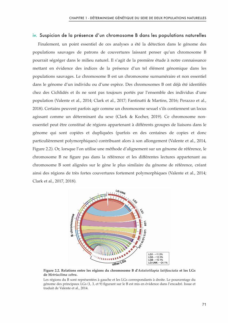
CHAPITRE 1 - DÉTERMINISME GÉNÉTIQUE DU SEXE DE DEUX POPULATIONS NATURELLES
iv. Suspicion de la présence d’un chromosome B dans les populations naturelles
Finalement, un point essentiel de ces analyses a été la détection dans le génome des
populations sauvages de patrons de couvertures laissant penser qu’un chromosome B
pourrait ségréger dans le milieu naturel. Il s’agit de la première étude à notre connaissance
mettant en évidence des indices de la présence d’un tel élément génomique dans les
populations sauvages. Le chromosome B est un chromosome surnuméraire et non essentiel
dans le génome d’un individu ou d’une espèce. Des chromosomes B ont déjà été identifiés
chez des Cichlidés et ils ne sont pas toujours portés par l’ensemble des individus d’une
population (Valente et al., 2014; Clark et al., 2017; Fantinatti & Martins, 2016; Perazzo et al.,
2018). Certains peuvent parfois agir comme un chromosome sexuel s’ils contiennent un locus
agissant comme un déterminant du sexe (Clark & Kocher, 2019). Ce chromosome non-
essentiel peut être constitué de régions appartenant à différents groupes de liaisons dans le
génome qui sont copiées et dupliquées (parfois en des centaines de copies et donc
particulièrement polymorphiques) contribuant alors à son allongement (Valente et al., 2014,
Figure 2.2). Or, lorsque l’on utilise une méthode d’alignement sur un génome de référence, le
chromosome B ne figure pas dans la référence et les différentes lectures appartenant au
chromosome B sont alignées sur le gène le plus similaire du génome de référence, créant
ainsi des régions de très fortes couvertures fortement polymorphiques (Valente et al., 2014;
Clark et al., 2017, 2018).
Figure 2.2. Relations entre les régions du chromosome B d'Astatotilapia latifasciata et les LGs de Metriaclima zebra.Les régions du B sont représentées à gauche et les LGs correspondants à droite. Le pourcentage du génome des principaux LGs (1, 3, et 9) figurant sur le B est mis en évidence dans l’encadré. Issue et traduit de Valente et al., 2014.
71
CHAPITRE 1 - DÉTERMINISME GÉNÉTIQUE DU SEXE DE DEUX POPULATIONS NATURELLES
Ce chromosome B n’a pas montré d’association avec le sexe à l’échelle de l’espèce
puisqu’il est retrouvé chez les mâles dans une population et chez les femelles dans l’autre.
Cependant, sa présence a été une réelle contrainte à l’exploitation du WGS puisque nous
avons effectué les analyses pour chacune des populations (Kpandu et Koka) séparément et
qu’il n’est présent que dans l’un des deux pools par population. Ainsi, la présence de ces
régions apparait spécifique du sexe et la forte densité de SNPs présents mène à
l’identification de forts pics d’association au sexe sur l’ensemble des groupes de liaisons.
La suspicion de ce chromosome B dans les pools a donc limité nos analyses à la région
associée au sexe préalablement identifiée par les analyses de ddRAD. S’il ne figure pas de
caractérisation propre du chromosome B dans l’article, certaines analyses complémentaires
ont été effectuées afin de mieux appréhender sa présence dans nos populations sauvages
ainsi que son contenu en gènes. Ces analyses sont explicitées à la fin de ce chapitre dans la
section «!Analyses Complémentaires!».
Conclusion
Cette étude a été la première caractérisation génomique du déterminisme du sexe utilisant
des populations de tilapia du Nil issues du milieu naturel. Nous avons pu mettre en
évidence que le déterminisme du sexe chez cette espèce est en lien étroit avec la région de
l’amh sur LG23 et suit un patron hétérogamétique mâle dans les populations sauvages. Nous
avons également pu confirmer l’existence d’une duplication de l’amh spécifique aux mâles
dans les deux populations naturelles, ce qui n’avait pas pu être fait par le passé en utilisant
des approches de marqueurs PCR chez les populations Ethiopiennes. Ces analyses ont
également pu mettre en lumière la structure partagée de cette duplication sur le Y en en
précisant le contenu, les éléments spécifiques au Y ainsi que les limites de cette duplication
malgré les conditions écologiques et les aires géographiques différentes de ces populations.
Des SNPs spécifiquement mâles, et partagés par d’autres souches, ont été identifiés comme
candidats pour le déterminisme du sexe et des variants spécifiques à chacune des
populations ont également été mis en évidence. Enfin, il s’agit de la première étude qui fait
état d’indices suggérant la présence d’un chromosome B, non lié au sexe à l’échelle des
populations, qui ségrégerait en milieu naturel.
72
CHAPITRE 1 - DÉTERMINISME GÉNÉTIQUE DU SEXE DE DEUX POPULATIONS NATURELLES
Références
Cáceres, G., López, M.E., Cádiz, M.I., Yoshida, G.M., Jedlicki, A., Palma-Véjares, R., Travisany, D., Díaz-Domínguez, D., Maass, A., Lhorente, J.P., et al. (2019). Fine mapping using whole-genome sequencing confirms anti-müllerian hormone as a major gene for sex determination in farmed Nile tilapia (Oreochromis niloticus L.). G3 (Bethesda) 9, 3213–3223.
Carrasco, L.A.P., Penman, D.J., and Bromage, N. (1999). Evidence for the presence of sex chromosomes in the Nile tilapia (Oreochromis niloticus) from synaptonemal complex analysis of XX, XY and YY genotypes. Aquaculture 173, 207–218.
Clark, F.E., and Kocher, T.D. (2019). Changing sex for selfish gain: B chromosomes of Lake Malawi cichlid fish. Scientific Reports 9, 1–10.
Clark, F.E., Conte, M.A., Ferreira-Bravo, I.A., Poletto, A.B., Martins, C., and Kocher, T.D. (2017). Dynamic sequence evolution of a sex-associated B chromosome in Lake Malawi cichlid fish. JHERED 108, 53–62.
Clark, F.E., Conte, M.A., and Kocher, T.D. (2018). Genomic characterization of a B chromosome in Lake Malawi cichlid fishes. Genes 9, 610.
Cnaani, A., Lee, B.-Y., Zilberman, N., Ozouf-Costaz, C., Hulata, G., Ron, M., D’Hont, A., Baroiller, J.-F., D’Cotta, H., Penman, D.J., et al. (2008). Genetics of sex determination in tilapiine species. Sexual Development 2, 43–54.
Conte, M.A., Joshi, R., Moore, E.C., Nandamuri, S.P., Gammerdinger, W.J., Roberts, R.B., Carleton, K.L., Lien, S., and Kocher, T.D. (2019). Chromosome-scale assemblies reveal the structural evolution of African cichlid genomes. Gigascience 8.
Eshel, O., Shirak, A., Weller, J.I., Hulata, G., and Ron, M. (2012). Linkage and physical mapping of sex Rregion on LG23 of Nile tilapia (Oreochromis niloticus). G3: Genes, Genomes, Genetics 2, 35–42.
Eshel, O., Shirak, A., Dor, L., Band, M., Zak, T., Markovich-Gordon, M., Chalifa-Caspi, V., Feldmesser, E., Weller, J.I., Seroussi, E., et al. (2014). Identification of male-specific amh duplication, sexually differentially expressed genes and microRNAs at early embryonic development of Nile tilapia (Oreochromis niloticus). BMC Genomics 15, 774.
Fantinatti, B.E.A., and Martins, C. (2016). Development of chromosomal markers based on next-generation sequencing: the B chromosome of the cichlid fish Astatotilapia latifasciata as a model. BMC Genetics 17, 119.
Gammerdinger, W.J., Conte, M.A., Acquah, E.A., Roberts, R.B., and Kocher, T.D. (2014). Structure and decay of a proto-Y region in Tilapia, Oreochromis niloticus. BMC Genomics 15, 975.
Hattori, R.S., Murai, Y., Oura, M., Masuda, S., Majhi, S.K., Sakamoto, T., Fernandino, J.I., Somoza, G.M., Yokota, M., and Strüssmann, C.A. (2012). A Y-linked anti-Müllerian hormone duplication takes over a critical role in sex determination. PNAS 109, 2955–2959.
Jalabert, B., Moreau, J., Planquette, P., and Billard, R. (1974). Déterminisme du sexe chez tilapia macrochir et tilapia nilotica : action de la méthyltestostérone dans l’alimentation des
73

CHAPITRE 1 - DÉTERMINISME GÉNÉTIQUE DU SEXE DE DEUX POPULATIONS NATURELLES
alevins sur la différenciation sexuelle; proportion des sexes dans la descendance des mâles «inversés». Annales de Biologie Animale Biochimie Biophysique Vol. 14, 729–739.
Lee, B.-Y., Penman, D.J., and Kocher, T.D. (2003). Identification of a sex-determining region in Nile tilapia (Oreochromis niloticus) using bulked segregant analysis. Animal Genetics 34, 379–383.
Lee, B.-Y., Coutanceau, J.-P., Ozouf-Costaz, C., D’Cotta, H., Baroiller, J.-F., and Kocher, T.D. (2011). Genetic and physical mapping of sex-linked AFLP markers in Nile tilapia (Oreochromis niloticus). Mar Biotechnol (NY) 13, 557–562.
Li, M., Sun, Y., Zhao, J., Shi, H., Zeng, S., Ye, K., Jiang, D., Zhou, L., Sun, L., Tao, W., et al. (2015). A tandem duplicate of anti-müllerian hormone with a missense SNP on the Y chromosome is essential for male sex determination in Nile tilapia, Oreochromis niloticus. PLOS Genetics 11, e1005678.
Palaiokostas, C., Bekaert, M., Khan, M.G.Q., Taggart, J.B., Gharbi, K., McAndrew, B.J., and Penman, D.J. (2013). Mapping and validation of the major sex-determining region in Nile tilapia (Oreochromis niloticus L.) using RAD sequencing. PLoS ONE 8, e68389.
Palaiokostas, C., Bekaert, M., Khan, M., Taggart, J.B., Gharbi, K., McAndrew, B.J., and Penman, D.J. (2015). A novel sex-determining QTL in Nile tilapia (Oreochromis niloticus). BMC Genomics 16, 171.
Pan, Q., Feron, R., Yano, A., Guyomard, R., Jouanno, E., Vigouroux, E., Wen, M., Busnel, J.-M., Bobe, J., Concordet, J.-P., et al. (2019). Identification of the master sex determining gene in Northern pike (Esox lucius) reveals restricted sex chromosome differentiation. PLOS Genetics 15, e1008013.
Perazzo, G.X., Noleto, R.B., Vicari, M.R., Gava, A., and Cestari, M.M. (2018). B chromosome polymorphism in South American cichlid. Neotropical Biodiversity 4, 3–9.
Pfennig, F., Standke, A., and Gutzeit, H.O. (2015). The role of Amh signaling in teleost fish – Multiple functions not restricted to the gonads. General and Comparative Endocrinology 223, 87–107.
Poonlaphdecha, S., Pepey, E., Huang, S.-H., Canonne, M., Soler, L., Mortaji, S., Morand, S., Pfennig, F., Mélard, C., Baroiller, J.F., et al. (2011). Elevated amh Gene Expression in the Brain of Male Tilapia (Oreochromis niloticus) during Testis Differentiation. Sex Dev 5, 33–47.
Sissao, R., D’Cotta, H., Baroiller, J.-F., and Toguyeni, A. (2019). Mismatches between the genetic and phenotypic sex in the wild Kou population of Nile tilapia Oreochromis
niloticus. PeerJ 7.
Taslima, K., Wehner, S., Taggart, J.B., de Verdal, H., Benzie, J.A.H., Bekaert, M., McAndrew, B.J., and Penman, D.J. (2020). Sex determination in the GIFT strain of tilapia is controlled by a locus in linkage group 23. BMC Genet 21, 49.
Taslima, K., Khan, M.G.Q., McAndrew, B.J., and Penman, D.J. (2021). Evidence of two XX/XY sex-determining loci in the Stirling stock of Nile tilapia (Oreochromis niloticus). Aquaculture 532, 735995.
Valente, G.T., Conte, M.A., Fantinatti, B.E.A., Cabral-de-Mello, D.C., Carvalho, R.F., Vicari, M.R., Kocher, T.D., and Martins, C. (2014). Origin and evolution of B chromosomes in the
74

CHAPITRE 1 - DÉTERMINISME GÉNÉTIQUE DU SEXE DE DEUX POPULATIONS NATURELLES
cichlid fish Astatotilapia latifasciata based on integrated genomic analyses. Mol Biol Evol 31, 2061–2072.
Wessels, S., Krause, I., Floren, C., Schütz, E., Beck, J., and Knorr, C. (2017). ddRADseq reveals determinants for temperature-dependent sex reversal in Nile tilapia on LG23. BMC Genomics 18, 531.
Wilson, C.A., High, S.K., McCluskey, B.M., Amores, A., Yan, Y., Titus, T.A., Anderson, J.L., Batzel, P., Carvan, M.J., Schartl, M., et al. (2014). Wild sex in zebrafish: loss of the natural sex determinant in domesticated strains. Genetics 198, 1291–1308.
75


PUBLICATION ASSOCIÉE


genesG C A T
T A C G
G C A T
Article
Structure and Sequence of the Sex Determining Locusin Two Wild Populations of Nile Tilapia
Cécile Triay 1,2, Matthew A. Conte 3, Jean-François Baroiller 1,2, Etienne Bezault 4 ,
Frances E. Clark 3 , David J. Penman 5, Thomas D. Kocher 3 and Helena D’Cotta 1,2,*
1 UMR-Institut des Sciences de l’Evolution de Montpellier, Centre National de la Recherche Scientifique,
Institut de Recherche pour le Développement, Ecole Pratique des Hautes Etudes, University of Montpellier,
34090 Montpellier, France; [email protected] (C.T.); [email protected] (J.-F.B.)2 UMR-Institut des Sciences de l’Evolution de Montpellier, Centre de Coopération Internationale en Recherche
Agronomique pour le Développement, Campus Int. Baillarguet, 34398 Montpellier, France3 Department of Biology, University of Maryland, College Park, MD 20742, USA; [email protected] (M.A.C.);
[email protected] (F.E.C.); [email protected] (T.D.K.)4 UMR BOREA, CNRS-7208/MNHN/UPMC/IRD-207/UCN/UA, Université des Antilles,
97159 Guadeloupe, France; [email protected] Institute of Aquaculture, School of Natural Sciences, University of Stirling, Stirling, Scotland FK9 4LA, UK;
* Correspondence: [email protected]; Tel.: +33-4-67593994
Received: 17 July 2020; Accepted: 27 August 2020; Published: 29 August 2020�����������������
Abstract: In domesticated strains of the Nile tilapia, phenotypic sex has been linked to genetic variants
on linkage groups 1, 20 and 23. This diversity of sex-loci might reflect a naturally polymorphic
sex determination system in Nile tilapia, or it might be an artefact arising from the process of
domestication. Here, we searched for sex-determiners in wild populations from Kpandu, Lake Volta
(Ghana-West Africa), and from Lake Koka (Ethiopia-East Africa) that have not been subjected to any
genetic manipulation. We analysed lab-reared families using double-digest Restriction Associated
DNA sequencing (ddRAD) and analysed wild-caught males and females with pooled whole-genome
sequencing (WGS). Strong sex-linked signals were found on LG23 in both populations, and sex-linked
signals with LG3 were observed in Kpandu samples. WGS uncovered blocks of high sequence
coverage, suggesting the presence of B chromosomes. We confirmed the existence of a tandem amh
duplication in LG23 in both populations and determined its breakpoints between the oaz1 and dot1l
genes. We found two common deletions of ~5 kb in males and confirmed the presence of both amhY
and amh∆Y genes. Males from Lake Koka lack both the previously reported 234 bp deletion and the
5 bp frameshift-insertion that creates a premature stop codon in amh∆Y.
Keywords: sex-determinant; Y-chromosome; amh; male duplication; Nile tilapia
1. Introduction
The male heterogametic (XY) system of sex determination in mammals, and the female
heterogametic (ZW) system in birds, are evolutionarily conserved in most species of their respective
clades [1,2]. Furthermore, the master sex-determining gene on the Y chromosome, sry, is almost
universal across the mammalian class [3]. The sex chromosomes of most mammals and birds are
highly differentiated due to the suppression of recombination, and the subsequent accumulation
of deleterious mutations which has resulted in the degeneration of the Y (or W) chromosomes
(reviewed by Bachtrog [4]). Fish in contrast, usually have homomorphic sex chromosomes, so the
sex of an individual typically cannot be distinguished by karyotyping. Genetic sex determination
(GSD) has been established for many fish species by progeny testing, sex-reversal treatments and by
Genes 2020, 11, 1017; doi:10.3390/genes11091017 www.mdpi.com/journal/genes

Genes 2020, 11, 1017 2 of 28
the association of genetic markers with phenotypic sex (see reviews of Baroiller et al. [5]; Devlin and
Nagahama [6]). These studies have demonstrated a wide range of GSD mechanisms, including both
mono- and polygenic systems. Sex determiners have been described on undifferentiated autosomes
and well-differentiated sex chromosomes, and in some cases interact with environmental factors to
determine phenotypic sex [7].
Frequent replacement of fish sex-determining systems is inferred from the fact that both male
XY and female ZW heterogametic systems can be found between and even within fish lineages [8,9].
The existence of multiple sex chromosomes (e.g., Xiphophorus) or polygenic systems (e.g., Dicentrarchus
labrax) within species also suggest the dynamic evolution of fish sex chromosomes [10–12]. The diversity
and rapid turnover of fish sex chromosomes have been made more apparent with the identification
of sex-determination genes. The master sex-determining gene of the medaka Oryzias latipes evolved
from a duplication of dmrt1 [13,14]. In another Oryzias species, the sex-determinant is a Y-specific copy
of the gsdf gene [15]. The sex-determining gene in the Patagonian pejerrey Odontesthes hatcheri is a
duplication of the anti-Müllerian hormone amh [16,17]. Amh has also been duplicated to become the
sex-determining gene in the Northern pike Esox lucius [18]. A male-specific allele of its receptor amhr2
was identified as the sex-determinant in Takifugu species [19]. In contrast, the master sex-determination
gene sdy of salmonids arose from the duplication of an immune-related gene [20].
African cichlids, particularly those that have undergone adaptive radiation in the large East
African Lakes, show an extraordinary diversity of sex chromosomes. Studies of cichlids from Lake
Tanganyika and Lake Malawi have identified more than twelve different sex-determining systems
that have been mapped to at least ten distinct chromosomes, including ZW systems on LG5 and LG7
and XY systems mapping to LG7, LG19 and LG20 [9,21–23]. A complex scenario has been found for
various populations of Astatotilapia burtoni, including an XYW system mapped to LG13, an XY system
on LG18 and an XY system on a fusion comprising LG5 and LG14 [24,25]. In addition, some species
from Lake Malawi possess female-specific B chromosomes that carry a female sex determiner, that is
epistatically dominant to the LG7 XY system [26,27].
Due to economic interest in producing fast-growing all-male stocks for aquaculture, numerous
studies have examined the sex-determining system in the Nile tilapia (Oreochromis niloticus) [28].
These studies have revealed a complex sex-determining system, involving both genetic and
environmental factors [12,29–31]. Many of these studies have been performed on domesticated or
laboratory strains, which may have experienced several rounds of selection, introgressive hybridizations,
and/or significant inbreeding [32,33].
Cytogenetic observations of the synaptonemal complex in Nile tilapia revealed that the terminal
ends of the largest chromosome (LG3) remained unpaired in males, suggesting this was the sex
chromosome [34]. However, the genetic mapping of families showed a sex linkage of markers on a
smaller chromosome (LG1) [29,30,35]. In other strains of O. niloticus sex was linked to LG23 [36,37].
Genomic studies also showed that sex was associated with either LG1 [31,38–40] or LG23 [40–44]
depending on the strain. The amh gene, on LG23, has since been identified as the sex-determining
gene in a Japanese strain [43]. The Y has two tandem copies, amhY [43] and amh∆Y, which has a
premature stop codon due to an insertion [42,43], while the X chromosome has a single copy, here called
amhX. Knock-out studies showed that amhY is necessary for determining maleness [43]. When not a
sex determinant, the amh still intervenes in gonad development, in germ cell regulation and is even
expressed in the brains of some teleost species (reviewed by Pfenning et al. [45]).
It is not clear to what extent these differences in sex-determination of Nile tilapia are due to natural
diversity in the mechanisms of sex determination or due to processes of domestication (including
introgressive hybridizations whether incidental or not). Domestication of zebrafish (Danio rerio) caused
the loss of the strong sex determiner on chromosome 4 present in wild fish [46]. Studies of wild Nile
tilapia from Lake Kou showed variability in the Y-linked amh sequences on LG23 and also suggested
that another locus was also involved in sex determination [47]. The objective of the present study was
to characterize the sex determiner(s) in wild populations of Nile tilapia that have not been subjected to

Genes 2020, 11, 1017 3 of 28
any form of domestication or genetic manipulations. We investigated a population from West Africa
(Ghana) and another from East Africa (Ethiopia), using two complementary genomic approaches to
analyse lab-reared families as well as pools of wild-caught males and females.
2. Materials and Methods
2.1. Fish and DNA Samples
Nile tilapia samples originated from two wild populations caught in Lake Volta (Kpandu, Ghana)
in 2002 and 2003 and Lake Koka (Ethiopia) in 2002. The fish from Kpandu belong to the subspecies
Oreochromis niloticus niloticus, whereas those from Lake Koka are the subspecies Oreochromis niloticus
cancellatus. They were sexually mature and could be phenotypically sexed by their genital papilla,
as described in Bezault et al. (2007) [48]. The collection site near Kpandu is a dendritic expansion
on the eastern side of Lake Volta in Ghana (Figure 1) where temperature varies throughout the year
between 27 ◦C and 32 ◦C. Lake Koka is a reservoir of the Awash River in the Ethiopian Highlands of
East Africa, where temperatures vary throughout the year between 21–26 ◦C and therefore it can be
considered a “cold lake” (Figure 1). Wild individuals were transferred to the CIRAD experimental
facilities in Montpellier (France), where they were progressively acclimated to a standard temperature
of 27 ◦C ± 1 ◦C in a thermo-regulated re-circulating system and individually tagged.
!"#$%&$%!"'()*
+&,&
!"#$%&'()*+(,-
./#$#
!"#$%&'#"
-.*/0
1'2)1-.*/0
1'2)1
!01#'2)*(3,-
45/60"6#
()%&*
34%56
7*8)9&#:*
Figure 1. Map of Africa showing Ghana in West Africa with the location of the Kpandu population in
Lake Volta (with medium to hot temperatures) and Lake Koka (a cold lake) located in the Ethiopian
highlands of the East African Rift.
For the double-digest Restriction Associated DNA (ddRAD) experiment, we produced an F1
family from each population by crossing the wild-caught individuals in 240 L aquaria. The Kpandu
Kp20 progeny was obtained by crossing male KpM17 to female KpF32. The Koka Kk-6 progeny was
obtained by crossing male KkM39 and female KkF4 (Table 1). Fry were collected from the mother’s
mouth at 3 days post-fertilisation (dpf), transferred to McDonald jars for incubation until 9 or 10 dpf,
and then reared in 40 L aquaria at 27 ± 1 ◦C. At 90 dpf, fish were euthanised with 2-phenoxyethanol
(1 mL/L) and 100 randomly sampled individuals per group were sexed by microscopic observation of
the gonads [28] to determine the phenotypic sex and define the sex ratios of the progenies. Fin clips

Genes 2020, 11, 1017 4 of 28
from the parents, and from 14 females and 15 males per family were collected and stored in 100%
ethanol until DNA extraction. We deliberately selected families that gave balanced sex-ratios when
reared at 27 ◦C. This minimized the effects of environmental or minor genetic factors that might bias the
proportion of males. The Kpandu Kp-20 family showed a sex-ratio of 46% males at 27 ◦C which was not
significantly different from the standard balanced (1:1) sex ratio (Chi-square test NS (Non Significant)
p > 0.05) (Table 1). The Koka Kk-6 progeny also gave a balanced sex ratio of 55% males NS for p > 0.05)
at 27 ◦C.
Fin clips for the Whole Genome Sequencing (WGS) were taken from the wild-caught individuals,
consisting of 14 females and 20 males from Koka, and 27 females and 27 males from Kpandu (Table 1).
To validate the sex chromosome markers we used fin clips from fish of known sexual genotype from
the Manzala-Tihange strain (Belgium) kept at CIRAD. They were an XX female, an XY male and a
YY supermale.
Table 1. Fish samples were used in this study.
Approach Population Family Sire Dam Sex-RatioSampled
MalesSampledFemales
TotalAnalyzed
ddRADsequencing
Kpandu Kp-20 KpM17 KpF32 49% (NS) 15 14 31Koka Kk-6 KkM39 KkF4 55% (NS) 15 14 31
Whole GenomeSequencing
Kpandu 27 27 54Koka 20 14 34
Chi-square tests of observed vs. expected (1:1). NS for non-significant.
2.2. Ethics Statement
All animal and experimental procedures were performed at the CIRAD facilities in Montpellier in
accordance with the French protocol N◦2016101810463 and the author’s personal authorization for
animal experimentation N◦ 35–15, both delivered by the French Government. Fish captures followed
the laws and veterinary agreements of each country as stipulated in 2003.
2.3. DNA Extraction
Genomic DNA (gDNA) was extracted in 96 well plates from fin clips cut to 0.5 × 0.5 cm size,
digested in 300 µL lysis solution containing 0.3 M NaCl, 50 mM Tris-base, 0.2 Mm EDTA, 0.2 mM
EGTA, 0.356 mM spermidine, 0.256 mM spermine, 4.8% SDS and 10 µg proteinase K at 55 ◦C overnight.
This was followed by an inactivation at 70 ◦C for 10 min and then by treatment with 6.6 µg RNAse
for 1 h at 37 ◦C. After brief centrifugation, the clear supernatant was extracted with the King Fisher
Flex robot using the Nucleomag 96 Tissue Kit (Macherey-Nagel, Dueren, Germany) following the
commercial protocol. We only changed the buffer quantities, using 200 µL of MBE and MB4 per sample,
300 µL of MB5/sample, and 335 µL of MB2 + 25 µL of magnetic beads (diluted at 1/2 with Ambion
water). The gDNA was eluted in 100 µL of MB6 (5 mM Tris) and after the residual magnetic beads
were settled on a magnetic rack, a clear eluate was retained. DNA was quantified using the Qubit
2.0 fluorometer (Invitrogen, Carlsbad, CA, USA) dsDNA BR kit and the quality was checked with a
Nanodrop spectrophotometer (ThermoFisher, Waltham, MA, USA) and on a 0.8% agarose gel. Finally,
the gDNA was diluted to 50 ng/µL.
2.4. Sexual Genotyping with Amh X and Y Chromosome Markers
We genotyped the sex of the wild individuals (11 females and 15 males from Koka and 27 females
and 27 males from Kpandu) using PCR amplification of four markers on LG23 shown to be specific
for the X and/or the Y chromosome in domestic strains of Nile tilapia. These markers are located
within the amh genes and adjacent promoter regions. Due to the high sequence homology of the 3 amh
genes, the markers were designed to cover indels. The procedures were performed as described in
Sissao et al. (2019) [47]. The first marker, amhX+36 (primers F1-GTTTGCAATAGTTAGGGTGCTGCTG;

Genes 2020, 11, 1017 5 of 28
R- GGAAATGCAGCCATTCCTGAG) which amplifies a 1000 base pair (bp) fragment present on
the promoter region of the amh gene located on the X chromosome [43]. The second marker,
amh∆Y+5 (primers F2-AAACCTCCTTCCTTTGTGAATGTC; R2-CTAGCGGCATCCACACTCCCTCAC)
amplifies a 1500 bp fragment that corresponds to a 5 bp insertion in exon6 that is present in the amh∆Y
gene, located on the Y chromosome. This insertion causes a change in the reading frame resulting in
a truncated protein [43]. The third marker, amh∆Y-233 (primers F3-CGGTCCCAGTGACCTATGAG;
R3-AAGTACACGTGGTGTATTGTAATTGA) gives two fragments; one of 1000 bp corresponding
to either amhX and/or amhY genes, and another fragment at 767 bp, which corresponds to the
amh∆Y gene [42]. The fourth marker, amhY-5608 (primers F4-GAAAGGGGTGTTTTGGTGCTGGC;
R4-ACCCAGGAAGCGTTTCATCTCA) amplifies two fragments: a 2414 bp band that specifically
amplifies the amhY gene present on the Y chromosome, and a fragment of 8022 bp which specifically
amplifies the amhX gene on the X chromosome [43]. We used this marker to detect the presence of the
amhY gene in males, because the large amhX fragment does not amplify consistently. An XX female,
XY male and YY male of the Manzala-Tihange (Belgium) strain were included as positive controls.
2.5. Double Digest RAD Sequencing
2.5.1. Library Construction
The ddRAD library contained 64 individuals from two F1 families obtained from wild-caught
crossings (Table 1) and was constructed following a protocol modified from Palaiokostas et al. (2015) [39],
using 100 ng of gDNA per individual and a no-DNA control. Briefly, the gDNA was double digested
for 3 h at 37 ◦C with 0.5 µL of SbfI and 0.5 µL of SphI (20 U/µL each, New England Biolabs (NEB),
Ipswich, MA, USA) with 2 µL of 10X Cutsmart Buffer in a reaction volume of 20 µL. Restriction
enzymes were then heat-inactivated at 65 ◦C for 15 min. Unique barcodes with specific combinations
of P1 and P2 adaptors were used to identify all samples individually. We used 4 µL/sample of a
1:16 ratio consisting in 120 nM SbfI compatible P1 adaptor/1920 nM SphI compatible P2 adaptor
with 0.5 µL of T4 Ligase (1000 U, 2000 U/µL, NEB), 0.3 µL rATP (100 nM, Promega, Madison, WI,
USA), 1 µL 10X Cutsmart Buffer in a total volume of 6 µL/sample. Samples were incubated for 2 h
at 22 ◦C. Subsequently, all samples were combined into a single pool, purified with the Nucleospin
Gel & PCR clean-up kit (Macherel-Nagel, Dueren, Germany) and then eluted in 40 µL Elution Buffer
(5 mM Tris). Size selection was performed by separating the eluate on a 1.2% agarose gel and excising
fragments between 300 and 700 bp. These gel fragments were then purified with the QIAquick gel
extraction kit (Qiagen, Dueren, Germany) and eluted in 3 × 30 µL Elution Buffer. The library was
subsequently PCR amplified in 22 replicates, each containing 4 µL template, 1.35 µL of each PCR
P1/P2 primers and 12.54 µL of Q5 Hot start HF 2X Master mix (NEB). The PCR program was: initial
denaturation at 98 ◦C for 40 s, followed by 13 cycles of 10 s at 98 ◦C, 30 s at 65 ◦C, 45 s at 72 ◦C with a
final elongation for 2 min at 72 ◦C. All PCR products were pooled and purified using the MinElute
PCR purification kit (Qiagen). They were subsequently cleaned twice with AMPure XP magnetic
beads (1× vol. followed by 0.7× vol.) to eliminate residual fragments under 300 bp and above 700 bp.
The final library concentration was quantified with the HS dsDNA Qubit kit, and the quality and size
estimated on a Bioanalyzer 2100 using a High Sensitivity DNA Chip (Agilent, Santa Clara, Californie,
CA, USA), which confirmed that the majority of fragments were between 300–600 bp. The ddRAD
library was sequenced at the GeT-PlaGe platform (Toulouse, Auzeville, France) on one lane of 150 bp
paired-end reads on a HiSeq3000 (Illumina, San Diego, CA, USA).
2.5.2. Data Processing
The data is accessible under the BioProject accession number PRJNA657179. The quality of the
reads was checked using FastQC (0.11.7) [49]. Raw data were demultiplexed using the STACKS
‘process_radtags.pl’ script for paired-end reads [50]. The SbfI-SphI restriction enzymes and adapter
sequences were specified and trimmed. Reads were then mapped to the O. niloticus reference genome

Genes 2020, 11, 1017 6 of 28
O_niloticus_UMD_NMBU [51] using the BWA-MEM mapper (version 0.7.15) [52]. Output files from
BWA-MEM (SAM files) were converted to BAM files, and then sorted and indexed with SAMtools
(version 1.9) [53]. We then used STACKS (version 2.2) to complete the single nucleotide polymorphism
(SNP) calling and population genetic statistics for each population separately [50]. STACKS implements
a maximum likelihood statistical model algorithm to identify loci and call genotypes, thus reducing false
polymorphic sites (e.g., due to sequencing errors). The STACKS script ‘ref_map.pl’ combines ‘pstacks’
to build loci, ‘cstacks’ to assemble a catalogue of loci, and ‘gstacks’ to match samples reads against
the catalog. Finally, it runs ‘populations’ which computes the population genetics statistic AMOVA
FST (derived from [54]) and Fisher’s Exact test between sexes at each nucleotide position. Relevant
options passed to ‘populations’ were -M specifying a ‘population map’ with 2 groups, splitting samples
according to their phenotypic sex; –smooth; –hwe; –fstats - f p_value –vcf. The output VCF files
were filtered using VCF tools (0.1.14) [55] to keep sites with a maximum of 2 alleles (–max-alleles 2),
and minimum read depth of 5 (–min-meanDP 5). We removed one phenotypic male of the Kpandu and
one phenoypic female of the Koka offspring that had 2098 and 3261 times fewer reads than the mean
of all individuals (–remove-indiv) in VCF files. Plain text table outputs were filtered using custom
python and bash scripts. Only loci that had reads for at least half of the males and half of the females
(per family) were kept.
2.6. Whole-Genome Sequencing
2.6.1. Library Construction
Two libraries per population were produced for the WGS consisting of a pool of wild-caught
females and a pool of wild-caught males. The two Kpandu libraries contained a pool of 27 females and
a pool of 27 males, whilst the Koka libraries consisted of a pool of 14 females and a pool of 20 males
(Table 1). Each pool contained an equal amount of individual gDNA, for a total of 2200 ng in 55 µL.
Pooled DNA were sonicated using a Covaris S220 (Woburn, Massachusetts, MA, USA) to obtain
550 bp fragments. Libraries were subsequently prepared using the TruSeq DNA PCR-Free kit (Illumina,
San Diego, CA, USA). Each pool was tagged with unique barcodes. Library quality and fragment size
range were assessed using a High Sensitivity DNA Chip on the Bioanalyzer (Agilent, Santa Clara, CA,
USA). Libraries were quantified by qPCR with the Library Quantification Kit (Takara Bio USA, Inc,
Mountain View, CA, USA) on a Lightcycler 96 with the following program: initial denaturation of
2 min at 98 ◦C; 35 cycles of 10 s at 98 ◦C, 15 s at 60 ◦C and 45 s at 68 ◦C, finishing with a melting curve.
The 4 libraries were sequenced on 2 lanes of a HiSeq 4000 (Illumina), using paired-end read lengths of
100 bp at the GenomEast Platform (Strasbourg, Illkirch, France).
2.6.2. Data Processing
The data is accessible under the BioProject accession number PRJNA657179. The quality of
the reads was checked using FastQC (version 0.11.7) [49]. Illumina TruSeq Adapters were trimmed
using Trimmomatic (version 0.33) [56] for paired-end reads. The reads were mapped onto the
XX female O. niloticus reference genome O_niloticus_UMD_NMBU using BWA mem mapper
(version 0.7.15) [52]. SAM files were then converted to BAM files and sorted using SAMtools
(version 1.9) [53]. PCR duplicates were identified using GATK MarkDuplicates (version 2.1.0) [57].
Files were subsequently indexed using Samtools index, and mpileup files were generated using the
Samtools mpileup program. The output mpileup files were converted to sync files from Popoolation2 [58]
with the ‘mpileup2sync.pl’ script and used as input for the next step. Fisher’s Exact Test were
computed for both populations using popoolation2, designed for comparing allele frequencies
between pooled sequencing data. FST was computed using the SEX_SNP_Finder_GA.pl pipeline [38]
to identify SNPs at intermediate frequencies in males and fixed (or nearly fixed) in females. This was
achieved by looking at both XY and ZW patterns for each population independently. The relevant
parameters used were the following: –fixed_threshold = 0.9; –minimum_polymorphic_frequency = 0.3;

Genes 2020, 11, 1017 7 of 28
–maximum_polymorphic_frequency = 0.7; –minimum_read_depth = 10; –maximum_read_depth =
100; –minimum_read_count = 2; –sex_SNP_finder_window_size = 10000. Structural variants were
called using DELLY (0.8.1) [59], which focuses on paired-ends, split-reads, and read-depth to predict
and discover genomic rearrangement. Small variants in the whole genome data were then called using
FreeBayes (1.2.0) [60], a haplotype-based variant detector designed for small indels and polymorphisms.
A filter was set for each pool independently to keep only variants for which the number of occurrences
was at least 5% of the overall coverage. All graphs were made using R [61] with the qqman package
for the Manhattan plot [62].
3. Results
3.1. Genotyping with Amh X and Y Chromosome Markers
The genotypic sex of the parents of the two families, and the other wild-caught fish from the Koka
and Kpandu populations, was predicted by using four amh markers that have distinguished the X and
Y chromosomes in the domesticated Manzala and Swansea strains (Israel) [42], the Japanese strain [43],
and the Manzala-Tihange strain (Belgium) [47]. The presence of the Y chromosome in the majority of
Kpandu males was proven with the successful amplification of the two amh∆Y markers as well as the
amhY marker. Indeed we amplified the 1400 bp band of the amh∆Y+5 marker and the 767 bp fragment
of the amh∆Y-233 marker in the Kpandu males, like the control Manzala-Tihange males that possess
the LG23 Y Chromosome (Figure 2). These results indicate the presence of the amh∆Y gene and that
these males, including the KPM17 sire, possess a Y chromosome and consequently are XY individuals.
We found three Kpandu males without these bands suggesting they were XX males (Table 2; Figure 2
shows one of these males “M23”). All Kpandu females, including the KpF32 dam, were negative for
these bands, indicating they were all XX females. None of the Koka males (or females) amplified the
two amh∆Y gene markers.
The presence of the amhY gene was confirmed using the amhY-5608 marker which gives a 2414 bp
fragment belonging to the amhY and an 8022 bp fragment for amhX [43]. We used this marker to
verify the presence of amhY, but the 8 kb amhX band amplifies poorly. We saw only the X fragment in
the Manzala-Tihange XX female used as PCR control (Figure 2). We amplified the amhY fragment in
all Kpandu males except the three XX males and did not amplify this fragment in Kpandu females.
Weak amplification of the amhY fragment was observed in some Koka males, but not in any Koka females.
All of the fish from Koka, Kpandu and the Manzala-Tihange strain (excluding the Manzala-Tihange YY
male) amplified the 1000 bp amhX+36 marker and the X-specific 1000 bp band from the amh∆Y-233 marker.
Figure 2. Amh genotyping of the Kpandu and Koka breeders used in ddRAD and a subset of individuals
used in the whole-genome sequencing (WGS) pools. The complete genotyping of all individuals is
shown in Figures S1 and S2.

Genes 2020, 11, 1017 8 of 28
Table 2. Sex chromosomic LG23 amh markers analysed in the Kpandu and Koka wild-caught fish to predict the sexual genotype. The Manzala strain with known sex
genotypes was used to validate the amplification.
Population or Strain Phenotype N Analyzed Genotype amhX Genotype amh∆Y Genotype amhY Genotype Predicted Genotype
Domestic strain Manzala
Females 1 XX amhX+ amh∆Y− amhY− XX
Males1 XY amhX+ amh∆Y+ amhY+ XY1 YY amhX− amh∆Y+ amhY+ YY
Wild populations
Kpandu
Females 27 amhX+ amh∆Y− amhY− XX
Males 24 amhX+ amh∆Y+ amhY+ XY3 amhX+ amh∆Y− amhY− XX
Koka
Females 14 amhX+ amh∆Y− amhY− XX
Males 8 amhX+ amh∆Y− amhY+ XY9 amhX+ amh∆Y− ND ND

Genes 2020, 11, 1017 9 of 28
3.2. Read Quality and Mapping
3.2.1. ddRAD Sequencing Libraries
We obtained a total of ~221 million clean reads for the Kpandu family after removing ambiguous
RAD barcodes, orphaned paired-ends and low-quality reads. From these reads, an average of 94.9%
per sample mapped properly paired to the O. niloticus reference genome (corresponding to an average
of 7.1 million reads per individual). A total of 145,258 loci were built in STACKS, with an average insert
length of 336.2 bp, and a mean of 7.6 males and 7.0 females mapped per locus. For the Koka family,
a total of ~161 million clean reads were obtained, of which an average of 95.3% per sample mapped
properly paired on the O. niloticus reference genome (corresponding to an average of 5.2 million
reads per individual). This allowed us to detect 129,388 loci with a mean insert length of 329.3 bp.
An average of 8.2 males and 7.4 females were mapped per locus. The number of reads per tag and
other demultiplexing statistics for each sample are reported in Table S1.
3.2.2. Whole-Genome Sequencing Libraries
We performed a pooled WGS strategy on the wild-caught samples of males and females (Table 1).
We obtained ~115 million paired reads for the Koka female pool, ~93 million for Koka males, ~104 million
for Kpandu females and ~226 million for Kpandu males. After trimming, we retained between 98 and
99% of the reads from each library. The mapping success onto the O. niloticus reference genome was over
94% for each library, with a least 83% correct paired reads. The average coverage obtained for Kpandu
males was 39X, which is more than twice the average coverage for the other pools (Kpandu females:
18X, Koka females: 19X, Koka males: 16X).
3.3. Sex-Associated Loci Identified by ddRAD
3.3.1. Sex-Associated Signal is Observed on LG23 and LG3
For the Kpandu family, we identified 14,073 polymorphic sites in phenotypic males and females
located in 6437 RAD loci. For the Koka family, we identified 10,314 polymorphic sites in phenotypic
males and females located in 4890 RAD loci. In both families, we found a significant peak of
differentiation between phenotypic males and females on LG23, with 29 SNPs associated with sex
for Koka and 125 SNPs for Kpandu (Fisher’s Exact Test p-value < 0.01) (Figure 3A and Table S2).
The Manhattan plots of the FST values are similar for the two families. For the Kpandu family, 220 sites
had a higher FST than the mean FST of 0.028. For the Koka family, there were 151 sites above the mean
FST of 0.024.
The region of LG23 between 24 Mb and 40 Mb contains the majority of the signal for both families
(Figure 3B). The Kpandu family showed a strong sex association of markers in this region. This region
contained 62% of the significant sites, although we can observe significant sex-associated SNPs all
along this linkage group. In the Koka family, sex was even more strongly and specifically associated
with this region, which included 97% of the significantly differentiated sites. The overall lower number
of significantly differentiated sites in the Koka family might be due to the lower level of sequence
coverage (Table S1).
In the Kpandu family, we also obtained a sex-associated signal on LG3 (Figure 3C and Table S2).
The highest signals on LG3 were located at 20Mb according to Fisher’s exact test. It corresponds to two
RAD loci, one localised in a non-coding region and the other is in the “Fc fragment of IgG receptor IIb”
gene (fcgr2b) (Table S3 and Figure 3C). More than 40 other significant sex-patterned SNPs on LG3
(Fisher’s Exact Test (FET) p-value < 0.001) occur in a region spanning ~40 Mb between 40 to 80 Mb
(Table S3 and Figure 3C).

Genes 2020, 11, 1017 10 of 28
A
Kpandu Koka
B
C
-log10(P)
Fst
Fst
-log10(P)
Fst
-log10(P)
amh
amhamh
!
"
#
$
%
&
'
(
!)*!! ")*!( #)*!( $)*!( %)*!(
Linkage Group 23
Linkage Group 23
!"!
!"#
!"$
!"%
!"&
'"!
!()!! '()!* #()!* +()!* $()!*
amh
!
"
#
$
%
&
'
(
!)*!! ")*!( #)*!( $)*!( %)*!(
Linkage Group 23
!"!
!"#
!"$
!"%
!"&
'"!
!()!! '()!* #()!* +()!* $()!*
Linkage Group 23
fcgr2b
Linkage Group
!
"
#
$
%
&
'
(
" # $ % & ' ( ) * "" "$ "& "( "* ##"! "# "% "' ") #! #$
+,-,./
!"!
!"#
!"$
!"%
!"&
'"!
!()!! #()!* $()!* %()!* &()!*
Linkage Group 3
!
"
#
$
%
&
'
(
!)*!! #)*!( %)*!( ')*!( +)*!(
Linkage Group 3
fcgr2b
!
"
#
$
%
&
'
(
" # $ % & ' ( ) * "" "$ "& "( "* ##"! "# "% "' ") #! #$
+,-,./
Linkage Group
!"!
!"#
!"$
!"%
!"&
'"!
' # ( $ ) % * & + '' '( ') '* '+ ##'! '# '$ '% '& #! #(
,-.-/0
Linkage Group
!"!
!"#
!"$
!"%
!"&
'"!
' # ( $ ) % * & + '' '( ') '* '+ ##'! '# '$ '% '& #! #(
,-.-/0
!
"
#
$
%
&
'
(
" # $ % & ' ( ) * "" "$ "& "( "* ##"! "# "% "' ") #! #$
+,-,./
Linkage Group
Fst
-log10(P)
-log10(P)
Fst
-log10(P)
Figure 3. Manhattan plots of log10-transformed p-values from Fisher’s Exact Test (FET) and AMOVA
FST of the association of single nucleotide polymorphisms (SNPs) with phenotypic sex. (A) Plots for the
Kpandu and the Koka families for all LGs. Unanchored contigs/scaffolds are grouped under the label
“Unanch”. (B) Plots for the LG23 region showing SNP sites that were associated with sex. Most of the
stronger sex-signals are found between 24 and 40 Mb. (C) Plots for Kpandu family with a close up of
the LG3 region showing a very localised signal at 20 Mb due to a RAD locus in the fcgr2b gene. The rest
of the signal observed on LG3 comes from the “long arm” of the chromosome known to carry repeated
elements. Red lines correspond to p-values of 0.01 in the FET plots while blue lines correspond to mean
AMOVA FST between males and females of each family.

Genes 2020, 11, 1017 11 of 28
3.3.2. Inherited Patterns of Offspring on LG23 Confirm a Male Heterogametic System
One advantage of using families for the ddRAD sequencing is that we can compare the SNPs of
father and sons, and likewise mother and daughters, to determine whether the pattern of inheritance
on LG23 corresponds to an XY or a ZW sex-determining system. We, therefore, focussed on sites for
which both parents were sequenced. We found that most of the LG23 sites with a significant FET were
consistent with an XX/XY pattern of inheritance (Figure 4).
In the Kpandu family, there was an increase in the concordance between genotype and phenotypic
sex as we approached the amh gene on LG23 (Figure 4). The amh gene, which remains the strongest
candidate for sex determination [43], is centred at 34.501 Mb. At the closest flanking SNPs (34,772,926 bp
and 35,124,733 bp) all the offspring have genotypes that match their phenotypic sex, following an
XY pattern of inheritance from their parents. The 7 SNP sites between 34,265,123 and 39,352,915 bp
show a maximum of 3.6% (1/28) discordance between an XY genotype and a male phenotype in the
offspring (Figure 4). Only one female shows a heterozygous genotype, at 34,443,504 bp, but she is
also heterozygous for all previous sites on this chromosome. This pattern suggests a recombination
breakpoint very close to the amh gene in this individual. At the 4 SNP sites after the amh gene
(at 35,941,307, 38,650,855, 38,767,722 and 39,352,915 bp) there is a perfect match between genotype
and phenotype, with the exception of one male presenting an XX genotype (Figure 4). Here again,
we suspect a recombination breakpoint just after the amh gene in this individual.
The heterozygous signals are less obvious in the Koka fish due to the overall lower coverage
and lower number of sites that passed the filters to build the genotypic table of parents and offspring.
Nevertheless, there is a good correspondence between the genotypes of markers near amh and the
sex of the offspring. The data are again consistent with an XX/XY pattern of inheritance (Figure 4).
The genotypes immediately adjacent to the amh gene on LG23 follow an XX/XY pattern and are
sufficient to predict the phenotypic sex of all but two individuals of the Kpandu and Koka families.
Although some markers on LG3 were statistically associated with phenotypic sex in the Kpandu
family, the genotypes of individual markers were not sufficient to accurately predict the sex of every
individual (Figure 4). Breakpoints between sites 52,823,319 and 66,689,764 may correspond to a region
of increased recombination or a genomic rearrangement so that these markers are further apart than
the reference genome suggests (Figure 4). However, the fact that females can be heterozygous or
homozygous on either side of these breakpoints, and the same for males, indicates that this interval on
LG3 probably does not contain a sex determiner. We also tested the Kpandu and Koka SNPs for a
female ZW pattern but failed to detect a female heterozygous pattern (Figure S3). The statistical signal
on LG3 may be an artifact arising from the highly repetitive sequences on this chromosome.

Genes 2020, 11, 1017 12 of 28
!"#
!"#
$%&'%&(
)*%+$(&
)&'&),)
&-($),+
(''%+''
()$+-'&
''*(),)*
'%,-')+%
'%$,-(*(
'+''*($$
')$'%'-+
')$'%'+%
'&%,(%'*
'(,'&&%%
'($,(*$'
-%(($'$&
-%(($'&*
-%(($--&
-+,(,-*'
-++-,-,*
-+)-(,+(
-+&'+$%$
-+&'+)+)
-)$--$'-
-)$--&-%
-)$--&**
-)$--&*(
-)$--&$+
-)$--&$*
-)$--(%)
-&'&%')-
-&'&%*$%
%'$',-*&
%+-$*'-%
%+++%*,+
%+))-(-$
%*'-+)%%
%*(+'%,)
%&$*,&**
%&)$))--
%(%*-('*
+%'&-)&,
++,'&($*
./0 1 2 3 4 2 3 3 2 2 1 3 3 4 3 3 3 1 1 4 4 2 4 2 4 2 2 1 2 3 2 4 1 4 1 2 2 4 4 4 3 4 2 4
452 4 1 2 3 1 4 4 1 3 2 4 4 1 4 4 4 2 2 1 3 1 3 1 3 1 1 4 1 2 4 3 2 1 2 1 1 3 1 3 4 2 3 3
! " ,6, ,6, ,6' ,6' ,6' ,6' ,6' ,6' ,6' ,6' ,6' ,6' ,6' ,6' ,6' ,6' ,6' ,6' ,6' ,6' ,6' ,6' ,6' ,6' ,6' ,6' ,6' ,6' ,6' ,6' ,6' ,6' ,6' ,6' ,6' ,6' ,6' ,6' ,6' ,6' ,6' ,6' ,6'
! # ,6' ,6' ,6' ,6' ,6' ,6' ,6' ,6' ,6' ,6' ,6' ,6' ,6' ,6' ,6' ,6' ,6' ,6' ,6' ,6' ,6' ,6' ,6' 767 767 767 767 767 767 767 ,6' ,6' ,6' ,6' ,6' ,6' ,6' ,6, ,6, ,6, ,6, ,6, ,6,
! $ ,6' ,6' ,6' ,6' ,6' ,6' ,6' ,6' ,6' ,6' ,6' ,6' ,6' ,6' ,6' ,6' ,6' ,6' ,6' ,6' ,6' ,6' ,6' ,6' ,6' ,6' ,6' ,6' ,6' ,6' ,6, ,6, ,6' ,6' ,6' ,6' ,6' ,6' ,6' ,6' ,6' ,6' ,6,
! % ,6' ,6' ,6' ,6' ,6' ,6' ,6' ,6' ,6' ,6' ,6' ,6' ,6' ,6' ,6' ,6' ,6' ,6' ,6' ,6' ,6' ,6' ,6' ,6' ,6' ,6' ,6' ,6' ,6' ,6' ,6' ,6' ,6' ,6' ,6' ,6' ,6' ,6' ,6' ,6' ,6' ,6' ,6'
! "& ,6' ,6' ,6' ,6' ,6' ,6' ,6' ,6' ,6' ,6' ,6' ,6' ,6' ,6' ,6' ,6' ,6' ,6' ,6' ,6' ,6' ,6' ,6' ,6' ,6' ,6' ,6' ,6' ,6' ,6' ,6' ,6' ,6' ,6' ,6' ,6' ,6' ,6' ,6' ,6' ,6' ,6' ,6'
! "# ,6' ,6' ,6' ,6' ,6' ,6' ,6' ,6' ,6' ,6' ,6' ,6' ,6' ,6' ,6' ,6' ,6' ,6' ,6' ,6' ,6' ,6' ,6' ,6' ,6' ,6' ,6' ,6' ,6' ,6' ,6' ,6' ,6' ,6' ,6' ,6' ,6' ,6' ,6' ,6' ,6' ,6' ,6'
! "$ ,6' ,6' ,6' ,6' ,6' ,6' ,6' ,6' ,6' ,6' ,6' ,6' ,6' ,6' ,6' ,6' ,6' ,6' ,6' ,6' ,6' ,6' ,6' ,6' ,6' ,6' ,6' ,6' ,6' ,6' ,6' ,6' ,6' ,6' ,6' ,6' ,6' ,6' ,6' ,6' ,6' ,6' ,6'
! "% ,6' ,6' ,6' ,6' ,6' ,6' ,6' ,6' ,6' ,6' ,6' ,6' ,6' ,6' ,6' ,6' ,6' ,6' ,6' ,6' ,6' ,6' ,6' ,6' ,6' ,6' ,6' ,6' ,6' ,6' ,6' ,6' ,6' ,6' ,6' ,6' ,6' ,6' ,6' ,6' ,6' ,6' ,6'
! "' ,6' ,6' ,6' ,6' ,6' ,6' ,6' ,6' ,6' ,6' ,6' ,6' ,6' ,6' ,6' ,6' ,6' ,6' ,6' ,6' ,6' ,6' ,6' ,6' ,6' ,6' ,6' ,6' ,6' ,6' ,6' ,6' ,6' ,6' ,6' ,6' ,6' ,6' ,6' ,6' ,6' ,6' ,6'
! "( ,6' ,6' ,6' ,6' ,6' ,6' ,6' ,6' ,6' ,6' ,6' ,6' ,6' ,6' ,6' ,6' ,6' ,6' ,6' ,6' ,6' ,6' ,6' ,6' ,6' ,6' ,6' ,6' ,6' ,6' ,6' ,6' ,6' ,6' ,6' ,6' ,6' ,6' ,6' ,6' ,6' ,6' ,6'
! #& ,6, ,6' ,6' ,6' ,6' ,6' ,6' ,6' ,6' ,6' ,6' ,6' ,6' ,6' ,6' ,6' ,6' ,6' ,6' ,6' ,6' ,6' ,6' ,6' ,6' ,6' ,6' ,6' ,6' ,6' ,6' ,6' ,6' ,6' ,6' ,6' ,6' ,6' ,6' ,6' ,6' ,6' ,6'
! #" ,6' ,6' ,6' ,6' ,6' ,6' ,6' ,6' ,6' ,6' ,6' ,6' ,6' ,6' ,6' ,6' ,6' ,6' ,6' ,6' ,6' ,6' ,6' ,6' ,6' ,6' ,6' ,6' ,6' ,6' ,6' ,6' ,6' ,6' ,6' ,6' ,6' ,6' ,6' ,6' ,6' ,6' ,6'
! ## ,6, ,6, ,6, ,6, ,6, ,6, ,6, ,6, ,6, ,6, ,6, ,6, ,6, ,6' ,6' ,6' ,6' ,6' ,6' ,6' ,6' ,6' ,6' ,6' ,6' ,6' ,6' ,6' ,6' ,6' ,6' ,6' ,6' ,6' ,6' ,6' ,6' ,6' ,6' ,6' ,6' ,6' ,6'
! #% ,6, ,6, ,6, ,6, ,6, ,6, ,6, ,6, ,6, ,6, ,6, ,6, ,6, ,6, ,6, ,6, ,6, ,6, ,6, ,6, ,6, ,6, ,6, ,6, ,6, ,6, ,6, ,6, ,6, ,6, ,6, ,6, ,6' ,6' ,6' ,6' ,6' ,6' ,6' ,6' ,6' ,6' ,6'
) * ,6, ,6, ,6, ,6, ,6, ,6, ,6, ,6, ,6, ,6, ,6, ,6, ,6, ,6, ,6, ,6, ,6, ,6, ,6, ,6, ,6, ,6, ,6, ,6, ,6, ,6, ,6, ,6, ,6, ,6, ,6, ,6, ,6, ,6, ,6, ,6, ,6, ,6, ,6, ,6, ,6, ,6, ,6,
) + ,6, ,6, ,6, ,6, ,6, ,6, ,6, ,6, ,6, ,6, ,6, ,6, ,6, ,6, ,6, ,6, ,6, ,6, ,6, ,6, ,6, ,6, ,6, ,6, ,6, ,6, ,6, ,6, ,6, ,6, ,6, ,6, ,6, ,6, ,6, ,6, ,6, ,6, ,6, ,6, ,6, ,6, ,6,
) ' ,6, ,6, ,6, ,6, ,6, ,6, ,6, ,6, ,6, ,6, ,6, ,6, ,6, ,6, ,6' ,6, ,6, ,6, ,6, ,6, ,6, ,6, ,6, ,6' ,6' ,6' ,6' ,6' ,6' ,6' ,6, ,6, ,6, ,6, ,6, ,6, ,6, ,6, ,6, ,6, ,6, ,6, ,6,
) , ,6, ,6, ,6, ,6, ,6, ,6, ,6, ,6, ,6, ,6, ,6, ,6, ,6, ,6, ,6, ,6, ,6, ,6, ,6, ,6, ,6, ,6, ,6, ,6, ,6, 767 767 ,6, ,6, ,6, ,6, ,6, ,6, ,6, ,6, ,6, ,6, ,6, ,6, ,6, ,6, ,6, ,6,
) "" ,6' ,6' ,6' ,6' ,6' ,6' ,6' ,6' ,6' ,6' ,6' ,6' ,6' ,6' ,6' ,6' ,6' ,6' ,6' ,6' ,6' ,6' ,6' ,6' ,6' ,6' ,6' ,6' ,6' ,6' ,6, 767 ,6' ,6, ,6, ,6, ,6, ,6, ,6, ,6, ,6, ,6, ,6,
) "* ,6, ,6, ,6, ,6, ,6, ,6, ,6, ,6, ,6, ,6, ,6, ,6, ,6, ,6, ,6, ,6, ,6, ,6, ,6, ,6, ,6, ,6, ,6, ,6, ,6, ,6, ,6, ,6, ,6, ,6, ,6, ,6, ,6, ,6, ,6, ,6, ,6, ,6, ,6, ,6, ,6, ,6, ,6,
) ", ,6, ,6, ,6, ,6, ,6, ,6, ,6, ,6, ,6, ,6, ,6, ,6, ,6, ,6, ,6, ,6, ,6, ,6, ,6, ,6, ,6, ,6, ,6, ,6, ,6, ,6, ,6, ,6, ,6, ,6, ,6, ,6, ,6, ,6, ,6, ,6, ,6, ,6, ,6, ,6, ,6, ,6' ,6'
) #$ ,6, ,6, ,6, ,6, ,6, ,6, ,6, ,6, ,6, ,6, ,6, ,6, ,6, ,6, ,6, ,6, ,6, ,6, ,6, ,6, ,6, ,6, ,6, ,6, ,6, ,6, ,6, ,6, ,6, ,6, ,6, ,6, ,6, ,6, ,6, ,6, ,6, ,6, ,6, ,6, ,6, ,6, ,6,
) #* ,6' ,6' ,6' ,6' ,6' ,6' ,6' ,6' ,6' ,6' ,6' ,6' ,6' ,6' ,6' ,6' ,6' ,6' ,6' ,6' ,6' ,6' ,6' ,6' ,6' ,6' ,6' ,6' ,6' ,6' ,6' ,6' ,6' ,6' ,6' ,6, ,6, ,6, ,6, ,6, ,6, ,6, ,6,
) #+ ,6, ,6, ,6, ,6, ,6, 767 ,6, ,6, ,6, ,6, ,6, ,6, ,6, ,6, ,6, ,6, ,6, ,6, ,6, ,6, ,6, ,6, ,6, ,6, ,6, ,6, ,6, ,6, ,6, ,6, ,6, ,6, ,6, ,6, ,6, ,6, ,6, ,6, ,6, ,6, ,6, ,6, ,6,
) #' ,6, ,6, ,6, ,6, ,6, ,6, ,6, ,6, ,6, ,6, ,6, ,6, ,6, ,6, ,6, ,6, ,6, ,6, ,6, ,6, ,6, ,6, ,6, ,6, ,6, ,6, ,6, ,6, ,6, ,6, ,6, ,6, ,6, ,6, ,6, ,6, ,6, ,6, ,6, ,6, ,6, ,6, ,6,
) #( ,6, ,6, ,6, ,6, ,6, ,6, ,6, ,6, ,6, ,6, ,6, ,6, ,6, ,6, ,6, ,6, ,6, ,6, ,6, ,6, ,6, ,6, ,6, ,6, ,6, ,6, ,6, ,6, ,6, ,6, ,6, ,6, ,6, ,6, ,6, ,6, ,6, ,6, ,6, ,6, ,6, ,6, ,6,
) #, ,6, ,6, ,6, ,6, ,6, ,6, ,6, ,6, ,6, ,6, ,6, ,6, ,6, ,6, ,6, ,6, ,6, ,6, ,6, ,6, ,6, ,6, ,6, ,6, ,6, ,6, ,6, ,6, ,6, ,6, ,6, ,6, ,6, ,6, ,6, ,6, ,6, ,6, ,6, ,6, ,6, ,6, ,6'
) $& ,6, ,6, ,6, ,6, ,6, ,6, ,6, ,6, ,6, ,6, ,6, ,6, ,6, ,6, ,6, ,6, ,6, ,6, ,6, ,6, ,6, ,6, ,6, ,6, ,6, ,6, ,6, ,6, ,6, ,6, ,6, ,6, ,6, ,6, ,6, ,6, ,6, ,6, ,6, ,6, ,6, ,6' ,6'
-./ ,6, ,6, ,6, ,6, ,6, ,6, ,6, ,6, ,6, ,6, ,6, ,6, ,6, ,6, ,6, ,6, ,6, ,6, ,6, ,6, ,6, ,6, ,6, ,6, ,6, ,6, ,6, ,6, ,6, ,6, ,6, ,6, ,6, ,6, ,6, ,6, ,6, ,6, ,6, ,6, ,6, ,6, ,6,
0123 ,6' ,6' ,6' ,6' ,6' ,6' ,6' ,6' ,6' ,6' ,6' ,6' ,6' ,6' ,6' ,6' ,6' ,6' ,6' ,6' ,6' ,6' ,6' ,6' ,6' ,6' ,6' ,6' ,6' ,6' ,6' ,6' ,6' ,6' ,6' ,6' ,6' ,6' ,6' ,6' ,6' ,6' ,6'
!"#
-++-,+-(
-++-,+%%
-(%-*&%*
-(%-$,*,
-(%-$,*'
-(('$$*$
-(('$$*&
%,-*+',%
%(%+$)$-
%(%+$&&)
./0 3 3 4 3 3 1 4 4 1 2
452 1 4 2 4 4 2 1 2 2 1
! " 767 767 ,6' ,6' ,6' ,6' ,6' ,6' ,6' ,6'
! # ,6' ,6' ,6' ,6' ,6' ,6' ,6' ,6' ,6' ,6'
! $ ,6' ,6' ,6, ,6, ,6, ,6' ,6' ,6' ,6, ,6,
! % ,6' ,6' ,6' ,6' ,6' ,6' ,6' ,6' ,6, ,6,
! ( ,6' ,6' ,6' ,6' ,6' ,6' ,6' ,6' ,6' ,6'
! "& ,6' ,6' 767 767 767 ,6' ,6' ,6' ,6' ,6'
! "$ ,6' ,6' ,6, ,6, ,6, ,6' ,6' ,6' 767 767
! "* ,6' ,6' ,6' ,6' ,6' ,6' ,6' ,6' ,6' ,6'
! "' ,6, ,6, ,6, ,6, ,6, ,6, ,6, ,6, 767 767
! "( ,6' ,6' ,6' ,6' ,6' ,6' ,6' ,6' 767 767
! ", ,6' ,6' ,6' ,6' ,6' ,6' ,6' ,6' ,6' ,6'
! #& ,6' ,6' ,6' ,6' ,6' ,6' ,6' ,6' ,6' ,6'
! ## ,6' ,6' 767 767 767 ,6' ,6' ,6' ,6' ,6'
! #% ,6' ,6' ,6' ,6' ,6' ,6' ,6' ,6' ,6' ,6'
! #+ ,6' ,6' ,6' ,6' ,6' ,6' ,6' ,6' 767 767
) * ,6, ,6, ,6, ,6, ,6, ,6, ,6, ,6, ,6, ,6,
) + ,6, ,6, 767 767 767 ,6, ,6, ,6, ,6, ,6,
) ' ,6, ,6, ,6, ,6, ,6, ,6, ,6, ,6, ,6, ,6,
) , ,6, ,6, ,6, ,6, ,6, ,6, ,6, ,6, ,6, ,6,
) "" ,6, ,6, ,6, ,6, ,6, ,6, ,6, ,6, 767 767
) "# ,6, ,6, ,6, ,6, ,6, ,6, ,6, ,6, ,6, ,6,
) "% ,6' ,6' ,6' ,6' ,6' ,6' ,6' ,6' ,6, ,6,
) "+ ,6, ,6, ,6, ,6, ,6, ,6, ,6, ,6, ,6, ,6,
) #$ ,6, ,6, ,6, ,6, ,6, ,6, ,6, ,6, 767 767
) #* ,6' ,6' ,6, ,6, ,6, ,6, ,6, ,6, ,6, ,6,
) #' ,6, ,6, ,6, ,6, ,6, ,6, ,6, ,6, ,6, ,6,
) #( ,6, ,6, ,6, ,6, ,6, ,6, ,6, ,6, ,6, ,6,
) $& ,6, ,6, ,6, ,6, ,6, ,6, ,6, ,6, 767 767
-./ ,6, ,6, ,6, ,6, ,6, ,6, ,6, ,6, ,6, ,6,
0123 ,6' ,6' ,6' ,6' ,6' ,6' ,6' ,6' ,6' ,6'
!"#
*-&-%-((
*-&-%%'(
$$$&()$+
$$$&(&-*
$$$&(&+)
)'&&$%-&
)'&&$%+&
)-)+*$&+
)-)+*)($
)),-%)('
)),(-$('
))')%%+'
))')%%+(
))')%%)(
))')%%&(
))')%+%,
./0 1 3 2 2 4 1 1 1 4 1 2 3 2 3 1 3
452 2 4 1 4 3 3 3 2 2 3 1 4 1 4 3 4
! " 767 767 ,6, ,6, ,6, ,6, 767 ,6, ,6, ,6, ,6, ,6, ,6, ,6, ,6, ,6,
! # ,6, ,6, '6' '6' '6' ,6' ,6' '6' '6' '6' '6' '6' '6' '6' '6' '6'
! $ ,6, ,6, '6' '6' '6' ,6' ,6' '6' '6' '6' '6' '6' '6' '6' '6' '6'
! % ,6, ,6, ,6, ,6, ,6, ,6, ,6, ,6, ,6, ,6, ,6, ,6, ,6, ,6, ,6, ,6,
! "& ,6, ,6, ,6, ,6, ,6, ,6, ,6, ,6, ,6, ,6, ,6, ,6, ,6, ,6, ,6, ,6,
! "# ,6, ,6, '6' '6' '6' ,6' ,6' '6' '6' '6' '6' '6' '6' '6' '6' '6'
! "$ ,6' ,6' 767 767 767 '6' '6' 767 767 ,6' ,6' 767 767 767 767 767
! "% 767 767 ,6, ,6, ,6, ,6, ,6, ,6, ,6, ,6, ,6, ,6, ,6, ,6, ,6, ,6,
! "' ,6' ,6' ,6' ,6' ,6' ,6' ,6' ,6' ,6' ,6' ,6' ,6' ,6' ,6' ,6' ,6'
! "( ,6' ,6' ,6, ,6, ,6, ,6, ,6, ,6, ,6, ,6, ,6, ,6, ,6, ,6, ,6, ,6,
! #& 767 767 '6' '6' '6' 767 767 '6' '6' '6' '6' '6' '6' '6' '6' '6'
! #" ,6, ,6, '6' '6' '6' ,6' ,6' '6' '6' '6' '6' '6' '6' '6' '6' '6'
! ## ,6, ,6, '6' '6' '6' ,6' ,6' '6' '6' '6' '6' '6' '6' '6' '6' '6'
! #% ,6, ,6, ,6, ,6, ,6, ,6, ,6, ,6, ,6, ,6, ,6, ,6, ,6, ,6, ,6, ,6,
) * ,6' 767 ,6, ,6, ,6, ,6, ,6, ,6, ,6, ,6, ,6, ,6, ,6, ,6, ,6, ,6,
) + ,6' ,6' ,6, ,6, ,6, ,6, 767 ,6, ,6, ,6, ,6, ,6, ,6, ,6, ,6, ,6,
) ' '6' '6' ,6, 767 767 767 767 ,6' ,6' ,6' ,6' 767 767 767 767 767
) , '6' '6' ,6, ,6, ,6, ,6, ,6, ,6, ,6, ,6, ,6, ,6, ,6, ,6, ,6, ,6,
) "" '6' '6' ,6, ,6, ,6, ,6, ,6, ,6, ,6, ,6, ,6, ,6, ,6, ,6, ,6, ,6,
) "* ,6, ,6, ,6, 767 767 ,6, ,6, ,6, ,6, ,6, ,6, ,6, ,6, ,6, ,6, ,6,
) ", '6' '6' ,6, ,6, ,6, ,6, ,6, ,6, ,6, ,6, ,6, ,6, ,6, ,6, ,6, ,6,
) #$ '6' '6' ,6, ,6, ,6, ,6, ,6, ,6, ,6, ,6, ,6, ,6, ,6, ,6, ,6, ,6,
) #* ,6, ,6, '6' '6' '6' ,6' ,6' '6' '6' '6' '6' '6' '6' '6' '6' '6'
) #+ '6' '6' ,6, ,6, ,6, ,6, ,6, ,6, ,6, ,6, ,6, ,6, ,6, ,6, ,6, ,6,
) #' '6' '6' ,6, ,6, ,6, ,6, ,6, ,6, ,6, ,6, ,6, ,6, ,6, ,6, ,6, ,6,
) #( ,6, ,6, ,6' ,6' ,6' 767 767 ,6' ,6' ,6' ,6' 767 767 767 767 767
) #, ,6, ,6, ,6, ,6, ,6, ,6, ,6, ,6, ,6, ,6, ,6, ,6, ,6, ,6, ,6, ,6,
) $& '6' '6' ,6, ,6, ,6, ,6, ,6, ,6, ,6, ,6, ,6, ,6, ,6, ,6, ,6, ,6,
-./ ,6, ,6, ,6, ,6, ,6, ,6, ,6, ,6, ,6, ,6, ,6, ,6, ,6, ,6, ,6, ,6,
0123 ,6' ,6' ,6' ,6' ,6' ,6' ,6' ,6' ,6' ,6' ,6' ,6' ,6' ,6' ,6' ,6'
45.6789:;#$
4<=.9:;#$
45.6789:;$
!"#
Figure 4. Genotypes of the offspring compared to their parents inferred from ddRAD FET significant
sites following an XX/XY pattern. Pink cells indicate an individual having the same genotype as their
mother at this site whereas blue cells indicate an individual having the same genotype as their father at
this site (dashed cells indicate homozygous YY sites). White cells are missing data. Only sites having
both parents and at least half males and females of the offspring sequenced were kept here. Those tested
for a ZZ/ZW pattern are shown in Figure S3.
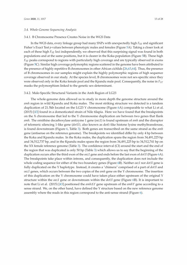
Genes 2020, 11, 1017 13 of 28
3.4. Whole-Genome Sequencing Analysis
3.4.1. B Chromosome Presence Creates Noise in the WGS Data
In the WGS data, every linkage group had many SNPs with unexpectedly high FST and significant
Fisher’s Exact Test p-values between phenotypic males and females (Figure 5A). Taking a closer look at
each of these high FST loci independently, we observed that this surprising signal was found in both
populations and at the same positions, but it is clearer in the Koka population (Figure 5B). These high
FST peaks correspond to regions with particularly high coverage and are typically observed in exons
(Figure 5C). Similar high coverage polymorphic regions scattered in the genome have been attributed to
the presence of highly repetitive B chromosomes in other African cichlids [26,63,64]. Thus, the presence
of B chromosomes in our samples might explain the highly polymorphic regions of high sequence
coverage observed in our study. At the species level, B chromosomes were not sex-specific since they
were observed only in the Koka female pool and the Kpandu male pool. Consequently, the noise in FST
masks the polymorphism linked to the genetic sex-determinant.
3.4.2. Male-Specific Structural Variants in the Amh Region of LG23
The whole-genome data allowed us to study in more depth the genome structure around the
amh region in wild Kpandu and Koka males. The most striking structure we detected is a tandem
duplication of 21.5kb located on the LG23 Y chromosome (Figure 6A) comparable to what Li et al.
(2015) [43] found in a domesticated strain of Nile tilapia. Here we have found that the breakpoints
on the X chromosome that led to the Y chromosome duplication are between two genes that flank
amh. The ornithine decarboxylase antizyme 1 gene (oaz1) is found upstream of amh and the disruptor
of telomeric silencing 1-like gene (dot1l), also known as dot1-like histone lysine methyltransferase,
is found downstream (Figure 6, Table 3). Both genes are transcribed on the same strand as the amh
gene (antisense on the reference genome). The breakpoints we identified differ by only 4 bp between
the Koka and Kpandu males. In the Koka males, the duplication spans the region from 34,491,225 bp
and 34,512,737 bp, and in the Kpandu males spans the region from 34,491,225 bp to 34,512,741 bp on
the XX female reference genome (Table 3). The confidence interval (CI) around the start and the end of
the region that was duplicated is only 50 bp (Table 3) which allows us to say that the beginning of the
duplication occurs after the third exon of the oaz1 gene and ends before the last exon of dot1l (Figure 6A).
The breakpoints take place within introns, and consequently, the duplication does not include the
whole coding sequence for either of the two boundary genes (Figure 6B). Neither oaz1 nor dot1l gene is
fully duplicated on the Y haplotype. Instead, it creates a "chimera" comprised of a part of dot1l and
oaz1 genes, which occurs between the two copies of the amh gene on the Y chromosome. The insertion
of this duplication on the Y chromosome could have taken place either upstream of the original Y
structure within the oaz1 gene or downstream within the dot1l gene (Figure 6B). It is important to
note that Li et al. (2015) [43] positioned the amh∆Y gene upstream of the amhY gene according to a
sense strand. We, on the other hand, have defined the Y structure based on the new reference genome
assembly where the reads in this region correspond to the anti-sense strand (Figure 6).

Genes 2020, 11, 1017 14 of 28
!
"#$%&'&()*+
",%-./
"#$%
0
"#$%%&'()*)+,-
./01%%(%2'(1
!"#$"%#"%%&'( +1 213 433 +1 213 533 +1 213 633 +1 213 733 +1 212 333 +1 212 233 +1 212 *33 +1 212 +33 +1 212 133 89,:
3%4-2'5
3%4-2'5
3%4-2'5
3%4-2'5
+1 213 ;33
!"#$%&'(%)*+#
678797:;7!<7:=>7
?=@A!!"
?=@A!!#
?BA:CD!!#
?BA:CD!!"
<
Figure 5. SNPs associated with phenotypic sex obtained from the WGS. (A) Manhattan plots of FST for
Kpandu and Koka populations obtained with Sex SNP Finder. High FST peaks are observed on every
LG meaning there is a signal associated to sex on all chromosomes; (B) Close up of FST on LG23 for
Koka showing three high Fst regions in grey and green boxes; (C) Integrative Genomics Viewer (IGV)
plot of sequence coverage at ~34.14 Mb on LG23 corresponding to the green box above for both sexes
of the two wild populations. High FST values in this region are due to a very high sequence coverage
(>10 times the mean coverage) likely corresponding to repetitive sequences on the B chromosome.
SNPs are represented by colour traits with a bicolour trait being a polymorphic site, with the size of the
colour band proportional to the nucleotide frequency. Frequencies of SNPs are conserved between
Kpandu males and Koka females.

Genes 2020, 11, 1017 15 of 28
!
"
#
!"#$%"&'$ &()
!"#$%%&%&'&$
&()* !"#$%"&'$ !"#$% &()+*
$%&%'( )* $%&+(( )* $%&+,( )* $%&+-( )* $%&+$( )*
!"#$
$%&+%( )*
!"#$ %!"#$ &!"#$ '!"#$ (!"#$ )!"#$
!"#$%"&'"(")"*+"&
,"*-#"
.$%"&/&0$1%-231"
!"#$%%&%&'&$
!"#$%%&%&'&$
"&'$
41$*56&71"+89+&:"%"28-*7
Figure 6. Schematic representation of the duplicated region of LG23 and its structural variants and
polymorphic sites found with DELLY and FreeBayes. The tandem duplication includes the amh gene
and part of the oaz1 and dot1l genes. The duplicated regions in each population (in green) are shown
with the corresponding deletions and suggested size (in red). The shared male SNPs are shown in
blue bars while the population male-specific SNPs are shown in light grey bars. (A) Schematic view of
mapping results of the Y chromosome by WGS of the Kpandu and Koka males against the female XX
reference genome. (B) Schematic representation of the two possible places of insertion of the tandem
duplication in oaz1 and dot1l genes. Both large deletions found with DELLY are likely to be placed in the
oaz1-dot1l chimera as the genes are already truncated on the Y haplotype. (C) Schematic representation
of the inferred Y haplotype of Kpandu and Koka males.
We also detected two large deletions of more than 5 kb that were common in males from both
populations (Table 3 and Figure 6A,B). The first deletion was 5273 bp in length in Koka males and

Genes 2020, 11, 1017 16 of 28
5,233bp in length in Kpandu males (CI ± 390 bp and ± 150 bp, respectively) (Table 3). This male-specific
variant encompasses a region between the first intron of oaz1 and the amh gene according to the female
XX reference genome (from 34,493,315 bp to 34,498,588/34,498,548 bp) (Figure 6). In the Y chromosome,
the location of this large deletion depends on where the insertion of the duplicated sequences occurred.
Either it starts in the complete copy of oaz1 and ends close to the 5’UTR region of the amhY copy, or it
starts in the truncated copy of oaz1 and goes nearly up to the 5’UTR region of amh∆Y (Figure 6A).
The first hypothesis implies that there would not be a complete oaz1 copy on the Y chromosome. Thus,
the second option where the deletion comprises an already truncated (and putatively non-functional)
gene seems more plausible (Figure 6B).
Table 3. Structural variants located in the oaz1-dot1l region of LG23 in wild populations of Nile tilapia
found with DELLY on whole-genome sequencing data.
PopulationsStart
PositionEnd
PositionConfidence Intervalaround Start & End
TypeSize of Structural
VariantShared between
Populations
Koka
34491225 34512737 −50,50 Duplication 21512 Yes34493315 34498588 −390,390 Deletion 5273 Yes34503117 34509103 −852,852 Deletion 5986 Yes
Kpandu
34491225 34512741 −50,50 Duplication 21516 Yes34491681 34491957 −2,2 Deletion 276 No34493315 34498548 −150,150 Deletion 5233 Yes34499596 34499830 −3,3 Deletion 234 No34503494 34509103 −2,2 Deletion 5609 Yes
The second common male deletion is 5,986 bp (CI ± 852 bp) in Koka, and 5,609 bp in Kpandu
(CI ± 2 bp). It starts around 34,503,000 bp and ends at the same position (34,509,103 bp) in both
populations (Figure 6A). This corresponds to the region downstream of the amh gene and upstream the
last exon of the dot1l gene in the X reference genome (Figure 6A). Here again, there are two hypotheses
for the position of this deletion on the Y chromosome. It could be located immediately after the
complete copy of the amh gene and overlap the truncated dot1l copy, or it could be placed after the
amh∆Y and overlap the complete copy of dot1l gene. For the same reasons as previously stated, the first
option where the deletion comprises an already truncated gene seems more likely (Figure 6B).
From these data, we were able to describe the SD region of the Y haplotype compared to the X
chromosome of the reference genome. After accounting for duplication and deletions sizes, the overall
length of this region in males (starting from the first copy of oaz1 to the last copy of dot1l genes) was
estimated to be ~52 Kb whereas it is ~42 Kb in length in the female reference genome (Figure 6C).
3.4.3. SNPs and Small Indels in the Amh Region in Males
A total of 60 SNPs and indels were found between males and females within the region
encompassing oaz1 to dot1l when mapping on the female reference genome (Table 4). However,
all those SNPs and indels were described based on the female reference genome and using Illumina
short reads. We are not able to confirm in which copies of genes (truncated or complete) these variants
take place within the male’s duplication. A multiple nucleotide polymorphism (mnp) in the 3’UTR
region, and 1 bp insertion in an intron within the amh gene, were observed in males of both populations.
Five deletions are also shared in males from both wild populations in this region, which all occur
in the 3’UTR part of the dot1l gene. A mnp is also detected in an intron of the dot1l gene. All other
52 shared polymorphisms are SNPs (Table 4), amongst which 9 are non-synonymous mutations: 2 in
oaz1, 6 in amh, and 1 in the dot1l gene. Among the 52 SNPs found, the 3 in the amh gene and the
1 in dot1l are shared between the males of our 2 wild populations and the commercial stocks used in
Cáceres et al. (2019) [44]. Among the 3 SNPs in amh, there is a T/C in exon 3, which corresponds to an
A/G mutation in the transcript, changing the amino acid from threonine into alanine. The second SNP
is a T/C occurring in exon 6, which corresponds to an A/G in the transcript, changing the amino acid
from an asparagine into a serine.

Genes 2020, 11, 1017 17 of 28
Table 4. SNPs and small indels found in the sex-determining oaz1-dot1l region on the Y haplotype between males and females of both populations.
Position Reference Alternative Length Type Gene Intron/Exon Change of Amino Acid Found in Literature
34491252 C G 1 snp oaz1 Exon34491407 A C 1 snp oaz1 Exon Leu→ Gly34491477 C T 1 snp oaz1 Intron34491574 T C 1 snp oaz1 Exon Lys→ Glu Cáceres et al., 2019 [44]34492052 A G 1 snp oaz1 Intron34492058 T C 1 snp oaz1 Intron34492381 A G 1 snp oaz1 Intron34493190 G C 1 snp oaz1 Intron34493277 A G 1 snp oaz1 Intron
34498917 A G 1 snp LOC100707471 Intron
34499201 CTAT TTAC 4 complex amh 3’ UTR34499475 A G 1 snp amh 3’ UTR34499597 C T 1 snp amh Exon 734499645 A C 1 snp amh Exon 734499675 A G 1 snp amh Exon 734499706 G C 1 snp amh Exon 7 Thr→ Ser34499810 A G 1 snp amh Exon 734499839 C T 1 snp amh Exon 7 Ala→ Thr34499987 C T 1 snp amh Intron34499994 A C 1 snp amh Intron34500307 C T 1 snp amh Intron34500348 T C 1 snp amh Intron34500364 G C 1 snp amh Intron34500464 G A 1 snp amh Intron34500668 C A 1 snp amh Intron34500750 G A 1 snp amh Intron34500773 C T 1 snp amh Exon 6 Glu→ Lys34501082 T C 1 snp amh Exon 6 Thr→ Ala Cáceres et al., 2019 [44]34501555 C T 1 snp amh Intron34502034 T C 1 snp amh Exon 3 Asn→ Ser Cáceres et al., 2019 [44]; Li et al., 2015 [43]34502075 T A 1 snp amh Exon 3 Li et al., 2015 [43]34502169 ATG AGTG 1 insertion amh Intron34502196 G A 1 snp amh Intron34502353 A C 1 snp amh Exon 2 Asp→ Glu Li et al., 2015 [43]34502501 T C 1 snp amh Intron34502686 T C 1 snp amh 5’ UTR34502748 T C 1 snp amh 5’ UTR Cáceres et al., 2019 [44]34502756 C T 1 snp amh 5’ UTR Cáceres et al., 2019 [44]

Genes 2020, 11, 1017 18 of 28
Table 4. Cont.
Position Reference Alternative Length Type Gene Intron/Exon Change of Amino Acid Found in Literature
34502954 G A 1 snp Non Coding DNA
34509735 T C 1 snp dot1l 3’ UTR34509898 T A 1 snp dot1l 3’ UTR34509976 T C 1 snp dot1l 3’ UTR34510100 G T 1 snp dot1l 3’ UTR34510131 C G 1 snp dot1l 3’ UTR34510239 TTTAACT TT 5 deletion dot1l 3’ UTR34510250 A C 1 snp dot1l 3’ UTR34510260 A G 1 snp dot1l 3’ UTR34510309 A G 1 snp dot1l 3’ UTR34510392 A G 1 snp dot1l 3’ UTR34510428 GACA GA 2 deletion dot1l 3’ UTR34510495 T G 1 snp dot1l 3’ UTR34510529 C T 1 snp dot1l 3’ UTR Cáceres et al., 2019 [44]34510747 G C 1 snp dot1l 3’ UTR34510768 TGTGTGCG TG 6 deletion dot1l 3’ UTR34510842 CTTTTTTTTTTTTTTC CTTTTTTTTTC, CTTTTTTTTC 5,6 deletion dot1l 3’ UTR34511324 TAAATG TAATG 1 deletion dot1l 3’ UTR34511937 T C 1 snp dot1l Exon34511950 A T 1 snp dot1l Exon Leu→ His34511973 T C 1 snp dot1l Exon34512585 ACAA GCAG 4 complex dot1l Intron
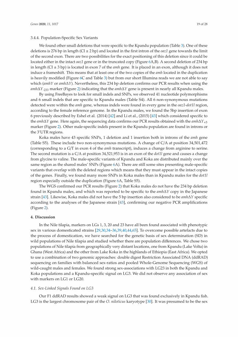
Genes 2020, 11, 1017 19 of 28
3.4.4. Population-Specific Sex Variants
We found other small deletions that were specific to the Kpandu population (Table 3). One of these
deletions is 276 bp in length (CI ± 2 bp) and located in the first intron of the oaz1 gene towards the limit
of the second exon. There are two possibilities for the exact positioning of this deletion since it could be
located either in the intact oaz1 gene or in the truncated copy (Figure 6A,B). A second deletion of 234 bp
in length (CI ± 3 bp) is located in exon 7 of the amh gene. It is placed in an exon, although it does not
induce a frameshift. This means that at least one of the two copies of the amh located in the duplication
is heavily modified (Figure 6C and Table 3) but from our short Illumina reads we are not able to say
which (amhY or amh∆Y). Nevertheless, this 234 bp deletion confirms our PCR results when using the
amh∆Y-233 marker (Figure 2) indicating that the amh∆Y gene is present in nearly all Kpandu males.
By using FreeBayes to look for small indels and SNPs, we observed 41 nucleotide polymorphisms
and 6 small indels that are specific to Kpandu males (Table S4). All 6 non-synonymous mutations
detected were within the amh gene, whereas indels were found in every gene in the oaz1-dot1l region,
according to the female reference genome. In the Kpandu males, we found the 5bp insertion of exon
6 previously described by Eshel et al. (2014) [42] and Li et al., (2015) [43] which considered specific to
the amh∆Y gene. Here again, the sequencing data confirms our PCR results obtained with the amh∆Y+5
marker (Figure 2). Other male-specific indels present in the Kpandu population are found in introns or
the 3’UTR regions.
Koka males have 43 specific SNPs, 1 deletion and 1 insertion both in introns of the amh gene
(Table S5). These include two non-synonymous mutations. A change of C/A at position 34,501,472
(corresponding to a G/T in exon 4 of the amh transcript), induces a change from arginine to serine.
The second mutation is a C/A at position 34,521,953 is in an exon of the dot1l gene and causes a change
from glycine to valine. The male-specific variants of Kpandu and Koka are distributed mainly over the
same region as the shared males’ SNPs (Figure 6A). There are still some sites presenting male-specific
variants that overlap with the deleted regions which means that they must appear in the intact copies
of the genes. Finally, we found many more SNPs in Koka males than in Kpandu males for the dot1l
region especially outside the duplication (Figure 6A, Table S5).
The WGS confirmed our PCR results (Figure 2) that Koka males do not have the 234 bp deletion
found in Kpandu males, and which was reported to be specific to the amh∆Y copy in the Japanese
strain [43]. Likewise, Koka males did not have the 5 bp insertion also considered to be amh∆Y specific
according to the analyses of the Japanese strain [43], confirming our negative PCR amplifications
(Figure 2).
4. Discussion
In the Nile tilapia, markers on LGs 1, 3, 20 and 23 have all been found associated with phenotypic
sex in various domesticated strains [29,30,34–36,39,40,44,65]. To overcome possible artefacts due to
the process of domestication, we have searched for the genetic basis of sex determination (SD) in
wild populations of Nile tilapia and studied whether there are population differences. We chose two
populations of Nile tilapia from geographically very distant locations, one from Kpandu (Lake Volta) in
Ghana (West Africa) and the other from Lake Koka in the highlands of Ethiopia (East Africa). We opted
to use a combination of two genomic approaches: double digest Restriction Associated DNA (ddRAD)
sequencing on families with balanced sex-ratios and pooled Whole-Genome Sequencing (WGS) of
wild-caught males and females. We found strong sex-associations with LG23 in both the Kpandu and
Koka populations and a Kpandu-specific signal on LG3. We did not observe any association of sex
with markers on LG1 or LG20.
4.1. Sex-Linked Signals Found on LG3
Our F1 ddRAD results showed a weak signal on LG3 that was found exclusively in Kpandu fish.
LG3 is the largest chromosome pair of the O. niloticus karyotype [30]. It was presumed to be the sex

Genes 2020, 11, 1017 20 of 28
chromosome after studies of chromosome pairing in the synaptonemal complex showed unpaired
terminal ends of the largest chromosome in XY males [34]. Subsequent studies showed stronger
linkage to LG1 [31,35,38,39] or LG23 [40,42–44]. In the sister species O. aureus, LG3 carries a female ZW
determiner [30,66,67]. Several sex-patterned variants were found along a ~50 Mbp region in the long
arm of LG3 in O. aureus when mapped onto the O. niloticus genome [67]. There is a very strong signal
in the Kpandu family at 2 RAD loci located around 20Mb. One is located in a region of non-coding
DNA. The other is found in the fcgr2b gene which codes for the receptor for Fc low-affinity II-b gamma
immunoglobulin. Curiously, sdy which arises from a duplication of the immune system gene irf9, has
become the master sex determinant in salmonid species [20]. The immune system showed higher sexual
dimorphism for the fcgr2a gene, another gene of the “low-affinity immunoglobulin gamma Fc region
receptor II” group in cichlids, a difference that was significantly greater in maternal mouth-brooders
compared to biparental mouth-brooders [68]. The Nile tilapia is a maternal mouth-brooder, and we
speculate that the difference in expression in fcgr2b might arise from sexual selection rather than being
implicated in sex-determination.
The other sex-patterned SNPs on LG3 were found all along the “long-arm” of LG3. This linkage
group is considered to have all the characteristics of an old sex chromosome in Nile tilapia, with signs
of differentiation due to its large size and extensive heterochromatic regions with the accumulation
of many repetitive sequences that may prevent recombination [51]. The SNPs we observe may result
from the history of this pair of chromosomes and the recent accumulation of polymorphisms that
do not follow sex-related patterns. This could explain why we did not find a definitive pattern of
an XY or a ZW system for the LG3 SNP sites, even though a large number of highly differentiated
SNPs were observed. Due to the high amount of repeated sequences in LG3 (54.7% repetitive vs. 37%
genome-wide) only ~87 Mb of the chromosome could be assembled instead of the estimated ~130 Mb,
and there might be incorrect anchoring to the O. niloticus genome [51]. We found on LG3 a very high
proportion of immune-related genes and they were present in multiple copies. For example, 96 genes
of the “Fc receptor” family are found on this linkage group, and among them 35 are “Low-affinity
immunoglobulin gamma Fc region receptor II”. Consequently, the sex differences we observed may
also result from mapping errors of reads and a lower quality of the assembly of this chromosome
amplified by the random sampling of the ddRAD method.
4.2. Strong XY Patterns on LG23
Our family ddRAD results reveal that LG23 is an XY sex chromosome in both the Kpandu and
Koka populations. LG23 has already been reported to be the sex chromosome in some domesticated
Nile tilapia [41,43,44,65]. Among the significant sex-variants between 24 and 40 Mb on LG23, one is
common to both populations: the anti-Müllerian hormone gene (amh). The amh gene has a primary
role in the sex differentiation pathway of vertebrates including fish [69] and is implicated in germ cell
proliferation in teleost fish (reviewed by Mullen et al. [70]). Amhy also appears to be the sex-determiner
in several domesticated strains of Nile tilapia [40–44,65]. A recent study of a wild Lake Kou population
(Burkina Faso) of Nile tilapia also showed that sex was linked to LG23 but the amh gene alone was not
sufficient to explain all the sex-ratios observed [47].
We also looked at the other sex-significant sites found on LG23 and found a male-specific SNP for
the lingo3 gene in Kpandu fish. Variants have also been identified in lingo3 in other Nile tilapia genomic
data when searching for sex-determining regions [41,44]. The close linkage of the lingo3 gene to the amh
region probably explains the signal found in those previous studies and could be enough to explain
the signal we found in the Kpandu individuals. In our two wild populations, we did not find any
sex-signals on LG1 nor on LG20 which have both been found in the Manzala-Stirling strain [31,38,39].
Genotyping at the region with the most significant sex-pattern variants on LG23 was enough
to perfectly predict the phenotypic sex of individuals in both populations. The Kpandu population
belongs to the widespread Sudan-Sahelian cluster that separated from the Nile basin through multi
paleo-geographic events [71]. The Koka population belonging to the Ethiopian Rift Valley showed

Genes 2020, 11, 1017 21 of 28
large genetic divergence from the Sudan-Sahelian but also from the Nile basin from which it was
isolated 12 to 8 thousand years ago [71]. The LG23 sex chromosome seems ancestral in O. niloticus
since it appeared in the two wild populations studied here. This observation however contrasts
with the apparent rapid evolution of the sex-determination in domesticated strains since at least
three different LGs have been reported to be sex chromosomes. However, wild populations might
segregate multiple sex determiners among which LG23 Y would be the most frequent and dominant.
This may also explain why we found only LG23 in both the Koka and Kpandu families we selected.
In wild Kou fish, for instance, sex was mainly associated with LG23 but it did not explain the
phenotypic sex in 18% of the individuals, suggesting segregation of another sex-determiner [47].
This has already been shown for the Manzala-Stirling strain in which sex is strongly associated with
LG1 and LG20 in some families [31,38] but it is also linked to LG23 in low frequency (Penman,
unpublished data). Founding effects or domestication events have led to sex being determined
exclusively by LG23 in the Manzala-Göttingen [65] and the Manzala-Tihange strains ([47], and this
study) although they both originated from Manzala-Stirling breeders. Even stronger differences have
been observed in the case of zebrafish where laboratory strains have lost the sex-chromosome found in
wild populations [46]. Our study further highlights the importance of characterising the sex determiner
in wild fish populations.
4.3. WGS Uncovers a Putative B Chromosome in O. niloticus
Blocks of high sequence coverage have been described previously in cichlid genomic data when B
chromosomes were present, such as in Astatotilapia latifasciata [63,72], Metriaclima lombardoi from Lake
Malawi [26], and the South American cichlid Crenicichla lepidota [73]. In these studies, high coverage
regions were due to A chromosome fragments copied onto a B chromosome, where they are highly
amplified and highly polymorphic. Because the reference assembly does not contain a B chromosome,
the whole genome shotgun reads from all the different copies are mapped at the same position on the
reference genome. The presence of these “B blocks” in our data, therefore, suggests the presence of a
B chromosome in these two wild populations of Nile tilapia. In our study, the high coverage blocks
mostly overlapped exons, which is a major difference from the B chromosomes previously described in
other cichlids. This could be because the B chromosome is ancient, and the sequences corresponding
to introns have diverged too much to map correctly onto the reference genome, unlike the exons that
would be more conserved over time. Another hypothesis would be that genes accumulate on the B
chromosome via an RNA intermediate, for instance through retrotransposition of mRNA, so the introns
are not copied. Because the B blocks were found in Kpandu males and Koka females, they created
strong differentiation between the sexes in each population, but these signals were independent of
the true genetic sex-determining locus. It appears unlikely that the putative B chromosomes play a
role as a female W sex-determiner such as in some Lake Malawi cichlids [27]. However, we are not
able to determine the frequency of this B chromosome in our wild populations since we pooled the
individual DNAs prior to sequencing. Even if the frequency is equal between sexes, if it is low in the
wild, then a random sampling of individuals in our study could make it look like the B chromosome
is sex-associated. B chromosomes have never before been described in Nile tilapia. Further studies
will be needed to thoroughly characterize them in these populations. If B chromosomes are present in
any of the domesticated strains, this can significantly impact genetic and genomic studies, from de
novo reference assembly to identify sex-determining regions, as seen in this study. As such it would be
beneficial to incorporate cytogenetic analysis as a control into the experimental design process.
4.4. Confirmation of the Tandem Duplication of the Amh Region on LG23
The Y chromosome amhY gene located on LG23 was shown to be necessary for maleness and is
possibly the primary gene in a Japanese strain [43]. These authors also found the presence of a tandem
amh duplication on the Y chromosome, with the existence of a truncated amh∆Y gene together with the
amhY gene. Our work confirms the existence of this tandem duplication on the Y chromosome of LG23
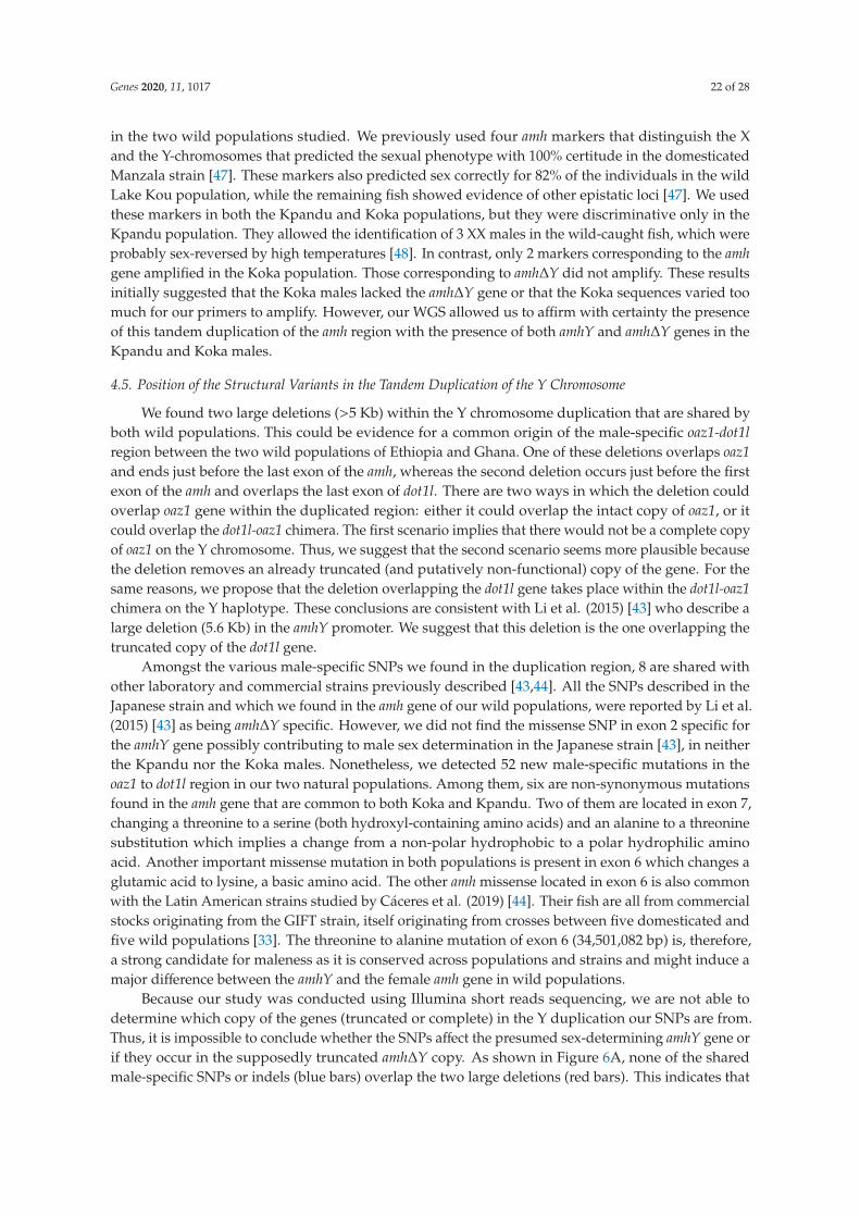
Genes 2020, 11, 1017 22 of 28
in the two wild populations studied. We previously used four amh markers that distinguish the X
and the Y-chromosomes that predicted the sexual phenotype with 100% certitude in the domesticated
Manzala strain [47]. These markers also predicted sex correctly for 82% of the individuals in the wild
Lake Kou population, while the remaining fish showed evidence of other epistatic loci [47]. We used
these markers in both the Kpandu and Koka populations, but they were discriminative only in the
Kpandu population. They allowed the identification of 3 XX males in the wild-caught fish, which were
probably sex-reversed by high temperatures [48]. In contrast, only 2 markers corresponding to the amh
gene amplified in the Koka population. Those corresponding to amh∆Y did not amplify. These results
initially suggested that the Koka males lacked the amh∆Y gene or that the Koka sequences varied too
much for our primers to amplify. However, our WGS allowed us to affirm with certainty the presence
of this tandem duplication of the amh region with the presence of both amhY and amh∆Y genes in the
Kpandu and Koka males.
4.5. Position of the Structural Variants in the Tandem Duplication of the Y Chromosome
We found two large deletions (>5 Kb) within the Y chromosome duplication that are shared by
both wild populations. This could be evidence for a common origin of the male-specific oaz1-dot1l
region between the two wild populations of Ethiopia and Ghana. One of these deletions overlaps oaz1
and ends just before the last exon of the amh, whereas the second deletion occurs just before the first
exon of the amh and overlaps the last exon of dot1l. There are two ways in which the deletion could
overlap oaz1 gene within the duplicated region: either it could overlap the intact copy of oaz1, or it
could overlap the dot1l-oaz1 chimera. The first scenario implies that there would not be a complete copy
of oaz1 on the Y chromosome. Thus, we suggest that the second scenario seems more plausible because
the deletion removes an already truncated (and putatively non-functional) copy of the gene. For the
same reasons, we propose that the deletion overlapping the dot1l gene takes place within the dot1l-oaz1
chimera on the Y haplotype. These conclusions are consistent with Li et al. (2015) [43] who describe a
large deletion (5.6 Kb) in the amhY promoter. We suggest that this deletion is the one overlapping the
truncated copy of the dot1l gene.
Amongst the various male-specific SNPs we found in the duplication region, 8 are shared with
other laboratory and commercial strains previously described [43,44]. All the SNPs described in the
Japanese strain and which we found in the amh gene of our wild populations, were reported by Li et al.
(2015) [43] as being amh∆Y specific. However, we did not find the missense SNP in exon 2 specific for
the amhY gene possibly contributing to male sex determination in the Japanese strain [43], in neither
the Kpandu nor the Koka males. Nonetheless, we detected 52 new male-specific mutations in the
oaz1 to dot1l region in our two natural populations. Among them, six are non-synonymous mutations
found in the amh gene that are common to both Koka and Kpandu. Two of them are located in exon 7,
changing a threonine to a serine (both hydroxyl-containing amino acids) and an alanine to a threonine
substitution which implies a change from a non-polar hydrophobic to a polar hydrophilic amino
acid. Another important missense mutation in both populations is present in exon 6 which changes a
glutamic acid to lysine, a basic amino acid. The other amh missense located in exon 6 is also common
with the Latin American strains studied by Cáceres et al. (2019) [44]. Their fish are all from commercial
stocks originating from the GIFT strain, itself originating from crosses between five domesticated and
five wild populations [33]. The threonine to alanine mutation of exon 6 (34,501,082 bp) is, therefore,
a strong candidate for maleness as it is conserved across populations and strains and might induce a
major difference between the amhY and the female amh gene in wild populations.
Because our study was conducted using Illumina short reads sequencing, we are not able to
determine which copy of the genes (truncated or complete) in the Y duplication our SNPs are from.
Thus, it is impossible to conclude whether the SNPs affect the presumed sex-determining amhY gene or
if they occur in the supposedly truncated amh∆Y copy. As shown in Figure 6A, none of the shared
male-specific SNPs or indels (blue bars) overlap the two large deletions (red bars). This indicates that
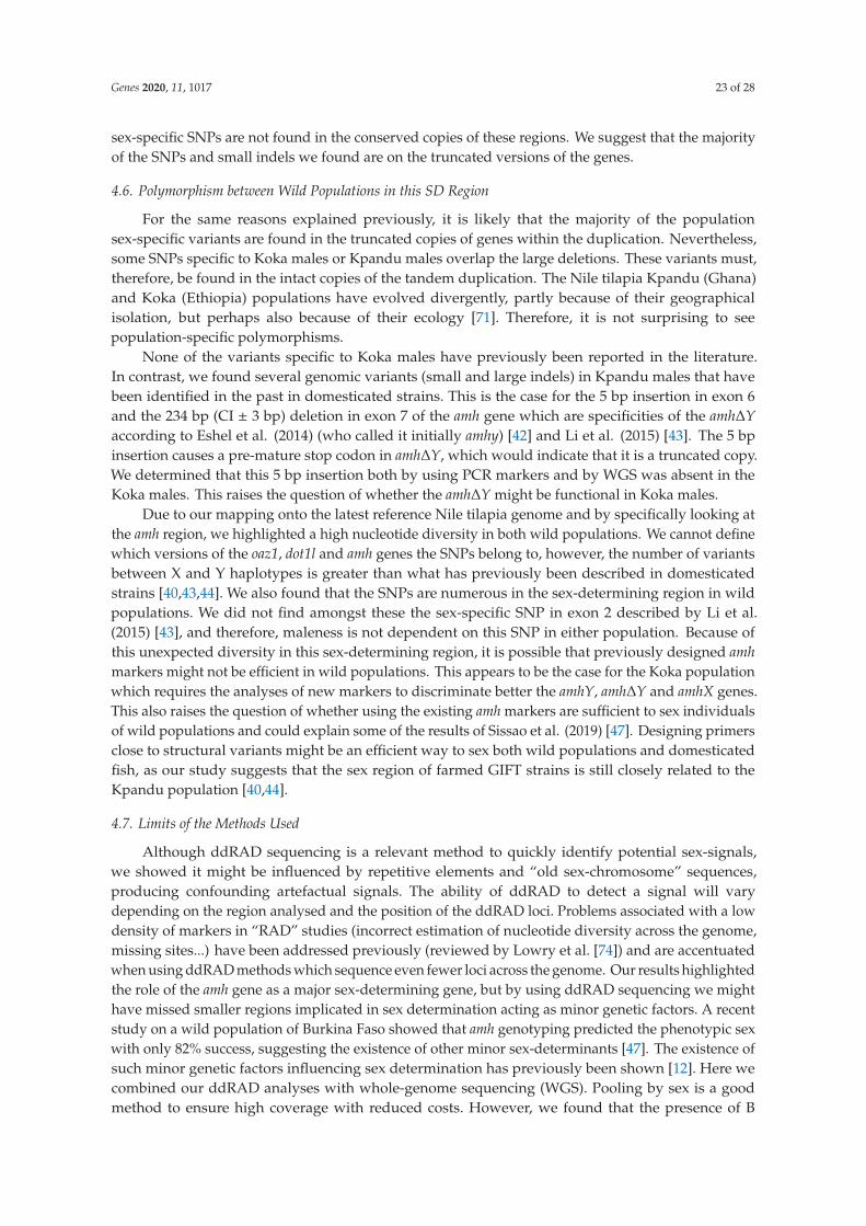
Genes 2020, 11, 1017 23 of 28
sex-specific SNPs are not found in the conserved copies of these regions. We suggest that the majority
of the SNPs and small indels we found are on the truncated versions of the genes.
4.6. Polymorphism between Wild Populations in this SD Region
For the same reasons explained previously, it is likely that the majority of the population
sex-specific variants are found in the truncated copies of genes within the duplication. Nevertheless,
some SNPs specific to Koka males or Kpandu males overlap the large deletions. These variants must,
therefore, be found in the intact copies of the tandem duplication. The Nile tilapia Kpandu (Ghana)
and Koka (Ethiopia) populations have evolved divergently, partly because of their geographical
isolation, but perhaps also because of their ecology [71]. Therefore, it is not surprising to see
population-specific polymorphisms.
None of the variants specific to Koka males have previously been reported in the literature.
In contrast, we found several genomic variants (small and large indels) in Kpandu males that have
been identified in the past in domesticated strains. This is the case for the 5 bp insertion in exon 6
and the 234 bp (CI ± 3 bp) deletion in exon 7 of the amh gene which are specificities of the amh∆Y
according to Eshel et al. (2014) (who called it initially amhy) [42] and Li et al. (2015) [43]. The 5 bp
insertion causes a pre-mature stop codon in amh∆Y, which would indicate that it is a truncated copy.
We determined that this 5 bp insertion both by using PCR markers and by WGS was absent in the
Koka males. This raises the question of whether the amh∆Y might be functional in Koka males.
Due to our mapping onto the latest reference Nile tilapia genome and by specifically looking at
the amh region, we highlighted a high nucleotide diversity in both wild populations. We cannot define
which versions of the oaz1, dot1l and amh genes the SNPs belong to, however, the number of variants
between X and Y haplotypes is greater than what has previously been described in domesticated
strains [40,43,44]. We also found that the SNPs are numerous in the sex-determining region in wild
populations. We did not find amongst these the sex-specific SNP in exon 2 described by Li et al.
(2015) [43], and therefore, maleness is not dependent on this SNP in either population. Because of
this unexpected diversity in this sex-determining region, it is possible that previously designed amh
markers might not be efficient in wild populations. This appears to be the case for the Koka population
which requires the analyses of new markers to discriminate better the amhY, amh∆Y and amhX genes.
This also raises the question of whether using the existing amh markers are sufficient to sex individuals
of wild populations and could explain some of the results of Sissao et al. (2019) [47]. Designing primers
close to structural variants might be an efficient way to sex both wild populations and domesticated
fish, as our study suggests that the sex region of farmed GIFT strains is still closely related to the
Kpandu population [40,44].
4.7. Limits of the Methods Used
Although ddRAD sequencing is a relevant method to quickly identify potential sex-signals,
we showed it might be influenced by repetitive elements and “old sex-chromosome” sequences,
producing confounding artefactual signals. The ability of ddRAD to detect a signal will vary
depending on the region analysed and the position of the ddRAD loci. Problems associated with a low
density of markers in “RAD” studies (incorrect estimation of nucleotide diversity across the genome,
missing sites...) have been addressed previously (reviewed by Lowry et al. [74]) and are accentuated
when using ddRAD methods which sequence even fewer loci across the genome. Our results highlighted
the role of the amh gene as a major sex-determining gene, but by using ddRAD sequencing we might
have missed smaller regions implicated in sex determination acting as minor genetic factors. A recent
study on a wild population of Burkina Faso showed that amh genotyping predicted the phenotypic sex
with only 82% success, suggesting the existence of other minor sex-determinants [47]. The existence of
such minor genetic factors influencing sex determination has previously been shown [12]. Here we
combined our ddRAD analyses with whole-genome sequencing (WGS). Pooling by sex is a good
method to ensure high coverage with reduced costs. However, we found that the presence of B

Genes 2020, 11, 1017 24 of 28
chromosomes with highly repeated sequences is an issue when using the sequencing of pooled
wild individuals to look for sex-specific signals with WGS. One should make sure of the absence
of such genomic structures, especially when studying wild populations as we found evidence for B
chromosomes in two distant populations across Africa. Nevertheless, WGS did enable us to study
in-depth the genome structure around the amh region in the genome of wild Kpandu and Koka males.
5. Conclusions
Our study is the first to search for the sex determiner in wild populations of Nile tilapia using
genomic approaches. F1 family analyses with ddRAD sequencing indicated that the major Y male
determinant was located in LG23, in both Lake Volta (Kpandu) in West Africa and Lake Koka from
East Africa. While the sex-variants covered a region of about 16 Mb in Koka, it encompassed nearly
the whole of LG23 in Kpandu. A weaker sex association was identified on LG3 only in the Kpandu
population, although neither XY nor ZW signals were conclusive. We confirmed the existence of a
tandem duplication of the sex determiner candidate amh gene, in the Y chromosome of both Kpandu
and Koka males. The breakpoints of the duplicated region were shown to be between the oaz1 and
dot1l genes. We found two common male deletions of ~5 kb, and the presence of both amhY and amh∆Y
genes in both populations. We did not find the missense SNP in exon 2 which was thought to be
important for maleness in a domesticated strain but found other male-specific variants. The amh∆Y
gene in Koka males presented no structural variation, which raises the question of whether it is still a
functional gene. Finally, WGS uncovered B blocks of high coverage suggesting for the first time in Nile
tilapia the presence of B chromosomes in both populations that were not sex-related at the species level.
Supplementary Materials: The following are available online at http://www.mdpi.com/2073-4425/11/9/1017/s1,Figure S1: Genotyping of wild-caught Kpandu fish of the AmhDY+5, AmhDY-233, AmhY-5608 and AmhX+36markers; Figure S2: Genotyping of wild-caught Koka fish of the AmhDY+5, AmhDY-233, AmhY-5608 andAmhX+36 markers; Figure S3: Genotypes of the offspring compared to their parents inferred from ddRADFET significant sites following a ZZ/ZW pattern; Table S1: Demultiplexing statistics for the ddRAD sequencing;Table S2: Details of the ddRAD FET-significant sites for LG23 found for both Kpandu and Koka populations;Table S3: Details of the ddRAD FET-significant sites for LG3 found for the Kpandu population; Table S4: SNPs andsmall indels found in the sex-determining oaz1-dot1l region on the Y haplotype-specific to the Kpandu populationbetween males and females; Table S5: SNPs and small indels found in the sex-determining oaz1-dot1l region on theY haplotype-specific to the Koka population between males and females.
Author Contributions: Conceptualization H.D., T.D.K. and J.-F.B.; methodology C.T., H.D., D.J.P. and T.D.K.;software C.T. and M.A.C.; validation C.T., H.D., T.D.K. and M.A.C.; formal analysis C.T., H.D., T.D.K., M.A.C.and J.-F.B.; investigation C.T., H.D. and F.E.C.; resources E.B. and J.-F.B.; data curation C.T., M.A.C., H.D., J.-F.B.and T.D.K.; writing—original draft preparation C.T., H.D. and J.-F.B.; writing—review and editing C.T., H.D.,J.-F.B., T.D.K., M.A.C., F.E.C., D.J.P. and E.B.; visualization C.T., H.D., J.-F.B. and T.D.K.; supervision, H.D. andT.D.K.; project administration J.-F.B.; funding acquisition J.-F.B. and H.D. All authors have read and agreed to thepublished version of the manuscript.
Funding: This research and Cécile Triay’s PhD was supported by the CLIMSEX project funded by the AgenceNational de la Recherche (ANR,) grant number ANR-15-CE02-0012. CIRAD gave an additional help for CécileTriay PhD. CIRAD and LabEx CEMEB contributed financially to Cécile Triay’s trip to work at the Universityof Maryland.
Acknowledgments: We are very grateful to John Taggart for his help with the ddRAD primers and protocol.We also thank Kerry Bartie for instructing Cécile Triay in ddRAD library preparation. The authors thank CharlesMélard for the Manzala-Tihange XX, XY and YY fish. gDNA extraction on the KingFisher robot was performedat the GenSeq platform. The sequencing of the ddRAD library was performed by the GeT-PlaGe platform.The sequencing of WG libraries was performed by the GenomEast platform, a member of the “France Génomique”consortium (ANR-10-INBS-0009).
Conflicts of Interest: The authors declare no conflict of interest. The funders had no role in the design of thestudy; in the collection, analyses, or interpretation of data; in the writing of the manuscript, or in the decision topublish the results.

Genes 2020, 11, 1017 25 of 28
References
1. Ellegren, H. Evolutionary stasis: The stable chromosomes of birds. Trends Ecol. Evol. 2010, 25, 283–291.
[CrossRef] [PubMed]
2. Cortez, D.; Marin, R.; Toledo-Flores, D.; Froidevaux, L.; Liechti, A.; Waters, P.D.; Grützner, F.; Kaessmann, H.
Origins and functional evolution of Y chromosomes across mammals. Nature 2014, 508, 488–493. [CrossRef]
[PubMed]
3. Koopman, P. The molecular biology of SRY and its role in sex determination in mammals. Reprod. Fertil. Dev.
1995, 7, 713–722. [CrossRef]
4. Bachtrog, D. Y-chromosome evolution: Emerging insights into processes of Y-chromosome degeneration.
Nat. Rev. Genet. 2013, 14, 113–124. [CrossRef] [PubMed]
5. Baroiller, J.-F.; Guiguen, Y.; Fostier, A. Endocrine and environmental aspects of sex differentiation in fish.
Cell. Mol. Life Sci. 1999, 55, 910. [CrossRef]
6. Devlin, R.H.; Nagahama, Y. Sex determination and sex differentiation in fish: An overview of genetic,
physiological, and environmental influences. Aquaculture 2002, 208, 191–364. [CrossRef]
7. Baroiller, J.-F.; D’Cotta, H.; Saillant, E. Environmental Effects on Fish Sex Determination and Differentiation.
Sex. Dev. 2009, 3, 118–135. [CrossRef]
8. Bachtrog, D.; Mank, J.E.; Peichel, C.L.; Kirkpatrick, M.; Otto, S.P.; Ashman, T.-L.; Hahn, M.W.; Kitano, J.;
Mayrose, V.B.; Ming, R.; et al. Sex Determination: Why So Many Ways of Doing It? PLoS Boil. 2014,
12, e1001899. [CrossRef]
9. Gammerdinger, W.J.; Kocher, T.D. Unusual Diversity of Sex Chromosomes in African Cichlid Fishes. Genes
2018, 9, 480. [CrossRef]
10. Volff, J.-N.; Schartl, M. Variability of genetic sex determination in poeciliid fishes. Genetica 2001, 111, 101–110.
[CrossRef]
11. Vandeputte, M.; Dupont-Nivet, M.; Chavanne, H.; Chatain, B. A Polygenic Hypothesis for Sex Determination
in the European Sea Bass Dicentrarchus labrax. Genetica 2007, 176, 1049–1057. [CrossRef]
12. Baroiller, J.-F.; D’Cotta, H.; Bezault, E.; Wessels, S.; Hoerstgen-Schwark, G. Tilapia sex determination:
Where temperature and genetics meet. Comp. Biochem. Physiol. Part A Mol. Integr. Physiol. 2009, 153, 30–38.
[CrossRef] [PubMed]
13. Matsuda, M.; Nagahama, Y.; Shinomiya, A.; Sato, T.; Matsuda, C.; Kobayashi, T.; Morrey, C.E.; Shibata, N.;
Asakawa, S.; Shimizu, N.; et al. DMY is a Y-specific DM-domain gene required for male development in the
medaka fish. Nature 2002, 417, 559–563. [CrossRef] [PubMed]
14. Nanda, I.; Kondo, M.; Hornung, U.; Asakawa, S.; Winkler, C.; Shimizu, A.; Shan, Z.; Haaf, T.; Shimizu, N.;
Shima, A.; et al. A duplicated copy of DMRT1 in the sex-determining region of the Y chromosome of the
medaka, Oryzias latipes. Proc. Natl. Acad. Sci. USA 2002, 99, 11778–11783. [CrossRef] [PubMed]
15. Myosho, T.; Otake, H.; Masuyama, H.; Matsuda, M.; Kuroki, Y.; Fujiyama, A.; Naruse, K.; Hamaguchi, S.;
Sakaizumi, M. Tracing the Emergence of a Novel Sex-Determining Gene in Medaka, Oryzias luzonensis.
Genetics 2012, 191, 163–170. [CrossRef]
16. Hattori, R.S.; Murai, Y.; Oura, M.; Masuda, S.; Majhi, S.K.; Sakamoto, T.; Fernandino, J.I.; Somoza, G.M.;
Yokota, M.; Strüssmann, C.A. A Y-linked anti-Mullerian hormone duplication takes over a critical role in sex
determination. Proc. Natl. Acad. Sci. USA 2012, 109, 2955–2959. [CrossRef]
17. Hattori, R.S.; Somoza, G.M.; Fernandino, J.I.; Colautti, D.; Miyoshi, K.; Gong, Z.; Yamamoto, Y.;
Strüssmann, C.A. The Duplicated Y-specific amhy Gene Is Conserved and Linked to Maleness in Silversides
of the Genus Odontesthes. Genes 2019, 10, 679. [CrossRef]
18. Pan, Q.; Feron, R.; Yano, A.; Guyomard, R.; Jouanno, E.; Vigouroux, E.; Wen, M.; Busnel, J.-M.; Bobe, J.;
Concordet, J.-P.; et al. Identification of the master sex determining gene in Northern pike (Esox lucius)
reveals restricted sex chromosome differentiation. PLoS Genet. 2019, 15, e1008013. [CrossRef]
19. Kamiya, T.; Kai, W.; Tasumi, S.; Oka, A.; Matsunaga, T.; Mizuno, N.; Fujita, M.; Suetake, H.; Suzuki, S.;
Hosoya, S.; et al. A Trans-Species Missense SNP in Amhr2 Is Associated with Sex Determination in the Tiger
Pufferfish, Takifugu rubripes (Fugu). PLoS Genet. 2012, 8, e1002798. [CrossRef]
20. Yano, A.; Guyomard, R.; Nicol, B.; Jouanno, E.; Quillet, E.; Klopp, C.; Cabau, C.; Bouchez, O.; Fostier, A.;
Guiguen, Y. An Immune-Related Gene Evolved into the Master Sex-Determining Gene in Rainbow Trout,
Oncorhynchus mykiss. Curr. Boil. 2012, 22, 1423–1428. [CrossRef]

Genes 2020, 11, 1017 26 of 28
21. Ser, J.R.; Roberts, R.B.; Kocher, T.D. Multiple Interacting Loci Control Sex Determination in Lake Malawi
Cichlid Fish. Evolution 2009, 64, 486–501. [CrossRef] [PubMed]
22. Parnell, N.F.; Streelman, J.T. Genetic interactions controlling sex and color establish the potential for sexual
conflict in Lake Malawi cichlid fishes. Heredity 2012, 110, 239–246. [CrossRef] [PubMed]
23. Gammerdinger, W.J.; Conte, M.A.; Sandkam, B.A.; Ziegelbecker, A.; Koblmüller, S.; Kocher, T.D. Novel Sex
Chromosomes in 3 Cichlid Fishes from Lake Tanganyika. J. Hered. 2018, 109, 489–500. [CrossRef] [PubMed]
24. Böhne, A.; Wilson, C.; Postlethwait, J.H.; Salzburger, W. Variations on a theme: Genomics of sex determination
in the cichlid fish Astatotilapia burtoni. BMC Genom. 2016, 17, 1–12. [CrossRef]
25. Roberts, N.B.; Juntti, S.A.; Coyle, K.P.; Dumont, B.L.; Stanley, M.K.; Ryan, A.Q.; Fernald, R.D.; Roberts, R.B.
Polygenic sex determination in the cichlid fish Astatotilapia burtoni. BMC Genom. 2016, 17, 835. [CrossRef]
26. Clark, F.B.; A Conte, M.; Ferreira-Bravo, I.A.; Poletto, A.B.; Martins, C.; Kocher, T.D. Dynamic Sequence
Evolution of a Sex-Associated B Chromosome in Lake Malawi Cichlid Fish. J. Hered. 2016, 108, 53–62.
[CrossRef] [PubMed]
27. Clark, F.B.; Kocher, T.D. Changing sex for selfish gain: B chromosomes of Lake Malawi cichlid fish. Sci. Rep.
2019, 9, 1–10. [CrossRef] [PubMed]
28. Baroiller, J.-F.; D’Cotta, H. Sex Control in Tilapias. In Sex Control in Aquaculture; John Wiley & Sons Ltd.:
Chichester, UK, 2018; pp. 189–234.
29. Lee, B.-Y.; Penman, D.J.; Kocher, T.D. Identification of a sex-determining region in Nile tilapia
(Oreochromis niloticus) using bulked segregant analysis. Anim. Genet. 2003, 34, 379–383. [CrossRef]
30. Cnaani, A.; Lee, B.Y.; Zilberman, N.; Ozouf−Costaz, C.; Hulata, G.; Ron, M.; D’Hont, A.; Baroiller, J.-F.;
D’Cotta, H.; Penman, D.; et al. Genetics of Sex Determination in Tilapiine Species. Sex. Dev. 2008, 2, 43–54.
[CrossRef]
31. Palaiokostas, C.; Bekaert, M.; Khan, M.G.Q.; Taggart, J.B.; Gharbi, K.; McAndrew, B.J.; Penman, D.J. Mapping
and Validation of the Major Sex-Determining Region in Nile Tilapia (Oreochromis niloticus L.) Using RAD
Sequencing. PLoS ONE 2013, 8, e68389. [CrossRef]
32. Mair, G.C.; Scott, A.G.; Penman, D.J.; Beardmore, J.A.; Skibinski, D.O.F. Sex determination in the genus
Oreochromis. Theor. Appl. Genet. 1991, 82, 144–152. [CrossRef] [PubMed]
33. Ponzoni, R.W.; Nguyen, N.H.; Khaw, H.L.; Hamzah, A.; Abu Bakar, K.R.; Yee, H.Y. Genetic improvement of
Nile tilapia (Oreochromis niloticus) with special reference to the work conducted by the WorldFish Center
with the GIFT strain. Rev. Aquac. 2011, 3, 27–41. [CrossRef]
34. Carrasco, L.A.; Penman, D.J.; Bromage, N. Evidence for the presence of sex chromosomes in the Nile tilapia
(Oreochromis niloticus) from synaptonemal complex analysis of XX, XY and YY genotypes. Aquaculture 1999,
173, 207–218. [CrossRef]
35. Lee, B.-Y.; Coutanceau, J.-P.; Ozouf-Costaz, C.; D’Cotta, H.; Baroiller, J.-F.; Kocher, T.D. Genetic and Physical
Mapping of Sex-Linked AFLP Markers in Nile Tilapia (Oreochromis niloticus). Mar. Biotechnol. 2010, 13,
557–562. [CrossRef] [PubMed]
36. Shirak, A.; Seroussi, E.; Cnaani, A.; Howe, A.E.; Domokhovsky, R.; Zilberman, N.; Kocher, T.D.; Hulata, G.;
Ron, M. AmhandDmrta2Genes Map to Tilapia (Oreochromis spp.) Linkage Group 23 Within Quantitative
Trait Locus Regions for Sex Determination. Genetics 2006, 174, 1573–1581. [CrossRef]
37. Eshel, O.; Weller, J.I.; Slossman, T.; Ron, M.; Shirak, A.; Hulata, G.; Cnaani, A. Fine-mapping of a locus on
linkage group 23 for sex determination in Nile tilapia (Oreochromis niloticus). Anim. Genet. 2010, 42, 222–224.
[CrossRef]
38. Gammerdinger, W.J.; Conte, M.A.; Acquah, E.A.; Roberts, R.B.; Kocher, T.D. Structure and decay of a proto-Y
region in Tilapia, Oreochromis niloticus. BMC Genom. 2014, 15, 975. [CrossRef]
39. Palaiokostas, C.; Bekaert, M.; Khan, M.G.Q.; Taggart, J.B.; Gharbi, K.; McAndrew, B.J.; Penman, D.J. A novel
sex-determining QTL in Nile tilapia (Oreochromis niloticus). BMC Genom. 2015, 16, 171. [CrossRef]
40. Taslima, K.; Wehner, S.; Taggart, J.B.; de Verdal, H.; Benzie, J.A.H.; Bekaert, M.; McAndrew, B.J.; Penman, D.J.
Sex determination in the GIFT strain of tilapia is controlled by a locus in linkage group 23. BMC Genet. 2020,
21, 1–15. [CrossRef]
41. Eshel, O.; Shirak, A.; Weller, J.I.; Hulata, G.; Ron, M. Linkage and Physical Mapping of Sex Region on LG23
of Nile Tilapia (Oreochromis niloticus). G3 Genes Genomes Genet. 2012, 2, 35–42. [CrossRef]

Genes 2020, 11, 1017 27 of 28
42. Eshel, O.; Shirak, A.; Dor, L.; Band, M.; Zak, T.; Markovich-Gordon, M.; Chalifa-Caspi, V.; Feldmesser, E.;
Weller, J.I.; Seroussi, E.; et al. Identification of male-specific amh duplication, sexually differentially expressed
genes and microRNAs at early embryonic development of Nile tilapia (Oreochromis niloticus). BMC Genom.
2014, 15, 774. [CrossRef] [PubMed]
43. Li, M.; Sun, Y.; Zhao, J.; Shi, H.; Zeng, S.; Ye, K.; Jiang, D.; Zhou, L.; Sun, L.; Tao, W.; et al. A Tandem
Duplicate of Anti-Müllerian Hormone with a Missense SNP on the Y Chromosome Is Essential for Male Sex
Determination in Nile Tilapia, Oreochromis niloticus. PLoS Genet. 2015, 11, e1005678. [CrossRef] [PubMed]
44. Cáceres, G.; López, M.E.; Cádiz, M.I.; Yoshida, G.M.; Jedlicki, A.M.; Palma-Véjares, R.; Travisany, D.;
Díaz-Domínguez, D.; Maass, A.; Lhorente, J.P.; et al. Fine Mapping Using Whole-Genome Sequencing
Confirms Anti-Müllerian Hormone as a Major Gene for Sex Determination in Farmed Nile Tilapia
(Oreochromis niloticus L.). G3 Genes Genomes Genet. 2019, 9, 3213–3223. [CrossRef] [PubMed]
45. Pfennig, F.; Standke, A.; Gutzeit, H.O. The role of Amh signaling in teleost fish—Multiple functions not
restricted to the gonads. Gen. Comp. Endocrinol. 2015, 223, 87–107. [CrossRef] [PubMed]
46. Wilson, C.A.; High, S.K.; McCluskey, B.M.; Amores, A.; Yan, Y.-L.; Titus, T.A.; Anderson, J.L.; Batzel, P.;
Carvan, M.J.; Schartl, M.; et al. Wild Sex in Zebrafish: Loss of the Natural Sex Determinant in Domesticated
Strains. Genetics 2014, 198, 1291–1308. [CrossRef]
47. Sissao, R.; D’Cotta, H.; Baroiller, J.-F.; Toguyeni, A. Mismatches between the genetic and phenotypic sex in
the wild Kou population of Nile tilapia Oreochromis niloticus. PeerJ 2019, 7, e7709. [CrossRef]
48. Bezault, E.; Clota, F.; Derivaz, M.; Chevassus, B.; Baroiller, J.-F. Sex determination and temperature-induced
sex differentiation in three natural populations of Nile tilapia (Oreochromis niloticus) adapted to extreme
temperature conditions. Aquaculture 2007, 272, S3–S16. [CrossRef]
49. Andrews, S. FastQC: A Quality Control Tool for High Throughput Sequence Data. 2010. Available online:
http://www.bioinformatics.babraham.ac.uk/projects/fastqc (accessed on 27 August 2020).
50. Catchen, J.M.; Amores, A.; Hohenlohe, P.; Cresko, W.A.; Postlethwait, J.H. Stacks: Building and Genotyping
Loci De Novo From Short-Read Sequences. G3 Genes Genomes Genet. 2011, 1, 171–182. [CrossRef]
51. A Conte, M.; Joshi, R.; Moore, E.C.; Nandamuri, S.P.; Gammerdinger, W.J.; Roberts, R.B.; Carleton, K.L.;
Lien, S.; Kocher, T.D. Chromosome-scale assemblies reveal the structural evolution of African cichlid genomes.
GigaScience 2019, 8, 8. [CrossRef]
52. Li, H.; Durbin, R. Fast and accurate short read alignment with Burrows-Wheeler transform. Bioinformatics
2009, 25, 1754–1760. [CrossRef]
53. Li, H.; Handsaker, B.; Wysoker, A.; Fennell, T.; Ruan, J.; Homer, N.; Marth, G.; Abecasis, G.R.; Durbin, R.
1000 Genome Project Data Processing Subgroup The Sequence Alignment/Map format and SAMtools.
Bioinformatics 2009, 25, 2078–2079. [CrossRef] [PubMed]
54. Weir, B.S. Genetic Data Analysis II: Methods for Discrete Population Genetic Data; Sinauer Assoc. Inc.: Sunderland,
MA, USA, 1996; ISBN 0878939024.
55. Danecek, P.; Auton, A.; Abecasis, G.R.; Albers, C.A.; Banks, E.; Depristo, M.A.; Ha, G.; Lunter, G.; Marth, G.T.;
Sherry, S.T.; et al. The variant call format and VCFtools. Bioinformatics 2011, 27, 2156–2158. [CrossRef]
[PubMed]
56. Bolger, A.M.; Lohse, M.; Usadel, B. Trimmomatic: A flexible trimmer for Illumina sequence data. Bioinformatics
2014, 30, 2114–2120. [CrossRef] [PubMed]
57. DePristo, M.A.; Banks, E.; Poplin, R.; Garimella, K.V.; Maguire, J.R.; Hartl, C.; Philippakis, A.A.; Del Angel, G.;
Rivas, M.A.; Hanna, M.; et al. A framework for variation discovery and genotyping using next-generation
DNA sequencing data. Nat. Genet. 2011, 43, 491–498. [CrossRef]
58. Kofler, R.; Pandey, R.V.; Schlötterer, C. PoPoolation2: Identifying differentiation between populations using
sequencing of pooled DNA samples (Pool-Seq). Bioinformatics 2011, 27, 3435–3436. [CrossRef] [PubMed]
59. Rausch, T.; Zichner, T.; Schlattl, A.; Stütz, A.M.; Benes, V.; Korbel, J.O. DELLY: Structural variant discovery
by integrated paired-end and split-read analysis. Bioinformatics 2012, 28, i333–i339. [CrossRef]
60. Garrison, E.; Marth, G. Haplotype-based variant detection from short-read sequencing. arXiv 2012, arXiv:1207.3907.
61. R Core Team. R: A Language and Environment for Statistical Computing; R Foundation for Statistical Computing:
Vienna, Austria, 2018.
62. Turner, S.D. QQman: An R Package for Visualizing GWAS Results Using Q-Q and Manhattan Plots BioRxiv.
Available online: https://www.biorxiv.org/content/10.1101/005165v1 (accessed on 8 June 2020).

Genes 2020, 11, 1017 28 of 28
63. Valente, G.T.; Conte, M.A.; Fantinatti, B.E.; Cabral-de-Mello, D.C.; Carvalho, R.; Vicari, M.R.; Kocher, T.D.;
Martins, C. Origin and Evolution of B Chromosomes in the Cichlid Fish Astatotilapia latifasciata Based on
Integrated Genomic Analyses. Mol. Boil. Evol. 2014, 31, 2061–2072. [CrossRef]
64. Clark, F.B.; Conte, M.A.; Kocher, T.D. Genomic Characterization of a B Chromosome in Lake Malawi Cichlid
Fishes. Genes 2018, 9, 610. [CrossRef]
65. Wessels, S.; Krause, I.; Floren, C.; Schütz, E.; Beck, J.; Knorr, C. ddRADseq reveals determinants for
temperature-dependent sex reversal in Nile tilapia on LG23. BMC Genom. 2017, 18, 531. [CrossRef]
66. Lee, B.-Y.; Hulata, G.; Kocher, T.D. Two unlinked loci controlling the sex of blue tilapia (Oreochromis aureus).
Heredity 2004, 92, 543–549. [CrossRef] [PubMed]
67. A Conte, M.; Gammerdinger, W.J.; Bartie, K.L.; Penman, D.J.; Kocher, T.D. A high quality assembly of the Nile
Tilapia (Oreochromis niloticus) genome reveals the structure of two sex determination regions. BMC Genom.
2017, 18, 341. [CrossRef]
68. Tanger, I.S.; Bayer, T.; Salzburger, W.; Roth, O. Effects of parental care on resource allocation into immune
defense and buccal microbiota in mouthbrooding cichlid fishes*. Evolution 2018, 72, 1109–1123. [CrossRef]
69. Poonlaphdecha, S.; Pepey, E.; Canonne, M.; de Verdal, H.; Baroiller, J.-F.; D’Cotta, H. Temperature
induced-masculinisation in the Nile tilapia causes rapid up-regulation of both dmrt1 and amh expressions.
Gen. Comp. Endocrinol. 2013, 193, 234–242. [CrossRef] [PubMed]
70. Mullen, R.D.; Ontiveros, A.E.; Moses, M.M.; Behringer, R.R. AMH and AMHR2 mutations: A spectrum of
reproductive phenotypes across vertebrate species. Dev. Boil. 2019, 455, 1–9. [CrossRef] [PubMed]
71. Bezault, E.; Balaresque, P.; Toguyeni, A.; Fermon, Y.; Araki, H.; Baroiller, J.-F.; Rognon, X. Spatial and temporal
variation in population genetic structure of wild Nile tilapia (Oreochromis niloticus) across Africa. BMC Genet.
2011, 12, 102. [CrossRef]
72. Fantinatti, B.E.A.; Martins, C. Development of chromosomal markers based on next-generation sequencing:
The B chromosome of the cichlid fish Astatotilapia latifasciata as a model. BMC Genet. 2016, 17, 119. [CrossRef]
73. Perazzo, G.X.; Noleto, R.B.; Vicari, M.R.; Gava, A.; Cestari, M.M. B chromosome polymorphism in South
American cichlid. Neotrop. Biodivers. 2018, 4, 3–9. [CrossRef]
74. Lowry, D.B.; Hoban, S.; Kelley, J.L.; Lotterhos, K.E.; Reed, L.K.; Antolin, M.F.; Storfer, A. Breaking RAD:
An evaluation of the utility of restriction site-associated DNA sequencing for genome scans of adaptation.
Mol. Ecol. Resour. 2016, 17, 142–152. [CrossRef]
© 2020 by the authors. Licensee MDPI, Basel, Switzerland. This article is an open access
article distributed under the terms and conditions of the Creative Commons Attribution
(CC BY) license (http://creativecommons.org/licenses/by/4.0/).
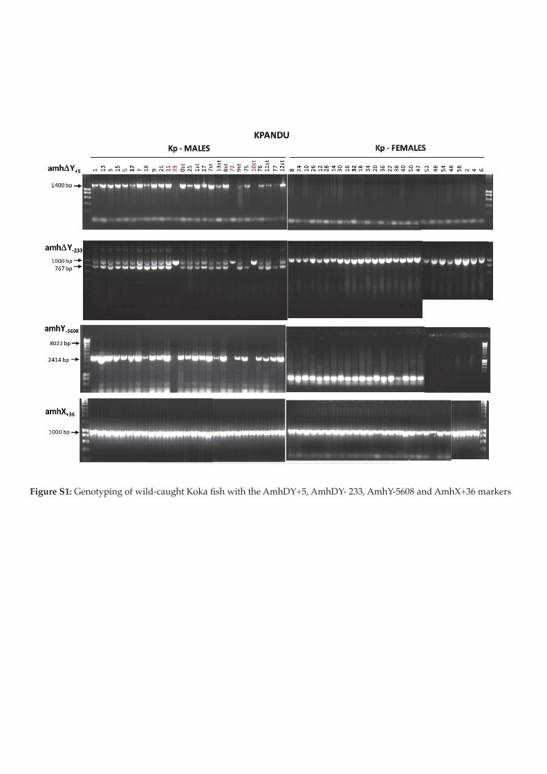
!"#$%&'()*!"#$%&'()$*!%+!,)-./012*3&!4%51!673!,)&3!&3#!893:;<=>!893:;/!?@@>!893;/=ABC!1$.!893D<@A!91E5#E7!

!"#$%&'()*'!"#$%&'(#)*$+*,(-./012)3%*4$51*673*,(%3*%3"*893:;<=>*893:;/*?@@>*893;/=ABC*1#.*893D<@A*91E5"E7*
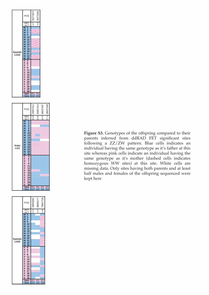
!"#$%&'()*!"#$%&'(#)!%*!&+#!%**)(,-$.!/%0(1,#2!&%!&+#-,!(1,#$&)! -$*#,,#2! *,%0! 22345! 678! )-.$-9/1$&! )-&#)!*%::%;-$.! 1! <<=<>! (1&&#,$?! @:A#! /#::)! -$2-/1&#)! 1$!-$2-B-2A1:!+1B-$.!&+#!)10#!.#$%&'(#!1)!-&C)!*1&+#,!1&!&+-)!)-&#!;+#,#1)!(-$D!/#::)!-$2-/1&#!1$!-$2-B-2A1:!+1B-$.!&+#!)10#! .#$%&'(#! 1)! -&E)! 0%&+#,! F21)+#2! /#::)! -$2-/1&#)!+%0%G'.%A)! >>! )-&#)H! 1&! &+-)! )-&#?! >+-&#! /#::)! 1,#!0-))-$.!21&1?!I$:'!)-&#)!+1B-$.!J%&+!(1,#$&)!1$2!1&!:#1)&!+1:*!01:#)!1$2!*#01:#)!%*! &+#!%**)(,-$.!)#KA#$/#2!;#,#!D#(&!+#,#
!"#
$%&'()'&
$%&'(*$+
,-. / 0
012 2 2
! " +3+ +3+
! # (3( (3(
! $ (3( (3(
! % +3+ +3+
! "& 434 434
! "# (3( (3(
! "$ (3( (3(
! "% (3( (3(
! "' (3( (3(
! "( +3+ +3+
! #& (3( (3(
! #" +3+ +3+
! ## (3( (3(
! #% (3( (3(
) * +3( +3(
) + +3( +3(
) ' +3( +3(
) , 434 434
) "" +3+ +3+
) "* +3+ +3+
) ", +3+ +3+
) #$ +3( +3(
) #* +3+ +3+
) #+ 434 434
) #' 434 434
) #( +3+ +3+
) #, +3+ +3+
) $& 434 434
-./ +3( +3(
0123 +3+ +3+
!"#
&%*%'5((
&%*%'5(5
$)+$'5*'
$)+$'%$)
,-. 6 0 / 2
012 0 6 0 /
! " +3( +3( +3( +3(
! # +3( +3( +3( +3(
! $ 434 434 +3+ +3+
! % +3( +3( +3( +3(
! ( +3( +3( +3( +3(
! "& +3( +3( +3( +3(
! "$ +3( +3+ +3( +3(
! "* +3( +3( +3( +3(
! "' +3+ +3+ 434 434
! "( +3( +3( +3( +3(
! ", (3( (3( +3( +3(
! #& +3( +3( +3( +3(
! ## +3( +3( +3( +3(
! #% +3( +3( +3( +3(
! #+ +3( +3( +3( +3(
) * +3+ +3+ +3+ +3+
) + +3+ +3+ +3+ +3+
) ' +3+ +3+ +3+ +3+
) , +3+ +3+ +3+ +3+
) "" +3+ +3+ +3+ +3+
) "# +3+ +3+ +3+ +3+
) "% +3( +3( +3+ +3+
) "+ +3+ +3+ +3+ +3+
) #$ +3+ +3+ +3( +3(
) #* +3+ +3+ +3+ +3+
) #' +3+ +3+ +3+ +3+
) #( +3+ +3+ +3+ +3+
) $& +3+ +3+ +3+ +3+
-./ +3( +3( +3( +3(
0123 +3+ +3+ +3+ +3+
!"#
$)*%%+)(
$)*%%(''
'*7%''&)
,-. / / 0
012 2 2 2
! " +3+ 434 +3+
! # +3+ +3+ (3(
! $ +3+ +3+ (3(
! % 434 434 +3(
! "& +3+ 434 +3(
! "# +3+ +3+ (3(
! "$ +3+ +3+ +3+
! "% 434 434 +3+
! "' +3+ +3+ +3(
! "( 434 434 +3+
! #& 434 434 (3(
! #" +3+ +3+ (3(
! ## +3+ +3+ (3(
! #% +3+ +3+ +3(
) * 434 434 +3+
) + +3( +3( +3+
) ' +3( +3( +3+
) , 434 434 +3+
) "" +3( +3( +3+
) "* 434 434 +3(
) ", 434 434 +3+
) #$ +3( +3( +3+
) #* +3+ +3+ (3(
) #+ 434 434 +3+
) #' +3( +3( +3+
) #( +3+ +3+ +3+
) #, +3+ +3+ +3(
) $& +3+ 434 +3+
-./ +3( +3( +3(
0123 +3+ +3+ +3+
45.6789:;#$
4<=.9:;$
45.6789:;&$

Filename Barcode Total No RadTag Low Quality Retained
Kk06_27-01 TCAGA-TAGCA 6519498 62393 10 6432161
Kk06_27-02 TGCAACA-AGCTGTC 6488002 42432 10 6421987
Kk06_27-03 GATCG-AGTCA 6130008 59742 12 6046134
Kk06_27-04 CGTATCA-TACGTGT 4786780 30645 11 4738595
Kk06_27-05 CATGA-GCATA 9155348 118757 17 9000160
Kk06_27-06 CACAGAC-GAGATGT 4304474 65547 8 4219379
Kk06_27-07 ATCGA-CGATC 6587634 72103 7 6492862
Kk06_27-08 ACTGCAC-CATCTGT 4087738 56399 2 4016341
Kk06_27-09 TCGAG-CTGGT 6728002 69386 11 6633318
Kk06_27-10 TCTCTCA-GTCAAGT 3997246 31884 7 3948143
Kk06_27-11 GTCAC-GAAGC 5398064 73342 3 5302251
Kk06_27-12 GTACACA-ATACGGT 5160480 37936 8 5104071
Kk06_27-13 GCATT-TAGCA 4329904 63290 8 4251195
Kk06_27-14 CTCTTCA-AGCTGTC 5150804 63152 12 5066501
Kk06_27-15 CGATA-AGTCA 6795198 67075 10 6702247
Kk06_27-16 CTAGGAC-TACGTGT 4458124 25108 8 4416516
Kk06_27-17 ACGTA-GCATA 8356696 150957 20 8170910
Kk06_27-18 CAGTCAC-GAGATGT 4402590 52193 9 4333027
Kk06_27-19 AGAGT-CGATC 4436860 57079 7 4363275
Kk06_27-20 GCTAACA-CATCTGT 6733466 59885 10 6650901
Kk06_27-21 ATGCT-CTGGT 45828 30344 0 15428
Kk06_27-22 ACACGAG-GTCAAGT 2569408 24098 3 2534472
Kk06_27-23 GACTA-GAAGC 3222708 63755 2 3145377
Kk06_27-24 AGGACAC-ATACGGT 3725288 58448 3 3652006
Kk06_27-25 TCAGA-AGCTGTC 2807592 36100 4 2760146
Kk06_27-26 TGCAACA-AGTCA 7424726 71086 9 7326480
Kk06_27-27 GATCG-TACGTGT 3747604 24415 3 3707727
Kk06_27-28 CGTATCA-GCATA 10693924 146273 20 10502658
Kk06_27-30 CATGA-GAGATGT 5827138 90528 4 5712289
KkGF04_27 GTACACA-AGCTGTC 4455130 44283 7 4392216
KkGM39_27 GCATT-AGTCA 3663614 43555 3 3606067
Kp20_27-01 CTCTTCA-TACGTGT 9118810 64960 19 9022280
Kp20_27-02 CGATA-GCATA 12392034 156916 21 12189103
Kp20_27-03 CTAGGAC-GAGATGT 7313068 69919 11 7218594
Kp20_27-04 ACGTA-CGATC 8325070 75518 20 8223412
Kp20_27-05 CAGTCAC-CATCTGT 7442942 72497 11 7344093
Kp20_27-06 AGAGT-CTGGT 5390846 113234 4 5259540
Kp20_27-07 GCTAACA-GTCAAGT 4501978 41902 9 4443363
Kp20_27-08 ATGCT-GAAGC 38146 28303 0 9777
Kp20_27-09 ACACGAG-ATACGGT 4740246 66828 2 4657183
Kp20_27-10 GACTA-TAGCA 6519380 92477 16 6403155
Kp20_27-11 AGGACAC-AGCTGTC 3317674 26002 9 3280807
Kp20_27-12 TCAGA-TACGTGT 5258990 48510 4 5192469
Kp20_27-13 TGCAACA-GCATA 10216096 71647 12 10106968
Kp20_27-14 GATCG-GAGATGT 6292344 36782 14 6232033
Kp20_27-15 CGTATCA-CGATC 7721200 71407 17 7624258
Kp20_27-17 CATGA-CATCTGT 11741310 81447 21 11625140
Kp20_27-18 CACAGAC-CTGGT 8255206 90916 12 8135409
Kp20_27-19 ATCGA-GTCAAGT 4667892 25809 5 4625598
Kp20_27-20 ACTGCAC-GAAGC 6783430 146471 12 6612036
Kp20_27-21 TCGAG-ATACGGT 7855194 42422 14 7787581
Kp20_27-22 TCTCTCA-TAGCA 7878918 111920 11 7741960
Kp20_27-23 GTCAC-AGCTGTC 7542616 44289 8 7472232
Kp20_27-24 GTACACA-AGTCA 8141530 65761 13 8047445
Kp20_27-25 GCATT-TACGTGT 5461980 34237 6 5408628
Kp20_27-26 CTCTTCA-GCATA 15576420 125831 22 15395325
Kp20_27-27 CGATA-GAGATGT 4719534 39821 6 4662161
Kp20_27-28 CTAGGAC-CGATC 10168514 70911 13 10064095
Kp20_27-29 ACGTA-CATCTGT 8314098 50526 16 8237185
Kp20_27-30 CAGTCAC-CTGGT 7810766 115129 8 7669778
KpGF32_27 AGAGT-GTCAAGT 2776938 36878 4 2729496
KpM17_27 GCTAACA-GAAGC 6458654 42653 7 6393873
Neg Control ATGCT-ATACGGT 4408 2433 0 1965
!"#$%&'()&!"#$%&'(%")'*+ ,&-&',&'.,/012/&3"/4456! ,"7$"*.'*+

BP Gene ID Gene End Product description
3268204 5.42E-03 0.156LOC109197015 3145390 4078398 ATP-dependent DNA helicase PIF1
3268469 6.52E-03 0.148
5578395 2.36E-03 0.185 LOC100702162 5544973 5584661 multidrug resistance-associated protein 4
6381389 5.65E-03 0.133 trak2 6373940 6389882 trafficking protein, kinesin binding 2
6790753 9.93E-03 0.136 zranb3 6724257 6842613 zinc finger RANBP2-type containing 3
7534698 2.60E-03 0.155 LOC100689728 7501806 7649676 gamma-aminobutyric acid receptor subunit gamma-3
7818707 1.15E-03 0.179
Non Coding
8296704 1.15E-03 0.179
9113411 1.15E-03 0.179
9161445 2.61E-03 0.174
9161653 8.86E-03 0.134
9764218 2.25E-03 0.171
9982398 3.39E-03 0.190
11597075 1.15E-03 0.179
13021743 1.15E-03 0.179 LOC100699606 13020746 13022102 gamma-crystallin M3-like
13602491 1.77E-03 0.203
Non Coding
13602504 4.45E-03 0.189
13602569 9.65E-03 0.165
13602575 1.95E-03 0.200
13602590 4.70E-03 0.171
13602591 1.95E-03 0.200
13602694 1.95E-03 0.200
13602701 9.19E-03 0.182
13602732 3.19E-03 0.196
13602959 1.15E-03 0.179
14115966 1.15E-03 0.179
17141915 8.95E-04 0.296
17613124 1.15E-03 0.179als2 17587289 17614314 alsin Rho guanine nucleotide exchange factor
17613143 1.15E-03 0.179
18309315 1.15E-03 0.179
Non Coding
19018833 9.10E-04 0.205
19031065 1.93E-03 0.221
19373150 7.34E-03 0.176
19609561 1.90E-03 0.166
19915347 2.61E-03 0.174
22988019 1.15E-03 0.232
22988087 1.15E-03 0.232
22988389 1.15E-03 0.232
23996168 9.10E-04 0.205
23996185 9.10E-04 0.205
23996228 9.10E-04 0.205
24090251 9.10E-04 0.205 LOC100690062 24067428 24119616 CLIP-associating protein 1-A
24420205 9.10E-04 0.205 Non Coding
24729049 9.10E-04 0.205
agap1 24688557 24858705 arf-GAP with GTPase ANK repeat and PH domain-containing protein 124814636 9.10E-04 0.205
24814747 9.10E-04 0.205
24932340 5.77E-03 0.156LOC109196997 24910543 25412345 NLR family CARD domain-containing protein 3
24932396 5.67E-03 0.156
27622612 2.88E-03 0.166
Non Coding
27622823 2.88E-03 0.166
27622855 3.73E-03 0.156
27622859 3.73E-03 0.156
27622864 2.88E-03 0.166
27622865 2.88E-03 0.166
27622937 2.88E-03 0.166
28006648 2.34E-04 0.276 LOC102077606 27999712 28030875 uncharacterized LOC102077606
28183172 2.24E-04 0.224LOC102077510 28169381 28190713 adhesion G-protein coupled receptor G7
28183563 4.60E-04 0.216
28427026 3.64E-03 0.197 Non Coding
28720524 3.64E-03 0.156
LOC100701816 28694684 28746200 NACHT LRR and PYD domains-containing protein 328720537 3.64E-03 0.156
28720563 3.64E-03 0.156
28720615 3.64E-03 0.156
28720616 3.64E-03 0.156
LOC100701273 28706535 28721156 protein NLRC3-like28720801 3.64E-03 0.156
28720868 3.64E-03 0.156
29326015 6.40E-04 0.255 mcf2l2 29307709 29404131 guanine nucleotide exchange factor DBS
30529202 4.05E-04 0.221LOC102076398 30446019 30539111 uncharacterized LOC102076398
30529404 4.05E-04 0.221
30929265 7.83E-07 0.455
LOC109196929 30927917 30936814 aldehyde dehydrogenase family 9 member A1-like30929418 6.70E-05 0.329
30929419 3.19E-03 0.229
30944555 8.49E-04 0.214 LOC100707441 30940790 30950350 aldehyde dehydrogenase family 9 member A1
31271536 4.05E-04 0.221
evi5 31250049 31293025 ecotropic viral integration site 531271549 1.06E-04 0.260
31272084 1.06E-04 0.260
31610258 1.97E-04 0.231 Non Coding
33734321 8.90E-06 0.381cyfip1 33706514 33746187 cytoplasmic FMR1 interacting protein 1
33734328 8.90E-06 0.381
33907878 2.57E-05 0.309 celf5 33898573 34074566 CUGBP Elav-like family member 5
34265123 3.30E-05 0.278 LOC100701388 34260519 34285917 zinc finger and BTB domain-containing protein 7A
Fisher’s
Exact Test p-value
Amova Fst
Gene Start
Kpandu LG23
!"#$%&'*)&!"&-'%,/10/4456! 89:;,'+*'0'.-*&/,'&",/012/<=>?/01$*4/012/@1&3/A(-*4$ -*4/A1B- 0-#'%'",

BP Gene ID Gene End Product description
34443504 3.30E-05 0.278 lingo3 34417222 34451018 leucine rich repeat and Ig domain containing 3
34502301 2.82E-05 0.308
amh 34498945 34502800 anti-Mullerian hormone34502353 1.83E-05 0.297
34502501 1.83E-05 0.297
34772926 2.92E-06 0.333tmem59l 34764172 34775066 transmembrane protein 59 like
34774781 3.94E-05 0.286
35124733 2.92E-06 0.333 sema6b 35024697 35148164 semaphorin 6B
35271872 4.63E-03 0.171Non Coding
35271930 4.63E-03 0.171
35825054 1.76E-04 0.417 nwd1 35824755 35843686 NACHT and WD repeat domain containing 1
35941307 8.38E-06 0.304 dbt 35940945 35947903 dihydrolipoamide branched chain transacylase E2
35953054 2.89E-03 0.207 lrrc39 35948014 35953096 leucine rich repeat containing 39
36092189 9.85E-03 0.176 grin3b 36077197 36130419 glutamate ionotropic receptor NMDA type subunit 3B
36219951 1.83E-05 0.297 LOC102079807 36219513 36228399 leukemia NUP98 fusion partner 1
36702603 3.54E-04 0.219 Non Coding
37501005 2.63E-03 0.180 lnx1 37471068 37507778 ligand of numb-protein X 1
37989174 2.33E-03 0.161
Non Coding
38142119 7.70E-04 0.211
38142263 7.70E-04 0.211
38635419 3.81E-04 0.274
38650855 8.38E-06 0.304
38767722 8.38E-06 0.304 vamp4 38761399 38771204 vesicle associated membrane protein 4
39345791 8.00E-03 0.143
LOC106097024 39345054 39350322 cytochrome P450 2J639345873 8.00E-03 0.143
39345898 8.00E-03 0.143
39352915 8.38E-06 0.304 LOC102080408 39352504 39355741 cytochrome P450 2J6
39495317 1.71E-03 0.259
LOC100701255 39408394 39510887 cytochrome P450 2J2
39495320 2.90E-03 0.258
39495321 1.71E-03 0.259
39495351 1.71E-03 0.259
39495368 1.71E-03 0.259
39495382 1.71E-03 0.259
39495394 1.71E-03 0.259
39495409 2.90E-03 0.258
39687063 1.48E-04 0.282 ddr2 39674574 39709288 discoidin domain receptor tyrosine kinase 2
39785274 4.25E-04 0.338fsd1 39782941 39792956 fibronectin type III and SPRY domain containing 1
39785344 2.58E-04 0.308
40703079 4.42E-04 0.304 LOC100691436 40679580 40708220 midnolin
41258852 6.04E-03 0.178 LOC100710029 41229967 41308807 ELAV-like protein 4
41863916 9.86E-03 0.121safb2 41860641 41868251 scaffold attachment factor B2
41864056 9.86E-03 0.121
43182780 9.10E-04 0.205 Non Coding
44018965 4.16E-03 0.142 LOC100706720 43894847 44022814 voltage-dependent R-type calcium channel subunit alpha-1E
BP Gene ID Gene End Product description
24256201 1.62E-03 0.240 Non Coding
24377090 1.00E-03 0.208
LOC106098969 24373894 24379069 E3 ubiquitin-protein ligase TRIM21-like24377195 1.00E-03 0.208
24377630 1.07E-03 0.208
24420429 9.76E-04 0.195Non Coding
24420433 9.76E-04 0.195
24728973 3.19E-03 0.229agap1 24688557 24858705 arf-GAP with GTPase, ANK repeat and PH domain-containing protein 17
24729049 3.19E-03 0.229
25957411 9.76E-04 0.195Non Coding
25957414 2.25E-03 0.171
29325835 2.38E-03 0.181
mcf2l2 29307709 29404131 guanine nucleotide exchange factor DBS529326050 2.38E-03 0.181
29326051 2.38E-03 0.181
29595691 8.81E-03 0.131 Non Coding
29916656 1.18E-04 0.239nlgn1 29685708 29978507 neuroligin 11
29916658 1.18E-04 0.239
30030174 3.62E-03 1.000naaladl2 29983730 30443760 N-acetylated alpha-linked acidic dipeptidase like 22
30254103 1.18E-04 0.239
34502301 1.56E-03 0.188
amh 34498945 34502800 anti-Mullerian hormone34502339 1.62E-03 0.188
34502353 9.24E-03 0.133
34502501 1.56E-03 0.188
35097073 1.05E-06 0.762 sema6b 35024697 35148164 semaphorin 6B1
35271920 1.13E-05 0.292 Non Coding
38037497 2.25E-03 0.171impact 38027394 38052075 impact RWD domain protein
38037538 2.25E-03 0.171
39346762 6.40E-04 0.255LOC106097024 39345054 39350322 cytochrome P450 2J6
39346887 6.40E-04 0.255
44145775 8.10E-05 0.493 LOC100702067 44143983 44228029 xenotropic and polytropic retrovirus receptor 1 homolog
Fisher’s
Exact Test p-value
Amova Fst
Gene Start
Kpandu LG23
Fisher’s
Exact Test p-value
Amova Fst
Gene Start
Koka LG23
!"#$%&'*)&C1*&'*$"4

BP Gene ID Product description
20413224 7.68E-05 0.392
Non Coding
20413329 2.07E-04 0.36120413333 2.07E-04 0.36120413350 9.58E-04 0.32220413371 7.68E-05 0.39220413372 2.07E-04 0.36120413385 2.07E-04 0.36120413387 7.68E-05 0.39220413395 7.68E-05 0.39220413397 7.68E-05 0.39220413407 7.68E-05 0.39220766014 6.43E-03 0.357
LOC109198185 20483081 21480470 low affinity immunoglobulin gamma Fc region receptor II-b
20766054 6.43E-03 0.35720766055 6.43E-03 0.35720766056 6.43E-03 0.35720766057 6.43E-03 0.35720766122 6.43E-03 0.35720766236 6.43E-03 0.35736091489 9.47E-03 0.190 LOC109198941 36064938 36268996 polymeric immunoglobulin receptor38181758 8.30E-03 0.154
Non Coding38187085 4.82E-03 0.13838955081 7.39E-03 0.18338955177 9.53E-03 0.20042505631 2.24E-03 0.381 LOC100695786 42240535 42871489 zinc finger protein 23942505666 9.66E-04 0.400 LOC102079722 42486440 42551726 uncharacterized LOC10207972246867058 1.80E-03 0.210
LOC102075779 46859269 46890096 interferon-induced protein 44
46867125 1.80E-03 0.21046867130 1.80E-03 0.21046867131 1.80E-03 0.21046867155 1.80E-03 0.21046867199 1.80E-03 0.21046867207 1.80E-03 0.21046867229 1.80E-03 0.21046867235 1.80E-03 0.21046867244 1.80E-03 0.21046867260 1.80E-03 0.21046867275 1.80E-03 0.21052823299 1.35E-03 0.218
Non Coding52823319 1.16E-03 0.22856398606 5.81E-03 0.13256398607 5.81E-03 0.13259781296 1.54E-03 0.314
LOC102081234 59776051 59874428 interferon-induced protein with tetratricopeptide repeats 5
59781378 1.54E-03 0.31459781461 1.54E-03 0.31459781466 1.54E-03 0.31459781493 1.54E-03 0.31459781499 1.54E-03 0.31459781506 1.54E-03 0.31459781526 1.54E-03 0.31461393476 4.82E-03 0.138 nuclear factor 7, ovary
scavenger receptor cysteine-rich type 1 protein M13061443409 8.31E-04 0.325 zinc finger BED domain-containing protein 1-like62886249 1.85E-03 0.174
Non Coding62886306 1.85E-03 0.17462925690 7.20E-03 0.16964212311 3.97E-03 0.220 LOC112846310 64212138 64216441 selection and upkeep of intraepithelial T-cells protein 8-like65461749 2.14E-03 0.197 Non Coding66629972 8.45E-03 0.130
LOC109199604 66617589 66632971 uncharacterized LOC109199604
66630036 3.46E-03 0.15966630046 3.46E-03 0.15966630065 2.52E-03 0.17866630117 3.46E-03 0.15966630142 3.46E-03 0.15966630215 3.46E-03 0.15966689731 6.85E-03 0.159
LOC102076984 66675533 66697030 immunoglobulin superfamily member 266689764 1.23E-03 0.19366689825 3.22E-03 0.17166689847 3.22E-03 0.17167568787 1.62E-03 0.185
Non Coding67799323 1.41E-03 0.26867799376 1.91E-03 0.22870202835 4.34E-03 0.179
LOC106096754 70191359 70259626 coxsackievirus and adenovirus receptor70202841 4.34E-03 0.17970202862 4.34E-03 0.17970202908 4.34E-03 0.17970202916 4.34E-03 0.17971886328 5.42E-03 0.156
Non Coding71886348 5.05E-03 0.17172745678 3.90E-03 0.15872745684 3.90E-03 0.15872745796 3.90E-03 0.158
Fisher’s
Exact Test p-value
Amova Fst
Gene Start
Gene End
Kpandu LG3
LOC100703578 LOC100708849 LOC109196347
60783576 61376869 61441293
61755773 61661012 61444643
!"#$%&'+)&!"&-'%,/10/4456! 89:;,'+*'0'.-*&/,'&",/012/<=?/01$*4/012/&3"/A(-*4$ 0-#'%D

BP Gene ID Product description
74381680 1.48E-03 0.188
LOC102082603 74377971 74409771 uncharacterized LOC102082603
74381681 1.48E-03 0.18874381692 1.48E-03 0.18874381744 1.48E-03 0.18874381746 1.48E-03 0.18874381750 1.48E-03 0.18874381754 1.48E-03 0.18874381780 1.48E-03 0.18874381798 1.48E-03 0.18877023791 4.79E-03 0.155
Non Coding77092691 4.79E-03 0.15577173341 8.70E-04 0.218
cnga1 77170311 77177913 cyclic nucleotide gated channel alpha 1
77173349 8.70E-04 0.218
77173379 8.70E-04 0.218
77173389 8.70E-04 0.218
77173430 8.70E-04 0.218
77201534 4.82E-03 0.138 nfxl1 77178317 77205828 nuclear transcription factor, X-box binding like 1
77266911 9.03E-03 0.125 LOC109194306 77255116 77269156 sodium/hydrogen exchanger 9B2
79657728 3.51E-03 0.148 LOC100699677 79653968 79662438 Beta-enolase
Fisher’s
Exact Test p-value
Amova Fst
Gene Start
Gene End
Kpandu LG3
!"#$%&'+)&C1*&'*$"4

Position Reference Alternative Length Type GeneIntron /
Exon
Change of
amino acidFound in literature
34492258 C G 1 snp oaz1 Intron
34492617 G A 1 snp oaz1 Intron
34492713 C T 1 snp oaz1 Intron
34492746
ATAGTGTTT
GTAGTGTTT
GT
ATAGTGTTT
GT9 deletion oaz1 Intron
34492977 AAG AACAG 2 insertion oaz1 Intron
34493031 A G 1 snp oaz1 Intron
34497933 G C 1 snpLOC1007074
71Intron
34498818 A C 1 snpLOC1007074
71Intron
34499366 CAAAATATC CAAATATC 1 deletionLOC1007074
71 amh
5’UTR /
3’UTR
34499592 G A 1 snp amh Exon 7 Ala → Val Eshel et al., 2014
34499740 T C 1 snp amh Exon 7 Asn → Asp
34499957 CA TG,CG 2,1 mnp,snp amh Exon 7 Eshel et al., 2014
34500039 T C 1 snp amh Intron
34500045 C G 1 snp amh Intron
34500046 G A 1 snp amh Intron
34500246 A C 1 snp amh Intron
34500780 C T 1 snp amh Exon 6
34501028 C T 1 snp amh Exon 6 Gly → Ser
34501098 G C 1 snp amh Exon 6
34501125 CTC CGACATTC 5 insertion amh Exon 6 Frame Shift Li et al., 2015
34501180 G A 1 snp amh Intron
34501759 C T 1 snp amh Intron
34502301 A G 1 snp amh Exon 2 Phe → Leu
34502780 G A 1 snp amh Intron
34502864 G C 1 snpNon coding
region
34502934 CCG TCA 3 complexNon coding
region
34503031 C T 1 snpNon coding
region
34503062 G C 1 snpNon coding
region
34503220 A G 1 snpNon coding
region
34503326 CTC CTCTTC 3 insertionNon coding
region
34505875 A C 1 snpNon coding
region
34509196 T C 1 snp dot1l 3’UTR
34509215 T C 1 snp dot1l 3’UTR
34509624 A T 1 snp dot1l 3’UTR
34509723 C T 1 snp dot1l 3’UTR
34510382 GAAATATTT
GAAATAGC
TACAAAATA
TTT
11 insertion dot1l 3’UTR
34510584 C T 1 snp dot1l 3’UTR
34510698 C T 1 snp dot1l 3’UTR
34510781 CATG CGTC,CGTG 4,1 complex,snp dot1l 3’UTR
34511070 G T 1 snp dot1l 3’UTR
34512003 CA TG,CG 2,1 mnp,snp dot1l Exon
34512023 A G 1 snp dot1l Exon
34512038 G A 1 snp dot1l Exon
34512159 A G 1 snp dot1l Exon
34512701 G A 1 snp dot1l Intron
34512721 CAG TAT 3 complex dot1l Intron
34524877 G A 1 snp dot1l Intron
!"#$%&',)&EFG,/-*4/,#-%%/'*4"%, 01$*4/@"&H""*/#-%",/-*4/0"#-%",/'*/&3"/,");4"&"2#'*'*+ !"#$%&!'$()2"+'1*/1*/&3"/I/3-(%1&D("/,(".'0'./&1/&3"/A(-*4$ (1($%-&'1*/
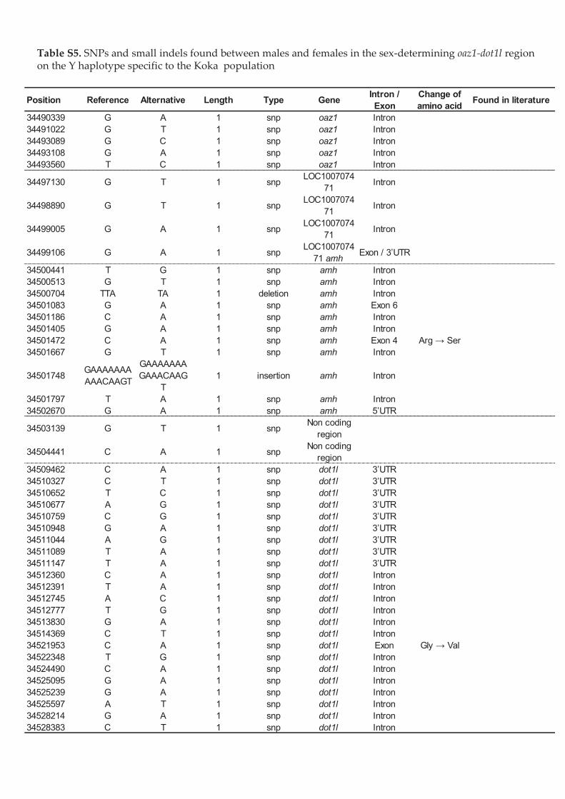
!"#$%&'-)&EFG,/-*4/,#-%%/'*4"%, 01$*4/@"&H""*/#-%",/-*4/0"#-%",/'*/&3"/,");4"&"2#'*'*+ !"#$%&!'$()2"+'1*/1*/&3"/I/3-(%1&D("/,(".'0'./&1/&3"/A1B- (1($%-&'1*/
Position Reference Alternative Length Type GeneIntron /
Exon
Change of
amino acidFound in literature
34490339 G A 1 snp oaz1 Intron
34491022 G T 1 snp oaz1 Intron
34493089 G C 1 snp oaz1 Intron
34493108 G A 1 snp oaz1 Intron
34493560 T C 1 snp oaz1 Intron
34497130 G T 1 snpLOC1007074
71Intron
34498890 G T 1 snpLOC1007074
71Intron
34499005 G A 1 snpLOC1007074
71Intron
34499106 G A 1 snpLOC1007074
71 amhExon / 3’UTR
34500441 T G 1 snp amh Intron
34500513 G T 1 snp amh Intron
34500704 TTA TA 1 deletion amh Intron
34501083 G A 1 snp amh Exon 6
34501186 C A 1 snp amh Intron
34501405 G A 1 snp amh Intron
34501472 C A 1 snp amh Exon 4 Arg → Ser
34501667 G T 1 snp amh Intron
34501748GAAAAAAA
AAACAAGT
GAAAAAAA
GAAACAAG
T
1 insertion amh Intron
34501797 T A 1 snp amh Intron
34502670 G A 1 snp amh 5’UTR
34503139 G T 1 snpNon coding
region
34504441 C A 1 snpNon coding
region
34509462 C A 1 snp dot1l 3’UTR
34510327 C T 1 snp dot1l 3’UTR
34510652 T C 1 snp dot1l 3’UTR
34510677 A G 1 snp dot1l 3’UTR
34510759 C G 1 snp dot1l 3’UTR
34510948 G A 1 snp dot1l 3’UTR
34511044 A G 1 snp dot1l 3’UTR
34511089 T A 1 snp dot1l 3’UTR
34511147 T A 1 snp dot1l 3’UTR
34512360 C A 1 snp dot1l Intron
34512391 T A 1 snp dot1l Intron
34512745 A C 1 snp dot1l Intron
34512777 T G 1 snp dot1l Intron
34513830 G A 1 snp dot1l Intron
34514369 C T 1 snp dot1l Intron
34521953 C A 1 snp dot1l Exon Gly → Val
34522348 T G 1 snp dot1l Intron
34524490 C A 1 snp dot1l Intron
34525095 G A 1 snp dot1l Intron
34525239 G A 1 snp dot1l Intron
34525597 A T 1 snp dot1l Intron
34528214 G A 1 snp dot1l Intron
34528383 C T 1 snp dot1l Intron


CHAPITRE 1 - DÉTERMINISME GÉNÉTIQUE DU SEXE DE DEUX POPULATIONS NATURELLES
Analyses complémentaires
Certaines analyses complémentaires ont été effectuées en complément de celles présentées
précédemment et ne figurent pas dans l’article publié. Ces analyses sont restées à un stade
préliminaire et n’ont pas permis d’aboutir à des résultats suffisamment concluants pour
donner lieu à une publication scientifique.
Cependant, certains aspects de ces études complémentaires donnent des indices
supplémentaires sur les faits présentés dans l’article et sont donc présentés succinctement ci-
après.
• Analyse ddRAD préliminaire de la population naturelle Kou (Burkina-Faso)
L’ensemble des échantillons utilisés pour ces analyses proviennent de collaborateurs du
projet ANR Climsex, Aboubacar Toguyeni et Rokyatou Sissao, qui ont conduit l’ensemble
des élevages et prélèvements de tissus ainsi que l’analyse de marqueurs PCR. Les banques
de ddRAD ont été produites conjointement par Helena D’Cotta et Rokyatou Sissao. Le
protocole utilisé est exactement le même que celui publié dans Triay et al., (2020).
Cette population est intéressante car d’après Sissao et al., 2019, le génotypage des
individus par les marqueurs PCR de l’amh et l’amh∆Y ne permet d’expliquer que près de 80%
des sex-ratios obtenus dans les descendances après des croisements en conditions contrôlés.
Il est attendu d’après Li et al., 2019 que la présence de l’amh∆Y implique forcément celle de
l’amhY puisque les deux copies sont en tandem. Cet agencement est également suggéré par
les analyses que nous avons conduits pour ce chapitre et publiés dans Triay et al., 2020,
même si elles sont en réalité sûrement séparées par des copies tronquées des gènes dot1l et
oaz1. Cependant, il existe des incongruences dans le génotypage des individus de la
population Kou, et certains individus peuvent amplifier l’amh∆Y mais pas l’amhY par
exemple, laissant un doute sur le génotype sexuel réel de ces individus. Ces résultats
surprenants semblent suggérer l’existence d’un second locus impliqué dans le déterminisme
du sexe chez cette population (Sissao et al., 2019). Ainsi, nous avons utilisé exactement la
même approche de ddRAD que dans Triay et al., 2020 sur une famille (D14) issue de la
population Kou (Lac Kou, Burkina-Faso). Pour le séquençage ont été utilisés : les deux
géniteurs dont une femelle XX et mâles XY d’après les génotypages avec les marqueurs PCR
à l’amh ainsi que 58 individus de la descendance. Parmi les descendants, 6 mâles ont été
119
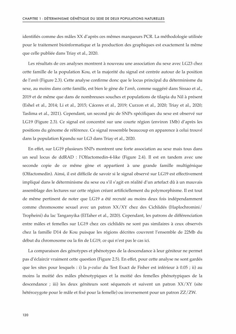
CHAPITRE 1 - DÉTERMINISME GÉNÉTIQUE DU SEXE DE DEUX POPULATIONS NATURELLES
identifiés comme des mâles XX d’après ces mêmes marqueurs PCR. La méthodologie utilisée
pour le traitement bioinformatique et la production des graphiques est exactement la même
que celle publiée dans Triay et al., 2020.
Les résultats de ces analyses montrent à nouveau une association du sexe avec LG23 chez
cette famille de la population Kou, et la majorité du signal est centrée autour de la position
de l’amh (Figure 2.3). Cette analyse confirme donc que le locus principal du déterminisme du
sexe, au moins dans cette famille, est bien le gène de l’amh, comme suggéré dans Sissao et al.,
2019 et de même que dans de nombreuses souches et populations de tilapia du Nil à présent
(Eshel et al., 2014; Li et al., 2015; Cáceres et al., 2019; Curzon et al., 2020; Triay et al., 2020;
Taslima et al., 2021). Cependant, un second pic de SNPs spécifiques du sexe est observé sur
LG19 (Figure 2.3). Ce signal est concentré sur une courte région (environ 1Mb) d’après les
positions du génome de référence. Ce signal ressemble beaucoup en apparence à celui trouvé
dans la population Kpandu sur LG3 dans Triay et al., 2020.
En effet, sur LG19 plusieurs SNPs montrent une forte association au sexe mais tous dans
un seul locus de ddRAD : l’Olfactomedin-4-like (Figure 2.4). Il est en tandem avec une
seconde copie de ce même gène et appartient à une grande famille multigénique
(Olfactomedin). Ainsi, il est difficile de savoir si le signal observé sur LG19 est effectivement
impliqué dans le déterminisme du sexe ou s’il s’agit en réalité d’un artefact dû à un mauvais
assemblage des lectures sur cette région créant artificiellement du polymorphisme. Il est tout
de même pertinent de noter que LG19 a été recruté au moins deux fois indépendamment
comme chromosome sexuel avec un patron XX/XY chez des Cichlidés (Haplochromini/
Tropheini) du lac Tanganyika (ElTaher et al., 2020). Cependant, les patrons de différenciation
entre mâles et femelles sur LG19 chez ces cichlidés ne sont pas similaires à ceux observés
chez la famille D14 de Kou puisque les régions décrites couvrent l’ensemble de 22Mb du
début du chromosome ou la fin de LG19, ce qui n’est pas le cas ici.
La comparaison des génotypes et phénotypes de la descendance à leur géniteur ne permet
pas d’éclaircir vraiment cette question (Figure 2.5). En effet, pour cette analyse ne sont gardés
que les sites pour lesquels : i) la p-value du Test Exact de Fisher est inférieur à 0.05 ; ii) au
moins la moitié des mâles phénotypiques et la moitié des femelles phénotypiques de la
descendance ; iii) les deux géniteurs sont séquencés et suivent un patron XX/XY (site
hétérozygote pour le mâle et fixé pour la femelle) ou inversement pour un patron ZZ/ZW.
120
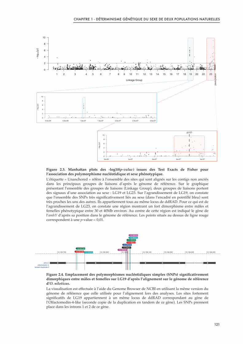
CHAPITRE 1 - DÉTERMINISME GÉNÉTIQUE DU SEXE DE DEUX POPULATIONS NATURELLES
Figure 2.3. Manhattan plots des -log10(p-value) issues des Test Exacts de Fisher pour l’association des polymorphisme nucléotidique et sexe phénotypique.L’étiquette « Unanchored » réfère à l’ensemble des sites qui sont alignés sur les contigs non ancrés dans les principaux groupes de liaisons d’après le génome de référence. Sur le graphique présentant l’ensemble des groupes de liaisons (Linkage Group), deux groupes de liaisons portent des signaux d’une association au sexe : LG19 et LG23. Sur l’agrandissement de LG19, on constate que l’ensemble des SNPs très significativement liés au sexe (dans l’encadré en pointillé bleu) sont très proches les uns des autres. Ils appartiennent tous au même locus de ddRAD. Pour ce qui est de l’agrandissement de LG23, on constate une région montrant un fort dimorphisme entre mâles et femelles phénotypique entre 30 et 40Mb environ. Au centre de cette région est indiqué le gène de l’amhY d’après sa position dans le génome de référence. Les points situés au dessus de ligne rouge correspondent à une p-value < 0,01.
Figure 2.4. Emplacement des polymorphismes nucléotidiques simples (SNPs) significativement dimorphiques entre mâles et femelles sur LG19 d’après l’alignement sur le génome de référence d’O. niloticus. La visualisation est effectuée à l’aide du Genome Browser de NCBI en utilisant la même version du génome de référence que celle utilisée pour l’alignement lors des analyses. Les sites fortement significatifs de LG19 appartiennent à un même locus de ddRAD correspondant au gène de l’Olfactomedin-4-like (seconde copie de la duplication en tandem de ce gène). Les SNPs prennent place dans les introns 1 et 2 de ce gène.
0
2
4
6
8
10
Linkage Group
−lo
g10(p)
1 2 3 4 5 6 7 8 9 10 11 12 13 14 15 16 17 18 19 20 22 23
unanchored
19 23
0
2
4
6
8
10
−lo
g1
0(p)
0e+00 1e+07 2e+07 3e+07 4e+07
amh
0
2
4
6
8
10
D14 − 27°C
−lo
g1
0(p)
0.0e+00 5.0e+06 1.0e+07 1.5e+07 2.0e+07 2.5e+07 3.0e+07
!"!#$"%&
!"'!#$'%""!"'!#$'(""!"'!#$'&""!"'!#$'!""!"'!#$)*!"'!#+'#""!"'!#+',""
-./0%1&)
-./234-05678)%')
428650)69:.73245)&
!"!#+,;;
!"!#+,#&
!"!#+;&#
!"!#$"&&
!"!#$"&,
!"!#$"&;
!"!#$"(!
!"!#$"(&
!"!#$"%!
121

CHAPITRE 1 - DÉTERMINISME GÉNÉTIQUE DU SEXE DE DEUX POPULATIONS NATURELLES
Or, pour LG19, seuls deux sites correspondent à ces critères, un pour le patron XX/XY et
l’autre pour ZZ/ZW. Il est donc impossible de tirer des conclusions en se basant sur ces deux
sites uniquement. En revanche, dans le cas de LG23, plus de sites sont disponibles.
L’interprétation pour ce groupe de liaison n’est cependant pas beaucoup plus claire car pour
le site le plus proche de l’amh, toutes les femelles possèdent le même génotype, et tous les
mâles (sauf les supposés XX d’après les marqueurs de l’amh) possèdent également le même
génotype ; seulement ceux-ci sont l’opposé des génotypes attendus. En effet, toutes les
femelles portent le génotype hétérozygote du père alors que les mâles portent le génotype
homozygote de la mère, et les mâles XX sont également porteurs du génotype paternel. Un
tel phénomène paraît difficile à expliquer biologiquement et il est possible qu’une erreur de
manipulation lors de la préparation de la banque soit en réalité la cause de ces patrons
surprenants.
Figure 2.5. Génotypes de la descendance par rapport à leurs parents inférés à partir des sites significatifs du FET du ddRAD.Les cases roses indiquent un individu ayant le même génotype que sa mère sur ce site tandis que les cases bleues foncées indiquent un individu ayant le même génotype que son père sur ce site et les cellules bleues claires indiquent un homozygote à l’allèle alternatif. Les cellules blanches correspondent à des données manquantes. Les sites conservés sont ceux pour lesquels i) la p-value du Test Exact de Fisher est inférieure à 0.05 ; ii) les deux parents sont séquencés et ils correspondent à un patron XX/XY pour LG23 et XX/XY ou ZZ/ZW pour LG19 ; iii) au moins la moitié des mâles et des femelles de la descendance ont été séquencés.
Comme nous n’avons pas eu le temps de re-séquencer une banque pour cette famille,
nous n’avons pas prolongé les analyses au-delà de ce qui a été présenté ici. Cependant, cette
analyse préliminaire confirme une forte association du sexe sur LG23 dans cette famille de la
population Kou. Des analyses complémentaires seront nécessaires pour comprendre
l’implication ou non de LG19 dans le déterminisme du sexe dans cette population. De plus, il
s’agit ici seulement d’une famille et il nécessaire de séquencer un plus grand nombre de
géniteurs et de familles afin de pouvoir asseoir l’existence ou non d’un déterminant mineur,
EL4K G4HFKGH D L :P4 :P: QPQ :P: 4P4 4P4 :P: :P: :P: QPQ :P: 4P4 QPQ QPQ QPQ QPQ QPQ QPQ :P4 4P4 QPQ :P: :P: :P: QPQ :P4 :P: :P: 4P4 4P4 4P4 QPQ :P: QPQ QPQ :P: :P4 4P4 4P4 QPQ 4P4 :P: 4P4 4P4 :P4 4P4 4P4 :P: :P: QPQ 4P4 QPQ QPQ QPQ QPQ :P4 QPQ :P: :P: :P4 NO
EL4K F:IJ5IKH 9 ? :P: QPQ 4P4 :P4 :P4 :P: 4P4 4P4 :P4 4P4 :P4 :P: :P4 QPQ :P4 :P: :P: 4P4 :P4 QPQ QPQ :P: :P4 QPQ :P4 :P4 QPQ :P4 :P4 :P: :P4 :P: QPQ :P4 QPQ QPQ :P: QPQ QPQ :P: QPQ :P4 :P: :P4 :P: :P: :P4 :P4 :P: :P4 :P: QPQ :P: :P: :P: QPQ QPQ :P4 :P4 :P: RS
ELFJ F:5:5JIH 9 ? :P: :P4 :P: :P4 :P4 :P4 :P4 :P: :P4 :P: :P: :P4 :P4 :P: :P4 :P: :P: :P: :P4 :P: :P4 :P: :P: :P: :P: :P4 :P: :P: :P: :P4 :P4 :P4 :P4 :P4 QPQ :P4 :P: :P4 :P4 :P: :P4 :P4 :P4 :P4 :P4 :P4 :P4 :P4 :P4 :P4 :P4 :P4 QPQ :P4 :P: :P4 :P4 :P4 :P: :P4
NO
ELFJ F:5:55J; L ? :P: :P4 :P: :P4 :P4 :P4 :P4 :P: :P4 :P: :P: :P4 :P4 :P: :P4 :P: :P: :P: :P4 :P4 :P4 :P: :P: :P: :P: :P4 :P: :P: :P: :P4 :P4 :P4 :P4 :P4 QPQ :P4 :P: :P4 :P4 :P: :P4 :P4 :P4 :P4 :P4 :P4 :P4 :P4 :P4 :P4 :P4 :P4 :P4 :P4 :P: :P4 :P4 :P4 :P: :P4
ELFJ F:5:5;;5 ? D :P: :P4 :P: :P4 :P4 :P4 :P4 :P: :P4 :P: :P: :P4 :P4 :P: :P4 :P: :P: :P: :P4 :P4 :P4 :P: :P: :P: :P: :P4 :P: :P: :P: :P4 :P4 :P4 :P4 :P4 QPQ :P4 :P: :P4 :P4 :P: :P4 :P4 :P4 :P4 :P4 :P4 :P4 :P4 :P4 :P4 :P4 :P4 :P4 :P4 :P: :P4 :P4 :P4 :P: :P4
ELFJ J:K555KF D L :P: :P: :P: :P4 :P4 4P4 QPQ :P: :P4 :P: :P: :P4 :P4 :P: :P: :P: QPQ :P: :P4 :P: QPQ QPQ :P: :P: :P: :P4 QPQ :P: QPQ :P4 QPQ QPQ :P: :P4 QPQ QPQ :P: QPQ :P4 :P: :P4 :P4 QPQ :P: :P4 QPQ 4P4 :P4 :P4 :P4 :P4 :P4 :P4 :P4 QPQ :P4 :P4 :P4 :P: :P4
ELFJ J45K;4F: 9 D :P: 4P4 :P4 :P: 4P4 QPQ :P4 QPQ 4P4 :P4 4P4 :P: :P: 4P4 :P4 :P4 QPQ 4P4 :P: 4P4 QPQ 4P4 QPQ QPQ 4P4 :P: :P4 QPQ 4P4 :P: :P: QPQ 4P4 :P: QPQ :P: :P4 :P: :P4 QPQ :P: :P: QPQ :P: :P: :P: :P: :P: 4P4 :P4 :P: :P: 4P4 :P: :P: :P: :P: :P4 :P: :P4
ELFJ J5FG;4FJ 9 ? :P: :P: :P: :P4 :P: :P4 :P: :P: :P4 :P: :P: :P: :P4 :P: :P4 :P: :P: :P: :P: :P: :P: :P: :P: :P: :P: :P4 :P: :P: :P: :P4 :P4 :P4 :P4 :P4 :P4 :P4 :P4 :P4 :P4 :P4 :P4 :P4 :P4 :P4 :P4 :P4 :P4 :P4 :P4 :P4 :P4 :P4 :P4 :P4 :P4 :P4 :P4 :P4 :P: :P4
ELFJ J555J;:5 9 ? :P: :P: :P: :P4 :P: :P4 :P: :P: :P4 :P: :P: :P: :P4 :P: :P4 :P: :P: :P: :P: :P: :P: :P: :P: :P: :P: :P4 :P: :P: :P: :P4 :P4 :P4 :P4 :P4 :P4 :P4 :P4 :P4 :P4 :P4 :P4 :P4 :P4 :P4 :P4 :P4 :P4 :P4 :P4 :P4 :P4 :P4 :P4 :P4 :P4 :P4 :P4 :P4 :P: :P4
ELFJ J;4F5HJJ L D :P: :P4 :P: :P4 :P: :P4 :P: :P: :P4 :P: :P: :P: :P4 :P: :P4 :P: :P: :P: :P: :P: :P4 :P: :P: :P: :P: :P4 :P: :P: :P: :P4 :P4 :P4 :P4 :P4 :P4 :P4 :P4 :P4 :P4 :P4 :P4 :P4 :P4 :P4 :P4 :P: :P4 :P4 :P4 :P4 :P4 :P4 :P4 :P4 :P4 :P4 :P4 :P4 :P: :P4
ELFJ J;FH4GIK 9 D :P: :P: :P: :P: :P: :P: :P: :P: :P4 :P: :P: :P: :P: :P: :P4 :P: :P: :P: :P: :P: :P: :P: :P: :P: :P: :P: :P: :P: :P: 4P4 4P4 :P: 4P4 :P: QPQ :P: :P: 4P4 :P: 4P4 4P4 :P4 :P: 4P4 4P4 :P: 4P4 :P4 :P4 4P4 :P: 4P4 4P4 :P4 :P4 4P4 :P: 4P4 :P: :P4
ELFJ JG545J5G ? 9 :P: :P: :P: :P4 :P: :P4 :P: :P: :P4 :P: :P: :P: :P4 :P: :P4 :P: :P: :P: :P: :P: :P4 :P: :P: :P: :P: :P4 :P: :P: :P: :P4 :P4 :P4 :P4 :P4 :P4 :P4 :P4 :P4 :P4 :P4 :P4 :P4 :P4 :P4 :P4 :P: :P4 :P4 :P4 :P4 :P4 :P4 :P4 :P4 :P4 :P4 :P4 :P4 :P: :P4
ELFJ JI;;HGFH D L :P4 :P4 :P4 QPQ :P4 :P: 4P4 :P4 :P4 :P4 4P4 4P4 :P: 4P4 :P4 :P4 4P4 4P4 :P4 4P4 :P: 4P4 :P4 :P4 4P4 :P: :P: 4P4 4P4 :P4 :P4 :P4 :P4 :P: :P4 :P: :P4 :P4 :P4 :P4 :P4 :P: :P4 :P4 :P4 :P4 :P4 :P: :P4 :P4 :P4 :P: :P4 :P: :P: :P4 :P: :P4 :P: :P4
ELFJ JKJJIF5H L ? :P: :P4 QPQ :P4 :P4 :P4 :P: :P: :P4 :P: :P: :P: :P4 :P: :P4 :P: :P: 4P4 :P: :P4 :P4 :P: :P4 :P: :P: :P: :P4 :P: :P: 4P4 :P: :P: 4P4 :P4 QPQ :P4 4P4 4P4 4P4 4P4 4P4 4P4 :P: 4P4 4P4 :P: 4P4 :P4 :P4 4P4 QPQ :P4 4P4 :P4 :P4 4P4 :P4 4P4 :P: :P4
Mâles XX d’après les marqueurs PCR à l’amh
IndividusSites
Site le plus proche de l’amh
> > > > > > > > > > > > > > > > > > > > > > > > > > > > > 7 7 7 7 7 7 7 7 7 7 7 7 7 7 7 7 7 7 7 7 7 7 7 7 7 7 7 7 7 7 >
?@AB> <BC A87 DE9 4 44 4F 4G 4H 4I F F: F4 FF F5 F; FG FH FI J: J4 JJ JH JK 5 5: 5F 55 ;4 ;F ;J G I 4: 4; 4K FJ FK J JF J5 J; JG JI 54 5J 5; 5G 5H 5I 5K ; ;: ;; GF G; GG H H: H4 IK K L7M7NN L>M>NOMère Père
122
CHAPITRE 1 - DÉTERMINISME GÉNÉTIQUE DU SEXE DE DEUX POPULATIONS NATURELLES
qui pourrait notamment expliquer l’existence de mâles XX à l’amh alors que les individus ont
été élevés en conditions contrôlées et à une température constamment inférieure à la
température de masculinisation du tilapia du Nil.
• Exploration de la composition du chromosome B
Afin de mieux caractériser le chromosome B suspecté d’être présent dans le pool mâles
Kpandu et le pool femelles Koka, des analyses complémentaires de biologie moléculaire ainsi
que de bioinformatique ont été effectuées.
La première curiosité à laquelle nous voulions répondre était de savoir si ce que nous
avions identifié comme un potentiel chromosome B présentait des régions similaires à celui
des chromosomes B des cichlidés Africain déjà identifiés et notamment celui de Metriaclima
lombardoi (qui agit comme un chromosome W, dominant sur le système XX/XY déjà existant).
Pour ce faire, nous avons appliqué la méthodologie de détection des régions de chromosome
B décrite dans Clark et al., 2018 qui se base sur les différentiels de couverture entre le pool
d’individus contenant le chromosome B et celui ne le contenant pas. En utilisant un test
binomial, et en tenant compte du nombre total de lectures dans les deux pools, le but est de
définir la probabilité que le nombre observé de lectures alignés à une position soit seulement
le fruit du hasard. Pour cette analyse, seules les régions de plus de 500kb ont été conservées.
Cinquante et un segments du génome chez Koka et 54 segments du génome chez Kpandu
ont été identifiés comme appartenant au chromosome B (Figure 2.6). Ces segments forment
un total de 41 régions distinctes chez Koka et 23 régions distinctes chez Kpandu (en sachant
que plusieurs segments peuvent appartenir à la même région). Parmi l’ensemble de ces
régions identifiées chez Koka et Kpandu, 16 sont communes aux deux populations et
présentent des caractéristiques extrêmement similaires de couverture et de fréquences
alléliques des SNPs. Aucun de ces 16 locus identifiés ne correspond à des séquences du
chromosome B de Metriaclima lombardoi.
Afin de pouvoir identifier combien d’individus sont porteurs de chromosomes B dans
chacun des pools, des analyses de biologie moléculaires ont été menées. Malheureusement,
malgré le design de nombreux couples de primers sur des régions spécifiques de nombreuses
régions B spécifiques, nous n’avons pas réussi à obtenir un couple capable d’amplifier les
régions correspondantes.
123

CHAPITRE 1 - DÉTERMINISME GÉNÉTIQUE DU SEXE DE DEUX POPULATIONS NATURELLES
Figure 2.6. Diagramme en barres du nombre de segments et du nombre de locus identifiés comme appartenant au chromosome B dans le pool de femelles Koka et le pool de mâles Kpandu utilisés dans Triay et al., 2020.Plusieurs segments peuvent appartenir à un même locus. Au total, 51 segments ont été identifiés chez Koka, appartenant à 41 locus différents et 54 segments appartenant à 23 locus différents ont été identifiés chez Kpandu
Ce problème est notamment dû au fait que les copies présentes sur le chromosome B et la
version originelle sur le A peuvent avoir accumulé beaucoup de divergences et rendent
fastidieux le design de primers. De plus, le chromosome B, s’il est en une seule copie comme
chez les autres cichlidés africain, ne peut pas recombiner et profiter du balayage sélectif.
Ainsi, ce chromosome est sujet à l’accumulation de mutations et de variants structuraux qui
peuvent être différents entre tous les paralogues d’un même gène sur ce chromosome. Or, en
utilisant les méthodes d’alignement sur un génome de référence, l’ensemble des lectures de
toutes les différentes copies viennent s’aligner sur l’unique copie existante dans le génome de
référence et créent ainsi un polymorphisme nucléotidique qui reflète en réalité les SNPs
trouvés possiblement sur différentes copies de gènes sur le B. De plus, les locus identifiés
comme appartenant au B et partagés entre les deux populations peuvent correspondre à des
régions répétées du génome et être confondus avec des segments de chromosome B
authentiques. Il est donc laborieux de designer des primers permettant d’amplifier des
régions spécifiques du chromosome B et la caractérisation en détails de cet élément génétique
demandera des optimisations à la fois de la méthodologie bioinformatique pour détecter
plus de régions possiblement appartenant au chromosome B ainsi que des optimisations à la
paillasse pour essayer d’identifier les individus du pool porteur du B et ainsi, peut-être se
donner une idée de la prévalence de ce chromosome B dans les populations sauvages.
0
15
30
45
60
KOKA KPANDU
31
10
23
41 Locus du B identifiés
Segments du B identifiées
124

CHAPITRE 1 - DÉTERMINISME GÉNÉTIQUE DU SEXE DE DEUX POPULATIONS NATURELLES
Références
Cáceres, G., López, M.E., Cádiz, M.I., Yoshida, G.M., Jedlicki, A., Palma-Véjares, R., Travisany, D., Díaz-Domínguez, D., Maass, A., Lhorente, J.P., et al. (2019). Fine mapping using whole-genome sequencing confirms anti-müllerian hormone as a major gene for sex determination in farmed Nile tilapia (Oreochromis niloticus L.). G3 (Bethesda) 9, 3213–3223.
Clark, F.E., Conte, M.A., and Kocher, T.D. (2018). Genomic characterization of a B chromosome in Lake Malawi cichlid fishes. Genes 9, 610.
Curzon, A.Y., Shirak, A., Dor, L., Zak, T., Perelberg, A., Seroussi, E., and Ron, M. (2020). A duplication of the Anti-Müllerian hormone gene is associated with genetic sex determination of different Oreochromis niloticus strains. Heredity 125, 317–327.
El Taher, A., Ronco, F., Matschiner, M., Salzburger, W., and Böhne, A. (2020). Dynamics of sex chromosome evolution in a rapid radiation of cichlid fishes (Evolutionary Biology). Unpublished. Available at http://biorxiv.org/lookup/doi/10.1101/2020.10.23.335596
Eshel, O., Shirak, A., Dor, L., Band, M., Zak, T., Markovich-Gordon, M., Chalifa-Caspi, V., Feldmesser, E., Weller, J.I., Seroussi, E., et al. (2014). Identification of male-specific amh duplication, sexually differentially expressed genes and microRNAs at early embryonic development of Nile tilapia (Oreochromis niloticus). BMC Genomics 15, 774.
Li, M., Sun, Y., Zhao, J., Shi, H., Zeng, S., Ye, K., Jiang, D., Zhou, L., Sun, L., Tao, W., et al. (2015). A tandem duplicate of anti-müllerian hormone with a missense SNP on the Y chromosome is essential for male sex determination in Nile tilapia, Oreochromis niloticus. PLOS Genetics 11, e1005678.
Sissao, R., D’Cotta, H., Baroiller, J.-F., and Toguyeni, A. (2019). Mismatches between the genetic and phenotypic sex in the wild Kou population of Nile tilapia Oreochromis
niloticus. PeerJ 7.
Taslima, K., Khan, M.G.Q., McAndrew, B.J., and Penman, D.J. (2021). Evidence of two XX/XY sex-determining loci in the Stirling stock of Nile tilapia (Oreochromis niloticus). Aquaculture 532, 735995.
Triay, C., Conte, M.A., Baroiller, J.-F., Bezault, E., Clark, F.E., Penman, D.J., Kocher, T.D., and D’Cotta, H. (2020). Structure and Sequence of the Sex Determining Locus in Two Wild Populations of Nile Tilapia. Genes 11, 1017.
125


CHAPITRE 2 : DIVERSITÉ ET PERTE DE L’HAPLOTYPE Y
ANCESTRAL EN POPULATIONS NATURELLES


CHAPITRE 2 - DIVERSITÉ ET PERTE DE L’HAPLOTYPE Y ANCESTRAL
Objectifs
Dans le chapitre précédent, nous avons conclu que le déterminisme du sexe chez le tilapia
du Nil en population sauvage est régi par une région dupliquée sur LG23 contenant deux
copies du gène de l’amh. Comme les deux populations étudiées précédemment sont
géographiquement éloignées, nous avons émis l’hypothèse que ce déterminisme sur LG23 Y
soit ancestral à la divergence des populations de l’Est et de l’Ouest de l’Afrique. Pour ce
chapitre l’objectif était donc de mieux caractériser l’haplotype LG23 Y. Pour passer outre les
limites des méthodes précédemment utilisées, nous avons utilisé des séquençages longs
fragments (Long Reads) afin de traverser l’ensemble de la structure complexe. De plus,
comme le déterminisme est a priori ancestral, nous avons choisi d’ajouter une seconde
population Ethiopienne à nos analyses, pour laquelle les marqueurs PCR de l’amh n’étaient
pas fiables.
Définir au mieux la structure de la région du déterminisme du sexe dans les populations
sauvages (similarité et différences) est essentiel pour pouvoir identifier des régions propices
au design de primers dans les populations sauvages et appréhender au mieux l’histoire
évolutive de ce déterminisme LG23-Y.
Introduction et contexte
Chez le tilapia du Nil, les analyses du déterminisme du sexe sur les souches domestiques
(commerciales ou de laboratoires) identifient le déterminisme du sexe sur différents groupes
de liaisons selon la souche étudiée. Ainsi, des associations génétiques au sexe phénotypique
ont été trouvées sur LG1 chez une souche commerciale des Etats-Unis ou encore sur la
souche Manzala-Stirling de l’Université de Stirling (Ecosse) par exemple (Gammerdinger et
al., 2014; Palaiokostas et al., 2015; Taslima et al., 2021). En revanche, aucun locus précis n’a
été proposé pour comme déterminant sur ce LG. D’autre part, des associations génétiques au
sexe ont été montrées sur LG23 dans la région du gène de l’Hormone Anti-Müllerienne (amh)
sur des stocks commerciaux d’Amérique Latine, sur la souche Manzala-Striling (en parallèle
de LG1, mais en plus faible fréquence), sur la souche GIFT, la souche Japonaise ou encore les
souches israélienne et belge originellement issues de la souche Manzala-Stirling (Eshel et al.,
2014; Li et al., 2015; Wessels et al., 2017; Cáceres et al., 2020; Taslima et al., 2020). Une forte
association au sexe est également montrée dans la population naturelle Kou du Burkina-
129
CHAPITRE 2 - DIVERSITÉ ET PERTE DE L’HAPLOTYPE Y ANCESTRAL
Faso, mais un second déterminant non identifié semble co-ségréger pour déterminer le sexe
(Sissao et al., 2019). Nous avons montré dans nos analyses précédentes de deux populations
naturelles que le déterminisme était lié à la présence d’une duplication du gène de l’amh sur
LG23-Y (Triay et al., 2020), comme suggéré sur la souche Japonaise par Li et al., (2015).
Cependant, sur la souche Japonaise, les deux copies de l’amh sur le Y sont divergentes, et
l’une d’entre elle présente un indel qui change le cadre de lecture et provoque un codon stop
prématuré (Li et al., 2015). Si dans la population sauvage de Kpandu (Ghana) nous avons
bien identifié cette copie tronquée de l’amh, en revanche, chez la population Koka (Ethiopie)
les deux copies semblent être relativement conservées (Triay et al., 2020). De plus, dans cette
étude précédente, nous avons pu définir les limites de cette duplication mâle-spécifique sur
LG23-Y. Cependant, si les séquençages de génomes complets en lectures courtes nous ont
permis de proposer un haplotype Y attendu, des doutes subsistaient sur la structure de cette
région, et notamment quant à l’ordre successif des deux copies de l’amh ou encore la position
des variants structuraux identifiés dans les gènes dupliqués.
Matériels et méthodes
Afin de préciser la structure de la duplication spécifique des mâles sur LG23-Y, nous
avons de nouveau étudié les deux populations naturelles utilisées auparavant : Kpandu (Lac
Volta, Ghana) et Koka (Lac Koka, Ethiopie). Comme les mâles de la population Koka
présentent la duplication dans la région liée au sexe sur LG23-Y alors que les marqueurs PCR
à l’amh ne permettaient pas de la mettre en évidence, nous avons ajouté une seconde
population Ethiopienne (Hora, Lac Hora) à notre analyse avec l’a priori de détecter à nouveau
cette duplication.
Trois séquençages de génomes complets en lectures longues (MinION Oxford Nanopore
Long reads) ont été effectués sur un mâle (F0) de chacune des populations sauvages. Cette
technologie permet d’obtenir des lectures suffisamment longues pour traverser plusieurs
régions qui resteraient litigieuses avec des lectures courtes. Comme nous nous attendions à
une duplication dans la région du sexe, utiliser des lectures longues permet de reconstruire
l’ordre des gènes sans faire face aux difficultés rencontrées dans Triay et al., 2020 (Figure 3.1).
Afin d’optimiser les temps d’assemblages, nous avons concentré notre analyse uniquement
sur la région du déterminisme du sexe identifiée préalablement sur LG23. Ainsi, un filtre a
130

CHAPITRE 2 - DIVERSITÉ ET PERTE DE L’HAPLOTYPE Y ANCESTRAL
été appliqué sur les lectures pour ne conserver que celles qui étaient composées des gènes
identifiés précédemment dans les haplotypes X et Y de LG23. Ces reads ont ensuite été
assemblés de deux manières : alignés à un génome de référence, soit l’haplotype Y de
référence (d’après l’haplotype Y attendu de Triay et al., 2020), soit le génome de référence
(XX) ; et assemblés de manière de novo afin de reconstruire la séquence du Y sans a priori sur
sa structure.
Figure 3.1. Intérêt du séquençage lectures longues (long reads) pour correctement assembler une région dupliquée.A) Une lecture courte peut être alignée sur les deux copies dupliquées. Les lectures longues à l’inverse traversent la totalité de la région dupliquée, ne laissant pas de doute quant à l’ordre des séquences. B) Exemple d’une lecture longue d’un mâle Koka séquence pendant cette thèse, contenant les deux copies du gène de l’amh et permettant de définir la structure de la région du sexe sur LG23-Y.
Aucun haplotype Y n’a pu être reconstruit avec ces méthodes pour le mâle Hora utilisé (cf.
Principaux résultats et discussion). Ce résultat surprenant pouvait être dû à 2 facteurs : soit le
mâle utilisé est un mâle XX spontané du milieu naturel, puisque nous avons utilisé des mâles
F0 ; soit les mâles Hora ne présentent pas (ou pas systématiquement) la duplication de la
Région
avec
duplication
Lecture longues
Co Co
Copie 1 Copie 2
Copie 1 Copie 2
Copie 1 Copie 2
Copie 1 Copie 2
Co
Lectures courtes? ?
!"#$%%&%&'&$
!"#$ !"# $
!"#$%
"&'$
!"#$%%&%&'&$
$ ()%%% ')%%% *)%%% +)%%% $%)%%% $()%%% $')%%% $*)%%% $+)%%% (%)%%%
A)
B)
131
CHAPITRE 2 - DIVERSITÉ ET PERTE DE L’HAPLOTYPE Y ANCESTRAL
région de l’amh sur LG23. Afin de pouvoir élucider ce fait, nous avons également produit un
séquençage de génome complet en lecture courtes pour pouvoir exploiter la méthode
exploratoire déjà utilisée dans Triay et al., 2020 et vérifier si l’on observe un signal spécifique
du sexe sur LG23 entre un pool de mâles phénotypiques et un pool de femelles
phénotypiques (tous F0). Les analyses bioinformatiques conduites sur ces données sont les
mêmes que celles publiées dans Triay et al., 2020, c’est à dire un alignement du génome
complet sur le génome de référence, puis une identification des sites polymorphiques et une
comparaison des signaux entre le pool de mâles et de femelles pour rechercher des régions
dimorphiques entre les deux sexes.
Enfin, des analyses phylogénétiques nucléaires, mitochondriales et spécifiques à la région
du sexe chez les femelles ont été réalisées. Pour ce faire, nous avons utilisé les séquençages
WGS du chapitre précédant ainsi que les lectures courtes de Hora produites pour ce chapitre
afin de définir les relations de parentés entre les trois populations.
Principaux résultats et discussion
i. Reconstruction de la duplication dans la région du sexe sur LG23-Y
Nous avons pu reconstruire l’haplotype Y chez les deux mâles Kpandu et Koka. Ces
résultats ont confirmé la structure attendue du Y décrite précédemment en détaillant l’ordre
des gènes au sein de cette duplication ainsi que les copies des gènes portant les indels mâles
spécifiques identifiés dans Triay et al., 2020.
D’une part ces résultats confirment à nouveau l’existence d'un système XX/XY sur LG23
avec une duplication du gène de l'amh. De plus, ils permettent de constater que les mâles
Kpandu ont, dans cette région, une structure très similaire à celle décrite par Li et al., 2015,
avec la présence d’une copie conservée de l’amh (amhY) et une copie tronquée (amh∆Y). Chez
Koka en revanche, les deux copies de l’amh semblent conservées. Nous avons pu confirmer
également que l’amh∆Y se situe en amont de l’amhY (dans le sens de la transcription), séparés
par une chimère composée de copies tronquées des gènes oaz1 et dot1l. Cependant, la
manière dont cette région régit le déterminisme du sexe reste incomprise car nous n’avons
pas pu exploiter la présence de variants nucléotidiques du fait d’une trop faible couverture
des données de MinION.
132

CHAPITRE 2 - DIVERSITÉ ET PERTE DE L’HAPLOTYPE Y ANCESTRAL
ii. Une perte du déterminant ancestral (LG23 XX/XY) chez les mâles Hora
Nous avons pu mettre en évidence que les mâles de la population Hora ne présentent pas
la duplication de la région de l’amh sur LG23-Y, contrairement aux mâles des populations
Koka et Kpandu. De plus, aucun polymorphisme nucléotidique n’a été identifié entre les
mâles et les femelles sur l’amh suggérant que cette région ne détermine pas le sexe chez la
population Hora. Les analyses phylogénétiques ont pourtant confirmé que les individus
Hora et Koka sont plus proches entre eux qu’ils ne le sont des individus de la population
Kpandu. Ce résultat était attendu puisque les populations Hora et Koka appartiennent à la
sous-espèce O. niloticus cancelatus et sont proches géographiquement alors que la population
Kpandu est une population d’O. niloticus niloticus et que des analyses de données de
microsatellites insinuaient déjà de telles relations (Bezault et al︎., 2011). Le fait que les mâles
Koka et Kpandu présentent le déterminant LG23-Y suggère que ce déterminant est ancestral
à la divergence de ces populations. Ainsi, nous avons interprété l’absence du déterminisme
LG23-Y chez Hora comme une perte de ce déterminant ancestral.
ii.a. LG23, une paire de chromosomes sexuels «!ever-young!»
Si le déterminisme LG23 XX/XY de Kpandu et Koka est certainement ancestral à la
divergence des populations, les haplotypes X et Y ne sont pour autant pas très divergents.
Nous avons montré que la différence entre les deux chromosomes pour la région spécifique
du sexe est de moins de 10kb. Ce résultat, ainsi que l’observation de recombinaisons proches
de la région de l’amh dans les données de ddRAD du premier chapitre (Triay et al., 2020)
semblent indiquer que les chromosomes X et Y sont encore très similaires. Dans l’article nous
discutons donc de la possibilité qu’il s’agit de chromosomes sexuels qui peu importe l’âge
n’entament pas de processus de différenciation (arrêt de la recombinaison, diminution du
contenu en gène etc…), et nous proposons quelques mécanismes et processus qui pourraient
mener à un tel phénomène.
ii.b. Pas d’indice du déterminant ayant pris le relais
Aucun autre groupe de liaison n’a pu être identifié comme potentiel chromosome sexuel
chez Hora avec les analyses menées jusqu’à présent. Nous discutons donc dans cet article
deux questions : pourquoi la population Hora a-t-elle connu un turnover du déterminisme
du sexe ? Et quel est le système actuel régissant le déterminisme du sexe dans cette
population ? Même si nous n’avons pas de piste concrète actuellement, les possibilités
133

CHAPITRE 2 - DIVERSITÉ ET PERTE DE L’HAPLOTYPE Y ANCESTRAL
écologiques et évolutives pouvant mener à un changement de déterminisme du sexe dans
cette population sont envisagées. Nous discutons également des limites méthodologiques
qui pourraient expliquer que nous n’ayons pas trouvé de signal clair sur un autre groupe de
liaison avec les approches utilisées dans l’article.
ii.c. Diversité intraspécifique du déterminisme du sexe en milieu naturel
La diversité du déterminisme du sexe dans les souches domestiques de tilapia du Nil et
maintenant la démonstration de sa variabilité dans les populations naturelles avec les
publications de Sissao et al., (2019) et Triay et al., (2020 et actuelle) démontrent l’importance
d’étudier les systèmes de déterminisme à l’échelle des populations. En effet, comme suggéré
par Furman et al., (2020), la diversité des chromosomes sexuels est peut-être parfois aussi
importante au niveau intraspécifique qu’interspécifique.
iii. Une introgression entre O. niloticus et son espèce sœur O. aureus
Enfin, nos analyses phylogénétiques ont permis de mettre en évidence une incongruence
entre le signal nucléaire et le signal mitochondrial. La position de la population de l’Ouest
Africain (Kpandu) varie entre les arbres : les données mitochondriales rapprochent les
individus Kpandu d’O. niloticus de l’espère sœur O. aureus. Dans la phylogénie nucléaire en
revanche les trois populations sauvages de O. niloticus forment un groupe monophylétique.
Ces résultats vont dans le sens de ceux de Rognon & Guyomard (2003), qui avaient émis
l’hypothèse d’une introgression complète du génome mitochondrial de O. aureus vers O.
niloticus dans les populations d’Afriques de l’ouest et des lacs égyptiens.
Conclusion
Dans cet article nous avons défini en détails la structure (taille, ordre des gènes,
positionnement des indels identifiés préalablement comme spécifiques des mâles) de la
région du déterminisme du sexe chez le tilapia du Nil sur LG23-Y. Nous avons également
établi les relations de parenté entre les trois populations sauvages, suggérant une origine
ancestrale du déterminisme XX/XY de LG23. Curieusement, nous avons montré que les
deux populations éthiopiennes, malgré qu’elles soient proches parents ne présentent pas le
même système de déterminisme que les populations Koka et Kpandu pourtant plus
éloignées phylogénétiquement. Nous interprétons cette absence de LG23-Y chez Hora
134

CHAPITRE 2 - DIVERSITÉ ET PERTE DE L’HAPLOTYPE Y ANCESTRAL
comme une perte du déterminant ancestral, mais n’avons cependant pas pu identifier de
locus ni même de groupe de liaison en lien avec le sexe chez cette population.
Enfin nous avons confirmé une introgression mitochondriale entre O. aureus et O. niloticus.
135

CHAPITRE 2 - DIVERSITÉ ET PERTE DE L’HAPLOTYPE Y ANCESTRAL
Références
Bezault, E., Balaresque, P., Toguyeni, A., Fermon, Y., Araki, H., Baroiller, J.-F., and Rognon, X. (2011). Spatial and temporal variation in population genetic structure of wild Nile tilapia (Oreochromis niloticus) across Africa. BMC Genetics 12, 102.
Cáceres, G., López, M.E., Cádiz, M.I., Yoshida, G.M., Jedlicki, A., Palma-Véjares, R., Travisany, D., Díaz-Domínguez, D., Maass, A., Lhorente, J.P., et al. (2019). Fine mapping using whole-genome sequencing confirms anti-müllerian hormone as a major gene for sex determination in farmed Nile tilapia (Oreochromis niloticus L.). G3 (Bethesda) 9, 3213–3223.
Carrasco, L.A.P., Penman, D.J., and Bromage, N. (1999). Evidence for the presence of sex chromosomes in the Nile tilapia (Oreochromis niloticus) from synaptonemal complex analysis of XX, XY and YY genotypes. Aquaculture 173, 207–218.
Cnaani, A., Lee, B.-Y., Zilberman, N., Ozouf-Costaz, C., Hulata, G., Ron, M., D'Hont, A., Baroiller, J.-F., D'Cotta, H., Penman, D.J., et al. (2008). Genetics of sex determination in tilapiine species. Sexual Development 2, 43–54.
Eshel, O., Shirak, A., Weller, J.I., Hulata, G., and Ron, M. (2012). Linkage and physical mapping of sex Rregion on LG23 of Nile tilapia (Oreochromis niloticus). G3: Genes, Genomes, Genetics 2, 35–42.
Eshel, O., Shirak, A., Dor, L., Band, M., Zak, T., Markovich-Gordon, M., Chalifa-Caspi, V., Feldmesser, E., Weller, J.I., Seroussi, E., et al. (2014). Identification of male-specific amh duplication, sexually differentially expressed genes and microRNAs at early embryonic development of Nile tilapia (Oreochromis niloticus). BMC Genomics 15, 774.
Furman, B.L.S., Metzger, D.C.H., Darolti, I., Wright, A.E., Sandkam, B.A., Almeida, P., Shu, J.J., and Mank, J.E. (2020). Sex chromosome evolution: so many exceptions to the rules. Genome Biol Evol 12, 750–763.
Gammerdinger, W.J., Conte, M.A., Acquah, E.A., Roberts, R.B., and Kocher, T.D. (2014). Structure and decay of a proto-Y region in Tilapia, Oreochromis niloticus. BMC Genomics 15, 975.
Jalabert, B., Moreau, J., Planquette, P., and Billard, R. (1974). Déterminisme du sexe chez tilapia macrochir et tilapia nilotica : action de la méthyltestostérone dans l’alimentation des alevins sur la différenciation sexuelle; proportion des sexes dans la descendance des mâles «inversés». Annales de Biologie Animale Biochimie Biophysique Vol. 14, 729–739.
Lee, B.-Y., Penman, D.J., and Kocher, T.D. (2003). Identification of a sex-determining region in Nile tilapia (Oreochromis niloticus) using bulked segregant analysis. Animal Genetics 34, 379–383.
Lee, B.-Y., Coutanceau, J.-P., Ozouf-Costaz, C., D’Cotta, H., Baroiller, J.-F., and Kocher, T.D. (2011). Genetic and physical mapping of sex-linked AFLP markers in Nile tilapia (Oreochromis niloticus). Mar Biotechnol (NY) 13, 557–562.
Li, M., Sun, Y., Zhao, J., Shi, H., Zeng, S., Ye, K., Jiang, D., Zhou, L., Sun, L., Tao, W., et al. (2015). A tandem duplicate of anti-müllerian hormone with a missense SNP on the Y
136
CHAPITRE 2 - DIVERSITÉ ET PERTE DE L’HAPLOTYPE Y ANCESTRAL
chromosome is essential for male sex determination in Nile tilapia, Oreochromis niloticus. PLOS Genetics 11, e1005678.
Palaiokostas, C., Bekaert, M., Khan, M.G.Q., Taggart, J.B., Gharbi, K., McAndrew, B.J., and Penman, D.J. (2013). Mapping and validation of the major sex-determining region in Nile tilapia (Oreochromis niloticus L.) using RAD sequencing. PLoS ONE 8, e68389.
Palaiokostas, C., Bekaert, M., Khan, M., Taggart, J.B., Gharbi, K., McAndrew, B.J., and Penman, D.J. (2015). A novel sex-determining QTL in Nile tilapia (Oreochromis niloticus). BMC Genomics 16, 171.
Pfennig, F., Standke, A., and Gutzeit, H.O. (2015). The role of Amh signaling in teleost fish – Multiple functions not restricted to the gonads. General and Comparative Endocrinology 223, 87–107.
Poonlaphdecha, S., Pepey, E., Huang, S.-H., Canonne, M., Soler, L., Mortaji, S., Morand, S., Pfennig, F., Mélard, C., Baroiller, J.F., et al. (2011). Elevated amh Gene Expression in the Brain of Male Tilapia (Oreochromis niloticus) during Testis Differentiation. Sex Dev 5, 33–47.
Rognon, X., and Guyomard, R. (2003). Large extent of mitochondrial DNA transfer from Oreochromis aureus to O. niloticus in West Africa. Mol Ecol 12, 435–445.
Sissao, R., D’Cotta, H., Baroiller, J.-F., and Toguyeni, A. (2019). Mismatches between the genetic and phenotypic sex in the wild Kou population of Nile tilapia Oreochromis niloticus. PeerJ 7.
Taslima, K., Wehner, S., Taggart, J.B., de Verdal, H., Benzie, J.A.H., Bekaert, M., McAndrew, B.J., and Penman, D.J. (2020). Sex determination in the GIFT strain of tilapia is controlled by a locus in linkage group 23. BMC Genet 21, 49.
Taslima, K., Khan, M.G.Q., McAndrew, B.J., and Penman, D.J. (2021). Evidence of two XX/XY sex-determining loci in the Stirling stock of Nile tilapia (Oreochromis niloticus). Aquaculture 532, 735995.
Triay, C., Conte, M.A., Baroiller, J.-F., Bezault, E., Clark, F.E., Penman, D.J., Kocher, T.D., and D’Cotta, H. (2020). Structure and Sequence of the Sex Determining Locus in Two Wild Populations of Nile Tilapia. Genes 11, 1017.
Wessels, S., Krause, I., Floren, C., Schütz, E., Beck, J., and Knorr, C. (2017). ddRADseq reveals determinants for temperature-dependent sex reversal in Nile tilapia on LG23. BMC Genomics 18, 531.
137


PUBLICATION ASSOCIÉE


Article (unpublished) /1 36
Polymorphism of Y haplotypes or loss of ancestral LG23 sex determinant in three wild populations of Nile tilapia
Cécile Triay 1,2, Maxime Courcelle 1, Pierre Caminade 1, Etienne Bezault 3, Jean-François
Baroiller 1,2, Thomas D. Kocher 4 & Helena D’Cotta 1,2
1. UMR-Institut des Sciences de l’Evolution de Montpellier, Centre National de la Recherche Scientifique,
Institut de Recherche pour le Développement, Ecole Pratique des Hautes Etudes, University of Montpellier,
34090 Montpellier, France; [email protected] (C.T), [email protected] (M.C);
[email protected] (J-F.B.); [email protected] (H.D)
2. UMR-Institut des Sciences de l’Evolution de Montpellier, Centre de Coopération Internationale en
Recherche Agronomique pour le Développement, Campus Int. Baillarguet, 34398 Montpellier, France
3. UMR BOREA, CNRS-7208/MNHN/UPMC/IRD-207/UCN/UA, Université des Antilles, 97159
Guadeloupe, France; [email protected]
4. Department of Biology, University of Maryland, College Park, MD 20742, USA; [email protected] (T.D.K.)
Abstract: Sex-determining regions have been identified on linkage groups (LG) 1 and 23 in
the Nile tilapia, depending on the domesticated strains used. Sex determining studies on
wild populations are extremely scarce. We previously worked on two wild populations
originating from Lake Volta (Ghana) and from Lake Koka (Ethiopia) and showed that LG23
also carries the sex-determining region in these wild populations, and more precisely they
have a Y tandem duplication containing two copies of the Anti-Müllerian Hormone amh
gene (named amhY and amh∆Y). Here, we extend our focus on this Y chromosome
duplicated region in the two populations of Lake Volta and Lake Koka and added a new
Ethiopian population to the analysis, from Lake Hora. Using long reads of whole genome
sequencing of a male from each population, we confirmed the duplicated structure, its
breakpoints as well as genes’ order on the Y chromosome of LG23 in both Lake Volta and
Lake Koka populations. We did not, however, identify the LG23 Y haplotype in the Hora
male. A whole-genome short-reads analysis, carried out between pools of phenotypic males
and females of Hora confirmed the absence of a Y amh duplication on LG23 Y. The
phylogenies conducted on the nuclear and mitochondrial genomes, nevertheless show a
closer relationship between the two Ethiopian populations compared to the Ghanaian
population. In lake Hora population the absence of the LG23Y sex-determination region is
interpreted as a loss. The potential reasons for this loss and the implications it may have on
the evolution of sex chromosomes are discussed in this paper.
Keywords: Nile tilapia; Sex chromosomes; amh; Duplication; Y-haplotype

Article (unpublished) /2 36
1. Introduction
Sex determination (SD) initiates the development of males or females. Genetic sex
determination (GSD) inherited through sex chromosomes has been highly conserved and
stable in mammals and birds for millions of years (Cortez et al., 2014). In mammals, sex
chromosomes are heteromorphic and follow a XX/XY system. The degenerated Y
chromosome carries in most mammals the Sry gene, the master gene that triggers the
development of male gonads (Sinclair et al., 1990). In birds, ZW embryos become females.
The W chromosome is also small in carinate birds but testis develop in the presence of a
double dose of the dmrt1 gene on the Z chromosome (Smith et al., 2009; Graves, 2014).
However, SD stability does not appear to be the norm in vertebrates (Bachtrog et al., 2014).
Reptiles, amphibians and fish have a remarkable diversity of sex chromosomes,
heterogamety and sex-determination genes found even among closely related species (Ezaz
et al., 2009; Myosho et al., 2015; Jeffries et al., 2018). In some of these vertebrates, sex can be
triggered by environmental cues (Environmental Sex Determination ESD) such as
temperature (e.g. reptiles), pH or even density, or through a combination of both GSD and
the environment which is the case in many teleost fishes (Mank and Avise, 2009; Baroiller
and D’Cotta, 2016).
Sex chromosomes do not evolve like autosomes. They undergo differential sexual forces
in the rates of mutations, selection and recombination (Bachtrog et al., 2011). The classical
canonical model based mostly on the XY system of mammals considers that a sex
chromosome originated on an autosome pair that acquired a sex-determining mutation. This
sex-locus, particularly when close to male-beneficial alleles that are detriment for females
(sexually antagonistic alleles), is expected to spread in the population because of their Y
linkage and therefore, transmitted to sons and not daughters (Rice, 1987; Charlesworth et al.,
2005; van Doorn and Kirkpatrick, 2010). Suppressed recombination will isolate this sex-
limited region, which expands by mechanisms such as accumulation of mutations,
inversions, repetitive elements, deletions and insertions (Kirkpatrick, 2010; Bachtrog, 2013;
Blaser et al., 2014). Over time the differences between the X and Y are such that they become
morphologically distinct, reaching a degree of degeneration where turnover is no longer
possible (Bachtrog, 2013; Abbott et al., 2017).
In contrast, 90% of teleost fishes have homomorphic sex chromosomes (Devlin and
Nagahama, 2002), implying that recombination suppression is probably recent. Differences
between sexes can be reduced to a single missense SNP such as in pufferfish (Kamiya et al.,
2012). The identification of undifferentiated sex chromosomes which have similar gene
content is however a challenging process. Genomic sequencing has shed light in fish
lineages evidencing the diversity of sex systems and sex chromosomes. A striking
characteristic of teleost sex chromosomes is their rapid turnover. There are transitions in the
sex chromosome system (Roberts et al., 2009; El Taher et al., 2020) even within the same
genus (Cnaani et al., 2008; Takehana et al., 2008; Böhne et al., 2019). Furthermore, many sex-
determining genes have been conserved but translocated into different chromosomes

Article (unpublished) /3 36
(Guiguen et al., 2018) or have emerged in a short evolutionary time with switches in the sex
chromosome pair (Gammerdinger and Kocher, 2018). In ricefishes, the sex-determination
gene dmY (also called dmrt1bY) was found on Linkage Group (LG) 1 in the medaka Oryzias
latipes (Nanda et al., 2002), whereas in O. luzenensis it is a Y-copy of the gsdf gene located on
LG12 (Myosho et al., 2012) and in another Oryzias it is sox3 on LG10 (Takehana et al., 2014).
Turnovers in sex chromosomes are remarkable in the large Cichlid family that has
undergone adaptive radiation, where over 20 sex-determinants have been identified on 17
different LGs as well as on B chromosomes, with the turnover driven possibly by sexual
antagonistic selection (Gammerdinger and Kocher, 2018; El Taher et al., 2020). Some sex
chromosomes appeared multiple times (e.g. LG5, LG7 and LG19) (Böhne et al., 2019; El Taher
et al., 2020). Differentiation of the sex chromosomes can be small, or comprise the whole LG
which suggests widespread recombination suppression (El Taher et al., 2020). Some “usual
suspects” are found amongst the master genes and they tend to play musical chairs to
endorse the role of major sex-determinant (Böhne et al., 2016).
The high diversity observed in teleost fish (approximately 27,000 living species) has been
proposed to be related to the genomic plasticity of this clade, matching their extensive
ecology (Helfman et al., 2009). Fishes have a higher rate of evolution and gene duplications
than other vertebrates (Mank and Avise, 2009). Indeed, gene duplications are a substrate for
the evolution of innovations by giving the duplicated copy (paralog) the possibility to
complete the function of the original gene (subfunctionalization), or by acquiring a new
function (neofunctionalization) (Sandve et al., 2018). Several sex-determining genes of
teleosts have emerged from duplications of sex-differentiating downstream genes. The
duplication of a gene already involved in the sex determination pathway can thus result in a
new locus that can potentially carry mutations significantly increasing the fitness of one of
the sexes and thus become a major determinant (Mank and Avise, 2009; Ortega-Recalde et
al., 2020). Dmy was duplicated from the male-differentiating dmrt1 gene found on the
autosomal LG9, that was translocated to LG1 in the medaka (Matsuda et al., 2002; Nanda et
al., 2002). In the Patagonian pejerrey (Odontesthes hatcheri) and the Northern pike (Esox
lucius) duplicated copies of the amh gene have taken on the role of master sex-determinant
(Hattori et al., 2012; Pan et al., 2019). Duplication of genes outside the sex pathway can also
result in a major sex-determinant such as the sdy gene in salmonids that is a
neofunctionalized paralog of an immune-related gene (irf9) (Yano et al., 2012).
The Nile tilapia is a XX/XY farmed cichlid where the GSD can be overridden by
temperature generating XX males (Baroiller et al., 2009). This has complicated the finding of
the sex chromosomes added by the use of intermixed domesticated strains. In these strains
sex has been linked either to LG1 (Cnaani et al., 2008; Gammerdinger et al., 2014;
Palaiokostas et al., 2015) or LG23 (Eshel et al., 2014; Li et al., 2015; Wessels et al., 2017;
Cáceres et al., 2019) or both (Taslima et al., 2021). No causal gene has been found on LG1. In
the Japanese domesticated strain of Nile tilapia, the amh gene on LG23 has been identified as
the sex-determination gene, which evolved through a tandem duplication within the same
LG. Hence, males have three copies of the amh gene, two copies located in tandem on the Y

Article (unpublished) /4 36
chromosome, one with a missense SNP (amhY) and the other theoretically with an insertion
that causes a premature stop codon (amh∆Y), and the third copy on the X chromosome (Li et
al., 2015). We found that sex in various wild populations was also linked to LG23 and to the
amh gene (Sissao et al., 2019; Triay et al., 2020). The Y chromosome sex-region which contains
this amh duplication was estimated to be ~51 kb using short reads from Whole Genome
sequencing (WGS) and comprised the adjacent oaz1 and dot1l genes (Triay et al., 2020).
However, we were not able to know how the duplication was inserted because there were
two possible scenarios and the order of the genes within the duplication. We also found
major differences within the duplicated region and in the amh∆Y copy between the two
populations compared with domesticated strains. Amh∆Y in the Ethiopian Koka population
seemed to lack the 5 bp insertion which causes premature stop codon (Triay et al., 2020). In
addition, the amh markers we used for genotyping the sex did not discriminate between XX
and XY individuals in the Ethiopian population (Sissao et al., 2019; Triay et al., 2020).
Molecular and genomic analyses have helped to better detect sex linked loci and identify
sex chromosomes, particularly in facilitating the reconstruction of Y or W chromosomes
(Abbott et al., 2017). Calibration and choice of different methods of new generation
sequencing (NGS) technologies are aiming to better detect sex chromosomes depending on
the divergence between the two sex chromosomes, from strongly heteromorphic to
homomorphic (Palmer et al., 2019). However, the reconstruction of a duplicated region with
short reads is tricky if two homologous paralogs exist, and even more when working with
reads that are shorter than the similar regions.
Long reads, if they allow a better description of a duplicated structure, are still prone to
sequencing error with rates up to 5-15% (Carneiro et al., 2012; Menegon et al., 2017). To
counter this issue and still be able to interpret SNPs, the coverage has to be high enough to
compensate for uncertainties and sequencing errors. Moreover, usual assembly tools are not
developed specifically for sex chromosomes and thus neither mapping methods nor de novo
assemblies are optimized to reconstruct regions specific to the heterogametic sex.
In this study, we wanted to define more accurately the structure and sequences of the
duplicated sex-region on the Y chromosome, and compare the differences between
populations, which would also allow sexing of Ethiopian wild individuals for population
genetics. To build the Y haplotypes, we performed long reads Nanopore sequencing on
males from two East African populations (originating from Lake Koka and Lake Hora in
Ethiopia) and one West African population (originating from Lake Volta in Ghana). We
expected the Hora male to be similar to the Koka male. However, the Lake Hora male
appeared to lack the Y amh duplication. This could be due to this male being an XX male.
Therefore, we performed WGS on several Hora males and females to study the SD and see
whether males lacked the amh duplication on LG23. We confirmed that Hora males did not
have the amh duplication thus looked for new sex-association in the whole genome of these
samples.
Article (unpublished) /5 36
2. Material and Methods
2.1. Sample collection
2.1.1. Fish samples and DNA extractions
Three wild populations of Nile tilapia Oreochromis niloticus were caught in 2002 and
2003 from the Sudano-Sahelian basin in West Africa and the Ethiopian Rift Valley basin of
East Africa (Bezault, 2005). Lake Koka and Lake Hora populations belong to the endemic O.
niloticus cancellatus subspecies and were collected from two cold-temperature (between 17°C
and 26°C) reservoirs of the Awash River in the Ethiopian highlands. Lake Koka is a large
artificial reservoir of the Awash River of 255 km2 with an average depth of 9 m while Lake
Hora is an isolated lake of about 1 km2 with a depth of up to 35 m. The Kpandu population
on the other hand belong to the O. niloticus niloticus subspecies captured in Ghana from a
dentritic expansion of the eastern side of Lake Volta (8,482 km2 with an average depth of 19
m) where temperatures fluctuate between 27°C to 32°C. Fully mature fish were collected
using several seine net captures and the phenotypic sex was then defined. Fish were
anaesthetized with 2-phenoxyethanol to sample the fin clips which were preserved in
96-100% ethanol.
High-molecular weight genomic DNA (gDNA) was lysed from the fin clips in TNES-Urea
buffer (4 M urea; 10 mM Tris-HCl, pH 7.5; 125 mM NaCl; 10 mM EDTA; 1% SDS) and
extracted using the phenol-chloroform procedure (Barker, 1998). The gDNA was stored in
Tris-EDTA buffer at -20°C. DNA was quantified first on a NanoDrop spectrophotometer
(ThermoFisher, France) to estimate the absorbance ratios and the concentration was then
measured on a Qubit 2.0 fluorometer (Invitrogen Carlsbad, USA). gDNA quality was also
verified on a 0,8% agarose gel.
2.1.2. Ethics Statement
Fish captures complied with the veterinary laws of each country as stipulated in 2003. All
animal and experimental procedures were performed in accordance with the French
protocol N°2016101810463 and the French Government’s authorization for animal
experimentation N° 35-15.
2.2. Whole genome short reads sequencing analysis of Hora
2.2.1. Libraries & sequencing
Twenty WGS libraries were prepared at Macrogen (Seoul, Korea) from ten males and ten
females’ gDNA which were sonicated to obtain 550 bp fragments. All libraries were
prepared using the TruSeq DNA PCR-Free kit (Illumina) except one male library where

Article (unpublished) /6 36
TruSeq Nano DNA Kit (Illumina) was used due to limited amounts of gDNA. Libraries were
2 x 150 bp paired-end sequenced on a Illumina NovaSeq 6000 (Flowcell S4).
2.2.2. Data processing
Raw data quality was checked using FastQC (0.11.7). Illumina TruSeq adapters were
trimmed and short reads (< 15bp) were removed using Fastp (version 0.21.0). Samples were
pooled according to their phenotypic sex, resulting in a pool of females (with 10 samples)
and a pool of males (with 10 samples). The two pools were mapped to the latest O. niloticus
reference genome available on NCBI (O_niloticus_UMD_NMBU, Genbank ID:
GCA_001858045.3) using BWA mem mapper (version 0.7.15). Sam files were then converted
to Bam files and sorted using SAMtools (version 1.9) and read groups were added to the two
pools using Picard AddOrReplaceReadGroups (2.7.0). The output files were then combined
with SAMtools mpileup (version 1.9). Using Popoolation2 mpileup2sync.pl (version 1.2.2),
the mpileup file was transformed to a synchronized file (sync). Finally, the sync file was
processed with Sex_SNP_finder_GA.pl to obtain Fst between males and females. This
pipeline allows to identify sites that are fixed (or nearly fixed) in one sex and at intermediate
frequencies in the other sex. The following parameters were used in Sex_SNP_finder:
fixed_threshold = 0.9; minimum_polymorphic_frequency = 0.3; maximum_polymorphic_frequency =
0.7; minimum_read_depth = 10; maximum_read_depth = 100; minimum_read_count = 2;
sex_SNP_finder_window_size = 10000. Finally, the Fst were plotted on a manhattan plot using
R and the qqman package (version 0.1.4).
2.2.3. Library construction for MinION sequencing
One Kpandu male, one Koka male and one Hora male were used for the MinION
sequencing (Oxford Nanopore Technology). The male genomic DNA libraries were
constructed with 3 to 4 µg of high molecular weight gDNA by MGX (Montpellier, France)
using the Ligation Sequencing Kit 1D from (ONT). Samples were fragmented, the
extremities repaired and adapters ligated. The flow cell FLO-MIN106 (flow cell 9.4.1) was
charged twice, with a DNase treatment performed on the flow cell to reactivate the pores
when half of the library was sequenced.
2.2.4. Subsetting of read datasets and identification of Y specific reads
Using the results of the Illumina short reads (2 x 150 bp) whole genome sequencing
published in Triay et al., (2020), we built the theoretical Y haplotype for the LG23 sex region
(Figure 1). This theoretical haplotype is based on the X haplotype from the reference genome
sequences (an XX female), duplicated according to the breakpoints described in Triay et al.,
(2020). We voluntarily did not include the small deletions (<500bp) found using the short
reads WGS data, as we expect long reads to be long enough to cross the deleted regions of
more than 500bp so that we can detect and thus confirm these «!small!» deletions. Although

Article (unpublished) /7 36
some of the long reads are less than 2Kb in length, we included the two large (>5kb)
deletions to obtain the final sequence of the theoretical Y.
Figure 1. Alignment of both the X haplotype and the Y theoretical haplotype built according to the descriptions of the tandem duplication in Triay et al., (2020). A) Geneious alignment of both the X haplotype from the reference genome and the Y theoretical haplotype. Grey bars represent homologous sequences between the X and the Y haplotypes whereas the black region in the Y haplotype represents Y specific regions that appeared due to a duplication of the dot1l, oaz1, LOC100707471 and amh genes. A chimera of the dot1l and oaz1 genes is seen as the result of large deletions that occurred after the duplication. The second copy of the amh gene however is well conserved in this inserted region. B) Y haplotype prediction published in Triay et al., (2020) with the position of the large deleted regions (in red boxes) removed from the tandem duplication to build the Y of the alignment.
Oxford Nanopore MinION raw reads were used to build a BLAST database against
which were blasted the sequences of oaz1, amh and dot1l genes extracted from the latest
available O. niloticus XX female reference genome (O_niloticus_UMD_NMBU, Genbank ID:
GCA_001858045.3). Since the first intron of the dot1l gene is very similar to the sequence of
the unrelated vasa gene, we split the sequence of dot1l in order to remove this intron and
limit its influences in blast results.
All raw reads for which at least one of the sequences presented blast hits were kept. This
subset of reads was then trimmed for 23bp at 3’ and 5’ extremities, corresponding to regions
with low quality bases.
This set of trimmed reads was mapped onto the theoretical Y haplotype in order to
identify Y specific reads according to the presence of Y specific indels described in Triay et
al., (2020). We mainly focused on the deletion of about 270bp in length found only in oaz1
known to be Y specific in both Kpandu and Koka males (Triay et al., 2020). All reads
presenting this deletion were thus labelled as « Y-specific » reads. Conversely, all other reads
were labelled as « Non Y-specific ». The sequential steps for this process are illustrated in
Figure 2.
X
Y
!"#$%&%''%(%
!"#$%%&%&'&$
!"#$%%&%&'&$!"#$%&%''%(%
Y predicted from
Triay et al., 2020
A)
B)
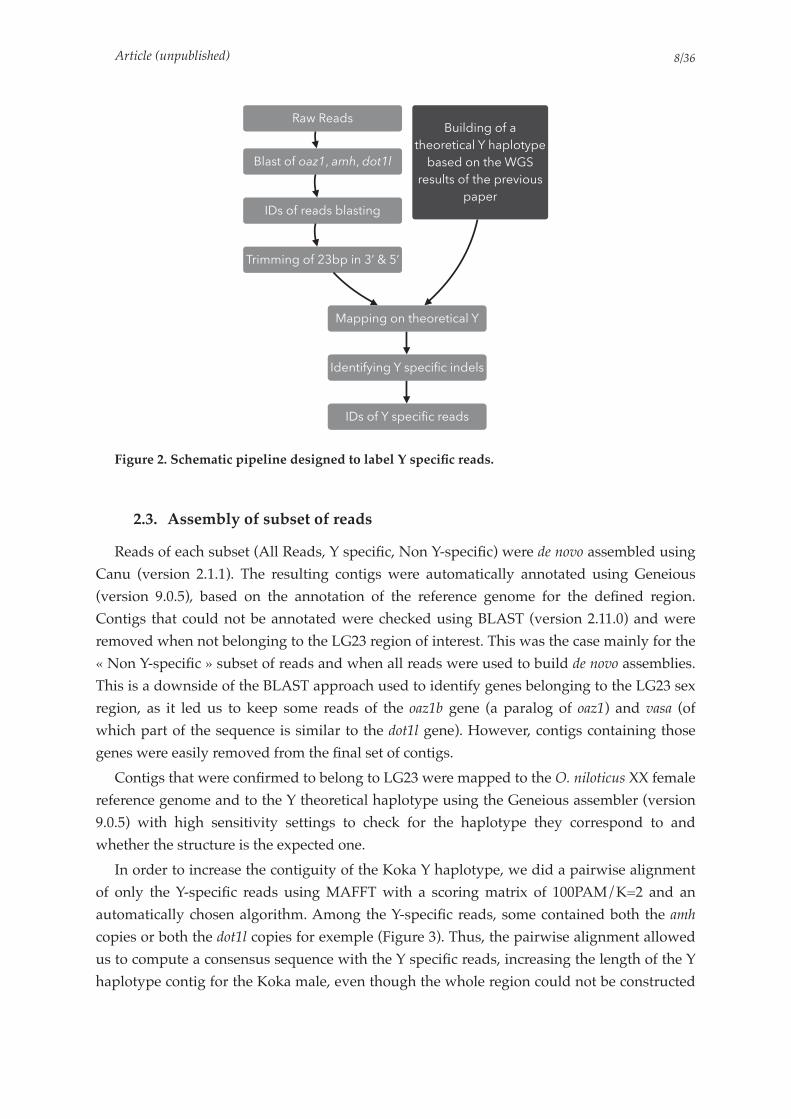
Article (unpublished) /8 36
Figure 2. Schematic pipeline designed to label Y specific reads.
2.3. Assembly of subset of reads
Reads of each subset (All Reads, Y specific, Non Y-specific) were de novo assembled using
Canu (version 2.1.1). The resulting contigs were automatically annotated using Geneious
(version 9.0.5), based on the annotation of the reference genome for the defined region.
Contigs that could not be annotated were checked using BLAST (version 2.11.0) and were
removed when not belonging to the LG23 region of interest. This was the case mainly for the
«!Non Y-specific!» subset of reads and when all reads were used to build de novo assemblies.
This is a downside of the BLAST approach used to identify genes belonging to the LG23 sex
region, as it led us to keep some reads of the oaz1b gene (a paralog of oaz1) and vasa (of
which part of the sequence is similar to the dot1l gene). However, contigs containing those
genes were easily removed from the final set of contigs.
Contigs that were confirmed to belong to LG23 were mapped to the O. niloticus XX female
reference genome and to the Y theoretical haplotype using the Geneious assembler (version
9.0.5) with high sensitivity settings to check for the haplotype they correspond to and
whether the structure is the expected one.
In order to increase the contiguity of the Koka Y haplotype, we did a pairwise alignment
of only the Y-specific reads using MAFFT with a scoring matrix of 100PAM/K=2 and an
automatically chosen algorithm. Among the Y-specific reads, some contained both the amh
copies or both the dot1l copies for exemple (Figure 3). Thus, the pairwise alignment allowed
us to compute a consensus sequence with the Y specific reads, increasing the length of the Y
haplotype contig for the Koka male, even though the whole region could not be constructed
IDs of Y specific reads
Identifying Y specific indels
Building of a
theoretical Y haplotype
based on the WGS
results of the previous
paper
Blast of oaz1, amh, dot1l
Raw Reads
IDs of reads blasting
Trimming of 23bp in 3’ & 5’
Mapping on theoretical Y
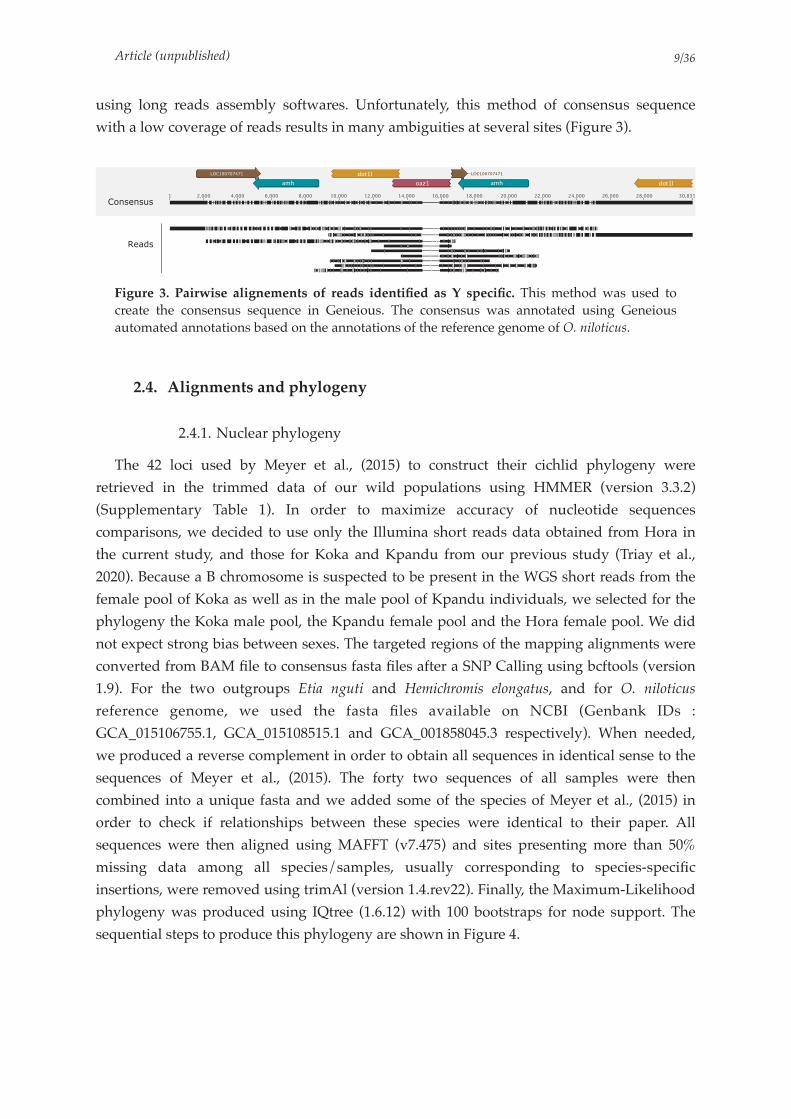
Article (unpublished) /9 36
using long reads assembly softwares. Unfortunately, this method of consensus sequence
with a low coverage of reads results in many ambiguities at several sites (Figure 3).
Figure 3. Pairwise alignements of reads identified as Y specific. This method was used to create the consensus sequence in Geneious. The consensus was annotated using Geneious automated annotations based on the annotations of the reference genome of O. niloticus.
2.4. Alignments and phylogeny
2.4.1. Nuclear phylogeny
The 42 loci used by Meyer et al., (2015) to construct their cichlid phylogeny were
retrieved in the trimmed data of our wild populations using HMMER (version 3.3.2)
(Supplementary Table 1). In order to maximize accuracy of nucleotide sequences
comparisons, we decided to use only the Illumina short reads data obtained from Hora in
the current study, and those for Koka and Kpandu from our previous study (Triay et al.,
2020). Because a B chromosome is suspected to be present in the WGS short reads from the
female pool of Koka as well as in the male pool of Kpandu individuals, we selected for the
phylogeny the Koka male pool, the Kpandu female pool and the Hora female pool. We did
not expect strong bias between sexes. The targeted regions of the mapping alignments were
converted from BAM file to consensus fasta files after a SNP Calling using bcftools (version
1.9). For the two outgroups Etia nguti and Hemichromis elongatus, and for O. niloticus
reference genome, we used the fasta files available on NCBI (Genbank IDs :
GCA_015106755.1, GCA_015108515.1 and GCA_001858045.3 respectively). When needed,
we produced a reverse complement in order to obtain all sequences in identical sense to the
sequences of Meyer et al., (2015). The forty two sequences of all samples were then
combined into a unique fasta and we added some of the species of Meyer et al., (2015) in
order to check if relationships between these species were identical to their paper. All
sequences were then aligned using MAFFT (v7.475) and sites presenting more than 50%
missing data among all species/samples, usually corresponding to species-specific
insertions, were removed using trimAl (version 1.4.rev22). Finally, the Maximum-Likelihood
phylogeny was produced using IQtree (1.6.12) with 100 bootstraps for node support. The
sequential steps to produce this phylogeny are shown in Figure 4.
!"#$%#$&$
'%()$
*+!,--.-./.,

Article (unpublished) /10 36
Figure 4. Schematic representation the sequential steps used to produce the nuclear phylogeny.
2.4.2. Mitochondrial phylogeny
We assembled the mitochondrial genomes from Koka, Hora and Kpandu O. niloticus wild
populations obtained from the short reads of our Whole Genome Sequencing trimmed data
using MitoFinder (version 1.4) (Allio et al., 2020). We used the reference mitochondrial
genome as the basis for annotation (Genbank ID: GU238433.1). In addition, we used the
closely related species Haplochromis burtoni as outgroup (NCBI ID: NC_027289.1). In order to
use the same basis as the one used for gene annotation, we also ran the reference
mitochondrial genome and Haplochromis burtoni through Mitofinder to extract individual
genes. The sequences of the 13 mitochondrial protein-coding genes and the two ribosomal
subunits (Supplementary Table 2) were then aligned using MAFFT (v7.475) (Katoh and
Standley, 2013). A maximum-likelihood phylogeny was inferred from the concatenated
sequence alignments using IQtree 1.6.12 (Nguyen et al., 2015) with 100 bootstraps for node
support.
2.4.3. Phylogeny of Sex haplotypes
Because of the low coverage of long reads data and due to the high error rate of Oxford
Nanopore MinION data, we could not use directly the Y haplotypes built from the de novo
analyses. Thus, we focused on the amh gene region and more precisely on the LG23 region
Mafft alignment
Retrieving regions in mapping alignments and
reference genomes
Hmmer with the 42 loci of Meyer et al., 2015
Building consensus sequences
Reverse complement if needed
Combining fasta
Remove sites with more than 50% missing data
IQtree Maximum Likelihood Phylogeny

Article (unpublished) /11 36
from 34,489,620 to 34,532,693 bp according to the XX reference genome and thus only
analysed females individuals. This corresponds to the region defined as carrying the major
sex-determinant in previous studies (Li et al., 2015; Cáceres et al., 2019; Triay et al., 2020;
Taslima et al., 2021). This region was extracted in Kpandu, Koka and Hora females mapping
files using SAMtools (version 1.9) (Li et al., 2009). These regions were then converted to
consensus sequences using Geneious (version 9.0.5) (Kearse et al., 2012). The sequences,
along with fasta files of this region from O. niloticus and O. aureus were aligned using
MAFFT (v7.475) (Katoh and Standley, 2013). The subsequent alignments were then used to
build a phylogeny of Females X haplotypes with 100 bootstraps with IQtree (1.6.12)
(Nguyen et al., 2015). The final visualization of all the trees (nuclear, mitochondrion and X
haplotypes) were formatted using Geneious (version 9.0.5) (Kearse et al., 2012).
3. Results
3.1. Identification of Y specific reads in the LG23 duplicated sex-region
3.1.1. Mapping of long reads confirms the existence of Y specific structures in Kpandu and Koka but not in Hora natural populations
For the Kpandu male we obtained a total of 4,474,365 Nanopore reads corresponding to
22,830,560,109 nucleotides. A total of 5,266,388 reads corresponding to 14,776,650,374
nucleotides were obtained for the Koka male and a total of 777,765 reads for the Hora male
corresponding to 4,034,783,321 nucleotides were used. The average error rate was 6,44%.
This resulted in 228 of Kpandu, 287 of Koka and 236 reads of Hora that passed the Blast filter
using the oaz1, amh and dot1l genes (Table 1).
The coverage is different along this region for the three samples. If it drops from the end
of the left amh copy in the Kpandu, it appears to be almost constant along the region for the
Koka male and finally approaches 0 in the Y specific region in the Hora male (Figure 5). This
result indicates the theoretical Y haplotype structure we constructed is correct since reads are
mapping with high quality to the Y haplotype, confirming the structure previously
described using WGS short reads data for the Kpandu and Koka populations (Triay et al.,
2020).
Coverage drop over the Y specific structures (such as seen over the dot1l-oaz1 chimera,
e.g. dashed arrows in Figure 5) in Kpandu is due to the mapping of only male-specific reads,
whereas, reads of both X and Y chromosomes are mapping over the amh gene, common to
both X and Y. In the Koka male assembly, the coverage pattern might be explained by the
absence of an X chromosome, thus Y reads show a good mapping all along the region,
corresponding to the constant coverage seen over and outside the Y-specific regions.
However, we still observe some reads that present a bad mapping quality. They could be
sequencing artefacts, or the sequenced male could be an XY male with low coverage for the
X resulting in the visual aspect homogeneous coverage along the region.

Article (unpublished) /12 36
Figure 5. IGV snapshot of long reads mapping obtained from the three males Kpandu (Lake Volta, Ghana), Koka (Lake Koka, Ethiopia) and Hora (Lake Hora, Ethiopia). Reads were mapped onto a Y theoretical haplotype. Dashed arrows are showing the coverage level over the beginning of the Y specific region. Reads mapping to the oaz1-dot1l chimera presenting a 275bp deletion found in the oaz1 truncated copy (within the dashed rectangle) were labelled as Y-specific.
The structure of the Y haplotype was confirmed for Koka and Kpandu males, but this was
not the case for the Hora male. This male individual presents no reads with a good mapping
score over the Y specific regions. Moreover, it presents none of the expected Y specific indels
(such as the -275 bp in the truncated copy of the oaz1 gene). Either this male could be an XX
male or males of the Hora population do not carry any tandem duplication in this region.
This raised the question of the amh being the sex-determining region in the Hora population
and lead to the short reads whole genome sequencing of several other samples of this
population.
3.1.2. Indels usage to identify Y specific reads
Reads presenting the previously described deletion in the truncated copy of oaz1 gene of
~275 bp were labelled as Y specific, whereas those not showing it were presumed to belong
to the X chromosome. This step was carried out only for Kpandu and Koka males as the
Hora male did not present the expected Y haplotype. This resulted in 15 long reads for
Kpandu and 11 for Koka labelled as Y-specific (Table 1 and Figure 5).
!"#$ !"#$ "%&"%&
'!($) '!($)*+,$--.-./.$ *+,$--.-./.$
Bad mappingGood mapping
Coverage
Reads
Coverage
Reads
Coverage
Reads
Y theoretical
haplotype
Kpandu
Koka
Hora
oaz1 amh oaz1 amh
dot1l dot1lLOC100707471 LOC100707471
- 275bp

Article (unpublished) /13 36
Table 1. Number of reads in the output of the filtering pipeline for MinION long reads sequencing.
3.2. Assembly of sex haplotypes
3.2.1. De novo assemblies and sex haplotypes constructions
In order to get the Y haplotype and study it in more detail, our first approach was to do a
de novo assembly of the long reads from the sex-region. We obtained very different results
depending on the male’s origin (Figure 6). We compared the obtained contigs to the O.
niloticus XX reference genome (female from the Stirling-Manzala strain). The automatic
annotation of contigs based on these two references allowed us to label the different contigs
as X or Y haplotypes based on the presence or not of Y specific duplicated structures.
We observed only one long contig for the Hora male, fitting with an X haplotype. No Y
haplotype was assembled, as was expected according to the mapping results where no reads
presented Y-specific structures. On the contrary, two contigs were assembled for the Kpandu
male: one perfectly corresponding to the X reference genome haplotype, and another one
presenting the Y-specific structures. The second contig however is much smaller than the X
haplotype and is centered around the oaz1-dot1l chimera and a second copy of the amh gene
(corresponding to the amh∆Y described in the Japanese strain (Li et al., 2015)). Finally, three
contigs were built from the Koka reads. The longest contig carries Y-specific structures, such
as the dot1l-oaz1 chimera and the Y-specific indels corresponding to the duplicated region.
However, the first copy of the amh gene (which is the amhY in the Japanese strain) is not as
fully reconstructed as for the Kpandu male. Two smaller contigs contain respectively the
conserved version of the oaz1 gene and the beginning of the amhY gene. No clear X
haplotype was built from the de novo using all reads for the Koka male. This absence of an X
contig is in line with the observed coverage of the mapping on the Y theoretical haplotype.
De novo assemblies of « Non-Y specific » reads did not produce any contig long enough to be
associated to the X haplotype either. Moreover, the longest contig assembled using the non
Y-specific reads was identified as belonging to the Y haplotype as part of the chimera that
could be identified with the automated annotation. As described in section « 2.4. Subsetting
of read datasets and identification of Y specific reads », the longest Y haplotype produced for
the Koka male was processed through pairwise alignment of Y-specific reads only. This
haplotype was used for further analyses.
Reads passing the filtering
and blast steps
Reads labeled as Y
haplotypes
Kpandu 228 15
Koka 287 11
Hora 236 –
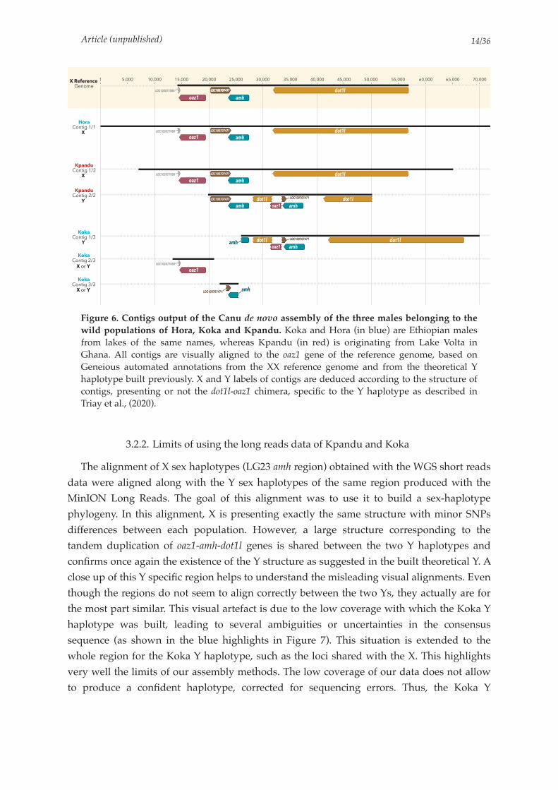
Article (unpublished) /14 36
Figure 6. Contigs output of the Canu de novo assembly of the three males belonging to the wild populations of Hora, Koka and Kpandu. Koka and Hora (in blue) are Ethiopian males from lakes of the same names, whereas Kpandu (in red) is originating from Lake Volta in Ghana. All contigs are visually aligned to the oaz1 gene of the reference genome, based on Geneious automated annotations from the XX reference genome and from the theoretical Y haplotype built previously. X and Y labels of contigs are deduced according to the structure of contigs, presenting or not the dot1l-oaz1 chimera, specific to the Y haplotype as described in Triay et al., (2020).
3.2.2. Limits of using the long reads data of Kpandu and Koka
The alignment of X sex haplotypes (LG23 amh region) obtained with the WGS short reads
data were aligned along with the Y sex haplotypes of the same region produced with the
MinION Long Reads. The goal of this alignment was to use it to build a sex-haplotype
phylogeny. In this alignment, X is presenting exactly the same structure with minor SNPs
differences between each population. However, a large structure corresponding to the
tandem duplication of oaz1-amh-dot1l genes is shared between the two Y haplotypes and
confirms once again the existence of the Y structure as suggested in the built theoretical Y. A
close up of this Y specific region helps to understand the misleading visual alignments. Even
though the regions do not seem to align correctly between the two Ys, they actually are for
the most part similar. This visual artefact is due to the low coverage with which the Koka Y
haplotype was built, leading to several ambiguities or uncertainties in the consensus
sequence (as shown in the blue highlights in Figure 7). This situation is extended to the
whole region for the Koka Y haplotype, such as the loci shared with the X. This highlights
very well the limits of our assembly methods. The low coverage of our data does not allow
to produce a confident haplotype, corrected for sequencing errors. Thus, the Koka Y
!
!"#$
%&'$(()()*)$
"+,
-!.$/
"+,
"+,
"+,
"+,
%&'$(()()*)$
%&'$(()()*)$
%&'$(()()*)$
%&'$(()()*)$
!"#$
!"#$
!"#$
%&'$(0())(1(
%&'$(0())(1(
%&'$(0())(1(
%&'$(0())(1(
"+,
!"#$
!"#$
-!.$/
-!.$/
"+,
"+,
-!.$/
-!.$/
-!.$/
-!.$/
%&'$(()()*)$
%&'$(()()*)$
%&'$(()()*)$
!"#$%$&$'($"#$%&#
)*&+'%$()*+!,!
,-+'./'%$()*+!,-
,-+'./'%$()*+-,-
,*0+'%$()*+!,.
,*0+'%$()*+-,.
,*0+'%$()*+.,.
!
!
1
1
!"!""1
/0111 !10111 !/0111 -10111 -/0111 .10111 ./0111 210111 2/0111 /10111 //0111 310111 3/0111 410111
!"!""1
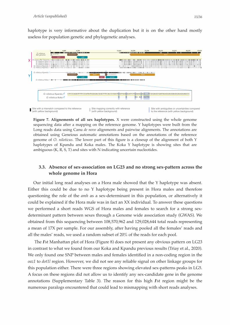
Article (unpublished) /15 36
haplotype is very informative about the duplication but it is on the other hand mostly
useless for population genetic and phylogenetic analyses.
Figure 7. Alignements of all sex haplotypes. X were constructed using the whole genome sequencing data after a mapping on the reference genome. Y haplotypes were built from the Long reads data using Canu de novo alignments and pairwise alignments. The annotations are obtained using Geneious automatic annotations based on the annotations of the reference genome of O. niloticus. The lower part of this figure is a closeup of the alignment of both Y haplotypes of Kpandu and Koka males. The Koka Y haplotype is showing sites that are ambiguous (K, R, S, T) and sites with N indicating uncertain nucleotides.
3.3. Absence of sex-association on LG23 and no strong sex-pattern across the
whole genome in Hora
Our initial long read analyses on a Hora male showed that the Y haplotype was absent.
Either this could be due to no Y haplotype being present in Hora males and therefore
questioning the role of the amh as a sex-determinant in this population, or alternatively it
could be explained if the Hora male was in fact an XX individual. To answer these questions
we performed a short reads WGS of Hora males and females to search for a strong sex-
determinant pattern between sexes through a Genome wide association study (GWAS). We
obtained from this sequencing between 108,570,962 and 129,028,644 total reads representing
a mean of 17X per sample. For our assembly, after having pooled all the females’ reads and
all the males’ reads, we used a random subset of 20% of the reads for each pool.
The Fst Manhattan plot of Hora (Figure 8) does not present any obvious pattern on LG23
in contrast to what we found from our Koka and Kpandu previous results (Triay et al., 2020).
We only found one SNP between males and females identified in a non-coding region in the
oaz1 to dot1l region. However, we did not see any reliable signal on other linkage groups for
this population either. There were three regions showing elevated sex-patterns peaks in LG5.
A focus on these regions did not allow us to identify any sex-candidate gene in the genome
annotations (Supplementary Table 3). The reason for this high Fst region might be the
numerous paralogs encountered that could lead to mismapping with short reads analyses.
!"#$%%&%&'&$!"#$%&%''%(%
!"#$%&'(%)*+
,-.-/-$)-#0-$'1-
!"#$%&'%( !"#$%&"'()"!"#)*+,-*.%( *(+,%-% !!
O. niloticus Ref. genome
!"#$%%&%&'&$
X
Y
Site with a mismatch compared to the reference
(with yellow background)
Site mapping correctly with reference
(with yellow background)Site with ambiguities or uncertainties compared
to the reference (with yellow background)
O. aureus Ref. GenomeO. niloticus Hora ♀O. niloticus Hora ♂O. niloticus Koka ♀
O. niloticus Kpandu ♀O. niloticus Kpandu ♂
O. niloticus Kpandu ♂
O. niloticus Koka ♂
O. niloticus Kpandu ♂
O. niloticus Koka ♂

Article (unpublished) /16 36
Although the signal is overall confusing, we did not find any of the 16 shared regions of
high coverage that potentially belong to a B chromosome, which we found in Kpandu males
and Koka females. Thus, the cause of this noise along the genome is not easy to explain and
further study of males-females comparisons will be needed to conclude what are the causes
of the high Fst peaks along the genome.
Figure 8. Manhattan plots of Fst for sites fixed or nearly fixed for one sex and at intermediate frequencies for the other sex in Hora population. A) Plot for all Linkage Groups. Unanchored contigs are grouped into a single label. A close up of LG05 present a region of divergence between males and females highlighted, that was not convincing after a check for genes in this region. The close up of LG23 only has the amh region highlighted. No particular high peak is observed in this region where there were expected.
3.4. Comparing sex haplotypes to population phylogeny
Both the nuclear and the mitochondrion phylogenies show that the two Koka and Hora,
populations from Ethiopia, are more closely related to each other than to the Kpandu
population originating from Ghana (Figure 9). These results are coherent with their
subspecies taxonomy as Oreochromis niloticus cancellatus for Koka and Hora versus
Oreochromis niloticus niloticus for the Kpandu population as well as the geographical location
of these three populations (Trewavas, 1983; Bezault et al., 2011). The individual of O. niloticus
used for the reference genome is placed as a sister clade of the Ethiopian populations in the
mitochondrion phylogeny, whereas the Kpandu male is placed as a sister group in the
nuclear phylogeny. The mitochondrion phylogeny also strongly supports a sister clade
0
0.2
0.4
0.6
0.8
1.0
1 2 3 4 5 6 7 8 9 10 11 12 13 14 15 16 17 18 19 20 22 23
0 10 20 30 40 Mb
0
0.2
0.4
0.6
0.8
1.0
Fst
0 10 20 30 40 Mb
0
0.2
0.4
0.6
0.8
1.0
Fst
Fst
Unanchored

Article (unpublished) /17 36
relationship between the Kpandu O. niloticus population and the O. aureus sample. The X
haplotypes of Ethiopian populations are also forming a monophyletic group, with the
Ghanaian Kpandu population as a sister group. In X haplotypes, the divergence between
Hora and Koka females is hardly noticeable.
The position of the reference genome is changing in the three trees as it forms a
monophyletic clade with Kpandu in the X haplotype phylogeny, but it is the sister group of
the Ethiopian populations in the Mitochondrion phylogeny and finally it is placed as a sister
group of Koka, Hora and Kpandu in the nuclear phylogeny. The reference genome is
showing a longer branch than all other samples in the three trees, with it being the longest
branch of the tree in the nuclear phylogeny, apart from the outgroups.
These phylogenies results are however surprising considering no Y haplotype could be
constructed in the Hora male using long reads sequencing, even if this structure is shared
between Kpandu and Koka.

Art
icle
(u
npu
blis
hed)
/18
36
Figure 9. Maximum likelihood phylogenies of Nile tilapia populations and related cichlids. Node support correspond to bootstrap values. Koka and Hora (in blue) are Ethiopian populations of O. niloticus cancellatus and Kpandu (in red) is a Lake Volta population of O. niloticus niloticus originating from Ghana. A) Nuclear DNA phylogeny built using the 42 loci from Meyer et al., (2015). The Oreochromis species are forming a monophyletic clade in which the three wild populations are more closely related to each other than to the individual used from the reference genome. The tree is rooted with Hemichromis elongatus. The species from Meyer et al., 2015 are positioned identically to their phylogeny. These results are also showing that the O. niloticus cancellatus Koka and Hora populations are more closely related to each other than to the O. niltocus niloticus of Kpandu population. B) Mitochondrial DNA phylogeny built from 13 mitochondrial protein-coding genes and the two ribosomal subunits. The tree is rooted with Haplochromis burtoni. This phylogeny is confirming that Koka and Hora populations are more closely related to each other than to Kpandu population. O. niloticus samples from Kpandu population are forming a monophyletic clade with O. aureus. C) LG23-X haplotype phylogeny from LG23 oaz1 to dot1l genes. The tree is rooted with O. aureus. Koka and Hora O. niloticus populations are forming a monophyletic clade.
!"#$&'()"
*+(,%',%*+-.$"
/'0'
!"#$%&()*+(,-
./#$#
!"#$%&'#"
()"*+,-
.$,
12"345-6$
012#((*(3,-
!14#5)*(6,-
78/91"9#
/0%&1
78%9:
%;88%
%<=%
%;88%
%;88%
%>?%
%>?%
%;88%
%;88%
%;88%
%;88%
%;88%
%=;%
%;88%
%;88%
!"#$%&'()&*+,!,#-.#/0+!
!10&'()&*+,!)-2&'#-3#%+,!
!4,0-3&5)&$(0-,!,$667#'0+7!
!"#$%&'()&*+,!8-)5&9+!
!4,0-3&')09+%#8)-,!$(+%#930)!
!:$(5(#%*&5+%#$+#!.095)#%+,!
!"#$%&5#;&3&9!*+')&%0$+,!
!<%5&%#*$)&%&/-,!'&*$)0,,+'0$,!
!=#)+#8+%+'()&*+,!*&&)++!
!>#5(?8#50,!/)#-0)+!
!@+%#$+#!,$#))*#9++!
:)0&'()&*+,!9+%&5+'-,%@A'5"%2-(B%C'C+(")-'.D
:)0&'()&*+,!9+%&5+'-,%@9'#"%2-(B%C'C+(")-'.D
:)0&'()&*+,!9+%&5+'-,%@9C".B+%2-(B%C'C+(")-'.D
!:)0&'()&*+,!5#9/#9+'#0!
!A5+#!9/-5+!
!"0*+'()&*+,!0%&9/#5-,!
%;88%
%;88%
:)0&'()&*+,!9+%&5+'-,
@E$,$5$.F$%*$.':$D
8G8;
;88
E$,$5$.F$%*$.':$
9C".B+
9'#"
A'5"
:6!#-)0-,
;88
%<?%
;88
"#$%&'()&*+,!8-)5&9+
8G8H
;88
9'#"
A'5"
9C".B+
E$,$5$.F$%*$.':$
:6!#-)0-,
;88
;88
8G88H
!"#$%&$'()"$'
*+&,-.$#/-01
23%.0,)

Article (unpublished) /19 36
4. Discussion
An ancestral XX/XY system on LG23 with a tandem duplication of the amh gene
In the past, we had thought that the Ethiopian wild populations lacked this duplication
because X and Y discriminating markers based on amh indels (Eshel et al., 2014; Li et al.,
2015; Sissao et al., 2019) did not work in the Koka Ethiopian population (Triay et al., 2020).
PCR markers have shown the presence of amhY and/or amh∆Y on LG23 now in several
laboratory and aquaculture strains of Nile tilapia (Eshel et al., 2014; Li et al., 2015; Wessels et
al., 2017; Curzon et al., 2020; Taslima et al., 2020) as well as in the wild populations of Lake
Kou, Burkina Faso (Sissao et al., 2019). Subsequently, we found that two wild populations of
Nile tilapia O. niloticus, including an Ethiopian population (Lake Koka), shared the same sex
chromosome (Linkage group 23) as several domesticated strains (Triay et al., 2020). These
previous results used short Illumina reads obtained from genome-wide sequencing, and
suggested that males had a Y specific duplication of the amh gene.
Here, we have conducted a study using Nanopore long reads to build Y haplotypes in
wild populations. Because the minION reads are long enough to cross complex regions of
the duplication encompassing the amh genes on LG23, we were able to confirm the presence
of this Y-specific duplication and its structure, in wild populations from Lake Koka in
Ethiopia (East Africa) and Lake Volta in Ghana (West Africa). In both populations, the
duplication on the Y haplotype contains two amh genes and the boundaries comprise the
oaz1 and dot1l genes, with the duplication harbouring truncated copies of dot1l and oaz1
found upstream (in the - strand) of the amhY promoter. Kpandu males from Lake Volta have
a similar structure and the same indels to the males from the Japanese strain analysed by Li
et al., (2015). One of the amh genes in Kpandu males resembles the truncated amh∆Y copy
found in the males from the Japanese strain (Li et al., 2015) since it also has the characteristic
indels of 5 bp insertion that causes a premature stop codon and the deletion of about 233bp
in the last exon. Therefore, the other Kpandu amh gene is presumably the SD amhY gene but
it does not have the same missense SNP that may cause maleness (Li et al., 2015). In contrast,
the two amh genes in Koka males do not have neither the missense SNP of amhY, nor the
indels within or surrounding amh∆Y, particularly the 5 bp insertion (Triay et al., 2020).
Consequently, we cannot say which amh gene might be the master SD amhY gene because
both might be functional.
Amh is a gene of the sex-differentiating network of vertebrates, and despite teleosts
lacking Müllerian ducts, it plays a role in testis development, regulating germ cell
proliferation (Poonlaphdecha et al., 2011; Pfennig et al., 2015; Adolfi et al., 2019). Duplicated
amh genes have emerged repeatedly from autosomes in several fish clades, taking on the role
of master male-determinant. In silversides, which like Nile tilapia, have the coexistence of
genetics and temperature effects on sex determination, Y chromosome amhy genes have
appeared in 2 species studied (Hattori et al., 2012; Yamamoto et al., 2014) with a third,
surprisingly truncated, amhy that evolved independently in another clade (Bej et al., 2017).

Article (unpublished) /20 36
Northern Pike’s amhby is also an amh duplication that diverged from the autosomal amha
gene (Pan et al., 2019) in the common ancestor of the Esocidae family, but amhby and its
surrounding Y region was subsequently lost in a Northern American Pike population (Pan et
al., 2021). In two Esocidae species the predicted amhby protein is truncated either at the N-
terminal or C-terminal regions, but their tight amhby sex linkage suggests that they remain
functional genes that have altered protein sequences (Pan et al., 2021).
We were unable to estimate the divergence between the Koka and Kpandu Y haplotypes.
Nevertheless, it is parsimonious to think that the Y-amh duplication appearance on LG23 is
ancestral since it is found in Nile tilapia populations from two distant African hydrographic
basins. Microsatellites’ structuring showed that the Koka population which belongs to the
Ethiopian Awash basin was genetically different at the macrogeographical level from the
Kpandu population located in the Volta basin that comprises the large Sudano-Sahelian
region (Bezault et al., 2011). The phylogeny we inferred is in accordance with this previous
study: the two subspecies O. n. cancellatus found in Lake Koka and Lake Hora in the
Ethiopian basin are sister populations, reflecting the genetic proximity seen at the micro-
geographic level (Bezault et al., 2011), while Kpandu is more divergent. It would be
interesting to analyse Y haplotypes in more wild populations in order to time the evolution
of the Y amh duplication and when LG23 became a sex chromosome.
LG23 has also been recruited as the pair of sex chromosome in the Pundamilia cichlids of
Lake Victoria in which a QTL approach identified a sex-linked region of 1.9 Mb,
encompassing the amh gene (Feulner et al., 2018). In addition, previous QTL presenting sex-
associated markers on LG23 were found in a meiogynogenetic line of O. aureus and in the F2
offspring of interspecific cross between O. aureus and O. mossambicus (Cnaani et al., 2004).
This suggest that LG23 of cichlids might have a high potential to become a sex chromosome
pair.
We could not construct a Y haplotype using the long reads of the Hora male, suggesting
this haplotype does not exist in the male used. This conclusion is reinforced by the fact that
no sex-specific pattern could be identified on LG23 amh region in the Hora genome short-
reads analysis. Even if we suppose that the male used for the long read sequencing was a
sex-reversed XX male, it is unlikely that all of the 10 males used for the WGS short reads,
were XX sex-reversed males. This is surprising considering that Lake Hora is closely related
to Lake Koka and belongs to the same subspecies. Therefore, it raises the question of
whether amh is the sex-determining gene in the Hora population (discussed in more detail
below).
Diversity of sex-determination system at the intraspecific level
Within Nile tilapia, some strains and stocks present their major sex-determining region
not on LG23 but on LG1, found in some USA commercial stocks (Gammerdinger et al.,
2014), in a Ghanaian family (Cnaani et al., 2008) and in the Manzala-Stirling strain, where
LG23 Y is still segregating at low frequencies (Taslima et al., 2021). Surprisingly though, sex

Article (unpublished) /21 36
segregates to LG23 in both the Manzala-Göttingen stock (Wessels et al., 2017) and the
Manzala-Tihange stock (Sissao et al., 2019) which originated from the Manzala-Stirling
stock. Likewise, the ARO-Israeli stock, originating from the Swansea strain, which itself
originated from the Stirling stock, have the LG23 locus controlling sex (Eshel et al., 2014).
This could suggest that LG1 with potential sex-loci is segregating in Lake Manzala (Egypt)
and that LG1 could under some conditions, such as domestication inducing gene loss or
fixation by drift, become the pair of sex chromosome. It would be intriguing to study SD in
the wild population of Lake Manzala in Egypt but also in other subspecies, and populations
belonging notably to the Nile basin to see if sex-patterns appear in LG1 and/or LG23.
Interestingly, the reference genome used in our study is an individual originating from the
Manzala-Stirling lab stock. The fact that this sample presents such a long branch in our
phylogenies reinforce the idea of a strong molecular divergence of this stock, probably after
its domestication. The domestication could be the reason for these strong changes as effects
of domestications on stocks have already been associated to strong differences between lab
stocks and natural population genetics (including sex-determination systems) (Wilson et al.,
2014). Alternatively, this molecular divergence could result from a hybridization in this stock
that might have happened during the process of domestication, which could explain the
large divergence observed with other O. niloticus fish. This could even be the cause of the
emergence of a new sex-determining system on LG1. For instance, the Thai-Chiltralada
hybrid, a strain originating from crosses of O. niloticus, O. mossambicus and O. aureus,
presents sex-associated markers on LG1, LG3 and LG23 (Curzon et al., 2019). An
introgression of closely related species of O. niloticus could thus favor the turnover of sex-
chromosomes so that LG1 became fixed as a new sex chromosome.
Loss of the Y amh duplication in the Hora Lake population reflects a rapid change in SD
within Nile tilapia populations (discussed below). Indeed, more than 20 different major sex-
determining genes, on more than 17 LGs, have now been described in cichlids, highligting
the fact that in this family there are a great diversity of heterogamety systems but also that
many sex chromosomes have emerged between closely related species (Gammerdinger and
Kocher, 2018; El Taher et al., 2020). To date, cichlids from Lake Tanganyika, appear to have
the highest rates of sex chromosome turnover as well as heterogamety transitions (El Taher
et al., 2020).
Studies to look at sex-determination systems in many species, typically only examine a
few individuals of each species. Furman et al., (2020) suggested the intraspecific diversity of
sex chromosomes might be as high as what is observed at the interspecific level. Variance in
sexual conflict and recombination patterns, two important factors possibly for the evolution
of sex chromosomes, could induce divergence between sex chromosomes of different
populations.
Our study again emphasizes the need to analyse the evolution of sex chromosomes at the
population level. Studies in Nile tilapia have also demonstrated that in domesticated strains
(Wessels et al., 2014; Taslima et al., 2021) and wild populations (Bezault et al., 2007; Sissao et
al., 2019) the sex-determination system can be more complex than just the amh gene on
Article (unpublished) /22 36
LG23. Sex in the Nile tilapia is genetically inherited through the sex chromosomes but there
is also a genetic inheritance of autosomal factors and of temperature sensitivity (Baroiller
and D’Cotta, 2001; Bezault et al., 2007; Baroiller et al., 2009; Wessels et al., 2017). Temperature
could be the trigger causing the turnover, towards a new sex chromosome system (Grossen
et al., 2011; Baroiller and D’Cotta, 2016). This, along with the results of the Hora population
lacking the LG23 Y sex-determinant suggest that the turnover in sex chromosomes and sex-
determination systems might not be rare events at the intraspecific level.
Ever-young sex-chromosomes ?
The X and Y chromosomes of wild populations of O. niloticus studied here are visually
homomorphic, like all cichlids sex chromosomes karyotyped to date (reviewed in
Gammerdinger and Kocher, (2018)). Reduced sex chromosome differentiation is evidenced
by the small size of the sex-specific region (<0.5Mb) of LG23 suggesting that recombination
still occurs around the sex-region. The study of genotype-phenotype associations using
ddRAD methods on families of Kpandu and Koka populations already suggested that
recombination could occur close to the male-specific region (Triay et al., 2020). This reduced
recombination further highlights the fact that LG23 has remained a «!young!» pair of sex
chromosomes. The shared structure between males of the two wild populations suggests
this XX/XY system predates the divergence of populations of O. niloticus. Thus, even if
chronologically old, Nile Tilapia sex chromosomes do not seem to have evolved to a non-
recombinant Y that starts to differentiate, but is maintained at a state of low recombination
for long evolutionary times.
This finding further reinforces the idea that sex chromosomes do not always cease
recombination and begin degenerating after acquiring a major sex-determinant. There are
more and more examples of pairs of sex chromosomes that are homomorphic, and
sometimes even so similar at the molecular level that they differ only by a SNP, as in
Takifugu (Kamiya et al., 2012). Two alternative models (which might not be exclusive) were
proposed to occur in cold-blooded vertebrates with homomorphic sex chromosomes. The
“high-turnover hypothesis” suggests that the master-sex-determining genes switch
frequently, and the regions not recombining around the new SD gene do not have time to
decay (Volff et al., 2007). In cichlids this model has indeed been evidenced (Gammerdinger
and Kocher, 2018; El Taher et al., 2020). The “fountain-of-youth model” on the other hand
proposes that the sex chromosomes remain stable over long periods with some
recombination occurring in XY females (Perrin, 2009). European tree frogs and ratites also
have very old sex chromosomes at the species level with no reduction of recombination
outside the sex region (Stöck et al., 2011). The authors suggested that the presence of
supposedly young sex chromosomes in these frogs that contain nevertheless old SD genes,
occurred due to occasional recombination between the X and Y chromosomes that took place
in males or in sex-reversed XY females (Stöck et al., 2011).

Article (unpublished) /23 36
We do not know to what extent sex-reversed XY females may exist in wild populations of
Nile tilapia, although preliminary studies suggested they existed (Baroiller and D’Cotta,
2016). We still do not know what mechanisms exist in the Nile tilapia for the prevalence of
LG23 as a sex chromosome. Turnover of sex chromosomes (appearance of LG1?) or in the SD
system exemplified here with the Hora population, might as evoked previously, be more
frequent than what is apparent. We need to date the divergence between Koka and Kpandu
populations compared to the divergence of their Y haplotypes in order to understand if this
«!young!» pair of sex chromosome is in an evolutionary pathway to stay «!ever-young!».
Introgression of wild populations?
We noted an incongruence for Kpandu position between the nuclear and mitochondrial
phylogenies. Our mitochondrial DNA analysis demonstrates a clustering of Kpandu O.
niloticus population with the sample of O. aureus, also a Nilo-Sudanic species. The formation
of a monophyletic group is consistent with the study of Rognon and Guyomard, (2003) in
which they hypothesize a complete introgression of the mitochondrial DNA from O. aureus
to O. niloticus in Western African populations, but also in Egyptian lakes. Clustering of
Kpandu Nile tilapia and O. aureus was also observed when using mtDNA but they were
differentiated in phylogenetic trees derived from nuclear DNA markers (SNPs) (Syaifudin et
al., 2019). Discordance between mtDNA and nuclear DNA has been shown in several studies
within the Oreochromine cichlids (Ford et al., 2019). Mitochondria DNA reflects only maternal
inheritance, whereas species topology is probably more reliable when using multi-nuclear
markers (Meyer et al., 2015; Ford et al., 2019). Ancient hybridization was suggested as being
the most likely explanation for the discordance between mtDNA and nuDNA (Dunz and
Schliewen, 2013).
Our results suggest that at some point in the evolutionary history of O. niloticus
introgression with other closely related cichlids species might have occurred. Ancient
hybridization of O. niloticus with other closely related species would not be surprising since
several species coexist in many African fresh water basins. O. aureus and O. niloticus
divergence was estimated to be 2.93 MYA (Xiao et al., 2015). They cohabitate in several river
basins, both being Nilo-Sudano species (Trewavas, 1983), but they are not known to
hybridize naturally (Payne and Collinson, 1983). In Lake Manzala there is a question on
whether hybridization might have occurred between Nile tilapia and S. galilaeus confused
perhaps as O. aureus (Ford et al., 2019). This might be the explanation of a SD on LG1
appearing in several O. niloticus strains (Cnaani et al., 2008). Thus, particularly when
studying sex chromosomes, it might be essential to consider the other sex-determining loci
segregating in the sympatric species of studied populations.
Loss of the Y-specific region on LG23 in the Ethiopian population of Lake Hora
We did not find a Y duplication of the amh sex region on the LG23 in the Hora males nor
were we able to detect any SD system in this population. The phylogenetic studies

Article (unpublished) /24 36
confirmed that the Ethiopian Koka and Hora populations, both from the subspecies
Oreochromis niloticus cancellatus, belonged to a monophyletic clade with the Kpandu
population O. niloticus niloticus branching as a sister group. These results appear
contradictory with a shared ancestral Y chromosome between Koka and Kpandu males that
is not shared between Koka and Hora. A better estimation of the divergence between Koka
and Kpandu would have been the use of the Y haplotypes for phylogeny construction,
helped by additional populations, to understand the evolutionary history of the LG23 Y
chromosome. This would also enable us to date the event that led to the loss of LG23Y in the
Hora population. We constructed the phylogeny using multinuclear markers analysed with
a concatenation method, which generates artefacts since we assume the same evolution
history for all loci. A better estimation of the evolution of LG23 would be obtained using the
Y haplotypes, which we were unable to do. It would be necessary to first increase the long-
reads sequencing to improve the coverage of the Y haplotypes of Kpandu and Koka males,
correct for sequencing errors to avoid ambiguities and uncertainties in final sequences, and
then rerun the phylogenies using coalescent methods. This would improve the phylogenies
and would presumably allow us to know if this pair of sex chromosomes is chronologically
young or if it is in a stable state and will possibly remain forever young.
Hybridization with other species would certainly give different results in the phylogeny
and potentially a different position of Hora samples in the trees or at least a long branch for
this population. From the numerous studies where LG23 Y is found as the major sex-
determinant in O. niloticus, we can assume that there has been a loss of the ancestral Y
haplotype in the Hora population. This loss raises two questions: Why has the Hora
population experienced a turnover in the system of sex determination? What is the current
system of sex determination in this population?
Possible causes of the loss of LG23-Y
Causes for sex chromosome turnovers can be numerous, and among all, a loss of the sex-
determinant by drift, sex ratio bias (e.g. environmental effects inducing sex-reversal), or
pleiotropic effects of a sexually antagonistic mutation can facilitate the fixation of a new sex-
determinant (reviewed in Tao et al., (2021)). Demographic events might have caused changes
in Lake Hora SD. Lake Hora is thought to have been restocked at the beginning of the
twentieth century with the endemic subspecies O. n. cancellatus (Bezault, 2005). Lake Hora is,
nevertheless, a small lake (~1,15 Km²) and the effective population size was supposedly
small. The Hora population might have experienced a genetic bottleneck that led to the loss
of the LG23-Y by drift. Intraspecific differences in SD was also observed for the
haplochromine cichlid Pseudocrenilabrus philander for which WGS detected a strong XY locus
in LG7 in a lake population whereas no GSD was found in the outflow creek population
which experienced genetic bottleneck (Böhne et al., 2019).
Sex ratio bias can also occur in O. niloticus species because of the environmental effect of
temperature. This species can undergo a sex reversal over the threshold temperature of 32°C

Article (unpublished) /25 36
causing XX individuals to develop as viable and fertile males, and this temperature
sensitivity is a variable and heritable trait (Baroiller and D’Cotta, 2001; Bezault et al., 2007;
Wessels and Hörstgen-Schwark, 2007; Baroiller et al., 2009; Wessels and Hörstgen-Schwark,
2011). In the worst case scenario, the increasing number of XX individuals (males and
females) in a population could progressively lead to the loss of the Y chromosome and
permit a new sex-determinant master gene to ensure the sex-determination of the
population. However, Lake Hora (like Lake Koka) is referred to as a « Cold » Lake because
its temperature varies between 22 to 25°C during the year. This hypothesis does not seem
relevant to explain the loss of the Y chromosome according to the current environment of the
Hora population. Still, the history of past temperatures in the lake have not been
investigated. Hence, the loss of an ancestral LG23 Y system in the Hora fish could have been
facilitated by demographic events of this small population, with colonisation, and perhaps
further influenced if part of the founders were XX neo-males.
Finally, we cannot exclude the possibility that there has been a transition in the Hora
population from a genetic sex determination system to an environmental sex determination
system.
What could be the current sex determining system(s) of the Hora population?
When a major sex-determinant is lost, it can be replaced by a new « master » gene,
possibly already implied in the sex-determination and differentiation pathway. In the case of
the Hora population, we did not identify any obvious sex-determinant loci between the
pools of wild males and females. This could be due to the new major sex-determinant region
being too recent to have accumulated enough SNPs to be detected through windowed
Genome wide Fst analysis. It is also possible that after the loss of the major determinant
LG23 Y, other loci involved in sex determination and differentiation pathway acquire a more
important role and coexist during a transition period until the fixation and potential
appearance of a new major determinant (Furman et al., 2020; Tao et al., 2020). In this case,
more than one loci is segregating on several chromosomes, appearing as a polygenic sex
determining system (Mank and Avise, 2009; Roberts et al., 2016). Although WGS of pooled
sexes is a very efficient method to quickly detect sex determination locus and the linkage
group acting as a sex chromosome when the system is monogenic, it is however not as
efficient to detect polygenic systems (Gammerdinger and Kocher, 2018). We can not exclude
the scenario of two sex-determining loci (or more?) segregating in the Hora population as
recently suggested by (Taslima et al., 2021) for the Manzala-Stirling stock.
Ideally when sex chromosomes are strongly homomorphic, as it is expected in the Hora
population, Palmer et al., (2019) suggest studying the segregation of SNPs in families
between parents and progeny. Tools such as SEX-DETector perform this kind of analysis,
ideally on RNAseq data (Muyle et al., 2016). The downside of this method is that it is not
optimized for strongly inbred populations and that it requires live fish to produce families
data.

Article (unpublished) /26 36
5. Conclusion
In conclusion, we have been able to reconstruct using long reads the Y haplotype
corresponding to a duplicated Y-specific region on the LG23 sex chromosome in two wild
populations of Nile tilapia. The Y haplotypes contain a duplication in tandem of two amh
genes, a truncated chimera comprising the oaz1 and dotl1 genes, and the complete genes
found in the breakpoints of the duplication. We evidenced polymorphisms in the Y
haplotype between the two wild populations. The West African population of Kpandu,
(Volta basin), has similar indels and sequences as previously described in domesticated
strains of Nile tilapia, containing the master sex determinant amhy gene and a possibly
truncated amh∆Y gene. In sharp contrast, the East African Ethiopian population of Lake
Koka does not possess these indels, and we found no distinctions between the duplicated
amh genes (supposedly amhy and amh∆Y) since both may be functional. We studied an
additional Ethiopian population (Hora Lake), which has lost the Y amh duplication. Sex no
longer segregates on LG23 and we were unable to find a GSD in this population. We suggest
that the SD has changed to become either a polygenic SD or it has acquired an ESD system.
Alternatively a new SD locus might be still too reduced to be identified. Further analysis will
be required to understand the sex-determination system in Hora population and potentially
find the major sex-determination gene. An analysis of the SNPs density between males and
females at a genome wide scale could already bring some clues about key regions in linkage
disequilibrium with sex.

Article (unpublished) /27 36
References
Abbott, J.K., Nordén, A.K., and Hansson, B. (2017). Sex chromosome evolution: historical insights and future perspectives. Proceedings of the Royal Society B: Biological Sciences 284, 20162806.
Adolfi, M.C., Nakajima, R.T., Nóbrega, R.H., and Schartl, M. (2019). Intersex, Hermaphroditism, and Gonadal Plasticity in Vertebrates: Evolution of the Müllerian Duct and Amh/Amhr2 Signaling. Annu. Rev. Anim. Biosci. 7, 149–172.
Allio, R., Schomaker-Bastos, A., Romiguier, J., Prosdocimi, F., Nabholz, B., and Delsuc, F. (2020). MitoFinder: Efficient automated large-scale extraction of mitogenomic data in target enrichment phylogenomics. Mol Ecol Resour 20, 892–905.
Bachtrog, D. (2013). Y-chromosome evolution: emerging insights into processes of Y-chromosome degeneration. Nature Reviews Genetics 14, 113–124.
Bachtrog, D., Kirkpatrick, M., Mank, J.E., McDaniel, S.F., Pires, J.C., Rice, W., and Valenzuela, N. (2011). Are all sex chromosomes created equal? Trends in Genetics 27, 350–357.
Bachtrog, D., Mank, J.E., Peichel, C.L., Kirkpatrick, M., Otto, S.P., Ashman, T.-L., Hahn, M.W., Kitano, J., Mayrose, I., Ming, R., et al. (2014). Sex determination: why so many ways of doing it? PLOS Biology 12, e1001899.
Baroiller, J.F., and D’Cotta, H. (2001). Environment and sex determination in farmed fish. Comparative Biochemistry and Physiology Part C: Toxicology & Pharmacology 130, 399–409.
Baroiller, J.-F., and D’Cotta, H. (2016). The Reversible Sex of Gonochoristic Fish: Insights and Consequences. Sexual Development 10, 242–266.
Baroiller, J.F., D’Cotta, H., Bezault, E., Wessels, S., and Hoerstgen-Schwark, G. (2009). Tilapia sex determination: where temperature and genetics meet. Comparative Biochemistry and Physiology Part A: Molecular & Integrative Physiology 153, 30–38.
Bej, D.K., Miyoshi, K., Hattori, R.S., Strüssmann, C.A., and Yamamoto, Y. (2017). A Duplicated, Truncated amh Gene Is Involved in Male Sex Determination in an Old World Silverside. G3: Genes, Genomes, Genetics 7.
Bezault, E. (2005). Etude Du Systeme De Determinisme Du Sexe au Sein De Populations Naturelles De Tilapia Du Nil, Oreochromis niloticus (linnaeus, 1758) : importance Des Composantes Genetiques Et Environnementales. UNIVERSITE DE PARIS-SUD.
Bezault, E., Clota, F., Derivaz, M., Chevassus, B., and Baroiller, J.-F. (2007). Sex determination and temperature-induced sex differentiation in three natural populations of Nile tilapia (Oreochromis niloticus) adapted to extreme temperature conditions. Aquaculture 272, S3–S16.
Bezault, E., Balaresque, P., Toguyeni, A., Fermon, Y., Araki, H., Baroiller, J.-F., and Rognon, X. (2011). Spatial and temporal variation in population genetic structure of wild Nile tilapia (Oreochromis niloticus) across Africa. BMC Genetics 12, 102.
Blaser, O., Neuenschwander, S., and Perrin, N. (2014). Sex-Chromosome Turnovers: The Hot-Potato Model. The American Naturalist 183, 140–146.
Böhne, A., Wilson, C.A., Postlethwait, J.H., and Salzburger, W. (2016). Variations on a theme: Genomics of sex determination in the cichlid fish Astatotilapia burtoni. BMC Genomics 17.
Böhne, A., Weber, A.A.-T., Rajkov, J., Rechsteiner, M., Riss, A., Egger, B., and Salzburger, W. (2019). Repeated Evolution Versus Common Ancestry: Sex Chromosome Evolution in the Haplochromine Cichlid Pseudocrenilabrus philander. Genome Biology and Evolution 11, 439–458.

Article (unpublished) /28 36
Cáceres, G., López, M.E., Cádiz, M.I., Yoshida, G.M., Jedlicki, A., Palma-Véjares, R., Travisany, D., Díaz-Domínguez, D., Maass, A., Lhorente, J.P., et al. (2019). Fine mapping using whole-genome sequencing confirms anti-müllerian hormone as a major gene for sex determination in farmed Nile tilapia (Oreochromis niloticus L.). G3 (Bethesda) 9, 3213–3223.
Carneiro, M.O., Russ, C., Ross, M.G., Gabriel, S.B., Nusbaum, C., and DePristo, M.A. (2012). Pacific biosciences sequencing technology for genotyping and variation discovery in human data. BMC Genomics 13, 375.
Charlesworth, D., Charlesworth, B., and Marais, G. (2005). Steps in the evolution of heteromorphic sex chromosomes. Heredity 95, 118–128.
Cnaani, A., Zilberman, N., Tinman, S., Hulata, G., and Ron, M. (2004). Genome-scan analysis for quantitative trait loci in an F2 tilapia hybrid. Mol Genet Genomics 272, 162–172.
Cnaani, A., Lee, B.-Y., Zilberman, N., Ozouf-Costaz, C., Hulata, G., Ron, M., D’Hont, A., Baroiller, J.-F., D’Cotta, H., Penman, D.J., et al. (2008). Genetics of sex determination in tilapiine species. Sexual Development 2, 43–54.
Cortez, D., Marin, R., Toledo-Flores, D., Froidevaux, L., Liechti, A., Waters, P.D., Grützner, F., and Kaessmann, H. (2014). Origins and functional evolution of Y chromosomes across mammals. Nature 508, 488–493.
Curzon, A.Y., Seroussi, E., Benet-Perelberg, A., Zak, T., Dor, L., Shirak, A., and Ron, M. (2019). Hybrid Origin of the Thai-Chitralada Tilapia Strain Using DNA Barcoding and Microsatellite Analysis. Israeli Journal of Aquaculture - Bamidgeh.
Curzon, A.Y., Shirak, A., Dor, L., Zak, T., Perelberg, A., Seroussi, E., and Ron, M. (2020). A duplication of the Anti-Müllerian hormone gene is associated with genetic sex determination of different Oreochromis niloticus strains. Heredity 125, 317–327.
Devlin, R.H., and Nagahama, Y. (2002). Sex determination and sex differentiation in fish: an overview of genetic, physiological, and environmental influences. Aquaculture 208, 191–364.
van Doorn, G.S., and Kirkpatrick, M. (2010). Transitions Between Male and Female Heterogamety Caused by Sex-Antagonistic Selection. Genetics 186, 629–645.
Dunz, A.R., and Schliewen, U.K. (2013). Molecular phylogeny and revised classification of the haplotilapiine cichlid fishes formerly referred to as “Tilapia.” Molecular Phylogenetics and Evolution 68, 64–80.
El Taher, A., Ronco, F., Matschiner, M., Salzburger, W., and Böhne, A. (2020). Dynamics of sex chromosome evolution in a rapid radiation of cichlid fishes (Evolutionary Biology).
Eshel, O., Shirak, A., Dor, L., Band, M., Zak, T., Markovich-Gordon, M., Chalifa-Caspi, V., Feldmesser, E., Weller, J.I., Seroussi, E., et al. (2014). Identification of male-specific amh duplication, sexually differentially expressed genes and microRNAs at early embryonic development of Nile tilapia (Oreochromis niloticus). BMC Genomics 15, 774.
Ezaz, T., Sarre, S.D., O’Meally, D., Marshall Graves, J.A., and Georges, A. (2009). Sex Chromosome Evolution in Lizards: Independent Origins and Rapid Transitions. Cytogenet Genome Res 127, 249–260.
Feulner, P.G.D., Schwarzer, J., Haesler, M.P., Meier, J.I., and Seehausen, O. (2018). A Dense Linkage Map of Lake Victoria Cichlids Improved the Pundamilia Genome Assembly and Revealed a Major QTL for Sex-Determination. G3 8, 2411–2420.
Ford, A.G.P., Bullen, T.R., Pang, L., Genner, M.J., Bills, R., Flouri, T., Ngatunga, B.P., Rüber, L., Schliewen, U.K., Seehausen, O., et al. (2019). Molecular phylogeny of Oreochromis

Article (unpublished) /29 36
(Cichlidae: Oreochromini) reveals mito-nuclear discordance and multiple colonisation of adverse aquatic environments. Molecular Phylogenetics and Evolution 136, 215–226.
Furman, B.L.S., Metzger, D.C.H., Darolti, I., Wright, A.E., Sandkam, B.A., Almeida, P., Shu, J.J., and Mank, J.E. (2020). Sex chromosome evolution: so many exceptions to the rules. Genome Biol Evol 12, 750–763.
Gammerdinger, W.J., and Kocher, T.D. (2018). Unusual diversity of sex chromosomes in African cichlid fishes. Genes 9, 480.
Gammerdinger, W.J., Conte, M.A., Acquah, E.A., Roberts, R.B., and Kocher, T.D. (2014). Structure and decay of a proto-Y region in Tilapia, Oreochromis niloticus. BMC Genomics 15, 975.
Graves, J.A.M. (2014). Avian sex, sex chromosomes, and dosage compensation in the age of genomics. Chromosome Res 22, 45–57.
Grossen, C., Neuenschwander, S., and Perrin, N. (2011). Temperature-dependent turnovers in sex-determination mechanisms: a quantitative model. Evolution 65, 64–78.
Guiguen, Y., Bertho, S., Herpin, A., and Fostier, A. (2018). Sex Determination and Sex Control in Salmonidae. In Sex Control in Aquaculture, (Chichester, UK: John Wiley & Sons, Ltd), pp. 251–281.
Hattori, R.S., Murai, Y., Oura, M., Masuda, S., Majhi, S.K., Sakamoto, T., Fernandino, J.I., Somoza, G.M., Yokota, M., and Strüssmann, C.A. (2012). A Y-linked anti-Müllerian hormone duplication takes over a critical role in sex determination. PNAS 109, 2955–2959.
Helfman, G.S., Colette, B.B., Facey, D.E., and Bowen, B.W. (2009). The diversity of fishes: biology, evolution, and ecology (Chichester, UK ; Hoboken, NJ: Blackwell).
Jeffries, D.L., Lavanchy, G., Sermier, R., Sredl, M.J., Miura, I., Borzée, A., Barrow, L.N., Canestrelli, D., Crochet, P.-A., Dufresnes, C., et al. (2018). A rapid rate of sex-chromosome turnover and non-random transitions in true frogs. Nature Communications 9.
Kamiya, T., Kai, W., Tasumi, S., Oka, A., Matsunaga, T., Mizuno, N., Fujita, M., Suetake, H., Suzuki, S., Hosoya, S., et al. (2012). A trans-species missense SNP in Amhr2 is associated with sex determination in the tiger pufferfish, Takifugu rubripes (Fugu). PLoS Genet 8.
Katoh, K., and Standley, D.M. (2013). MAFFT Multiple Sequence Alignment Software Version 7: Improvements in Performance and Usability. Molecular Biology and Evolution 30, 772–780.
Kearse, M., Moir, R., Wilson, A., Stones-Havas, S., Cheung, M., Sturrock, S., Buxton, S., Cooper, A., Markowitz, S., Duran, C., et al. (2012). Geneious Basic: An integrated and extendable desktop software platform for the organization and analysis of sequence data. Bioinformatics 28, 1647–1649.
Kirkpatrick, M. (2010). How and Why Chromosome Inversions Evolve. PLoS Biol 8, e1000501.
Li, H., Handsaker, B., Wysoker, A., Fennell, T., Ruan, J., Homer, N., Marth, G., Abecasis, G., Durbin, R., and 1000 Genome Project Data Processing Subgroup (2009). The Sequence Alignment/Map format and SAMtools. Bioinformatics 25, 2078–2079.
Li, M., Sun, Y., Zhao, J., Shi, H., Zeng, S., Ye, K., Jiang, D., Zhou, L., Sun, L., Tao, W., et al. (2015). A tandem duplicate of anti-müllerian hormone with a missense SNP on the Y chromosome is essential for male sex determination in Nile tilapia, Oreochromis niloticus. PLOS Genetics 11, e1005678.
Mank, J.E., and Avise, J.C. (2009). Evolutionary Diversity and Turn-Over of Sex Determination in Teleost Fishes. SXD 3, 60–67.

Article (unpublished) /30 36
Matsuda, M., Nagahama, Y., Shinomiya, A., Sato, T., Matsuda, C., Kobayashi, T., Morrey, C.E., Shibata, N., Asakawa, S., Shimizu, N., et al. (2002). DMY is a Y-specific DM-domain gene required for male development in the medaka fish. Nature 417, 559–563.
Menegon, M., Cantaloni, C., Rodriguez-Prieto, A., Centomo, C., Abdelfattah, A., Rossato, M., Bernardi, M., Xumerle, L., Loader, S., and Delledonne, M. (2017). On site DNA barcoding by nanopore sequencing. PLoS ONE 12, e0184741.
Meyer, B.S., Matschiner, M., and Salzburger, W. (2015). A tribal level phylogeny of Lake Tanganyika cichlid fishes based on a genomic multi-marker approach. Molecular Phylogenetics and Evolution 83, 56–71.
Muyle, A., Käfer, J., Zemp, N., Mousset, S., Picard, F., and Marais, G.A. (2016). SEX-DETector: A Probabilistic Approach to Study Sex Chromosomes in Non-Model Organisms. Genome Biol Evol 8, 2530–2543.
Myosho, T., Otake, H., Masuyama, H., Matsuda, M., Kuroki, Y., Fujiyama, A., Naruse, K., Hamaguchi, S., and Sakaizumi, M. (2012). Tracing the emergence of a novel sex-determining gene in medaka, Oryzias luzonensis. Genetics 191, 163–170.
Myosho, T., Takehana, Y., Hamaguchi, S., and Sakaizumi, M. (2015). Turnover of Sex Chromosomes in Celebensis Group Medaka Fishes. G3 Genes|Genomes|Genetics 5, 2685–2691.
Nanda, I., Kondo, M., Hornung, U., Asakawa, S., Winkler, C., Shimizu, A., Shan, Z., Haaf, T., Shimizu, N., Shima, A., et al. (2002). A duplicated copy of DMRT1 in the sex-determining region of the Y chromosome of the medaka, Oryzias latipes. PNAS 99, 11778–11783.
Nguyen, L.-T., Schmidt, H.A., von Haeseler, A., and Minh, B.Q. (2015). IQ-TREE: A Fast and Effective Stochastic Algorithm for Estimating Maximum-Likelihood Phylogenies. Molecular Biology and Evolution 32, 268–274.
Ortega-Recalde, O., Goikoetxea, A., Hore, T.A., Todd, E.V., and Gemmell, N.J. (2020). The Genetics and Epigenetics of Sex Change in Fish. Annual Review of Animal Biosciences 8, null.
Palaiokostas, C., Bekaert, M., Khan, M., Taggart, J.B., Gharbi, K., McAndrew, B.J., and Penman, D.J. (2015). A novel sex-determining QTL in Nile tilapia (Oreochromis niloticus). BMC Genomics 16, 171.
Palmer, D.H., Rogers, T.F., Dean, R., and Wright, A.E. (2019). How to identify sex chromosomes and their turnover. Molecular Ecology 28, 4709–4724.
Pan, Q., Feron, R., Yano, A., Guyomard, R., Jouanno, E., Vigouroux, E., Wen, M., Busnel, J.-M., Bobe, J., Concordet, J.-P., et al. (2019). Identification of the master sex determining gene in Northern pike (Esox lucius) reveals restricted sex chromosome differentiation. PLOS Genetics 15, e1008013.
Pan, Q., Feron, R., Jouanno, E., Darras, H., Herpin, A., Koop, B., Rondeau, E., Goetz, F.W., Larson, W.A., Bernatchez, L., et al. (2021). The rise and fall of the ancient northern pike master sex-determining gene. ELife 10, e62858.
Payne, A.I., and Collinson, R.I. (1983). A comparison of the biological characteristics of Sarotherodon niloticus (L.) with those of S. aureus (Steindachner) and other tilapia of the delta and lower Nile. Aquaculture 30, 335–351.
Perrin, N. (2009). Sex Reversal: A Fountain of Youth for Sex Chromosomes? Evolution 63, 3043–3049.
Pfennig, F., Standke, A., and Gutzeit, H.O. (2015). The role of Amh signaling in teleost fish – Multiple functions not restricted to the gonads. General and Comparative Endocrinology 223, 87–107.
Article (unpublished) /31 36
Poonlaphdecha, S., Pepey, E., Huang, S.-H., Canonne, M., Soler, L., Mortaji, S., Morand, S., Pfennig, F., Mélard, C., Baroiller, J.F., et al. (2011). Elevated amh Gene Expression in the Brain of Male Tilapia (Oreochromis niloticus) during Testis Differentiation. Sex Dev 5, 33–47.
Rice, W.R. (1987). The Accumulation of Sexually Antagonistic Genes as a Selective Agent Promoting the Evolution of Reduced Recombination between Primitive Sex Chromosomes. Evolution 41, 911–914.
Roberts, N.B., Juntti, S.A., Coyle, K.P., Dumont, B.L., Stanley, M.K., Ryan, A.Q., Fernald, R.D., and Roberts, R.B. (2016). Polygenic sex determination in the cichlid fish Astatotilapia burtoni. BMC Genomics 17, 835.
Roberts, R.B., Ser, J.R., and Kocher, T.D. (2009). Sexual Conflict Resolved by Invasion of a Novel Sex Determiner in Lake Malawi Cichlid Fishes. Science 326, 998–1001.
Rognon, X., and Guyomard, R. (2003). Large extent of mitochondrial DNA transfer from Oreochromis aureus to O. niloticus in West Africa. Mol Ecol 12, 435–445.
Sandve, S.R., Rohlfs, R.V., and Hvidsten, T.R. (2018). Subfunctionalization versus neofunctionalization after whole-genome duplication. Nat Genet 50, 908–909.
Sinclair, A.H., Berta, P., Palmer, M.S., Hawkins, J.R., Griffiths, B.L., Smith, M.J., Foster, J.W., Frischauf, A.-M., Lovell-Badge, R., and Goodfellow, P.N. (1990). A gene from the human sex-determining region encodes a protein with homology to a conserved DNA-binding motif. Nature 346, 240–244.
Sissao, R., D’Cotta, H., Baroiller, J.-F., and Toguyeni, A. (2019). Mismatches between the genetic and phenotypic sex in the wild Kou population of Nile tilapia Oreochromis niloticus. PeerJ 7.
Smith, C.A., Roeszler, K.N., Ohnesorg, T., Cummins, D.M., Farlie, P.G., Doran, T.J., and Sinclair, A.H. (2009). The avian Z-linked gene DMRT1 is required for male sex determination in the chicken. Nature 461, 267–271.
Stöck, M., Horn, A., Grossen, C., Lindtke, D., Sermier, R., Betto-Colliard, C., Dufresnes, C., Bonjour, E., Dumas, Z., Luquet, E., et al. (2011). Ever-Young Sex Chromosomes in European Tree Frogs. PLoS Biol 9, e1001062.
Syaifudin, M., Bekaert, M., Taggart, J.B., Bartie, K.L., Wehner, S., Palaiokostas, C., Khan, M.G.Q., Selly, S.-L.C., Hulata, G., D’Cotta, H., et al. (2019). Species-Specific Marker Discovery in Tilapia. Sci Rep 9, 13001.
Takehana, Y., Hamaguchi, S., and Sakaizumi, M. (2008). Different origins of ZZ/ZW sex chromosomes in closely related medaka fishes, Oryzias javanicus and O. hubbsi. Chromosome Res 16, 801–811.
Takehana, Y., Matsuda, M., Myosho, T., Suster, M.L., Kawakami, K., Shin-I, T., Kohara, Y., Kuroki, Y., Toyoda, A., Fujiyama, A., et al. (2014). Co-option of Sox3 as the male-determining factor on the Y chromosome in the fish Oryzias dancena. Nat Commun 5, 4157.
Tao, W., Xu, L., Zhao, L., Zhu, Z., Wu, X., Min, Q., Wang, D., and Zhou, Q. (2020). High-quality chromosome-level genomes of two tilapia species reveal their evolution of repeat sequences and sex chromosomes (Genomics).
Tao, W., Conte, M.A., Wang, D., and Kocher, T.D. (2021). Network architecture and sex chromosome turnovers: Do epistatic interactions shape patterns of sex chromosome replacement? BioEssays 43, 2000161.

Article (unpublished) /32 36
Taslima, K., Wehner, S., Taggart, J.B., de Verdal, H., Benzie, J.A.H., Bekaert, M., McAndrew, B.J., and Penman, D.J. (2020). Sex determination in the GIFT strain of tilapia is controlled by a locus in linkage group 23. BMC Genet 21, 49.
Taslima, K., Khan, M.G.Q., McAndrew, B.J., and Penman, D.J. (2021). Evidence of two XX/XY sex-determining loci in the Stirling stock of Nile tilapia (Oreochromis niloticus). Aquaculture 532, 735995.
Trewavas, E. (1983). Tilapiine fishes of the genera Sarotherodon, Oreochromis and Danakilia.
Triay, C., Conte, M.A., Baroiller, J.-F., Bezault, E., Clark, F.E., Penman, D.J., Kocher, T.D., and D’Cotta, H. (2020). Structure and Sequence of the Sex Determining Locus in Two Wild Populations of Nile Tilapia. Genes 11, 1017.
Volff, J.-N., Nanda, I., Schmid, M., and Schartl, M. (2007). Governing Sex Determination in Fish: Regulatory Putsches and Ephemeral Dictators. Sex Dev 1, 85–99.
Wessels, S., and Hörstgen-Schwark, G. (2007). Selection experiments to increase the proportion of males in Nile tilapia (Oreochromis niloticus) by means of temperature treatment. Aquaculture 272, S80–S87.
Wessels, S., and Hörstgen-Schwark, G. (2011). Temperature dependent sex ratios in selected lines and crosses with a YY-male in Nile tilapia (Oreochromis niloticus). Aquaculture 318, 79–84.
Wessels, S., Sharifi, R.A., Luehmann, L.M., Rueangsri, S., Krause, I., Pach, S., Hoerstgen-Schwark, G., and Knorr, C. (2014). Allelic Variant in the Anti-Müllerian Hormone Gene Leads to Autosomal and Temperature-Dependent Sex Reversal in a Selected Nile Tilapia Line. PLoS ONE 9, e104795.
Wessels, S., Krause, I., Floren, C., Schütz, E., Beck, J., and Knorr, C. (2017). ddRADseq reveals determinants for temperature-dependent sex reversal in Nile tilapia on LG23. BMC Genomics 18, 531.
Wilson, C.A., High, S.K., McCluskey, B.M., Amores, A., Yan, Y., Titus, T.A., Anderson, J.L., Batzel, P., Carvan, M.J., Schartl, M., et al. (2014). Wild sex in zebrafish: loss of the natural sex determinant in domesticated strains. Genetics 198, 1291–1308.
Xiao, J., Zhong, H., Liu, Z., Yu, F., Luo, Y., Gan, X., and Zhou, Y. (2015). Transcriptome analysis revealed positive selection of immune-related genes in tilapia. Fish & Shellfish Immunology 44, 60–65.
Yamamoto, Y., Zhang, Y., Sarida, M., Hattori, R.S., and Strüssmann, C.A. (2014). Coexistence of Genotypic and Temperature-Dependent Sex Determination in Pejerrey Odontesthes bonariensis. PLOS ONE 9, e102574.
Yano, A., Guyomard, R., Nicol, B., Jouanno, E., Quillet, E., Klopp, C., Cabau, C., Bouchez, O., Fostier, A., and Guiguen, Y. (2012). An immune-related gene evolved into the master sex-determining gene in rainbow trout, Oncorhynchus"mykiss. Current Biology 22, 1423–1428.

Supplementary Table 1. Genes used to infer the nuclear phylogeny. Gene nomenclature, species and accession numbers are excerpted from Meyer et al. 2015.
Gene_ID Altolamprologus
_compressiceps
Bathybates_
graueri
Haplochromis_
burtoni*
Haplotaxodon_
microlepis
Neochromis_
rufocaudalis
Ophthalmotilapia_
ventralis
Oreochromis
_tanganicae
rag1 KP131329 KP131317 KM263618 KP131326 KP131331 KM263621 KP131316
b2m KP129986 KP129970 KP129955 KP129983 KP129989 KP129959 KP129969
gapdhs KP130246 KP130234 KM263633 KP130243 KP130248 KM263636 KP130233
Ptchd4 KP131371 KP131354 KP131338 KP131368 KP131374 KP131342 KP131353
enc1 KP131215 KP131198 KP131182 KP131212 KP131218 KP131186 KP131197
phpt1 KP130420 KP130403 KP130388 KP130417 KP130422 KP130392 KP130402
rps7 KP129946 KP129934 KM263648 KP129943 KP129948 KM263651 KP129933
tbr1 KP130121 KP130104 KP130088 KP130118 KP130124 KP130092 KP130103
aqp1a.1 KP131036 KP131020 KP131004 KP131033 KP131039 KP131008 KP131019
hprt1 KP130992 KP130976 KP130960 KP130989 KP130995 KP130964 KP130975
anxa4 KP130031 KP130014 KP129998 KP130028 KP130034 KP130002 KP130013
pgk1 KP129879 KP129862 KP129846 KP129876 KP129882 KP129850 KP129861
bmp4 KP130912 KP130900 KM263663 KP130909 KP130914 KM263666 KP130899
bmp2 KP129756 KP129739 KP129723 KP129753 KP129759 KP129727 KP129738
TMO-4C4 KP130705 KP130688 KP130672 KP130702 KP130708 KP130676 KP130687
fgf6b KP130076 KP130059 KP130043 KP130073 KP130079 KP130047 KP130058
runx2 KP131416 KP131399 KP131383 KP131413 KP131419 KP131387 KP131398
furina KP130750 KP130733 KP130717 KP130747 KP130753 KP130721 KP130732
wnt7b KP130166 KP130149 KP130133 KP130163 KP130169 KP130137 KP130148
pax9 KP130333 KP130316 KP130300 KP130330 KP130335 KP130304 KP130315
sox10b KP129711 KP129695 KP129679 KP129708 KP129714 KP129683 KP129694
otx2 KP130464 KP130447 KP130431 KP130461 KP130467 KP130435 KP130446
otx1 KP131081 KP131064 KP131048 KP131078 KP131084 KP131052 KP131063
dlx2a KP130551 KP130535 KP130519 KP130548 KP130554 KP130523 KP130534
dlx4b KP131293 KP131276 KP131260 KP131290 KP131296 KP131264 KP131275
barx1 KP130628 KP130612 KP130596 KP130626 KP130631 KP130600 KP130611
ednrb1a KP131251 KP131239 KM263678 KP131248 KP131253 KM263681 KP131238
mc1r KP130288 KP130271 KP130255 KP130285 KP130291 KP130259 KP130270
skia KP130376 KP130359 KP130343 KP130373 KP130379 KP130347 KP130358
kita KP129801 KP129784 KP129768 KP129798 KP129804 KP129772 KP129783
mitfa KP129837 KP129825 KM263693 KP129834 KP129839 KM263696 KP129824
tyr KP129914 - KM263708 KP129911 KP129916 KM263711 KP129902
hagoromo
(fbxw4)
KP130587 KP130575 KM263723 KP130584 KP130589 KM263726 KP130574
slc45a2
(aim)
KP131126 KP131109 KP131093 KP131123 KP131129 KP131097 KP131108
rh1 KP130833 KP130816 KP130800 KP130830 KP130836 KP130804 KP130815
opn1mw
(lws)
KP130509 KP130492 KP130476 KP130506 KP130511 KP130480 KP130491
opn1sw
(sws)
KP130877 - KP130845 KP130874 KP130880 KP130849 KP130860
ccng1 KP130950 KP130934 KP130920 KP130947 KP130953 KP130923 -
snx33 - KP130776 KP130762 KP130788 KP130793 KP130766 KP130775
rpl13a KP130210 KP130193 KP130178 KP130207 KP130213 KP130182 -
edar KP131170 KP131154 KP131138 KP131167 KP131173 KP131142 KP131153
csf1ra KP130663 KP130651 KM263738 KP130660 KP130665 KM263741 KP130650
Gene_ID

Pseudocrenilabrus
_philander
Pseudotropheus
_sp
Tilapia_sparrmanii Tylochromis_
polylepis
Variabilichromis_
moorii
KM263622 KP131335 KP131337 KP131325 KP131314
KP129973 KP129994 KP129997 - KP129968
KM263637 KP130252 KP130254 KP130242 KP130231
KP131357 KP131379 KP131382 KP131366 KP131351
KP131201 KP131223 KP131226 KP131210 KP131195
KP130406 KP130427 KP130430 KP130415 -
KM263652 KP129952 KP129954 KP129942 KP129931
KP130107 KP130129 KP130132 KP130116 KP130101
KP131023 KP131044 KP131047 - KP131017
KP130979 KP131000 KP131003 - KP130973
KP130017 KP130039 KP130042 KP130026 KP130011
KP129865 KP129887 KP129890 KP129874 KP129859
KM263674 KP130918 - KP130908 KP130897
KP129742 KP129764 KP129767 KP129751 KP129736
KP130691 KP130713 KP130716 KP130700 KP130685
KP130062 KP130084 KP130087 KP130071 KP130056
KP131402 KP131424 KP131427 KP131411 KP131396
KP130736 KP130758 KP130761 KP130745 KP130730
KP130152 KP130174 KP130177 KP130161 KP130146
KP130319 KP130340 KP130342 KP130328 KP130313
KP129698 KP129719 KP129722 KP129706 KP129692
KP130450 KP130472 KP130475 KP130459 KP130444
KP131067 KP131089 KP131092 KP131076 KP131061
KP130538 KP130559 KP130562 - KP130532
KP131279 KP131301 KP131304 KP131288 KP131273
KP130615 KP130636 KP130638 KP130624 KP130609
KM263682 KP131257 KP131259 KP131247 KP131236
KP130274 KP130296 KP130299 KP130283 KP130268
KP130362 KP130384 KP130387 KP130371 KP130356
KP129787 KP129809 KP129812 KP129796 KP129781
KM263697 KP129843 KP129845 KP129833 KP129822
KM263712 KP129919 KP129921 KP129910 KP129900
KM263727 KP130593 KP130595 KP130583 KP130572
KP131112 KP131134 KP131137 KP131121 KP131106
KP130819 KP130841 KP130844 KP130828 KP130813
KP130495 KP130516 KP130518 KP130504 KP130489
KP130863 KP130885 KP130887 KP130872 KP130858
KP130937 KP130958 - - KP130932
KP130779 KP130798 - - KP130774
KP130196 KP130218 KP130221 KP130205 KP130191
KP131157 KP131178 KP131181 KP131165 KP131151
KM263742 KP130669 KP130671 KP130659 KP130648
rag1
b2m
gapdhs
Ptchd4
enc1
phpt1
rps7
tbr1
aqp1a.1
hprt1
anxa4
pgk1
bmp4
bmp2
TMO-4C4
fgf6b
runx2
furina
wnt7b
pax9
sox10b
otx2
otx1
dlx2a
dlx4b
barx1
ednrb1a
mc1r
skia
kita
mitfa
tyr
hagoromo
(fbxw4)
slc45a2
(aim)
rh1
opn1mw
(lws)
opn1sw
(sws)
ccng1
snx33
rpl13a
edar
csf1ra
Gene_ID

Supplementary Table 2. Mitochondrial sequences used to infer the phylogeny. The 13 mitochondrial protein-coding genes
and large and small ribosomal subunits (rrnL and rrns respectively) alignments length are shown.
Gene Alignement length
(bp)
ATP6 184
ATP8 168
COX1 1596
COX2 699
COX3 783
CYTB 1140
ND1 975
ND2 1047
ND3 351
ND4 1386
ND4L 297
ND5 1959
ND6 642
rrnL 1821
rrnS 1005

!"##$%&%'()*+,-).$%,/0,1''2()(32'4,25,6789,!3:'3;<)'(,!3(%4,=3:>$3:>(%?,3',@3:"*%,A
!"#$%&'"%$( !)*+ ,-).-%/0 ,-10%/0 &+23-4 (5.+6-512 7899%!)*+
!""#"#!$% $&%'('% $#%)')$ (*#)%' +,-./-0 01023/4,-5,+5364,72819295:-,41285$
!"("'!!$& $%*#()) $#"$$"( %"&' -1;1-91 :<4/42;1591=1342,85/805<:711:5,+5284-/1:24>1=2/=5?@31==95:-,41285!5>,A,=,B
!"("'!$** $##"!#% $#'!*$# !!#%( -1;1-91 C?D/915EFGD5+/A2=65A1AH1-5'
!"*(""&%$ $'$('%$ $'$)("" ($$' +,-./-0 <83>/-/341-2I105
!"*(""&%& $'$*!&" $'%("$! (('*( -1;1-91 :-,H/H=15:,=6:1:42015J@/3146=B/=/34,9/A286=4-/89+1-/915'
!""#"")$" $''$!)' $*"$')! ("%*& -1;1-91 :-,H/H=15:,=6:1:42015J@/3146=B/=/34,9/A286=4-/89+1-/915'
!"*(""&%) $*()#&& $*(%*)! !("' +,-./-0 <83>/-/341-2I105
!"*!*&$)& $*(#"$( $*(*)$# ()"% +,-./-0 9184-28@9:132+235:-,41/915!
!"("'!#$! $*&!))$ $*%)**' (&&&% -1;1-91 H<46-,:>2=2859<H+/A2=65(5A1AH1-5G(
!"*("((## &"&&!"" &"&%'#* (#'" -1;1-91 <83>/-/341-2I105
!"("'!%$* &!!%%'( &!(#$!# !"%$% -1;1-91 <83>/-/341-2I105
!"*("((#% &!'$*'! &!'##*# $'!# -1;1-91 <83>/-/341-2I105
!"("#'%"" &!*!('& &!*#&%( %!#* -1;1-91 H<46-,:>2=28@=2715:-,41285!"
!!('&%'%' &("('!) &("#"$( &(!' -1;1-91 <83>/-/341-2I105
!"*("((#) &(!)*!' &($)!!* !*("( -1;1-91 91=1342,85/805<:711:5,+5284-/1:24>1=2/=5?@31==95:-,41285$@=271
!"("#'$#' &(&(&!# &(%(&#! ("")) -1;1-91 <83>/-/341-2I105
!"("#'$!! &$"""&* &$"&'%! &'!$ -1;1-91 K@91450,A/28@3,84/2828B5?@31==5/342;/42,8528>2H24,-5!
!"%"*#)#' &$!%##" &$%$#'& &#"!) -1;1-91 91=1342,85/805<:711:5,+5284-/1:24>1=2/=5?@31==95:-,41285!
!"*("((#$ &$&')(' &$)")!% !*'* -1;1-91 <83>/-/341-2I105
!!('&!#%" &$)"'(* &$))$%# &)$* -1;1-91 <83>/-/341-2I105
!""#"#!"% -H:L= &$'%#$( &&(%"'# $*$)% -1;1-91 -13,AH28/42,8592B8/=5H28028B5:-,41285+,-52AA<8,B=,H<=2857/::/5M5-1B2,8@=271
!"("###*' &$*(%($ &$*)$"" (%#' +,-./-0 <83>/-/341-2I105
!""#"%'&& A/48& &&!#")' &&)!%*% $&%$* +,-./-0 A/4-2=285&
!""#"%)#( &&%")(& &&%!#%$ !(&" +,-./-0 ,0,-/845-131:4,-5!$!@(@=271
!!('&%'&' &&%'&"& &&#!*)" $)&# -1;1-91 :-,=281@-23>5:-,41285$%@=271
!""#"%$"$ &&*&#(" &&*#!%# (&&' +,-./-0 ,0,-/845-131:4,-5!$!@(@=271
!"("'!(#' &)"(#'! &)"&%#' !'*' +,-./-0 ,0,-/845-131:4,-5!$!@(@=271
!!('&#""# &)"&#$& &)")(*& )%! +,-./-0 <83>/-/341-2I105
!""#")##* &)!!##* &)!$!'! !&"$ +,-./-0 ,0,-/845-131:4,-5!$!@(@=271
!""#")(&' &)(%!%# &)('$&" (!#& +,-./-0 ,0,-/845-131:4,-5!$!@(@=271
!"*("((#( &)$))!% &)$'""" (&') +,-./-0 <83>/-/341-2I105
!!('&%*'$ &)&(!!) &)&$&&' !$$& -1;1-91 <83>/-/341-2I105
!"*("(!$& &)%$(#& &)%)%*! (&!' +,-./-0 ,0,-/845-131:4,-5!$!@(@=271
!"%"*'#(* &)#&&$& &)#)#)" !$!# +,-./-0 ,0,-/845-131:4,-5!$!@(@=271
!""#"&!#' &)#%$%! &)')$#" *"!" +,-./-0 ,0,-/845-131:4,-5!$!@(@=271
!""#"$*"* &)'%*$) &)'*%'' (#)& +,-./-0 ,0,-/845-131:4,-5!$!@(@=271
!""#"$%$' &)*!*&# &)*$#%( !'!% +,-./-0 ,0,-/845-131:4,-5!$!@(@=271
!""#"$$%$ &)*%!#) &%!$%!' !#&&& +,-./-0 :,4/992<A5;,=4/B1@B/41053>/881=59<H+/A2=65N5A1AH1-5(
!""%*)$!* &%!*%#% &%)##(" $'"&) -1;1-91 91-281O4>-1,8281@:-,41285728/915&
!""%*)")( &%)'*)( &%%$'!# &'%% -1;1-91 364,3>-,A1535,P20/9159<H<8245%Q@!
!""#"$"*$ /8B:4& &%%#'"$ &#$''(' #!"(% -1;1-91 /8B2,:,214285&
!"*("(!)& &#)(')( &#%"$)( #)"! +,-./-0 <83>/-/341-2I105
!"("'"&%! &#%&(&& &#%*''# )%&& +,-./-0 3,P9/3721;2-<95/805/018,;2-<95-131:4,-5>,A,=,B
!!('&%'&! &##)'"' &###%&( !'$) +,-./-0 <83>/-/341-2I105
!"("'"$%) &#'%*$" &#*$'## %*&' +,-./-0 5:,=6A1-2352AA<8,B=,H<=285-131:4,-@=271
!"*("((#! &#*%'%% &'!")%% !$#"! +,-./-0 <83>/-/341-2I105
!"("'"()$ &'!&)'$ &'(&(!* *%$# +,-./-0 <83>/-/341-2I105
!"("'"!%) &'$()&# &'&&""* !!&%$ +,-./-0 <83>/-/341-2I105
!""%*&#*( &'&#(%' &'%%#)& !*&'# -1;1-91 3<==28@/99,32/4105/80581006=/42,8@0299,32/4105(5R:<4/42;1S
!""#"('(' 913!$ &'%''&" &'#'$&$ *)"& +,-./-0 NTQ!$5>,A,=,BU58<3=1/-5:,-15/805QVDEE53,/453,A:=1P53,A:,8184
!!('&%'%* &'#%#(" &'##(%$ )&& -1;1-91 <83>/-/341-2I105
!"*!*&('! &'#*%") &'*&*!! !)$"# +,-./-0 01,P6-2H,8<3=1/91@!
!""%*&)(& &'*')'! &*!&"!" !)&$" +,-./-0 <83>/-/341-2I105
!""#"()%" &*!&""' &*!)&') !&#' -1;1-91 B-/8I6A15W
!""#"((*" &*$!&#" &*&)$&% !$'## +,-./-0 01,P6-2H,8<3=1/91@!
!""#"("(# &*%)&(( &*%#&") !*'& -1;1-91 B-/8I6A15WRCUXS
!""#"!#)$ )""('(% )"%!##" )'*&) -1;1-91 A/94531==5:-,41/915!G@=271
!"*!*&)"% )"**!!& )!""#"( !)'* -1;1-91 /82,82354-6:928@!@=271
!"("#%"*& )!!&&*% )!!*%&# )!)( +,-./-0 01,P6-2H,8<3=1/91@!@=271
!""#""*(' )!(!!'% )!(*#&* ')%& +,-./-0 01,P6-2H,8<3=1/91@!
!"*!*&)&! )!$!!() )!%%!%" $)"$% +,-./-0 01,P6-2H,8<3=1/91@!
!""#"!(") )!&(#$& )!&&)(# !#*& -1;1-91 B-/8I6A15WRCUXS
!"%"*'#(' )!%%)&' )!%'")& !)"# -1;1-91 B-/8I6A15W@=271
!"")$&)(* )!'"()% )!'!%)& !$** -1;1-91 B-/8I6A1@!
!""#""!!* )!')()$ )!*&'!" *))' +,-./-0 71-/428U546:15EE5364,971=14/=5'
!!('&%'#" )!'#($' )!*!&$# &("" -1;1-91 <83>/-/341-2I105
!"("#)'(& )!*(%#! )!*)*$( $(%( -1;1-91 <83>/-/341-2I105
!""%**')! -H=! )!*%("$ )(!&&%" !'()' +,-./-0 -1428,H=/94,A/@=2715!5R:!"#S
!""%**)'# 4A1A#&H )(!#'&' )(('#"% !"')* +,-./-0 4-/89A1AH-/815:-,41285#&W
!""%*$#!& :9A+! )(&('!) )())$&* !()$) -1;1-91 :-,41/9,A1528>2H24,-59<H<8245!
!""%*$&)( :2B< )()%*&" )(%#$(* !"$*" +,-./-0 :>,9:>/4206=28,924,=5B=63/85/83>,-5H2,9684>1929U53=/995Y
!""%**$!% )(%*'!% )(#!$%$ !)&' +,-./-0 4-2:/-42415A,42+@3,84/2828B5:-,41285$)@=271
!""%**"&) )(#)!)& )(#'(&% $"*$ +,-./-0 I283@H28028B5:-,41285G$$@=271
!"*("(!$$ )$!&("# )$!'*)! &#&) +,-./-0 <83>/-/341-2I105
!"*("((#" )$(#)%* )$(*%*& (!(% +,-./-0 <83>/-/341-2I105
!"%"*'#$! )$$$(#' )$$#(&) $*%' +,-./-0 Z@91=13428
!"("'(&&# )$)$&"" )$%&!(' !"#(* +,-./-0 :-,412859>29/@)
!"("'((*) )$%*""* )$'!)#( !()%& +,-./-0 :-,412859>29/@)
!"("'(("* )$'$)*( )$'#""' $&!# +,-./-0 :-,412859>29/@)@=271
!"("'("*( )$'*)*& )$*(!&" ()&# +,-./-0 :-,412859>29/@&@=271
!!('&%'(% )$*&&&" )&!"'!( !%$#$ -1;1-91 31==5./==5/0>19285TGD!@=271
!""%*$!#' 24B/# )&!*)## )&)#($) $#%)* +,-./-0 2841B-28U5/=:>/5#
!""%*(*"% 8:++ )&)'**! )&%!$'( ($*( +,-./-0 81<-,:1:42015[[@/A2015:1:42015:-13<-9,-5=271
!"%"*'#(% :::!-!/ )&%%$%( ))"(#&# $%$'% +,-./-0 :-,41285:>,9:>/4/915!5-1B<=/4,-6528>2H24,-59<H<8245!G
!""%*(%$% :01!H ))'*%"# )%$&"&! &&&$) -1;1-91 :>,9:>,021941-/915!W


CHAPITRE 2 - DIVERSITÉ ET PERTE DE L’HAPLOTYPE Y ANCESTRAL
Le challenge de l'assemblage des chromosomes sexuels
De nouvelles méthodes de séquençage pour étudier les chromosomes sexuels
L'identification des chromosomes sexuels peut être un défi, surtout quand ceux-ci sont
homomorphes car ils ne peuvent pas être différenciés par des analyses cytogénétiques. Les
analyses moléculaires et génomiques offrent de bonnes opportunités pour mieux détecter les
loci liés au sexe et identifier les chromosomes sexuels. L'amélioration des technologies de
séquençage de nouvelle génération (NGS) facilitera particulièrement la reconstruction des
chromosomes Y ou W grâce à l'utilisation des lectures courtes d'Illumina et plus récemment
grâce au séquençage des lectures longues (Abbott et al., 2017). Au fil des années, différentes
méthodes ont été optimisées pour étudier les chromosomes sexuels selon leur homomorphie,
leur hétéromorphie ou le long d'un gradient d'intermédiaire (Palmer et al., 2019).
Chaque technologie de séquençage présente ses avantages et ses inconvénients. Le
séquençage Illumina short reads, bien qu'ayant l'avantage de produire une très grande
quantité de lectures avec peu d'erreurs, ne produit que de courtes séquences. Elles
permettent d'identifier efficacement les groupes de liaison portant des marqueurs liés au
sexe. Mais, en raison de leur longueur de 100-250pb en général, les short reads ne sont pas
toujours assez longs pour distinguer les régions répétées. Ainsi, les régions dupliquées
demeurent non assemblées ou les lectures des deux copies sont fusionnées comme un seul
locus lorsque les paralogues ne sont pas suffisamment divergents.
Les longs reads peuvent cependant surmonter ce problème. La longueur des lectures
permet de franchir non seulement les limites des architectures complexes mais aussi de
chevaucher entièrement ces structures lorsqu'elles sont présentes dans le génome. Elles
permettent de distinguer plus facilement les régions dupliquées, les régions répétées ou
d'autres types de variants structuraux, comme le montrent diverses études (Audano et al.,
2019 ; Wenger et al., 2019 ; Nattestad et al., 2018 ; Sedlazeck et al., 2018). Cependant,
l'inconvénient du séquençage de longs reads est le taux d'erreur plus élevé dans les
séquences. Les lectures longues peuvent être affectées par 5 à 15% d'erreurs de séquençage
(Carneiro et al. 2012 ; Menegon et al. 2017). La seule façon de contrer cet inconvénient est
d'augmenter la couverture, ce qui peut être coûteux en fonction de la taille du génome étudié
179

CHAPITRE 2 - DIVERSITÉ ET PERTE DE L’HAPLOTYPE Y ANCESTRAL
Certaines contraintes ont un impact sur les deux technologies. Lors de l'étude du sexe
hétérogamétique, la première contrainte est la présence de deux haplotypes divergents dans
le même génome. Par conséquent, la couverture de chaque haplotype (par exemple X et Y)
sur la région spécifique au sexe est seulement de la moitié de celle des autosomes. Ainsi,
l'effort de séquençage doit être doublé pour obtenir une couverture équivalente dans la
région du sexe d'un individu hétérogamétique. Une conséquence directe est que lors du
séquençage d'un individu pour produire le génome de référence d'une espèce, c'est
généralement le sexe homogamétique qui est préférentiellement utilisé (Tomaszkiewicz et al.,
2017).
Mapping sur un génome de référence
Lorsqu'un génome de référence est disponible, utiliser un mapping sur cette référence est
généralement plus rapide que de générer un assemblage de novo. Cependant, sauf dans le cas
d'organismes modèles, les génomes de référence sont généralement réalisés sur des
échantillons homogamétiques car ils ont tendance à produire de meilleurs assemblages.
Ceci constitue une limite lors de l'étude des chromosomes sexuels. Si le locus déterminant
le sexe est partagé entre les deux haplotypes (par exemple X et Y ou Z et W) et qu'ils
cumulent des polymorphismes nucléotidiques liés au sexe, alors un simple mapping,
accompagné d'une détection des SNPs permet de décrire correctement la région. Mais dans
l'éventualité où les chromosomes sexuels ne sont pas totalement homologues, différents
scénarios peuvent se produire et avoir un impact sur la reconstruction des chromosomes Y
ou W.
i) S'il existe des loci spécifiques au X (ou Z) et absents du Y (ou W), lors du mapping d'un
individu XY (ou ZW) sur un génome de référence homogamétique, aucune lecture provenant
du Y (ou W) ne traversera cette région spécifique. Seule la moitié de la couverture attendue
sera observée sur les régions X (ou Z) (Figure 3.2 A).
ii) Si une région présente dans le Y (ou le W) n'a pas de copie homologue sur le X (ou le
Z), le mapping d'un individu du sexe hétérogamétique donnera lieu à un mapping
satisfaisant dans l'ensemble. Cependant, les lectures chevauchant la région homologue et
l'insertion ne seront pas mappées sur le génome de référence. Une baisse de couverture est
donc attendue pour la région étroite correspondant aux limites de l'insertion sur le Y (ou le
W) (Figure 3.2 B).
180

CHAPITRE 2 - DIVERSITÉ ET PERTE DE L’HAPLOTYPE Y ANCESTRAL
iii) S'il existe une duplication spécifique au chromosome Y (ou W) et que les deux copies
sont relativement similaires à la seule région homologue sur le chromosome X (ou Z), la
duplication des chromosomes Y ne sera pas entièrement assemblée. Les lectures appartenant
aux deux copies du Y (ou W) vont mapper sur l'unique région homologue du X (ou Z),
augmentant la couverture de cette région en comparaison des régions autosomales (Figure
3.2 C).
Figure 3.2. Problèmes rencontrés lors de la cartographie d'un individu hétérogamétique sur un génome de référence homogamétique.L'impact attendu sur la couverture des lectures est présenté pour trois situations. Toutes les situations avec un système de déterminisme du sexe XY sont équivalentes pour un système ZW. A) Mapping attendu lorsque le chromosome Y a perdu une partie de son contenu en gènes (région b du X). Seules les lectures du chromosome X (en rouge) mappent sur le génome de référence, ce qui entraîne une baisse de la couverture sur cette région. B) mapping attendu lorsque le chromosome Y a acquis un locus absent de l'haplotype X (région b du Y). Les lectures spécifiques à la région b du Y ne mapperont pas sur le génome de référence. Seules les lectures du chromosome X (en rouge) mapperont sur les limites de l'insertion (région de transition a à c), ce qui entraîne une baisse locale de la couverture. C) mapping attendu lorsqu'une duplication s'est produite uniquement sur le chromosome Y (deux régions b sur le Y contre une région sur le X) et que les paralogues copiés ne sont pas fortement divergents avec la copie X. Les lectures appartenant aux deux copies b sur les chromosomes Y mapperont sur l'unique région b du génome de référence. Cette situation se traduira par une couverture accrue au niveau de la région correspondante.
Ces différents cas ne permettent pas de correctement assembler les deux haplotypes du
sexe hétérogamétique, cependant, les variations de couverture résultant de ces situations
X Y
4
3
2
1
Co
vera
ge
X
Y
a b c
a c
X Reference genome
a b c
X & Y Reads
Mb
Reads
Mapping
X Y
X
Y
a b c
a
X Reference genome
a b c
X & Y Reads
b cb
4
3
2
1
Co
vera
ge
Mb
Reads
Mapping
X Y
4
3
2
1
Co
vera
ge
X Reference genome
X & Y Reads
Mb
X
Ya b c
a c
Reads
a c
Mapping
A B C
181

CHAPITRE 2 - DIVERSITÉ ET PERTE DE L’HAPLOTYPE Y ANCESTRAL
peuvent à leur tour être utilisées pour distinguer les loci liés au sexe de ceux des régions
autosomales (Vicoso et al., 2013 ; Darolti et al., 2019).
En utilisant des lectures courtes pairées, la détection de tels variants structuraux peut être
améliorée. Certains logiciels, tels que DELLY, LUMPY ou GRIDSS (Rausch et al., 2012 ; Layer
et al., 2014 ; Cameron et al., 2017) comparent la taille de l'insert entre les couples de lectures
ainsi que leurs directions et la couverture par rapport aux attendus et peuvent ainsi
déterminer le type et la longueur des variants structuraux le long du génome. Néanmoins,
les structures complexes spécifiques aux seuls haplotypes Y ou W ne peuvent être décrites
avec précision et, à titre d'exemple, il est difficile de déterminer à quelles copies
appartiennent les SNP identifiés lorsque toutes les lectures mappent sur une unique copie
dans le génome de référence.
Il existe également des méthodes pour améliorer la détection des variants structuraux
dans les données de long reads (basées sur l'analyse de la couverture entre autres paramètres
d'assemblage) telles que Sniffles, SMRT-SV (pour les données PacBio) ou NanoSV (pour les
données Oxford Nanopore) (Huddleston et al., 2017 ; Cretu Stancu et al., 2017 ; Sedlazeck et
al., 2018). La détection de variants structuraux à l'aide de longs reads permet une meilleure
détection de ces régions complexes même si le taux d'erreur de séquençage est plus élevé que
pour le séquençage de short reads. Pourtant, certaines limites apparaissent en présence de
très grandes insertions ou inversions (par exemple, plus de 5kb). Cela est dû au fait que les
lectures sont plus courtes que l'allèle de la variation. Pour une revue complète de la méthode
de détection des variants structuraux avec des lectures courtes et longues suite à un
mapping, voir Mahmoud et al, (2019).
Brève introduction à l'assemblage de graphes
L'utilisation de l'assemblage de novo pour reconstruire les haplotypes des deux sexes est
une approche prometteuse lorsqu'on veut étudier des haplotypes avec des variants
structuraux tels que des duplications en tandem qui sont spécifiquement liés à un des sexes.
La majorité des logiciels d'assemblage récents utilisent la méthode des graphes. En
quelques mots, les graphes sont composés d'un ensemble de sommets reliés par des arêtes
(qui peuvent avoir une direction) reflétant l'association entre les objets. Dans le cas de
l'assemblage de long reads, le type de graphe utilisé est souvent un "graphe de De Bruijn". Ils
sont construits à partir des lectures et des informations relatives à leur chevauchement. Pour
182

CHAPITRE 2 - DIVERSITÉ ET PERTE DE L’HAPLOTYPE Y ANCESTRAL
une revue détaillée de la théorie et de la pratique de l'assemblage de séquences génomiques
et des graphes, voir Simpson & Pop (2015).
En présence d'un variant structurel, le graphe peut ne plus être linéaire et certaines arêtes
peuvent être liées dans la même direction à de multiples arêtes. C'est le cas par exemple
lorsqu'une répétition en tandem est présente. La formation non linéaire peut être appelée une
"bulle". Le but de ces logiciels d'assemblage est de linéariser les graphes obtenus et ceci
notamment en résolvant les bulles le long du graphe.
La qualité d'un assemblage se mesure principalement sur deux critères : la fidélité
("accuracy") et la contiguïté de l'assemblage. La contiguïté est accrue lorsque les graphes
linéaires résultent en des chaînes les plus longues possibles avec le moins de ruptures
possible. D'autre part, la précision est plus élevée lorsque l'hétérozygotie est faible et donc
que la séquence consensus résultant du graphe contient le moins d'ambiguïtés possible. Pour
produire les meilleurs assemblages possibles, des outils sont développés afin d'éliminer les
variants structuraux, tels que les duplications, qui sont spécifiques à un haplotype dans un
génome diploïde (Guan et al., 2020).
Le cas des chromosomes sexuels
Les chromosomes sexuels sont par définition des régions de forte hétérozygotie car ils
contiennent des loci qui peuvent être spécifiques à un sexe et donc susceptibles de ne plus
recombiner et d'accumuler des SNP spécifiques au sexe. De plus, ces régions sont sujettes à
des variants structurels spécifiques des haplotypes. Chez les poissons, par exemple,
plusieurs espèces présentent une duplication du gène déterminant le sexe sur un seul des
deux chromosomes sexuels (par exemple, la duplication du gène de l'amh sur le Y chez le
Pejerrey (Odontesthes hatcheri) et le Brochet (Esox lucius) (Hattori et al., 2012 ; Pan et al., 2019)).
La présence de telles structures créerait des bulles dans le graphe (figure 3.3 A). Lors de la
résolution de cette bulle, le compromis entre fidélité et contiguïté peut conduire à différents
scénarios. Si la région spécifique au sexe est suffisamment divergente entre les deux
haplotypes, alors l'assemblage pourrait aboutir à deux contigs, ce qui est le cas idéal
lorsqu'on étudie les chromosomes sexuels (figure 3.3 B). Mais, si la région sexuelle est petite
par rapport à la longueur totale du graphe du contig, alors un hybride des deux haplotypes
sexuels pourrait être construit (Figure 3.3 C). Cela pourrait notamment être le cas pour une
région sexuelle de petite taille portée par des chromosomes homomorphes. La fusion des
183
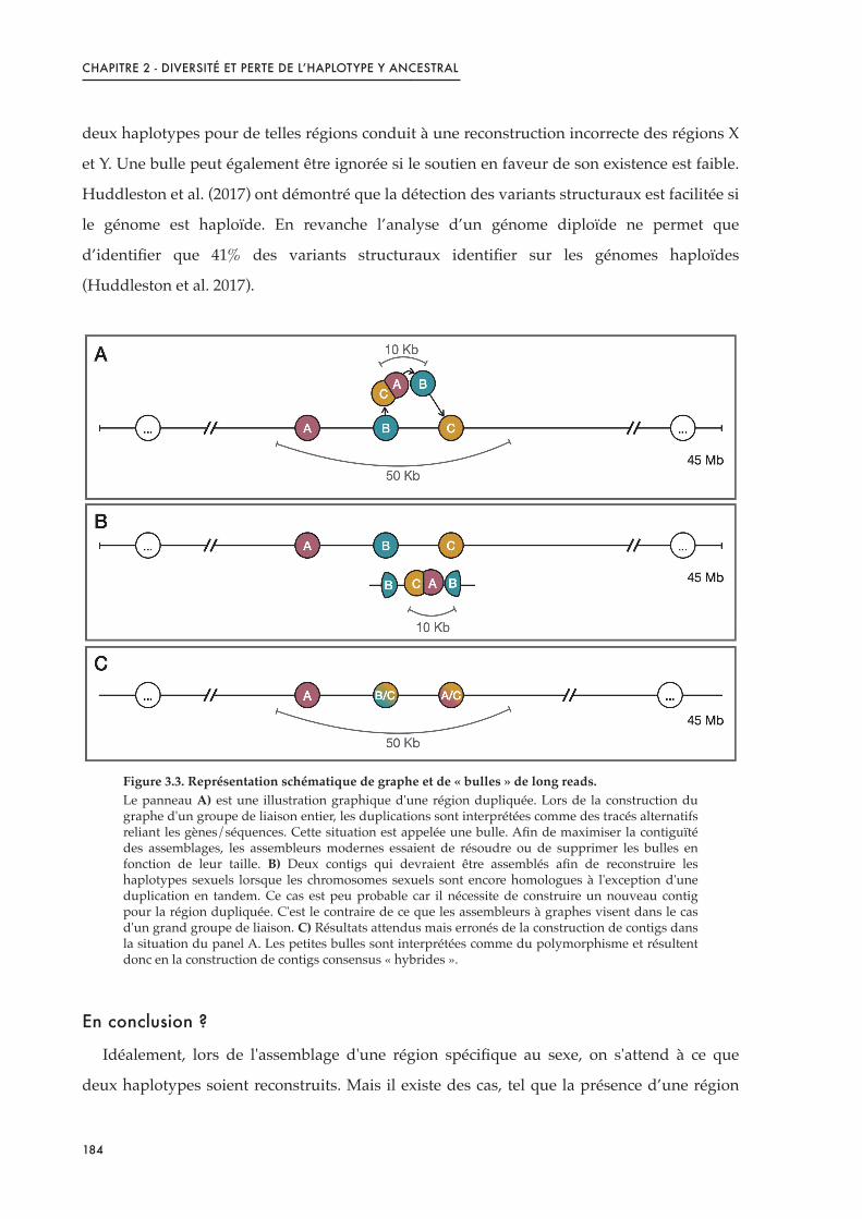
CHAPITRE 2 - DIVERSITÉ ET PERTE DE L’HAPLOTYPE Y ANCESTRAL
deux haplotypes pour de telles régions conduit à une reconstruction incorrecte des régions X
et Y. Une bulle peut également être ignorée si le soutien en faveur de son existence est faible.
Huddleston et al. (2017) ont démontré que la détection des variants structuraux est facilitée si
le génome est haploïde. En revanche l’analyse d’un génome diploïde ne permet que
d’identifier que 41% des variants structuraux identifier sur les génomes haploïdes
(Huddleston et al. 2017).
Figure 3.3. Représentation schématique de graphe et de « bulles » de long reads.Le panneau A) est une illustration graphique d'une région dupliquée. Lors de la construction du graphe d'un groupe de liaison entier, les duplications sont interprétées comme des tracés alternatifs reliant les gènes/séquences. Cette situation est appelée une bulle. Afin de maximiser la contiguïté des assemblages, les assembleurs modernes essaient de résoudre ou de supprimer les bulles en fonction de leur taille. B) Deux contigs qui devraient être assemblés afin de reconstruire les haplotypes sexuels lorsque les chromosomes sexuels sont encore homologues à l'exception d'une duplication en tandem. Ce cas est peu probable car il nécessite de construire un nouveau contig pour la région dupliquée. C'est le contraire de ce que les assembleurs à graphes visent dans le cas d'un grand groupe de liaison. C) Résultats attendus mais erronés de la construction de contigs dans la situation du panel A. Les petites bulles sont interprétées comme du polymorphisme et résultent donc en la construction de contigs consensus « hybrides ».
En conclusion ?
Idéalement, lors de l'assemblage d'une région spécifique au sexe, on s'attend à ce que
deux haplotypes soient reconstruits. Mais il existe des cas, tel que la présence d’une région
184
CHAPITRE 2 - DIVERSITÉ ET PERTE DE L’HAPLOTYPE Y ANCESTRAL
du déterminisme du sexe réduite par rapport à l'ensemble de la taille du contig, dans
lesquels il peut être difficile de reconstruire correctement les deux haplotypes. Une bonne
option serait de séquencer un génome homozygote pour la région sexuelle, comme des
individus XX et YY ou des individus ZZ et WW. Cependant, cela n'est pas toujours possible
car les génotypes YY ou WW ne sont pas toujours viables ou facile à produire.
Pour compenser l'absence d'échantillons YY ou WW, l'étude du sexe hétérogamétique
requiert des efforts plus importants. La profondeur de séquençage doit être doublée pour
obtenir une couverture de la région du sexe aussi satisfaisante que celle des régions
autosomiques. Les paramètres d'assemblage devront également faire l'objet d'une attention
particulière pour calibrer les logiciels d'assemblage afin de produire deux contigs différents
correspondant aux deux haplotypes sexuels en réalisant un compromis entre la fidélité et la
contiguïté dans ces régions en particulier.
Finalement, ces problèmes ne perdureront pas éternellement car les technologies de
séquençage deviennent moins chères, plus efficaces et produisent des lectures plus longues
et en plus grande quantité au fil du temps. Pour citer Phillippy (2017) qui passe en revue les
nouvelles avancées dans l'assemblage de séquences : "dès qu'une technologie de séquençage
pourra produire des lectures suffisamment bonnes, de sorte que toutes les régions du
génome puissent être distinguées de manière unique, l'assemblage du génome deviendra
trivial". Espérons qu'à ce stade, les séquences de chromosomes sexuels seront plus faciles à
démasquer et amélioreront considérablement notre compréhension de l'évolution des
chromosomes sexuels.
185

CHAPITRE 2 - DIVERSITÉ ET PERTE DE L’HAPLOTYPE Y ANCESTRAL
Références
Abbott, J.K., Nordén, A.K., and Hansson, B. (2017). Sex chromosome evolution: historical insights and future perspectives. Proceedings of the Royal Society B: Biological Sciences 284, 20162806.
Audano, P.A., Sulovari, A., Graves-Lindsay, T.A., Cantsilieris, S., Sorensen, M., Welch, A.E., Dougherty, M.L., Nelson, B.J., Shah, A., Dutcher, S.K., et al. (2019). Characterizing the Major Structural Variant Alleles of the Human Genome. Cell 176, 663-675.e19.
Cameron, D.L., Schröder, J., Penington, J.S., Do, H., Molania, R., Dobrovic, A., Speed, T.P., and Papenfuss, A.T. (2017). GRIDSS: sensitive and specific genomic rearrangement detection using positional de Bruijn graph assembly. Genome Res. 27, 2050–2060.
Carneiro, M.O., Russ, C., Ross, M.G., Gabriel, S.B., Nusbaum, C., and DePristo, M.A. (2012). Pacific biosciences sequencing technology for genotyping and variation discovery in human data. BMC Genomics 13, 375.
Cretu Stancu, M., van Roosmalen, M.J., Renkens, I., Nieboer, M.M., Middelkamp, S., de Ligt, J., Pregno, G., Giachino, D., Mandrile, G., Espejo Valle-Inclan, J., et al. (2017). Mapping and phasing of structural variation in patient genomes using nanopore sequencing. Nat Commun 8, 1326.
Darolti, I., Wright, A.E., Sandkam, B.A., Morris, J., Bloch, N.I., Farré, M., Fuller, R.C., Bourne, G.R., Larkin, D.M., Breden, F., et al. (2019). Extreme heterogeneity in sex chromosome differentiation and dosage compensation in livebearers. Proc Natl Acad Sci USA 116, 19031–19036.
Guan, D., McCarthy, S.A., Wood, J., Howe, K., Wang, Y., and Durbin, R. (2020). Identifying and removing haplotypic duplication in primary genome assemblies. Bioinformatics 36, 2896–2898.
Hattori, R.S., Murai, Y., Oura, M., Masuda, S., Majhi, S.K., Sakamoto, T., Fernandino, J.I., Somoza, G.M., Yokota, M., and Strüssmann, C.A. (2012). A Y-linked anti-Müllerian hormone duplication takes over a critical role in sex determination. PNAS 109, 2955–2959.
Huddleston, J., Chaisson, M.J.P., Steinberg, K.M., Warren, W., Hoekzema, K., Gordon, D., Graves-Lindsay, T.A., Munson, K.M., Kronenberg, Z.N., Vives, L., et al. (2017). Discovery and genotyping of structural variation from long-read haploid genome sequence data. Genome Res. 27, 677–685.
Layer, R.M., Chiang, C., Quinlan, A.R., and Hall, I.M. (2014). LUMPY: a probabilistic framework for structural variant discovery. Genome Biol 15, R84.
Mahmoud, M., Gobet, N., Cruz-Dávalos, D.I., Mounier, N., Dessimoz, C., and Sedlazeck, F.J. (2019). Structural variant calling: the long and the short of it. Genome Biol 20, 246.
Menegon, M., Cantaloni, C., Rodriguez-Prieto, A., Centomo, C., Abdelfattah, A., Rossato, M., Bernardi, M., Xumerle, L., Loader, S., and Delledonne, M. (2017). On site DNA barcoding by nanopore sequencing. PLoS ONE 12, e0184741.
Nattestad, M., Goodwin, S., Ng, K., Baslan, T., Sedlazeck, F.J., Rescheneder, P., Garvin, T., Fang, H., Gurtowski, J., Hutton, E., et al. (2018). Complex rearrangements and oncogene
186

CHAPITRE 2 - DIVERSITÉ ET PERTE DE L’HAPLOTYPE Y ANCESTRAL
amplifications revealed by long-read DNA and RNA sequencing of a breast cancer cell line. Genome Res. 28, 1126–1135.
Palmer, D.H., Rogers, T.F., Dean, R., and Wright, A.E. (2019). How to identify sex chromosomes and their turnover. Molecular Ecology 28, 4709–4724.
Pan, Q., Feron, R., Yano, A., Guyomard, R., Jouanno, E., Vigouroux, E., Wen, M., Busnel, J.-M., Bobe, J., Concordet, J.-P., et al. (2019). Identification of the master sex determining gene in Northern pike (Esox lucius) reveals restricted sex chromosome differentiation. PLOS Genetics 15, e1008013.
Phillippy, A.M. (2017). New advances in sequence assembly. Genome Res. 27, xi–xiii.
Rausch, T., Zichner, T., Schlattl, A., Stütz, A.M., Benes, V., and Korbel, J.O. (2012). DELLY: structural variant discovery by integrated paired-end and split-read analysis. Bioinformatics 28, i333–i339.
Sedlazeck, F.J., Rescheneder, P., Smolka, M., Fang, H., Nattestad, M., von Haeseler, A., and Schatz, M.C. (2018). Accurate detection of complex structural variations using single-molecule sequencing. Nat Methods 15, 461–468.
Simpson, J.T., and Pop, M. (2015). The Theory and Practice of Genome Sequence Assembly. 22.
Tomaszkiewicz, M., Medvedev, P., and Makova, K.D. (2017). Y and W Chromosome Assemblies: Approaches and Discoveries. Trends in Genetics 33, 266–282.
Vicoso, B., Emerson, J.J., Zektser, Y., Mahajan, S., and Bachtrog, D. (2013). Comparative Sex Chromosome Genomics in Snakes: Differentiation, Evolutionary Strata, and Lack of Global Dosage Compensation. PLoS Biol 11, e1001643.
Wenger, A.M., Peluso, P., Rowell, W.J., Chang, P.-C., Hall, R.J., Concepcion, G.T., Ebler, J., Fungtammasan, A., Kolesnikov, A., Olson, N.D., et al. (2019). Accurate circular consensus long-read sequencing improves variant detection and assembly of a human genome. Nat Biotechnol 37, 1155–1162.
187

CHAPITRE 2 - DIVERSITÉ ET PERTE DE L’HAPLOTYPE Y ANCESTRAL
The challenge of sex chromosome assembly
New sequencing methods to study sex chromosomes
Identifying sex chromosomes can be challenging especially when they are homomorphic
as they can not be differentiated by cytogenetic visualization. Molecular and genomic
analyses offer great opportunities to better detect sex-linked loci and identify sex
chromosomes. The improvement of new generation sequencing (NGS) technologies can
facilitate particularly the reconstruction of Y or W chromosomes through the use of Illumina
short reads, and more recently through long reads sequencing (Abbott et al., 2017). Over the
years different methods have been optimised to study sex chromosomes according to their
homomorphy, heteromorphy or along an intermediate gradient (Palmer et al., 2019).
Each sequencing technology presents its advantages and disadvantages. Illumina short
reads sequencing, while having the advantage of producing a very large amount of reads
with few errors, only produces small size sequences. They allow to efficiently identify
linkage groups carrying markers linked to sex. But, because of their usual length of
100-250bp, short reads are not always long enough to distinguish between repeat regions. As
such, duplicated regions are left unassembled or reads of both copies are merged as a single
locus when the paralogs are not divergent enough.
Long reads, however, can overcome this issue. The length of the reads allows to cross not
only the breakpoints but they can also overlap whole complex architectures when these
occur in the genome. Consequently, they untangle duplicated regions, repeated regions or
other types of structural variants more easily, as shown in various studies (Audano et al.,
2019; Nattestad et al., 2018; Sedlazeck et al., 2018; Wenger et al., 2019). However, the
downside of long reads sequencing is the higher error rate in the sequences. Long reads can
be impacted with 5 to 15% sequencing errors (Carneiro et al., 2012; Menegon et al., 2017). The
only way to counterbalance this downside is to increase the coverage, which can be
expensive depending on the size of the genome studied.
Some constraints impact both technologies. When studying the heterogametic sex, the first
concern is the presence of two divergent haplotypes in the same genome. As a result, the
coverage of each haplotype (e.g. X and Y) over a sex-specific region is only half of that of the
autosomes. Thus, the sequencing effort has to be doubled to obtain an equivalent coverage in
188

CHAPITRE 2 - DIVERSITÉ ET PERTE DE L’HAPLOTYPE Y ANCESTRAL
the sex-region of an heterogametic individual. This is why in the majority of cases the
reference genome of a species is chosen preferentially from an individual corresponding to
the homogametic sex (Tomaszkiewicz et al., 2017).
Mapping on a reference genome
When a reference genome is available, it is usually faster to map onto this reference than
generating a de novo assembly. However, except in the case of model organisms, reference
genomes, as previously mentioned, are usually conducted on homogametic individuals as
they tend to produce better assemblies. This is indeed a limitation when studying sex
chromosomes. If the sex determining locus is shared between the two haplotypes (e.g. X and
Y or Z and W) and these haplotypes have accumulated sex-specific nucleotide
polymorphisms, then a simple mapping, along with a SNPs detection will allow to correctly
describe the region. But in the eventuality where the sex chromosomes are not fully
homologous, different scenario can occur and impact the reconstruction of the Y or W
chromosomes.
i) If there are loci that are specific to the X (or Z) and absent in the Y (or W), when
mapping an XY (or ZW) individual over an homogametic based reference genome, there will
be no reads from the Y (or W) that cross this specific region. Consequently, only half of the
expected coverage will be observed over the X (or Z) regions (Figure 3.2 A).
ii) If there is a region present in the Y (or W) that has no homologous copy on the X (or Z),
a mapping of an individual of the heterogametic sex will result in an overall good mapping.
However, reads overlapping the homologous region and the insertion will not map to the
reference genome. A drop of coverage is thus expected for the narrow region corresponding
to the breakpoints of the insertion on the Y (or W) (Figure 3.2 B).
iii) If there is a duplication that is specific to the Y (or W) chromosome and both copies are
relatively similar to the single homologous region on the X (or Z) chromosome, the Y
chromosome duplication will not be fully assembled. The reads belonging to both copies
present on the Y (or W) will map to the single copy of the X (or Z), which will increase the
coverage of this region compared to autosomal regions (Figure 3.2 C).
These three different cases will not enable to properly assemble the two sex haplotypes of
the heterogametic sex but, nevertheless, the coverage variations resulting from these
189

CHAPITRE 2 - DIVERSITÉ ET PERTE DE L’HAPLOTYPE Y ANCESTRAL
situations can in turn be helpful to distinguish sex-linked loci from autosomal regions
(Vicoso et al., 2013; Darolti et al., 2019).
Figure 3.2. Problems encountered when mapping a heterogametic individual on a homogametic reference genome.The expected impact on reads coverage is displayed for three situations. All situations with a XY sex system are equivalent for a ZW system. A) Expected mapping when the Y chromosome lost part of its genes content (region b of the X). Only reads from the X chromosome (in red) map on the reference genome resulting in a drop of coverage over this region. B) Expected mapping when the Y chromosome acquired a locus absent from the X haplotype (region b of the Y). Reads specific to the b region of the Y will not map on the reference genome. Only reads from the X chromosome (in red) will map on the breakpoints of the insertion (a to c transition region) resulting in a local drop of coverage. C) Expected mapping when a duplication occurred only on the Y chromosome (two region b on the Y vs one region on the X) and copied are paralogs are not strongly divergent with the X copy. Reads belonging to both b copies on the Y chromosomes will map on the single b region on the reference genome. This situation will result in an increased coverage over the corresponding region.
By using pair-end short reads, the detection of such structural variants can be improved.
Some software, such as DELLY, LUMPY, GRIDSS (Rausch et al., 2012; Layer et al., 2014;
Cameron et al., 2017), compare the insert size between mate pairs along with their direction
and the coverage to what is expected and thus can determine the type and the length of
structural variants along the genome. Nevertheless, complex structures that are specific to
only Y or W haplotypes cannot be precisely described and as an illustration, it is hard to
determine to which copies belong the SNPs identified when all reads map to a single copy on
the reference genome.
X Y
4
3
2
1
Co
vera
ge
X
Y
a b c
a c
X Reference genome
a b c
X & Y Reads
Mb
Reads
Mapping
X Y
X
Y
a b c
a
X Reference genome
a b c
X & Y Reads
b cb
4
3
2
1
Co
vera
ge
Mb
Reads
Mapping
X Y
4
3
2
1
Co
vera
ge
X Reference genome
X & Y Reads
Mb
X
Ya b c
a c
Reads
a c
Mapping
A B C
190

CHAPITRE 2 - DIVERSITÉ ET PERTE DE L’HAPLOTYPE Y ANCESTRAL
Methods also exist to improve the detection of structural variants in long reads data
(based on coverage analysis among other assembly parameters) such as Sniffles, SMRT-SV
(for PacBio data) or NanoSV (for Oxford Nanopore data) (Cretu Stancu et al., 2017;
Huddleston et al., 2017; Sedlazeck et al., 2018). The detection of structural variants using long
reads results in better detection of these complexe regions even if the sequencing error rate is
higher than for short reads sequencing. Still, some limits appear in the presence of very large
insertions or inversions (eg. larger than 5kb). This is due to the reads being shorter than the
allele of the variant. For a complete review of the structural variants calling with short and
long reads following a mapping process, see Mahmoud et al., (2019).
Quick introduction to graph assembly
Using de novo assembly to reconstruct both sex haplotypes is thus a promising approach
when one wants to study haplotypes with structural variants such as tandem duplications
that are specific to one sex.
The majority of recent assembly software uses graph theory. Briefly, graphs are composed
of a set of vertices connected by edges (that can have a direction) reflecting association
between objects. In the case of long reads assembly, the type of graph used is often a “De
Bruijn Graph”. They are built based on reads and their overlapping information. For a
detailed review of theory and practice of genome sequence assembly and graph, see Simpson
and Pop, (2015).
When a structural variant occurs, the graph might not be linear anymore and some edges
might be linked in the same direction to several edges. It is the case for example when a
tandem repeat is present. The non-linear formation can be called a “bubble”. The aim of
these assembly software is to linearise the graphs obtained and this notably by resolving the
bubbles along the graph.
The quality of the resulting assembly is measured mainly on two criteria: the accuracy
and the contiguity of the assembly. The contiguity is increased when the linear graphs are
resulting in the longest chains possible with the least breaks possible. The accuracy on the
other hand is higher when the heterozygosity is low and thus the consensus sequence
resulting from the graph contains the least ambiguities possible. To produce the best
assemblies possible, tools are being developed in order to remove structural variants, such as
duplications, that are specific to an haplotype in a diploid genome (Guan et al., 2020).
191
CHAPITRE 2 - DIVERSITÉ ET PERTE DE L’HAPLOTYPE Y ANCESTRAL
The case of sex chromosomes
Sex chromosomes are by definition regions of high heterozygosity because they contain
loci that can be sex-specific and thus might not recombine anymore and accumulate sex-
specific SNPs. In addition, these regions are prone to haplotype-specific structural variants.
In fish for exemple, several species present a duplication of the sex-determinant gene in only
one of the two sex chromosomes (e.g. amh duplication on the Y in the Patagonian pejerrey
(Odontesthes hatcheri) and the Northern pike (Esox lucius) (Hattori et al., 2012; Pan et al.,
2019)). The presence of such structures would create bubbles in a graph (Figure 3.3 A). When
resolving this bubble, the compromise between accuracy and contiguity can lead to different
scenario. If the sex specific region is divergent enough between the two haplotypes, then the
assembly might results in two contigs, which is the ideal case when studying sex
chromosomes (Figure 3.3 B). But, if the sex region is small compared to the overall length of
the contig graph, then a hybrid of the two sex haplotypes might be built (Figure 3.3 C). This
might notably be the case for a short sex-specific region carried by homomorphic
chromosomes. Merging the two haplotypes for such regions lead to false reconstruction of
both X and Y regions. A bubble can also be ignored if the support for its existence is low.
Huddleston et al., (2017) demonstrated that haploid genomes help to better detect structural
variants. They performed that same analysis on a pseudo-diploid genome (merging two
haploids) and detected 59% less structural variants than on the haploid dataset.
What are the options?
Ideally, when assembling a sex region, two haplotypes are expected to be reconstructed. In
the case of a tandem duplication where the divergent region between the two chromosomes
is small compared to the whole contig, it might be a challenge to correctly reconstruct the
two sequences. A good option would be to sequence homozygous genomes for the sex
region, such as XX and YY individuals or ZZ and WW individuals. However, this is not
always possible as YY or WW genotypes are not always viable or easy to obtain.
To compensate for the absence of YY or WW samples, the study of heterogametic sex will
require greater efforts. The sequencing depth has to be doubled to obtain a coverage over the
sex-region that is as good as over autosomal regions. Assembly parameters will also require a
close attention to calibrate assembly software in order to produce two different contigs
192

CHAPITRE 2 - DIVERSITÉ ET PERTE DE L’HAPLOTYPE Y ANCESTRAL
corresponding to sex-specific loci with a compromise between accuracy and contiguity in
these regions particularly.
Figure 3.3. Schematic representation of graph and « bubbles » of long reads and hybrid assembly. Panel A) is a graph illustration of a duplicated region. While constructing the graph of a whole linkage group, duplications are interpreted as alternative pathways linking genes/sequences. This situation is called a bubble. In order to maximise the contiguity of assemblies, modern graph assemblers are trying to resolve or suppress bubbles depending on their sizes. B) Two contigs that should be built in order to re-construct sex haplotypes when sex chromosomes are still homologous with exception of a tandem duplication. This case is unlikely to happen as it requires to build a new contig for the duplicated region. This is the opposite of what graphs assemblers are aiming for in the case of a large linkage group. C) Expected but erroneous results of contig construction with graph assembler in the panel A situation. Small bubbles are interpreted as polymorphism and thus result in the construction of « hybrid » consensus contig.
Eventually, these problems will not last forever as sequencing technologies are becoming
cheaper, more efficient and are producing longer reads and in larger amounts through time.
To quote Phillippy, (2017) reviewing the new advances in sequence assembly: “as soon as a
sequencing technology can produce good enough reads, such that all regions of the genome
can be uniquely distinguished, genome assembly will become trivial”. Hopefully, at this
point in time, the sequences of sex chromosomes will be easier to uncover, greatly improving
our comprehension of sex chromosome evolution.
193

CHAPITRE 2 - DIVERSITÉ ET PERTE DE L’HAPLOTYPE Y ANCESTRAL
References
Abbott, J.K., Nordén, A.K., and Hansson, B. (2017). Sex chromosome evolution: historical insights and future perspectives. Proceedings of the Royal Society B: Biological Sciences 284, 20162806.
Audano, P.A., Sulovari, A., Graves-Lindsay, T.A., Cantsilieris, S., Sorensen, M., Welch, A.E., Dougherty, M.L., Nelson, B.J., Shah, A., Dutcher, S.K., et al. (2019). Characterizing the Major Structural Variant Alleles of the Human Genome. Cell 176, 663-675.e19.
Cameron, D.L., Schröder, J., Penington, J.S., Do, H., Molania, R., Dobrovic, A., Speed, T.P., and Papenfuss, A.T. (2017). GRIDSS: sensitive and specific genomic rearrangement detection using positional de Bruijn graph assembly. Genome Res. 27, 2050–2060.
Carneiro, M.O., Russ, C., Ross, M.G., Gabriel, S.B., Nusbaum, C., and DePristo, M.A. (2012). Pacific biosciences sequencing technology for genotyping and variation discovery in human data. BMC Genomics 13, 375.
Cretu Stancu, M., van Roosmalen, M.J., Renkens, I., Nieboer, M.M., Middelkamp, S., de Ligt, J., Pregno, G., Giachino, D., Mandrile, G., Espejo Valle-Inclan, J., et al. (2017). Mapping and phasing of structural variation in patient genomes using nanopore sequencing. Nat Commun 8, 1326.
Darolti, I., Wright, A.E., Sandkam, B.A., Morris, J., Bloch, N.I., Farré, M., Fuller, R.C., Bourne, G.R., Larkin, D.M., Breden, F., et al. (2019). Extreme heterogeneity in sex chromosome differentiation and dosage compensation in livebearers. Proc Natl Acad Sci USA 116, 19031–19036.
Guan, D., McCarthy, S.A., Wood, J., Howe, K., Wang, Y., and Durbin, R. (2020). Identifying and removing haplotypic duplication in primary genome assemblies. Bioinformatics 36, 2896–2898.
Hattori, R.S., Murai, Y., Oura, M., Masuda, S., Majhi, S.K., Sakamoto, T., Fernandino, J.I., Somoza, G.M., Yokota, M., and Strüssmann, C.A. (2012). A Y-linked anti-Müllerian hormone duplication takes over a critical role in sex determination. PNAS 109, 2955–2959.
Huddleston, J., Chaisson, M.J.P., Steinberg, K.M., Warren, W., Hoekzema, K., Gordon, D., Graves-Lindsay, T.A., Munson, K.M., Kronenberg, Z.N., Vives, L., et al. (2017). Discovery and genotyping of structural variation from long-read haploid genome sequence data. Genome Res. 27, 677–685.
Layer, R.M., Chiang, C., Quinlan, A.R., and Hall, I.M. (2014). LUMPY: a probabilistic framework for structural variant discovery. Genome Biol 15, R84.
Mahmoud, M., Gobet, N., Cruz-Dávalos, D.I., Mounier, N., Dessimoz, C., and Sedlazeck, F.J. (2019). Structural variant calling: the long and the short of it. Genome Biol 20, 246.
Menegon, M., Cantaloni, C., Rodriguez-Prieto, A., Centomo, C., Abdelfattah, A., Rossato, M., Bernardi, M., Xumerle, L., Loader, S., and Delledonne, M. (2017). On site DNA barcoding by nanopore sequencing. PLoS ONE 12, e0184741.
Nattestad, M., Goodwin, S., Ng, K., Baslan, T., Sedlazeck, F.J., Rescheneder, P., Garvin, T., Fang, H., Gurtowski, J., Hutton, E., et al. (2018). Complex rearrangements and oncogene
194

CHAPITRE 2 - DIVERSITÉ ET PERTE DE L’HAPLOTYPE Y ANCESTRAL
amplifications revealed by long-read DNA and RNA sequencing of a breast cancer cell line. Genome Res. 28, 1126–1135.
Palmer, D.H., Rogers, T.F., Dean, R., and Wright, A.E. (2019). How to identify sex chromosomes and their turnover. Molecular Ecology 28, 4709–4724.
Pan, Q., Feron, R., Yano, A., Guyomard, R., Jouanno, E., Vigouroux, E., Wen, M., Busnel, J.-M., Bobe, J., Concordet, J.-P., et al. (2019). Identification of the master sex determining gene in Northern pike (Esox lucius) reveals restricted sex chromosome differentiation. PLOS Genetics 15, e1008013.
Phillippy, A.M. (2017). New advances in sequence assembly. Genome Res. 27, xi–xiii.
Rausch, T., Zichner, T., Schlattl, A., Stütz, A.M., Benes, V., and Korbel, J.O. (2012). DELLY: structural variant discovery by integrated paired-end and split-read analysis. Bioinformatics 28, i333–i339.
Sedlazeck, F.J., Rescheneder, P., Smolka, M., Fang, H., Nattestad, M., von Haeseler, A., and Schatz, M.C. (2018). Accurate detection of complex structural variations using single-molecule sequencing. Nat Methods 15, 461–468.
Simpson, J.T., and Pop, M. (2015). The Theory and Practice of Genome Sequence Assembly. 22.
Tomaszkiewicz, M., Medvedev, P., and Makova, K.D. (2017). Y and W Chromosome Assemblies: Approaches and Discoveries. Trends in Genetics 33, 266–282.
Vicoso, B., Emerson, J.J., Zektser, Y., Mahajan, S., and Bachtrog, D. (2013). Comparative Sex Chromosome Genomics in Snakes: Differentiation, Evolutionary Strata, and Lack of Global Dosage Compensation. PLoS Biol 11, e1001643.
Wenger, A.M., Peluso, P., Rowell, W.J., Chang, P.-C., Hall, R.J., Concepcion, G.T., Ebler, J., Fungtammasan, A., Kolesnikov, A., Olson, N.D., et al. (2019). Accurate circular consensus long-read sequencing improves variant detection and assembly of a human genome. Nat Biotechnol 37, 1155–1162
195


CHAPITRE 3 : DIFFÉRENCIATION DU SEXE CHEZ UNE POPULATION
QUI NE PRÉSENTE PAS LE CHROMOSOME Y ANCESTRAL


CHAPITRE 3 - DIFFÉRENCIATION DU SEXE SANS LE Y ANCESTRAL
Objectifs
Dans la population Ethiopienne du lac Hora, étudiée précédemment, aucune duplication
de l’amh n’a pu être identifiée sur LG23-Y et aucun autre groupe de liaison n’a pour le
moment été identifié comme la nouvelle paire de chromosomes sexuels. Dans ce chapitre,
l’objectif était donc d’explorer le transcriptome de cette population Ethiopienne Hora pour le
comparer à celui des souches domestiques dont le sexe est déterminé par LG23-Y. En effet, à
un stade précoce de la différenciation, il peut déjà exister un dimorphisme dans l’expression
des gènes clés de la cascade du sexe. Nous avons aussi analysé l’effet de la température sur le
transcriptome de cette population.
Introduction et contexte
Le tilapia du Nil (Oreochromis niloticus) est une espèce de cichlidé Africain d’intérêt
économique (FAO, 2019). Son aire de répartition est grande puisqu’on la retrouve du Sénégal
à l’ouest de l’Afrique, jusqu’à l’est de l’Afrique dans le Rift Ethiopien, en passant par les
bassins du Nil (Trewavas, 1983; Bezault et al., 2011). Cette espèce présente une grande
capacité d’adaptation à différentes conditions écologiques et notamment thermiques avec
des populations vivant dans des eaux «!froides!» (17 à 26°C), des eaux tempérées (28 à 34°C)
mais aussi des sources chaudes (36 à 40°C) (Bezault et al., 2007; Ndiwa et al., 2014). Comme
nous l’avons vu dans les chapitres précédents, le déterminisme du sexe chez cette espèce est
complexe et reste mal compris. En effet, des signaux associés au sexe ont été identifiés sur les
groupes de liaison (LG) 1 (Lee et al., 2003; Palaiokostas et al., 2013; Gammerdinger et al.,
2014; Palaiokostas et al., 2015; Taslima et al., 2020) ou 23 (Eshel et al., 2012, 2014; Li et al.,
2015; Wessels et al., 2017; Cáceres et al., 2019), ou même les deux en parallèle (Taslima et al.,
2021), selon la provenance des souches domestiques étudiées. Dans les populations
naturelles étudiées jusqu’alors, LG23 a aussi été identifié comme fortement associé au sexe
dans les individus du lac Kou (Burkina-Faso), Kpandu du lac Volta (Ghana) et Koka du lac
Koka (Ethiopie) (Sissao et al., 2019; Triay et al., 2020). La région du sexe identifiée sur LG23
correspond à une région dupliquée donnant lieu à deux copies du gène de l’amh dans
l’haplotype Y (Li et al., 2015; Triay et al., 2020). Des analyses phylogénétiques sur les
populations sauvages de Kpandu, Koka et Hora (Lac Hora, Ethiopie) ont permis de conclure
que ce déterminisme est certainement ancestral à la divergence des populations de l’Est et de
199

CHAPITRE 3 - DIFFÉRENCIATION DU SEXE SANS LE Y ANCESTRAL
l’Ouest de l’Afrique. Cependant, dans la population Hora aucune duplication de l’amh n’a pu
être identifiée sur LG23-Y et aucun autre groupe de liaison n’a pour le moment été identifié
comme la nouvelle paire de chromosomes sexuels.
Les cichlidés présentent tout particulièrement une grande diversité des déterminants du
sexe avec près de 20 systèmes de déterminismes différents sur 17 LGs (Gammerdinger &
Kocher, 2018; El Taher et al., 2020). Cependant, certains gènes candidats («!usual suspects!»)
reviennent régulièrement et «! jouent aux chaises musicales! » chez les cichlidés et plus
largement chez les téléostéens (Böhne et al., 2016; Pan et al., 2016). Ainsi, même si le
déterminant du sexe peut changer, les gènes impliqués en aval de la cascade de la
différenciation du sexe sont généralement conservés chez les téléostéens (revues de Todd et
al., 2016 et Ortega-Recalde et al., 2020) et pourraient jouer un rôle chez la population Hora
(Figure 4.1).
Figure 4.1. Modèle de réseaux de gènes antagonistes spécifiques à chaque sexe contrôlant la différenciation sexuelle chez les poissons.Des facteurs conservés en aval favorisent la voie de féminisation ou de masculinisation, respectivement, tout en inhibant activement la voie sexuelle opposée. La testostérone peut être convertie soit en 17β-œstradiol (E2, l'œstrogène le plus actif chez le poisson) par l'aromatase gonadique (Cyp19a1a), soit en 11KT (l'androgène le plus actif chez les poissons) par les enzymes 11β-hydroxylase (Cyp11c1) et 11β-hydroxystéroïde déshydrogénase de type 2 (Hsd11b2). Cyp11c1 et Hsd11b2 sont critiques non seulement pour la synthèse des androgènes 11-oxygénés mais aussi pour le métabolisme des glucocorticoïdes (par exemple, le cortisol). Abréviations : 11bOHT, 11β-hydroxytestosterone; 11KT, 11-ketotestosterone; Amh, anti-Müllerian hormone; Dmrt1, doublesex and mab-3 related transcription factor 1; Foxl2, forkhead transcriptional factor L2; S, 11-deoxycortisol; Sf1, steroidogenic factor 1; Sox9, SRY-related HMG box 9. Issu et traduit de Ortega-Recalde et al., (2020), déjà adapté de Todd et al., (2016).
!"#$%
&'( !)*+,(
-*.
&"#/!
012((3%
& 4"+,51"67
8% 971,"1,7+"67 ((3:09
4;<((=(4;<(/>(>?3
4"+,51"$
((@9
"#$%&#'(#)#*+,'%)-
#.+(#)#*+,'./0(/%$
1%&#'(#)#*+,'%)-
#.+(#)#*+,'./0(/%$
200

CHAPITRE 3 - DIFFÉRENCIATION DU SEXE SANS LE Y ANCESTRAL
De plus, à un stade précoce de la différenciation, il peut déjà exister un dimorphisme dans
l’expression des gènes clés de la cascade du sexe chez le tilapia du Nil (Poonlaphdecha et al.,
2011; Zhao et al., 2019). L’étude de l’expression des gènes candidats pourrait donc nous
permettre de comparer le transcriptome de cette population Ethiopienne Hora avec celui des
souches domestiques dont le sexe est déterminé par LG23-Y. Enfin, nous avons également
analysé l’effet de la température sur le transcriptome de cette population pendant le
développement des alevins.
Matériels et méthodes
Des individus d’O. niloticus cancellatus originaires du lac Hora (Ethiopie) on été maintenus
pendant plusieurs années au CIRAD à Montpellier, sans sélection particulière ou
domestication particulières. En 2016, une descendance a été produite par reproduction
naturelle entre deux géniteurs. Les œufs fertilisés se sont développés en conditions
contrôlées et au stade de 10 jours après fécondation (10 dpf) les alevins ont été séparés en
deux groupes : un groupe contrôle, maintenu dans un bassin à 27°C et un groupe traité par la
température, maintenu dans un bassin à 36°C. À 14 dpf et 23 dpf pendant la période de
différenciation du sexe où notamment l’expression de l’amh est déjà dimorphique
(Poonlaphdecha et al., 2011), les gonades (toujours attachées au tronc), ainsi que les cerveaux
(contenus dans la tête entière) ont été prélevés (n=16/lot) des deux groupes expérimentaux.
A 14 dpf, 8 individus (8 têtes et 8 troncs) à 27°C et 9 troncs d’individus à 36°C (contenant
potentiellement des mâles XX inversés par la température) ont servi à produire des banques
de RNAseq, séquencées en lectures pairées de 100bp.
L’ensemble des données de RNAseq de chaque individu a suivi le même traitement
bioinformatique, à savoir : un nettoyage des lectures, un alignement sur le génome de
référence du tilapia du Nil, les lectures alignées sur chaque locus pour chaque individu sont
dénombrées et analysées sur R pour comparer les différences d’expressions entre tissus
(cerveaux vs troncs) et traitements températures (27°C vs 36°C). Nous avons également
procédé à une identification des voies métaboliques sur- ou sous-représentées selon les tissus
et les températures d’exposition.
Enfin, l’ensemble de ces analyses (choix des individus et étapes bioinformatiques) ont dû
être menées sans a priori sur le sexe phénotypique des individus puisqu’ils sont trop petits à
201

CHAPITRE 3 - DIFFÉRENCIATION DU SEXE SANS LE Y ANCESTRAL
14 dpf pour être sexés sur la base de la morphologie des gonades, et sans a priori
génotypiques puisque les marqueurs de la duplication de l’amh chez les mâles ne sont pas
fiables pour les populations Ethiopiennes (même celle présentant la duplication de la région
de l’amh sur LG23-Y).
Afin d’identifier de potentielles modifications de la cascade du déterminisme du sexe chez
Hora nous avons également porté une attention particulière aux niveaux d’expressions dans
le cerveau et les gonades de certains gènes préalablement identifiés comme
différentiellement exprimés entre mâles et femelles ou appartenant à la cascade du
déterminisme du sexe chez des espèces proches.
Des analyses de PCR quantitative en temps réelles (qPCR) ont aussi été mené sur les
troncs à 14 et 23 dpf en utilisant des amorces de gènes clefs de la différenciation testiculaire
(gsdf, dmrt1, amh et arα) et de la différenciation ovarienne (foxl2, cyp19a1a, cyp19alb). Nous
avons aussi vérifié la correspondance entre les analyses RNASeq et qPCR.
Principaux résultats et discussion
i. Les fortes températures impactent les voies métaboliques de la synthèse des stéroïdes dans les gonades
Nous avons observés des différences d’expressions des cascades de production de
stéroïdes dans les gonades chez les individus traités à la température, en comparaison de
ceux maintenus à température contrôle. Les voies de biosynthèse des stéroïdes étaient et de
la biosynthèse des hormones stéroïdiennes était particulièrement impactées par les hautes
températures.
Les hormones stéroïdiennes sont impliquées dans le développement embryonnaire, la
différenciation du sexe, la reproduction ainsi que la réponse au stress chez les poissons
téléostéens (Goikoetxea et al., 2017). On retrouve entre autre les œstrogènes, qui sont
synthétisés à partir des androgènes par l'enzyme aromatase codée par les gènes cyp19a1a et
cyp19a1b (Baroiller et al., 1999; Guiguen et al., 2010). Les ovaires pendant la différenciation
chez le tilapia du Nil ont des niveaux plus élevés d'estradiol-17 et de cyp19a1a par rapport
aux testicules (Baroiller et D'Cotta, 2001; D'Cotta et al., 2001). Nous avons observé que la
température avait un effet répressif sur l'expression de cyp19a1a à 23 dpf dans les troncs de
Hora à 36°C ce qui n'était pas le cas dans les troncs à 27°C. Une répression similaire de
202
CHAPITRE 3 - DIFFÉRENCIATION DU SEXE SANS LE Y ANCESTRAL
l'expression de cyp19a1a (affectant les niveaux d'œstrogènes), a été montrée dans les gonades
de tilapia XX masculinisé par la température (traitées à 36°C) (D'Cotta et al., 2001;
Poonlaphdecha et al., 2013; Sun et al., 2018).
Parmi ces hormones stéroïdiennes on retrouve également le cortisol, un glucocorticoïde
qui est considéré comme «! l’hormone du stress!» chez les vertébrés. L’augmentation de la
production de cortisol a déjà été mise en lien avec des conditions de fortes températures chez
le Pejerrey et le Medaka notamment (Hattori et al., 2009; Yamaguchi et al., 2010). De plus,
l’augmentation du cortisol peut impacter la cascade de différenciation du sexe en influant
par exemple sur la régulation du gène de l’aromatase (Cyp19a1a) ou encore du gène de la
11β-hydroxysteroid dehydrogenase type 2 (Hsd11b2) (Wang et al., 2007; Quek & Chan, 2009;
Rajakumar & Senthilkumaran, 2016; Goikoetxea et al., 2017). Or, la 11β-hydroxylase, codée
par le gène cyp11c1, est impliquée dans la synthèse du cortisol mais aussi de la 11-
Kétotestostérone, le principal androgène chez les téléostéens (Zheng et al., 2020). Enfin, les
niveaux d'expression de cyp11c1 et 11β-hsd montrent des augmentations considérables chez
de nombreux individus à 36°C, suggérant que de forte température puissent induire une
masculinisation liée aux niveaux de cortisol ou de 11-kétotestostérone. En revanche, les
températures du milieu ne dépassent pas la température connue de masculinisation du sexe
(32°C) et ce phénomène n’est peut-être pas représentatif des conditions naturelles (Baroiller
& D’Cotta, 2001).
ii. Certains «!suspects habituels!» de la différenciation du sexe sont différentiellement exprimés dans les gonades exposées à de fortes températures
Entre les gonades dans les deux conditions de température, nous avons observé des
expressions différentielles de certains gènes qui s’avèrent également faire partie des locus
régulièrement retrouvés dans la cascade du déterminisme et de la différenciation du sexe
(Böhne et al., 2016; Pan et al., 2016). Parmi eux, des membres de la famille des «!Jumonji!»,
des «!Heatshock protein!».
Certains gènes de la famille des Jumonji ont déjà été identifiés chez les reptiles dans les
processus de réversion du sexe (Deveson et al., 2017). Des changements d’expression de
gènes de la famille des «!Heatshock proteins!» ont lieu chez certains poissons pendant les
périodes les plus chaudes de l’année en milieu naturel (Domingos et al., 2013). Elles sont
203

CHAPITRE 3 - DIFFÉRENCIATION DU SEXE SANS LE Y ANCESTRAL
également liées à la prolifération plus ou moins altérée des cellules germinales dans la région
des gonades chez le danio notamment (Pfeiffer et al., 2018) et de fortes températures
entrainent des modifications d’expression des heatshock protein dans les gonades mâles chez
l’Anguille de marais asiatique (He et al. 2010). Enfin, on retrouve aussi Kdm6bb, qui chez la
tortue de Floride (dont le déterminisme du sexe est dépendant de la température) favorise la
transcription du gène du déterminisme du sexe mâle dmrt1 (Ge et al., 2018). La répression de
l’expression de l'aromatase cyp19a1a à 23dpf dans les troncs à 36°C ainsi que de
l'augmentation des gènes de gsdf, dmrt1, cyp11c1, 11β-hsd et de kdm6bb et jarid2, tend
également à montrer que les individus Hora sont sensibles à la températures et sujets à la
masculinisation.
iii. Gsdf permet d’identifier deux groupes au sein de chaque traitement température, suggérant un rôle important dans la cascade de différenciation du sexe
Gsdf (gonadal soma-derived factor) a déjà régulièrement été identifié dans les cascades de
différenciation mâle chez les poissons et notamment chez le médaka, le Danio, le Bar ou le
Cardeau hirame (Crespo et al., 2013; Zhang et al., 2016; Horie et al., 2016; Yan et al., 2017;
Yang et al., 2019). L’expression de gsdf chez le tilapia du Nil est dimorphique entre mâles et
femelles avant même la différenciation morphologique des gonades (Kaneko et al. 2015) et a
été mis en lien avec la masculinisation dans les souches de laboratoires (Jiang et al., 2016).
Parmi les gènes candidats du déterminisme et de la différenciation du sexe, gsdf se
détache des autres dans nos analyses sur les gonades.
Nous avons constaté que les différences de niveaux d’expressions pour ce gène
permettent de former deux groupes : les individus à 27 et 36°C présentant une forte
expression de gsdf et ceux à 27 et 36°C ne présentant pas une très forte expression de gsdf et
que ce profil plus marqué encore à 23 dpf. Ces deux groupes observés pourraient
correspondre aux sexes phénotypiques et les individus présentant de fortes expressions
seraient des mâles en suivant cette logique. Ce résultat indiquerait donc un rôle important et
conservé de gsdf au stade précoce du développement chez la population Hora même si celle-
ci ne présente pas le même déterminant majeur du sexe que les autres populations sauvages
analysées.
204

CHAPITRE 3 - DIFFÉRENCIATION DU SEXE SANS LE Y ANCESTRAL
De plus, on note que la variance des expressions entre les individus à 36°C est bien
supérieure à celle des individus à température contrôle. Il est possible que cela soit dû à la
présence d’individus dont le sexe est inversé par la température et ainsi que le groupe des
mâles 36°C soit en réalité composé d’individus XX et XY qui n’auraient pas exactement les
mêmes expressions. Gsdf est d’ailleurs plus fortement exprimé chez les individus à 36°C que
chez les individus à 27°C, or une augmentation de son expression par la température a déjà
été identifié comme élément déclencheur du développement de mâles XX chez le tilapia du
Nil (Yang et al., 2019, 2020). Malheureusement, comme nous de disposons pas de marqueurs
pour sexer génotypiquement les individus, nous n’avons pas été en mesure d’identifier ou
non la présence de mâles XX dans le groupe des potentiels mâles 36°C.
iv. Parmi les gènes candidats, l’amh ne contribue pas à former les groupes phénotypiques potentiels identifiés
Contrairement aux résultats de Poonlaphdecha et al., (2011) montrant un clair
dimorphisme de l’expression de l’amh dans le cerveau entre 10 et 15 dpf entre les mâles XY et
les femelles XX, nous n’avons pas pu observer de clustering dans les cerveaux des individus.
L’amh n’était donc pas suffisamment dimorphique pour pouvoir former deux groupes
d’individus correspondant aux sexes phénotypiques. Comme l’amh ne semble pas être le
déterminant du sexe dans la population Hora, le fait qu’elle n’apparaisse pas comme
fortement dimorphique n’est peut-être pas si surprenant. Cependant, nous n’avons pas pu
observer de clustering qui pourrait correspondre au sexe phénotypique en utilisant un set
entier de gènes distinguant mâles et femelles dans les cerveaux chez le tilapia du Nil selon
Zhao et al., (2019). Une raison pourrait être que l’analyse sur les cerveaux que nous avons
effectuée était biaisée par le faible nombre d’individus, et le fort nombre de gènes analysés en
parallèle.
Conclusion
Cette étude de la différenciation sexuelle chez le tilapia du Nil Hora a montré que gsdf est
déjà exprimé à 14 dpf et son expression semble distinguer deux groupes d’individus
identifiés comment potentiels mâles et potentielles femelles. En revanche, ni dmrt1 ni l’amh
ne semble contribue à identifier ces potentiels groupes phénotypiques. Ceci indique que la
différenciation sexuelle chez le tilapia Hora suit une voie différente de celle que l'on trouve
205

CHAPITRE 3 - DIFFÉRENCIATION DU SEXE SANS LE Y ANCESTRAL
généralement chez les souches domestiques de tilapia du Nil. Des différences ont également
été observées dans les têtes, puisque l'expression de l’amh ne semble pas dimorphique à 14
dpf. Nous avons également trouvé que les fortes températures dans les troncs influençait
l’expression de plusieurs gènes identifiés comme des régulateurs épigénétiques tels que les
modificateurs d'histone kdm6bb et les gènes jarid2, liés à la synthèse du cortisol et de la 11-
ketotestostérone. Ces résultats, ainsi que l'augmentation des niveaux d’expression de gsdf,
dmrt1 et la répression du cyp19a1a ovarien dans les gonades potentiellement mâles,
suggèrent que des températures élevées induisent une masculinisation dans cette famille
Hora. Enfin, cette famille semble thermosensible bien qu’elle vive dans un lac froid.
206

CHAPITRE 3 - DIFFÉRENCIATION DU SEXE SANS LE Y ANCESTRAL
Références
Baroiller, J.F., and D’Cotta, H. (2001). Environment and sex determination in farmed fish. Comparative Biochemistry and Physiology Part C: Toxicology & Pharmacology 130, 399–409.
Baroiller, J.-F., Guiguen, Y., and Fostier, A. (1999). Endocrine and environmental aspects of sex differentiation in fish. CMLS, Cell. Mol. Life Sci. 55, 910–931.
Bezault, E., Clota, F., Derivaz, M., Chevassus, B., and Baroiller, J.-F. (2007). Sex determination and temperature-induced sex differentiation in three natural populations of Nile tilapia (Oreochromis niloticus) adapted to extreme temperature conditions. Aquaculture 272, S3–S16.
Bezault, E., Balaresque, P., Toguyeni, A., Fermon, Y., Araki, H., Baroiller, J.-F., and Rognon, X. (2011). Spatial and temporal variation in population genetic structure of wild Nile tilapia (Oreochromis niloticus) across Africa. BMC Genetics 12, 102.
Böhne, A., Wilson, C.A., Postlethwait, J.H., and Salzburger, W. (2016). Variations on a theme: Genomics of sex determination in the cichlid fish Astatotilapia burtoni. BMC Genomics 17.
Cáceres, G., López, M.E., Cádiz, M.I., Yoshida, G.M., Jedlicki, A., Palma-Véjares, R., Travisany, D., Díaz-Domínguez, D., Maass, A., Lhorente, J.P., et al. (2019). Fine mapping using whole-genome sequencing confirms anti-müllerian hormone as a major gene for sex determination in farmed Nile tilapia (Oreochromis niloticus L.). G3 (Bethesda) 9, 3213–3223.
Crespo, B., Gómez, A., Mazón, M.J., Carrillo, M., and Zanuy, S. (2013). Isolation and characterization of Ff1 and Gsdf family genes in European sea bass and identification of early gonadal markers of precocious puberty in males. General and Comparative Endocrinology 191, 155–167.
D’Cotta, H., Fostier, A., Guiguen, Y., Govoroun, M., and Baroiller, J.-F. (2001). Aromatase plays a key role during normal and temperature-induced sex differentiation of tilapia Oreochromis niloticus. Molecular Reproduction and Development 59, 265–276.
Deveson, I.W., Holleley, C.E., Blackburn, J., Marshall Graves, J.A., Mattick, J.S., Waters, P.D., and Georges, A. (2017). Differential intron retention in Jumonji chromatin modifier genes is implicated in reptile temperature-dependent sex determination. Sci. Adv. 3, e1700731.
Domingos, F.F.T., Thomé, R.G., Martinelli, P.M., Sato, Y., Bazzoli, N., and Rizzo, E. (2013). Role of HSP70 in the regulation of the testicular apoptosis in a seasonal breeding teleost Prochilodus argenteus from the São Francisco River, Brazil. Microsc Res Tech 76, 350–356.
El Taher, A., Ronco, F., Matschiner, M., Salzburger, W., and Böhne, A. (2020). Dynamics of sex chromosome evolution in a rapid radiation of cichlid fishes (Evolutionary Biology).
Eshel, O., Shirak, A., Weller, J.I., Hulata, G., and Ron, M. (2012). Linkage and physical mapping of sex Rregion on LG23 of Nile tilapia (Oreochromis niloticus). G3: Genes, Genomes, Genetics 2, 35–42.
Eshel, O., Shirak, A., Dor, L., Band, M., Zak, T., Markovich-Gordon, M., Chalifa-Caspi, V., Feldmesser, E., Weller, J.I., Seroussi, E., et al. (2014). Identification of male-specific amh
207

CHAPITRE 3 - DIFFÉRENCIATION DU SEXE SANS LE Y ANCESTRAL
duplication, sexually differentially expressed genes and microRNAs at early embryonic development of Nile tilapia (Oreochromis niloticus). BMC Genomics 15, 774.
FAO (2021). The State of World Fisheries and Aquaculture 2021 (FAO).
Gammerdinger, W.J., and Kocher, T.D. (2018). Unusual diversity of sex chromosomes in African cichlid fishes. Genes 9, 480.
Gammerdinger, W.J., Conte, M.A., Acquah, E.A., Roberts, R.B., and Kocher, T.D. (2014). Structure and decay of a proto-Y region in Tilapia, Oreochromis niloticus. BMC Genomics 15, 975.
Ge, C., Ye, J., Weber, C., Sun, W., Zhang, H., Zhou, Y., Cai, C., Qian, G., and Capel, B. (2018). The histone demethylase KDM6B regulates temperature-dependent sex determination in a turtle species. Science.
Goikoetxea, A., Todd, E.V., and Gemmell, N.J. (2017). Stress and sex: does cortisol mediate sex change in fish? Reproduction 154, R149–R160.
Guiguen, Y., Fostier, A., Piferrer, F., and Chang, C.-F. (2010). Ovarian aromatase and estrogens: A pivotal role for gonadal sex differentiation and sex change in fish. General and Comparative Endocrinology 165, 352–366.
Hattori, R.S., Fernandino, J.I., Kishii, A., Kimura, H., Kinno, T., Oura, M., Somoza, G.M., Yokota, M., Strüssmann, C.A., and Watanabe, S. (2009). Cortisol-Induced Masculinization: Does Thermal Stress Affect Gonadal Fate in Pejerrey, a Teleost Fish with Temperature-Dependent Sex Determination? PLOS ONE 4, e6548.
He, Y., Shang, X., Sun, J., Zhang, L., Zhao, W., Tian, Y., Cheng, H., and Zhou, R. (2010). Gonadal apoptosis during sex reversal of the rice field eel: implications for an evolutionarily conserved role of the molecular chaperone heat shock protein 10. Journal of Experimental Zoology Part B: Molecular and Developmental Evolution 314B, 257–266.
Horie, Y., Myosho, T., Sato, T., Sakaizumi, M., Hamaguchi, S., and Kobayashi, T. (2016). Androgen induces gonadal soma-derived factor, Gsdf, in XX gonads correlated to sex-reversal but not Dmrt1 directly, in the teleost fish, northern medaka (Oryzias sakaizumii). Molecular and Cellular Endocrinology 436, 141–149.
Jiang, D.-N., Yang, H.-H., Li, M.-H., Shi, H.-J., Zhang, X.-B., and Wang, D.-S. (2016). gsdf is a downstream gene of dmrt1 that functions in the male sex determination pathway of the Nile tilapia. Molecular Reproduction and Development 83, 497–508.
Kaneko, H., Ijiri, S., Kobayashi, T., Izumi, H., Kuramochi, Y., Wang, D.-S., Mizuno, S., and Nagahama, Y. (2015). Gonadal soma-derived factor (gsdf), a TGF-beta superfamily gene, induces testis differentiation in the teleost fish Oreochromis niloticus. Molecular and Cellular Endocrinology 415, 87–99.
Lee, B.-Y., Penman, D.J., and Kocher, T.D. (2003). Identification of a sex-determining region in Nile tilapia (Oreochromis niloticus) using bulked segregant analysis. Animal Genetics 34, 379–383.
Li, M., Sun, Y., Zhao, J., Shi, H., Zeng, S., Ye, K., Jiang, D., Zhou, L., Sun, L., Tao, W., et al. (2015). A tandem duplicate of anti-müllerian hormone with a missense SNP on the Y
208

CHAPITRE 3 - DIFFÉRENCIATION DU SEXE SANS LE Y ANCESTRAL
chromosome is essential for male sex determination in Nile tilapia, Oreochromis niloticus. PLOS Genetics 11, e1005678.
Ndiwa, T.C., Nyingi, D.W., and Agnese, J.-F. (2014). An Important Natural Genetic Resource of Oreochromis niloticus (Linnaeus, 1758) Threatened by Aquaculture Activities in Loboi Drainage, Kenya. PLOS ONE 9, e106972.
Ortega-Recalde, O., Goikoetxea, A., Hore, T.A., Todd, E.V., and Gemmell, N.J. (2020). The Genetics and Epigenetics of Sex Change in Fish. Annual Review of Animal Biosciences 8, null.
Palaiokostas, C., Bekaert, M., Khan, M.G.Q., Taggart, J.B., Gharbi, K., McAndrew, B.J., and Penman, D.J. (2013). Mapping and validation of the major sex-determining region in Nile tilapia (Oreochromis niloticus L.) using RAD sequencing. PLoS ONE 8, e68389.
Palaiokostas, C., Bekaert, M., Khan, M., Taggart, J.B., Gharbi, K., McAndrew, B.J., and Penman, D.J. (2015). A novel sex-determining QTL in Nile tilapia (Oreochromis niloticus). BMC Genomics 16, 171.
Pan, Q., Anderson, J., Bertho, S., Herpin, A., Wilson, C., Postlethwait, J.H., Schartl, M., and Guiguen, Y. (2016). Vertebrate sex-determining genes play musical chairs. Comptes Rendus Biologies 339, 258–262.
Pfeiffer, J. (2018). Rapid progression through the cell cycle ensures efficient migration of primordial germ cells – The role of Hsp90. Developmental Biology 10.
Poonlaphdecha, S., Pepey, E., Huang, S.-H., Canonne, M., Soler, L., Mortaji, S., Morand, S., Pfennig, F., Mélard, C., Baroiller, J.F., et al. (2011). Elevated amh Gene Expression in the Brain of Male Tilapia (Oreochromis niloticus) during Testis Differentiation. Sex Dev 5, 33–47.
Poonlaphdecha, S., Pepey, E., Canonne, M., de Verdal, H., Baroiller, J.-F., and D’Cotta, H. (2013). Temperature induced-masculinisation in the Nile tilapia causes rapid up-regulation of both dmrt1 and amh expressions. General and Comparative Endocrinology 193, 234–242.
Quek, S.-I., and Chan, W.-K. (2009). Transcriptional activation of zebrafish cyp11a1 promoter is dependent on the nuclear receptor Ff1b. Journal of Molecular Endocrinology 43, 121–130.
Rajakumar, A., and Senthilkumaran, B. (2016). Sox3 binds to 11β-hydroxysteroid dehydrogenase gene promoter suggesting transcriptional interaction in catfish. The Journal of Steroid Biochemistry and Molecular Biology 158, 90–103.
Sissao, R., D’Cotta, H., Baroiller, J.-F., and Toguyeni, A. (2019). Mismatches between the genetic and phenotypic sex in the wild Kou population of Nile tilapia Oreochromis
niloticus. PeerJ 7.
Sun, L.X., Teng, J., Zhao, Y., Li, N., Wang, H., and Ji, X.S. (2018). Gonad Transcriptome Analysis of High-Temperature-Treated Females and High-Temperature-Induced Sex-Reversed Neomales in Nile Tilapia. International Journal of Molecular Sciences 19, 689.
Taslima, K., Wehner, S., Taggart, J.B., de Verdal, H., Benzie, J.A.H., Bekaert, M., McAndrew, B.J., and Penman, D.J. (2020). Sex determination in the GIFT strain of tilapia is controlled by a locus in linkage group 23. BMC Genet 21, 49.
209

CHAPITRE 3 - DIFFÉRENCIATION DU SEXE SANS LE Y ANCESTRAL
Taslima, K., Khan, M.G.Q., McAndrew, B.J., and Penman, D.J. (2021). Evidence of two XX/XY sex-determining loci in the Stirling stock of Nile tilapia (Oreochromis niloticus). Aquaculture 532, 735995.
Todd, E.V., Liu, H., Muncaster, S., and Gemmell, N.J. (2016). Bending Genders: The Biology of Natural Sex Change in Fish. Sex Dev 10, 223–241.
Trewavas, E. (1983). Tilapiine fishes of the genera Sarotherodon, Oreochromis and Danakilia.
Triay, C., Conte, M.A., Baroiller, J.-F., Bezault, E., Clark, F.E., Penman, D.J., Kocher, T.D., and D’Cotta, H. (2020). Structure and Sequence of the Sex Determining Locus in Two Wild Populations of Nile Tilapia. Genes 11, 1017.
Wang, D.-S., Kobayashi, T., Zhou, L.-Y., Paul-Prasanth, B., Ijiri, S., Sakai, F., Okubo, K., Morohashi, K., and Nagahama, Y. (2007). Foxl2 up-regulates aromatase gene transcription in a female-specific manner by binding to the promoter as well as interacting with ad4 binding protein/steroidogenic factor 1. Mol Endocrinol 21, 712–725.
Wessels, S., Krause, I., Floren, C., Schütz, E., Beck, J., and Knorr, C. (2017). ddRADseq reveals determinants for temperature-dependent sex reversal in Nile tilapia on LG23. BMC Genomics 18, 531.
Yamaguchi, T., Yoshinaga, N., Yazawa, T., Gen, K., and Kitano, T. (2010). Cortisol Is Involved in Temperature-Dependent Sex Determination in the Japanese Flounder. Endocrinology 151, 3900–3908.
Yan, Y.-L., Desvignes, T., Bremiller, R., Wilson, C., Dillon, D., High, S., Draper, B., Buck, C.L., and Postlethwait, J. (2017). Gonadal soma controls ovarian follicle proliferation through Gsdf in zebrafish. Dev. Dyn. 246, 925–945.
Yang, Y., Liu, Q., Xiao, Y., Xu, S., Wang, X., Yang, J., Song, Z., You, F., and Li, J. (2019). High temperature increases the gsdf expression in masculinization of genetically female Japanese flounder (Paralichthys olivaceus). General and Comparative Endocrinology 274, 17–25.
Yang, Y., Liu, Q., Xiao, Y., Xu, S., Wang, X., Yang, J., Song, Z., You, F., and Li, J. (2020). Effects of environmental stress (sex steroids and heat) during sex differentiation in Japanese flounder (Paralichthys olivaceus): insight from germ cell proliferation and gsdf-amh-cyp19a1a expression. Aquaculture 515, 734536.
Zhang, X., Guan, G., Li, M., Zhu, F., Liu, Q., Naruse, K., Herpin, A., Nagahama, Y., Li, J., and Hong, Y. (2016). Autosomal gsdf acts as a male sex initiator in the fish medaka. Scientific Reports 6, 19738.
Zhao, Y., Chen, H.J., Wang, Y.Y., Mei, Y., Huang, L.B., Wang, H., and Ji, X.S. (2019). Gonad development examination of high-temperature–treated genetically female Nile tilapia. Aquaculture 515, 734535.
Zheng, Q., Xiao, H., Shi, H., Wang, T., Sun, L., Tao, W., Kocher, T.D., Li, M., and Wang, D. (2020). Loss of Cyp11c1 causes delayed spermatogenesis due to the absence of 11-ketotestosterone. Journal of Endocrinology 244, 487–499.
210


PUBLICATION ASSOCIÉE


Article (unpublished) /1 45
Transcriptome analyses of a wild population of Nile tilapia
lacking the ancestral LG23 Y suggests gsdf plays a key role in
their sex differentiation
Cécile Triay 1,2, Astrid Böhne 3, Jean-François Baroiller 1,2, Matteo Barbarini 1,2, Thomas
D. Kocher 4 & Helena D’Cotta 1,2
1. UMR-Institut des Sciences de l’Evolution de Montpellier, Centre National de la Recherche Scientifique, Institut de Recherche pour le Développement, Ecole Pratique des Hautes Etudes, University of Montpellier, 34090 Montpellier, France; [email protected] (C.T); [email protected] (J-F.B.); [email protected] (H.D)
2. UMR-Institut des Sciences de l’Evolution de Montpellier, Centre de Coopération Internationale en Recherche Agronomique pour le Développement, Campus Int. Baillarguet, 34398 Montpellier, France
3. Center for Molecular Biodiversity Research, Zoological Research Museum Alexander Koenig, Bonn, Germany; [email protected] (A.B)
4. Department of Biology, University of Maryland, College Park, MD 20742, USA; [email protected] (T.D.K.)
Abstract: In Nile tilapia, the sex-determining region is composed of two copies of the amh gene (amhY and amh∆Y) and has been found on Linkage group (LG) 23-Y in many domesticated strains and recently also in an Ethiopian wild population of Lake Koka. However, Lake Hora, another Ethiopian wild population, does not have the amh duplication and no association to sex was found on LG23. Here we studied the downstream gene network of sex-differentiation at 27°C in a Lake Hora offspring and compared it to what is described in domesticated tilapia strains. Part of the offspring was treated to 36°C to study early effects of high-temperature on the sex-differentiating pathway in order to analyze the persistence of thermosensitivity occurring in this cold lake (22-24°C). Heads and trunks (containing gonads) were sampled at the pivotal stage of 14 days post-fertilization (dpf) and at 23 dpf. RNASeq of 14 dpf heads showed no cluster division of sexual phenotypes. In trunks in contrast, we identified 3018 genes up-regulated in 36°C and 1319 genes down-regulated compared to 27°C with impacts on the pathways of the steroid synthesis (including sex and stress hormones) as well as of replication and repair. Subsets of genes, previously identified in sex-differentiation pathways were used to look for a putative sex clustering. Expression levels of gsdf, nr3c1 and kdm6bb formed clusters interpreted as phenotypic sex. However, neither dmrt1 nor amh expressions were dimorphic in 27°C trunks of putative males and females. This suggests that the sex-differentiating pathway is not fully conserved in Hora tilapia. qPCR detected low expression of the ovarian cyp19a1a gene at 14 dpf with increases observed in some trunks at 23 dpf. The cyp19a1a repression in the 36°C trunks at 23 dpf together with the increase of gsdf, dmrt1, jarid2, kdm6bb, nr3c1, 11β-hsd and cyp11c1 (involved in 11-ketotestosterone synthesis), all suggest that temperature induced masculinization of this Hora family. The Hora population is hence probably thermosensitive but sex must be determined genetically since we obtained a 50:50 sex ratio at 27°C.
Keywords: Sex-differentiation; gsdf ; transcriptomic; Nile tilapia

Article (unpublished) /2 45
1. Introduction
The Nile tilapia (Oreochromis niloticus), is a cichlid fish with a broad distribution across
Africa, found from Senegal to the Nile basins, covering the Valleys of the Ethiopian and part
of the Northern East African Rifts, as well as the Kivu and North Tanganyika provinces
(Trewavas, 1983; Bezault et al., 2011). It has adapted to freshwaters with a wide range of
temperatures from cold (17 to 26°C), over temperate (28 to 34°C) to hot springs (36 to 40°C)
(Bezault et al., 2007; Ndiwa et al., 2014). Nile tilapia has become an aquaculture fish of global
importance with 5.5 million tons produced in 2019, (FAO, 2021). Despite the economic
interest in producing males as the larger and hence more profitable sex, sex determination in
this species is complex, and the regulation of its sex-differentiating network is still unclear.
While this species has a genetic sex-determination with an XX/XY system (Jalabert et al.,
1974), minor genetic factors brought by both parents also contribute to offspring sex-ratios
(Baroiller & D’Cotta, 2001). In addition, temperature can also influence sex differentiation
and override the genetic sex determination signal. Water temperatures above 32°C during
the time window from 10 to 20 days post fertilization (dpf) prior to the beginning of gonadal
sex differentiation can induce sex-reversal of XX individuals into viable and fertile
phenotypic males (Baroiller et al., 1995, 2009a). This thermosensitivity, considered as the
proportion of males induced by temperature, is a heritable trait that can be selected for in a
few generations (Baroiller and D’Cotta, 2001; Wessels and Hörstgen-Schwark, 2007). It has
been found in all wild Nile tilapia populations studied, whatever their thermal regime, but
presenting different intensity of response (Bezault et al., 2007; Baroiller et al., 2009a).
Furthermore, putative XX males (siring all-female progenies) have been reported in several
wild populations (Bezault et al., 2007; Sissao et al., 2019).
Sex determination (SD) has mainly been studied in domesticated (laboratory or
commercial) strains of Nile tilapia. A sex-determining XY locus has been found either on
linkage group (LG)1 (Lee et al., 2003; Palaiokostas et al., 2013, 2015; Gammerdinger et al.,
2014; Taslima et al., 2020) or LG23 (Eshel et al., 2012, 2014; Li et al., 2015; Wessels et al., 2017;
Cáceres et al., 2019; Taslima et al., 2020), depending on the strain. The master sex-
determining gene on LG23 corresponds to a tandem duplication of the anti-Müllerian
hormone (amh) gene, consisting in the amhy gene and a supposedly truncated amh∆Y gene
(Eshel et al., 2014; Li et al., 2015).
Recruitment of new master SD genes in teleosts is not unusual and has occurred mainly
through the duplication of genes from the sex-differentiating network (Guiguen et al., 2018).
Amh belonging to the TGF-β signaling pathway has been duplicated not only in the Nile
tilapia but also in three Atherinidae (Hattori et al., 2012; Yamamoto et al., 2014; Bej et al., 2017)
and in some pikes from the esocids family (Pan et al., 2019, 2021). Other genes from the TGF-
β superfamily have also risen to become master SD alleles or genes, such as amhr2 (anti-
Müllerian hormone receptor type 2) (Kamiya et al., 2012; Feron et al., 2020), gsdf (gonadal
soma derived factor) (Myosho et al., 2012; Fowler and Buonaccorsi, 2016) and gdf6 (growth
differentiation factor 6) (Reichwald et al., 2015). Transcription factors involved in the sex-

Article (unpublished) /3 45
differentiating network have also become Y-linked master genes such as dmy/dmrt1by which
originated from the duplication of the dmrt1 (doublesex and mab-3 related transcription
factor 1) gene in the medaka Oryzias latipes (Matsuda et al., 2002; Nanda et al., 2002) and the
cis-regulator of sox3Y (SRY sex determining region Y-box 3) in Oryzias dancena (Takehana et
al., 2014). In the genus Seriola the female master gene is hsd17b1 (17-beta-steroid
dehydrogenase) involved in steroid biosynthesis (Purcell et al., 2018; Koyama et al., 2019).
The reuse of the same and of a small number of sex-differentiating genes has led to the
“limited options” hypothesis (Marshall Graves and Peichel, 2010). An exception is the
salmonid sdy (sexually dimorphic on the Y) gene, which is probably a modified paralog of
an immune gene (Yano et al., 2012). Sdy integrated the SD network by disrupting the usual
female differentiating pathway (preventing activation of the aromatase cyp19a1a promoter),
causing testicular differentiation (Bertho et al., 2018).
We have initiated the study of SD in wild populations of Nile tilapia. Findings that sex-
determining loci are associated to either LG1 or LG23 in domesticated strains may stem from
wild populations’ differences. In addition, because several populations of Nile tilapia have
already adapted to high temperatures, it is an ideal model to understand how climate
changes are affecting fish sex determination and differentiation, and to follow the dynamics
of sex-ratios and population resilience to future climate changes. We used on several wild
populations (Baroiller and D’Cotta, 2016) PCR-based amh markers that discriminated the X
and the Y chromosomes successfully in several domesticated strains (Eshel et al., 2014; Sun
et al., 2014; Li et al., 2015; Sissao et al., 2019). However, they proved inefficient in some
populations (Sissao et al., 2019; Triay et al., 2020). The amh genotyping in Lake Kou (Burkina
Faso) assigned the sex correctly in only 82% of wild individuals (Sissao et al., 2019).
Furthermore, some of the sex ratios obtained with Lake Kou broodstock could not be
explained by LG23 only (Sissao et al., 2019). In Ethiopian populations, we were unable to
genotype the sex of individuals, which we subsequently proved during this thesis, were due
to the absence of indels and other sequence differences in the amh duplicated region between
males and females (Triay et al., 2020; Triay et al., unpublished – Chapter 2).
We therefore, decided to investigate further the Ethiopian populations at the beginning
of this thesis, studying their sex determination in Lake Koka and Lake Hora (Chapter 1, &
2). In parallel, we carried out here a transcriptome analysis to study the sex-differentiating
network in the Ethiopian population of Lake Hora, to see whether differences in the
regulation of sex-differentiating genes might be occurring. Lake Hora is an isolated small
crater lake, that like Lake Koka are part of the reservoir of the Awash River in the Ethiopian
Highlands (altitude of 1700 m). Both Lakes are considered to be “cold” as temperatures vary
throughout the year between 17-26°C for Lake Koka and between 22-24.5 °C for Lake Hora
(Bezault, 2005; Bezault et al., 2007). Despite these cold temperature habitats, Lake Koka
displayed a thermosensitivity of sex-differentiation with high temperatures increasing the
proportion of males (Bezault et al., 2007).
In this work, we used a RNASeq approach to study the sex-differentiating pathway at
27°C and compared it to the “genetic” pathway described in domesticated tilapia strains

Article (unpublished) /4 45
(D’Cotta et al., 2001; Ijiri et al., 2008; Baroiller et al., 2009a; Tao et al., 2013; Sun et al., 2018;
Wang et al., 2019; Teng et al., 2020; Yao et al., 2021). We wanted also to search for early
sexually-dimorphic expressed genes that could highlight a putative master sex determinant
in the Hora Ethiopian population. In addition, we wanted to study early effects of high-
temperature on the sex-differentiating pathway in order to analyze the persistence of
thermosensitivity in populations living under cold temperature regime as already evidenced
in the Koka population (Bezault et al., 2007). Based on the hypothesis of “limited options”,
we searched amongst the expressed genes those considered “usual suspects” (Herpin and
Schartl, 2015). Our search was performed in the gonads but we extended it to the brain since
we have found that it harbors numerous sexually dimorphic genes at early stages in Nile
tilapia (Poonlaphdecha et al., 2011; Gennotte et al., 2014).
2. Material and Methods
2.1. Biological Material
Wild individuals of Nile tilapia (Oreochromis niloticus, subspecies cancellatus) were caught
in 2002 in Lake Hora (Ethiopia) from the Awash drainage (Figure 1).
Figure 1. Localization of the Hora wild population and Google Earth aerial view of the
Hora Lake.
Sexually mature individuals (phenotypically sexed by their genital papilla), were
captured in the wild in 2002 and fin clips taken at capture (n=47). Live fish were transferred
to the CIRAD experimental facilities in Montpellier (France) and progressively acclimated to
a standard temperature of 27 °C ± 1 °C in a thermo-regulated re-circulating system in 200L
aquaria. This stock was then maintained for 14 years.
In 2016, a progeny was produced by natural reproduction. Fertilized eggs were taken
from the mother’s mouth at 2 days post-fertilization (dpf), placed in McDonald bottles until
hatching (4-5 dpf at 27°C) and then transferred into aquaria. The high temperature treatment
of 36°C was started at 10 dpf, when the gonads in Nile tilapia are bipotential and sensitive to
temperature treatments (Baroiller et al., 2009b). For this, we divided the offspring at 10 dpf
!"#$%&'
()&
&'()*+,-.
(%)'++,+-./
0%1'23,+4./
!"#$%
!"#$%&$'
!"#$
%$&'

Article (unpublished) /5 45
into two batches: one batch reared at the control temperature of 27°C and the second batch
at 36°C, for 30 days until 40 dpf. Both batches were subsequently reared at 27°C until 160
dpf when the phenotypic sex was defined. Fish were kept under a stable 12h light:12h dark
photoperiod and fed ad libitum.
For the sampling, the fish were first euthanized with a lethal dose of Eugenol according
to legal regulations (directive 2010-63-UE and French decree 2013-118). Fish (n=16/batch)
were sampled during the period of sex differentiation at 14 and 23 dpf (D’cotta et al., 2001;
Kobayashi and Nagahama, 2009). For each individual, the trunk containing the gonads (after
removing the viscera) and the head were dissected under a stereo microscope, placed in a
solution of RNAsave (Biological Industries, Israel) and stocked at -20°C until RNA
extraction. Caudal ends of the fingerlings were placed in 100% ethanol for genetic analysis.
At 160 dpf fish were sexed by macroscopic gonad visualization or by gonadal squash under
a microscope. Fin clips were collected from all the fish, placed in 100% ethanol until genomic
DNA extraction.
2.2. Ethics Statement
Fish captures were performed with the Ethiopian Law and Veterinary 2002 Agreements. All
animal and experimental procedures were performed at the CIRAD facilities in Montpellier
in accordance with the French and European legislations on animal research, under the
protocol N_2016101810463 and JB personal authorization for animal experimentation N_ 35–
15, both delivered by the French Government.
2.3. RNA extraction
Total RNA was extracted from heads (aiming for brain transcripts) and trunks (aiming for
gonad transcripts) with 1 ml TRIzol (Life Technologies, Cergy-Pontoise, France) after
homogenization with glass beads on a TissueLyser (Qiagen, Germany) as described in
Poonlaphdecha et al., (2011). Extracted RNA was solubilized in 30 µl RNAase free water, and
then further purified with the RNA clean-up and concentrator kit (Zymo, Ozyme, France)
following the manufacturer’s protocol. Removal of residual gDNA was performed with 10
µl of rDNase (Macherey-Nagel, France) on the column during this purification. RNA was
eluted in 25 µl of water and then quantified on a Nanodrop (Thermo scientific, Wilmington,
Delaware). RNA quality was also verified on a 2100 Bioanalyzer (Agilent, France) using a
Total RNA Nano kit (Agilent, France).
2.4. Production of RNA Sequencing libraries
We selected the RNA from 8 fish (heads and trunks) from the 27°C batch, sampled at 14
dpf. From the 36°C batch we sampled 9 individuals, which potentially included early
temperature-induced sex reversed males. All RNAs had RINs (RNA integrity number) of >
8.5 on the Bioanalyzer. RNASeq libraries were generated by the GenoEast platform (CERBM

Article (unpublished) /6 45
GIE – IGBMC, Illkirch, France) with the Directional mRNASeq samplePrep (Illumina,
France) and sequenced on two lanes of a HiSeq4000 (100 bp paired-end).
2.5. Bioinformatic analysis
RNAseq raw reads were quality filtered using Trimmomatic (Bolger et al., 2014) for
paired-end reads with the following parameters: ILLUMINACLIP 2:30:10:2,
SLIDINGWINDOW 4:15, MINLEN 30 to remove illumina adapters and filter reads shorter
than 30bp with a sliding-window approach. Clean reads were mapped with STAR (version
2.5.0b) (Dobin et al., 2013) adding a ‘read_len 50’ argument to the latest reference genome
available to us at the time (O_niloticus_UMD_NMBU, Genbank ID: GCA_001858045.3).
Alignment files in SAM output were transformed to BAM, sorted and indexed using
SAMtools (version 1.9) (Li et al., 2009).
Mapped reads were transformed into counts per gene with HTSeq-counts (version 0.11.0)
(Anders et al., 2015) based on the reference genome annotations of O. niloticus. Gene level
counts were then processed with R using the Bioconductor DESeq2 package (version 1.28.1)
(Love et al., 2014) to analyze differentially expressed (DE) genes between temperature
conditions (27°C and 36°C, assuming 27°C as the control temperature) and between head
and trunk tissues. At this step, genes with count levels equal to zero for all individuals were
removed. All following analyses were carried out with no a priori on genetic nor phenotypic
sex of samples. Counts were normalized using the rlog function of DESeq2 to transform
counts to a log2 scale, minimizing differences between samples for genes with small counts,
with respect to library size. Transcripts were considered DE with an absolute value of log2-
fold change > 1.
We visualized global gene expression clustering with the Pheatmap package (version
1.0.12) (Kolde, 2015) using a ward.D2 clustering method. PCA plot representations were also
used to exploit variance observed between and within trunks’ expressions (package
Factoextra version 1.0.7) (Kassambara and Mundt, 2020). Analyses were made with count
data of all types of tissues (heads and trunks) and temperature treatments (27°C and 36°C)
combined and on a subset of trunk data only. We further used count data of all genes, as
well as a subset of 141 candidate genes implied in sex determination or sex differentiation
pathways in vertebrates (Supplementary Table 1). For heads only, a third subset of genes
was analyzed based on the results of Zhao et al., (2019). Among the genes differentially
expressed in brains of males and females raised under control temperature in their Chinese
GIFT stock of Nile tilapia at 21 dpf, 2887 were properly annotated and could be retrieved to
compose this third subset which could be a marker set for phenotypic sex.
KEGG analyses were carried out using the KEGGprofiler package in R (version 1.30.0) in
order to identify genes and pathways over-represented at 36°C compared to 27°C control
temperature among the differentially expressed genes. GO identifiers of genes were
retrieved using the BiomaRt bioconductor package (version 2.44.1) (Durinck et al., 2009).

Article (unpublished) /7 45
2.6. Expression analysis by Quantitative real-time PCR
Complementary DNA (cDNA) was generated from 3 µg of total purified RNA from the
heads’ and 2 µg from the trunks. RNA was first DNase treated with 0.5 U Turbo DNase
(Ambion, Austin Texas, USA) at 37°C for 30 min, and then inactivated at 70°C for 10 min.
RNA was then reverse transcribed by the addition of 0.5 µg (Oligo dT) and 10 mM dNTP,
with the Superscript II enzyme (Life Technologies, France) incubated at 42°C for 1 hr. The
resulting cDNA was then diluted five-fold (heads) and four-fold (trunks), (estimated to be 30
ng/µl based on initial RNA amounts) for quantitative real-time PCR (qPCR) analysis. A total
of twelve genes were analyzed, seven were sex-differentiating or gonad developing pathway
genes and five were used to validate RNASeq data. Primers used for these genes’ analyses
are listed in Supplementary Table 5. Transcript quantification was performed in duplicates in
a 10 µl reaction containing 400nM of each primer, 2 µl of diluted cDNA and 2x Forget-Me-
Not™ EvaGreen® qPCR Master Mix (Biotium, USA), on a Stratagene MX3005 QPCR
machine (Stratagene, La Jolla, USA) with the following program: Activation at 95°C for 2
min, and then 40 cycles of 5 sec at 95°C and 30 sec at 60°C, with a final melting curve. A
reaction without DNA was systematically used. Normalized gene expression was the ratio
of the housekeeping gene ef1 (Elongation factor 1 alpha) expression calculated as (1 +
Eef1)Ctef1, versus the target gene expressions (1 + Etarget)Cttarget, where E is the efficiency which
was calculated using a standard curve. Values of relative gene expressions were then
calculated as a function of the maximum normalized expression (100%), and as Log2 of this
maximum expression.
3. Results
We chose to analyze the Hora progeny at 14 dpf as an early stage of differentiation
selected for the RNASeq analyses and 23 dpf as a mid-stage of sex-differentiation. Studies on
Nile tilapia sex-differentiation performed by our group using the Bouaké strain (D’Cotta et
al., 2001) and the Manzala strain (Berishvili et al., 2006; Poonlaphdecha et al., 2013) and by
other groups on the Japanese strain (Ijiri et al., 2008; Kobayashi and Nagahama, 2009), have
shown that the period comprised between ~9 and 15 dpf corresponds to a stage when the
gonads start to be sensitive to estrogen or androgen hormones as well as to temperature
treatments. We consider it the critical period when the fate of the gonad is decided, and
although the gonads are morphologically undifferentiated, molecular differences are already
observed between the future ovary and testis (Ijiri et al., 2008; Baroiller et al., 2009a). At
around 17-18 dpf there is active mitosis and primordial germ cells start to multiply in the
future ovary, clearly visible at 23 dpf (D’cotta et al., 2001).
3.1. Genotype and Sex-ratios of the Hora progeny
We had to wait until 160 dpf in order to sex the phenotypes of the 27°C and 36°C
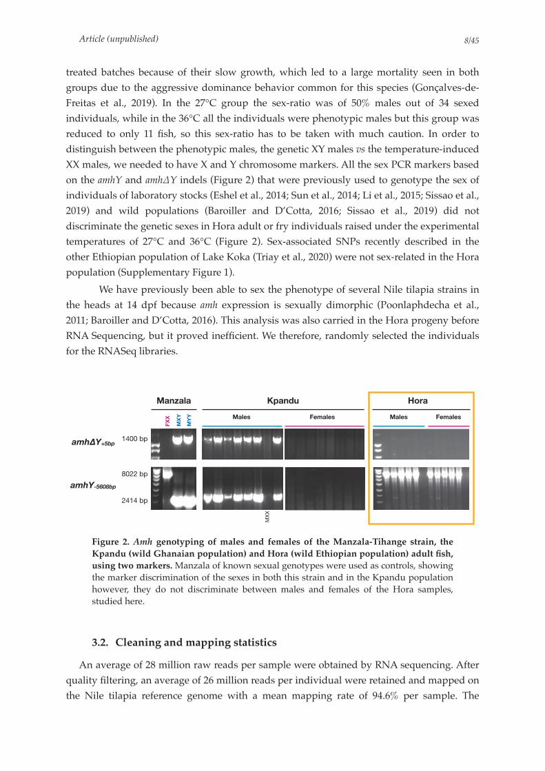
Article (unpublished) /8 45
treated batches because of their slow growth, which led to a large mortality seen in both
groups due to the aggressive dominance behavior common for this species (Gonçalves-de-
Freitas et al., 2019). In the 27°C group the sex-ratio was of 50% males out of 34 sexed
individuals, while in the 36°C all the individuals were phenotypic males but this group was
reduced to only 11 fish, so this sex-ratio has to be taken with much caution. In order to
distinguish between the phenotypic males, the genetic XY males vs the temperature-induced
XX males, we needed to have X and Y chromosome markers. All the sex PCR markers based
on the amhY and amh∆Y indels (Figure 2) that were previously used to genotype the sex of
individuals of laboratory stocks (Eshel et al., 2014; Sun et al., 2014; Li et al., 2015; Sissao et al.,
2019) and wild populations (Baroiller and D’Cotta, 2016; Sissao et al., 2019) did not
discriminate the genetic sexes in Hora adult or fry individuals raised under the experimental
temperatures of 27°C and 36°C (Figure 2). Sex-associated SNPs recently described in the
other Ethiopian population of Lake Koka (Triay et al., 2020) were not sex-related in the Hora
population (Supplementary Figure 1).
We have previously been able to sex the phenotype of several Nile tilapia strains in
the heads at 14 dpf because amh expression is sexually dimorphic (Poonlaphdecha et al.,
2011; Baroiller and D’Cotta, 2016). This analysis was also carried in the Hora progeny before
RNA Sequencing, but it proved inefficient. We therefore, randomly selected the individuals
for the RNASeq libraries.
Figure 2. Amh genotyping of males and females of the Manzala-Tihange strain, the
Kpandu (wild Ghanaian population) and Hora (wild Ethiopian population) adult fish,
using two markers. Manzala of known sexual genotypes were used as controls, showing the marker discrimination of the sexes in both this strain and in the Kpandu population however, they do not discriminate between males and females of the Hora samples, studied here.
3.2. Cleaning and mapping statistics
An average of 28 million raw reads per sample were obtained by RNA sequencing. After
quality filtering, an average of 26 million reads per individual were retained and mapped on
the Nile tilapia reference genome with a mean mapping rate of 94.6% per sample. The
MX
X
Manzala Kpandu Hora
Males Males FemalesFemales
FXX
MXY
MYY
amhΔY+5bp
amhY-5608bp
1400 bp
8022 bp
2414 bp
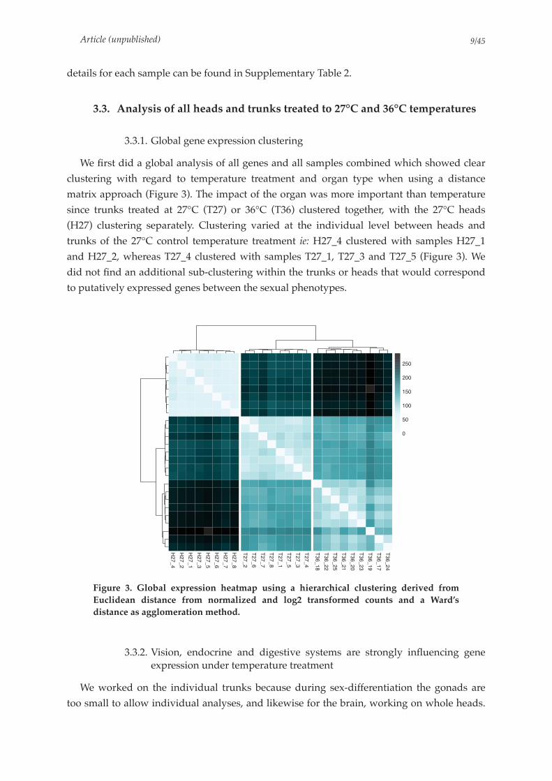
Article (unpublished) /9 45
details for each sample can be found in Supplementary Table 2.
3.3. Analysis of all heads and trunks treated to 27°C and 36°C temperatures
3.3.1. Global gene expression clustering
We first did a global analysis of all genes and all samples combined which showed clear
clustering with regard to temperature treatment and organ type when using a distance
matrix approach (Figure 3). The impact of the organ was more important than temperature
since trunks treated at 27°C (T27) or 36°C (T36) clustered together, with the 27°C heads
(H27) clustering separately. Clustering varied at the individual level between heads and
trunks of the 27°C control temperature treatment ie: H27_4 clustered with samples H27_1
and H27_2, whereas T27_4 clustered with samples T27_1, T27_3 and T27_5 (Figure 3). We
did not find an additional sub-clustering within the trunks or heads that would correspond
to putatively expressed genes between the sexual phenotypes.
Figure 3. Global expression heatmap using a hierarchical clustering derived from
Euclidean distance from normalized and log2 transformed counts and a Ward’s
distance as agglomeration method.
3.3.2. Vision, endocrine and digestive systems are strongly influencing gene expression under temperature treatment
We worked on the individual trunks because during sex-differentiation the gonads are
too small to allow individual analyses, and likewise for the brain, working on whole heads.
H27_4
H27_2
H27_1
H27_3
H27_5
H27_6
H27_7
H27_8
T27_2
T27_6
T27_7
T27_8
T27_1
T27_5
T27_3
T27_4
T36_18
T36_22
T36_25
T36_21
T36_20
T36_23
T36_19
T36_17
T36_24
0
50
100
150
200
250

Article (unpublished) /10 45
This implies that we are “diluting” the transcripts aimed for (gonads and brain) in both
these organs, with the transcripts that are more abundant in heads and trunks. We therefore,
analyzed the top 50 as well as the top 500 most variable genes of heads, and trunks treated
to the two temperature conditions in all the samples to see if the clustering was different. We
obtained a similar sample clustering as the global expression matrix with three main clusters
corresponding to H27, T27 and T36 (Figure 4 and Supplementary Figure 2). Here again, we
did not observe within each replicate group any clear sub-clustering which could
correspond to the putative sex but found defined clusters among genes by expression levels.
We obtained for the heads a higher gene diversity showing a large cluster of over-expressed
genes. All genes identified in this clustering were related to vision (such as rhodopsin, or the
cry gene family) (Figure 4). In the trunks the over-expressed genes’ cluster belonged to
endocrine and digestive functions (such as the elastase-1 gene). The comparison between
T27 vs T36 showed a cluster of over-expressed genes in 36°C trunks that appeared to be
linked to hemoglobin production and oxygen binding.
Figure 4. Heatmap of the top 50 most variable genes using a hierarchical clustering
derived from Euclidean distance from normalized and log2 transformed counts and a
Ward’s distance as agglomeration method.
Manzala Each row represents a gene with it NCBI gene ID on the right. The expression of genes is color coded from low (light) to high (dark).
H27_7
H27_3
H27_8
H27_5
H27_6
H27_1
H27_2
H27_4
T27_1
T27_4
T27_5
T27_2
T27_3
T27_7
T27_6
T27_8
T36_18
T36_25
T36_19
T36_21
T36_24
T36_22
T36_20
T36_17
T36_23
100691236100712271100705620109197692100705848100694450100707334100690909100706115109197491100707596100698940100694720109197490100706349100703065100700411100692739100703904100695749109194189100705132100706288100705765100710942106096620100694761100711209100710676100710249100709091100703906100695768100693035100709427106098796100711075100694113100696806100695135
100711148100695916100534516100534520100695248
102080222100704059109201896109201892109201893
Gene_Category
Gene_Category
Vision
Digestion & Endocrine
Hemoglobin & Oxygen binding
2
4
6
8
10
12
14
16

Article (unpublished) /11 45
We performed complementary GO (Supplementary Figure 3) and KEGG Pathway
enrichment (Table 1) analyses of the top 500 most variable genes amongst all samples which
confirmed the strong representation of signaling processes connected to vision. As such, 62%
genes of the phototransduction pathway are present. There was also, an overrepresentation
of purine metabolism (7%), Arachidonic acid metabolism (7%), and starch and sucrose
metabolism (13%) pathways seen among the top 500 variable genes. Nine out of the 48 genes
(~18%) of the phototransduction pathway were also found among the top 50 most variable
genes.
Table 1. KEGG Pathway output of the top 500 most variable genes among all conditions.
3.3.3. PCA analysis confirms KEGG and heatmap conclusions
The PCA produced using all samples confirmed the results found in hierarchical
clustering based on euclidean distances of gene expression and KEGG analysis (Figure 5).
The first two dimensions of the PCA explained more than 78% of the variance among
samples, with respectively 66.3% and 12.1%. PC1 clearly corresponds to a separation of the
two organ types, whereas PC2 splits trunks according to temperature conditions. In
addition, the top genes contributing to the PCA are confirming the GO and KEGG analysis
as dimension 1 shows genes involved in vision (thus found only in heads), and in dimension
2 we find genes implicated in hemoglobin production, as shown by heatmaps.
(See legend on following page)
KEGG ID Pathway Name Gene Found Gene in Pathway Percentage pvalue
04744 Phototransduction 30 48 0.62 5.344551e-44
00500 Starch and sucrose metabolism 6 45 0.13 1.207691e-05
00590 Arachidonic acid metabolism 6 87 0.07 8.135076e-04
00230 Purine metabolism 13 188 0.07 4.411590e-06
01100 Metabolic pathways 54 1978 0.03 1.596558e-07
!!"#$
%&"%$
'"%$ &"($ %")$*
&+
+*
)+
%**
% & # ' +
,-./01-201
3/45/0678/92:9/;<=7-0/>9?74-705/1
@AB
C&)D&
C&)D#C&)D'
C&)D+
C&)D!
C&)D)
C&)D(
C&)D%
E&)D%E&)D&
E&)D#E&)D'
E&)D+
E&)D!
E&)D)E&)D(
E#!D%)
E#!D%(
E#!D%F
E#!D&*E#!D&%
E#!D&&
E#!D&#
E#!D&'
E#!D&+
'*
*
'*
%** * %**
3G%H9!!"#$9?74-705/
3G&H9%&"%$9?74-705/
E/.</476I4/
7
7
&)
#!
E-11I/1
CJA,
EKLMN
@OB

Article (unpublished) /12 45
Figure 5. Principal component analysis of all samples and all transcripts.
A) Barplot of the percentage of explained variance for the first five dimensions of the PCA. B) Samples PCA plot. Dimension 1 is separating heads from trunks with a major contribution of the following genes to the PC1 axis: rhodopsin (LOC100691236), gamma-crystallin M2-like (LOC100705620), gamma-crystallin M3 family genes (LOC109197692, LOC100694720, LOC100694450, LOC109197490), crystallin, beta B1, like 1 (LOC100705848). Whereas dimension 2 is separating samples in 27°C control temperature from the 36°C treated trunk samples. The genes contributing strongly to the PC2 axis and thus to this clustering are the following: hemoglobin subunit beta-A (LOC100704059), hemoglobin subunit alpha-B (LOC102080222), hemoglobin subunit alpha-A (LOC109201892).
3.4. Gene expressions of sex differentiating candidates in the head
We analyzed three different sets of genes by PCA on head samples to search for
potentially sex-dimorphic gene expressions in brains. The PCA using all genes showed no
obvious clustering on the first dimension even though it represented 35.6% of the variance
(Figure 6). The second set consisted in 2887 genes that we, selected based on their
differential expression in the brain transcriptome of Nile tilapia at 21 dpf (Zhao et al., 2019),
performed on all-females and all-males offspring of a Chinese GIFT strain. The PCA of these
2887 genes revealed a different clustering of the head samples (Figure 6) compared to the
first PCA set but it did not separate the heads into two groups which could be attributed to a
sexual phenotype.
The third PCA analysis was conducted on 141 candidate genes that have been shown to
belong to the sex-determining or differentiating network. The samples showed no clear
grouping (Figure 6), with samples positioned all along the PC1 axis (yet explaining more
than 45% of the variance between individuals). Within the 141 genes, we found elevated
expressions in heads for a glucocorticoid receptor gene nr3c1, hsp70 (heatshock protein 70),
adenylate cyclase type 3 (LOC100710007), genes related to epigenetic modifications such as
dnmt3a (DNA methyltransferase 3 subfamily a) and the histone demethylase kdm6bb gene
(lysine demethylase 6Bb) as well as the steroid biosynthesis enzyme hsd11b2 gene (11-beta
hydroxy steroid dehydrogenase) involved in the 11-ketotestosterone synthesis (Figure 6).
None of the sets of genes studied led to a dichotomous clustering in the heads that would
correspond to females on the one hand and males on the other hand. Considering the
disequilibrium between the number of genes and the number of individuals, the variance
might be dragged by just one extreme sample. This seems to be the case as sample 4 in the
“All genes” PCA is contributing more to the observed variance than the other samples. The
variance explained on the first two axis of these PCAs are mostly due to sample 4 being very
different from other individuals (Supplementary Figure 4).

Article (unpublished) /13 45
Figure 6. Comparison of three PCAs on head samples built with different gene sets.
Left panel: All transcripts; Middle panel: Genes identified by Zhao et al., (2019) as differentially expressed between males and females at control temperature; Right panel:
Set of 141 sex-related candidate genes.
3.5. Effect of rearing temperature on gene expression in the developing
gonad
We subsequently focused on the temperature effects of the trunks (dimension 2 of the
previous PCA, Figure 5).
3.5.1. Global expression clustering
The global expression clustering of all trunks indicates a clear clustering of samples
according to their temperature treatment (Figure 7). In addition, two sub clusters within the
27°C samples were detected with T27_1, T27_3, T27_4, T27_5 on one side, and T27_2, T27_6,
T27_7, T27_8 on the other side. However, the length of the branch splitting of the two sub-
clusters is really short and therefore they may not be relevant with regard to sex clusters.
3.5.2. Gene Ontology and KEGG analysis of Differentially Expressed genes
We identified 4337 genes to be differentially expressed (DE) between 27°C and 36°C
conditions in trunks of which, 1319 genes were identified as down-regulated at 36°C and
3018 genes as up-regulated. Among the DE genes, “Metabolism genes” were over-
represented according to the KEGG pathway analysis. Genes involved in lipid metabolism
were particularly numerous. We found amongst them, more than 40% of these genes
corresponding to the steroid biosynthesis pathways and 20% represented steroid hormones
biosynthesis pathways genes (as named in the KEGG nomenclature) that were differentially
;)$<)
;)$<.
;)$<%
;)$<&
;)$<'
;)$<$
;)$<(;)$<!
!""'#...#
!!)(%!''%
!""'#'$.(
!""$"!'$$
!""'(#$!&
!""$"#"#'
!""$"$%%.
!""$"#)".
!""'#(&$.
!""'(#$!&
!""'#(&$.!""'#(&$.
!""'(#$!&
!""'#(&$.!""'#(&$.!""'#(&$.
!""'(#$!&
!""'#(&$.!""'#(&$.
!""'(#$!&!""'(#$!&!""'(#$!&
!""'#(&$.!""'#(&$.!""'#(&$.!""'#(&$.!""'#(&$.
!"#!#$%&'
(
%
"
%
(
!)
!" " !"*+!,-.&/'0-12342567
*+),-!$/$0-12342567
"/.
"/%
"/&
"/'
685934:
;)$<)
;)$<.
;)$<%
;)$<&
;)$<'
;)$<$
;)$<(
;)$<!
=>?'::
@AB$"<=>2<B389745)
53.6!
>5?9.2
2>75CD297
6C6D2A7
9CB7-.
@A>!!:)
A8E#2
>5?9.:2
4FG !
A8E.A8E.A8E.
!!!!!!
"/&
"/"
"/&
!/"
!/" "/& "/" "/& !/"*+!,-%&/.0-12342567
*+),-!(/.0-12342567
)/&
&/"
$/&
!"/"
!)/&
685934:
;)$<)
;)$<.
;)$<%
;)$<&
;)$<'
;)$<$
;)$<(
;)$<!
!""$"#"#'
!""'#(%$$
!""'#%%%(
!""'#'&!$
!""$!!%()
!""$"!'$$
!""$"""'(
!""'(#$!&
!""'#'()$
!""$"(&".!""'#'&!$!""'#'&!$
!""$"(&".!""$"(&".
!""$!!%()
!""'(#$!&!""'(#$!&!""'(#$!&
&/"
)/&
"/"
)/&
&/"
% " %*+!,-)$/.0-12342567
*+),-)%/.0-12342567
)
.
%
&
685934:
!""#$%&%'()#*%&%'
+,-./#%0#."12#345678.&9:9.0%#$%&%'

Article (unpublished) /14 45
expressed in 36°C samples compared to 27°C control individuals (Figure 8 & Supplementary
Table 3 and 4). We found an enrichment of genes implicated in the DNA replication and in
the mismatch repair pathways, with more than half of these genes shown to be down-
regulated at 36°C compared to the 27°C control temperature (Figure 9).
Figure 7. Global expression heatmap of trunk samples using a hierarchical clustering
derived from Euclidean distance from normalized and log2 transformed counts and a
Ward’s distance as agglomeration method.
Figure 8. Barplot of the overrepresented pathways among the differentially expressed
genes between the 27°C (control) and 36°C (treated) trunk samples.
T27_7
T27_8
T27_2
T27_6
T27_1
T27_5
T27_3
T27_4
T36_19
T36_17
T36_24
T36_18
T36_22
T36_25
T36_21
T36_20
T36_23
0
20
40
60
80
100
120
!"#$%&'(%)*+'*,+-"#'%-
.%#%,+/"('0)$%1/
!"#$%&'()*+$#,-$./
012*#3$4/+&3#
53&#"(4*+3&)"//(46*(4*"47&+'$/8()*3"#()9'98:4#"/#(4$'*(8894"*4"#-&3;*<&3*:62*+3&79)#(&4
=$3%&4*8"#$%&'(/8
5,$6&/&8"
>'9#$#,(&4"*8"#$%&'(/8
5.39?$#"*8"#$%&'(/8
0"#(4&'*8"#$%&'(/8
>'.)&'./(/*@*>'9)&4"&6"4"/(/
A#"3&(7*,&38&4"*%(&/.4#,"/(/
236(4(4"*$47*+3&'(4"*8"#$%&'(/82'$4(4"B*$/+$3#$#"*$47*6'9#$8$#"*8"#$%&'(/8
23$),(7&4()*$)(7*8"#$%&'(/8
5&3+,.3(4*$47*),'&3&+,.''*8"#$%&'(/8
5.3(8(7(4"*8"#$%&'(/8
!"#$%&'(/8*&<*C"4&%(&#()/*%.*).#&),3&8"*5DEF
G$4)&4(*$4"8($*+$#,-$.
>'.)(4"B*/"3(4"*$47*#,3"&4(4"*8"#$%&'(/8=./#"(4"*$47*8"#,(&4(4"*8"#$%&'(/8
H(&/.4#,"/(/*&<*)&<$)#&3/
012*+&'.8"3$/"
5"4#&/"*$47*6'9)93&4$#"*(4#"3)&4?"3/(&4/
236(4(4"*%(&/.4#,"/(/
H(&/.4#,"/(/*&<*$8(4&*$)(7/
19)'"&#(7"*"C)(/(&4*3"+$(3I&8&'&6&9/*3")&8%(4$#(&4
J396*8"#$%&'(/8* *).#&),3&8"*5DEFJ396*8"#$%&'(/8* *&#,"3*"4K.8"/
A"'"4&)&8+&947*8"#$%&'(/8
28(4&$).' #012*%(&/.4#,"/(/
L(4&'"()*$)(7*8"#$%&'(/8
2/)&3%$#"*$47*$'7$3$#"*8"#$%&'(/8
H$/"*"C)(/(&4*3"+$(3
0(%&/&8"*%(&6"4"/(/*(4*"9;$3.&#"/
M4"*)$3%&4*+&&'*%.*<&'$#"
A#"3&(7*%(&/.4#,"/(/
!(/8$#),*3"+$(3J12*3"+'()$#(&4
>"4"*3$#(&*&<*&?"33"+3"/"4#"7*+$#,-$.
FNF FNO FNP FNQ FND FNE FNR FNS
2"('0)$%1/+)3+&)3'&()-1
'*,+4%('/%*1
'-0)67,-'(" /"('0)$%1/
8$)0'$ '*, )4"-4%"9 /'#1
:/%*) '&%, /"('0)$%1/
;-'*1&-%#(%)*+<+(-'*1$'(%)*
=%4"-1
>"*)0%)(%&1 0%),"?-','(%)*
'*, /"('0)$%1/

Article (unpublished) /15 45
Figure 9. Barplot showing the gene ratios of the over-represented pathways among the
differentially expressed genes between the 27°C (control) and 36°C (treated) trunk
samples according to KEGG pathways analyses and categories.
3.5.3. PCA analysis
The PCA based on the expression of all the genes across all the trunk samples (27°C and
36°C) showed a strong clustering linked to the temperature treatments (Figure 10). There
was a large individual variance observed in the 36°C samples, which was not the case for the
27°C samples on dimension 2. This is not surprising given that in the 36°C group we expect
to find differentiating females, differentiating males and putative individuals for which the
sex is being reversed by temperature. The details of genes contributing the most to the PC
axis can be found in Supplementary Figure 5.
Figure 10. Principal component analysis of trunks with all genes.
A) Barplot of the percentage of explained variance for the 5 first dimensions of the PCA. B) Samples PCA plot.
!"#$%&'()*+$#,-$./
0.#&1(2" ).#&1(2"*3")"+#&3*(2#"3$)#(&2
4"3+"/*/(5+'"6*7(38/*9*(2:")#(&2
0$3%&2*5"#$%&'(/5
!(#&+,$;.* *$2(5$'
<&'' '(1"*3")"+#&3*/(;2$'(2;*+$#,-$.
=(&/.2#,"/(/*&:*)&:$)#&3/
0"''*$>,"/(&2*5&'")8'"/
<3.+#&+,$2*5"#$%&'(/5
=(&/.2#,"/(/*&:*$5(2&*$)(>/
0./#"(2"*$2>*5"#,(&2(2"*5"#$%&'(/5
?#$3),*$2>*/8)3&/"*5"#$%&'(/5
@,$;&/&5"
@"2#&/"*+,&/+,$#"*+$#,-$.
A38;*5"#$%&'(/5* *&#,"3*"2B.5"/
C2#"/#(2$'*(5582"*2"#-&31*:&3*C;D*+3&>8)#(&2
D3;(2(2"*%(&/.2#,"/(/
@.387$#"*5"#$%&'(/5
D3$),(>&2()*$)(>*5"#$%&'(/5
E'.)&'./(/*F*E'8)&2"&;"2"/(/
@3(5$3.*%('"*$)(>*%(&/.2#,"/(/
?#"3&(>*,&35&2"*%(&/.2#,"/(/
!"#$%&'(/5*&:*6"2&%(&#()/*%.*).#&),3&5"*@GHI
@,"2.'$'$2(2"*5"#$%&'(/5
@&3+,.3(2*$2>*),'&3&+,.''*5"#$%&'(/5
J"#(2&'*5"#$%&'(/5
A38;*5"#$%&'(/5* *).#&),3&5"*@GHI
@"2#&/"*$2>*;'8)83&2$#"*(2#"3)&27"3/(&2/
K(2&'"()*$)(>*5"#$%&'(/5
D/)&3%$#"*$2>*$'>$3$#"*5"#$%&'(/5
E"2"*3$#(&
I LI I LM I LG I LN
!"#$%&'()*+$#,-$./
@83(2"*5"#$%&'(/5
A38;*5"#$%&'(/5* *&#,"3*"2B.5"/
0./#"(2"*$2>*5"#,(&2(2"*5"#$%&'(/5
D3;(2(2"*$2>*+3&'(2"*5"#$%&'(/5
E'8#$#,(&2"*5"#$%&'(/5
O E'.)$2*%(&/.2#,"/(/
0"''*).)'"
P$3(&8/*#.+"/*&:*O ;'.)$2*%(&/.2#,"/(/
?+'()"&/&5"
@3&#"(2*+3&)"//(2;*(2*"2>&+'$/5()*3"#()8'85
D'$2(2"Q*$/+$3#$#"*$2>*;'8#$5$#"*5"#$%&'(/5
=(&/.2#,"/(/*&:*)&:$)#&3/
JOD*#3$2/+&3#
R&'$#"*%(&/.2#,"/(/
=(&/.2#,"/(/*&:*$5(2&*$)(>/
E'.)(2"Q*/"3(2"*$2>*#,3"&2(2"*5"#$%&'(/5
@.3(5(>(2"*5"#$%&'(/5
@3&#"(2*"6+&3#
O8)'"&#(>"*"6)(/(&2*3"+$(3
R$2)&2(*$2"5($*+$#,-$.
JOD*+&'.5"3$/"
4&5&'&;&8/*3")&5%(2$#(&2
D5(2&$).' #JOD*%(&/.2#,"/(/
=$/"*"6)(/(&2*3"+$(3
S2"*)$3%&2*+&&'*%.*:&'$#"
J(%&/&5"*%(&;"2"/(/*(2*"81$3.&#"/
?#"3&(>*%(&/.2#,"/(/
!(/5$#),*3"+$(3
AOD*3"+'()$#(&2
E"2"*3$#(&
I LII LMI LGI LN
!"#$%&'()*%+#)*#,-./0123#$%&'()*%+#)*#,-./
!"#$%
&'#(%
$#)%
!#!% !#*%
+
&+
*+
'+
!+
,+
& * ' ! ,
-./012.312
4056017890:3;:0<=>8.10?:@85.81602
A6500:=>37
B*)C&
B*)C* B*)C'
B*)C!
B*)C,
B*)C"
B*)C)B*)C$
B'"C&)
B'"C&$
B'"C&(
B'"C*+
B'"C*&
B'"C**
B'"C*'
B'"C*!
B'"C*,
*,
+
*,
,+
,+ *, + *, ,+
4D&E:!)%:@85.8160
4D*E:&!%:@85.8160
B0/=0587F50:75087/017
8
8
*)
'"
G1?.@.?F8>2: :4DH

Article (unpublished) /16 45
3.5.4. Volcano plot visualization of differentially expressed genes
Some of the genes identified as differentially expressed between trunks at 27°C and 36°C
that have also been shown to be implicated in the sex-determining pathway in other teleost
fish or more generally in vertebrates fish (Supplementary Table 1). Among these, we found
the early male differentiating dmrt1 transcription factor gene (found on LG12), several genes
belonging to the jmjd family as well as the hsp90aa1.1, kdm6bb, nr5a5, and dnmt1 genes
(Figure 11).
Figure 11. Volcano plot (log2 Fold Change vs. statistical significance) of transcriptome
analysis of trunks.
Points outside the red lines are genes that are differentially expressed in 36°C samples with a Log2 Fold Change > 2 compared to 27°C trunks samples. Points over the grey line are significant points with a p-value < 0.01. Finally, blue points are genes that are differen-tially expressed at 36°C compared to the 27°C control and that are among the list of 141 sex-related candidate genes.
3.6. Candidate genes approach on the RNASeq data
We next focused our study on our list of 141 candidate genes involved in the sex-
determining and sex-differentiating pathways in vertebrates.
3.6.1. PCA analysis of the 141 candidate genes shows 4 clusters
A PCA based on expression data of the 141 candidate genes (Figure 12 & Figure 13)
showed that dimension 1 represents ~55% of the variance of the samples and draw a clear

Article (unpublished) /17 45
separation between temperature treatments. Dimension 2, led mostly by gsdf, is strongly
splitting T27 and T36 into sub-clusters. Because gsdf particularly (Kaneko et al., 2015), but
also, kdm6bb and gr genes have been shown to be up-regulated in testis development in
teleost fish or vertebrates (Fernandino et al., 2012; Ge et al., 2018) this could indicate that this
division indeed separates differentiating males from differentiating females.
At control temperature (27°C), the clustering is clear and the variance in each sub cluster
seems low. In general, we expect the variance of expression levels to be higher between 36°C
samples as temperature alters several molecular mechanisms regulating the sex-
differentiating pathways (Baroiller and D’Cotta, 2016). The higher variance observed in the
two sub-clusters of the 36°C samples might be strengthened by the presence of XX female
individuals, with some being sex-reversing individuals with levels of expression that are still
not comparable to those of XY males.
Figure 12. Principal component analysis of trunks samples performed using the 141
sex-differentiating candidate genes.
Left: Barplot of the percentage of explained variance for the first five dimensions of the PCA. Right: Biplot of samples with the top 10 genes contributing to the PCA. Because gsdf, nr3c1 and kdm6bb are genes that are associated to masculinization in teleost fish, the upper part (in a blue circle) of the PCA might group the male samples, whereas the lower part (in a pink circle) might group the female samples.
!"#$%
!"#$"
!"#$&
!"#$'
!"#$(
!"#$)
!"#$#
!"#$*
!&)$%#
!&)$%*
!&)$%+
!&)$",
!&)$"%
!&)$""
!&)$"&
!&)$"'
!&)$"(
-./+,00%1%
2.34
536)77
-./+,7%8683*
8683'396:%
;24 %<
;24 %
9<&=%
"
%
,
%
"
' " , "
>;6%?@('1)AB
>;6"?@%,1(AB
!C6/C<0:D<C:<C0:6C9:
0
0
"#
&)
('1)A
%,1(A*1#A
)1"A'1(A
,
",
',
),
% " & ' (
>;6C9.;E9.
FC<=C9:02C?E4?CG/H0;9C3?I0<;09=C.
J=<CC?/HE:
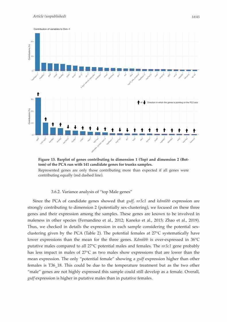
Article (unpublished) /18 45
Figure 13. Barplot of genes contributing to dimension 1 (Top) and dimension 2 (Bot-
tom) of the PCA run with 141 candidate genes for trunks samples.
Represented genes are only those contributing more than expected if all genes were contributing equally (red dashed line).
3.6.2. Variance analysis of “top Male genes”
Since the PCA of candidate genes showed that gsdf, nr3c1 and kdm6bb expression are
strongly contributing to dimension 2 (potentially sex-clustering), we focused on these three
genes and their expression among the samples. These genes are known to be involved in
maleness in other species (Fernandino et al., 2012; Kaneko et al., 2015; Zhao et al., 2019).
Thus, we checked in details the expression in each sample considering the potential sex-
clustering given by the PCA (Table 2). The potential females at 27°C systematically have
lower expressions than the mean for the three genes. Kdm6bb is over-expressed in 36°C
putative males compared to all 27°C potential males and females. The nr3c1 gene probably
has less impact in males of 27°C as two males show expressions that are lower than the
mean expression. The only “potential female” showing a gsdf expression higher than other
females is T36_18. This could be due to the temperature treatment but as the two other
“male” genes are not highly expressed this sample could still develop as a female. Overall,
gsdf expression is higher in putative males than in putative females.
0
10
20
hsp9
0aa1
.1
hsp9
0b1
gsdf
jmjd8
kdm
6bb
jmjd4
dnm
t1
igf−
1r
igf−
1
3−ke
to−ster
oid_
redu
ctas
e
dnm
t3bb
1
nr5a
5
dnm
t3aa
ghr1 ar
aldlra
hsp7
0_kd
a_pr
otein2
hsp9
0aa1
.2
cyp1
9a1b
insr
b
ctnn
b1b
fgfr2
wt1
a
smad
3b
nano
s1a
tgf−
b3
Co
ntr
ibu
tio
ns (
%)
Contribution of variables to Dim−1
!
"!
#!
$!
%&'(
)*$+",%*#-
.'/011
')/2$3
+45"63"1
7&'$18
)*939
*&5:"
.'/833
3';)4<32;=+4+<3&;=245;=$
7&56!33">"
7&'""1#
%7*
(:?<#
<'<*3
'/*23"
@/@'A
7&56!1"
')/2$13
&:?$
.'/B33
)3):&"1
C:)2*D1E2D:)&F,G-
HD*;+2D:)FD)FI7D+7F27;F%;);&FD&F5:D)2D)%F:)F27;FJC#F3?D&:*

Article (unpublished) /19 45
Table 2. Log2 transformed and normalized counts of the top 3 “male genes” of the PCA of
27°C and 36°C trunks samples.
3.7. Real-time PCR of key sex-differentiating genes
In order to better define the putative phenotypic sex of the Hora samples used, we also
analyzed the trunks by quantitative real-time PCR (qPCR), of well-known testicular
differentiating genes (gsdf, dmrt1, amh, arα) and ovarian differentiating genes (cyp19a1a, foxl2
and cyp19a1b). This analysis was performed at 14 dpf on the same samples used for the
RNASeq study when these were still available with additional samples, as well as in 23 dpf
individuals. Expression of gsdf, dmrt1 and even arα showed higher gene expressions in
T-36°C samples compared to T-27°C already at 14 dpf, with a similar pattern at 23 dpf but
with higher individual variance (Figure 14). Surprisingly, the amh did not follow this profile
neither at 14 nor at 23 dpf. Expression of the ovarian aromatase cyp19a1a gene was low at 14
dpf with some individuals showing higher expressions at 36°C (Figure 15). The elevated
levels found for some T-27°C individuals at 23 dpf suggests a higher sexual dimorphism in
the expression at this stage. In contrast, expression is down-regulated in the 36°C trunks at
23 dpf. Foxl2 showed a somewhat similar pattern as the brain aromatase cyp19a1b gene that
was in fact expressed at much stronger levels than the gonad cyp19a1a gene, although we
found no inverse correlation with the expression of male genes (Supplementary Figure 6).
Candidate
genes for
maleness
Potential Males 27°C Potential Males 36°CPotential Females
27°C
Potential Females
36°C Mean
of All
samples
Mean of
Potential
Males
Mean of
Potential
FemalesT27
_1
T27
_4
T27
_6
T27
_7
T27
_8
T36
_17
T36
_19
T36
_20
T36
_21
T36
_23
T36
_24
T27
_2
T27
_3
T27
_5
T36
_18
T36
_22
T36
_25
nr3c1 6.96 7.58 7.46 7.29 7.18 7.35 7.83 7.44 8.29 7.55 7.79 6.31 7.24 6.58 6.35 6.75 7.25 7.25 7.52 6.75
kdm6bb 11.29 11.69 11.94 11.57 11.65 12.51 12.44 12.52 13.37 12.68 13.19 11.16 11.59 10.97 11.62 11.80 12.32 12.02 12.26 11.58
gsdf 8.55 8.52 8.26 8.39 8.36 8.83 8.37 9.03 9.74 9.25 8.85 6.91 5.95 6.74 8.90 7.30 7.06 8.18 8.74 7.14
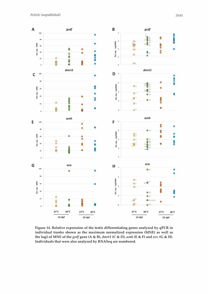
Article (unpublished) /20 45
Figure 14. Relative expression of the testis differentiating genes analyzed by qPCR in
individual trunks shown as the maximum normalized expression (MNE) as well as
the log2 of MNE of the gsdf gene (A & B), dmrt1 (C & D), amh (E & F) and arα (G & H).
Individuals that were also analyzed by RNASeq are numbered.
!"#$%"&'$%(%)*+%
,
-.
/-
01
2/
.,3
!"#$%
!"#$%"&'$%(%)*+%
,
-.
/-
01
2/
.,3
&"'
!"#$%"&'$%(%)*+%
,
-.
/-
01
2/
.,3
&#&
!"#$%"&'$%(%)*+
,
-.
/-
01
2/
.,3
4
5
+
6
()!*
-785 -785 10851085
./%9': -1%9':
-785 -785 10851085
./%9': -1%9':
!"#$%"&'$%(%#;<-)*+%
(1
(.
-
3
7
= ()!*
!"
!#!$
#%#&
!'!(
#)
!!
"
(
#
&!
*%
>!"#$%
!"#$%"&'$%(%#;<-)*+%
,
-
/
3
7
&
%
!
#
(
"
!#
#&
!!!$
!'
#)!"
!(#%
?&"'
!"#$%"&'$%(%#;<-)*+%
1
/
3
0
7
(
!
#
"*
%&
!'
#%
!$!#
#&#)
!!!"
!(
&#&
!"#$%"&'$%(%#;<-)*+%
(,
-
1
3
7@!"
#%!$
#&
!#
!(
#)+,!!+,!'
(
!
#
&
*
%
"

Article (unpublished) /21 45
Figure 15. Relative expression of the Ovarian differentiating genes analyzed by qPCR
in individual trunks shown as the maximum normalized expression (MNE) as well as
the log2 of MNE of the cyp19a1a gene (A & B), foxl2 gene (C & D) and the brain
cyp19a1b gene (E & F). Individuals that were also analyzed by RNASeq are numbered.
To predict putative males and females, we plotted the expression levels of the gonad
aromatase cyp19a1a relative to those of gsdf, dmrt1 and amh. This separated relatively well
two groups for the 27°C samples and the clustering observed was mostly consistent with the
results of the RNAseq candidate gene approach (Figure 16). The sample T27-3 however
showed low expressions levels for several of the genes studied by qPCR, including cyp19a1a
(Figure 14 & 15). Thus, even if the RNAseq suggested this sample was a putative female, this
sample does not cluster as expected (Figure 16).
!"#$%
!"#$%"&'$%(%)*+%
,
-.
/-
01
2/
.,3
&'()*+)+
!"#$%"&'$%(%)*+%
,
-.
/-
01
2/
.,3
&'()*+),
!"#$%"&'$%(%)*+%
,
-.
/-
01
2/
.,3
4
5
+
-675 -675 10751075
./%8'9 -1%8'9
-675 -675 10751075
./%8'9 -1%8'9
: &'()*+)+
!"#$%"&'$%(%#;<-)*+%
(-
.
1
3
6
!!
!"
!#
$%$&
!'
#
!
$
(%
)
= !"#$%
!"#$%"&'$%(%#;<-)*+%
(-
.
1
3
6)
(
#!
#)
!
%
($
> &'()*+),
!"#$%"&'$%(%#;<-)*+%
1
3
6
)
(
#*+!$
%
,
!!
!$!#
!($&
!"!'
$%
$)

Article (unpublished) /22 45
Figure 16. Relative expression of individual trunks of cyp19a1a plotted relative to gsdf
(A), dmrt1 (B) and amh (C) expressed as the log2 of maximum normalized expression
(MNE).
Individuals that were also analyzed by RNASeq are numbered and putative males and females identified by the candidate gene approach the 27°C RNAseq data are shaded respectively in blue and pink.
3.8. Validation of differentially expressed genes by real-time PCR
To validate further some of our transcriptome results, we selected five genes shown to be
differentially expressed between 27°C and 36°C trunks analyzed by qPCR. Similarly to the
transcriptome analyses we detected higher expression for kdm6bb in the 36°C trunks
compared to the 27°C at 14 dpf and this profile persisted at 23 dpf (Figure 17A). We
observed a similar pattern of expression for jarid2 (jumonji and AT-rich interaction domain
containing 2) gene (Figure 17B). Mean expressions for the hsd11β2 and gr2 were likewise
higher in the T-36°C samples compared to T-27°C and this trend was more marked at 23 dpf
(Figure 17D & 17E). We also analyzed the steroidogenic 11β-hydroxylase cyp11c1 gene
involved in the synthesis of both 11-ketotestosterone and cortisol (Baroiller et al., 1999;
Zheng et al., 2020). At 14 dpf cyp11c1 expression levels remained low for both 27°C and 36°C
trunks (Figure 17C) increasing strongly at 23 dpf in the trunks treated at 36°C.
!"#$%&' "#()&'
!
"
#
$
%
%&
#'
#"
%(
%)%%
!
"
#
$$
%
%&
#'
#"
%(
%)%%
*+,$--./012
343
245
(43
645
)43
*+,$-7892:;
3 2 $ ( 6 5 )
(
'"#$%&' "#()&'
$
!
"
#
%&
%
(
#'
#"
%(
%)
%%
$
!
"
#
%&
%
(
#'
#"
%(
%)
%%
$
*+,$--;/<
(43
643
543
)43
%43
*+,$-7892:;
3 2 $ ( 6 5 )
="#$%&' "#()&'
#
!
"
$
%
(
%%
%&
#'
%(
#"
%)
#
!!
"
$
%
(
%%
%&
#'
%(
#"
%)
*+,$--,>.?
#(43
#245
343
245
(43
645
)43
*+,$-7892:;
#$ #2 3 2 $ ( 6 5 )

Article (unpublished) /23 45
Figure 17. Relative expression of DE genes found in our RNASeq data analyzed by
qPCR in individual trunks shown as the log2 of the maximum normalized expression
(MNE) of the kdm6bb gene (A), jarid2 (B), cyp11c1 (C), 11β-hsd (D) and gr2 (E).

Article (unpublished) /24 45
4. Discussion
The Hora population of Nile tilapia belongs to the subspecies O. niloticus cancellatus like
the Lake Koka population, both found in reservoirs of the Awash River in Ethiopian
highlands. Although the sex-determining gene in the Koka population is centered around
the amh gene on LG23 (Triay et al., unpublished – Chapter 2), this is not the case for Hora
fish. No other linkage group was found segregating for sex. Despite this, we thought that
genetic factors - perhaps through allelic variations - could be determining the sex in the
Hora populations since we obtained in this family (but also in other families) a balanced
phenotypic sex-ratio, when experimenting at control 27°C conditions. Although the trigger
of sex in Hora is different from other Nile tilapia stocks, we wanted to know whether the
downstream process of sex-differentiation was conserved, to understand better the early
regulating network that is acting in the putative genetic and temperature-induced pathways
of Hora fish.
Here we initiated this work by doing a transcriptome analysis at 14 dpf together with
qPCR analyses at 14 and 23 dpf. We were hampered by the fact that we could not genetically
sex the fry. Sex-differentiation of Nile tilapia has been studied mostly (if not only) in
domesticated strains where the phenotypic sex is known before which greatly simplifies the
search. Crossings of YY males that are fertile and viable, with genetic XX females gives all
XY individuals (theoretically all males) while crossings of XX males with genetic XX females
results in all XX individuals (presumably all females) (Baroiller and D’Cotta, 2018). Males
can therefore, be easily detected when all-XX females are treated to masculinizing
temperatures (Baroiller et al., 2009b). The critical period when undifferentiated gonads begin
to differentiate into ovaries or testis in Nile tilapia is around 9 to 10 dpf, and this can be
perceptible already at the molecular level (D’cotta et al., 2001; Ijiri et al., 2008;
Poonlaphdecha et al., 2011; Tao et al., 2013; Kaneko et al., 2015). At this stage gonads are
already responsive to temperature (and hormone) treatments which can induce sex changes
(Baroiller and D’Cotta, 2016). The gonads in domestic strains start to be morphologically
distinguishable between testis and ovaries at ~12 dpf. The number of germ cells stop
increasing in XY male gonads during 9 to 14 dah (13 to 18 dpf), in contrast to the ongoing
increase in XX female gonads (Kobayashi et al., 2008). Primordial germ cells increase then
drastically in ovaries at ~17 dpf, clearly visible at 23 dpf (Nakamura et al., 1998; D’cotta et
al., 2001; Ijiri et al., 2008; Kobayashi and Nagahama, 2009).
Our study showed that in Hora the molecular changes are subtle at 14 dpf. Sexual-
dimorphism was not readily discernible by our global transcriptome analysis in neither
heads nor trunks. Nevertheless, our enhanced analyses in trunks of 141 sex-related
candidate genes showed that some could be differentially expressed, notably the gsdf gene,
allowing us to define what could be putative males and females.

Article (unpublished) /25 45
4.1. gsdf is differentially expressed in Hora trunks at 14dpf
We have found that the gsdf (gonadal soma-derived factor) gene is differentially
expressed in Hora fish as early as 14 dpf in the 27°C trunks and even more in the 36°C
trunks, and this profile became more marked at 23 dpf with high levels in presumably
differentiating testis. The strong differences in gsdf expression levels at 14 dpf identified by
RNASeq, two clusters among the 27°C and 36°C samples in trunks that could fit a sex-
clustering pattern with males presenting a high gsdf expression in the upper cluster. Gsdf is a
fish-specific gene of the TGF-β superfamily which also contains the amh gene (De Caestecker,
2004). It is the first target of the sex-determining dmy/dmrt1Yb gene in medaka (Zhang et al.,
2016). Gsdf plays an important role in testis differentiation in many other teleosts such as in
tilapia (Kaneko et al., 2015), the zebrafish (Yan et al., 2017), the European sea bass (Crespo et
al., 2013), the wrasse (Horiguchi et al., 2013) and the Japanese flounder (Yang et al., 2019).
The functional role of gsdf appears to be related to germ cells and spermatogonia
proliferation in several of these species including the Nile tilapia (Sawatari et al., 2007;
Horiguchi et al., 2013; Jiang et al., 2016; Yang et al., 2019). Higher expression of the gsdf gene
might inhibit primordial germ cell proliferation in the Japanese flounder (Yang et al., 2020).
Kaneko et al., (2015) showed that gsdf expression and its protein are sexually dimorphic
as early as 9 dpf even before the morphological differentiation in gonads of the Nile tilapia.
They also highlighted that an over-expression of gsdf was enough to induce testis
development in XX individuals. Knockout of gsdf in Nile tilapia caused a complete male-to-
female sex reversal, establishing that gsdf expression in differentiating testis is downstream
from dmrt1 (Jiang et al., 2016). Estrogen synthesis by the aromatase enzyme Cyp19a1 is
essential for ovarian differentiation (D’Cotta et al., 2001; Wang et al., 2007; Guiguen et al.,
2010). In tilapia, gsdf might be linked to the repression of estrogen production, thus
triggering testicular differentiation (Jiang et al., 2016). In Hora gsdf expression is overall
higher than dmrt1 at 14 dpf which resembles more the pathway proposed by Kaneko et al.,
(2015) that suggested gsdf preceded dmrt1 expression in tilapia. Our results are suggesting
that gsdf might also be expressed in a sexual dimorphic manner in Hora fish. Gsdf could be a
good sex marker since Hora lack the amhy/am∆Y genes (Sissao et al., 2019; Triay et al., 2020;
Triay et al., unpublished – Chapter 2).
4.2. Temperature treatment affects gonad gene expression in Hora
In the Japanese flounder, the expression of gsdf is increased by high temperature
treatments and results in XX female to male sex-reversal (Yang et al., 2019, 2020). Our results
also confirm those observed in Yang et al., (2019, 2020) since we found that Hora fish treated
to 36°C showed even higher gsdf expression levels than the 27°C trunks and this was more
evident at 23 dpf suggesting a higher proportion of testis differentiating. The individual
variation observed at 23 dpf might stem from the presence of genetic and temperature
induced males as well as females.

Article (unpublished) /26 45
4.2.1. Steroid and Steroid hormones biosynthesis
When comparing 27°C trunk samples to 36°C treated samples, we noticed pronounced
differences in gene expression. Among these we observed a down regulation by the high
temperature treatment of genes involved in steroid biosynthesis while genes of steroid
hormone biosynthesis were up regulated. Steroid hormones are implicated in embryonic
development, sex differentiation, reproduction as well as in stress response in teleost fish
(Baroiller et al., 1999; Goikoetxea et al., 2017). Estrogens are synthesized from androgens by
the aromatase enzyme encoded by the cyp19a1a and cyp19a1b genes (Baroiller et al., 1999;
Guiguen et al., 2010). Differentiating ovaries of Nile tilapia were found to have higher levels
of estradiol-17β and cyp19a1a levels compared to differentiating testis (Baroiller and D’Cotta,
2001; D’Cotta et al., 2001). Hora trunks showed higher levels of the brain aromatase cyp19a1b
expression at 14 dpf compared to the gonad aromatase cyp19a1a, particularly in the 36°C
trunks. Elevated levels of cyp19a1b have also been detected in early testis differentiation of
Nile tilapia but its role is unknown (Sudhakumari et al., 2005). In Hora we plotted the
expression of cyp19a1b with respect to those of gsdf at 14 dpf however they gave no evidence
of a dimorphism related to sex. In contrast, expressions of cyp19a1a together with
expressions of gsdf or dmrt1 or even amh did separate 27°C individuals into two, presumably
putative males from putative females. Double plotting of expressed genes allowed the
prediction of males and females of wild Southern flounder that have ESD (Honeycutt et al.,
2019).
In the Hora wild population of Nile tilapia, temperature does produce differential
expression of genes involved in the steroid hormone biosynthesis. We observed that
temperature had a repressive effect on the cyp19a1a expression at 23 dpf in the 36°C Hora
trunks which was not the case in the 27°C trunks. Similar repression of cyp19a1a expression
(affecting estrogen levels), was shown in tilapia XX gonads treated to 36°C which were
masculinized by temperature (D’Cotta et al., 2001; Poonlaphdecha et al., 2013; Sun et al.,
2018), and one of the mechanisms of repression is through the increase in methylation levels
on the cyp19a1a promoter (Wang et al., 2017).
Among the different categories of steroid hormones are the glucocorticoids of which
cortisol is the most important. This hormone might be an important link between the
environmental induction and the sex differentiation pathway, as cortisol production is
increased by high temperatures in the Patagonian pejerrey, and the Japanese flounder
leading to high male ratios (Hattori et al., 2009; Yamaguchi et al., 2010; Fernandino et al.,
2013). In these fish, the increase of cortisol levels induces repression of the aromatase
cyp19a1a expression and thus blocking estrogen synthesis and the ovarian differentiating
pathway. This process is favoring the activation of the androgen pathway by regulating 11β-
hydroxylase (Cyp11b) and 11β-hydroxysteroid dehydrogenase (Hsd11β) enzymes, resulting
in a female to male sex-reversal (reviewed in Goikoetxea et al., 2017). 11β-hydroxylase,
encoded by the cyp11c1 gene, is involved in the synthesis of 11-Ketotestosterone the main
fish androgen but also in cortisol synthesis (Zheng et al., 2020). Cortisol action, seen in the
Japanese flounder, appeared to be through its glucocorticoid receptor, which regulated the

Article (unpublished) /27 45
cyp19a1a gene expression by binding to the GRE site in the promoter (Yamaguchi et al.,
2010). RNAseq in Hora showed elevated expression of the gr2 gene in 27°C trunks (in
presumed males) which was validated by qPCR, but levels increased thereafter in both 27°C
and 36°C trunks. Expression levels of cyp11c1 and 11β-hsd were similar between 27°C and
36°C trunks at 14 dpf but increased considerably in many individuals at 36°C, which
suggests that temperature was inducing maleness, but whether this is due to 11-
ketotestosterone or/and via cortisol remains to be verified.
4.2.2. DNA replication and mismatch repair is down-regulated at high temperature
Interestingly, more than half of the genes implicated in the DNA replication pathway and
in the mismatch repair pathway were down-regulated at 36°C compared to the control
temperature. This is surprising as high temperatures are generally stimulating more errors
and thus more corrections over the mitosis period. If we do not have a strong clue to explain
this phenomenon, it is interesting to note that it might result in higher mutation rates but
lower rates of mitosis.
4.3. Some of the usual suspects are DE at 36°C in gonads
Among the DE genes at 36°C, we identified a dozen of genes that are implicated in the
sex determination/sex-differentiation pathway. These identified genes might be good
starting points to look for modifications of sex pathways between 36°C and control
temperatures. Indeed, some of these genes might be tightly linked with temperature-
induced sex-reversal in the Nile tilapia.
4.3.1. Heatshock proteins hsp90
Heatshock protein complex are linked to gamete quality in some fish. As such, lack of hsp
protein in gonads leads to poorer quality of eggs for females or apoptosis of testicular cells
in males (He et al., 2010; Chapman et al., 2014; Kohn et al., 2015). An explanation is the role
of hsp proteins in cell migration during embryonic stages. In the zebrafish, Pfeiffer, (2018)
demonstrated that a misregulation of Hsp90 proteins induces a slower migration rate of
primordial germ cells to the gonadal region. In the more distant species of Drosophilia
melanogaster, a lack of regulation of Hsp90 proteins can lead to defects in cell proliferation
and is thus essential for a proper progression through the cell cycle at early stages (Bandura
et al., 2013). In addition, we found that the expression of hsp90aa1.1 is higher in 27°C than in
36°C gonads of O. niloticus Hora population. This result is in agreement with those of
Mahanty et al., (2019) that showed an increased temperature induced decrease of hsp90
expression in the Barb (Puntius sophore). The rice field eel (Monopterus albus) also shows a
down-regulation of hsps expression in testes of individuals exposed to heat-stress (He et al.,
2010). Finally, in natural environments, some fish also present a down-regulation of

Article (unpublished) /28 45
heatshock proteins during warmer seasons of the year (Domingos et al., 2013).
4.3.2. Epigenetic regulators
Genes of the Jumonji family are also showing a differential expression with high
temperature in Hora individuals. In the Hora population of Nile tilapia the sexual
dimorphism of expression of the histone demethylase Kdm6bb (lysine-specific demethylase
6Bb) gene in the control temperature is not obvious. According to the clusters designed
primarily by gsdf expression, we expect to have only 3 females amongst the 27°C trunks so
this lack of sexual dimorphic expression might only be due to the low number of samples
used for the comparison. A strong difference, however, is seen between the 36°C putative
males and all the other samples, including control temperature males. In the TDS Red-eared
slider turtle, Kdm6b promotes transcription of the male sex-determining dmrt1 gene (Ge et
al., 2018). The expression of Kdm6b is sexually dimorphic even before gonads are distinct.
Kdm6b can be regulated by the STAT3 gene that encodes for the signal transducer and
activator of transcription 3. When temperature increases, STAT3 products get
phosphorylated and bind to the Kdm6b gene, repressing its expression and blocking the male
pathway (Weber et al., 2020). In the Chinese GIFT strain of the Nile tilapia however, high
temperature treatments on females leads to increased expression of kdm6bb compared to
control temperature females (Zhao et al., 2019). In addition, kdm6bb expression is in general
higher in males than in control temperature females. Hora showed higher kdm6bb expression
in 36°C trunks at 14 dpf and even more at 23 dpf. Thus, higher expression of kdm6bb might
up-regulate dmrt1 expression and be involved in XX sex-reversal individuals in
thermosensitive families of Hora population.
Jarid 2 was shown to be expressed in Hora at 14 dpf and up-regulated at 23 dpf with
higher levels of expression in both stages in the 36°C trunks. Jarid2 has also been shown to
be elevated in males and up-regulated with temperature in Nile tilapia brains (Zhao et al.,
2019). Jarid2 and Jmjd3, genes of this family, have been identified in reptiles with
temperature-induced sex reversal (Deveson et al., 2017). These genes are tightly linked to
epigenetics and perturbation of their function can occur through intron retention (which
caused a premature stop codon) that triggered sex reversal under high temperature
conditions.
Based on the downregulation of the aromatase cyp19a1a at 23dpf in the 36°C trunks
together with the increase of gsdf, dmrt1, cyp11c1, 11b-hsd as well as kdm6bb and jarid2, we
consider that temperature was probably inducing masculinization of the Hora individuals.
4.4. Head RNA analyses
Our transcriptome analysis of heads at 14 dpf showed an enrichment of genes involved in
vision, which is not surprising since the heads contained the eyes. The brain harbors many
sexually dimorphic genes. In a previous study of brain transcripts of O. niloticus (Chinese
GIFT strain, with amh SD) during the sex-differentiation period, a large number of genes

Article (unpublished) /29 45
were found to be differentially expressed between males and females at 21 dpf (Zhao et al.,
2019). The analyses of 2887 genes found by Zhao et al., (2019) on Hora did not highlight the
same genes, which might suggest different regulating pathways in the brain sexualisation of
Hora fish. The first clear difference is that in Hora brains at 14 dpf amh is not sexually
dimorphic. For strains of O. niloticus in which the amh gene is the major SD, some specific
genes are dimorphically expressed at 14 dpf. As such, the amh gene itself is found highly
expressed in brains of XY samples whereas its expression is practically undetectable in XX
individuals (Poonlaphdecha et al., 2011; Baroiller and D’Cotta, 2018). This gene, though
present in all three datasets used for the PCA (all genes, DE genes according to Zhao et al.,
(2019) and 141 candidate genes), is not among the genes influencing the brain clustering.
In conclusion, this study of sex-differentiation in Hora Nile tilapia showed that gsdf is
already expressed at 14 dpf with higher expression in putative differentiating testis, while
neither dmrt1 nor amh are. This indicates that sex-differentiation in Hora tilapia follows a
different pathway from that found commonly in domesticated Nile tilapia strains.
Differences were also observed in Hora heads, since amh expression was not dimorphic at 14
dpf. We found in the trunks several genes that are epigenetic regulators such as the histone
modifiers kdm6bb and jarid2 genes, and genes related to cortisol and 11-ketotestosterone
synthesis in putative testis that were up-regulated in trunks treated to 36°C. These results
together with the increase of the testis-differentiating genes gsdf, dmrt1 and the repression of
the ovarian cyp19a1a suggests that high-temperature induced masculinization in this Hora
family. The Hora population is hence probably thermosensitive despite living in a cold lake.
Article (unpublished) /30 45
References
Anders, S., Pyl, P.T., and Huber, W. (2015). HTSeq—a Python framework to work with high-throughput sequencing data. Bioinformatics 31, 166–169.
Bandura, J.L., Jiang, H., Nickerson, D.W., and Edgar, B.A. (2013). The Molecular Chaperone Hsp90 Is Required for Cell Cycle Exit in Drosophila melanogaster. PLoS Genet 9, e1003835.
Baroiller, J.F., and D’Cotta, H. (2001). Environment and sex determination in farmed fish. Comparative Biochemistry and Physiology Part C: Toxicology & Pharmacology 130, 399–409.
Baroiller, J.-F., and D’Cotta, H. (2016). The Reversible Sex of Gonochoristic Fish: Insights and Consequences. Sexual Development 10, 242–266.
Baroiller, J.-F., and D’Cotta, H. (2018). Sex Control in Tilapias. In Sex Control in Aquaculture, (John Wiley & Sons, Ltd), pp. 189–234.
Baroiller, J.F., Chourrout, D., Fostier, A., and Jalabert, B. (1995). Temperature and sex chro-mosomes govern sex ratios of the mouthbrooding Cichlid fish Oreochromis niloticus. J. Exp. Zool. 273, 216–223.
Baroiller, J.-F., Guiguen, Y., and Fostier, A. (1999). Endocrine and environmental aspects of sex differentiation in fish. CMLS, Cell. Mol. Life Sci. 55, 910–931.
Baroiller, J.F., D’Cotta, H., and Saillant, E. (2009a). Environmental effects on fish sex determi-nation and differentiation. SXD 3, 118–135.
Baroiller, J.F., D’Cotta, H., Bezault, E., Wessels, S., and Hoerstgen-Schwark, G. (2009b). Tila-pia sex determination: where temperature and genetics meet. Comparative Biochemistry and Physiology Part A: Molecular & Integrative Physiology 153, 30–38.
Bej, D.K., Miyoshi, K., Hattori, R.S., Strüssmann, C.A., and Yamamoto, Y. (2017). A Duplica-ted, Truncated amh Gene Is Involved in Male Sex Determination in an Old World Silver-side. G3: Genes, Genomes, Genetics 7.
Berishvili, G., D’Cotta, H., Baroiller, J.-F., Segner, H., and Reinecke, M. (2006). Differential expression of IGF-I mRNA and peptide in the male and female gonad during early deve-lopment of a bony fish, the tilapia Oreochromis niloticus. General and Comparative Endo-crinology 146, 204–210.
Bertho, S., Herpin, A., Branthonne, A., Jouanno, E., Yano, A., Nicol, B., Muller, T., Pannetier, M., Pailhoux, E., Miwa, M., et al. (2018). The unusual rainbow trout sex determination gene hijacked the canonical vertebrate gonadal differentiation pathway. Proc Natl Acad Sci USA 115, 12781–12786.
Bezault, E. (2005). Etude Du Systeme De Determinisme Du Sexe au Sein De Populations Na-turelles De Tilapia Du Nil, Oreochromis Niloticus (linnaeus, 1758) : importance Des Com-posantes Genetiques Et Environnementales. UNIVERSITE DE PARIS-SUD.
Bezault, E., Clota, F., Derivaz, M., Chevassus, B., and Baroiller, J.-F. (2007). Sex determination and temperature-induced sex differentiation in three natural populations of Nile tilapia (Oreochromis niloticus) adapted to extreme temperature conditions. Aquaculture 272, S3–S16.
Bezault, E., Balaresque, P., Toguyeni, A., Fermon, Y., Araki, H., Baroiller, J.-F., and Rognon, X. (2011). Spatial and temporal variation in population genetic structure of wild Nile tilapia (Oreochromis niloticus) across Africa. BMC Genetics 12, 102.
Bolger, A.M., Lohse, M., and Usadel, B. (2014). Trimmomatic: a flexible trimmer for Illumina sequence data. Bioinformatics 30, 2114–2120.
Article (unpublished) /31 45
Cáceres, G., López, M.E., Cádiz, M.I., Yoshida, G.M., Jedlicki, A., Palma-Véjares, R., Travisa-ny, D., Díaz-Domínguez, D., Maass, A., Lhorente, J.P., et al. (2019). Fine mapping using whole-genome sequencing confirms anti-müllerian hormone as a major gene for sex de-termination in farmed Nile tilapia (Oreochromis niloticus L.). G3 (Bethesda) 9, 3213–3223.
de Caestecker, M. (2004). The transforming growth factor-β superfamily of receptors. Cyto-kine & Growth Factor Reviews 15, 1–11.
Chapman, R.W., Reading, B.J., and Sullivan, C.V. (2014). Ovary Transcriptome Profiling via Artificial Intelligence Reveals a Transcriptomic Fingerprint Predicting Egg Quality in Striped Bass, Morone saxatilis. PLOS ONE 9, e96818.
Crespo, B., Gómez, A., Mazón, M.J., Carrillo, M., and Zanuy, S. (2013). Isolation and charac-terization of Ff1 and Gsdf family genes in European sea bass and identification of early gonadal markers of precocious puberty in males. General and Comparative Endocrinolo-gy 191, 155–167.
D’Cotta, H., Fostier, A., Guiguen, Y., Govoroun, M., and Baroiller, J.-F. (2001). Aromatase plays a key role during normal and temperature-induced sex differentiation of tilapia Oreochromis niloticus. Molecular Reproduction and Development 59, 265–276.
Deveson, I.W., Holleley, C.E., Blackburn, J., Marshall Graves, J.A., Mattick, J.S., Waters, P.D., and Georges, A. (2017). Differential intron retention in Jumonji chromatin modifier genes is implicated in reptile temperature-dependent sex determination. Sci. Adv. 3, e1700731.
Dobin, A., Davis, C.A., Schlesinger, F., Drenkow, J., Zaleski, C., Jha, S., Batut, P., Chaisson, M., and Gingeras, T.R. (2013). STAR: ultrafast universal RNA-seq aligner. Bioinformatics 29, 15–21.
Domingos, F.F.T., Thomé, R.G., Martinelli, P.M., Sato, Y., Bazzoli, N., and Rizzo, E. (2013). Role of HSP70 in the regulation of the testicular apoptosis in a seasonal breeding teleost Prochilodus argenteus from the São Francisco River, Brazil. Microsc Res Tech 76, 350–356.
Durinck, S., Spellman, P.T., Birney, E., and Huber, W. (2009). Mapping Identifiers for the In-tegration of Genomic Datasets with the R/Bioconductor package biomaRt. Nat Protoc 4, 1184–1191.
Eshel, O., Shirak, A., Weller, J.I., Hulata, G., and Ron, M. (2012). Linkage and physical map-ping of sex Rregion on LG23 of Nile tilapia (Oreochromis niloticus). G3: Genes, Genomes, Genetics 2, 35–42.
Eshel, O., Shirak, A., Dor, L., Band, M., Zak, T., Markovich-Gordon, M., Chalifa-Caspi, V., Feldmesser, E., Weller, J.I., Seroussi, E., et al. (2014). Identification of male-specific amh duplication, sexually differentially expressed genes and microRNAs at early embryonic development of Nile tilapia (Oreochromis niloticus). BMC Genomics 15, 774.
FAO (2021). The State of World Fisheries and Aquaculture 2021 (FAO).
Fernandino, J.I., Hattori, R.S., Kishii, A., Strüssmann, C.A., and Somoza, G.M. (2012). The Cortisol and Androgen Pathways Cross Talk in High Temperature-Induced Masculiniza-tion: The 11β-Hydroxysteroid Dehydrogenase as a Key Enzyme. Endocrinology 153, 6003–6011.
Fernandino, J.I., Hattori, R.S., Moreno Acosta, O.D., Strüssmann, C.A., and Somoza, G.M. (2013). Environmental stress-induced testis differentiation: Androgen as a by-product of cortisol inactivation. General and Comparative Endocrinology 192, 36–44.
Feron, R., Zahm, M., Cabau, C., Klopp, C., Roques, C., Bouchez, O., Eché, C., Valière, S., Donnadieu, C., Haffray, P., et al. (2020). Characterization of a Y-specific duplication/inser-tion of the anti-Mullerian hormone type II receptor gene based on a chromosome-scale

Article (unpublished) /32 45
genome assembly of yellow perch, Perca flavescens. Molecular Ecology Resources 20, 531–543.
Fowler, B.L.S., and Buonaccorsi, V.P. (2016). Genomic characterization of sex-identification markers in Sebastes carnatus and Sebastes chrysomelas rockfishes. Molecular Ecology 25, 2165–2175.
Gammerdinger, W.J., Conte, M.A., Acquah, E.A., Roberts, R.B., and Kocher, T.D. (2014). Structure and decay of a proto-Y region in Tilapia, Oreochromis niloticus. BMC Genomics 15, 975.
Ge, C., Ye, J., Weber, C., Sun, W., Zhang, H., Zhou, Y., Cai, C., Qian, G., and Capel, B. (2018). The histone demethylase KDM6B regulates temperature-dependent sex determination in a turtle species. Science.
Gennotte, V., Mélard, C., D’Cotta, H., Baroiller, J.-F., and Rougeot, C. (2014). The sensitive period for male-to-female sex reversal begins at the embryonic stage in the Nile tilapia and is associated with the sexual genotype. Molecular Reproduction and Development 81, 1146–1158.
Goikoetxea, A., Todd, E.V., and Gemmell, N.J. (2017). Stress and sex: does cortisol mediate sex change in fish? Reproduction 154, R149–R160.
Gonçalves-de-Freitas, E., Bolognesi, M.C., Gauy, A.C. dos S., Brandão, M.L., Giaquinto, P.C., and Fernandes-Castilho, M. (2019). Social Behavior and Welfare in Nile Tilapia. Fishes 4, 23.
Guiguen, Y., Fostier, A., Piferrer, F., and Chang, C.-F. (2010). Ovarian aromatase and estro-gens: A pivotal role for gonadal sex differentiation and sex change in fish. General and Comparative Endocrinology 165, 352–366.
Guiguen, Y., Fostier, A., and Herpin, A. (2018). Sex Determination and Differentiation in Fish: Genetic, Genomic, and Endocrine Aspects. In Sex Control in Aquaculture, (John Wi-ley & Sons), pp. 35–64.
Hattori, R.S., Fernandino, J.I., Kishii, A., Kimura, H., Kinno, T., Oura, M., Somoza, G.M., Yo-kota, M., Strüssmann, C.A., and Watanabe, S. (2009). Cortisol-Induced Masculinization: Does Thermal Stress Affect Gonadal Fate in Pejerrey, a Teleost Fish with Temperature-Dependent Sex Determination? PLOS ONE 4, e6548.
Hattori, R.S., Murai, Y., Oura, M., Masuda, S., Majhi, S.K., Sakamoto, T., Fernandino, J.I., So-moza, G.M., Yokota, M., and Strüssmann, C.A. (2012). A Y-linked anti-Müllerian hormone duplication takes over a critical role in sex determination. PNAS 109, 2955–2959.
He, Y., Shang, X., Sun, J., Zhang, L., Zhao, W., Tian, Y., Cheng, H., and Zhou, R. (2010). Go-nadal apoptosis during sex reversal of the rice field eel: implications for an evolutionarily conserved role of the molecular chaperone heat shock protein 10. Journal of Experimental Zoology Part B: Molecular and Developmental Evolution 314B, 257–266.
Herpin, A., and Schartl, M. (2015). Plasticity of gene-regulatory networks controlling sex de-termination: of masters, slaves, usual suspects, newcomers, and usurpators. EMBO Re-ports 16, 1260–1274.
Honeycutt, J.L., Deck, C.A., Miller, S.C., Severance, M.E., Atkins, E.B., Luckenbach, J.A., Bu-ckel, J.A., Daniels, H.V., Rice, J.A., Borski, R.J., et al. (2019). Warmer waters masculinize wild populations of a fish with temperature-dependent sex determination. Scientific Re-
ports 9, 6527.
Horiguchi, R., Nozu, R., Hirai, T., Kobayashi, Y., Nagahama, Y., and Nakamura, M. (2013). Characterization of gonadal soma-derived factor expression during sex change in the pro-togynous wrasse, Halichoeres trimaculatus. Developmental Dynamics 242, 388–399.
Article (unpublished) /33 45
Ijiri, S., Kaneko, H., Kobayashi, T., Wang, D.-S., Sakai, F., Paul-Prasanth, B., Nakamura, M., and Nagahama, Y. (2008). Sexual Dimorphic Expression of Genes in Gonads During Early Differentiation of a Teleost Fish, the Nile Tilapia Oreochromis niloticus. Biol Reprod 78, 333–341.
Jalabert, B., Moreau, J., Planquette, P., and Billard, R. (1974). Déterminisme du sexe chez tila-pia macrochir et tilapia nilotica : action de la méthyltestostérone dans l’alimentation des ale-vins sur la différenciation sexuelle; proportion des sexes dans la descendance des mâles «inversés». Annales de Biologie Animale Biochimie Biophysique Vol. 14, 729–739.
Jiang, D.-N., Yang, H.-H., Li, M.-H., Shi, H.-J., Zhang, X.-B., and Wang, D.-S. (2016). gsdf is a downstream gene of dmrt1 that functions in the male sex determination pathway of the Nile tilapia. Molecular Reproduction and Development 83, 497–508.
Kamiya, T., Kai, W., Tasumi, S., Oka, A., Matsunaga, T., Mizuno, N., Fujita, M., Suetake, H., Suzuki, S., Hosoya, S., et al. (2012). A trans-species missense SNP in Amhr2 is associated with sex determination in the tiger pufferfish, Takifugu rubripes (Fugu). PLoS Genet 8.
Kaneko, H., Ijiri, S., Kobayashi, T., Izumi, H., Kuramochi, Y., Wang, D.-S., Mizuno, S., and Nagahama, Y. (2015). Gonadal soma-derived factor (gsdf), a TGF-beta superfamily gene, induces testis differentiation in the teleost fish Oreochromis niloticus. Molecular and Cellu-
lar Endocrinology 415, 87–99.
Kassambara, A., and Mundt, F. (2020). Package ‘factoextra’. Extract and visualize the results of multivariate data analyses.
Kobayashi, T., and Nagahama, Y. (2009). Molecular aspects of gonadal differentiation in a teleost fish, the Nile tilapia. Sex Dev 3, 108–117.
Kobayashi, T., Kajiura-Kobayashi, H., Guan, G., and Nagahama, Y. (2008). Sexual dimorphic expression of DMRT1 and Sox9a during gonadal differentiation and hormone-induced sex reversal in the teleost fish Nile tilapia (Oreochromis niloticus). Developmental Dyna-mics 237, 297–306.
Kohn, Y.Y., Symonds, J.E., Kleffmann, T., Nakagawa, S., Lagisz, M., and Lokman, P.M. (2015). Proteomic analysis of early-stage embryos: implications for egg quality in hapuku (Poly-prion oxygeneios). Fish Physiol Biochem 41, 1403–1417.
Kolde, R. (2015). Package ‘pheatmap’, R package.
Koyama, T., Nakamoto, M., Morishima, K., Yamashita, R., Yamashita, T., Sasaki, K., Kuruma, Y., Mizuno, N., Suzuki, M., Okada, Y., et al. (2019). A SNP in a Steroidogenic Enzyme Is Associated with Phenotypic Sex in Seriola Fishes. Current Biology 29, 1901-1909.e8.
Lee, B.-Y., Penman, D.J., and Kocher, T.D. (2003). Identification of a sex-determining region in Nile tilapia (Oreochromis niloticus) using bulked segregant analysis. Animal Genetics 34, 379–383.
Li, H., Handsaker, B., Wysoker, A., Fennell, T., Ruan, J., Homer, N., Marth, G., Abecasis, G., Durbin, R., and 1000 Genome Project Data Processing Subgroup (2009). The Sequence Alignment/Map format and SAMtools. Bioinformatics 25, 2078–2079.
Li, M., Sun, Y., Zhao, J., Shi, H., Zeng, S., Ye, K., Jiang, D., Zhou, L., Sun, L., Tao, W., et al. (2015). A tandem duplicate of anti-müllerian hormone with a missense SNP on the Y chromosome is essential for male sex determination in Nile tilapia, Oreochromis niloticus. PLOS Genetics 11, e1005678.
Love, M.I., Huber, W., and Anders, S. (2014). Moderated estimation of fold change and dis-persion for RNA-seq data with DESeq2. Genome Biology 15, 550.

Article (unpublished) /34 45
Mahanty, A., Purohit, G.K., Mohanty, S., and Mohanty, B.P. (2019). Heat stress–induced alte-rations in the expression of genes associated with gonadal integrity of the teleost Puntius sophore. Fish Physiol Biochem 45, 1409–1417.
Marshall Graves, J.A., and Peichel, C.L. (2010). Are homologies in vertebrate sex determina-tion due to shared ancestry or to limited options? Genome Biol 11, 205.
Matsuda, M., Nagahama, Y., Shinomiya, A., Sato, T., Matsuda, C., Kobayashi, T., Morrey, C.E., Shibata, N., Asakawa, S., Shimizu, N., et al. (2002). DMY is a Y-specific DM-domain gene required for male development in the medaka fish. Nature 417, 559–563.
Myosho, T., Otake, H., Masuyama, H., Matsuda, M., Kuroki, Y., Fujiyama, A., Naruse, K., Hamaguchi, S., and Sakaizumi, M. (2012). Tracing the emergence of a novel sex-determi-ning gene in medaka, Oryzias luzonensis. Genetics 191, 163–170.
Nakamura, M., Kobayashi, T., Chang, X.-T., and Nagahama, Y. (1998). Gonadal Sex Differen-tiation in Teleost Fish. The Journal of Experimental Zoology 281, 11.
Nanda, I., Kondo, M., Hornung, U., Asakawa, S., Winkler, C., Shimizu, A., Shan, Z., Haaf, T., Shimizu, N., Shima, A., et al. (2002). A duplicated copy of DMRT1 in the sex-determining region of the Y chromosome of the medaka, Oryzias latipes. PNAS 99, 11778–11783.
Ndiwa, T.C., Nyingi, D.W., and Agnese, J.-F. (2014). An Important Natural Genetic Resource of Oreochromis niloticus (Linnaeus, 1758) Threatened by Aquaculture Activities in Loboi Drainage, Kenya. PLOS ONE 9, e106972.
Palaiokostas, C., Bekaert, M., Khan, M.G.Q., Taggart, J.B., Gharbi, K., McAndrew, B.J., and Penman, D.J. (2013). Mapping and validation of the major sex-determining region in Nile tilapia (Oreochromis niloticus L.) using RAD sequencing. PLoS ONE 8, e68389.
Palaiokostas, C., Bekaert, M., Khan, M., Taggart, J.B., Gharbi, K., McAndrew, B.J., and Pen-man, D.J. (2015). A novel sex-determining QTL in Nile tilapia (Oreochromis niloticus). BMC Genomics 16, 171.
Pan, Q., Feron, R., Yano, A., Guyomard, R., Jouanno, E., Vigouroux, E., Wen, M., Busnel, J.-M., Bobe, J., Concordet, J.-P., et al. (2019). Identification of the master sex determining gene in Northern pike (Esox lucius) reveals restricted sex chromosome differentiation. PLOS Genetics 15, e1008013.
Pan, Q., Feron, R., Jouanno, E., Darras, H., Herpin, A., Koop, B., Rondeau, E., Goetz, F.W., Larson, W.A., Bernatchez, L., et al. (2021). The rise and fall of the ancient northern pike master sex-determining gene. ELife 10, e62858.
Pfeiffer, J. (2018). Rapid progression through the cell cycle ensures efficient migration of primordial germ cells – The role of Hsp90. Developmental Biology 10.
Poonlaphdecha, S., Pepey, E., Huang, S.-H., Canonne, M., Soler, L., Mortaji, S., Morand, S., Pfennig, F., Mélard, C., Baroiller, J.F., et al. (2011). Elevated amh Gene Expression in the Brain of Male Tilapia (Oreochromis niloticus) during Testis Differentiation. Sex Dev 5, 33–47.
Poonlaphdecha, S., Pepey, E., Canonne, M., de Verdal, H., Baroiller, J.-F., and D’Cotta, H. (2013). Temperature induced-masculinisation in the Nile tilapia causes rapid up-regula-tion of both dmrt1 and amh expressions. General and Comparative Endocrinology 193, 234–242.
Purcell, C.M., Seetharam, A.S., Snodgrass, O., Ortega-García, S., Hyde, J.R., and Severin, A.J. (2018). Insights into teleost sex determination from the Seriola dorsalis genome assembly. BMC Genomics 19, 31.

Article (unpublished) /35 45
Reichwald, K., Petzold, A., Koch, P., Downie, B.R., Hartmann, N., Pietsch, S., Baumgart, M., Chalopin, D., Felder, M., Bens, M., et al. (2015). Insights into Sex Chromosome Evolution and Aging from the Genome of a Short-Lived Fish. Cell 163, 1527–1538.
Sawatari, E., Shikina, S., Takeuchi, T., and Yoshizaki, G. (2007). A novel transforming growth factor-β$ superfamily member expressed in gonadal somatic cells enhances primordial germ cell and spermatogonial proliferation in rainbow trout (Oncorhynchus mykiss). Deve-lopmental Biology 301, 266–275.
Sissao, R., D’Cotta, H., Baroiller, J.-F., and Toguyeni, A. (2019). Mismatches between the ge-netic and phenotypic sex in the wild Kou population of Nile tilapia Oreochromis niloticus. PeerJ 7.
Sudhakumari, C.C., Senthilkumaran, B., Kobayashi, T., Kajiura-Kobayashi, H., Wang, D.S., Yoshikuni, M., and Nagahama, Y. (2005). Ontogenic expression patterns of several nuclear receptors and cytochrome P450 aromatases in brain and gonads of the Nile tilapia Oreo-chromis niloticus suggests their involvement in sex differentiation. Fish Physiol Biochem 31, 129.
Sun, L.X., Teng, J., Zhao, Y., Li, N., Wang, H., and Ji, X.S. (2018). Gonad Transcriptome Ana-lysis of High-Temperature-Treated Females and High-Temperature-Induced Sex-Rever-sed Neomales in Nile Tilapia. International Journal of Molecular Sciences 19, 689.
Sun, Y.-L., Jiang, D.-N., Zeng, S., Hu, C.-J., Ye, K., Yang, C., Yang, S.-J., Li, M.-H., and Wang, D.-S. (2014). Screening and characterization of sex-linked DNA markers and marker-assis-ted selection in the Nile tilapia (Oreochromis niloticus). Aquaculture 433, 19–27.
Takehana, Y., Matsuda, M., Myosho, T., Suster, M.L., Kawakami, K., Shin-I, T., Kohara, Y., Kuroki, Y., Toyoda, A., Fujiyama, A., et al. (2014). Co-option of Sox3 as the male-determi-ning factor on the Y chromosome in the fish Oryzias dancena. Nat Commun 5, 4157.
Tao, W., Yuan, J., Zhou, L., Sun, L., Sun, Y., Yang, S., Li, M., Zeng, S., Huang, B., and Wang, D. (2013). Characterization of Gonadal Transcriptomes from Nile Tilapia (Oreochromis niloti-cus) Reveals Differentially Expressed Genes. PLOS ONE 8, e63604.
Taslima, K., Wehner, S., Taggart, J.B., de Verdal, H., Benzie, J.A.H., Bekaert, M., McAndrew, B.J., and Penman, D.J. (2020). Sex determination in the GIFT strain of tilapia is controlled by a locus in linkage group 23. BMC Genet 21, 49.
Teng, J., Zhao, Y., Chen, H.J., Wang, H., and Ji, X.S. (2020). Transcriptome profiling and ana-lysis of genes associated with high temperature–induced masculinization in sex-undiffe-rentiated Nile tilapia gonad. Mar Biotechnol 22, 367–379.
Trewavas, E. (1983). Tilapiine fishes of the genera Sarotherodon, Oreochromis and Danakilia.
Triay, C., Conte, M.A., Baroiller, J.-F., Bezault, E., Clark, F.E., Penman, D.J., Kocher, T.D., and D’Cotta, H. (2020). Structure and Sequence of the Sex Determining Locus in Two Wild Populations of Nile Tilapia. Genes 11, 1017.
Wang, D.-S., Kobayashi, T., Zhou, L.-Y., Paul-Prasanth, B., Ijiri, S., Sakai, F., Okubo, K., Mo-rohashi, K., and Nagahama, Y. (2007). Foxl2 up-regulates aromatase gene transcription in a female-specific manner by binding to the promoter as well as interacting with ad4 bin-ding protein/steroidogenic factor 1. Mol Endocrinol 21, 712–725.
Wang, J., Liu, Y., Jiang, S., Li, W., Gui, L., Zhou, T., Zhai, W., Lin, Z., Lu, J., and Chen, L. (2019). Transcriptomic and epigenomic alterations of Nile tilapia gonads sexually rever-sed by high temperature. Aquaculture 508, 167–177.
Wang, Y.Y., Sun, L.X., Zhu, J.J., Zhao, Y., Wang, H., Liu, H.J., and Ji, X.S. (2017). Epigenetic control of cyp19a1a expression is critical for high temperature induced Nile tilapia mascu-linization. J. Therm. Biol. 69, 76–84.

Article (unpublished) /36 45
Weber, C., Zhou, Y., Lee, J.G., Looger, L.L., Qian, G., Ge, C., and Capel, B. (2020). Tempera-ture-dependent sex determination is mediated by pSTAT3 repression of Kdm6b. Science 368, 303–306.
Wessels, S., and Hörstgen-Schwark, G. (2007). Selection experiments to increase the propor-tion of males in Nile tilapia (Oreochromis niloticus) by means of temperature treatment. Aquaculture 272, S80–S87.
Wessels, S., Krause, I., Floren, C., Schütz, E., Beck, J., and Knorr, C. (2017). ddRADseq reveals determinants for temperature-dependent sex reversal in Nile tilapia on LG23. BMC Ge-nomics 18, 531.
Yamaguchi, T., Yoshinaga, N., Yazawa, T., Gen, K., and Kitano, T. (2010). Cortisol Is Involved in Temperature-Dependent Sex Determination in the Japanese Flounder. Endocrinology 151, 3900–3908.
Yamamoto, Y., Zhang, Y., Sarida, M., Hattori, R.S., and Strüssmann, C.A. (2014). Coexistence of Genotypic and Temperature-Dependent Sex Determination in Pejerrey Odontesthes bo-nariensis. PLOS ONE 9, e102574.
Yan, Y.-L., Desvignes, T., Bremiller, R., Wilson, C., Dillon, D., High, S., Draper, B., Buck, C.L., and Postlethwait, J. (2017). Gonadal soma controls ovarian follicle proliferation through Gsdf in zebrafish. Dev. Dyn. 246, 925–945.
Yang, Y., Liu, Q., Xiao, Y., Xu, S., Wang, X., Yang, J., Song, Z., You, F., and Li, J. (2019). High temperature increases the gsdf expression in masculinization of genetically female Japa-nese flounder (Paralichthys olivaceus). General and Comparative Endocrinology 274, 17–25.
Yang, Y., Liu, Q., Xiao, Y., Xu, S., Wang, X., Yang, J., Song, Z., You, F., and Li, J. (2020). Effects of environmental stress (sex steroids and heat) during sex differentiation in Japanese flounder (Paralichthys olivaceus): insight from germ cell proliferation and gsdf-amh-cyp19a1a expression. Aquaculture 515, 734536.
Yano, A., Guyomard, R., Nicol, B., Jouanno, E., Quillet, E., Klopp, C., Cabau, C., Bouchez, O., Fostier, A., and Guiguen, Y. (2012). An immune-related gene evolved into the master sex-determining gene in rainbow trout, Oncorhynchus$mykiss. Current Biology 22, 1423–1428.
Yao, Z.L., Chen, H.J., Zhao, Y., Cao, Z.J., Wang, H., and Ji, X.S. (2021). A time course trans-criptome analysis of brains from sex-undifferentiated Nile tilapia discloses genes associa-ted with high-temperature-induced masculinization. Aquaculture 530, 735762.
Zhang, X., Guan, G., Li, M., Zhu, F., Liu, Q., Naruse, K., Herpin, A., Nagahama, Y., Li, J., and Hong, Y. (2016). Autosomal gsdf acts as a male sex initiator in the fish medaka. Scientific Reports 6, 19738.
Zhao, Y., Mei, Y., Chen, H.J., Zhang, L.T., Wang, H., and Ji, X.S. (2019). Profiling expression changes of genes associated with temperature and sex during high temperature-induced masculinization in the Nile tilapia brain. Physiological Genomics 51, 159–168.
Zheng, Q., Xiao, H., Shi, H., Wang, T., Sun, L., Tao, W., Kocher, T.D., Li, M., and Wang, D. (2020). Loss of Cyp11c1 causes delayed spermatogenesis due to the absence of 11-ketotes-tosterone. Journal of Endocrinology 244, 487–499.

Article (unpublished) /37 45
Supplementary Materials
Supplementary Figure 1. Comparison of males pool and females pool amh gene map-
ping of Hora short reads whole genome sequencing to the O. niloticus reference ge-
nome.
No sex-specific SNP can be found between Hora males and females over the amh gene, and none of the Koka Male specific SNP are existing in the Hora males.
Supplementary Figure 2. Heatmap of the top 500 most variable genes using a hierar-
chical clustering derived from Euclidean distance from normalized and log2 transfor-
med counts and a Ward’s distance as agglomeration method. Each line represent a
gene.
H27_6
H27_7
H27_3
H27_5
H27_8
H27_4
H27_2
H27_1
T27_7
T27_1
T27_2
T27_6
T27_8
T27_5
T27_3
T27_4
T36_17
T36_24
T36_22
T36_20
T36_23
T36_19
T36_18
T36_21
T36_25
106098072100708970100703025100694464100708171102076305100707837100702800100706716100696706100697173100707689100690715100712122102081937109203025112842020100708479100703294100693472100705127100534523100709510100707923100702701100694616100696521100706554100698151100704704100710882100705376100697498102078454100698014102078298100709774100706717100709802100690409100694384100697045100692077100704279102078095100700336100692617100708335100704795100703879100691128100704801102083298100695939100691832100700975100692288100707003100704078100697502100700102100696764100708705100695920100699029100694152100704837100690212100689970100711180100699863100534452100702453100694101100694307100698061100711751100696831100706327100698069100705505100707431100711336100706677100691069100689833100694459100700289100704557100708799100701610100693564100700469100691966100710505100702630100704643100690310100703465100706574100702340100701006100697958102080126100705871109194497102081787100707031100704566100700508100711350100696479100709379100710162100708607100702856100691394100693233100705645100691535100710475100711952100711787100697182100707107100695018100695250100700681100696158100706802102078439100706315100691470100702093100690340100707476100709902
100691236100705620109197692100705848100694450100707334109194189100705132100706288100705765100710942106096620100694761100711209100690909100706115109197491100707596100698940100694720109197490100708410100702037100712271100709554100697993100710201100711989100693995102080296102080449100710321102079741102078447102080419100691161100700231100696010100694893100701151112847094100697206100700919100700385100699795100701070100697449100700337100708779100691355100712127100706732100699404100697191100693903100690809100693630100704616100704481102078671100702274100699485100709097106098714100706915100700184100697897100702412100706349100703065100700411100692739100703904100695749100697310102076813100700585100710676100710249100709091100703906100695768100693035100709427106098796100711075100694113100696806100695135100690031100701549100708574100707765100691869100693003100706871100695717100703019100695252100697980100704658100693336100701351100534490100711016100699476100711296100689818100706955100710300100712077100708927100708600100699133100701591100696248100706074100709340102076330100706644100693385100696535100690422100693873100703259100705502100701911100708712100706956100691418100706449100700454100699335109203697100691885100691863100704676100706945100695278100700766100695187100696641100711130100699518100712099100712335100702145
100712026112841692100707698100698998100707505100692910100709026100699230100697027100707421112847396100703573
109199076100702610100699499100703411100708891100701014100534494100698021100695484109195663100699793100694785100711599106098660100705260100698255100706961100695714100696148100692120100705290100709096100707879100705143100697891100698050100690043100689734100712291100703603100694504102081275100692894102078430100700852100703052102083025100690003100699850102082836100695133109194844100710384100711056100708623100694386102082340102076431100697976
109201896100690202100693964100712552100708241100699710100693750100693243109201974100701102100704253100534553100700205100699559100534425100706111102080273100692627100694819100694939100691892100692275100702671106096858109195206100708966100707132109194353100698027100696798100689963100712323100710144100708655100706723100694811100698070100690761100706481100695754100697338100698259100692377100692920100692649100698380102078725100711121100701305100534419106098904100710799100690779106098353100703132100692424100692902100696780100689902100695489100698860100694631102082320100697872100690984100703561100707024100695812100695136100697903100692607100709162100534433100710262100709652100700394100705287100693790100692150100703438102082546100710803106097880100698818100700121100691660100706977102080590100702141100700739109194306100709522100696410100708656100696406100698331100693802106098710109204592106097367102080777100694069109195677100706552
109194289106097655100697799100702694100708633100707571102078829109199077109204265100706558100698328100534516100534520100695248100695916100696175100691584100694894100694176100711148100708221102080222100704059109201892109201893102083312100694520102078887100701173100703396100703345100707233100703730100700934100703732100693999100700401102075815100700068100691815102076719109194893100689952109195779100689935100692065100703374100710944102080640106097127100711101
5
10
15
20

Article (u
npu
blished)/
3845
Su
pp
lem
en
tary
Fig
ure
3. G
O ca
teg
orie
s barp
lot o
f the to
p 5
00 m
ost v
aria
ble
gen
es
am
on
g a
ll tissues a
nd
sam
ple
s.
On
ly 208 g
enes o
ut o
f the 500 id
entifi
ed h
ad an
no
tation
s in th
e GO
datab
ase and
are thu
s rep
resented
in th
is barp
lot.
Su
pp
lem
en
tary
Fig
ure
4. B
oxp
lot o
f the co
ntrib
utio
n o
f each
sam
ple
to th
e P
CA
.
Th
e samp
le’s con
tribu
tion
s to P
C1 (left) an
d P
C2 (rig
ht) sh
ow
that sam
ple 4 is th
e mo
st extrem
e po
int fo
r these tw
o P
C axes an
d exp
lains th
e pattern
seen o
n o
ur b
iplo
t PC
A.
!"#$%&'&("#$')!"#$%*+"!
,-%.#/)*'"0'*!-+$
%-1$#/)!"#$%2.0!-+$
*"+!'+/3%-%
"'1./#!-+$)+4)!"#$%0"-*!-+$5)678 !'&*/#!'2
9)*"+!'-$ 0+.*/'2)"'0'*!+")%-1$#/-$1)*#!:;#3
-+$)!"#$%*+"!
&./!-0'//./#")+"1#$-%&)2','/+*&'$!
"'1./#!-+$)+4)!"#$%0"-*!-+$)(3)<78)*+/3&'"#%')==
"'%*+$%')!+)%!-&./.%
*:+!+!"#$%2.0!-+$
%+2-.&)-+$)!"#$%*+"!
+>-2#!-+$ "'2.0!-+$)*"+0'%%
*"+!'-$ 0:"+&+*:+"')/-$?#1'
0'// 0'//)#2:'%-+$
0+$')*:+!+"'%*+$%')"'0+,'"3
-$!"#0'//./#")%-1$#/)!"#$%2.0!-+$
+>31'$)!"#$%*+"!
*+!#%%-.&)-+$)!"#$%*+"!
*"+!'-$)*:+%*:+"3/#!-+$
0#/0-.&)-+$)!"#$%*+"!
0:/+"-2')!"#$%&'&("#$')!"#$%*+"!
/-*-2)&'!#(+/-0)*"+0'%%
*:+%*:+"3/#!-+$
0#"(+:32"#!')&'!#(+/-0)*"+0'%%
0'//)#2:'%-+$
'3')2','/+*&'$!
-+$)!"#$%&'&("#$')!"#$%*+"!
/'$%)2','/+*&'$!)-$)0#&'"# !3*')'3'
/-*-2)!"#$%*+"!
$'1#!-,')"'1./#!-+$)+4)#*+*!+!-0)*"+0'%%
$'."+!"#$%&-!!'")!"#$%*+"!
*'*!-2')0"+%% /-$?-$1
*+%-!-,')"'1./#!-+$)+4)1.#$3/#!')030/#%')#0!-,-!3
*"+!'-$):+&++/-1+&'"-@#!-+$
*"+!'-$)*:+%*:+*#$!'!:'-$3/#!-+$
*"+!'-$).(-A.-!-$#!-+$
"'1./#!-+$)+4)-+$)!"#$%&'&("#$')!"#$%*+"!
"'!-$+/)!"#$%*+"!
!"#$%0"-*!-+$5)678 !'&*/#!'2
B C
DB
DC
EB
EC
FB
9'$')$.&('"
9+)G#!'1+"-'%
:+&+*:-/-0)0'//)#2:'%-+$),-#)*/#%&#&'&("#$')#2:'%-+$)&+/'0./'%
! " #! #" $! $" %! %"
!"#
!"$
%&'()*+,-./0123/1-.+/-+!"+&415+678
9$:;<

Article (unpublished) /39 45
Supplementary Figure 5. Barplot of genes contribution to dimension 1 (Top) and di-
mension 2 (Bottom) of the PCA run with all genes for trunks samples.
Only the top 30 genes contributing to PC1 and PC2 are represented.
Supplementary Figure 6. Relative expression of individual trunks of gsdf plotted rela-
tive to foxl2 (A), and cyp19a1b (B) expressed as the log2 of maximum normalized ex-
pression (MNE).
Individuals that were also analyzed by RNASeq are numbered.
0.0
0.2
0.4
0.6
0.8
1020
8022
2
1007
0405
9
1092
0189
2
1092
0189
3
1092
0189
6
1006
9020
2
1006
8993
5
1091
9577
9
1020
8302
5
1006
9181
5
1006
9206
5
1007
1094
4
1020
7671
9
1006
9833
1
1007
0006
8
1006
9380
2
1060
9712
7
1007
0337
4
1006
8995
2
1091
9489
3
1007
0978
4
1020
8193
0
1020
8064
0
1007
1110
1
1006
9985
0
1060
9871
0
1092
0459
2
1007
0976
9
1007
0262
2
1007
0387
7
Co
ntr
ibu
tio
ns (
%)
Contribution of variables to Dim−1
0.0
0.1
0.2
1006
9000
3
1007
0360
3
1020
8041
9
1006
9985
0
1007
0841
0
1020
8302
5
1060
9886
2
1091
9484
4
1020
7974
1
1007
0526
0
1060
9866
0
1007
0909
6
1007
0800
3
1020
7562
0
1007
0203
7
1020
8283
6
1007
0862
3
1007
1159
9
1007
1028
3
1060
9871
0
1006
9235
9
1006
9116
1
1006
9803
2
1006
9450
4
1006
9438
6
1006
9093
5
1007
1198
9
1091
9598
3
1060
9785
3
1020
7995
9
Co
ntr
ibu
tio
ns (
%)
Contribution of variables to Dim−2
!"#$%&
!"'(%&
)*+#,,+-./
"'01
"203
101
203
'01
403
(01
)*+#,/*5)#
"206 101 206 '03 30' $01
7!
"
#$
%
&
#'
!$
#(
#%
!)
## )*+#,,+-./
"'01
"203
101
203
'01
403
(01
)*+#,89:2;<
101 206 '03 30' $01
=
%
!"!$
#!
#(
#%
!)
##
$
# "
!
&
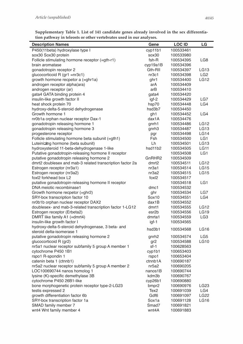
Article (unpublished) /40 45
Supplementary Table 1. List of 141 candidate genes already involved in the sex differentia-
tion pathway in teleosts or other vertebrates used in our analyses.
Description Names Gene LOC ID LG
P450(11beta) hydroxylase type I cyp11b1 100533461
sox30 Sox30 protein sox30 100533980
Follicle stimulating hormone receptor (=gth-r1) fsh-R 100534395 LG8
brain aromatase cyp19a1B 100534396
gonadotropin receptor 2 Gth-RII 100534397 LG13
glucocorticoid R (gr1 =nr3c1) nr3c1 100534398 LG2
growth hormone recpetor a (=ghr1a) ghr1 100534400 LG12
androgen receptor alpha(ara) arA 100534409
androgen receptor (ar) arB 100534410
gata4 GATA binding protein 4 gata4 100534420
insulin-like growth factor II igf-2 100534429 LG7
heat shock protein 70 hsp70 100534448 LG4
hydroxy-delta-5-steroid dehydrogenase hsd3b7 100534450
Growth hormone 1 gh1 100534452 LG4
nr0b1a orphan nuclear receptor Dax-1 dax1A 100534476
gonadotropin releasing hormone 1 gnrh1 100534486 LG12
gonadotropin releasing hormone 3 gnrh3 100534487 LG13
progesterone receptor pgr 100534498 LG14
Follicle stimulating hormone beta subunit (=gth1) Fsh 100534500 LG1
Luteinizing hormone (beta subunit) Lh 100534501 LG13
hydroxysteroid 11-beta-dehydrogenase 1-like hsd11b2 100534505 LG11
Putative gonadotropin-releasing hormone II receptor 100534508 LG1
putative gonadotropin releasing hormone 2 GnRHR2 100534509
dmrt2 doublesex and mab-3 related transcription factor 2a dmrt2 100534511 LG12
Estrogen receptor (nr3a1) nr3a1 100534514 LG15
Estrogen receptor (nr3a2) nr3a2 100534515 LG15
foxl2 forkhead box L2 foxl2 100534517
putative gonadotropin-releasing hormone II receptor 100534518 LG1
DNA meiotic recombinase1 dmc1 100534532
Growth hormone recpetor (=ghr2) ghr 100534534 LG7
SRY-box transcription factor 10 Sox10 100534551 LG4
nr0b1b orphan nuclear receptor DAX2 dax1B 100534552
doublesex- and mab-3-related transcription factor 1-LG12 dmrt1 100534555 LG12
Estrogen receptor (Erbeta2) esr2b 100534556 LG19
DMRT like family A1 (=dmrt4) dmrta1 100534559 LG3
insulin-like growth factor I igf-1 100534565
hydroxy-delta-5-steroid dehydrogenase, 3 beta- and
steroid delta-isomerase 1hsd3b1 100534568 LG16
putative gonadotropin releasing hormone 2 gnrh2 100534574 LG5
glucocorticoid R (gr2) gr2 100534588 LG10
nr5a1 nuclear receptor subfamily 5 group A member 1 sf-1 100628563
cytochrome P450 1B1 cyp1b1 100653403
rspo1 R-spondin 1 rspo1 100653404
catenin beta 1 (ctnnb1) ctnnb1A 100690187
nr5a2 nuclear receptor subfamily 5 group A member 2 nr5a2 100690205
LOC100690744 nanos homolog 1 nanos1B 100690744
lysine (K)-specific demethylase 3B kdm3b 100690767
cytochrome P450 26B1-like cyp26b1 100690880
bone morphogenetic protein receptor type-2-LG23 bmpr2 100690976 LG23
testis expressed 2 Tex2 100691039 LG4
growth differentiation factor 6b Gdf6 100691097 LG22
SRY-box transcription factor 1a Sox1a 100691128 LG16
SMAD family member 7 Smad7 100691821
wnt4 Wnt family member 4 wnt4A 100691883

Article (unpublished) /41 45
SMAD family member 3a Smad3a 100692385 LG1
DMRT-like family A2 (=dmrt5) dmrta2 100692595 LG23
DNA (cytosine-5-)-methyltransferase 3 beta, duplicate b.1 dnmt3bb1 100692621
DNA (cytosine-5)-methyltransferase 3A dnmt3a 100693331
LOC100693400 transcription factor Sox-9-A sox9A 100693400
SRY-box transcription factor 19b sox19b 100693580 LG3
Smad2 mothers against decapentaplegic homolog 2 Smad2 100693777 LG7
mothers against decapentaplegic homolog 4 100693833 LG11
LOC100693957 nanos homolog 1 nanos1A 100693957
catenin beta-1 (LOC100693960) ctnnb1B 100693960
lysine-specific demethylase 6A 100694035 LG23
cytosolic phospholipase A2 pla2g4a 100694325
factor in the germline alpha-like (LOC100694426) figla 100694426
LOC100694473 Wilms tumor protein homolog wt1A 100694473
Kisspeptin 2 kiss2 100694550 LG15
SRY-box transcription factor 6 sox6 100694759 LG1
ysine (K)-specific demethylase 4A, genome duplicate a kdm4aa 100694970
bone morphogenetic protein receptor type-2-LG16 100694986 LG16
SMAD family member 6b Smad6b 100695008 LG7
SRY-box transcription factor 2 Sox2 100695175 LG17
SMAD family member 3b Smad3b 100695277 LG7
jumonji domain containing 1Cb jmjd1cb 100695422
methionine adenosyltransferase II, beta mat2b 100695434
ysine (K)-specific demethylase 5Bb kdm5bb 100695530
lysine (K)-specific demethylase 2Ab kdm2ab 100695561
DNA (cytosine-5-)-methyltransferase 3 beta, duplicate a dnmt3ba 100696323
heat shock protein 90, alpha (cytosolic), class B member 1 hsp90ab1 100696358
lysine-specific demethylase 4A 100696578
nuclear receptor subfamily 5 group A member 2 nr5a5 100696603
SRY-box transcription factor 1b Sox1b 100697857 LG23
jumonji domain containing 4 jmjd4 100698106
fgfr2 fibroblast growth factor receptor 1a FgFr1a 100698136
Vasa (3 different genes see with Matt) vas 100698352
SRY-box transcription factor 3 sox3 100698575 LG2
jumonji domain containing 6, arginine demethylase and
lysine hydroxylasejmjd6 100698738
lysine (K)-specific demethylase 8 kdm8 100699006
activator of hsp90 ahsa1 100699088
lysine (K)-specific demethylase 7Aa kdm7aa 100699317
fgfr2 fibroblast growth factor receptor 2 FgFr2 100699731
LOC100699759 transcription factor Sox-9 B sox9B 100699759
lysine (K)-specific demethylase 6B, b kdm6bb 100699848
heat shock 70 kDa protein 1 100700083
cAMP-dependent protein kinase catalytic subunit alpha 100700452
heat shock protein 90 hsp90b1 100700552
mothers against decapentaplegic homolog 4 100700558 LG12
jumonji domain containing 7 jmjd7 100700949
synaptonemal complex protein 3 sycp3 100700972
LOC100701078 Wilms tumor protein homolog wt1B 100701078
jumonji domain containing 8 jmjd8 100701174
heat shock protein 8 hsp9 100701377
lysine (K)-specific demethylase 5A kdm5a 100701637
lysine (K)-specific demethylase 5C kdm5c 100701700
heat shock protein 90, alpha (cytosolic), class A member
1, tandem duplicate 2hsp90aa1.2 100701982
Description Names Gene LOC ID LG
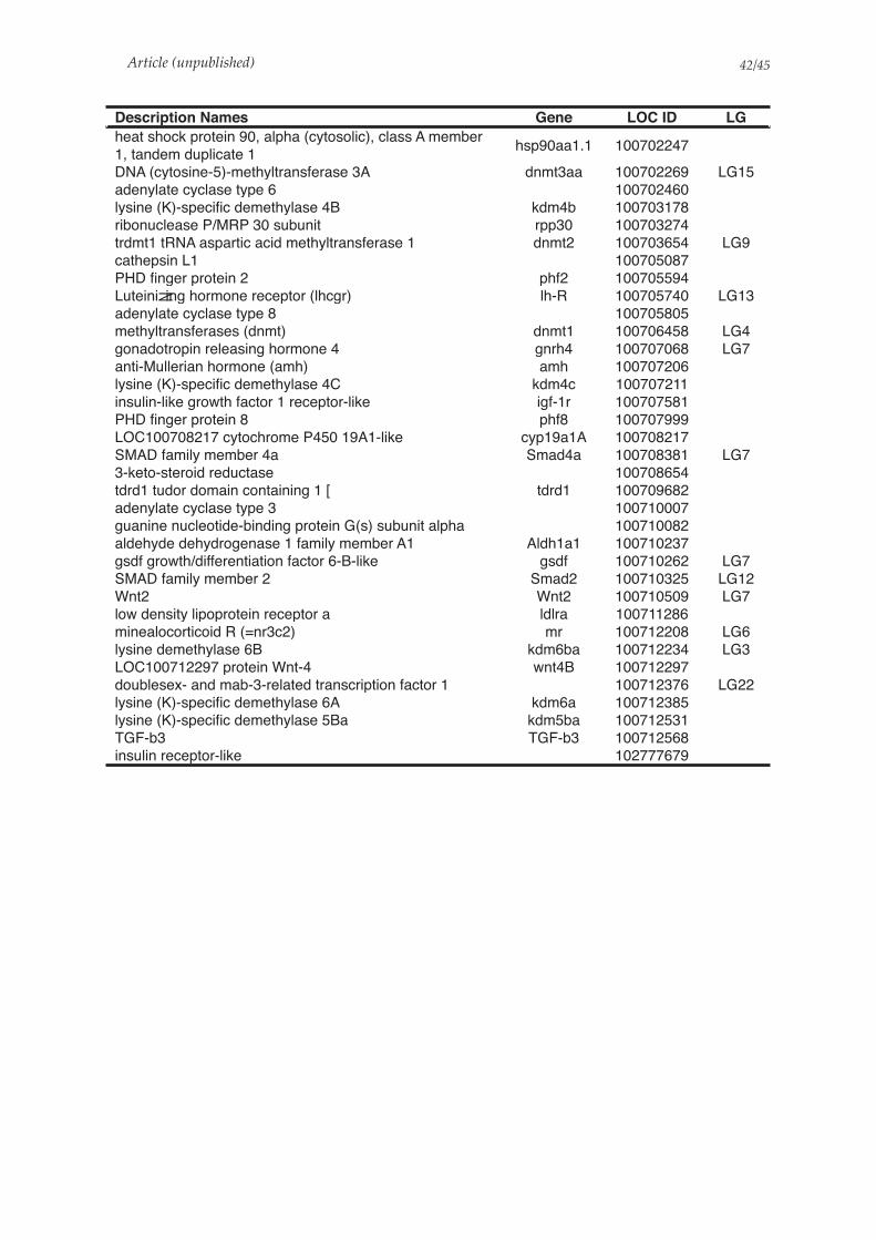
Article (unpublished) /42 45
heat shock protein 90, alpha (cytosolic), class A member
1, tandem duplicate 1hsp90aa1.1 100702247
DNA (cytosine-5)-methyltransferase 3A dnmt3aa 100702269 LG15
adenylate cyclase type 6 100702460
lysine (K)-specific demethylase 4B kdm4b 100703178
ribonuclease P/MRP 30 subunit rpp30 100703274
trdmt1 tRNA aspartic acid methyltransferase 1 dnmt2 100703654 LG9
cathepsin L1 100705087
PHD finger protein 2 phf2 100705594
Luteinizing hormone receptor (lhcgr) lh-R 100705740 LG13
adenylate cyclase type 8 100705805
methyltransferases (dnmt) dnmt1 100706458 LG4
gonadotropin releasing hormone 4 gnrh4 100707068 LG7
anti-Mullerian hormone (amh) amh 100707206
lysine (K)-specific demethylase 4C kdm4c 100707211
insulin-like growth factor 1 receptor-like igf-1r 100707581
PHD finger protein 8 phf8 100707999
LOC100708217 cytochrome P450 19A1-like cyp19a1A 100708217
SMAD family member 4a Smad4a 100708381 LG7
3-keto-steroid reductase 100708654
tdrd1 tudor domain containing 1 [ tdrd1 100709682
adenylate cyclase type 3 100710007
guanine nucleotide-binding protein G(s) subunit alpha 100710082
aldehyde dehydrogenase 1 family member A1 Aldh1a1 100710237
gsdf growth/differentiation factor 6-B-like gsdf 100710262 LG7
SMAD family member 2 Smad2 100710325 LG12
Wnt2 Wnt2 100710509 LG7
low density lipoprotein receptor a ldlra 100711286
minealocorticoid R (=nr3c2) mr 100712208 LG6
lysine demethylase 6B kdm6ba 100712234 LG3
LOC100712297 protein Wnt-4 wnt4B 100712297
doublesex- and mab-3-related transcription factor 1 100712376 LG22
lysine (K)-specific demethylase 6A kdm6a 100712385
lysine (K)-specific demethylase 5Ba kdm5ba 100712531
TGF-b3 TGF-b3 100712568
insulin receptor-like 102777679
Description Names Gene LOC ID LG

Article (unpublished) /43 45
Supplementary Table 2. Number of raw, clean and mapped reads of Hora RNA sequencing.
SampleSample
IDRaw reads
Trimming
output
Mapping output
Uniquely mapped
reads
% of uniquely mapped
reads
HNDT1 T27_1 23491158 21013911 18153700 86.39%
HNDT2 T27_2 31899564 29712909 28322680 95.32%
HNDT3 T27_3 21748948 20058428 19175366 95.60%
HNDT4 T27_4 26674375 24838808 23723420 95.51%
HNDT5 T27_5 22874995 21246013 20340942 95.74%
HNDT6 T27_6 31514924 29306575 28001583 95.55%
HNDT7 T27_7 24391045 22855747 21700772 94.95%
HNDT8 T27_8 27149842 25250651 24159627 95.68%
HNDT9 H27_1 30828164 28278223 26518845 93.78%
HNDT10 H27_2 33991742 31463225 29795137 94.70%
HNDT11 H27_3 26573407 24029331 22782663 94.81%
HNDT12 H27_4 33281072 30884281 29242232 94.68%
HNDT13 H27_5 24380059 22288264 21163260 94.95%
HNDT14 H27_6 39037928 35601928 33507930 94.12%
HNDT15 H27_7 26910813 24684283 23517533 95.27%
HNDT16 H27_8 27455526 24829974 23519105 94.72%
HNDT17 T36_17 28704221 26597398 25114469 94.42%
HNDT18 T36_18 28542948 26548881 25433502 95.80%
HNDT19 T36_19 30027239 27977437 25867525 92.46%
HNDT20 T36_20 29128715 27011150 25806943 95.54%
HNDT21 T36_21 27473253 25374350 24431107 96.28%
HNDT22 T36_22 31871493 29846203 28403104 95.16%
HNDT23 T36_23 36067322 33508249 31975889 95.43%
HNDT24 T36_24 30094046 27882642 26321388 94.40%
HNDT25 T36_25 27625046 24979069 23692622 94.85%

Article (unpublished) /44 45
Supplementary Table 3. KEGG pathways overrepresented among the down-regulated genes
at 36°C compared to 27°C control temperature
Supplementary Table 4. KEGG pathways overrepresented among the up-regulated genes at
36°C compared to 27°C control temperature
!"##$%& '()*+(,$-(./ '/01/2)(3/ 45(67/ 45(67/89: ;7<1()/3=0>/? @()/3=0>/?
!"##! $%&&'()(&% *! #+, !-#. #-/!01!" #-"*01!. $%&&'234567'89:':%867 $%&&;&83'<34(%==%=
!.!!, >?@4=4A%'@?42%9%=?='?9'%;B83)46%= .# ,! !-.C +-/D01*! "-D/01#,
E389=&86?49
F%9%6?('G9H43A86?49'<34(%==?92
!!CD! IA?948()&16>JI'@?4=)967%=?= #" ". !-.. "-CD01!C #-/D01!D
!.!#. >JI'6389=K436 *, #,D !-#+ .-/.01!D +-!,01!/
!.!*! >JI'K4&)A%38=% C .# !-*C "-.,01!/ "-C#01!+E389=(3?K6?49
!.!"! LK&?(%4=4A% ** #+D !-#" #-/+01!+ #-/.01!"
!.!.! MJI'3%K&?(86?49 *. .D ABCD #-!"01*# #-D"01#C
>%K&?(86?49'89:'3%K8?3
!.".! N?=A86(7'3%K8?3 #* *. ABED .-.*01## #-,/01!C
!."#! O8=%'%P(?=?49'3%K8?3 #. ./ !-./ .-#/01!C #-..01!D
!.""! Q4A4&424;='3%(4A@?986?49 #* "! !-. #-"#01!D *-/*01!/
!."/! R89(49?'89%A?8'K86758) #+ +. !-*, #-/*01!, .-C!01!D
!."*! J;(&%46?:%'%P(?=?49'3%K8?3 #* "" !-*D "-C#01!D /-."01!/
!.!/! <346%?9'%PK436 D .# !-*. *-./01!" #-C,01!.R4&:?92S'=436?92'89:':%238:86?49
!"#"# <346%?9'K34(%==?92'?9'%9:4K&8=A?('3%6?(;&;A .# *#C !-#" .-."01!D +-!,01!/
!!C,. M3;2'A%68@4&?=A'1'467%3'%9T)A%= #. #!" !-#* #-,#01!. #-**01!* U%94@?46?(='@?4:%238:86?49'89:'A%68@4&?=A
N%68@4&?=A
!!*"! <)3?A?:?9%'A%68@4&?=A #" D/ !-#, #-+C01!+ #-/.01!"J;(&%46?:%'A%68@4&?=A
!!*.! <;3?9%'A%68@4&?=A ** #,, !-#* *-/"01!" *-##01!.
!!",! F&;6867?49%'A%68@4&?=A #* C" !-#. *-!/01!. #-..01!* N%68@4&?=A'4H'467%3'8A?94'8(?:=
!!/D! V9%'(83@49'K44&'@)'H4&86% , *# !-., ,-"+01!D #-!#01!+N%68@4&?=A'4H'(4H8(643='89:'W?68A?9=
!!DC! R4&86%'@?4=)967%=?= + .* !-#/ D-C"01!. "-*.01!*
!!#!! L6%34?:'@?4=)967%=?= #! *" !-"* *-!/01!, "-.*01!D X?K?:'A%68@4&?=A
!!+#. Y83?4;='6)K%='4H'J12&)(89'@?4=)967%=?= D +! !-#" /-#C01!. .-D*01!*F&)(89'@?4=)967%=?='89:'A%68@4&?=A
!!+#! J1F&)(89'@?4=)967%=?= , /# !-#. /-"C01!. .-D/01!*
!#*.! O?4=)967%=?='4H'8A?94'8(?:= #+ C/ !-#/ D-*"01!+ /-D/01!"
F&4@8&'89:'4W%3W?%5'A8K=!#*"! O?4=)967%=?='4H'(4H8(643= .! *!# !-#+ #-+/01!D *-/*01!/
!##!! N%68@4&?('K86758)= #.C #CD. !-!D #-!,01!, .-!#01!D
!!*/! F&)(?9%S'=%3?9%'89:'673%49?9%'A%68@4&?=A C +* !-#D +-*D01!" "-!.01!.
IA?94'8(?:'A%68@4&?=A!!*+! I&89?9%S'8=K83686%'89:'2&;68A86%'A%68@4&?=A C +C !-#+ #-"/01!. #-!*01!*
!!*D! $)=6%?9%'89:'A%67?49?9%'A%68@4&?=A , /. !-#. ,-!#01!. "-*.01!*
!!..! I32?9?9%'89:'K34&?9%'A%68@4&?=A C /C !-#. "-D/01!. *-C/01!*
#/2/?$F=729
#/2/?$>2$'()*+(,
!"##$%& '()*+(,$-(./ '/01/2)(3/ 45(67/ 45(67/89: ;7<1()/3=0>/? @()/3=0>/?
!"#"$ %&'()*)+, $- ..! !/#0 ./#123#! -/4!23!1 56'7*8)69:'7;:<'9'=)>?*+ @,>>A>'6:%6)<,**,*
!"$#" @,>>:';&,*?)7:+)>,<A>,* 00 "1! !/#" ./"023#! -/4!23!1B?(7'>?7(:+)>,<A>,*:'7;:?79,6'<9?)7 27C?6)7+,79'>:D7E)6+'9?)7:%6)<,**?7(
!"!0! @F9)G?7,3<F9)G?7,:6,<,89)6:?79,6'<9?)7 .$ ."- !/# -/#!23!. #/0!23!-
!$#01 H,68,*:*?+8>,I:C?6A*:#:?7E,<9?)7 $1 $.# !/## 4/0-23!0 #/-"23!" D7E,<9?)A*:;?*,'*,J:C?6'> HA+'7:K?*,'*,*
!!.0! %&,7F>'>'7?7,:+,9'=)>?*+ $ -- !/-. -/"423!. #/L023!-
M+?7):'<?;:+,9'=)>?*+
N,9'=)>?*+
!!--! M6(?7?7,:=?)*F79&,*?* $ -L !/#4 L/"-23!. "/.!23!-
!!-L! @F*9,?7,:'7;:+,9&?)7?7,:+,9'=)>?*+ #! 0. !/#0 -/$-23!. #/L023!-
!!.1! 56F89)8&'7:+,9'=)>?*+ 4 $4 !/#$ "/1123!. ./#$23!-
!!!$. M*<)6='9,:'7;:'>;'6'9,:+,9'=)>?*+ #0 "L !/." ./!$23!4 #/L#23!L
@'6=)&F;6'9,:+,9'=)>?*+
!!!"! %,79)*,:'7;:(>A<A6)7'9,:?79,6<)7C,6*?)7* #. "L !/-1 #/#123!0 -/"123!$
!!!#! O>F<)>F*?*:P:O>A<)7,)(,7,*?* #4 4# !/-# #/.$23!0 -/"123!$
!!!.! %,79)*,:8&)*8&'9,:8'9&Q'F 0 .$ !/#L L/#023!. "/-423!-
!!$!! B9'6<&:'7;:*A<6)*,:+,9'=)>?*+ L "$ !/#0 1/$123!. "/1#23!-
!!0-! %F6AC'9,:+,9'=)>?*+ 1 $# !/#0 $/0423!. ./$"23!-
!#-.! R?)*F79&,*?*:)E:'+?7):'<?;* #" 40 !/#$ #/"-23!. #/#423!-
O>)='>:'7;:)C,6C?,Q:+'8*!#-"! R?)*F79&,*?*:)E:<)E'<9)6* -1 -!# !/#" ./4L23!$ ./4-23!"
!#-!! @'6=)7:+,9'=)>?*+ #4 #0! !/#- ./LL23!. -/$"23!-
!##!! N,9'=)>?<:8'9&Q'F* #$- #4L. !/!1 $/L"23!L #/0#23!$
!!$4# S?7)>,?<:'<?;:+,9'=)>?*+ ## .$ !/.# #/.#23!0 -/"123!$
S?8?;:+,9'=)>?*+!!#-! %6?+'6F:=?>,:'<?;:=?)*F79&,*?* 0 -1 !/-# #/4!23!. #/$-23!-
!!#"! B9,6)?;:&)6+)7,:=?)*F79&,*?* #L 1! !/-# ./-"23!0 "/4$23!$
!!$4! M6'<&?;)7?<:'<?;:+,9'=)>?*+ #0 L4 !/- #/#$23!$ #/-423!"
!!1.! T,9?7)>:+,9'=)>?*+ #4 1! !/-" #/"123!L "/4L23!0N,9'=)>?*+:)E:<)E'<9)6*:'7;:C?9'+?7*
!!10! %)68&F6?7:'7;:<&>)6)8&F>>:+,9'=)>?*+ #- $- !/-. -/.$23!$ -/"L23!"
!!41- K6A(:+,9'=)>?*+:3:<F9)<&6)+,:%"$! #4 L" !/-0 ./0.23!1 #/$.23!0
U,7)=?)9?<*:=?);,(6';'9?)7:'7;:+,9'=)>?*+!!41! N,9'=)>?*+:)E:I,7)=?)9?<*:=F:<F9)<&6)+,:%"$! #L L0 !/-- #/"123!0 -/"123!$
!!41. K6A(:+,9'=)>?*+:3:)9&,6:,7VF+,* #4 #!" !/#1 #/#$23!$ #/-423!"
!"0L- D79,*9?7'>:?++A7,:7,9Q)6G:E)6:D(M:86);A<9?)7 -- #-" !/#1 "/0123!0 0/$$23!$D++A7,:*F*9,+ W6('7?*+'>:BF*9,+*
!"0-! 5)>>3>?G,:6,<,89)6:*?(7'>?7(:8'9&Q'F -# #0. !/#. 1/4.23!" L/1423!.
#/2/?$A=729
#/2/?$>2$'()*+(,

Article (unpublished) /45 45
Supplementary Table 5: Primers used for quantitative realtime PCR
Gene namePrimer name (F=forward) (R=reverse)
Primer sequence (5' - 3') Tm
amh anti-Müllerian hormoneAmh-F358 CCACAGTAACCGAGTACTCAC 60°C
Amh-R640 GAACCAAACTGCTGTTTGTCAG
ara androgen receptor alphaAra-F840 GCCTTCGGTGTGGATGAATTGC 60°C
Ara-R976 AGGTGACTGCTCCATAGTGACAAC
cyp19a1Aaromatase cytochrome P450 family 19 subfamily A polypeptide 1 A
Cyp19A-F1462 CTACTTTCAGCCGTTCGGTTCAG 60°C
Cyp19A-R1744 GTTCGGGTCTCGGAGGGTTTG
cyp19a1Baromatase cytochrome P450 family 19 subfamily A polypeptide 1 B
Cyp19B-F957 GATTCATGAAGCCGAGAAGC 60°C
Cyp19B-R1184 TTCAAGATGGTGTTCATCATCTCCT
cyp11c1 cytochrome P450 -11 beta hydoxylase cyp11c1-F-DW AAAGAAGTCCTCAGGTTGTACC 60°C
cyp11c1-R-DW GACCAAAGTTCCAGCAGGTATG
dmrt1doublesex and mab-3 related transcrip-tion factor 1
Dmrt1-F972 CGGATTGCAGCGGACCGA 63°C
Dmrt1-R1232 GGACAGAGACACAGGACTAC
ef1 enlongation factor 1 alphaEf1-F1014 AGAACGTCTCCGTCAAGGAAAT 60°C
Ef1-R1115 GATGATGACCTGAGCGTTGAAG
foxl2 forkhead transcription factor L2 Foxl2-F310 AAGAGGAGCCGGTTCAGGACAA 60°C
Foxl2-R396 GCTCTCCCGGATAGCCATGG
gr2 Glucocorticoid receptor 2
GR2-F GCCCGAGTAGATGATCTCTGGTT 60°C
GR2-R CAGGAGCATGCCCCAACT
gsdf Gonadal soma-derived factorGsdf-F194 CCACCATGGCCTTTCCATTC 60°C
Gsdf-R930 GACTGCTGGGGTTGCAGTAT
hsd11b2 hydroxysteroid 11-beta dehydrogenaseHsd11B2-F546 CTGGGCTTTGAAGTGTTCGC 60°C
Hsd11B2-R847 AGAAGGCCTTGGTGATGCTC
jarid2jumonji and AT-rich interaction domain containing 2
Jarid2-F233 AGAGGAAGTTTGCCCAGTCG 60°C
Jarid2-R389 CTGGAGAGGTCCTGGATGGA
Kdm6bb lysine demethylase 6BbKdm6bb-F GCAGTTTGAGGTCCAGAACTGGCA 62°C
Kdm6bb-R GAAGCTCCCTGTTGTTGCGATGCA


DISCUSSION GÉNÉRALE ET PERSPECTIVES


DISCUSSION GÉNÉRALE ET PERSPECTIVES
Un système ancestral qui garde des zones d’ombres
Les études antérieures portant sur le déterminisme du sexe chez le tilapia du Nil ont été
menées sur des souches domestiques de laboratoire ou des souches commerciales. Ces
prospections ont conclu à un déterminisme génétique du sexe suivant un patron XX/XY
pouvant être influencé par la température ainsi que des facteurs génétiques parentaux
mineurs (Baroiller et al., 2009). Les régions responsables du déterminisme du sexe dans le
génome de cette espèce ont été identifiées préalablement sur deux groupes de liaison (LG) :
LG1 et/ou LG23 selon les souches utilisées (Lee et al., 2003; Cnaani et al., 2008; Lee et al.,
2011; Eshel et al., 2012; Palaiokostas et al., 2013; Eshel et al., 2014; Gammerdinger et al., 2014;
Li et al., 2015; Palaiokostas et al., 2015; Wessels et al., 2017; Cáceres et al., 2019; Curzon et al.,
2020; Taslima et al., 2020, 2021). Sur LG23, une duplication en tandem du gène de l’Hormone
Anti-Müllerienne (amh) sur le Y est considérée comme la région du déterminisme du sexe (Li
et al., 2015). Une première copie de l’amh, nommée amhY possède une mutation non-
synonyme par rapport au X, responsable de la masculinisation (Li et al., 2015). La seconde
copie présente quant à elle une insertion de 5bp changeant le cadre de lecture et induisant un
codon stop prématuré dans la séquence (Li et al., 2015). Aucun locus n’a été identifié
précisément sur LG1 (Gammerdinger et al., 2014).
Nos études génomiques sur les populations naturelles du lac Volta (Afrique de l’Ouest) et
du lac Koka en Ethiopie (Afrique de l’Est) ont permis d’établir que la région de la duplication
de l’amh sur LG23-Y ségrège également pour déterminer le sexe dans les populations
naturelles. De plus, nos résultats indiquent que ce système de déterminisme semble être
ancestral à la divergence des populations d’Afrique de l’Est et de l’Ouest. Nos analyses ont
révélé une structure conservée dont nous avons pu définir en détails les limites ainsi que la
composition en gènes. Nous avons également pu identifier des mutations spécifiques aux
mâles et communes aux deux populations et à certaines souches commerciales,
particulièrement au sein de l’amh, qui pourraient induire des changements d’acides aminés
dans la séquence protéique. En revanche, parmi ces polymorphismes nucléotidiques entre
mâles et femelles, nous n’avons pas observé la mutation non-synonyme décrite comme
responsable de la masculinisation dans la souche japonaise (Li et al., 2015).
Une autre différence majeure mise en lumière par nos analyses est le fait qu’une des
copies de l’amh est tronquée dans une population de l’Ouest, comme décrit sur la souche
263
DISCUSSION GÉNÉRALE ET PERSPECTIVES
japonaise (Li et al., 2015), en revanche, les deux copies de ce gène sont intactes (bien qu’avec
quelques polymorphismes nucléotidiques) au sein de la région du sexe chez une des
populations de l’Est (Koka, Ethiopie). Ainsi, bien que nous ayons pu identifier avec précision
la région associée au sexe dans ces deux populations, nos analyses ne permettent pas de
conclure quant à l’aspect fonctionnel des deux copies de l’amh et nous ne sommes pas à
même de définir la manière dont cette région détermine le sexe. Il semble tout de même que
cela ne tienne pas au SNP décrit précédemment dans l’amhY par Li et al., (2015). Une étude
récente a indiqué que l’amh agissait d’une manière dose-dépendante chez le tilapia du Nil
pour réguler la folliculogénèse en se liant à l’amhr2 (Liu et al., 2020). Il est possible que la
présence/expression de deux ou bien de trois copies de ce gène induise d’une manière dose-
dépendante le développement des mâles. Des études fonctionnelles sont donc nécessaires
pour déterminer si la dose a une importance ou s’il s’agit par exemple d’une régulation
différentielle au niveau des promoteurs.
Des études améliorant la couverture des données «!long reads!» permettraient de préciser
les séquences nucléotidiques des deux copies de l’amh sur le Y et ainsi d’avoir des séquences
de références pour chacune des copies de l’amh (X, Y et ∆Y). Avec les données dont nous
disposons actuellement, il est déjà possible de produire ce genre de références en corrigeant
les contigs construits de novo à partir des lectures longues à l’aide des lectures courtes de ces
mêmes populations en passant par des logiciels comme LoRDEC ou Jabba par exemple
(Salmela and Rivals, 2014; Miclotte et al., 2016). Il serait également intéressant de produire de
nouveaux assemblages de novo par des méthodes dites «!hybrides!» qui utilisent directement
une combinaison des lectures courtes (fiables et avec une grande couverture) et des lectures
longues (traversant les régions complexes comme les duplications) pour optimiser la
construction des contigs. Des logiciels comme WENGAN ou HASLR sont disponibles et
présentent l’avantage de prendre peu de temps pour les assemblages car ils se basent sur la
construction de graphes (Haghshenas et al., 2020; Di Genova et al., 2021).
Pour compléter ces analyses et comprendre le fonctionnement de cette duplication dans le
déterminisme du sexe, il est important de savoir si les deux copies du Y sont transcrites, et si
oui, dans quelles mesures. Une analyse du transcriptome pourrait dans un premier temps
permettre de définir si les deux copies sont traduites dans les deux populations en les
comparant aux transcrits attendus d’après les séquences de référence (X, Y et ∆Y). Si la copie
264

DISCUSSION GÉNÉRALE ET PERSPECTIVES
dupliquée produit des protéines, même tronquées, qui sont en mesure de se fixer sur le
récepteur de l’amhr2 alors on peut imaginer que cela ait un impact fonctionnel sur la cascade
du déterminisme du sexe.
Enfin, des approches d’édition du génome, notamment par CRISPR/Cas9, peuvent
permettre d’étudier les conséquences de modifications ou de knockout de certains gènes sur
le phénotype et cela a déjà été utilisé chez le tilapia du Nil (Jiang et al., 2017; Li et al., 2019,
2020; Liu et al., 2020). Cela a été réalisé notamment par Li et al., (2015) pour identifier le SNP
non-synonyme de l’amhY, considéré par les auteurs comme essentielle pour la
masculinisation. Cependant, il n’est formellement précisé si cela a été conduit uniquement
sur l’amhY ou bien une région plus vaste de forte homologie entre l’amhY et amh∆Y. Mettre
en place de tels protocoles pour tenter de déduire l’effet du nombre de copies de l’amh sur le
phénotype, ou encore étudier leurs promoteurs est certainement prometteur, cependant la
similarité qui existe entre les différentes copies de l’amh du Y mais aussi du X, notamment
chez les mâles Koka par exemple, imposera une difficulté supplémentaire pour cibler une
unique copie avec précision.
La variabilité au niveau intra-spécifique, un atout pour
comprendre l’évolution des chromosomes sexuels
Malgré la présence d’un déterminisme ancestral qui ségrège dans les populations
domestiques comme dans les populations sauvages, nos résultats montrent que ce système
n’est pas immuable. Nous avons mis en évidence qu’une des populations éthiopiennes
(Hora) ne présente pas la duplication de la région de l’amh sur LG23-Y et plus ne présente
aucune association de l’amh avec le sexe. Ce système sur LG23-Y peut donc être perdu et
suggère qu’un polymorphisme du déterminisme du sexe puisse exister chez cette espèce
puisque différentes populations n’ont pas systématiquement le même déterminant. D’autres
systèmes de déterminismes du sexe ont déjà été mis en évidence chez le tilapia du Nil
puisqu’un QTL sur LG1 est associé au sexe chez certaines souches commerciales Américaines
ou encore dans la souche de laboratoire Manzala-Stirling (Palaiokostas et al., 2013, 2015;
Gammerdinger et al., 2014). Il serait maintenant intéressant d’étudier davantage de
populations sauvages pour déterminer la prévalence des systèmes alternatifs à LG23-Y, et
s’ils font intervenir les mêmes gènes ou les mêmes conditions environnementales par
265
DISCUSSION GÉNÉRALE ET PERSPECTIVES
exemple. Il serait notamment intéressant de savoir si certaines populations sauvages
présentent un déterminisme porté par LG1 ou s’il s’agit d’un artefact de la domestication. Il
paraît notamment intéressant d’étudier le(s) déterminisme(s) génétique(s) qui existe(nt) dans
le lac Manzala (Basin du Nil, Egypte) d’où sont originaires les souches de Manzala-Stirling
qui présentent une association au sexe sur LG1.
De par les motivations économiques pour optimiser les productions aquacoles, les études
sur le déterminisme du sexe ont principalement été conduites sur des souches domestiquées.
Or, le tilapia du Nil est présent dans des environnements variés en terme de température, de
surface, de profondeur et de salinité ce qui en fait une espèce de choix pour comprendre
l’effet des facteurs environnementaux sur l’évolution des chromosomes sexuels. De plus,
travailler à l’échelle populationnelle permettrait de limiter l’impact de la variation entre les
taxons dû aux temps de divergences importants entre espèces et ainsi diminuer les facteurs
confondants lors de l’analyse de l’évolution des chromosomes sexuels (Furman et al., 2020).
Par exemple, travailler au niveau intra-spécifique a permis de montrer que les
réarrangements chromosomiques peuvent être une conséquence et non une cause de la
diminution de la recombinaison comme chez le champignon Neurospora tetrasperma et le killi
turquoise, petit poisson africain (Reichwald et al., 2015; Sun et al., 2017).
Identifier le système de déterminisme du sexe de Hora
Pour mémoire, nous avons vu que pour l’une des populations étudiées (Hora), la région
du déterminisme du sexe semble avoir changé. Différents scenarios pourraient expliquer un
changement de déterminant et parmi eux, nous avons particulièrement évoqué la possibilité
que la disparition de LG23-Y soit simplement due à la dérive génétique. Comme le lac Hora
est particulièrement petit (à peine plus de 1 km²) par rapport aux autres étudiés, et qu’il y a
certainement eu un repeuplement de tilapia du Nil au début du vingtième siècle avec des
individus d’O. niloticus cancellatus endémique d’Ethiopie dans ce lac (Bezault, 2005), on peut
penser que cette population a une petite taille efficace. Les goulots d’étranglement des
individus fondateurs et introduits, ainsi que ceux causés par la superficie du lac rendent la
population plus sensible à la stochasticité et ont pu conduire à une perte du déterminant
majeur du sexe par dérive. Ce phénomène aurait pu être d’autant plus important si parmi les
individus introduits se trouvaient des mâles XX contribuant à produire des descendances
266

DISCUSSION GÉNÉRALE ET PERSPECTIVES
100!% XX. Si nous privilégions cette hypothèse, nous ne pouvons en revanche pas exclure les
autres à ce stade. Il est possible également que cette population ait accumulé des locus
faiblement délétères près de la région du sexe qui apparaît ne pas recombiner avec le X sur
quelques kb. Pour les même raisons de taille efficace et d’effet de la dérive génétique, il est
possible que le Y ait accumulé rapidement des allèles (faiblement) délétères, entrainant la
contre-sélection de cette région. Nos analyses n’ont pas permis d’identifier de locus qui
endosserait le rôle de déterminant, ni plus largement de groupe de liaison, en association
avec le sexe dans la population Hora. Il semble peu probable que le déterminant du sexe (la
région de l’amh) se soit déplacé sur une nouvelle paire de chromosomes puisque
l’assemblage de novo des lectures longues a construit exactement l’haplotype X et les locus
voisins attendus d’après le génome de référence. Cependant, s’il s’agit d’une translocation
d’une région plus large que 50kb autour de l’amh alors cette méthode n’aurait pas été en
mesure de la détecter. De même, les analyses utilisées sur les génomes complets des pools ne
permettent pas d’identifier efficacement les systèmes polygéniques. Ainsi, si plusieurs locus
interagissent pour définir le sexe suite à la perte du rôle de LG23-Y, nous ne serions pas en
mesure de détecter de tels signaux. Des systèmes polygéniques ont déjà été mis en évidence
chez les vertébrés, incluant des cichlidés des grands lac Africains (Vandeputte et al., 2007;
Liew et al., 2012; Moore and Roberts, 2013; Roberts et al., 2016; Clark and Kocher, 2019). Une
difficulté réside alors dans le choix de la méthode à utiliser pour détecter un système
polygénique car les analyses classiquement utilisées ne font souvent ressortir que le facteur
majeur ou attendu (Moore and Roberts, 2013; Wellenreuther and Hansson, 2016). Dans notre
cas, il serait possible d’analyser les génomes individuellement pour rechercher des
déséquilibres de liaison récurrents entre certains allèles et une association avec les sexes
phénotypiques. Ces données sont déjà disponibles puisque les pools par sexe des individus
Hora ont été faits par informatique, après production des génomes complets individuels.
Une approche intéressante pourrait être le «!polygenic score!» qui permet de déterminer les
associations entre des combinaisons de SNPs et un trait phénotypique, habituellement utilisé
en médecine pour détecter le risque de développer une maladie particulière d’après les
mutations dans le génome (Choi et al., 2020; Khera et al., 2019).
Nous savons également que l’expression précoce de gsdf (gonadal soma-derived factor,
membre de la super-famille des TGF-β) joue certainement un rôle dans le développement des
gonades chez cette population, comme cela a déjà pu être reporté chez d’autres souches de
267

DISCUSSION GÉNÉRALE ET PERSPECTIVES
tilapia du Nil (Kaneko et al., 2015; Jiang et al., 2016). Si nous sommes en mesure d’identifier
des locus spécifiques ou au moins en fort déséquilibre de liaison avec le sexe, nous pouvons
imaginer génotyper les individus utilisés dans les banques de RNAseq et s’assurer que les
groupes identifiés correspondent bien aux mâles et aux femelles au moins en température
contrôle. On pourrait alors exploiter beaucoup plus en profondeur les données de ce
séquençage et en déduire plus finement la cascade précocement impliquée dans la
différenciation du sexe dans la famille analysée.
D’autre part, il existe des méthodes telles que Sex-DETector qui permettent de détecter
des chromosomes sexuels (particulièrement adapté aux chromosomes sexuels encore peu
divergents) à partir de séquençage d’ARN en se basant sur l’analyse de familles avec
idéalement les 2 géniteurs et au moins 5 individus de chaque sexe issus de la descendance
(Muyle et al., 2016). En étudiant les génotypes de la descendance et ceux des parents, les
méthodes probabilistes de ce logiciel permettent de grouper des contigs en fonction de leur
type de ségrégation (XY ou ZW dans notre cas par exemple). On pourrait envisager d’utiliser
cette méthode également pour identifier les chromosomes sexuels dans la population Hora
puisque des croisements ont été produits et que certains individus ont été maintenus jusqu’à
un âge suffisamment avancé pour permettre le sexage par observation des gonades. Ainsi,
des banques de RNAseq sur des adultes Hora, de la même famille que celle utilisée dans le
Chapitre 3, sont en cours de séquençage.
Enfin, en lien avec la thermosensibilité, il serait intéressant d’étudier les modifications
épigénétiques puisque celles-ci peuvent entraîner des régulations d’expression, par des
mécanismes tels que l’épissage alternatif. Par exemple ces mécanismes sont impliqués dans
la réversion du sexe chez le poisson plat Cynoglossus semilaevis (Shao et al., 2014) et chez
l’Agame barbu (Pogona vitticeps) (Deveson et al., 2017).
Les limites de nos études
Un focus sur certaines régions et certains gènes
Une limite récurrente dans nos analyses est l’utilisation de sous-ensembles de régions
précises du génome. Dans le cas des séquençages ddRAD, le concept même de la méthode
consiste à cibler uniquement les régions du génome comprises entre deux sites d’enzymes de
restrictions et dont l’insert entre les deux sites est de 300 à 700bp environ. Cette approche
268

DISCUSSION GÉNÉRALE ET PERSPECTIVES
permet une exploration d’un grand nombre d’individus simultanément, à un coût réduit,
puisque peu de sites sont séquencés contrairement à un génome complet. En revanche, les
assemblages obtenus sont un peu à l’image d’un texte à trous et si l’information recherchée
se situe dans une région non séquencée, alors ce type d’analyses ne permet pas de répondre
complètement aux questions posées. De plus, en se focalisant sur des sites de restrictions, on
prend le risque de séquencer de manière non aléatoire certaines familles multigéniques qui
présenteraient ces sites spécifiques. Il est possible que nous ayons rencontré ce biais à la fois
pour la famille Kpandu (Ghana) et pour la famille Kou (Burkina-Faso) analysées. La présence
de polymorphisme entre les différents gènes d’une même famille crée artificiellement des
régions particulièrement polymorphiques pouvant induire un signal artefactuel.
Lors des analyses de «!long reads!», une fois encore nous nous sommes focalisés sur une
partie précise du génome. Parce que les analyses précédentes ont suggéré que la région de la
duplication de l'amh sur LG23-Y était le principal gène déterminant le sexe sur les
populations naturelles étudiées, nous avons sélectionné un sous-ensemble de lectures
correspondant à ces gènes spécifiques parmi l'ensemble de données brutes afin de gagner du
temps lors des assemblages en réduisant la taille de la région à assembler.
Ces approches peuvent se comprendre puisque le même ensemble de gènes «! joue
souvent aux chaises musicales! » pour endosser le rôle de déterminant du sexe chez les
poissons téléostéens (Böhne et al., 2016; Pan et al., 2016; Ortega-Recalde et al., 2020; Tao et al.,
2021), justifiant que certains suspects «!habituels!» soient recherchés en priorité. Toutefois,
certains gènes a priori inattendus peuvent être impliqués dans le déterminisme du sexe. C’est
le cas chez la truite arc-en-ciel pour laquelle une néo-fonctionnalisation d’un gène
immunitaire à la suite d’une duplication a conduit à en faire le gène majeur du déterminisme
du sexe (Yano et al., 2012). Des systèmes polygéniques moins évidents peuvent aussi ne pas
être détectés (Moore and Roberts, 2013; Wellenreuther and Hansson, 2016). En ne regardant
que les gènes candidats et les suspects habituels, nous nous sommes concentrés sur les gènes
attendus de la voie du sexe mais avons peut-être ignorés d’autres gènes qui pourraient jouer
un rôle dans le déterminisme du sexe.
Identification d’un chromosome B!: une difficulté supplémentaire
Dans le cas des analyses de génomes complets des individus sauvages des populations
Kpandu (Ghana) et Koka (Ethiopie), le soupçon de la présence d’un chromosome B nous a
269
DISCUSSION GÉNÉRALE ET PERSPECTIVES
conduit à nous focaliser uniquement sur une petite région du génome, identifiée
préalablement par les analyses de ddRAD. Or, comme nous avions utilisé des familles pour
le ddRAD, il est possible que, si plusieurs locus du déterminisme du sexe ségrègent dans les
populations, nous ayons échantillonné une famille portant le déterminant majoritaire et que
nous passions à côté de déterminant qui ségrégerait en faible fréquences dans les
populations.
Cette approche nous empêche donc de tirer profit de l’étude de l’ensemble des génomes
complets d’individus du milieu naturel qui auraient pu contenir cette information.
Idéalement, il faudrait être en mesure soit! : i) d’identifier quels individus sont porteurs du
chromosome B dans chacun des pools et séquencer à nouveaux en séparant ces individus des
autres!; ii) de caractériser la vaste majorité des régions appartenant au chromosome B et les
retirer du jeu de données pour pouvoir avoir une approche globale du génome.
i) La mise en place d’amorces PCR pour détecter les individus porteurs de chromosome(s)
B a été un travail important dans ce projet de thèse afin de pouvoir identifier les individus
porteurs dans les pools. Cependant, il est particulièrement compliqué de concevoir des
amorces PCR dans des régions de chromosome B sans en avoir la séquence exacte. Le design
des amorces a été effectué sur la base de la séquence consensus de ces régions de forte
couverture en ciblant les zones riches en SNPs spécifiques au pool contenant le B. Seulement,
une région de forte couverture indique en réalité que de nombreuses copies de ce gène
existent sur le chromosome B et il est difficile de savoir si elles possèdent toutes exactement
le même enchainement de SNPs. Ainsi, en dessinant des amorces sur le consensus, on
dessine peut-être des amorces sur la base d’un hybride de 10 copies de ce locus qui ne
possèdent pas toutes les mêmes SNPs. De plus, l’utilisation d’un mapping sur un génome de
référence ne nous permet pas de voir avec certitude l’agencement des régions spécifiques B
les unes par rapports aux autres. Il est possible que nous ayons essayé d’amplifier des
régions dont nous estimions la taille à 300bp qui s’avéraient être bien au-delà de la taille
raisonnable amplifiable en PCR. D’autres amorces peuvent être dessinées, mais cette
approche restera problématique tant que nous ne connaitrons pas exactement la séquence du
chromosome B.
ii) Caractériser un chromosome B et définir précisément son contenu est un travail
fastidieux qui peut être conduit de deux manières : directe ou indirecte (revue dans Ruban et
270

DISCUSSION GÉNÉRALE ET PERSPECTIVES
al., 2017). Cela passe idéalement par le séquençage du chromosome B préalablement isolé
des autres chromosomes par exemple par micro-dissection ou par «!flow sorting! » (tri de
cellules et de chromosomes). Ce type d’analyse cytogénétique permet d’une part de
confirmer la présence d’un tel élément génétique et d’autre part d’obtenir un assemblage du
chromosome B et donc son contenu en gènes (Vij et al., 2016). Cependant, la microdissection
nécessite d’avoir des chromosomes en métaphase bien étaler et, de plus, est prône aux
contaminations par les autres chromosomes (Valente et al., 2014!; D’Cotta et al., 2014). Dans
notre cas, aucun individu vivant n’était disponible pour procéder à de tels analyses.
Néanmoins, nous avons des suspensions chromosomiques de certains individus sauvages
qui pourrait éventuellement être étalées s’ils s’avèrent porteur d’un chromosome B.
Cependant, il existe également des méthodes indirectes pour caractériser le contenu d’un
chromosome B lorsque que les approches cytogénétiques ne sont pas envisageables. À partir
de séquençages de génomes complets d’individus portant le B et d’individus sans B, il est
possible d’identifier des régions appartenant potentiellement au chromosome B. Dans les
analyses complémentaires du Chapitre 1, nous avons utilisé une de ces méthodes indirectes,
basée sur les ratios de couvertures entre individus présentant le B ou non (Valente et al.,
2014; Fantinatti & Martins, 2016; Clark et al., 2018). Il est aussi possible de se baser sur les
ratios des fréquences de k-mer (Kurtz et al., 2008; Marçais & Kingsford, 2011; Navarro-
Domínguez et al., 2019) ou encore sur le clustering des lectures d’après leur similarités (un
peu à la manière d’un assembleur à graphe) (Novák et al., 2010). La méthode de clustering
par graphe contrairement aux deux autres ne nécessite pas de génome de référence. Une
utilisation de différentes méthodes en parallèle est préférable puisque cela permet d’assoir
l’identification de certaines régions si elles sont retrouvées avec plusieurs approches. Les jeux
de données dont nous disposons actuellement répondent aux prérequis nécessaires pour
pouvoir caractériser ce B et font parties des ambitions de la suite de ce projet de thèse.
Enfin, les chromosomes B chez les cichlidés ne sont pas forcément présents (ni en même
nombre de copies) chez tous les individus d’une population ou d’une population à l’autre
(Poletto et al., 2010; Perazzo et al., 2018; Clark & Kocher, 2019). Si à terme il est possible
d’identifier ce chromosome B facilement (comme avec des marqueurs PCR) dans l’ensemble
des échantillons encore disponibles au laboratoire on pourrait estimer la fréquence à laquelle
celui-ci ségrège dans les populations naturelles et si on le retrouve dans les deux sexes dans
les populations Koka et Kpandu. En effet, nous avons soupçonné un chromosome B dans le
271

DISCUSSION GÉNÉRALE ET PERSPECTIVES
pool mâles de la population Kpandu et dans le pool femelles de la population Koka, ce qui
laisse penser qu’il n’agit pas comme déterminant du sexe à l’échelle de l’espèce. Cependant,
il n’est pas impossible que dans l’une des populations il agisse comme un déterminant du
sexe comme cela a déjà été vu chez Metriaclima lombardoi (Clark and Kocher, 2019).
L’acquisition de ces informations repose donc sur les deux points (i) et (ii) explicités
précédemment.
L’Hybridation!: une alternative pour expliquer nos résultats!?
Les espèces du genre Oreochromis ont une grande capacité d’hybridation interspécifique et
les descendances produites sont, pour la plupart des combinaisons, viables et fertiles
(Hickling, 1960; Jalabert et al., 1971; revue dans Baroiller and D’Cotta, 2018). Pour
l’aquaculture, de nombreux croisements ont été effectués pour profiter entre autre de la
vigueur hybride et/ou obtenir des souches piscicoles ayant les traits les plus favorables de
deux espèces différentes (résistances aux maladies, à la salinité, croissance rapide etc...)
(Yehya et al., 2012; de Verdal et al., 2014; Rahman et al., 2019; Nzohabonayo et al., 2021). Ces
croisements entre espèces proches ont également été utilisés pour étudier le déterminisme du
sexe et rechercher les paires de chromosomes sexuels chez les cichlidés du genre Oreochromis
(Jalabert et al., 1971; Cnaani et al., 2004). De plus, les systèmes hétérogamétiques différents ne
sont pas une barrière à ces croisements et permettent entre autre de produire des souches
monosexes mâles en pisciculture (Hickling, 1960; Baroiller and D’Cotta, 2018).
Les cichlidés utilisés en aquaculture, particulièrement des genres Tilapia, Sarotherodon et
Oreochromis, ont fait l’objet de nombreux transferts par la main de l’homme en Afrique
durant la seconde moitié du XXe siècle (revue dans Bezault, 2005). Comme le tilapia du Nil
(O. niloticus) représente la très grande majorité de la production piscicole parmi ces
différentes espèces, ses transferts ont été particulièrement importants à la fois au sein de son
aire de répartition mais également en dehors de celle-ci (Canonico et al., 2005; D’Amato et al.,
2007; Firmat et al., 2013; Deines et al., 2014). La souche d’O. niloticus Bouaké (aussi appelée
«!Souche Côte d’Ivoire!») est une souche synthétique issue de deux populations d’Afrique de
l’Ouest qui a par exemple été expédiée dans différents pays d’Afrique (Burkina Faso,
Sénégal, Bénin…) mais aussi en France, aux Etats-Unis et en Amérique du Sud, au Brésil
(Bezault, 2005). Un autre exemple marquant est l’utilisation commerciale de la souche GIFT
(Genetically Improved Farmed Tilapia) qui a été très largement distribuée à travers le monde
272

DISCUSSION GÉNÉRALE ET PERSPECTIVES
et qui est une souche synthétique produite à partir de quatre populations provenant
d’Égypte, du Ghana, du Kenya et du Sénégal et de quatre souches d’aquaculture provenant
d’Israël, de Singapour, de Taiwan et de Thaïlande (Eknath et al., 1998).
Dans le milieu naturel, ces introductions ont pu mener à de l’hybridation avec les espèces
endémiques comme cela a été le cas dans le lac Naivasha (Kenya) entre O. niloticus et O.
variabilis et dans le lac Itasy (Madagascar) entre O. macrochir et O. niloticus (tous deux
introduits) (Lévêque and Paugy, 2006). Des études ont également suggéré l’existence
d’hybridations entre O. aureus et O. niloticus suite à l’observation d’introgression de génome
mitochondrial dans les populations d’Afrique de l’Ouest (Rognon and Guyomard, 2003)
ainsi que dans le génome mitochondrial de O. leucosticus vers O. niloticus au Kenya dans le
lac Baringo (Nyingi and Agnèse, 2007). Des hybridations ont également été rapportées dans
le sud de l’Afrique entre O. mossambicus (endémique) et O. niloticus (introduit) (D’Amato et
al., 2007; Firmat et al., 2013).
En sachant que les aires de répartitions de certaines de ces espèces peuvent se recouper,
comme O. niloticus et O. aureus en Afrique de l’Ouest (Rognon and Guyomard, 2003) (Figure
5.1) et que des hybridations/introgressions ont lieu dans le milieu naturel, il est légitime de
se demander si les populations que nous avons étudiées ont pu être confrontées à de tels
évènements. Par exemple, il est surprenant de retrouver la présence de chromosomes B et le
déterminisme du sexe par l’amh sur LG23-Y dans les populations Kpandu et Koka alors que
celles-ci sont éloignées géographiquement, et que Koka est phylogénétiquement plus proche
de Hora. Cependant, les analyses phylogénétiques nucléaires et mitochondriales ne semblent
pas soutenir l’hypothèse d’hybridations. En effet, si des croisements avaient eu lieu entre des
individus de Kpandu et Koka, on pourrait s’attendre à ce que ces populations s’en retrouvent
rapprochées dans la phylogénie nucléaire. A contrario, on observe une plus grande proximité
entre Koka et Hora dans l’ensemble des analyses phylogénétiques. Cependant, dans le cas où
l’évènement d’introgression serait suffisamment lointain pour que la majorité du génome ait
subi un balayage sélectif ou l’effet de la dérive génétique, alors nous ne pourrions pas
forcément observer de conséquences de cette introgression en se basant seulement sur
l’analyse de 42 marqueurs nucléaires (Figure 5.2) (Folk et al., 2018). Tout de même, l’analyse
de la région de l’amh sur LG23 montre que les haplotypes X sont plus similaires entre Koka et
Hora qu’entre Koka et Kpandu.
273
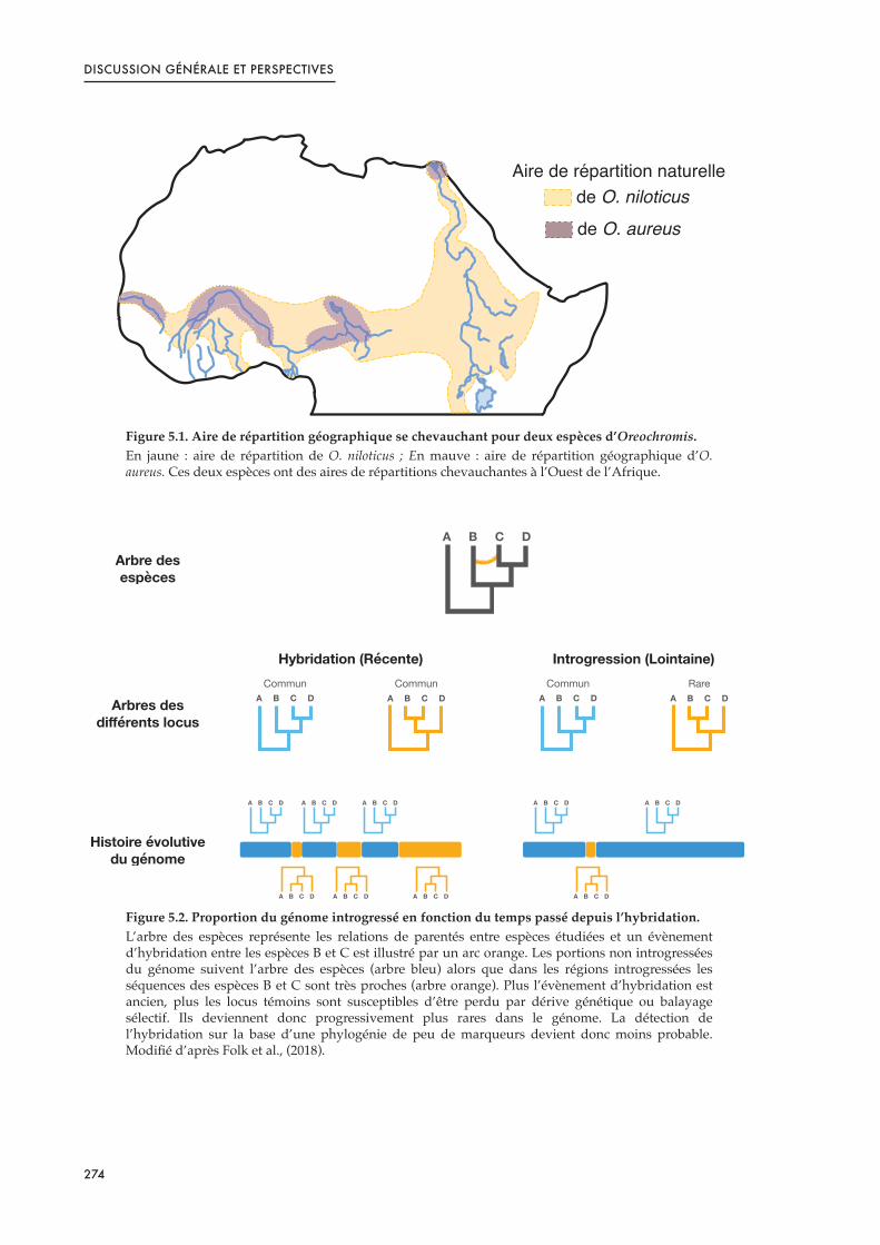
DISCUSSION GÉNÉRALE ET PERSPECTIVES
Figure 5.1. Aire de répartition géographique se chevauchant pour deux espèces d’Oreochromis.En jaune : aire de répartition de O. niloticus ; En mauve : aire de répartition géographique d’O. aureus. Ces deux espèces ont des aires de répartitions chevauchantes à l’Ouest de l’Afrique.
Figure 5.2. Proportion du génome introgressé en fonction du temps passé depuis l’hybridation.L’arbre des espèces représente les relations de parentés entre espèces étudiées et un évènement d’hybridation entre les espèces B et C est illustré par un arc orange. Les portions non introgressées du génome suivent l’arbre des espèces (arbre bleu) alors que dans les régions introgressées les séquences des espèces B et C sont très proches (arbre orange). Plus l’évènement d’hybridation est ancien, plus les locus témoins sont susceptibles d’être perdu par dérive génétique ou balayage sélectif. Ils deviennent donc progressivement plus rares dans le génome. La détection de l’hybridation sur la base d’une phylogénie de peu de marqueurs devient donc moins probable. Modifié d’après Folk et al., (2018).
!"#$%&$%#'()#*"*"+,%,)*-#$..$
&$%!"#$%&'(%)*+
&$%!"#,*-.*+
A B C D
A B C D
A B C D
A B C D
A B C DA B C DA B C D
A B C D A B C D A B C D
A B C DA B C D
Commun RareCommun Commun
Hybridation (Récente) Introgression (Lointaine)
A B C DA B C D
Arbre des espèces
Arbres des différents locus
Histoire évolutive du génome
274
DISCUSSION GÉNÉRALE ET PERSPECTIVES
Si les deux haplotypes ont cessé de recombiner sur cette région, il n’y a pas de raisons
qu’après l’introgression il ne reste pas de traces dans les haplotypes X de Kpandu et Koka, ni
que les populations éthiopiennes aient le schéma d’une histoire évolutive commune avec des
SNPs partagés dans cette région.
Il serait également envisageable que la population Hora ne présente plus l’amh comme
déterminant du sexe sur LG23-Y suite à une hybridation qui aurait entrainé la colonisation
de cette région par l’haplotype de l’espèce introgressante. Cependant, les relations de parenté
décrites par les analyses phylogénétiques montrent Koka et Hora en groupe monophylétique
et indiquent que ces espèces sont peu divergentes en comparaison de Kpandu ou de
l’individu séquencé pour produire le génome de référence par exemple. Ici encore, si
l’hybridation est suffisamment lointaine, la majeure partie du génome «!introgressant!» a pu
être nettoyé et nos analyses phylogénétiques pourraient ne pas en rendre compte. De plus,
des SNPs spécifiques à Koka et Hora sur l’haplotype X semblent décrire une histoire
évolutive commune de ces régions et ne supportent pas à première vue l’hypothèse d’une
introgression. Enfin, les espèces sympatriques de O. niloticus dans le lac Hora sont
essentiellement des poissons-chats (Clarias sp.) ainsi que de Cyprinodontidae (Paropanchax
sp.) qui ne pourraient pas faire l’objets de flux de gènes avec le tilapia du Nil et il n’y a pas
non plus de traces de transferts d’autres espèces de tilapia en Ethiopie qui soit en mesure de
s’hybrider avec le tilapia du Nil (Bezault, 2005). L’ensemble de ces indices nous laisse donc
penser que l’introgression de LG23-Y par une autre espèce n’est probablement pas la raison
de l’absence de la région du déterminisme du sexe chez cette population.
Déterminer l’état ancestral du déterminisme du sexe chez le tilapia du Nil pourrait nous
aider à trancher entre les hypothèses d’une acquisition de la duplication de l’amh sur LG23-Y
par flux de gènes entre Koka et Kpandu (probablement par la main de l’homme dans ce cas)
ou bien d’une perte secondaire de cette structure chez Hora. Des améliorations de nos
analyses phylogénétiques pourraient par exemple aider à éclaircir les différents scénarios
décrits ci-dessus. D’une part, nous pourrions intégrer un plus grand nombre de locus, qui
idéalement possèderaient des caractéristiques variées d’évolution des séquences, afin de
mieux retracer l’histoire évolutive des génomes et des populations. D’autre part, il semble
essentiel d’ajouter des populations supplémentaires pour pouvoir retracer au mieux
l’histoire évolutive de la paire de chromosome sexuel LG23. Nous pourrions également
275

DISCUSSION GÉNÉRALE ET PERSPECTIVES
envisager d’utiliser des méthodes de coalescence dans ces nouvelles analyses
phylogénétiques (Hibbins and Hahn, 2019). Cela permettrait notamment de limiter les biais
induits par la concaténation, qui présuppose une histoire évolutive commune des différents
locus analysés (Gatesy et al., 2017). Enfin, l’ensemble de ces améliorations permettrait
d’obtenir des données solides pour comprendre l’évolution des paires de chromosomes
sexuels si LG23 est une «!vieille!» paire de chromosomes à l’échelle de l’espèce, avec le X et le
Y peu divergents. Malheureusement, nos données ne permettent pas en l’état d’effectuer ces
comparaisons et un re-séquençage des lectures longues pour augmenter la couverture sera
nécessaire pour reconstruire les haplotypes Y avec fiabilité. Les lectures courtes pourront
ensuite aider à corriger finement ces haplotypes Y reconstruits.
Conclusion
Ce manuscrit de thèse représente le premier complexe d’études de génétique et
génomique sur le déterminisme du sexe chez le tilapia du Nil en populations naturelles.
Malgré les difficultés techniques que peuvent entraîner l’étude des chromosomes sexuels, en
couplant différentes approches méthodologiques nous avons mis en évidence un
polymorphisme du déterminisme du sexe dans trois populations naturelles de cette espèce.
Ce résultat met en lumière un patron d’évolution complexe de ce système que nous pensons
être lié à la perte du déterminisme ancestral dans l’une des populations étudiées. Ces travaux
montrent donc l’intérêt d’analyser le déterminisme du sexe à une échelle populationnelle
pour estimer l’importance de la diversité qui peut exister en milieu naturel. De plus, étudier
le déterminisme du sexe au niveau intraspécifique chez des espèces adaptées à des milieux
variés représente une excellente opportunité pour comprendre les mécanismes à l’origine de
l’évolution des chromosomes sexuels. Nous sommes également parvenus à détailler une
région du déterminisme du sexe dont l’architecture complexe (et variable entre populations)
n’avait jusqu’ici pas été décrite. En particulier nous avons proposé pour la première fois un
haplotype du chromosome Y dans cette région. Ces analyses représentent un socle sur lequel
il sera possible de s’appuyer pour faciliter la reconstruction des chromosomes sexuels lors de
l’assemblage de génome de tilapia. Cependant, la manière exacte, dont cet haplotype Y agit
pour déterminer le sexe et notamment le rôle des paralogues de l’amh, reste à définir. La
question du mécanisme du déterminisme du sexe reste également ouverte chez la population
276

DISCUSSION GÉNÉRALE ET PERSPECTIVES
ne présentant pas la région ancestrale sur le Y et des analyses complémentaires à la fois de
génomique et de transcriptomique seront nécessaires. Enfin, ces travaux suggèrent aussi
pour la première fois l’existence d’un chromosome B (chromosome surnuméraire, non
essentiel et au comportement égoïste) qui ségrégerait dans certaines populations naturelles
de tilapia du Nil.
277

DISCUSSION GÉNÉRALE ET PERSPECTIVES
Références
Baroiller, J.-F., and D’Cotta, H. (2018). Sex Control in Tilapias. In Sex Control in Aquaculture, (John Wiley & Sons, Ltd), pp. 189–234.
Baroiller, J.F., D’Cotta, H., Bezault, E., Wessels, S., and Hoerstgen-Schwark, G. (2009). Tilapia sex determination: where temperature and genetics meet. Comparative Biochemistry and Physiology Part A: Molecular & Integrative Physiology 153, 30–38.
Bezault, E. (2005). Etude Du Systeme De Determinisme Du Sexe au Sein De Populations Naturelles De Tilapia Du Nil, Oreochromis niloticus (linnaeus, 1758) : importance Des Composantes Genetiques Et Environnementales. UNIVERSITE DE PARIS-SUD.
Böhne, A., Wilson, C.A., Postlethwait, J.H., and Salzburger, W. (2016). Variations on a theme: Genomics of sex determination in the cichlid fish Astatotilapia burtoni. BMC Genomics 17.
Cáceres, G., López, M.E., Cádiz, M.I., Yoshida, G.M., Jedlicki, A., Palma-Véjares, R., Travisany, D., Díaz-Domínguez, D., Maass, A., Lhorente, J.P., et al. (2019). Fine mapping using whole-genome sequencing confirms anti-müllerian hormone as a major gene for sex determination in farmed Nile tilapia (Oreochromis niloticus L.). G3 (Bethesda) 9, 3213–3223.
Canonico, G.C., Arthington, A., McCrary, J.K., and Thieme, M.L. (2005). The effects of introduced tilapias on native biodiversity. Aquatic Conservation: Marine and Freshwater Ecosystems 15, 463–483.
Choi, S.W., Mak, T.S.-H., and O’Reilly, P.F. (2020). Tutorial: a guide to performing polygenic risk score analyses. Nature Protocols 15, 2759–2772.
Clark, F.E., and Kocher, T.D. (2019). Changing sex for selfish gain: B chromosomes of Lake Malawi cichlid fish. Scientific Reports 9, 1–10.
Clark, F.E., Conte, M.A., and Kocher, T.D. (2018). Genomic characterization of a B chromosome in Lake Malawi cichlid fishes. Genes 9, 610.
Cnaani, A., Zilberman, N., Tinman, S., Hulata, G., and Ron, M. (2004). Genome-scan analysis for quantitative trait loci in an F2 tilapia hybrid. Mol Genet Genomics 272, 162–172.
Cnaani, A., Lee, B.-Y., Zilberman, N., Ozouf-Costaz, C., Hulata, G., Ron, M., D'Hont, A., Baroiller, J.-F., D'Cotta, H., Penman, D.J., et al. (2008). Genetics of sex determination in tilapiine species. Sexual Development 2, 43–54.
Curzon, A.Y., Shirak, A., Dor, L., Zak, T., Perelberg, A., Seroussi, E., and Ron, M. (2020). A duplication of the Anti-Müllerian hormone gene is associated with genetic sex determination of different Oreochromis niloticus strains. Heredity 125, 317–327.
D’Amato, M.E., Esterhuyse, M.M., van der Waal, B.C.W., Brink, D., and Volckaert, F.A.M. (2007). Hybridization and phylogeography of the Mozambique tilapia Oreochromis mossambicus in southern Africa evidenced by mitochondrial and microsatellite DNA genotyping. Conserv Genet 8, 475–488.
D'Cotta H., Coutanceau J-P., Pepey E., Baaziz S.B., Coriton O., Bonillo C., Rodier-Goud M., Soler L., D'Hont A., Kocher T.D., Ozouf-Costaz C., Baroiller J-F. (2014). Fishing for sex genes by chromosome microdissection in two tilapia species with different sex
278

DISCUSSION GÉNÉRALE ET PERSPECTIVES
determining systems. 21st International Colloquium on Animal Cytogenetics and Gene Mapping (ICACGM).
Deines, A., Bbole, I., Katongo, C., Feder, J., and Lodge, D. (2014). Hybridisation between native Oreochromis species and introduced Nile tilapia O. niloticus in the Kafue River, Zambia. African Journal of Aquatic Science 39, 23–34.
Deveson, I.W., Holleley, C.E., Blackburn, J., Marshall Graves, J.A., Mattick, J.S., Waters, P.D., and Georges, A. (2017). Differential intron retention in Jumonji chromatin modifier genes is implicated in reptile temperature-dependent sex determination. Sci. Adv. 3, e1700731.
Di Genova, A., Buena-Atienza, E., Ossowski, S., and Sagot, M.-F. (2021). Efficient hybrid de novo assembly of human genomes with WENGAN. Nature Biotechnology 39, 422–430.
Eknath, A., Dey, M., Rye, M., Gjerde, B., Abella, T.A., Sevilleja, R., Tayamen, M.M., Reyes, R.A., and Bentsen, H. (1998). Selective breeding of Nile tilapia for Asia.
Eshel, O., Shirak, A., Weller, J.I., Hulata, G., and Ron, M. (2012). Linkage and physical mapping of sex Rregion on LG23 of Nile tilapia (Oreochromis niloticus). G3: Genes, Genomes, Genetics 2, 35–42.
Eshel, O., Shirak, A., Dor, L., Band, M., Zak, T., Markovich-Gordon, M., Chalifa-Caspi, V., Feldmesser, E., Weller, J.I., Seroussi, E., et al. (2014). Identification of male-specific amh duplication, sexually differentially expressed genes and microRNAs at early embryonic development of Nile tilapia (Oreochromis niloticus). BMC Genomics 15, 774.
Fantinatti, B.E.A., and Martins, C. (2016). Development of chromosomal markers based on next-generation sequencing: the B chromosome of the cichlid fish Astatotilapia latifasciata as a model. BMC Genetics 17, 119.
Firmat, C., Alibert, P., Losseau, M., Baroiller, J.-F., and Schliewen, U.K. (2013). Successive Invasion-Mediated Interspecific Hybridizations and Population Structure in the Endangered Cichlid Oreochromis mossambicus. PLOS ONE 8, e63880.
Folk, R.A., Soltis, P.S., Soltis, D.E., and Guralnick, R. (2018). New prospects in the detection and comparative analysis of hybridization in the tree of life. American Journal of Botany 105, 364–375.
Furman, B.L.S., Metzger, D.C.H., Darolti, I., Wright, A.E., Sandkam, B.A., Almeida, P., Shu, J.J., and Mank, J.E. (2020). Sex chromosome evolution: so many exceptions to the rules. Genome Biol Evol 12, 750–763.
Gammerdinger, W.J., Conte, M.A., Acquah, E.A., Roberts, R.B., and Kocher, T.D. (2014). Structure and decay of a proto-Y region in Tilapia, Oreochromis niloticus. BMC Genomics 15, 975.
Gatesy, J., Meredith, R.W., Janecka, J.E., Simmons, M.P., Murphy, W.J., and Springer, M.S. (2017). Resolution of a concatenation/coalescence kerfuffle: partitioned coalescence support and a robust family-level tree for Mammalia. Cladistics 33, 295–332.
Haghshenas, E., Asghari, H., Stoye, J., Chauve, C., and Hach, F. (2020). HASLR: Fast Hybrid Assembly of Long Reads. IScience 23, 101389.
Hibbins, M.S., and Hahn, M.W. (2019). The Timing and Direction of Introgression Under the Multispecies Network Coalescent. Genetics 211, 1059–1073.
279

DISCUSSION GÉNÉRALE ET PERSPECTIVES
Hickling, C.F. (1960). The Malacca tilapia hybrids. J Genet 57, 1–10.
Jalabert, B., Kammacher, P., and lessent, P. (1971). Déterminisme du sexe chez les hybrides entre tilapia macrochir et tilapia nilotica. étude de la sex-ratio dans les recroisements des hybrides de premiere génération par les espèces parentes. Annales de Biologie Animale, Biochimie, Biophysique 11, 155–165.
Jiang, D., Chen, J., Fan, Z., Tan, D., Zhao, J., Shi, H., Liu, Z., Tao, W., Li, M., and Wang, D. (2017). CRISPR/Cas9-induced disruption of wt1a and wt1b reveals their different roles in kidney and gonad development in Nile tilapia. Developmental Biology 428, 63–73.
Jiang, D.-N., Yang, H.-H., Li, M.-H., Shi, H.-J., Zhang, X.-B., and Wang, D.-S. (2016). gsdf is a downstream gene of dmrt1 that functions in the male sex determination pathway of the Nile tilapia. Molecular Reproduction and Development 83, 497–508.
Kaneko, H., Ijiri, S., Kobayashi, T., Izumi, H., Kuramochi, Y., Wang, D.-S., Mizuno, S., and Nagahama, Y. (2015). Gonadal soma-derived factor (gsdf), a TGF-beta superfamily gene, induces testis differentiation in the teleost fish Oreochromis niloticus. Molecular and Cellular Endocrinology 415, 87–99.
Khera, A.V., Chaffin, M., Zekavat, S.M., Collins, R.L., Carolina, R., Natarajan, P., Lichtman, J.H., D’Onofrio, G., Mattera, J., Dreyer, R., et al. (2019). Whole-Genome Sequencing to Characterize Monogenic and Polygenic Contributions in Patients Hospitalized With Early-Onset Myocardial Infarction. Circulation 139, 1593–1602.
Kurtz, S., Narechania, A., Stein, J.C., and Ware, D. (2008). A new method to compute K-mer frequencies and its application to annotate large repetitive plant genomes. BMC Genomics 9, 517.
Lee, B.-Y., Penman, D.J., and Kocher, T.D. (2003). Identification of a sex-determining region in Nile tilapia (Oreochromis niloticus) using bulked segregant analysis. Animal Genetics 34, 379–383.
Lee, B.-Y., Coutanceau, J.-P., Ozouf-Costaz, C., D’Cotta, H., Baroiller, J.-F., and Kocher, T.D. (2011). Genetic and physical mapping of sex-linked AFLP markers in Nile tilapia (Oreochromis niloticus). Mar Biotechnol (NY) 13, 557–562.
Lévêque, C., and Paugy, D. (2006). Les poissons des eaux continentales africaines: diversité, écologie, utilisation par l’homme (Paris: IRD éd.).
Li, M., Sun, Y., Zhao, J., Shi, H., Zeng, S., Ye, K., Jiang, D., Zhou, L., Sun, L., Tao, W., et al. (2015). A tandem duplicate of anti-müllerian hormone with a missense SNP on the Y chromosome is essential for male sex determination in Nile tilapia, Oreochromis niloticus. PLOS Genetics 11, e1005678.
Li, M., Liu, X., Dai, S., Xiao, H., and Wang, D. (2019). High Efficiency Targeting of Non-coding Sequences Using CRISPR/Cas9 System in Tilapia. G3 Genes|Genomes|Genetics 9, 287–295.
Li, M., Dai, S., Liu, X., Xiao, H., and Wang, D. (2020). A detailed procedure for CRISPR/Cas9-mediated gene editing in tilapia. Hydrobiologia.
Liew, W.C., Bartfai, R., Lim, Z., Sreenivasan, R., Siegfried, K.R., and Orban, L. (2012). Polygenic Sex Determination System in Zebrafish. PLoS ONE 7, e34397.
280

DISCUSSION GÉNÉRALE ET PERSPECTIVES
Liu, X., Xiao, H., Jie, M., Dai, S., Wu, X., Li, M., and Wang, D. (2020). Amh regulate female folliculogenesis and fertility in a dose-dependent manner through Amhr2 in Nile tilapia. Molecular and Cellular Endocrinology 499, 110593.
Marçais, G., and Kingsford, C. (2011). A fast, lock-free approach for efficient parallel counting of occurrences of k-mers. Bioinformatics 27, 764–770.
Miclotte, G., Heydari, M., Demeester, P., Rombauts, S., Van de Peer, Y., Audenaert, P., and Fostier, J. (2016). Jabba: hybrid error correction for long sequencing reads. Algorithms Mol Biol 11, 10.
Moore, E.C., and Roberts, R.B. (2013). Polygenic sex determination. Current Biology 23, R510–R512.
Muyle, A., Käfer, J., Zemp, N., Mousset, S., Picard, F., and Marais, G.A. (2016). SEX-DETector: A Probabilistic Approach to Study Sex Chromosomes in Non-Model Organisms. Genome Biol Evol 8, 2530–2543.
Navarro-Domínguez, B., Martín-Peciña, M., Ruiz-Ruano, F.J., Cabrero, J., Corral, J.M., López-León, M.D., Sharbel, T.F., and Camacho, J.P.M. (2019). Gene expression changes elicited by a parasitic B chromosome in the grasshopper Eyprepocnemis plorans are consistent with its phenotypic effects. Chromosoma 128, 53–67.
Novák, P., Neumann, P., and Macas, J. (2010). Graph-based clustering and characterization of repetitive sequences in next-generation sequencing data. BMC Bioinformatics 11, 378.
Nyingi, D.W., and Agnèse, J.-F. (2007). Recent introgressive hybridization revealed by exclusive mtDNA transfer from Oreochromis leucostictus (Trewavas, 1933) to Oreochromis niloticus (Linnaeus, 1758) in Lake Baringo, Kenya. Journal of Fish Biology 70, 148–154.
Nzohabonayo, E., Manyala, J., Kang’ombe, J., and Kassam, D. (2021). Effect of hybridization on reproductive performance of Oreochromis karongae , Oreochromis shiranus and Oreochromis mossambicus. Aquac. Res. 52, 302–308.
Ortega-Recalde, O., Goikoetxea, A., Hore, T.A., Todd, E.V., and Gemmell, N.J. (2020). The Genetics and Epigenetics of Sex Change in Fish. Annual Review of Animal Biosciences 8, null.
Palaiokostas, C., Bekaert, M., Khan, M.G.Q., Taggart, J.B., Gharbi, K., McAndrew, B.J., and Penman, D.J. (2013). Mapping and validation of the major sex-determining region in Nile tilapia (Oreochromis niloticus L.) using RAD sequencing. PLoS ONE 8, e68389.
Palaiokostas, C., Bekaert, M., Khan, M., Taggart, J.B., Gharbi, K., McAndrew, B.J., and Penman, D.J. (2015). A novel sex-determining QTL in Nile tilapia (Oreochromis niloticus). BMC Genomics 16, 171.
Pan, Q., Anderson, J., Bertho, S., Herpin, A., Wilson, C., Postlethwait, J.H., Schartl, M., and Guiguen, Y. (2016). Vertebrate sex-determining genes play musical chairs. Comptes Rendus Biologies 339, 258–262.
Perazzo, G.X., Noleto, R.B., Vicari, M.R., Gava, A., and Cestari, M.M. (2018). B chromosome polymorphism in South American cichlid. Neotropical Biodiversity 4, 3–9.
281

DISCUSSION GÉNÉRALE ET PERSPECTIVES
Poletto, A.B., Ferreira, I.A., Cabral-de-Mello, D.C., Nakajima, R.T., Mazzuchelli, J., Ribeiro, H.B., Venere, P.C., Nirchio, M., Kocher, T.D., and Martins, C. (2010). Chromosome differentiation patterns during cichlid fish evolution. BMC Genet 11, 50.
Rahman, M.L., Zahangir, Md.M., Kitahashi, T., Shahjahan, Md., and Ando, H. (2019). Effects of high and low temperature on expression of GnIH, GnIH receptor, GH and PRL genes in the male grass puffer during breeding season. General and Comparative Endocrinology 282, 113200.
Reichwald, K., Petzold, A., Koch, P., Downie, B.R., Hartmann, N., Pietsch, S., Baumgart, M., Chalopin, D., Felder, M., Bens, M., et al. (2015). Insights into Sex Chromosome Evolution and Aging from the Genome of a Short-Lived Fish. Cell 163, 1527–1538.
Roberts, N.B., Juntti, S.A., Coyle, K.P., Dumont, B.L., Stanley, M.K., Ryan, A.Q., Fernald, R.D., and Roberts, R.B. (2016). Polygenic sex determination in the cichlid fish Astatotilapia burtoni. BMC Genomics 17, 835.
Rognon, X., and Guyomard, R. (2003). Large extent of mitochondrial DNA transfer from Oreochromis aureus to O. niloticus in West Africa. Mol Ecol 12, 435–445.
Ruban, A., Schmutzer, T., Scholz, U., and Houben, A. (2017). How Next-Generation Sequencing Has Aided Our Understanding of the Sequence Composition and Origin of B Chromosomes. Genes 8, 294.
Salmela, L., and Rivals, E. (2014). LoRDEC: accurate and efficient long read error correction. Bioinformatics 30, 3506–3514.
Shao, C., Li, Q., Chen, S., Zhang, P., Lian, J., Hu, Q., Sun, B., Jin, L., Liu, S., Wang, Z., et al. (2014). Epigenetic modification and inheritance in sexual reversal of fish. Genome Res 24, 604–615.
Sun, Y., Svedberg, J., Hiltunen, M., Corcoran, P., and Johannesson, H. (2017). Large-scale suppression of recombination predates genomic rearrangements in Neurospora tetrasperma. Nature Communications 8, 1140.
Tao, W., Conte, M.A., Wang, D., and Kocher, T.D. (2021). Network architecture and sex chromosome turnovers: Do epistatic interactions shape patterns of sex chromosome replacement? BioEssays 43, 2000161.
Taslima, K., Wehner, S., Taggart, J.B., de Verdal, H., Benzie, J.A.H., Bekaert, M., McAndrew, B.J., and Penman, D.J. (2020). Sex determination in the GIFT strain of tilapia is controlled by a locus in linkage group 23. BMC Genet 21, 49.
Taslima, K., Khan, M.G.Q., McAndrew, B.J., and Penman, D.J. (2021). Evidence of two XX/XY sex-determining loci in the Stirling stock of Nile tilapia (Oreochromis niloticus). Aquaculture 532, 735995.
Valente, G.T., Conte, M.A., Fantinatti, B.E.A., Cabral-de-Mello, D.C., Carvalho, R.F., Vicari, M.R., Kocher, T.D., and Martins, C. (2014). Origin and evolution of B chromosomes in the cichlid fish Astatotilapia latifasciata based on integrated genomic analyses. Mol Biol Evol 31, 2061–2072.
282

DISCUSSION GÉNÉRALE ET PERSPECTIVES
Vandeputte, M., Dupont-Nivet, M., Chavanne, H., and Chatain, B. (2007). A polygenic hypothesis for sex determination in the european sea bass Dicentrarchus labrax. Genetics 176, 1049–1057.
de Verdal, H., Rosario, W., Vandeputte, M., Muyalde, N., Morissens, P., Baroiller, J.-F., and Chevassus, B. (2014). Response to selection for growth in an interspecific hybrid between Oreochromis mossambicus and O. niloticus in two distinct environments. Aquaculture 430, 159–165.
Vij, S., Kuhl, H., Kuznetsova, I.S., Komissarov, A., Yurchenko, A.A., Van Heusden, P., Singh, S., Thevasagayam, N.M., Prakki, S.R.S., Purushothaman, K., et al. (2016). Chromosomal-Level Assembly of the Asian Seabass Genome Using Long Sequence Reads and Multi-layered Scaffolding. PLoS Genet 12, e1005954.
Wellenreuther, M., and Hansson, B. (2016). Detecting Polygenic Evolution: Problems, Pitfalls, and Promises. Trends in Genetics 32, 155–164.
Wessels, S., Krause, I., Floren, C., Schütz, E., Beck, J., and Knorr, C. (2017). ddRADseq reveals determinants for temperature-dependent sex reversal in Nile tilapia on LG23. BMC Genomics 18, 531.
Yano, A., Guyomard, R., Nicol, B., Jouanno, E., Quillet, E., Klopp, C., Cabau, C., Bouchez, O., Fostier, A., and Guiguen, Y. (2012). An immune-related gene evolved into the master sex-determining gene in rainbow trout, Oncorhynchus!mykiss. Current Biology 22, 1423–1428.
Yehya, S., El-Zaeem, S., Morsi, M., Ahmed, M., El, M., Mohamed, D., and Darwesh, F. (2012). Production of salinity tolerant tilapia through interspecific hybridization between Nile tilapia (Oreochromis niloticus) and red tilapia (Oreochromis sp.). African Journal of Agricultural Research 7, 2955–2961.
283

CONTRIBUTIONS SCIENTIFIQUES ET ENSEIGNEMENTS
Articles scientifiques
1. Triay Cécile, Conte M.A., Baroiller J.-F., Bezault E., Clark F.E., Penman D.J., Kocher T.D. and D’Cotta H. (2020). Structure and Sequence of the Sex Determining Locus in Two Wild Populations of Nile Tilapia. Genes 11, 1017
Contribution personnelle : Paillasse ; Bioinformatique ; Rédaction
2. Triay Cécile, Courcelle M., Caminade P., Bezault E., Baroiller J-F., Kocher T.D & D’Cotta H. (Unpublished). Polymorphism of Y haplotypes or loss of ancestral LG23 sex determinant in three wild populations of Nile tilapia.
Contribution personnelle : Paillasse ; Bioinformatique ; Rédaction
3. Triay Cécile, Böhne A., Baroiller J-F., Barbarini M., Kocher T.D & D’Cotta H.(Unpublished). Transcriptome analyses of a wild population of Nile tilapia lacking the ancestral LG23 Y suggests gsdf plays a key role in their sex differentiation.
Contribution personnelle : Bioinformatique ; Rédaction
Communications scientifiques internationales
Poster
Triay Cécile, Conte M.A., Baroiller J.-F., Bezault E., Clark F.E., Penman D.J., Kocher T.D. and D’Cotta H. (2019). Sex determination of the Nile tilapia shows polymorphism between two wild populations from East and West Africa. Paradigm shift in sex chromosome evolution
Workshop, Berlin.
Communication orales
Les communications orales listées ci-dessous sont acceptées mais n’ont pas pu être présentées en 2020 comme prévu du fait des conditions sanitaires. La date affichée correspond à la date de la nouvelle tenue de ces congrès.
1. Triay Cécile, Conte M.A., Baroiller J.-F., Bezault E., Clark F.E., Penman D.J., Kocher T.D. and D’Cotta H. (Automne, 2021). Exploring the sex determining region and its structural variants between wild populations of Nile tilapia. 2nd European Symposium on Sex
Determination in Vertebrates.
2. Triay Cécile, Sissao R., Conte M.A., Baroiller J.-F., Bezault E., Toguyeni A., Kocher T.D. and D’Cotta H. (Mai, 2021). Mapping for Males: sustainable sex-control in Nile tilapia. 6th
International Symposium on Genomics in Aquaculture.
Enseignements
Bio-statistiques appliquées à la biologie – Niveau Master 1 Biologie Santé à l’Université de Montpellier (2017–2019, 2 semestres)
285


Cette thèse a été financée par le projet CLIMSEX de l'Agence Nationale
de la Recherche (ANR) sous le numéro de subvention ANR-15-
CE02-0012 et le CIRAD et le LabEx CEMEB ont également contribué à
une aide financièrement.

Résumé : Le déterminisme du sexe chez le tilapia du Nil dépend de facteurs génétiques de type
XX/XY et peut être influencé par la température ainsi que par des facteurs parentaux mineurs. Des associations avec le sexe ont été identifiées précédemment sur plusieurs groupes de liaison (LG) et notamment LG1 et LG23 selon la souche étudiée. Les travaux précédents n'ont été réalisés que sur des souches domestiquées (de laboratoire ou commerciales) potentiellement fortement consanguines qui ont fait l'objet de nombreux transferts et de goulots d'étranglement génétiques. Ainsi, il est difficile de savoir si la complexité du déterminisme du sexe génétique est due aux processus de domestication ou si elle reflète la diversité rencontrée dans les populations naturelles. Afin d'étudier si les populations sauvages suivent un système XY hétérogamétique mâle tel que détecté dans les souches domestiquées, et de trouver quels LGs sont impliqués dans le sexe phénotypique, nous avons étudié trois populations sauvages : deux d'Afrique de l'Est (Ethiopie) et une d'Afrique de l'Ouest (Ghana). Différentes analyses génomiques ont été menées au moyen de séquençages de type ddRAD, de séquençages de génomes complets et de séquençage d'ARN en utilisant des technologies de lectures courtes et/ou longues. Nos résultats ont confirmé l’existence du système hétérogamétique mâle sur LG23-Y avec une région spécifique du sexe correspondant à une duplication en tandem qui comporte deux copies du gène de l'Hormone Anti-Müllerienne (amh) pour deux des populations sauvages. Nous avons pu définir précisément cette région spécifique du sexe et décrire le contenu et l'ordre des gènes ainsi que les limites de cette duplication. Nos études ont également suggéré que ce système est ancestral à la divergence des populations de l'Est et de l'Ouest. Cependant, nous présentons des preuves de la perte de ce système sexuel ancestral dans l’une des populations éthiopiennes, suggérant un polymorphisme du déterminisme du sexe non seulement dans les souches domestiquées mais aussi dans les populations sauvages de Tilapia du Nil. L'analyse du transcriptome de cette population éthiopienne a donné des indices quant à l’importance de l’expression de gsdf dans les stades précoces de la différenciation sexuelle. Aucune nouvelle paire de chromosomes sexuels n'a été mise en évidence pour cette population dépourvue de la duplication de l'amh sur LG23-Y, laissant en suspens la question de l'identité ou même de l'existence d'un nouveau déterminant génétique du sexe. Ce travail de thèse met en évidence la possibilité que différents systèmes de déterminisme du sexe ségrègent dans les populations sauvages de tilapia du Nil et, plus généralement, renforce l’idée qu’il est nécessaire de considérer la diversité des déterminismes sexuels au niveau intraspécifique, car celle-ci pourrait être aussi importante qu'à l'échelle interspécifique.
Abstract: Sex determination in the Nile tilapia depends on genetic factors following an XX/XY
pattern and can be influenced by temperature as well as minor parental factors. Associations with sex have been identified previously on several linkage groups (LG) and especially LG1 and LG23 depending on the strain studied. These studies have been performed only on domesticated (laboratory or commercial) strains which have undergone considerable stock transfers, inbreeding and genetic bottlenecks. It is unclear to what extent the complexity of the genetic sex determination is due to the processes of domestication or if it reflects a diversity of sex determination systems encountered in natural populations. To study whether wild populations followed a male heterogametic XY system as detected in domesticated strains, and find what LGs were segregating for the phenotypic sex we studied three different wild populations: two from East Africa (Ethiopia) and one from West Africa (Ghana). A broad range of genomic studies have been conducted through ddRAD sequencing, Whole Genome sequencing and RNA sequencing using both short and long reads technologies. Our results confirmed a male heterogametic sex determination system on LG23-Y with a sex region composed of a tandem duplication which involved two copies of the Anti-Müllerian Hormone gene (amh) for two of the wild populations. We were able to define precisely the sex-region and describe the gene content and order, along with the boundaries of the duplication. Our studies also suggested that this system is ancestral to the divergence of Eastern and Western populations. However, we produced evidence for the loss of this ancestral sex-system in one of the Ethiopian population, suggesting polymorphism of sex determination system in wild populations, similarly to what occurs in domesticated strains. Transcriptome analysis of this Ethiopian population gave clues for the early involvement of the gsdf gene in sex-differentiation. No evidence for a new pair of sex chromosomes were found for this population lacking the LG23-Y amh duplication, leaving open the question of the identity or even the existence of a new genetic sex determinant that would assume this role. This thesis work highlights the potential for different sex-determination system segregating in wild populations of Nile tilapia and more generally emphasizes the need to look for the diversity of sex-system at intra-specific level as it might be as important as at the interspecific scale.