Barhl1 Regulates Migration and Survival of Cerebellar Granule Cells by Controlling Expression of the...
11
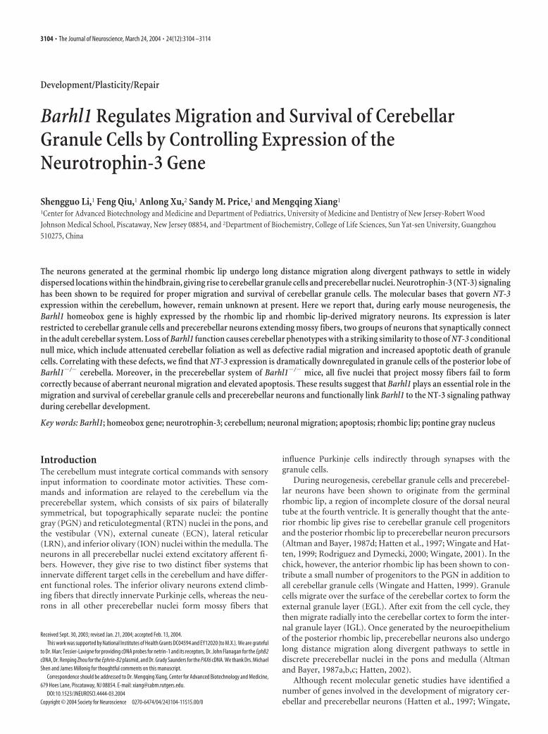
Development/Plasticity/Repair Barhl1 Regulates Migration and Survival of Cerebellar Granule Cells by Controlling Expression of the Neurotrophin-3 Gene Shengguo Li, 1 Feng Qiu, 1 Anlong Xu, 2 Sandy M. Price, 1 and Mengqing Xiang 1 1 Center for Advanced Biotechnology and Medicine and Department of Pediatrics, University of Medicine and Dentistry of New Jersey-Robert Wood Johnson Medical School, Piscataway, New Jersey 08854, and 2 Department of Biochemistry, College of Life Sciences, Sun Yat-sen University, Guangzhou 510275, China The neurons generated at the germinal rhombic lip undergo long distance migration along divergent pathways to settle in widely dispersed locations within the hindbrain, giving rise to cerebellar granule cells and precerebellar nuclei. Neurotrophin-3 (NT-3) signaling has been shown to be required for proper migration and survival of cerebellar granule cells. The molecular bases that govern NT-3 expression within the cerebellum, however, remain unknown at present. Here we report that, during early mouse neurogenesis, the Barhl1 homeobox gene is highly expressed by the rhombic lip and rhombic lip-derived migratory neurons. Its expression is later restricted to cerebellar granule cells and precerebellar neurons extending mossy fibers, two groups of neurons that synaptically connect in the adult cerebellar system. Loss of Barhl1 function causes cerebellar phenotypes with a striking similarity to those of NT-3 conditional null mice, which include attenuated cerebellar foliation as well as defective radial migration and increased apoptotic death of granule cells. Correlating with these defects, we find that NT-3 expression is dramatically downregulated in granule cells of the posterior lobe of Barhl1 / cerebella. Moreover, in the precerebellar system of Barhl1 / mice, all five nuclei that project mossy fibers fail to form correctly because of aberrant neuronal migration and elevated apoptosis. These results suggest that Barhl1 plays an essential role in the migration and survival of cerebellar granule cells and precerebellar neurons and functionally link Barhl1 to the NT-3 signaling pathway during cerebellar development. Key words: Barhl1; homeobox gene; neurotrophin-3; cerebellum; neuronal migration; apoptosis; rhombic lip; pontine gray nucleus Introduction The cerebellum must integrate cortical commands with sensory input information to coordinate motor activities. These com- mands and information are relayed to the cerebellum via the precerebellar system, which consists of six pairs of bilaterally symmetrical, but topographically separate nuclei: the pontine gray (PGN) and reticulotegmental (RTN) nuclei in the pons, and the vestibular (VN), external cuneate (ECN), lateral reticular (LRN), and inferior olivary (ION) nuclei within the medulla. The neurons in all precerebellar nuclei extend excitatory afferent fi- bers. However, they give rise to two distinct fiber systems that innervate different target cells in the cerebellum and have differ- ent functional roles. The inferior olivary neurons extend climb- ing fibers that directly innervate Purkinje cells, whereas the neu- rons in all other precerebellar nuclei form mossy fibers that influence Purkinje cells indirectly through synapses with the granule cells. During neurogenesis, cerebellar granule cells and precerebel- lar neurons have been shown to originate from the germinal rhombic lip, a region of incomplete closure of the dorsal neural tube at the fourth ventricle. It is generally thought that the ante- rior rhombic lip gives rise to cerebellar granule cell progenitors and the posterior rhombic lip to precerebellar neuron precursors (Altman and Bayer, 1987d; Hatten et al., 1997; Wingate and Hat- ten, 1999; Rodriguez and Dymecki, 2000; Wingate, 2001). In the chick, however, the anterior rhombic lip has been shown to con- tribute a small number of progenitors to the PGN in addition to all cerebellar granule cells (Wingate and Hatten, 1999). Granule cells migrate over the surface of the cerebellar cortex to form the external granule layer (EGL). After exit from the cell cycle, they then migrate radially into the cerebellar cortex to form the inter- nal granule layer (IGL). Once generated by the neuroepithelium of the posterior rhombic lip, precerebellar neurons also undergo long distance migration along divergent pathways to settle in discrete precerebellar nuclei in the pons and medulla (Altman and Bayer, 1987a,b,c; Hatten, 2002). Although recent molecular genetic studies have identified a number of genes involved in the development of migratory cer- ebellar and precerebellar neurons (Hatten et al., 1997; Wingate, Received Sept. 30, 2003; revised Jan. 21, 2004; accepted Feb. 13, 2004. This work was supported by National Institutes of Health Grants DC04594 and EY12020 (to M.X.). We are grateful to Dr. Marc Tessier-Lavigne for providing cDNA probes for netrin-1 and its receptors, Dr. John Flanagan for the EphB2 cDNA, Dr. Renping Zhou for the Ephrin-B2 plasmid, and Dr. Grady Saunders for the PAX6 cDNA. We thank Drs. Michael Shen and James Millonig for thoughtful comments on this manuscript. Correspondence should be addressed to Dr. Mengqing Xiang, Center for Advanced Biotechnology and Medicine, 679 Hoes Lane, Piscataway, NJ 08854. E-mail: [email protected]. DOI:10.1523/JNEUROSCI.4444-03.2004 Copyright © 2004 Society for Neuroscience 0270-6474/04/243104-11$15.00/0 3104 • The Journal of Neuroscience, March 24, 2004 • 24(12):3104 –3114
-
Upload
poltek-malang -
Category
Documents
-
view
0 -
download
0
Transcript of Barhl1 Regulates Migration and Survival of Cerebellar Granule Cells by Controlling Expression of the...
Development/Plasticity/Repair
Barhl1 Regulates Migration and Survival of CerebellarGranule Cells by Controlling Expression of theNeurotrophin-3 Gene
Shengguo Li,1 Feng Qiu,1 Anlong Xu,2 Sandy M. Price,1 and Mengqing Xiang1
1Center for Advanced Biotechnology and Medicine and Department of Pediatrics, University of Medicine and Dentistry of New Jersey-Robert WoodJohnson Medical School, Piscataway, New Jersey 08854, and 2Department of Biochemistry, College of Life Sciences, Sun Yat-sen University, Guangzhou510275, China
The neurons generated at the germinal rhombic lip undergo long distance migration along divergent pathways to settle in widelydispersed locations within the hindbrain, giving rise to cerebellar granule cells and precerebellar nuclei. Neurotrophin-3 (NT-3) signalinghas been shown to be required for proper migration and survival of cerebellar granule cells. The molecular bases that govern NT-3expression within the cerebellum, however, remain unknown at present. Here we report that, during early mouse neurogenesis, theBarhl1 homeobox gene is highly expressed by the rhombic lip and rhombic lip-derived migratory neurons. Its expression is laterrestricted to cerebellar granule cells and precerebellar neurons extending mossy fibers, two groups of neurons that synaptically connectin the adult cerebellar system. Loss of Barhl1 function causes cerebellar phenotypes with a striking similarity to those of NT-3 conditionalnull mice, which include attenuated cerebellar foliation as well as defective radial migration and increased apoptotic death of granulecells. Correlating with these defects, we find that NT-3 expression is dramatically downregulated in granule cells of the posterior lobe ofBarhl1�/� cerebella. Moreover, in the precerebellar system of Barhl1�/� mice, all five nuclei that project mossy fibers fail to formcorrectly because of aberrant neuronal migration and elevated apoptosis. These results suggest that Barhl1 plays an essential role in themigration and survival of cerebellar granule cells and precerebellar neurons and functionally link Barhl1 to the NT-3 signaling pathwayduring cerebellar development.
Key words: Barhl1; homeobox gene; neurotrophin-3; cerebellum; neuronal migration; apoptosis; rhombic lip; pontine gray nucleus
IntroductionThe cerebellum must integrate cortical commands with sensoryinput information to coordinate motor activities. These com-mands and information are relayed to the cerebellum via theprecerebellar system, which consists of six pairs of bilaterallysymmetrical, but topographically separate nuclei: the pontinegray (PGN) and reticulotegmental (RTN) nuclei in the pons, andthe vestibular (VN), external cuneate (ECN), lateral reticular(LRN), and inferior olivary (ION) nuclei within the medulla. Theneurons in all precerebellar nuclei extend excitatory afferent fi-bers. However, they give rise to two distinct fiber systems thatinnervate different target cells in the cerebellum and have differ-ent functional roles. The inferior olivary neurons extend climb-ing fibers that directly innervate Purkinje cells, whereas the neu-rons in all other precerebellar nuclei form mossy fibers that
influence Purkinje cells indirectly through synapses with thegranule cells.
During neurogenesis, cerebellar granule cells and precerebel-lar neurons have been shown to originate from the germinalrhombic lip, a region of incomplete closure of the dorsal neuraltube at the fourth ventricle. It is generally thought that the ante-rior rhombic lip gives rise to cerebellar granule cell progenitorsand the posterior rhombic lip to precerebellar neuron precursors(Altman and Bayer, 1987d; Hatten et al., 1997; Wingate and Hat-ten, 1999; Rodriguez and Dymecki, 2000; Wingate, 2001). In thechick, however, the anterior rhombic lip has been shown to con-tribute a small number of progenitors to the PGN in addition toall cerebellar granule cells (Wingate and Hatten, 1999). Granulecells migrate over the surface of the cerebellar cortex to form theexternal granule layer (EGL). After exit from the cell cycle, theythen migrate radially into the cerebellar cortex to form the inter-nal granule layer (IGL). Once generated by the neuroepitheliumof the posterior rhombic lip, precerebellar neurons also undergolong distance migration along divergent pathways to settle indiscrete precerebellar nuclei in the pons and medulla (Altmanand Bayer, 1987a,b,c; Hatten, 2002).
Although recent molecular genetic studies have identified anumber of genes involved in the development of migratory cer-ebellar and precerebellar neurons (Hatten et al., 1997; Wingate,
Received Sept. 30, 2003; revised Jan. 21, 2004; accepted Feb. 13, 2004.This work was supported by National Institutes of Health Grants DC04594 and EY12020 (to M.X.). We are grateful
to Dr. Marc Tessier-Lavigne for providing cDNA probes for netrin-1 and its receptors, Dr. John Flanagan for the EphB2cDNA, Dr. Renping Zhou for the Ephrin-B2 plasmid, and Dr. Grady Saunders for the PAX6 cDNA. We thank Drs. MichaelShen and James Millonig for thoughtful comments on this manuscript.
Correspondence should be addressed to Dr. Mengqing Xiang, Center for Advanced Biotechnology and Medicine,679 Hoes Lane, Piscataway, NJ 08854. E-mail: [email protected].
DOI:10.1523/JNEUROSCI.4444-03.2004Copyright © 2004 Society for Neuroscience 0270-6474/04/243104-11$15.00/0
3104 • The Journal of Neuroscience, March 24, 2004 • 24(12):3104 –3114
2001), the molecular mechanisms underlying their migration,differentiation, and maintenance remain largely unknown.Barhl1 is a mammalian homolog of the Drosophila BarH genes,which encode homeodomain transcription factors that are re-quired for normal development of the compound eye and exter-nal sensory organs (Kojima et al., 1991; Higashijima et al.,1992a,b; Bulfone et al., 2000; Li et al., 2002). During mouse em-bryogenesis, Barhl1 is expressed in the CNS and inner ear haircells (Bulfone et al., 2000; Li et al., 2002). Our recent gene-targeting study has demonstrated an essential role for Barhl1 inthe long-term maintenance of cochlear hair cells (Li et al., 2002).To understand if Barhl1 also has a role during CNS development,in this work, we analyzed the expression pattern of Barhl1 duringCNS development and investigated CNS defects in Barhl1 nullmice. We found that Barhl1 displayed a distinct expression pat-tern in the cerebellar and precerebellar systems. The absence ofBarhl1 caused a dramatic downregulation of NT-3 expression incerebellar granule cells, resulting in attenuated foliation and hy-potrophy of the cerebellum, as well as aberrant radial migrationand increased death of granule cells. Similarly, it caused anoma-lous migration and loss of mossy fiber-extending precerebellarneurons. Therefore, our data have uncovered a crucial role forBarhl1 in the control of migration and survival of cerebellar andprecerebellar neurons and identified NT-3 as a major Barhl1downstream gene during cerebellar development.
Materials and MethodsAnimals. The Barhl1 knock-out mice were generated previously (Li et al.,2002) and maintained in our laboratory. The stage of mouse embryoswas determined by taking the morning when the copulation plug wasshown as embryonic day 0.5 (E0.5). All genotypes described were con-firmed by PCR (Li et al., 2002).
Real-time quantitative RT-PCR and Northern blot analysis. Cerebellafrom five each of P6 Barhl1�/�, Barhl1�/�, Barhl1�/� animals weredissected in RNAlater solution (Ambion, Austin, TX), and total RNA wasextracted using TRIzol (Invitrogen, Carlsbad, CA) according to the manu-facturer’s protocol. RNA was resuspended in RNase-free ddH2O and furtherpurified and treated with DNase I using the RNeasy total RNA isolation kitfollowing the manufacturer’s instructions (Qiagen, Valencia, CA). QRT-PCR was performed in duplicate for each RNA sample (100 ng) using theQuantiTect SYBR green one-step RT-PCR kit (Qiagen). The following se-quence-specific primers were designed using the MacVector software (Ac-celrys, San Diego, CA): Barhl1, 5�-CAAAGTGAAGGAGGAGGGCG-3� and5�-GTGTCGGTGAGGTTGAGCGA-3�; NT-3, 5�-GATTGATGACAAA-CACTGGAAC-3� and 5�-CACAGGAAGTGTCTATTCGTATC-3�; BDNF,5�-CGGGACGGTCACAGTCCTA-3� and 5�-GGGATTACACTTG-GTCTCGTAGAAATAC-3�; and GAPDH, 5�-TCACCACCATG-GAGAAGGC-3� and 5�-GCTAAGCAGTTGGTGGTGCA-3�. PCRproducts were monitored in real time (Mx4000 multiplex quantitativePCR system; Stratagene, La Jolla, CA), and the threshold cycles (Ct) weredetermined using the Mx4000 software. For each set of primers, a notemplate control and a no reverse amplification control were included.Postamplification dissociation curves were performed to verify the pres-ence of single amplification product in the absence of DNA contamina-tion. Relative quantities of copy numbers were calculated from knownquantities of input copy numbers of cloned Barhl1, NT-3, BDNF, andGAPDH cDNA plasmids using the comparative threshold cycle numberof each sample fitted to a seven-point standard curve (r2 � 0.99) (Over-bergh et al., 1999). All data were tested for significance using two sampleStudent’s t test with unequal variances. Northern blot analysis was per-formed according to standard methods.
RNA in situ hybridization. RNA in situ hybridization was performed aspreviously described (Sciavolino et al., 1997) using digoxigenin-labeledriboprobes prepared following the manufacturer’s protocol (Roche Di-agnostics, IN). Probes: Barhl1 was a previously isolated mouse cDNAclone (Li et al., 2002); NT-3 was a coding segment amplified by RT-PCR
from mouse cerebellar RNA; Math1 and NeuroD coding sequences wereamplified by PCR from mouse genomic DNA; the human PAX6 plasmidwas described by Singh et al. (1998); the mouse Netrin-1 by Serafini et al.(1996), the rat DCC and Neogenin by Keino-Masu et al. (1996), the ratUnc5h1, Unc5h2 and Unc5h3 by Leonardo et al. (1997), the mouse EphB2by Lu et al. (2001), and the human Ephrin-B2 by Yue et al. (1999).
�-Galactosidase staining. �-Galactosidase staining was conducted es-sentially as described (Ben-Arie et al., 2000; Eng et al., 2001). Briefly, forstaining of whole-mount embryos and brains, animals were fixed in 4%paraformaldehyde–PBS at 4°C for 2–12 hr depending on the stages andthen rinsed for 20 min in PBS containing 0.02% Nonidet P-40 and 0.01%sodium deoxycholate. Staining was performed overnight either at 30°Cor 37°C in PBS buffer containing 0.02% Nonidet P-40, 0.01% sodiumdeoxycholate, 5 mM potassium ferricyanide, 5 mM potassium ferrocya-nide, and 0.5 mg/ml X-gal. Some whole-mount-stained embryos weredehydrated in graded ethanol and cleared in 1:2 benzyl alcohol– benzylbenzoate. Section staining was performed following the same procedureas whole-mount staining except that all sections were counterstainedwith Fast Red (Vector Laboratories, Burlingame, CA).
Generation of polyclonal anti-Barhl1 antibody and immunohistochem-istry. DNA fragment corresponding to amino acids 3–92 of the mouseBarhl1 protein was amplified by PCR and inserted into the pGEMEX(Promega, Madison, WI) and pMAL-cR1 (New England Biolabs, Bev-erly, MA) vectors to express fusion proteins with the bacteriophage T7gene 10 protein and bacterial maltose-binding protein, respectively. An-tibody production and affinity purification were performed as describedpreviously by Xiang et al. (1993, 1995).
For immunohistochemistry, cryosections were treated in methanolwith 3% of hydrogen peroxide for 3 min to quench endogenous peroxi-dase activity. After three washes in PBS, they were blocked in 5% ofnormal goat serum for 1 hr before overnight incubation at 4°C withprimary antibodies [anti-Barhl1, 1:10; anti-Brn3a (Xiang et al., 1995),1:5; anti-active caspase-3 (BD Pharmingen, San Diego, CA), 1:100]. Thesections were then washed in PBS for three times, 7 min each, incubatedwith biotinylated goat anti-rabbit IgG (1:200; Vector Laboratories) for 1hr, and subsequently processed using the ABC kit (Vector Laboratories).Color reaction was performed using the NovaRed substrate kit (VectorLaboratories). For double staining, postnatal day 5 (P5) brain sectionswere first immunostained with the anti-Brn3a antibody, rinsed in PBSfor three times, and then stained for �-galactosidase activity for 2 hr at37°C as described above. The labeled sections were dehydrated andmounted with Permount (Fisher Scientific, Springfield, NJ).
Quantitation of neuron number and cell death assay. To quantify thenumber of neurons in the PGN and RTN, serial coronal sections (16 �m)of P100 mouse brains were stained with cresyl violet. Images of the PGNand RTN were then captured using a SPOT digital camera (DiagnosticInstruments Inc., Sterling Heights, MI), and the number of neurons wasscored from captured images. Only neurons with a clear nucleus andnucleoli were counted. Every other section was scored, and three or fournuclei were counted for each genotype. All data were tested for signifi-cance using two sample Student’s t test with unequal variances.
Cell death assay was performed by measuring the expression of theproteolytically activated form of caspase-3 as an indicator of apoptosis(Panchision et al., 2001). To determine the number of apoptotic cells inthe PGN, coronal cryosections from E16.5, P0, P2, and P4 brains wereimmunostained with an antibody against the active caspase-3 as de-scribed above and then counterstained with methyl green. The caspase-3-positive cells were counted in all serial sections of the PGN and aStudent’s t test was used to determine the significance. Four samples werecollected for each genotype. To measure the number of apoptotic cells inthe cerebellum, sagittal sections of P8 cerebella were similarly analyzed.
BrdU and DiI labeling. BrdU labeling was performed as described(Borghesani et al., 2002). Retrograde DiI (1.1�-dioctadecyl-3,3,3�,3�-tetramethylindocarbocyanine) labeling was performed essentially as de-scribed (Bloch-Gallego et al., 1999). In brief, P1 brains were dissected outof the skull following fixation of neonates with 4% paraformaldehyde–PBS. Several DiI crystals were then inserted into one of both hemicere-bella using a glass pipette tip under a dissection microscope. The DiI-inserted brains were incubated in 2% paraformaldehyde–PBS at 37°C for
Li et al. • Barhl1 Regulates NT-3 Expression in the Cerebellum J. Neurosci., March 24, 2004 • 24(12):3104 –3114 • 3105
4 weeks in the dark. They were then rinsed inPBS, embedded in 3% agarose, and cut at athickness of 100 �m with a vibratome. The sec-tions were mounted in Aqua polymount (Poly-sciences, Warrington, PA) and sealed with nailpolish.
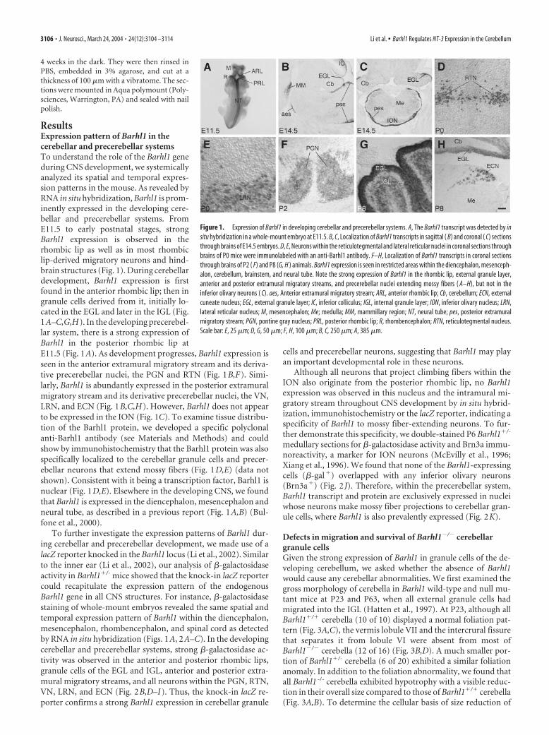
ResultsExpression pattern of Barhl1 in thecerebellar and precerebellar systemsTo understand the role of the Barhl1 geneduring CNS development, we systemicallyanalyzed its spatial and temporal expres-sion patterns in the mouse. As revealed byRNA in situ hybridization, Barhl1 is prom-inently expressed in the developing cere-bellar and precerebellar systems. FromE11.5 to early postnatal stages, strongBarhl1 expression is observed in therhombic lip as well as in most rhombiclip-derived migratory neurons and hind-brain structures (Fig. 1). During cerebellardevelopment, Barhl1 expression is firstfound in the anterior rhombic lip; then ingranule cells derived from it, initially lo-cated in the EGL and later in the IGL (Fig.1A–C,G,H). In the developing precerebel-lar system, there is a strong expression ofBarhl1 in the posterior rhombic lip atE11.5 (Fig. 1A). As development progresses, Barhl1 expression isseen in the anterior extramural migratory stream and its deriva-tive precerebellar nuclei, the PGN and RTN (Fig. 1B,F). Simi-larly, Barhl1 is abundantly expressed in the posterior extramuralmigratory stream and its derivative precerebellar nuclei, the VN,LRN, and ECN (Fig. 1B,C,H). However, Barhl1 does not appearto be expressed in the ION (Fig. 1C). To examine tissue distribu-tion of the Barhl1 protein, we developed a specific polyclonalanti-Barhl1 antibody (see Materials and Methods) and couldshow by immunohistochemistry that the Barhl1 protein was alsospecifically localized to the cerebellar granule cells and precer-ebellar neurons that extend mossy fibers (Fig. 1D,E) (data notshown). Consistent with it being a transcription factor, Barhl1 isnuclear (Fig. 1D,E). Elsewhere in the developing CNS, we foundthat Barhl1 is expressed in the diencephalon, mesencephalon andneural tube, as described in a previous report (Fig. 1A,B) (Bul-fone et al., 2000).
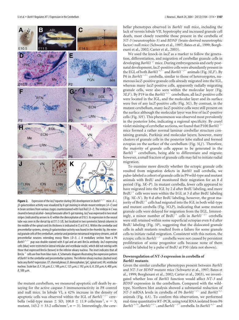
To further investigate the expression patterns of Barhl1 dur-ing cerebellar and precerebellar development, we made use of alacZ reporter knocked in the Barhl1 locus (Li et al., 2002). Similarto the inner ear (Li et al., 2002), our analysis of �-galactosidaseactivity in Barhl1�/- mice showed that the knock-in lacZ reportercould recapitulate the expression pattern of the endogenousBarhl1 gene in all CNS structures. For instance, �-galactosidasestaining of whole-mount embryos revealed the same spatial andtemporal expression pattern of Barhl1 within the diencephalon,mesencephalon, rhombencephalon, and spinal cord as detectedby RNA in situ hybridization (Figs. 1A, 2A–C). In the developingcerebellar and precerebellar systems, strong �-galactosidase ac-tivity was observed in the anterior and posterior rhombic lips,granule cells of the EGL and IGL, anterior and posterior extra-mural migratory streams, and all neurons within the PGN, RTN,VN, LRN, and ECN (Fig. 2B,D–I). Thus, the knock-in lacZ re-porter confirms a strong Barhl1 expression in cerebellar granule
cells and precerebellar neurons, suggesting that Barhl1 may playan important developmental role in these neurons.
Although all neurons that project climbing fibers within theION also originate from the posterior rhombic lip, no Barhl1expression was observed in this nucleus and the intramural mi-gratory stream throughout CNS development by in situ hybrid-ization, immunohistochemistry or the lacZ reporter, indicating aspecificity of Barhl1 to mossy fiber-extending neurons. To fur-ther demonstrate this specificity, we double-stained P6 Barhl1�/-
medullary sections for �-galactosidase activity and Brn3a immu-noreactivity, a marker for ION neurons (McEvilly et al., 1996;Xiang et al., 1996). We found that none of the Barhl1-expressingcells (�-gal�) overlapped with any inferior olivary neurons(Brn3a�) (Fig. 2 J). Therefore, within the precerebellar system,Barhl1 transcript and protein are exclusively expressed in nucleiwhose neurons make mossy fiber projections to cerebellar gran-ule cells, where Barhl1 is also prevalently expressed (Fig. 2K).
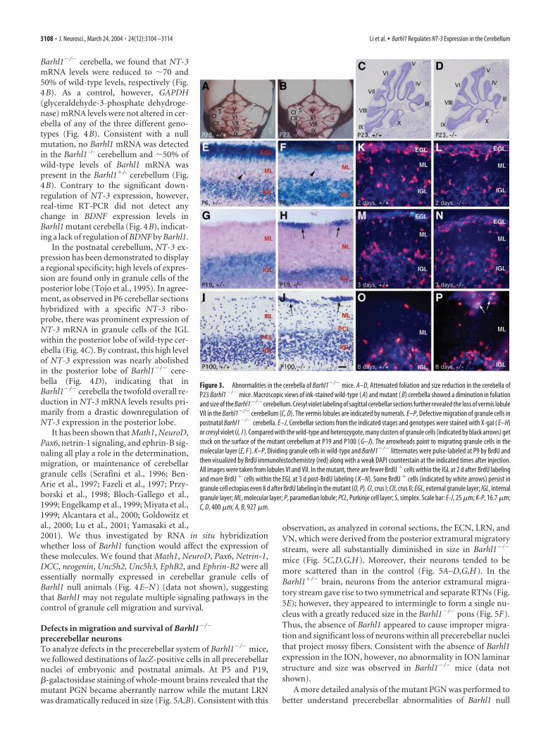
Defects in migration and survival of Barhl1�/� cerebellargranule cellsGiven the strong expression of Barhl1 in granule cells of the de-veloping cerebellum, we asked whether the absence of Barhl1would cause any cerebellar abnormalities. We first examined thegross morphology of cerebella in Barhl1 wild-type and null mu-tant mice at P23 and P63, when all external granule cells hadmigrated into the IGL (Hatten et al., 1997). At P23, although allBarhl1�/� cerebella (10 of 10) displayed a normal foliation pat-tern (Fig. 3A,C), the vermis lobule VII and the intercrural fissurethat separates it from lobule VI were absent from most ofBarhl1�/� cerebella (12 of 16) (Fig. 3B,D). A much smaller por-tion of Barhl1�/- cerebella (6 of 20) exhibited a similar foliationanomaly. In addition to the foliation abnormality, we found thatall Barhl1 -/- cerebella exhibited hypotrophy with a visible reduc-tion in their overall size compared to those of Barhl1�/� cerebella(Fig. 3A,B). To determine the cellular basis of size reduction of
Figure 1. Expression of Barhl1 in developing cerebellar and precerebellar systems. A, The Barhl1 transcript was detected by insitu hybridization in a whole-mount embryo at E11.5. B, C, Localization of Barhl1 transcripts in sagittal ( B) and coronal ( C) sectionsthrough brains of E14.5 embryos. D, E, Neurons within the reticulotegmental and lateral reticular nuclei in coronal sections throughbrains of P0 mice were immunolabeled with an anti-Barhl1 antibody. F–H, Localization of Barhl1 transcripts in coronal sectionsthrough brains of P2 ( F) and P8 (G, H ) animals. Barhl1 expression is seen in restricted areas within the diencephalon, mesenceph-alon, cerebellum, brainstem, and neural tube. Note the strong expression of Barhl1 in the rhombic lip, external granule layer,anterior and posterior extramural migratory streams, and precerebellar nuclei extending mossy fibers ( A–H), but not in theinferior olivary neurons ( C). aes, Anterior extramural migratory stream; ARL, anterior rhombic lip; Cb, cerebellum; ECN, externalcuneate nucleus; EGL, external granule layer; IC, inferior colliculus; IGL, internal granule layer; ION, inferior olivary nucleus; LRN,lateral reticular nucleus; M, mesencephalon; Me; medulla; MM, mammillary region; NT, neural tube; pes, posterior extramuralmigratory stream; PGN, pontine gray nucleus; PRL, posterior rhombic lip; R, rhombencephalon; RTN, reticulotegmental nucleus.Scale bar: E, 25 �m; D, G, 50 �m; F, H, 100 �m; B, C, 250 �m; A, 385 �m.
3106 • J. Neurosci., March 24, 2004 • 24(12):3104 –3114 Li et al. • Barhl1 Regulates NT-3 Expression in the Cerebellum
the mutant cerebellum, we measured apoptotic cell death by as-saying for the active caspase-3 immunoreactivity in P8 controland null mice. In lobule VI, a 42% increase in the density ofapoptotic cells was observed within the IGL of Barhl1�/� cere-bella (wild-type mean � SD, 100.8 � 11.9 cells/mm 2, n � 3;mutant, 143.2 � 33.2 cells/mm 2, n � 3). Interestingly, the cere-
bellar phenotypes observed in Barhl1 null mice, including thelack of vermis lobule VII, hypotrophy and increased granule celldeath, most closely resemble those present in the cerebella ofNT-3 (neurotrophin-3) and BDNF (brain-derived neurotrophicfactor) null mice (Schwartz et al., 1997; Bates et al., 1999; Borgh-esani et al., 2002; Carter et al., 2003).
We used the knock-in lacZ as a marker to follow the genera-tion, differentiation, and migration of cerebellar granule cells indeveloping Barhl1 -/- mice. During embryogenesis and early post-natal development, lacZ-positive cells were abundantly present inthe EGL of both Barhl1�/� and Barhl1�/� animals (Fig. 3E,F). ByP6 in Barhl1�/� cerebella, similar to those of heterozygotes, nu-merous lacZ-positive granule cells already migrated into the IGL,whereas many lacZ-positive cells, apparently radially migratinggranule cells, were also seen within the molecular layer (Fig.3E,F). By P19 in the Barhl1�/� cerebellum, all lacZ-positive cellswere located in the IGL, and the molecular layer and its surfacewere free of any lacZ-positive cells (Fig. 3G). By contrast, in themutant cerebellum, many lacZ-positive cells were still present onthe surface although the molecular layer was free of lacZ-positivecells (Fig. 3H). This phenomenon was observed most prevalentlyin the posterior lobe, indicating a regional specificity. By cresylviolet staining of cerebellar sections, we found that P100 Barhl1 -/-
mice formed a rather normal laminar cerebellar structure con-taining granule, Purkinje and molecular layers; however, manyclusters of granule cells in the posterior lobe stalled and formedectopias on the surface of the cerebellum (Fig. 3I,J). Therefore,the majority of granule cells appear to be generated in theBarhl1 -/- cerebellum, being able to differentiate and migrate;however, a small fraction of granule cells may fail to initiate radialmigration.
To examine more directly whether the ectopic granule cellsresulted from migration defects in Barhl1 null cerebella, wepulse-labeled a cohort of granule cells in P9 wild-type and mutantanimals with BrdU and monitored their migration for an 8 dperiod (Fig. 3K–P). In mutant cerebella, fewer cells appeared tohave migrated into the IGL by 2 d after BrdU labeling, and moreBrdU� cells were seen within the EGL at 3 d after BrdU labeling(Fig. 3K–N). By 8 d after BrdU labeling, however, the great ma-jority of BrdU� cells had migrated into the IGL in both wild-typeand mutant cerebella (Fig. 3O,P), indicating that some mutantgranule cells were delayed for migration from the EGL. Interest-ingly, a minor number of BrdU� cells in Barhl1�/� cerebellawere still retained within some superficial ectopias even 8 d afterBrdU labeling (Fig. 3P), suggesting that the dislocated granulecells in adult mutants resulted from a failure for some granulecells to initiate radial migration. Consistent with this notion, theectopic cells in Barhl1�/� cerebella were not caused by persistentproliferation of some progenitor cells because none of themcould be labeled by a pulse of BrdU at P30 (data not shown).
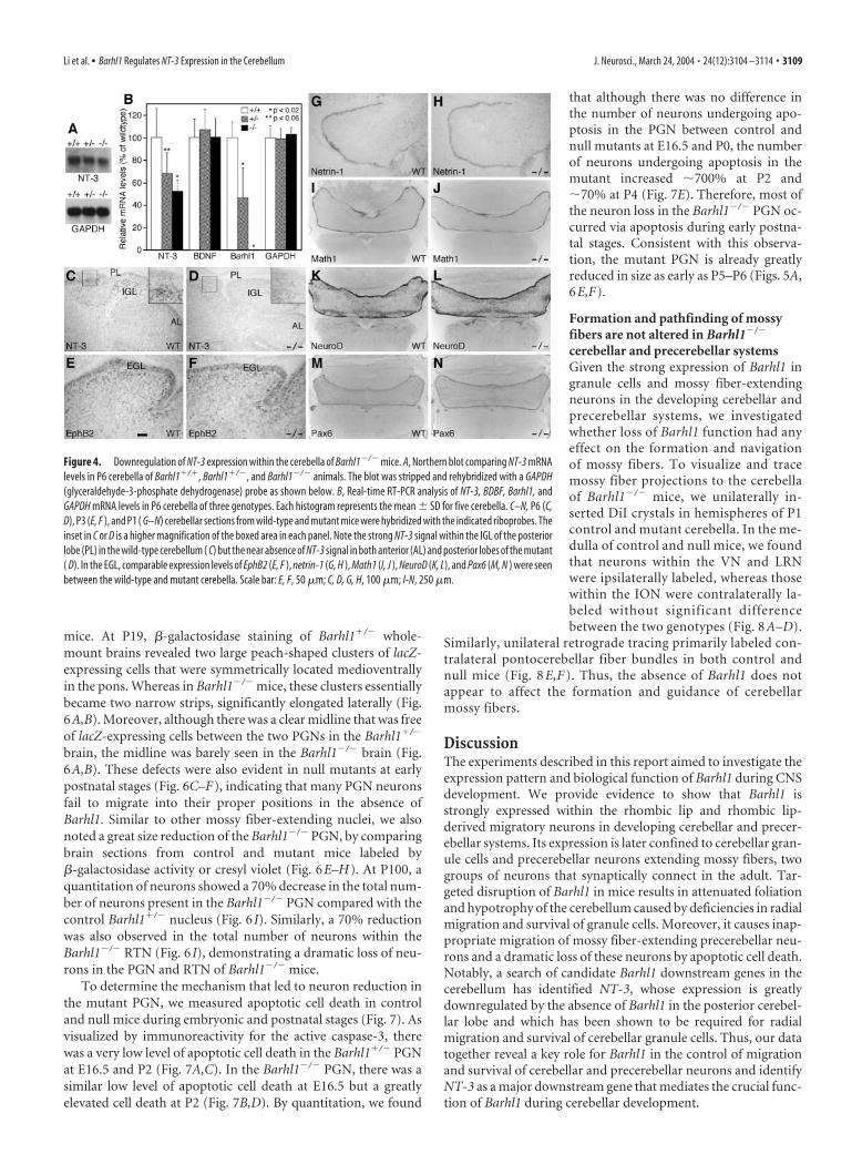
Downregulation of NT-3 expression in cerebella ofBarhl1 mutantsGiven the similar cerebellar phenotypes present between Barhl1and NT-3 or BDNF mutant mice (Schwartz et al., 1997; Bates etal., 1999; Borghesani et al., 2002; Carter et al., 2003), we investi-gated whether loss of Barhl1 function would affect NT-3 andBDNF expression in the cerebellum. Compared with the wild-type, Northern blot analysis showed a substantial reduction ofNT-3 mRNA levels in cerebella of P6 Barhl1�/� and Barhl1�/-
animals (Fig. 4A). To confirm this observation, we performedreal-time quantitative RT-PCR, using total RNA isolated from P6Barhl1�/�, Barhl1�/�, and Barhl1�/� cerebella. In Barhl1�/� and
Figure 2. Expression of the lacZ reporter during CNS development in Barhl1�/� mice. A–I,�-galactosidase activity was visualized by X-gal staining in whole-mount embryos ( A–C) andin brain sections from various stages counterstained with Fast Red ( D–I). The embryo in C wascleared in benzyl alcohol– benzyl benzoate after X-gal staining. lacZ was expressed in two smallstripes (indicated by arrows in A) within the diencephalon at E10.5. Its expression in the neuraltube was seen in the dorsal tip at E11.5 ( B), but localized in two symmetric lateral columns inthe middle of the spinal cord (its thickness is indicated in C) at E14.5. Within the cerebellar andprecerebellar systems, strong �-galactosidase activity was found in the rhombic lip, the exter-nal granule cells of the cerebellum, anterior and posterior extramural migratory streams, and allprecerebellar neurons extending mossy fibers ( D–I). J, A medullary section from a P6Barhl1�/� pup was double-stained with X-gal and an anti-Brn3a antibody. lacZ-expressingcells (blue) were restricted in lateral reticular and vestibular nuclei, which did not overlap withthose that expressed Brn3a (brown) in the inferior olivary nucleus. The inset indicates that allBrn3a � cells are free from blue stain. K, Schematic diagram illustrating the expression patternof Barhl1 in the cerebellar and precerebellar systems. The inferior olivary nucleus (dashed oval)lacks any Barhl1 expression. CP, choroid plexus; D, diencephalon; SpC, spinal cord; VN, vestibularnucleus. Scale bar: D, F, 50 �m; E, I, 100 �m; C, 125 �m; J, 192 �m; G, H, 250 �m; A, 400 �m;B, 500 �m.
Li et al. • Barhl1 Regulates NT-3 Expression in the Cerebellum J. Neurosci., March 24, 2004 • 24(12):3104 –3114 • 3107
Barhl1�/� cerebella, we found that NT-3mRNA levels were reduced to �70 and50% of wild-type levels, respectively (Fig.4B). As a control, however, GAPDH(glyceraldehyde-3-phosphate dehydroge-nase) mRNA levels were not altered in cer-ebella of any of the three different geno-types (Fig. 4B). Consistent with a nullmutation, no Barhl1 mRNA was detectedin the Barhl1 -/- cerebellum and �50% ofwild-type levels of Barhl1 mRNA waspresent in the Barhl1�/- cerebellum (Fig.4B). Contrary to the significant down-regulation of NT-3 expression, however,real-time RT-PCR did not detect anychange in BDNF expression levels inBarhl1 mutant cerebella (Fig. 4B), indicat-ing a lack of regulation of BDNF by Barhl1.
In the postnatal cerebellum, NT-3 ex-pression has been demonstrated to displaya regional specificity; high levels of expres-sion are found only in granule cells of theposterior lobe (Tojo et al., 1995). In agree-ment, as observed in P6 cerebellar sectionshybridized with a specific NT-3 ribo-probe, there was prominent expression ofNT-3 mRNA in granule cells of the IGLwithin the posterior lobe of wild-type cer-ebella (Fig. 4C). By contrast, this high levelof NT-3 expression was nearly abolishedin the posterior lobe of Barhl1�/� cere-bella (Fig. 4D), indicating that inBarhl1�/� cerebella the twofold overall re-duction in NT-3 mRNA levels results pri-marily from a drastic downregulation ofNT-3 expression in the posterior lobe.
It has been shown that Math1, NeuroD,Pax6, netrin-1 signaling, and ephrin-B sig-naling all play a role in the determination,migration, or maintenance of cerebellargranule cells (Serafini et al., 1996; Ben-Arie et al., 1997; Fazeli et al., 1997; Przy-borski et al., 1998; Bloch-Gallego et al.,1999; Engelkamp et al., 1999; Miyata et al.,1999; Alcantara et al., 2000; Goldowitz etal., 2000; Lu et al., 2001; Yamasaki et al.,2001). We thus investigated by RNA in situ hybridizationwhether loss of Barhl1 function would affect the expression ofthese molecules. We found that Math1, NeuroD, Pax6, Netrin-1,DCC, neogenin, Unc5h2, Unc5h3, EphB2, and Ephrin-B2 were allessentially normally expressed in cerebellar granule cells ofBarhl1 null animals (Fig. 4E–N) (data not shown), suggestingthat Barhl1 may not regulate multiple signaling pathways in thecontrol of granule cell migration and survival.
Defects in migration and survival of Barhl1�/�
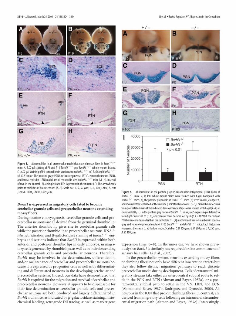
precerebellar neuronsTo analyze defects in the precerebellar system of Barhl1�/� mice,we followed destinations of lacZ-positive cells in all precerebellarnuclei of embryonic and postnatal animals. At P5 and P19,�-galactosidase staining of whole-mount brains revealed that themutant PGN became aberrantly narrow while the mutant LRNwas dramatically reduced in size (Fig. 5A,B). Consistent with this
observation, as analyzed in coronal sections, the ECN, LRN, andVN, which were derived from the posterior extramural migratorystream, were all substantially diminished in size in Barhl1�/�
mice (Fig. 5C,D,G,H). Moreover, their neurons tended to bemore scattered than in the control (Fig. 5A–D,G,H). In theBarhl1�/� brain, neurons from the anterior extramural migra-tory stream gave rise to two symmetrical and separate RTNs (Fig.5E); however, they appeared to intermingle to form a single nu-cleus with a greatly reduced size in the Barhl1�/� pons (Fig. 5F).Thus, the absence of Barhl1 appeared to cause improper migra-tion and significant loss of neurons within all precerebellar nucleithat project mossy fibers. Consistent with the absence of Barhl1expression in the ION, however, no abnormality in ION laminarstructure and size was observed in Barhl1�/� mice (data notshown).
A more detailed analysis of the mutant PGN was performed tobetter understand precerebellar abnormalities of Barhl1 null
Figure 3. Abnormalities in the cerebella of Barhl1�/� mice. A–D, Attenuated foliation and size reduction in the cerebella ofP23 Barhl1�/� mice. Macroscopic views of ink-stained wild-type ( A) and mutant ( B) cerebella showed a diminution in foliationand size of the Barhl1�/� cerebellum. Cresyl violet labeling of sagittal cerebellar sections further revealed the loss of vermis lobuleVII in the Barhl1�/� cerebellum (C, D). The vermis lobules are indicated by numerals. E–P, Defective migration of granule cells inpostnatal Barhl1�/� cerebella. E–J, Cerebellar sections from the indicated stages and genotypes were stained with X-gal ( E–H)or cresyl violet (I, J ). Compared with the wild-type and heterozygote, many clusters of granule cells (indicated by black arrows) getstuck on the surface of the mutant cerebellum at P19 and P100 ( G–J). The arrowheads point to migrating granule cells in themolecular layer (E, F ). K–P, Dividing granule cells in wild-type and Barhl1�/� littermates were pulse-labeled at P9 by BrdU andthen visualized by BrdU immunohistochemistry (red) along with a weak DAPI counterstain at the indicated times after injection.All images were taken from lobules VI and VII. In the mutant, there are fewer BrdU � cells within the IGL at 2 d after BrdU labelingand more BrdU � cells within the EGL at 3 d post-BrdU labeling ( K–N). Some BrdU � cells (indicated by white arrows) persist ingranule cell ectopias even 8 d after BrdU labeling in the mutant (O, P). CI, crus I; CII, crus II; EGL, external granule layer; IGL, internalgranule layer; ML, molecular layer; P, paramedian lobule; PCL, Purkinje cell layer; S, simplex. Scale bar: E-J, 25 �m; K-P, 16.7 �m;C, D, 400 �m; A, B, 927 �m.
3108 • J. Neurosci., March 24, 2004 • 24(12):3104 –3114 Li et al. • Barhl1 Regulates NT-3 Expression in the Cerebellum
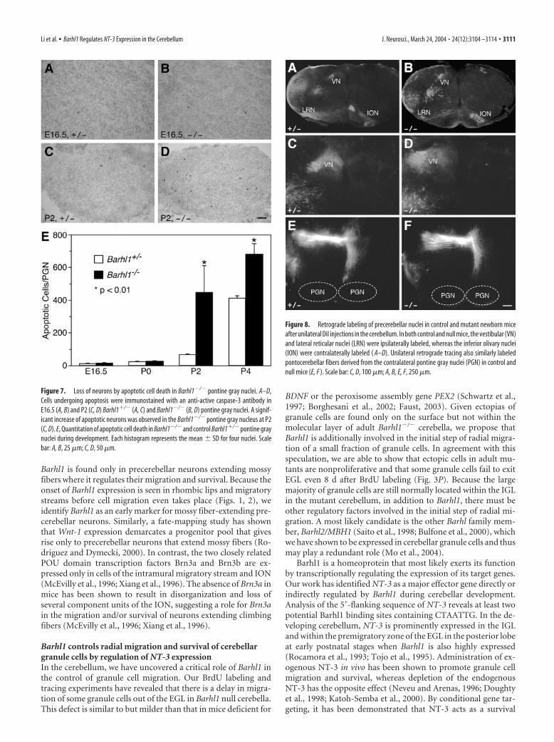
mice. At P19, �-galactosidase staining of Barhl1�/� whole-mount brains revealed two large peach-shaped clusters of lacZ-expressing cells that were symmetrically located medioventrallyin the pons. Whereas in Barhl1�/� mice, these clusters essentiallybecame two narrow strips, significantly elongated laterally (Fig.6A,B). Moreover, although there was a clear midline that was freeof lacZ-expressing cells between the two PGNs in the Barhl1�/�
brain, the midline was barely seen in the Barhl1�/� brain (Fig.6A,B). These defects were also evident in null mutants at earlypostnatal stages (Fig. 6C–F), indicating that many PGN neuronsfail to migrate into their proper positions in the absence ofBarhl1. Similar to other mossy fiber-extending nuclei, we alsonoted a great size reduction of the Barhl1�/� PGN, by comparingbrain sections from control and mutant mice labeled by�-galactosidase activity or cresyl violet (Fig. 6E–H). At P100, aquantitation of neurons showed a 70% decrease in the total num-ber of neurons present in the Barhl1�/� PGN compared with thecontrol Barhl1�/� nucleus (Fig. 6 I). Similarly, a 70% reductionwas also observed in the total number of neurons within theBarhl1�/� RTN (Fig. 6 I), demonstrating a dramatic loss of neu-rons in the PGN and RTN of Barhl1�/� mice.
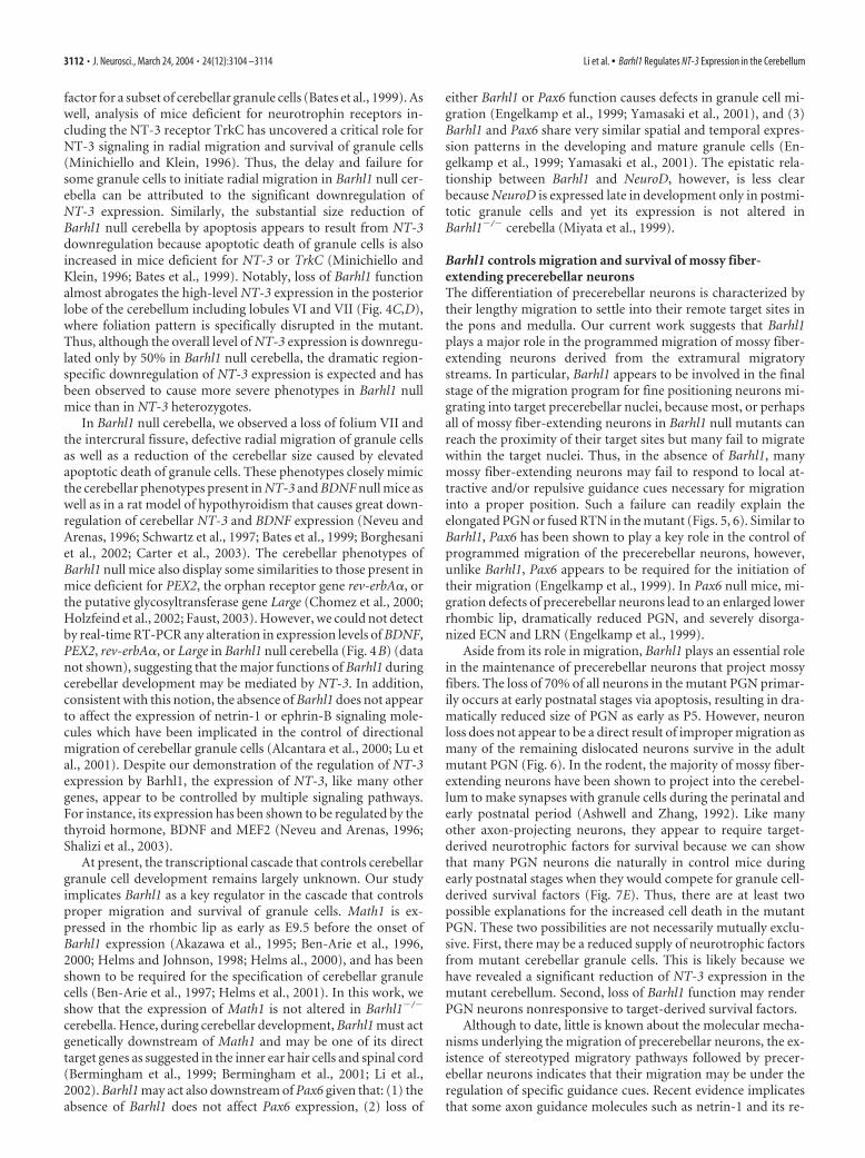
To determine the mechanism that led to neuron reduction inthe mutant PGN, we measured apoptotic cell death in controland null mice during embryonic and postnatal stages (Fig. 7). Asvisualized by immunoreactivity for the active caspase-3, therewas a very low level of apoptotic cell death in the Barhl1�/� PGNat E16.5 and P2 (Fig. 7A,C). In the Barhl1�/� PGN, there was asimilar low level of apoptotic cell death at E16.5 but a greatlyelevated cell death at P2 (Fig. 7B,D). By quantitation, we found
that although there was no difference inthe number of neurons undergoing apo-ptosis in the PGN between control andnull mutants at E16.5 and P0, the numberof neurons undergoing apoptosis in themutant increased �700% at P2 and�70% at P4 (Fig. 7E). Therefore, most ofthe neuron loss in the Barhl1�/� PGN oc-curred via apoptosis during early postna-tal stages. Consistent with this observa-tion, the mutant PGN is already greatlyreduced in size as early as P5–P6 (Figs. 5A,6E,F).
Formation and pathfinding of mossyfibers are not altered in Barhl1�/�
cerebellar and precerebellar systemsGiven the strong expression of Barhl1 ingranule cells and mossy fiber-extendingneurons in the developing cerebellar andprecerebellar systems, we investigatedwhether loss of Barhl1 function had anyeffect on the formation and navigationof mossy fibers. To visualize and tracemossy fiber projections to the cerebellaof Barhl1�/� mice, we unilaterally in-serted DiI crystals in hemispheres of P1control and mutant cerebella. In the me-dulla of control and null mice, we foundthat neurons within the VN and LRNwere ipsilaterally labeled, whereas thosewithin the ION were contralaterally la-beled without significant differencebetween the two genotypes (Fig. 8 A–D).
Similarly, unilateral retrograde tracing primarily labeled con-tralateral pontocerebellar fiber bundles in both control andnull mice (Fig. 8 E,F ). Thus, the absence of Barhl1 does notappear to affect the formation and guidance of cerebellarmossy fibers.
DiscussionThe experiments described in this report aimed to investigate theexpression pattern and biological function of Barhl1 during CNSdevelopment. We provide evidence to show that Barhl1 isstrongly expressed within the rhombic lip and rhombic lip-derived migratory neurons in developing cerebellar and precer-ebellar systems. Its expression is later confined to cerebellar gran-ule cells and precerebellar neurons extending mossy fibers, twogroups of neurons that synaptically connect in the adult. Tar-geted disruption of Barhl1 in mice results in attenuated foliationand hypotrophy of the cerebellum caused by deficiencies in radialmigration and survival of granule cells. Moreover, it causes inap-propriate migration of mossy fiber-extending precerebellar neu-rons and a dramatic loss of these neurons by apoptotic cell death.Notably, a search of candidate Barhl1 downstream genes in thecerebellum has identified NT-3, whose expression is greatlydownregulated by the absence of Barhl1 in the posterior cerebel-lar lobe and which has been shown to be required for radialmigration and survival of cerebellar granule cells. Thus, our datatogether reveal a key role for Barhl1 in the control of migrationand survival of cerebellar and precerebellar neurons and identifyNT-3 as a major downstream gene that mediates the crucial func-tion of Barhl1 during cerebellar development.
Figure 4. Downregulation of NT-3 expression within the cerebella of Barhl1�/� mice. A, Northern blot comparing NT-3 mRNAlevels in P6 cerebella of Barhl1�/�, Barhl1�/�, and Barhl1�/� animals. The blot was stripped and rehybridized with a GAPDH(glyceraldehyde-3-phosphate dehydrogenase) probe as shown below. B, Real-time RT-PCR analysis of NT-3, BDBF, Barhl1, andGAPDH mRNA levels in P6 cerebella of three genotypes. Each histogram represents the mean � SD for five cerebella. C–N, P6 (C,D), P3 (E, F ), and P1 ( G–N) cerebellar sections from wild-type and mutant mice were hybridized with the indicated riboprobes. Theinset in C or D is a higher magnification of the boxed area in each panel. Note the strong NT-3 signal within the IGL of the posteriorlobe (PL) in the wild-type cerebellum ( C) but the near absence of NT-3 signal in both anterior (AL) and posterior lobes of the mutant( D). In the EGL, comparable expression levels of EphB2 (E, F ), netrin-1 (G, H ), Math1 (I, J ), NeuroD (K, L), and Pax6 (M, N ) were seenbetween the wild-type and mutant cerebella. Scale bar: E, F, 50 �m; C, D, G, H, 100 �m; I-N, 250 �m.
Li et al. • Barhl1 Regulates NT-3 Expression in the Cerebellum J. Neurosci., March 24, 2004 • 24(12):3104 –3114 • 3109
Barhl1 is expressed in migratory cells fated to becomecerebellar granule cells and precerebellar neurons extendingmossy fibersDuring murine embryogenesis, cerebellar granule cells and pre-cerebellar neurons are all derived from the germinal rhombic lip.The anterior rhombic lip gives rise to cerebellar granule cellswhile the posterior rhombic lip to precerebellar neurons. RNA insitu hybridization and �-galactosidase staining of Barhl1�/� em-bryos and sections indicate that Barhl1 is expressed within bothanterior and posterior rhombic lips in early embryos, in migra-tory cells generated by rhombic lips, as well as in their descendingcerebellar granule cells and precerebellar neurons. Therefore,Barhl1 may be involved in the determination, differentiation,and/or maintenance of cerebellar and precerebellar neurons be-cause it is expressed by progenitor cells as well as by differentiat-ing and differentiated neurons in the developing cerebellar andprecerebellar systems. Indeed, our data have demonstrated thatBarhl1 is required for the migration and survival of cerebellar andprecerebellar neurons. However, it appears to be dispensable fortheir fate determination as cerebellar granule cells and precer-ebellar neurons are both produced and largely differentiated inBarhl1 null mice, as indicated by �-galactosidase staining, histo-chemical labeling, retrograde DiI tracing, as well as marker gene
expression (Figs. 3– 8). In the inner ear, we have shown previ-ously that Barhl1 is similarly not required for fate commitment ofsensory hair cells (Li et al., 2002).
In the precerebellar system, neurons extending mossy fibersor climbing fibers not only have different innervation targets butthey also follow distinct migration pathways to reach discreteprecerebellar nuclei during development. Cells of extramural mi-gratory streams take either an anteroventral subpial route to set-tle in the PGN and RTN (Altman and Bayer, 1987a), or a pos-teroventral subpial path to settle in the VN, LRN, and ECN(Altman and Bayer, 1987b; Rodriguez and Dymecki, 2000). Allneurons in the ION that project climbing fibers, in contrast, arederived from migratory cells following an intramural circumfer-ential migration path (Altman and Bayer, 1987c). Interestingly,
Figure 5. Abnormalities in all precerebellar nuclei that extend mossy fibers in Barhl1�/�
mice. A, B, X-gal staining of P5 and P19 Barhl1�/� and Barhl1�/� whole-mount brains.C–H, X-gal staining of P6 coronal brain sections from Barhl1�/� (C, E, G) and Barhl1�/�
(D, F, H ) mice. The pontine gray (PGN), reticulotegmental (RTN), external cuneate (ECN),and lateral reticular (LRN) nuclei are all reduced in size in Barhl1�/� mice ( A–H). Insteadof two in the control ( E), a single fused RTN is present in the mutant ( F). The arrowheadspoint to midlines of brain sections (E, F ). Scale bar: C, D, 50 �m; G, H, 100 �m; E, F, 250�m; A, 1000 �m; B, 1429 �m. Figure 6. Abnormalities in the pontine gray (PGN) and reticulotegmental (RTN) nuclei of
Barhl1�/� mice. A, B, P19 whole-mount brains were stained with X-gal. Compared withBarhl1�/� mice ( A), the pontine gray nuclei in Barhl1�/� mice ( B) were smaller, elongated,and incompletely separated at the midline (indicated by arrows). C–H, Coronal brain sectionsfrom postnatal animals at the indicated developmental stages were stained with X-gal ( C–F) orcresyl violet (G, H ). In the pontine gray nuclei of Barhl1�/� mice, lacZ-expressing cells failed toform tight clusters at P0 (C, D), and many of them became lost by P6 (E, F ). At P100, the mutantPGN became much smaller than the control (G, H ). I, Quantitation of neuron numbers in pontinegray and reticulotegmental nuclei of P100 Barhl1�/� and Barhl1�/� mice. Each histogramrepresents the mean � SD for four nuclei. Scale bar: C, D, 156 �m; G, H, 200 �m; E, F, 250 �m;A, B, 400 �m.
3110 • J. Neurosci., March 24, 2004 • 24(12):3104 –3114 Li et al. • Barhl1 Regulates NT-3 Expression in the Cerebellum
Barhl1 is found only in precerebellar neurons extending mossyfibers where it regulates their migration and survival. Because theonset of Barhl1 expression is seen in rhombic lips and migratorystreams before cell migration even takes place (Figs. 1, 2), weidentify Barhl1 as an early marker for mossy fiber-extending pre-cerebellar neurons. Similarly, a fate-mapping study has shownthat Wnt-1 expression demarcates a progenitor pool that givesrise only to precerebellar neurons that extend mossy fibers (Ro-driguez and Dymecki, 2000). In contrast, the two closely relatedPOU domain transcription factors Brn3a and Brn3b are ex-pressed only in cells of the intramural migratory stream and ION(McEvilly et al., 1996; Xiang et al., 1996). The absence of Brn3a inmice has been shown to result in disorganization and loss ofseveral component units of the ION, suggesting a role for Brn3ain the migration and/or survival of neurons extending climbingfibers (McEvilly et al., 1996; Xiang et al., 1996).
Barhl1 controls radial migration and survival of cerebellargranule cells by regulation of NT-3 expressionIn the cerebellum, we have uncovered a critical role of Barhl1 inthe control of granule cell migration. Our BrdU labeling andtracing experiments have revealed that there is a delay in migra-tion of some granule cells out of the EGL in Barhl1 null cerebella.This defect is similar to but milder than that in mice deficient for
BDNF or the peroxisome assembly gene PEX2 (Schwartz et al.,1997; Borghesani et al., 2002; Faust, 2003). Given ectopias ofgranule cells are found only on the surface but not within themolecular layer of adult Barhl1�/� cerebella, we propose thatBarhl1 is additionally involved in the initial step of radial migra-tion of a small fraction of granule cells. In agreement with thisspeculation, we are able to show that ectopic cells in adult mu-tants are nonproliferative and that some granule cells fail to exitEGL even 8 d after BrdU labeling (Fig. 3P). Because the largemajority of granule cells are still normally located within the IGLin the mutant cerebellum, in addition to Barhl1, there must beother regulatory factors involved in the initial step of radial mi-gration. A most likely candidate is the other Barhl family mem-ber, Barhl2/MBH1 (Saito et al., 1998; Bulfone et al., 2000), whichwe have shown to be expressed in cerebellar granule cells and thusmay play a redundant role (Mo et al., 2004).
Barhl1 is a homeoprotein that most likely exerts its functionby transcriptionally regulating the expression of its target genes.Our work has identified NT-3 as a major effector gene directly orindirectly regulated by Barhl1 during cerebellar development.Analysis of the 5�-flanking sequence of NT-3 reveals at least twopotential Barhl1 binding sites containing CTAATTG. In the de-veloping cerebellum, NT-3 is prominently expressed in the IGLand within the premigratory zone of the EGL in the posterior lobeat early postnatal stages when Barhl1 is also highly expressed(Rocamora et al., 1993; Tojo et al., 1995). Administration of ex-ogenous NT-3 in vivo has been shown to promote granule cellmigration and survival, whereas depletion of the endogenousNT-3 has the opposite effect (Neveu and Arenas, 1996; Doughtyet al., 1998; Katoh-Semba et al., 2000). By conditional gene tar-geting, it has been demonstrated that NT-3 acts as a survival
Figure 7. Loss of neurons by apoptotic cell death in Barhl1�/� pontine gray nuclei. A–D,Cells undergoing apoptosis were immunostained with an anti-active caspase-3 antibody inE16.5 (A, B) and P2 (C, D) Barhl1�/� (A, C) and Barhl1�/� (B, D) pontine gray nuclei. A signif-icant increase of apoptotic neurons was observed in the Barhl1�/� pontine gray nucleus at P2(C, D). E, Quantitation of apoptotic cell death in Barhl1�/� and control Barhl1�/� pontine graynuclei during development. Each histogram represents the mean � SD for four nuclei. Scalebar: A, B, 25 �m; C, D, 50 �m.
Figure 8. Retrograde labeling of precerebellar nuclei in control and mutant newborn miceafter unilateral DiI injections in the cerebellum. In both control and null mice, the vestibular (VN)and lateral reticular nuclei (LRN) were ipsilaterally labeled, whereas the inferior olivary nuclei(ION) were contralaterally labeled ( A–D). Unilateral retrograde tracing also similarly labeledpontocerebellar fibers derived from the contralateral pontine gray nuclei (PGN) in control andnull mice (E, F ). Scale bar: C, D, 100 �m; A, B, E, F, 250 �m.
Li et al. • Barhl1 Regulates NT-3 Expression in the Cerebellum J. Neurosci., March 24, 2004 • 24(12):3104 –3114 • 3111
factor for a subset of cerebellar granule cells (Bates et al., 1999). Aswell, analysis of mice deficient for neurotrophin receptors in-cluding the NT-3 receptor TrkC has uncovered a critical role forNT-3 signaling in radial migration and survival of granule cells(Minichiello and Klein, 1996). Thus, the delay and failure forsome granule cells to initiate radial migration in Barhl1 null cer-ebella can be attributed to the significant downregulation ofNT-3 expression. Similarly, the substantial size reduction ofBarhl1 null cerebella by apoptosis appears to result from NT-3downregulation because apoptotic death of granule cells is alsoincreased in mice deficient for NT-3 or TrkC (Minichiello andKlein, 1996; Bates et al., 1999). Notably, loss of Barhl1 functionalmost abrogates the high-level NT-3 expression in the posteriorlobe of the cerebellum including lobules VI and VII (Fig. 4C,D),where foliation pattern is specifically disrupted in the mutant.Thus, although the overall level of NT-3 expression is downregu-lated only by 50% in Barhl1 null cerebella, the dramatic region-specific downregulation of NT-3 expression is expected and hasbeen observed to cause more severe phenotypes in Barhl1 nullmice than in NT-3 heterozygotes.
In Barhl1 null cerebella, we observed a loss of folium VII andthe intercrural fissure, defective radial migration of granule cellsas well as a reduction of the cerebellar size caused by elevatedapoptotic death of granule cells. These phenotypes closely mimicthe cerebellar phenotypes present in NT-3 and BDNF null mice aswell as in a rat model of hypothyroidism that causes great down-regulation of cerebellar NT-3 and BDNF expression (Neveu andArenas, 1996; Schwartz et al., 1997; Bates et al., 1999; Borghesaniet al., 2002; Carter et al., 2003). The cerebellar phenotypes ofBarhl1 null mice also display some similarities to those present inmice deficient for PEX2, the orphan receptor gene rev-erbA�, orthe putative glycosyltransferase gene Large (Chomez et al., 2000;Holzfeind et al., 2002; Faust, 2003). However, we could not detectby real-time RT-PCR any alteration in expression levels of BDNF,PEX2, rev-erbA�, or Large in Barhl1 null cerebella (Fig. 4B) (datanot shown), suggesting that the major functions of Barhl1 duringcerebellar development may be mediated by NT-3. In addition,consistent with this notion, the absence of Barhl1 does not appearto affect the expression of netrin-1 or ephrin-B signaling mole-cules which have been implicated in the control of directionalmigration of cerebellar granule cells (Alcantara et al., 2000; Lu etal., 2001). Despite our demonstration of the regulation of NT-3expression by Barhl1, the expression of NT-3, like many othergenes, appear to be controlled by multiple signaling pathways.For instance, its expression has been shown to be regulated by thethyroid hormone, BDNF and MEF2 (Neveu and Arenas, 1996;Shalizi et al., 2003).
At present, the transcriptional cascade that controls cerebellargranule cell development remains largely unknown. Our studyimplicates Barhl1 as a key regulator in the cascade that controlsproper migration and survival of granule cells. Math1 is ex-pressed in the rhombic lip as early as E9.5 before the onset ofBarhl1 expression (Akazawa et al., 1995; Ben-Arie et al., 1996,2000; Helms and Johnson, 1998; Helms al., 2000), and has beenshown to be required for the specification of cerebellar granulecells (Ben-Arie et al., 1997; Helms et al., 2001). In this work, weshow that the expression of Math1 is not altered in Barhl1�/�
cerebella. Hence, during cerebellar development, Barhl1 must actgenetically downstream of Math1 and may be one of its directtarget genes as suggested in the inner ear hair cells and spinal cord(Bermingham et al., 1999; Bermingham et al., 2001; Li et al.,2002). Barhl1 may act also downstream of Pax6 given that: (1) theabsence of Barhl1 does not affect Pax6 expression, (2) loss of
either Barhl1 or Pax6 function causes defects in granule cell mi-gration (Engelkamp et al., 1999; Yamasaki et al., 2001), and (3)Barhl1 and Pax6 share very similar spatial and temporal expres-sion patterns in the developing and mature granule cells (En-gelkamp et al., 1999; Yamasaki et al., 2001). The epistatic rela-tionship between Barhl1 and NeuroD, however, is less clearbecause NeuroD is expressed late in development only in postmi-totic granule cells and yet its expression is not altered inBarhl1�/� cerebella (Miyata et al., 1999).
Barhl1 controls migration and survival of mossy fiber-extending precerebellar neuronsThe differentiation of precerebellar neurons is characterized bytheir lengthy migration to settle into their remote target sites inthe pons and medulla. Our current work suggests that Barhl1plays a major role in the programmed migration of mossy fiber-extending neurons derived from the extramural migratorystreams. In particular, Barhl1 appears to be involved in the finalstage of the migration program for fine positioning neurons mi-grating into target precerebellar nuclei, because most, or perhapsall of mossy fiber-extending neurons in Barhl1 null mutants canreach the proximity of their target sites but many fail to migratewithin the target nuclei. Thus, in the absence of Barhl1, manymossy fiber-extending neurons may fail to respond to local at-tractive and/or repulsive guidance cues necessary for migrationinto a proper position. Such a failure can readily explain theelongated PGN or fused RTN in the mutant (Figs. 5, 6). Similar toBarhl1, Pax6 has been shown to play a key role in the control ofprogrammed migration of the precerebellar neurons, however,unlike Barhl1, Pax6 appears to be required for the initiation oftheir migration (Engelkamp et al., 1999). In Pax6 null mice, mi-gration defects of precerebellar neurons lead to an enlarged lowerrhombic lip, dramatically reduced PGN, and severely disorga-nized ECN and LRN (Engelkamp et al., 1999).
Aside from its role in migration, Barhl1 plays an essential rolein the maintenance of precerebellar neurons that project mossyfibers. The loss of 70% of all neurons in the mutant PGN primar-ily occurs at early postnatal stages via apoptosis, resulting in dra-matically reduced size of PGN as early as P5. However, neuronloss does not appear to be a direct result of improper migration asmany of the remaining dislocated neurons survive in the adultmutant PGN (Fig. 6). In the rodent, the majority of mossy fiber-extending neurons have been shown to project into the cerebel-lum to make synapses with granule cells during the perinatal andearly postnatal period (Ashwell and Zhang, 1992). Like manyother axon-projecting neurons, they appear to require target-derived neurotrophic factors for survival because we can showthat many PGN neurons die naturally in control mice duringearly postnatal stages when they would compete for granule cell-derived survival factors (Fig. 7E). Thus, there are at least twopossible explanations for the increased cell death in the mutantPGN. These two possibilities are not necessarily mutually exclu-sive. First, there may be a reduced supply of neurotrophic factorsfrom mutant cerebellar granule cells. This is likely because wehave revealed a significant reduction of NT-3 expression in themutant cerebellum. Second, loss of Barhl1 function may renderPGN neurons nonresponsive to target-derived survival factors.
Although to date, little is known about the molecular mecha-nisms underlying the migration of precerebellar neurons, the ex-istence of stereotyped migratory pathways followed by precer-ebellar neurons indicates that their migration may be under theregulation of specific guidance cues. Recent evidence implicatesthat some axon guidance molecules such as netrin-1 and its re-
3112 • J. Neurosci., March 24, 2004 • 24(12):3104 –3114 Li et al. • Barhl1 Regulates NT-3 Expression in the Cerebellum
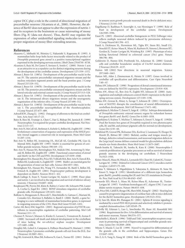
ceptor DCC play a role in the control of directional migration ofprecerebellar neurons (Alcantara et al., 2000). However, the ab-sence of Barhl1 does not appear to alter the expression of netrin-1and its receptors in the brainstem or cause misrouting of mossyfibers (Fig. 8) (data not shown). Thus, Barhl1 may regulate theexpression of other unidentified signaling molecules to controlproper migration of mossy fiber-extending neurons.
ReferencesAkazawa C, Ishibashi M, Shimizu C, Nakanishi S, Kageyama R (1995) A
mammalian helix-loop-helix factor structurally related to the product ofDrosophila proneural gene atonal is a positive transcriptional regulatorexpressed in the developing nervous system. J Biol Chem 270:8730 – 8738.
Alcantara S, Ruiz M, De Castro F, Soriano E, Sotelo C (2000) Netrin-1 actsas an attractive or as a repulsive cue for distinct migrating neurons duringthe development of the cerebellar system. Development 127:1359 –1372.
Altman J, Bayer SA (1987a) Development of the precerebellar nuclei in therat: IV. The anterior precerebellar extramural migratory stream and thenucleus reticularis tegmenti pontis and the basal pontine gray. J CompNeurol 257:529 –552.
Altman J, Bayer SA (1987b) Development of the precerebellar nuclei in therat: III. The posterior precerebellar extramural migratory stream and thelateral reticular and external cuneate nuclei. J Comp Neurol 257:513–528.
Altman J, Bayer SA (1987c) Development of the precerebellar nuclei in therat: II. The intramural olivary migratory stream and the neurogeneticorganization of the inferior olive. J Comp Neurol 257:490 –512.
Altman J, Bayer SA (1987d) Development of the precerebellar nuclei in therat: I. The precerebellar neuroepithelium of the rhombencephalon.J Comp Neurol 257:477– 489.
Ashwell KW, Zhang LL (1992) Ontogeny of afferents to the fetal rat cerebel-lum. Acta Anat 145:17–23.
Bates B, Rios M, Trumpp A, Chen C, Fan G, Bishop JM, Jaenisch R (1999)Neurotrophin-3 is required for proper cerebellar development. Nat Neu-rosci 2:115–117.
Ben-Arie N, McCall AE, Berkman S, Eichele G, Bellen HJ, Zoghbi HY (1996)Evolutionary conservation of sequence and expression of the bHLH pro-tein Atonal suggests a conserved role in neurogenesis. Hum Mol Genet5:1207–1216.
Ben-Arie N, Bellen HJ, Armstrong DL, McCall AE, Gordadze PR, Guo Q,Matzuk MM, Zoghbi HY (1997) Math1 is essential for genesis of cere-bellar granule neurons. Nature 390:169 –172.
Ben-Arie N, Hassan BA, Bermingham NA, Malicki DM, Armstrong D, Mat-zuk M, Bellen HJ, Zoghbi HY (2000) Functional conservation of atonaland Math1 in the CNS and PNS. Development 127:1039 –1048.
Bermingham NA, Hassan BA, Price SD, Vollrath MA, Ben-Arie N, Eatock RA,Bellen HJ, Lysakowski A, Zoghbi HY (1999) Math1: an essential gene forthe generation of inner ear hair cells. Science 284:1837–1841.
Bermingham NA, Hassan BA, Wang VY, Fernandez M, Banfi S, Bellen HJ,Fritzsch B, Zoghbi HY (2001) Proprioceptor pathway development isdependent on Math1. Neuron 30:411– 422.
Bloch-Gallego E, Ezan F, Tessier-Lavigne M, Sotelo C (1999) Floor plateand netrin-1 are involved in the migration and survival of inferior olivaryneurons. J Neurosci 19:4407– 4420.
Borghesani PR, Peyrin JM, Klein R, Rubin J, Carter AR, Schwartz PM, LusterA, Corfas G, Segal RA (2002) BDNF stimulates migration of cerebellargranule cells. Development 129:1435–1442.
Bulfone A, Menguzzato E, Broccoli V, Marchitiello A, Gattuso C, Mariani M,Consalez GG, Martinez S, Ballabio A, Banfi S (2000) Barhl1, a gene be-longing to a new subfamily of mammalian homeobox genes, is expressedin migrating neurons of the CNS. Hum Mol Genet 9:1443–1452.
Carter AR, Berry EM, Segal RA (2003) Regional expression of p75NTR con-tributes to neurotrophin regulation of cerebellar patterning. Mol CellNeurosci 22:1–13.
Chomez P, Neveu I, Mansen A, Kiesler E, Larsson L, Vennstrom B, Arenas E(2000) Increased cell death and delayed development in the cerebellumof mice lacking the rev-erbA(�) orphan receptor. Development127:1489 –1498.
Doughty ML, Lohof A, Campana A, Delhaye-Bouchaud N, Mariani J (1998)Neurotrophin-3 promotes cerebellar granule cell exit from the EGL. EurJ Neurosci 10:3007–3011.
Eng SR, Gratwick K, Rhee JM, Fedtsova N, Gan L, Turner EE (2001) Defects
in sensory axon growth precede neuronal death in Brn3a-deficient mice.J Neurosci 21:541–549.
Engelkamp D, Rashbass P, Seawright A, van Heyningen V (1999) Role ofPax6 in development of the cerebellar system. Development126:3585–3596.
Faust PL (2003) Abnormal cerebellar histogenesis in PEX2 Zellweger micereflects multiple neuronal defects induced by peroxisome deficiency.J Comp Neurol 461:394 – 413.
Fazeli A, Dickinson SL, Hermiston ML, Tighe RV, Steen RG, Small CG,Stoeckli ET, Keino-Masu K, Masu M, Rayburn H, Simons J, Bronson RT,Gordon JI, Tessier-Lavigne M, Weinberg RA (1997) Phenotype of micelacking functional Deleted in colorectal cancer (Dcc) gene. Nature386:796 – 804.
Goldowitz D, Hamre KM, Przyborski SA, Ackerman SL (2000) Granulecells and cerebellar boundaries: analysis of Unc5h3 mutant chimeras.J Neurosci 20:4129 – 4137.
Hatten ME (2002) New directions in neuronal migration. Science297:1660 –1663.
Hatten ME, Alder J, Zimmerman K, Heintz N (1997) Genes involved incerebellar cell specification and differentiation. Curr Opin Neurobiol7:40 – 47.
Helms AW, Johnson JE (1998) Progenitors of dorsal commissural interneu-rons are defined by MATH1 expression. Development 125:919 –928.
Helms AW, Abney AL, Ben-Arie N, Zoghbi HY, Johnson JE (2000) Auto-regulation and multiple enhancers control Math1 expression in the devel-oping nervous system. Development 127:1185–1196.
Helms AW, Gowan K, Abney A, Savage T, Johnson JE (2001) Overexpres-sion of MATH1 disrupts the coordination of neural differentiation incerebellum development. Mol Cell Neurosci 17:671– 682.
Higashijima S, Michiue T, Emori Y, Saigo K (1992a) Subtype determinationof Drosophila embryonic external sensory organs by redundant homeobox genes BarH1 and. BarH2. Genes Dev 6:1005–1018.
Higashijima S, Kojima T, Michiue T, Ishimaru S, Emori Y, Saigo K (1992b)Dual Bar homeo box genes of Drosophila required in two photoreceptorcells, R1 and R6, and primary pigment cells for normal eye development.Genes Dev 6:50 – 60.
Holzfeind PJ, Grewal PK, Reitsamer HA, Kechvar J, Lassmann H, Hoeger H,Hewitt JE, Bittner RE (2002) Skeletal, cardiac and tongue muscle pa-thology, defective retinal transmission, and neuronal migration defects inthe Large(myd) mouse defines a natural model for glycosylation-deficientmuscle-eye-brain disorders. Hum Mol Genet 11:2673–2687.
Katoh-Semba R, Takeuchi IK, Semba R, Kato K (2000) Neurotrophin-3controls proliferation of granular precursors as well as survival of maturegranule neurons in the developing rat cerebellum. J Neurochem74:1923–1930.
Keino-Masu K, Masu M, Hinck L, Leonardo ED, Chan SS, Culotti JG, Tessier-Lavigne M (1996) Deleted in Colorectal Cancer (DCC) encodes a netrinreceptor. Cell 87:175–185.
Kojima T, Ishimaru S, Higashijima S, Takayama E, Akimaru H, Sone M,Emori Y, Saigo K (1991) Identification of a different-type homeoboxgene, BarH1, possibly causing Bar(B) and Om(1D) mutations in Drosoph-ila. Proc Natl Acad Sci USA 88:4343– 4347.
Leonardo ED, Hinck L, Masu M, Keino-Masu K, Ackerman SL, Tessier-Lavigne M (1997) Vertebrate homologues of C. elegans UNC-5 are can-didate netrin receptors. Nature 386:833– 838.
Li S, Price SM, Cahill H, Ryugo DK, Shen MM, Xiang M (2002) Hearing losscaused by progressive degeneration of cochlear hair cells in mice deficientfor the Barhl1 homeobox gene. Development 129:3523–3532.
Lu Q, Sun EE, Klein RS, Flanagan JG (2001) Ephrin-B reverse signaling ismediated by a novel PDZ-RGS protein and selectively inhibits G protein-coupled chemoattraction. Cell 105:69 –79.
McEvilly RJ, Erkman L, Luo L, Sawchenko PE, Ryan AF, Rosenfeld MG(1996) Requirement for Brn-3.0 in differentiation and survival of sensoryand motor neurons. Nature 384:574 –577.
Minichiello L, Klein R (1996) TrkB and TrkC neurotrophin receptors coop-erate in promoting survival of hippocampal and cerebellar granule neu-rons. Genes Dev 10:2849 –2858.
Miyata T, Maeda T, Lee JE (1999) NeuroD is required for differentiation ofthe granule cells in the cerebellum and hippocampus. Genes Dev13:1647–1652.
Mo Z, Li S, Yang X, Xiang M (2004) Role of the Barhl2 homeobox gene in
Li et al. • Barhl1 Regulates NT-3 Expression in the Cerebellum J. Neurosci., March 24, 2004 • 24(12):3104 –3114 • 3113
the specification of glycinergic amacrine cells. Development 131:1607–1618.
Neveu I, Arenas E (1996) Neurotrophins promote the survival and develop-ment of neurons in the cerebellum of hypothyroid rats in vivo. J Cell Biol133:631– 646.
Overbergh L, Valckx D, Waer M, Mathieu C (1999) Quantification of mu-rine cytokine mRNAs using real time quantitative reverse transcriptasePCR. Cytokine 11:305–312.
Panchision DM, Pickel JM, Studer L, Lee SH, Turner PA, Hazel TG, McKayRD (2001) Sequential actions of BMP receptors control neural precur-sor cell production and fate. Genes Dev 15:2094 –2110.
Przyborski SA, Knowles BB, Ackerman SL (1998) Embryonic phenotype ofUnc5h3 mutant mice suggests chemorepulsion during the formation ofthe rostral cerebellar boundary. Development 125:41–50.
Rocamora N, Garcia-Ladona FJ, Palacios JM, Mengod G (1993) Differentialexpression of brain-derived neurotrophic factor, neurotrophin-3, andlow-affinity nerve growth factor receptor during the postnatal develop-ment of the rat cerebellar system. Brain Res Mol Brain Res 17:1– 8.
Rodriguez CI, Dymecki SM (2000) Origin of the precerebellar system. Neu-ron 27:475– 486.
Saito T, Sawamoto K, Okano H, Anderson DJ, Mikoshiba K (1998) Mam-malian BarH homologue is a potential regulator of neural bHLH genes.Dev Biol 199:216 –225.
Schwartz PM, Borghesani PR, Levy RL, Pomeroy SL, Segal RA (1997) Ab-normal cerebellar development and foliation in BDNF-/- mice reveals arole for neurotrophins in CNS patterning. Neuron 19:269 –281.
Sciavolino PJ, Abrams EW, Yang L, Austenberg LP, Shen MM, Abate-Shen C(1997) Tissue-specific expression of murine Nkx3.1 in the male urogen-ital system. Dev Dyn 209:127–138.
Serafini T, Colamarino SA, Leonardo ED, Wang H, Beddington R, SkarnesWC, Tessier-Lavigne M (1996) Netrin-1 is required for commissuralaxon guidance in the developing vertebrate nervous system. Cell87:1001–1014.
Shalizi A, Lehtinen M, Gaudilliere B, Donovan N, Han J, Konishi Y, Bonni A(2003) Characterization of a neurotrophin signaling mechanism thatmediates neuron survival in a temporally specific pattern. J Neurosci23:7326 –7336.
Singh S, Tang HK, Lee JY, Saunders GF (1998) Truncation mutations in thetransactivation region of PAX6 result in dominant-negative mutants.J Biol Chem 273:21531–21541.
Tojo H, Takami K, Kaisho Y, Nakata M, Abe T, Shiho O, Igarashi K (1995)Neurotrophin-3 is expressed in the posterior lobe of mouse cerebellum,but does not affect the cerebellar development. Neurosci Lett192:169 –172.
Wingate RJ (2001) The rhombic lip and early cerebellar development. CurrOpin Neurobiol 11:82– 88.
Wingate RJ, Hatten ME (1999) The role of the rhombic lip in avian cerebel-lum development. Development 126:4395– 4404.
Xiang M, Zhou L, Peng YW, Eddy RL, Shows TB, Nathans J (1993) Brn-3b:a POU domain gene expressed in a subset of retinal ganglion cells. Neuron11:689 –701.
Xiang M, Zhou L, Macke JP, Yoshioka T, Hendry SH, Eddy RL, Shows TB,Nathans J (1995) The Brn-3 family of POU-domain factors: primarystructure, binding specificity, and expression in subsets of retinal ganglioncells and somatosensory neurons. J Neurosci 15:4762– 4785.
Xiang M, Gan L, Zhou L, Klein WH, Nathans J (1996) Targeted deletion ofthe mouse POU domain gene Brn-3a causes selective loss of neurons inthe brainstem and trigeminal ganglion, uncoordinated limb movement,and impaired suckling. Proc Natl Acad Sci USA 93:11950 –11955.
Yamasaki T, Kawaji K, Ono K, Bito H, Hirano T, Osumi N, Kengaku M(2001) Pax6 regulates granule cell polarization during parallel fiber for-mation in the developing cerebellum. Development 128:3133–3144.
Yue Y, Widmer DA, Halladay AK, Cerretti DP, Wagner GC, Dreyer JL, ZhouR (1999) Specification of distinct dopaminergic neural pathways: rolesof the Eph family receptor EphB1 and ligand ephrin-B2. J Neurosci 19:2090 –2101.
3114 • J. Neurosci., March 24, 2004 • 24(12):3104 –3114 Li et al. • Barhl1 Regulates NT-3 Expression in the Cerebellum