Bacterial DNA induces pulmonary damage via TLR-9 through cross-talk with neutrophils
12
Bacterial DNA Induces Pulmonary Damage via TLR9 through Cross-Talk with Neutrophils Kiyoshi Itagaki, Yasaman Adibnia * , Shiqin Sun, Cong Zhao, Tolga Sursal ** , Yu Chen, Wolfgang Junger, and Carl J. Hauser Department of Surgery, Beth Israel Deaconess Medical Center, Boston, MA. * Istanbul Medical School, Istanbul, Turkey ** Eastern Virginia Medical School, Norfolk, VA Abstract Bacterial DNA (bDNA) contains hypo-methylated “CpG” repeats that can be recognized by toll- like receptor (TLR)-9 as a pathogen-associated molecular pattern (PAMP). The ability of bDNA to initiate lung injury via TLR-9 has been inferred on the basis of studies using artificial CpG DNA. But the role of authentic bDNA in lung injury is still unknown. Moreover, the mechanisms by which CpG DNA species can lead to pulmonary injury are unknown, although neutrophils (PMN) are thought to play a key role in the genesis of septic acute lung injury (ALI). We evaluated the effects of bDNA on PMN-endothelial cell (EC) interactions thought critical for initiation of ALI. Using a bio-capacitance system to monitor real-time changes in endothelial permeability, we demonstrate here that bDNA causes EC permeability in a dose-dependent manner uniquely in the presence of PMN. These permeability changes are inhibited by chloroquine, suggesting TLR9- dependency. When PMN were pre-incubated with bDNA and applied to EC or when bDNA was applied to EC without PMN, no permeability changes were detected. To study the underlying mechanisms we evaluated the effects of bDNA on PMN-EC adherence. bDNA significantly increased PMN adherence to EC in association with up-regulated adhesion molecules in both cell types. Taken together, our results strongly support the conclusion that bDNA can initiate lung injury by stimulating PMN-EC adhesive interactions predisposing to endothelial permeability. bDNA stimulation of TLR9 appears to promote enhanced gene expression of adhesion molecules in both cell types. This leads to PMN-EC cross-talk which is required for injury to occur. Keywords Pathogen Associated Molecular Patterns (PAMPs); CpG DNA; bacterial DNA; lung injury; vascular permeability; ECIS; Adherence; qPCR INTRODUCTION Neutrophils (PMN play an essential role in infection and innate immunity, both providing early defense against invading microorganisms or initiating deleterious SIRS responses (1, Corresponding Author: Name: Kiyoshi Itagaki, Ph.D., Institution: Beth Israel Deaconess Medical Center, Address: 330 Brookline Av. ST-8M10A, Boston, MA 02215. Phone: (617) 667-5382. Fax: (617) 667-8172. [email protected]. Institution at which the work was performed: Beth Israel Deaconess Medical Center, 330 Brookline Av. Boston, MA 02215. This is a PDF file of an unedited manuscript that has been accepted for publication. As a service to our customers we are providing this early version of the manuscript. The manuscript will undergo copyediting, typesetting, and review of the resulting proof before it is published in its final citable form. Please note that during the production process errors may be discovered which could affect the content, and all legal disclaimers that apply to the journal pertain. NIH Public Access Author Manuscript Shock. Author manuscript; available in PMC 2012 December 1. Published in final edited form as: Shock. 2011 December ; 36(6): 548–552. doi:10.1097/SHK.0b013e3182369fb2. NIH-PA Author Manuscript NIH-PA Author Manuscript NIH-PA Author Manuscript
-
Upload
hms-harvard -
Category
Documents
-
view
0 -
download
0
Transcript of Bacterial DNA induces pulmonary damage via TLR-9 through cross-talk with neutrophils
Bacterial DNA Induces Pulmonary Damage via TLR9 throughCross-Talk with Neutrophils
Kiyoshi Itagaki, Yasaman Adibnia*, Shiqin Sun, Cong Zhao, Tolga Sursal**, Yu Chen,Wolfgang Junger, and Carl J. HauserDepartment of Surgery, Beth Israel Deaconess Medical Center, Boston, MA.*Istanbul Medical School, Istanbul, Turkey**Eastern Virginia Medical School, Norfolk, VA
AbstractBacterial DNA (bDNA) contains hypo-methylated “CpG” repeats that can be recognized by toll-like receptor (TLR)-9 as a pathogen-associated molecular pattern (PAMP). The ability of bDNA toinitiate lung injury via TLR-9 has been inferred on the basis of studies using artificial CpG DNA.But the role of authentic bDNA in lung injury is still unknown. Moreover, the mechanisms bywhich CpG DNA species can lead to pulmonary injury are unknown, although neutrophils (PMN)are thought to play a key role in the genesis of septic acute lung injury (ALI). We evaluated theeffects of bDNA on PMN-endothelial cell (EC) interactions thought critical for initiation of ALI.Using a bio-capacitance system to monitor real-time changes in endothelial permeability, wedemonstrate here that bDNA causes EC permeability in a dose-dependent manner uniquely in thepresence of PMN. These permeability changes are inhibited by chloroquine, suggesting TLR9-dependency. When PMN were pre-incubated with bDNA and applied to EC or when bDNA wasapplied to EC without PMN, no permeability changes were detected. To study the underlyingmechanisms we evaluated the effects of bDNA on PMN-EC adherence. bDNA significantlyincreased PMN adherence to EC in association with up-regulated adhesion molecules in both celltypes. Taken together, our results strongly support the conclusion that bDNA can initiate lunginjury by stimulating PMN-EC adhesive interactions predisposing to endothelial permeability.bDNA stimulation of TLR9 appears to promote enhanced gene expression of adhesion moleculesin both cell types. This leads to PMN-EC cross-talk which is required for injury to occur.
KeywordsPathogen Associated Molecular Patterns (PAMPs); CpG DNA; bacterial DNA; lung injury;vascular permeability; ECIS; Adherence; qPCR
INTRODUCTIONNeutrophils (PMN play an essential role in infection and innate immunity, both providingearly defense against invading microorganisms or initiating deleterious SIRS responses (1,
Corresponding Author: Name: Kiyoshi Itagaki, Ph.D., Institution: Beth Israel Deaconess Medical Center, Address: 330 BrooklineAv. ST-8M10A, Boston, MA 02215. Phone: (617) 667-5382. Fax: (617) 667-8172. [email protected] at which the work was performed: Beth Israel Deaconess Medical Center, 330 Brookline Av. Boston, MA 02215.This is a PDF file of an unedited manuscript that has been accepted for publication. As a service to our customers we are providingthis early version of the manuscript. The manuscript will undergo copyediting, typesetting, and review of the resulting proof before itis published in its final citable form. Please note that during the production process errors may be discovered which could affect thecontent, and all legal disclaimers that apply to the journal pertain.
NIH Public AccessAuthor ManuscriptShock. Author manuscript; available in PMC 2012 December 1.
Published in final edited form as:Shock. 2011 December ; 36(6): 548–552. doi:10.1097/SHK.0b013e3182369fb2.
NIH
-PA Author Manuscript
NIH
-PA Author Manuscript
NIH
-PA Author Manuscript
2). PMN comprise approximately two-thirds of peripheral blood leukocytes and transitrapidly to sites of infection. There, they migrate down chemotactic gradients to limitinfection and allow recruitment and activation of other immune cells through the release ofinflammatory mediators and antimicrobial products. This results in pathogen clearance andultimately, in the initiation of an adaptive immune response (3). Conversely, excessive orinappropriate PMN activation result in inflammation that causes tissue injury and contributeto the initiation of a variety of noninfectious illnesses including rheumatoid arthritis,inflammatory bowel disease, asthma or chronic obstructive pulmonary disease. In the settingof acute sepsis however, the most feared complication of PMN activation is Acute LungInjury (ALI), which can lead to Acute Respiratory Distress Syndrome (ARDS) (4, 5).
The toll-like receptors (TLRs) are a family of innate immune receptors that can recognizeconserved pathogen associated molecular patterns (6). Recognition of such ‘Danger’ signals(7) by the TLRs leads to activation of a group of overlapping signaling systems that utilizethe adaptor molecule MyD88. These systems appear to play a central role in theinflammatory responses to sepsis. The prototype for TLR activation in sepsis is immune cellstimulation by endotoxin via TLR4 (8). Less is known about how other bacterial PAMPsactivate immune cells although bDNA is thought to activate innate immunity by binding tothe endocytic receptor TLR-9 (9).
To date, studies (10-12) evaluating the role of TLR9 in sepsis and lung injury have focusedon the interactions of TLR9 with artificial CpG DNAs. These are synthesized to havehypomethylated areas that bind TLR9. No studies have assessed the effects of authenticbacterial DNA (bDNA) on human immune cells and moreover, no studies have examinedhow bDNA-induced events might cause interactions between immunocytes and the lung thatcould contribute to the pathogenesis of ALI/ARDS.
We hypothesized that if bDNA contributed to lung injury, it was likely to activate PMNand / or pulmonary endothelial cells (EC), promoting PMN-EC adherence and interactionsthat led to increased endothelial permeability. We therefore investigated whether and howbDNA acted on PMN and EC in co-culture and affected the permeability of EC-monolayers.
MATERIALS AND METHODSCompliance
All studies performed were approved by the IRB of Beth Israel Deaconess Medical Center.Human blood specimens were obtained from volunteer donors by venipuncture afterinformed consent.
Endothelial Cell cultures and chemicalsEA.hy926 cells, derived by the fusion of HUVECs with the continuous human lungcarcinoma cell line A549, were obtained from Dr. Cora-Jean C. Edgell (University of NorthCarolina at Chapel Hill, NC) and maintained in DMEM medium with 10% FBS andpenicillin and streptomycin in a CO2 (5%) incubator at 37°C (13). Bacterial DNAs (single ordouble stranded) were purchased from Sigma (St. Louis, MO) or Invivogen (San Diego,CA). Bacterial DNAs were endotoxin-free. Human TLR9 specific inhibitor, ODNTTAGGG, and an inhibitor for endosomal acidification, chloroquine, were purchased fromInvivogen. Other chemicals were purchase from Sigma.
Expression of ICAM-1, E-selectin, and TLR9 in EA.hy926 cellsEA cells grown in 6 well plates were treated with bacterial DNA (10 or 20 μg/mL), LPS(100 ng/mL), or medium for 6 hrs at 37°C in 5% CO2. RNA and cDNA were prepared from
Itagaki et al. Page 2
Shock. Author manuscript; available in PMC 2012 December 1.
NIH
-PA Author Manuscript
NIH
-PA Author Manuscript
NIH
-PA Author Manuscript
the cells using RNeasy (Qiagen, Valencia, CA) and VILO (Invitrogen, Carlsbad, CA), kitsrespectively, following the manufacturers’ protocols. RNA and cDNA concentrations weredetermined by the Nanodrop (Thermo Fisher Scientific, Hudson, NH). Gene expressionlevels of ICAM-1, E-selectin, and TLR9 were assayed by qPCR (Realplex, Eppendorf,Hauppauge, NY) using pre-validated specific primers from OriGene (Rockville, MD). Datawere normalized using GAPDH as the housekeeping gene and gene expression by medium-treated samples was used as 100%.
Gene expression of CD11b, CD18, and TLR9 in human PMNFreshly isolated PMN were seeded on 6 well-plate and were treated with bacterial DNA (10or 20 μg/mL), LPS (100 ng/mL), or medium for 90 min at 37°C, 5% CO2 incubator. Asshown above, qPCR for CD11b, CD18, and TLR9 was performed. Data were normalized asdescribed above. In addition, CD11b expression was evaluated using FACS (FACSCalibur,BD Bioscience, Chicago, IL) with specific CD11b antibody by following manufacturer’sprotocol.
Human PMN preparationsPMN were isolated fresh from healthy volunteer donor blood. Detailed methods for PMNpreparation can be found elsewhere (14). Briefly, PMN were isolated from minimallyheparinized whole blood using a one-step centrifugation procedure on PMN IsolationMedium (Thermo Fisher Scientific, Hudson, NH). The neutrophil layer was collected andosmolality was restored. Cells were then washed and suspended in HEPES buffer. Residualred blood cells were lysed briefly to increase PMN purity.
Human PMN-Endothelial Cell adhesion assayPMN were incubated for 30 minutes at 37°C in the dark in HEPES buffer containing 1mMCa2+ and 3 μM Calcein-AM. Calcein-labeled PMN were set aside for use as a standardcurve and PMN-endothelial cell adhesion assays were performed as previously described(15, 16). Near confluent EC monolayers were incubated with E.coli DNA (10-20 μg/mL),LPS (100 ng/mL) or medium for 6 hours with 2.25 × 10⁵ PMN, after which the plates wereinverted and centrifuged at 200g for 5 min at room temperature to remove non-adherentPMN. Calcein fluorescence of neutrophils was measured using a fluorescent plate reader(SpectaMax: Molecular Devices, Sunnyvale, CA) set at an excitation wavelength of 485 nmand an emission wavelength of 520 nm.
EC permeability measurementsTransendothelial Electrical Resistance (TER) of EC monolayers was measured by seeding2-3 ×10⁵ EA hy.926 cells onto cysteine and fibronectin-pretreated 8W10E+ cultureware (40electrodes per well) and incubated (37°C, 5% CO2) in an ECIS system (Applied BioPhysics,Troy, NY) (17, 18). This system measures changes in impedance of the monolayer to ACcurrent flow. The micro-currents used have no detectable effect on the cells (17).Confluence was determined by capacitance at 64 kHz coming to a plateau (~10nF) afterseveral hours of incubation (17). In this system impedance decreases and capacitanceincreases as permeability increases (17, 19). EC permeability change was assessed ascapacitance change at 64 KHz (19) after addition of 1) media, 2) human PMN (2×10⁵)freshly isolated from healthy volunteers, 3) bacterial DNA or 4) bacterial DNA and PMN.Capacitance values in each well were normalized to capacitance at the start of treatments.Finally, values from each microelectrode were pooled at discrete time points and plottedversus time as the mean ±SE.
Itagaki et al. Page 3
Shock. Author manuscript; available in PMC 2012 December 1.
NIH
-PA Author Manuscript
NIH
-PA Author Manuscript
NIH
-PA Author Manuscript
ResultsBacterial DNA increases endothelial permeability
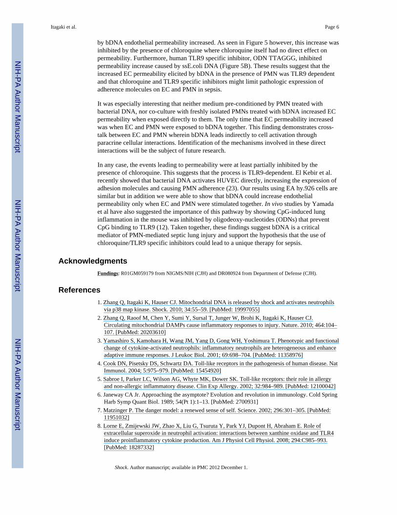
Increased endothelial permeability links immunologic ALI to the altered lung mechanics andimpaired gas exchange seen in ARDS. We hypothesized that authentic E.coli DNA (bDNA);acting either alone or through the agency of activated PMN, might act on EC monolayers toincrease their permeability. As shown in Figure 1A, untreated EA cells or cells having onlya medium change at T=0 show no permeability changes over the time course studied. Whenthe EA cells without PMN were treated with CpG DNA (1, 10 or 20 μg/mL) there was nosignificant change in permeability seen. Similarly, PMN exposed to bDNA and then appliedto the EC failed to induce a permeability change (data not shown). But when PMN wereapplied to confluent EC and bDNA was added to the intact system we saw brisk, dose-dependent increases in permeability (Figure 1A). Similar to CpG, when E.coli DNA (10 or20 μg/mL) was applied to confluent EA cells, permeability was increased only in thepresence of PMN (Figure 1B). However, E. coli DNA seems to be effective at the lowerconcentrations (10 μg/mL) than the higher concentrations (20 μg/mL).
Bacterial DNA increases PMN-EA adherencePMN adhesion to endothelial cells is an important first step on the recruitment of PMN toareas of active infection. Adherence to activated EC is a pre-requisite for PMNtransmigration into the alveoli and PMN-EC interactions during adherence andtransmigration can lead directly to increased vascular permeability (19). PMN were appliedto endothelial monolayers in the presence of E. coli DNA or medium and assayed foradherence. LPS (100 ng/mL) served as a positive control. As shown in Figure 2, bDNA(10-20 μg/mL) significantly increased the adherence of calcein-loaded PMN to EC ascompared to medium treated controls (One Way ANOVA, p<0.001).
Bacterial DNA increases adherence molecule gene expression in both EC and PMNSince bDNA increased adherence of PMN to EA monolayers we evaluated whether itaffected expression of the molecules that cause those cells to adhere. Thus we incubated EAcells (6h) or human PMN (90 min) with bDNA at various concentrations and performedqPCR to determine gene expression levels of endothelial ICAM-1 and E-selectin. TLR9expression was also assessed using specific primers. Data were normalized to GAPDHlevels with expression after medium treatment used as a negative baseline control. Responseto LPS was used as a positive control. As shown in Figure 3, treatment with bDNAincreased EC expression of ICAM-1 and E-selectin in a dose-dependent fashion. No changewas detected in the expression of TLR9. We then evaluated PMN CD11b and CD18expression after bDNA treatment. In Figure 4A we see that bDNA markedly increasesPMN expression of CD11b and CD18 whereas TLR9 expression is unchanged. Similarly,CD11b increase in PMN by bDNA treatment was detected by FACS using human CD11bspecific antibody (Figure 4B).
Endothelial permeability increase by bacterial DNA is TLR9-dependentPrior studies have shown that CpG DNA can act on immunocytes via TLR9. Chloroquine(CQ) is an inhibitor of TLR interactions with CpG DNAs and also of endosomalacidification. To provide proof-of-concept for in vivo therapeutic studies, we studiedwhether CQ inhibition could inhibit bDNA-induced increases in permeability in our PMN-EC co-culture systems. As clearly shown in Figure 5A, the increased in endothelialpermeability that was caused by bDNA in the presence of PMN was markedly attenuated byCQ. CQ had no direct effect on EC permeability.
Itagaki et al. Page 4
Shock. Author manuscript; available in PMC 2012 December 1.
NIH
-PA Author Manuscript
NIH
-PA Author Manuscript
NIH
-PA Author Manuscript
Furthermore, the permeability increase caused by single stranded E.coli DNA (ssE.coliDNA, 10 μg/mL) was inhibited in the presence of human TLR9 specific inhibitor, ODNTTAGGG (Figure 5B). This inhibitor may slightly increase barrier integrity although it maynot be significant.
DISCUSSIONEndothelial cell (EC) permeability is often pathologically increased in sepsis and relatedinflammatory conditions. This process plays a central role in initiating septic acute lunginjury (ALI) and its progression to acute respiratory distress syndrome (ARDS). EC can beactivated either through the actions of plasma-borne mediators of inflammation andcoagulation, or through cell-cell interactions with immunocytes like PMN. It is well knownthat release of bacterial PAMPs in sepsis can activate EC through ligation of TLR2 or 4 byLPS (20). A similar role has more recently been postulated for bacterial DNA since it canactivate immune cells via TLR9. No direct studies exist however, assessing whether bDNAdoes in fact, activate interactions of WBC and EC that lead to increased capillarypermeability, and if so, what mechanisms are involved.
We studied the contribution of bDNA to lung injury in sepsis by assaying its effects on ECmonolayer permeability. The ECIS system allows real-time study of changes in monolayerimpedance and capacitance that reflect resistance and permeability respectively (17, 19, 21).Interestingly, we found that applying bDNA directly either to the endothelial monolayers orto PMN prior to co-culture with the EC failed to result in any changes in permeability. Indistinction, exposure to G-protein ligands like thrombin or histamine cause immediateincreases in EC permeability (19). But when PMN and EC were stimulated in co-culture, thepresence of bDNA resulted in dose-dependent increases in EC monolayer permeability(Figure 1A, B). Thus bacterial DNA does not increase EC permeability by isolated effectseither on PMN or EC. Rather, direct physical interaction between bDNA-activated PMN andbDNA-activated EC is an absolute requirement for bDNA to mediate EC permeability.
EC-WBC interactions that lead to permeability changes typically begin with adherence ofthe WBCs to the endothelium (22). So next we examined whether increased permeabilitywas associated with increased PMN adherence to the EC monolayer and we found thatbDNA did enhance PMN adherence to EA cell monolayers (Figure 2). Finding that to be thecase, we next used qPCR to examine whether the enhanced adherence of PMN to EC afterbDNA exposure reflected up-regulation of adhesion molecule gene expression on the ECand/or PMN. As shown in Figure 3, EC gene expression of selectins was strongly increasedby incubation with bDNA. Selectins however, only initiate neutrophil ‘rolling’. Strongadhesion and transmigration are initiated by interactions of the PMN integrin CD11b/CD18with ICAM-1 on the endothelial surface. Gene expression of each of these molecules wasenhanced by exposure to bacterial DNA. In contrast, TLR 9 expression level was unchangedby exposure to bDNA (Figure 3, 4A, 4B).
The results above all suggest that bacterial DNA increases PMN-endothelial cell adherenceby increasing expression of adherence molecules on endothelial cells and PMN. BacterialDNA is thought to activate inflammation through ligation of the endosomal receptor TLR9.Indeed, lung injury caused by artificial CpG DNA depends upon the presence of TLR9receptors (10).
We sought to study the mechanisms underlying the regulation of adherence molecules andalso to begin evaluation of potential therapeutic means of preventing bDNA mediatedincreases in EC permeability. We therefore applied the TLR9 inhibitor chloroquine (5 μM)to ECs during ECIS experiments. As before, when EC and PMN were stimulated together
Itagaki et al. Page 5
Shock. Author manuscript; available in PMC 2012 December 1.
NIH
-PA Author Manuscript
NIH
-PA Author Manuscript
NIH
-PA Author Manuscript
by bDNA endothelial permeability increased. As seen in Figure 5 however, this increase wasinhibited by the presence of chloroquine where chloroquine itself had no direct effect onpermeability. Furthermore, human TLR9 specific inhibitor, ODN TTAGGG, inhibitedpermeability increase caused by ssE.coli DNA (Figure 5B). These results suggest that theincreased EC permeability elicited by bDNA in the presence of PMN was TLR9 dependentand that chloroquine and TLR9 specific inhibitors might limit pathologic expression ofadherence molecules on EC and PMN in sepsis.
It was especially interesting that neither medium pre-conditioned by PMN treated withbacterial DNA, nor co-culture with freshly isolated PMNs treated with bDNA increased ECpermeability when exposed directly to them. The only time that EC permeability increasedwas when EC and PMN were exposed to bDNA together. This finding demonstrates cross-talk between EC and PMN wherein bDNA leads indirectly to cell activation throughparacrine cellular interactions. Identification of the mechanisms involved in these directinteractions will be the subject of future research.
In any case, the events leading to permeability were at least partially inhibited by thepresence of chloroquine. This suggests that the process is TLR9-dependent. El Kebir et al.recently showed that bacterial DNA activates HUVEC directly, increasing the expression ofadhesion molecules and causing PMN adherence (23). Our results using EA hy.926 cells aresimilar but in addition we were able to show that bDNA could increase endothelialpermeability only when EC and PMN were stimulated together. In vivo studies by Yamadaet al have also suggested the importance of this pathway by showing CpG-induced lunginflammation in the mouse was inhibited by oligodeoxy-nucleotides (ODNs) that preventCpG binding to TLR9 (12). Taken together, these findings suggest bDNA is a criticalmediator of PMN-mediated septic lung injury and support the hypothesis that the use ofchloroquine/TLR9 specific inhibitors could lead to a unique therapy for sepsis.
AcknowledgmentsFundings: R01GM059179 from NIGMS/NIH (CJH) and DR080924 from Department of Defense (CJH).
References1. Zhang Q, Itagaki K, Hauser CJ. Mitochondrial DNA is released by shock and activates neutrophils
via p38 map kinase. Shock. 2010; 34:55–59. [PubMed: 19997055]2. Zhang Q, Raoof M, Chen Y, Sumi Y, Sursal T, Junger W, Brohi K, Itagaki K, Hauser CJ.
Circulating mitochondrial DAMPs cause inflammatory responses to injury. Nature. 2010; 464:104–107. [PubMed: 20203610]
3. Yamashiro S, Kamohara H, Wang JM, Yang D, Gong WH, Yoshimura T. Phenotypic and functionalchange of cytokine-activated neutrophils: inflammatory neutrophils are heterogeneous and enhanceadaptive immune responses. J Leukoc Biol. 2001; 69:698–704. [PubMed: 11358976]
4. Cook DN, Pisetsky DS, Schwartz DA. Toll-like receptors in the pathogenesis of human disease. NatImmunol. 2004; 5:975–979. [PubMed: 15454920]
5. Sabroe I, Parker LC, Wilson AG, Whyte MK, Dower SK. Toll-like receptors: their role in allergyand non-allergic inflammatory disease. Clin Exp Allergy. 2002; 32:984–989. [PubMed: 12100042]
6. Janeway CA Jr. Approaching the asymptote? Evolution and revolution in immunology. Cold SpringHarb Symp Quant Biol. 1989; 54(Pt 1):1–13. [PubMed: 2700931]
7. Matzinger P. The danger model: a renewed sense of self. Science. 2002; 296:301–305. [PubMed:11951032]
8. Lorne E, Zmijewski JW, Zhao X, Liu G, Tsuruta Y, Park YJ, Dupont H, Abraham E. Role ofextracellular superoxide in neutrophil activation: interactions between xanthine oxidase and TLR4induce proinflammatory cytokine production. Am J Physiol Cell Physiol. 2008; 294:C985–993.[PubMed: 18287332]
Itagaki et al. Page 6
Shock. Author manuscript; available in PMC 2012 December 1.
NIH
-PA Author Manuscript
NIH
-PA Author Manuscript
NIH
-PA Author Manuscript

9. Akira S, Hemmi H. Recognition of pathogen-associated molecular patterns by TLR family.Immunol Lett. 2003; 85:85–95. [PubMed: 12527213]
10. Knuefermann P, Baumgarten G, Koch A, Schwederski M, Velten M, Ehrentraut H, Mersmann J,Meyer R, Hoeft A, Zacharowski K, Grohe C. CpG oligonucleotide activates Toll-like receptor 9and causes lung inflammation in vivo. Respir Res. 2007; 8:72. [PubMed: 17925007]
11. Tasaka S, Kamata H, Miyamoto K, Nakano Y, Shinoda H, Kimizuka Y, Fujiwara H, Hasegawa N,Fujishima S, Miyasho T, Ishizaka A. Intratracheal synthetic CpG oligodeoxynucleotide causesacute lung injury with systemic inflammatory response. Respir Res. 2009; 10:84. [PubMed:19772669]
12. Yamada H, Ishii KJ, Klinman DM. Suppressive oligodeoxynucleotides inhibit CpG-inducedinflammation of the mouse lung. Crit Care Med. 2004; 32:2045–2049. [PubMed: 15483413]
13. Edgell CJ, McDonald CC, Graham JB. Permanent cell line expressing human factor VIII-relatedantigen established by hybridization. Proc Natl Acad Sci U S A. 1983; 80:3734–3737. [PubMed:6407019]
14. Itagaki K, Kannan KB, Livingston DH, Deitch EA, Fekete Z, Hauser CJ. Store-operated calciumentry in human neutrophils reflects multiple contributions from independently regulated pathways.J Immunol. 2002; 168:4063–4069. [PubMed: 11937565]
15. Fomsgaard A, Zhang GH, Shand GH, Bendtzen K, Baek L. Immunochemical and biologicalreactivity of human anti-lipopolysaccharide IgG obtained by screening of blood donors. Scand JImmunol. 1989; 29:309–316. [PubMed: 2717879]
16. Sanchez-Cantu L, Rode HN, Christou NV. Endotoxin tolerance is associated with reducedsecretion of tumor necrosis factor. Arch Surg. 1989; 124:1432–1435. [PubMed: 2589967]
17. Giaever I, Keese CR. Micromotion of mammalian cells measured electrically. Proc Natl Acad SciU S A. 1991; 88:7896–7900. [PubMed: 1881923]
18. Wegener J, Keese CR, Giaever I. Electric cell-substrate impedance sensing (ECIS) as anoninvasive means to monitor the kinetics of cell spreading to artificial surfaces. Exp Cell Res.2000; 259:158–166. [PubMed: 10942588]
19. Itagaki K, Barton BE, Murphy TF, Taheri S, Shu P, Huang H, Jordan ML. Eicosanoid-InducedStore-Operated Calcium Entry in Dendritic Cells. J Surg Res. 2009; 169(2):301–310. [PubMed:20080257]
20. Vandenbroucke E, Mehta D, Minshall R, Malik AB. Regulation of endothelial junctionalpermeability. Ann N Y Acad Sci. 2008; 1123:134–145. [PubMed: 18375586]
21. Garcia JG. Concepts in microvascular endothelial barrier regulation in health and disease.Microvasc Res. 2009; 77:1–3. [PubMed: 19232241]
22. DiStasi MR, Ley K. Opening the flood-gates: how neutrophil-endothelial interactions regulatepermeability. Trends Immunol. 2009; 30:547–556. [PubMed: 19783480]
23. El Kebir D, Jozsef L, Pan W, Wang L, Filep JG. Bacterial DNA activates endothelial cells andpromotes neutrophil adherence through TLR9 signaling. J Immunol. 2009; 182:4386–4394.[PubMed: 19299739]
Itagaki et al. Page 7
Shock. Author manuscript; available in PMC 2012 December 1.
NIH
-PA Author Manuscript
NIH
-PA Author Manuscript
NIH
-PA Author Manuscript
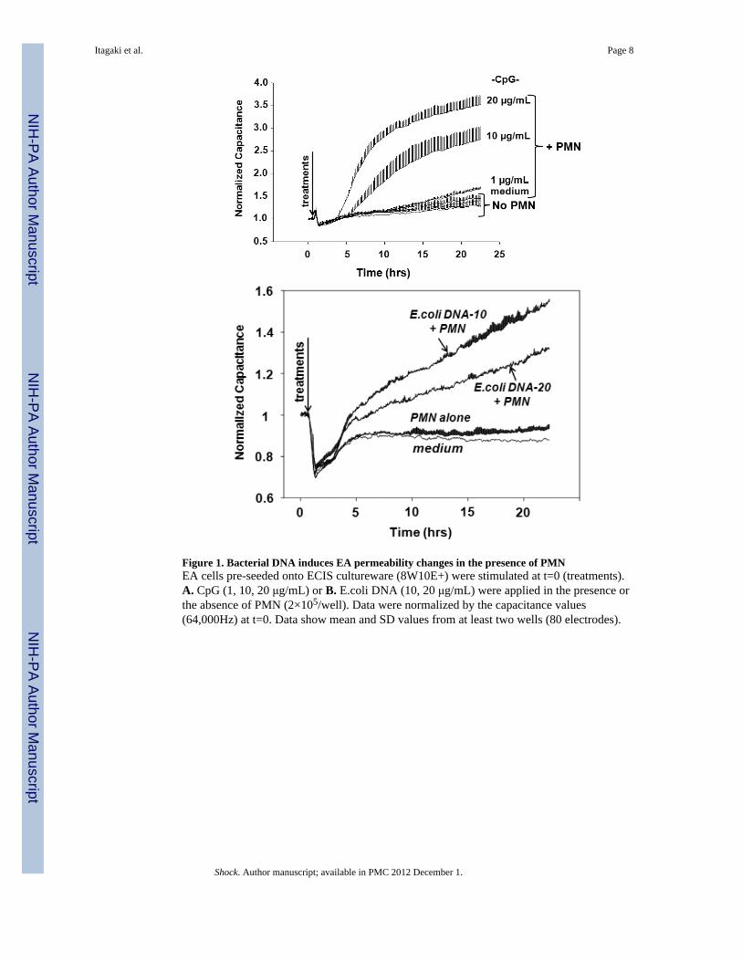
Figure 1. Bacterial DNA induces EA permeability changes in the presence of PMNEA cells pre-seeded onto ECIS cultureware (8W10E+) were stimulated at t=0 (treatments).A. CpG (1, 10, 20 μg/mL) or B. E.coli DNA (10, 20 μg/mL) were applied in the presence orthe absence of PMN (2×10⁵/well). Data were normalized by the capacitance values(64,000Hz) at t=0. Data show mean and SD values from at least two wells (80 electrodes).
Itagaki et al. Page 8
Shock. Author manuscript; available in PMC 2012 December 1.
NIH
-PA Author Manuscript
NIH
-PA Author Manuscript
NIH
-PA Author Manuscript
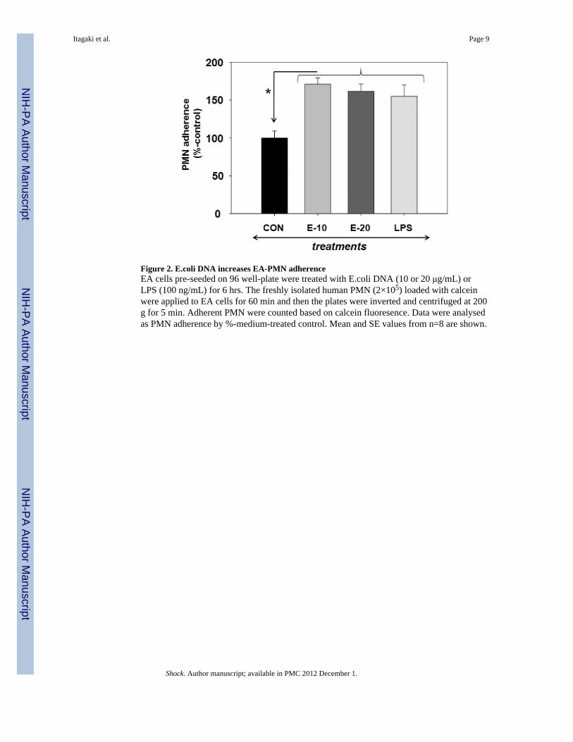
Figure 2. E.coli DNA increases EA-PMN adherenceEA cells pre-seeded on 96 well-plate were treated with E.coli DNA (10 or 20 μg/mL) orLPS (100 ng/mL) for 6 hrs. The freshly isolated human PMN (2×10⁵) loaded with calceinwere applied to EA cells for 60 min and then the plates were inverted and centrifuged at 200g for 5 min. Adherent PMN were counted based on calcein fluoresence. Data were analysedas PMN adherence by %-medium-treated control. Mean and SE values from n=8 are shown.
Itagaki et al. Page 9
Shock. Author manuscript; available in PMC 2012 December 1.
NIH
-PA Author Manuscript
NIH
-PA Author Manuscript
NIH
-PA Author Manuscript
Figure 3. Bacterial DNA increased adherence molecule gene expression in EA cellsEA cells were treated with E.coli DNA (10, 20 μg/mL) for 6 hrs and then RNA/cDNA wereprepared using RNeasy and VILO cDNA synthesis kit from Qiagen and Invitrogen,respectedly. ICAM-1, E-selectin, and TLR9 gene expression levels were evaluated usingGAPDH as a house keeping gene. Levels of gene expression were shown as %-mediumcontrol. Mean and SE values are shown (n=3).
Itagaki et al. Page 10
Shock. Author manuscript; available in PMC 2012 December 1.
NIH
-PA Author Manuscript
NIH
-PA Author Manuscript
NIH
-PA Author Manuscript
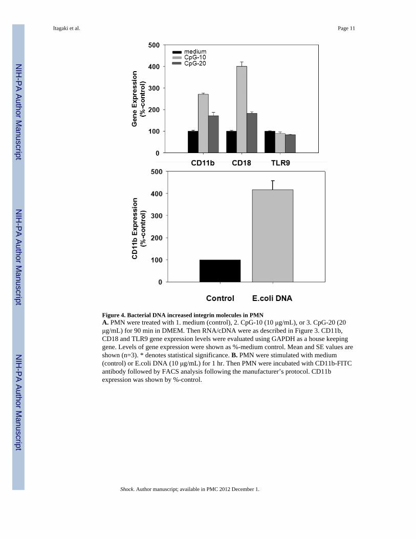
Figure 4. Bacterial DNA increased integrin molecules in PMNA. PMN were treated with 1. medium (control), 2. CpG-10 (10 μg/mL), or 3. CpG-20 (20μg/mL) for 90 min in DMEM. Then RNA/cDNA were as described in Figure 3. CD11b,CD18 and TLR9 gene expression levels were evaluated using GAPDH as a house keepinggene. Levels of gene expression were shown as %-medium control. Mean and SE values areshown (n=3). * denotes statistical significance. B. PMN were stimulated with medium(control) or E.coli DNA (10 μg/mL) for 1 hr. Then PMN were incubated with CD11b-FITCantibody followed by FACS analysis following the manufacturer’s protocol. CD11bexpression was shown by %-control.
Itagaki et al. Page 11
Shock. Author manuscript; available in PMC 2012 December 1.
NIH
-PA Author Manuscript
NIH
-PA Author Manuscript
NIH
-PA Author Manuscript
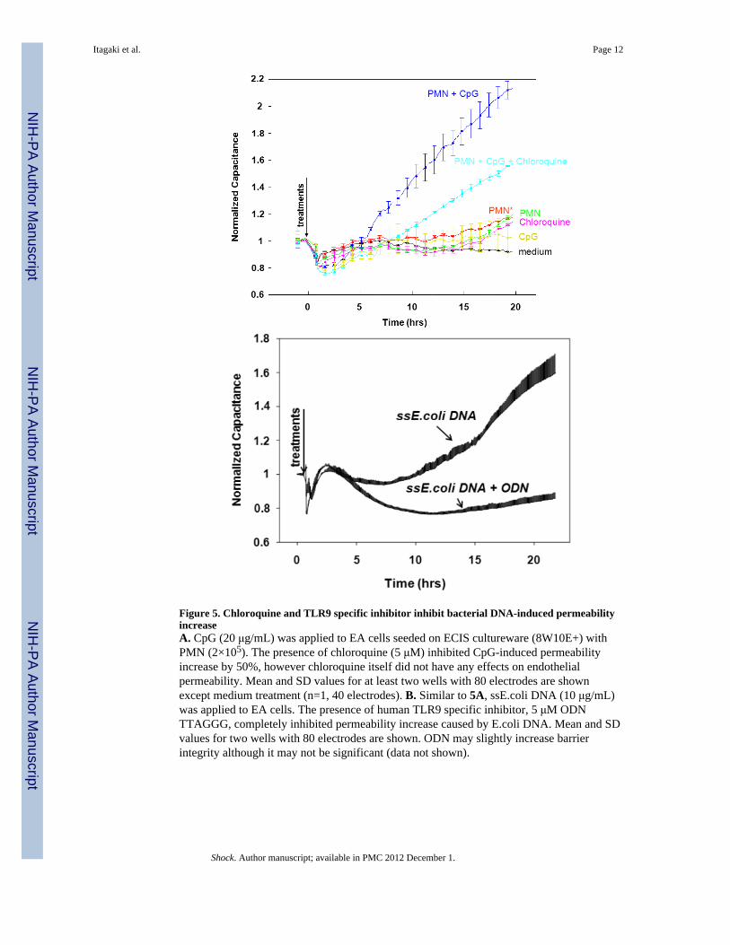
Figure 5. Chloroquine and TLR9 specific inhibitor inhibit bacterial DNA-induced permeabilityincreaseA. CpG (20 μg/mL) was applied to EA cells seeded on ECIS cultureware (8W10E+) withPMN (2×10⁵). The presence of chloroquine (5 μM) inhibited CpG-induced permeabilityincrease by 50%, however chloroquine itself did not have any effects on endothelialpermeability. Mean and SD values for at least two wells with 80 electrodes are shownexcept medium treatment (n=1, 40 electrodes). B. Similar to 5A, ssE.coli DNA (10 μg/mL)was applied to EA cells. The presence of human TLR9 specific inhibitor, 5 μM ODNTTAGGG, completely inhibited permeability increase caused by E.coli DNA. Mean and SDvalues for two wells with 80 electrodes are shown. ODN may slightly increase barrierintegrity although it may not be significant (data not shown).
Itagaki et al. Page 12
Shock. Author manuscript; available in PMC 2012 December 1.
NIH
-PA Author Manuscript
NIH
-PA Author Manuscript
NIH
-PA Author Manuscript