Bacterial cellulose films with controlled microstructure–mechanical property relationships
9
Bacterial cellulose films with controlled microstructure–mechanical property relationships A. Retegi • N. Gabilondo • C. Pen ˜a • R. Zuluaga • C. Castro • P. Gan ˜an • K. de la Caba • I. Mondragon Received: 12 August 2009 / Accepted: 8 December 2009 / Published online: 25 December 2009 Ó Springer Science+Business Media B.V. 2009 Abstract Bacterial cellulose (BC) films with dif- ferent porosities have been developed in order to obtain improved mechanical properties. After 13 days of incubation of Gluconobacter xylinum bacteria in static culture, BC pellicles have been set. BC films have been compression molded after water dispersion of BC pellicles and filtration by applying different pressures (10, 50, and 100 MPa) to obtain films with different porosities. Tensile behavior has been analyzed in order to discuss the microstructure– property relationships. Compression pressure has been found as an important parameter to control the final mechanical properties of BC films where slightly enhanced tensile strength and deformation at break are obtained increasing mold compression pressure, while modulus also increases following a nearly linear dependence upon film porosity. This behavior is related to the higher densification by increasing mold compression pressure that reduces the interfibrillar space, thus increasing the possibility of interfibrillar bonding zones. Network theories have been applied to relate film elastic properties with individual nanofiber properties. Keywords Bacterial cellulose Tensile properties Porosity X-ray Introduction Cellulose is the most abundant biopolymer on earth, recognized as the major component of plant biomass but also a representative of microbial extracellular polymers. Bacterial cellulose (BC) belongs to specific products of primary metabolism. Cellulose is synthe- sized by bacteria belonging to the genera of Aceto- bacter, Rhizobium, Agrobacterium, and Sarcina. Its most efficient producers are Gram-negative, acetic acid bacteria Acetobacter xylinum (reclassified as Gluconobacter xylinus, Yamanaka et al. 2000), which have been applied as model microorganisms for basic and applied studies on cellulose. The formation of cellulose by laboratory bacterial cultures is an interesting and attractive biomimetic access to obtain pure cellulose for both organic and polymer chemists. By selecting the substrates, cultivation conditions, various additives, and finally the bacterial strain, it is possible to control the molar mass, molar mass distribution, and the supramolecular structure. Thus, it is possible to control important cellulose properties A. Retegi N. Gabilondo C. Pen ˜a K. de la Caba I. Mondragon (&) Materials ? Technologies Group, Polytechnic School, Dpto. Ingenierı ´a Quı ´mica y M. Ambiente, Universidad Paı ´s Vasco/Euskal Herriko Unibertsitatea, Pza. Europa 1, 20018 Donostia-San Sebastia ´n, Spain e-mail: [email protected] R. Zuluaga C. Castro P. Gan ˜an New Materials Group, Universidad Pontificia Bolivariana Circular 1 # 70-01, Medellin, Colombia 123 Cellulose (2010) 17:661–669 DOI 10.1007/s10570-009-9389-7
-
Upload
independent -
Category
Documents
-
view
0 -
download
0
Transcript of Bacterial cellulose films with controlled microstructure–mechanical property relationships
Bacterial cellulose films with controlledmicrostructure–mechanical property relationships
A. Retegi • N. Gabilondo • C. Pena • R. Zuluaga •
C. Castro • P. Ganan • K. de la Caba •
I. Mondragon
Received: 12 August 2009 / Accepted: 8 December 2009 / Published online: 25 December 2009
� Springer Science+Business Media B.V. 2009
Abstract Bacterial cellulose (BC) films with dif-
ferent porosities have been developed in order to
obtain improved mechanical properties. After
13 days of incubation of Gluconobacter xylinum
bacteria in static culture, BC pellicles have been set.
BC films have been compression molded after water
dispersion of BC pellicles and filtration by applying
different pressures (10, 50, and 100 MPa) to obtain
films with different porosities. Tensile behavior has
been analyzed in order to discuss the microstructure–
property relationships. Compression pressure has
been found as an important parameter to control the
final mechanical properties of BC films where
slightly enhanced tensile strength and deformation
at break are obtained increasing mold compression
pressure, while modulus also increases following a
nearly linear dependence upon film porosity. This
behavior is related to the higher densification by
increasing mold compression pressure that reduces
the interfibrillar space, thus increasing the possibility
of interfibrillar bonding zones. Network theories have
been applied to relate film elastic properties with
individual nanofiber properties.
Keywords Bacterial cellulose � Tensile properties �Porosity � X-ray
Introduction
Cellulose is the most abundant biopolymer on earth,
recognized as the major component of plant biomass
but also a representative of microbial extracellular
polymers. Bacterial cellulose (BC) belongs to specific
products of primary metabolism. Cellulose is synthe-
sized by bacteria belonging to the genera of Aceto-
bacter, Rhizobium, Agrobacterium, and Sarcina. Its
most efficient producers are Gram-negative, acetic
acid bacteria Acetobacter xylinum (reclassified as
Gluconobacter xylinus, Yamanaka et al. 2000), which
have been applied as model microorganisms for basic
and applied studies on cellulose. The formation of
cellulose by laboratory bacterial cultures is an
interesting and attractive biomimetic access to obtain
pure cellulose for both organic and polymer chemists.
By selecting the substrates, cultivation conditions,
various additives, and finally the bacterial strain, it is
possible to control the molar mass, molar mass
distribution, and the supramolecular structure. Thus,
it is possible to control important cellulose properties
A. Retegi � N. Gabilondo � C. Pena � K. de la Caba �I. Mondragon (&)
Materials ? Technologies Group, Polytechnic School,
Dpto. Ingenierıa Quımica y M. Ambiente, Universidad
Paıs Vasco/Euskal Herriko Unibertsitatea, Pza. Europa 1,
20018 Donostia-San Sebastian, Spain
e-mail: [email protected]
R. Zuluaga � C. Castro � P. Ganan
New Materials Group, Universidad Pontificia Bolivariana
Circular 1 # 70-01, Medellin, Colombia
123
Cellulose (2010) 17:661–669
DOI 10.1007/s10570-009-9389-7
and also the course of biosynthesis (Klemm et al.
2005). Despite their identical chemical composition,
the structure and mechanical properties of BC
microfibrils differ from those of plant cellulose. BC
microfibrils have high mechanical properties includ-
ing tensile strength and modulus but also high water-
holding capacity, moldability, crystallinity, and
biocompatibility (Putra et al. 2008). Despite the
excellent mechanical properties of BC films
(Nakagaito et al. 2005) and BC composites (Gindl
and Keckes 2004), values are far away from the
possibilities offered by the high modulus of micro-
fibrils, estimated to be around 140 GPa in the
longitudinal direction (Nakagaito et al. 2005). For
this reason, microstructural information is necessary
for greater understanding of structure–mechanical
property relationships and for applications both as a
nanostructured material itself but also as a constituent
combined with other materials.
Several groups have used different characterization
techniques to investigate changes on bacterial cellu-
lose properties (Watanabe et al. 1998) upon the
cultivation conditions, but few works (Nishi et al.
1990) have analyzed the influence of film preparation
conditions on their final behavior. In this work, a study
on the influence of bacterial film preparation method
on mechanical properties has been carried out analyz-
ing the effect of compressive pressure in the porosity
of BC films. Moreover, microstructure and morphol-
ogy of BC have been investigated in order to establish
microstructure–mechanical behavior relationships.
Materials and methods
BC films
Bacterial cellulose has been obtained in our labora-
tory from a Gluconobacter xylinum pellicle incubated
for 13 days at 28 �C in a static culture containing
13.0% (wt/vol) sugar and agricultural pineapple
residues in water adjusting pH with acetic acid to
3.3 in a glass flask. BC pellicles, grown in the air/
liquid interface with a thickness of 0.5–0.7 cm, have
been boiled in 5 wt% KOH solution for 60 min at
120 �C in order to remove non-cellulosic compounds
and then thoroughly washed under running water for
2 days. Then, KOH-treated BC has been suspended
in water (0.5 wt%), and mixture has been stirred for
48 h. Suspension has been vacuum filtered using
nylon membrane filters (0.2-mm mesh) producing BC
mats 25 mm in diameter that have been oven dried at
70 �C for 48 h between metal plates. Mats have been
compressed at 10, 50, and 100 MPa at 70 �C in order
to analyze the effect of compressive pressure in the
mechanical properties of BC films.
Characterization methods
Atomic force microscopy (AFM)
Atomic force microscopy (Digital Instruments) having
a NanoScope III controller with a multimode head has
been used in order to characterize the size and
morphological distribution of BC microfibrils in the
films. For every sample, analysis has been performed in
tapping mode (TM) in air under moderate conditions
for recording height and phase images. Silicon canti-
levers with a resonance frequency of about 200–
400 kHz and a spring constant of 12–103 N/m have
been used for imaging. A drop of BC/water suspension
has been deposited onto freshly cleaved mica. Water
has been removed before imaging by holding the
sample in a silica gel ambient for 24 h. Surface images
of compression molded BC films have been obtained
sticking a piece of the film directly on AFM support.
X-ray diffractometry (XRD)
X-ray diffraction patterns have been measured using
a X-ray diffractometer PW1710 (Philips) with CuKaradiation (k = 1.54 A). Samples have been scanned
from 10 to 40 2h with a step size of 0.02 and 1.25 s
time per step. The crystallinity index (CI) of BC films
has been calculated from the reflected intensity data
using Segal et al. method (1959):
CI ¼ I020 � Iam
I020
ð1Þ
where I020 is the maximum intensity of the lattice
diffraction, and Iam is the intensity at 2h = 18�.
Polymerization degree (DP)
In order to estimate the average polymerization
degree of bacterial cellulose, ASTM D 1795 method
has been used. All experiments have been performed
at 25 ± 0.5 �C using an Ubbelholde capillary
662 Cellulose (2010) 17:661–669
123
viscometer and cupriethylendiamine (CED) as a
solvent. The intrinsic viscosity has been calculated
from an abacus from the standard that relates
concentration and relative viscosity (gr), which has
been calculated as follows:
gr ¼gg0
� t
t0
ð2Þ
where t and t0 are the efflux time for cellulose
solution and solvent, respectively. The intrinsic
viscosity is related to the DP by an empirical
relationship for this polymer–solvent system, that is
defined as [g] = 2.42 DP 0.76 (Marx-Figini 1987).
Porosity (P)
The porosity of BC films has been determined using
the absolute density of cellulose (qc = 1.592 g/mL)
and film density (qf) by the following equation
(Mwaikambo and Ansell 2001):
P ¼ 1�qf
qc
� �100 ð3Þ
Film density has been calculated in an Archimedes
balance using benzene, with a density of 0.870 g/mL,
as a solvent.
Tensile tests
Compressed films have been punched to prepare
dogbone-shape specimens using Metrotec test cutter
press. Tensile tests have been performed in a Minimat
miniature mechanical testing machine using a 200 N
load cell. The crosshead rate used in the test has been
1 mm/min, and the distance between grips used
22 mm. At least five specimens have been tested for
each set of samples being the average values
reported. As displacement has not been measured
with an extensometer, it has been not possible to
calculate real strain. Thereby, measured modulus
values are lower than expected.
Results and discussion
Morphological and microstructural
characterization of BC
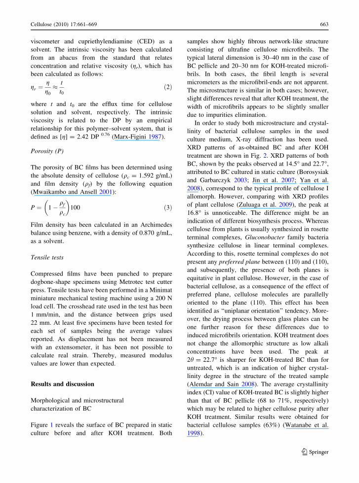
Figure 1 reveals the surface of BC prepared in static
culture before and after KOH treatment. Both
samples show highly fibrous network-like structure
consisting of ultrafine cellulose microfibrils. The
typical lateral dimension is 30–40 nm in the case of
BC pellicle and 20–30 nm for KOH-treated microfi-
brils. In both cases, the fibril length is several
micrometers as the microfibril-ends are not apparent.
The microstructure is similar in both cases; however,
slight differences reveal that after KOH treatment, the
width of microfibrils appears to be slightly smaller
due to impurities elimination.
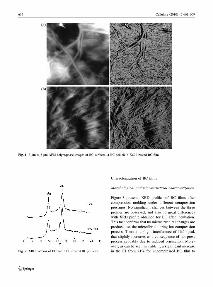
In order to study both microstructure and crystal-
linity of bacterial cellulose samples in the used
culture medium, X-ray diffraction has been used.
XRD patterns of as-obtained BC and after KOH
treatment are shown in Fig. 2. XRD patterns of both
BC, shown by the peaks observed at 14.5� and 22.7�,
attributed to BC cultured in static culture (Borosysiak
and Garbarczyk 2003; Jin et al. 2007; Yan et al.
2008), correspond to the typical profile of cellulose I
allomorph. However, comparing with XRD profiles
of plant cellulose (Zuluaga et al. 2009), the peak at
16.8� is unnoticeable. The difference might be an
indication of different biosynthesis process. Whereas
cellulose from plants is usually synthesized in rosette
terminal complexes, Gluconobacter family bacteria
synthesize cellulose in linear terminal complexes.
According to this, rosette terminal complexes do not
present any preferred plane between (110) and (110),
and subsequently, the presence of both planes is
equitative in plant cellulose. However, in the case of
bacterial cellulose, as a consequence of the effect of
preferred plane, cellulose molecules are parallelly
oriented to the plane (110). This effect has been
identified as ‘‘uniplanar orientation’’ tendency. More-
over, the drying process between glass plates can be
one further reason for these differences due to
induced microfibrils orientation. KOH treatment does
not change the allomorphic structure as low alkali
concentrations have been used. The peak at
2h = 22.7� is sharper for KOH-treated BC than for
untreated, which is an indication of higher crystal-
linity degree in the structure of the treated sample
(Alemdar and Sain 2008). The average crystallinity
index (CI) value of KOH-treated BC is slightly higher
than that of BC pellicle (68 to 71%, respectively)
which may be related to higher cellulose purity after
KOH treatment. Similar results were obtained for
bacterial cellulose samples (63%) (Watanabe et al.
1998).
Cellulose (2010) 17:661–669 663
123
Characterization of BC films
Morphological and microstructural characterization
Figure 3 presents XRD profiles of BC films after
compression molding under different compression
pressures. No significant changes between the three
profiles are observed, and also no great differences
with XRD profile obtained for BC after incubation.
This fact confirms that no microstructural changes are
produced on the microfibrils during hot compression
process. There is a slight interference of 16.58 peak
that slightly increases as a consequence of hot-press
process probably due to induced orientation. More-
over, as can be seen in Table 1, a significant increase
in the CI from 71% for uncompressed BC film to
Fig. 1 3 lm 9 3 lm AFM height/phase images of BC surfaces: a BC pellicle b KOH-treated BC film
Fig. 2 XRD patterns of BC and KOH-treated BC pellicles
664 Cellulose (2010) 17:661–669
123
more than 84.3% for films is produced in the molding
process. Similar increases in crystallinity were
detected by other authors (Kumar and Kothari
1999) for low compression pressure-molded micro-
crystalline cellulose samples. This variation was
attributed to the ability of cellulose crystallites to
reorient themselves or to an increase in size of
existing crystallite domains under compressional
force. In addition, as shown in following paragraphs,
as a consequence of the combination of the temper-
ature–pressure effects, the decrease in the porosity in
the film induces overall crystallinity.
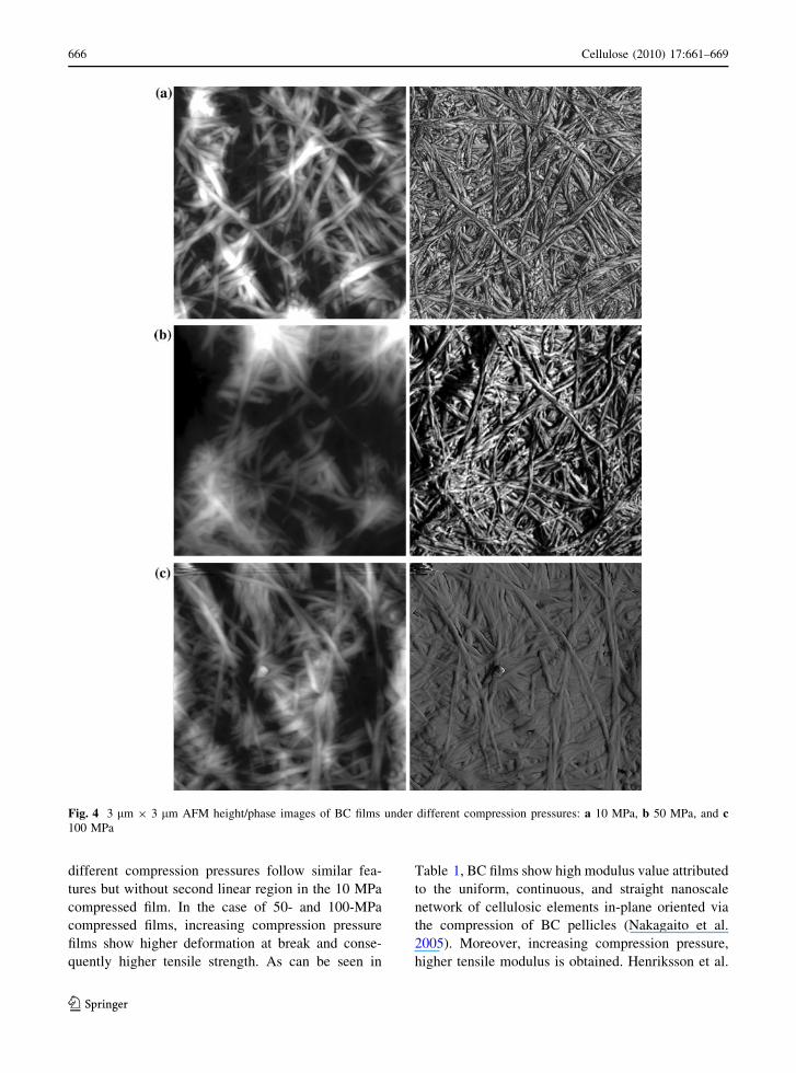
In Fig. 4, surface images of BC films obtained
under different compression pressures are presented.
There is no preferential orientation of microfibrils in
the film plane. In all cases, similar microfibrillar
network-like structures are seen, but compared to the
microstructure of Fig. 1, microfibrils seem to be more
physically entangled with respect to each other. The
same effect was visualized for cellulose nanopaper
films obtained by water casting, as this method is
known to be a very effective method to create strong
secondary interfibrillar interaction including hydrogen
bonds (Henriksson et al. 2008). Moreover, AFM
images reveal that morphology of the samples is
slightly dependent on compression pressure, as can be
seen in both BC microfibrils diameter change and
reduced porosity. Increasing compression pressure,
microfibril diameter is slightly increased, 5–10 nm
difference, which might be due to local deformations
on the BC film when compressed (Johansson and
Alderborn 2001). Thus, related to those local defor-
mations, it seems that porosity of the samples is
slightly lower for higher compression pressure. Those
results are in good agreement with porosity measure-
ments where a decrease from 13.6 to 9.7% and 3.2% in
porosity is observed for BC films obtained under 10,
50, and 100 MPa compression pressures, respectively
(Table 1). Porosity values reported are slightly lower
than those reported for other BC of plant cellulose
films or sheets (Henriksson et al. 2008). An explana-
tion for this fact may be the magnitude of the applied
pressure, which in our work is remarkably higher.
Mechanical behavior
In Fig. 5, the stress–strain curves for BC films molded
under different compression pressures are shown.
Obtained data are presented in Table 1. Mechanical
properties of cellulose films depend not only on fibril
modulus but also on orientation and degree of
interaction between microfibrils within the film
obtained by water suspension casting (Henriksson
et al. 2008). In this case, the methodology used for film
preparation has been the same up to the final stage
where different compression pressures have been
applied. The curves follow an initial linear zone up
to deviation from elasticity takes place, which occurs
around 0.5–0.7% apparent strain values in all cases,
followed by a second linear region up to fracture.
Hsieh et al. (2008) prepared BC films for tensile testing
and related the non-linear curve to the breakdown
within the network of fibers during deformation (e.g.,
bond breaking, fiber pull out). BC films molded with
Fig. 3 XRD patterns of BC obtained upon different mold
compression pressures: a 10 MPa b 50 MPa, and c 100 MPa
Table 1 BC film properties under different compression molding pressures
Sample Porosity (%) CIXRD (%) Tensile modulus (GPa) Tensile strength (MPa) Elongation at break (%)
BC 10 MPa 13.6 86.5 9.8 ± 0.8 87.5 ± 11.0 1.1 ± 0.1
BC 50 MPa 9.7 87.2 10.5 ± 1.0 165.0 ± 8.0 2.5 ± 0.1
BC 100 MPa 3.2 84.3 10.6 ± 0.5 182.5 ± 12.5 3.8 ± 0.2
Cellulose (2010) 17:661–669 665
123
different compression pressures follow similar fea-
tures but without second linear region in the 10 MPa
compressed film. In the case of 50- and 100-MPa
compressed films, increasing compression pressure
films show higher deformation at break and conse-
quently higher tensile strength. As can be seen in
Table 1, BC films show high modulus value attributed
to the uniform, continuous, and straight nanoscale
network of cellulosic elements in-plane oriented via
the compression of BC pellicles (Nakagaito et al.
2005). Moreover, increasing compression pressure,
higher tensile modulus is obtained. Henriksson et al.
Fig. 4 3 lm 9 3 lm AFM height/phase images of BC films under different compression pressures: a 10 MPa, b 50 MPa, and c100 MPa
666 Cellulose (2010) 17:661–669
123

(2008) obtained cellulose nanopaper films with dif-
ferent porosities after water suspension casting using
different DP and found that elastic modulus did not
follow any tendency as similar modulus values were
obtained. This conclusion is also supported by other
studies where elastic modulus of BC is attributed to the
planar and straight orientation of the continuous and
dimensionally uniform three-dimensional cellulose
structure (Eichhorn and Young 2001; Nakagaito and
Yano 2004). Even microfibril diameter is not consid-
ered an important factor on the elastic modulus
contribution, Gushados et al. (2005) measured the
elastic modulus of single bacterial cellulose fibers.
Using AFM with diameter ranging from 27 to 88 nm,
obtaining a constant value of 78 GPa. Other
parameters as crystallinity may play a more important
contribution in the modulus of semicrystalline or
composite materials such as cellulose where the elastic
modulus is a combination of crystalline (128 GPa) and
amorphous (5 GPa) contribution (Iwamoto et al. 2007;
Gushados et al. 2005). The first one contributes to the
elasticity of the material and the second one to the
flexibility and plasticity of bulk materials. Based on
Reuss model, Eichhorn and Young (2001) developed a
relationship between elastic modulus and percent
crystallinity and predicted a modulus of 12 GPa for a
60%-crystallinity cellulose mat.
On the contrary, as mentioned above, Young
modulus of BC films is dependent upon the applied
compression pressure. As a first approach, a mixture
law has been applied to estimate the elastic modulus
evolution with the porosity of the films. Film
modulus can be calculated as:
Efilm ¼ U1E1 þ U2E2 ð4Þ
where U1 is the cellulose volume fraction, E1 BC
elastic modulus, and U2 air volume fraction, and E2
air elastic modulus. Taking into account that air does
not contribute to the modulus, mixtures law fits rather
well with experimental values, as can be observed in
Fig. 6, in which modulus is plotted as a function of
cellulose content in the films.
However, for bacterial cellulose films, network
theories can be applied, in order to attempt to describe
the elastic properties of films in terms of individual
microfiber properties and the arrangement of nanofiber
and microfiber bonds. The microfiber is assumed to be
embedded in the network through a bonding zone. The
joint or bonding area comes from both the synthesizing
process during cultivation of bacterial cellulose, and it
can increase through film production and compression
molding process. Through compression, the surface of
the microfiber is covered, and microfiber network is
coupled. If assumed that all fibers lie in the plane of the
film, they can be modeled as plane stress structures.
Moreover, by also supposing random distributions of
microfibers and mean values for fiber properties,
several 2D network models can be applied in order to
relate film properties with individual microfiber prop-
erties. Cox (1952) developed the original theory for a
random fiber network consisting of long straight fibers
attached at the ends, assuming that the strain in the
fibers was equal to the strain in the sheet, a uniform
strain theory. Cox proposed:
Fig. 5 Stress–strain curves for BC films obtained under
different mold compression pressures
0,7 0,8 0,9 1,06
8
10
12
14
φ1)
Fig. 6 Elastic modulus of BC films vs. cellulose volume
fraction on the films: (bullet) experimental data and (dash)
mixture model
Cellulose (2010) 17:661–669 667
123
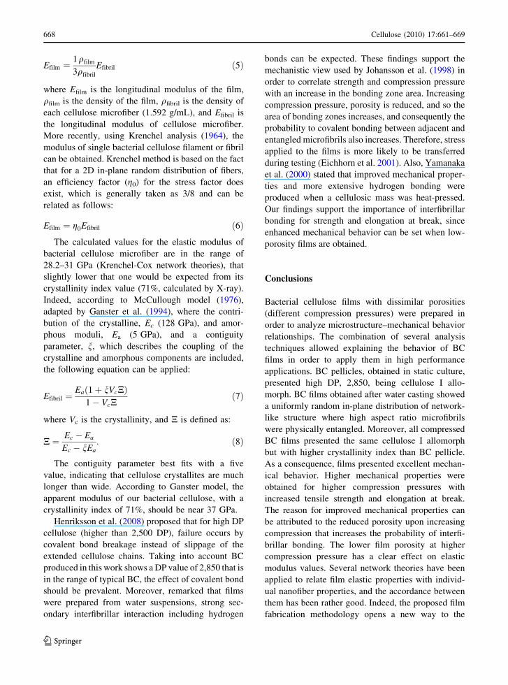
Efilm ¼1
3
qfilm
qfibril
Efibril ð5Þ
where Efilm is the longitudinal modulus of the film,
qfilm is the density of the film, qfibril is the density of
each cellulose microfiber (1.592 g/mL), and Efibril is
the longitudinal modulus of cellulose microfiber.
More recently, using Krenchel analysis (1964), the
modulus of single bacterial cellulose filament or fibril
can be obtained. Krenchel method is based on the fact
that for a 2D in-plane random distribution of fibers,
an efficiency factor (g0) for the stress factor does
exist, which is generally taken as 3/8 and can be
related as follows:
Efilm ¼ g0Efibril ð6ÞThe calculated values for the elastic modulus of
bacterial cellulose microfiber are in the range of
28.2–31 GPa (Krenchel-Cox network theories), that
slightly lower that one would be expected from its
crystallinity index value (71%, calculated by X-ray).
Indeed, according to McCullough model (1976),
adapted by Ganster et al. (1994), where the contri-
bution of the crystalline, Ec (128 GPa), and amor-
phous moduli, Ea (5 GPa), and a contiguity
parameter, n, which describes the coupling of the
crystalline and amorphous components are included,
the following equation can be applied:
Efibril ¼Eað1þ nVcNÞ
1� VcNð7Þ
where Vc is the crystallinity, and N is defined as:
N ¼ Ec � Ea
Ec � nEa: ð8Þ
The contiguity parameter best fits with a five
value, indicating that cellulose crystallites are much
longer than wide. According to Ganster model, the
apparent modulus of our bacterial cellulose, with a
crystallinity index of 71%, should be near 37 GPa.
Henriksson et al. (2008) proposed that for high DP
cellulose (higher than 2,500 DP), failure occurs by
covalent bond breakage instead of slippage of the
extended cellulose chains. Taking into account BC
produced in this work shows a DP value of 2,850 that is
in the range of typical BC, the effect of covalent bond
should be prevalent. Moreover, remarked that films
were prepared from water suspensions, strong sec-
ondary interfibrillar interaction including hydrogen
bonds can be expected. These findings support the
mechanistic view used by Johansson et al. (1998) in
order to correlate strength and compression pressure
with an increase in the bonding zone area. Increasing
compression pressure, porosity is reduced, and so the
area of bonding zones increases, and consequently the
probability to covalent bonding between adjacent and
entangled microfibrils also increases. Therefore, stress
applied to the films is more likely to be transferred
during testing (Eichhorn et al. 2001). Also, Yamanaka
et al. (2000) stated that improved mechanical proper-
ties and more extensive hydrogen bonding were
produced when a cellulosic mass was heat-pressed.
Our findings support the importance of interfibrillar
bonding for strength and elongation at break, since
enhanced mechanical behavior can be set when low-
porosity films are obtained.
Conclusions
Bacterial cellulose films with dissimilar porosities
(different compression pressures) were prepared in
order to analyze microstructure–mechanical behavior
relationships. The combination of several analysis
techniques allowed explaining the behavior of BC
films in order to apply them in high performance
applications. BC pellicles, obtained in static culture,
presented high DP, 2,850, being cellulose I allo-
morph. BC films obtained after water casting showed
a uniformly random in-plane distribution of network-
like structure where high aspect ratio microfibrils
were physically entangled. Moreover, all compressed
BC films presented the same cellulose I allomorph
but with higher crystallinity index than BC pellicle.
As a consequence, films presented excellent mechan-
ical behavior. Higher mechanical properties were
obtained for higher compression pressures with
increased tensile strength and elongation at break.
The reason for improved mechanical properties can
be attributed to the reduced porosity upon increasing
compression that increases the probability of interfi-
brillar bonding. The lower film porosity at higher
compression pressure has a clear effect on elastic
modulus values. Several network theories have been
applied to relate film elastic properties with individ-
ual nanofiber properties, and the accordance between
them has been rather good. Indeed, the proposed film
fabrication methodology opens a new way to the
668 Cellulose (2010) 17:661–669
123
design of nanostructured materials with high mechan-
ical performance using a biodegradable material from
a renewable resource as bacterial cellulose.
Acknowledgments The authors gratefully acknowledge
Eusko Jaurlaritza, Grupo Consolidado IT-365-07 and
ETORTEK-inanoGUNE for financial assistance to carry out
this investigation.
References
Alemdar A, Sain M (2008) Biocomposites from wheat straw
nanofibres: morphology, thermal and mechanical proper-
ties. Comp Sci Technol 68:557–565
Borosysiak S, Garbarczyk J (2003) Applying the WAXS
method to estimate the supermolecular structure of cel-
lulose fibres after mercerisation. Fibres & Text 11:104–
106
Cox HL (1952) The elasticity and strength of paper and other
fibrous materials. Brit J Appl Phys 3:72–79
Eichhorn SJ, Young RJ (2001) The Young’s modulus of a
microcrystalline cellulose. Cellulose 8:197–207
Eichhorn SJ, Sirichaisit J, Young RJ (2001) Deformation
mechanisms in cellulose fibres, paper and wood. J Mater
Sci 36:3129–3135
Ganster J, Fink H-P, Fraatz J, Nywlt M (1994) Relation
between structure and elastic constants of man-made
cellulosic fibres: I. A two phase anisotropioc model with
contiguity parameter. Acta Polymer 45:312–318
Gindl W, Keckes J (2004) Tensile properties of cellulose
acetate butyrate composites reinforced with bacterial
cellulose. Comp Sci Technol 64:2407–2413
Gushados G, Wan W, Hutter JL (2005) Measurement of the
elastic modulus of single bacterial cellulose fibers using
atomic force microscopy. Langmuir 21:6642–6646
Henriksson M, Berglund LA, Isaksson P, Lindstrom T, Nishino
T (2008) Cellulose nanopaper structures of high tough-
ness. Biomacromolecules 9:1579–1585
Hsieh YC, Yano H, Nogi M, Eichhorn S (2008) An estimation
of the Young’s modulus of bacterial cellulose filaments.
Cellulose 15:507–513
Iwamoto, Nakagaito AN, Yano H (2007) Nano-fibrillation of
pulp fibers for the processing of transparent nanocom-
posites. Appli Phys A 89:461–466
Jin H, Zha C, Gu L (2007) Direct dissolution of cellulose in
NaOH/thiourea/urea aqueous solution. Carbohydr Res
342:851–858
Johansson B, Alderborn G (2001) The effect of shape and
porosity on the compression behaviour and tablet forming
ability of granular materials formed from microcrystalline
cellulose. Eur J Pharm Biopharm 52:347–357
Johansson B, Nicklasson F, Alderborn G (1998) Effect of pellet
size on degree of deformation and densification during
compresion and on compatibility of microcrystalline cel-
lulose pellets. Int J Pharm 52:347–357
Klemm D, Heublein B, Fink HP, Bohn A (2005) Cellulose:
fascinating biopolymer and sustainable raw material.
Angew Chem Int Ed 44:3358–3393
Krenchel H (1964) Fibre reinforcement. Akademisk Forlag,
Copenhagen
Kumar V, Kothari SH (1999) Effect of compressional force on
the crystallinity of directly compressible cellulose excip-
ients. Int J Pharm 177:173–182
Marx-Figini M (1987) Evaluation of the accessibility of cel-
luloses by intrinsic viscosity ratio. Polym Bull 17:
225–229
McCullough RL, Wu CT, Seferis JC, Lindenmeyer PH (1976)
Predictions of limiting mechanical performance for
anisotropic crystalline polymers. Polym Eng Sci 16:
371–387
Mwaikambo LY, Ansell MP (2001) The determination of
porosity and cellulose content of plant fibres by density
method. J Mater Sci Lett 20:2095–2096
Nakagaito AN, Yano H (2004) The effect of morphological
changes from pulp fiber towards nano-scale fibrillated
cellulose on the mechanical properties of high-strength
plant fiber based composites. Appl Phys A: Mater Sci
Process 78:547–552
Nakagaito AN, Iwamoto S, Yano H (2005) Bacterial cellulose:
the ultimate nano-scalar cellulose morphology for the
production of high-strength composites. Appl Phys A:
Mater Sci Process 80:93–97
Nishi Y, Uryu M, Yamanaka S, Watanabe K, Kitamura N,
Iguchi M, Mitsuhashi S (1990) The structure and
mechanical properties of sheets prepared from bacterial
cellulose. J Mater Sci 25:2997–3001
Putra A, Kakugo A, Furukawa H, Gong JP, Osada Y (2008)
Tubular bacterial cellulose gel with oriented fibrils on the
curved surface. Polymer 49:1885–1891
Segal L, Creely J, Martin A, Conrad C (1959) An empirical
method for estimating the degree of crystallinity of native
cellulose using the X-ray diffractometer. Text Res J
29:786–794
Watanabe K, Tabuchi M, Morinaga Y, Yoshinaga F (1998)
Structural features and properties of bacterial cellulose
produced in agitated culture. Cellulose 5:187–200
Yamanaka S, Ishihara M, Sugiyama J (2000) Structural mod-
ification of bacterial cellulose. Cellulose 7:213–225
Yan Z, Chen S, Wang H, Wang B, Wang C, Jiang J (2008)
Cellulose synthesized by Acetobacter xylinum in the
presence of multi-walled carbon nanotubes. Carbohydr
Res 343:73–80
Zuluaga R, Putaux JL, Cruz J, Velez J, Mondragon I, Ganan P
(2009) Cellulose microfibrils from banana rachis: effect of
alkaline treatments on structural and morphological fea-
tures. Carbohydr Polym 76:51–59
Cellulose (2010) 17:661–669 669
123