Avian Influenza Viruses Infect Primary Human Bronchial Epithelial Cells Unconstrained by Sialic Acid...
12
Avian Influenza Viruses Infect Primary Human Bronchial Epithelial Cells Unconstrained by Sialic Acid a2,3 Residues Christine M. Oshansky 1 , Jennifer A. Pickens 1 , Konrad C. Bradley 2 , Les P. Jones 1 , Geraldine M. Saavedra- Ebner 1 , James P. Barber 1 , Jackelyn M. Crabtree 1 , David A. Steinhauer 2 , S. Mark Tompkins 1 , Ralph A. Tripp 1 * 1 Department of Infectious Diseases, College of Veterinary Medicine, University of Georgia, Athens, Georgia, United States of America, 2 Department of Microbiology and Immunology, Emory University School of Medicine, Atlanta, Georgia, United States of America Abstract Avian influenza viruses (AIV) are an important emerging threat to public health. It is thought that sialic acid (sia) receptors are barriers in cross-species transmission where the binding preferences of AIV and human influenza viruses are sias a2,3 versus a2,6, respectively. In this study, we show that a normal fully differentiated, primary human bronchial epithelial cell model is readily infected by low pathogenic H5N1, H5N2 and H5N3 AIV, which primarily bind to sia a2,3 moieties, and replicate in these cells independent of specific sias on the cell surface. NHBE cells treated with neuraminidase prior to infection are infected by AIV despite removal of sia a2,3 moieties. Following AIV infection, higher levels of IP-10 and RANTES are secreted compared to human influenza virus infection, indicating differential chemokine expression patterns, a feature that may contribute to differences in disease pathogenesis between avian and human influenza virus infections in humans. Citation: Oshansky CM, Pickens JA, Bradley KC, Jones LP, Saavedra-Ebner GM, et al. (2011) Avian Influenza Viruses Infect Primary Human Bronchial Epithelial Cells Unconstrained by Sialic Acid a2,3 Residues. PLoS ONE 6(6): e21183. doi:10.1371/journal.pone.0021183 Editor: Leo L. M. Poon, University of Hong Kong, Hong Kong Received November 1, 2010; Accepted May 23, 2011; Published June 23, 2011 Copyright: ß 2011 Oshansky et al. This is an open-access article distributed under the terms of the Creative Commons Attribution License, which permits unrestricted use, distribution, and reproduction in any medium, provided the original author and source are credited. Funding: This work was funded by the Center of Excellence for Influenza Research and Surveillance (CEIRS) contract HHSN266200700006C and the Georgia Research Alliance (http://www.gra.org/). The funders had no role in study design, data collection and analysis, decision to publish, or preparation of the manuscript. Competing Interests: The authors have declared that no competing interests exist. * E-mail: [email protected] Introduction Influenza A viruses are important pathogens that present a significant threat to public health, causing an extensive economic burden particularly for avian influenza virus (AIV) infection of poultry. Influenza viruses are segmented, enveloped, negative- strand RNA viruses belonging to the Orthomyxoviridae family. They comprise a diverse array of subtypes due to their propensity to change antigenic profiles and are subtyped based on the antigenic properties of two surface glycoproteins, i.e. hemagglutinin (HA) and neuraminidase (NA). Seasonal epidemics cause more than 200,000 hospitalizations and more than 41,000 deaths each year in the United States alone [1]. Four novel influenza viruses caused pandemics in 1918, 1957, 1968, and most recently in 2009. The 1918 influenza pandemic was the most severe resulting in unusually high mortality among healthy young adults [2]. It remains unclear the precise features that contributed to the high rate of mortality due to infection with the 1918 influenza virus, but it has been shown that a single mutation in the PB1-F2 genome of 1918 influenza A viruses (also recognized for highly pathogenic H5N1 avian influenza) contributed to increased virulence [3,4,5]. Moreover, since 2003, there has been an increased incidence of highly pathogenic avian influenza (HPAI) virus outbreaks in poultry, and HPAI H5N1 has crossed species barriers to infect .500 humans resulting in nearly a 60% fatality rate (.300 deaths) as of April 2011 [6]. Influenza HA binds to host cell sialic acid residues (sias) coating the host cell surface [7] and mediates viral entry via its receptor binding domain. Human influenza viruses preferentially bind sia a2,6 linkages, while AIV preferentially bind sia a2,3 linkages that are highly expressed in the gastrointestinal tracts of aquatic birds [8,9,10,11,12,13,14,15], thus it is thought that sialic acid residues are important barriers in cross-species transmission. Sias are nine- carbon monosaccharides found at the ends of glycan chains. Sias coat many host cell surfaces and secreted proteins [16,17,18,19]. The most common sias found in mammals are N-acetylneur- aminic acid (Neu5Ac) and N-glycolylneuraminic acid (Neu5Gc). Sias are transferred to terminal sugars of glycoproteins and glycolipids by sialyltransferases, and can be added to the galactose carbon-6 forming an a2,6 linkage or to galactose carbon-3 forming an a2,3 linkage [14,16,19]. The detection of a2,3 or a2,6 linkages can be determined by use of plant lectins that specifically bind to glycolipids and glycoproteins containing sia a2,6 or a2,3 configurations. A lectin from the seed of Maackia amurensis tree (MAA) is specific for sia a2,3, while a lectin obtained from the elderberry plant Sambucus nigra (SNA) is specific for sia a2,6 [20,21]. Early experiments showed that SNA preferentially bound to the surface of ciliated tracheal epithelial cells indicating the presence of sia a2,6, and MAA bound goblet cells indicating the presence of sia a2,3 [22]. These studies suggested that ciliated cells, but not goblet cells, were a primary target for human H3 influenza infection and were subsequently confirmed by using a PLoS ONE | www.plosone.org 1 June 2011 | Volume 6 | Issue 6 | e21183
Transcript of Avian Influenza Viruses Infect Primary Human Bronchial Epithelial Cells Unconstrained by Sialic Acid...

Avian Influenza Viruses Infect Primary Human BronchialEpithelial Cells Unconstrained by Sialic Acid a2,3ResiduesChristine M. Oshansky1, Jennifer A. Pickens1, Konrad C. Bradley2, Les P. Jones1, Geraldine M. Saavedra-
Ebner1, James P. Barber1, Jackelyn M. Crabtree1, David A. Steinhauer2, S. Mark Tompkins1, Ralph A.
Tripp1*
1 Department of Infectious Diseases, College of Veterinary Medicine, University of Georgia, Athens, Georgia, United States of America, 2 Department of Microbiology and
Immunology, Emory University School of Medicine, Atlanta, Georgia, United States of America
Abstract
Avian influenza viruses (AIV) are an important emerging threat to public health. It is thought that sialic acid (sia) receptorsare barriers in cross-species transmission where the binding preferences of AIV and human influenza viruses are sias a2,3versus a2,6, respectively. In this study, we show that a normal fully differentiated, primary human bronchial epithelial cellmodel is readily infected by low pathogenic H5N1, H5N2 and H5N3 AIV, which primarily bind to sia a2,3 moieties, andreplicate in these cells independent of specific sias on the cell surface. NHBE cells treated with neuraminidase prior toinfection are infected by AIV despite removal of sia a2,3 moieties. Following AIV infection, higher levels of IP-10 and RANTESare secreted compared to human influenza virus infection, indicating differential chemokine expression patterns, a featurethat may contribute to differences in disease pathogenesis between avian and human influenza virus infections in humans.
Citation: Oshansky CM, Pickens JA, Bradley KC, Jones LP, Saavedra-Ebner GM, et al. (2011) Avian Influenza Viruses Infect Primary Human Bronchial Epithelial CellsUnconstrained by Sialic Acid a2,3 Residues. PLoS ONE 6(6): e21183. doi:10.1371/journal.pone.0021183
Editor: Leo L. M. Poon, University of Hong Kong, Hong Kong
Received November 1, 2010; Accepted May 23, 2011; Published June 23, 2011
Copyright: � 2011 Oshansky et al. This is an open-access article distributed under the terms of the Creative Commons Attribution License, which permitsunrestricted use, distribution, and reproduction in any medium, provided the original author and source are credited.
Funding: This work was funded by the Center of Excellence for Influenza Research and Surveillance (CEIRS) contract HHSN266200700006C and the GeorgiaResearch Alliance (http://www.gra.org/). The funders had no role in study design, data collection and analysis, decision to publish, or preparation of themanuscript.
Competing Interests: The authors have declared that no competing interests exist.
* E-mail: [email protected]
Introduction
Influenza A viruses are important pathogens that present a
significant threat to public health, causing an extensive economic
burden particularly for avian influenza virus (AIV) infection of
poultry. Influenza viruses are segmented, enveloped, negative-
strand RNA viruses belonging to the Orthomyxoviridae family. They
comprise a diverse array of subtypes due to their propensity to
change antigenic profiles and are subtyped based on the antigenic
properties of two surface glycoproteins, i.e. hemagglutinin (HA)
and neuraminidase (NA). Seasonal epidemics cause more than
200,000 hospitalizations and more than 41,000 deaths each year
in the United States alone [1]. Four novel influenza viruses caused
pandemics in 1918, 1957, 1968, and most recently in 2009. The
1918 influenza pandemic was the most severe resulting in
unusually high mortality among healthy young adults [2]. It
remains unclear the precise features that contributed to the high
rate of mortality due to infection with the 1918 influenza virus, but
it has been shown that a single mutation in the PB1-F2 genome of
1918 influenza A viruses (also recognized for highly pathogenic
H5N1 avian influenza) contributed to increased virulence [3,4,5].
Moreover, since 2003, there has been an increased incidence of
highly pathogenic avian influenza (HPAI) virus outbreaks in
poultry, and HPAI H5N1 has crossed species barriers to infect
.500 humans resulting in nearly a 60% fatality rate (.300 deaths)
as of April 2011 [6].
Influenza HA binds to host cell sialic acid residues (sias) coating
the host cell surface [7] and mediates viral entry via its receptor
binding domain. Human influenza viruses preferentially bind sia
a2,6 linkages, while AIV preferentially bind sia a2,3 linkages that
are highly expressed in the gastrointestinal tracts of aquatic birds
[8,9,10,11,12,13,14,15], thus it is thought that sialic acid residues
are important barriers in cross-species transmission. Sias are nine-
carbon monosaccharides found at the ends of glycan chains. Sias
coat many host cell surfaces and secreted proteins [16,17,18,19].
The most common sias found in mammals are N-acetylneur-
aminic acid (Neu5Ac) and N-glycolylneuraminic acid (Neu5Gc).
Sias are transferred to terminal sugars of glycoproteins and
glycolipids by sialyltransferases, and can be added to the galactose
carbon-6 forming an a2,6 linkage or to galactose carbon-3
forming an a2,3 linkage [14,16,19]. The detection of a2,3 or a2,6
linkages can be determined by use of plant lectins that specifically
bind to glycolipids and glycoproteins containing sia a2,6 or a2,3
configurations. A lectin from the seed of Maackia amurensis tree
(MAA) is specific for sia a2,3, while a lectin obtained from the
elderberry plant Sambucus nigra (SNA) is specific for sia a2,6
[20,21]. Early experiments showed that SNA preferentially bound
to the surface of ciliated tracheal epithelial cells indicating the
presence of sia a2,6, and MAA bound goblet cells indicating the
presence of sia a2,3 [22]. These studies suggested that ciliated
cells, but not goblet cells, were a primary target for human H3
influenza infection and were subsequently confirmed by using a
PLoS ONE | www.plosone.org 1 June 2011 | Volume 6 | Issue 6 | e21183

fluorescently-labeled H3 virus which primarily attached to ciliated
cells [23]. However, later studies using differentiated human
tracheal bronchial epithelial cells found that human influenza
viruses infect non-ciliated cells expressing sia a2,6, and AIV infect
ciliated cells expressing sia a2,3 [24]. More recent evidence
suggests that H5N1 influenza can replicate within ex vivo human
respiratory epithelial tissues, despite the lack of sia a2,3 staining
[25]. Regardless of the predilection of AIV for sia a2,3, a H5N1
AIV (A/Hong Kong/156/1997) outbreak occurred in humans in
Hong Kong in 1997 where all eight viral genes were of avian
origin. The currently circulating H5N1 AIV strains primarily
infect birds and fowl maintaining a sia a2,3 binding preference;
however, AIV can acquire mutations changing their HA binding
specificity from avian-like, a2,3, to human-like, a2,6 [8,10,26].
In these studies, we determined if low pathogenic H5N1, H5N2
and H5N3 AIV isolates of chicken or wild bird origin could infect
and replicate in fully differentiated, normal human bronchial
epithelial (NHBE) cells. We show that these viruses infect,
replicate, and are released from NHBE cells independent of
detectable sia a2,3 or a2,6 moieties present on the cell surface,
and show that LPAI H5N1, H5N2 and H5N3 viruses induce
higher IP-10 and RANTES responses early during infection
compared to human H3N2 infection indicating differential
chemokine expression patterns that may contribute to the unique
aspects of disease pathogenesis between avian and human
influenza virus infection.
Materials and Methods
Cells and virusesNormal human bronchial epithelial (NHBE) cells (Lonza,
Walkersville, MD) from a single 17 year old healthy male donor
were expanded, cryopreserved, and cultured in an air-liquid
interface system as previously described [27]. The cells from the
same donor were used in all assays for assay consistency. The
apical surface of the cells was exposed to a humidified 95% air/5%
CO2 environment, and the basolateral medium was changed every
two days.
The low pathogenic AIV (LPAI) strains A/Mute Swan/MI/06/
451072-2/2006 (H5N1), A/chicken/Pennsylvania/13609/1993
(H5N2), and A/chicken/TX/167280-4/02 (H5N3) were kindly
provided by Dr. David Suarez, USDA-Southeast Poultry Re-
search Laboratory, Athens, GA. These viruses were previously
passaged once in embryonated chicken eggs. A/New York/55/
2004 (H3N2) was kindly provided by Dr. Richard Webby, St. Jude
Children’s Research Hospital, Memphis, TN. A single stock of
these viruses was prepared for use in all assays by inoculating 9-
day old specific pathogen-free (SPF) eggs and harvesting the
allantoic fluid 48 h post-inoculation. Viral titers were obtained by
serial dilution on Madin-Darby canine kidney (MDCK) cells in the
presence of 1 mg/ml trypsin (Sigma), and 50% egg infectious doses
(EID50) were performed in 9-day old SPF chicken embryos and
calculated according to the method of Reed and Muench [28].
Sequencing of influenza hemagglutinin andneuraminidase genes
To determine if mutations in the HA or neuraminidase gene
occurred after single egg passage, these genes were sequenced.
Briefly, the RNeasy Kit (Qiagen, Valencia, CA) to extract RNA,
and the One-step RT-PCR Kit (Qiagen) was employed to amplify
the HA and NA gene segments for direct sequencing of PCR
products using gene segment-specific amplification primers (Table
S1). Full-length amplicons were subjected to purification by
agarose gel electrophoresis for cycle sequencing. Cycle sequencing
reactions were carried out using an ABI 9700 thermocycler and
optimized to produce the maximal length of read while
economizing the use of BigDye reagent (Applied Biosystems
Inc., Foster City, CA). The resulting 10 ml cycle sequencing
reaction was comprised of: 2 ml template, 1 ml ABI BigDye v3.1,
1 ml (1 pmole) sequencing primer, 2 ml ABI 56 sequencing buffer,
4 ml distilled water. Each amplicon was subjected to cycle
sequencing reactions using both the forward and reverse
amplifying primers. Internal primers were employed to fill in gaps
and generate sequence at the 59 and 39 termini of each amplicon
(Table S2). This scheme resulted in at least two reads for each
nucleotide of the sequence. Cycle sequencing reactions were
purified using Cleanseq reagent (Agencourt, Beverly, MA) and
eluted in 40 ml of 0.1 mM EDTA. Purified cycle sequencing
products were loaded onto an ABI 3130XL genetic analyzer and
separated by capillary electrophoresis through an 80 cm capillary
array. The resulting sequence traces were trimmed and assembled
using Sequencher software (Genecodes, Ann Arbor, MI). No
mutations in either gene were identified.
Viral infection of NHBE cellsHuman and LPAI viruses were diluted in BEBM (Lonza) to
equal titers as determined by MDCK plaque assay. NHBE cells
were washed three times with PBS to remove excess mucus
secretion on the apical surface prior to infection. Viruses were
allowed to adsorb for 1 h at 37uC, the virus dilutions were
removed by aspiration and washed again with PBS 3 times. NHBE
cells were incubated for the indicated times pi at 37uC. Viruses
released apically were harvested by the apical addition and
collection of 300 ml of 0.05% BSA-BEBM allowed to equilibrate at
37uC for 30 min. Samples were stored at 280uC until assayed.
Neuraminidase Treatment and Influenza Infection ofNHBE Cells
To remove sia moieties from the cell surface, and to confirm the
specificity of lectin binding, NHBE cells were apically treated with
the indicated concentration of neuraminidase from Clostridium
perfringens (Sigma, St. Louis, MO) in PBS for 1 hour at 37uC as
previously described [29]. Following sialidase incubation, cells
were washed three times with PBS. NHBE cells were apically
mock infected or infected with A/Mute Swan/MI/06/451072-2/
2006 (H5N1), A/chicken/Pennsylvania/13609/1993 (H5N2), A/
chicken/TX/167280-4/02 (H5N3), or NY/04/55/2004 (H3N2)
at the indicated multiplicities of infection (MOI). Cells were fixed
in 3.7% formaldehyde for 30 min or harvested in triplicate at the
times indicated post-infection.
Quantitative RT-PCRTotal RNA was isolated using RNeasy Mini kit (Qiagen,
Valencia, CA) and stored at 280uC until used. Reverse
transcription was performed using random hexamers and MuLV
reverse transcriptase (Applied Biosystems, Foster City, CA).
Influenza M gene expression were measured using a TaqMan
real-time quantitative reverse transcriptase PCR (qRT-PCR) assay
using previously described primers and probe [30]. Transcript
levels were determined following a 10-minute hot start at 95uC in
a three-step protocol with 15 s of denaturation (95uC), 30 s of
annealing (60uC) and extension at 72uC for 15 s and analyzed
using MXPro software by Stratagene (La Jolla, CA). Copy
numbers were determined by generation of a standard curve
using plasmid DNA encoding influenza M gene. Plasmid
DNA concentrations were measured by optical density using a
spectrophotometer.
AIV Infection of Human Bronchial Epithelial Cells
PLoS ONE | www.plosone.org 2 June 2011 | Volume 6 | Issue 6 | e21183

Flow cytometry analysis of sialic acid residuesNHBE cells staining for sias a2,3 or a2,6 was determined by
flow cytometry. Briefly, NHBE cells were washed with PBS and
trypsinized for 10 min at 37uC. To determine lectin staining, cells
were collected and centrifuged at 2206 g for 5 min and
resuspended in 2% formaldehyde for 30 min on ice and washed
with flow buffer (1% BSA, 0.1% NaN3 in PBS). To determine the
level of sialic acid residues detectable following neuraminidase
treatment, trypsinized cells were treated with increasing concen-
trations of neuraminidase from Clostridium perfringens (Sigma, St.
Louis, MO) in PBS for 1 hour at 37uC and then fixed in 2%
formaldehyde for 30 min on ice and washed with flow buffer.
Surface sias expression was determined by primary staining with
20 mg/mL biotinylated Maackia amurensis lectin-II (MAA-II) (B-
1265, Vector Laboratories, Burlingame, CA) for sias a2,3, or
20 mg/mL biotinylated Sambucus nigra lectin (SNA) (B-1305, Vector
Laboratories) for sias a2,6 for 1 hour on ice. Secondary staining
was performed with APC-conjugated streptavidin (BD, Mountain
View, CA) diluted in flow buffer for 1 hour on ice. Cells were
washed with flow buffer and analyzed on a LSRII flow cytometer
using FACSDiva software (BD). Additional analysis was also
performed using FlowJo software (TreeStar, Ashland, OR).
Confocal MicroscopyNHBE cells were fixed for 30 minutes in 3.7% formaldehyde at
the times indicated post-infection. Sialic acid staining was
performed as previously described [31]. Briefly, to stain for sias,
cells were incubated with 20 mg/mL biotinylated MAA-II (Vector
Laboratories) to detect a2,3, or 20 mg/mL biotinylated SNA
(Vector Laboratories) to detect sias a2,6 for 1 hour at room
temperature, washed with PBS, and incubated with 15 mg/mL
Texas Red streptavidin (Vector Laboratories). MAA-II was
specifically chosen because it preferentially binds to sias a2-
3Galb1-3(Siaa2-6)GalNAc and not to non-sialic acid residues as
do other isoforms of MAA [32]. Following washing, cells were
permeabilized in PBS containing 0.5% TX-100, washed in PBS-
0.05%TWEEN (PBS-T) and incubated with mouse anti-NP
IgG2a diluted in 3% bovine serum albumin (BSA) in PBS-T.
The cells were then washed with PBS-T, incubated for one hour
with anti-mouse IgG AlexaFluor488 (Molecular Probes, Carlsbad,
California) and anti-b-tubulin directly conjugated to FITC (cilia
stain). Cells were rapid stained with DAPI (1 mg/mL). After
washing with PBS-T, membranes were excised from their culture
inserts and mounted on glass slides.
Glycan array analysis of influenza virus strainsGlycan arrays were used to examine the receptor specificity of
the A/Mute Swan/MI/06/451072-2/2006 (H5N1), A/chicken/
Pennsylvania/13609/1993 (H5N2), A/chicken/TX/167280-4/02
(H5N3), and A/NY/04/55/2004 (H3N2) strains. Briefly, the
strains were purified from allantoic fluid on a 25/60 percent
sucrose gradient by ultracentrifugation and resuspended in 1 mM
EDTA/PBS. All purified viral stocks were stored at 280uC. Viral
titers were determined by standard plaque assay of Madin-Darby
kidney cells. The minimum viral titer for glycan analysis was
16105 pfu/ml, where all viral strains were labeled with 25 mg of
AlexaFluor 488 dye (Invitrogen, Carlsbad, CA) in 1 M NaHCO3
(pH 9) for 1 hour at 4uC. To remove residual dye, each sample
was dialyzed in a 7000 MWCO Slide-A-Lyzer MINI dialysis
cassette (Thermo Scientific, Rockford, IL) against 1 mM EDTA/
PBS overnight at4uC. The labeled viruses were analyzed via
mammalian printed array, version 4.2 (contains 511 glycans) or
5.0 (contains 611 glycans), by the Core H of the Consortium of
Functional Glycomics (www.functionalglycomics.org). The PA/93
and MI/06 were evaluated against 511 glycans, while the TX/02
and A/NY were evaluated against 611 glycans. Background
fluorescence was determined by averaging the relative fluorescent
units (RFU) of all glycans on the array and multiplied by 2. Glycan
binding peaks that were above background with a %CV greater
than 50% were not considered significant.
Bead-based detection of cytokines and chemokinesThe LuminexH xMAPTM system, a high-throughput micro-
sphere-based suspension array was used with a MILLIPLEX MAP
human cytokine/chemokine immunoassay (Millipore, St. Charles,
MO) for the rapid immunological detection of secreted cytokines
and chemokines from NHBE cell supernatants according to the
manufacturer protocol. Briefly, beads coupled with biotinylated
anti-IL-1a, anti-IL-1b, anti-IL-8, anti-MCP-1, anti-MIP-1a, anti-
MIP1b, anti-IP-10, anti-RANTES monoclonal antibodies were
sonicated, mixed, and diluted in bead diluent. For the assay, beads
were diluted 1:4 in bead diluent and incubated overnight at 4uCwith NHBE apical wash or basolateral supernatant. After washing,
beads were incubated with streptavidin-phycoerythrin for 1 hour
at room temperature, washed, and resuspended in wash buffer.
The assay was analyzed on a Luminex 200 instrument (Luminex
Corporation, Austin, TX) using Luminex xPONENT 3.1
software. Additional analysis was performed using MILLIPLEX
Analyst (Millipore).
Statistical analysis of dataDifferences in chemokine expression in LuminexH analysis were
evaluated by Student t test and considered significant when
p,0.05. Data are shown as means 6 standard deviation (SD).
Results
NHBE cells express a2,6 and a2,3 sialic acid receptorsTo determine if the propensity of AIVs to infect NHBE cells was
related to sia a2,3 tropism, the cells were stained with sia-specific
lectins. MAA-II lectin preferentially binds to a2,3 sialic acids [32],
and SNA lectin preferentially binds to a2,6 sialic acids. NHBE
cells abundantly express a2,6 sialic acids on the cell surface
(Fig. 1A), while a2,3 sias are expressed at a lower level (Fig. 1B).
Previous studies suggest that AIV infect ciliated cells which
primarily express sias a2,3, while human viruses preferentially
infect non-ciliated cells expressing sias a2,6 [24]. The specificity of
staining using MAA-II or SNA lectins was confirmed by pre-
treating the apical surface of NHBE cells with neuraminidase
(image inserts in Figure 2A and 2B) which shows that treatment
removed detectable sias from the cell surface.
To determine the relative distribution of a2,3 or a2,6 sias
moieties on the NHBE cell surface, the cells were lectin-stained
and analyzed by flow cytometry. Figure 1C shows that a2,6 sias
are abundantly expressed on most NHBE cells. However, staining
for a2,3 sias showed that while many cells express a2,3 sias there
are two levels expressed, i.e. dimly positive and brightly positive as
determined by flow cytometry. To determine the extent of cell
surface sia residues removed by neuraminidase, NHBE cells were
treated with increasing levels of neuraminidase (Fig. 1D). NHBE
cells treated with the highest neuraminidase concentration
(1000 mU/mL) removed .60% all detectable a2,6 sias (data
not shown), while similar treatment removed .95% of detectable
a2,3 sias.
To determine sias expression on ciliated cells and goblet cells,
fully differentiated NHBE cells were immunostained for MU-
C5AC to indicate goblet cells, and b-tubulin to indicate ciliated
cells, and lectin-stained for determining the corresponding surface
AIV Infection of Human Bronchial Epithelial Cells
PLoS ONE | www.plosone.org 3 June 2011 | Volume 6 | Issue 6 | e21183

AIV Infection of Human Bronchial Epithelial Cells
PLoS ONE | www.plosone.org 4 June 2011 | Volume 6 | Issue 6 | e21183
levels of a2,3 or a2,6 sias. The results show that the NHBE cells
have both ciliated and goblet cells (Fig. 2), and while many ciliated
and most goblet cells display a2,6 sia residues (Fig. 2A and 2B; co-
expression indicated in yellow), none of the ciliated or goblet cells
expressed detectable a2,3 sias (Fig. 2C and 2D).
LPAI virus replicates and are shed from NHBE cellsTo determine if LPAI viruses can infect NHBE cells, the cells
were apically infected with A/chicken/Pennsylvania/13609/1993
(H5N2; PA/93), A/chicken/TX/167280-4/02 (H5N3; TX/02),
or NY/04/55/04 (H3N2) (Fig. 3) at a multiplicity of infection
(MOI) of 0.001 (equivalent to 104.38 EID50/mL for PA/93, 103.86
EID50/mL for TX/02, or 104.99 EID50/mL for NY/04/55/04).
This low MOI was chosen to allow for better detection of virus
replication in subsequent apical cell washings at the time-points
indicated. Within 24 h pi, NHBE cells infected with PA/93 had
apical wash virus titers of 105 EID50/mL which peaked by 48 h pi
to 105.8 EID50/mL (Fig. 3). NHBE cells infected with TX/02 had
apical wash titers that increased slightly at 24 h pi to 104.3 EID50/
mL, subsequently increased to 105.8 EID50/mL at 48 h pi, and
peaked at 106.1 EID50/mL at 72 h pi (Fig. 3). As the EID50 values
were determined from apical washes, the results suggest that both
PA/93 and TX/02 replicate and are shed apically from NHBE
cells, however we cannot exclude the possibility that virus shed
Figure 1. Fully differentiated NHBE cells express both a2,6 and a2,3 sias. NHBE cells were stained for a2,6 (A) or a2,3 (B) sias shown in red.Cells pre-treated with neuraminidase abolished sias residue staining (image inserts). (C) NHBE cells were trypsinized, fixed with 2% formaldehyde, andanalyzed by flow cytometry to determine relative percentage of cells staining positive for a2,3 (blue), or a2,6 sia moieties (green). The x-axis showsthe mean fluorescence intensity and the y-axis shows the percent positive staining cells. Results shown are representative of four independentexperiments. (D) NHBE cells were trypsinized, treated with the indicated concentrations of neuraminidase, and analyzed as in (C) to determine thepercentage of cells staining positive for detectable a2,3 sialic acid residues. Results shown are representative of two independent experiments.doi:10.1371/journal.pone.0021183.g001
Figure 2. Ciliated and goblet cells express mainly a2,6 sialic acid linkages. NHBE cells were stained for a2,6 (A, B) or a2,3 (C, D) linked siasshown in red, and b-tubulin (A, C) or MUC5AC (B, D) shown in green. Cells expressing a2,3 sias are indicated with arrows. Results shown arerepresentative of two independent experiments.doi:10.1371/journal.pone.0021183.g002
AIV Infection of Human Bronchial Epithelial Cells
PLoS ONE | www.plosone.org 5 June 2011 | Volume 6 | Issue 6 | e21183

from the basolateral side of the culture did not leak upward toward
the apical side. As expected, NHBE cells infected with human
influenza NY/04/55/04 (H3N2) supported a productive infection
in the first 24 h pi (105.9 EID50/mL), but due to considerable cell
death related to virus replication, the apical wash titers were
decreased by 48 h pi (104.8 EID50/mL), and few cells remained at
72 h pi.
AIVs infect NHBE cells bearing a2,6 sialic acid expressionTo determine if a2,3 sias expression is required for AIV
infection of NHBE cells, the cells were infected (MOI = 0.5) with
MI/06, PA/93, TX/02, or human A/NY, and lectin-stained for
a2,6 sias (Fig. 4A), or a2,3 sias (Fig. 4B), and immunostained for
viral NP to detect replicating virus at the time-points indicated.
Both human and AIVs infected and replicated in NHBE cells
(Fig. 4). The results showing co-staining of a2,6 sias and viral NP
at 72 h post-MI/06 infection indicate that this H5N1 wild bird
isolate is not restricted to cells expressing a2,3 sias. In addition, the
other AIVs infected and replicated in NHBE cells independent of
significant a2,3 sias expression (Fig. 4B). For the AIV, replication
determined by NP expression, occurred by 24 h and was robust up
to 72 h pi where MI/06 (H5N1) and PA/93 (H5N2) replication
induced severe cytopathic effects, changes in cell morphology, and
loss of the confluent cell monolayer. Similarly, human A/NY
(H3N2) quickly spread throughout the NHBE cell culture and
induced substantial cytopathic effects and cell loss. Over the time-
period of replication by AIVs or human virus, there was a
progressive decline of cell surface expression of a2,6 sias, albeit to
a lesser extent for TX/02 (H5N3) infection (Fig. 4A). These effects
may be linked to influenza neuraminidase expression during
replication.
AIVs infect neuraminidase-treated NHBE cellsSince AIV infection and replication in NHBE cells did not
appear to be constrained by a HA-a2,3 sias barrier (Fig. 5), the
NHBE cells were treated with neuraminidase to remove detectable
sias and determine if infection could be inhibited. Recent findings
suggest that neuraminidase treatment can reduce influenza virus
infection, but total inhibition does not occur [33]. Similar to
previous findings, neuraminidase treatment of NHBE cells had
little effect on AIV or human influenza virus infection, as
determined by NP (Fig. 5).
AIVs predominantly bind a2,3 sialic acid glycansGlycan arrays were utilized to further characterize the sialic
acid moieties involved in receptor specificity of each virus. The
array results show that MI/06, PA/93, and TX/02 avian strains
preferentially bind a2,3 glycans with reduced binding observed for
a2,6 oligosaccharides (Fig. 6A–C). After closer examination of the
a2,6 glycans that appeared to bind MI/06, PA/93, and TX/02
avian strains, several of the glycans were classified as Neu5Aca2-6
glycans because of their terminal position but actually contain a2,3
oligosaccharide branches (e.g. Neu5Aca2-6(Neu5Aca2-3Galb1-
3)GalNAca-Sp8) that are presumably utilized in avian specific
receptor recognition. Conversely, the human A/NY/55/04
predictably binds the a2,6 sias glycans with less recognition of
a2,3 glycans (Fig. 6D). All strains exhibited below background
binding to a2,8 glycans (Fig. 6A–D).
AIVs induce differential chemokine expression patternsby NHBE cells
Previous studies have shown that pro-inflammatory cytokines
and chemokines including interferon (IFN) a/b, interleukin (IL)-
1a, IL-1b, IL-6, IL-8, tumor necrosis factor alpha (TNFa),
macrophage inflammatory protein (MIP)-1a, MIP-1b, and
monocyte chemotactic protein (MCP)-1 are detected at elevated
levels in the respiratory tracts of individuals during the acute phase
of influenza infection [34,35]. Patients infected with highly
pathogenic H5N1 have been shown to have higher levels of
systemic IFNc, IL-6, interferon-inducible protein of 10 kD (IP-10)
Figure 3. Avian influenza viruses replicate and are shed apically from NHBE cells. NHBE cells were infected with PA/93 (H5N2), TX/02(H5N3), or A/NY/55/04 at MOI = 0.001. At the times indicated post-infection, BEBM-0.05% BSA was added to the apical surface of the cells andincubated for 30 minutes at 37uC. EID50 titers were determined according the method of Reed and Meunch [28]. Data are shown as means 6 SD fromtwo independent experiments. The dashed line represents the limit of detection.doi:10.1371/journal.pone.0021183.g003
AIV Infection of Human Bronchial Epithelial Cells
PLoS ONE | www.plosone.org 6 June 2011 | Volume 6 | Issue 6 | e21183

Figure 4. AIVs infect NHBE cells independent of a2,3 sias expression. NHBE cells were infected with MI/06, PA/93, TX/02 or NY/04 atMOI = 0.5. At the times indicated post-infection, cells were fixed with 3.7% formaldehyde in PBS for 30 minutes. Cells were stained for a2,6 (A) or a2,3(B) sialic acids (red) and influenza NP (green). Results shown are representative of three independent experiments.doi:10.1371/journal.pone.0021183.g004
Figure 5. Neuraminidase (sialidase)-treated NHBE cells are robustly infected by AIVs. NHBE cells were mock-treated (top panels) ortreated with 25 mU/mL neuraminidase (bottom panels) for 1 hour at 37uC, washed with PBS, and infected with the indicated viruses at a MOI of 0.5.Cells were fixed in 3.7% formaldehyde at 24 h pi and immunostained for influenza NP expression. Results shown are representative of twoexperiments.doi:10.1371/journal.pone.0021183.g005
AIV Infection of Human Bronchial Epithelial Cells
PLoS ONE | www.plosone.org 7 June 2011 | Volume 6 | Issue 6 | e21183

and MCP-1 compared to individuals infected with human
influenza subtypes [36,37,38]. Thus, the NHBE cell model was
used to evaluate patterns of chemokine expression induced by
human and AIV infection. Both the apical and basolateral
secretion patterns of IL-1a, IL-1b, IL-8, IP-10, MIP-1a, MIP-
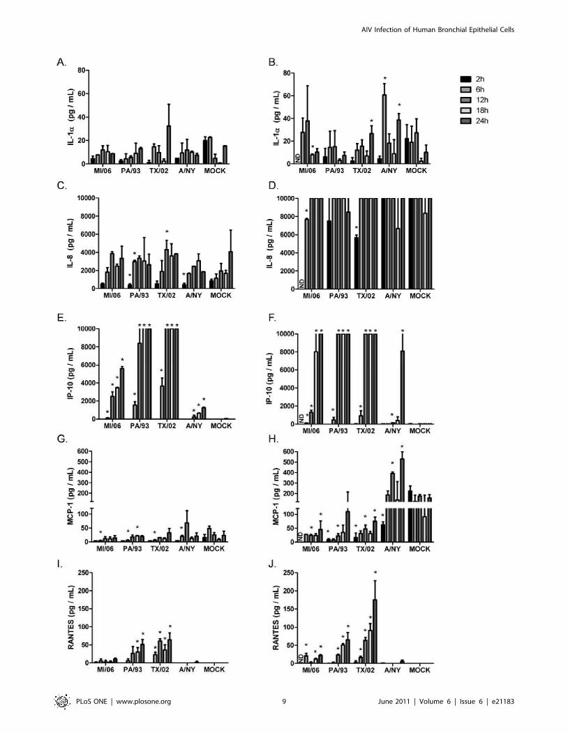
1b, MCP-1, and RANTES expression were determined (Fig. 7).
No appreciable levels of IL-1b, MIP-1a or MIP-1b were detected
from the basolateral or apical compartments of AIV or human
influenza infected NHBE cells (data not shown); however, AIVs
induced differential apical and basolateral expression patterns of
IL-1a, IL-8, IP-10, MCP-1, and RANTES over time (Fig. 7).
NHBE cells infected (MOI = 0.5) with TX/02 (H5N3) induced
higher apical IL-1a expression and significantly more basolateral
expression (p,0.05), evident at 24 h pi, compared to infection
with the other AIV viruses or mock treatment (Fig. 7A, B). In
contrast, MI/06 (H5N1) induced a higher level of basolateral IL-
1a expression compared to the other AIV between 2–6 h pi
(Fig. 7B). Likewise, human A/NY infection induced a significantly
(p,0.05) higher level of basolateral IL-1a expression at 6 h and
24 h pi compared to mock controls (Fig. 7B).
Apically-expressed IL-8 levels detected following AIV or human
influenza virus infection were similar, and expressed to slightly
higher but insignificant (p,0.05) levels compared to mock-treated
cells between 6–18 h pi (Fig. 7C). Interestingly, basolateral
expression of IL-8 was beyond the upper limits of detection for
the assay system between 6 h and 24 h pi following infection by
any virus and in mock-treated cells; however, lower levels of
basolateral IL-8 were detected for MI/06 and TX/02 infected
cells at 2 h pi (Fig. 7D). These findings are consistent with IL-8
being important in communication between the airway epithelium
and the stroma, a feature linked to control of airway remodeling
[39].
Apical IP-10 expression was differentially induced by AIV
infection. Infection of NHBE cells with PA/93 or TX/02 induced
apical IP-10 expression that was low at 6 h pi but significantly
(p,0.05) higher than mock treated cells, and between 6 h and
24 h pi, IP-10 levels substantially increased to levels beyond the
upper limits of detection of the assay system (Fig. 7E). In contrast,
insignificant levels of apical IP-10 expression were detected 2 h
post-MI/06 virus infection compared to mock controls, but
between 6 h–24 h, apical IP-10 levels steadily increased peaking
at 24 h pi. Of note, PA/93 and TX/02 strains were isolated from
chickens, while MI/06 is a wild bird (mute swan) isolate. These
results suggest that differential levels of IP-10 expression may
characterize a unique host response to avian isolates. This feature
may also be relevant for unique host responses between AIV and
human viruses. NHBE cells infected with A/NY expressed low
levels of apical IP-10 relative to AIV infection, although a
significant (p,0.05) level of IP-10 expression was evident
throughout the time course compared to mock-treated cells.
Basolateral expression of IP-10 was similar among AIV. For
example, AIVs induced significant (p,0.05) and high IP-10
between 6–24 h pi, while NY/04 infection did not stimulate
significant (p,0.05) basolateral IP-10 expression until 24 h pi
(Fig. 7F). Similar to IL-8 expression (Fig. 7C and D), avian and
human influenza infection of NHBE cells did not induce an
appreciable or significant level of apical MCP-1 expression relative
to mock-infected cells (Fig. 7G), however, NY/04 infection was
Figure 6. Avian influenza viruses preferentially bind a2,3 glycan moieties. Purified viral stocks ($105 pfu/ml) labeled with AlexaFluor 488dye were analyzed via glycan array. The PA/33 and MI/06 were evaluated against 511 glycans, while the TX/02 and NY/04 were evaluated against 611glycans. The graph represents the N-acetylneuraminic acid (Neu5Ac) and N-glycolylneuraminic acid (Neu5Gc) a2,3, a2,6, and a2,8 glycans. RFU,relative fluorescent units.doi:10.1371/journal.pone.0021183.g006
AIV Infection of Human Bronchial Epithelial Cells
PLoS ONE | www.plosone.org 8 June 2011 | Volume 6 | Issue 6 | e21183
AIV Infection of Human Bronchial Epithelial Cells
PLoS ONE | www.plosone.org 9 June 2011 | Volume 6 | Issue 6 | e21183
associated with an approximate 2-fold significant (p,0.05)
increase of basolateral MCP-1 above mock-infected cells at 12 h
and 24 h pi (Fig. 7H). Interestingly, infection with AIVs
significantly (p,0.05) inhibits basolateral MCP-1 secretion relative
to mock-infected NHBE cells. This is in contrast to findings in vivo
where individuals infected with highly pathogenic H5N1 showed
high serum levels of MCP-1 that appeared to correlate with
disease severity [36,38]. It is likely that differences in severity of
disease pathogenesis linked to low pathogenic and high pathogenic
AIV infections affect MCP-1 expression via differences in levels of
inflammation linked to recruitment of different cell types to sites of
infection.
Similar to levels of apical IP-10 expression (Fig. 7E), AIV
isolated from chickens, i.e. PA/93 (H5N2) and TX/02 (H5N3),
induced higher levels of apical and basolateral RANTES
expression compared to infection by the wild bird isolate, MI/06
(H5N1) Fig. 7I and J). In addition, AIV also induced higher levels
of RANTES expression from both the apical and basolateral
surfaces of NHBE cells compared to human A/NY infected cells
(Fig. 7I and J). These results are analogous to in vivo findings in
which individuals infected with highly pathogenic H5N1 had
higher systemic levels of RANTES compared to individuals
infected with influenza A and B [36]. These results suggest that
differential expression of IP-10 (Fig. 7E and F) and RANTES
(Fig. 7I and J) during the early response to infection may be a
biomarker differentiating AIV from human influenza virus
infection, and may highlight host adaptation within avian
influenza virus species, i.e. between chicken and wild bird AIV
infections.
Discussion
While AIV primarily infect gastrointestinal epithelial cells of
aquatic birds, human influenza viruses primarily infect respiratory
epithelial cells. In these studies, we used fully differentiated NHBE
cells which closely emulate the human upper respiratory tract
epithelium [40]. NHBE cell cultures are recognized as a good in
vitro correlate to evaluate respiratory virus infection and the host
response to infection [41,42,43]. HA receptors on human
influenza viruses have a preference for cell surface glycans
terminating in sias linked to galactose by an a2,6 linkage [11].
Plant lectins have been used to detect a2,3 or a2,6 linkages, which
specifically bind to glycolipids and glycoproteins containing sia
a2,6 or a2,3 configurations. These sias are expressed on
respiratory epithelial cells lining the respiratory tract, e.g. nasal
mucosa, trachea, bronchi, bronchioles, and alveoli; however, their
abundance varies by tissue location [11] and, at least in culture, by
cellular differentiation status [44]. In the tracheal-bronchial tree,
human influenza viruses attach predominantly to ciliated epithelial
cells [11,29,45,46], but the virus may also attach to non-ciliated
cells [24,29,47]. At least one explanation for these differences is the
MAA preparation used to stain for sias. MAAI and MAAII are
both isoforms derived from Maackia amurensis, however, MAAI has
a greater affinity for SAa2-3Galb1–4GlcNAc and MAAII has
greater affinity for SAa2-3Galb1–3GalNAc [21,32,48,49]. Bind-
ing profiles also showed that MAAI binds to non-sialic acid-
containing residues [45].
Influenza A viruses infects a broad range of mammalian species.
Interspecies transmission of AIVs, such as human H5N1 infections
[50,51], and the recent swine-origin H1N1 infections [52,53,54]
have shed light on molecular changes in influenza A viruses that
are involved with their adaptation to new species [55]. One recent
study suggests that a HA with truncated glycans can recognize
a2,3 sias with increased affinity and decreased specificity [56], and
single amino acid changes within the HA can lead to complete loss
of binding to sias residues and subsequent replication within the
lungs [57]. Understanding these features is critical for disease
intervention, as these steps are central in emergence of pandemic
viruses.
The requirement of HA-sialic acid receptor binding for
influenza virus infection has been recognized as a target for
disease intervention. Recent studies suggest that an inhaled
neuraminidase fusion protein can be used to removal of sias from
the airway epithelium as a possible prophylactic and treatment for
influenza infection [58]. The rationale for this approach centers on
the hypothesis that a2,3- and a2,6 sias on human airway
epithelium are in large part barriers for avian and human viruses,
and that reducing sias levels on the airway surface would have
significant impact on influenza virus infectivity. In this study, we
confirmed using NHBE cells that human bronchial epithelial cells
express both forms of sialic acid, and that a2,6 sias are more
abundant than a2,3 sias. While we show higher levels of sias
staining by flow cytometry than by immunofluorescence, this is
likely due to the increased sensitivity of flow cytometry as
compared to confocal microscopy. We further show that despite
neuraminidase treatment, NHBE cells are readily infected by AIV
and human influenza strains. These findings are consistent with
similar studies demonstrating that H5N1 influenza can replicate
within ex vivo human respiratory epithelial tissues, despite the lack
of sia a2,3 staining [25]. Moreover, neuraminidase-treated
MDCK cells can still be infected with influenza [33], and
neuraminidase-treated human airway epithelial cells can be
infected with a H3N2 virus [29]. ST6Gal I sialyltransferase
knockout mice, which lack the enzyme necessary for the
attachment of a2,6 sialic acid to N-linked glycoproteins on the
cell surface, can be infected with human influenza and produce
similar lung virus titers compared to wild-type mice [59].
Therefore, it is likely sias provide a relatively low-affinity
interaction for influenza viruses while other potential influenza
virus receptors remain to be identified. Furthermore, one study
using recombinant HAs showed that several avian HAs exhibited
human-like binding profiles to a2,3 sias [60]. The results from our
studies show that in the absence of detectable sias moieties on
neuraminidase-treated NHBE cells, both human and AIV can
readily infect, and that there is evidence that the wild bird isolate
(MI/06; H5N1) also infects and replicates in NHBE cells that co-
stain for a2,6 sias. It is important to emphasize that neuraminidase
treatment reduced .95% of the a2,3 sias expression on the cell
surface, and despite this, AIV had the same level of infection in
these cells as compared to mock-treated cells. The AIV strains
predictably exhibit a2,3 receptor specificity as illustrated in the
glycan array with minimal recognition of a2,6 sias glycans,
showing that glycan arrays are not a conclusive means for
identifying viral receptor binding. The array contains approxi-
Figure 7. Avian influenza viruses elicit differential chemokine secretion patterns from NHBE cells. NHBE cells were infected in triplicatewith the indicated viruses at MOI = 0.5. Apical washes (A, C, E, G, I) and basolateral media (B, D, F, H, J) were collected at the indicated times post-infection and analyzed for the presence of IL-1a (A and B), IL-8 (C and D), MCP-1 (E and F), IP-10 (G and H), and RANTES (I and J). Differences inchemokine expression were evaluated by Student t test and considered significant when p,0.05. The highest detectable concentration was10,000 pg/mL. Data are shown as means 6 standard deviation (SD). ND, not determined.doi:10.1371/journal.pone.0021183.g007
AIV Infection of Human Bronchial Epithelial Cells
PLoS ONE | www.plosone.org 10 June 2011 | Volume 6 | Issue 6 | e21183

mately 100 influenza-specific sialic acid targets with only 32
glycans representing the a2,6 sias repertoire, which is a minor
representation of all possible a2,6 sias that may be present in
nature. The a2,3 moieties included in the array contained
complex modifications (i.e. fucosylation, sulfation) that were
excluded from the a2,6 glycans, so with the limited number of
a2,6 sias on the glycan array it would be difficult to exclude that
these avian strains do not bind a2,6 linked sialic acid receptors. A
more comprehensive array would need to be employed to fully
characterize the receptor specificity of these AIV strains.
The characteristic indications of uncomplicated influenza
infection are often nasal obstruction, cough, sore throat, headache,
fever, and myalgia which are due to cellular damage at the site of
virus replication, and to the cytokines, chemokines, and other
inflammatory mediators expressed at the sites of infection [35].
Studies of humans infected with highly pathogenic H5N1 virus
who had severe disease showed that these individuals also had high
serum levels of IP-10 and monokine-induced by IFNc (MIG) [36],
and H5N1 viruses induced higher levels of TNFa and IP-10 in
human macrophages compared to H1N1 viruses [61]. Further-
more, H5N1 virus has been shown to induce IP-10, IFNb,
RANTES and IL-6 mRNA in human primary alveolar type II
epithelial and NHBE cells [62]. Interestingly, a recent study
showed that viruses with a predilection for sia a2,3 induced higher
levels of proinflammatory cytokines than viruses with sia a2,6
binding specificity [63], and studies with Calu-3 cells (derived from
human bronchial epithelium) have shown that H5N1 infection
results in a weak anti-viral response characterized by little
interferon regulatory factor (IRF)-3 nuclear accumulation, reduced
IFNb production and limited interferon stimulated gene (ISG)
induction compared to H3N2 infection [31]. In accordance with a
recent study that showed robust induction of IP-10, RANTES,
and IL-6 production following infection with HPAI H5N1 in
alveolar epithelial cells [64], we show in this study that fully
differentiated NHBE cells infected with LPAI H5N1, H5N2 and
H5N3 induce robust IP-10 and RANTES responses early during
infection compared to human H3N2 infection. Moreover, our
results show that the origin of the virus isolates e.g. wild bird vs.
poultry, or AIV vs. human, differentially affects chemokine
expression. NHBE cells infected with H5N2 and H5N3 viruses
of chicken origin induced a more potent chemokine response than
H5N1 isolated from a mute swan, where for example, apical IP-10
expression was differentially induced by AIV infection. Similarly,
NHBE cells infected with A/NY expressed low levels of apical IP-
10 relative to AIV infection. Of note, NHBE cells infected with
AIV significantly inhibited basolateral MCP-1 secretion relative to
mock-infected NHBE cells. Taken together, these findings indicate
that human and AIV induce different patterns of chemokine
expression following infection of fully differentiated NHBE cells,
suggesting that this may contribute to differences in disease
pathogenesis between avian and human influenza virus infections
in humans.
Supporting Information
Table S1 Gene segment amplification primers.
(DOCX)
Table S2 Internal sequencing primers.
(DOCX)
Author Contributions
Conceived and designed the experiments: CO JP KB LPJ SMT RT.
Performed the experiments: CO JP KB GSE JC JB. Analyzed the data:
CO JP. Contributed reagents/materials/analysis tools: DAS SMT RT.
Wrote the paper: CO JP RT.
References
1. Dushoff J, Plotkin JB, Viboud C, Earn DJ, Simonsen L (2006) Mortality due to
influenza in the United States–an annualized regression approach usingmultiple-cause mortality data. Am J Epidemiol 163: 181–187.
2. Simonsen L, Clarke MJ, Schonberger LB, Arden NH, Cox NJ, et al. (1998)
Pandemic versus epidemic influenza mortality: a pattern of changing agedistribution. J Infect Dis 178: 53–60.
3. McAuley JL, Hornung F, Boyd KL, Smith AM, McKeon R, et al. (2007)Expression of the 1918 influenza A virus PB1-F2 enhances the pathogenesis of
viral and secondary bacterial pneumonia. Cell Host Microbe 2: 240–249.
4. Conenello GM, Zamarin D, Perrone LA, Tumpey T, Palese P (2007) A singlemutation in the PB1-F2 of H5N1 (HK/97) and 1918 influenza A viruses
contributes to increased virulence. PLoS Pathog 3: 1414–1421.
5. Basler CF, Aguilar PV (2008) Progress in identifying virulence determinants ofthe 1918 H1N1 and the Southeast Asian H5N1 influenza A viruses. Antiviral
Res 79: 166–178.
6. World Health Organization (WHO) (2010) Cumulative Number of Confirmed
Human Cases of Avian Influenza A/(H5N1) Reported to WHO. WHO.
7. Springer GF, Schwick HG, Fletcher MA (1969) The relationship of the influenzavirus inhibitory activity of glycoproteins to their molecular size and sialic acid
content. Proc Natl Acad Sci U S A 64: 634–641.
8. Connor RJ, Kawaoka Y, Webster RG, Paulson JC (1994) Receptor specificity inhuman, avian, and equine H2 and H3 influenza virus isolates. Virology 205:
17–23.
9. Ito T, Couceiro JN, Kelm S, Baum LG, Krauss S, et al. (1998) Molecular basis
for the generation in pigs of influenza A viruses with pandemic potential. J Virol
72: 7367–7373.
10. Matrosovich M, Tuzikov A, Bovin N, Gambaryan A, Klimov A, et al. (2000)
Early alterations of the receptor-binding properties of H1, H2, and H3 avianinfluenza virus hemagglutinins after their introduction into mammals. J Virol 74:
8502–8512.
11. Shinya K, Ebina M, Yamada S, Ono M, Kasai N, et al. (2006) Avian flu:influenza virus receptors in the human airway. Nature 440: 435–436.
12. Suzuki Y, Matsunaga M, Nagao Y, Taki T, Hirabayashi Y, et al. (1985)
Ganglioside GM1b as an influenza virus receptor. Vaccine 3: 201–203.
13. Suzuki Y, Matsunaga M, Matsumoto M (1985) N-Acetylneuraminyllactosylcer-
amide, GM3-NeuAc, a new influenza A virus receptor which mediates theadsorption-fusion process of viral infection. Binding specificity of influenza virus
A/Aichi/2/68 (H3N2) to membrane-associated GM3 with different molecular
species of sialic acid. J Biol Chem 260: 1362–1365.
14. Suzuki Y, Ito T, Suzuki T, Holland RE, Jr., Chambers TM, et al. (2000) Sialic
acid species as a determinant of the host range of influenza A viruses. J Virol 74:
11825–11831.
15. Suzuki Y, Nagao Y, Kato H, Matsumoto M, Nerome K, et al. (1986) Human
influenza A virus hemagglutinin distinguishes sialyloligosaccharides in mem-
brane-associated gangliosides as its receptor which mediates the adsorption and
fusion processes of virus infection. Specificity for oligosaccharides and sialic acids
and the sequence to which sialic acid is attached. J Biol Chem 261:
17057–17061.
16. Angata T, Varki A (2002) Chemical diversity in the sialic acids and related
alpha-keto acids: an evolutionary perspective. Chem Rev 102: 439–469.
17. Schauer R (2000) Achievements and challenges of sialic acid research.
Glycoconj J 17: 485–499.
18. Varki A (2007) Glycan-based interactions involving vertebrate sialic-acid-
recognizing proteins. Nature 446: 1023–1029.
19. Varki A (2008) Sialic acids in human health and disease. Trends Mol Med 14:
351–360.
20. Shibuya N, Goldstein IJ, Broekaert WF, Nsimba-Lubaki M, Peeters B, et al.
(1987) The elderberry (Sambucus nigra L.) bark lectin recognizes the
Neu5Ac(alpha 2–6)Gal/GalNAc sequence. J Biol Chem 262: 1596–1601.
21. Wang WC, Cummings RD (1988) The immobilized leukoagglutinin from the
seeds of Maackia amurensis binds with high affinity to complex-type Asn-linked
oligosaccharides containing terminal sialic acid-linked alpha-2,3 to penultimate
galactose residues. J Biol Chem 263: 4576–4585.
22. Baum LG, Paulson JC (1990) Sialyloligosaccharides of the respiratory epithelium
in the selection of human influenza virus receptor specificity. Acta Histochem
Suppl 40: 35–38.
23. Couceiro JN, Paulson JC, Baum LG (1993) Influenza virus strains selectively
recognize sialyloligosaccharides on human respiratory epithelium; the role of the
host cell in selection of hemagglutinin receptor specificity. Virus Res 29:
155–165.
24. Matrosovich MN, Matrosovich TY, Gray T, Roberts NA, Klenk HD (2004)
Human and avian influenza viruses target different cell types in cultures of
human airway epithelium. Proc Natl Acad Sci U S A 101: 4620–4624.
AIV Infection of Human Bronchial Epithelial Cells
PLoS ONE | www.plosone.org 11 June 2011 | Volume 6 | Issue 6 | e21183

25. Nicholls JM, Chan MC, Chan WY, Wong HK, Cheung CY, et al. (2007)
Tropism of avian influenza A (H5N1) in the upper and lower respiratory tract.Nat Med 13: 147–149.
26. Glaser L, Stevens J, Zamarin D, Wilson IA, Garcia-Sastre A, et al. (2005) A
single amino acid substitution in 1918 influenza virus hemagglutinin changesreceptor binding specificity. J Virol 79: 11533–11536.
27. Krunkosky TM, Fischer BM, Martin LD, Jones N, Akley NJ, et al. (2000) Effectsof TNF-alpha on expression of ICAM-1 in human airway epithelial cells in vitro.
Signaling pathways controlling surface and gene expression. Am J Respir Cell
Mol Biol 22: 685–692.28. Reed LJ, Muench H (1938) A simple method of estimating fifty percent
endpoints. The American Journal of Hygiene 27: 493–497.29. Thompson CI, Barclay WS, Zambon MC, Pickles RJ (2006) Infection of human
airway epithelium by human and avian strains of influenza a virus. J Virol 80:8060–8068.
30. Spackman E, Senne DA, Myers TJ, Bulaga LL, Garber LP, et al. (2002)
Development of a real-time reverse transcriptase PCR assay for type A influenzavirus and the avian H5 and H7 hemagglutinin subtypes. J Clin Microbiol 40:
3256–3260.31. Zeng H, Goldsmith C, Thawatsupha P, Chittaganpitch M, Waicharoen S, et al.
(2007) Highly pathogenic avian influenza H5N1 viruses elicit an attenuated type
i interferon response in polarized human bronchial epithelial cells. J Virol 81:12439–12449.
32. Knibbs RN, Goldstein IJ, Ratcliffe RM, Shibuya N (1991) Characterization ofthe carbohydrate binding specificity of the leukoagglutinating lectin from
Maackia amurensis. Comparison with other sialic acid-specific lectins. J BiolChem 266: 83–88.
33. Stray SJ, Cummings RD, Air GM (2000) Influenza virus infection of desialylated
cells. Glycobiology 10: 649–658.34. Fritz RS, Hayden FG, Calfee DP, Cass LM, Peng AW, et al. (1999) Nasal
cytokine and chemokine responses in experimental influenza A virus infection:results of a placebo-controlled trial of intravenous zanamivir treatment. J Infect
Dis 180: 586–593.
35. Hayden FG, Fritz R, Lobo MC, Alvord W, Strober W, et al. (1998) Local andsystemic cytokine responses during experimental human influenza A virus
infection. Relation to symptom formation and host defense. J Clin Invest 101:643–649.
36. Peiris JS, Yu WC, Leung CW, Cheung CY, Ng WF, et al. (2004) Re-emergenceof fatal human influenza A subtype H5N1 disease. Lancet 363: 617–619.
37. To KF, Chan PK, Chan KF, Lee WK, Lam WY, et al. (2001) Pathology of fatal
human infection associated with avian influenza A H5N1 virus. J Med Virol 63:242–246.
38. de Jong MD, Simmons CP, Thanh TT, Hien VM, Smith GJ, et al. (2006) Fataloutcome of human influenza A (H5N1) is associated with high viral load and
hypercytokinemia. Nat Med 12: 1203–1207.
39. Malavia NK, Raub CB, Mahon SB, Brenner M, Panettieri RA, Jr., et al. (2009)Airway epithelium stimulates smooth muscle proliferation. Am J Respir Cell Mol
Biol 41: 297–304.40. Rose MC, Piazza FM, Chen YA, Alimam MZ, Bautista MV, et al. (2000) Model
systems for investigating mucin gene expression in airway diseases. J AerosolMed 13: 245–261.
41. Kogure T, Suzuki T, Takahashi T, Miyamoto D, Hidari KI, et al. (2006)
Human trachea primary epithelial cells express both sialyl(alpha2–3)Galreceptor for human parainfluenza virus type 1 and avian influenza viruses,
and sialyl(alpha2–6)Gal receptor for human influenza viruses. Glycoconj J 23:101–106.
42. Ilyushina NA, Govorkova EA, Gray TE, Bovin NV, Webster RG (2008)
Human-like receptor specificity does not affect the neuraminidase-inhibitorsusceptibility of H5N1 influenza viruses. PLoS Pathog 4: e1000043.
43. Oshansky CM, Krunkosky TM, Barber J, Jones LP, Tripp RA (2009)Respiratory syncytial virus proteins modulate suppressors of cytokine signaling
1 and 3 and the type I interferon response to infection by a toll-like receptor
pathway. Viral Immunol 22: 147–161.
44. Chan RW, Yuen KM, Yu WC, Ho CC, Nicholls JM, et al. (2010) Influenza
H5N1 and H1N1 virus replication and innate immune responses in bronchial
epithelial cells are influenced by the state of differentiation. PLoS One 5: e8713.
45. Nicholls JM, Chan RW, Russell RJ, Air GM, Peiris JS (2008) Evolving
complexities of influenza virus and its receptors. Trends Microbiol 16: 149–157.
46. Matrosovich M, Stech J, Klenk HD (2009) Influenza receptors, polymerase and
host range. Rev Sci Tech 28: 203–217.
47. Tateno I, Kitamoto O, Kawamura A, Jr. (1966) Diverse immunocytologic
findings of nasal smears in influenza. N Engl J Med 274: 237–242.
48. Konami Y, Yamamoto K, Osawa T, Irimura T (1994) Strong affinity of
Maackia amurensis hemagglutinin (MAH) for sialic acid-containing Ser/Thr-
linked carbohydrate chains of N-terminal octapeptides from human glycophorin
A. FEBS Lett 342: 334–338.
49. Imberty A, Gautier C, Lescar J, Perez S, Wyns L, et al. (2000) An unusual
carbohydrate binding site revealed by the structures of two Maackia amurensis
lectins complexed with sialic acid-containing oligosaccharides. J Biol Chem 275:
17541–17548.
50. World Health Organization (WHO) (2009) Cumulative Number of Confirmed
Human Cases of Avian Influenza A/(H5N1) Reported to WHO. WHO.
51. Pappaioanou M (2009) Highly pathogenic H5N1 avian influenza virus: cause of
the next pandemic? Comp Immunol Microbiol Infect Dis 32: 287–300.
52. Neumann G, Noda T, Kawaoka Y (2009) Emergence and pandemic potential of
swine-origin H1N1 influenza virus. Nature 459: 931–939.
53. Olsen CW (2002) The emergence of novel swine influenza viruses in North
America. Virus Res 85: 199–210.
54. Schnitzler SU, Schnitzler P (2009) An update on swine-origin influenza virus A/
H1N1: a review. Virus Genes.
55. Webby R, Hoffmann E, Webster R (2004) Molecular constraints to interspecies
transmission of viral pathogens. Nat Med 10: S77–81.
56. Wang CC, Chen JR, Tseng YC, Hsu CH, Hung YF, et al. (2009) Glycans on
influenza hemagglutinin affect receptor binding and immune response. Proc
Natl Acad Sci U S A 106: 18137–18142.
57. Xu Q, Wang W, Cheng X, Zengel J, Jin H (2010) Influenza H1N1 A/Solomon
Island/3/06 virus receptor binding specificity correlates with virus pathogenic-
ity, antigenicity, and immunogenicity in ferrets. J Virol 84: 4936–4945.
58. Malakhov MP, Aschenbrenner LM, Smee DF, Wandersee MK, Sidwell RW,
et al. (2006) Sialidase fusion protein as a novel broad-spectrum inhibitor of
influenza virus infection. Antimicrob Agents Chemother 50: 1470–1479.
59. Glaser L, Conenello G, Paulson J, Palese P (2007) Effective replication of human
influenza viruses in mice lacking a major alpha2,6 sialyltransferase. Virus Res
126: 9–18.
60. Shelton H, Ayora-Talavera G, Ren J, Loureiro S, Pickles RJ, et al. Receptor
binding profiles of avian influenza virus hemagglutinin subtypes on human cells
as a predictor of pandemic potential. J Virol 85: 1875–1880.
61. Cheung CY, Poon LL, Lau AS, Luk W, Lau YL, et al. (2002) Induction of
proinflammatory cytokines in human macrophages by influenza A (H5N1)
viruses: a mechanism for the unusual severity of human disease? Lancet 360:
1831–1837.
62. Chan MC, Cheung CY, Chui WH, Tsao SW, Nicholls JM, et al. (2005)
Proinflammatory cytokine responses induced by influenza A (H5N1) viruses in
primary human alveolar and bronchial epithelial cells. Respir Res 6: 135.
63. Ramos I, Bernal-Rubio D, Durham N, Belicha-Villanueva A, Lowen AC, et al.
(2011) Effects of receptor binding specificity of avian influenza virus on the
human innate immune response. J Virol 85: 4421–4431.
64. Chan MC, Chan RW, Yu WC, Ho CC, Chui WH, et al. (2009) Influenza H5N1
virus infection of polarized human alveolar epithelial cells and lung
microvascular endothelial cells. Respir Res 10: 102.
AIV Infection of Human Bronchial Epithelial Cells
PLoS ONE | www.plosone.org 12 June 2011 | Volume 6 | Issue 6 | e21183