Avian influenza virus isolates from wild birds replicate and cause disease in a mouse model of...
10
Avian influenza virus isolates from wild birds replicate and cause disease in a mouse model of infection Elizabeth A. Driskell a , Cheryl A. Jones b , David E. Stallknecht c , Elizabeth W. Howerth a , S. Mark Tompkins b, ⁎ a Department of Pathology, University of Georgia, Athens, GA 30602, USA b Department of Infectious Diseases, University of Georgia, 111 Carlton St., Athens, GA 30602, USA c Department of Population Health, University of Georgia, Athens, GA 30602, USA abstract article info Article history: Received 3 November 2009 Returned to author for revision 9 December 2009 Accepted 5 January 2010 Available online 2 February 2010 Keywords: Avian influenza Mammals Mice Pathogenicity The direct transmission of highly pathogenic avian influenza (HPAI) viruses to humans in Eurasia and subsequent disease has sparked research efforts leading to better understanding of HPAI virus transmission and pathogenicity in mammals. There has been minimal focus on examining the capacity of circulating low pathogenic wild bird avian influenza viruses to infect mammals. We have utilized a mouse model for influenza virus infection to examine 28 North American wild bird avian influenza virus isolates that include the hemagglutinin subtypes H2, H3, H4, H6, H7, and H11. We demonstrate that many wild bird avian influenza viruses of several different hemagglutinin types replicate in this mouse model without adaptation and induce histopathologic lesions similar to other influenza virus infections but cause minimal morbidity. These findings demonstrate the potential of wild avian influenza viruses to directly infect mice without prior adaptation and support their potential role in emergence of pandemic influenza. © 2010 Elsevier Inc. All rights reserved. Introduction The 1997 outbreak of highly pathogenic H5N1 avian influenza (HPAI) virus in humans marked the beginning of intense investigation into the pathogenesis of human infections with avian influenza viruses (AIV) (Subbarao et al., 1998). These “novel” AIVs are of concern not only because of the severity of disease observed but also because they have a pandemic potential (Li et al., 2004; Maines et al., 2005). Since the 1997 H5N1 outbreak, many other examples of human infections with AIVs have occurred. These outbreaks have also demonstrated that other hemagglutinin subtypes of AIVs are capable of direct human infection, such as H7 and H9, with varying morbidity and mortality (Belser et al., 2009; Butt et al., 2005; Fouchier et al., 2004; Guo, Li, and Cheng, 1999; Koopmans et al., 2004). In cases of H5N1 infections, disease in humans has been geographically and temporally associated with a HPAI outbreak in poultry (Beigel et al., 2005). Human infections with H7 and H9 AIV subtypes have also most frequently been attributed to transmission from poultry (Belser et al., 2009; Butt et al., 2005). The mechanism of how AIVs are capable of directly infecting humans and other mammals is multifactorial, including differences in hemagglutinin receptor specificity and polymerase activity, and is still being elucidated (Gabriel et al., 2007; Gambaryan et al., 2006; Labadie et al., 2007; Li et al., 2005; Rogers and Paulson, 1983; Thompson et al., 2006; van Riel et al., 2007). These studies have primarily focused on H5N1 virus isolates, and there is minimal information available on the abundant other AIVs that exist in nature. Direct transmission of AIV from birds is not unique to humans but also observed in other mammals not traditionally considered susceptible to influenza, such as domestic cats and dogs, wild cats, and seals (Geraci et al., 1982; Hinshaw et al., 1984; Keawcharoen et al., 2004; Klingeborn et al., 1985; Klopfleisch et al., 2007; Song et al., 2008; Songserm et al., 2006a, 2006b). Again, a range of avian influenza hemagglutinin subtypes have been demonstrated to infect other mammals, including H4, H5, H6, H7, H9 and H10, by natural or experimental infection. Investigators are finding increasingly more avian influenza subtypes that can productively infect mammals resulting in varied morbidity (Belser et al., 2007; Dybing et al., 2000; Gillim-Ross et al., 2008; Hinshaw et al., 1981; Joseph et al., 2007; Rigoni et al., 2007; Wan et al., 2008). The mouse model for influenza infection has been widely used in studies of human and avian influenza to elucidate virulence and pathogenesis of these viruses (Fislova et al., 2009; Lu et al., 1999; Tumpey et al., 2007; Ward, 1997). Pathogenicity studies have primarily focused on HPAI isolates that have been involved in human infections, with only a small number of studies examining low pathogenic avian influenza (LPAI) isolates from poultry and even fewer isolates examined from wild birds (Gillim-Ross et al., 2008; Henzler et al., 2003; Joseph et al., 2007; Wan et al., 2008). Wild birds are the reservoirs for all influenza subtypes and recently have been implicated in the spread of HPAI H5N1 (Cattoli and Capua, 2007; Keawcharoen et al., 2008; Li et al., 2004; Olsen et al., 2006; Stallknecht and Brown, 2007). Examining the Virology 399 (2010) 280–289 ⁎ Corresponding author. Fax: +1 706 583 0176. E-mail address: [email protected] (S.M. Tompkins). 0042-6822/$ – see front matter © 2010 Elsevier Inc. All rights reserved. doi:10.1016/j.virol.2010.01.005 Contents lists available at ScienceDirect Virology journal homepage: www.elsevier.com/locate/yviro
Transcript of Avian influenza virus isolates from wild birds replicate and cause disease in a mouse model of...

Virology 399 (2010) 280–289
Contents lists available at ScienceDirect
Virology
j ourna l homepage: www.e lsev ie r.com/ locate /yv i ro
Avian influenza virus isolates from wild birds replicate and cause disease in a mousemodel of infection
Elizabeth A. Driskell a, Cheryl A. Jones b, David E. Stallknecht c, Elizabeth W. Howerth a, S. Mark Tompkins b,⁎a Department of Pathology, University of Georgia, Athens, GA 30602, USAb Department of Infectious Diseases, University of Georgia, 111 Carlton St., Athens, GA 30602, USAc Department of Population Health, University of Georgia, Athens, GA 30602, USA
⁎ Corresponding author. Fax: +1 706 583 0176.E-mail address: [email protected] (S.M. Tompkins).
0042-6822/$ – see front matter © 2010 Elsevier Inc. Adoi:10.1016/j.virol.2010.01.005
a b s t r a c t
a r t i c l e i n f oArticle history:Received 3 November 2009Returned to author for revision9 December 2009Accepted 5 January 2010Available online 2 February 2010
Keywords:Avian influenzaMammalsMicePathogenicity
The direct transmission of highly pathogenic avian influenza (HPAI) viruses to humans in Eurasia andsubsequent disease has sparked research efforts leading to better understanding of HPAI virus transmissionand pathogenicity in mammals. There has been minimal focus on examining the capacity of circulating lowpathogenic wild bird avian influenza viruses to infect mammals. We have utilized a mouse model forinfluenza virus infection to examine 28 North American wild bird avian influenza virus isolates that includethe hemagglutinin subtypes H2, H3, H4, H6, H7, and H11. We demonstrate that many wild bird avianinfluenza viruses of several different hemagglutinin types replicate in this mouse model without adaptationand induce histopathologic lesions similar to other influenza virus infections but cause minimal morbidity.These findings demonstrate the potential of wild avian influenza viruses to directly infect mice without prioradaptation and support their potential role in emergence of pandemic influenza.
ll rights reserved.
© 2010 Elsevier Inc. All rights reserved.
Introduction
The 1997 outbreak of highly pathogenic H5N1 avian influenza(HPAI) virus in humansmarked the beginning of intense investigationinto the pathogenesis of human infections with avian influenzaviruses (AIV) (Subbarao et al., 1998). These “novel” AIVs are ofconcern not only because of the severity of disease observed but alsobecause they have a pandemic potential (Li et al., 2004; Maines et al.,2005). Since the 1997 H5N1 outbreak, many other examples of humaninfections with AIVs have occurred. These outbreaks have alsodemonstrated that other hemagglutinin subtypes of AIVs are capableof direct human infection, such as H7 and H9, with varying morbidityand mortality (Belser et al., 2009; Butt et al., 2005; Fouchier et al.,2004; Guo, Li, and Cheng, 1999; Koopmans et al., 2004). In cases ofH5N1 infections, disease in humans has been geographically andtemporally associated with a HPAI outbreak in poultry (Beigel et al.,2005). Human infections with H7 and H9 AIV subtypes have also mostfrequently been attributed to transmission from poultry (Belser et al.,2009; Butt et al., 2005). The mechanism of how AIVs are capable ofdirectly infecting humans and other mammals is multifactorial,including differences in hemagglutinin receptor specificity andpolymerase activity, and is still being elucidated (Gabriel et al.,2007; Gambaryan et al., 2006; Labadie et al., 2007; Li et al., 2005;Rogers and Paulson, 1983; Thompson et al., 2006; van Riel et al.,
2007). These studies have primarily focused on H5N1 virus isolates,and there is minimal information available on the abundant otherAIVs that exist in nature.
Direct transmission of AIV from birds is not unique to humans butalso observed in other mammals not traditionally consideredsusceptible to influenza, such as domestic cats and dogs, wild cats,and seals (Geraci et al., 1982; Hinshaw et al., 1984; Keawcharoenet al., 2004; Klingeborn et al., 1985; Klopfleisch et al., 2007; Song et al.,2008; Songserm et al., 2006a, 2006b). Again, a range of avian influenzahemagglutinin subtypes have been demonstrated to infect othermammals, including H4, H5, H6, H7, H9 and H10, by natural orexperimental infection. Investigators are finding increasingly moreavian influenza subtypes that can productively infect mammalsresulting in varied morbidity (Belser et al., 2007; Dybing et al.,2000; Gillim-Ross et al., 2008; Hinshaw et al., 1981; Joseph et al.,2007; Rigoni et al., 2007; Wan et al., 2008). The mouse model forinfluenza infection has been widely used in studies of human andavian influenza to elucidate virulence and pathogenesis of theseviruses (Fislova et al., 2009; Lu et al., 1999; Tumpey et al., 2007;Ward,1997). Pathogenicity studies have primarily focused on HPAI isolatesthat have been involved in human infections, with only a smallnumber of studies examining low pathogenic avian influenza (LPAI)isolates from poultry and even fewer isolates examined from wildbirds (Gillim-Ross et al., 2008; Henzler et al., 2003; Joseph et al., 2007;Wan et al., 2008). Wild birds are the reservoirs for all influenzasubtypes and recently have been implicated in the spread of HPAIH5N1 (Cattoli and Capua, 2007; Keawcharoen et al., 2008; Li et al.,2004; Olsen et al., 2006; Stallknecht and Brown, 2007). Examining the

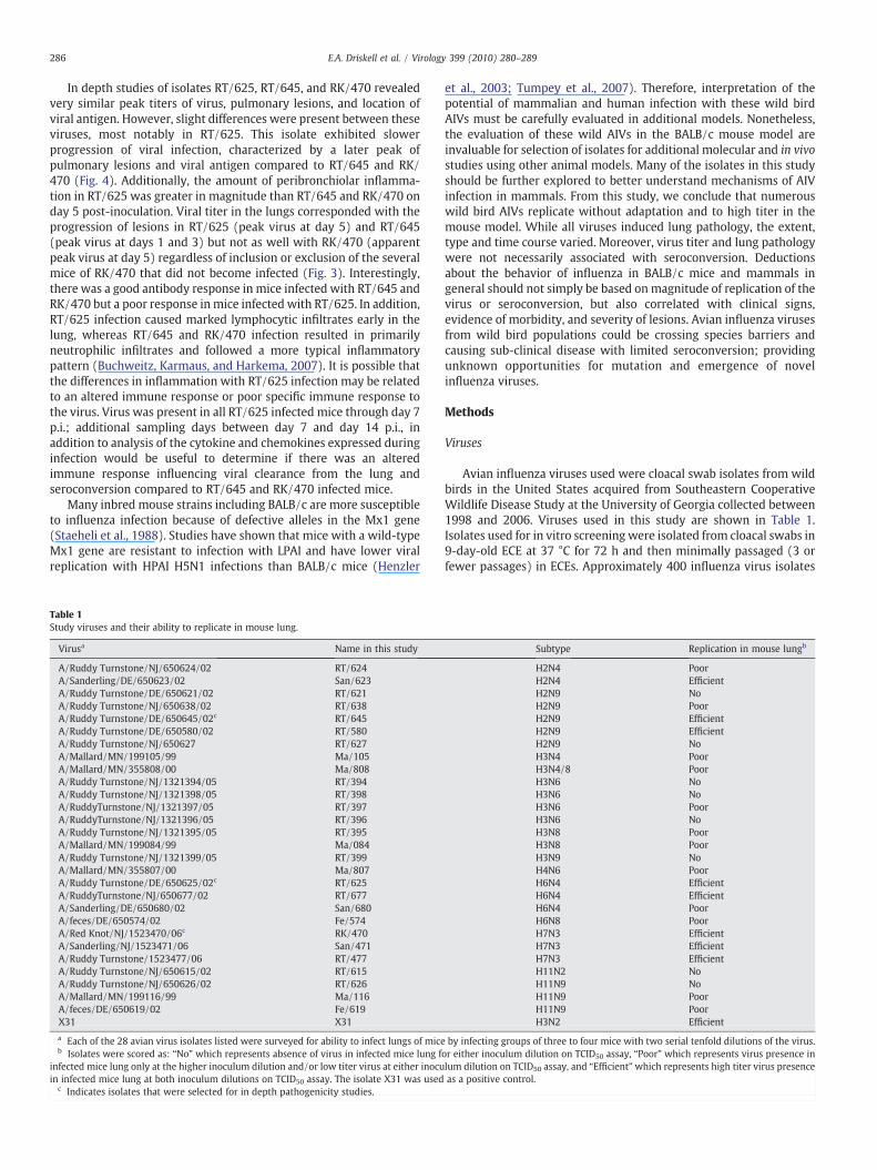
Fig. 1. Wild aquatic bird influenza virus isolates replicate to high titer in mice withoutadaptation. Groups of two bars represent a single isolate that replicated efficiently inmice (from Table 1) inoculated i.n. at two dilutions (1:10 and 1:100) with lungcollected and titrated at day 4 p.i. in MDCK cells. The concentration of the inoculum forthe 1:10 dilution is listed after the isolate name (expressed in PFU/mouse). Each barrepresents the average lung titer for a group of four mice. The dashed line is the limit ofdetection (2.4 log10TCID50/g).
281E.A. Driskell et al. / Virology 399 (2010) 280–289
behavior of a large variety of wild bird avian influenza isolates in themouse model is a step towards deeper understanding of the risks andmechanisms of avian influenza infections in mammals.
All pandemic influenza viruses since 1918, including the 2009H1N1 virus have at least an AIV component (Glaser et al., 2005;Matrosovich et al., 2000; Stevens et al., 2006). There is a possibilitythat the next pandemic influenza precursor is circulating in the wildbird population. In this study, the mouse model is utilized forinfluenza to examine a large variety of AIVs isolated from wild birdsfrom influenza surveillance in the United States. Results demonstratethat many of these isolates can replicate in the lung of mice andinduce pulmonary lesions with minimal morbidity. For some isolates,virus was localized with immunohistochemistry and demonstratedrobust replication in respiratory epithelial tissues. Serology afterinfection showed that immunogenicity was variable and unrelated toreplication or pathology. These data support the idea that humans andother mammals may be directly infected with wild bird AIVs, in somecases subclinically and without seroconversion, providing a potentialavenue for emergence of influenza viruses with pandemic potentialvia mutation and/or reassortment.
Results
Wild bird avian influenza viruses replicate robustly in mice
As a primary in vitro screen, more than 400 wild bird avianinfluenza virus isolates were screened for replication inMDCK cells byplaque assay. As plaquing in cell culture in the absence of exogenoustrypsin has been shown to indicate the potential for increasedpathogenicity for avian influenza viruses (Rimmelzwaan et al., 2006;Zitzow et al., 2002) and the 1918 influenza virus was shown to plaqueto a high titer in culture without addition of trypsin (Tumpey et al.,2005), wild bird isolates were tested for replication in MDCK cells inthe presence and absence of trypsin. While this phenotype is notassociated with infectivity in mice, it provided ameasure of infectivityin a mammalian culture system and a means to select for potentiallypathogenic viruses. TheMDCK plaque assay identified 114 viruses thatreplicated to high titer inMDCK cells andmost isolates lost less than 20fold of the titer the absence of exogenous trypsin (data not shown).Twenty-eight of these viruses were selected for in vivo screening.
Wild bird isolates selected for in vivo replication analysis coveredsix hemagglutinin (HA) subtypes and four known avian species(Table 1). Replication of each isolate in mice was categorized as: “Noreplication” (no virus was detected by TCID50 assay from lungsamples), “poor replication” (virus detected from lung samples waslow titer and/or only present in mice inoculated with a highconcentration inoculum), and “efficient replication” (virus detectedfrom lung samples was high titer and present in mice inoculated withboth concentrations of virus). Many of the isolates (71%) were able toreplicate in the mouse lung when assessed on day 4 post-inoculation(p.i.) by TCID50 assay, where 29% of these isolates showed efficientreplication (Table 1). Lung titers in individual mice from isolates thatdemonstrated efficient replication are shown in Fig. 1. Inoculumconcentration was not standardized as viruses only grew to amoderate titer and repeated passage of virus in embryonated chickeneggs (ECE) to derive a high-titer stock was specifically avoided.Despite this lack of standardization, the wide range of inoculumconcentrations (as low as 5.5E+02 PFU/mouse and as high as 3.5E+05 PFU/mouse) between isolates that had efficient replication in themouse lung did not appear to be associated with the magnitude ofTCID50 virus titer from the lung on day 4 p.i. (Fig. 1). Virus was notdetected in mock infected mice via TCID50 assay.
Three virus isolates that exhibited efficient replication in themouse lung were further selected for detailed pathogenesis studies(viruses RT/645, RT/625, and RK/470). These particular isolates wereselected to diversify the HA subtypes examined. A 50% mouse
infectious dose (MID50) was elucidated for each of these threeselected isolates. The MID50 was similar for RT/645 (2.5E+03 PFU/mouse) and RT/625 (1.2E+03 PFU/mouse) but much lower for RK/470 (3.0E+01 PFU/mouse).
Wild bird avian influenza virus infection exhibits low pathogenicity inmice despite robust replication in mouse lung
Clinical signs in mice inoculated with 20MID50 of RT/645, RT/625,or RK/470 were mild and included slightly ruffled fur on days 1 and 3post-inoculation. Similar clinical signs were observed on day 1 p.i. inmock infected mice; these clinical signs were attributed to anestheticrecovery. Clinical signs in X31 inoculated mice included ruffled furand lethargy on days 3 and 5 post-inoculation. Mice infected with RT/645 and RK/470 exhibited more weight loss than mice infected withRT/625 and mock infected control mice on day 2 p.i. and weight gainremained poor in mice infected with RK/470 on day 3 p.i. (Fig. 2).Despite these differences, mice infected with RT/625, RT/645 and RK/470 exhibited overall minimal weight loss early p.i. that was notsignificantly different from mock infected control mice by a Student'st test. In comparison, mice infected intranasally with X31 andmonitored for weight loss for other studies in our laboratory exhibitapproximately 15–20% peak weight loss on day 5 post-inoculation.One PBS inoculatedmousewas found to be an outlier by Dixon'sQ testbased on weight loss data and dropped from the group.
Virus replication in the mouse lung was further examined over timeby infectingmicewith 20MID50 of each of the selected isolates (RT/645,RT/625, and RK/470) and euthanizing mice to determine virus titer inthe lung at multiple time points (Fig. 3). All viruses reached a similarpeak titer in mouse lungs (2.4E +07 to 7.7E+07 TCID50/g). Miceinfectedwith RT/625 andRK/470 bothhad a pattern of peak virus in thelung at day 5 p.i. but RT/645 had a less clear time point of peak virus,with highest titers around days 1 and 3 post-inoculation. Virus waspresent inmouse lung up to day 7 p.i. for all three viruses and cleared byday 14 p.i.
The spleen, liver, and kidney of mice infected with RT/625 and RT/645 (day 5 p.i.) and the brain of mice infected with RT/625 (day 5 p.i.)

Fig. 2. Weight loss in mice infected with selected wild aquatic bird influenza virusisolates. Groups of five mice were inoculated with 20MID50 of RT/625 (H6N4), RT/645(H2N9), RK/470 (H7N3), or mock-infected with PBS only. Weights were tracked dailyand percent weight lost compared to starting weight calculated. Daily weight loss wasnot statistically significant by Student's t test between virus inoculated mice and mockinfected mice.
Fig. 3. Kinetics of viral infection of RT/625, RT/645, and RK/470 in the lung of infectedmice. Each bar represents the viral titer in the lung of an individual mouse infected with20MID50 of virus for viruses RT/625 (H6N4), RT/645 (H2N9), and RK/470 (H7N3). Thedashed line is the limit of detection (2.4 log10TCID50/g).
282 E.A. Driskell et al. / Virology 399 (2010) 280–289
was examined for presence of virus via titration on MDCK cells. Asubset of mice from both RT/625 and RT/645 had evidence of lowtiters of virus in the spleen and liver and these samples were furtherevaluated via virus isolation in ECE. Virus isolation in eggs indicatedthat a single mouse infected with RT/645 had influenza virus presentin the liver, which was confirmed by real-time PCR for influenza (datanot shown).
Wild bird avian influenza virus infection induces pulmonary lesionsin mice
The lungs of mice infected with RT/645, RT/625, and RK/470exhibited similar histopathologic lesions aswell as a similar resolutionof lesions; however, development of lesions differed with RT/625lagging slightly behind RT/645 and RK/470. No significant histopath-ologic lesions were present in RT/645, RT/625, or RK/470 infectedmice day 1 post-inoculation. By day 3 p.i., mice infected with RT/645and RK/470 had a necrotizing bronchiolitis in many bronchioles andperibronchiolar inflammation, but mice infected with RT/625 exhib-ited only rare scattered areas of peribronchiolar inflammation (Fig. 4).A similar average percentage of lung was affected by inflammation onday 3 p.i. for all viruses (1.5% for RT/625, 2.7% for RT/645, and 2.0% forRK/470). Peribronchiolar inflammation present in mice infected withRT/625 at day 3 p.i. was primarily lymphocytes, with fewer macro-phages and neutrophils present in adjacent alveoli, where in RT/645and RK/470 infected mice the composition of inflammatory cells wasmore neutrophilic. Mild tracheal inflammation, characterized by smallnumbers of lymphocytes within the tracheal submucosa, was presentin some RT/625 and RK/470 infected mice. Many apoptotic trachealepithelial cells were observed only in RK/470 infected mice at day 3post-inoculation. In X31 infectedmice on day 3 p.i., therewas a strikingnecrotizing bronchitis and bronchiolitis with loss of epithelium andperibronchiolar cell debris with some inflammatory cells.
Histopathology in mice on day 5 p.i. again showed that lesions inmice infected with RT/645 and RK/470 were ahead of mice infectedwith RT/625. Mice infected with RT/645 and RK/470 had continuedperibronchiolar pneumonia and necrotizing bronchiolitis; however,the necrotizing bronchiolitis was resolving by this point in time andthe bronchiolar epithelium was beginning to become hyperplastic(Fig. 4). Conversely, mice infected with RT/625 had continuedperibronchiolar pneumonia that increased in severity and also anecrotizing bronchitis and bronchiolitis appeared at this time point.
Mice infected with RT/625 exhibited prominent lymphocytic peri-vascular cuffing. The degree of inflammation present in the lung wasslightly higher on day 5 p.i. compared to day 3 p.i., but similarbetween all viruses examined (8.6% for RT/625, 5.7% for RT/645, and3.5% for RK/470). No lesions were present in the lungs of mockinfectedmice (Fig. 4). Mice infectedwith X31 had continued lesions ofnecrotizing bronchitis and bronchiolitis with peribronchiolar

Fig. 4. Histopathology in lung of AIV infected mice on days 3 through 28 post-inoculation (p.i.). Mice infected with RT/625 (H6N4), RT/645 (H2N9), or RK/470 (H7N3) havebronchiolar necrosis (arrow) and peribronchiolar inflammation (asterisk). Inflammation was most severe in RT/625 (H6N4) infected mice on day 7 post-inoculation.Peribronchiolar inflammation was resolving as dense nodular collections of lymphocytes on days 14 and 28 post-inoculation. Positive control infected mice (X31 day 3 p.i.) havesimilar bronchiolar necrosis as AIV inoculated mice. Mock infected mice have no lesions on histopathology.
283E.A. Driskell et al. / Virology 399 (2010) 280–289
inflammation day 5 p.i., but many areas of bronchiolar epitheliumwere hyperplastic at this time point.
Mice infected with RT/625 had continued peribronchiolar pneu-monia and necrotizing bronchiolitis at day 7 p.i.; however, thepneumonia was more widespread (Fig. 4). Mice infected with RT/645and RK/470 had similar peribronchiolar pneumonia day 7 p.i. asdescribed for day 5 p.i.; however, the degree of inflammation wasmuch less in severity than RT/625 (24.9% lung affected for RT/625,3.6% for RT/645, and 5.6% for RK/470) and there was completeresolution of bronchiolar necrosis. Additionally, there was prominenttype II pneumocyte hyperplasia observed in some mice infected withRT/645 beginning at day 7 postinoculation.
Examination of lungs in infected mice on days 14 and 28 p.i.revealed similar resolution of mice infected with RT/625, RT/645, andRK/470. Peribronchiolar inflammation in all of these mice wasprimarily consolidated areas of lymphocytes and there were manyperibronchiolar areas of type II pneumocyte hyperplasia. Lesionsprogressed to scattered nodular collections of peribronchiolarlymphocytes by day 28 p.i. in all virus infected mice. Mice infectedwith X31 had similar histopathologic changes of lymphocyticinflammation and type II pnuemocyte hyperplasia on day 14 as thethree AIVs that were examined.
Immunohistochemistry (IHC) for the nucleoprotein of influenza Aon the lungs of mice infected with RT/625, RT/645, and RK/470 all
exhibited strong positive intranuclear staining of tracheal, bronchio-lar, and/or alveolar epithelium in areas of histopathologic lesionsbetween days 3 and 5 p.i. (Fig. 5). Mice infected with RT/625 had veryminimal positive staining on day 3 p.i. but staining became moreprominent by day 5 p.i., versus RT/645 and RK/470, which both hadmore prominent positive staining on day 3 p.i. and magnitude ofpositive staining was lessened by day 5 post-inoculation. Theseobservations are in concord with histopathology that indicates earlierproductive infection in mice infected with RT/645 and RK/470 versusmice infected with RT/625. Additionally, no positive staining oftracheal epithelium was observed in mice infected with RT/625, butthere was good staining of the tracheal epithelium on day 3 p.i. formice infectedwith RT/645 and RK/470. Therewas no positive stainingin the lung of mock infectedmice examined on day 5 post-inoculation.
Mice infected with RT/625 and RT/645 did not have anyhistopathologic lesions in the nasal turbinates at any time point(days 1, 3, 5, 7, and 14 p.i.). Two mice that were infected with RK/470(day 5 p.i.) and one mouse that was infected with RK/470 (day 7 p.i.)had a single area in the nasal cavity that contained mixed mucus andneutrophils, but there were no lesions in the nasal epithelium in anyof the RK/470 infected mice. Although epithelial lesions were notobserved, there was positive intranuclear staining in very rare ciliatedepithelial cells of the paranasal sinuses in mice infected with RT/645and in mice infected with RK/470. There was no positive staining for

Fig. 5. Viral antigen in the lung and nasal turbinates of AIV infected mice on days 3 and 5 post-inoculation. By immunohistochemistry, mice infected with RT/625 (H6N4), RT/645(H2N9), or RK/470 (H7N3) have strong positive intranuclear staining for the nucleoprotein of influenza A in tracheal epithelium (RK/470 day 3 p.i.) , bronchiolar epithelium (RT/645 day 3 p.i.), and alveolar epithelium (RT/625 day 5 p.i.) for all three AIV isolates in infected mice. There is also strong positive intranuclear staining of the nasal turbinateepithelium (RT/645 NT day 3 p.i.) in some AIV infectedmice. Mock infected mice have no staining for viral antigen (Allantoic fluid day 5 p.i.). Positive control infected mice (X31 day3 p.i.) have similar strong positive intranuclear staining of bronchiolar epithelium as AIV inoculated mice. Cellular necrosis is evident by luminal debris in the airways (arrow).
Fig. 6. Serum antibody response in mice after wild AIV infection. Groups of 5 mice wereinoculated with 20MID50 of RT/625 (H6N4), RT/645 (H2N9), or RK/470 (H7N3) andsera were collected day 23 post-inoculation. Sera from individual mice were testedagainst whole virus and allantoic fluid, and the difference was averaged and plotted.Sera from PBS inoculated mice were tested against allantoic fluid.
284 E.A. Driskell et al. / Virology 399 (2010) 280–289
influenza in other organs examined on day 5 p.i. for any of the threeviruses.
There was only a single extrapulmonary lesion observed in miceinfected with the three AIVs, this was thymic atrophy with lymphocytedepletion present in a mouse infected with RT/645 on day 3 post-inoculation. Thymic atrophy has been associated with influenzainfection in mice; however, we did not consistently observe this lesionin mice infected with this virus and observation of thymic atrophy in adifferent study occurredmuch later in infection (Fislova et al., 2009). Noother lesions were observed in any of the organs examined for miceinfected with RT/625, RT/645 or RK/470 on days 1, 3, 5, 7, or 14 p.i.
Wild bird avian influenza viruses exhibit differences in immunogenicityin mice
A serum ELISAwas performed on sera collected frommice infectedwith RT/625, RT/645, or RK/470 on day 23 p.i. to assess seroconver-sion after AIV infection. All five mice infected with RT/645 or RK/470had a robust IgG response to the specific AIV with a prominent signalfrom serum dilutions up to 1:320 (Fig. 6). In contrast, mice infectedwith RT/625 had a poor IgG response to the viral infection, with littledifference in signal from serum dilutions when compared to mockinfected mice (Fig. 6).

285E.A. Driskell et al. / Virology 399 (2010) 280–289
Discussion
The perspectives regarding the host range and mechanisms ofinfluenza infections are ever broadening to a view of a morepromiscuous virus than previously thought. Through natural andexperimental infections, the list of mammals that are susceptible toAIV infection is rapidly increasing along with the scope of organtropism of these viruses. These findings apply not only to highlypathogenic AIVs, but also to low pathogenic viruses from poultry(Belser et al., 2007; Gillim-Ross et al., 2008; Joseph et al., 2007; Wanet al., 2008). These evolving viewpoints are further supported withthese data that includes a large scope of avian influenza isolates fromwild birds, demonstrating that many AIVs from wild birds canreplicate in the BALB/c mouse model without adaptation and thathemagglutinin subtype was not a barrier to infection (Table 1).Methodology in screening the isolates for replication in the mouselung was limited by the titer of the virus stock, as the virus isolateswere minimally passaged to avoid mutations associated with eggadaptation. For the purpose of these studies it was critical to keepthese isolates as close as possible to the original sample from the wildbird, to better assess replication of the original isolate. This resulted inmany isolates that were only of moderate titer. The inoculumconcentration for the primary screening experiment could not bestandardized, as lowering the inoculum concentration for someisolates to normalize the concentration could have resulted in missingsome isolates that were able to replicate. This method enabledidentification of the maximum number of isolates that replicated inthemice and did not have any effect on peak viral titer for isolates thatexhibited efficient replication (Fig. 2), although some concentrationeffect was observed on isolates that exhibited poor replication (datanot shown).
Mice did not require a high concentration inoculum for infectionwith RT/625, RT/645, or RK/470, as evident in the MID50 for eachvirus. Studies with LPAI H7 viruses have also shown that lowconcentrations of virus are adequate for infection of mice (Belseret al., 2007). RK/470 had a very low MID50 in mice, however, whenmice were inoculated with 20MID50 for pathogenicity studies; severalmice did not become infected. Therefore, it is possible that a higherconcentration of inoculum was required for consistent infection of allthese mice. Despite these observations, it is also interesting to notethat all H7 subtype viruses examined in this screen replicated to ahigh titer despite a low inoculum concentration, suggesting there is alower threshold for infection in the H7 subtypes (Fig. 1). Additionally,although there was not an equal number of hemagglutinin subtypesrepresented, all viruses of the H7 subtype exhibited efficientreplication compared to other subtypes that exhibited varied abilityto replicate inmouse lung (Table 1). The H7 subtype of avian influenzais of particular concern in human infections (Belser et al., 2009;Gillim-Ross and Subbarao, 2006). The data shown here is suggestive ofan increased ability of the H7 viruses to replicate in the mouse modeland further investigation to examine more isolates of this subtypemay provide insight to mechanisms of mammalian infection. Whileprevious studies focused on LPAI and HPAI H7 influenza virusesisolated from humans and poultry, the viruses tested here wereexclusively isolates from North American waterfowl and shore birds.This presents additional opportunities for transmission; whetherthrough contact with infected wild birds or environmental exposurein the birds' habitats (Rohani et al., 2009).
Infections in these mice were very similar to infections withhuman or other LPAIVs in BALB/c mice regarding pathogenesis andlesion development (Buchweitz, Karmaus, and Harkema, 2007). Peakviral titers in the lung for other LPAIVs are generally at day 4 but canoccur as early as day 2 with virus present in the lung through day 7(Gillim-Ross et al., 2008; Joseph et al., 2007). In this study, there is asimilar peak of infection based on virus titers from the lung as RT/625and RT/470 had peak titers in most mice on day 5 p.i. and RT/645 had
peak titers in most mice at days 1 and 3 p.i. (Fig. 3). Previousimmunohistochemistry studies on influenza infected mice alsodemonstrate similar strong intranuclear staining of bronchiolarepithelium (Fislova et al., 2009; Rigoni et al., 2007). Interestingly, allof the viruses we examined demonstrated positive viral antigenstaining of not only bronchiolar epithelial cells but also alveolarepithelial cells, but in the X31 infected mice viral antigen staining wasrestricted to bronchiolar epithelium. Similar findings were present ina study of mouse adapted influenza viruses, where IHC staining forinfluenza was only observed in bronchiolar epithelial cells (Fislovaet al., 2009). Moreover, there was prominent type II pneumocytehyperplasia present in mice infected with viruses RT/625, RT/645,and RK/470 by day 14 p.i., also supporting that there was significantalveolar epithelial damage. HA receptor specificity is thought to be alarge contributor to host range, as avian influenza strains preferen-tially bind to cell glycoproteins/glycolipids that have terminal sialyl-galactosyl (SA) residues with a 2–3 linkage [Neu5Ac(α2-3)Gal] andhuman influenza strains preferentially bind to terminal 2–6 linkedglycoproteins/glycolipids [Neu5Ac(α2-6)Gal] (Connor et al., 1994;Mansfield, 2007). Additionally, the distribution of these receptors oncells of the respiratory tract is demonstrated to vary betweenmammalian species and mice have been shown to have primarilythe 2–3 linked SA receptor in ciliated epithelial cells in large airwaysand in type II pneumocytes (Ibricevic et al., 2006; van Riel et al., 2007).Indeed, many studies of HPAI H5N1 have demonstrated enhancedalveolar damage in humans andmice, suggesting increased affinity forbinding of these cells compared to human strains of influenza (Abdel-Ghafar et al., 2008; Dybing et al., 2000). This affinity for binding cellsof the lower respiratory tract was also demonstrated with some LPAIisolates (van Riel et al., 2007). Our results are also supportive thatthese wild bird AIVs have an increased binding affinity for receptorspresent on alveolar epithelial cells compared to human strains. Inmice infected with RT/645 and RK/470 there was minimal presenceof viral antigen in the nasal cavity via IHC with no antigen present inRT/625. Other influenza isolates, particularly human origin, typicallyexhibit replication in this area of the upper respiratory tract. Theminimal presence of antigen in the nasal turbinates despite efficientreplication and lesions in the lung supports that these isolates, similarto other avian isolates, have a preference for replication in the lowerrespiratory tract. There is recent evidence that the regional replicationpreferences of avian influenza in mammals are in part due tomutations in PB2 that change temperature and cell type replicationpreferences in the virus (Hatta et al., 2007). Further investigation intothe molecular aspects of these wild type avian viruses may provideinsight into common molecular mechanisms of host range andpathogenicity in mammals.
It has been shown that many HPAI viruses have extrapulmonaryspread, with virus presence and lesions most frequently in the brainand spleen of infected mice (Belser et al., 2007; Joseph et al., 2007;Maines et al., 2005; Rigoni et al., 2007). Examination of LPAIVs hasdemonstrated that some of these viruses that exhibit pulmonaryreplication also have the capacity for extrapulmonary spread, whileothers do not (Gillim-Ross et al., 2008; Joseph et al., 2007).Interestingly, mouse adapted influenza viruses have also beenshown to have extrapulmonary spread to numerous organs withpossible viremia, regardless of virulence (Fislova et al., 2009).Additionally, a case study of a human infection with HPAI H5N1demonstrated viral antigen in extrapulmonary sites without lesions inthese sites (Zhang et al., 2009). We also have evidence of inconsistentextrapulmonary spread of one of the viruses examined (RT/645)without significant morbidity or extrapulmonary lesions in thesemice. This may be further supportive evidence that viremia andextrapulmonary spread is not as uncommon as previously believedand conclusions of virulence of an influenza isolate must be evaluatednot only in context of viral presence, but also with consideration ofmorbidity and severity of lesions in the mouse model.
286 E.A. Driskell et al. / Virology 399 (2010) 280–289
In depth studies of isolates RT/625, RT/645, and RK/470 revealedvery similar peak titers of virus, pulmonary lesions, and location ofviral antigen. However, slight differences were present between theseviruses, most notably in RT/625. This isolate exhibited slowerprogression of viral infection, characterized by a later peak ofpulmonary lesions and viral antigen compared to RT/645 and RK/470 (Fig. 4). Additionally, the amount of peribronchiolar inflamma-tion in RT/625 was greater in magnitude than RT/645 and RK/470 onday 5 post-inoculation. Viral titer in the lungs corresponded with theprogression of lesions in RT/625 (peak virus at day 5) and RT/645(peak virus at days 1 and 3) but not as well with RK/470 (apparentpeak virus at day 5) regardless of inclusion or exclusion of the severalmice of RK/470 that did not become infected (Fig. 3). Interestingly,there was a good antibody response in mice infected with RT/645 andRK/470 but a poor response inmice infected with RT/625. In addition,RT/625 infection caused marked lymphocytic infiltrates early in thelung, whereas RT/645 and RK/470 infection resulted in primarilyneutrophilic infiltrates and followed a more typical inflammatorypattern (Buchweitz, Karmaus, and Harkema, 2007). It is possible thatthe differences in inflammation with RT/625 infection may be relatedto an altered immune response or poor specific immune response tothe virus. Virus was present in all RT/625 infected mice through day 7p.i.; additional sampling days between day 7 and day 14 p.i., inaddition to analysis of the cytokine and chemokines expressed duringinfection would be useful to determine if there was an alteredimmune response influencing viral clearance from the lung andseroconversion compared to RT/645 and RK/470 infected mice.
Many inbred mouse strains including BALB/c are more susceptibleto influenza infection because of defective alleles in the Mx1 gene(Staeheli et al., 1988). Studies have shown that mice with a wild-typeMx1 gene are resistant to infection with LPAI and have lower viralreplication with HPAI H5N1 infections than BALB/c mice (Henzler
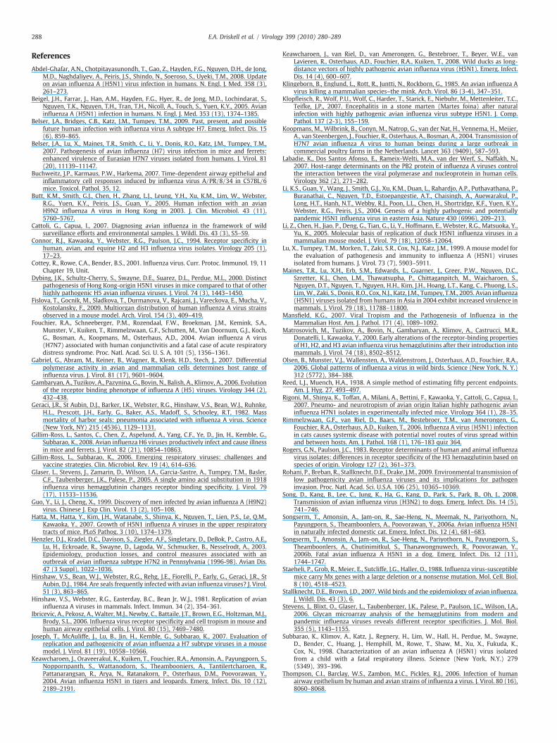
Table 1Study viruses and their ability to replicate in mouse lung.
Virusa Name in this study
A/Ruddy Turnstone/NJ/650624/02 RT/624A/Sanderling/DE/650623/02 San/623A/Ruddy Turnstone/DE/650621/02 RT/621A/Ruddy Turnstone/NJ/650638/02 RT/638A/Ruddy Turnstone/DE/650645/02c RT/645A/Ruddy Turnstone/DE/650580/02 RT/580A/Ruddy Turnstone/NJ/650627 RT/627A/Mallard/MN/199105/99 Ma/105A/Mallard/MN/355808/00 Ma/808A/Ruddy Turnstone/NJ/1321394/05 RT/394A/Ruddy Turnstone/NJ/1321398/05 RT/398A/RuddyTurnstone/NJ/1321397/05 RT/397A/RuddyTurnstone/NJ/1321396/05 RT/396A/Ruddy Turnstone/NJ/1321395/05 RT/395A/Mallard/MN/199084/99 Ma/084A/Ruddy Turnstone/NJ/1321399/05 RT/399A/Mallard/MN/355807/00 Ma/807A/Ruddy Turnstone/DE/650625/02c RT/625A/RuddyTurnstone/NJ/650677/02 RT/677A/Sanderling/DE/650680/02 San/680A/feces/DE/650574/02 Fe/574A/Red Knot/NJ/1523470/06c RK/470A/Sanderling/NJ/1523471/06 San/471A/Ruddy Turnstone/1523477/06 RT/477A/Ruddy Turnstone/NJ/650615/02 RT/615A/Ruddy Turnstone/NJ/650626/02 RT/626A/Mallard/MN/199116/99 Ma/116A/feces/DE/650619/02 Fe/619X31 X31
a Each of the 28 avian virus isolates listed were surveyed for ability to infect lungs of miceb Isolates were scored as: “No” which represents absence of virus in infected mice lung f
infected mice lung only at the higher inoculum dilution and/or low titer virus at either inocuin infected mice lung at both inoculum dilutions on TCID50 assay. The isolate X31 was used
c Indicates isolates that were selected for in depth pathogenicity studies.
et al., 2003; Tumpey et al., 2007). Therefore, interpretation of thepotential of mammalian and human infection with these wild birdAIVs must be carefully evaluated in additional models. Nonetheless,the evaluation of these wild AIVs in the BALB/c mouse model areinvaluable for selection of isolates for additional molecular and in vivostudies using other animal models. Many of the isolates in this studyshould be further explored to better understand mechanisms of AIVinfection in mammals. From this study, we conclude that numerouswild bird AIVs replicate without adaptation and to high titer in themouse model. While all viruses induced lung pathology, the extent,type and time course varied. Moreover, virus titer and lung pathologywere not necessarily associated with seroconversion. Deductionsabout the behavior of influenza in BALB/c mice and mammals ingeneral should not simply be based onmagnitude of replication of thevirus or seroconversion, but also correlated with clinical signs,evidence of morbidity, and severity of lesions. Avian influenza virusesfrom wild bird populations could be crossing species barriers andcausing sub-clinical disease with limited seroconversion; providingunknown opportunities for mutation and emergence of novelinfluenza viruses.
Methods
Viruses
Avian influenza viruses used were cloacal swab isolates from wildbirds in the United States acquired from Southeastern CooperativeWildlife Disease Study at the University of Georgia collected between1998 and 2006. Viruses used in this study are shown in Table 1.Isolates used for in vitro screening were isolated from cloacal swabs in9-day-old ECE at 37 °C for 72 h and then minimally passaged (3 orfewer passages) in ECEs. Approximately 400 influenza virus isolates
Subtype Replication in mouse lungb
H2N4 PoorH2N4 EfficientH2N9 NoH2N9 PoorH2N9 EfficientH2N9 EfficientH2N9 NoH3N4 PoorH3N4/8 PoorH3N6 NoH3N6 NoH3N6 PoorH3N6 NoH3N8 PoorH3N8 PoorH3N9 NoH4N6 PoorH6N4 EfficientH6N4 EfficientH6N4 PoorH6N8 PoorH7N3 EfficientH7N3 EfficientH7N3 EfficientH11N2 NoH11N9 NoH11N9 PoorH11N9 PoorH3N2 Efficient
by infecting groups of three to four mice with two serial tenfold dilutions of the virus.or either inoculum dilution on TCID50 assay, “Poor” which represents virus presence inlum dilution on TCID50 assay, and “Efficient”which represents high titer virus presenceas a positive control.

287E.A. Driskell et al. / Virology 399 (2010) 280–289
were examined in vitro. Viruses that exhibited plaquing on Madin-Darby canine kidney (MDCK) cells without the addition of trypsinwere selected for additional in vivo selection in mice, as thisphenotype is suggestive of enhanced pathogenicity of influenzaviruses. The original low passage isolates, once selected by in vitroscreening methods, were grown once more in 9- to 10-day-old ECE togenerate a stock of the virus. Allantoic fluid pooled from eggs wasfrozen in aliquots at−80 °C. Virus stocks were plaqued onMDCK cellswith trypsin to elucidate a PFU/mL titer.
Mouse experiments
Female 6- to 8-week-old BALB/c mice (Harlan Laboratories,Indianapolis, IN) were anesthetized with intraperitoneal injection of2,2,2-tribromoethanol in tert-amyl alcohol and inoculated intranasal-ly with 50 μL of diluted virus in sterile phosphate buffered saline(PBS). A screen to determine the replication capacity of selected AIVisolates in mice was performed by harvesting lungs of mice inoculatedwith a 1:10 dilution of virus stock (3–4 mice per virus) or a 1:100dilution of virus stock (3–4 mice per virus) on day 4 post-inoculation.A group of mice was mock infected with PBS and another group wasinfected with X31 to serve as controls. Mouse experiments wereperformed in enhanced BSL2 facilities in HEPA filtered isolators.Studies were conducted under guidelines approved by the AnimalCare and Use Committee of the University of Georgia. Clarified lunghomogenate and fixed lung tissue from selected influenza inoculatedmice (RT/625) exhibiting histopathologic lesions were negative forMycoplasma pulmonis via PCR.
The MID50 was determined as previously described (Cottey, Rowe,and Bender, 2001). Briefly, mice were infected with 10-fold dilutionsof each virus. Five mice per group were euthanized on day 4 p.i. andclarified lung homogenate was serially titrated in MDCK cells todetermine the MID50 calculated by the method of Reed and Muench(1938). The pathogenesis of RT/625, RT/645, and RK/470 wasdetermined by inoculation of groups of mice with 20MID50 of virusand harvesting lungs on days 1, 3, 5, 7, and 14 p.i. and harvestingspleen, liver, kidney and brain on day 5 p.i (5 mice per virus per day).Tissues were homogenized in 1 mL PBS, clarified by centrifugation,and frozen at−80 °C for later titration. Clarified lung homogenatewastitrated in MDCK cells starting at a 1:10 dilution with a limit ofdetection at 102.4 TCID50/gram. Clarified organ homogenate (spleen,liver, kidney, and brain) was initially titrated in MDCK cells starting ata 1:10 dilution. Samples were further selected for virus isolation in 9-to 10-day-old ECE. Real time RT-PCR was performed on hemagglu-tination assay positive samples from virus isolation. Briefly, viral RNAwas extracted from allantoic fluid by using RNeasy mini kit (Qiagen,Valencia, CA) and the Qiagen one-step RT-PCR kit was used for RRT-PCR with a Stratagene MX300P/3005P thermocyler and Mx Pro QPCRsoftware (La Jolla, CA). Reaction mixture and PCR cycling protocol isavailable upon request. An influenza virus matrix gene specific primerand probe set were used as follows: primerM+25, sequence AGA TGAGTC TTC TAA CCG AGG TCG; primer M-124, sequence TGC AAA AACATC TTC AAG TCT CTG; and probeM+64, sequence FAM-TCA GGC CCCCTC AAA GCC GA-TAMRA (Biosearch Technologies, Novato, CA).
For morbidity and seroconversion studies, five mice per group forviruses RT/625, RT/645, RK/470, and a mock infected PBS group wereinoculated with 20MID50 of virus and weighed daily for 11 days andthen every other day for an additional 4 days. Serum was collectedfrom thesemice on day 23 p.i. to assay serum antibody response to theAIVs. Statistical significance of weight loss between groups of micewas determined using Student's t test.
Histopathology and immunohistochemistry
Tissues from infected and control mice were examined byhistopathology and immunohistochemistry. Mice were inoculated
with 20MID50 virus and lung, trachea, thymus, thyroid, esophagus,heart, spleen, liver, stomach, intestine, pancreas, kidneys, adrenalgland, ovary, uterus, bladder, brain, and nasal turbinates werecollected on days 1, 3, 5, 7, 14, and 28 p.i. (three mice per virus perday). The lungs were inflated with 10% neutral buffered formalin andall tissues were preserved in 10% neutral buffered formalin. Additionalgroups of mice were inoculated with allantoic fluid in sterile PBS (3mice) or with X31 (9 mice) to serve as controls and organs werecollected on day 5 p.i (PBS) or days 3, 5, and 14 p.i. (X31). Five equaltransverse sections were made through the entire lungs. Mouse skullswere decalcified and four transverse sections were made through thenasal cavity to examine the nasal turbinates. Tissues were routinelyprocessed, embedded and stained with hematoxylin and eosin. Theseverity of inflammation in lungs of infected mice was calculated byfinding the average percent area of lung affected by inflammation perday p.i. for each virus using Image Pro Plus software vs. 4.5.1(MediaCybernetics, Bethesda, MD).
Immunohistochemical staining was performed on lung tissue(days 1, 3, 5, and 7 p.i. for 3 mice per day) or on nasal turbinates (days3 and 5 p.i. for 2 mice per day) and all other organs (day 5 p.i. for 2mice per day) in mice infected with RT/625, RT/645, and RK/470.Immunohistochemistry on mouse lung tissue was performed using acommercially available mouse monoclonal antibody to the nucleo-protein of influenza A virus at a 1:200 dilution (Biodesign Inter-national, Sako, Maine) or on mouse nasal turbinates and organs otherthan lung using a commercially available goat polyclonal antibody tothe nucleoprotein of influenza A virus at a 1:10,000 dilution (Bio-design International, Sako, Maine), as excessive background stainingwas observed in nasal turbinates and organs using the mousemonoclonal. Tissues were deparaffinized and blocked with a com-mercial protein blocking agent (Dako Cytomation, Carpinteria, CA)and a linked strepavidin-biotin immunoperoxidase system was usedfor immunolabeling. The reaction was visualized with 3,3′-diamino-benzidine substrate (Dako Cytomation, Carpinteria, CA).
Serum ELISA assay
Sera from individual mice were assayed via ELISA against eachwhole influenza virus (RT/625, RT/645, or RK/470) inoculated in theindividual mouse. Additionally, the same sera were assayed againstallantoic fluid to account for any antibodies generated againstcomponents other than the virus. Virus or allantoic fluid was coated100 μL per well on 96 well Immulon 2HB microtiter plates andincubated 24 h at 4 °C. Two-fold dilutions of sera were applied topre-absorbed plates, starting with a dilution of 1:20 and virusspecific antibodies were measured using alkaline phosphataselabeled goat anti mouse IgG(H+L) (Kirkegaard and Perry Laborato-ries, Gaithersburg, MD). The p-nitrophenyl phosphate substrate(Kiregaard and Perry Laboratories, Gaithersburg, MD) was added andabsorbance measured at 405 nm on a 96-well format plate reader(BioTek, Winooski, VT ). Absorbance readings for sera againstallantoic fluid were subtracted from absorbance readings for seraagainst virus and plotted.
Acknowledgments
The authors wish to thank the Animal Resources personnel atthe College of Veterinary Medicine, University of Georgia forexcellent animal husbandry. We would also like to thank histo-technicians in the Veterinary Pathology Department at University ofGeorgia for their assistance, especially Abbie Butler for heroutstanding immunohistochemistry support. Additional thanks toRaydel Mair, James Gordy, Ginger Goekjian, and Becky Poulson at theCollege of Veterinary Medicine, University of Georgia for excellenttechnical support. This work was supported by a grant from the CDC:5U19Cl000401-02.
288 E.A. Driskell et al. / Virology 399 (2010) 280–289
References
Abdel-Ghafar, A.N., Chotpitayasunondh, T., Gao, Z., Hayden, F.G., Nguyen, D.H., de Jong,M.D., Naghdaliyev, A., Peiris, J.S., Shindo, N., Soeroso, S., Uyeki, T.M., 2008. Updateon avian influenza A (H5N1) virus infection in humans. N. Engl. J. Med. 358 (3),261–273.
Beigel, J.H., Farrar, J., Han, A.M., Hayden, F.G., Hyer, R., de Jong, M.D., Lochindarat, S.,Nguyen, T.K., Nguyen, T.H., Tran, T.H., Nicoll, A., Touch, S., Yuen, K.Y., 2005. Avianinfluenza A (H5N1) infection in humans. N. Engl. J. Med. 353 (13), 1374–1385.
Belser, J.A., Bridges, C.B., Katz, J.M., Tumpey, T.M., 2009. Past, present, and possiblefuture human infection with influenza virus A subtype H7. Emerg. Infect. Dis. 15(6), 859–865.
Belser, J.A., Lu, X., Maines, T.R., Smith, C., Li, Y., Donis, R.O., Katz, J.M., Tumpey, T.M.,2007. Pathogenesis of avian influenza (H7) virus infection in mice and ferrets:enhanced virulence of Eurasian H7N7 viruses isolated from humans. J. Virol. 81(20), 11139–11147.
Buchweitz, J.P., Karmaus, P.W., Harkema, 2007. Time-dependent airway epithelial andinflammatory cell responses induced by influenza virus A/PR/8/34 in C57BL/6mice. Toxicol. Pathol. 35, 12.
Butt, K.M., Smith, G.J., Chen, H., Zhang, L.J., Leung, Y.H., Xu, K.M., Lim, W., Webster,R.G., Yuen, K.Y., Peiris, J.S., Guan, Y., 2005. Human infection with an avianH9N2 influenza A virus in Hong Kong in 2003. J. Clin. Microbiol. 43 (11),5760–5767.
Cattoli, G., Capua, I., 2007. Diagnosing avian influenza in the framework of wildsurveillance efforts and environmental samples. J. Wildl. Dis. 43 (3), S5–S9.
Connor, R.J., Kawaoka, Y., Webster, R.G., Paulson, J.C., 1994. Receptor specificity inhuman, avian, and equine H2 and H3 influenza virus isolates. Virology 205 (1),17–23.
Cottey, R., Rowe, C.A., Bender, B.S., 2001. Influenza virus. Curr. Protoc. Immunol. 19, 11Chapter 19, Unit.
Dybing, J.K., Schultz-Cherry, S., Swayne, D.E., Suarez, D.L., Perdue, M.L., 2000. Distinctpathogenesis of Hong Kong-origin H5N1 viruses in mice compared to that of otherhighly pathogenic H5 avian influenza viruses. J. Virol. 74 (3), 1443–1450.
Fislova, T., Gocnik, M., Sladkova, T., Durmanova, V., Rajcani, J., Vareckova, E., Mucha, V.,Kostolansky, F., 2009. Multiorgan distribution of human influenza A virus strainsobserved in a mouse model. Arch. Virol. 154 (3), 409–419.
Fouchier, R.A., Schneeberger, P.M., Rozendaal, F.W., Broekman, J.M., Kemink, S.A.,Munster, V., Kuiken, T., Rimmelzwaan, G.F., Schutten, M., Van Doornum, G.J., Koch,G., Bosman, A., Koopmans, M., Osterhaus, A.D., 2004. Avian influenza A virus(H7N7) associated with human conjunctivitis and a fatal case of acute respiratorydistress syndrome. Proc. Natl. Acad. Sci. U. S. A. 101 (5), 1356–1361.
Gabriel, G., Abram, M., Keiner, B., Wagner, R., Klenk, H.D., Stech, J., 2007. Differentialpolymerase activity in avian and mammalian cells determines host range ofinfluenza virus. J. Virol. 81 (17), 9601–9604.
Gambaryan, A., Tuzikov, A., Pazynina, G., Bovin, N., Balish, A., Klimov, A., 2006. Evolutionof the receptor binding phenotype of influenza A (H5) viruses. Virology 344 (2),432–438.
Geraci, J.R., St Aubin, D.J., Barker, I.K., Webster, R.G., Hinshaw, V.S., Bean, W.J., Ruhnke,H.L., Prescott, J.H., Early, G., Baker, A.S., Madoff, S., Schooley, R.T, 1982. Massmortality of harbor seals: pneumonia associated with influenza A virus. Science(New York, NY) 215 (4536), 1129–1131.
Gillim-Ross, L., Santos, C., Chen, Z., Aspelund, A., Yang, C.F., Ye, D., Jin, H., Kemble, G.,Subbarao, K., 2008. Avian influenza H6 viruses productively infect and cause illnessin mice and ferrets. J. Virol. 82 (21), 10854–10863.
Gillim-Ross, L., Subbarao, K., 2006. Emerging respiratory viruses: challenges andvaccine strategies. Clin. Microbiol. Rev. 19 (4), 614–636.
Glaser, L., Stevens, J., Zamarin, D., Wilson, I.A., Garcia-Sastre, A., Tumpey, T.M., Basler,C.F., Taubenberger, J.K., Palese, P., 2005. A single amino acid substitution in 1918influenza virus hemagglutinin changes receptor binding specificity. J. Virol. 79(17), 11533–11536.
Guo, Y., Li, J., Cheng, X., 1999. Discovery of men infected by avian influenza A (H9N2)virus. Chinese J. Exp Clin. Virol. 13 (2), 105–108.
Hatta, M., Hatta, Y., Kim, J.H., Watanabe, S., Shinya, K., Nguyen, T., Lien, P.S., Le, Q.M.,Kawaoka, Y., 2007. Growth of H5N1 influenza A viruses in the upper respiratorytracts of mice. PLoS Pathog. 3 (10), 1374–1379.
Henzler, D.J., Kradel, D.C., Davison, S., Ziegler, A.F., Singletary, D., DeBok, P., Castro, A.E.,Lu, H., Eckroade, R., Swayne, D., Lagoda, W., Schmucker, B., Nesselrodt, A., 2003.Epidemiology, production losses, and control measures associated with anoutbreak of avian influenza subtype H7N2 in Pennsylvania (1996-98). Avian Dis.47 (3 Suppl), 1022–1036.
Hinshaw, V.S., Bean, W.J., Webster, R.G., Rehg, J.E., Fiorelli, P., Early, G., Geraci, J.R., StAubin, D.J., 1984. Are seals frequently infectedwith avian influenza viruses? J. Virol.51 (3), 863–865.
Hinshaw, V.S., Webster, R.G., Easterday, B.C., Bean Jr, W.J., 1981. Replication of avianinfluenza A viruses in mammals. Infect. Immun. 34 (2), 354–361.
Ibricevic, A., Pekosz, A., Walter, M.J., Newby, C., Battaile, J.T., Brown, E.G., Holtzman, M.J.,Brody, S.L., 2006. Influenza virus receptor specificity and cell tropism in mouse andhuman airway epithelial cells. J. Virol. 80 (15), 7469–7480.
Joseph, T., McAuliffe, J., Lu, B., Jin, H., Kemble, G., Subbarao, K., 2007. Evaluation ofreplication and pathogenicity of avian influenza a H7 subtype viruses in a mousemodel. J. Virol. 81 (19), 10558–10566.
Keawcharoen, J., Oraveerakul, K., Kuiken, T., Fouchier, R.A., Amonsin, A., Payungporn, S.,Noppornpanth, S., Wattanodorn, S., Theambooniers, A., Tantilertcharoen, R.,Pattanarangsan, R., Arya, N., Ratanakorn, P., Osterhaus, D.M., Poovorawan, Y.,2004. Avian influenza H5N1 in tigers and leopards. Emerg. Infect. Dis. 10 (12),2189–2191.
Keawcharoen, J., van Riel, D., van Amerongen, G., Bestebroer, T., Beyer, W.E., vanLavieren, R., Osterhaus, A.D., Fouchier, R.A., Kuiken, T., 2008. Wild ducks as long-distance vectors of highly pathogenic avian influenza virus (H5N1). Emerg. Infect.Dis. 14 (4), 600–607.
Klingeborn, B., Englund, L., Rott, R., Juntti, N., Rockborn, G., 1985. An avian influenza Avirus killing a mammalian species–the mink. Arch. Virol. 86 (3-4), 347–351.
Klopfleisch, R., Wolf, P.U., Wolf, C., Harder, T., Starick, E., Niebuhr, M., Mettenleiter, T.C.,Teifke, J.P., 2007. Encephalitis in a stone marten (Martes foina) after naturalinfection with highly pathogenic avian influenza virus subtype H5N1. J. Comp.Pathol. 137 (2-3), 155–159.
Koopmans, M., Wilbrink, B., Conyn, M., Natrop, G., van der Nat, H., Vennema, H., Meijer,A., van Steenbergen, J., Fouchier, R., Osterhaus, A., Bosman, A., 2004. Transmission ofH7N7 avian influenza A virus to human beings during a large outbreak incommercial poultry farms in the Netherlands. Lancet 363 (9409), 587–593.
Labadie, K., Dos Santos Afonso, E., Rameix-Welti, M.A., van der Werf, S., Naffakh, N.,2007. Host-range determinants on the PB2 protein of influenza A viruses controlthe interaction between the viral polymerase and nucleoprotein in human cells.Virology 362 (2), 271–282.
Li, K.S., Guan, Y., Wang, J., Smith, G.J., Xu, K.M., Duan, L., Rahardjo, A.P., Puthavathana, P.,Buranathai, C., Nguyen, T.D., Estoepangestie, A.T., Chaisingh, A., Auewarakul, P.,Long, H.T., Hanh, N.T., Webby, R.J., Poon, L.L., Chen, H., Shortridge, K.F., Yuen, K.Y.,Webster, R.G., Peiris, J.S., 2004. Genesis of a highly pathogenic and potentiallypandemic H5N1 influenza virus in eastern Asia. Nature 430 (6996), 209–213.
Li, Z., Chen, H., Jiao, P., Deng, G., Tian, G., Li, Y., Hoffmann, E., Webster, R.G., Matsuoka, Y.,Yu, K., 2005. Molecular basis of replication of duck H5N1 influenza viruses in amammalian mouse model. J. Virol. 79 (18), 12058–12064.
Lu, X., Tumpey, T.M., Morken, T., Zaki, S.R., Cox, N.J., Katz, J.M., 1999. A mouse model forthe evaluation of pathogenesis and immunity to influenza A (H5N1) virusesisolated from humans. J. Virol. 73 (7), 5903–5911.
Maines, T.R., Lu, X.H., Erb, S.M., Edwards, L., Guarner, J., Greer, P.W., Nguyen, D.C.,Szretter, K.J., Chen, L.M., Thawatsupha, P., Chittaganpitch, M., Waicharoen, S.,Nguyen, D.T., Nguyen, T., Nguyen, H.H., Kim, J.H., Hoang, L.T., Kang, C., Phuong, L.S.,Lim,W., Zaki, S., Donis, R.O., Cox, N.J., Katz, J.M., Tumpey, T.M., 2005. Avian influenza(H5N1) viruses isolated from humans in Asia in 2004 exhibit increased virulence inmammals. J. Virol. 79 (18), 11788–11800.
Mansfield, K.G., 2007. Viral Tropism and the Pathogenesis of Influenza in theMammalian Host. Am. J. Pathol. 171 (4), 1089–1092.
Matrosovich, M., Tuzikov, A., Bovin, N., Gambaryan, A., Klimov, A., Castrucci, M.R.,Donatelli, I., Kawaoka, Y., 2000. Early alterations of the receptor-binding propertiesof H1, H2, and H3 avian influenza virus hemagglutinins after their introduction intomammals. J. Virol. 74 (18), 8502–8512.
Olsen, B., Munster, V.J., Wallensten, A., Waldenstrom, J., Osterhaus, A.D., Fouchier, R.A.,2006. Global patterns of influenza a virus in wild birds. Science (New York, N. Y.)312 (5772), 384–388.
Reed, L.J., Muench, H.A., 1938. A simple method of estimating fifty percent endpoints.Am. J. Hyg. 27, 493–497.
Rigoni, M., Shinya, K., Toffan, A., Milani, A., Bettini, F., Kawaoka, Y., Cattoli, G., Capua, I.,2007. Pneumo- and neurotropism of avian origin Italian highly pathogenic avianinfluenza H7N1 isolates in experimentally infected mice. Virology 364 (1), 28–35.
Rimmelzwaan, G.F., van Riel, D., Baars, M., Bestebroer, T.M., van Amerongen, G.,Fouchier, R.A., Osterhaus, A.D., Kuiken, T., 2006. Influenza A virus (H5N1) infectionin cats causes systemic disease with potential novel routes of virus spread withinand between hosts. Am. J. Pathol. 168 (1), 176–183 quiz 364.
Rogers, G.N., Paulson, J.C., 1983. Receptor determinants of human and animal influenzavirus isolates: differences in receptor specificity of the H3 hemagglutinin based onspecies of origin. Virology 127 (2), 361–373.
Rohani, P., Breban, R., Stallknecht, D.E., Drake, J.M., 2009. Environmental transmission oflow pathogenicity avian influenza viruses and its implications for pathogeninvasion. Proc. Natl. Acad. Sci. U.S.A. 106 (25), 10365–10369.
Song, D., Kang, B., Lee, C., Jung, K., Ha, G., Kang, D., Park, S., Park, B., Oh, J., 2008.Transmission of avian influenza virus (H3N2) to dogs. Emerg. Infect. Dis. 14 (5),741–746.
Songserm, T., Amonsin, A., Jam-on, R., Sae-Heng, N., Meemak, N., Pariyothorn, N.,Payungporn, S., Theamboonlers, A., Poovorawan, Y., 2006a. Avian influenza H5N1in naturally infected domestic cat. Emerg. Infect. Dis. 12 (4), 681–683.
Songserm, T., Amonsin, A., Jam-on, R., Sae-Heng, N., Pariyothorn, N., Payungporn, S.,Theamboonlers, A., Chutinimitkul, S., Thanawongnuwech, R., Poovorawan, Y.,2006b. Fatal avian influenza A H5N1 in a dog. Emerg. Infect. Dis. 12 (11),1744–1747.
Staeheli, P., Grob, R., Meier, E., Sutcliffe, J.G., Haller, O., 1988. Influenza virus-susceptiblemice carry Mx genes with a large deletion or a nonsense mutation. Mol. Cell. Biol.8 (10), 4518–4523.
Stallknecht, D.E., Brown, J.D., 2007. Wild birds and the epidemiology of avian influenza.J. Wildl. Dis. 43 (3), 6.
Stevens, J., Blixt, O., Glaser, L., Taubenberger, J.K., Palese, P., Paulson, J.C., Wilson, I.A.,2006. Glycan microarray analysis of the hemagglutinins from modern andpandemic influenza viruses reveals different receptor specificities. J. Mol. Biol.355 (5), 1143–1155.
Subbarao, K., Klimov, A., Katz, J., Regnery, H., Lim, W., Hall, H., Perdue, M., Swayne,D., Bender, C., Huang, J., Hemphill, M., Rowe, T., Shaw, M., Xu, X., Fukuda, K.,Cox, N., 1998. Characterization of an avian influenza A (H5N1) virus isolatedfrom a child with a fatal respiratory illness. Science (New York, N.Y.) 279(5349), 393–396.
Thompson, C.I., Barclay, W.S., Zambon, M.C., Pickles, R.J., 2006. Infection of humanairway epithelium by human and avian strains of influenza a virus. J. Virol. 80 (16),8060–8068.

289E.A. Driskell et al. / Virology 399 (2010) 280–289
Tumpey, T.M., Basler, C.F., Aguilar, P.V., Zeng, H., Solorzano, A., Swayne, D.E., Cox, N.J.,Katz, J.M., Taubenberger, J.K., Palese, P., Garcia-Sastre, A., 2005. Characterization ofthe reconstructed 1918 Spanish influenza pandemic virus. Science (New York, N.Y.)310 (5745), 77–80.
Tumpey, T.M., Szretter, K.J., Van Hoeven, N., Katz, J.M., Kochs, G., Haller, O., Garcia-Sastre, A., Staeheli, P., 2007. The Mx1 gene protects mice against the pandemic1918 and highly lethal human H5N1 influenza viruses. J. Virol. 81 (19),10818–10821.
van Riel, D., Munster, V.J., de Wit, E., Rimmelzwaan, G.F., Fouchier, R.A., Osterhaus, A.D.,Kuiken, T., 2007. Human and Avian Influenza Viruses Target Different Cells in theLower Respiratory Tract of Humans and Other Mammals. Am. J. Pathol. 171 (4),1215–1223.
Wan, H., Sorrell, E.M., Song, H., Hossain, M.J., Ramirez-Nieto, G., Monne, I., Stevens, J.,Cattoli, G., Capua, I., Chen, L.M., Donis, R.O., Busch, J., Paulson, J.C., Brockwell, C.,Webby, R., Blanco, J., Al-Natour,M.Q., Perez, D.R., 2008. Replication and transmissionof H9N2 influenza viruses in ferrets: evaluation of pandemic potential. PLoS One 3(8), e2923.
Ward, A.C., 1997. Virulence of influenza A virus for mouse lung. Virus Genes 14 (3),187–194.
Zhang, Z., Zhang, J., Huang, K., Li, K.S., Yuen, K.Y., Guan, Y., Chen, H., Ng, W.F., 2009.Systemic infection of avian influenza A virus H5N1 subtype in humans. Hum.Pathol. 40 (5), 735–739.
Zitzow, L.A., Rowe, T., Morken, T., Shieh, W.J., Zaki, S., Katz, J.M., 2002. Pathogenesis ofavian influenza A (H5N1) viruses in ferrets. J. Virol. 76 (9), 4420–4429.