Associations between Canine Male Reproductive Parameters ...
Upload
marionegriCategory
view
4download
0
Avian and Canine Aldehyde OxidasesNOVEL INSIGHTS INTO THE BIOLOGY AND EVOLUTION OF MOLYBDO-FLAVOENZYMES*□S
Received for publication, January 27, 2006, and in revised form, April 28, 2006 Published, JBC Papers in Press, May 3, 2006, DOI 10.1074/jbc.M600850200
Mineko Terao1, Mami Kurosaki1, Maria Monica Barzago, Emanuela Varasano2, Andrea Boldetti, Antonio Bastone,Maddalena Fratelli, and Enrico Garattini3
From the Laboratory of Molecular Biology, Centro Catullo e Daniela Borgomainerio, Istituto di Ricerche Farmacologiche“Mario Negri,” via Eritrea 62, 20157 Milano, Italy
Aldehyde oxidases are molybdo-flavoenzymes structurallyrelated to xanthine oxidoreductase. They catalyze the oxida-tion of aldehydes orN-heterocycles of physiological, pharma-cological, and toxicological relevance. Rodents are character-ized by four aldehyde oxidases as follows: AOX1 and aldehydeoxidase homologs 1–3 (AOH1, AOH2, and AOH3). Humanssynthesize a single functional aldehyde oxidase, AOX1. Herewe define the structure and the characteristics of the alde-hyde oxidase genes and proteins in chicken and dog. Theavian genome contains two aldehyde oxidase genes, AOX1and AOH, mapping to chromosome 7. AOX1 and AOH arestructurally very similar and code for proteins whose se-quence was deduced from the corresponding cDNAs.AOX1 isthe ortholog of the same gene in mammals, whereas AOHrepresents the likely ancestor of rodent AOH1, AOH2, andAOH3. The dog genome is endowed with two structurallyconserved and active aldehyde oxidases clustering on chro-mosome 37. Cloning of the corresponding cDNAs and tissuedistribution studies demonstrate that they are the orthologsof rodent AOH2 and AOH3. The vestiges of dog AOX1 andAOH1 are recognizable upstream of AOH2 and AOH3 on thesame chromosome. Comparison of the complement and thestructure of the aldehyde oxidase and xanthine oxidoreduc-tase genes in vertebrates and other animal species indicatesthat they evolved through a series of duplication and inacti-vation events. Purification of the chicken AOX1 protein tohomogeneity from kidney demonstrates that the enzyme pos-sesses retinaldehyde oxidase activity. Unlike humans andmost other mammals, dog and chicken are devoid of liveraldehyde oxidase activity.
Molybdo-flavoenzymes (MOFEs)4 constitute a small familyof homodimeric oxidoreductases characterized by conservedstructures (1). Until a few years ago, it was believed that thefamily of mammalian MOFEs consisted of only two members,i.e. xanthine oxidoreductase (XOR) (2–4) and the aldehyde oxi-daseAOX15 (5–8). XORhas been extensively studied and is thekey enzyme in the catabolism of purines, oxidizing hypoxan-thine to xanthine and xanthine to uric acid (9–14). This func-tion is conserved throughout evolution, as the enzyme is pres-ent frombacteria toman (1). Inmammals, the protein also playsan important role in milk secretion (15–17) and kidney devel-opment (18). The function of AOX1 is ill-defined, and theenzyme lacks a recognized physiological substrate. AOX1metabolizesN-heterocyclic compounds and aldehydes of phar-macological and toxicological relevance (19–22). XOR andAOX1 are the products of two genes mapping on distinct chro-mosomes in rodents and different arms of chromosome 2 inhumans (4, 7, 23, 24).Recently, we demonstrated that the family of mammalian
MOFEs is larger than originally anticipated (25–28). Mice andrats are endowed with three extra MOFEs structurally and bio-chemically more similar to AOX1 than to XOR. We namedthese proteins aldehyde oxidase homologs 1–3 (AOH1, AOH2,andAOH3). In rodents, AOH1 is synthesized predominantly inliver and lung, the only two organs that express significantamounts of AOX1 as well (28). AOH2 was originally identifiedin the keratinized epithelia of the stomach, esophagus, and skin(28), although the richest source of the enzyme is theHarderiangland,6 a specialized structure present in the orbital cavity ofvarious types of animals (29). The tissue and cell distribution of
* This work was supported in part by grants from the Fondo d’Investimentoper la Ricerca di Base, the Fondazione Monzino, and the Associazione per laRicerca Contro il Cancro. The costs of publication of this article weredefrayed in part by the payment of page charges. This article must there-fore be hereby marked “advertisement” in accordance with 18 U.S.C. Sec-tion 1734 solely to indicate this fact.
The nucleotide sequence(s) reported in this paper has been submitted to the Gen-BankTM/EBI Data Bank with accession number(s) DQ150102–DQ150105.
□S The on-line version of this article (available at http://www.jbc.org) containssupplemental Figs. 1–3 and supplemental Tables 1– 6.
1 Both authors contributed equally to this work.2 Recipient of a fellowship from the Banca Popolare di Todi.3 To whom correspondence should be addressed: Laboratory of Molecular
Biology, Centro Catullo e Daniela Borgomainerio, Istituto di RicercheFarmacologiche “Mario Negri,” via Eritrea 62, 20157 Milano, Italy. Tel.:39-02-39014533; Fax: 39-02-3546277; E-mail: [email protected].
4 The abbreviations used are: MOFEs, molybdo-flavoenzymes; XOR, xanthineoxidoreductase; RAL, retinaldehyde; MoCo, molybdenum cofactor; AOH,aldehyde oxidase homolog; RACE, rapid amplification of cDNA ends; RT,reverse transcription; nt, nucleotide; HPLC, high pressure liquid chroma-tography; MALDI-TOF, matrix-assisted laser desorption ionization time-of-flight; LOC, locus.
5 The nomenclature adopted in this study is as follows. AOX1 refers to the firstidentified MOFE with aldehyde oxidase activity and is the product of thegene originally annotated as AOX1 in the human and mouse sections of theNCBI data base. AOH1 and AOH2 refer to the proteins originally identifiedin mice as the aldehyde oxidase homologs 1 and 2 (27). In the proteinsection of the NCBI data base, the two proteins are also annotated asAOX3 (accession number NP_076106) and AOX4 (accession numberNP_076120). AOH3 refers to the last member of the MOFE family identified(25). The protein is annotated as aldehyde oxidase 3-like 1 (Aox3l1, acces-sion number NP_001008419) in the protein section of the NCBI database.Aldehyde oxidase is used as a general term and refers to any member ofthe MOFE subgroup.
6 M. Terao and E. Garattini, unpublished observations.
THE JOURNAL OF BIOLOGICAL CHEMISTRY VOL. 281, NO. 28, pp. 19748 –19761, July 14, 2006© 2006 by The American Society for Biochemistry and Molecular Biology, Inc. Printed in the U.S.A.
19748 JOURNAL OF BIOLOGICAL CHEMISTRY VOLUME 281 • NUMBER 28 • JULY 14, 2006

AOH3 is also peculiar, as the enzyme is selectively expressed innasal mucosa (25). Given the recent identification of AOH1,AOH2, and AOH3, the corresponding physiological substratesand homeostatic roles are unknown.The mouse AOX1, AOH1, AOH2, and AOH3 genes have
strictly conserved exon structures and cluster in a small chro-mosomal region (aldehyde oxidase gene cluster) (25, 27). A sim-ilar arrangement of the four orthologous genes is present in rat(25). A striking conservation of exon structure is also evidentwhen theAOX1,AOH1,AOH2, andAOH3 genes are comparedwith the mouse and rat XOR orthologs (1, 25, 27). Based onthese as well as other observations, we proposed that allMOFEgenes arose through one or more duplication events from asingle ancestor with structural similarity to XOR (1). Duplica-tions ofMOFE genes are not a peculiarity of rodents, as they arealso observed in plants and insects (30–32).The availability of the complete sequence of an ever
increasing number of genomes provides a unique opportu-nity to determine the number and the structure of MOFEgenes in different animal species. In this study, we describethe cloning and sequencing of the avian and canine cDNAsencoding aldehyde oxidase and paralogous proteins. In addi-tion, we reconstruct the exon structures of MOFE genes inthe sequenced genomes of other vertebrates. Purification ofchicken AOX1 demonstrates that the enzyme is capable ofmetabolizing a physiological substrate like retinaldehyde(RAL). Our results contribute to the elucidation of the biol-ogy and evolution of MOFEs.
EXPERIMENTAL PROCEDURES
Purification of Chicken Kidney AOX1 Protein, Electrophore-sis, andWestern Blot Analysis—Unless otherwise stated, all thepurification steps were carried out at 4 °C. Male chicken kid-neys (75 g) were isolated and homogenized in 3 volumes of 100mMsodiumphosphate buffer, pH7.5, with amechanical Turraxhomogenizer. Homogenates were centrifuged at 100,000 � gfor 45 min to obtain cytosolic extracts. Extracts were heated at55 °C for 10 min and centrifuged at 15,000 � g to remove pre-cipitated proteins. Solid ammonium sulfate was added to thesupernatant (40% w/v). The precipitate was collected by cen-trifugation at 100,000 � g, resuspended in 50mMTris-HCl, pH7.5, and dialyzed overnight against the same buffer. The samplewas applied batchwise to benzamidine-Sepharose (AmershamBiosciences) equilibrated in 100 mM Tris-HCl, containing 100mMNaCl, pH 7.5, and rolled overnight. After extensivewashingof the phase in loading buffer, AOX1 was eluted in the samebuffer containing 10 mM benzamidine. The eluate was concen-trated using Centricon YM-100 ultrafiltration devices anddiluted (1:10 v/v) in 100mMTris-HCl, pH 7.4. Thematerial wasapplied to a 5:5 fast protein liquid chromatography Mono Qcolumn (Amersham Biosciences) equilibrated in 100 mM Tris-HCl, pH 7.4. The AOX1 protein was eluted at 0.5 ml/min witha linear gradient from0 to 1MNaCl in 100mMTris-HCl, pH7.5.The purification of AOX1 was monitored by RAL oxidizingactivity and quantitative Western blot analysis (25) using ananti-bovineAOX1antibody described previously (5), according
to a chemiluminescence-based protocol (ECL, Amersham Bio-sciences). The antibodies do not cross-react with bovine XOR.7Chemiluminescent signals corresponding to AOX1 bands werequantitated with a scanning densitometer (Hoefer ScientificInstruments, San Francisco). The total amount of AOX1immunoreactive protein in the various experimental samples isexpressed in arbitrary units and is calculated on the basis of theintensity of the Western blot signal in OD multiplied by thetotal volume of each purification step. One arbitrary unit ofimmunoreactive protein corresponds to 1.0 OD of the specificAOX1 band in each experimental sample (25). The anti-ratXOR antibodies have been described (13).SDS-PAGE was performed following standard techniques
(33). Proteins were measured according to the Bradfordmethod with a commercially available kit (Bio-Rad).Determination of the Chicken and Dog Cross-reactivity Pro-
files of Anti-bovine AOX1 Antibodies—The spectrum of cross-reactivity of the anti-bovine AOX1 antibodies against chicken,dog, and mouse MOFEs was determined on extracts of COS-7transfected with chicken AOX1, AOH, and XOR as well as dogAOH2, AOH3, and XOR full-length cDNAs. The completecoding regions of the various cDNAs were cloned in thepCMV� plasmid expression vector (Clontech). COS-7 cellswere cultured and transfected with cationic liposomes, asdescribed previously (27).Determination of Retinaldehyde Oxidase Activity in Tissue
Cytosolic Extracts—RAL oxidase activity was measured inchicken liver, kidney, and heart, C57/Bl and DBA/2 mouseliver, as well as dog liver and kidney (26). Organs were dis-sected, frozen, and stored at �80 °C until processed for thedetermination of RAL activity. Organs were homogenized in3 volumes (w/v) of 10 mM potassium phosphate, pH 7.4.Samples were ultracentrifuged at 100,000 � g for 45 min.Supernatants were collected and passed on PD10 (Amer-sham Biosciences) columns to eliminate endogenous NAD�.Desalted samples were incubated in 100 �l of 10 mM potas-sium phosphate, pH 7.4, containing all-trans-retinaldehyde(Sigma) for 10 min. The reaction was stopped by addition of100 �l of n-butanol/methanol (95:5 v/v) containing 0.005%w/v of butylated hydroxytoluene (Sigma) and was vortexed.The organic phase was separated, and an aliquot (20 �l) wasloaded onto RP-18 reverse phase HPLC columns (Waters),using a Beckman apparatus equipped with a UV-visibledetector (Beckman Instruments, Palo Alto, CA). The reten-tion times of all-trans-retinoic acid and all-trans-RAL weredetermined using authentic standards of the two compounds(Sigma). The amounts of retinoic acid were determined byintegrating the area of the specific chromatographic peakand comparing it to an appropriate calibration curve. Theenzymatic activity equivalent to the oxidation of 1 nmol ofRAL to retinoic acid/min is defined as 1 unit.Characterization of the Purified Chicken AOX1 Protein by
Mass Spectrometry—MALDI-mass spectrometric and electro-spray ionization tandem mass spectrometric analyses ofchicken AOX1 tryptic peptides were performed according to
7 E. Garattini and M. Terao, unpublished results.
Avian and Canine Aldehyde Oxidases
JULY 14, 2006 • VOLUME 281 • NUMBER 28 JOURNAL OF BIOLOGICAL CHEMISTRY 19749

standard protocols following in gel tryptic digestion (25).Briefly, the Coomassie-stained gel slice corresponding to puri-fied AOX1 was incubated with 10 mM dithiothreitol in 100 mMammonium bicarbonate at 56 °C for 30 min to reduce disulfidebridges. Thiol groups were alkylated upon reaction with 55mMiodoacetamide in 100 mM ammonium bicarbonate at roomtemperature in the dark for 20 min. Tryptic digestion was car-ried out overnight at 37 °C in 50 mM ammonium bicarbonateand 12.5 ng/�l of trypsin (Promega, Madison, WI). Peptideswere extracted twice in 50% acetonitrile, 5% formic acid. Thecombined extractswere lyophilized and redissolved in 0.5% for-mic acid and desalted using ZipTip (Millipore, Bedford, MA).Peptides were eluted in 50% acetonitrile, 0.5% formic acid. Theeluate was mixed 1:1 (v/v) with a saturated matrix solution of�-cyano-4-hydroxycinnamic acid in acetonitrile, 0.1% triflu-oroacetic acid 1:3 (v/v). Mass mapping of tryptic peptides wasperformed with a Bruker Reflex III MALDI-TOF mass spec-trometer (Bruker, Bremen, Germany). Data generated wereprocessed with theMascot program (25) allowing a mass toler-ance of �0.1 Da.cDNACloning, Nucleotide Sequencing, and Determination of
the Intron/Exon Structure of the Corresponding Genes—Thechicken AOX1 cDNA was isolated as three overlapping frag-ments (corresponding to exons 3–17, 17–31, and 31–35) byRT-PCR from kidney RNA. The couples of oligonucleotidesused as primers are as follows: 5�-CAGGAACTAAGTATG-GCTGTGGAG3-� (nt 130–153 of the chicken AOX1 cDNA);5�-ATCTTAGCATGAGCTCTGGAACTAG-3� (complemen-tary to nt 1855–1879); 5�-AATGTAGAACTGAGTCAGTC-TCCC-3� (nt 1713–1736); 5�-CAACCTCTGAACAAGCAGT-TCCAT-3� (complementary to nt 3499–3522); 5�-ACGATG-CAAATATGGACTGGGAGAA-3� (nt 3442–3466); and 5�-CTGTTCAGGTCTCATGCATTCTGG-3� (complementary tont 4012–4035).The chicken AOH cDNA was isolated as three overlapping
fragments (corresponding to exons 3–16, 15–27, and 26–35) byRT-PCR from Harderian gland RNA. The couples of oligonu-cleotides used as primers are as follows: 5�-GTGGTGCATG-CACTGTGATGTTGT-3� (nt 211–234 of the chicken AOHcDNA); 5�-CTTTGTAGCACTCCCAAAGCACTC-3� (com-plementary to nt 1712–1735); 5�-ACAGTGGAATGATCAGAT-GCTGAGT-3� (nt 1511–1535); 5�-TGAGTGACTAGTACAG-ACCCATCTA-3� (complementary to nt 3148–3172); 5�-CAT-GTACAGAGGAGTTAACCGGAC-3� (nt 2909–2932); and5�-GGATATATCAATGGCCCACGGCTT-3� (complementaryto nt 4041–4064).The dog AOH2 cDNA was isolated as three overlapping
fragments (corresponding to exons 1–15, 14–26, and 25–35)by RT-PCR from lacrimal gland RNA. The couples of oligo-nucleotides used as primers are as follows: 5�-GGTATGAT-GGCTTCTGTTCCCAAT-3� (nt 15–38 of the dog AOH2cDNA); 5�-TATTCAGTCCTCGCCTCACTTTGA-3� (com-plementary to nt 1606–1629); 5�-CATTGTCAATGCTGG-CATGAGTGT-3� (nt 1340–1363); 5�-CCCCTCTTCTTCCA-GTAGTTCTTT-3� (complementary to nt 3005–3028); 5�-TAC-ATAACTGCTGTGGCATCTCAG-3� (nt 2814–2837); and5�-GGATCAAGACACACGGATAGACCA-3� (complementaryto nt 4008–4031).
The dog AOH3 cDNA was isolated as three overlappingfragments (corresponding to exons 1–15, 14–26, and 25–34)by RT-PCR from nasal mucosa RNA. The couples of oligo-nucleotides used as primers are as follows: 5�-ACAATGC-CTTGCCCATCGAAATCC-3� (nt 136–159 of the dog AOH3cDNA); 5�-CACCAGAGTCCTCTTGAATTCCAC-3� (com-plementary to nt 1681–1704); 5�-AGGAAGGCACAGGCAC-TATTGAGG-3� (nt 1496–1519); 5�-CCAACTGAAAACTT-CATGGGGACG-3� (complementary to nt 3177–3200); 5�-ATTTGGCTTCCCACAAGGAACCCT-3� (nt 2916–2939);and 5�-CATCTCTGTGAACCGATCTGCACA-3� (complemen-tary to nt 4093–4116).The appropriate DNA fragments were subcloned into the
pCR2.1 plasmid using the TA cloning kit (Invitrogen) andsequenced according to the Sanger dideoxy chain terminationmethod, using double-stranded DNA as template and T7 DNApolymerase (Amersham Biosciences). Oligodeoxynucleotideprimers were custom synthesized by Sigma. Computer analysisof the DNA sequences was performed using the GeneWorkssequence analysis system (Intelligenetics, San Diego, CA). Thenucleotide and protein sequences of the full-length chickenAOX1 andAOH, as well as dog AOH2 andAOH3 cDNAswerecompared with the corresponding genomic sequences presentin the NCBI public data bases. This resulted in the determina-tion of the exon/intron structure of the corresponding genes.Determination of the 5� and 3� Ends of the Chicken and Dog
Transcripts—Total RNA was extracted from chicken kidney(AOX1), chicken Harderian glands (AOH), dog lacrimal glands(AOH2), and dog nasalmucosa (AOH3). The poly(A�) fractionof the RNAwas selected according to standard procedures (33).5�-rapid amplification of cDNA ends (RACE) was performedwith the commercially availableMarathon cDNAamplificationkit (Clontech), according to the nested PCR protocol included,using the primers indicated as follows: chicken AOX1 specificprimer (SP1), 5�-TACTTCCAACACCTTCCACTGTGG-3�(complementary to nt 277–300 of the cDNA), and nestedprimer (SP2), 5�-CTGTGGTGACTGCCATACCATACA-3�(complementary to nt 259–282); chicken AOH SP1, 5�-CTTT-GTAGCACTCCCAAAGCACTC-3� (complementary to nt1712–1735), and SP2, 5�-CCTGCTGACAACATGTCCTC-3�(complementary to nt 1129–1148); dog AOH2 SP1, 5�-GT-CACTGGATAGTGGTGGATCTTC3-� (complementary to nt221–244), and SP2, 5�-GGATAGTGGTGGATCTTCTTG-GTC-3� (complementary to nt 215–238); and dog AOH3 SP1,5�-TCCTGTGAGGCGTAAGTTCTTTCG-3� (complemen-tary to nt 241–264). This 5�-RACE reaction did not require anested protocol.The 3�-RACE was conducted as above with the following
primers: chicken AOX1 SP1, 5�-TTTGCACTGAACAGCCC-TCTGACT-3� (nt 3906–3929 of the cDNA), and SP2, 5�-TGAACAAATACGAGCAGCCTGCATA-3� (nt 3932–3956);chicken AOH SP1, 5�-GCCCAGATACATACAAGATCC-CTG-3� (nt 3742–3765), andSP2, 5�-CGGATTCGTATGGCCT-GTGATGAT-3� (nt 3975–3998); dog AOH2 SP1, 5�-GGGT-GAATCTGGAATGTTCTTGGG-3� (nt 3812–3835), and SP2,5�-ATCTGGAATGTTCTTGGGATCCTC-3� (nt 3818–3841);dog AOH3 SP1, 5�-TGAAGAGCCCAGCAACGCCAGAAT-3�(nt 4055–4078), and SP2, 5�-CAGCAACGCCAGAATGGAT-
Avian and Canine Aldehyde Oxidases
19750 JOURNAL OF BIOLOGICAL CHEMISTRY VOLUME 281 • NUMBER 28 • JULY 14, 2006

TCGAA-3� (nt 4064–4087). PCR products were subcloned inpCR2.1 andmultiple clones were sequenced.Phylogenetic Analysis—Multiple sequence alignment was
performed using the ClustalW program with default settings(Protein Gap Open Penalty, 10.0; Protein Gap Extension Penalty,0.2; Protein matrix, Gonnet) (34). The multiple alignment wasthen used to produce a true phylogenetic tree, in the Phylip typeoutput format, always with the ClustalW program that is basedupontheneighbor-joiningmethodofSaitouandNei (35).The treewas then drawn using the Phylodendron software package. Thealignment shown in supplemental Fig. 3 was drawn with ColorINteractive Editor forMultiple Alignments (66).
RESULTS
The Complement of Avian MOFEs Consists of XOR and TwoProteins of the Aldehyde Oxidase Type, AOX1 and AOH—Chicken XOR is the only avianMOFE for which primary struc-tural information is available (36).We interrogated the genomeof Gallus gallus present in GenBankTM for the presence ofother MOFE genes showing structural similarity with mousealdehyde oxidases, and we identified two potential genetic loci.Partial reconstruction of the exon structure of the genes per-mitted the design of specific primers that were used for thecloning of two distinct and incomplete MOFE cDNAs by RT-PCR. We named the cDNA isolated from chicken kidney,AOX1, and that cloned from the Harderian gland, AOH. Themissing 5�- and 3�-regions of AOX1 and AOHwere isolated by5�- and 3�-RACE experiments.Chicken AOX1 shows the highest degree of similarity to
mouse AOX1 (64% amino acid identity) followed by murineAOH1 (63%), AOH3 (61%), AOH2 (59%), and XOR (52%). Theamino acid identity of AOH and the various members of themurine MOFE is equally high and of the same order of magni-tude (61% to AOH2 and AOH3; 60% to AOH1; 58% to AOX1;and 48% to XOR). The deduced amino acid sequences ofchicken AOX1 and AOH are easily aligned along their entirelength with chicken XOR as well as mouse AOX1 and AOH1(supplemental Fig. 1).Chicken AOX1 and AOH are characterized by the typical
tripartite structure of all MOFEs, in which three conserveddomains of 25, 45, and 85 kDa are observed from the aminoto the carboxyl terminus. The structural domains are con-nected by ill-conserved hinge regions. Chicken AOX1 andAOH are bona fide MOFEs, because they show the finger-print sequence of all the proteins capable of binding MoCo(1). This sequence is located in the large 85-kDa domain ofthe two proteins that is likely to also accommodate the sub-strate-binding site.The 45-kDa domain of chicken XOR contains a conserved
amino acid sequence that serves as the binding site for NAD.This sequence is characterized by the presence of a Tyr residue(408 amino acids), which is retained in all theXOR sequences sofar determined and is covalently labeled by a NAD analog (37,38). This Tyr is substituted by a variable amino acid in rodentAOX1, AOH1, AOH2, and AOH3 (25, 27, 28), as well as in allthe other aldehyde oxidases of plant and animal origin forwhich sequence data are available. Neither AOX1 nor AOH ofchicken origin shows the presence of a Tyr residue in the
sequence corresponding to the NAD-binding site of XOR, sup-porting the concept that the two avian proteins are MOFEs ofthe aldehyde oxidase type. As such, chicken AOX1 and AOHare predicted to be unable to oxidize hypoxanthine and xan-thine which are specific XOR substrates. In line with this pre-diction, the critical glutamic acid and arginine residues respon-sible for the positioning of hypoxanthine and xanthine into thesubstrate pocket of bovine XOR (Glu-802 equivalent to Glu-805 and Arg-880 equivalent to Arg-883 in bovine and mouseXOR, respectively) (3) are substituted by different amino acidsin chickenAOX1 (Leu-813 and Ile-809) andAOH (Val-891 andTyr-887). Interestingly, Glu-1261, an amino acid playing a cru-cial role in the substrate pocket of bovine XOR (3) and con-served in all the known MOFEs, is present also in chickenAOX1 and AOH (Glu-1272 and Glu-1268, respectively).Finally, chicken AOX1 and AOH demonstrate the presence
of conserved regions that fold into the two domains containingthe nonidentical 2Fe/2S redox centers typical of all MOFEs.The amino-terminal domains of AOX1 (1–164 amino acids)andAOH (1–162 amino acids) contain the eight conserved cys-teine residues responsible for the coordination to the ironatoms of the 2Fe/2S cofactors.Dogs Express XOR as Well as the AOH2 and AOH3 Ortholo-
gous Proteins—In silico scanning of the Boxer dog (Canis famil-iaris) genome resulted in the identification of potential genescoding for five types of MOFEs. The exon structure of the dogXOR gene was entirely reconstructed and the predicted proteindetermined. The full-length cDNAs coding for two potentialaldehyde oxidases were cloned, using RNA extracted from thenictitating gland and the nasal mucosa, respectively. The clon-ing strategies used are similar to those described in the case ofchicken aldehyde oxidase cDNAs.As observed in the case of avian AOX1 and AOH, the two
novel dog proteins are bona fideMOFEs, as indicated by thepresence of the two spectroscopically nonidentical 2Fe/2Sredox centers, the FAD-binding region and the MoCo-bind-ing/substrate pocket (supplemental Fig. 2). The proteinencoded by the cDNA isolated from the dog nictitating glandis 82, 65, 64, 60, and 48% identical to murine AOH2, AOH3,AOH1, AOX1, and XOR, respectively. The dog cDNAcloned from the nasal mucosa codes for a polypeptide thatshares the highest level of amino acid identity with mouseAOH3 (84%) followed by AOH1 (65%), AOH2 or AOX1(63%), and XOR (52%). Based on these results, we named thenictitating gland and the nasal mucosa MOFEs, AOH2 andAOH3, respectively.In line with the aldehyde oxidase nature of the two proteins,
the FAD domains lack the tyrosines responsible for the bindingto NAD in XORs. This residue is substituted by a serine in dogAOH2 (Ser-401), similarly to what observed in the rodentorthologous proteins. In contrast, the same tyrosine is substi-tuted by a leucine in dog (Leu-403),mouse, and ratAOH3.As tothe two glutamic acids and the arginine residues involved in theoxidation of hypoxanthine and xanthine, Glu-802 is substitutedby valine in both dog AOH2 and AOH3 (Val-805 and Val-806),whereasGlu-1261 is conserved in the two canine proteins (Glu-1260 in AOH2 and Glu-1268 in AOH3). This is exactly what isobserved in the mouse and rat orthologs. As expected, the crit-
Avian and Canine Aldehyde Oxidases
JULY 14, 2006 • VOLUME 281 • NUMBER 28 JOURNAL OF BIOLOGICAL CHEMISTRY 19751
ical arginine present in the substrate pocket of all the XORs(Arg-883 in the mouse protein) is not conserved in dog AOH2and AOH3. It is worth noticing that this residue is substitutedby a phenylalanine in rodent and dog AOH2 (Phe-886) orAOH3 (Phe-893).
TheChickenAldehydeOxidaseCluster Consists of TwoActiveGenes with Conserved Exon-Intron Junctions Mapping to theSame Chromosome—Computational analysis of the chickenand dog genomes permitted the definition of the chromosomallocation and exon structure of the XOR, AOX1, AOH, AOH2,
FIGURE 1. Molybdo-flavoenzyme genes in vertebrates. The figure shows a schematic representation of the MOFE genes in vertebrates for which completeor almost complete genomic data are available. Orthologous genes are indicated with the same color. The direction of transcription relative to the strand isindicated by arrows. Genes transcribed from the upper strand are indicated by an arrow pointing rightward, and the arrows representing genes transcribed fromthe lower strand point leftward. The number of exons is indicated above each gene. The chromosomal location of the various genes is indicated on the right.ND indicates chromosomal position not yet determined. Pseudogenes are crossed in red and indicated by an asterisk in proximity of the name. Whenever thestructure of the gene is predicted solely on the basis of the genomic sequence, and the corresponding cDNAs have not been isolated and sequenced, theGenBankTM locus number (LOC) is indicated. The rooted phylogenetic diagram on the left indicates the relative evolutionary distance between the variousanimal species considered (67).
Avian and Canine Aldehyde Oxidases
19752 JOURNAL OF BIOLOGICAL CHEMISTRY VOLUME 281 • NUMBER 28 • JULY 14, 2006

and AOH3 genes. A schematic representation of the MOFEgene complement in chicken and dog is shown in Fig. 1.The chicken XOR gene has the typical 36-exon structure
observed in humans, mice, rats, and bovines (4, 23, 39, 40) andmaps to chromosome 3. TheAOX1 gene is located on chromo-some 7, is at least 40 kbp long, and consists of 35 coding exons.Except for exon 16, whose splice donor site is atypical (GC), allthe other exons are interrupted by typical exon-intron junc-tions conforming to the AG/GT rule for the splicing of nuclearpre-mRNA (supplemental Table 1). The AOH locus is also onchromosome 7, has an overall length of at least 42 kbp, is char-acterized by the same number of coding exons as AOX1, anddoes not show atypical exon-intron junctions (supplementalTable 2). Both genes are transcribed in the samedirection. Exon35 ofAOX1 ends�4 kbp upstream of the first exon of theAOHgene. Notably, the exon/intron structures of the AOX1 andAOH genes determined on the basis of the nucleotide sequenceof the two cloned cDNAs are different from those present inGenBankTM and predicted by implementation of appropriatealgorithms (LOC424071 and LOC424072). AOX1 exon 7 is notpredicted, whereas exons 21 and 22 are much longer inLOC424071. Other differences are observed in exons 3, 11, 34,and 35. As toAOH, exons 2, 12, 13, 24, and 28 are not predictedin LOC424072. In addition, exons 1, 7, 20, and 35 are different.When the sequences of the chicken AOX1 and AOH proteinsare compared and the intron/exon junctions of the correspond-ing genes are aligned, a striking conservation is observed (sup-plemental Fig. 1). A similar analysis performed with XOR dem-onstrates conservation of 33 of 35 intron-exon junctions.Conservation is not limited to the position but extends to thetype of junctions (type 0, I, or II). No trace of further MOFEduplications is evident on chicken chromosome 3.The Dog Aldehyde Oxidase Cluster Consists of Two Active
Genes and Two Pseudogenes—Fig. 1 shows that the dog XORgene maps to chromosome 17, whereas AOH2 and AOH3 arelocated on chromosome 37 at a short distance from each other.Dog XOR is a 36-exon gene with a minimal length of 59 kbp.AOH2 consists of 35 exons (supplemental Table 3) for a totallength of 60 kbp. AOH3 is 79 kbp long and composed of 36exons (supplemental Table 4). The exon-intron junctions ofthese genes are almost superimposable (supplemental Fig. 2)and show the same positional conservation already observed inthe case of chicken. Similar to what was reported for themouseortholog (25), the extra exon inAOH3 contains the last portionof the 3�-untranslated region of the corresponding transcript.Dog AOH2 and AOH3 are separated by �12 kbp and tran-scribed in the same direction. Implementation of the TBlastnalgorithm using the amino acid sequence of mouse AOX1 andAOH1 demonstrates the presence of two DNA additionalregions of homology (Dupl 1-AOX1* and Dupl 2-AOH1*), 34kbp upstream of theAOH2 gene. The two regions are separatedby �10 kbp and do not seem to represent genes coding forfunctional MOFEs.Dupl 1-AOX1* spans �66 kbp and consists of 30 recogniz-
able exons, equivalent to exons 2–22, 24–27, and 29–33 of therodent AOX1 genes. A complete sequence coding for a typicalAOX1 protein cannot be predicted from the exon structure,given the presence ofmultiple in-frame stop codons in the open
reading frames determined. Nevertheless, some of these exonsare transcribed, albeit at low levels, in certain tissues. In fact,RT-PCR experiments on dog liver and kidney poly(A�) RNA,using primers designed against sequences of the putative Dupl1-AOX1* exons, resulted in the amplification of cDNA frag-ments. However, the sequence of these cDNAs could never beextended in the 5�-direction by RACE. In particular, we couldnever extend this cDNA beyond the 5� boundary of exon 6.Thus,Dupl 1-AOX1* does not seem to code for a catalyticallyactive protein. Lack of a translation product is supported bythe absence of protein bands after Western blot analysis ofdog liver extracts with a polyclonal anti-bovine AOX1 anti-body (see Fig. 4).Dupl 2-AOH1* is �51 kbp long and contains recognizable
exons (5, 8, 17, 25–28) equivalent to the rodentAOH1 counter-parts. The exons of Dupl 2-AOH1* are not transcribed, as nocDNA fragment could ever be isolated, despite the use of vari-ous PCR primers and RNA fromdifferent organs and tissues. Inconclusion, Dupl 1-AOX1* and Dupl 2-AOH1* are likely to bepseudogenes, which represent the vestiges of rodentAOX1 andAOH1.The Prediction MOFE Genes in Other Vertebrates Indicates
Multiple GeneDuplication and Suppression Events—The chro-mosomal localization and the general organization of theMOFE loci identified in other vertebrates are summarized inFig. 1. The structure of the Danio rerio XOR and aldehydeoxidase genes is based on the exon prediction available inGenBankTM. We reconstructed the structure of bovine andsimian MOFE genes as well as putative protein products (sup-plemental Fig. 3) by interrogating the corresponding genomesfor sequences similar to mouse XOR and aldehyde oxidases,using the TBlastn or the Blastn algorithms. The rodent andhuman genomes serve as a comparison for the other species.Themarine organism and least evolved vertebrate,D. rerio, is
characterized by the presence of two distinct XOR (XOR1 andXOR2) genes on chromosome 17 and a single aldehyde oxidasegene (AOX1) on chromosome 22. Interestingly, even in thisfish, all theMOFE genes consist of 35 exonswhose junctions arelargely conserved relative to all the other vertebrates. Shortstretches of nucleotide similarities withXOR and aldehyde oxi-dases of various origin are also present on chromosome 4 andan as yet unclassified chromosomal segment. These last twosequences consist of DNA stretches bearing similarities withexons 4–14 and 12–15, respectively, and are unlikely to codefor functional MOFE proteins.Bovines show a peculiar composition and arrangement of the
aldehyde oxidase gene cluster. The cluster is composed of threegenes with similarity to rodent AOX1, AOH2, and AOH3.AOX1 is separated from the other two aldehyde oxidase genesby �3 Mb, which is a long distance relative to what wasobserved in the cluster of the other vertebrates analyzed. TheAOH2 andAOH3 orthologs are transcribed on the same strandas AOX1. However, the relative position of the three genes inbovines and other mammals is different. The data availableindicate the absence of genomic sequences with similarity torodent AOH1.As reported, the genomes of mice and rats contain four
active aldehyde oxidase genes, AOX1, AOH1, AOH2 and
Avian and Canine Aldehyde Oxidases
JULY 14, 2006 • VOLUME 281 • NUMBER 28 JOURNAL OF BIOLOGICAL CHEMISTRY 19753
AOH3, clustered on chromosome 1 in mice and chromo-some 9 in rats (25). The single XOR genes map to chromo-some 17 in mice and chromosome 6 in rats. Incidentally,reconstruction of the latter gene indicates the presence of along (114 kb) intron 4 (GenBankTM, LOC 497811), which isnot observed in any of the other vertebrate species.The two functional human XOR and AOX1 genes map to
chromosome 2q and 2p, respectively (23, 24). At a short dis-tance from AOX1, there are two duplicated DNA regions,which we named Dupl 1 and Dupl 2 in a previous publication(1), with evident similarity to the aldehyde oxidase genes.Dupl1 contains recognizable exons with highest similarity to exons5, 17, 19, 26, and 27 of mouse AOH1. Sequences with highestsimilarity to exons 11–15, 17, 19, 21, 22, 25–27, 29, and 31–34of mouse AOH3 are evident in Dupl 2. As originally proposed,Dupl 1 andDupl 2 are likely to be pseudogenes. Now, based oncluster analysis with all the known mammalian aldehyde oxi-dases (data not shown), we propose that they represent thevestiges of the AOH1 and AOH3 genes that underwent a proc-ess of genetic suppression. Interestingly, the human genome isdevoid of an AOH2 equivalent.Not surprisingly, the characteristics of the simian and human
MOFE genes are almost identical. In fact, Pan troglodytes XORmaps to chromosome 2a, whereas chromosome 2b containsAOX1 and the DNA regions corresponding to human Dupl 1and Dupl 2.Phylogenetic Analysis of the Aldehyde Oxidase Proteins Sup-
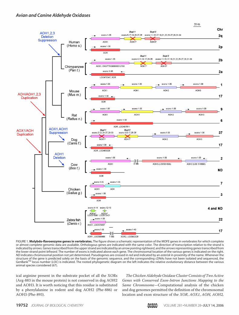
ports Divergent Gene Duplication Events during Evolution—Fig. 2 shows an unrooted phylogenetic tree obtained after com-parison of plant and animal XOR as well as aldehyde oxidaseproteins (supplemental Fig. 3 for the alignment) for whichamino acid sequences are available or can be deduced from thecorresponding genomes (see supplemental Table 5 for the listof the protein sequences and accession numbers). The centralportion (Fig. 2, blue) of the phylogram is dominated by thepresence of the characterized or putative XOR enzymes identi-fied in the plant and animal species considered. The XORbranching pattern reflects the phylogenetic relationships of liv-ing organisms. Notably, the genomes of Arabidopsis thalianaand D. rerio contain genes coding for two predicted XOR pro-teins of extremely high amino acid identity.Our phylogenetic tree demonstrates two distinct groups of
aldehyde oxidases. Indeed plant, insect, and flatworm aldehydeoxidases (Fig. 2, yellow) form a distant cluster relative to thevertebrate counterpart (Fig. 2, green), consistent with divergentevolution from a commonXORancestor (1). This confirms andextends the hypothesis of Rodriguez-Trelles et al. (41) thataldehyde oxidases evolved twice independently from two dif-ferent XOR paralogs, the second time in chordates, before thediversification of vertebrates.In the vertebrate aldehyde oxidase cluster, putative AOX1
orthologous proteins are present in all the classes (fishes,amphibians, birds, andmammals). The position of AOX1 in thetree suggests that the corresponding gene is the precursor of allthe subsequent duplications. The first evidence of an aldehydeoxidase duplication is present in birds. Interestingly, the prod-uct of this duplication (AOH) is more related to mammalianAOH1, AOH2, and AOH3 than to AOX1, suggesting ancestor-
ship. In mammals there is a clear definition of three furtheraldehyde oxidase isoenzymatic forms: AOH2, AOH1, andAOH3 in order of distance from avian AOH. Overall, the alde-hyde oxidase clusters have longer branches than theXORcoun-terparts, demonstrating faster evolution rates after separationfrom the ancestor.Purification of Chicken Kidney AOX1 Demonstrates That the
Protein Oxidizes Retinaldehyde to Retinoic Acid—The exist-ence of active aldehyde oxidase enzymes in vertebrates otherthan mammals has not been proven. To this purpose and as afirst step in the characterization of the protein products corre-sponding to the aldehyde oxidase transcripts identified inchicken and dogs, we focused on avianAOX1.We chose kidneyas the primary source of the enzyme, as this organ containsrelatively abundant amounts of the AOX1 transcript (see Fig.3A). Purification of the AOX1 protein was followed by meas-urement of RALoxidase activity, as RAL is a natural substrate ofthe enzyme in other mammalian species (26, 42). QuantitativeWestern blot analysis was also conducted on all the purifiedfractions with polyclonal antibody raised against bovine AOX1.The purification scheme is the same used for other types ofaldehyde oxidases (25, 27, 28).In a typical experiment, the calculated RAL oxidase-specific
activity of the purified protein was 5.0 units/mg as comparedwith 0.012 units/mg of the initial cytosolic extract. The overallpurification factor was 417-fold. In contrast, the final purifica-tion factor calculated by quantitativeWestern blot analysis wasonly 48.7. This suggests that the original kidney extract con-tains large proportions of catalytically inactive enzyme (�98%).The phenomenon may be the result of intrinsic enzyme insta-bility, as observed in at least three independent purificationexperiments. Alternatively, it may reflect the real presence of alarge pool of inactive enzyme in chicken kidney. This is notunprecedented, as high levels of catalytically inactive XORhavebeen reported in milk (10, 44). In this biological fluid, inactiveXOR results presumably from the loss of the terminal MoCosulfur ligand, which is likely to be present in chicken AOX1, aswell as many other mammalian aldehyde oxidases. The overallyield of purified chicken AOX1 is 1 order of magnitude lowerthan observed in the case of the bovine or mouse counterparts(25, 27, 28). The phenomenon is the consequence of the lowaffinity of catalytically active chicken AOX1 for the benzami-dine-Sepharose chromatography support (data not shown).Nevertheless, the method results in the recovery of a highlypurified and catalytically active form of AOX1.Purified RAL oxidase co-elutes with a single protein band of
�150 kDa, as assessed by electrophoresis under reducing anddenaturing conditions. The apparent molecular weight of puri-fied RAL oxidase represents the monomeric subunit of theenzyme, as predicted from the translation product of theAOX1cDNA. Upon Western blot analysis, the purified RAL oxidaseband is recognized by the anti-AOX1 antibody. Polyclonal anti-rat XOR antibodies (13) cross-react with a similar 150-kDaband throughout the procedure until the benzamidine-Sepha-rose purification step. This indicates that the final AOX1 prep-aration does not contain contaminations of the structurally andfunctionally related XOR protein. To establish its identity tochicken AOX1, the purified protein was trypsinized after
Avian and Canine Aldehyde Oxidases
19754 JOURNAL OF BIOLOGICAL CHEMISTRY VOLUME 281 • NUMBER 28 • JULY 14, 2006

reduction and carboxymethylation, and the tryptic digest wassubjected to MALDI-TOF mass spectrometry. SupplementalTable 6 demonstrates that 54 peptides could be unequivocallyidentified on the basis of the masses of the tryptic fragmentspredicted from the open reading frame of the cloned chickenAOX1 cDNA. The other sequences listed are likely to be mix-tures of the indicated peptides characterized by identicalmasses. For all peptides identified, the difference between the
calculated and experimentalmasses is less than 0.05mass units.Altogether, the identified peptides cover �55% of the entiresequence of chicken AOX1. A computer-assisted search in theNCBI Protein Database using themasses determined for the 50most abundant identified peaks did not result in any significanthit. This demonstrates that the protein band with RAL oxidaseactivity corresponds to chicken AOX1. Most interestingly, theMALDI-TOF analysis did not reveal any tryptic peptide specific
FIGURE 2. Phylogeny of eukaryotic molybdo-flavoenzymes. An unrooted dendrogram was obtained by the Phylip method after a ClustalW computer-aidedalignment of the indicated proteins. Besides G. gallus (Gg) AOX1, AOH, and C. familiaris (Cf) AOH2 and AOH3, the following amino acid sequences wereconsidered: A. nidulans (An) XOR; N. crassa (Nc) XOR; A. thaliana (At) AOX1– 4, as well as XOR1 and XOR2; Lycopersicon esculentum (Le) AOX1, AOX2, and AOX3;Rattus norvegicus (Rn) AOX1, AOH1, AOH2, AOH3, and XOR; Mus musculus (Mm) AOX1, AOH1, AOH2, AOH3, and XOR; P. troglodytes (Pt) XOR; Homo sapiens (Hs)AOX1 and XOR; Macaca fascicularis (Mf) AOX1; Pongo pygmaeus (Gog) AOX1; Felis catus (Fc) XOR; Bos taurus (Bt) AOX1, AOH2, AOH3, and XOR; G. gallus XOR; C.familiaris XOR; T. rubripes (Tr) AOX1 and XOR; D. rerio (Dr) AOX1, XOR1, and XOR2; D. melanogaster (Dm) AOX1– 4 and XOR; Bombyx mori (Bm) XOR; C. elegans (Ce)AOX1, AOX2, and XOR; Zea mays (Zm) AOX1 and AOX2; Oryctolagus cuniculus (Oc) AOX1; Strongylocentrotus purpuratus (Sp) XOR1 and XOR2; X. laevis (Xl) AOX1;Tetraodon nigroviridis (Tn) AOX1 and XOR; and Monodelphis domestica (Md) AOX1, AOH1, AOH2, AOH3, and XOR.
Avian and Canine Aldehyde Oxidases
JULY 14, 2006 • VOLUME 281 • NUMBER 28 JOURNAL OF BIOLOGICAL CHEMISTRY 19755

FIGURE 3. Tissue-specific expression of chicken AOX and AOH and dog AOH3 and XOR. The indicated tissues were dissected and total RNA extracted. A, upperpanels, Northern blot analysis of chicken AOX1, AOH, and XOR. Equal amounts (20 �g) of total RNA were run on a 1% agarose gel, blotted on separate nylon filters thatwere hybridized with radiolabeled chicken AOX1 (nt 3442–4035), AOH (nt 2909–4064), and XOR (nt 3476–3929) cDNA fragments, and oligodeoxynucleotide thatrecognizes 18 S ribosomal RNA (5�-ACGGTATCTGATCGTCTTGGAACC3�). The autoradiograms of the experiment are shown. The position of the 18 S and 28 S riboso-mal RNAs is indicated on the right. Lower panels, semiquantitative amplification of chicken AOX1, AOH, and XOR cDNAs by RT-PCR. Equal amounts of total RNA (1 �g)were reverse-transcribed and subjected to PCR amplification (30 cycles) using primers specific for the cDNAs encoding: chicken AOX1 (5�-ACGATGCAAATATGGAAT-GGGAGAA-3�, nt 3442–3466, and 5�-CTGTTCAGGTCTCATGCATTCTGG-3�, complementary to nt 4012–4035), AOH (5�-CATGTACAGAGGAGTTAACCGGAC-3�, nt 2909–2932, and 5�-TTCAGAACAGGCAGCTCCAT-3�, complementary to nt 3538–3557), XOR (5�-GGATGGCTGTTCATAATGCATGTC-3�, nt 3476–3499, and 5�-TCTGTTGGGAT-GTCTCCAAATGCT-3�, complementary to nt 3906–3929). Amplification of the chicken �-actin cDNA was used as a positive control of the experiment. The amplifiedbands were run on a 1% agarose gel and stained with ethidium bromide. M indicates DNA molecular weight markers (DNA ladder, 100–1,000 bp); H2O indicatesnegative controls for the amplification reactions run in the absence of RNA. B, upper panels, Northern blot analysis of dog AOH3 and XOR. The experiment wasperformed exactly as in A using dog AOH3 (nt 2916–4116), XOR cDNA (nt 1780–4040) fragments, and oligodeoxynucleotide that recognizes 18 S ribosomal RNA asprobes. Lower panels, amplification of dog AOH2, AOH3, and XOR cDNAs by RT-PCR. The experiment was performed as in A using primers specific for the cDNAsencoding dog AOH2 (5�-CCCAGTTGAATGATGCCTTACATC-3�, nt 961–984, and 5�-TGGCTTGCAGAGACAACAGTGGAG-3�, complementary to nt 1424–1447), AOH3(5�-CCCAAGTGAAAGACATTTTGGCTG-3�, nt 1085–1108, and 5�-TTCTGTGCACTGACCATGACAGCC-3�, complementary to nt 1548–1571), and the putative XOR tran-script (XM_857591, 5�-TGTGGAAAAGACACTGCATGATGC-3�, nt 963–986, and 5�-CTTGAGAGCTGAGATGGTTCTGTC-3�, complementary to nt 1423–1446). Amplifica-tion of the dog �-actin cDNA was used as a positive internal control of the experiment.
Avian and Canine Aldehyde Oxidases
19756 JOURNAL OF BIOLOGICAL CHEMISTRY VOLUME 281 • NUMBER 28 • JULY 14, 2006

for AOH, whose transcript is present in kidney (see Fig. 3A).Although the low yields preclude a thorough structural andenzymatic characterization of chicken AOX1, the purifiedenzyme is capable of oxidizing not only retinaldehyde but alsobenzaldehyde (data not shown) as the rodent counterpart. Fur-thermore, chicken AOX1 is devoid of significant xanthine orhypoxanthine oxidase activity.The Tissue Distribution of the Aldehyde Oxidase Transcripts
and Proteins Varies in Different Animal Species—The two richestsources of human and cow AOX1, as well as rodent AOX1 andAOH1, are liver and lung (1). High levels of mouse and rat AOH2are present in Harderian glands and keratinized epithelia, such asskin and esophagus. Rodent AOH3 is expressed selectively in thenasal mucosa (25). We determined the tissue-specific expressionof the chicken and dog aldehyde oxidase genes by Northern blotand RT-PCR analyses. The distribution of the XOR transcripts inthe two animal species is included for comparison.The Northern blot data (Fig. 3A, upper panels) indicate
that chicken AOX1 mRNA is measurable only in kidney andheart. However, the much more sensitive RT-PCR technol-ogy (Fig. 3A, lower panels) highlights the transcript in otherorgans, including liver. Both the Northern blot and RT-PCRdata indicate that the AOH mRNA is co-expressed withAOX1 in several tissues, such as kidney, heart, trachea, liver,and stomach. In addition, brain, skin, and thymus demon-strate selective expression of the AOHmRNA. Northern blotanalysis of AOX1 and AOH demonstrates the presence oftwo specific bands. Although the nature of the more slowlymigrating AOX1 and AOH bands is unknown, alternativelyspliced forms of mouse aldehyde oxidase transcripts involvingthe 3�-untranslated region have already been described inmouse (25). Similar to what was observed in rodents (45), theorgans expressing the highest levels of chicken XORmRNA arekidney, liver, and small intestine. Once again and consistentwith the rodent situation, RT-PCR demonstrates generalizedexpression of low levels of this transcript. Compared with theexisting literature on other species (5, 6, 8), our data indicatethat the chicken and mammalian AOX1 transcripts show dif-ferent profiles of tissue-specific expression. Thus, although thestructural data indicate orthology, chicken and mammalianAOX1 may serve slightly different tissue-specific functionsin the two vertebrates. The different tissue distribution ofchicken AOH relative to AOH1, AOH2, and AOH3 is con-sistent with neo-functionalization of the rodent gene dupli-cation products (41).The expression of dog AOH2 and AOH3 was also studied
across a panel of tissues both by Northern blot and RT-PCR (Fig.3B). To avoid cross-hybridization, probes and primers wereselected fromthe sequenceswith the lowest similaritybetween theAOH2andAOH3cDNAs.Theabundanceof thedogAOH2tran-script in target tissues is too low to permit detection by Northernblot experiments performed on total RNA. Nevertheless, asrevealed by RT-PCR, dog AOH2 expression is restricted and verysimilar to what was reported in mice (28). This is consistent withthe proposed orthology with rodent AOH2. The high levels ofAOH2 transcript detected in the lacrimal gland suggest that thecorresponding protein plays an organ-specific function in thislocation. Interestingly, in dogs, this gland is the functional substi-
tute of the rodent Harderian gland. The expression of the canineAOH3gene is evenmore restricted than that ofAOH2. Indeed, theonly tissue where significant amounts of the corresponding tran-script are evident is the nasal mucosa. Once again this is in linewith the fact that dog and rodent AOH3 are the products oforthologous genes, as large amounts of theprotein are synthesizedselectively inmouse Bowman glands, the primary exocrine glandspresent in nasal mucosa. Consistent with what is stated above,although RT-PCR demonstrates the presence of dog AOX1mRNA fragments, detectable levels of a full-length transcriptswere not measured after Northern blot analysis (data not shown).The pattern ofXOR expression revealed by the same technique isdifferent from that observed in chicken and rodents (45). How-ever, the ubiquitous expression of low levels of XOR mRNA isdemonstrated by RT-PCR.Chicken and Dog Livers Do Not Express Detectable Amounts
of AOX1andDoNotContain RALOxidaseActivity—Tissue- orcell-specific expression of a given mRNA is not necessarilyassociated with synthesis and accumulation of the correspond-ing protein. Translational and post-translational control of theXOR enzyme was reported in the mouse hepatic tissue and incertain cell lines (13, 45).We verified the presence of the AOX1and AOH1 proteins in the two chicken tissues, kidney andheart, characterized by the highest levels of AOX1 and AOHmRNAs. Given the importance of liver in the metabolism ofxenobiotics mediated by cytosolic aldehyde oxidases (46–49),we extended our analysis of this organ in both chicken and dog.To do this, we used a polyclonal antibody raised against bovineAOX1 that does not recognize XORs of different origin.6
First, we determined the cross-reactivity of the anti-bovineantibody with chicken and dog aldehyde oxidases (Fig. 4A).Transfection of COS-7 cells, with the cDNAs coding forchicken AOX1 and AOH, dog AOH2 (data not shown) andAOH3, and the mouse AOH1-positive control, demonstratesthat the antibody cross-reacts with the four proteins. In con-trast, the antibody recognizes neither chicken nor dog XOR(data not shown).Fig. 4Bdemonstrates that avianheart andkidney contain signif-
icant amounts of aldehyde oxidase immunoreactivity. In bothcases, this is likely to be the result of AOX1 expression, as sug-gested by theNorthern blot data (Fig. 3A). Chicken and dog liversare devoid of aldehyde oxidase immunoreactive bands. In the caseof chicken, this indicates that the low levels of AOX1 and AOHtranscripts measurable by RT-PCR are insufficient to result in thetranslationofdetectableamountsof thecorrespondingprotein.Asto dog, the result is consistent with the lack of detectable amountsof AOX1, AOH2, or AOH3mRNAs in the hepatic tissue.That both chicken and dog livers are devoid of aldehyde oxi-
dase immunoreactivity is different from what was observed inhumans, mice, and rats, where the hepatic tissue is one of therichest source ofAOX1 and/orAOH1 (26, 49, 50). Aldehydes ofphysiological, toxicological, and pharmacological interest arepotential substrates of both aldehyde oxidases of the MOFEfamily and aldehyde dehydrogenases. The latter family consistsof numerous isoenzymes (51) and utilizes NAD� as a cofactorfor the catalyzed oxidation reaction. To evaluate the functionalsignificance of the aldehyde oxidase deficit in dogs and chickenliver, we determined the ability of cytosolic extracts to oxidize
Avian and Canine Aldehyde Oxidases
JULY 14, 2006 • VOLUME 281 • NUMBER 28 JOURNAL OF BIOLOGICAL CHEMISTRY 19757

RAL in the presence and absence of NAD�. For this purpose,liver cytosolic extracts were depleted of NAD� by passage onsize exclusion columns and subsequently repleted or not withexogenously added dinucleotide. The results were comparedwith those obtained in cytosolic extracts of C57/Bl and DBA/2mice that express high and very low levels, respectively, of bothAOX1 and AOH1 (26). Fig. 4C shows typical HPLCs obtainedafter incubation ofmurine, canine, and avian liver extracts withRAL. Fig. 4D summarizes the quantitative results obtainedwiththis type of assay. C57/Bl cytosols oxidize RAL to retinoic acidvery rapidly. Addition of NAD� prior to incubation with theRAL substrate does not result in a significant increase in theamount of retinoic acid produced. The contention is supportedby the fact that DBA/2mice cytosol contains less than 5% of theNAD�-independent RALoxidase activity present in theC57/Blcounterpart. This activity is because of the residual AOX1 pro-tein present in the hepatic tissue of this strain (26). A modestincrease in the production of retinoic acid is observed uponaddition of NAD� to the DBA/2 extracts. In the absence ofNAD�, chicken and dog liver extracts do not show detectableRAL metabolizing activity. Addition of NAD� causes a slightbut significant increase in the production of retinoic acid inchicken liver but not in dog liver. The cytosolic fraction ofchicken kidney contains significant levels of NAD�-independ-ent RAL oxidase activity, which is not further augmented byaddition of the dinucleotide. Taken together, our results indi-cate that the predominant enzymatic activity responsible forthe metabolism of RAL in the cytosol of organs such as mouseliver and chicken kidney is an aldehyde oxidase and not a dehy-drogenase. In addition, the data confirm the absence of signifi-cant amounts of catalytically active MOFEs of the aldehydeoxidase type in the liver of dogs and chicken.
DISCUSSION
In this study, we identify and characterize the cDNAs and thecorresponding genes coding for the MOFE synthesized in dogand chicken. Our results demonstrate the presence of oneXORand two aldehyde oxidase genes in avians. In contrast, thecanine organism is endowedwith oneXOR, two active aldehydeoxidase genes, and an equal number of cognate pseudogenes.Purification of chicken AOX1 permitted the first biochemicalcharacterization of a nonmammalian aldehyde oxidase. Ourresults are relevant in terms of the biology of aldehyde oxidases.More importantly, they cast new light on the evolution of theMOFE family of genes.MOFEs of the aldehyde oxidase type are believed to repre-
sent an importantmetabolic system capable of oxidizing a largearray of endogenous and exogenous substrates (1). The recentdemonstration of multiple rodent aldehyde oxidases, charac-terized by tissue- and cell-specific expression, suggests that spe-cific members of this protein family play a role in the localmetabolism of unidentified compounds of physiological, phar-macological, and toxicological significance. The four rodent
FIGURE 4. AOX1 and retinaldehyde oxidase activity in chicken, dog, andmouse liver cytosolic fractions. A, cross-reactivity of the anti-bovine AOX1(anti-bAOX1) and anti-rat XOR (anti-rXOR) antibodies with the indicatedmouse, chicken, and dog proteins. Expression vector (pcMY�, Invitrogen)containing cDNAs corresponding to the indicated proteins or the void vector(vector) were transfected in COS-7 cells. Two days following transfection,cytosolic extracts were subjected to Western blot analysis with the indicatedantibodies. The position of relevant protein molecular mass markers is indicatedon the right. B, Western blot analysis of aldehyde oxidase immunoreactive pro-teins in chicken, dog, and mouse tissues. Equal amounts of 100,000 � g cytosolicextracts obtained from the indicated tissues were subjected to Western blot anal-ysis, using anti-bovine AOX1 antibodies. The position of relevant protein molec-ular mass markers is shown on the right. C and D, retinaldehyde oxidase activity inmouse, dog, and chicken cytosolic extracts. Equal amounts of desalted 100,000�g cytosolic extracts obtained from the indicated tissues were incubated for 10min with retinaldehyde in the absence or presence of exogenously added NAD�.Samples were extracted in organic solvent and subjected to HPLC analysis. C,illustrates representative HPLCs of the indicated samples in the absence of NAD�:RA, retinoic acid. D shows the levels of RAL oxidase activity measured using theindicated extracts in the presence (�NAD) or absence (�NAD) of NAD�. The
results obtained in the absence of NAD� are due to AOX1, whereas thoseobtained in the presence of the cofactor are because of both AOX1 and alde-hyde dehydrogenase(s). *, significantly higher than the corresponding value(�NAD) (p � 0.01 after Student’s t test).
Avian and Canine Aldehyde Oxidases
19758 JOURNAL OF BIOLOGICAL CHEMISTRY VOLUME 281 • NUMBER 28 • JULY 14, 2006

aldehyde oxidases are structurally related enzymes, have broadsubstrate specificity, and do not show major differences interms of substrate preference (25, 27, 28).6 Among the generalsubstrates of potential physiological relevance, RAL stands out(25, 27, 28, 42) and has been the object of the enzymatic studiesreported here. As the rodent counterparts (25, 27, 28, 42), puri-fied chicken AOX1 oxidizes RAL to retinoic acid. RAL isbelieved to represent the physiological precursor of retinoicacid, a recognized morphogen and an important regulator ofthe homeostasis of different tissues in the adult. RAL is theoxidation product of vitamin A (retinol) and is thought to betransformed to retinoic acid predominantly by RAL dehydro-genase (52), a specific isoenzyme of the largeNAD�-dependentaldehyde dehydrogenase family (51). In mammals, a major siteof RAL metabolism is the liver. Mouse liver cytosol oxidizesRAL to retinoic acid predominantly in a NAD�-independentmanner. Thus, we propose that aldehyde oxidases and not alde-hyde dehydrogenases are a major determinant of retinoic acidsynthesis in this organ. This metabolic step is catalyzed by analdehyde oxidase of the AOX1 or AOH1 type, as indicated bythe results obtained in C57/Bl and DBA/2 mice (26). Interest-ingly, the liver of both chicken and dog is devoid of AOX1 orother types of aldehyde oxidase of the MOFE family. As a con-sequence of this, the cytosol of avian and canine hepatic tissueslack RAL oxidizing activity. At present, we do not knowwhether the deficit of RAL oxidase observed in chickens anddogs is species-specific or limited to certain strains, as observedin mice (26). Indeed, the data presented in this study wereobtained in one strain of chicken (Broiler) and one strain ofdogs (Beagle). However, at least in dogs, we favor the formerpossibility, as the available genomic data demonstrate that theorthologs of the murine AOX1 and AOH1 genes are function-ally inactive and have been transformed into pseudogenes inboth Boxers andBeagles. Regardless of this detail, our data indi-cate that the metabolism of vitamin A in a major storage organis different in avians and dogs relative to rodents and humans.The lack of aldehyde oxidase activity in dog liver is of greatinterest also in a pharmacological and toxicological perspec-tive. In fact, dogs are common experimental models in xenobi-otic metabolism studies, and aldehyde oxidases are known tometabolize drugs, like methotrexate (53) and 6-mercaptopu-rine (54) and environmental pollutants like nitropolycyclic aro-matic hydrocarbons.Definition of the structure of the chicken and dog proteins
and corresponding genes contributes to generate a moredetailed picture of the evolutional history of MOFEs. The evo-lution of MOFE genes is complex and characterized by dupli-cation and suppression events, whose traces are present in thegenomes of the extant plant and animal species. The aminoacid sequence of all MOFEs is highly conserved throughoutevolution, although the following four distinct groups ofenzymes can be recognized: XORs, aldehyde oxidases, car-bon monoxide dehydrogenases, and oxidoreductases of thequinoline and isoquinoline oxidase type (1, 55–58). The lasttwo groups of proteins are present only in bacteria and arenot relevant for our discussion, which focuses on the evolu-tion of MOFEs in vertebrates.XORs and aldehyde oxidases are very similar proteins and
are distinguished for their ability or inability to use hypox-anthine and xanthine as substrates (1). From a structuralpoint of view, XORs are identified on the basis of the pres-ence and conservation of two charged amino acids, which areimportant for the positioning of hypoxanthine and xanthinein the substrate pocket (2, 3). Furthermore, XORs can utilizeNAD� as a cofactor and act as dehydrogenases (59). As such,XORs are characterized by the presence of a conserved tyro-sine in the FAD binding domain (see supplemental Figs. 1and 2). These structural characteristics were used for theidentification of XORs and aldehyde oxidases in the genomeof various organisms and in the reconstruction of the phylo-genetic tree presented in Fig. 2.The similarity between the amino acid sequences of XORs
and the various aldehyde oxidases indicates that the corre-sponding genes must have evolved from an ancient commonprecursor (1, 60). This putative precursor enzyme is likely to bemore closely related to the extant XORs than to aldehyde oxi-dases. Indeed, relative to XORs, which are already present inbacteria (61, 62), aldehyde oxidases are believed to be a morerecent set of genes identified only in multicellular eukaryoticorganisms of plant and animal origin (1). However, such a viewmay be challenged by the recent report describing an aldehydeoxidase fromMethylobacillus (63).As suggested previously (1), the first step in the evolution of
MOFEsmust have been the consolidation of the genes respon-sible for the synthesis of the distinct peptide chains typical ofbacterial XOR proteins into a single chain characteristic ofeukaryotic XORs. Bacteria, such as Escherichia coli, containstructural genes encoding three chains corresponding to the2Fe/2S-containing amino terminus, the FAD-binding interme-diate, and the MoCo/substrate carboxyl-terminal domains ofXOR (61). Other bacterial XORs, like Rhodobacter capsulatus,consist of �2�2 tetramers (62). Consolidation of XOR into asingle polypeptide chain must have been an early event duringevolution, as fungi like Aspergillus nidulans and Neurosporacrassa are already endowed with a single gene coding for theentire protein subunit (64, 65). This was followed by an increasein the number of the XOR exons. The phenomenon is evidentwhen the exon structure of XORs from A. nidulans (4 exons),Drosophila melanogaster (4 exons), Caenorhabditis elegans (16exons), D. rerio (35 exons), and all the other vertebrates (35exons) is compared. The process of exon multiplication is notprogressive and does not follow a simple pattern, as a phyloge-netically more ancient organism, like the flatworm C. elegans,presents an XOR gene with a larger number of exons than themore recent D. melanogaster counterpart (1).Additional and more recent steps in the evolution ofMOFEs
were represented by one or more rounds of duplication result-ing in the appearance of up to four distinct genes coding forenzymes of the aldehyde oxidase type. The data presented inthis and other papers (41) suggest that the process followeddifferent pathways in plants, insects, and vertebrates. Theobservation suggests that insect and vertebrate aldehyde oxi-dase genes are not orthologs, and the corresponding proteinproducts must serve different physiological functions.The available data on similarity, gene structure, and partial
tissue distribution suggest the following evolutionarymodel for
Avian and Canine Aldehyde Oxidases
JULY 14, 2006 • VOLUME 281 • NUMBER 28 JOURNAL OF BIOLOGICAL CHEMISTRY 19759

this group ofMOFEs. VertebrateAOX1 is the proximal paralogof the ancestor XOR gene. In fact, the genome of fishes, such asTakifugu rubripes and D. rerio, contains a single aldehyde oxi-dase, which is more closely related to rodent, bovine, or dogAOX1 than toAOH1,AOH2, orAOH3 of the same origin, indi-cating orthology. The transition of sea vertebrates into amphib-ians, likeXenopus laevis, does not seem to be associated with anincrease in the number of aldehyde oxidase genes, as this spe-cies is characterized by the presence of the soleAOX1 ortholog.G. gallus is the first animal in which the presence of an AOX1duplication (AOH) is evident. Chicken AOX1 and AOHmap tothe same chromosome a short distance from each other andrepresent the first seed of the more complex gene clusterobserved in rodents. The presence of an increasing number ofaldehyde oxidases from birds to rodents suggests that the proc-ess of gene duplication is sequential and proceeds throughduplication events involving one gene at a time, not necessarilyin the 5� to 3� direction of the gene cluster. The AOH gene islikely to be the ancestor of mammalian AOH1, AOH2, andAOH3. The phylogenetic analysis presented indicates that theclosest relatives to chicken AOH are the AOH2 proteins. Thusit is possible that AOH1 and AOH3 are further duplications ofthe AOH2 gene. However, the tissue distribution of chickenAOH and mouse AOH2 does not match, indicating differentfunctional roles.The generation of the two further aldehyde oxidase gene
duplications observed in rodents is a relatively recent event andparallels the evolution of terrestrial mammals. The appearanceof AOH1, AOH2, and AOH3 is likely to be dictated by the needfor new tissue- or cell-specific functions. In the case of AOH2and AOH3, this contention is supported by the very restricteddistribution of the two corresponding mouse and rat enzymesin specialized structures like Bowman (25) and Harderianglands. Regardless of the functional significance, the last stepsof the process of mammalian MOFE gene multiplication pre-ceded the development of placentation, as the genome of opos-sum (see Fig. 2), a monotremate, is predicted to contain thewhole complement of the AOX1, AOH1, AOH2, and AOH3orthologs. In this context, it would be interesting to determinethe number of aldehyde oxidase genes in marine mammals,such as dolphins and whales.The process of expansion of the aldehyde oxidase gene
cluster stops with the evolution of rodents. Indeed, thecanine, primate, and human genomes are characterized by areduction in the number of the aldehyde oxidases, which isthe consequence of selective gene suppression and inactiva-tion events. The traces of these events are recognizable in thegenomes of the above-mentioned species under the form ofpseudogenes. The aldehyde oxidases inactivated in dogs areAOX1 and AOH1, whereas humans and primates suppressAOH1, AOH2, and AOH3. Humans and primates have lost anyevidence of the AOH2 duplication, although they maintain thevestiges of the rodent AOH1 and AOH3 genes. Relative tohumans, rodents and dogs are characterized by a much moredeveloped olfactory system, which is of the utmost importancefor the survival of these animals in the environment. Given thespecific localization in the exocrine glands of the nasal mucosa(25), it is plausible that AOH3 plays a role in the perception of
odorants. Indeed, the enzymemight control the duration or theintensity of the olfactory stimuli through rapid metabolism ofvolatile aldehyde odorants. This functionmay have becomedis-pensable or redundant inmen, leading to the suppression of theAOH3 gene. This is not unprecedented, because themajority ofthe genes coding for odorant receptors inmice has undergone aprocess of progressive inactivation in humans (43). Similarly,suppression of theAOH2 genemay be related to the disappear-ance of a functional equivalent of the Harderian gland or to thedifferent characteristics of the skin and oral mucosa in men,primates, and other mammals.In summary, the aldehyde oxidase gene cluster represents a
complex and interesting model of enzymatic evolution. Thereasons at the basis of this complexity will become clear asfurther information on the functional significance of theseenzymes is available.
Acknowledgments—We are grateful to Dr. Mario Salmona and Prof.Silvio Garattini for critical revision of the manuscript.
REFERENCES1. Garattini, E., Mendel, R., Romao, M. J., Wright, R., and Terao, M. (2003)
Biochem. J. 372, 15–322. Enroth, C., Eger, B. T., Okamoto, K., Nishino, T., Nishino, T., and Pai, E. F.
(2000) Proc. Natl. Acad. Sci. U. S. A. 97, 10723–107283. Okamoto, K., Matsumoto, K., Hille, R., Eger, B. T., Pai, E. F., and Nishino,
T. (2004) Proc. Natl. Acad. Sci. U. S. A. 101, 7931–79364. Cazzaniga, G., Terao, M., Lo Schiavo, P., Galbiati, F., Segalla, F., Seldin,
M. F., and Garattini, E. (1994) Genomics 23, 390–4025. Calzi, M. L., Raviolo, C., Ghibaudi, E., de Gioia, L., Salmona, M., Cazza-
niga, G., Kurosaki, M., Terao, M., and Garattini, E. (1995) J. Biol. Chem.270, 31037–31045
6. Wright, R. M., Vaitaitis, G. M., Wilson, C. M., Repine, T. B., Terada, L. S.,and Repine, J. E. (1993) Proc. Natl. Acad. Sci. U. S. A. 90, 10690–10694
7. Demontis, S., Kurosaki,M., Saccone, S.,Motta, S., Garattini, E., andTerao,M. (1999) Biochim. Biophys. Acta 1489, 207–222
8. Kurosaki, M., Demontis, S., Barzago, M. M., Garattini, E., and Terao, M.(1999) Biochem. J. 341, 71–80
9. Avis, P. G., Bergel, F., Bray, R. C., and Shooter, K. V. (1954) Nature 173,1230–1231
10. Godber, B. L., Schwarz, G.,Mendel, R. R., Lowe,D. J., Bray, R. C., Eisenthal,R., and Harrison, R. (2005) Biochem. J. 388, 501–508
11. Martin, H.M., Hancock, J. T., Salisbury, V., and Harrison, R. (2004) Infect.Immun. 72, 4933–4939
12. Harrison, R. (2004) Drug. Metab. Rev. 36, 363–37513. Falciani, F., Terao, M., Goldwurm, S., Ronchi, A., Gatti, A., Minoia, C., Li
Calzi, M., Salmona, M., Cazzaniga, G., and Garattini, E. (1994) Biochem. J.298, 69–77
14. Falciani, F., Ghezzi, P., Terao, M., Cazzaniga, G., and Garattini, E. (1992)Biochem. J. 285, 1001–1008
15. Vorbach, C., Scriven, A., and Capecchi, M. R. (2002) Genes Dev. 16,3223–3235
16. McManaman, J. L., Palmer, C. A.,Wright, R. M., and Neville, M. C. (2002)J. Physiol. (Lond.) 545, 567–579
17. Kurosaki, M., Zanotta, S., Li Calzi, M., Garattini, E., and Terao, M. (1996)Biochem. J. 319, 801–810
18. Rovira, O. T., II, Starost, M. F., Liu, C., and Finkel, T. (2004) Circ. Res. 95,1118–1124
19. Fabre, G., Fabre, I., Matherly, L. H., Cano, J. P., and Goldman, I. D. (1984)J. Biol. Chem. 259, 5066–5072
20. Beedham,C.,Miceli, J. J., andObach, R. S. (2003) J. Clin. Psychopharmacol.23, 229–232
21. Beedham, C. (1997) Pharm. World Sci. 19, 255–263
Avian and Canine Aldehyde Oxidases
19760 JOURNAL OF BIOLOGICAL CHEMISTRY VOLUME 281 • NUMBER 28 • JULY 14, 2006

22. Beedham, C., Peet, C. F., Panoutsopoulos, G. I., Carter, H., and Smith, J. A.(1995) Prog. Brain Res. 106, 345–353
23. Xu, P., Huecksteadt, T. P., and Hoidal, J. R. (1996) Genomics 34, 173–18024. Terao,M., Kurosaki,M., Demontis, S., Zanotta, S., andGarattini, E. (1998)
Biochem. J. 332, 383–39325. Kurosaki, M., Terao, M., Barzago, M. M., Bastone, A., Bernardinello, D.,
Salmona, M., and Garattini, E. (2004) J. Biol. Chem. 279, 50482–5049826. Vila, R., Kurosaki, M., Barzago, M. M., Kolek, M., Bastone, A., Colombo,
L., Salmona, M., Terao, M., and Garattini, E. (2004) J. Biol. Chem. 279,8668–8883
27. Terao, M., Kurosaki, M., Marini, M., Vanoni, M. A., Saltini, G., Bonetto,V., Bastone, A., Federico, C., Saccone, S., Fanelli, R., Salmona, M., andGarattini, E. (2001) J. Biol. Chem. 276, 46347–46363
28. Terao, M., Kurosaki, M., Saltini, G., Demontis, S., Marini, M., Salmona,M., and Garattini, E. (2000) J. Biol. Chem. 275, 30690–30700
29. Buzzell, G. R. (1996)Microsc. Res. Tech. 34, 2–530. Seo, M., Peeters, A. J., Koiwai, H., Oritani, T., Marion-Poll, A., Zeevaart,
J. A., Koornneef, M., Kamiya, Y., and Koshiba, T. (2000) Proc. Natl. Acad.Sci. U. S. A. 97, 12908–12913
31. Ori, N., Eshed, Y., Pinto, P., Paran, I., Zamir, D., and Fluhr, R. (1997) J. Biol.Chem. 272, 1019–1025
32. Misra, S., Crosby, M. A., Mungall, C. J., Matthews, B. B., Campbell, K. S.,Hradecky, P., Huang, Y., Kaminker, J. S., Millburn, G. H., Prochnik, S. E.,Smith, C. D., Tupy, J. L., Whitfied, E. J., Bayraktaroglu, L., Berman, B. P.,Bettencourt, B. R., Celniker, S. E., de Grey, A. D., Drysdale, R. A., Harris,N. L., Richter, J., Russo, S., Schroeder, A. J., Shu, S. Q., Stapleton, M.,Yamada, C., Ashburner, M., Gelbart,W.M., Rubin, G.M., and Lewis, S. E.(2002) Genome Biol. 3, RESEARCH0083
33. Sambrook, J., Fritsch, E. F., and Maniatis, T. (eds) (1989)Molecular Clon-ing: A LaboratoryManual, 2nd Ed., Cold Spring Harbor Laboratory Press,Cold Spring Harbor, NY
34. Chenna, R., Sugawara,H., Koike, T., Lopez, R., Gibson, T. J., Higgins, D.G.,and Thompson, J. D. (2003) Nucleic Acids Res. 31, 3497–3500
35. Saitou, N., and Nei, M. (1987)Mol. Biol. Evol. 4, 406–42536. Sato, A., Nishino, T., Noda, K., Amaya, Y., and Nishino, T. (1995) J. Biol.
Chem. 270, 2818–282637. Nishino, T., and Nishino, T. (1987) Biochemistry 26, 3068–307238. Nishino, T., and Nishino, T. (1989) J. Biol. Chem. 264, 5468–547339. Clark, M. P., Chow, C. W., Rinaldo, J. E., and Chalkley, R. (1998) Nucleic
Acids Res. 26, 1801–180640. Terao,M., Kurosaki,M., Zanotta, S., andGarattini, E. (1997)Biochem. Soc.
Trans. 25, 791–79641. Rodriguez-Trelles, F., Tarrio, R., and Ayala, F. J. (2003) Proc. Natl. Acad.
Sci. U. S. A. 100, 13413–1341742. Huang, D. Y., Furukawa, A., and Ichikawa, Y. (1999) Arch. Biochem. Bio-
phys. 364, 264–272
43. Buck, L. B. (2004) Nutr. Rev. 62, S184–S18844. Godber, B., Sanders, S., Harrison, R., Eisenthal, R., and Bray, R. C. (1997)
Biochem. Soc. Trans 25, 519S45. Kurosaki,M., Li Calzi,M., Scanziani, E., Garattini, E., andTerao,M. (1995)
Biochem. J. 306, 225–23446. Obach, R. S. (2004) Drug Metab. Dispos. 32, 89–9747. Rivera, S. P., Choi, H. H., Chapman, B., Whitekus, M. J., Terao, M., Garat-
tini, E., and Hankinson, O. (2005) Toxicology 207, 401–40948. Obach, R. S., Huynh, P., Allen, M. C., and Beedham, C. (2004) J. Clin.
Pharmacol. 44, 7–1949. Renwick, A. B., Ball, S. E., Tredger, J. M., Price, R. J., Walters, D. G., Kao, J.,
Scatina, J. A., and Lake, B. G. (2002) Xenobiotica 32, 849–86250. Vasiliou, V., Pappa, A., and Estey, T. (2004)DrugMetab. Rev. 36, 279–29951. Vasiliou, V., Bairoch, A., Tipton, K. F., and Nebert, D. W. (1999) Pharma-
cogenetics 9, 421–43452. Duester, G. (2000) Eur. J. Biochem. 267, 4315–432453. Jordan, C. G., Rashidi, M. R., Laljee, H., Clarke, S. E., Brown, J. E., and
Beedham, C. (1999) J. Pharm. Pharmacol. 51, 411–41854. Rooseboom, M., Commandeur, J. N., and Vermeulen, N. P. (2004) Phar-
macol. Rev. 56, 53–10255. Dobbek, H., Gremer, L., Meyer, O., and Huber, R. (1999) Proc. Natl. Acad.
Sci. U. S. A. 96, 8884–888956. Gremer, L., Kellner, S., Dobbek, H., Huber, R., andMeyer, O. (2000) J. Biol.
Chem. 275, 1864–187257. Frerichs-Deeken, U., Ranguelova, K., Kappl, R., Huttermann, J., and
Fetzner, S. (2004) Biochemistry 43, 14485–1449958. Parschat, K., Hauer, B., Kappl, R., Kraft, R., Huttermann, J., and Fetzner, S.
(2003) J. Biol. Chem. 278, 27483–2749459. Nishino, T., Okamoto, K., Kawaguchi, Y., Hori, H., Matsumura, T., Eger,
B. T., Pai, E. F., and Nishino, T. (2005) J. Biol. Chem. 280, 24888–2489460. Wurzinger, K. H., and Hartenstein, R. (1974) Comp. Biochem. Physiol. B.
49, 171–18561. Yamamoto, Y., Aiba, H., Baba, T., Hayashi, K., Inada, T., Isono, K., Itoh, T.,
Kimura, S., Kitagawa,M.,Makino, K.,Miki, T.,Mitsuhashi,N.,Mizobuchi,K., Mori, H., Nakade, S., Nakamura, Y., Nashimoto, H., Oshima, T.,Oyama, S., Saito, N., Sampei, G., Satoh, Y., Sivasundaram, S., Tagami, H.,and Horiuchi, T. (1997) DNA Res. 4, 91–113
62. Leimkuhler, S., Stockert, A. L., Igarashi, K., Nishino, T., andHille, R. (2004)J. Biol. Chem. 279, 40437–40444
63. Yasuhara, A., Akiba-Goto, M., and Aisaka, K. (2005) Biosci. Biotechnol.Biochem. 69, 2435–2438
64. Glatigny, A., and Scazzocchio, C. (1995) J. Biol. Chem. 270, 3534–355065. Griffith, A. B., and Garrett, R. H. (1988) Biochem. Genet. 26, 37–5266. Attwood, T. K., Beck, M. E., Bleasby, A. J., Degtyarenko, K., Michie, A. D.,
and Parry-Smith, D. J. (1997) Nucleic Acids Res. 25, 212–21767. Hedges, S. B. (2002) Nat. Rev. Genet. 3, 838–849
Avian and Canine Aldehyde Oxidases
JULY 14, 2006 • VOLUME 281 • NUMBER 28 JOURNAL OF BIOLOGICAL CHEMISTRY 19761
Copyright © 2022 FDOKUMEN