
Australian Standard Diagnostic Procedures for for Animal Health
19
Australian Standard Diagnostic Techniques for Animal Diseases STANDING COMMITTEE ON AGRICULTURE AND RESOURCE MANAGEMENT Eolsolr* Leigh A. Corner Trevor J. Bagust Sub-Committee on Animal Health Laboratory Standards
-
Upload
khangminh22 -
Category
Documents
-
view
0 -
download
0
Transcript of Australian Standard Diagnostic Procedures for for Animal Health
Australian
Standard
Diagnostic
Techniques
for Animal Diseases
STANDING COMMITTEE ON AGRICULTURE
AND RESOURCE MANAGEMENT
Eolsolr*
Leigh A. Corner
Trevor J. Bagust
Sub-Committee
on Animal Health
Laboratory Standards
John A Whitlock
Permission by CSIRO Publishing to reproduce this material is gratefully acknowledged. Source material is by courtesy of the Standing Committee on Agriculture and Resource Management [SCARM].
John A Whitlock
Contact us: John Whitlock. J.A. Whitlock & Co PO Box 51 EASTWOOD NSW 2122 Australia. = = = = [email protected] -- www.whitlock.com.au/slides
John A Whitlock
This document was scanned by me and converted to an original image Acrobat document with searchable text.
John A Whitlock
This edition was "out of print" and under review in 2001 with a new edition expected by mid 2002. Please check CSIRO Publishing for the current version.

.
National Library of Australia Cataloguing-in-Publication Entry
Australian Standard Diagnostic Techniques for Animal Diseases
ISBN 0 643 05243 7
1. Veterinary medicine - Australia - Diagnosis. 2. Animals - Diseases - Australia -Diagnosis. I. Comer, L.A. (Leigh Austin). II. Bagust, Trevor John. III. CSIRO. IV. Agricultural Council of Australia and New Zealand. Standing Committee on Agriculture and Resource Management. V. Agricultural Council of Australia and New Zealand. Sub-Committee on Animal Health Laboratory Standards.
636.0896075
0 Commonwealth of Australia and each of its States and Territories 1993 This work is copyright and apart from dny use as permitted under the Copyright Ad 1968, no part may be reproduced by any process without the written permission from the publisher, CSIRO, acting on behalf of the Standing Committee on Agriculture and Resource Management (SCARh4). Requests and enquiries concerning reproduction and rights should be addressed to: Managing Editor, CSIRO Publications, at the address below.
Managing Editor: Kevin Jeans Editor: Nexa Cloud-Guest Cover design: Melissa Spencer Production Manager: Jim Quinlan
Available from: CSIRO Information Services PO Box 89 (314 Albert Street) East Melbourne Victoria, 3002 Australia
Tel: (03) 418 7217 Int: (+613) 418 7217 Fax: (03) 419 0459 Lnt: (+613) 419 0459

AUSTRALIAN STANDARD DIAGNOSTIC TECHNIQUES FOR ANIMAL DISEASES
STANDING COMMITTEE ON AGRICULTURE AND RESOURCE MANAGEMENT
ANIMAL HEALTH COMMITTEE
SUB-COMMITTEE ON ANIMAL HEALTH LABORATORY STANDARDS
Anthelmintic Resistance
in she&p
M. Lyndal-Murphy Animal Research Institute, Queensland Department of Primary Industries, Locked Mail Bag 4, Moorooka, Qld 4105, Australia.
Contents 1. Introduction
2. Resistance diagnosis
3. Resistance diagnosed on faecal egg counts 4. Failure to obtain a response to drenching
4.1. Anthelmintic choice 4.2. Anthelmintic administration 4.3. Parasite factors 4.4. Concurrent conditions
5. Resistance diagnosed on a faecal egg count reduction test 5.1. Field trials 5.2. Laboratory procedures
6. Resistance diagnosed on an anthelmintic drench and slaughter trial 6.1. Worm-free host lambs 6.2. Production of infective larvae 6.3. Infection of worm-free lambs 6.4. Testing the drench 6.5. Calculation of results
7. Resistance diagnosed on egg hatch assay 7.1. Reagents 7.2. Egg recovery 7.3. Statistical analysis
8. Broad spectrum anthelmintic resistance diagnosed by in vitro larval development assay 8.1. Field collection 8.2. Laboratory procedure 8.3. Calculation of results and interpretation
9. Counting techniques for strongyle eggs 9.1. The modified McMaster method 9.2. Composite bulking technique 9.3. Centrifugation method
10. Faecal culture and identification of nematode larvae 10.1. Setting the culture up 10.2. Reading the culture
11 .Total worm count - gastro-intestinal nematodes 11 .l Equipment 11.2. Autopsy 11.3. Worm recovery
3 3 3 4 4 4 4 5
5 5 6
6 6 6 6 6 7
7 7 7 7
7 8 8 8
8 8 9 9
10 10 10
11 11 11 11
Contents continued
11.4. Digestion technique for the recovery of immature nematodes
11.5. Worm counting 11.6. Differential worm count 11.7. Worm identification
12. References
13. Appendixes 13.1. Appendix 1 - Preparation of reagents 13.2. Appendix 2 - Equipment 13.3. Appendix 3 - Anthelmintics for the control of
gastrointestinal nematodes of sheep
First published as: Diagnosis of At~tlrclnrir~tic Resisfmce in Slreep and Goak, by C. A. Hall, by the Australian Bureau of Animal Health (1982).
Revised by C. A. Hall (1985) and published by CSIRO for the Australian Agricultural Council (1987) as:
q 2 Australian Standard Diagnostic Techniques for Animal Diseases, No. 46, Diagnosis of Anthelmintic Resistance in Sheep and Goats.
14 14 14 14
15
15 15 17
17
1. Introduction Sheep nematodes have developed resistance to many of the currently available anthelmintics more extensively in Australia than in any other sheep-producing country (Wailer, 1986).
The cost of disease caused by worm parasites was estimated at $A4700 per farm in 1984. Of that cost, 80% was attributed to production losses and the remaining 20% to treatments (Beck et al., 1985). In today’s terms, with resistance increas- ingly widespread and expensive chemicals being used, anthelmintic resistance is the most impor- tant problem confronting the sheep industry.
Strategic worm control programs based pri- marily on using effective anthelmintics are now advocated by all Australian Departments of Agriculture (Anonymous, 1989). Resistance changes with continuous chemical usage: it is not a stable situation. It is central to every worm control program to be able to identify anthelmintics effective for a particular property and to apply information gained about that anthelmintics useful life.
Laboratories adapt procedures to suit their purposes and their techniques undergo continu- ous modification and improvement. This docu- ment provides a starting point to detect anthelmintic resistance and select anthelmintics that are effective for any given property (Table 1).
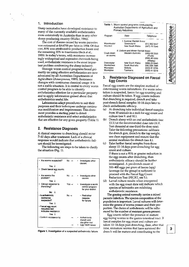
2. Resistance Diagnosis A clinical response to drenching should occur 7-10 days after treatment. Lack of a clinical response would indicate that anthelmintic fail- ure should be investigated.
The following are steps to be taken to clarify the situation (Fig. 1).
* Are worms the problem?
Yes L
l Clinical response to drenching?
No 1 * Is anthelmintic
reshanoe suspected?
Yes L - Faecal egg count
reduction test indites resistance
Yes 1 * Confirm resistance by
No+ . Investigate other causes
Yes 4 * Investigate a worm control program for your district
No -i * Find other reasons for failure
-3 * Anthelmlntic drench and slaughter trial
+ * Egg hatch assay
Figure 1. Investigation of a suspected anthelmintic failure.
Table 1. Worm control programs conducted by Australian Departments of Agriculture and Primary Industries
Program Safe Telephone -
1. Summer Rainfall Areas Wormbuster Queensland (07) 362 9447 Wormkill New South Wales (067) 73 7248
II. Unilon and Winter RaMall Areas Crack down Western Australia (093) 68 3333
on worms (Perth) (098) 42 0500
Drenchplan New South Wales Wormcheck South Australia Wormplan Victoria Wormplan Tasmania
WW (069) 23 O&5 (08) 228 7585 (053) 37 0755 (003) 41 5218
3. Resistance Diagnosed on Faecal Egg Counts
Faecal egg counts are the simplest method of determining worm infestations. If a worm infes- tation is suspected, faeces for egg counting and culture should be taken. If egg counts indicate that drenching is required, drench and take post-drench faecal samples 10-14 days later to check anthelmintic efficacy. (a) At drenching take individual faecal samples
from 10 animals in a mob for egg count and culture (see 9. and 10.).
(lo) Drench sheep with an oral anthelmintic (see 13.3.) at the recommended dose rate (N.B. Test closantel at one-third the dose rate). Take the following precautions: calibrate the drench gun, drench to the top weight, use clean equipment and ensure that the animal swallows the drench (see 4.).
(c) Take further faecal samples from these sheep 10-14 days post-drenching for egg count and culture. If there is not a 95% or greater reduction in the egg counts after drenching, then anthelmintic efficacy should be further investigated. A predrench count of 3OOXKl eggs per gram of faeces (epg) (average for the group) is sufficient to proceed with the Faecal Egg Count Reduction Test (FECRT, see 5.).
(d) Larval culture results when interpreted with the egg count data will indicate which species of helminths are exhibiting anthelmintic resistance.
The grazing animal normally carries a mixed parasite infection. The species composition of the population is important. Larval cultures will deter- mine the genera of worms present and their pro- portion. The choice of anthelmintic will be influ- enced by the number of resistant genera present.
Egg counts reflect the presence of mature egg-laying ~~rrns in the gastro-intestinal tract. If faecal samples for egg count and culture are taken 10-14 days post-drenching, then, after that time, immature worms that have survived the drench will be mature and contributing to the I! 3
post-treatment egg count. This time interval will also eliminate the problem of re-infestation. Incoming larvae from the paddock will not be mature by day 14 and their presence will not be reflected in the egg count.
Some drenches, e.g. benzimidazoles, suppress (for up to five days) egg laying by adult worms surviving the drench. By day 10, however, normal egg laying will have resumed.
When evaluating an egg count result, the level of the count, thefecundily and pathogenically of the helminth and the climatic conditions need to be considered.
Most worm confrol programs advocate routine faecal egg count monitoring of frocks before and after drenching. If drenches are omitted from a program then regular egg count monitoring becomes essential.
4. Failure to Obtain a Response to Drenching
The lack of response to drenching may be due to several factors including inappropriate drench selection, faulty equipment or drench adminis- tration techniques, rather than anthelmintic resistance. Investigate the following options.
4.1. Anthelmintic Choice Was the correct drench family used? An under- standing of anthelmintics and their mode of action is essential for appropriate anthelmintic choice, e.g. a narrow-spectrum drench may not control Trichostrongylus or Oskrtagia.
4.2. Anthelmintic Administration Did the animals receive the correct volume of drench? Were the animals weighed to calculate dose to body weight? Was the drench adminis- tered correctly? Was the drench gun calibrated? Did the animals swallow the drench?
4.3. Parasite Factors Response to drenching can be masked by the following. (a)
(b)
(cl
4.4
Larvae unaffected by treatment will contin- ue to develop rapidly and produce signs of disease. Rapid re-infection from the pasture. A closantel drench will stop development of incoming Haemonchus contortus larvae. Broad spectrum drenches have no effect on incoming larvae (except for sustained release capsules). A heavy H. contortus burden in weaners will cause severe anaemia and mask the benefits of the drench.
Concurrent Conditions Other factors that can hinder recovery are:
r-l (a) ill thrift due to poor nutrition; and
4 (b) bacterial or protozoan diseases, e.g. coccidiosis, salmonellosis, epervthrozoonosis.
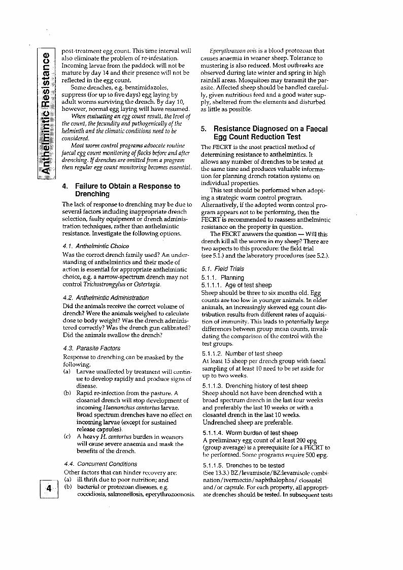
Eperythrozoor~ ovis is a blood protozoan that causes anaemia in weaner sheep. Tolerance to mustering is also reduced. Most outbreaks are observed during late winter and spring in high rainfall areas. Mosquitoes may transmit the par- asite. Affected sheep should be handled careful- ly, given nutritious feed and a good water sup- ply, sheltered from the elements and disturbed as little as possible.
5. Resistance Diagnosed on a Faecal Egg Count Reduction Test
The FJECRT is the most practical method of determining resistance to anthelmintics. It allows any number of drenches to be tested at the same time and produces valuable informa- tion for planning drench rotation systems on individual properties.
This test should be performed when adopt- ing a strategic worm control program. Alternatively, if the adopted worm control pro- gram appears not to be performing, then the FECRT is recommended to reassess anthelmintic resistance on the property in question.
The FECRT answers the question - Will this drench kill all the worms in my sheep? There are two aspects to this procedure: the field trial (see 5.1.) and the laboratory procedures (see 5.2.).
5.1. Field Trials 5.1 .l. Planning 5.1.1.1. Age of test sheep Sheep should be three to six months old. Egg counts are too low in younger animals. In older animals, an increasingly skewed egg count dis- tribution results from different rates of acquisi- tion of immunity. This leads to potentially large differences between group mean counts, invali- dating the comparison of the control with the test groups.
5.1 .1.2. Number of test sheep At least 15 sheep per drench group with faecal sampling of at least 10 need to be set aside for up to two weeks.
5.1.1.3. Drenching history of test sheep Sheep should not have been drenched with a broad spectrum drench in the last four weeks and preferably the last 10 weeks or with a closantel drench in the last 10 weeks. Undrenched sheep are preferable.
5.1 .1.4. Worm burden of test sheep A preliminary egg count of at least 200 epg (group average) is a prerequisite for a FECRT to be performed. Some programs require 500 epg.
5.1 .1.5. Drenches to be tested (See 13.3.) BZ/levamisole/BZ:levamisole combi- nation/iverm&in/naphthalophos/ closantel and/or capsule. For each property, all appropri- ate drenches should be tested. In subseouent tests
a prodtict to which extreme resistance was previ- ously detected, may be omitted on the basis that reversion to susceptibility would not have occurred.
5.1 .1.6. Time of the year Tests performed soon after a summer drought may overestimate the degree of resistance on that property. The population of worms in the host are the resistant survivors of the last drench. No larval pickup from the paddock, due to the drought, would have occurred to reveal the true situation for that property.
5.1.2. Equipment
(a) Scales to weigh the sheep (bathroom scales are adequate).
(b) Colour marking paint (e.g. SIROMARK or easily scourable dry raddle) to indicate drench groups on the sheep.
(c) Faecal collection bottles or plastic bags. (d) A permanent marking pen for labelling col-
lection containers. Drench gun(s) and back- pack.
(e) Associated paper work, i.e. Laboratory Advice Sheets.
51.3. Operation 5.1.3.1. On day 1 of the test
5.1.3.1 .l. Select suitable sheep. Draft off enough sheep to allow 15 per drench group - six drench groups plus one control group (6x15) +15 = 105 lambs. They should be this years lambs, of even size and preferably not previously drenched.
5.1.3.1.2. Determine weight for drench dose calculations. First exclude atypically heavy sheep. Weigh five of the heaviest looking sheep in the draft. Use the heaviest weight for all dose calculations.
5.1.3.1.3. Randomise sheep into drench groups. A form of systematic randomisation is used. Sheep in the race are allocated into groups on the basis of No. 1 to the first group, No. 2 to the second group and so on. The common prac- tice of allocating the first 10 sheep into the first group, the second 10 into the second group and so on does not constitute proper randomisation. Colour mark the sheep sequentially down the race using head, neck, back and rump marks until all sheep are accounted for. There should be 15 sheep per group.
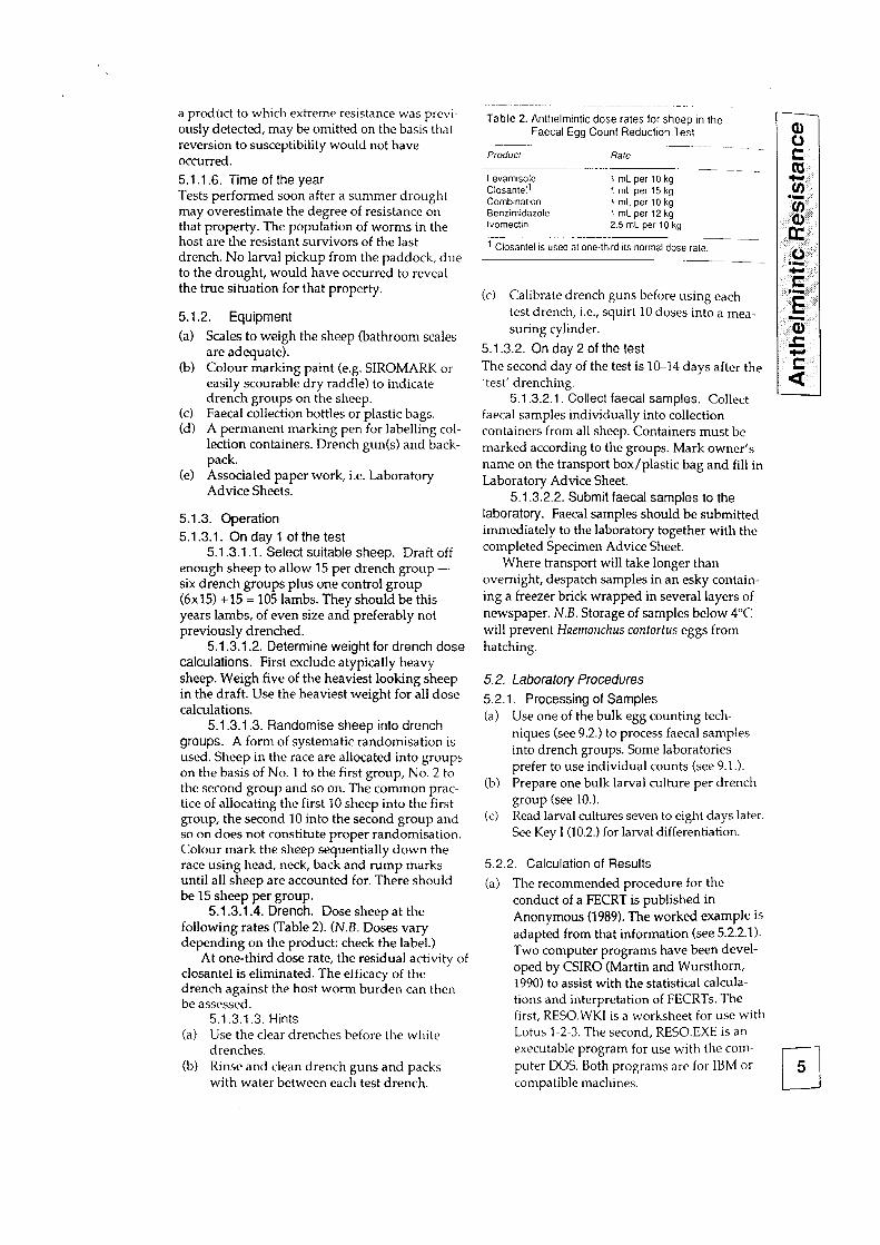
5.1.3.1.4. Drench. Dose sheep at the following rates (Table 2). (N.B. Doses vary depending on the product: check the label.)
At one-third dose rate, the residual activity of closantel is eliminated. The efficacy of the drench against the host worm burden can then be assessed.
5.1.3.1.3. Hints (a) Use the clear drenches before the white
drenches. (b) Rinse and clean drench guns and packs
Table 2. Anthelmintic dose rates for sheep in the Faecal Egg Count Reduction Test
Producr Rafe
Levamisole Closantel’
1mLperlOkg 1 mLper15kg
Combination 1 mLperlOkg Benzimidazole 1 mLper 12kg lvomectin 2.5 mL per 10 kg
’ Closantel is used at one-third its normal dose rate.
(c) Calibrate drench guns before using each test drench, i.e., squirt 10 doses into a mea- suring cylinder.
5.1.3.2. On day 2 of the test The second day of the test is lCL14 days after the ‘test’ drenching.
5.1.3.2.1. Collect faecal samples. Collect
faecal samples individually into collection containers from all sheep. Containers must be marked according to the groups. Mark owner’s name on the transport box/plastic bag and fill in Laboratory Advice Sheet.
5.1.3.2.2. Submit faecal samples to the laboratory. Faecal samples should be submitted immediately to the laboratory together with the completed Specimen Advice Sheet.
Where transport will take longer than overnight, despatch samples in an esky contain- ing a freezer brick wrapped in several layers of newspaper. N.B. Storage of samples below 4°C will prevent Huelnolrchus contortus eggs from hatching.
5.2. laboratory Procedures
5.2.1. Processing of Samples (a) Use one of the bulk egg counting tech-
niques (see 9.2.) to process faecal samples into drench groups. Some laboratories prefer to use individual counts (see 9.1.).
(b) Prepare one bulk larval culture per drench group (see 10.).
(c) Read larval cultures seven to eight days later. See Key I (10.2.) for larval differentiation.
5.2.2. Calculation of Results
(a) The recommended procedure for the conduct of a FECRT is published in Anonymous (1989). The worked example is adapted from that information (see 5.2.2.1). Two computer programs have been devel- oped by CSIRO (Martin and Wursthom 1990) to assist with the statistical calcula- tions and interpretation of FECRTs. The first, RESO.WKI is a worksheet for use with Lotus l-2-3. The second, RESO.EXE is an executable program for use with the com- puter DOS. Both programs are for IBM or r-l 5
with water between each test drench, compatible machines.
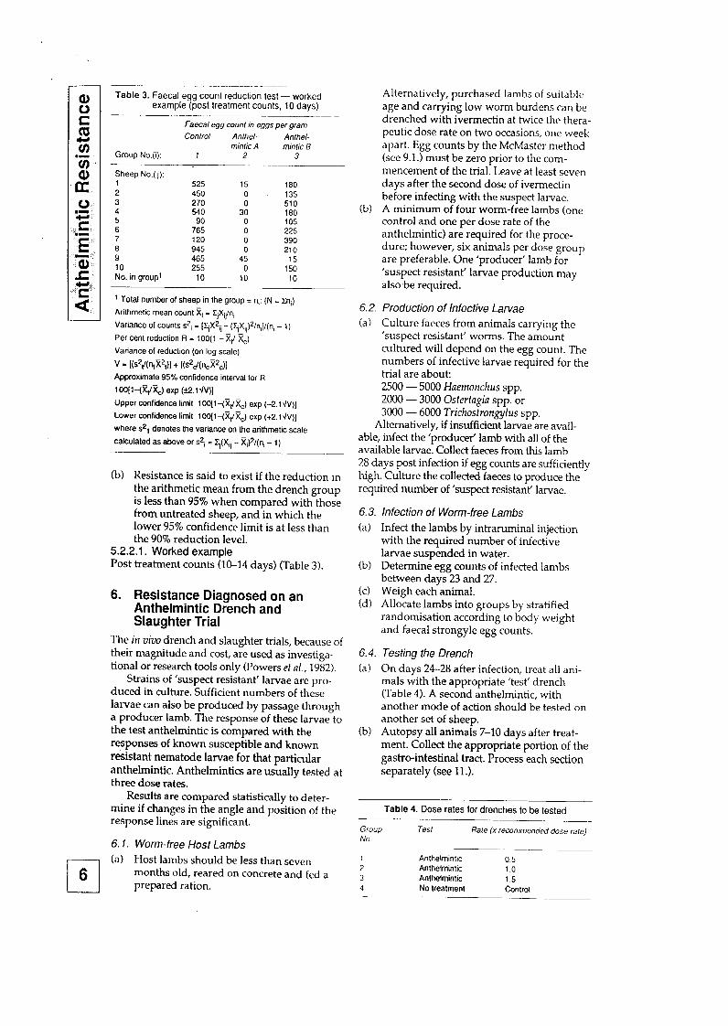
Table 3. Faecal egg count reduction lest - worked example (post lreatment counts, 10 days)
~~ Faecal egg counl in eggs per gram
Control Anlhel- Anlhel- mfntic A mink B
Group No.(i): 1 2 3 -
Sheep No.(j): 1 525 15 180 2 450 0 135 3 270
3: 510
4 540 180 5 90 0 105 6 765 0 225 7 120 0 390 6 945 0 210 9 465 45 15 10 255 0 150 No. in group’ 10 10 10
1 Total number of sheep in the group = n,: (N = Zn,)
Arithmetic mean count Ei = Z,Xri/n,
Variance Of counts S2i = [~jX*ij - (YjXij)*/n,]/(n, - 1)
Per cent reduction R = 100( 1 - X,/ Xc)
Variance of reduction (on log scale)
V = I(s2&-&)1 + [(s*d(nJ*,)l Approximate 95% confidence interval for R
-- 1 OO(l-(XdXJ exp (ti.l\1V)]
Upper confidence limit 1 OO[l-(XdXJ exp (-2.1 &)I
Lower confidence limit lOOll-(qXJ exp (+2.lJV)]
where s21 denotes the variance on the arithmetic scale
calculated as above or s*i = q(Xij - XJ*/(ni - I)
(b) Resistance is said to exist if the reduction in the arithmetic mean from the drench group is less than 95% when compared with those from untreated sheep, and in which the lower 95% confidence limit is at less than the 90% reduction level.
5.2.2.1. Worked example Post treatment counts (10-14 days) (Table 3).
6. Resistance Diagnosed on an Anthelmintic Drench and Slaughter Trial
The in uivo drench and slaughter trials, because of their magnitude and cost, are used as investiga- tional or research tools only (Powers et al., 1982).
Strains of ‘suspect resistant’ larvae are pro- duced in culture. Sufficient numbers of these larvae can also be produced by passage through a producer lamb. The response of these larvae to the test anthelmintic is compared with the responses of known susceptible and known resistant nematode larvae for that particular anthelmintic. Anthelmintics are usually tested at three dose rates.
Results are compared statistically to deter- mine if changes in the angle and position of the response lines are significant.
6.7. Worm-free Host Lambs
cl (a) Host lambs should be less than seven
6 months old, reared on concrete and fed a prepared ration.
Alternatively, purchased lambs of suitable age and carrying low worm burdens can be drenched with ivermectin at twice the thera- peutic dose rate on two occasions, one week apart. Egg counts by the McMaster method (see 9.1.) must be zero prior to the com- mencement of the trial. Leave at least seven days after the second dose of ivermectin before infecting with the suspect larvae.
(b) A minimum of four worm-free iambs (one control and one per dose rate of the anthelmintic) are required for the proce- dure; however, six animals per dose group are preferable. One ‘producer’ lamb for ‘suspect resistant’ larvae production may also.be required.
6.2. Production of Infective Larvae (a) Culture faeces from animals carrying the
‘suspect resistant’ worms. The amount cultured will depend on the egg count. The numbers of infective larvae required for the trial are about: 2500 - 5000 Haemonchus spp. 2000 - 3000 Ostertagia spp. or 3000 - 6000 Trichostrongylus spp.
Alternatively, if insufficient larvae are avail- able, infect the ‘producer’ lamb with all of the available larvae. Collect faeces from this lamb 28 days post infection if egg counts are sufficiently high. Culture the collected faeces to produce the required number of ‘suspect resistanY larvae.
6.3. Infection of Worm-free Lambs
(a) Infect the lambs by intraruminal injection with the required number of infective larvae suspended in water.
(b) Determine egg counts of infected lambs between days 23 and 27.
(c) Weigh each animal. (d) Allocate lambs into groups by stratified
randomisation according to body weight and faecal strongyle egg counts.
6.4. Testing the Drench
(a) On days 24-28 after infection, treat all ani- mals with the appropriate ‘test’ drench (Table 4). A second anthehnintic, with another mode of action should be tested on another set of sheep.
(b) Autopsy all animals 7-10 days after treat- ment. Collect the appropriate portion of the gastro-intestinal tract. Process each section separately (see 11.).
Table 4. Dose rates for drenches to be tested
Group NO
Test Rate (x recommended dose rare)
1 Anthelmintic 2 Anthelminlic 3 Anthelmintic 4 No treatment
~__
0.5 1.0 1.5 Control

6.5 (a)
(b)
(4
(d)
7.
Calculation of Results Calculate the per cent efficiency. For each anthelmintic compare the average number of worms per dose rate with those of the control group. Produce dose response lines (Clark and Turton, 1973). Compare these lines of dose response with those produced for the same anthelmintic against susceptible strains of the same nematode species. Resistance is diagnosed if the change in the angle of the response and the position of the response is significantly different from the susceptible strain and similar to the resistant strain.
Resistance Diagnosed on Egg Hatch Assay
Benzirnidazole (BZ) anthelmintics prevent nema- tode egg embryonation and hatching. This char- acteristic can be used to detect BZ resistance (Le Jambre, 1976). Eggs are collected immediately from fresh faeces and incubated in serial concentrations of the BZ anthelmintic. A pure strain of nematodes is desirable.
7.1 (4
Reagents
(b)
(4
Dissolve 8 mg of pure TBZ (Tbiabendazole) in 5 mL of dimethyl sulfoxide (DMSO) (see 13.3.). Make to 1 L with 0.1% sodium chloride (0.017 mol/L) (stock solution). Make serial dilutions of TBZ, ranging in concentration from 3.0 to 0.007 pg/mL using a standard diluent of 5 pL/mL DMSO in 0.1% sodium chloride. Pipette 1 mL of TBZ serial dilutions into each well of a 2!5-well petri dish (Sterilin No. 234095).
7.2. Egg Recovery (a) Collect 50 g of faeces directly from the ani-
mal(s) infected with the strain to be tested. A pure infection is required.
(b) Process faeces immediately. Do not chill. (c) Mix faeces with water to a slurry. (d) Pour through two thicknesses of surgical
gauze or muslin into a 3 L side-arm flask. (e) Wash the slurry again with 1 L of saturated
sodium chloride solution. (f) Discard gauze and debris. Evacuate the
sieved sample with a tap aspirator to remove bubbles.
(g) Sieve evacuated sample into a plastic pho- tographic tray. Gently place a perspex sheet, cut to fit the tray, on the surface of the sample.
(h) Store at 4°C for 15-20 min. Lift up perspex sheet and wash off adhered eggs into a 250 mL cylinder.
(i)
Cj)
(k)
(1)
b-d
(n)
7.3.
Add chilled water up to Ihe mark and stand at 4°C for 90 min. Decant the supernatant to a volume of 22
50 mL. Further decant in a 50 mL cylinder S’ or cenhifuge at 2000 rpm for five minutes to concentrate eggs. ;z? Adjust the volume of the egg suspension to co
200 eggs/O.1 mL. Add the 0.1 mL to the Q)..
1 mL TBZ solutions and the control well. ,[r,, Incubate at 22°C for 48 hours. Terminate c) with a drop of dilute iodine solution. Count eggs and first stage larvae in each well
.:g a- ‘.
under x40 magnification. Count a duplicate. ,-E Calculate per cent hatch.
Q>
Statistical Analysis c, M,, .r
For each TBZ concentration, the proportion of eggs killed by the TBZ alone (I’) is calculated I
;i
from Abbott’s formula which adjusts the total proportion killed (I”) in relation to the number dying in the TBZ free control sample (C) by:
P = (l” - C)/(l - C) (Finney, 1971).
The percentage of unhatched eggs at each concentration of TBZ is converted to probits and plotted against the logarithm of the TBZ concen- tration. A log concentration-probit (lc-p) line can be fitted using a computer program written following the methods published by Davies (1971). The lethal concentration to 50% of eggs (LC,) and the resistance ratio [RR = exp(log LC50 test strain - log LC50 susceptible strain) ] gives quantitative measures of resistance. The 95% confidence intervals for a resistance ratio can be calculated from the formula:
exp[log(RR) * 2 * (S12 + S22)J
where s2 is the variance for each strain from the probit analysis. (PROBIT is an IBM PC computer program for probit analysis. Copies of PROBIT are available from: Leo Wursthom, CSIRO, Animal Health Research Laboratories, Private Bag No. 1, Parkville, Vic. 3052, Australia).
8. Broad Spectrum Anthelmintic Resistance Diagnosed by in vitro Larval Development Assay
The Larval Development Assay (LDA) is an ill vitro technique for detection of broad spectrum anthelmintic resistance in nematodes. Each row of 12 wells, of a 96-well microtitre plate, contains a lOOO-fold concentration range of a specific anthelmintic in an agar matrix. At present, the anthelmintics used are specifically for detection of benzimidazole, levamisole/morantel and ivermectin resistances.
Nematode eggs are isolated from a faecal sam- ple, applied to the wells and allowed to develop to infective 5 larvae over six to seven days. Eggs in wells will hatch and develop through L, and L2 stages depending on concentration of the
8.1.
(a)
(b)
(cl
(d)
8.2. (a)
(b)
(4
8.3 (a)
Field Collection Randomly select at least 10 animals from the flock to be tested and collect no less than 100 g of faeces as a pool sample. Take a representative egg count on the bulked sample. Submit samples with an epg > 100 for LDA. Gently press the faeces to exclude air and seal tightly in a plastic container. Do not crush pellets into a single mass. Hold and transport sample at room temper- ature. The time between collection and assay should be less than seven days.
Laboratory Procedure Eggs are isolated by modified sucrose flotation technique. Eggs are applied to pre-prepared plate and incubated for seven days at 22°C by which time control (no drug) wells have devel- oped to infective La larvae. Larvae are killed with iodine and develop- ment assessed by two methods: 6) Qualifatiue (by eye). Transition between L3 (uninhibited) and L,, (inhibited) wells can be assessed by eye to identify well number at which about 50% inhibition of development occurs. (ii) Quantitative (counting). Proportions of L,, and La larvae (and eggs) can be count- ed and data computer-fitted by logit-log concentration model to derive LD,.
Calculation of Results and interpretation Approximate LDx values for a field isolate can be obtained by conversion from well number using the concentration factor b where b is the concentration in well No. 1, thus:
LD, (in umol/L) = a x b
where a is well number in which inhibited L,-, larvae and uninhibited La are in roughly equal abundance. N.B. Qualitative LD@ normally agree within two-fold of the computed quantitative value.
anthelmintics. Thus, isolates resistant to an anthelmintic will develop in wells containing higher concentrations than susceptible isolates (Lacey et al., 1991).
The LDA offers the following advantages over existing techniques: (a) simultaneous evaluation of all broad spec-
trum drenches in a single assay; (b) single farm visit with minimal on-farm
experimentation; and (4 eliminates between-animal variation as a
source of poor data quality to give improved precision of resistance status.
The LDA is currently under field evaluation for commercial development by Horizon Agriculture and CSIRO, Division of Animal Health.
(b)
9.
Resistance factors (RFs) can be calculated by:
where II and c are the well numbers in which approximate LD,, values of field and susceptible isolates are noted. Alternatively, if a susceptible isolate is not run with the field isolate, RF can be calculated as:
RF = [(a x b)/S&,l
where SLD, is the data base susceptible value and b is concentration factor. The RF for a field isolate is used with the accompanying data base to predict the fae- cal egg reduction achievable for the field isolate. The data base has been constructed using 40 sets of field FECRT and LDA data. For more precise property monitoring, the LDA RF values can be used initially in con- junction with a FECRT in year 1 to estab- lish correlation on the property, then sub- sequently monitored using LDA.
Counting Techniques for Strongyle UP
Strongyle eggs are floated in a known volume of faecal suspension and then counted microscopi- cally on a Whitlock Universal or a McMaster slide. Direct extrapolation of the number of worms to be found in the gut from the epg of faeces calculation is only approximate. Egg pro- duction is influenced by many things, e.g. immunity of the host, species of worm, maturity of worms, season of the year and stage of preg- nancy and lactation.
Egg counts can give valuable information on existing worm burdens and larval paddock pop- ulations if samples are taken just prior to drenching and on anthelmintic efficacy if faecal samples are taken 10-14 days post drenching.
9.1. The Modified McMaster Method This method is based on the McMaster Method (Whitlock, 1948) and uses a Whitlock Universal (4 x 0.5 mL) or a McMaster (3 x 0.3 mL) slide and is the standard procedure adopted for egg counting in individual animals (M&aster and Whitlock Universal egg counting chambers are available from: J. A. Whitlock and Co., PO Box 51, Eastwood, NSW 2122, Australia). A variation of this method uses a bulking technique prior to mixing for the batch processing of large numbers of samples.
For liquid or soft faeces only, discard the top layer of the faecal sample (0.5 g). Mix the remainder. No correction for faecal consistency is necessary for FECRT samples. Correct randomisation of animals will allow for variabil- ity in faecal consistency.
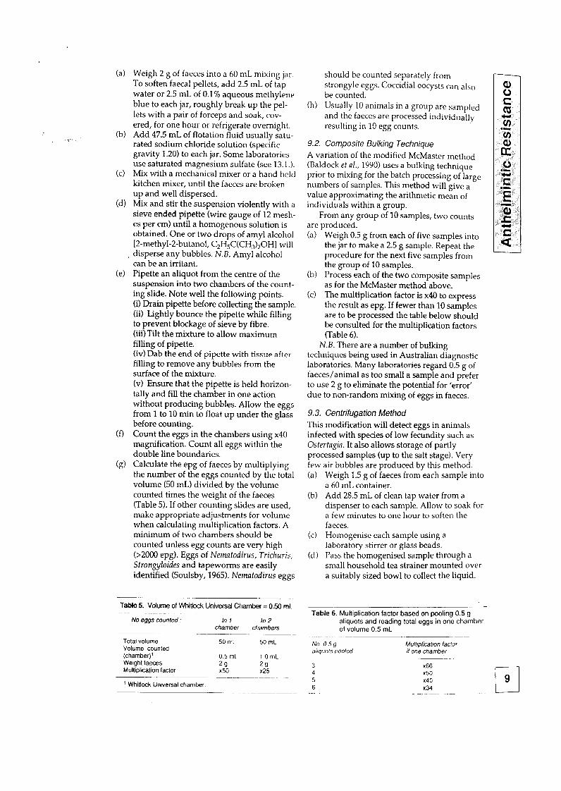
(a) Weigh 2 g of faeces into a 60 mL mixing jar. To soften faecal pellets, add 2.5 mL of tap water or 2.5 mL of 0.1% aqueous methylene blue to each jar, roughly break up the pel- lets with a pair of forceps and soak, cov-
(b) ered, for one hour or refrigerate overnight. Add 47.5 mL of flotation fluid usually satu- rated sodium chloride solution (specific gravity 1.20) to each jar. Some laboratories use saturated magnesium sulfate (see 13.1.).
(c) Mix with a mechanical mixer or a hand held kitchen mixer, until the faeces are broken up and well dispersed.
(d) Mix and stir the suspension violently with a sieve ended pipette (wire gauge of 12 mesh- es per cm) until a homogenous solution is obtained. One or two drops of amyl alcohol [2-methyl-2-butanol, C,H,C(CHJ,OHl will
~ disperse any bubbles. N.B. Amy1 alcohol can be an irritant.
(e) Pipette an aliquot from the centre of the suspension into two chambers of the count- ing slide. Note well the following points. (i) Drain pipette before collecting the sample. (ii) Lightly bounce the pipette while filling to prevent blockage of sieve by fibre. (iii) Tilt the mixture to allow maximum filling of pipette. (iv) Dab the end of pipette with tissue after filling to remove any bubbles from the surface of the mixture. (v) Ensure that the pipette is held horizon- tally and fill the chamber in one action without producing bubbles. Allow the eggs from 1 to 10 min to float up under the glass before counting.
(0 Count the eggs in the chambers using x40 magnification. Count all eggs within the double line boundaries.
(g) Calculate the epg of faeces by multiplying the number of the eggs counted by the total volume (50 mL) divided by the volume counted times the weight of the faeces (Table 5). If other counting slides are used, make appropriate adjustments for volume when calculating multiplication factors. A minimum of two chambers should be counted unless egg counts are very high (>2000 epg). Eggs of Ncmatodirus, Trichuris, Strongyloides and tapeworms are easily identified (Soulsby, 1965). Nernatodirus eggs
Table 5. Volume of Whitlock Universal Chamber = 0.50 mL
No eggs cuunled : In 1 In 2 chamber chambers
Total volume 50 mL 50 mL Volume counted (chamber)’ 0.5 mL 1 .O mL Weight laeces 29 29 Multiplication (actor x50 x25
’ Whitlock Universal chamber.
should be counted separately from strongyle eggs. Coccidial oocysts can also be counted.
(11) Usually 10 animals in a group are sampled and the faeces are processed individually resulting in 10 egg counts.
9.2. Composite Bulking Technique A variation of the modified McMaster method (Baldock et al., 1990) uses a bulking technique prior to mixing for the batch processing of large numbers of samples. This method will give a value approximating the arithmetic mean of individuals within a group.
From any group of 10 samples, two counts are produced. (a) Weigh 0.5 g from each of five samples into
the jar to make a 2.5 g sample. Repeat the procedure for the next five samples from the group of 10 samples.
(b) Process each of the two composite samples as for the McMaster method above.
(c) The multiplication factor is x40 to express the result as epg. If fewer than 10 samples are to be processed the table below should be consulted for the multiplication factors (Table 6).
N.B. There are a number of bulking techniques being used in Australian diagnostic laboratories. Many laboratories regard 0.5 g of faeces/animal as too small a sample and prefer to use 2 g to eliminate the potential for ‘error’ due to non-random mixing of eggs in faeces.
9.3. Centrifugafion Method
This modification will detect eggs in animals infected with species of low fecundity such as Ostertagiu. It also allows storage of partly processed samples (up to the salt stage). Very few air bubbles are produced by this method. (a) Weigh 1.5 g of faeces from each sample into
a 60 mL container. (b) Add 28.5 mL of clean tap water from a
dispenser to each sample. Allow to soak for a few minutes to one hour to soften the faeces.
(c) Homogenise each sample using a laboratory stirrer or glass beads.
(d) Pass the homogenised sample through a small household tea strainer mounted over a suitably sized bowl to collect the liquid.
Table 6. Multiplication factor based on pooling 0.5 g aliquots and reading total eggs in one chamber of volume 0.5 mL
-. No 059 Mulf~pkal~on facror allquots pooled II one chamber
3 x66 4 x50 5 x40 6 x34
(e) Swirl to mix the liquid, then pour immedi- ately into a 15 mL numbered centrifuge tube. Excess liquid can flow over the tube once it is full.
(f) Centrifuge for two minutes at 2000 rpm to produce a plug of debris containing the eggs. Pour off the supernatant carefully or remove it using a water-driven suction pipette.
(g) Add saturated sodium chloride solution up to the 10 mL mark. Resuspend the plug by repeated inversions of the tube. Fill the tube up to the 15 mL mark with more saturated sodium chloride solution and mix thor- oughly using inversion or a Pasteur pipette.
(h) After mixing the faecal suspension, fill the chamber of the Universal Whitlock Slide (0.5 mL in each chamber). Count all the eggs within the first two lines of the cham- ber. The multiplication factor is 100. If few eggs are present, count chambers one and two to give a sensitivity of 20 epg.
(i) Wash the stirrer, the mixing container, the sieve and the collecting bowl between samples.
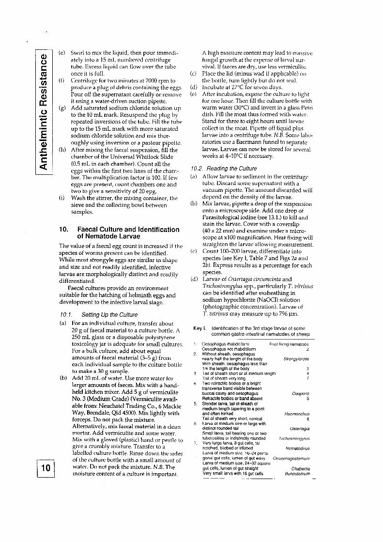
10. Faecal Culture and Identification of Nematode Larvae
The value of a faecal egg count is increased if the species of worms present can be identified. While most strongyle eggs are similar in shape and size and not readily identified, infective larvae are morphologically distinct and readily differentiated.
Faecal cultures provide an environment suitable for the hatching of helminth eggs and development to the infective larval stage.
10.1. Setting Up fhe Culture
(a) For an individual culture, transfer about 20 g of faecal material to a culture bottle. A 250 mL glass or a disposable polystyrene toxicology jar is adequate for small cultures. For a bulk culture, add about equal amounts of faecal material (3-5 g) from each individual sample to the culture bottle to make a 30 g sample.
(b) Add 20 mL of water. Use more water for larger amounts of faeces. Mix with a hand- held kitchen mixer. Add 5 g of vermiculite No. 3 (Medium Grade) (Vermiculite avail- able from: Neuchatel Trading Co., 6 Mackie Way, Brendale, Qld 4500). Mix lightly with forceps. Do not pack the mixture. Alternatively, mix faecal material in a clean mortar. Add vermiculite and some water. Mix with a gloved (plastic) hand or pestle to give a crumbly mixture. Transfer to a labelled culture bottle. Rinse down the sides of the culture bottle with a small amount of water. Do not pack the mixture. N.B. The moisture content of a culture is important.
A high moisture content may lead to massive fungal growth at the expense of larval sur- vival. If faeces are dry, use less vermiculite.
(c) Place the lid (minus wad if applicable) on
the bottle, turn lightly but do not seal. (d) Incubate at 27°C for seven days. (e) After incubation, expose the culture to light
for one hour. Then fill the culture bottle with warm water (30°C) and invert in a glass Petri dish. Fill the moat thus formed with water. Stand for three to eight hours until larvae collect in the moat. Pipette off liquid plus larvae into a centrifuge tube. N.B. Some labo- ratories use a Baermann funnel to separate larvae. Larvae can now be stored for several weeks at 4-10°C if necessary.
10.2. Reading the Culture (a) Allow larvae to sediment in the centrifuge
tube. Discard some supematant with a vacuum pipette. The amount discarded will depend on the density of the larvae.
(b) Mix larvae, pipette a drop of the suspension onto a microscope side. Add one drop of Parasitological iodine (see 13.1.) to kill and stain the larvae. Cover with a coverslip (40 x 22 mm) and examine under a micro- scope at xl00 magnification. Heat fixing will straighten the larvae allowing measurement.
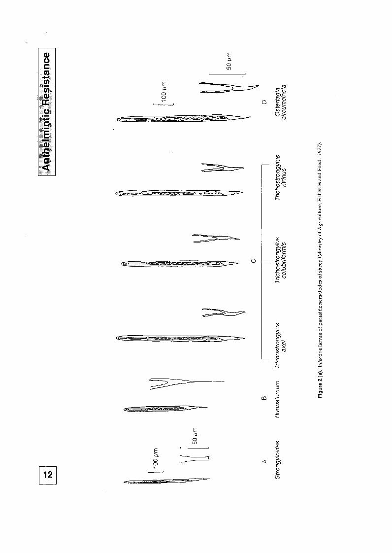
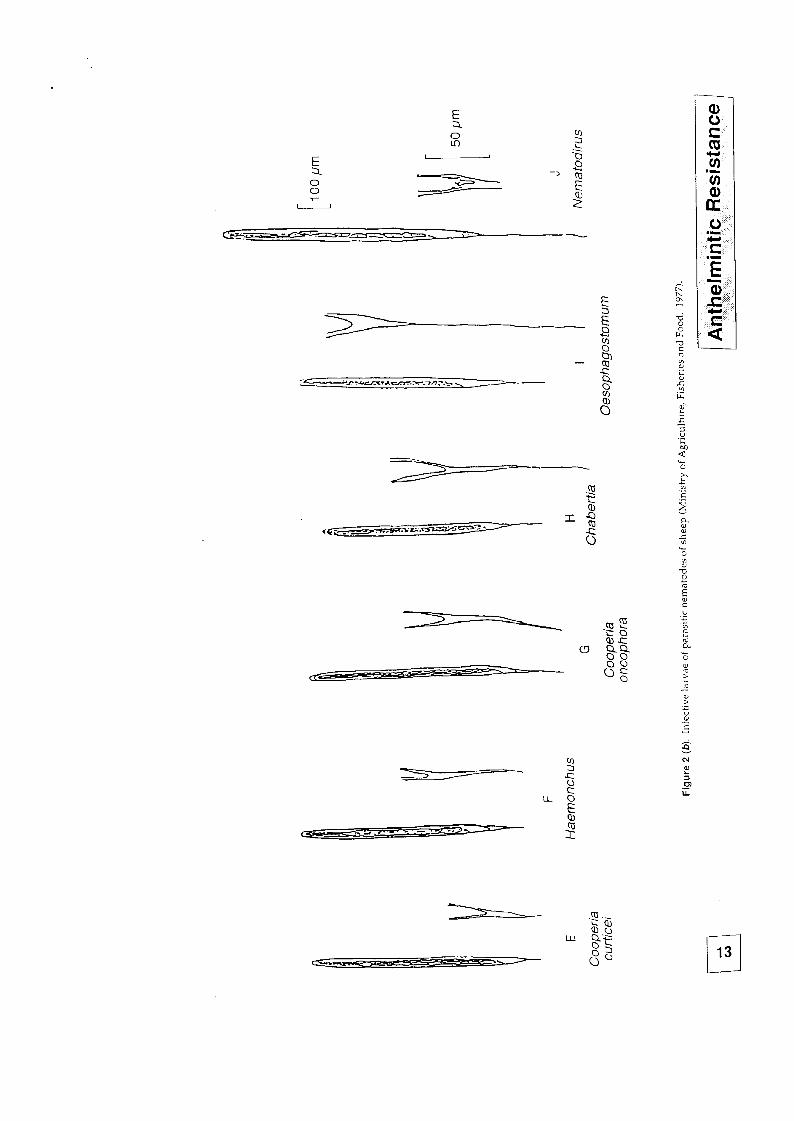
(c) Count 100-200 larvae, differentiate into species (see Key I, Table 7 and Figs 2a and 2bJ. Express results as a percentage for each species.
(d) Larvae of Ostertagia circumcih and Trichastroqylus spp., particularly T. uitrims can be identified after exsheathing in sodium hypochlorite (NaOCl) solution (photographic concentration). Larvae of T. uitrims may measure up to 796 pm.
Key I. Identification of the 3rd stage larvae of some common gastrointestinal nematodes of sheep
1. Oesophagus rhabditfform Free living nematode Oesophagus not rhabditiform 2
2. Without sheath, oesophagus nearly half the length of the body Slrongyloides With sheath, oesophagus less than 114 the length of the body 3
3. Tail of sheath short or of medium length 4 Tail of sheath very long 7
4. Two refractile bodies or a bright transverse band visible between buccaf cavity and oesophagus Cooperia Refractile bodies or band absent 5
5. Slender larva, tail of sheath of medium length tapering to a point and often kinked Haemonchus Tail of sheath very short, conical 6
6. Larva of medium size or large with distinct rounded tail Osfertagia Small larva, tail bearing one or two tuberosities or indistinctly rounded Jr!chostrongylus
7 Very large larva. 8 gut cells, tall notched, bilobed or trilobed Nemalodirus Larva of medium size, 1624 penta~ gonai gut cells. lumen 01 gut wavy Oesophagostomum Larva of medium size, 24-32 square gut cells, lumen of gut straight Chaberfia Very small larva with 16 gut cells Bunosfomum
__-
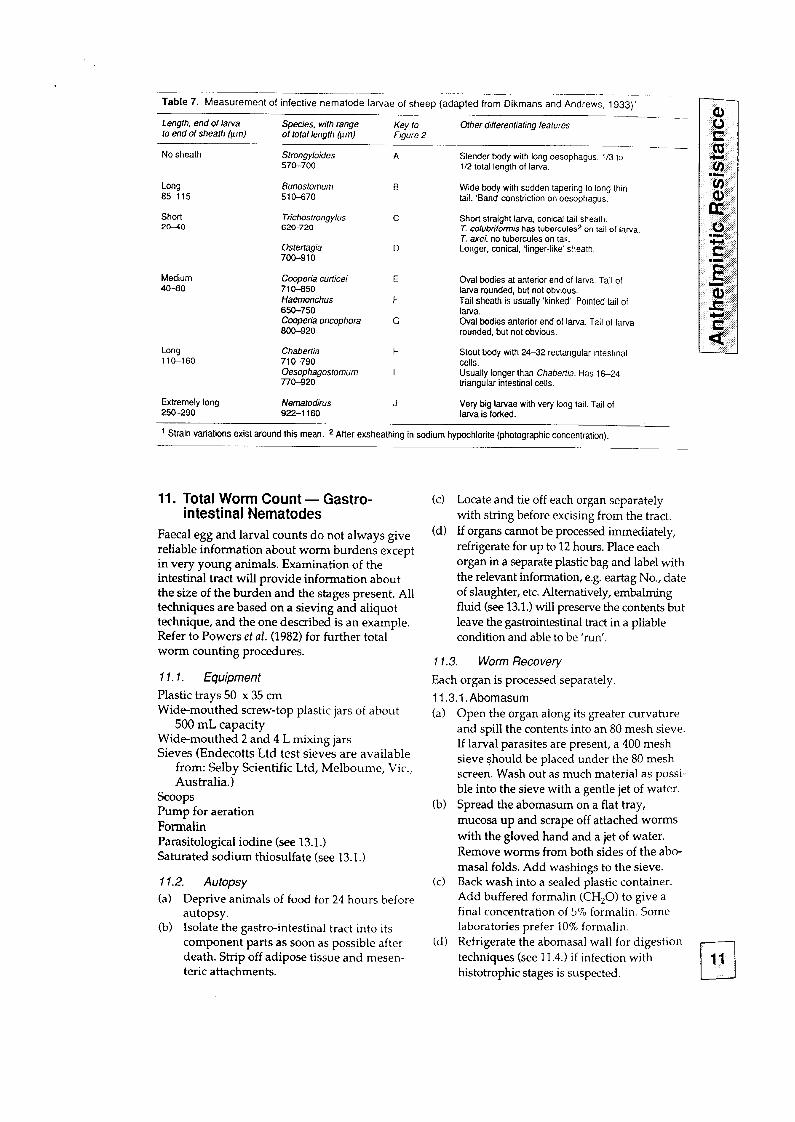
-Table 7. Measurement of infective nematode larvae of sheep (adapted from Dikmans and Andrews, 1933)’ ----..-p
Length, end of larva Species, with range to end of sheath (pm)
Key to of total length Itim)
Other ditterentiating features Fiaure 2
No sheath Strongyloides 570-700
Long Bunostamum 85115 510-670
Short Tkhostrongylus 20-%0 620.720
Medium 40-80
OsterTagia 700-910
Cooperia curticei 71 O-850 tiaemonchus 650-750 Coopefia oncophora 6cO-920
Long 11&160
Chabertia 710-790 Oesophagostomum 770420
A Slender body with long oesophagus, l/3 to l/2 total length of larva.
B Wide body with sudden tapering 10 long thin tail. ‘Band constriction on oesophagus.
C
D
Short straight larva, conical tail sheath. T. culubrilormis has tubercules* on tail of larva T. axei. no tubercules on tail. Longer, conical. ‘finger-like’ sheath.
Oval bodies at anterior end of larva. Tail of larva rounded, but not obvious. Tail sheath is usually ‘kinked’. Pointed tail Of larva. Oval bodies anterior end of larva. Tail of larva rounded, but not obvious.
H
I
Stout body with 24-32 rectangular intestinal cells. Usually longer than Chabertia. Has 1 G-24 triangular intestinal cells.
Extremely long Nematodirus J 250-290
Very big larvae with very long tail. Tail of 922-l 180 larva is forked.
t Strain variations exist around this mean. 2 After exsheathing in sodium hypochlorite (photographic concentration).
11. Total Worm Count - Gastro- intestinal Nematodes
Faecal egg and larval counts do not always give reliable information about worm burdens except in very young animals. Examination of the intestinal tract will provide information about the size of the burden and the stages present. All techniques are based on a sieving and aliquot technique, and the one described is an example. Refer to Powers et al. (1982) for further total worm counting procedures.
11.7. Equipment
Plastic trays 50 x 35 cm Wide-mouthed screw-top plastic jars of about
500 mL capacity Wide-mouthed 2 and 4 L mixing jars Sieves (Endecotts Ltd test sieves are available
from: Selby Scientific Ltd, Melbourne, Vic., Australia.)
Scoops Pump for aeration Formalin Parasitological iodine (see 13.1.) Saturated sodium thiosulfate (see 13.1.)
11.2. Autopsy
(a) Deprive animals of food for 24 hours before autopsy.
(b) Isolate the gastro-intestinal tract into its component parts as soon as possible after death. Strip off adipose tissue and mesen- teric attachments.
(cl
(d)
11.3. Worm Recovery
Locate and tie off each organ separately with string before excising from the tract. If organs cannot be processed immediately, refrigerate for up to 12 hours. Place each organ in a separate plastic bag and label with the relevant information, e.g. eartag No., date of slaughter, etc. Alternatively, embalming fluid (see 13.1.) will preserve the contents but leave the gastrointestinal tract in a pliable condition and able to be ‘run’.
Each organ is processed separately
11.3.1 .Abomasum (a)
(b)
(c)
Cd)
Open the organ along its greater curvature and spill the contents into an 80 mesh sieve. If larval parasites are present, a 400 mesh sieve Should be placed under the 80 mesh screen. Wash out as much material as possi- ble into the sieve with a gentle jet of water. Spread the abomasum on a flat tray, mucosa up and scrape off attached worms with the gloved hand and a jet of water. Remove worms from both sides of the abo- masal folds. Add washings to the sieve. Back wash into a sealed plastic container. Add buffered formalin (CH,O) to give a final concentration of 5% formalin. Some laboratories prefer 10% formalin. Refrigerate the abomasal wall for digestion techniques (see 11.4.) if infection with histotrouhic stages is susoected. Ti 11
q 12
I
w
.
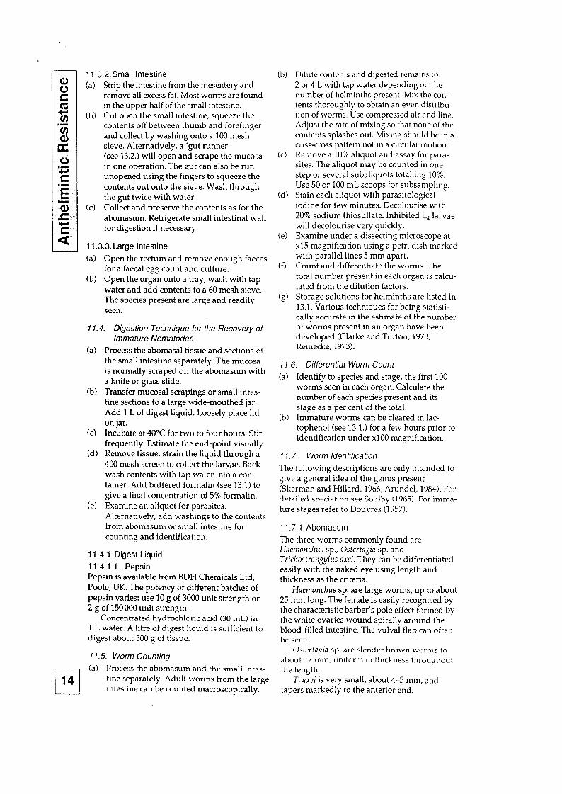
11.3.2.Small Intestine (a)
6)
Strip the intestine from the mesentery and remove all excess fat. Most worms are found in the upper half of the small intestine. Cut open the small intestine, squeeze the contents off between thumb and forefinger and collect by washing onto a 100 mesh sieve. Alternatively, a ‘gut runner’ (see 13.2.) will open and scrape the mucosa in one operation. The gut can also be run unopened using the fingers to squeeze the contents out onto the sieve. Wash through the gut twice with water. Collect and preserve the contents as for the abomasum. Refrigerate small intestinal wall for digestion if necessary.
(cl
11.3.3. Large Intestine
(a) Open the rectum and remove enough faeces for a faecal egg count and culture.
(b) Open the organ onto a tray, wash with tap water and add contents to a 60 mesh sieve. The species present are large and readily seen.
11.4. Digestion Technique for the Recovery of
(a)
6)
(cl
(d)
(4
immature Nematodes Process the abomasal tissue and sections of the small intestine separately. The mucosa is normally scraped off the abomasum with a knife or glass slide. Transfer mucosal scrapings or small intes- tine sections to a large wide-mouthed jar. Add 1 L of digest liquid. Loosely place lid on jar. Incubate at 40°C for two to four hours. Stir frequently. Estimate the end-point visually. Remove tissue, strain the liquid through a 400 mesh screen to collect the larvae. Back wash contents with tap water into a con- tainer. Add buffered formalin (see 13.1) to give a final concentration of 5% formalin. Examine an aliquot for parasites. Alternatively, add washings to the contents from abomasum or small intestine for counting and identification.
11.4.1. Digest Liquid
11.4.1 .l . Pepsin Pepsin is available from BDH Chemicals Ltd, Poole, UK. The potency of different batches of pepsin varies: use 10 g of 3000 unit strength or 2 g of 150 000 unit strength.
Concentrated hydrochloric acid (30 mL) in 1 L water. A litre of digest liquid is sufficient lo digest about 500 g of tissue.
Il. 5. Worm Counting
q (a) Process the abomasum and the small intes-
14 tine separately. Adult worms from the large intestine can be counted macroscopically.
(b)
(cl
(d)
k)
(0
w
Dilute contents and digested remains to 2 or 4 L with tap water depending on the number of helminths present. Mix the col’l- tents thoroughly to obtain an even distribu- tion of worms. Use compressed air and line. Adjust the rate of mixing so that none of the contents splashes out. Mixing should be in a criss-cross pattern not in a circular motion. Remove a 10% aliquot and assay for para- sites. The aliquot may be counted in one step or several subaliquots totalling 10%. Use 50 or 100 mL scoops for subsampling. Stain each aliquot with parasitological iodine for few minutes. Decolourise with 20% sodium thiosulfate. Inhibited L, larvae will decolourise very quickly. Examine under a dissecting microscope at x15 magnification using a petri dish marked with parallel lines 5 mm apart. Count and differentiate the worms. The total number present in each organ is calcu- lated from the dilution factors. Storage solutions for helminths are listed in 13.1. Various techniques for being statisti- cally accurate in the estimate of the number of worms present in an organ have been developed (Clarke and Turton, 1973; Reinecke, 1973).
Il. 6. Differential Worm Count
(a) Identify to species and stage, the first 100 worms seen in each organ. Calculate the number of each species present and its stage as a per cent of the total.
(b) Immature worms can be cleared in lac- tophenol (see 13.1.) for a few hours prior to identification under xl00 magnification.
Il. 7. Worm /den tifica tion
The following descriptions are only intended to give a general idea of the genus present (Skerman and Hillard, 1966; Arundel, 1984). For detailed speciation see Soulby (1965). For imma- ture stages refer to Douvres (1957).
11.7.1. Abomasum
The three worms commonly found are Haemonchus sp., Ostertagia sp. and Trichostro+ylus axei. They can be differentiated easily with the naked eye using length and thickness as the criteria.
Haemollchus sp. are large worms, up to about 25 mm long. The female is easily recognised by the characteristic barber’s pole effect formed by the white ovaries wound spirally around the blood-filled intes,tine. The vulva1 flap can often be seen.
Ostertayia sp. are slender brown worms to about 12 mm, uniform in thickness throughout the length.
T. axei is very small, about 4-5 mm, and tapers markedly to the anterior end.
11.7.2. Small Intestine Davies, R.G. (1971). Three special-purpose programs. III [ 1
The worms commonly present are Trichostrongylus spp. and Nematodirus spp. and these can be identified macroscopically on size and the marked tapering of Trichostroqylus.
Trichostrongylus spp. are small, slender, strongly tapering worms, about 11 mm.
Nematodirus spp. are much longer, the female reaching a length of about 23 mm. The character- istic filiform anterior end is usually tightly coiled. The male is much smaller, 10-15 mm long and is often coiled. It rarely stains as deeply as the other worms present and care must be taken not to confuse it with Trichostrorqylus spp.
Cooperiu spp. rarely occur in large numbers in sheep, but are common in cattle. They are red- dish in colour and are larger, thicker and more uniform in thickness than Trichostrongylus spp. and are usually found in a flat coil. The male bursa is obvious to the naked eye.
Strongyloides pupillosus is occasionally seen in large numbers. They are small parasites reaching about 6 mm. They never stain well with iodine and care must be taken to differentiate them from immature forms of Trichostroq-ylus spp. and Nema todirus spp.
Microscopically the oesophagus of S. papillosus is about one-third of the length of the worm, while the 4th larval stage of Nematodirus has a spine on the blunt tail.
11.7.3. Large intestine Trichuris ovis and Oesophagostomm venulosum are seen in the caecum of the sheep and can be differentiated easily on the characteristic whip worm morphology of Trichuris.
Chabertia ouina and Oe. columbianum (sheep) are mainly found in the colon but in heavily infested animals Oesophagostomum spp. may be found also in the caecum. C. ouina can be readily identified by the large buccal capsule while the Oesophagostomum spp. taper at both ends.
12. References Anonymous (1989). Anthelmintic Resistance: Report of
the Working Party for the Animal Health Committee of the Standing Committee on Agriculture. (SCA Technical Report Series, No. 2s). (CSIRO for the Australian Agricultural Council Standing Committee on Agriculture: East Melbourne, Victoria.)
Anmdel, J.H. (19&1). ‘Laboratory Manual for Veterinary Parasitology’, School of Veterinary Science, University of Melbourne.
Baldock, F.C., Lyndal-Murphy, M., and Pearse, B. (1990). An assessment of a composite sampling method for counting strongyle eggs in sheep faeces. Auslruliarl Veterinary jolrrnaf 67,16%7.
Beck, T., Moir, B., and Meppem, T. (1985). The cost of parasites to the Australian sheep industry. Q~tarferl!/ Review of the Rrtral Economy 7,33&43.
Clark, C.J., and Turton, J.A. (1973). Estimating roundworm burden and group sizes in anthelmintic trials with sheep and cattle. Experimental Parasitolop~ 34,69-75.
‘Computer Programming in Quantitative Biology’, Chapter 14, pp 410-21. (Academic Press: London,)
Dikmans, G., and Andrews, J.S. (1933). Infective .c Nematode Larvae of Sheep and Goats. Trurlsactiorls a of lhc Americarl Microscopical Society 52,1-25.
4- to
Douvres, F.W. (1957). Key to the identification and .I
differentiation of the immature parasitic stages of gastrointestinal nematodes of cattle. America,1 8: Iournal of Velerinary Research 18,81-5. oe
Finney, D.J. (1971). ‘Probit Analysis.’ 3rd Edn. (Cambridge University Press: Cambridge.) .2
Lacey, E., Redwin, J.M., Gill, J.H., Demargheriti, V.M., and Wailer, PJ. (1990). A larval development assay 5
for the simultaneous detection of broad spectrum E anthelmintic resistance. In ‘Resistance of Parasites to Antiparasitic Drugs’. (Eds J.C. Boray, P.J. Martin .Q)
and R.P. Roush.) VIIth International Congress of Parasitology. MSD AgVet 1990.
s,,
Le Jambre, L.F. (1976). Egg hatch as an irl vitro assay of thiabendazole resistance in nematodes. VeterinanJ
2
Parasitology 2,385-91. Martin, P.J., and Wursthorn, L. (1990). RESO, CSIRO
Animal Health Research Lab., Private Bag No. 1, l’arkville, Victoria 3052.
Ministry of Agriculture, Fisheries and Food (1977). ‘Manual of Veterinary Parasitological Laboratory Techniques.’ Technical Bulletin No. 18, Ministry of Agriculture, Fisheries and Food. (Her Majesty’s Stationery Office: London.)
Powers, KG., Wood, LB., Eckert, J., Gibson, T., and Smith, H.J. (1982). World Association for the Advancement of Veterinary Parasitology (W.A.A.V.P.) guidelines for evaluating the efficacy of anthelmintics in rumi- nants (bovine and ovine). Veterinanj Parasitology 10, 265-84.
Reinecke, R.K. (1973). The larval anthelmintic test in ruminants.’ Technical Communication No. 106, Department of Agricultural Technical Services, Republic of South Africa X06,1-20.
Skerman, K.D., and Hillard, J.J. (1966). ‘A Handbook for Studies of Helminth Parasites of Ruminants.’ Near East Animal Health Institute Handbook No. 2. (Food and Agriculture Organisation: Rome.)
Soulsby, E.J.L. (1965). ‘Textbook of Veterinary Clinical Parasitology.’ Volume 1. Helminths. (Blackwell Scientific Publications: Oxford.)
Wailer, P.J. (1986). Anthelmintic resistance in nematode parasite of sheep. In ‘Agricultural Zoology Reviews’. Vol. 1. (Ed. GE. Russel.) 3rd Edn. pp. 333-73. (Intercept Porteland: Newcastle upon Tyne, England.)
Whitlock, H.V. (1948). Some modifications of the McMaster helminth egg counting technique and apparatus. ]ownaI of the Council for Scientific and Zndustriul Research in Australia 21,177-80.
13. Appendixes 13.1. Appendix I - Preparafion of ReagentS 13.1 .l . Embalming Fluid Embalming Fluid (EF) is used to preserve gas- trointestinal tracts submitted for total worm counts.
EF is a 20% aqueous solution of ethanol (C2H50H) with a little formaldehyde, lysol (see I 3.1.7.) and glycerol (CH20H.CHOH.CH20H) added. It will preserve small ruminant gastro- intestinal tracts for at least two weeks. Formalin is unsuitable for this purpose as it makes the tract stiff and worm recovery very difficult.
To make 20 L of EF: Concentrated formalin Lysol Glycerol 95% Ethanol (absolute alcohol) Tap water Shake before use.
400 mL 400 mL
1200 mL 4L
14 L
13.1 .I .I. Removal and preservation of small ruminant gastro-intestinal tracts for total worm counts
(a) Open carcase and locate various organs of the gas&r-intestinal tract.
(b) Tie off (with string) abomasum at junction with omasum and pylorus. Sever connection with omasum, Collect faeces into a 25 mL bottle. Tie off rectum and sever at pelvic inlet.
(c) Free mesenteric attachment at the root of the mesentery and remove entire closed tract from carcase. Trim as much omentum as possible from tract.
G-4
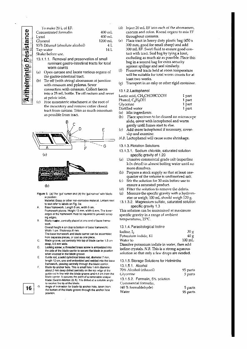
(b)
Figure 3. (a) The ‘gut’ runner and (IJ) the ‘gut runner’ with blade in position.
A.
8.
Material: Brass or other noncorrosive material. Letters next to text refer to labels on Fig. 3a. Base framework. Length 8 cm, width 5 cm. Framework pieces. Height 13 mm, width 6 mm. The lower edges of the framework must be squared to provide scrap- ing edges. Blade caer, centrally placed at one end of base frame- work. Overall height 4 cm (top to bottom of base framework). Widlh 1 cm. Thickness 8 mm. The base framework and blade carrier can be assembled from separate pieces, or cast as one piece.
C. Blade groove, cut centrally into top of blade carrier 1.5 cm deep, 0.5 mm wide.
D. Locking screw; a threaded brass screw is embedded into the side of the blade carrier to secure the blade in position when inserted in the blade groove.
E. Guide rod, a solid cylindrical brass rod, diameter 7 mm, length 12 cm, one end embedded and welded into the base framework, passing centrally through the blade carrier.
F. Blade-tip anchor hole. This is small hole 1 mm diameter about 2 mm deep drilled centrally on the top edge of the guide rod in line with the blade groove and 4.4 cm from the blade carrier. It secures the point of a removable scalpel blade (Swann-Morton 22 A). It is drilled at a suitable angle
q lo receive the tip of the blade.
16 G. Angie of inclination for blade-lip anchor hole, taken from the bottom of the blade groove through the anchor hole position.
(d) Inject 20 mL EF into each of the abomasum, caecum and colon. Knead organs to mix EF throughout contents.
(e) Place tract in heavy duty plastic bag (450 x 300 mm, good for small sheep) and add 300 mL EF. Swirl fluid to ensure good con- tact with tract. Seal bag by tying a knot, excluding as much air as possible. Place this bag in a second bag for extra security against spillage and seal similarly.
(f) Preserved tracts held at room temperature will be suitable for total worm counts for at least two weeks.
(g) Transport in an esky or other rigid container.
13.1.2. Lactophenol Lactic acid, CH&HOHCOOH 1 part Phenol, C,HsOH 1 part Glycerine 1 part Distilled water 1 part (a) Mix ingredients. (b) Place specimen to be cleared on microscope
slide, cover with lactophenol and warm gently until fumes start to rise.
(c) Add more lactophenol if necessary, cover- slip and examine.
N.B. Lactophenol will cause some shrinkage.
13.1.3. Flotation Solutions 13.1.3.1. Sodium chloride, saturated solution
specific gravity of 1.20 (a) Dissolve commercial grade salt (superfine
kiln dried) in almost boiling water until no more dissolves.
(b) Prepare a stock supply so that at least one- quarter of the volume is undissolved salt.
(c) Stir the solution for 30 min before use to ensure a saturated product.
(d) Filter the solution to remove the debris. (e) Measure the specific gravity with a hydrom-
eter or weigh; 100 mL should weigh 120 g. 13.1.3.2. Magnesium sulfate, saturated solution
specific gravity 1.3 This solution can be maintained at maximum specific gravity in a range of ambient temperatures, 25°C.
13.1.4. Parasitological Iodine Iodine, I, 3Og Potassium iodide, KI a?3 Water to 100 mL Dissolve potassium iodide in water, then add iodine crystals. N.B. This is a strong aqueous solution so that only a few drops are needed.
13.15. Storage Solutions for Helminths 13.1.5.1. Alcohol 70% Alcohol (ethanol) 95 parts Glycerine 5 parts 13.152. Formalin, 5% solution Commercial formalin, (40 % formaldehyde) 5 parts Water 95 parts
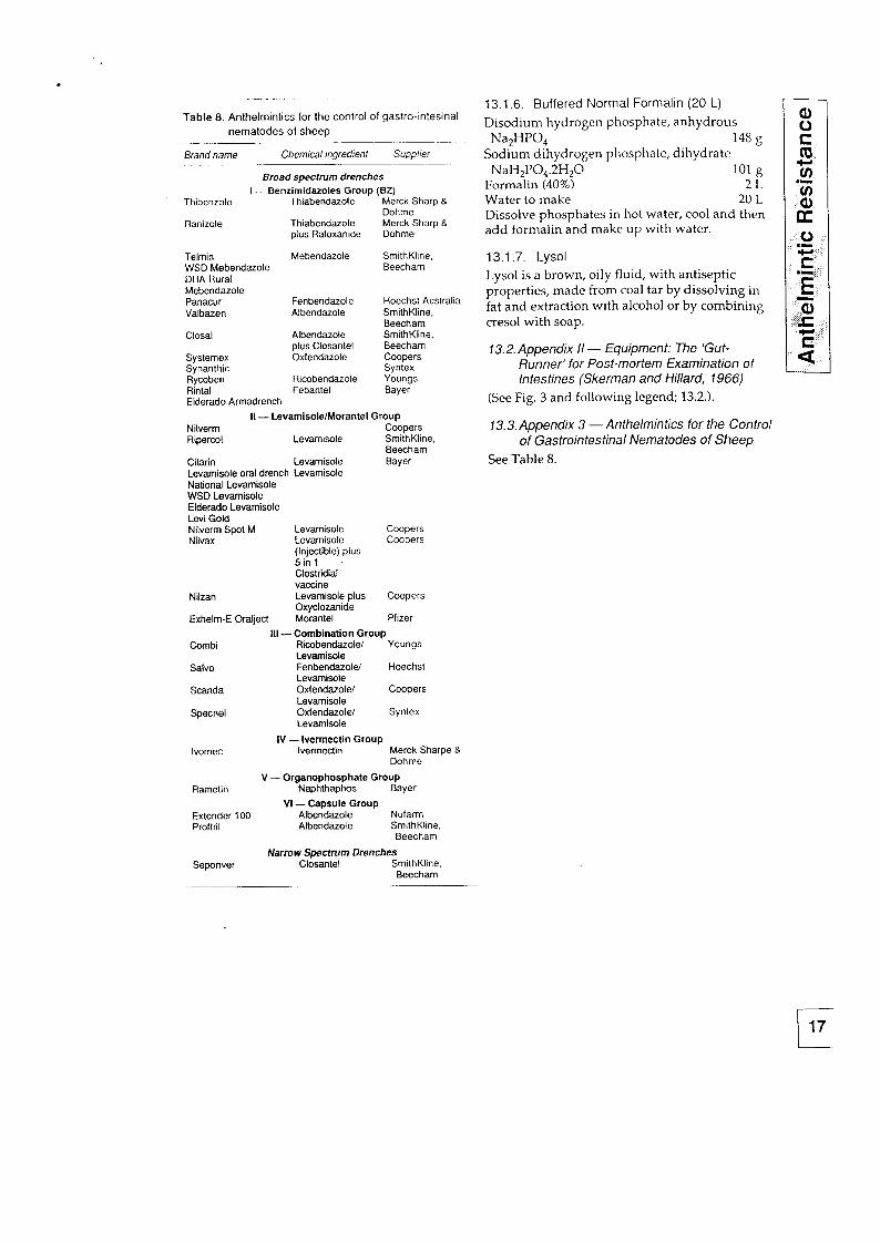
Table 8. Anlhelmintics for the control of gastro-intesinal nematodes of sheep
Brand name Chemical ingredienf Supplier
Broad spectrum drenches I - Benzimidazoles Group (62)
Thibenzole Thiabendazote Merck Sharp & Dohme
Ranizole Thiabendazole Merck Sharp B plus Rafoxanide Dohme
Telmin Mebendazole SmithKline, WSD Mebendazole Beecham DHA Rural Mebendazole Panacur Fenbendazole Hoechst Australia Valbazen Albendazole SmithKline,
Beecham Closal Albendazole SmithKline,
plus Closantel Beecham Systemex Oxfendazole Coopers Synanthic Syntex Rycoben Ricobendazole Youngs Rintal Febantel Bayer Elderado Armadrench
II - LevamisolelMorantel Group Nilverm Coopers Ripercol Levamisole SmithKline,
Beecham Citarin Levamisole Bayer Levarnisole oral drench Levamisole National Levamisole WSD Levamisole Elderado Levamisole Levi Gold Nilverm Spot M Coopers Nilvax Coopers
Nilzan
Exhelm-E Oralject
Levamisole Levamisole (Injeclible) plus 5inl Clostridial vaccine Levamisole plus Oxyclozanide Morantel
Coopers
Pfizer
Salvo
Scanda
Specnel
lvomec
Rametin
Extender 100 Proftril
Seponver
Ill-Combination Group Ricobendazolel Youngs Levamisole FenbendazoleI Hoechst Levamisole Oxfendazolel Coopers Levamisole Oxfendazole/ Syntex Levamisole
IV - lvermectin Group lvermectin Merck Sharpe 8
Dohme
V - Organophosphate Group Naphthaphos Bayer
VI -Capsule Group Albendazole Nufarm Albendazole SmithKline.
Beecham
Narrow Spectrum Drenches Closantel SmithKline.
Beecham
13.1.6. Buffered Normal Formalin (20 L)
Disodium hydrogen phosphate, anhydrous Na2HP0, 148 g
Sodium dihydrogen phosphate, dihydrate m NaIH,P0,.2H,O 101 g
Formalin (40%) 2L z
Water to make 20 L iii Dissolve phosphates in hot water, cool and then K add formalin and make up with water.
0 .I 13.1.7. Lysol Lysol is a brown, oily fluid, with antiseptic E
properties, made from coal tar by dissolving in E fat and extraction with alcohol or by combining cresol with soap. I!
,c, 13.2. Appendix II - Equipment: The ‘Gut-
Runner’ for Post-mortem Examination of Intestines (Skerman and Hi/lard, 1966)
(See Fig. 3 and following legend; 13.2.).
13.3. Appendix 3 - Anthelmin tics for the Control of Gastrointestinal Nematodes of Sheep
See Table 8.