Assessment of Metallothioneins in Tissues of the Clam Megapitaria squalida as Biomarkers for...
9
Assessment of Metallothioneins in Tissues of the Clam Megapitaria squalida as Biomarkers for Environmental Cadmium Pollution From Areas Enriched in Phosphorite Cristina Escobedo-Fregoso • Lia C. Mendez-Rodriguez • Pablo Monsalvo-Spencer • Raul A. Llera-Herrera • Tania Zenteno-Savin • Baudilio Acosta-Vargas Received: 9 October 2009 / Accepted: 28 January 2010 / Published online: 17 February 2010 Ó Springer Science+Business Media, LLC 2010 Abstract The aim of this study was to evaluate the use of metallothionein (MT) concentrations in tissues of the clam Megapitaria squalida as biomarkers of environmental cadmium (Cd) pollution from phosphorite enrichments in the marine environment, which resulted from mining activities in La Paz Bay, Baja California Sur, Mexico. Cd and MT were quantified in gills, digestive gland, and kid- ney of clams exposed to 0.2 or 0.5 mg Cd l -1 for 10, 20, or 30 days. In addition, clams from four strategically selected natural sites of La Paz Bay were collected for analysis. In tissues of bioassayed and untreated clams, the gradient of Cd concentrations was digestive gland [[ gills [ kidney, whereas that of MT was digestive gland [ gills [ kidney. Digestive gland of the clams exposed to 0.5 mg Cd l -1 for 30 days showed the highest concentrations of Cd (16.3 ± 3.9 lg Cd g -1 ). The highest statistically signifi- cant MT concentrations were found in digestive gland at 10 days of exposure to Cd. In the untreated clams, one of the highest Cd concentrations, but not MT levels, was found in digestive glands of the organisms collected from the area close to phosphorite mining activities. For envi- ronmental monitoring, MT levels in digestive gland can be used as a first approximation of the presence of high levels of divalent metals in the environment. However, in this study, MT levels did not correlate with high Cd levels in clams that had been collected from areas associated with phosphorite enrichment. Metallothioneins (MTs) are low molecular–weight, metal- binding, cytosolic proteins (Mouneyrac et al. 1999) with a high number of sulfhydryl groups due to their cysteine content. MT are involved in several metabolic functions, including the homeostasis of essential metals (e.g., copper [Cu] and zinc [Zn]) and the detoxification of nonessential metals (e.g., cadmium [Cd] and mercury [Hg]) (Klaassen et al. 1999). These proteins have been associated with important cellular protective processes, such as inactivation of hydroxyl radicals and protection against immunotoxins, hematoxins, and nephrotoxins (Bordin 2000). Because of these characteristics, MT has been proposed for use as a biomarker for heavy-metal pollution (Dabrio et al. 2002). For most studies of MT induction, bivalves have been used because these sedentary, filter-feeding organisms have subcellular systems that are involved in the accumulation of metals (Bebianno and Serafim 1998). Induction of MT can occur in several tissues, depending on the exposure period as well as the chemical presentation and concen- tration of the inducer. In the clam Corbicula fluminea from freshwater lakes (Aquitaine, France), gills are the main organ of MT synthesis (Baudrimont et al.1997). In Rudi- tapes decussates, the highest MT levels were found in digestive gland (Bebianno and Serafim 2003; Serafim and Bebianno 2007). In bivalves, divalent metals, such as Cd and Zn, are accumulated in gills, digestive gland (Boutet et al. 2002; Tanguy et al. 2003; Serafim and Bebianno 2007), mantle (Serra et al. 1995; Ka ´da ´r 2007), and kidney (In-Young et al. 2001; Blackmore and Wang 2004). Dabrio et al. (2002) demonstrated that Cd is the major inducer of MT in C. Escobedo-Fregoso Centro de Investigacio ´n Cientı ´fica y de Educacio ´n Superior de Ensenada, Ensenada, Baja California 22860, Mexico L. C. Mendez-Rodriguez (&) Á P. Monsalvo-Spencer Á R. A. Llera-Herrera Á T. Zenteno-Savin Á B. Acosta-Vargas Centro de Investigaciones Biolo ´gicas del Noroeste, La Paz, Baja California Sur 23090, Mexico e-mail: [email protected] 123 Arch Environ Contam Toxicol (2010) 59:255–263 DOI 10.1007/s00244-010-9484-7
-
Upload
independent -
Category
Documents
-
view
5 -
download
0
Transcript of Assessment of Metallothioneins in Tissues of the Clam Megapitaria squalida as Biomarkers for...

Assessment of Metallothioneins in Tissues of the Clam Megapitariasqualida as Biomarkers for Environmental Cadmium PollutionFrom Areas Enriched in Phosphorite
Cristina Escobedo-Fregoso • Lia C. Mendez-Rodriguez •
Pablo Monsalvo-Spencer • Raul A. Llera-Herrera •
Tania Zenteno-Savin • Baudilio Acosta-Vargas
Received: 9 October 2009 / Accepted: 28 January 2010 / Published online: 17 February 2010
� Springer Science+Business Media, LLC 2010
Abstract The aim of this study was to evaluate the use of
metallothionein (MT) concentrations in tissues of the clam
Megapitaria squalida as biomarkers of environmental
cadmium (Cd) pollution from phosphorite enrichments in
the marine environment, which resulted from mining
activities in La Paz Bay, Baja California Sur, Mexico. Cd
and MT were quantified in gills, digestive gland, and kid-
ney of clams exposed to 0.2 or 0.5 mg Cd l-1 for 10, 20, or
30 days. In addition, clams from four strategically selected
natural sites of La Paz Bay were collected for analysis. In
tissues of bioassayed and untreated clams, the gradient of
Cd concentrations was digestive gland [[ gills [ kidney,
whereas that of MT was digestive gland [ gills [ kidney.
Digestive gland of the clams exposed to 0.5 mg Cd l-1 for
30 days showed the highest concentrations of Cd
(16.3 ± 3.9 lg Cd g-1). The highest statistically signifi-
cant MT concentrations were found in digestive gland at
10 days of exposure to Cd. In the untreated clams, one of
the highest Cd concentrations, but not MT levels, was
found in digestive glands of the organisms collected from
the area close to phosphorite mining activities. For envi-
ronmental monitoring, MT levels in digestive gland can be
used as a first approximation of the presence of high levels
of divalent metals in the environment. However, in this
study, MT levels did not correlate with high Cd levels in
clams that had been collected from areas associated with
phosphorite enrichment.
Metallothioneins (MTs) are low molecular–weight, metal-
binding, cytosolic proteins (Mouneyrac et al. 1999) with a
high number of sulfhydryl groups due to their cysteine
content. MT are involved in several metabolic functions,
including the homeostasis of essential metals (e.g., copper
[Cu] and zinc [Zn]) and the detoxification of nonessential
metals (e.g., cadmium [Cd] and mercury [Hg]) (Klaassen
et al. 1999). These proteins have been associated with
important cellular protective processes, such as inactivation
of hydroxyl radicals and protection against immunotoxins,
hematoxins, and nephrotoxins (Bordin 2000). Because of
these characteristics, MT has been proposed for use as a
biomarker for heavy-metal pollution (Dabrio et al. 2002).
For most studies of MT induction, bivalves have been
used because these sedentary, filter-feeding organisms have
subcellular systems that are involved in the accumulation
of metals (Bebianno and Serafim 1998). Induction of MT
can occur in several tissues, depending on the exposure
period as well as the chemical presentation and concen-
tration of the inducer. In the clam Corbicula fluminea from
freshwater lakes (Aquitaine, France), gills are the main
organ of MT synthesis (Baudrimont et al.1997). In Rudi-
tapes decussates, the highest MT levels were found in
digestive gland (Bebianno and Serafim 2003; Serafim and
Bebianno 2007).
In bivalves, divalent metals, such as Cd and Zn, are
accumulated in gills, digestive gland (Boutet et al. 2002;
Tanguy et al. 2003; Serafim and Bebianno 2007), mantle
(Serra et al. 1995; Kadar 2007), and kidney (In-Young
et al. 2001; Blackmore and Wang 2004). Dabrio et al.
(2002) demonstrated that Cd is the major inducer of MT in
C. Escobedo-Fregoso
Centro de Investigacion Cientıfica y de Educacion Superior de
Ensenada, Ensenada, Baja California 22860, Mexico
L. C. Mendez-Rodriguez (&) � P. Monsalvo-Spencer �R. A. Llera-Herrera � T. Zenteno-Savin � B. Acosta-Vargas
Centro de Investigaciones Biologicas del Noroeste, La Paz,
Baja California Sur 23090, Mexico
e-mail: [email protected]
123
Arch Environ Contam Toxicol (2010) 59:255–263
DOI 10.1007/s00244-010-9484-7

mollusks. Cd constitutes a major environmental health
problem because Cd increases oxidative stress, mainly
causing renal injury, which leads to renal dysfunction
(Morales-Martın et al. 2004).
One of the most important sources of Cd in aquatic
ecosystems is that of mineral deposits, such as phosphorite
banks, which contain heavy metals (such as Cd) as com-
mon impurities (Mann and Ritchie 1995). Therefore, close
to areas containing the tailings from phosphorite mines, it
is common to find increased levels of Cd in sediments,
water, plants, and marine organisms (Gnandi et al. 2006).
One of the largest natural phosphorite banks in the world is
located in the peninsula of Baja California, Mexico (Riley
1989). In La Paz, at a site close to one of these phosphorite
deposits, Mendez et al. (2006) found high Cd concentra-
tions in the clam Megapitaria squalida (2.22 lg Cd g-1
wet weight [ww]). These levels are higher than the
concentration (2 lg Cd g-1 ww) considered by interna-
tional agencies with jurisdiction over seafood, such as the
Australia New Zealand Food Authority (Abbott et al. 2003)
and the Hong Kong Food and Environmental Hygiene
Department (Copes et al. 2008), as being the maximum
concentration permissible in clams for human
consumption.
The quantification of MT has been used as a sensitive
biomarker of anomalous levels of divalent metals, such as
Cd. For the clam Crassostrea gigas, Boutet et al. (2002)
recorded statistically significant differences between MT
levels in organisms collected in Oleron compared with
those collected from Pointe du Bendy (both sites in
France): an average of 5.55 and 2.25 lg Cd g-1 dry weight
(dw) (approximately 1.11 and 0.45 lg Cd g-1 wet weight
[ww]), respectively.
There is little information related to MT in organisms
from tropical or subtropical marine environments. Thus, in
environmental monitoring along tropical coasts, the eval-
uation of MT levels in bivalves could be useful as a first
approximation of the presence of toxic heavy metals, such
as Cd.
The clam M. squalida is a sediment-burrowing, filter-
feeding, bivalve mollusk (Schweers et al. 2006) that is
abundant in La Paz Bay. This organism has a geographic
distribution from Baja California, Mexico, to Mancora,
Peru (Keen 1971). This species, which is economically
important to the area of La Paz Bay, has been used as a
bioindicator of Cd, nickel [Ni], iron [Fe], magnesium [Mn],
lead [Pb], Cu, and Zn levels (Mendez et al. 2006; Cantu-
Medellın et al. 2009). With the overall goal of evaluating
the use of MTs as biomarkers of Cd contamination, we
selected two concentrations of Cd, which we determined to
be sublethal for adult M. squalida, to analyze the accu-
mulation of this element in three different tissues with
time. We compared the levels of MT found in these
bioassays with the levels found in untreated organisms
collected either close to or at a distance from areas with
phosphorite mining activities.
Materials and Methods
Biologic Material
Adult M. squalida clams (n = 400) were collected in a
natural area in the north of La Paz Bay. The harvesting site
is located far (approximately 35 km) from the area of
mining activities and undergoes no other anthropogenic
activities. The clams were transported in seawater to the
laboratory where they were placed in 40-l plastic tanks
containing 2 lm filtered seawater (maintained at 21 ± 1�C
and exchanged daily). During the course of 1 month, the
clams were acclimated and their digestive systems depu-
rated of sediment content. The clams were fed with the
microalgae Isochrysis galbana.
In addition, sampling of adult M. squalida (15 clams/
site) was performed at 4 sites along the shore of La Paz
Bay: Animas, El Sausozo, El Quelele, and Balandra. Each
site was visited once (Fig. 1). The organisms were trans-
ported to the laboratory on ice and frozen (-20�C) until
they were assayed. The sampled sites were chosen for the
following reasons: (1) In a previous study, the highest and
La Paz
La Paz Bay
Pacific Ocean
Gulf of California
110°30'110°66'
24°15'
24°55'
1
2
3
4
Gulf of C
alifornia
Phosphorite mine
Fig. 1 Sampling sites along the coastline of La Paz Bay, Baja
California Sur, Mexico. Clams (M. squalida) were harvested from
four natural areas: Animas, El Sausozo, El Quelele, and Balandra
256 Arch Environ Contam Toxicol (2010) 59:255–263
123

lowest levels of Cd in M. squalida were recorded at Ani-
mas and Balandra, respectively (Mendez et al. 2006); (2) El
Sausozo is the site closest to the phosphorite mine
(approximately 10 km); and (3) marine currents may
transport marine sediments from El Sausozo to El Quelele
(Monteforte and Carino 1992), thereby increasing the
levels of Cd in that area of La Paz Bay.
All clams used in this study ranged between 6 and 8 cm
in length and were harvested, at a depth of 3–6 m, from
sediment that consisted of sand containing \2% organic
carbon (Rodrıguez-Castaneda 2001). The areas of collec-
tion share the same arid climate. They generally do not
receive fresh water, with the only exception being episodic
discharges of ephemeral water streams (‘‘arroyos’’) that are
formed after rare but heavy rains (average rainfall
175 mm year-1; average salinity 36 psu) (Gonzalez-Aco-
sta et al. 2006).
Experimental Cd Exposure
Bioassays and Tissue Extracts
To obtain sublethal doses of Cd for adult M. squalida, a 30-
day bioassay was performed. Of the clams that were viable
after the acclimation period, 324 (all between 6 and 8 cm
in length) were used for this purpose, with the remainder
being discarded. Clams were exposed for 15 days to CdCl2dissolved in 2 lm filtered seawater at final concentrations
of 0.1, 0.2, 0.5, 0.7, 1, 2, 3, or 6 mg Cd l-1 (0.89 to
53.38 lM Cd). Twelve clams were exposed to each con-
centration, with treatments run in triplicate. An untreated
group (36 clams) was included as the control group. Water
in each tank (20 ± 1�C and 38 psu) was changed twice a
week, and Cd dosing was repeated at each change; clams
were fed with I. galbana. Based on the results of the bio-
assay, two concentrations that had proven to be sublethal
were selected, and treatment of the clams exposed to these
concentrations was continued for B30 days.
Thus, the clams that had been treated with either 0.2 or
0.5 mg Cd l-1 (1.77 or 4.44 lM Cd, respectively), as well
as those in the control group, were used for the MT and Cd
studies. On completion of 10, 20, or 30 days of exposure,
three clams from each group were taken to quantify MT
induction and Cd concentrations. To this end, gills,
digestive gland, and kidney were dissected from each clam,
and each organ was processed individually. Each organ
was weighed and homogenized in three volumes of Tris
buffer (20 mM, pH 8.4) in an ice bath, and each resulting
mixture was centrifuged (at 10,000 rpm; 4�C; 1 h). Each
recovered supernatant was heated (80�C; 10 min) and
centrifuged (10,000 rpm; 4�C; 1 h). Each resultant super-
natant was recovered for analysis of Cd and MT levels
(Bebianno and Serafim 1998; Wolf et al. 2000).
Natural Cd Exposure
Tissue Extracts
Of the 15 untreated clams per site, 9 were thawed, and each
was individually treated, as previously described, to
determine the levels of MT and Cd in gills, digestive gland,
and kidney.
Whole-Clam Extracts
For the analysis of total Cd and MT, the whole tissue of
each of the remaining untreated clams (six per site) was
processed individually. After being thawed, each clam was
weighed, dried at 70�C, digested in a microwave oven
(CEM model Mars 5X; CEM, Matthews, NC), and
extracted (Mendez et al. 2006).
MT Analysis
The MT concentration (lg g-1 ww) in each of the super-
natants was estimated by reverse-phase high performance
liquid chromatography (HPLC) on an HPLC chromato-
graph (Agilent, HP Agilent 1100, Waldbronn, Germany)
using a Supelcosil LC-318 column; the detector diode array
was configured at 230 nm. The eluants were 0.1% trifluo-
roacetic acid (TFA) in water at pH 3 (solvent A) and 0.1%
TFA in acetonitrile (solvent B). Commercial rabbit liver
MT (Sigma-Aldrich, St. Louis, MO) dissolved in Tris–HCl
buffer (20 mM, pH 8.4) was used as calibration standard as
described by Bordin et al. (1996). For each sample,
supernatant (5 ll) was loaded, and the separation was
performed at a flow rate of 1 ml min-1. Fractions (1 ml)
were collected for Cd quantification as described later in
text. Results are expressed as mean ± SE.
Cd Quantification
To quantify the Cd levels (all data expressed as ww), each
fraction collected from the high-pressure liquid chromato-
graph (see previous section) and analyzed directly by
atomic absorption spectrophotometry (AAS) using an
Avanta air-acetylene flame (GBC Scientific Equipment,
Dandenog, Australia). The detection limit was 0.04 lg l-1.
Concentrations were measured with a relative precision of
2%. For tissue analysis, certified standard reference mate-
rial TORT-2 (National Research Council of Canada,
Ottawa) was used for accurate calibration (95% recovery).
Statistical Analyses
Statistical significance (p \ 0.05) was assessed using
Kruskal–Wallis nonparametric significance test. For all
Arch Environ Contam Toxicol (2010) 59:255–263 257
123
statistical methods, STATISTICA (version 7; StatSoft,
Tulsa, OK) was used. The level of significance (p) is
indicated when statistical differences are recorded between
the samples.
Results
MT Induction Assay
During the course of the bioassay, mean survival of the
clams that had been exposed to 1 and 2 mg Cd l-1 was 50
and 0%, respectively. Thus, 0.2 and 0.5 mg Cd L-1 were
selected for use in the MT induction assay. No mortality
was observed during the course of the induction study.
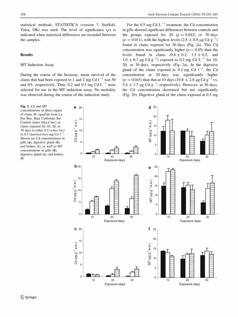
For the 0.5 mg Cd L-1 treatment, the Cd concentration
in gills showed significant differences between controls and
the groups exposed for 20 (p = 0.042) or 30 days
(p = 0.011), with the highest levels (2.8 ± 0.8 lg Cd g-1)
found in clams exposed for 30 days (Fig. 2a). This Cd
concentration was significantly higher (p \ 0.05) than the
levels found in clams (0.8 ± 0.2, 1.5 ± 0.5, and
1.0 ± 0.7 lg Cd g-1) exposed to 0.2 mg Cd L-1 for 10,
20, or 30 days, respectively (Fig. 2a). In the digestive
gland of the clams exposed to 0.2 mg Cd l-1, the Cd
concentration at 20 days was significantly higher
(p = 0.045) than that at 10 days (10.8 ± 2.8 lg Cd g-1 vs.
5.4 ± 1.7 lg Cd g-1, respectively). However, at 30 days,
the Cd concentration decreased but not significantly
(Fig. 2b). Digestive gland of the clams exposed at 0.5 mg
0
5
10
15
20a
b
c f
e
d
10 20 30Exposure days
Cd
(µg
g -1
w.w
.)
0
5
10
15
20
10 20 30
Exposure days
Cd
(µg
g -1 w
.w.)
0
5
10
15
20
10 20 30
Exposure days
Cd
(µg
g -1
w.w
.)
0
5
10
15
20
25
10 20 30Exposure days
MT
(µg
g -
1 w.w
.)
0
5
10
15
20
25
10 20 30
Exposure days
MT
(µg
g -1
w.w
.)
0
5
10
15
20
25
10 20 30
Exposure days
MT
(µg
g -1 w
.w.)
Fig. 2 Cd and MT
concentrations in three organs
of clams M. squalida from La
Paz Bay, Baja California Sur.
Control clams (black bar) or
clams exposed for 10, 20, or
30 days to either 0.2 (white bar)
or 0.5 (hatched bar) mg Cd l-1.
Shown are Cd concentrations in
gills (a), digestive gland (b),
and kidney (c), as well as MT
concentrations in gills (d),
digestive gland (e), and kidney
(f)
258 Arch Environ Contam Toxicol (2010) 59:255–263
123

Cd l-1 showed a significant increase over controls at 20
(p = 0.045) and 30 days (p = 0.049) of exposure
(14.1 ± 0.5 and 16.3 ± 3.9 lg Cd g-1, respectively). In
kidney, significant differences (p \ 0.05) in Cd levels were
found between controls and clams exposed to 0.5 mg
Cd l-1 for 20 or 30 days (3.3 ± 1.2 and 3.3 ± 0.9 lg
Cd g-1, respectively) (Fig. 2c).
In reverse-phase HPLC, MT was detected at minute 2.7.
The sample fraction having the highest Cd concentration
had the same retention time as did a peak for the MT
standard, which is known to contain a Cd-binding protein.
For the three tissues analyzed, all MT-related peaks were
detected at the same retention time.
In general, higher variability and lower concentrations
of MT were found in kidney (Fig. 2f) compared with the
other two tissues. No significant differences in MT levels
were observed in gills or kidney compared with controls
(Figs. 2d and 3f). The highest MT levels (22.6 ± 1 and
23.6 ± 0.08 lg MT g-1) were found in digestive gland of
clams at 10 days of exposure to 0.2 or 0.5 mg Cd l-1,
respectively (Fig. 2e). In digestive gland, although Cd
concentrations increased at 20 and 30 days of exposure,
MT tended to show decreasing levels with time; however,
the latter values were not statistically significant (Fig. 2).
Cd and MT Concentrations in Untreated Clams
In untreated clams, the pattern of Cd accumulation in the
three organs studied was similar to that observed in clams
in the MT induction assay, with digestive gland having the
highest Cd concentration (Fig. 3). Of the four sites, the Cd
levels in digestive gland in organisms from Quelele were
significantly lower (p \ 0.05) than those from the other
sites. The lowest levels of Cd in kidney were found in
organisms from Balandra (0.02 ± 0.05 lg Cd g-1). When
whole organisms were analyzed, higher Cd levels
(p \ 0.05) were found for clams from Animas
(1.10 ± 0.16 lg Cd g-1) and El Sausozo (1.12 ± 0.16 lg
Cd g-1) than for those recorded in Quelele
(0.77 ± 0.11 lg Cd g-1) and Balandra (0.69 ± 0.11 lg
Cd g-1).
No relation was found between the concentrations of
MT and Cd in untreated clams. MT concentrations in gills
from untreated clams (Fig. 4) were lower (p \ 0.05) for
those from El Sausozo (9.2 ± 1.2 lg MT g-1) than for
those from Balandra (14 ± 1.4 lg MT g-1). MT levels in
digestive gland of clams from Animas were higher
(p \ 0.05) (13.9 ± 1.2 lg MT g-1) than those from El
Sausozo (10.2 ± 1.3 lg MT g-1). MT levels were higher
(p \ 0.05) in kidney of clams from Animas and Balandra
(14.3 ± 2.3 and 12.9 ± 1.3 lg MT g-1, respectively)
compared with those from El Sausozo and Quelele
(6.9 ± 1.3 and 5.1 ± 0.8 lg g-1, respectively) (Fig. 4).
Discussion
The Cd concentrations used in this study are similar to
those employed in comparable studies (Bebianno et al.
1993; Boutet et al. 2002). The differences between the
present work and other MT studies were not only in the
organisms employed but also in the temperatures of the
seawater from which the organisms were collected as well
as that in which the clams were maintained and tested.
Temperature increases the filtration rate of the clams, thus
increasing the Cd levels that can be accumulated in their
tissues (Bebianno and Serafim 2003; Croteau et al. 2005).
Annual recorded temperatures for La Paz Bay range from
20 to 31�C (Gonzalez-Acosta et al. 2006), whereas for
most other MT studies the stated temperatures were
0
1
2
3
4
1 2 3 4Sites
Cd
(µg
g-1 w
.w.)
Fig. 3 Cd concentrations in whole tissue and three organs of
untreated clams M. squalida collected from four sites of La Paz
Bay. Shown are gills (hatched bar), digestive gland (white bar),
kidney (black bar), and whole tissue (point bar) of clams from four
natural areas: Animas, El Sausozo, El Quelele, and Balandra
0
5
10
15
20
25
1 2 3 4Sites
MT
(µg
g-1
w.w
.)
Fig. 4 MT concentrations in three organs of untreated clams M.squalida collected from four sites of La Paz Bay. Shown are gills
(hatched bar), digestive gland (white bar), and kidney (black bar) of
clams from four natural areas: Animas, El Sausozo, El Quelele, and
Balandra
Arch Environ Contam Toxicol (2010) 59:255–263 259
123

between 10 and 15�C (Bebianno et al. 1993; Boutet et al.
2002; Podgurskaya and Kavun2005).
The highest concentrations of Cd and MT in digestive
gland are probably due to dietary exposure, a major route
by which metals enter marine organisms (Rainbow 2002;
Wang 2002). In this route of exposure, metals arrive first to
the digestive gland, activating mechanisms (such as MT
synthesis) involved in cellular regulation and detoxification
of essential (such as Zn) and nonessential (such as Cd)
metals (Rainbow 2002). Cd is mainly bioaccumulated by
aquatic invertebrates bound to MT in the cytosol of the
organ predominantly used for accumulated Cd (Bebianno
et al. 1993), which in this study was found to be the
digestive gland, which is in agreement with results reported
previously by several other investigators (Amiard et al.
1989; Boutet et al. 2002; Bebianno and Serafim 2003;
Machreki-Ajmi et al. 2008). In the present study, digestive
gland showed the highest MT concentration in clams
exposed for 10 days to both experimental concentrations;
however, the levels decreased at 20 days. A similar
response was reported in C. gigas (Boutet et al. 2002) and
M. edulis (Bebianno and Langston 1991). The latter study
suggested an overall decrease in digestive-gland metabolic
rate due to the cellular toxicity of the heavy metal (Geret
et al. 2002). This decrease in MT levels could be explained
by hormesis, which is characterized by low-dose stimula-
tion/high-dose inhibition of the detoxification mechanism.
However, it is also important to consider that the half-life
of MT is estimated to be between 4 and 20 days, depending
on the time and concentration at which the organisms are
exposed to Cd, and then the degraded MT is rebound to
newly synthesized MT (Roesijadi and Robinson 1994) or
detoxified in phosphate granules (Rainbow 2002).
The induction mechanisms by which the metal will be
bioaccumulated could differ among organisms (Rainbow
2002). Therefore, two species that live in a same place can
differ in the types and concentrations of metals they
accumulate (Rainbow 2002; Wang 2002). However, in the
present study, M. squalida showed a pattern of bioaccu-
mulation similar to that of other bivalves, such as the clam
R. decussatus (Bebianno and Serafim 2003), the cockle
Cerastoderma glaucum (Machreki-Ajmi et al. 2008), and
the oyster Ostrea edulis (Tanguy et al. 2003). In all of these
studies, higher levels of Cd and MT were recorded for
digestive gland than for gills. The investigators attributed
their results to the fact that although gills and digestive
gland act as the main reservoirs of metals, as described by
Boutet et al. (2002) and Tanguy et al. (2003), these organs
have different physiologic roles: The gills are in direct
contact with the surrounding environment and reflect short-
term metal exposure, and the digestive glandwhere several
number of metalloenzymes bind, metabolize, and accu-
mulate excess metal concentrationsacts as a storage organ,
thus reflecting long-term metal exposure (Amiard et al.
1989; Bebianno et al. 1993). These results seem to indicate
that the physiologic function of each tissue is independent
(Bebianno and Serafim 2003).
In the present study, during the bioassays, kidney
showed the lowest degree of MT induction. This is in
agreement with Rainbow (2002), who established that not
all organisms exhibit the same pattern of metal bioaccu-
mulation and detoxification. For example, after 4 weeks of
exposure to 0.5 lg Cd ml-1 seawater, the clam Scapharca
inaequivalvis accumulated more Cd (with concentrations
B725.70 lg Cd g-1 dw) bound to a low molecular–weight
(10,000 Da) protein in kidney compared with the levels in
digestive gland or gills (Serra et al. 1995).
In the current study, the lack of induction of MT in
kidney and in gills could be attributed to the low Cd
accumulation in these tissues. It has been suggested that the
MT induction depends on the concentration of divalent
metals in the tissue. However, although kidney, as the
primary organ of metal excretion, is most exposed to the
effect of accumulated toxins, MT expression was in general
lower in this tissue than in digestive gland (Podgurskaya
and Kavun 2005).
MT and Cd Levels in Untreated Clams
Experimental conditions significantly differ from natural
conditions. The concentration of metals in water usually
exceeds the content in nature, metal input from food is
usually not taken into account, etc. (Croteau et al. 2005;
Podgurskaya and Kavun2005). Our results showed a spe-
cific response in the three tissues analyzed in this study.
Higher MT concentrations were found in digestive gland
than in gills or kidney of the untreated clams as well as
those included in the bioassays. This coincides with the
results reported by Boutet (2002). MT levels recorded in
untreated clams were similar to MT levels obtained at 20
and 30 days of the bioassay at both Cd concentrations
tested (0.2 and 0.5 lg Cd ml-1). However, the highest MT
concentrations in digestive gland were obtained at day 10,
regardless of the Cd concentration used in the bioassay.
Under these conditions, MT concentrations were almost
two times higher than those recorded in untreated clams.
This was probably caused by induction mechanisms being
drastically different in clams moving from an environment
with concentrations \0.006 lg Cd ml-1 (natural condi-
tions used during the acclimation) to one with concentra-
tions B0.5 lg Cd ml-1 (concentrations used during the
bioassays).
In gills of untreated clams collected from the four study
sites, the content of Cd was low and not significantly dif-
ferent among samples. Similar results were found in gills of
C. gigas collected from Gironde Estuary (France). High
260 Arch Environ Contam Toxicol (2010) 59:255–263
123

levels of Cd were found both in oysters and sediment,
supporting the idea that gills are not a storage site for this
metal (Boisson et al. 2003). Figure 3 shows the difference
found in the present study between gills, a short-term
storage organ, and digestive gland, a tissue that accumu-
lates and stores toxic metals (Amiard et al. 1989). The high
Cd levels in digestive gland of clams from Animas and El
Sausozo may be explained by their proximity to the
phosphorite banks and, especially, to the mining activities
at the second site. Both Animas and Balandra are influ-
enced by currents that transport Cd (among other elements)
as a result of upwellings (Monteforte and Carino 1992) that
supplies not only nutrients but also heavy metals to the
surface waters (Kavun 2008). The highest and lowest Cd
levels (B10.05 and 1.74 g g-1 dw) in total tissue were
found in El Sausozo and in Balandra, respectively, which is
in agreement with the data obtained by Mendez et al.
(2006) for M. squalida.
In the present study, we found that M. squalida clams
from the four sites had Cd concentrations in whole tissue
that was approximately 50% that in digestive gland. A
similar relation was previously reported by Baudrimont
et al. (1997), who found that Cd concentration in the whole
organism of the freshwater clam C. fluminea was 40% less
than those in organs or tissues that are known storage sites
for metals. Although Cd levels in digestive gland and
kidney were higher in clams from El Sausozo and Animas
than those from Balandra, the clams from El Sausozo had
the lowest statistically significant concentration of MT.
This unexpected finding may be the result of a metabolic
alteration, as was suggested in a study of C. gigas (Boutet
et al. 2002). Boutet et al. (2002) found abnormally low MT
levels in digestive gland of C. gigas collected in Royan,
France, and concluded that the relation between MT
induction and metal bioaccumulation is not linear, sug-
gesting the existence of other mechanisms for metal
sequestration. Roesijadi and Robinson (1994) demonstrated
that in environments with high levels of metals (especially
Cd), organisms have the capacity for partitioning the
accumulated metals in noncytosolic compartments, such as
phosphate granules. Nott and Nicolaidou (1990) showed
that the high metal concentration in digestive gland is
caused by the presence of Mg3(PO4) in the granules, which
serves as a ligand for binding metal ions.
In a previous study of M. squalida in La Paz Bay, sig-
nificantly higher activity of glutathione S-transferase
(GST) was found in organisms from Animas and Balandra
than in those from El Sausozo (Cantu-Medellın 2006).
GST, as MT, is associated with detoxification processes
because of its involvement in xenobiotic metabolism,
elimination of waste products, and regulation of hemo-
lymph electrolyte composition (Gamble et al. 1995).
Cantu-Medellın et al. (2009) found the activities of GST to
be lower in clams from El Sausozo, although the levels of
Cd in digestive gland and total content in clams were found
to be as high as those from Animas, which is also in
agreement with the results obtained in the present study.
This suggests that mollusks from Animas and Balandra
may be better adapted to an increased metal concentration
in the environment than are the organisms from El Sau-
sozo. The organisms at the latter site may be influenced by
other chemical components present in the environment,
thus decreasing their efficiency in maintaining certain
processes, such as the homeostasis of elements and
mechanisms of detoxification. For example, for the Chlo-
rophyceae algae Enteromorpha intestinalis, Rodrıguez-
Castaneda et al. (2006) reported the highest levels of nine
heavy metals, other than Cd, in those algae from an area
closer to El Sausozo compared with levels in algae from
other areas of La Paz Bay. As a probable cause of such
levels of heavy metals in the studied algae, Rodrıguez-
Castaneda et al. (2006) suggested not only that the
weathering of the natural rocks (mainly sedimentary and
volcanic rocks) causes runoff of minerals into drainage
basins but also that the nearby phosphorite mining opera-
tions influence the composition of the seawater and marine
sediment.
In the present study, although Cd levels were low in the
organisms from Quelele, MT concentrations in gills and
digestive gland were high. In a previous monitoring study,
levels of Ni, Cd, Mn, Zn, Cu, and Fe in clams from Quelele
were not high (Mendez et al. 2006). However, in the
digestive gland of M. squalida also collected in El Quelele,
Cantu-Medellın et al. (2009) found a positive correlation
between GST activity and superoxide dismutase (SOD) and
Fe levels (r2 = 0.89 and r2 = 0.97, respectively;
p \ 0.05). MTs are considered an indicator of oxidative
stress (Geret et al. 2002), as are GST and SOD. Therefore,
when taken together, these results are indicative of free-
radical production and antioxidant enzyme activities, per-
haps caused by the presence of an organic compound (such
as hydrocarbons not yet analyzed). Such a compound can
enhance the liberation of Fe from ferritin (Winterbourn
et al. 1991), thus causing free-radical production and the
subsequent induction of MT.
The results of this study in the clam M. squalida showed
that of the three organs analyzed (gills, digestive gland, and
kidney), digestive gland was the most informative regard-
ing both Cd accumulation and MT synthesis. Continuous
monitoring of this tissue in the clam M. squalida could be
used to provide information on Cd pollution. However, no
specific relation between Cd content and MT induction was
found for clams collected in an area influenced by phos-
phorite tailings. Therefore, these results indicate that MT
levels cannot be used as unique and specific biomarkers of
contamination by Cd or other elements associated with
Arch Environ Contam Toxicol (2010) 59:255–263 261
123

phosphorite mine tailings in marine environments. More
studies in relation to MT induction, such as simultaneously
measuring other detoxification mechanisms of divalent
metals, e.g., phosphate granules, must be carried out.
Acknowledgments Authors thank Lilia Ibarra Martinez for analytic
technical support. This work was funded by CIBNOR project P.C.
2.2. The authors thank Veronica Yakoleff for reviewing and editing
the manuscript.
References
Abbott P, Baines J, Fox P, Graf L, Kelly L, Stanley G et al (2003)
Review of the regulations for contaminants and natural toxicants.
Food Control 14:383–389
Amiard JC, Amiard-Triquet C, Ballan-Dufrancais C, Berthet B,
Jeantet AY, Martoja R et al (1989) Study of the bioaccumulation
at the molecular, cellular and organism levels of lead and copper
transferred to the oyster Crassostrea gigas Thunberg directly
from water or via food. Polish Acad Sci 34:521–529
Baudrimont M, Metivaud J, Maury-Brachet R, Ribeyre F, Boudou A
(1997) Bioaccumulation and metallothionein response in the
Asiatic clam (Corbicula fluminea) after experiment exposure to
cadmium and inorganic mercury. Environ Toxicol Chem
16:2096–2105
Bebianno MJ, Langston WJ (1991) Metallothionein induction in
Mytilus edulis exposed to cadmium. Mar Biol 108:91–96
Bebianno MJ, Serafim MA (1998) Comparison of metallothionein
induction in response to cadmium in the gills of the bivalve
mollusks Mytilus galloprovincialis and Ruditapes decussatus.
Sci Total Environ 214:123–131
Bebianno MJ, Serafim MA (2003) Variation of metal and metallo-
thionein concentrations in a natural population of Ruditapesdecussatus. Arch Environ Contam Toxicol 44:53–66
Bebianno MJ, Nott JA, Langston WJ (1993) Aquatic cadmium
metabolism in the clam Ruditapes decussata: the role of
metallothioneins. Toxicology 27:315–334
Blackmore G, Wang WX (2004) Relationships between metallothio-
neins and metal accumulation in the whelk Thais clavigera. Mar
Ecol Prog Ser 277:135–145
Boisson F, Goudard F, Durand JP, Barbot C, Pieri J, Amiard JC et al
(2003) Comparative radiotracer study of cadmium uptake, storage,
detoxification and depuration in the oyster Crassostrea gigas:
potential adaptive mechanisms. Mar Ecol Prog Ser 254:177–186
Bordin G (2000) Metallothionein. Cell Mol Biol 46:123–128
Bordin G, Raposo C, Rodrıguez AR (1996) Characterization of
metallothionein isoforms by reverse phase high performance
liquid chromatography with on-line UV and electrochemical
detection. J Liq Chrom Technol 19:3085–3104
Boutet I, Tanguy A, Auffret M, Riso R, Moraga D (2002)
Immunochemical quantification of metallothioneins in marine
mollusks: characterization of a metal exposure bioindicator.
Environ Toxicol Chem 21:1009–1014
Cantu-Medellın N (2006) Variacion espacial de los indicadores del
estres oxidativo en tejidos de almeja chocolata (Megapitariasqualida) (Sowerby, 1835) (Bivalvia: Veneridae) de Bahıa de La
Paz, B.C.S., Mexico. Tesis de Maestrıa, Universidad Autonoma
de Baja California Sur. La Paz, BCS, Mexico
Cantu-Medellın N, Olguin-Monroy NO, Mendez-Rodriguez LC,
Zenteno-Savin T (2009) Antioxidant enzymes and heavy metal
levels in tissues of the black chocolate clam Megapitariasqualida in Bahia de La Paz, Mexico. Arch Environ Contam
Toxicol 56:60–66
Copes R, Clark NA, Rideout K, Palat J, Teschke K (2008) Uptake of
cadmium from Pacific oysters (Crassostrea gigas) in British
Columbia oyster growers. Environ Res 107:160–169
Croteau MN, Luoma SN, Stewart RA (2005) Trophic transfer of
metals along freshwater food webs: evidence of cadmium
biomagnification in nature. Limnol Oceanogr 50:1511–1519
Dabrio M, Rodrıguez AR, Bordin G, Bebianno MJ, De Ley M,
Sestakova I et al (2002) Recent developments in quantification
methods for metallothionein. J Inorg Biochem 88:123–134
Gamble S, Goldfarb P, Porte C, Livingstone D (1995) Glutathione
peroxidase and other antioxidant enzyme function in marine
invertebrates (Mytilus edilus, Pecten maximus, Carcinus maenasand Asterias rubens). Mar Environ Res 39:191–195
Geret F, Jouan-Turpin V, Bebianno M, Cosson R (2002) Influence of
metal exposure on metallothionein synthesis and lipid peroxida-
tion in two bivalve mollusks: the oyster (Crassostrea gigas) and
mussel (Mytilus edulis). Aquat Living Resour 15:61–66
Gnandi K, Tchangbedji G, Killi K, Baba G, Abbe K (2006) The
impact of phosphate mine tailings on the bioaccumulation of
heavy metals in marine fish and crustaceans from the coastal
zone of Togo. Mine Water Environ 25:56–62
Gonzalez-Acosta B, Bashan Y, Hernandez-Saavedra NY, Ascencio F,
De la Cruz-Aguero G (2006) Seasonal seawater temperature as
the major determinant for populations of culturable bacteria in
the sediments of an intact mangrove in an arid region. FEMS
Microbiol Ecol 55:311–321
In-Young A, Jaekyoon K, Ko-Woon K (2001) The effect of body size
on metal accumulations in the bivalve Laternula elliptica.
Antarctic Sci 13:355–362
Kadar E (2007) Postcapture depuration of essential metals in the deep
sea mussel Bathymodiolus azoricus. Bull Environ Contam
Toxicol 78:99–106
Kavun VY (2008) Content of microelements in the grass shrimp
Pandalus kessleri (Decapoda: Pandalidae) from coastal waters of
the Lesser Kurilskaya Ridge. J Mar Biol Russian 34:64–72
Keen AM (1971) Sea shells of tropical West America. Marine
mollusks from Baja California to Peru, 2nd edn. Stanford
University Press, Stanford, CA
Klaassen CD, Liu J, Choudhuri S (1999) Metallothionein: an
intracellular protein to protect against cadmium toxicity. Ann
Rev Pharmacol Toxicol 36:267–294
Machreki-Ajmi M, Ketata I, Ladhar-Chaabouni R, Hamza-Chaffai A
(2008) The effect of in situ cadmium contamination on some
biomarkers in Cerastoderma glaucum. Ecotoxicology 17:1–11
Mann SS, Ritchie GSP (1995) Forms of cadmium in sandy soils after
amendment with soils of higher fixing capacity. Environ Pollut
87:23–29
Mendez L, Palacios E, Acosta B, Monsalvo-Spencer P, Alvarez-
Castaneda T (2006) Heavy metals in the clam Megapitariasqualida collected from wild and phosphorite mine-impacted sites
in Baja California, Mexico. Biol Trace Elem Res 110:275–287
Monteforte M, Carino M (1992) Exploration and evaluation of natural
stocks of pearl oysters Pinctada mazatlanica and Pteria sterna(Bivalvia: Pteriidae): La Paz Bay, South Baja California,
Mexico. Ambio 21:314–320
Morales-Martın AI, Vicente-Sanchez C, Sandoval SJ, Tagarro FM,
Lopez-Novoa SJM, Perez-Barriocanal F (2004) Efecto de la
quercetina sobre la nefrotoxicidad producida por cadmio. Rev
Toxicol 21:23–30
Mouneyrac C, Berthet BA, Amiard JC (1999) Cd distribution in the
tissues of oyster (Crassostrea gigas) exposed chronically in situ.
Water Air Soil Pollut 112:187–196
Nott JA, Nicolaidou A (1990) Transfer of metal detoxification along
marine food chains. J Mar Biol Ass U K 70:905–912
Podgurskaya OV, Kavun VY (2005) Comparative analysis of
subcellular distribution of heavy metals in organs of the bivalve
262 Arch Environ Contam Toxicol (2010) 59:255–263
123
mollusks Crenomytilus grayanus and Modiolus modiolus in a
continuously polluted environment. Russ J Mar Biol 31:373–381
Rainbow PS (2002) Trace metal concentrations in aquatic inverte-
brates: why and so what? Environ Pollut 120:497–507
Riley JP (1989) Los elementos mas abundantes y menores en el agua
de mar. In: Riley JP, Chester R (eds) Introduccion a la quımica
marina. AGT Editor, S.A, Mexico City, Mexico, pp 61–104
Rodrıguez-Castaneda AP (2001) Elementos mayores y traza en
sedimentos y macroalgas de la Bahıa de la Paz, Baja California
Sur, Mexico. Tesis de Maestrıa, Centro Interdisciplinario de
Ciencias Marinas. La Paz, BCS, Mexico
Rodrıguez-Castaneda AP, Sanchez-Rodrıguez I, Shumilin EN, Sapo-
zhnikov D (2006) Element concentrations in some species of
seaweeds from La Paz Bay and La Paz Lagoon, southwestern
Baja California, Mexico. J Appl Phycol 18:399–408
Roesijadi G, Robinson WE (1994) Metal regulation in aquatic
animals: mechanisms of uptake, accumulation and release. In:
Martins DC, Ostrander GK (eds) Aquatic toxicology: molecular,
biochemical and cellular perspectives. Lewis, Boca Raton, FL,
pp 387–420
Schweers T, Wolff M, Koch V, Sinsel-Duarte F (2006) Population
dynamics of Megapitaria squalida (Bivalvia: Veneridae) at
Magdalena Bay, Baja California Sur, Mexico. Rev Biol Trop
54:1003–1017
Serafim V, Bebianno MJ (2007) Involvement of metallothionein in Zn
accumulation and elimination strategies in Ruditapes decussatus.
Arch Environ Contam Toxicol 52:189–199
Serra R, Carpene E, Marcantonio C, Isani G (1995) Cadmium
accumulation and Cd-binding proteins in the bivalve Scapharcainaequivalvis. Comp Biochem Physiol C 111:165–174
Tanguy A, Boutet I, Riso R, Boudry P, Auffret M, Moraga D (2003)
Metallothionein genes in the European flat oyster Ostrea edulis:
a potential ecological tool for environmental monitoring. Mar
Ecol Prog Ser 257:87–97
Wang WX (2002) Interactions of trace metals and different marine
food chains. Mar Ecol Prog Ser 243:295–309
Winterbourn CC, Glenn FV, Monteiro HP (1991) Ferritin, lipid
peroxidation and redox-cycling xenobiotics. Free Radic Res
12:107–114
Wolf C, Rosick U, Brater P (2000) Quantification of the metal
distribution in metallothioneins of the human liver by HPLC
coupled with ICP-AES. Fresenius J Anal Chem 368:839–843
Arch Environ Contam Toxicol (2010) 59:255–263 263
123