The Comet assay for the evaluation of genotoxic impact in aquatic environments

A k Dixit etal ,J Biosci Tech, Vol 2 (2),2011,247-257
247
Assessment of Basal DNA Damage in Normal and Healthy
Young Individuals by COMET Assay A K DIXIT* and ARUN N.
Dept of Plant Biology and Biotechnology
KM Centre for PG studies, Pondicherry 605008 India
*[email protected] Abstract
The present study has designed to assess the levels of DNA damage in healthy
individual. To compare the levels of DNA damage among the individuals of
similar age group with gender, food habits and lifestyle as well as the
environmental factors. Life style and personal habits can produce a low level
DNA damage and Comet formation in normal healthy individuals are due to this
primary DNA lesions. These primary DNA damage may leads to biologically
relevant chromosome or gene mutations. This study was undertaken with the
aim of assessing the status of DNA damage in a normal healthy population of
Pondicherry by using COMET assay. The 60 male and 30 female volunteers in
this study belonged to the smoking, non-smoking, vegetarian and non-
vegetarian, non-alcoholic and alcoholic categories and aged between 25 and 35
years. Data from the present study showed a marked difference in the Comet
formation of the individuals when compare their area of residents i.e. in terms of
exposure to environmental pollutants and eating habits. However factors like
level of exercise, infection, occupational exposure i.e. exposure of the
individuals to chemicals and other mutagens should be recorded and studied to
make conclusion about the basal level of DNA damage in healthy individuals.
Keywords:
Basal DNA damage,
Healthy individual,
Comet assay
1. INTRODUCTION
DNA is a relatively fragile molecule which
can be easily damaged in vivo by a number
of different elements. It is a well known that
there is a certain level of basal DNA damage
even in normal healthy individuals due to
environmental pollutants/mutagens exposure
as well as from personal habits of the
individual. For example, a local pH change
may cause the loss of constituent purine
bases; chemical agents from the
environment may modify one or more bases;
and X-rays or ultraviolet (UV) radiation
(long term exposure to sunlight) may bring
about a chemical change in a base. Such
changes in DNA can be the result of normal
metabolism and this kind of damage can be
increased by malnutrition, various toxins, or
a disease state.
Comet assay has become a principle tool in
human bio monitoring studies. In the last
few decades the assay has got wide
acceptance due to its simplicity, sensitivity,
versatility, speed, and economy. The assay
is less expensive when compare to other
biomonitering assays. Samples can be
prepared and evaluated very quickly by
visual scoring [1] or by using image analysis
systems. Data can be generated fast and
efficiently. The comet assay has widespread
applications in genotoxicity testing in vitro
and in vivo, human biomonitoring or
ecotoxicology research [2] [3].
Ostling and Johannson developed a
microelectrophoretic technique under
neutral conditions to study DNA damage in
individual cells after gamma-irradiation and
proposed that strand breaks would enable
DNA loops to stretch out upon
electrophoresis and form the comet tail [4].
Singh et al. introduced the alkaline version
of the comet assay which was able to detect
alkaline-labile sites, crosslink and transient
DNA strand breaks arising due to DNA
repair processes in addition to DNA breaks
detection [5] . Relaxation of loops was also

A k Dixit etal ,J Biosci Tech, Vol 2 (2),2011,247-257
248
proposed to be the primary underlying basis
for comet formation under alkaline
conditions [6] [7] demonstrates that the
comet tail under neutral conditions consists
of relaxed loops, whereas comet tails under
alkaline conditions consist of DNA
fragments. Singh and Stephens proposed
that some single strand breaks might reflect
DNA-protein complexes [8]. They
suggested that residual protein in microgel
slides should be removed and, thereby,
increases the sensitivity of the assay. It has
been proposed that Cytotoxic effects can be
detected and distinguished from genotoxic
effects and, therefore, should have no
confounding effects on results [9] [10] [11].
Cytotoxicity produce cell death and dead
cells show specific kinds of comets called
'clouds' or 'hedgehogs'.
The comet assay has also shown high
sensitivity for detecting carcinogenicity and
due to this it is highly useful in predicting
the risk for Cancer, so it is a very useful tool
for
human biomonitoring studies. Several
versions of the comet assay are in use and
can be done in alkaline or neutral conditions,
the neutral method for the detection of DNA
double-strand
breaks, and the alkaline
method, which detects DNA single-strand
breaks and alkali-labile lesions. Glei et.al.
studied the susceptibility and potential
exposure risks or risk reductions of diet and
lifestyle in various tissues by comet assay
and suggested that food habits and lifestyle
of the individuals had a role in
tumerogenesis [12].
2. MATERIALS AND METHODS
This study was undertaken with the aim of
assessing the status of DNA damage in a
normal healthy population of Pondicherry
by using COMET assy. All individuals
answered a questionnaire that assessed their
health status, dietary habits, lifestyle, and
medication. These healthy selected
volunteers signed a letter of consent before
participation in this study. The 60 male and
30 female volunteers in this study belonged
to the smoking, non-smoking, vegetarian
and non-vegetarian, non-alcoholic and
alcoholic categories and aged between 25
and 35 years. The details of all the healthy
subjects are mention in Table.-1. The reason
for selection of particular age group for the
study was they were apart from diabetes;
leukaemia, cancer and they may have less
DNA damage.
The single-cell gel electrophoresis( SCGE)
or Comet assay is a rapid non-invasive
technique which measures DNA damage in
individual cells and useful in biomonitoring
studies to determine the level of DNA
damage resulting from exposure to
occupational and environmental mutagens
and also from lifestyle of individuals. This
simple and sensitive technique, first
introduced by [13] to visualize the relative
extent of single stranded DNA to double
stranded DNA. The cells were embedded in
an agarose layer, exposed to alkaline
condition (pH-12) thus allow the unwinding
of the double stranded DNA. When stained
with Acridine orange the single stranded
DNA emitted red signals and double strands
emitted green signals. The amount of single
stranded DNA was calculated from the
intensity of red signals relative to green
signals. In 1984 this technique was
modified by including electrophoresis
instead of alkaline treatment and there by
stretching of damaged DNA towards the
anode for direct visualization of damaged
DNA in individual cell [5]. The alkaline
version of this method was introduced by
[14] by including alkaline condition (pH
>12) during the electrophoresis. Comet
assay has become a major tool in the fields
like toxicology, apoptosis, DNA repair,
ageing, cell cycle analysis and free radical
biology due to its sensitivity to express the

A k Dixit etal ,J Biosci Tech, Vol 2 (2),2011,247-257
249
single/double-strand breaks, DNA-drug
crosslinking, DNA-protein or DNA-DNA
crosslinking and oxidative DNA base
damage.
Generally 03 ml of Heparinised blood has
collected from all the individuals and the
lymphocytes were separated using
Histopaque® 1077 [15].
2.1. Lymphocyte separation:
From 02 ml of whole blood white cells are
separated using Histopaque 1077®
. In brief 2
ml of Histopaque has transferred to a 15 ml
conical centrifuge tube and 02 ml of whole
blood (mixed with Sodium heparin) layered
over it without disturbing it. Centrifuged the
tube for 30 mins at 400g. After the
centrifugation the middle layer containing the
Lymphocytes are carefully pipetted out and
transferred to a microcentifuge tube
containing PBS (pH 7.2). Mix the cells in
PBS with help of pipette. Cells were diluted
in PBS to a concentration of 4x10⁴ cells/m.
2.2. Encapsulation:
Slides are prepared in an air conditioned
room adjusted at 25ºC in brief onto clean,
dry, plain glass slides about 150µl of hot, 1%
NMPA is dropped and smeared in one
direction with the help of another plain slide
inclined at about 45ºC. Once NMPA is set,
75µl LMPA (37%C) mixed with 20µl of
lymphocytes (from the above preparation), is
added onto the NMPA layer. The cover slip
is replaced and the gel is again allowed to set
at 4%C for 5-10mins. After 10 mins the
cover slip is gently removed and 75µl of
LMPA is added onto the second agarose
layer. The cover slip is replaced and agarose
is allowed to set again at 4ºC for 5-10mins.
Three slides were made for each sample. The
agarose forms a matrix of carbohydrate fibres
that encapsulate the cells, anchoring them in
place. The agarose is considered to be
osmotic-neutral, therefore solutions can
penetrate the gel and affect the cells without
cells shifting position.
2.3. Lysis:
The cover slip are removed again and the
slides are gently submersed into cold Lysing
solution and refrigerated for a minimum
12hrs at 4ºC. The lysis solution often used in
the comet assay consists of a highly
concentrated aqueous salt (often, common
table salt can be used) and a detergent (such
as Triton X-100 or sarcosinate). The aqueous
salt disrupts proteins and their bonding
patterns within the cell as well as disrupting
the RNA content of the cell. The detergent
dissolves the cellular membranes. Through
the action of the lysis solution the cells are
destroyed. All proteins, RNA, membranes
and cytoplasmic and nucleoplasmic
constituents are disrupted and diffuse into the
agarose matrix. Only the DNA of the cell
remains, and unravels to fill the cavity in the
agarose that the whole cell formerly filled.
This structure is called nucleoid (a general
term for a structure in which DNA is
concentrated).
2.4. Electrophoresis:
After lysis of the cells (typically 1 to 2 hours
at 4°C) the slides are washed in distilled
water to remove all salts and immersed in a
second solution - an electrophoresis solution.
Again this solution can have its pH adjusted
depending upon the type of damage that is
being investigated.
The slides are left for ~30 minutes in the
electrophoresis solution prior to an electric
field being applied. In alkaline conditions the
DNA double helix is denatured and the
nucleoid becomes single stranded.
An electric field is applied (typically 1 V/cm)
for ~20 minutes.
A k Dixit etal ,J Biosci Tech, Vol 2 (2),2011,247-257
250
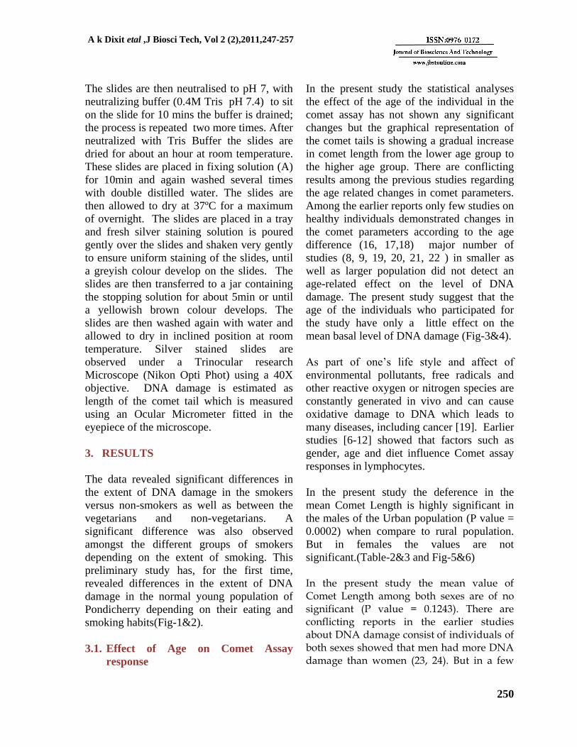
The slides are then neutralised to pH 7, with
neutralizing buffer (0.4M Tris pH 7.4) to sit
on the slide for 10 mins the buffer is drained;
the process is repeated two more times. After
neutralized with Tris Buffer the slides are
dried for about an hour at room temperature.
These slides are placed in fixing solution (A)
for 10min and again washed several times
with double distilled water. The slides are
then allowed to dry at 37ºC for a maximum
of overnight. The slides are placed in a tray
and fresh silver staining solution is poured
gently over the slides and shaken very gently
to ensure uniform staining of the slides, until
a greyish colour develop on the slides. The
slides are then transferred to a jar containing
the stopping solution for about 5min or until
a yellowish brown colour develops. The
slides are then washed again with water and
allowed to dry in inclined position at room
temperature. Silver stained slides are
observed under a Trinocular research
Microscope (Nikon Opti Phot) using a 40X
objective. DNA damage is estimated as
length of the comet tail which is measured
using an Ocular Micrometer fitted in the
eyepiece of the microscope.
3. RESULTS
The data revealed significant differences in
the extent of DNA damage in the smokers
versus non-smokers as well as between the
vegetarians and non-vegetarians. A
significant difference was also observed
amongst the different groups of smokers
depending on the extent of smoking. This
preliminary study has, for the first time,
revealed differences in the extent of DNA
damage in the normal young population of
Pondicherry depending on their eating and
smoking habits(Fig-1&2).
3.1. Effect of Age on Comet Assay
response
In the present study the statistical analyses
the effect of the age of the individual in the
comet assay has not shown any significant
changes but the graphical representation of
the comet tails is showing a gradual increase
in comet length from the lower age group to
the higher age group. There are conflicting
results among the previous studies regarding
the age related changes in comet parameters.
Among the earlier reports only few studies on
healthy individuals demonstrated changes in
the comet parameters according to the age
difference (16, 17,18) major number of
studies (8, 9, 19, 20, 21, 22 ) in smaller as
well as larger population did not detect an
age-related effect on the level of
DNA
damage. The present study suggest that the
age of the individuals who participated for
the study have only a little effect on the
mean basal level of DNA damage (Fig-3&4).
As part of one’s life style and affect of
environmental pollutants, free radicals and
other reactive oxygen or nitrogen species are
constantly generated in vivo and can cause
oxidative damage to DNA which leads to
many diseases, including cancer [19]. Earlier
studies [6-12] showed that factors such as
gender, age and diet influence Comet assay
responses in lymphocytes.
In the present study the deference in the
mean Comet Length is highly significant in
the males of the Urban population (P value =
0.0002) when compare to rural population.
But in females the values are not
significant.(Table-2&3 and Fig-5&6)
In the present study the mean value of Comet Length among both sexes are of no significant (P value = 0.1243). There are conflicting reports in the earlier studies about DNA damage consist of individuals of both sexes showed that men had more DNA damage than women (23, 24). But in a few

A k Dixit etal ,J Biosci Tech, Vol 2 (2),2011,247-257
251
reports [15] women had more basal DNA damage than men.
4. DISCUSSION
Life style and personal habits can produce a low level DNA damage and Comet formation in normal healthy individuals are due to primary DNA lesions. These primary
DNA damage may leads to biologically relevant chromosome or gene mutations. However factors like level of exercise, infection, and diet, occupational exposure i.e. exposure of the individuals to chemicals and other mutagens should be recorded and studied to make conclusion about the basal level of DNA damage in healthy individuals.
FIG: 1
FIG: 2
FIG: 3
Gender deference in Comet Length
0
0.02
0.04
0.06
0.08
0.1
0.12
0.14
1 2 3 4 5 6 7 8 9 10 11 12 13 14 15 16 17 18 19 20
Male & Female subjects
Mea
n C
omet
Len
gth
Age vs Comet Length in the Study Group
0
0.02
0.04
0.06
0.08
0.1
0.12
Min Age Group Max Age Group
Lower & Higher Group of Ages
Mea
n Co
met
Len
gth
Mean Comet Length Males Mean Comet Length Females

A k Dixit etal ,J Biosci Tech, Vol 2 (2),2011,247-257
252
Age deference & Comet Length in male subjects
FIG: 4
Age deference & Comet Length in female subjects
Age Below 29 Age above 30
Mean Comet
Length
Males 0.09283 0.107
Females 0.07956652 0.09398926
FIG: 5
25
2627 28
30
3134
35
0
0.02
0.04
0.06
0.08
0.1
0.12
25 26 27 28 30 31 34 35
26
2829
30
31
34
35
0
0.02
0.04
0.06
0.08
0.1
0.12
0.14
26 28 29 30 31 34 35

A k Dixit etal ,J Biosci Tech, Vol 2 (2),2011,247-257
253
Deference in area of residence & Comet Length in male subjects
FIG: 6
Deference in area of residence & Comet Length in female subjects
Table - 1:
Subject details
Subject Sample
size
Marital
status Food
Drinking
Residence Alcoholic
smoking
M UM NV PV Cof. Tea R U A NA
Male 60 33 27 56 04 13 47 22 38 49 11 41
Female 30 24 06 21 09 03 27 17 13 09 21 03
Total 90 57 33 77 13 16 74 39 51 58 32 44
M-Married, UM-Unmarried, NV-Non vegetarian, PV-Pure vegetarian,
Cof. - Coffee, R-Rural, U-Urban, A—Alcoholic, NA-Non alcoholic.
Table - 2
Rural vs. Urban in Male subjects
0.01
0.1
1
1 2 3 4 5 6 7 8 9 10 11 12 13 14 15 16 17
Rural Urban
Female subjects -Rural vs Urban
0.01
0.1
1
1 2 3 4 5 6
Female subjects
Mea
n c
om
et le
ng
th

A k Dixit etal ,J Biosci Tech, Vol 2 (2),2011,247-257
254
Parameter: Rural Urban
Men Comet Length 0.0894646154 0.1051828571
Standard deviation (SD) 0.007245 0.006783
Std. error of mean(SEM) 0.002009 0.002564
Minimum 0.07960 0.09688
Median (50th percentile) 0.08772 0.1046
Maximum 0.1020 0.1144
Unpaired t test two-tailed P value = 0.0002(very significant).
Table – 3
Rural vs. Urban in Female subjects
Parameter: Rural Urban
Mean Comet Length: 0.08079 0.09576
Std deviation: 0.01271 0.02317
Std error: 0.005189 0.01158
Minimum: 0.06296 0.07280
Maximum: 0.09599 0.1270
Median: 0.08214 0.09164
Two-tailed P value = 0.2189 (not significant)
Nakayama et al. reported DNA strand break
in smokers due to the effect of electrophillic
substances in tobacco [25]. Jayakumar and
Sasikala reported cigarette smoking habitual
has a synergistic effect on including DNA
damage among the jewellery workers are
occupationally exposed to nitric oxide [26].
The present findings highlight the
importance of using comet assay to detect
DNA damage and genotoxic effect of
various confounding factors, since this
information provides an increased degree of
identification for the positive response.
In order to study comet-effects in more
detail, [6] proposed to use lesion-specific
enzymes also in human monitoring studies.
Physical exercise-induced DNA damage due
to oxidative stress has studied extensively. A
reduction in DNA damage was noticed after
exercise by the supplementation of vitamins
in the volunteers [27][28][29][30][31].
DNA strand breaks are believed to arise
naturally as part of a developmental
program. Vatolin et al. [32] used comet
assay and studied the origin of single strand
breaks as part of the development & cell
division. They found a dramatic increase in
SSB during the first 2-4 mitosis after the
beginning of differentiation of embryonic

A k Dixit etal ,J Biosci Tech, Vol 2 (2),2011,247-257
255
stem cells in vitro. The increased levels
corresponded with increased levels of
SCGE. It was concluded that about half of
the chromatin of the cells had been nicked at
those stages, which, however, did not result
in a phenotypic manifestation. In another
similar study using comet assay to measure
DNA effects in embryos from 7.5 day old
mice and also found high levels of DNA
breaks in the cells [25].These results showed
though, that there are comets which do not
have a biological significance in terms of
mutagenesis.
Some investigators describe increased
comets in leukocytes of smokers [25] or
show decreasing comets when volunteers
quit smoking [27] others do not find a
difference in smokers versus non-smokers
[6][24]. This may, in part, be attributed to
the examination of cells such as leukocytes,
which are not directly exposed to cigarette
smoke. By using exfoliated buccal cells,
[33] demonstrated clearly increased DNA
damage in smokers. Radak et al.
[34]
showed an increased repair of oxidative
DNA damage directly after exercise.
5. CONCLUSION
In conclusion bio-monitoring studies of
healthy young populations are rather
specific because each population has a
different life style factors in different areas
under different climatic and environmental
conditions and are exposed to
indistinguishable of mutagen. Since DNA
damage is an important step in events
leading from carcinogen exposure to cancer
disease, our study represents an important
contribution to the correct evaluation of the
potential health risk associated with
exposure. The comet assay detects primary
DNA lesions (DNA damage) such as DNA
strand breaks. These lesions may not be of
much relevance since they may be repaired
error-free or represent transient repair sites.
We conclude that environmental conditions
play a role in the accumulation of DNA
damage. Our study investigated the
relationship between age and environmental
conditions which contribute a great deal in
setting a higher degree of basal DNA
damage. An antioxidant-rich environment
may help to provide optimal conditions for
maintaining DNA and prevent DNA
damage. The use of different antioxidants
and scavengers in maintaining DNA and
prevent DNA damage is in progress.
6. REFERENCES:
[1] Collins, A. R., Duthie, S. J., Dobson V. L.,
Comet assay in human biomonitoring studies:
reliability, validation and applications.
Carcinogenesis. 1993, 14: 1, 733–735.
[2] Tice, R. R., Agurell, E., Anderson, D.,
Burlinson, B., Hartmann, A., Kobayashi,
H.,Muyamae,Y., Rojas, E., Ryu, J. C., and
Sasaki, Y. F., Single cell gel/ Comet assay:
Guideline for in vitro and in vivo genetic
toxicology testing. Environ. Mol. Mutagen.
(2000), 35, 206–221.
[3] Anderson, M., Agurell, E., Vaghef, H.,
Bolcsfoldi, G., and Hellman, B., Extended- term
cultures of human T-lymphocytes and the Comet
assay: a useful combination when testing for
genotoxicity in vitro? Mutat.Res.2003, 40,43–55.
[4] Ostling, O., Johanson, K, H.,
Microelectrophoretic study of radiation-induced
DNA damages in individual mammalian cells.
Biochem Biophys Res Commun. 1984,123,291-
98.
[5] Singh, N, P., McCoy M, T., Tice, R, R.,
Schneider, E, L., A simple technique for quantita
-tion of low levels of DNA damage in individual
cells. Exp Cell Res, 1988, 175: 184-191.
[6] Collins, A. R., Dusinska, M., Franklin, M.,
Somorovska, M., Petrovska, H., Duthie, S.,
Fillion, L., Panayiotidis, M., Raslova, K.,
Vaughan, N., Comet assay in human
biomonitoring studies: reliability, validation and

A k Dixit etal ,J Biosci Tech, Vol 2 (2),2011,247-257
256
applications. Environ Mol Mutagen, 1997, 30,
139–146.
[7] Klaude, M., Eriksson, S., Nyrgen, J., Ahnstrom,
G., The comet assay: mechanisms and technical
considerations. Mutation Research. 1996,
363(2): 89-96.
[8] Singh, N, P., Stephens, R, E., Microgel
electrophoresis: sensitivity, mechanism, and
DNA electrostretching. Mutat Res, 1997, 383:
167-175.
[9] Andreas Hartmann and Günter Speit. 1997. The
contribution of cytotoxicity to effects seen in the
alkaline comet assay, Toxicology Letters, 1997,
90(2-3), 7, 183-188.
[10] Gunter Speit and Oliver Merk, Significance of
formaldehyde-induced DNA–protein. Environ-
mental and Molecular Mutagenesis. 1998,
32(3), 260–268.
[11] Henderson, L., Wolfreys, A., Fedyk, J.,
Bourner, C., Windebank, S., The ability of the
Comet assay to discriminate between
genotoxins and cytotoxins. Mutagenesis, 1981,
3, 89–94.
[12] Glei, M., Habermann , N., Osswald, K.,
Seidel, C., Persin, C., Jahreis, G., Pool- Zobel,
B.L., Biomarkers 2005, 10(2–3):203-217.
[13] Rydberg, B., Johanson, K, J., Hanawalt, P,C.,
Friedberg, E,C., Fox, C.F., (Eds.), Antioxidant
methodology: in vivo and in vitro
concepts, Academic Press, New York 1978,
465-468.
[14] McKelvey-Martin V. J., Green M. H.,
Schmezer P., Pool-Zobel B. L., De Meo M. P.,
Collins A. The single cell gel electrophoresis
assay (comet assay): a European review.
Mutat. Res., 1993, 288: 47-63.
[15] Boyum, A. Separation of leucocytes from
blood and bone marrow. Scand. J. Clin. Lab.
Invest. 21, 1968, Suppl. 97.
[16] Singh N. P., Charles H. Muller and Richard E.
Berger, Effects of age on DNA double-strand
breaks and apoptosis in human sperm. Fertility
and Sterility, 2003, 80( 6), 1420-1430
[17] Ford WCL, North K, Taylor H, Farrow A,
Hull MGR, Golding J., Increasing paternal age
is associated with delayed conception in a
large population of fertile couples: evidence
for declining fecundity in older men. Hum
Reprod , 2000;15,1703–1708.
[18] De La Rochebrochard E., Thonneau P.,
Paternal age and maternal age are risk factors
for miscarriage; results of a multicentre
European study. Hum Reprod., 2002;17:1649–
56.
[19] Bajpayee M, Dhawan A, Parmar D, Pandey
AK, Mathur N, Seth PK., Gender-related
differences in basal DNA damage in
lymphocytes of a healthy Indian population
using alkaline Comet assay. Mutat Res., 2002,
520:83–91.
[20] Niess, A.M., Hartmann, A., Grünert-Fuchs,
M., Poch, B., Speit, G., Int J Sports Med. DNA
damage after exhaustive treadmill running in
trained and untrained men.1996, 17(6):397-
403.
[21] Hwang ES, Bowen PE. DNA damage, a
biomarker of carcinogenesis: its measurement
and modulation by diet and environment. Crit
Rev Food Sci Nutr. 2007, 47(1):27-50.
[22] Zsolt Radák., Takao Kaneko., Shoichi Tahara.,
Hideko Nakamoto., Hideki Ohno., Mária
Sasvári., Csaba Nyakas., Sataro Goto., Free
Radical Biology and Medicine, 1999, 27(1-2),
69-74.
[23] Calderon-Garcidueñas L., Osnaya N.,
Rodriguez-Alcaraz A., Villareal C. A. DNA
damage in nasal respiratory epithelium from
children exposed to urban pollution. Environ.
Mol. Mutagen., 1997, 30: 11-20.
[24] Fairbarin DW, Olive PL, Neill KLO. The
comet assay comprehensive review. Mutation
Research, 1995, 339: 37-59.
[25] Nakayama,T., Kodama,M., Generation of
hydrogen peroxide and superoxide anion
radicals from cigarette smoking. Gann, 1984,
75, 95–98.
[26] [26] Jayakumar R, Sasikala K 2008.
Evaluation of DNA damage in jewellery
workers occupationally exposed to nitric
oxide. Environ Toxi Pharmacol, 26(2): 259-
261
[27] Zhu, C. Q., Lam, T.H., Jiang, C. Q., Wei, B.
X., Xu, Q. R., and Chen, Y. H. Increased
lymphocyte DNA strand breaks in rubber
workers. Mutat. Res. 2000, 470,201–209.
[28] Hartmann, A., Fender, H., Speit, G.,
Comparative bio-monitoring study of workers
at a waste disposal site using cytogenetic tests
and the comet (single cell gel) assay. Environ.
Mol. Mutagen., 1998, 32: 17-24.
[29] Frenzilli.G., Betti.C., Davini.T., Desideri.M.,
Fornai.E., GiannessiJL.,Maggiorelli,F.,
Paoletti,P. and Barale.R., Evaluation of DNA
damage in leukocytes of ex-smokers by single

A k Dixit etal ,J Biosci Tech, Vol 2 (2),2011,247-257
257
cell gel electrophoresis. Mutat. Res., 1996,
375, 117-123.
[30] Tebbs, R.S., Flannery, M.L., Meneses, J.J.,
Hartmann, A., Tucker, J.D., Thompson, L.H.,
Cleaver, J.E., Pedersen, R A., Modification of
comet assey for detection of DNA strand
breaks in extremely small tissue samples.
Mutagenesis, 1999, 14(4), 437-438.
[31] Cecilia Betti., Tania Davini., Liliana
Giannessi., Nicola Loprieno., Roberto Barale.,
Comparative studies by comet test and SCE
analysis in human lymphocytes from 200
healthy subjects, Mutation Research/Genetic
Toxicology, 1995, 343(4), 201-207.
[32] Vatolin,S.Y., Okhapinka, E.V., Matveea,
N.M., Shiloh, A.G., Baiborodin, S.I.,
Philimonenko, V.V., Zhdanova, N.S., Serov,
O.L., Scheduled perturbation in DNA during
in vitro differentiation of mouse embryo-
derived cells, Mol. Reprod. Dev.1997, 47, 1–
10.
[33] Rojas, E., Lopez, M.C., Valade, M., Single
cell gel electrophoresis: methodology and
applications Journal of Chromatography B,
1999, 722 (1-2): 225-254.
[34] Radak, Z., Naito, H., Kaneko, T., Tahara, S.,
Nakamoto, H., Takahashi, R., Cardozo-Pelaez,
F., Goto, S. Exercise training decreases DNA
damage and increases DNA repair and
resistance against oxidative stress of proteins
in aged rat skeletal muscle. Pflugers Arch.,
2002, 445, 273–278.
Copyright © 2022 FDOKUMEN