Asito et al., 2010
10
RESEARCH ARTICLE Open Access Elevated anti-Zta IgG levels and EBV viral load are associated with site of tumor presentation in endemic Burkitt’s lymphoma patients: a case control study Amolo S Asito 1,2 , Erwan Piriou 3 , Peter Sumba Odada 2 , Nancy Fiore 3 , Jaap M Middeldorp 4 , Carole Long 5 , Sheetij Dutta 6 , David E Lanar 6 , Walter GZO Jura 1 , Collins Ouma 1 , Juliana A Otieno 7 , Ann M Moormann 8 , Rosemary Rochford 3* Abstract Background: Endemic Burkitt’s lymphoma (BL) is an extranodal tumor appearing predominantly in the jaw in younger children while abdominal tumors predominate with increasing age. Previous studies have identified elevated levels of antibodies to Plasmodium falciparum schizont extracts and Epstein-Barr virus (EBV) viral capsid antigens (VCA) in endemic BL relative to malaria exposed controls. However, these studies have neither determined if there were any differences based on the site of clinical presentation of the tumor nor examined a broader panel of EBV and P. falciparum antigens. Methods: We used a suspension bead Luminex assay to measure the IgG levels against EBV antigens, VCA, EAd, EBNA-1 and Zta as well as P. falciparum MSP-1, LSA-1, and AMA-1 antigens in children with BL (n = 32) and in population-based age-and sex-matched controls (n = 25) from a malaria endemic region in Western Kenya with high incidence of BL. EBV viral load in plasma was determined by quantitative PCR. Results: Relative to healthy controls, BL patients had significantly increased anti-Zta (p = 0.0017) and VCA IgG levels (p < 0.0001) and plasma EBV viral loads (p < 0.0001). In contrast, comparable IgG levels to all P. falciparum antigens tested were observed in BL patients compared to controls. Interestingly, when we grouped BL patients into those presenting with abdominal tumors or with jaw tumors, we observed significantly higher levels of anti- Zta IgG levels (p < 0.0065) and plasma EBV viral loads (p < 0.033) in patients with abdominal tumors compared to patients with jaw tumors. Conclusion: Elevated antibodies to Zta and elevated plasma EBV viral load could be relevant biomarkers for BL and could also be used to confirm BL presenting in the abdominal region. Background Endemic Burkitt’s lymphoma (BL) is the most common pediatric cancer in areas that experience stable Plasmo- dium falciparum transmission, and both Epstein-Barr virus infection (EBV) and holoendemic P. falciparum are thought to be etiologically linked to this B cell neo- plasm [1-4]. BL is an aggressive extranodal B cell lym- phoma that can present in a number of different sites including jaw, abdomen, central nervous system, thyroid gland, orbital, and breast or a combination of these areas [5,6]. However, jaw and abdominal tumors are the most common sites of presentation. Interestingly, there are different epidemiologic patterns associated with chil- dren presenting with jaw compared to abdominal tumors. For example, while the median age of onset is 6 to 7 years for BL [7,8], jaw tumors are associated with a younger age of presentation and more frequently found in males, while abdominal tumors are common in older children, and there is a more equal distribution among males and females [6]. Despite the fact that endemic BL * Correspondence: [email protected] 3 SUNY Upstate Medical University, Syracuse, NY, USA Asito et al. Infectious Agents and Cancer 2010, 5:13 http://www.infectagentscancer.com/content/5/1/13 © 2010 Asito et al; licensee BioMed Central Ltd. This is an Open Access article distributed under the terms of the Creative Commons Attribution License (http://creativecommons.org/licenses/by/2.0), which permits unrestricted use, distribution, and reproduction in any medium, provided the original work is properly cited.
Transcript of Asito et al., 2010

RESEARCH ARTICLE Open Access
Elevated anti-Zta IgG levels and EBV viral load areassociated with site of tumor presentation inendemic Burkitt’s lymphoma patients:a case control studyAmolo S Asito1,2, Erwan Piriou3, Peter Sumba Odada2, Nancy Fiore3, Jaap M Middeldorp4, Carole Long5,Sheetij Dutta6, David E Lanar6, Walter GZO Jura1, Collins Ouma1, Juliana A Otieno7, Ann M Moormann8,Rosemary Rochford3*
Abstract
Background: Endemic Burkitt’s lymphoma (BL) is an extranodal tumor appearing predominantly in the jaw inyounger children while abdominal tumors predominate with increasing age. Previous studies have identifiedelevated levels of antibodies to Plasmodium falciparum schizont extracts and Epstein-Barr virus (EBV) viral capsidantigens (VCA) in endemic BL relative to malaria exposed controls. However, these studies have neither determinedif there were any differences based on the site of clinical presentation of the tumor nor examined a broader panelof EBV and P. falciparum antigens.
Methods: We used a suspension bead Luminex assay to measure the IgG levels against EBV antigens, VCA, EAd,EBNA-1 and Zta as well as P. falciparum MSP-1, LSA-1, and AMA-1 antigens in children with BL (n = 32) and inpopulation-based age-and sex-matched controls (n = 25) from a malaria endemic region in Western Kenya withhigh incidence of BL. EBV viral load in plasma was determined by quantitative PCR.
Results: Relative to healthy controls, BL patients had significantly increased anti-Zta (p = 0.0017) and VCA IgGlevels (p < 0.0001) and plasma EBV viral loads (p < 0.0001). In contrast, comparable IgG levels to all P. falciparumantigens tested were observed in BL patients compared to controls. Interestingly, when we grouped BL patientsinto those presenting with abdominal tumors or with jaw tumors, we observed significantly higher levels of anti-Zta IgG levels (p < 0.0065) and plasma EBV viral loads (p < 0.033) in patients with abdominal tumors compared topatients with jaw tumors.
Conclusion: Elevated antibodies to Zta and elevated plasma EBV viral load could be relevant biomarkers for BLand could also be used to confirm BL presenting in the abdominal region.
BackgroundEndemic Burkitt’s lymphoma (BL) is the most commonpediatric cancer in areas that experience stable Plasmo-dium falciparum transmission, and both Epstein-Barrvirus infection (EBV) and holoendemic P. falciparumare thought to be etiologically linked to this B cell neo-plasm [1-4]. BL is an aggressive extranodal B cell lym-phoma that can present in a number of different sitesincluding jaw, abdomen, central nervous system, thyroid
gland, orbital, and breast or a combination of theseareas [5,6]. However, jaw and abdominal tumors are themost common sites of presentation. Interestingly, thereare different epidemiologic patterns associated with chil-dren presenting with jaw compared to abdominaltumors. For example, while the median age of onset is 6to 7 years for BL [7,8], jaw tumors are associated with ayounger age of presentation and more frequently foundin males, while abdominal tumors are common in olderchildren, and there is a more equal distribution amongmales and females [6]. Despite the fact that endemic BL* Correspondence: [email protected]
3SUNY Upstate Medical University, Syracuse, NY, USA
Asito et al. Infectious Agents and Cancer 2010, 5:13http://www.infectagentscancer.com/content/5/1/13
© 2010 Asito et al; licensee BioMed Central Ltd. This is an Open Access article distributed under the terms of the Creative CommonsAttribution License (http://creativecommons.org/licenses/by/2.0), which permits unrestricted use, distribution, and reproduction inany medium, provided the original work is properly cited.
has several distinct clinical pictures, most studies havelooked at endemic BL as a single clinical entity.The EBV life cycle comprises both lytic and latent
phases, both of which play a role in the pathogenesis ofEBV-associated malignancies, and induce different anti-body responses [4,9]. EBV nuclear antigen (EBNA)-1 isa latent antigen and is the only latent antigen consis-tently expressed in BL tumors [10]. The EBV immediateearly lytic transcript BZLF1 which encodes the Zta pro-tein can also be detected in a subset of BL tumors[11,12] but whether elevated levels of antibodies againstZta occur in children with BL is unknown. Two addi-tional lytic antigens, viral capsid antigen (VCA) andearly antigen (EAd) have been used as serologic markersof past EBV infection (e.g. VCA IgG) or viral reactiva-tion (e.g. EAd IgG) [13]. Seminal studies in Ugandashowed that elevated titers of IgG against VCA were aprognostic risk factor for BL development [14]. In addi-tion, two recent case control studies in Uganda andMalawi have found that children with BL are more likelyto have elevated antibody titers against VCA as com-pared to controls [8,15]. These same studies alsoreported elevated IgG antibody titers against P. falci-parum schizont extracts in BL patients compared tocontrols [8,15].Until recently, measurement of pathogen-specific anti-
bodies was done with indirect immunofluorescenceassays or ELISA assays. These assays have the disadvan-tage of allowing the measurement of only one antibody:antigen complex per sample, which can otherwiserestrict the study of antibodies to multiple proteinswhen sample volumes are limited. In order to overcomethis problem, multiplexed serology has recently beendeveloped for the study of antibodies to multiple anti-gens within the same sample [16-18]. This assay cananalyze more than 100 analytes simultaneously from asmall sample volume enabling not only mass screeningof several antigens, but also studies in pediatric popula-tions with limited sample volumes [17,18]. In addition,this assay is less laborious and has fewer reaction stepscompared to the traditional ELISA [17] and the resultsare highly reproducible [18]. We have recently devel-oped a Luminex-based suspension bead assay, allowingthe determination of levels of IgG to four different EBVantigens (EBNA, VCA, EAd and Zta) within the samesample [16]. In this study, we used this technology toenable more detailed study of antibody responses to anincreased panel of antigens in BL patients in an age-andsex-matched controls living in a region where the riskof BL is high [7]. The panel included P. falciparumblood stage antigens (apical merozoite antigen (AMA)-1,merozoite surface protein (MSP)-1 that were specific fortwo strains of P. falciparum (3D7 and FV0) and a liverstage antigen (LSA)-1, in addition to the earlier
mentioned EBV antigens. In addition, we also measuredEBV viral load in the plasma. We report differences inboth EBV serology and viral load profiles in childrenwith BL compared to control children with the unex-pected finding that the clinical presentation of thetumor correlated with levels of EBV Zta antibody andplasma viral load.
ResultsStudy participant demographic characteristicsChildren with endemic BL were recruited at admissionto Nyanza Provincial General Hospital (NPGH), themajor referral hospital for cancer treatment in Nyanzaprovince, Kenya. Children admitted to this hospital forreasons other than cancer typically are from Kisumucity. However, children with BL tend to come fromrural areas [7] and this was also true in the currentstudy. Therefore, our control population for this studywas from a rural area with high BL incidence and per-sistent P. falciparum malaria transmission [7]. Thedemographic and clinical characteristics of the studyparticipants are presented in Table 1. There were 32children with BL and 25 controls frequency matched forage and gender. The mean age of the control childrenwas 7.6 years and the mean age of the BL patients was7.5 years. There were 21 male and 11 female childrenwith BL (66% male) and 16 males and 9 females (64%male) in the control group of children. Of the BLpatients, there were 14 that exclusively presented withtumor in the jaw and 16 that had only abdominaltumors. In addition, there was 1 patient that had bothjaw and abdominal tumors and 1 patient with a lowerleft limb tumor. When the BL patients were stratifiedinto clinical groups based on site of tumor presentation,children presenting with jaw tumors were younger[mean age, 5.6 years (IQR, 3.8-6.5)] than those present-ing with abdominal tumors [mean age, 9.2 years (IQR,6.3-11.00)] (p = 0.0018). Elevated serum lactate dehydro-genase (LDH) is used clinically as a marker of overalltumor burden [19]. Interestingly, BL patients withabdominal tumors had significantly higher LDH levelscompared to patients with facial tumors (p = 0.0394).
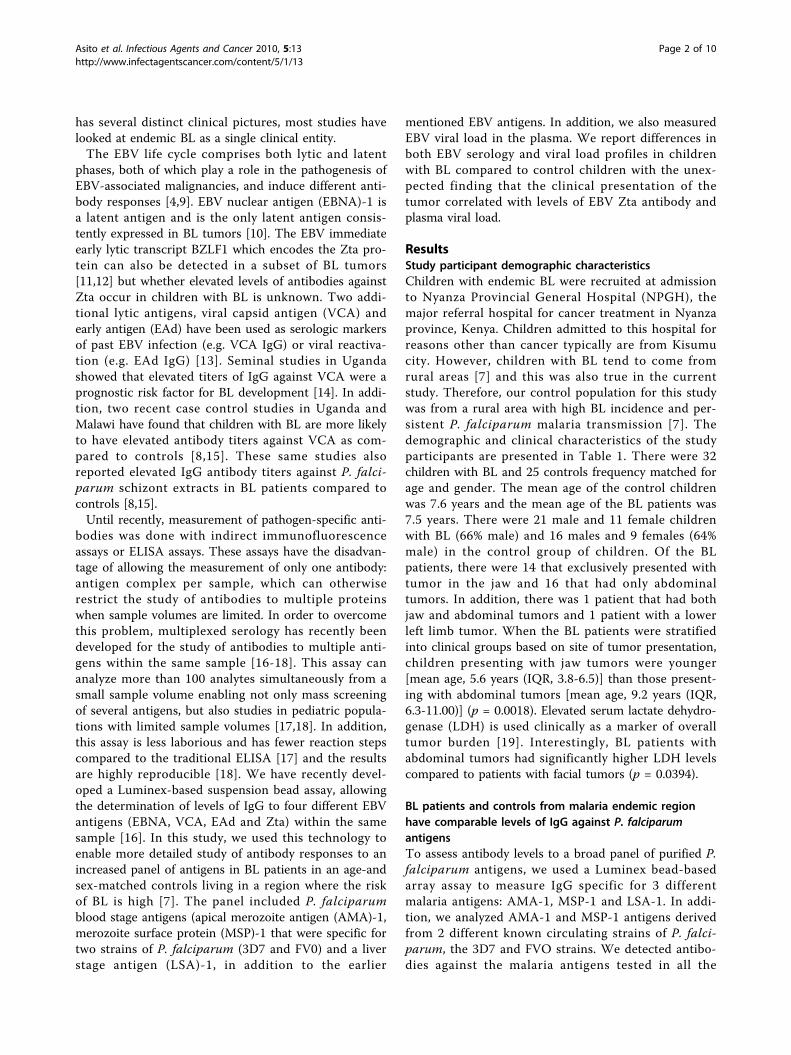
BL patients and controls from malaria endemic regionhave comparable levels of IgG against P. falciparumantigensTo assess antibody levels to a broad panel of purified P.falciparum antigens, we used a Luminex bead-basedarray assay to measure IgG specific for 3 differentmalaria antigens: AMA-1, MSP-1 and LSA-1. In addi-tion, we analyzed AMA-1 and MSP-1 antigens derivedfrom 2 different known circulating strains of P. falci-parum, the 3D7 and FVO strains. We detected antibo-dies against the malaria antigens tested in all the
Asito et al. Infectious Agents and Cancer 2010, 5:13http://www.infectagentscancer.com/content/5/1/13
Page 2 of 10
patients and controls (Fig. 1). Remarkably, the relativelevels of IgG against all P. falciparum antigens testedwere comparable between BL patients and controls andno significant differences were detected. Sixty-eight per-cent of the controls had P. falciparum parasites in theirblood while only 13% of the BL patients were P. falci-parum positive (Table 1). However, most children pre-senting with BL at NPGH have been referred fromanother hospital, and in most cases it was reported thatthese patients have been treated with anti-malarial drugsprior to admission at NPGH.
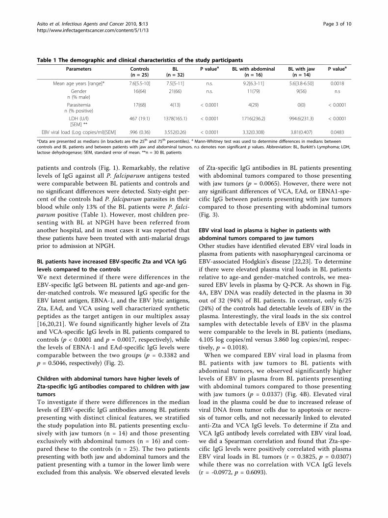
BL patients have increased EBV-specific Zta and VCA IgGlevels compared to the controlsWe next determined if there were differences in theEBV-specific IgG between BL patients and age-and gen-der-matched controls. We measured IgG specific for theEBV latent antigen, EBNA-1, and the EBV lytic antigens,Zta, EAd, and VCA using well characterized syntheticpeptides as the target antigen in our multiplex assay[16,20,21]. We found significantly higher levels of Ztaand VCA-specific IgG levels in BL patients compared tocontrols (p < 0.0001 and p = 0.0017, respectively), whilethe levels of EBNA-1 and EAd-specific IgG levels werecomparable between the two groups (p = 0.3382 andp = 0.5046, respectively) (Fig. 2).
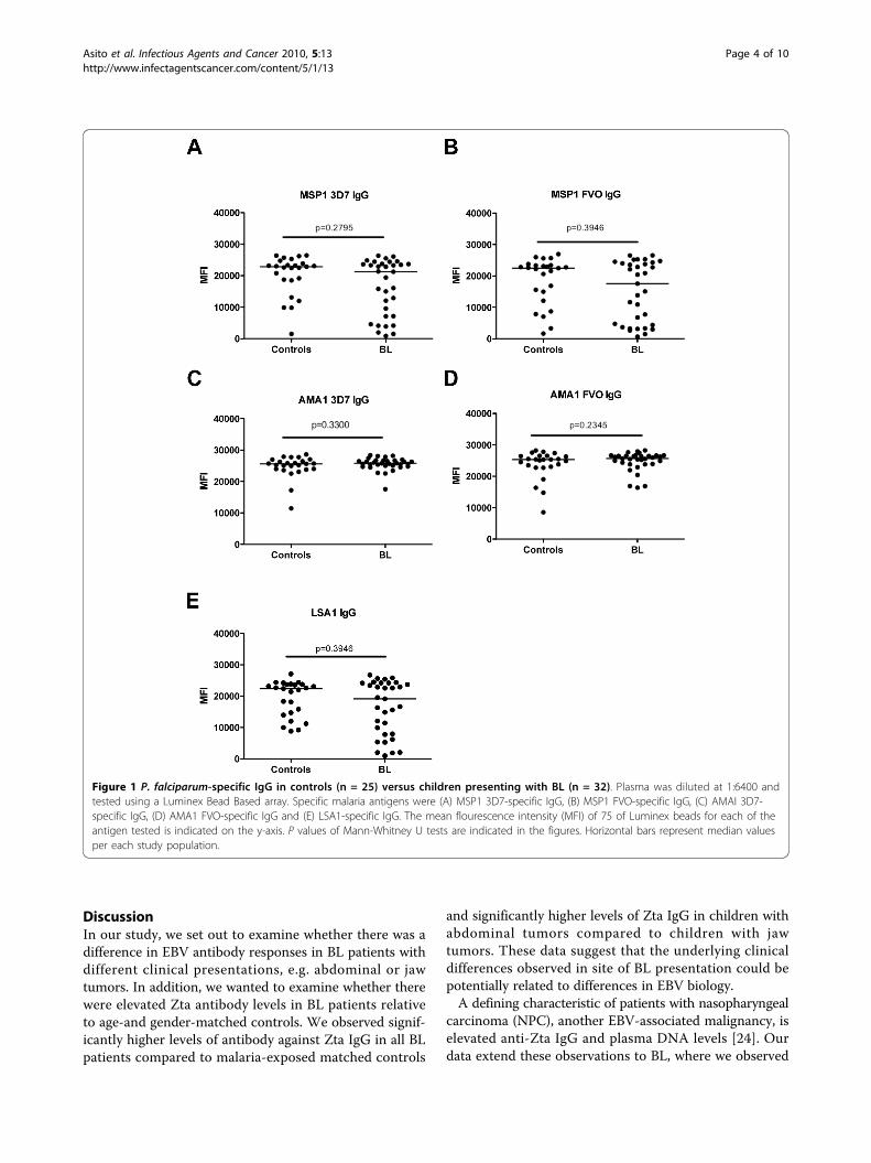
Children with abdominal tumors have higher levels ofZta-specific IgG antibodies compared to children with jawtumorsTo investigate if there were differences in the medianlevels of EBV-specific IgG antibodies among BL patientspresenting with distinct clinical features, we stratifiedthe study population into BL patients presenting exclu-sively with jaw tumors (n = 14) and those presentingexclusively with abdominal tumors (n = 16) and com-pared these to the controls (n = 25). The two patientspresenting with both jaw and abdominal tumors and thepatient presenting with a tumor in the lower limb wereexcluded from this analysis. We observed elevated levels
of Zta-specific IgG antibodies in BL patients presentingwith abdominal tumors compared to those presentingwith jaw tumors (p = 0.0065). However, there were notany significant differences of VCA, EAd, or EBNA1-spe-cific IgG between patients presenting with jaw tumorscompared to those presenting with abdominal tumors(Fig. 3).
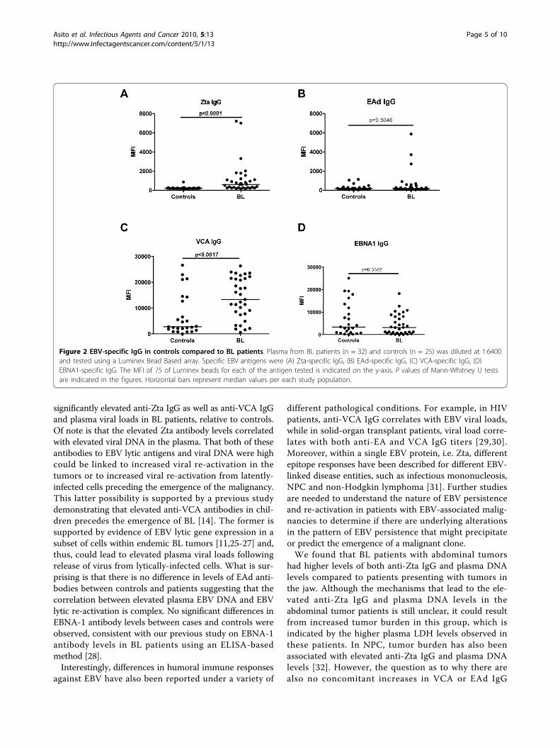
EBV viral load in plasma is higher in patients withabdominal tumors compared to jaw tumorsOther studies have identified elevated EBV viral loads inplasma from patients with nasopharyngeal carcinoma orEBV-associated Hodgkin’s disease [22,23]. To determineif there were elevated plasma viral loads in BL patientsrelative to age-and gender-matched controls, we mea-sured EBV levels in plasma by Q-PCR. As shown in Fig.4A, EBV DNA was readily detected in the plasma in 30out of 32 (94%) of BL patients. In contrast, only 6/25(24%) of the controls had detectable levels of EBV in theplasma. Interestingly, the viral loads in the six controlsamples with detectable levels of EBV in the plasmawere comparable to the levels in BL patients (medians,4.105 log copies/ml versus 3.860 log copies/ml, respec-tively, p = 0.1018).When we compared EBV viral load in plasma from
BL patients with jaw tumors to BL patients withabdominal tumors, we observed significantly higherlevels of EBV in plasma from BL patients presentingwith abdominal tumors compared to those presentingwith jaw tumors (p = 0.0337) (Fig. 4B). Elevated viralload in the plasma could be due to increased release ofviral DNA from tumor cells due to apoptosis or necro-sis of tumor cells, and not necessarily linked to elevatedanti-Zta and VCA IgG levels. To determine if Zta andVCA IgG antibody levels correlated with EBV viral load,we did a Spearman correlation and found that Zta-spe-cific IgG levels were positively correlated with plasmaEBV viral loads in BL tumors (r = 0.3825, p = 0.0307)while there was no correlation with VCA IgG levels(r = -0.0972, p = 0.6093).
Table 1 The demographic and clinical characteristics of the study participants
Parameters Controls(n = 25)
BL(n = 32)
P valuea BL with abdominal(n = 16)
BL with jaw(n = 14)
P valuea
Mean age years [range]* 7.6[5.5-10] 7.5[5-11] n.s. 9.2[6.3-11] 5.6[3.8-6.50] 0.0018
Gendern (% male)
16(64) 21(66) n.s. 11(79) 9(56) n.s
Parasitemian (% positive)
17(68) 4(13) < 0.0001 4(29) 0(0) < 0.0001
LDH (U/l)[SEM] **
467 (19.1) 1378(165.1) < 0.0001 1716(236.2) 994.6(231.3) < 0.0001
EBV viral load (Log copies/ml)[SEM] .996 (0.36) 3.552(0.26) < 0.0001 3.32(0.308) 3.81(0.407) 0.0483
*Data are presented as medians (in brackets are the 25th and 75th percentiles). a Mann-Whitney test was used to determine differences in medians betweencontrols and BL patients and between patients with jaw and abdominal tumors. n.s denotes non significant p values. Abbreviation: BL, Burkitt’s Lymphoma; LDH,lactose dehydrogenase; SEM, standard error of mean. **n = 30 BL patients
Asito et al. Infectious Agents and Cancer 2010, 5:13http://www.infectagentscancer.com/content/5/1/13
Page 3 of 10
DiscussionIn our study, we set out to examine whether there was adifference in EBV antibody responses in BL patients withdifferent clinical presentations, e.g. abdominal or jawtumors. In addition, we wanted to examine whether therewere elevated Zta antibody levels in BL patients relativeto age-and gender-matched controls. We observed signif-icantly higher levels of antibody against Zta IgG in all BLpatients compared to malaria-exposed matched controls
and significantly higher levels of Zta IgG in children withabdominal tumors compared to children with jawtumors. These data suggest that the underlying clinicaldifferences observed in site of BL presentation could bepotentially related to differences in EBV biology.A defining characteristic of patients with nasopharyngeal
carcinoma (NPC), another EBV-associated malignancy, iselevated anti-Zta IgG and plasma DNA levels [24]. Ourdata extend these observations to BL, where we observed
Figure 1 P. falciparum-specific IgG in controls (n = 25) versus children presenting with BL (n = 32). Plasma was diluted at 1:6400 andtested using a Luminex Bead Based array. Specific malaria antigens were (A) MSP1 3D7-specific IgG, (B) MSP1 FVO-specific IgG, (C) AMAI 3D7-specific IgG, (D) AMA1 FVO-specific IgG and (E) LSA1-specific IgG. The mean flourescence intensity (MFI) of 75 of Luminex beads for each of theantigen tested is indicated on the y-axis. P values of Mann-Whitney U tests are indicated in the figures. Horizontal bars represent median valuesper each study population.
Asito et al. Infectious Agents and Cancer 2010, 5:13http://www.infectagentscancer.com/content/5/1/13
Page 4 of 10
significantly elevated anti-Zta IgG as well as anti-VCA IgGand plasma viral loads in BL patients, relative to controls.Of note is that the elevated Zta antibody levels correlatedwith elevated viral DNA in the plasma. That both of theseantibodies to EBV lytic antigens and viral DNA were highcould be linked to increased viral re-activation in thetumors or to increased viral re-activation from latently-infected cells preceding the emergence of the malignancy.This latter possibility is supported by a previous studydemonstrating that elevated anti-VCA antibodies in chil-dren precedes the emergence of BL [14]. The former issupported by evidence of EBV lytic gene expression in asubset of cells within endemic BL tumors [11,25-27] and,thus, could lead to elevated plasma viral loads followingrelease of virus from lytically-infected cells. What is sur-prising is that there is no difference in levels of EAd anti-bodies between controls and patients suggesting that thecorrelation between elevated plasma EBV DNA and EBVlytic re-activation is complex. No significant differences inEBNA-1 antibody levels between cases and controls wereobserved, consistent with our previous study on EBNA-1antibody levels in BL patients using an ELISA-basedmethod [28].Interestingly, differences in humoral immune responses
against EBV have also been reported under a variety of
different pathological conditions. For example, in HIVpatients, anti-VCA IgG correlates with EBV viral loads,while in solid-organ transplant patients, viral load corre-lates with both anti-EA and VCA IgG titers [29,30].Moreover, within a single EBV protein, i.e. Zta, differentepitope responses have been described for different EBV-linked disease entities, such as infectious mononucleosis,NPC and non-Hodgkin lymphoma [31]. Further studiesare needed to understand the nature of EBV persistenceand re-activation in patients with EBV-associated malig-nancies to determine if there are underlying alterationsin the pattern of EBV persistence that might precipitateor predict the emergence of a malignant clone.We found that BL patients with abdominal tumors
had higher levels of both anti-Zta IgG and plasma DNAlevels compared to patients presenting with tumors inthe jaw. Although the mechanisms that lead to the ele-vated anti-Zta IgG and plasma DNA levels in theabdominal tumor patients is still unclear, it could resultfrom increased tumor burden in this group, which isindicated by the higher plasma LDH levels observed inthese patients. In NPC, tumor burden has also beenassociated with elevated anti-Zta IgG and plasma DNAlevels [32]. However, the question as to why there arealso no concomitant increases in VCA or EAd IgG
Figure 2 EBV-specific IgG in controls compared to BL patients. Plasma from BL patients (n = 32) and controls (n = 25) was diluted at 1:6400and tested using a Luminex Bead Based array. Specific EBV antigens were (A) Zta-specific IgG, (B) EAd-specific IgG, (C) VCA-specific IgG, (D)EBNA1-specific IgG. The MFI of 75 of Luminex beads for each of the antigen tested is indicated on the y-axis. P values of Mann-Whitney U testsare indicated in the figures. Horizontal bars represent median values per each study population.
Asito et al. Infectious Agents and Cancer 2010, 5:13http://www.infectagentscancer.com/content/5/1/13
Page 5 of 10
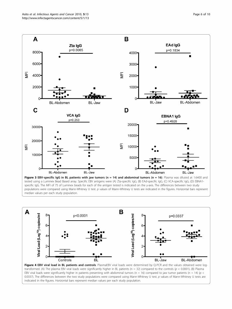
Figure 3 EBV-specific IgG in BL patients with jaw tumors (n = 14) and abdominal tumors (n = 16). Plasma was diluted at 1:6400 andtested using a Luminex Bead Based array. Specific EBV antigens were (A) Zta-specific IgG, (B) EAd-specific IgG, (C) VCA-specific IgG, (D) EBNA1-specific IgG. The MFI of 75 of Luminex beads for each of the antigen tested is indicated on the y-axis. The differences between two studypopulations were compared using Mann-Whitney U test. p values of Mann-Whitney U tests are indicated in the figures. Horizontal bars representmedian values per each study population.
Figure 4 EBV viral load in BL patients and controls. PlasmaEBV viral loads were determined by Q-PCR and the values obtained were log-transformed. (A) The plasma EBV viral loads were significantly higher in BL patients (n = 32) compared to the controls (p < 0.0001), (B) PlasmaEBV viral loads were significantly higher in patients presenting with abdominal tumors (n = 16) compared to jaw tumor patients (n = 14) (p <0.0337). The differences between the two study populations were compared using Mann-Whitney U test. p values of Mann-Whitney U tests areindicated in the figures. Horizontal bars represent median values per each study population.
Asito et al. Infectious Agents and Cancer 2010, 5:13http://www.infectagentscancer.com/content/5/1/13
Page 6 of 10

levels in the patients presenting with abdominal tumors,need to be addressed. In one study of BL, only mRNAfrom Zta could be detected in the tumors but notmRNA from another lytic transcript [25] suggesting thatthere is an abortive lytic cycle in the tumors.Chronic exposure of children to P. falciparum malaria
has been etiologically linked to Burkitt’s lymphoma(reviewed in [3,4,14]). In support of these previous stu-dies, two recent case control studies demonstrated thatBL cases had elevated anti-malaria antibody titers com-pared to controls [8,15]. However, in contrast to thesestudies, we found no differences in IgG levels to a broadpanel of pre-erythrocytic and erythrocytic antigens fromthe two common circulating P. falciparum strains (3D7and FVO) in BL patients and controls. Discrepanciesbetween these studies and our current findings may beattributed to differences in study design. In our study,we used controls from an area that has high incidenceof BL and experiences persistent P. falciparum transmis-sion [7], while in the previous studies, the controls weresick children presenting at the hospital with malignan-cies and non-malignant conditions and were from pri-marily urban areas as compared to the cases [8,15]. Wechose a population-based control group because mostchildren admitted to the Nyanza Provincial GeneralHospital for non-malignant illness are from Kisumu citywhereas the children with BL are typically from ruralregions within the Province [33]. This was true for ourcohort as most of the BL patients in the current studywere from rural settings. Thus, since both our cases andcontrols are from rural areas where malaria burden ishigher than in urban areas, it is more likely that ourcases and controls have similar exposure to P. falci-parum. This could have, however, resulted in over-matching of cases to controls and limited our ability toidentify differences in P. falciparum antibody responses.A larger population based case-control study is clearlyneeded to resolve the differences between our smallstudy and the hospital case-control studies done inUganda and Malawi [8,15].
ConclusionsChildren presenting with abdominal BL have elevatedplasma EBV DNA, paralleled by elevated IgG levels toZta, compared to children presenting with jaw tumors,suggesting differences in underlying patterns of EBVreplication. Whether BL presenting in the abdomen is adifferent clinical entity will require more detailed studiesof the tumors themselves.
MethodsStudy populationDuring the period from August 2007 and September2008, we prospectively enrolled 34 children presenting
with endemic Burkitt’s lymphoma at the Nyanza Provin-cial General Hospital, Kisumu, Kenya. Nyanza ProvincialGeneral Hospital is located in Kisumu city. As the lar-gest hospital in the Province, it provides out-patient andin-patient services, and maintains its own pathology andradiology laboratories. The hospital is the referral centerfor childhood cancer cases and is the only medical facil-ity that maintains chemotherapeutic treatment for BL inthe region. The BL patients in this study predominantlycame from all Districts in Nyanza Province (e.g. Siaya,Homa Bay, Kisumu, Migori, Kisii) as well as some Dis-tricts in Western Province (e.g. Kakamega, Vihiga,Nandi, and Busia). With the exception of Kisumu Dis-trict which includes Kisumu City, these Districts arepredominantly rural. BL diagnosis was based on histolo-gical assessment of fine needle aspirate (FNA) stainedwith May-Grunewald Giemsa. Assessment was done byboth the clinical cytologist and pathologist at NPGH.One patient was diagnosed with acute leukemia and wasexcluded from further analysis. Routine HIV testing isdone on all patients admitted to NPGH. HIV exposurewas determined in venipuncture blood using two rapidserological assays: Unigold (Trinity Biotech, Bray,County Wicklow, Ireland) and Determine (AbbottLaboratories, Chicago, Illinois, USA). One BL patientwas HIV+ and was excluded from further analysis. Allblood samples were taken from these patients prior tochemotherapy.Controls were identified from Kanyawegi village along
the shores of Lake Victoria in Kisumu West district,Nyanza province, after physical and clinical evaluationof their health status by the study clinician officers.Kanyawegi is in a rural district that has a high incidencerate of BL and experiences persistent P. falciparumtransmission [7]. After informed consent, blood sampleswere also collected from 25 healthy age-and gender-matched children (herein referred to as controls).
Microscopic investigation of P. falciparum parasitesParasitemia was determined at time of blood collectionby performing a thick smear and staining with 5%Giemsa. The slides were examined by two microscopists,any discrepancies in the slide reading were resolved by athird microscopist. Parasite density was expressed as thenumber of asexual P. falciparum per μL of blood assum-ing a leukocyte count of 8000 per μL.
Blood collection and processingEthical approval for this study was given by the KenyaMedical Research Institute (KEMRI) Ethical ReviewCommittee, Institutional Review Board for Human stu-dies at the Case Western Reserve University (Dr. Moor-mann’s institutional affiliation at time of samplecollection) and SUNY Upstate Medical University. After
Asito et al. Infectious Agents and Cancer 2010, 5:13http://www.infectagentscancer.com/content/5/1/13
Page 7 of 10
informed consent was given by the study participants’parent or guardians, finger prick blood was collected inEDTA tubes and measurements of hemoglobin levelswere determined using a portable b-haemoglobin photo-meter (Hemocue AB Angelholm, Sweden). Additionally,2-5 ml of venous blood was drawn from both BLpatients and controls. Ficoll density gradient centrifuga-tion of blood was done within 1 hour of blood collec-tion; the plasma was removed for subsequent serologicor virologic analysis. The aliquots were then stored in a-80°C freezer. Biochemical analysis of lactate dehydro-genase (LDH) levels to determine tumor burden wascarried out on frozen plasma samples using Selectra Eauto analyzer (Vita lab, Amsterdam, The Netherlands)following standard biochemistry procedures usingFortress diagnostics kits (Fortress Diagnostics Limited,Belfast, UK).
P. falciparum and EBV antigensP. falciparum-specific IgG was detected using five ofrecombinant P. falciparum antigens, MSP-1(3D7), MSP-1 (FVO), AMA-1 (3D7), AMA-1 (FVO) and LSA-1 (incollaboration with Dr. Carole Long for MSP-1, Dr.David Lanar for AMA-1, and Dr. Sheetij Dupta forLSA-1) and EBV-specific IgG was determined using foursynthetic peptides representing immuno-dominant epi-topes of the viral capsid antigen (VCA), EBV nuclearantigen 1 (EBNA1), diffuse early antigen complex (EAd)and immediate early protein (Zta) antigens of EBV. Thedefinition and serological use of these EBV peptidereagents has been described before [17,18]. In early stu-dies we defined that the predominant response to Zta inBL patients was directed against the N-terminal domain,spanning amino acids 1-44 (Middeldorp, unpublisheddata). Therefore, this domain was used as synthetic Ztaantigen throughout this study.
Luminex assayIn order to detect P. falciparum and EBV-specific IgGagainst a panel of peptides, we used the previouslydescribed protocol [15]. Briefly, different amounts ofpeptides or proteins were coupled with 1 × 10⁶ pre-acti-vated carboxylated microspheres (Luminex, Austin TE,USA) in 500 μl of 100 mM MES pH 6.0 buffer (pep-tides) or 50 mM MES pH 5.0 buffer (proteins). Thebeads were then washed and stored in PBS, 0.1% BSA,0.002% Tween-20, 0.05% Sodium Azide, pH 7.4 at 4°Cuntil use. The amounts of peptides used per 500 μLcoupling reaction were 20 μg for VCA, EBNA1 and Zta,and 50 μg was used for EAd. The amounts of recombi-nant malaria proteins per 500 μL reaction were 10 μgfor the AMA-1 and MSP-1 antigens, and 20 μg forLSA-1. During the development of the assay, severalsample dilutions were tested (ranging from 1:35 to
1:51,200) to determine the best dilution for testing IgGlevels to different antigens in the test panel. It appearedthat the Luminex assay was more sensitive than ELISAassays performed in parallel, and that in order for amajority of results to fall within the linear part of a stan-dard curve (e.g. dilutions of a positive control sampleversus fluorescent units) a dilution of 1:6400 was mostappropriate. Therefore, we tested all samples at the typi-cal 1:100 dilution as well as 1:6400. During the analysis,it appeared that indeed the 1:6400 dilution allowed forbetter distinction between high and low antibody levelsthan the 1:100 dilution. Thus, antigen specific IgG wasmeasured by incubating 1000 beads of each antigen perwell with plasma diluted 1:100 and 1:6400 in a finalvolume of 100 μl, but the results of the 1:6400 dilutionwere used in the final analysis. After washing, a 1:200dilution of PE-conjugated Goat F(ab)2 anti Human IgG(Biosource, Camarillo, CA) was added. At least 75 beadsof each antigen were then acquired on a Bioplex reader(Bio Rad, Hercules, CA). Sera from North Americanswho had no prior exposure to P. falciparum were usedas negative controls while pooled plasma from indivi-duals who were constantly positive for P. falciparumantibodies were used as positive controls for P. falci-parum antigens. In addition, EBV-seronegative and -ser-opositive controls were used in each plate. The resultsof the assay were expressed in Mean Fluorescent Inten-sity (MFI) of at least 75 beads for each P. falciparumand EBV antigen tested.
Quantitative PCR to quantify EBV DNADNA was extracted from 200 μl plasma using QiagenDNAeasy kit (Qiagen, Valencia, CA) according to themanufacturers’ protocol. DNA was eluted off the col-umn in an equivalent volume of H20 and stored at-20°C. Previously designed primers and probes thatdetect a 70 bp region of the EBV BALF5 gene wereused [34]. The quantitative (Q)-PCR cycle was as fol-lows: 2 min at 50°C, 10 min at 95°C, 42 cycles of 15 secat 95°C and 1 min at 60°C using thermal cycler model ICycler™ optical module (BioRad Laboratories, Hercules,CA). IQ SuperMix (BioRad Laboratories, Hercules, CA)was used for all reactions. To generate a standard curve,we used an EBV positive plasmid generated from thePCR product. The viral loads were log-transformed andthen calculated based on EBV genome copies/ml.
Statistical analysisGraphPad prism version 5 (GraphPad Software, Inc, LaJolla, CA) was used for all the data analysis. Differencesin the levels of P. falciparum and EBV-specific IgGlevels between controls and BL patients were comparedusing Mann-Whitney U test. The same test was used tocompare both antigen-specific IgG levels and viral loads
Asito et al. Infectious Agents and Cancer 2010, 5:13http://www.infectagentscancer.com/content/5/1/13
Page 8 of 10

between children presenting with jaw and abdominaltumors. Across group comparisons of antigen-specificIgG levels and viral loads in controls, BL patients withjaw and abdominal tumors, was carried out using Krus-kal-Wallis test. The association between categorical vari-ables for healthy and BL patients was assessed usingFisher’s exact test. Two-way ANOVA was used to evalu-ate the association between antigen-specific IgG levelswith diseases status. Correlations between antigen speci-fic IgG levels and viral load were done using Spearman’sRho test. p ≤ 0.05 was considered statistically significant.
AcknowledgementsThis work was supported by NIH R01 CA102667 (RR) and NIH R01 CA134051(AMM). We thank the parents and the guardians of both the BL patients andcontrols in allowing us to enroll their children in this study. We are alsograteful to the medical staff at the Nyanza Provincial General Hospital inassisting with both recruitment of the study participant and collection ofblood samples and Dr. Chandy John of the University of Minnesota/KEMRIproject for allowing us to use his Bioplex Luminex machine. We alsoappreciate Dorine Omenah’s efforts in co-ordinating the Burkitt’s lymphomastudy. This work is published with the permission of the Director of theKenya Medical Research Institute.
Author details1Maseno University, Maseno, Kenya. 2Center for Global Health Research,Kenya Medical Research Institute, Kisumu, Kenya. 3SUNY Upstate MedicalUniversity, Syracuse, NY, USA. 4VU University Medical Center, Amsterdam, TheNetherlands. 5National Institutes of Health, Bethesda, MD, USA. 6Walter ReedArmy Institute of Research, Silver Springs, MD, USA. 7Kenya Ministry ofHealth, Kisumu, Kenya. 8University of Massachusetts, Worcester, MA, USA.
Authors’ contributionsAA, EP, AM and RR conceived of the study, participated in the study designand helped draft the manuscript. AA, EP, and PSO performed the study. NFperformed PCR assays; JO provided clinical oversight; JMM, DEL, SD and CLprovided peptides for Luminex assay and analysis of data from assays, andWGZ and CO provided supervision of AA and assisted with study design. Allauthors read and approved the final manuscript.
Competing interestsThe authors have no financial conflict of interest. JM is owner and CEO ofCyto-Barr BV, but declares no commercial interest in this study.
Received: 7 May 2010 Accepted: 28 July 2010 Published: 28 July 2010
References1. Griffin BE, Xue SA: Epstein-Barr virus infections and their association with
human malignancies: some key questions. Ann Med 1998, 30:249-259.2. de-The G: Is Burkitt’s lymphoma related to perinatal infection by Epstein-
Barr virus? Lancet 1977, 1:335-338.3. Rochford R, Cannon MJ, Moormann AM: Endemic Burkitt’s lymphoma:
a polymicrobial disease? Nat Rev Microbiol 2005, 3:182-187.4. Thorley-Lawson DA, Allday MJ: The curious case of the tumour virus:
50 years of Burkitt’s lymphoma. Nat Rev Microbiol 2008, 6:913-924.5. Mwanda OW, Rochford R, Rainey J, Wilson ML: Challenges in the
epidemiological and clinical aspects of Burkitt’s lymphoma in Kenya:linking evidence and experience. East Afr Med J 2004, S111-116.
6. Ogwang MD, Bhatia K, Biggar RJ, Mbulaiteye SM: Incidence andgeographic distribution of endemic Burkitt lymphoma in northernUganda revisited. Int J Cancer 2008, 123:2658-2663.
7. Rainey JJ, Mwanda WO, Wairiumu P, Moormann AM, Wilson ML,Rochford R: Spatial distribution of Burkitt’s lymphoma in Kenya andassociation with malaria risk. Trop Med Int Health 2007, 12:936-943.
8. Carpenter LM, Newton R, Casabonne D, Ziegler J, Mbulaiteye S, Mbidde E,Wabinga H, Jaffe H, Beral V: Antibodies against malaria and Epstein-Barr
virus in childhood Burkitt lymphoma: a case-control study in Uganda. IntJ Cancer 2008, 122:1319-1323.
9. Rowe M, Kelly GL, Bell AI, Rickinson AB: Burkitt’s lymphoma: the RosettaStone deciphering Epstein-Barr virus biology. Semin Cancer Biol 2009,19:377-388.
10. Young LS, Rickinson AB: Epstein-Barr virus: 40 years on. Nat Rev Cancer2004, 4:757-768.
11. Xue SA, Labrecque LG, Lu QL, Ong SK, Lampert IA, Kazembe P, Molyneux E,Broadhead RL, Borgstein E, Griffin BE: Promiscuous expression of Epstein-Barr virus genes in Burkitt’s lymphoma from the central African countryMalawi. Int J Cancer 2002, 99:635-643.
12. Niedobitek G, Agathanggelou A, Rowe M, Jones EL, Jones DB, Turyaguma P,Oryema J, Wright DH, Young LS: Heterogeneous expression of Epstein-Barr virus latent proteins in endemic Burkitt’s lymphoma. Blood 1995,86:659-665.
13. Rickinson A, Kieff E: Epstein-Barr Virus. Fields Virology Philadelphia:Lippincott Williams and WilkinsKnipe DM, Howley PM , 5 2007, 2655-2700.
14. de-The G, Geser A, Day NE, Tukei PM, Williams EH, Beri DP, Smith PG,Dean AG, Bronkamm GW, Feorino P, Henle W: Epidemiological evidencefor causal relationship between Epstein-Barr virus and Burkitt’slymphoma from Ugandan prospective study. Nature 1978, 274:756-761.
15. Mutalima N, Molyneux E, Jaffe H, Kamiza S, Borgstein E, Mkandawire N,Liomba G, Batumba M, Lagos D, Gratrix F, et al: Associations betweenBurkitt lymphoma among children in Malawi and infection with HIV,EBV and malaria: results from a case-control study. PLoS One 2008, 3:e2505.
16. Piriou E, Kimmel R, Chelimo K, Middeldorp JM, Odada PS, Ploutz-Snyder R,Moormann AM, Rochford R: Serological evidence for long-term Epstein-Barr virus reactivation in children living in a holoendemic malaria regionof Kenya. J Med Virol 2009, 81:1088-1093.
17. Gu AD, Mo HY, Xie YB, Peng RJ, Bei JX, Peng J, Li MY, Chen LZ, Feng QS,Jia WH, Zeng YX: Evaluation of a multianalyte profiling assay and anenzyme-linked immunosorbent assay for serological examination ofEpstein-Barr virus-specific antibody responses in diagnosis ofnasopharyngeal carcinoma. Clin Vaccine Immunol 2008, 15:1684-1688.
18. Earley MC, Vogt RF, Shapiro HM, Mandy FF, Kellar KL, Bellisario R, Pass KA,Marti GE, Stewart CC, Hannon WH: Report from a workshop onmultianalyte microsphere assays. Cytometry 2002, 50:239-242.
19. Magrath I, Lee YJ, Anderson T, Henle W, Ziegler J, Simon R, Schein P:Prognostic factors in Burkitt’s lymphoma: importance of total tumorburden. Cancer 1980, 45:1507-1515.
20. van Grunsven WM, Spaan WJ, Middeldorp JM: Localization and diagnosticapplication of immunodominant domains of the BFRF3-encodedEpstein-Barr virus capsid protein. J Infect Dis 1994, 170:13-19.
21. Meij P, Vervoort MB, Aarbiou J, van Dissel P, Brink A, Bloemena E, Meijer CJ,Middeldorp JM: Restricted low-level human antibody responses againstEpstein-Barr virus (EBV)-encoded latent membrane protein 1 in asubgroup of patients with EBV-associated diseases. J Infect Dis 1999,179:1108-1115.
22. Drouet E, Brousset P, Fares F, Icart J, Verniol C, Meggetto F, Schlaifer D,Desmorat-Coat H, Rigal-Huguet F, Niveleau A, Delsol G: High Epstein-Barrvirus serum load and elevated titers of anti-ZEBRA antibodies in patientswith EBV-harboring tumor cells of Hodgkin’s disease. J Med Virol 1999,57:383-389.
23. Tan EL, Looi LM, Sam CK: Evaluation of plasma Epstein-Barr virus DNAload as a prognostic marker for nasopharyngeal carcinoma. SingaporeMed J 2006, 47:803-807.
24. Dardari R, Menezes J, Drouet E, Joab I, Benider A, Bakkali H, Kanouni L,Jouhadi H, Benjaafar N, El Gueddari B, et al: Analyses of the prognosticsignificance of the Epstein-Barr virus transactivator ZEBRA protein anddiagnostic value of its two synthetic peptides in nasopharyngealcarcinoma. J Clin Virol 2008, 41:96-103.
25. Labrecque LG, Xue SA, Kazembe P, Phillips J, Lampert I, Wedderburn N,Griffin BE: Expression of Epstein-Barr virus lytically related genes inAfrican Burkitt’s lymphoma: correlation with patient response totherapy. Int J Cancer 1999, 81:6-11.
26. Araujo I, Foss HD, Bittencourt A, Hummel M, Demel G, Mendonca N,Herbst H, Stein H: Expression of Epstein-Barr virus-gene products inBurkitt’s lymphoma in Northeast Brazil. Blood 1996, 87:5279-5286.
27. Fujita S, Buziba N, Kumatori A, Senba M, Yamaguchi A, Toriyama K: Earlystage of Epstein-Barr virus lytic infection leading to the “starry sky”
Asito et al. Infectious Agents and Cancer 2010, 5:13http://www.infectagentscancer.com/content/5/1/13
Page 9 of 10
pattern formation in endemic Burkitt lymphoma. Arch Pathol Lab Med2004, 128:549-552.
28. Moormann AM, Heller KN, Chelimo K, Embury P, Ploutz-Snyder R, Otieno JA,Oduor M, Munz C, Rochford R: Children with endemic Burkitt lymphomaare deficient in EBNA1-specific IFN-gamma T cell responses. Int J Cancer2009, 124:1721-1726.
29. Rogers BB, Conlin C, Timmons CF, Dawson DB, Krisher K, Andrews WS:Epstein-Barr virus PCR correlated with viral histology and serology inpediatric liver transplant patients. Pediatr Pathol Lab Med 1997,17:391-400.
30. Stevens SJ, Smits PH, Verkuijlen SA, Rockx DA, van Gorp EC, Mulder JW,Middeldorp JM: Aberrant Epstein-Barr virus persistence in HIV carriers ischaracterized by anti-Epstein-Barr virus IgA and high cellular viral loadswith restricted transcription. Aids 2007, 21:2141-2149.
31. Tedeschi R, Foong YT, Cheng HM, dePaoli P, Lehtinen T, Elfborg T, Dillner J:The disease associations of the antibody response against the Epstein-Barr virus transactivator protein ZEBRA can be separated into differentepitopes. J Gen Virol 1995, 76(Pt 6):1393-1400.
32. Shao JY, Zhang Y, Li YH, Gao HY, Feng HX, Wu QL, Cui NJ, Cheng G, Hu B,Hu LF, et al: Comparison of Epstein-Barr virus DNA level in plasma,peripheral blood cell and tumor tissue in nasopharyngeal carcinoma.Anticancer Res 2004, 24:4059-4066.
33. Rainey JJ, Omenah D, Sumba PO, Moormann AM, Rochford R, Wilson ML:Spatial clustering of endemic Burkitt’s lymphoma in high-risk regions ofKenya. Int J Cancer 2007, 120:121-127.
34. Moormann AM, Chelimo K, Sumba OP, Lutzke ML, Ploutz-Snyder R,Newton D, Kazura J, Rochford R: Exposure to holoendemic malaria resultsin elevated Epstein-Barr virus loads in children. J Infect Dis 2005,191:1233-1238.
doi:10.1186/1750-9378-5-13Cite this article as: Asito et al.: Elevated anti-Zta IgG levels and EBV viralload are associated with site of tumor presentation in endemic Burkitt’slymphoma patients: a case control study. Infectious Agents and Cancer2010 5:13.
Submit your next manuscript to BioMed Centraland take full advantage of:
• Convenient online submission
• Thorough peer review
• No space constraints or color figure charges
• Immediate publication on acceptance
• Inclusion in PubMed, CAS, Scopus and Google Scholar
• Research which is freely available for redistribution
Submit your manuscript at www.biomedcentral.com/submit
Asito et al. Infectious Agents and Cancer 2010, 5:13http://www.infectagentscancer.com/content/5/1/13
Page 10 of 10