APPROACH TO ABDOMINAL PAIN PPT KEYNOTE - KSUMSC
77
APPROACH TO ABDOMINAL PAIN Aref Melibary MD FRCPC ABEM Consultant of Emergency Medicine & Critical Care Medicine Assistant Prof. Dept of Emergency Medicine KSUMC
-
Upload
khangminh22 -
Category
Documents
-
view
3 -
download
0
Transcript of APPROACH TO ABDOMINAL PAIN PPT KEYNOTE - KSUMSC

APPROACH TO ABDOMINAL PAIN
Aref Melibary MD FRCPC ABEM Consultant of Emergency Medicine & Critical Care Medicine
Assistant Prof. Dept of Emergency Medicine KSUMC

Overview• Approach to abdominal pain
• Acute Appendicitis
• Intestinal Obstruction
• Acute Cholecystitis and Biliary Pain
• Acute Pancreatitis
• Abdominal Aortic Aneurysm
• Mesenteric Ischemia
• Inflammatory Bowel Disease
• Diverticulitis

High Yield Questions1. Age?
• Increased age = higher risk
2. Which came first the pain or the vomiting? • Pain first = usually surgical cause
3. How long did you have the pain? • Acute VS Chronic
4. Surgical history? • Previously opened abdomens are more likely to have surgical problems
5. Is the pain intermittent or constant? • Constant pain is worse

High Yield Questions
7. Do you have any PHx of immunocompromise, pancreatitis, diverticulitis, renal failure, cancer or IBD?
• PHx of any of those suggest serious cause
8. Pregnancy? • Child bearing age —> ectopic pregnancy should be considered
9. Recent antibiotics or steroids? • Abx and steroid use may mask serious conditions
10. PHx of heart disease, vascular disease , or AF? • Positive PHx suggests Mesenteric Ischemia

GI habit and Vomiting• Habit
• Change in bowel habit • Amount • Frequency • Consistency • Abnormal stools
• Vomiting • Amount • Color • Contents • Character

Abnormal Stool


The Pain progression

Extra-Abdominal Causes of Abdominal Pain
• ACS • Pneumonia • PE • Pericarditis / Myocarditis • Testicular Torsion • DKA • Glaucoma • AKA • Uremia • SCD
• Black widow spider bite
• Snake bite • Methanol
toxicity • Lead poisoning • Hyperthyroidism • SLE • GAS Pharyngitis • RMSF • Lyme Disease • Porphyria • Anaphylactoid
Purpura

Consider the life-threatening causes • Emergent• AMI • Leaking AAA • Aortoenteric fistula • Acute Hemorrhagic Pancreatitis • Mesenteric Ischemia / Infarction • Splenic Rupture • Ectopic Pregnancy
• Urgent• Perforated Bowel• Volvulus• Ascending
cholangitis• Strangulated
hernia• Tubo-ovarian
Abscess• Bowel
Obstruction• Diverticulitis• Ovarian Torsion

ACUTE APPENDICITIS

Common findings in Acute Appy
Symptoms
• Abdominal Pain • Anorexia • Nausea and Vomiting • Fever • Chills • Diarrhea
Signs – Abdominal Tenderness
• Periumbilical • RIF pain
– Rebound Tenderness – Guarding – Rigidity – CMT – Obturator sign – Psoas sign – Rovsing’s sign

Causes of Appendicitis• Feacolith
• Mesenteric Lymphadenopathy
• Worms
• Granulomatous Disease
• Tumors
• Adhesions
• Diet (seeds)

Diagnosis• Labs
• CBC • Pregnancy test • UA
• Imaging • AXR • US • CT

Mimics of Appendicitis• Mesenteric Adenitis
• Yersinia Gastroenteritis
• PID
• Ectopic Pregnancy
• Ruptured Ovarian cyst
• Pyelonephritis
• Crohn’s Disease
• Black Widow spider bite

Management • NPO
• Fluids
• Abx if suspected rupture
• Analgesia
• Early surgical consultation

INTESTINAL OBSTRUCTION

Causes of Bowel Obstruction
Small Bowel • Adhesions (MC) • Hernia (2nd MC) • Neoplasm • Intussusception • Gall stones • Bezoars / Worms • Crohn’s Disease • Radiation enteritis • FB ingestion
Large Bowel
– Tumor (most common) • Bowel tumor • External compression
– Crohn’s Disease – Diverticulitis – Volvulus – Fecal Impaction – Hernia


SSx• Pain
• Crampy, intermittent, and poorly localized
• Vomiting• The more prominent , the more proximal the obstruction is • Bilious vomiting suggest and obstruction distal to the pylorus • Feculent vomiting suggests distal SBO or LBO that is long standing,
probably with gangrenous bowel
• Abdominal Distension• The more the distension, the more distal the obstruction is • Obstipation vs. constipation
• 3rd Spacing

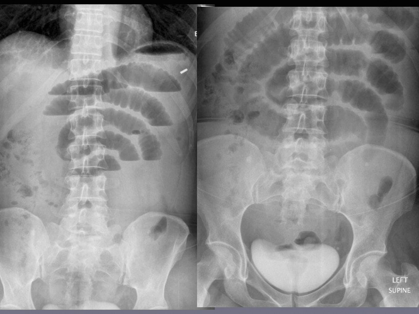
Imaging• Abdominal X ray (upright and supine)
• Small bowel should not be seen on AXR • Colonic gas is usually distributed peripherally • Air fluid levels should not exceed 4
• US in intestinal obstruction is useless due to the distended bowel
• CT is the best modality for diagnosing bowel obstruction
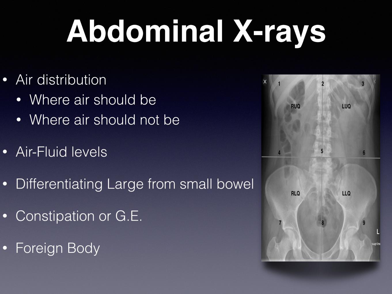
Abdominal X-rays• Air distribution
• Where air should be • Where air should not be
• Air-Fluid levels
• Differentiating Large from small bowel
• Constipation or G.E.
• Foreign Body


ACUTE CHOLECYSTITIS AND BILIARY COLIC

Risk Factors for Cholecystitis• Female gender (Female)
• Increasing parity (Fertile)
• Obese (Fat)
• Advancing age (Forty)
• Drugs
• Cystic Fibrosis
• State of chronic hemolysis


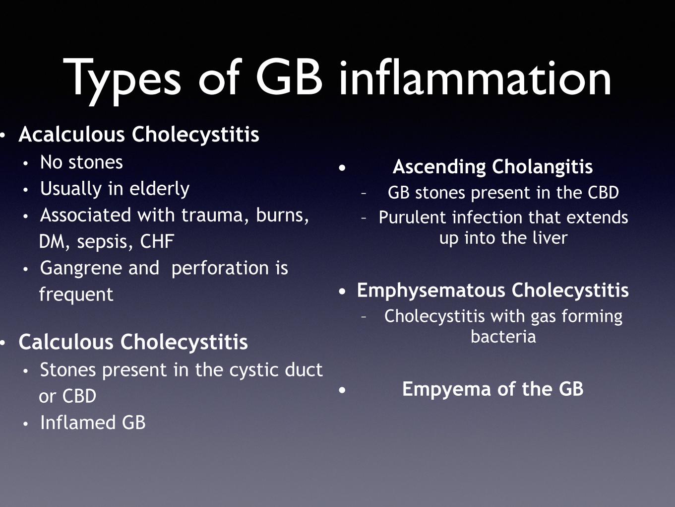
Types of GB inflammation• Acalculous Cholecystitis
• No stones • Usually in elderly • Associated with trauma, burns,
DM, sepsis, CHF • Gangrene and perforation is
frequent
• Calculous Cholecystitis • Stones present in the cystic duct
or CBD • Inflamed GB
• Ascending Cholangitis – GB stones present in the CBD – Purulent infection that extends
up into the liver
• Emphysematous Cholecystitis – Cholecystitis with gas forming
bacteria
• Empyema of the GB

Management• NPO
• IV fluids
• Antiemetics and Analgesics
• Broad Spectrum Abx
• Surgical Consultation

ACUTE PANCREATITIS

Presentation• Epigastric pain, dull aching and radiating to the back,
eased on leaning forward
• Nausea and Vomiting
• Tachycardia
• Tachypnea
• Rebound is not usually present
• Grey-turner and Cullen signs (rare)


Diagnosis and Management• Diagnosis
• Serum Amylase • Serum Lipase • Imaging (CT is the modality of choice)
• Management• Aggressive fluid therapy • NPO and Anti-emetics • Painkillers • Surgical Consultation

Signs of Poor Prognosis (Ranson’s Criteria)

Complications
• Pleural Effusion • ARDS • Fluid sequestration • ATN • DIC

ABDOMINAL AORTIC ANEURYSM

True and False Aneurysms
• A true aneurysm is a dilatation in the vessel wall, that involves ALL the wall layers
• A Pseudoaneurysm (false) is a dilatation in the vessel wall that only involves the adventitia


Risk Factors for AAA • Advanced age (75% of AAA are in pts. > 60yrs)
• Male sex (esp. Caucasians)
• Having a 1st degree relative with a AAA
• Smoking
• Hypertension
• Hx of CAD or PAD
• Dyslipidemia
• Collagen disease

Presentation• Sudden severe abdominal pain that is radiating to the back or
left flank
• May be associated with an episode of syncope
• Lower back pain with radiation to the legs
• Scrotal hematoma
• L.L. ischemia
• Pulsatile mass
• Bruits heard over abdominal aorta or femorals

Diagnosis• ANY UNSTABLE PATIENT WITH A SUSPECTED AAA GOES
DIRECTLY TO THE THEATER
• AXR can be suggestive
• US is a good diagnostic modality, it is easy , 100% Sn. > 95% Sp. and can be done at bedside
• CT angiography is the only way to tell if the AAA is leaking or not


Management• NPO
• 2 large bore IV lines
• Cross match for at least 4 units of blood
• IV fluids
• Vascular surgery consultation
• ICU and anesthesia consultation
• Control of BP
• Pain killers

ACUTE MESENTERIC ISCHEMIA

Types of Mesenteric Ischemia
• Embolic • Secondary to acute SMA occlusion by an embolus that
originated in the left heart (75% of the time)
• Thrombotic • This is mesenteric venous thrombosis due to the
presence of a hypercoagulable state
• Non-occlusive • This is ischemia that is occurring secondary to low flow
state due to cardiac pathology

The risk factors for Mes. Isch.
• Arterial • Dysrhythmias (esp. AF) • Atherosclerotic heart disease • Valvular Heart disease • Recent AMI
• Venous thrombosis • Hx of prior VTE • Hypercoagulable state (polycythemia vera, AT III def., chemotherapy,
estrogen use) • Non-occlusive
• Hypotension • Sepsis • CHF

Presentation
• Abdominal pain that is OUT OF PROPORTION with the clinical exam
• Diarrhea (with OB positive)
• Bleeding per rectum
• Anorexia
• Nausea and vomiting


Management• All types get:
• IV fluids • Abx • Pain killers
• Embolic type • Papaverine
• Venous thrombosis • Anticoagulation
• Low flow state • Correction of the underlying problem
Surgical Consultation is done in all but surgical
intervention is only done if:
1. A surgical abdomen and peritonitis develops
2. Patient requires revascularization
procedure
3. Resection of necrotic bowel is needed

ILEITIS AND COLITIS (INFLAMMATORY BOWEL
DISEASE)

Crohn’s Disease• An immune mediated disease AKA
• Terminal ileitis • Granulomatous ileocolitis • Regional Enteritis
• Can affect any segment of the GI tract from the mouth to the anus
• The disease is characterized by involved sections and “skip areas”
• The bowel wall is thickened, which leads to narrowing of the lumen
• There is longitudinal fissuring and ulceration of the bowel
• The pathology involves all the three layers of the bowel

Extra Intestinal Manifestations of IBD
• Arthritis • Ankylosing Spondylitis • Vasculitis • Venous Thromboembolism • Hepato-Biliary Disease • Erythema Nodosum • Pyoderma Gangrenosum • Iritis / Uveitis / Episclaritis • Hyperoxaluria and renal stones

Complications of Crohn’s D.• Perianal
• Abscess formation • Ileocutaneous fistula • Rectovaginal fistula • Rectal prolapse
• Intestinal • Obstruction • Abscess / Fistula • Perforation • Hemorrhage
• Toxic Megacolon – Associated with massive
GI bleed > 50% of cases
• Malignancy – Neoplasms of both the
small and large bowel


Management of Crohn’s• IV fluids and electrolyte replacement
• Steroid administration
• Antibiotic administration
• Azathioprine (immunosuppressant)
• Deal with complications

Ulcerative Colitis (UC)• UC is a chronic inflammatory and ulcerative disease of
colon and rectum
• Inflammation is limited to the mucosa and submucosa
• UC has no skip lesions, it is continuous
• UC is confined to the colon and does not extend beyond it


Severity of Attacks of UC• Mild > 60% of cases
• < 4 bowel movements per day • No systemic manifestations • Few extra-intestinal manifestations
• Severe • > 6 bowel motions per day • Fever • Tachycardia • Wt. loss • Anemia • Many Extra-intestinal manifestations

Complications of UC• Hemorrhage (most common)
• Toxic Megacolon
• Hemodynamic instability
• Perforation
• Obstruction due to stricture
• Fistula / Abscess formation
• Carcinoma transformation

Management• Mild attack
• Sulfasalazine • Steroids • Azathioprine • Iron and antidiarrheal agents
• Severe • IV fluid and electrolyte replenishment • IV steroids • IV Abx • Blood Tx • Surgical Consultation

DIVERTICULITIS

Definitions• Diverticular Disease
• The disease of having sac like herniations of colonic mucosa
• Diverticulosis • A clinical state of a patient with diverticular disease
presenting with lower GI bleeding
• Diverticulitis • The clinical state of a patient with diverticular disease with
inflammation, infection and hemorrhage


Presentation• Abdominal pain with signs of peritonitis mainly in the
left lower quadrant
• Bloody stools
• Fever
• Abdominal distention
• Anorexia
• Nausea and Vomiting

Management• NPO
• IV fluids
• Broad Spectrum Abx
• Surgical consult
• Analgesia

Take home Message• The abdomen is a closed box
• Always think of the structures present at the site of the pain, and then their neighbors
• We do not treat lab readings, (context trumps result always) look at the clinical status of your patient
• It does not matter if it a rare cause, if it will kill your patient , you need to think of it!

QUESTIONS?