
Applications to - Minerva
169
TESIS DOCTORAL Computational Approaches for the Characterization of the Structure and Dynamics of G Protein-Coupled Receptors: Applications to Drug Design David Rodríguez Díaz Departamento de Anatomía Patolóxica e Ciencias Forenses Facultade de Medicina e Odontoloxía Universidade de Santiago de Compostela
-
Upload
khangminh22 -
Category
Documents
-
view
0 -
download
0
Transcript of Applications to - Minerva
TESIS DOCTORAL
Computational Approaches for the Characterization of the
Structure and Dynamics of G Protein-Coupled Receptors:
Applications to Drug Design
David Rodríguez Díaz
Departamento de Anatomía Patolóxica e Ciencias Forenses
Facultade de Medicina e Odontoloxía
Universidade de Santiago de Compostela


DEPARTAMENTO DE ANATOMÍA PATOLÓXICA E CIENCIAS FORENSES
Facultade de Medicina e Odontoloxía R/ de San Francisco, s/n
15782 - Santiago de Compostela Teléfono: 881812215. Fax: 981580336
D. Ángel Carracedo Álvarez, profesor catedrático do Departamento de Anatomía
Patolóxica e Ciencias Forenses en calidade de Titor; e D. Hugo Gutiérrez de Terán
Castañón, doutor e investigador Isidro Parga Pondal da Fundación Pública Galega de
Medicina Xenómica (SERGAS) como Director da Tese de Doutoramento realizada por D. David Rodríguez Díaz titulada “Computational Approaches for the Characterization of the
Structure and Dynamics of G Protein-Coupled Receptors: Applications to Drug Design”.
FAN CONSTAR:
Que o citado traballo de investigación reúne os requisitos académicos e científicos
necesarios para proceder á súa lectura e defensa pública.
E para que así conste, damos o visto e prace en Santiago de Compostela a 12 de
setembro de 2012.
Prof. Ángel Carracedo Álvarez Dr. Hugo Gutiérrez de Téran Titor da Tese de Doutoramento Director da Tese de Doutoramento D. David Rodríguez Díaz Autor da Tese de Doutoramento


La realización de esta tesis doctoral ha sido financiada por el Fondo de Investigación
en Salud (Instituto Carlos III) mediante la Ayuda Predoctoral con expediente FI08/0799.
Adicionalmente, trabajos aquí presentados se enmarcan dentro de Proyectos de
Investigación financiados por la Consellería de Sanidade, Xunta de Galicia (PS09/63) y del
Ministerio de Ciencia e Innovación (SAF2011-30104).
Finalmente, se reconoce el tiempo de cómputo y asistencia ofrecida por el Centro de
Supercomputación de Galicia (CESGA), en el cual se ejecutaron dos proyectos ICTS
(MICINN).


A Tamara


AGRADECIMIENTOS
Por desgracia, esta sección de agradecimientos no recogerá todas las contribuciones que multitud de personas han realizado para que llegue este momento. Independientemente de su magnitud o naturaleza, dichas aportaciones cobran un especial valor echando la vista atrás, contradiciendo la sombría imagen que se atribuye a la sociedad de hoy en día.
Gracias a tantos amigos de As Pontes de los que he podido disfrutar de su complicidad y compañía, a pesar de que no entendáis muy bien en lo que trabajo ;) Basoa, Juanín, Fer, Rozenn (gracias por la genial portada artista!) y otros tantos, sois grandes! Por supuesto, lo mismo es aplicable a los compañeros de carrera, tanto en la USC como en la UAM, que hicieron que aquellos años fuesen maravillosos y siguen ahí para lo que haga falta: Jaime, Nuria, Álex; Lorena, Erika, Eli, Abel, las Rocíos… Igualmente hay que agradecer el apoyo y consejos de grandes profesores, especialmente a Cristina Murga.
En el ámbito más profesional, un recuerdo para José Antonio, Julián y Alejandro (UDC) en la primera etapa en la Rede Galega de Bioinformática, además de a otros miembros de ese rico colectivo que esperemos continúe vivo de una u otra forma. Asimismo, gracias a Eddy y Vicen por todo el valor que habéis dado a nuestro trabajo computacional y por vuestra cercanía, ojalá podamos continuar por esta senda! También gracias a José Manuel y Pepo de Biofarma por vuestra amabilidad y disponibilidad, al igual que a Mabel Loza por su accesibilidad y consejos. Igualmente gracias a tantos científicos con los que he podido coincidir en distintos eventos y colaboraciones, todas sus aportaciones son significativas.
Of course thanks to Andreas Bender, for being such a great host in Cambridge, together with Pete and Bobby. It has been an incredible experience to be in your laboratory for a while, for providing a stimulant scientific environment and a human group impossible to match. A big hug to all my fellows there: Giorgos, Eva, Shardul, Aakash, Tere, Pedro, Maite, Sonia, Florian, Susana, Richard, Hannes, Alex… hope to see you soon, come on! ;)
Gracias a Ángel Carracedo, por tu confianza y apoyo en esta línea de investigación y por tu calidad humana excepcional. También muchas gracias a todos los compañeros de despacho y de trabajo durante estos años: Ricardo, Montse Camiña, Raquel, Montse Fernández y Xabi. Aquí Hugo obviamente se merece una nota muy especial: además permitirme trabajar en lo que más me gusta en unas condiciones fenomenales en tu laboratorio, has sido un excepcional consejero y compañero a lo largo de estos años. Siempre estaré agradecido por todo lo que he aprendido a tu lado. Ah, y un abrazo muy grande a Raquel, Mar y Lola! :)
Y como no podía ser de otra forma, muchísimas gracias a mi entorno familiar más cercano, empezando por mis padres José Manuel y Charo: me habéis dado muchas cosas, incluyendo la oportunidad de llegar hasta aquí, siempre he podido contar con vuestro apoyo incondicional. Igualmente, gracias a mis parientes a lo largo y ancho de España, algunos ya no están por desgracia, vuestro cariño ha sido importante. Mil gracias también a todos los miembros de la familia que gané al unirme con Tamara. Un especial recuerdo para Ángeles y Toño, para o Doutor Paulo, y para mi ahijada política Icía ;)
Finalmente, si anteriormente me quedé corto en palabras de agradecimiento, ahora lo seré más. Tamara, sabes que eres y serás la persona más importante de mi vida, además de lo más maravilloso que me podría haber pasado. Sólo tú eres consciente de las fatigas (y momentos maravillosos, todo sea dicho) que hemos pasado juntos, la importancia de tu inmenso apoyo a lo largo de estos años es inconmensurable. Espero poder devolvértelo de alguna forma, de modo que al menos seas tan feliz como yo lo he sido gracias a ti.

ABSTRACT
G Protein-Coupled Receptors (GPCRs) constitute the most pharmacologically relevant superfamily of proteins. In this thesis, a computational pipeline for the structural modeling and the study of the dynamics of GPCRs is presented, and properly combined with experimental collaborations for the discovery and design of novel GPCR ligands. Our pipeline was first developed and applied for the characterization of the four subtypes of Adenosine Receptors, leading to the elucidation of ligand affinity and selectivity issues within this receptors family. Indeed, the employed implementation of Molecular Dynamics simulations has allowed the characterization of structural determinants of the activation process of adenosine receptors, and later in the assessment of the stability of GPCR dimers, in this case studying the CXCR4 receptor system. Both the activation and dimerization processes have great implications in the function and pharmacology of GPCRs, but we also provide specific applications in GPCR drug design projects. These include the discovery of novel scaffolds as potential antipsychotics, as well as the design of a new series of A3 adenosine receptor antagonists, by using a successful combination of structure-based and ligand-based drug design approaches. Finally, the computational pipeline here developed has been integrated into a semi-automated computational protocol in the web server GPCR-ModSim (http://gpcr.usc.es), which is open to the scientific community. Altogether, the results of this thesis represent a relevant contribution to the structural biology and drug discovery of this superfamily of highly relevant pharmacological targets.

LIST OF PUBLICATIONS INCLUDED IN THIS THESIS:
Paper I: Gutiérrez-de-Terán H., Correia C., Rodríguez D., Carvalho M.A., Brea J., Cadavid M.I., Loza M.I., Proença M.F. and Areias F. (2009) Identification of novel scaffolds from an original chemical library as potential antipsychotics. QSAR & Comb. Sci. 28(8):856-60. http://dx.doi.org/10.1002/qsar.200860198
Paper II: Michino M., Abola E., GPCR Dock 2008 participants*, Brooks III C.L., Dixon J.S., Moult J. and Stevens R.C. (2009) Community-wide assessment of GPCR structure modelling and ligand docking: GPCR Dock. Nat. Rev. Drug Discov. 8:455-63. http://dx.doi.org/10.1038/nrd2877 * David Rodríguez is part of the consortium of participant researchers.
Paper III: Yaziji V., Rodríguez D., Gutiérrez-de-Terán H., Coelho A., Caamaño O., García-Mera X., Cadavid M.I., Brea J., Loza M.I. and Sotelo E. (2011) Pyrimidine Derivatives as Potent and Selective A3 Adenosine Receptor Antagonists. J. Med. Chem. 54(2):457-71. http://dx.doi.org/10.1021/jm100843z
Paper IV: Rodríguez D., Piñeiro Á. and Gutiérrez-de-Terán H. (2011) Molecular Dynamics Simulations Reveal Insights into Key Structural Elements of Adenosine Receptors. Biochemistry 50(19):4194-208. http://dx.doi.org/10.1021/bi200100t
Paper V: Rodríguez D., Bello X. and Gutiérrez-de-Terán H. (2012) Molecular Modelling of G Protein-Coupled Receptors Through the Web. Mol. Inf., 31:334-41. http://dx.doi.org/10.1002/minf.201100162
Paper VI: Rodríguez D. and Gutiérrez-de-Terán H. (2012) Characterization of the homodimerization interface and functional hotspots of the CXCR4 chemokine receptor. Proteins 80(8):1919-28. http://dx.doi.org/10.1002/prot.24099
The publications indicated above are reproduced in Section 7 under the permission granted by the corresponding journals for this thesis (both in printed and electronic formats).
SUPPORTING INFORMATION:
Annex I: Rodríguez D. and Gutiérrez-de-Terán H. (2009) Adenosine Receptors: a systematic study of ligand binding based on the crystal structure of hA2A receptor. Poster presentation at the International Workshop In Memoriam of Ángel Ortiz. Madrid, January 26-28.
Annex II: Rodríguez D., Sotelo E., Bender A. and Gutiérrez-de-Terán H.: Discovery of potent and selective adenosine receptor ligands via multi-objective design. Abstract of the oral presentation given at the 243rd ACS National Meeting & Exposition. San Diego (California), March 25-29.
Annex III: SUMMARY IN SPANISH / RESUMEN EN CASTELLANO.

“Part of the inhumanity of the computer is that, once it is competently
programmed and working smoothly, it is completely honest.”
Isaac Asimov

TABLE OF CONTENTS 1. INTRODUCTION ................................................................................................................. 1
1.1. Biochemistry and pharmacology of G Protein-Coupled Receptors ................................ 21.1.1. Receptors involved in schizophrenia ....................................................................... 31.1.2. Adenosine receptors ................................................................................................. 41.1.3. Chemokine receptors. .............................................................................................. 6
1.2. Structural Biology of GPCRs.......................................................................................... 71.2.1. Conserved sequence patterns and structural microswitches of GPCRs ................... 81.2.2. Extracellular architecture of GPCRs ...................................................................... 101.2.3. Advances in the structural characterization of GPCR oligomerization ................. 13
2. MOLECULAR MODELLING METHODS........................................................................ 152.1. Computer-derived models of GPCR structures ............................................................ 15
2.1.1. GPCR modelling approaches ................................................................................. 152.1.2. Online resources for GPCR modelling .................................................................. 17
2.2. Computational techniques of ligand discovery and design ........................................... 192.2.1. Structure-Based approaches: ligand docking ......................................................... 192.2.2. Ligand-Based methods: linking chemical structure and bioactivity. ..................... 20
2.3. Molecular Dynamics simulations ................................................................................. 242.3.1. Force fields ............................................................................................................. 242.3.2. Molecular Dynamics .............................................................................................. 242.3.3. Considerations of MD simulations of GPCRs ....................................................... 25
3. OBJECTIVES ...................................................................................................................... 294. RESULTS AND DISCUSSION ........................................................................................... 315. CONCLUSIONS.................................................................................................................. 436. REFERENCES .................................................................................................................... 457. PUBLICATIONS AND SUPPORTING INFORMATION .................................................. 59

ABBREVIATIONS
5-HT: 5-hydroxitryptamine, serotonin ADR: adrenergic receptor AIDS: acquired immunodeficiency syndrome APC: antipsychotic compound AR: adenosine receptor cAMP: cyclic adenosine monophosphate CCR5: chemokine CC receptor 5 CV: cross-validation CXCR4: chemokine CXC receptor 4 EL: extracellular loop EPS: extrapyramidal side-effect FDA: Food and Drug Administration (USA) GDP: guanosine diphosphate GPCR: G Protein-Coupled Receptor GTP: guanosine triphosphate HIV: human immunodeficiency virus IL: intracellular loop LB: ligand-based; LBVS: ligand-based virtual screening LOO: leave-one out LV: latent variable MD: molecular dynamics MIF: molecular interaction field mGlu: metabotropic Glutamate receptor MSeqA: multiple sequence alignment MStA: multiple structure alignment OR: opioid receptor PC: principal component; PCA: principal component analysis PDB: Protein Data Bank PBC: periodic boundary conditions PLS: partial least squares PME: Particle Mesh Ewald POPC: palmitoyloleylphosphatidylcholine QSAR: quantitative structure-activity relationships RMSD: root-mean-square deviation SAR: structure-activity relationships SB: structure-based; SBVS: structure-based virtual screening T4L: bacteriophague T4 lysozyme TM: transmembrane helix VS: virtual screening

INTRODUCTION
1
1. INTRODUCTION
Currently, the pharmaceutical industry is facing a challenging scenario in the research and development of new drugs. The increasing costs and the difficulties in obtaining novel molecular entities demand an optimal performance in the different stages necessary for the eventual approval of a drug (1) (see Fig. 1).
Figure 1. Schematic pipeline of the drug discovery and development process. The computational studies presented in this thesis will be focused in stages from hit discovery to lead optimization.
Computational methods are broadly integrated in the pipeline of drug discovery and development (2). Their application ranges several areas, including the management of databases and the prediction of physicochemical and pharmacokinetic properties, such as those related with absorption, distribution, metabolism and excretion (ADME) of chemical compounds in the body. Probably, the areas where computational techniques have demonstrated a broader impact are the discovery of novel hits, i.e. Virtual Screening (VS), and the rational design in lead optimization stages —e.g., using structure-based methods such as docking and molecular dynamics simulations—.
Here, some of these computational methodologies are applied in a deep study of the superfamily of G Protein-Coupled Receptors (GPCRs), a group of membrane proteins with high pharmacological interest. The application of a wide range of computational tools and protocols result not only in a deeper structural and dynamic characterization of these receptors, but also in the discovery and design of novel compounds targeting some of their members.

INTRODUCTION
2
1.1. Biochemistry and pharmacology of G Protein-Coupled Receptors
G Protein-Coupled Receptors constitute the main superfamily of integral membrane proteins (3), the function of which is to transduce signals from the extracellular medium towards the cytoplasm. The canonical signalling mechanism of GPCRs involves the activation of the cognate heterotrimeric G Protein, located in the intracellular side of the receptor. Hence, the subunit of this G protein exchanges a bound GDP with GTP, and dissociates from subunits upon their interaction with a (typically activated) GPCR. Different types of G proteins exists, where the characterized subunits stimulate certain biochemical signalling pathways such as those involving cAMP and phosphatidylinositol (4), as illustrated in Fig. 2. Through this signalling diversity, GPCRs mediate a huge variety of physiological functions, which in part explains why they constitute the main drug target nowadays, accounting for at least 30% of the marketed drugs (5).
GPCRs are highly flexible receptors, exploring different conformations of varying functional significance, ranging from inactive (Ri) to active (Ra) forms of the receptor. The equilibrium between Ri and Ra in the apo form of the receptor dictates its basal activity. Extracellular ligands can influence this equilibrium in order to induce different biological responses, leading to an increase (full and partial agonists), maintenance (antagonists) or reduction (inverse agonist) of the basal activity of the receptor.
Figure 2. Signal transduction and conformational equilibrium of G Protein-Coupled receptors. (A) Cellular location of GPCRs and their interaction with cytoplasmatic G Proteins, including examples of induced intracellular signalling cascades. Extracted from (6). (B) Extended Ternary Complex (ETC) model for GPCR activation, as originally proposed in (7). The different conformational equilibriums for receptor activation, from Ri (inactive) to Ra (active), are represented. The explicit role of the binding of the natural agonist (A) and the G Protein (G) association is indicated as well.
Most of the GPCR families are characterized to bind a specific ligand, the natural agonist of the given receptor (7). Typically, GPCR ligands bind to the orthosteric site, the same of the natural agonist, located in the extracellular region of the receptor. Importantly, the higher sequence diversity among GPCRs is located in the extracellular half of the receptors (8). This is not surprising given the extremely diverse chemical nature of GPCR ligands: they include amines, nucleotides, peptides, proteins, lipids, organic odorants and ions. This is in line with the significant variety of physiological functions mediated by these receptors. Moreover, a classification of the GPCR superfamily, built on the basis of the pseudosequence formed by the hotspot residues within the binding crevice (9, 10) mostly agrees with the general phylogenetic tree of GPCRs (11), reflecting a balance in the evolution of ligands and receptors for the regulation of physiological processes.
A phylogenetic analysis of human GPCRs identified 5 groups of receptors, namely Glutamate, Rhodopsin, Adhesion, Frizzled/taste2, and Secretin clans, as defined in the so-caled GRAFS classification scheme (11). Among these, the Class A/rhodopsin-like is the most populated group (700 out of the 800 characterized GPCRs). Class A receptors are further divided into four branches: , , and , which can be classified according to the similarity of the natural ligand (e.g., aminergic

INTRODUCTION
3
receptors refer to all families which are activated by the endogenous biogenic amines), ultimately defining each family of receptors. The accumulated knowledge in medicinal chemistry on Class A GPCRs exemplifies their relevance among the whole superfamily. Indeed, the receptors considered in this thesis are members of rhodopsin-like GPCRs, and are introduced in the following subsections.
1.1.1. Receptors involved in schizophrenia
Schizophrenia is a psychiatric disorder that significantly affects the life quality of their patients, an estimated 1% of the worldwide population (12). It is a complex disease that, despite the constant advances in its pharmacological and biochemical characterization, still presents a challenging scenario for the discovery or design of novel compounds for its treatment.
The first antipsychotic compounds (APCs), termed as “typical”, were discovered during the middle of the 20th century (like chlorpromazine, see Fig. 3), and presented several therapeutic limitations including severe extrapyramidal side-effects (EPS) (13). Their antagonism on dopaminergic signaling was retrospectively discovered, and thus dopamine receptors were assigned as the first targets for the treatment of schizophrenia (14). However, latter works not only confirmed that observation, but also suggested the involvement of serotonin (5-hydroxitryptamine, 5-HT) receptors in the mechanism of action of novel antipsychotics (15). In fact, the relationship of affinities between serotonin and dopamine receptor subtypes was able to discriminate compounds with increased efficacy and less EPS: compounds showing a pKi ratio between 5-HT2A and D2 receptors (Meltzer Index) above 1.2 presented this enhanced pharmacological profile, and were labeled as “atypical” APCs (15).
Despite the big efforts in the development of novel APCs, clozapine (see Fig. 3) remains as the gold standard in this regard after almost 50 years of its discovery (16). This drug presents the pharmacological advantages of atypical APCs, including reduced suicidal rate of patients (17); although it also involves several adverse effects such as agranulocytosis, seizures and weight gain (18). Several potential APCs have been introduced into clinical development stages in the recent years, which would mostly be considered as “me-too” congeners of clozapine (19).
Figure 3. 2D structures of representative APCs. The positively-charged nitrogens at physiological pH are indicated accordingly.
The paradigm for the mechanism of action of APCs has evolved throughout the years into the current “magic shotgun” model of Roth and co-workers (20): compounds are hoped to present a specific pharmacological profile (including activity, efficacy and even pharmacokinetics considerations) against a panel of targets. This contrasts with the much earlier “magic bullet” analogy formulated by Ehrlich, where only the action of a compound against a single target is considered.

INTRODUCTION
4
Thus, a portion of the receptorome is necessary to understand the mode of action of APCs, involving many subtypes of families of aminergic receptors (dopamine, serotonin, histamine, muscarinic, α and β adrenergic…) and other GPCRs such as metabotropic Glutamate (mGlu) receptors. The ideal APC should be able to achieve the appropriate output in dopaminergic signaling in specific regions of the brain, by means of achieving a specific efficacy balance in a battery of receptors, while avoiding or minimizing the affinity in those targets related to side-effects. However, the modelling of such process is extremely complex. Novel APCs departing from known scaffolds towards new chemistries, at the same time complying with the desired pharmacological profile, are hoped to provide better tools for the future treatment of this elusive disease.
Figure 4. Receptors involved in schizophrenia. On the left hand side, the different affinity constants (Ki) of several APCs (columns) against a panel of targets involved in this disease (receptorome, in rows) are indicated, together with associated efficacies and side-effects of the receptors —chart taken from (20)—. On the right hand side, homology models and X-Ray structures of several serotonine and dopamine receptor subtypes are shown, together with computationally-predicted binding modes of the APCs clozapine and olanzapine. This is part of an ongoing study of our laboratory.
1.1.2. Adenosine receptors
The ubiquitous nucleoside adenosine regulates a wide range of biological functions through the activation of a family of GPCRs, the so-called adenosine receptors (ARs), which are subdivided in four subtypes: A1, A2A, A2B and A3 (21). ARs mediate the typically cytoprotective function of adenosine, leading to a different response to its variable extracellular levels depending on the corresponding tissue and physiopathological state (22). The four subtypes present different couplings with intracellular effectors, and consequently their activation transduces diverse intracellular signaling pathways. On one hand, A1 and A3 subtypes (sharing a 49% sequence identity) couple to Gi proteins, inhibiting the activity of adenylyl cyclase upon activation. On the other hand, A2A and A2B (59% seq. id.) interact with Gs proteins, eventually leading to the reduction of intracellular cAMP levels (22). At the same time, despite their relatively ubiquitous body distribution, the presence of A1 and A2A (high affinity for adenosine) is mainly focused in the nervous system and cardiovascular tissues, meanwhile A2B and A3 (low affinity for adenosine) are generally found in peripheral tissues such as kidney or lungs.
ARs are highly relevant drug targets due to their extensive potential in a wide variety of pathologies. Their modulation has interesting indications, and drug design efforts have been

INTRODUCTION
5
traditionally focused in the discovery of orthosteric ligands chemically similar respect to natural products that bind ARs (see below). However, the development of potent and selective drugs within this family is quite challenging due to the structural similarity of human ARs. Moreover, interspecies differences of these receptors have hampered the extrapolation of results from preclinical studies in animal models to humans. In fact, apart from the use of adenosine in the treatment of tachycardia, only regadenoson is a FDA-approved drug targeting an AR (see Fig. 5). Furthermore, this A2AAR selective agonist is used as a diagnostic tool in myocardial perfusion imaging, rather than as a therapeutic agent (23).
Regarding the efficacy of ligands targeting adenosine receptors, ARs agonists are specially interesting for the treatment of cardiovascular diseases such as arrhythmia or vasodilation processes (22), as well as for contributing to the preconditioning of cardiomyocytes in ischemia through the activation of A1 and A3 subtypes (24). The structure-activity relationships (SAR) around the adenosine scaffold have been thoroughly explored (25), exemplified by the selective A2AAR agonists CGS21680, UK-432079 and the aforementioned regadenoson (see Fig. 5).
Following the same rationale, the design of novel ARs antagonists was initially focused on substitutions on the xanthine scaffold (26), illustrated by the weak and non-specific natural products caffeine and theophylline (see Fig. 5). Many ARs antagonists are applied in nervous system disorders, where caffeine exerts stimulant and enhanced awareness effects. In fact, an inverse relationship between coffee intake and the development of Parkinson’s disease has been observed (27), and selective A2AAR antagonists such as preladenant or tozadenant (with a non-xanthine scaffold) are undergoing clinical trials for the treatment of this disease (28). ARs antagonists are also used in pathologies of peripheral tissues, including the development of selective A2BAR compounds as anti-asthmatic preclinical candidates (29) or A3AR antagonists for the treatment of glaucoma and ostheoarthritis already in initial clinical trials (30).
As exemplified above, intensive efforts are being carried out in the drug development of ARs chemical modulators. A special effort is given to the design and synthesis of compounds with selective profiles and novel chemical scaffolds towards this therapeutically underexploited family of receptors.
Figure 5. Representative ARs ligands, grouped according to their efficacy.
INTRODUCTION
6
1.1.3. Chemokine receptors.
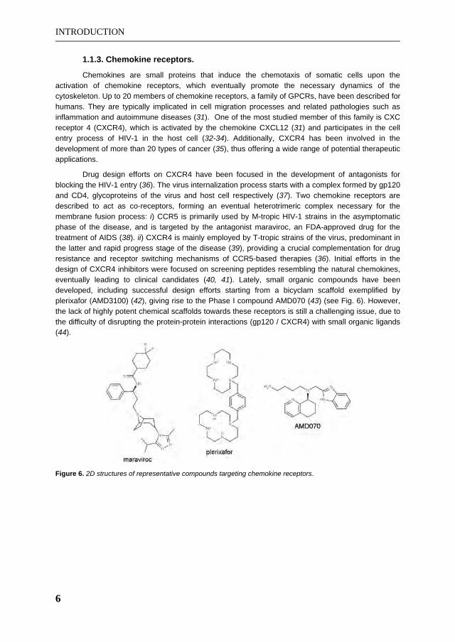
Chemokines are small proteins that induce the chemotaxis of somatic cells upon the activation of chemokine receptors, which eventually promote the necessary dynamics of the cytoskeleton. Up to 20 members of chemokine receptors, a family of GPCRs, have been described for humans. They are typically implicated in cell migration processes and related pathologies such as inflammation and autoimmune diseases (31). One of the most studied member of this family is CXC receptor 4 (CXCR4), which is activated by the chemokine CXCL12 (31) and participates in the cell entry process of HIV-1 in the host cell (32-34). Additionally, CXCR4 has been involved in the development of more than 20 types of cancer (35), thus offering a wide range of potential therapeutic applications.
Drug design efforts on CXCR4 have been focused in the development of antagonists for blocking the HIV-1 entry (36). The virus internalization process starts with a complex formed by gp120 and CD4, glycoproteins of the virus and host cell respectively (37). Two chemokine receptors are described to act as co-receptors, forming an eventual heterotrimeric complex necessary for the membrane fusion process: i) CCR5 is primarily used by M-tropic HIV-1 strains in the asymptomatic phase of the disease, and is targeted by the antagonist maraviroc, an FDA-approved drug for the treatment of AIDS (38). ii) CXCR4 is mainly employed by T-tropic strains of the virus, predominant in the latter and rapid progress stage of the disease (39), providing a crucial complementation for drug resistance and receptor switching mechanisms of CCR5-based therapies (36). Initial efforts in the design of CXCR4 inhibitors were focused on screening peptides resembling the natural chemokines, eventually leading to clinical candidates (40, 41). Lately, small organic compounds have been developed, including successful design efforts starting from a bicyclam scaffold exemplified by plerixafor (AMD3100) (42), giving rise to the Phase I compound AMD070 (43) (see Fig. 6). However, the lack of highly potent chemical scaffolds towards these receptors is still a challenging issue, due to the difficulty of disrupting the protein-protein interactions (gp120 / CXCR4) with small organic ligands (44).
Figure 6. 2D structures of representative compounds targeting chemokine receptors.

INTRODUCTION
7
1.2. Structural Biology of GPCRs
On one hand, the obtaining of high-resolution structures of GPCRs is an extremely difficult task due to the inherent flexibility of these receptors, and their lack of stability when they are extracted from their lipidic environment. On the other hand, the topology of GPCRs is one of their characteristic and more conserved features: a helical bundle constituted by 7 transmembrane helices (TMs), connected by 3 extracellular (EL) and 3 intracellular (IL) loops. Generally, an additional helix (H8) runs perpendicular to this bundle in the C-terminus region. This topology has been recognized for decades and has significantly contributed to the initial generation of GPCR structural models using bacteriorhodopsin as a template (45), followed by the trace of C atoms of frog rhodopsin obtained by electron cryomicroscopy 3D maps (46). Finally, the crystallographic structure of bovine rhodopsin was released in the year 2000 (47), being the unique template of atomic resolution for GPCR modelling during the subsequent seven years (see Section 2.1). Thereafter, the development of paradigmatic technologies for receptor stabilization and crystallization (48, 49) opened a new era in the structural biology of GPCRs. Two of the most successful approaches consider the fusion of bacteriophage T4 lysozyme (T4L) in the IL3 (50, 51), or the introduction of residue point mutations in order to increase the thermal stability of the receptors (49, 52). So far representative members of two branches, five sub-branches and eight GPCR families of rhodopsin-like receptors have been crystallized in their inactive conformation (see Table 1). In addition, members of three of these families (rhodopsin, adenosine and -adrenergic receptors) have also been solved in an active-like conformation.
Active < ------------------------------------------------------------------------------- > Inactive Ligand [PDB code] (reference)
Species Receptor Agonist Antagonist Inverse agonist
Human A2AAR NECA [2YDV] (53) Adenosine [2YDO] (53) UK-432097 [3QAK] (54)
T4G [3UZA] (55) T4E [3UZC] (55)
ZM241385 [3EML] (56) Caffeine [3RFM] (57) XAC [3REY] (57)
Turkey 1ADR
Carmoterol [2Y02] (58) Isoprenaline [2Y03] (58)
Dobutamine [2Y00] (58) Salbutamol [2Y04] (58)
Cyanopindolol [2VT4] (52)Iodocyanopindolol [2YCZ] (59)
Carazolol [2YCW] (59)Bucindolol [4AMI] (60)
Carvedilol [4AMJ] (60)
Human 2ADR
BI-167107 [3SN6] (61) FAUC50 [3PDS] (62)
Alprenolol [3NYA] (63) Carazolol [2RH1] (50)Timolol [3D4S] (64) JSZ [3NY9] (63) ICI118551 [3NY8] (63)
Human CXCR4 IT1t [3ODU] (65)
CVX15 [3OE0] (65)
Human D3R Eticlopride [3PBL] (66) Human H1R Doxepin [3RZE] (67) Human M2R QNB [3UON] (68) Mouse M3R Tiotropium [4DAJ] (69) Mouse -OR Naltrindole [4EJ4] (70) Mouse -OR -FNA [4DKL] (71) Human -OR JDTic [4DJH] (72) Human NOPR C-24 [4EA3] (73) Human S1P1R ML056 [3V2Y] (74)
Bovin Rhodopsin Trans-retinal [3PQR] (75)Apo-Opsin [3CAP] (76)
Cis-retinal [1U19] (77)-ionone [3OAX] (78)
Squid Rhodopsin Trans-retinal [3AYM] (79) Cis-retinal [2Z73] (80)
Table 1. Summary of the GPCR crystal structures available to the scientific community. Receptor-ligand structures are categorized on the basis of the biological response induced by the co-crystallized ligand. Only the structure with the highest resolution (typically below 3 Å) is indicated for those ligand-receptor complexes crystallized more than once.

INTRODUCTION
8
Finally, the first GPCR—G Protein complex has been solved for β2 adrenergic receptor (β2ADR) with its cognate Gs protein (61). The coordinates of the available GPCR structures can be found in the Protein Data Bank (PDB, http://www.pdb.org), the reference database in structural biology (81). The most representative GPCR-ligand complexes solved to date (with their corresponding PDB codes) are compiled in Table 1, more details can be found in recent reviews of the field (82, 83).
Despite the described experimental advances, the structural coverage of GPCRs is still sparse (see Fig. 7). Two out of four branches of Class A receptors remain uncharacterized (β and γ), despite the pharmaceutical interest of some of their members. The β branch includes targets such as neuropeptide Y (84) and gonadotropin-releasing hormone receptors (85), apart from others currently employed in clinical studies such as oxytocin hormone receptor (86). The populated family of purinergic P2Y receptors, attractive targets for diseases such as platelet aggregation (87), comprise one of the most relevant members of γ branch. Regarding α and δ branches, their structural coverage is below the 10% of their members (excluding olfactory receptors). In this scenario, computationally derived models of good quality are highly demanded to deepen the structural knowledge of the immense majority of Class A receptors, as well as for the rest of GPCR classes.
Figure 7. Structural characterization of GPCRs in the context of their phylogenetic tree. Each red flag marks a receptor that has been solved by X-Ray crystallography. This figure is extracted from the NIH GPCR Network website (http://gpcr.scripps.edu/, accessed on September 2012).
1.2.1. Conserved sequence patterns and structural microswitches of GPCRs
Several patterns have been traditionally observed in GPCR sequence alignments, exemplified by the Ballesteros & Weinstein scheme for residue numbering (88). Following this proposal, residues are numbered with the X.YY code, where X represents the TM helix, and YY is a correlative number according to the sequence of the receptor, assigning the number 50 to the most conserved GPCR residue in the corresponding helix. This scheme is adopted along this thesis (in uppercase) and related works. The first hallmark of GPCRs is the high conservation of proline residues in different TMs, including TM5, TM6 and TM7. These residues produce distortions in the canonical α-helix
INTRODUCTION
9
secondary structure that induce kinks and hinges, found to be important for the structure, dynamics and function of membrane proteins (89).
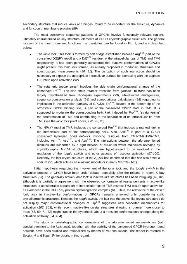
The most conserved sequence patterns of GPCRs involve functionally relevant regions, ultimately characterized as key structural elements of GPCR crystallographic structures. The general location of the most prominent functional microswitches can be found in Fig. 8, and are described below:
The ionic lock. This lock is formed by salt bridge established between Arg3.50 [part of the conserved D(E)RY motif] and a D/E6.30 residue, at the intracellular tips of TM3 and TM6 respectively. It has been generally considered that inactive conformations of GPCRs might present this ionic lock formed, as already proposed in rhodopsin structures and spectroscopic measurements (90, 91). The disruption of such interaction should be necessary to expose the appropriate intracellular surface for interacting with the cognate G Protein upon activation (92).
The rotameric toggle switch involves the side chain conformational change of the conserved Trp6.48. The side chain rotamer transition from gauche+ to trans has been largely hypothesized from rhodopsin experiments (93), site-directed mutagenesis, sequence conservation analysis (94) and computational calculations (95) regarding its implication in the activation pathway of GPCRs. Trp6.48, located in the bottom tip of the orthosteric GPCR binding site, is part of the conserved CWxP motif in TM6. It is supposed to modulate the corresponding helix kink induced by Pro6.50, “straightening” the conformation of TM6 and contributing to the separation of its intracellular tip from TM3 (see the ionic lock point above) (92, 95, 96).
The NPxxY motif at TM7, includes the conserved Pro7.50 that induces a marked kink in the intracellular part of the corresponding helix. Also, Asn7.49 is part of a GPCR conserved hydrogen bond network involving residues from TM1-TM2-TM6-TM7, including Asn1.50, Asp2.50, and Asn7.45. The interactions between the aforementioned residues are supported by a tight network of structural water molecules revealed by crystallographic GPCR structures, which are hypothesized to be involved in the regulation of the toggle switch and other aspects of receptor activation (97-100). Recently, the last crystal structure of the A2AAR has confirmed that this site also hosts a sodium ion, which acts as an allosteric modulator in many GPCRs (101).
Initial hypothesis regarding the involvement of the ionic lock and the toggle switch in the activation process of GPCR have been under debate, especially after the release of recent X-Ray structures (83). The generally broken ionic lock in inactive-like structures has been intriguing (48, 82), although it is partially in agreement with the observed conformational rearrangements in active-like structures: a considerable separation of intracellular tips of TM6 respect TM3 occurs upon activation, as evidenced in the GPCR-Gs protein crystallographic complex (61). Thus, the relevance of the closed ionic lock in inactive-like conformations of GPCRs remains unsolved only considering static crystallographic structures. Respect the toggle switch, the fact that the active-like crystal structures do not display major conformational changes of Trp6.48 suggested new concerted mechanisms for activation (102, 103), while recent inactive-like crystal structures showing a rotamer more closer to trans (68, 69, 71, 72) might support the hypothesis about a transient conformational change along the activation pathway (94, 104).
The study of crystallographic conformations of the aforementioned microswitches (with special attention to the ionic lock), together with the stability of the conserved GPCR hydrogen bond network, have been studied and rationalized by means of MD simulations. The reader is referred to Section 4 and Paper IV for details in this regard.

INTRODUCTION
10
Figure 8. Structural microswitches of GPCRs. These sites are exemplified by the structure of A2AAR, with its residues coloured from blue (Nt) to red (Ct). The location of the toggle switch, the ionic lock, the NPxxY motif and the conserved network of residues at TM1-TM2-TM6-TM7 are depicted.
1.2.2. Extracellular architecture of GPCRs
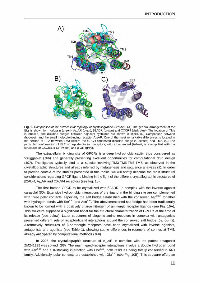
The functional microswitches presented in the previous section are located in the intracellular half of GPCRs (except the toggle switch, in between both halves), as they are involved in conformational changes necessary for the activation of the receptor, and the eventual signal transduction through cytoplasmatic effectors. On the other hand, the extracellular region of GPCRs, where the ligand binding occurs, presents a high sequence and topological diversity (82). All crystallographic structures, except rhodopsin, show open conformations of the 3 ELs (see Fig. 9). This is because rhodopsin function is mediated by a covalently-bound ligand (retinal), buried in the binding site, which is isomerized by photons in order to activate the receptor. Rhodopsin receptors have been widely employed for the theoretical elucidation of GPCR structures and activation mechanisms (105). However, the rest of GPCRs have to bind bioorganic molecules that stabilize the active state, and present significantly different extracellular topologies in order to accommodate the binding of the corresponding ligands, typically in a buried and hydrophobic site. In all cases, the EL2 (between TM4 and TM5) shows the most variable conformations among all GPCR crystallographic structures, as it presents the longest and most variable sequence length among the ELs. In fact, peptide-binding receptors present an exposed region favoured by a hairpin-shaped EL2 (with a beta-sheet segment), in order to adapt bulkier ligands compared to small organic compounds (see Fig. 9C). This can be observed in crystallographic structures of CXCR4 (65) and the four opioid receptors (ORs) solved to date (70-73). A very relevant anchoring point delimitates two sections of the EL2, made by the conserved disulfide bridge in Class A receptors. This bridge is formed between a cysteine located in EL2, and the other (Cys3.25) in TM3. The section between this disulfide bridge and TM5 generally delimitates one of the upper tips of the binding sites of GPCRs, and even establishes specific contacts with the co-crystallized ligands for adenosine, 2ADR and D3 receptors (see Table 1). Additional disulfides are observed between several ELs, where the paradigmatic case of A2AAR shows disulfides between EL1-EL2, and within EL2 and EL3 respectively (56), further constraining the conformational space of these flexible regions.
INTRODUCTION
11
.
Fig. 9. Comparison of the extracellular topology of crystallographic GPCRs. (A) The general arrangement of the ELs is shown for rhodopsin (green), A2AAR (cyan), β2ADR (brown) and CXCR4 (dark blue). The location of TMs is labelled, and disulfide bridges between adjacent cysteines are shown in sticks. (B) Comparison between rhodopsin and the small molecule-binding receptor A2AAR. One of the most remarkable differences is located in the section of EL2 between TM3 (where the GPCR-conserved disulfide bridge is located) and TM5. (C) The particular conformation of EL2 of peptide-binding receptors, with an extended β-sheet, is exemplified with the structures of CXCR4, κ-OR (violet) and μ-OR (grey).
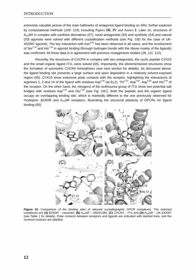
The extracellular binding site of GPCRs is a deep hydrophobic cavity, thus considered as “druggable” (106) and generally presenting excellent opportunities for computational drug design (107). The ligands typically bind to a subsite involving TM3-TM5-TM6-TM7, as observed in the crystallographic structures and already inferred by mutagenesis and sequence analyses (9). In order to provide context of the studies presented in this thesis, we will briefly describe the main structural considerations regarding GPCR ligand binding in the light of the different crystallographic structures of β2ADR, A2AAR and CXCR4 receptors (see Fig. 10).
The first human GPCR to be crystallized was β2ADR, in complex with the inverse agonist carazolol (50). Extensive hydrophobic interactions of the ligand in the binding site are complemented with three polar contacts, especially the salt bridge established with the conserved Asp3.32, together with hydrogen bonds with Ser5.42 and Asn7.39. The abovementioned salt bridge has been traditionally known to be formed with a positively charge nitrogen of aminergic receptor ligands (see Fig. 10A). This structure supposed a significant boost for the structural characterization of GPCRs at the time of its release (see below). Latter structures of biogenic amine receptors in complex with antagonists presented different sets of receptor-ligand interactions around the conserved salt bridge (59, 66-73). Alternatively, structures of β-adrenergic receptors have been crystallized with inverse agonists, antagonists and agonists (see Table 1), showing subtle differences in rotamers of serines at TM5, already anticipated by computational methods (108).
In 2008, the crystallographic structure of A2AAR in complex with the potent antagonist ZM241385 was solved (56). The main ligand-receptor interactions involve a double hydrogen bond with Asn6.55 and a π-stacking interaction with Phe5.29, both residues being totally conserved in ARs family. Additionally, polar contacts are established with Glu5.30 (see Fig. 10B). This structure offers an
INTRODUCTION
12
extremely valuable picture of the main hallmarks of antagonist ligand binding on ARs, further explored by computational methods (109, 110), including Papers III, IV and Annex I. Later on, structures of A2AAR in complex with xanthine derivatives (57), novel antagonists (55) and synthetic (54) and natural (53) agonists were solved with different crystallization methods (see Fig. 10D for the case of UK-432097 agonist). The key interaction with Asn6.55 has been observed in all cases, and the involvement of Ser7.42 and His7.43 in agonist binding (through hydrogen bonds with the ribose moiety of the ligands) was confirmed. All these data is in agreement with previous mutagenesis studies (26, 111, 112).
Recently, the structures of CXCR4 in complex with two antagonists, the cyclic peptide CVX15 and the small organic ligand IT1t, were solved (65). Importantly, the aforementioned structures show the formation of symmetric CXCR4 homodimers (see next section for details). As discussed above, the ligand binding site presents a large surface and open disposition in a relatively solvent-exposed region (65). CVX15 show extensive polar contacts with the receptor, highlighting the interactions of arginines 1, 2 and 14 of the ligand with residues Asp5.26 (at EL2), Thr3.33, Asp4.60, Asp6.58 and His3.29 of the receptor. On the other hand, the nitrogens of the isothiourea group of IT1t show two potential salt bridges with residues Asp2.63 and Glu7.39 (see Fig. 10C). Both the peptidic and the organic ligand occupy an overlapping binding site, which is markedly different to the one previously observed for rhodopsin, 2ADR and A2AAR receptors, illustrating the structural plasticity of GPCRs for ligand binding (65).
Figure 10. Comparison of the binding sites of relevant crystallographic GPCR complexes. The selected complexes are (A) 2ADR – carazolol, (B) A2AAR – ZM241385, (C) CXCR4 – IT1t and (D) A2AAR – UK-432097 (see Table 1 for details). Polar contacts between receptors and ligands are indicated with dashed lines, and the involved residues are labelled.

INTRODUCTION
13
1.2.3. Advances in the structural characterization of GPCR oligomerization
A phenomenon of increasing interest in the field of GPCR research is the growing evidence of dimerization and oligomerization processes in their function. Early studies shown that dimers of receptor chimeras were able to recover both ligand binding and function upon their interaction (113). Since then, the study of the biologically relevant quaternary structure of GPCRs has been pursued by a plethora of biochemical and pharmacological studies (114). The possible suprastructural architectures could involve several constituents (from dimers to oligomers of varying number), being either formed by the same (homo-) or different protomers (heteromers). These phenomenon offers new possibilities for the understanding of the signal diversification mediated by GPCRs, with consequent pharmacological applications (115). Paradigmatic examples of interacting GPCRs involve homodimers with implications in nervous system disorders such as 5-HT2A–mGluR in schizophrenia (116), or A2AAR–D2 in Parkinson’s disease (117).
Initial studies on the structural determinants of GPCR oligomerization relied on bioinformatics approaches (118), together with the semi-empirical model of rhodopsin oligomers (119, 120). This model constituted the most accepted GPCR dimerization mode for several years, involving the interaction of TM4 and TM5 between protomers (121); as well as contacts between TM1, TM2 and TM7 in order to complete oligomerization, observed in later rhod-opsin X-Ray structures (76, 122). Afterwards, the high resolution crystal structures of CXCR4 with different antagonists (see above) supposed a breakthrough in the field as they presented a novel GPCR dimerization interface. Interestingly, homodimerization of CXCR4 has been demonstrated as a key phenomenon in the biological function of this receptor (123). In fact, its ligand-independent homodimerization has been characterized by fluorescence and bioluminescence resonance energy transfer techniques (124, 125). Moreover, models of the possible stoichiometries of CXCR4-CXCL12 complexes (natural agonist binding), and gp120-CD4-CXCR4 heterotrimers (HIV-1 internalization process) have been suggested in the light of the here discussed crystallographic homodimers (65). All five solved structures of CXCR4 (PDB codes 3ODU, 3OE6, 3OE8, 3OE9 and 3OE0) show a similar dimerization interface (65). Protomers are arranged in a parallel and symmetric disposition, compatible with the orientation in the cellular membrane, and presenting a significant buried surface area ( 850 Å2). All these indications supported the potential biological relevance of the observed dimerization mode (118), where the main hydrophobic interactions involved TM5 (residues Leu5.33, Val5.36, Val5.37, Phe5.40 and Met5.44), with additional symmetric hydrogen bonds between residues in the extracellular tips of TM5 and TM6 of both protomers (Asn5.31-Leu6.62; Asn5.31-Glu6.64; Trp5.34-Leu6.63). Very recently, the crystal dimers of -opiod receptor (72) show similar contacts respect the TM1-TM2-TM7 interface observed in rhodopsin structures (see above). More importantly, an analogous dimerization mode compared to the one observed in CXCR4 structures has been observed in the crystal of -opiod receptor (71). In this case, the solved homodimer shows a higher buried surface area (up to 1300 Å2) between protomers, with an interface involving more extensive contacts between TM5 and TM6 (see Fig. 11).
The conjunction of biophysical experiments (126) with more reliable structural models of GPCR dimerization and oligomerization is hoped to increase the accuracy of the molecular understanding of these protein-protein interactions. Interesting applications include the design of bivalent and dimer-specific ligands, in order to design more selective drugs (121).

INTRODUCTION
14
Figure 11. Comparison of the dimerization mode of CXCR4 (green) and -OR (magenta) crystallographic structures. In the right hand side, general disposition of the dimers are shown in lateral (A) and top (B) views. For each point of view, specific interactions involving TM5 and TM6 (and IL2 for CXCR4) are highlighted. Residues establishing contacts with the opposite protomer are shown in sticks.

MOLECULAR MODELLING METHODS
15
2. MOLECULAR MODELLING METHODS
In this section, the main computational techniques for protein structure prediction, ligand design and molecular dynamics are outlined. A special stress is made on the methods employed through this thesis, together with particular considerations of their application in the field of GPCRs.
2.1. Computer-derived models of GPCR structures
2.1.1. GPCR modelling approaches
Despite the high sequence diversity among GPCRs, reliable computational 3D models of these receptors can be obtained taking advantage of the conserved 7TM topology in the whole superfamily, with applicability in different GPCR structure-based drug design projects (83). Three main techniques are distinguished for the prediction of the 3D structure of GPCRs (127):
Topology-based techniques (also known as ab initio modelling) have been specifically developed for the case of GPCRs (128, 129). The different TMs are built and lately packed reconstructing the conserved topology of a 7TM bundle using force field-based biophysical calculations, e.g., MD simulations. Historically, this was the first methodology to produce GPCR models on the basis of bacteriorhodopsin (45) and early rhodopsin structural information (130). Several programs with different protocols have been developed and employed throughout the years (129, 131, 132), being MembStruck the most extensively used to date (128, 133).
Threading (fold recognition) techniques evaluate the suitability of different templates and map the sequences of the target and templates, thereafter a range of assembly refinement methods are generally applied in order to build the final model (134).
Homology modelling: this procedure builds the 3D structure of the target protein on the basis of a sequence alignment with a template of known structure. Spatial restraints, derived from the query-sequence alignment and the 3D structure of the template, guide the initial modelling of the target, which is followed by additional refinement stages (135, 136).
The different modelling protocols have been evaluated in recent Critical Assessment of GPCR Structure Modelling and Docking (GPCR Dock) competitions (137, 138). Here, in a similar fashion as CASP and CAPRI contests, researchers were asked to submit computer-generated models of a query GPCR-ligand complex, prior to the release of the corresponding crystal structure. GPCR Dock 2008 was the first call, employing the A2AAR in complex with the potent antagonist ZM241385 (56) as test case. Our laboratory took part in this competition (see Section 4 and specifically Paper II). A latter contest was performed in 2010 (138), where the structures of D2 (66) and CXCR4 (65) receptors in complex with diverse antagonists were selected for the challenge. The different modelling procedures were evaluated for their ability to reproduce the structure of the target receptor, as well as the prediction of the native contacts with the co-crystallized ligand by means of ligand docking methods (see Section 2.2.1). Comparative modelling techniques emerged as the most popular ones, and indeed showed the best performance.
As it could be deduced from the GPCR Dock contests and the vast literature on the field, the application of several circumventions in GPCR homology modelling are typically adopted (127, 139).

MOLECULAR MODELLING METHODS
16
Specifically, i) a special attention is given to conserved motifs in TM helices, as they govern the most relevant part of the template-query alignment (46, 88). ii) The modelling of the loops should be considered as a separate issue, in particular the long EL2 often involved in ligand binding (140), iii) Some refinement of the model is advised, such as ligand-biased models (127, 141, 142) or other biophysical methods for incorporating flexibility of the receptor (143) including MD simulations (144-146). The selection of the best template and the use of one or several combined templates has also been assessed (139, 147-149), although it has been observed that the consideration of multiple templates would just slightly improve the use of one unique structure (147).
Homology modelling of GPCRs has been traditionally limited because of the reduced availability of appropriate templates, taking into account that a minimum threshold of 30% sequence identity between query and template is generally accepted (150). However, the new crystallographic information (82) and the conserved 7TM topology offers excellent starting points for producing high quality homology-derived models, specially for their application in the design of orthosteric ligands (83, 137, 138). Moreover, we estimate that currently 58% of the pharmacologically relevant human GPCRs (excluding olfactory and orphan receptors) present the minimum 30% of sequence identity in the TM region with at least one of the available templates (see Fig. 12). The inherent limitations of homology modelling methods, mostly due to the relative structural bias introduced by the template(s), has motivated the development of first principle methods (see above). Still, the performance of these methods did not show a clear superiority respect to homology-based methods (132, 137, 138). Encouragingly, homology-derived structures have shown their utility in SBVS (151, 152) and/or ligand design (152) projects. Also, the usefulness of such models in retrospective virtual screenings has been demonstrated when only a few crystal examples were available (127), and in a very recent analysis on 2ADR homology models (153).
Figure 12. The impact of crystal structures in the homology modeling of human GPCRs. A threshold of 30% sequence identity in the TM region is accepted to define a valid template for a given receptor. Orphan and olfactory receptors have been excluded from this analysis, in order to reflect the impact on the receptors with highest pharmacological interest. For each new template, the vertical bars represent the percentage of new human GPCRs that could be reliably modelled only after the release of that template (which date is indicated in horizontal bars). This analysis has been performed with the GPCR-ModSim web server (see below).
In this thesis, homology modelling has been the selected technique for predicting the structure of the GPCRs under study: the serotonin receptor 5-HT2A (Paper I) and all the four members of the AR family (Annex I, II and Papers II —IV). A specific protocol has been designed in these studies, being finally implemented in an automated fashion within a web-server developed and maintained by our laboratory (see Paper V). This protocol consists on the following steps:

MOLECULAR MODELLING METHODS
17
Sequence alignment with the considered template. This is performed with the software ClustalX2 (154) using PAM250 substitution matrices. The revision of the sequence alignments in the context of receptor families and manual refinements are performed when necessary, with special attention to the correct alignment of TM helices and disulfide bridges.
Generation of homology models with the software MODELLER (155), using the aforementioned target-template sequence alignment. Additional optimization of the conformation of loops with the loopmodel routine (156) was considered in Papers III, IV.
The selection of the output models is carried out attending to the energetic DOPE-HR scoring implemented in MODELLER, and to stereochemical quality reports produced by PROCHECK (157) and Molprobity server (158).
Molprobity is also employed for the addition of hydrogen atoms, optimizing their orientation based on potential hydrogen bond networks between residues. Additionally to Molprobity, PDB2PQR (159) and Schrödinger utilities (160) were employed for the assessment of tautomeric and protonation states of titratable residues. More exhaustive calculations of this kind were carried out with MCCE software (161) in Paper IV.
An energy minimization of the model is performed in order to relax the structure, avoiding possible clashes, with molecular modelling software suites such as MOE and Schrödinger (160). These optimizations are performed by algorithms that evaluate the negative gradient of the potential energy of the system with force fields, see Section 2.3.1 for details.
Finally a validation of the models is assessed with molecular docking approaches (see Section 2.2). This includes the agreement with mutagenesis data in ligand binding (Papers I, II), as well as the selectivity profiles of reference compounds (performed in Annex I and further used in Papers III, IV).
2.1.2. Online resources for GPCR modelling
Reliable comparative modelling can be performed by the automation of the necessary steps: selection of the best template(s), query-target sequence alignment, and generation of the homology model. This is exemplified by servers and repositories available in the web, such as ModBase (162) or SWISS-MODEL (163). However, this general purpose pipelines for homology modelling do not capture the specific nuances of membrane protein modelling in general, and GPCRs in particular. Taking the special features of these receptors into account, several web servers and databases have been elaborated for the case of GPCRs. They are reviewed and compared with the GPCR-ModSim web server (http://gpcr.usc.es), a service developed in the context of this thesis, in Paper VI.
Attending to the modelling technique, two resources employing derivations of the threading methodology of TASSER are available. GPCR-ITASSER takes into account protein-membrane interactions and mutagenesis restraints compiled in the GPCR Restraint Database (GPCRRD) (164). Very recently, TASSERVMT-lite has been presented as well (165), providing a database of generated models of all human GPCRs.
Web toolkits using homology modelling protocols are more popular, and some of them have been developed in the recent years. MEDELLER employs its own methodology, and considers the

MOLECULAR MODELLING METHODS
18
environment of the lipid bilayer for the generation of coordinates of membrane proteins (166), although the user must provide the template structure, and its sequence alignment with the query receptor. On the other hand, GPCR-SSFE (148) employs MODELLER, and incorporates the possibility of employing several templates for the generation of the TM bundle of the target receptor. Additionally, the GPCR DataBase (GPCRDB) (167) —a valuable resource for sequence, structure and mutagenesis data on these receptors— has recently deposited homology models generated with the software YASARA.
We have adapted the employed GPCR homology modelling protocol into the GPCR-ModSim web server. The capabilities of this service include the sequence alignment of the query receptor against a profile of the available templates, aiding the selection of the most appropriate template. Afterwards, the initial homology models can be built, followed by an additional loop optimization stage. Finally, the main novelty of this service is the possibility of performing MD simulations of the generated models. For a more details about the protocol, capabilities and performance of GPCR-ModSim, the reader is referred to Section 4 and Paper V.
The adoption of more accurate methodologies for receptor modelling, together with their easier access to researchers without extensive experience in molecular modelling, is hoped to enhance the utility of computational models of GPCRs in the drug research and biochemical fields.

MOLECULAR MODELLING METHODS
19
2.2. Computational techniques of ligand discovery and design
Here, a brief depiction of the computational approaches for ligand design employed in this thesis will be outlined, which can be mainly subdivided in structure-based (SB) and ligand-based (LB) methods. SB approaches take into account direct information of the structure of the receptor, meanwhile LB methods are explicitly unaware of it. Finally, proper combinations of SB and LB methods are especially suitable in drug design, an strategy that is carried out in Papers I, III, Annex II, and further outlined in following sections.
2.2.1. Structure-Based approaches: ligand docking
The spontaneous association of a ligand and a receptor in order to form a non-covalent complex is produced when the associated free energy of binding ( Gbind) is negative. The more negative the binding free energy, the tighter the ligand-receptor association is. This can be related to thermodynamic experimental observables such as the protein-ligand dissociation constant (Ki). Two main components of binding free energy are distinguished: the enthalpic ( H) and the entropic (-T S) terms, both containing solute and solvent contributions. Benefitting from the high-resolution structures of protein-ligand complexes (compiled in databases such as the PDB) and the available ligand affinity data, several methods have been developed in order to estimate the ligand binding affinities, given a reliable prediction of the correct geometry of the protein-ligand complexes. A widely employed approach in this regard is molecular docking (168), where docking software programs perform two main tasks: i) the generation of the possible conformations of the ligand in the binding site of the receptor and ii) the assessment of the latter with a scoring function. Different types of these mathematical functions are distinguished:
Force field-based: the binding energy is calculated from non-covalent intermolecular interactions by means of force fields, see Section 2.3.1.
Empirical: they consist on linear combinations of empirically-defined binding terms derived from experimental affinities.
Knowledge-based: they employ statistical potentials trained with intermolecular interactions extracted from 3D structure databases.
Either explicitly or implicitly, non-covalent interactions between the ligand and the protein are taken into account, including salt bridges, hydrogen bonds, van der Waals and hydrophobic contacts. Scoring functions might also consider other components of the binding energy, such as desolvation or entropic terms (169, 170). The predicted affinities can be used to computationally rank a chemical library according to the docking scores (structure-based virtual screening, SBVS) in order to prioritize the experimental ligand-binding assays; or the obtained docking poses can be employed for rationalizing chemical substitutions of compounds in lead optimization efforts (2).
Molecular docking considering full flexibility of the ligand, and a rigid receptor (except the rotamers of polar hydrogens) has been employed in several works of this thesis: for the GPCR Dock 2008 contest (see above and Paper II), and in order to provide a rationale of the binding affinity and selectivity of novel ligands on the basis of the predicted binding modes (see Annex I and Papers I, III). From the available docking software (169), GOLD (171) was employed in the different studies, together with the empirically-derived ChemScore (172) scoring function. ChemScore estimates the total free energy change upon ligand binding as:
Eq. (1)
Each component of the equation is calculated on the basis of different physical considerations of binding. An empirical scaling factor, extracted from a regression on binding and crystallographic

MOLECULAR MODELLING METHODS
20
data, is included in each of them. The final ChemScore is obtained by adding a clash penalty and internal torsion terms, in order to avoid close contacts and conformations of bad stereochemical quality. An optional covalent term can be added, not considered in the calculations here presented.
Eq. (2)
GOLD employs a Genetic Algorithm (GA) for the exploration of conformations of the ligand in the receptor. A population of initial random solutions is encoded as chromosomes, containing information regarding the physical complementarity of atoms of the ligand and the protein, in terms of establishing potential interactions (hydrogen bonds donors and acceptors, hydrophobic points…). Afterwards, a series of iterative optimizations are carried out, in a process emulating the evolution of populations. The fitness of the “chromosomes” is evaluated by the scoring function in every stage, until the generation of the final solutions and their corresponding scores, according to the selected parameters.
The exponential growth of available GPCR structures (82) has been translated into successful prospective SBVS by means of high-throughput docking on several crystallized GPCRs of different families (107). Novel pharmacological hits have been discovered with highly-enriched computational screenings of large chemical libraries on 2ADR (173, 174), A2AAR (110, 175), D3 (151), CXCR4 (176) and H1 (177) receptors. Interestingly, homology models were simultaneously employed in two of these studies, with similar performance and with complementary solutions compared to the corresponding crystallographic structures (151, 176). Thus, the increased structural knowledge of GPCRs, together with appropriate computational modelling of receptors of unknown structure, is hoped to provide further lead compounds with pharmacological applications in several GPCR families.
2.2.2. Ligand-Based methods: linking chemical structure and bioactivity.
The traditional lack of structural information within the GPCR superfamily has motivated the extended use of ligand-based (LB) drug design techniques to assess the medicinal chemistry of GPCRs. Indeed, despite the recent increase in the number of GPCR crystal structures, the accumulated experience on LB methods still provides a valuable tool in the process of ligand design (178). These methods rely on the molecular similarity principle, extensively employed in computational drug discovery and design, following the assumption that similar compounds might share comparable physicochemical and/or pharmacological properties, such as bioactivity (179). The final goal of these approaches is to come up with novel chemical entities with desired characteristics, usually by building and rationalizing structure-activity relationships (SAR). The general procedure for measuring molecular similarity by computational methods starts with i) the numerical representation of the chemical structure with descriptors, followed by ii) a metric measure that evaluates the eventual chemical likeness. Several methods in this regard have been developed throughout the years (179, 180). In this thesis we will focus on three specific methodologies: shape-based virtual screening, 3D quantitative structure-activity relationships (3D-QSAR) and molecular similarity searching on the basis of atom environments.
2.2.2.1. Shape-based virtual screening
Shape-based VS is a 3D-based computational method, the aim of which is to retrieve molecules with similar spatial features compared to a know query molecule (sort of encoding key binding characteristics) but hopefully with different structure, thus opening scaffold hopping opportunities by means of bioisosteric replacement (181). In this section, the basic concepts of shape-based VS are introduced in the context of the protocol followed in Paper I with software from OpenEye suite (www.eyesopen.com).
First, it is necessary to generate energetically-accessible 3D-conformers for each ligand, including the chemical database to be screened and the query compound. In our case, we employed

MOLECULAR MODELLING METHODS
21
the software OMEGA, which is well known for its ability to reproduce bioactive-like conformations of organic molecules (182). Afterwards, the Rapid Overlay of Chemical Structures (ROCS) program is used for VS by superimposing all conformers from the chemical database against the query —which in fact can be made of several molecules or even receptor grids—. The method employs a Gaussian-based function that produces molecular alignments maximizing the overlap between the compared molecules with a remarkable computational performance (183). The molecular alignment can be refined with a so-called color force field (not to confuse with Molecular Mechanics force fields described in Section 2.3.1), which evaluates the relative disposition of chemical groups with similar functionalities (hydrogen bond donors and acceptors, hydrophobic and charged groups, and rings). Finally, a combined score (ComboScore) is calculated for every screened compound, on the basis of the shape and chemical similarity with respect to the reference molecule, following the formula:
, , Eq. (3)
The , and , respectively measure the shape and chemical overlay of the reference (A) and screened (B) compound. Both take values from 0 to 1, and thus the ComboScore ranges from 0 to 2. On one hand, the shape superimposition is calculated with a Tanimoto coefficient:
,,
, Eq. (4)
The terms are the self overlaps of each molecule, and , represents the overlap between A and B. On the other hand, the ColorScore measures the superposition of chemically similar groups (see above) using a hard Gaussian function in a sphere around each atom.
The final computational step involves the visual inspection of the top-ranked molecules, according to their ComboScore, with the software VIDA (184). A special attention is paid to the overlays with the reference molecule, and finally the most promising compounds are selected for pharmacological evaluation.
2.2.2.2. 3D-QSAR
Unlike shape-based virtual screening, Quantitative Structure-Activity Relationships (QSAR) methods are focused on elucidating the structural determinants of bioactivity in series of relatively close analogs (185). Again, several descriptions and representations of the molecules (mainly 2D or 3D) can be considered. In this thesis, a 3D-QSAR approach has been employed in Paper III, using Molecular Interaction Fields (MIFs) as descriptors. This methodology follows the work of Goodford for characterizing protein binding sites with the GRID methodology (186). The aim of this approach is to obtain differences in the calculated fields between active and inactive molecules. Thus, a regularly-spaced 3D grid is defined in the surroundings of each compound. Then the energy of interaction between a chemical probe and the molecule is calculated in each point (node) of the grid with a Molecular Mechanics function. The chemical probes represent chemical groups of different nature, i.e., hydrogen bond donor and acceptor groups (N1 and O probes, respectively), hydrophobicity (DRY) and shape (TIP). These probes can be selected for the representation of potential interactions of the ligand with the target receptor. Finally, the calculated energies in each node of the grid are encoded into arrays, and the combination of the calculated arrays for each molecule forms a matrix which is ready to be analyzed with multivariate statistical analyses, as outlined below.
A key step for building classical QSAR models is the molecular alignment of the compounds in space. One option is to employ a biological superimposition of the ligands in the target receptor, generally by means of molecular docking (187). In parallel, several descriptors have been developed in order to represent the most relevant regions for ligand-receptor interactions, such as GRid Independent Descriptors (GRIND), including their latest implementation GRIND-2 (188). Although the

MOLECULAR MODELLING METHODS
22
structural alignment of the molecules is not strictly necessary for the GRIND-2 methodology, it has been shown to increase the accuracy of the method. Thus, both the docking-based alignment of the ligands and the GRIND-2 descriptors were our reference methodology in Paper III.
Given the extensive number of variables generated by MIF descriptors for each molecule, it is necessary to apply statistical methods in order to obtain correlations between structural variations of the ligands and their biological activities. Principal Component Analysis (PCA) is a multivariate analysis method that aims the reduction of the dimensionality of data, with the hope of discovering trends in sets of objects (in this case molecules) described by variables (from descriptors) given an initial data matrix. The initial variables are replaced by fewer new variables, so-called Principal Componentes (PC). These PCs must be orthogonal, i.e., they cannot be correlated, and thus the first PCs retain most of the information (variance) of the initial data. Partial Least Squares (PLS) is a regression method related to PCA, widely employed in 3D-QSAR. It is used to correlate sets of variables —in this case the ones generated by MIF descriptors— with the biological activity of the compounds. By linear combination of the initial variables, few Latent Variables (LV) are obtained in order to explain variations in activity. Taken the experimental values, a correlation coefficient (r2) with the model can be calculated. Additionally, cross-validation (CV) is typically performed for discarding possible over-fitting issues originated from spurious selection of variables. In Paper III, Leave-One-Out (LOO) was the selected CV method, where each compound is extracted from the set and predicted by a model formed by the rest of objects. Several metrics can be employed in this regard, where the predictive correlation coefficient (q2) is widely used:
1 Eq. (5)
Where and are the experimental (bioactivity) values and their average respectively, and ′ are the predicted values by the model. Typically, one should evaluate these statistics in models with different numbers of LVs, selecting the one where r2 and q2 start to reach a plateau. This practice ensures a good compromise between the descriptive ability of the model and precluding the possible over-fitting of variables against bioactivity.
2.2.2.3. Molecular similarity with atom environments
This LBVS technique relies on the 2D description of molecules in order to find new compounds with similar properties respect to compounds with desired characteristics, following the molecular similarity principle described above. The particularities of the method MOLPRINT2D (189, 190), used in this thesis in a Virtual Screening on adenosine receptors (see Section 4 and Annex II) are here presented. As other similarity searching approaches, MOLPRINT2D can be summarized in three steps:
Definition of the molecule. In this case, MOLPRINT2D employs atom environments of the molecules as descriptors, derived from the connectivity table. Sybyl MOL2 atom types are assigned to heavy atoms, describing their physicochemical properties. The corresponding fingerprints are calculated for every heavy atom, encoding their environment with all other atoms lying within to 2 bonds.
Feature selection. Given two subsets of molecules of different classes, i.e., active and inactive compounds, the information gain measure of Quinlan (191) is employed for obtaining the most relevant features for their separation.
Classification. A Naïve Bayes classifier (see Eq. 6) is finally employed for the scoring of new molecules, assigning a probability of class membership (active or inactive) according to the features and classes of the training sets (representing a vector connecting the chemical space from inactive to active molecules).

MOLECULAR MODELLING METHODS
23
Eq. (6)
Where two classes of molecules, 1 and 2 (e.g., active and inactive) and features (from the feature selection step) are considered. is a vector formed by the feature elements , and and are respectively the frequencies of active and inactive molecules. Consequently, the conditional probabilities represent the relative frequencies of the features in each active and inactive classes. Finally, the probability of a new molecule to belong to either class is obtained by the first ratio of Eq. 6, calculated according to its corresponding feature vector .
MOLPRINT2D has proven an impressive performance in the retrieval of active molecules compared to contemporary molecular similarity searching approaches (189, 190, 192). The increasing availability of bioactivity data of chemical compounds from public repositories (193) is hoped to provide enhanced opportunities for building bioactivity models with the potential of discovering new pharmacological hits.
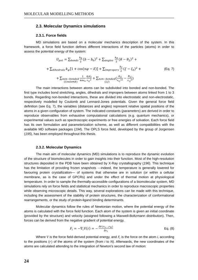
MOLECULAR MODELLING METHODS
24
2.3. Molecular Dynamics simulations
2.3.1. Force fields
MD simulations are based on a molecular mechanics description of the system. In this framework, a force field function defines different interactions of the particles (atoms) in order to assess the potential energy of the system:
(Eq. 7)
The main interactions between atoms can be subdivided into bonded and non-bonded. The first type includes bond stretching, angles, dihedrals and impropers between atoms linked from 1 to 3 bonds. Regarding non-bonded interactions, these are divided into electrostatic and non-electrostatic, respectively modelled by Coulomb and Lennard-Jones potentials. Given the general force field definition (see Eq. 7), the variables (distances and angles) represent relative spatial positions of the atoms in a given configuration of system. The indicated constants (parameters) are derived in order to reproduce observables from exhaustive computational calculations (e.g. quantum mechanics), or experimental values such as spectroscopic experiments or free energies of solvation. Each force field has its own formulation and parameterization scheme, as well as different compatibilities with the available MD software packages (194). The OPLS force field, developed by the group of Jorgensen (195), has been employed throughout this thesis.
2.3.2. Molecular Dynamics
The main aim of molecular dynamics (MD) simulations is to reproduce the dynamic evolution of the structure of biomolecules in order to gain insights into their function. Most of the high-resolution structures deposited in the PDB have been obtained by X-Ray crystallography (196). This technique has the limitation of providing frozen snapshots —indeed, the temperature is generally lowered for favouring protein crystallization— of systems that otherwise are in solution (or within a cellular membrane, as is the case of GPCRs) and under the effect of thermal motion at physiological temperature. In order to sample the thermally-accessible configurations of a biomolecular system, MD simulations rely on force fields and statistical mechanics in order to reproduce macroscopic properties while observing microscopic details. This way, several explorations can be made with this technique, including the assessment of the stability of protein structures, the characterization of conformational rearrangements, or the study of protein-ligand binding determinants.
Molecular dynamics follow the rules of Newtonian motion, where the potential energy of the atoms is calculated with the force field function. Each atom of the system is given an initial coordinate (provided by the structure) and velocity (assigned following a Maxwell-Boltzmann distribution). Then, forces can be derived from the negative gradient of potential energy.
Eq. (8)
Where is the force field-derived potential energy, and is the force on the atom i, according to the positions ( of the atoms of the system (from i to N). Afterwards, the new coordinates of the atoms are calculated attending to the integration of Newton’s second law of motion:

MOLECULAR MODELLING METHODS
25
Eq. (9)
The update of coordinates and velocities of the particles is typically performed by the leapfrog algorithm:
Eq. (10)
Eq. (11)
The is discretized in timesteps, typically in the order of 1 or 2 femtoseconds, for the integration of the equations of motion. The calculations described so far are iteratively repeated for a given number of steps. Consequently, coordinates, velocities and energies of the system are regularly collected into the so-called MD trajectory, describing the evolution of a biomolecular system in time with atomistic detail.
Since the systems should be treated as infinite, but the computational representations of the modelled system must be necessarily finite, several models exist in order to avoid spurious effects from the boundaries of these simulated systems. Here, the periodical boundary conditions (PBC) are a common practice, and are employed in the works presented in this thesis. In PBC, the simulation box has a given geometry, and adjacent images “see” each other, thus representing a continuous and infinite space: if a molecule crosses one boundary of the box, it will enter from the opposite edge, thus conserving the total number of particles in the system. The PBC representation is associated with other approximations, in particular for the evaluation of non-bonded interactions corresponding to atoms which are separated more than a given distance cutoff, which cannot be estimated with the regular force field terms outlined in Eq. 8 due to an exponential increase in computational time. A common technique for the computation of long-range electrostatic interactions beyond such cutoff is the Particle Mesh Ewald (PME) summation method (197). Other approximations that allow a reasonable computation time is the use of bond constraints for the bonds of hydrogen atoms (in our case, the LINCS method). Finally, there are several formal considerations in order to obtain thermodynamic ensembles, such as the coupling to a temperature bath and to a barostat in order to obtain a canonical ensemble (NPT), used through the works here presented. These and other technical issues are detailed in the methods section and supplementary material of Papers IV, VI, as well as in the GROMACS manual (198), which was the software used for MD simulations throughout this thesis.
2.3.3. Considerations of MD simulations of GPCRs
Membrane proteins are given increasing interest in the biochemical and pharmacology fields, as long as they are in charge of numerous cellular communication functions. Not only receptors (such as GPCRs), but also transporters and channels are targets of pharmaceutical interest. Consequently, it is important to decipher the molecular determinants of their function by studying relevant conformational rearrangements, and their interaction with other molecules such as lipids or small organic compounds (199). Given that the function of GPCRs is governed by a dynamic conformational equilibrium between active and inactive states (7, 102, 200), MD simulations are an appropriate technique to explore, at least partially, such conformational equilibrium. Moreover, these simulations offer an excellent complementation of the increasing structural information provided by the recent crystallographic efforts on GPCRs, combined with additional biophysical techniques (201-203).
Thus, several computational methods for MD simulations of membrane proteins have been developed in the recent years (204, 205). They facilitate the preparation of the initial systems, which typically model the biological environment of the protein with a lipid bilayer and solvent molecules. Added to the increase in computational resources and methodologies, longer timescales can be reached (from hundreds to thousands of nanoseconds in all-atom simulations) in order to explore

MOLECULAR MODELLING METHODS
26
relevant conformational rearrangements in the activation pathway of GPCRs, as exemplified by recent works in the field (206-208).
Within the framework of this thesis, a novel computational protocol for membrane protein insertion and equilibration has been developed and presented in detail in Paper IV, and further extended and automated in Papers V, VI. Several considerations of this procedure, which is based on the GROMACS suite of programs, are briefly explained and outlined in Fig. 13:
A palmitoyloleoylphosphatidylcholine (POPC) bilayer pre-equilibrated at 260K,1 thus in crystalline phase, in order to help the packing of the lipids around the protein.
The GPCR of interest is inserted into the bilayer, and overlapping lipid molecules are removed. The protein must be correctly aligned in the Z-axis, where TM4 is supposed to run parallel to the lipid tails, and H8 should cross the intracellular polar heads, in agreement with the assigned orientations of GPCRs deposited in the OMP database (209). Manual refinement of the position in the Z-axis is performed in order to obtain a balanced number of lipids in the upper and lower leaflets of the bilayer.
Finally, the system is solvated with water molecules and neutralized with counter-ions.
The geometry of the simulation box is a hexagonal prism, which optimizes the number of solvent molecules for saving computational time (with an estimated reduction that exceeds the 10% as compared with a regular rectangular-shaped box). The typical size of the final simulation box is constituted by around 60.000 atoms for protein monomers (Papers IV, V) and 100.000 atoms for protein dimers (Paper VI).
Figure 13. Outline of the computational protocol for the setup of MD simulation boxes of GPCRs. The different stages for the insertion of the GPCR structure, here exemplified by an A2AAR monomer, are depicted. More details can be found in the main text and Papers IV—VI.
The MD simulations force fields (see above) are parameterized for proteins, but not for other molecules such as organic ligands or lipids. However, it is necessary to obtain proper parameters for all the members of the system for a given force field to be employed in the simulations. For the case of small organic compounds, we generated reliable parameters for the selected OPLS-AA force field (195) with the molecular modelling software Schrödinger (160), a procedure that proved to be valid in
1 POPC bilayers have been employed in Papers IV—VI. All simulations were run at 310K, where POPC transitions towards the biologically-relevant fluid phase.
MOLECULAR MODELLING METHODS
27
recent protein-ligand binding free-energy calculations (210). For lipid molecules, one alternative is their reparameterization (211) from ad-hoc lipid force fields, such the one developed by Berger et al. (212). Another option is to adapt two force fields (one describing the lipid, and the other the protein/ligands), as proposed by the half- double pairlist method (213) developed for the GROMACS simulation package. This method ensures a proper scaling of 1-4 non-bonded interactions (those involving atoms separated by 3 consecutive bonds) for both the Berger (lipid) and OPLS-AA (protein) force fields, while successfully reproducing experimental macroscopic properties such as the area per lipid (214). We have used this methodology in Papers IV, VI, as well as in the MD pipeline of GPCR-ModSim server (Paper VI).
Once the system is set up, an equilibration scheme is necessary in order to allow the different constituents to adapt to each other, as the achievement of a stable configuration is necessary to obtain meaningful equilibrium simulations. In equilibration stages, the positions of certain protein atoms (and ligands and crystallographic waters where considered) are restricted by a force constant, which applies an energetic penalty for the movement of the restrained atoms. These constants are gradually reduced until the whole system is unrestrained. Then the production phase of the simulations starts, where the collection of data takes place. Several equilibration protocols have been explored, taking especial care of the loop regions in Paper IV. In that same work, independent replicas of selected systems were simulated in order to increase the thermodynamic sampling, and to gain statistical significance on the observed dynamic events.
The monitoring and analysis of MD trajectories is a key and time-consuming step. With the purpose of facilitating and automating this task, we developed a set of scripts that take care of the trajectory visualization (using the program PyMOL, www.pymol.org) and the calculation of geometrical and energetic observables, mainly using several tools in the GROMACS package as well as plotting programs (GNUplot, XMGrace). Regarding the latter calculations, they are important to assess the stability of the system and to quantify conformational changes and interactions of interest. Some of these analyses have been adapted for the special case of GPCRs, and implemented into the GPCR-ModSim server (see Section 4 and Paper V).


OBJECTIVES
29
3. OBJECTIVES
The main motivation of this thesis is to provide a methodology for characterizing the structure and dynamics of selected members of GPCRs, aimed to the elucidation of the mechanism of receptor activation, as well as in drug discovery and design efforts. We have pursued these challenging goals through the establishment of three milestones, which are here outlined and further analysed on the basis of our results in the next section:
Development and application of a robust homology modelling methodology in
GPCR computer-aided ligand discovery and design.
This milestone would be accomplished through the consideration of the following objectives: i) development of reliable structural models of selected GPCRs of interest, through a methodology that could be extended to any GPCR. ii) Use of this models for their latter application in structure-based ligand design methodologies. iii) Combination of these structure-based methods with ligand-based techniques. iv) Appropriate integration with experimental studies, mainly through collaborations with medicinal chemistry and pharmacology groups, in the interdisciplinary framework typical of ligand-design projects.
Study of the dynamics and conformational equilibrium of GPCRs.
With this in mind, we pursued the following objectives: i) The development of a specific protocol for the molecular dynamics simulations of GPCRs. ii) The application of MD simulations to study the conformational equilibrium of selected GPCRs, as well as to iii) the characterization of the receptor dimerization process.
Integration of the homology modelling and MD simulation protocols, developed in the previous milestones, into an automatic pipeline that is offered as a web service open to the scientific community.
This milestone will provide important benefits: i) to our research group, increasing its competitiveness in the field of GPCR structural biology, ii) and to the scientific community, which will have a free and easy to access methodology without needing prior computational modelling expertise, enabling an exhaustive structural and dynamic characterization of this pharmaceutically relevant superfamily of receptors.


RESULTS AND DISCUSSION
31
4. RESULTS AND DISCUSSION
Provided a general introduction of the biology and pharmacology of the considered GPCRs, and once detailed the computational techniques employed to achieve the main objectives of this thesis, the most remarkable results are presented below. These results are mainly reported as a series of scientific publications, reproduced in Section 7, and are here discussed and put in context of the objectives depicted in the previous section.
Development and application of a robust homology modelling methodology in GPCR computer-aided ligand discovery and design.
As described in detail in Section 2.1.1, we have employed a homology modelling protocol in
order to obtain high-quality structural models of GPCR structures. This general protocol has been used in several computational drug discovery and design efforts, including the combination of SB and LB approaches, as summarized below.
The first computer-aided ligand discovery study is described in Paper I, and corresponds to an interdisciplinary project carried out in collaboration with the group of Pharmacology of Prof. Loza (University of Santiago de Compostela, USC) and the Chemistry Department of University of Minho (Prof. Proença). Here we assisted in the discovery of novel potential antipsychotic scaffolds from the original chemical library of U. do Minho, previously uncharacterized in any GPCR. We computationally screened this library of 1622 compounds with the shape-based VS methodology from Openeye suite of programs. This approach was selected due to its ability for retrieving hits with novel chemistries (181), and demonstrated good performance in retrospective VS on aminergic receptors (183). In fact, seven pharmacological hits were discovered, using the reference APC clozapine as query for the screening. The in vitro affinity of the identified hits identified was evaluated in the human 5-HT2A receptor and showed a Ki in the micromolar range. Interestingly, these hits present a novel 9-methyl-purine scaffold with either piperazine or piperidine rings in the exocyclic N6, and different aryl substitutions in position C2. In order to rationalize the results of this VS, a homology model of the 5-HT2A receptor was built using the structure of 2ADR as template (50), employing for the first time the protocol for GPCR modelling described in Section 2.1.1. Interestingly, both the shape-based VS and the docking results agreed in their proposed biological superposition of the most active hit and the reference molecule, clozapine. The binding modes of the two compounds share the conserved salt bridge between Asp3.32 of the receptor and the positively-charged nitrogen of the ligands, in agreement with previous computational studies (215), and provided hints on the possible 10-fold lower affinity of the hit compounds, as compared to clozapine. Specifically, the purine-based ligand lacks the interaction with Thr3.37 predicted for clozapine, and instead establishes a hydrogen bond with Ser5.46 that leads to a steric clash of a methyl group of the 9-methyl-purine scaffold with TM5.
In order to further assess the homology modelling and ligand docking pipelines proposed in the previous work, our laboratory took part on the GPCR Dock 2008 assessment (see Section 2.1 and Paper II). Here, our group performed blind predictions of the structure of the complex of A2AAR with the potent antagonist ZM241385 (group code 5800 in the competition) right before the release of the crystallographic structure. After a template selection process, we generated and reported several homology models of the target receptor with MODELLER, using 2ADR as template, followed by a model assessment on the basis of their stereochemical quality with Procheck, and refined with the aid

RESULTS AND DISCUSSION
32
of the Molprobity server. We submitted 5 different complexes, selected and internally ranked on the basis of the mutagenesis data available for antagonist binding on A2AAR (26). The results of the contest, including all the structural models, are open to the scientific community and available at http://jcimpt.scripps.edu/gpcr_dock.html. The results of our research group are summarized in Fig. 14. Our complexes provide over-the-average predictions within the total number of submissions coming from 29 different research groups. In terms of predicting the ligand binding mode, our best complex reproduced 11 ligand-receptor native contacts and obtained a combined Z-score of 0.31,2 being ranked in position 43th within the total of 208 computationally-predicted complexes. In terms of protein homology modelling, our A2AAR models showed low root-mean square deviation (RMSD) for the trace of C atoms compared to the experimental structure (ranging from 3.3 to 3.8 Å depending on the model), being ranked as the top-15 homology model. From this analysis, it becomes clear that although having presented a high quality homology model, we did not perform as well in the prediction of the binding mode of ZM241385. Indeed, in our model of the complex, the ligand presented a flipped orientation compared to the crystallographic pose, undoubtedly influenced by the relative symmetry of the molecule around the exocyclic nitrogen. In fact, a latter review of the pool of docking solutions provided by GOLD revealed low-ranked binding modes we discarded for further consideration, which were much closer to the experimental one (see Fig. 14C). The lesson learned was that, on top of the ranking provided by automated scoring functions, the researcher must properly incorporate all the available experimental data into the molecular modelling process. This is also a general outcome of the two editions of the GPCR Dock competitions (in 2008 and 2010), where the most accurate models were produced by researchers with proven knowledge of the biochemistry and pharmacology of the considered receptors, as are the cases of Costanzi in ARs (137), Selent et al. in aminergic and de Graaf and colleagues in peptide-binding receptors (138).
Figure 14. Summary of the participation of our research group in the GPCR Dock competition (code 5800). (A) Statistics of the submitted models, following their ranking on several metrics respect the crystallographic structure. Average values of all submitted models are indicated in brackets in the first row. The rankings are referred to the individual models, numbers in brackets indicate the rank only taking into account the best solution of each research group, and asterisks denote degenerated ranking. (B) Best submitted complex solution (model code mod4veh) (C) Discarded solution from the final selection stage, significantly resembling the crystallographic pose.
2 The Z-score is a dimensionless measure that normalizes a value respect to the mean and standard deviation of the sample, in this case considering the RMSD and native contacts of the ligand.

RESULTS AND DISCUSSION
33
In a follow-up study, we generated homology models of the remaining human ARs subtypes: A1, A2B and A3 ARs, using the recent structure of A2AAR in inactive-like conformation (56). Despite the high sequence identity within the family —A2AAR presents a TM sequence identity between 52% and 72% with the rest of subtypes—, there are remarkable differences in ligand binding affinity among ARs. In order to investigate these selectivity issues, we generated a pseudo-sequence alignment of the binding site residues of ARs subtypes, in the line of previous chemogenomics efforts (9, 10), and as also attempted by other authors (109, 216). Such analysis, illustrated in Fig. 15A, allowed us to detect the most promising residue substitutions to explain ligand selectivities. We selected three antagonists with different chemical scaffolds and selectivity profiles in the ARs family to conduct a docking exploration on the generated homology models: the triazolo-quinazoline CGS15943, the xantine derivative DPCPX and the triazolo-triazine ZM241385 which had been co-crystallized with A2AAR (see Fig. 15B). The biological superimposition proposed for CGS15943 and DPCPX in the A2AAR structure, together with the co-crystallographic ligand (successfully re-docked as a test case), is in agreement with previous ligand-based studies (217). This was further corroborated for the case of DPCPX, where our predicted binding mode is in agreement with the recently solved structure of A2AAR with the xantine derivate XAC (17), shown in Fig. 15C. The same superimposition is observed for A2BAR, except subtle differences that might be probably attributed to the presence of Val6.51 in that receptor instead of the conserved Leu in the rest of the ARs. Regarding other selectivity issues that might be explained by this docking study, the ligand ZM241385 frequently presents a flipped pose in the A1 and A3AR as compared to the binding mode in the crystal structure with the A2AAR: these observations are in agreement with a 10- to 70-fold lower affinity of this ligand in A1AR and A3AR compared to both A2 subtypes. Also, the conserved solution of DPCPX in A3AR can be explained by the less voluminous Leu in position 7.35 in this receptor, as compared to the otherwise conserved Met7.35 in the family. Accordingly, this compound presents 50- to 1000-fold less affinity in the A3AR as compared to other subtypes.
Figure 15. Systematic homology modelling and ligand docking for studying selectivity issues on ARs. (A) The pseudo-sequence alignment of ARs residues at 5 of the crystallographic ligand as observed in PDB structure 3EML, together with the representation of key positions with different mutations in the rest of ARs subtypes. (B) Structures and Ki affinities (nM) of the selected antagonists for the docking study on the aforementioned structures. (C) Biological superimposition of DPCPX (dark sticks) after docking on 3EML structure (ZM241385 in white ball & sticks), in agreement with of A2A in complex with the analogous XAC. See Annex I for details.

RESULTS AND DISCUSSION
34
On the basis of this modelling study of the members of ARs family, and in collaboration with the medicinal chemistry group of Prof. Sotelo (USC), our laboratory contributed to the computational design and the rationalization of the pharmacological profile of a novel library of diaryl-pyrimidine derivatives with potent and selective profiles on A3AR. This study is presented in Paper III of this thesis. Here, a combination of SB and LB computational approaches was considered, using the homology model of the human A3AR developed in our laboratory (described above) as starting point. A systematic docking of the compounds of the chemical library allowed the determination of the bioactive conformations, which were used as input for a 3D-QSAR modelling that was ultimately used in the assessment and the growth of the chemical library. In the first stage, a consensus binding mode was determined for the compounds of the chemical library. Accordingly, a double hydrogen bond between the key residue Asn6.55 with both the exocyclic amido group and the closest nitrogen in the pyrimidine scaffold in all ligands was observed, together with a -stacking interaction with Phe5.29 at EL2 (both residues totally conserved in ARs), and van der Waals interactions with Leu6.51. Notably, the recent crystal structure of A2AAR in complex with a 1,2,4-triazine derivative (55), a scaffold structurally similar to the aforementioned pyrimidine derivatives, supports this hypothesized binding mode (see Fig. 16). The ligands exploit 3 lipophilic subsites of the receptor with chemical groups attached to their exocyclic amino/amido (L1) and aryl substituents (L2, L3). The biological molecular alignment obtained from the consensus docking pose was used as a starting point for a 3D-QSAR model with the second generation of GRid INdependent Descriptors (GRIND-2) (188) implemented in the software Pentacle (218). The resultant 3D-QSAR model presented a satisfactory statistical quality, with a fit of r2=0.86, and a predictive ability of q2=0.67 obtained by LOO-CV. The main pharmacophoric properties of the series are represented by corresponding correlograms, i.e., alignment-independent vectors of relevant MIF node pairs extracted from the model (see Section 2.2.2.2 for details). Most remarkably, these correlograms suggests a relationship between the size of substituents at L1 and L2/L3 subsites (the bulkier in L1, the smaller in L2/L3, and vice-versa) in order to achieve increased activity on A3AR. An advantage of this combination of SB and LB methodologies is that the 3D-QSAR descriptions are back-projectable to the binding site. Thus, hydrophobic interactions at the L1 site (extracellular tip of the binding site) are detected by O-TIP and DRY-TIP cross-correlograms, reflecting interactions with Ile6.58 and Leu7.35, and explaining the reduced affinity of aminopyrimidine derivates due to the lack of substitutions at that subsite. Also, the reduced potency of 2-amido-pyrimidines respect to the 4-amido congeners is represented by O-N1 and N1-N1 correlograms, also suggesting the presence of a structural water molecule mediating ligand-receptor interactions for the latter derivatives. Additionally, selectivity issues can be easily detected by the sequence analyses of binding sites of ARs described above. In this regard, positions 5.30, 5.42 and 6.52, together with the already mentioned 7.35, have been pointed out as the mostly relevant for selectivity issues of these ligands in the context of ARs. Specifically, His6.52 and Asn5.42 in A1, A2A and A2B ARs are substituted by a serine residue in both positions of A3AR, allowing bulkier substitutions of the ligands in this subsite (L3). Additionally, hydrophobic and more voluminous substitutions at the exocyclic amido group are favoured in A3AR due to the presence of Val5.30 at EL2 (where a Glu5.30 appears for the rest of subtypes) and a smaller Leu7.35 (compared to Met in A2 subtypes). The described computational pipeline was employed in the design of a set of 6 novel ligands, with bulky substituents in the amido group of the 2,6-diaryl-4-amidopyrimidine scaffold with excellent results (see Paper III), and is being currently employed in the design on novel ligands with enhanced pharmacological profiles towards A3AR.

RESULTS AND DISCUSSION
35
Figure 16. Comparison of the computationally-predicted binding mode of ISVY130 in A3AR (blue), and the crystallographic structure of a 1,2,4-triazine derivative in complex with A2AAR (PDB code 3UZC, in magenta). The double hydrogen bond with Asn6.55, together with key binding site residue residues, are indicated. Key residue substitutions in A3AR provide a broader sub-pocket between TM5 and TM6.
Following the structural characterization of ARs discussed above, we aimed to combine these studies with ligand-based approaches for the discovery of novel potent and selective ligands towards the different ARs. This was the subject of a research stay in the laboratory of Dr. Andreas Bender, at the Unilever Centre (University of Cambridge, UK) which main results are outlined in Annex II. In this work, we screened the chemical library of the Galician Network for Drug Discover, which counts with the participation of our group and is managed by the laboratory of Prof. Loza (USC). A VS protocol was designed and executed as follows:
i) Training bioactivity models for each ARs subtype (A1, A2A, A2B, A3) with the tool MOLPRINT2D (see Section 2.2.2.3), with data extracted and curated from the ChEMBL database (https://www.ebi.ac.uk/chembl/). Molecules with a pKi above 7 were considered as active, while those with pKi below 6 were categorized as inactive. Since most of the molecules deposited in this dataset come from successive medicinal chemistry campaigns that explore series of analogues, we removed this redundancy by means of clustering with JKlustor (ChemAxon, using ECFP4 fingerprints) in order to maximize the molecular diversity of the training sets. The datasets of diverse active and inactive ligands for each subtype, with an approximate population of 200 compounds each, were complemented with 800 random compounds from ChEMBL added to the inactive subset in order increase the resolution of the scoring. After obtaining the ligand score for each bioactivity model, we set a multi-objective selection criteria: a compound would have to be ranked in the top 10% of one subtype and below that threshold in the rest of subtypes for being a promising selective compound. An additional less restrictive criteria, termed as “promiscuous”, was selected for fewer ligands within the top10% in at least two out of the four possible AR models.
ii) A post-filtering stage on the basis of physicochemical properties and (dis)similiarity with known actives was performed in order to retain compounds with drug-like properties and novel scaffolds.
iii) The final selection of compounds to be experimentally tested on the four ARs was assessed by docking simulations on our 3D models of ARs, paying special attention to interactions with the key residue Asn6.55. Finally, 40 compounds were assayed on all subtypes, which generally present novel scaffolds in the medicinal chemistry of ARs (i.e., single-ringed cores and extended side chains). From these ligands, we found 11 compounds over the minimum threshold of 30% inhibition (at 10-5 M concentration) on at least one receptor subtype, with 2 of them above 50% in A1AR and A2BAR (see Fig. 17). The performance of the A3AR and promiscuous models are especially remarkable. The present results show that the LBVS methods are able to obtain high hit rates —

RESULTS AND DISCUSSION
36
especially considering the nature of the screened chemical library—, in agreement with previous published screenings on the A2AAR (219). This has also been one of the conclusions of a recent cross-screen on three GPCRs (220), where most importantly the prediction of selectivity appears to be still a difficult issue for the state-of-the-art techniques. This study is on a second phase at our research group at the time of the writing of this thesis, consisting on a search of analogues from vendors with prospectively enhanced properties in order to provide hits with higher affinities.
Figure 17. Pharmacological hits obtained in an ongoing campaign for discovering novel and selective ligands on ARs. (A) Discovered pharmacological hits, together with their molecular weight, the selection criteria met in the VS, and their pharmacological data on ARs. On the right hand side, binding modes of compound 1 in A1AR (B) and compound 5 on A2BAR (C) In the latter binding mode, no significant interactions were predicted for compound 5 in A2BAR (in fact, was selected for A1AR), precluded by the rotamer of Met7.35.

RESULTS AND DISCUSSION
37
Study of the dynamics and conformational equilibrium of GPCRs.
In the recent years, several studies of the dynamics of membrane receptors have been
performed through all-atom MD simulations. This has been possible due to the parallel advances in structural biology of GPCRs as well as important developments in computational software and hardware. In this scenario, our research group, in collaboration with Dr. Piñeiro and Prof. Brocos (Department of Applied Physics, USC), has developed of a computational protocol for the automated setup and performance of MD simulations for the particular case of GPCRs. The protocol, which is deeply described in Section 2.3.3 and originally published in Paper IV, considers an atomistic model of the biological membrane, consisting of a POPC bilayer, solvated with explicit water molecules and neutralized with counter-ions, under the PBC scheme implemented in the software package GROMACS (221). Using this pipeline we performed and analysed MD simulations of the X-Ray structure of A2AAR in its inactive-like conformation (PDB code 3EML) (56), as well as of a homology-derived model of the related A2BAR subtype, as reported in Paper IV. The use of simulation replicas (identical starting points with different random seeds for the assignment of initial velocities) and several setups (absence or presence of crystallographic ligands, consideration of crystallographic water molecules, and residues with different protonation states) were considered. After exhaustive equilibrations, the production phases of the main simulations spanned for 100 ns, an appropriate timescale for characterizing initial conformational rearrangements from the inactive-like state towards more active-like forms of the receptors. Additionally, the use of simulation replicas provides a better thermodynamic sampling, increasing the statistical significance of the observed events.
One of the microswitches studied in these simulations was the ionic lock, a salt bridge established in inactive receptor conformations between residues Arg3.50 and Glu6.30 (in the intracellular tips of TM3 and TM6, see Section 1.2.1). This interaction, however, was absent in the experimental inactive structure of the A2AAR (56), and consequently in the homology-derived model of the A2BAR. However, we observed a quick and stable formation of this lock in the different MD simulations replicas on both considered receptors. Moreover, we could characterize the tight hydrogen bond network that supports this “lock”, which includes Asp3.49 and Tyr3.60(IL2). This is in agreement with previous simulations of A2AAR (222) and with computational studies of inactive-like crystal structures of -adrenergic receptors with initially broken ionic locks (50, 52), where simulations around the microsecond scale showed a spontaneous and generally stable formation of this interaction (223-227). A common feature of the crystallization process of both A2AAR and -adrenergic receptors is the use of the T4L protein fusion strategy, which was lately discussed as the source of distortions in the intracellular region and probably responsible of the absence of this salt bridge (83). In fact, in the X-Ray structures of A2AAR in complex with inverse agonists, which were obtained using mutation-stabilized receptors, the ionic lock is formed (57). This result agrees with our simulations and exemplifies the predictive ability of MD in the refinement of crystal structures in a model resembling the physiological environment of the receptor. As a final note, the reformation of the ionic lock has been recently observed in MD simulations of active-like conformations of 2ADR in the microsecond scale (228).
The second examined microswitch was the rotameric transition of the side chain of Trp6.48 from gauche+ to trans, known as the toggle switch, and largely associated to the activation mechanism of GPCRs (93-95). This event was observed on both A2AAR and A2BAR, where a cluster of conserved residues in ARs —namely Asn5.42, Gln3.37, Cys5.46 and His6.52— was found to co-operate with the formation of this switch through concerted conformational changes in order to form a novel hydrogen bond network. While this rotameric transition was not observed in the only simulation of
2ADR crystal structure that explicitly analyzed this switch (227), it was indeed reported in large-scale MD simulations on a homology model of the dopamine D2 receptor, in this case considering the

RESULTS AND DISCUSSION
38
allosteric modulation of sodium ions located in the vicinity of this region (229). Very recently, this sodium allosteric binding site, largely proposed for many GPCRs, was experimentally observed in a crystal structure of the A2AAR and is located in the tight hydrogen bond network of GPCR conserved residues at TM1-TM2-TM6-TM7. Interestingly, in our simulations the solvent occupancy of this region in A2AAR is well reproduced, regardless of the consideration of crystallographic waters in the MD setup. Currently, we are studying the sensitivity of this receptor to the presence of sodium ions and other allosteric modulators, using a similar computational methodology, in collaboration with the groups of Prof. Stevens (Scripps Research Institute, California) and Prof. IJzerman (Leiden University).
Going back to Paper IV, we were also interested in the structural role of two other particular motifs in ARs that involve key histidine residues. The first one refers to the interaction between residues of EL2 (Glu5.30) and EL3 (position 7.29), closing the top of the binding site. A tighter interaction in A2AAR, especially considering the more plausible charged state of His7.29, is observed as compared to the A2BAR where this histidine is replaced by the neutral Asn7.29. This aminoacidic substitution is responsible for a higher mobility of the extracellular loops of the latter receptor according to our simulations, suggesting a higher diffusion rate of compounds in this receptor that might explains the lower affinity for most ARs ligands on A2BAR. The second motif involves the totally AR conserved residues Glu1.39 and His7.43. Here, the initial hydrogen bond suggested by the crystal structure is broken along the simulations, significantly regulating the dynamics of TM1 and TM7. The tautomeric state of His7.43 protonated in delta, as modelled by us, is in agreement with latter crystallographic structures of A2AAR in complex with agonists (53, 54). The exhaustive pKa calculations performed suggested a charged state for this histidine in the A2AAR, a possibility that was computationally assessed together with the in silico proposal of a His7.43Lys mutant. Our results propose the existence of a salt bridge between residues Glu1.39 and His7.43, and issue that remains to be confirmed by molecular biology and pharmacology experiments. Finally, a comparison between the lately crystallized structure of A2AAR in an active-like form (54) and the endpoint of one of A2AAR simulation replicas reveals a partial agreement of the conformations of the extracellular region of the receptor (see Supporting Information of Paper IV). In fact, the induced-fit promoted by the co-crystallized agonist UK-432097 results in a disruption of the interaction between EL2 and EL3, as well as a conformational change of the extracellular half of TM7 outwards the binding crevice, both events being well reproduced in our simulations. The experimental confirmation of our results is encouraging, although we acknowledge that the more drastic conformational changes that drive the receptor into its active state could not even be achieved by our computational experiments, due to the timescale affordable by our simulation approach (equilibrium-MD).
The computational protocol presented in Paper IV was adapted in order to analyse the phenomenon of receptor dimerization in Paper VI. Provided the novel experimental evidences of GPCR dimerization as revealed by the crystallographic structures of CXCR4 (65), released in October 2010, we decided to further investigate the structural determinants this protein-protein interactions. The CXCR4 homodimers show a previously unexpected dimerization mode involving TM5 and TM6 (see Section 1.2.3 and Paper IV for details), observed in two slightly different crystallographic dimers: one co-crystallized with the organic ligand 1T1t (PDB code 3ODU); and the other —assigned by the authors— co-crystallized with the cyclic peptide CVX15 (PDB code 3OE0). We performed extensive MD simulations of the apo forms of these two molecular models, considering both the stabilized mutant crystallographic form, as well as the wild-type forms of the receptors. In this respect, our MD simulations showed punctual distortions on the wild-type protomers, thus justifying the observed increased thermal stability of the mutant forms. Generally speaking, the simulations support the dimerization mode observed in both structures, since the position of the protomers remained stable on the initial pose, although certain rearrangements in the relative orientation of protomers were observed: the buried surface area of the dimerization interface growth in all cases, mainly driven by
RESULTS AND DISCUSSION
39
hydrophobic interactions between the receptors (keeping those observed in the crystallographic snapshots). The conformational changes were more significant for the case of 3ODU dimer, due to the lower initial contacts in the intracellular half of the dimerization interface compared to 3OE0 structure (65). This behaviour is also line with the observations reported in the recent structure of -OR (71), which adopts a higher-order oligomeric arrangement with commonalities with the dimerization mode observed for CXCR4 structures. The -OR structure further supports the relevance of the TM5-TM6 interface (see Fig. 11). However, the relatively high buried surface area between the intracellular T4L fusion proteins in 3ODU protomers —unlike 3OE0 or -OR structures— might induce a bias in its initial conformation justifying the observed conformational changes. Given the transient nature of GPCR dimers (230), this computational evaluation significantly explores expectable structural rearrangements from the crystallographic structures of CXCR4, supporting the ligand-independent dimerization of this receptor, and concluding that the interface of 3OE0 structure presents a reliable high-resolution depiction of GPCR dimerization.
Additionally, we also studied the dynamics of key residues of CXCR4 involved in ligand binding and in the process of HIV infection. We focused on differences of the tautomeric state of His3.29 according to the conformations of surrounding residues, characterizing the influenced of the co-crystallographic ligands in that region. Thus, while the initial salt bridge between Asp4.60 and Arg5.27(EL2) remained stable for the apo form of the IT1t-bound structure (3ODU), a strong hydrogen bond between His3.29 and Asp4.60 occurs instead in the apo version of the CVX15-bound structure (3OE0). We suggest that specific induce-fit effects occur upon the binding of the cyclic peptide, a conclusion which is further supported by the available mutagenesis data. Our work concludes that the conformation of 3ODU structure is more appropriate for the structure-based drug design of small ligands.
As a final note, despite that the simulations performed in Papers IV, VI do not reach the microsecond timescale —as opposed to other contemporary simulations of GPCRs— the range of hundreds of nanoseconds is considered as appropriate taking into account: i) the short timescale of the dynamic and structural events that were aimed (initial conformational rearrangements from an inactive conformation, or the stability of GPCR crystallographic dimers), ii) the use of simulation replicas to increase statistical sampling and iii) the available computational resources. We can conclude that our methodology is in the state-of-the-art of statistical dynamic sampling techniques, and at the same time provide several methodological advances for the MD simulation of GPCRs.

RESULTS AND DISCUSSION
40
Integration of the homology modelling and MD simulation protocols, developed in the previous milestones, into an automatic pipeline that is offered as a web service open to the scientific community.
As discussed before, the structures of many GPCRs remain experimentally unsolved despite the recent advances in their crystallization. Following the previously described methodologies on GPCR homology modelling and MD simulations, developed through this thesis with successful results, we considered the possibility of automating the process in order to access to a higher coverage of the structural knowledge of GPCRs. Several services for this purpose are available on the web, which we introduced in Section 2.1.2 and reviewed in Paper V, at the same time that we presented our GPCR-ModSim webserver (http://gpcr.usc.es). We here discuss the main advances of this pipeline:
i) The first critical stage of homology modelling is the selection of the most appropriate template for the target receptor. For this purpose we have elaborated a structural alignment of all members of GPCRs with solved structures, and divided them according to their functional conformation (either using the 14 inactive- or the 5 active-like receptors available to date), see Fig. 18. The last update of our structural alignment was performed with the aid of the software Strap (http://3d-alignment.eu), which produces a multiple-sequence alignment (MSeqA) derived from a multiple-structure alignment (MStA) with the TM-align algorithm (231). The resulting MSeqA was manually reviewed, paying special attention to gaps and indels within the TM region due to local distortions, and the correct alignment of the two cysteines involved in the conserved disulfide bridge (TM3-EL2), among other aspects. This refined MSeqA of the templates is stored in the server as the profile to which the sequence of the query receptor is aligned to. In this sense, it is worth noting that the query sequence can be retrieved directly from its UniprotID, or may consist of a “customized” protein mutant. The initial query-templates alignment is automatically made with Clustal0 (232), but can be further refined by the user with a JalView applet. The server automatically provides statistics of sequence identity for all the different regions of the receptor (individual loops and TMs), and by default suggests the template with the highest sequence identity in the TM region with the target receptor. A practical application of the template selection method offered by the server was performed in the homology modelling of neuropeptide Y2 receptor (233).
ii) Once the query-template sequence alignment is configured, the user is allowed to perform up to 10 models with MODELLER 9v8 (155), which are ranked according to the statistical potential DOPEHR (234). In order to further select the models, stereochemical quality reports generated by Molprobity (158) are provided, together with a JalView applet that allows a quick visual inspection. A Python script for a local visualization of all the models in PyMOL can be downloaded as well. On top of this initial modelling, the user is allowed to run up to 5 loop optimizations in selected model, as implemented in the loopmodel routine (156) with the same tools for selection and visualization as described above. The introduction of custom disulfide bridges can be performed in either initial and/or loop modelling stages.
iii) Finally, one of the main novelties of our web service is the setup and MD equilibration of the receptor in an atomistic model of the biological membrane, following the protocol developed in Paper IV. The user can perform this equilibration in one of the generated models from the server, or even upload an external PDB structure of a GPCR, which will be conveniently inserted in the simulation box and equilibrated automatically by the server. After 2.5 ns equilibration, the user will be provided the refined system (with the corresponding MD trajectory), a brief report and all necessary files for a local extension of the simulation with the GNU-licensed software GROMACS v4.0.5 (221).
Right after the crystallization of human histamine H1 receptor in complex with doxepin (67),

RESULTS AND DISCUSSION
41
we used this novel structure to carried out a comparative analysis of the performance of three web-based servers for the modelling of GPCRs, namely GPCR-ITASSER (164), GPCR-SSFE (148) and our GPCR-ModSim service, using default parameters and minimum user intervention. All the three tools were able to obtain good quality models, where the RMSD of C atoms of the TM bundle was below 2.0 Å. The accuracy achieved by GPCR-ITASSER and GPCR-ModSim was especially remarkable, including the loop regions (C RMSD for the whole receptor of 2.09 and 2.31 Å respectively, see Paper VI). It is important to note that the protocol implemented by GPCR-ModSim allows the generation of such models in minutes, while the exhaustive threading and loop refinement method of GPCR-ITASSER typically takes several days. A second advantage of our method, outlined in Fig. 18, is that it is the only service that allows an automated MD exploration of the model, which is a recognized step forward in the refinement and sampling of the receptor structure(s) for either drug design (235) or structural biology (228) applications.
Figure 18. Outline of the computational protocol implemented in GPCR-ModSim server. The main interactions (input/output) with the users in the three stages of the pipeline (sequence alignment of the query receptor, homology modelling and MD simulations) are indicated.

RESULTS AND DISCUSSION
42
As a conclusion, the GPCR-ModSim web server provides the most complete pipeline of GPCR homology modelling to our knowledge, including the performance of MD simulations for the first time. A minimum user intervention is necessary to obtain reliable homology models with state-of-the-art techniques, although extensive flexibility is considered in order to allow proper decision-making by the user in the key steps, such as the refinement of the target-template sequence alignment or the model selection. Users without a strong computational background can find this service useful for tasks such as the spatial location and surroundings of key residues (including protein mutants), the assessment of the stability of protein mutants, or the study of selectivity issues within a family of receptors (as we exemplified for ARs). Continuous updates are performed in the server, the most notable being: i) the incorporation of any novel GPCR-crystal structure, ii) the consideration of receptor dimers, iii) the inclusion of ligands, either allosteric and/or orthosteric, or cofactors (i.e. specific cholesterol or lipid molecules) in the MD protocol, to account for the complexity of the receptor system suggested by recent evidences (64, 101).
CONCLUSIONS
43
5. CONCLUSIONS
State-of-the-art techniques in the area of molecular modelling and computer-aided
drug discovery and design have been used as a basis for further methodological developments, as well as in applied studies in the biochemistry, medicinal chemistry and pharmacology of selected members of GPCRs. The main achievements of this thesis include:
The development of a complete and automated computational pipeline for the homology modelling, structural refinement and dynamic analysis of GPCRs, which has been tested in different systems such as a comparative analysis of adenosine receptors or the homodimerization of CXCR4. We have fully automated this pipeline in the GPCR-ModSim web server (http://gpcr.usc.es), which can be used by researchers worldwide without prior modelling knowledge.
A structural characterization of the conformational equilibrium and ligand selectivity on the Adenosine Receptors family, by a proper combination of the first experimental structure of the A2AAR in complex with a potent antagonist (ZM241385), the available mutagenesis data and our computational methodology. This provides a map of the functional and binding selectivity issues in this family of receptors, which is here used in drug design applications.
The successful exploration of combinations of Structure-Based and Ligand-Based approaches in GPCR drug discovery and design. The use of homology models together with techniques such as Shape-Based Virtual Screening and 3D-QSAR allowed the discovery and rationale of the binding of novel scaffolds for future antipsychotic compounds from an original chemical library, and the design of a novel series of diarylpyrimidines with a potent and selective pharmacological profile on the A3AR.


REFERENCES
45
6. REFERENCES
(1) Paul, S. M., Mytelka, D. S., Dunwiddie, C. T., Persinger, C. C., Munos, B. H., Lindborg, S. R., and Schacht, A. L. (2010) How to improve R&D productivity: the pharmaceutical industry's grand challenge. Nat. Rev. Drug Discov. 9, 203-14.
(2) Jorgensen, W. L. (2004) The many roles of computation in drug discovery. Science 303, 1813-1818.
(3) Strachan, R. T., Ferrara, G., and Roth, B. L. (2006) Screening the receptorome: an efficient approach for drug discovery and target validation. Drug Discov. Today 11, 708-716.
(4) Rosenbaum, D. M., Rasmussen, S. G., and Kobilka, B. K. (2009) The structure and function of G-protein-coupled receptors. Nature 459, 356-363.
(5) Overington, J. P., Al-Lazikani, B., and Hopkins, A. L. (2006) How many drug targets are there? Nat. Rev. Drug Discov. 5, 993-6.
(6) Gutkind, J. S. (1998) The pathways connecting G protein-coupled receptors to the nucleus through divergent mitogen-activated protein kinase cascades. J. Biol. Chem. 273, 1839-1842.
(7) Kenakin, T. (2004) Principles: receptor theory in pharmacology. Trends Pharmacol. Sci. 25, 186-192.
(8) Mirzadegan, T., Benko, G., Filipek, S., and Palczewski, K. (2003) Sequence analyses of G-protein-coupled receptors: Similarities to rhodopsin. Biochemistry 42, 2759-2767.
(9) Surgand, J. S., Rodrigo, J., Kellenberger, E., and Rognan, D. (2006) A chemogenomic analysis of the transmembrane binding cavity of human G-protein-coupled receptors. Proteins 62, 509-38.
(10) Gloriam, D. E., Foord, S. M., Blaney, F. E., and Garland, S. L. (2009) Definition of the G protein-coupled receptor transmembrane bundle binding pocket and calculation of receptor similarities for drug design. J. Med. Chem. 52, 4429-42.
(11) Fredriksson, R., Lagerström, M. C., Lundin, L.-G., and Schiöth, H. B. (2003) The G-protein-coupled receptors in the human genome form five main families. Phylogenetic analysis, paralogon groups, and fingerprints. Mol. Pharmacol. 63, 1256-1272.
(12) Mueser, K. T., and McGurk, S. R. (2004) Schizophrenia. Lancet 363, 2063-72. (13) Schultz, S. H., North, S. W., and Shields, C. G. (2007) Schizophrenia: a review. Am
Fam Physician 75, 1821-9. (14) Creese, I., Burt, D. R., and Snyder, S. H. (1976) Dopamine receptor binding predicts
clinical and pharmacological potencies of antischizophrenic drugs. Science 192, 481-3.
(15) Meltzer, H. Y., Matsubara, S., and Lee, J. C. (1989) Classification of typical and atypical antipsychotic drugs on the basis of dopamine D-1, D-2 and serotonin2 pKi values. The Journal of Pharmacology and Experimental Therapeutics 251, 238-246.
(16) Hippius, H. (1999) A historical perspective of clozapine. J. Clin. Psychiatry 60 Suppl 12, 22-3.
(17) Meltzer, H. Y., Alphs, L., Green, A. I., Altamura, A. C., Anand, R., Bertoldi, A., Bourgeois, M., Chouinard, G., Islam, M. Z., Kane, J., Krishnan, R., Lindenmayer, J. P., and Potkin, S. (2003) Clozapine treatment for suicidality in schizophrenia: International Suicide Prevention Trial (InterSePT). Arch. Gen. Psychiatry 60, 82-91.
(18) Arranz, M. J., and Kapur, S. (2008) Pharmacogenetics in psychiatry: are we ready for widespread clinical use? Schizophr. Bull. 34, 1130-44.

REFERENCES
46
(19) Gray, J. A., and Roth, B. L. (2007) The pipeline and future of drug development in schizophrenia. Mol. Psychiatry 12, 904-22.
(20) Roth, B. L., Sheffler, D. J., and Kroeze, W. K. (2004) Magic shotguns versus magic bullets: selectively non-selective drugs for mood disorders and schizophrenia. Nat. Rev. Drug Discov. 3, 353-359.
(21) Fredholm, B. B., AP, I. J., Jacobson, K. A., Linden, J., and Muller, C. E. (2011) International Union of Basic and Clinical Pharmacology. LXXXI. Nomenclature and classification of adenosine receptors--an update. Pharmacol. Rev. 63, 1-34.
(22) Jacobson, K. A., and Gao, Z.-G. (2006) Adenosine receptors as therapeutic targets. Nat. Rev. Drug Discov. 5, 247-264.
(23) Johnson, S. G., and Peters, S. (2010) Advances in pharmacologic stress agents: focus on regadenoson. J Nucl Med Technol 38, 163-71.
(24) Mozzicato, S., Joshi, B. V., Jacobson, K. A., and Liang, B. T. (2004) Role of direct RhoA-phospholipase D1 interaction in mediating adenosine-induced protection from cardiac ischemia. FASEB J. 18, 406-8.
(25) Yan, L., Burbiel, J. C., Maass, A., and Muller, C. E. (2003) Adenosine receptor agonists: from basic medicinal chemistry to clinical development. Expert Opin Emerg Drugs 8, 537-76.
(26) Moro, S., Gao, Z.-G., Jacobson, K. A., and Spalluto, G. (2006) Progress in the pursuit of therapeutic adenosine receptor antagonists. Med. Res. Rev. 26, 131-159.
(27) Hernán, M. A., Takkouche, B., Caamaño-Isorna, F., and Gestal-Otero, J. J. (2002) A meta-analysis of coffee drinking, cigarette smoking, and the risk of Parkinson's disease. Ann. Neurol. 52, 276-284.
(28) Muller, C. E., and Jacobson, K. A. (2011) Recent developments in adenosine receptor ligands and their potential as novel drugs. Biochim. Biophys. Acta 1808, 1290-308.
(29) Kalla, R. V., and Zablocki, J. (2009) Progress in the discovery of selective, high affinity A(2B) adenosine receptor antagonists as clinical candidates. Purinergic Signaling 5, 21-29.
(30) Baraldi, P. G., Preti, D., Borea, P. A., and Varani, K. (2012) Medicinal chemistry of a(3) adenosine receptor modulators: pharmacological activities and therapeutic implications. J. Med. Chem. 55, 5676-703.
(31) Viola, A., and Luster, A. D. (2008) Chemokines and their receptors: drug targets in immunity and inflammation. Annu. Rev. Pharmacol. Toxicol. 48, 171-97.
(32) Feng, Y., Broder, C. C., Kennedy, P. E., and Berger, E. A. (1996) HIV-1 entry cofactor: functional cDNA cloning of a seven-transmembrane, G protein-coupled receptor. Science 272, 872-7.
(33) Brelot, A., Heveker, N., Montes, M., and Alizon, M. (2000) Identification of residues of CXCR4 critical for human immunodeficiency virus coreceptor and chemokine receptor activities. J. Biol. Chem. 275, 23736-44.
(34) Berger, E. A., Murphy, P. M., and Farber, J. M. (1999) Chemokine receptors as HIV-1 coreceptors: roles in viral entry, tropism, and disease. Annu. Rev. Immunol. 17, 657-700.
(35) Balkwill, F. (2004) The significance of cancer cell expression of the chemokine receptor CXCR4. Semin. Cancer Biol. 14, 171-9.
(36) Choi, W. T., Duggineni, S., Xu, Y., Huang, Z., and An, J. (2011) Drug discovery research targeting the CXC chemokine receptor 4 (CXCR4). J. Med. Chem. 55, 977-94.
(37) Dragic, T., Litwin, V., Allaway, G. P., Martin, S. R., Huang, Y., Nagashima, K. A., Cayanan, C., Maddon, P. J., Koup, R. A., Moore, J. P., and Paxton, W. A. (1996) HIV-1 entry into CD4+ cells is mediated by the chemokine receptor CC-CKR-5. Nature

REFERENCES
47
381, 667-73. (38) Yost, R., Pasquale, T. R., and Sahloff, E. G. (2009) Maraviroc: a coreceptor CCR5
antagonist for management of HIV infection. Am J Health Syst Pharm 66, 715-26. (39) Schellekens, P. T., Tersmette, M., Roos, M. T., Keet, R. P., de Wolf, F., Coutinho, R.
A., and Miedema, F. (1992) Biphasic rate of CD4+ cell count decline during progression to AIDS correlates with HIV-1 phenotype. Aids 6, 665-9.
(40) Doranz, B. J., Grovit-Ferbas, K., Sharron, M. P., Mao, S. H., Goetz, M. B., Daar, E. S., Doms, R. W., and O'Brien, W. A. (1997) A small-molecule inhibitor directed against the chemokine receptor CXCR4 prevents its use as an HIV-1 coreceptor. J. Exp. Med. 186, 1395-400.
(41) Choi, W. T., Duggineni, S., Xu, Y., Huang, Z., and An, J. (2011) Drug Discovery Research Targeting the CXC Chemokine Receptor 4 (CXCR4). J. Med. Chem.
(42) Hatse, S., Princen, K., Gerlach, L. O., Bridger, G., Henson, G., De Clercq, E., Schwartz, T. W., and Schols, D. (2001) Mutation of Asp(171) and Asp(262) of the chemokine receptor CXCR4 impairs its coreceptor function for human immunodeficiency virus-1 entry and abrogates the antagonistic activity of AMD3100. Mol. Pharmacol. 60, 164-73.
(43) Stone, N. D., Dunaway, S. B., Flexner, C., Tierney, C., Calandra, G. B., Becker, S., Cao, Y. J., Wiggins, I. P., Conley, J., MacFarland, R. T., Park, J. G., Lalama, C., Snyder, S., Kallungal, B., Klingman, K. L., and Hendrix, C. W. (2007) Multiple-dose escalation study of the safety, pharmacokinetics, and biologic activity of oral AMD070, a selective CXCR4 receptor inhibitor, in human subjects. Antimicrob. Agents Chemother. 51, 2351-8.
(44) Blazer, L. L., and Neubig, R. R. (2009) Small molecule protein-protein interaction inhibitors as CNS therapeutic agents: current progress and future hurdles. Neuropsychopharmacology 34, 126-41.
(45) Hibert, M., Trumpp-Kallmeyer, S., Bruinvels, A., and Hoflack, J. (1991) Three-dimensional models of neurotransmitter G-binding protein-coupled receptors. Mol. Pharmacol. 40, 8-15.
(46) Unger, V. M., Hargrave, P. A., Baldwin, J. M., and Schertler, G. F. (1997) Arrangement of rhodopsin transmembrane alpha-helices. Nature 389, 203-6.
(47) Palczewski, K., Kumasaka, T., Hori, T., Behnke, C. A., Motoshima, H., Fox, B. A., Le Trong, I., Teller, D. C., Okada, T., Stenkamp, R. E., Yamamoto, M., and Miyano, M. (2000) Crystal structure of rhodopsin: A G protein-coupled receptor. Science 289, 739-45.
(48) Hanson, M. A., and Stevens, R. C. (2009) Discovery of new GPCR biology: one receptor structure at a time. Structure 17, 8-14.
(49) Robertson, N., Jazayeri, A., Errey, J., Baig, A., Hurrell, E., Zhukov, A., Langmead, C. J., Weir, M., and Marshall, F. H. (2011) The properties of thermostabilised G protein-coupled receptors (StaRs) and their use in drug discovery. Neuropharmacology 60, 36-44.
(50) Cherezov, V., Rosenbaum, D. M., Hanson, M. A., Rasmussen, S. G. F., Thian, F. S., Kobilka, T. S., Choi, H.-J., Kuhn, P., Weis, W. I., Kobilka, B. K., and Stevens, R. C. (2007) High-resolution crystal structure of an engineered human beta2-adrenergic G protein-coupled receptor. Science 318, 1258-1265.
(51) Rosenbaum, D. M., Cherezov, V., Hanson, M. A., Rasmussen, S. G. F., Thian, F. S., Kobilka, T. S., Choi, H.-J., Yao, X.-J., Weis, W. I., Stevens, R. C., and Kobilka, B. K. (2007) GPCR engineering yields high-resolution structural insights into beta2-adrenergic receptor function. Science 318, 1266-1273.
(52) Warne, T., Serrano-Vega, M. J., Baker, J. G., Moukhametzianov, R., Edwards, P. C.,

REFERENCES
48
Henderson, R., Leslie, A. G. W., Tate, C. G., and Schertler, G. F. X. (2008) Structure of a beta1-adrenergic G-protein-coupled receptor. Nature 454, 486-491.
(53) Lebon, G., Warne, T., Edwards, P. C., Bennett, K., Langmead, C. J., Leslie, A. G., and Tate, C. G. (2011) Agonist-bound adenosine A2A receptor structures reveal common features of GPCR activation. Nature 474, 521-5.
(54) Xu, F., Wu, H., Katritch, V., Han, G. W., Jacobson, K. A., Gao, Z. G., Cherezov, V., and Stevens, R. C. (2011) Structure of an Agonist-Bound Human A2A Adenosine Receptor. Science.
(55) Congreve, M., Andrews, S. P., Dore, A. S., Hollenstein, K., Hurrell, E., Langmead, C. J., Mason, J. S., Ng, I. W., Tehan, B., Zhukov, A., Weir, M., and Marshall, F. H. (2012) Discovery of 1,2,4-triazine derivatives as adenosine A(2A) antagonists using structure based drug design. J. Med. Chem. 55, 1898-903.
(56) Jaakola, V.-P., Griffith, M. T., Hanson, M. A., Cherezov, V., Chien, E. Y. T., Lane, J. R., Ijzerman, A. P., and Stevens, R. C. (2008) The 2.6 angstrom crystal structure of a human A2A adenosine receptor bound to an antagonist. Science 322, 1211-1217.
(57) Dore, A. S., Robertson, N., Errey, J. C., Ng, I., Hollenstein, K., Tehan, B., Hurrell, E., Bennett, K., Congreve, M., Magnani, F., Tate, C. G., Weir, M., and Marshall, F. H. (2011) Structure of the adenosine A(2A) receptor in complex with ZM241385 and the xanthines XAC and caffeine. Structure 19, 1283-93.
(58) Warne, T., Moukhametzianov, R., Baker, J. G., Nehme, R., Edwards, P. C., Leslie, A. G., Schertler, G. F., and Tate, C. G. (2011) The structural basis for agonist and partial agonist action on a beta(1)-adrenergic receptor. Nature 469, 241-244.
(59) Moukhametzianov, R., Warne, T., Edwards, P. C., Serrano-Vega, M. J., Leslie, A. G., Tate, C. G., and Schertler, G. F. (2011) Two distinct conformations of helix 6 observed in antagonist-bound structures of a beta1-adrenergic receptor. Proc. Natl. Acad. Sci. U. S. A. 108, 8228-32.
(60) Warne, T., Edwards, P. C., Leslie, A. G., and Tate, C. G. (2012) Crystal structures of a stabilized beta1-adrenoceptor bound to the biased agonists bucindolol and carvedilol. Structure 20, 841-9.
(61) Rasmussen, S. G., DeVree, B. T., Zou, Y., Kruse, A. C., Chung, K. Y., Kobilka, T. S., Thian, F. S., Chae, P. S., Pardon, E., Calinski, D., Mathiesen, J. M., Shah, S. T., Lyons, J. A., Caffrey, M., Gellman, S. H., Steyaert, J., Skiniotis, G., Weis, W. I., Sunahara, R. K., and Kobilka, B. K. (2011) Crystal structure of the beta2 adrenergic receptor-Gs protein complex. Nature 477, 549-55.
(62) Rosenbaum, D. M., Zhang, C., Lyons, J. A., Holl, R., Aragao, D., Arlow, D. H., Rasmussen, S. G., Choi, H. J., Devree, B. T., Sunahara, R. K., Chae, P. S., Gellman, S. H., Dror, R. O., Shaw, D. E., Weis, W. I., Caffrey, M., Gmeiner, P., and Kobilka, B. K. (2011) Structure and function of an irreversible agonist-beta(2) adrenoceptor complex. Nature 469, 236-240.
(63) Wacker, D., Fenalti, G., Brown, M. A., Katritch, V., Abagyan, R., Cherezov, V., and Stevens, R. C. (2010) Conserved binding mode of human beta2 adrenergic receptor inverse agonists and antagonist revealed by X-ray crystallography. J. Am. Chem. Soc. 132, 11443-5.
(64) Hanson, M. A., Cherezov, V., Griffith, M. T., Roth, C. B., Jaakola, V. P., Chien, E. Y., Velasquez, J., Kuhn, P., and Stevens, R. C. (2008) A specific cholesterol binding site is established by the 2.8 A structure of the human beta2-adrenergic receptor. Structure 16, 897-905.
(65) Wu, B., Chien, E. Y., Mol, C. D., Fenalti, G., Liu, W., Katritch, V., Abagyan, R., Brooun, A., Wells, P., Bi, F. C., Hamel, D. J., Kuhn, P., Handel, T. M., Cherezov, V., and Stevens, R. C. (2010) Structures of the CXCR4 chemokine GPCR with small-

REFERENCES
49
molecule and cyclic peptide antagonists. Science 330, 1066-1071. (66) Chien, E. Y. T., Liu, W., Zhao, Q., Katritch, V., Han, G. W., Hanson, M. A., Shi, L.,
Newman, A. H., Javitch, J. A., Cherezov, V., and Stevens, R. C. (2010) Structure of the human dopamine D3 receptor in complex with a D2/D3 selective antagonist. Science 330, 1091-1095.
(67) Shimamura, T., Shiroishi, M., Weyand, S., Tsujimoto, H., Winter, G., Katritch, V., Abagyan, R., Cherezov, V., Liu, W., Han, G. W., Kobayashi, T., Stevens, R. C., and Iwata, S. (2011) Structure of the human histamine H1 receptor complex with doxepin. Nature 475, 65-70.
(68) Haga, K., Kruse, A. C., Asada, H., Yurugi-Kobayashi, T., Shiroishi, M., Zhang, C., Weis, W. I., Okada, T., Kobilka, B. K., Haga, T., and Kobayashi, T. (2012) Structure of the human M2 muscarinic acetylcholine receptor bound to an antagonist. Nature 482, 547-51.
(69) Kruse, A. C., Hu, J., Pan, A. C., Arlow, D. H., Rosenbaum, D. M., Rosemond, E., Green, H. F., Liu, T., Chae, P. S., Dror, R. O., Shaw, D. E., Weis, W. I., Wess, J., and Kobilka, B. K. (2012) Structure and dynamics of the M3 muscarinic acetylcholine receptor. Nature 482, 552-6.
(70) Granier, S., Manglik, A., Kruse, A. C., Kobilka, T. S., Thian, F. S., Weis, W. I., and Kobilka, B. K. (2012) Structure of the delta-opioid receptor bound to naltrindole. Nature 485, 400-4.
(71) Manglik, A., Kruse, A. C., Kobilka, T. S., Thian, F. S., Mathiesen, J. M., Sunahara, R. K., Pardo, L., Weis, W. I., Kobilka, B. K., and Granier, S. (2012) Crystal structure of the mu-opioid receptor bound to a morphinan antagonist. Nature 485, 321-6.
(72) Wu, H., Wacker, D., Mileni, M., Katritch, V., Han, G. W., Vardy, E., Liu, W., Thompson, A. A., Huang, X. P., Carroll, F. I., Mascarella, S. W., Westkaemper, R. B., Mosier, P. D., Roth, B. L., Cherezov, V., and Stevens, R. C. (2012) Structure of the human kappa-opioid receptor in complex with JDTic. Nature 485, 327-32.
(73) Thompson, A. A., Liu, W., Chun, E., Katritch, V., Wu, H., Vardy, E., Huang, X.-P., Trapella, C., Guerrini, R., Calo, G., Roth, B. L., Cherezov, V., and Stevens, R. C. (2012) Structure of the nociceptin/orphanin FQ receptor in complex with a peptide mimetic. Nature 485, 395-399.
(74) Hanson, M. A., Roth, C. B., Jo, E., Griffith, M. T., Scott, F. L., Reinhart, G., Desale, H., Clemons, B., Cahalan, S. M., Schuerer, S. C., Sanna, M. G., Han, G. W., Kuhn, P., Rosen, H., and Stevens, R. C. (2012) Crystal structure of a lipid G protein-coupled receptor. Science 335, 851-5.
(75) Choe, H. W., Kim, Y. J., Park, J. H., Morizumi, T., Pai, E. F., Krauss, N., Hofmann, K. P., Scheerer, P., and Ernst, O. P. (2011) Crystal structure of metarhodopsin II. Nature 471, 651-5.
(76) Park, J. H., Scheerer, P., Hofmann, K. P., Choe, H.-W., and Ernst, O. P. (2008) Crystal structure of the ligand-free G-protein-coupled receptor opsin. Nature 454, 183-187.
(77) Okada, T., Sugihara, M., Bondar, A. N., Elstner, M., Entel, P., and Buss, V. (2004) The retinal conformation and its environment in rhodopsin in light of a new 2.2 A crystal structure. J. Mol. Biol. 342, 571-83.
(78) Makino, C. L., Riley, C. K., Looney, J., Crouch, R. K., and Okada, T. (2010) Binding of more than one retinoid to visual opsins. Biophys. J. 99, 2366-73.
(79) Murakami, M., and Kouyama, T. (2011) Crystallographic analysis of the primary photochemical reaction of squid rhodopsin. J. Mol. Biol. 413, 615-27.
(80) Murakami, M., and Kouyama, T. (2008) Crystal structure of squid rhodopsin. Nature 453, 363-7.
(81) Westbrook, J., Feng, Z. K., Chen, L., Yang, H. W., and Berman, H. M. (2003) The

REFERENCES
50
Protein Data Bank and structural genomics. Nucleic Acids Res. 31, 489-491. (82) Katritch, V., Cherezov, V., and Stevens, R. C. (2011) Diversity and modularity of G
protein-coupled receptor structures. Trends Pharmacol. Sci. 33, 17-27. (83) Congreve, M., Langmead, C. J., Mason, J. S., and Marshall, F. H. (2011) Progress in
structure based drug design for G protein-coupled receptors. J. Med. Chem. 54, 4283-311.
(84) Brothers, S. P., and Wahlestedt, C. (2010) Therapeutic potential of neuropeptide Y (NPY) receptor ligands. EMBO Mol Med 2, 429-39.
(85) Conn, P. M., and Ulloa-Aguirre, A. (2011) Pharmacological chaperones for misfolded gonadotropin-releasing hormone receptors. Adv. Pharmacol. 62, 109-41.
(86) Zingg, H. H., and Laporte, S. A. (2003) The oxytocin receptor. Trends Endocrinol Metab 14, 222-7.
(87) Jacobson, K. A., Deflorian, F., Mishra, S., and Costanzi, S. (2011) Pharmacochemistry of the platelet purinergic receptors. Purinergic Signal 7, 305-24.
(88) Ballesteros, J. A., and Weinstein, H. (1995) Integrated methods for the construction of three dimensional models and computational probing of structure-function relations in G-protein coupled receptors, in Methods Neurosci. pp 366-428, Academic Press, San Diego.
(89) Tieleman, D. P., Shrivastava, I. H., Ulmschneider, M. R., and Sansom, M. S. (2001) Proline-induced hinges in transmembrane helices: possible roles in ion channel gating. Proteins 44, 63-72.
(90) Ballesteros, J. A., Jensen, A. D., Liapakis, G., Rasmussen, S. G., Shi, L., Gether, U., and Javitch, J. A. (2001) Activation of the beta 2-adrenergic receptor involves disruption of an ionic lock between the cytoplasmic ends of transmembrane segments 3 and 6. J. Biol. Chem. 276, 29171-29177.
(91) Vogel, R., Mahalingam, M., Lüdeke, S., Huber, T., Siebert, F., and Sakmar, T. P. (2008) Functional role of the "ionic lock"--an interhelical hydrogen-bond network in family A heptahelical receptors. J. Mol. Biol. 380, 648-655.
(92) Audet, M., and Bouvier, M. (2008) Insights into signaling from the beta2-adrenergic receptor structure. Nat. Chem. Biol. 4, 397-403.
(93) Ruprecht, J. J., Mielke, T., Vogel, R., Villa, C., and Schertler, G. F. (2004) Electron crystallography reveals the structure of metarhodopsin I. EMBO J. 23, 3609-20.
(94) Nygaard, R., Frimurer, T. M., Holst, B., Rosenkilde, M. M., and Schwartz, T. W. (2009) Ligand binding and micro-switches in 7TM receptor structures. Trends Pharmacol. Sci. 30, 249-259.
(95) Shi, L., Liapakis, G., Xu, R., Guarnieri, F., Ballesteros, J. A., and Javitch, J. A. (2002) Beta2 adrenergic receptor activation. Modulation of the proline kink in transmembrane 6 by a rotamer toggle switch. J. Biol. Chem. 277, 40989-40996.
(96) Kobilka, B. K., and Deupi, X. (2007) Conformational complexity of G-protein-coupled receptors. Trends Pharmacol. Sci. 28, 397-406.
(97) Pardo, L., Deupi, X., Dölker, N., López-Rodríguez, M. L., and Campillo, M. (2007) The role of internal water molecules in the structure and function of the rhodopsin family of G protein-coupled receptors. ChemBioChem 8, 19-24.
(98) Angel, T. E., Chance, M. R., and Palczewski, K. (2009) Conserved waters mediate structural and functional activation of family A (rhodopsin-like) G protein-coupled receptors. Proc. Natl. Acad. Sci. U. S. A. 106, 8555-8560.
(99) Angel, T. E., Gupta, S., Jastrzebska, B., Palczewski, K., and Chance, M. R. (2009) Structural waters define a functional channel mediating activation of the GPCR, rhodopsin. Proc. Natl. Acad. Sci. U. S. A. 106, 14367-14372.
(100) Nygaard, R., Valentin-Hansen, L., Mokrosinski, J., Frimurer, T. M., and Schwartz, T.

REFERENCES
51
W. (2010) Conserved water-mediated hydrogen bond network between TM-I, -II, -VI, and -VII in 7TM receptor activation. J. Biol. Chem. 285, 19625-36.
(101) Liu, W., Chun, E., Thompson, A. A., Chubukov, P., Xu, F., Katritch, V., Han, G. W., Roth, C. B., Heitman, L. H., AP, I. J., Cherezov, V., and Stevens, R. C. (2012) Structural basis for allosteric regulation of GPCRs by sodium ions. Science 337, 232-6.
(102) Deupi, X., and Standfuss, J. (2011) Structural insights into agonist-induced activation of G-protein-coupled receptors. Curr. Opin. Struct. Biol. 21, 541-51.
(103) Trzaskowski, B., Latek, D., Yuan, S., Ghoshdastider, U., Debinski, A., and Filipek, S. (2012) Action of molecular switches in GPCRs--theoretical and experimental studies. Curr. Med. Chem. 19, 1090-109.
(104) Rodriguez, D., Pineiro, A., and Gutierrez-de-Teran, H. (2011) Molecular dynamics simulations reveal insights into key structural elements of adenosine receptors. Biochemistry 50, 4194-208.
(105) Filipek, S., Teller, D. C., Palczewski, K., and Stenkamp, R. (2003) The Crystallographic Model of Rhodopsin and Its Use in Studies of Other G Protein-Coupled Receptors. Annu. Rev. Biophys. Biomol. Struct. 32, 375-397.
(106) Hopkins, A. L., and Groom, C. R. (2002) The druggable genome. Nat. Rev. Drug Discov. 1, 727-730.
(107) Shoichet, B. K., and Kobilka, B. K. (2012) Structure-based drug screening for G-protein-coupled receptors. Trends Pharmacol. Sci. 33, 268-72.
(108) de Graaf, C., and Rognan, D. (2008) Selective structure-based virtual screening for full and partial agonists of the beta2 adrenergic receptor. J. Med. Chem. 51, 4978-85.
(109) Jaakola, V. P., Lane, J. R., Lin, J. Y., Katritch, V., Ijzerman, A. P., and Stevens, R. C. (2010) Ligand binding and subtype selectivity of the human A(2A) adenosine receptor: identification and characterization of essential amino acid residues. J. Biol. Chem. 285, 13032-13044.
(110) Katritch, V., Jaakola, V. P., Lane, J. R., Lin, J., Ijzerman, A. P., Yeager, M., Kufareva, I., Stevens, R. C., and Abagyan, R. (2010) Structure-based discovery of novel chemotypes for adenosine A(2A) receptor antagonists. J. Med. Chem. 53, 1799-809.
(111) Kim, J., Wess, J., van Rhee, A. M., Schöneberg, T., and Jacobson, K. A. (1995) Site-directed Mutagenesis Identifies Residues Involved in Ligand Recognition in the Human A2A Adenosine Receptor. J. Biol. Chem. 270, 13987-13997.
(112) Gao, Z. G., Jiang, Q., Jacobson, K. A., and Ijzerman, A. P. (2000) Site-directed mutagenesis studies of human A(2A) adenosine receptors: involvement of Glu(13) and His(278) in ligand binding and sodium modulation. Biochem. Pharmacol. 60, 661-668.
(113) Maggio, R., Vogel, Z., and Wess, J. (1993) Coexpression studies with mutant muscarinic/adrenergic receptors provide evidence for intermolecular "cross-talk" between G-protein-linked receptors. Proc. Natl. Acad. Sci. U. S. A. 90, 3103-7.
(114) Fanelli, F., and De Benedetti, P. G. (2011) Update 1 of: computational modeling approaches to structure-function analysis of G protein-coupled receptors. Chem Rev 111, PR438-535.
(115) George, S. R., O'Dowd, B. F., and Lee, S. R. (2002) G-protein-coupled receptor oligomerization and its potential for drug discovery. Nat. Rev. Drug Discovery 1, 808-820.
(116) Gonzalez-Maeso, J., Ang, R. L., Yuen, T., Chan, P., Weisstaub, N. V., Lopez-Gimenez, J. F., Zhou, M., Okawa, Y., Callado, L. F., Milligan, G., Gingrich, J. A., Filizola, M., Meana, J. J., and Sealfon, S. C. (2008) Identification of a serotonin/glutamate receptor complex implicated in psychosis. Nature 452, 93-7.

REFERENCES
52
(117) Fuxe, K., Marcellino, D., Genedani, S., and Agnati, L. (2007) Adenosine A(2A) receptors, dopamine D(2) receptors and their interactions in Parkinson's disease. Mov Disord 22, 1990-2017.
(118) Selent, J., and Kaczor, A. A. (2011) Oligomerization of g protein-coupled receptors: computational methods. Curr. Med. Chem. 18, 4588-605.
(119) Liang, Y., Fotiadis, D., Filipek, S., Saperstein, D. A., Palczewski, K., and Engel, A. (2003) Organization of the G protein-coupled receptors rhodopsin and opsin in native membranes. J. Biol. Chem. 278, 21655-62.
(120) Fotiadis, D., Liang, Y., Filipek, S., Saperstein, D. A., Engel, A., and Palczewski, K. (2003) Atomic-force microscopy: Rhodopsin dimers in native disc membranes. Nature 421, 127-8.
(121) Filizola, M. (2010) Increasingly accurate dynamic molecular models of G-protein coupled receptor oligomers: Panacea or Pandora's box for novel drug discovery? Life Sci. 86, 590-7.
(122) Salom, D., Lodowski, D. T., Stenkamp, R. E., Le Trong, I., Golczak, M., Jastrzebska, B., Harris, T., Ballesteros, J. A., and Palczewski, K. (2006) Crystal structure of a photoactivated deprotonated intermediate of rhodopsin. Proc. Natl. Acad. Sci. U. S. A. 103, 16123-8.
(123) Wang, J., and Norcross, M. (2008) Dimerization of chemokine receptors in living cells: key to receptor function and novel targets for therapy. Drug Discov. Today 13, 625-32.
(124) Babcock, G. J., Farzan, M., and Sodroski, J. (2003) Ligand-independent dimerization of CXCR4, a principal HIV-1 coreceptor. J. Biol. Chem. 278, 3378-3385.
(125) Wang, J., He, L., Combs, C. A., Roderiquez, G., and Norcross, M. A. (2006) Dimerization of CXCR4 in living malignant cells: control of cell migration by a synthetic peptide that reduces homologous CXCR4 interactions. Mol. Cancer Ther. 5, 2474-83.
(126) Kaczor, A. A., and Selent, J. (2011) Oligomerization of G protein-coupled receptors: biochemical and biophysical methods. Curr. Med. Chem. 18, 4606-34.
(127) de Graaf, C., and Rognan, D. (2009) Customizing G Protein-coupled receptor models for structure-based virtual screening. Curr. Pharm. Des. 15, 4026-48.
(128) Vaidehi, N., Floriano, W. B., Trabanino, R., Hall, S. E., Freddolino, P., Choi, E. J., Zamanakos, G., and Goddard, W. A. (2002) Prediction of structure and function of G protein-coupled receptors. PNAS 99, 12622-12627.
(129) Shacham, S., Topf, M., Avisar, N., Glaser, F., Marantz, Y., Bar-Haim, S., Noiman, S., Naor, Z., and Becker, O. M. (2001) Modeling the 3D structure of GPCRs from sequence. Med. Res. Rev. 21, 472-83.
(130) Carrieri, A., Centeno, N. B., Rodrigo, J., Sanz, F., and Carotti, A. (2001) Theoretical evidence of a salt bridge disfuption as the initiating process for the a1d-adrenergic receptor activation: a molecular dynamics and docking study. Proteins 43, 382-394.
(131) Goddard, W. A., 3rd, and Abrol, R. (2007) 3-Dimensional structures of G protein-coupled receptors and binding sites of agonists and antagonists. J. Nutr. 137, 1528S-1538S; discussion 1548S.
(132) Michino, M., Chen, J., Stevens, R. C., and Brooks, C. L., 3rd. (2010) FoldGPCR: structure prediction protocol for the transmembrane domain of G protein-coupled receptors from class A. Proteins 78, 2189-2201.
(133) Spijker, P., Vaidehi, N., Freddolino, P. L., Hilbers, P. A., and Goddard, W. A., 3rd. (2006) Dynamic behavior of fully solvated beta2-adrenergic receptor, embedded in the membrane with bound agonist or antagonist. Proc. Natl. Acad. Sci. U. S. A. 103, 4882-7.

REFERENCES
53
(134) Zhang, Y., Devries, M. E., and Skolnick, J. (2006) Structure modeling of all identified G protein-coupled receptors in the human genome. PLoS Comput. Biol. 2, e13.
(135) Martí-Renom, M. A., Stuart, A. C., Fiser, A., Sánchez, R., Melo, F., and Sali, A. (2000) Comparative protein structure modeling of genes and genomes. Annu. Rev. Biophys. Biomol. Struct. 29, 291-325.
(136) Kaufmann, K. W., Lemmon, G. H., Deluca, S. L., Sheehan, J. H., and Meiler, J. (2010) Practically useful: what the Rosetta protein modeling suite can do for you. Biochemistry 49, 2987-98.
(137) Michino, M., Abola, E., Brooks, C. L., Dixon, J. S., Moult, J., and Stevens, R. C. (2009) Community-wide assessment of GPCR structure modelling and ligand docking: GPCR Dock 2008. Nat. Rev. Drug Discov. 8, 455-463.
(138) Kufareva, I., Rueda, M., Katritch, V., Stevens, R. C., and Abagyan, R. (2011) Status of GPCR modeling and docking as reflected by community-wide GPCR Dock 2010 assessment. Structure 19, 1108-26.
(139) Costanzi, S. (2012) Homology modeling of class a G protein-coupled receptors. Methods Mol. Biol. 857, 259-79.
(140) de Graaf, C., Foata, N., Engkvist, O., and Rognan, D. (2008) Molecular modeling of the second extracellular loop of G-protein coupled receptors and its implication on structure-based virtual screening. Proteins 71, 599-620.
(141) Cavasotto, C. N., Orry, A. J., Murgolo, N. J., Czarniecki, M. F., Kocsi, S. A., Hawes, B. E., O'Neill, K. A., Hine, H., Burton, M. S., Voigt, J. H., Abagyan, R. A., Bayne, M. L., and Monsma, F. J., Jr. (2008) Discovery of novel chemotypes to a G-protein-coupled receptor through ligand-steered homology modeling and structure-based virtual screening. J. Med. Chem. 51, 581-8.
(142) Phatak, S. S., Gatica, E. A., and Cavasotto, C. N. (2010) Ligand-steered modeling and docking: A benchmarking study in class A G-protein-coupled receptors. J. Chem. Inf. Model. 50, 2119-28.
(143) Katritch, V., Rueda, M., Lam, P. C., Yeager, M., and Abagyan, R. (2010) GPCR 3D homology models for ligand screening: lessons learned from blind predictions of adenosine A2a receptor complex. Proteins 78, 197-211.
(144) Rodríguez, D., Bello, X., and Gutiérrez-de-Terán, H. (2012) Molecular Modelling of G Protein-Coupled Receptors Through the Web. Molecular Informatics 31, 334-341.
(145) Heifetz, A., Morris, G. B., Biggin, P. C., Barker, O., Fryatt, T., Bentley, J., Hallett, D., Manikowski, D., Pal, S., Reifegerste, R., Slack, M., and Law, R. (2012) Study of human Orexin-1 and -2 G-protein-coupled receptors with novel and published antagonists by modeling, molecular dynamics simulations, and site-directed mutagenesis. Biochemistry 51, 3178-97.
(146) Kimura, S. R., Tebben, A. J., and Langley, D. R. (2008) Expanding GPCR homology model binding sites via a balloon potential: A molecular dynamics refinement approach. Proteins 71, 1919-29.
(147) Mobarec, J. C., Sanchez, R., and Filizola, M. (2009) Modern homology modeling of G-protein coupled receptors: which structural template to use? J. Med. Chem. 52, 5207-5216.
(148) Worth, C. L., Kreuchwig, A., Kleinau, G., and Krause, G. (2011) GPCR-SSFE: a comprehensive database of G-protein-coupled receptor template predictions and homology models. BMC Bioinformatics 12, 185.
(149) Worth, C. L., Kleinau, G., and Krause, G. (2009) Comparative sequence and structural analyses of G-protein-coupled receptor crystal structures and implications for molecular models. PLoS One 4, e7011.
(150) Rost, B. (1999) Twilight zone of protein sequence alignments. Protein Eng. 12, 85-

REFERENCES
54
94.-85-94. (151) Carlsson, J., Coleman, R. G., Setola, V., Irwin, J. J., Fan, H., Schlessinger, A., Sali, A.,
Roth, B. L., and Shoichet, B. K. (2011) Ligand discovery from a dopamine D3 receptor homology model and crystal structure. Nat. Chem. Biol. 7, 769-78.
(152) Langmead, C. J., Andrews, S. P., Congreve, M., Errey, J. C., Hurrell, E., Marshall, F. H., Mason, J. S., Richardson, C. M., Robertson, N., Zhukov, A., and Weir, M. (2011) Identification of novel adenosine A(2A) receptor antagonists by virtual screening. J. Med. Chem. 55, 1904-9.
(153) Tang, H., Wang, X. S., Hsieh, J. H., and Tropsha, A. (2012) Do crystal structures obviate the need for theoretical models of GPCRs for structure-based virtual screening? Proteins 80, 1503-21.
(154) Larkin, M. A., Blackshields, G., Brown, N. P., Chenna, R., McGettigan, P. A., McWilliam, H., Valentin, F., Wallace, I. M., Wilm, A., Lopez, R., Thompson, J. D., Gibson, T. J., and Higgins, D. G. (2007) Clustal W and Clustal X version 2.0. Bioinformatics 23, 2947-8.
(155) Sali, A., and Blundell, T. L. (1993) Comparative protein modelling by satisfaction of spatial restraints. J. Mol. Biol. 234, 779-815.
(156) Fiser, A., Do, R. K., and Sali, A. (2000) Modeling of loops in protein structures. Protein Sci. 9, 1753-1773.
(157) Laskowski, R. A., MacArthur, M. W., Moss, D. S., and Thornton, J. M. (1993) PROCHECK: a program to check the stereochemical quality of protein structures. J. Appl. Crystallogr. 26, 283-291.
(158) Davis, I. W., Leaver-Fay, A., Chen, V. B., Block, J. N., Kapral, G. J., Wang, X., Murray, L. W., Arendall, W. B., Snoeyink, J., Richardson, J. S., and Richardson, D. C. (2007) MolProbity: all-atom contacts and structure validation for proteins and nucleic acids. Nucleic Acids Res. 35, W375-383.
(159) Dolinsky, T. J., Nielsen, J. E., McCammon, J. A., and Baker, N. A. (2004) PDB2PQR: an automated pipeline for the setup of Poisson-Boltzmann electrostatics calculations. Nucleic Acids Res. 32, W665-667.
(160) Macromodel, version9.7, New York, NY, 2009. (161) Song, Y., Mao, J., and Gunner, M. R. (2009) MCCE2: improving protein pKa
calculations with extensive side chain rotamer sampling. J. Comput. Chem. 30, 2231-2247.
(162) Pieper, U., Webb, B. M., Barkan, D. T., Schneidman-Duhovny, D., Schlessinger, A., Braberg, H., Yang, Z., Meng, E. C., Pettersen, E. F., Huang, C. C., Datta, R. S., Sampathkumar, P., Madhusudhan, M. S., Sjolander, K., Ferrin, T. E., Burley, S. K., and Sali, A. (2011) ModBase, a database of annotated comparative protein structure models, and associated resources. Nucleic Acids Res. 39, D465-74.
(163) Kiefer, F., Arnold, K., Kunzli, M., Bordoli, L., and Schwede, T. (2009) The SWISS-MODEL Repository and associated resources. Nucleic Acids Res. 37, D387-92.
(164) Zhang, J., and Zhang, Y. (2010) GPCRRD: G protein-coupled receptor spatial restraint database for 3D structure modeling and function annotation. Bioinformatics 26, 3004-5.
(165) Zhou, H., and Skolnick, J. (2012) FINDSITE(X): A Structure-Based, Small Molecule Virtual Screening Approach with Application to All Identified Human GPCRs. Mol. Pharm.
(166) Kelm, S., Shi, J., and Deane, C. M. (2010) MEDELLER: homology-based coordinate generation for membrane proteins. Bioinformatics 26, 2833-40.
(167) Vroling, B., Sanders, M., Baakman, C., Borrmann, A., Verhoeven, S., Klomp, J., Oliveira, L., de Vlieg, J., and Vriend, G. (2011) GPCRDB: information system for G

REFERENCES
55
protein-coupled receptors. Nucleic Acids Res. 39, D309-19. (168) Yuriev, E., Agostino, M., and Ramsland, P. A. (2011) Challenges and advances in
computational docking: 2009 in review. J. Mol. Recognit. 24, 149-64. (169) Sousa, S. F., Fernandes, P. A., and Ramos, M. J. (2006) Protein-ligand docking:
current status and future challenges. Proteins 65, 15-26. (170) Cheng, T., Li, Q., Zhou, Z., Wang, Y., and Bryant, S. H. (2012) Structure-based virtual
screening for drug discovery: a problem-centric review. Aaps J 14, 133-41. (171) Verdonk, M. L., Cole, J. C., Hartshorn, M. J., Murray, C. W., and Taylor, R. D. (2003)
Improved protein-ligand docking using GOLD. Proteins-Structure Function and Genetics 52, 609-623.
(172) Baxter, C. A., Murray, C. W., Clark, D. E., Westhead, D. R., and Eldridge, M. D. (1998) Flexible docking using Tabu search and an empirical estimate of binding affinity. Proteins 33, 367-82.
(173) Sabio, M., Jones, K., and Topiol, S. (2008) Use of the X-ray structure of the beta2-adrenergic receptor for drug discovery. Part 2: Identification of active compounds. Bioorg. Med. Chem. Lett. 18, 5391-5.
(174) Kolb, P., Rosenbaum, D. M., Irwin, J. J., Fung, J. J., Kobilka, B. K., and Shoichet, B. K. (2009) Structure-based discovery of beta2-adrenergic receptor ligands. Proc. Natl. Acad. Sci. U. S. A. 106, 6843-6848.
(175) Carlsson, J., Yoo, L., Gao, Z.-G., Irwin, J. J., Shoichet, B. K., and Jacobson, K. A. (2010) Structure-based discovery of A2A adenosine receptor ligands. J. Med. Chem. 53, 3748-3755.
(176) Mysinger, M. M., Weiss, D. R., Ziarek, J. J., Gravel, S., Doak, A. K., Karpiak, J., Heveker, N., Shoichet, B. K., and Volkman, B. F. (2012) Structure-based ligand discovery for the protein-protein interface of chemokine receptor CXCR4. Proc. Natl. Acad. Sci. U. S. A. 109, 5517-22.
(177) de Graaf, C., Kooistra, A. J., Vischer, H. F., Katritch, V., Kuijer, M., Shiroishi, M., Iwata, S., Shimamura, T., Stevens, R. C., de Esch, I. J., and Leurs, R. (2011) Crystal structure-based virtual screening for fragment-like ligands of the human histamine H(1) receptor. J. Med. Chem. 54, 8195-206.
(178) Costanzi, S., Tikhonova, I. G., Harden, T. K., and Jacobson, K. A. (2009) Ligand and structure-based methodologies for the prediction of the activity of G protein-coupled receptor ligands. J Comput Aided Mol Des 23, 747-54.
(179) Bender, A., and Glen, R. C. (2004) Molecular similarity: a key technique in molecular informatics. Org. Biomol. Chem. 2, 3204-18.
(180) Eckert, H., and Bajorath, J. (2007) Molecular similarity analysis in virtual screening: foundations, limitations and novel approaches. Drug Discov. Today 12, 225-33.
(181) Rush, T. S., 3rd, Grant, J. A., Mosyak, L., and Nicholls, A. (2005) A shape-based 3-D scaffold hopping method and its application to a bacterial protein-protein interaction. J. Med. Chem. 48, 1489-95.
(182) Hawkins, P. C., Skillman, A. G., Warren, G. L., Ellingson, B. A., and Stahl, M. T. (2010) Conformer generation with OMEGA: algorithm and validation using high quality structures from the Protein Databank and Cambridge Structural Database. J. Chem. Inf. Model. 50, 572-84.
(183) Hawkins, P. C. D., Skillman, A. G., and Nicholls, A. (2007) Comparison of shape-matching and docking as virtual screening tools. J. Med. Chem. 50, 74-82.
(184) VIDA, OpenEye Scientific Software, Inc., Santa Fe, NM, USA, www.eyesopen.com, 2008.
(185) Esposito, E. X., Hopfinger, A. J., and Madura, J. D. (2004) Methods for applying the quantitative structure-activity relationship paradigm. Methods Mol. Biol. 275, 131-

REFERENCES
56
214. (186) Goodford, P. J. (1985) A computational procedure for determining energetically
favorable binding sites of biological important macromolecules. Journal of Medicinal Chemistry. 28, 849-857.
(187) Moro, S., Braiuca, P., Deflorian, F., Ferrari, C., Pastorin, G., Cacciari, B., Baraldi, P. G., Varani, K., Borea, P. A., and Spalluto, G. (2005) Combined target-based and ligand-based drug design approach as a tool to define a novel 3D-pharmacophore model of human A3 adenosine receptor antagonists: pyrazolo[4,3-e]1,2,4-triazolo[1,5-c]pyrimidine derivatives as a key study. J. Med. Chem. 48, 152-62.
(188) Duran, A., Martinez, G. C., and Pastor, M. (2008) Development and validation of AMANDA, a new algorithm for selecting highly relevant regions in Molecular Interaction Fields. J. Chem. Inf. Model. 48, 1813-23.
(189) Bender, A., Mussa, H. Y., Glen, R. C., and Reiling, S. (2004) Similarity searching of chemical databases using atom environment descriptors (MOLPRINT 2D): evaluation of performance. J Chem Inf Comput Sci 44, 1708-18.
(190) Bender, A., Mussa, H. Y., Glen, R. C., and Reiling, S. (2004) Molecular similarity searching using atom environments, information-based feature selection, and a naive Bayesian classifier. J. Chem. Inf. Comput. Sci. 44, 170-8.
(191) Razzak, A. M., and Glen, R. C. (1992) Applications of rule-induction in the derivation of quantitative structure-activity relationships. J Comput Aided Mol Des 6, 349-83.
(192) Bender, A., Mussa, H. Y., and Glen, R. C. (2005) Screening for dihydrofolate reductase inhibitors using MOLPRINT 2D, a fast fragment-based method employing the naive Bayesian classifier: limitations of the descriptor and the importance of balanced chemistry in training and test sets. J. Biomol. Screen. 10, 658-66.
(193) Koutsoukas, A., Simms, B., Kirchmair, J., Bond, P. J., Whitmore, A. V., Zimmer, S., Young, M. P., Jenkins, J. L., Glick, M., Glen, R. C., and Bender, A. (2012) From in silico target prediction to multi-target drug design: current databases, methods and applications. J Proteomics 74, 2554-74.
(194) Guvench, O., and MacKerell, A. D., Jr. (2008) Comparison of protein force fields for molecular dynamics simulations. Methods Mol. Biol. 443, 63-88.
(195) Jorgensen, W. L., Maxwell, D. S., and Tirado-Rives, J. (1996) Development and testing of the OPLS all-atom force field on conformational energetics and properties of organic liquids. J. Am. Chem. Soc. 118, 11225-11236.
(196) Berman, H. M. (2008) The Protein Data Bank: a historical perspective. Acta Crystallogr A 64, 88-95.
(197) Darden, T., York, D., and Pedersen, L. (1993) Particle mesh Ewald: An N·log(N) method for Ewald sums in large systems. J. Chem. Phys. 98, 10089-10089.
(198) van der Spoel, D., Lindahl, E., Hess, B., van Buuren, A. R., Apol, E., Meulenhoff, P. J., Tieleman, D. P., Sijbers, A. L. T. M., Feenstra, K. A., van Drunen, R., and Berendsen, H. J. C. (2010) Gromacs User Manual version 4.5.4.
(199) Lindahl, E., and Sansom, M. S. (2008) Membrane proteins: molecular dynamics simulations. Curr. Opin. Struct. Biol. 18, 425-31.
(200) Park, P. S., Lodowski, D. T., and Palczewski, K. (2008) Activation of G protein-coupled receptors: beyond two-state models and tertiary conformational changes. Annu. Rev. Pharmacol. Toxicol. 48, 107-41.
(201) Bokoch, M. P., Zou, Y., Rasmussen, S. G. F., Liu, C. W., Nygaard, R., Rosenbaum, D. M., Fung, J. J., Choi, H.-J., Thian, F. S., Kobilka, T. S., Puglisi, J. D., Weis, W. I., Pardo, L., Prosser, R. S., Mueller, L., and Kobilka, B. K. (2010) Ligand-specific regulation of the extracellular surface of a G-protein-coupled receptor. Nature 463, 108-112.

REFERENCES
57
(202) Liu, J. J., Horst, R., Katritch, V., Stevens, R. C., and Wuthrich, K. (2012) Biased signaling pathways in beta2-adrenergic receptor characterized by 19F-NMR. Science 335, 1106-10.
(203) Hubbell, W. L., Altenbach, C., Hubbell, C. M., and Khorana, H. G. (2003) Rhodopsin structure, dynamics, and activation: a perspective from crystallography, site-directed spin labeling, sulfhydryl reactivity, and disulfide cross-linking. Adv. Protein Chem. 63, 243-90.
(204) Kandt, C., Ash, W. L., and Tieleman, D. P. (2007) Setting up and running molecular dynamics simulations of membrane proteins. Methods (San Diego, Calif.) 41, 475-488.
(205) Biggin, P. C., and Bond, P. J. (2008) Molecular dynamics simulations of membrane proteins. Methods Mol. Biol. 443, 147-60.
(206) Grossfield, A. (2011) Recent progress in the study of G protein-coupled receptors with molecular dynamics computer simulations. Biochim. Biophys. Acta 1808, 1868-78.
(207) Johnston, J. M., and Filizola, M. (2011) Showcasing modern molecular dynamics simulations of membrane proteins through G protein-coupled receptors. Curr. Opin. Struct. Biol. 21, 552-8.
(208) Bruno, A., and Costantino, G. (2012) Molecular Dynamics Simulations of G Protein-Coupled Receptors. Molecular Informatics 31, 222-230.
(209) Lomize, M. A., Lomize, A. L., Pogozheva, I. D., and Mosberg, H. I. (2006) OPM: orientations of proteins in membranes database. Bioinformatics 22, 623-5.
(210) Díaz, L., Bujons, J., Delgado, A., Gutiérrez-de-Terán, H., and Aqvist, J. (2011) Computational prediction of structure-activity relationships for the binding of aminocyclitols to beta-glucocerebrosidase. J. Chem. Inf. Model. 51, 601-11.
(211) Tieleman, D. P., Maccallum, J. L., Ash, W. L., Kandt, C., Xu, Z., and Monticelli, L. (2006) Membrane protein simulations with a united-atom lipid and all-atom protein model: lipid-protein interactions, side chain transfer free energies and model proteins. J Phys Condens Matter 18, S1221-34.
(212) Berger, O., Edholm, O., and Jähnig, F. (1997) Molecular dynamics simulations of a fluid bilayer of dipalmitoylphosphatidylcholine at full hydration, constant pressure, and constant temperature. Biophys. J. 72, 2002-2013.
(213) Chakrabarti, N., Neale, C., Payandeh, J., Pai, E. F., and Pomès, R. (2010) An iris-like mechanism of pore dilation in the CorA magnesium transport system. Biophys. J. 98, 784-792.
(214) Anézo, C., de Vries, A. H., Höltje, H.-D., Tieleman, D. P., and Marrink, S.-J. (2003) Methodological Issues in Lipid Bilayer Simulations. J. Phys. Chem. B 107, 9424-9433.
(215) Selent, J., López, L., Sanz, F., and Pastor, M. (2008) Multi-receptor binding profile of clozapine and olanzapine: a structural study based on the new beta2 adrenergic receptor template. ChemMedChem 3, 1194-1198.
(216) Katritch, V., Kufareva, I., and Abagyan, R. (2011) Structure based prediction of subtype-selectivity for adenosine receptor antagonists. Neuropharmacology 60, 108-15.
(217) Gutiérrez de Terán, H. Ph.D. thesis, Universitat Pompeu Fabra, Barcelona, 2004. http://hdl.handle.net/10803/7075
(218) Pentacle 1.1, Molecular Discovery, Ltd, Perugia, Italy, 2009. (219) van der Horst, E., van der Pijl, R., Mulder-Krieger, T., Bender, A., and Ijzerman, A. P.
(2011) Substructure-based virtual screening for adenosine A2A receptor ligands. ChemMedChem 6, 2302-11.

REFERENCES
58
(220) Sanders, M. P., Roumen, L., van der Horst, E., Lane, J. R., Vischer, H. F., van Offenbeek, J., de Vries, H., Verhoeven, S., Chow, K. Y., Verkaar, F., Beukers, M. W., McGuire, R., Leurs, R., Ijzerman, A. P., de Vlieg, J., de Esch, I. J., Zaman, G. J., Klomp, J. P., Bender, A., and de Graaf, C. (2012) A Prospective Cross-Screening Study on G-Protein-Coupled Receptors: Lessons Learned in Virtual Compound Library Design. J. Med. Chem.
(221) Hess, B., Kutzner, C., van der Spoel, D., and Lindahl, E. (2008) GROMACS 4: Algorithms for Highly Efficient, Load-Balanced, and Scalable Molecular Simulation. J. Chem. Theory Comput. 4, 435-447.
(222) Lyman, E., Higgs, C., Kim, B., Lupyan, D., Shelley, J. C., Farid, R., and Voth, G. A. (2009) A role for a specific cholesterol interaction in stabilizing the Apo configuration of the human A(2A) adenosine receptor. Structure 17, 1660-1668.
(223) Huber, T., Menon, S., and Sakmar, T. P. (2008) Structural basis for ligand binding and specificity in adrenergic receptors: implications for GPCR-targeted drug discovery. Biochemistry 47, 11013-11023.
(224) Dror, R. O., Arlow, D. H., Borhani, D. W., Jensen, M. Ø., Piana, S., and Shaw, D. E. (2009) Identification of two distinct inactive conformations of the beta2-adrenergic receptor reconciles structural and biochemical observations. Proc. Natl. Acad. Sci. U. S. A. 106, 4689-4694.
(225) Vanni, S., Neri, M., Tavernelli, I., and Rothlisberger, U. (2009) Observation of "ionic lock" formation in molecular dynamics simulations of wild-type beta(1) and beta(2) adrenergic receptors. Biochemistry 48, 4789-4797.
(226) Vanni, S., Neri, M., Tavernelli, I., and Rothlisberger, U. (2010) A conserved protonation-induced switch can trigger "ionic-lock" formation in adrenergic receptors. J. Mol. Biol. 397, 1339-1349.
(227) Romo, T. D., Grossfield, A., and Pitman, M. C. (2010) Concerted interconversion between ionic lock substates of the beta(2) adrenergic receptor revealed by microsecond timescale molecular dynamics. Biophys. J. 98, 76-84.
(228) Dror, R. O., Arlow, D. H., Maragakis, P., Mildorf, T. J., Pan, A. C., Xu, H., Borhani, D. W., and Shaw, D. E. (2011) Activation mechanism of the beta2-adrenergic receptor. Proc. Natl. Acad. Sci. U. S. A. 108, 18684-9.
(229) Selent, J., Sanz, F., Pastor, M., and De Fabritiis, G. (2010) Induced effects of sodium ions on dopaminergic G-protein coupled receptors. PLoS Comput. Biol. 6, e1000884.
(230) Gonzalez-Maeso, J. (2011) GPCR oligomers in pharmacology and signaling. Mol Brain 4, 20.
(231) Zhang, Y., and Skolnick, J. (2005) TM-align: a protein structure alignment algorithm based on the TM-score. Nucleic Acids Res. 33, 2302-9.
(232) Sievers, F., Wilm, A., Dineen, D., Gibson, T. J., Karplus, K., Li, W., Lopez, R., McWilliam, H., Remmert, M., Soding, J., Thompson, J. D., and Higgins, D. G. (2011) Fast, scalable generation of high-quality protein multiple sequence alignments using Clustal Omega. Mol. Syst. Biol. 7, 539.
(233) Fallmar, H., Kerberg, H., Gutierrez-de-Teran, H., Lundell, I., Mohell, N., and Larhammar, D. (2011) Identification of positions in the human neuropeptide Y/peptide YY receptor Y2 that contribute to pharmacological differences between receptor subtypes. Neuropeptides 45, 293-300.
(234) Shen, M. Y., and Sali, A. (2006) Statistical potential for assessment and prediction of protein structures. Protein Sci. 15, 2507-24.
(235) Wada, M., Kanamori, E., Nakamura, H., and Fukunishi, Y. (2011) Selection of in silico drug screening results for G-protein-coupled receptors by using universal active probes. J. Chem. Inf. Model. 51, 2398-407.

7. PUBLICATIONS AND SUPPORTING INFORMATION


Gutiérrez-de-Terán H., Correia C., Rodríguez D., Carvalho M.A., Brea J., Cadavid M.I., Loza M.I., Proença M.F. and Areias F.
Identification of novel scaffolds from an original chemical library as potential antipsychotics
QSAR & Comb. Sci., 2009 28(8):856-60
Paper I


Identification of Novel Scaffolds from an Original ChemicalLibrary as Potential Antipsychotics
Hugo Gutierrez-de-Teran,a Carla Correia,c David Rodr�guez,a Maria Alice Carvalho,c Jose Brea,b Mar�a IsabelCadavid,b Mar�a Isabel Loza,b Maria Fernanda ProenÅa,c Filipe Areiasb, c*a Fundacion Publica Galega de Medicina Xenomica. Hospital Cl�nico Universitario de Santiago de Compostela, Edificio ConsultasPlanta – 2, 15706 Santiago de Compostela, Spain
b Departamento de Farmacolox�a, Instituto de Farmacia Industrial. Facultade de Farmacia, Universidade de Santiago de Compostela,Campus sur, s/n 15706 Santiago de Compostela, Spain
c Centro de Qu�mica, Universidade do Minho. Campus de Gualtar 4710-057 Braga, Portugal*E-mail: [email protected]
Keywords: Antipsychotic, Virtual screening, Purine, Serotonin receptors, 5-HT2A, Drug design
Presented at the EuroQSAR Conference
Received: December 15, 2008; Accepted: March 20, 2009
DOI: 10.1002/qsar.200860198
AbstractThe ligand-based virtual screening of an original chemical library, using atypical antipsy-chotics as query compounds led to the identification of a novel scaffold with inhibitoryactivity at the 5-HT2A serotonin receptor. The hit compounds were confirmed by pharma-cological evaluation at the 5-HT2A receptor and complemented by the selection of otherrepresentatives of the same chemical family within our chemical library. A promisingscaffold of 6-(pyperazin-1-yl) purine was identified, and the binding mode is illustratedwith an automated docking exploration on a homology built model of the 5-HT2A
receptor. The present results constitute an excellent starting point for the discovery ofnew chemical entities with antipsychotic activity.
Schizophrenia is a severe disorder that affects around24 million people worldwide, typically beginning in lateadolescence or early adulthood. It constitutes one of themajor psychiatric disorders in the world, and it can impairfunctioning through the loss of the acquired capability ofearning one�s own livelihood or through the disruption ofstudies [1]. Up to date, pharmacological therapy with anti-psychotics constitutes the most effective way to maintainmost of the symptoms under control. Nowadays, clozapine,discovered nearly 50 years ago, remains as the gold stan-dard antipsychotic, being the only licensed drug indicatedfor treatment-resistant schizophrenia. However, its use isrestricted to these cases mainly due to the risk of agranulo-cytosis, its major adverse effect [2]. Based on its high affin-ity at the serotonin 5-HT2A receptors, modulating its inter-mediate affinity over dopamine D2 receptor, a putativemechanism of action of the so-called atypical antipsychoticdrugs has been primary established by the Meltzer index,which is the relationship between the affinities at theaforementioned receptors [3]. However, recent multirre-ceptorial profiling of clozapine has shown its affinity atseveral aminergic G protein-coupled receptors (GPCRs)[4], opening a new scenario where the beneficial effects of
atypical antipsychotics on cognition, negative symptoms,and the low incidence of EPS are mediated by a complexblend of interactions. Therefore, there has been growinginterest in the discovery of new compounds that exhibit asimilar pharmacological profile as clozapine, but possess-ing clozapine-unrelated chemical structures.Three dimensional ligand-based virtual screening (3D-
LBVS) represents a good strategy to retrieve original,chemically different compounds from a chemical database,displaying similar pharmacological profile to a given com-pound with clinical interest [5, 6]. This statement is not indisagreement with the excellent performance of 2D-basedLBVS methodologies, as it has been noted by several au-thors [6, 7]. For the particular case of antipsychotics, how-ever, 3D-LBVS appears as an especially well suited tech-nique attending to the following reasons: i) The target pro-teins are several aminergic receptors of the GPCR super-family. Even if the traditional handicap of lacking crystalstructures of aminergic GPCRs has very recently been par-tially overcome [8, 9], we still depend on the generation of
856 � 2009 WILEY-VCH Verlag GmbH&Co. KGaA, Weinheim QSAR Comb. Sci. 28, 2009, No. 8, 856 – 860
Supporting information for this article is available onthe WWW under www.qcs.wiley-vch.de
Short Communication

homology-derived models to perform receptor based (RB)VS. ii) The multirreceptorial profile, responsible for theclinical efficacy of this type of drugs, [10] points out thatone should in principle screen against a battery of recep-tors. Nevertheless, other alternative approaches have beenproposed for the design of novel scaffolds for GPCR li-gands with polipharmacology, which include hybrid struc-ture based method [11], or a proper combination of physi-cochemical descriptors that characterize the binders ofeach considered receptor [12].The present work is part of a collaborative project deal-
ing with an exhaustive pharmacological characterizationof an original chemical library, in order to find hit com-pounds for further development and lead optimization indifferent targets. We herein focus on the identification of anew family of compounds with promising affinities at the5-HT2A receptors, using LBVS. Hit compounds are con-firmed by binding assays further and characterized on thebasis of a receptor modeling and ligand docking.
Methods
Ligand-Based Virtual Screening
A database SDFile was carefully built and characterizedwith basic unidimensional chemical descriptors, including1622 compounds from our chemical library, with theChemaxon suite of programs [13]. The database was cura-ted with automated and manual filtering and the chemicaldiversity was assessed (data not shown).A set of 3D conformers for each compound in the data-
base was generated with OMEGA v2.2 [14] using the de-fault settings, while for the query ligand, a single confor-mer was generated using more restrictive conditions [15].It followed a shape-based overlay of conformers of eachcandidate molecule from the database to the query mole-cule with ROCS [14]. Default parameters where used, ex-cept for the minimum required Tanimoto shape overlap(Tanimoto_cutoff), which was set to 0.75. Shape overlayswere refined with the so-called color force-field (chemffImplicitMillsDean) [16], which scores the overlap ofgroups on the basis of their chemical properties (-optchemflag), using a Tanimoto-like scoring (known as colorscore). The method uses an implicit pKa model, thereforeavoiding the necessity of manual assessment of protona-tion states in our database. The Tanimoto and color scoresare combined in a combo score, which in principle canadopt values from 0.7 (minimum accepted Tanimoto) to 2(maximum value), and is used as scoring function in theranking of compounds. The minimum accepted comboscore was set to 1.2.
Binding Assays
The affinities of selected compounds for cloned human 5-HT2A receptors were evaluated by in vitro binding assays
that used the radioligand [3H]-ketanserin according to pre-viously described procedures [17]. Ki values expressed aspKi were calculated according to the Cheng –Prusoffequation [18].
Homology Modeling and Ligand Docking
A homology model for the h5-HT2A receptor was generat-ed on the basis of the new crystal structure of b2 adrenergicreceptor [8]. This model was used for an automated dock-ing exploration of compound 1 and clozapine, in order todetermine their binding mode. The protocol for the gener-ation of the receptor model and molecular docking is givenin the supplementary material.
Results and Discussion
A chemical database was built including all the com-pounds present in the chemical library at Centro de Qu�-mica (UM). The library was originally conceived to gener-ate molecules with potential antioxidant and antimicrobialactivity, and is actually composed of more than1.500 compounds, where most of the heterocyclic scaffoldsare combined with hydroxyl and phenolic substituents.The core structures of the molecules used in this study in-clude mainly substituted purines and imidazo-pyridines(approximately 43% of the compounds tested). Substitut-ed imidazoles, pyrimido-pyrimidines, pyrido-pyrimidinesand a number of diaminomaleonitrile derivatives and ni-trogen heterocycles including fused tricyclic structureswere also used.Our first concern was to identify the most suitable query
molecule(s) for retrieving compounds with potential anti-psychotic activity from our database by means of molecu-lar similarity. From the pool of antipsychotics in clinical orpreclinical stages, we first choose clozapine for two mainreasons: i) it is still a gold standard in atypical antipsychot-ic activity, despite its side effects not related to its mecha-nism of action and ii) the molecule is chemically simple,with a minimum conformational flexibility due to its fusedtricyclic structure. We have investigated the use of addi-tional query compounds with similar pharmacological andchemical properties, but found no improvement in the hitidentification results from independent or combinedROCS explorations (data not shown). Therefore, a singleclozapine conformer constituted the query compound fora ROCS exploration of our database. The multiconformerdatabase consisted of 46,959 conformers belonging to 1622original compounds. 50 compounds were potentially res-cued from the database, according to our filtering criteria.A good example of hit retrieving is shown in Figure 1,where a good overlay between the top ranked compound 1and clozapine can be observed. Manual inspection fol-lowed in order to identify false positives, in particular com-pounds that were overestimated in the Tanimoto shapecomplementarity due to the consideration of highly un-
QSAR Comb. Sci. 28, 2009, No. 8, 856 – 860 www.qcs.wiley-vch.de � 2009 WILEY-VCH Verlag GmbH&Co. KGaA, Weinheim 857
Novel Scaffolds as Potential Antipsychotics

likely conformations of the compounds in the database. Apossible solution to overcome this problem might be theuse of more restrictive OMEGA parameters in the confor-mer generation, but this would clearly increase the risk ofloosing real hits, and manual post-processing of the ROCSresults was preferred. It is worth to note than the defaultcolor forcefield [16] was accurate enough to retrieve hits,and there was no improvement in the results if we usedthe modifications proposed earlier for aminergic GPCRsligands by Hawkins et al. [19]. This observation is in agree-ment with original remarks from that work, where the au-thors argue that the rationale behind the design of suchmodified forcefield was to emulate the restraints used bythe docking methods (i.e., the interaction between thecharged amino group and the conserved Asp3.32 in ami-nergic GPCRs) and had little effect on the results. Notably,the ROCS methodology showed an impressive perfor-mance precisely in the identification of known 5-HT2A li-gands from a database of decoys [19]. Moreover, since ourdatabase is mainly composed of neutral compounds lack-ing such amino group, the use of specific forcefield devel-oped for charged compounds is precluded in the presentcase.A total of seven compounds were selected for radioli-
gand binding assays at h5-HT2A receptor. These com-pounds are all members of the 9-methyl-purine family,with N6 substituent being either N-methyl piperazine orpiperidine and an aryl derivative in position 2. Their prep-aration followed a new synthetic approach, where a substi-tuted imidazole, obtained according to a previously de-scribed procedure [20] was combined with a substituted al-dehyde under appropriate experimental conditions (un-published results). The products were isolated in yieldsranging from 40 to 65%. Table 1 shows these compoundswith their experimental binding affinities and the scoresobtained in the VS process. The pKi of the query com-pound, clozapine, measured under the same experimentalconditions was included for comparison [21]. The purine 1,with a chlorine atom in the meta position of the phenylgroup thus resembling clozapine (see fig. 1), presents apKi value of 5.53�0.17. The most active compounds, 4and 5, with pKi values of 5.81�0.32 and 5.83�0.13, re-spectively, bear electronegative groups in the meta andpara positions of the phenyl group, indicating that thismight be an important issue for the modulation of affinity.Finally, two compounds that lack a basic nitrogen (com-pounds 6 and 7) show moderate affinities for the 5-HT2A
receptor. This result is in agreement with recent work re-porting that a basic nitrogen is not mandatory for havingaffinity at these receptors, which opened the door for thedesign of new chemical compounds with lower affinity forantitargets, such as the IKr potassium channel [22].A closer analysis of these results shows that we have
been successful in the main goal of this study: to pick uporiginal scaffolds suitable for the development of a newgeneration of compounds with antipsychotic activity. The
use of a 3D-LBVS technique such as ROCS methodology,based on shape matching and overlay of chemical groupswith similar properties, was based on the ability to retrievehits with unrelated chemistry to the query compounds [9]as well as the demonstrated success on hit identificationfor the particular case of GPCRs [19, 23]. As discussedabove, the limited accuracy of homology models in VS and
858 � 2009 WILEY-VCH Verlag GmbH&Co. KGaA, Weinheim www.qcs.wiley-vch.de QSAR Comb. Sci. 28, 2009, No. 8, 856 – 860
Figure 1. Predicted three-dimensional overlay of compound 1(light gray, ball and sticks) and clozapine (dark gray, sticks).Connolly surface is displayed for each molecule as calculatedwith PyMOL (http://www.pymol.org)
Table 1. Hit compounds selected from the VS and confirmed byradioligand binding assays on the human 5-HT2A receptor.
X R1 Com-pound
pKi (h5-HT2A) Comboscore
N-Me Ph-3-Cl 1 5.53�0.17 1,42N-Me Ph-2-Cl 2 5.08�0.38 1,40N-Me Ph-4-Br 3 5.10�0.54 1,40N-Me Ph-3,4-Cl2 4 5.81�0.32 1,40N-Me Ph-4-Cl-3-CF3 5 5.83�0.13 1,33CH2 [a] Ph-3-Cl 6 64.39�3.03% [b] 1,27CH2 [a] Ph-2-Cl 7 64.91�2.64% [b] 1,25– – Clozapine 8.12�0.07 [c] query
[a] Compounds that lack a basic nitrogen;[b] % of inhibition at 10 mM;[c] Data from [21].
Short Communication H. Gutierrez-de-Teran et al.

the polipharmacology of antipsychotics precluded the useof a RBVS approach. However, the initial series of hitcompounds is still far from the nanomolar range in bindingaffinities at the 5-HT2A receptor. In order to get a deeperunderstanding of the ligand-receptor interactions responsi-ble to the modulation of binding affinity, we docked thequery compound (clozapine) and the hit compound 1 to ah5-HT2A receptor model (Fig. 2). As it can be appreciatedin the figure, an independent docking search also identifiesthe same superposition between clozapine and the purinederivative as previously reported with the ROCS explora-tion. The binding mode of clozapine resembles the oneproposed recently in an exhaustive docking exploration inseveral aminergic receptors [24], with the charged aminogroup interacting with the negatively charged Asp3.32,and the two aromatic rings located in the hydrophobic cav-ity of helix 6 (residues Phe6.51, Phe6.52 and Trp6.48) andhelix 5, while the hydrogen bond of the N5 of clozapine isachieved with Thr3.37. As expected, the charged aminogroup of our hit compound is interacting through saltbridge with Asp3.32, and two additional hydrogen bondsare satisfied with Ser3.36 and Ser5.46 (this last residue in-teracts with clozapine in the model of Selent et al. [24]).However, it seems that the docking pose of 1 clashes withthe helix 5 of the receptor, specially the methyl group atposition 9 of the purine ring which is not well accommo-dated in the binding site. This might be a hypothesis for itsrelatively low affinity.
Conclusions
In this work we describe a new scaffold for aminergic re-ceptors superimposable to clozapine as a first step for ob-taining new possible clozapine-like purinergic derivativesactive at serotonin 5-HT2A receptors. The compounds havebeen retrieved from an original database not designed foraminergic GPCRs by a 3D-ligand based virtual screeningtool and further confirmed by radioligand binding assays.A binding mode at the 5-HT2A receptor that explains thebioactive molecular overlay with clozapine is presented,and the structural basis for its relatively low affinity isspeculated. These findings are useful for the developmentof new scaffolds for antipsychotic drug discovery.
Acknowledgements
This work has been partially supported with funding fromthe Xunta de Galicia (Project PGIDIT07CSA003203PR).Mobility program from Spanish and Portuguese “Integrat-ed Actions” has allowed the mobility of researchers be-tween labs. H. G. T. is an Isidro Parga Pondal research Fel-low and J. B. acknowledges Isabel Barreto Fellowship(both programs from Xunta de Galicia, Spain). D. R. is re-cipient of a predoctoral grant from the Fondo de Investiga-cion Sanitaria (ISCIII, Spanish Ministry of Science and In-novation). C. C. and F. A. gratefully acknowledge Funda-Åao para a Ciencia e Tecnologia (Portugal) for Ph.D.(SFRH/BD/22270/2005) and Post-Ph.D. (SFRH/BPD/
QSAR Comb. Sci. 28, 2009, No. 8, 856 – 860 www.qcs.wiley-vch.de � 2009 WILEY-VCH Verlag GmbH&Co. KGaA, Weinheim 859
Figure 2. Binding mode proposed by molecular docking of clozapine (cyan, sticks) and compound 1 (yellow, ball and sticks) on theh5-HT2A receptor model as suggested by docking exploration. Dashed lines indicate hydrogen bonds. Note that the superposition ofthe two molecules in the binding site is the same as the overlay presented in Fig. 1.
Novel Scaffolds as Potential Antipsychotics

26106/2005) grants, respectively. We thank the referee forinvaluable comments that contributed to improve themanuscript.
References
[1]WHO Fact Sheet Number 265, The World Health Organiza-tion, Geneva, Switzerland 2001.
[2] J. A. Lieberman, C. A. Johns, J. M. Kane, K. Rai, A. V. Pis-ciotta, B. L. Saltz, A. Howard, J Clin. Psychiatry 1988, 49,271 – 277.
[3] H. Y. Meltzer, S. Matsubara, J. C. Lee, Pharmacol. Exp.Ther. 1989, 251, 238 – 246.
[4] B. L. Roth, D. J. Sheffler, W. K. Kroeze, Nat. Rev. Drug Dis-covery, 2004, 3,353 – 359.
[5] T. Lengauer, C. Lemmen, M. Rarey, M. Zimmermann,Drug Discov. Today 2004, 9, 27 – 34.
[6] J. H. Nettles, J. L. Jenkins, A. Bender, Z. Deng, J. W. Da-vies, M. Glick, J. Med. Chem. 2006, 49, 6802 – 6810
[7] E. Gregori-Puigjane, J. Mestres, J. Med. Chem. 2006, 46,1615 – 1622.
[8] V. Cherezov, D. M. Rosenbaum, M. A. Hanson, S. G. Ras-mussen, F. S. Thian, T. S. Kobilka, H. J. Choi, P. Kuhn, W. I.Weis, B. K. Kobilka, R. C. Stevens, Science 2007, 318, 1258 –1265.
[9] T. Warne, M. J. Serrano-Vega, J. G. Baker, R. Moukhamet-zianov, P. C. Edwards, R. Henderson, A. G. Leslie, C. G.Tate, G. F. Schertler, Nature 2008, 454, 486 – 492.
[10] J. H. Lange, J. H. Reinders, J. T. Tolboom, J. C. Glennon,H. K. Coolen, C. G. Kruse, J. Med. Chem. 2007, 50, 5103 –5108.
[11] S. Kortagere, W. J. Welsh, J. Comput. Aided Mol. Des. 2006,20, 789 – 802.
[12] R. Morphy, Z. Rankovic Z. J. Med. Chem. 2006, 49, 4961 –70.
[13] InstantJChem, Marvin and Calculator plugins, ChemAxonLtd, Budapest, Hungary. http://www.chemaxon.com
[14] OMEGA and ROCS, Openeye Scientific Software, SantaFe, NM. http://www.eyesopen.com
[15] J. Kirchmair, G. Wolber, C. Laggner, T. Langer, J. Chem.Inf. Model. 2006, 46, 1848 – 1861.
[16] J. E. Mills, P. Dean, J. Comput. Aided Mol. Des. 1996, 10,607 – 613.
[17] J. Brea, J. Rodrigo, A. Carrieri, F. Sanz, M. I. Cadavid, M. J.Enguix, M. Villazon, G. Mengod, Y. Caro, C. F. Masaguer,E. Ravina, N. B. Centeno, A. Carotti, M. I. Loza, J. Med.Chem. 2002, 45, 54 – 71.
[18] Y. Cheng, W. H. Prusoff, Biochem. Pharmacol. 1973, 22,3099 – 3108.
[19] P. C. D. Hawkins, A. G. Skillman, A. Nicholls, J. Med.Chem. 2007, 50, 74 – 82
[20] M. J. Alves, M. A. Carvalho, S. Carvalho, A. M. Dias, F. Fer-nandes, M. F, ProenÅa, Eur. J. Org. Chem. 2007, 4881 – 4887.
[21] J. Brea, M. Castro, M. I. Loza, C. F. Masaguer, E. Ravina,C. Dezi, M. Pastor, F. Sanz, A. Cabrero-Castel, B. Galan-Rodr�guez, E. Fernandez-Espejo, R. Maldonado, P. Roble-do, Neuropharmacology 2006, 51, 251 – 562
[22] T. Ladduwahetty, A. L. Boase, A. Mitchinson, C. Quin, S.Patel, K. Chapman, A. M. MacLeod, Bioorg. Med. Chem.Lett. 2006, 16, 3201 – 3204.
[23] S. W. Muchmore, A. J. Souers, I. Akritopoulou-Zanze,Chem. Biol. Drug Des. 2006, 67, 174 – 176.
[24] J. Selent, L. Lopez, F. Sanz, M. Pastor, ChemMedChem2008, 3, 1194 – 1198.
860 � 2009 WILEY-VCH Verlag GmbH&Co. KGaA, Weinheim www.qcs.wiley-vch.de QSAR Comb. Sci. 28, 2009, No. 8, 856 – 860
Short Communication H. Gutierrez-de-Teran et al.


Michino M., Abola E., GPCR Dock 2008 participants*, Brooks III C.L., Dixon J.S., Moult J. and Stevens R.C.
Community-wide assessment of GPCR structure modelling and ligand docking: GPCR Dock
Nat. Rev. Drug Discov, 2009 8:455-63
* David Rodríguez is part of the consortium of participant researchers
Paper II


Molecular modelling has an important role in rational drug design1,2. Reliable three-dimensional models can provide valuable insights into basic principles of molecu-lar recognition and aid in structure-based approaches to lead discovery and optimization3. G protein-coupled receptors (GPCRs) are membrane proteins involved in signal transduction pathways and are important thera-peutic targets for numerous diseases4,5. As such, signifi-cant structure prediction efforts using methods ranging from de novo to homology-based approaches have been applied to members of the GPCR family6,7.
Until recently, most GPCR homology modelling efforts have been based on the templates of bovine rhodopsin and
bacteriorhodopsin, with refinement of the models achieved through molecular dynamics simulations, ligand docking and incorporation of additional biochemical and biophysical data8–12. The refinement step is necessary in building accurate models, especially around the ligand-binding site, owing to the expected structural differences among members of the family. These differences result from the generally low sequence identity and the large diversity of ligands accommodated within the family7,13–15, and from the various conformational states that are associated with different levels of ligand efficacy16–18.
The most recently solved GPCR structure is the 2.6 Å crystal structure of the human adenosine A2A receptor bound to an antagonist19. Adenosine receptors belong to the class A rhodopsin-like GPCR family and represent promising therapeutic targets in a wide range of condi-tions, including cerebral and cardiac ischaemic diseases, sleep disorders, immune and inflammatory disorders, and cancer20. The A2A receptor structure shows an overall seven transmembrane (TM) helix architecture similar to that of the rhodopsin and adrenergic receptor structures, but with shifts in the positions and orientations of the helices and a markedly different structure of the extracellular loops19.
To evaluate current progress in GPCR structure pre-diction and the docking of potential ligands, as well as highlight areas for future efforts in method develop ment, we carried out a community-wide, blind prediction assessment — GPCR Dock 2008 — in coordination with the publication of the human adenosine A2A receptor structure in October 2008 (REF. 19). GPCR Dock 2008 was organized in a similar manner to the previous CASP (Critical Assessment of methods of Protein Structure) and CAPRI (Critical Assessment of PRediction of Interactions) studies21,22. In this paper, we report the
*Department of Molecular
Biology, The Scripps
Research Institute, La Jolla,
California 92037, USA. ‡Department of Chemistry
and Biophysics Program,
University of Michigan,
Ann Arbor, Michigan
48109, USA.
§Daylight Chemical
Information Systems Inc.,
Aliso Viejo, California
92656, USA.
||Center for Advanced
Research in Biotechnology,
University of Maryland
Biotechnology Institute,
Rockville, Maryland
20850, USA. ¶Departments of Molecular
Biology and Chemistry,
The Scripps Research
Institute, La Jolla,
California 92037, USA
Correspondence to R.C.S.
e-mail: [email protected]
doi:10.1038/nrd2877
Published online 22 May 2009
Community-wide assessment of GPCR structure modelling and ligand docking: GPCR Dock 2008Mayako Michino*, Enrique Abola*, GPCR Dock 2008 participants,
Charles L. Brooks III‡, J. Scott Dixon§, John Moult|| and Raymond C. Stevens¶
Abstract | Recent breakthroughs in the determination of the crystal structures of G protein-coupled receptors (GPCRs) have provided new opportunities for structure-based drug design strategies targeting this protein family. With the aim of evaluating the current status of GPCR structure prediction and ligand docking, a community-wide, blind prediction assessment — GPCR Dock 2008 — was conducted in coordination with the publication of the crystal structure of the human adenosine A
2A receptor bound to the ligand ZM241385.
Twenty-nine groups submitted 206 structural models before the release of the experimental structure, which were evaluated for the accuracy of the ligand binding mode and the overall receptor model compared with the crystal structure. This analysis highlights important aspects for success and future development, such as accurate modelling of structurally divergent regions and use of additional biochemical insight such as disulphide bridges in the extracellular loops.
ANALYSISNature Reviews Drug Discovery | AOP, published online 22 May 2009; doi:10.1038/nrd2877
NATURE REVIEWS | DRUG DISCOVERY ADVANCE ONLINE PUBLICATION | 1

Rhodopsin and
bacteriorhodopsin
These two light-activated
membrane proteins have
a seven transmembrane
alpha-helical bundle
architecture that is similar
to the general structure of
the larger GPCR family.
outcome of the assessment together with our analysis of the current status of GPCR structure and ligand docking predictions.
GPCR Dock 2008In August 2008, before the publication of the human adeno sine A2A receptor structure in October 2008 (REF. 19) and public release of the three-dimensional coordinates, participants were asked to predict and submit up to ten ranked models of the human A2A receptor in complex
with the ligand ZM241385, starting from the amino acid sequence of the receptor and a two-dimensional structure of the ligand (see BOX 1 for list of GPCR Dock 2008 participants). A total of 63 different groups initially registered, with 206 models submitted by 29 different groups in the final data set (see Supplementary informa-tion S1 (box) for details). Of the 206 submitted models, 37 were either missing the ligand or had incorrect bond connectivity for the ligand. We assessed the remaining 169 models for the prediction accuracy of the ligand binding mode, and all 206 models were assessed for the prediction accuracy of the receptor alone.
Assessment criteria. Assessment criteria are dependent on the purpose of the generated models. Given the value of the GPCR structural models in expanding our knowledge in basic molecular recognition and their potential use in the design and development of new small molecules, the quality of the models was primarily assessed by the accuracy of the ligand binding mode. Particular attention was given to the fact that the crystal structure is a static structure with positional errors, and the value of mod-elling is ultimately to guide drug discovery and provide biological insight. Our numerical measure of accuracy for the ligand binding mode was based on two metrics: ligand root mean square deviation (RMSD) and the number of correct receptor–ligand contacts. Neither metric alone was sufficient to capture the accuracy of prediction around the ligand binding site; hence, both were used and combined into a z-score to rank the models.
The ligand RMSD between the model and the crystal structure was calculated as the coordinate RMSD for the 25 non-hydrogen atoms of ZM241385 after superimposing the C atoms of the protein in the model and the crystal structure. In addition, the ligand RMSD is also calcu-lated excluding the phenoxy group of ZM241385 that has high B-factor values. The number of correct contacts is counted as the number of correctly predicted native contacts observed between protein atoms and the ligand. A native contact is defined as any interatomic distance within 4 Å of the ligand in the crystal structure. There are 75 such receptor–ligand contacts, and an additional 15 contacts formed with water.
The models were ranked by assigning a combined mixed z-score to each model. The combined z-score was calculated as the average of z-scores for ligand RMSD and the number of correct contacts:
Zcombined = (–Zligand RMSD + ZNumber of correct contacts)/2.
The z-scores for ligand RMSD and the number of correct contacts were computed by the following steps. First, a z-score was assigned to each model using the average and standard deviation (SD) values from all models. Second, the average and SD was re-computed excluding models with z-scores that were more than two SDs above (for ligand RMSD) or below (for the number of correct contacts) the average. Third, a z-score was reassigned to each model using the revised average and SD values obtained in step two. The best model — that is, the model with the highest combined z-score — from each group was analysed.
Box 1 | GPCR assessment participants
• Arthur Olson: Department of Molecular Biology, The Scripps Research Institute, USA
• Wiktor Jurkowski and Arne Elofsson: Center of Biomembrane Research, Department of Biochemistry & Biophysics, Stockholm University, Sweden
• Slawomir Filipek: Laboratory of Biomodelling, International Institute of Molecular and Cell Biology, Poland
• Irina Pogozheva and Andrei Lomize: Peptide Synthesis and Molecular Recognition Laboratory, University of Michigan, USA
• Bernard Maigret: Orpailleur team, LORIA, Nancy University, France
• Jeremy Horst, Brady Bernard, Shyamala Iyer and Ram Samudrala: Computational Biology Group, University of Washington, USA; Ambrish Roy and Yang Zhang: Department of Molecular Biosciences, Center for Bioinformatics, University of Kansas, USA
• Osman Ugur Sezerman: Biological Science and Bioengineering, Sabanci University, Turkey
• Gregory V. Nikiforovich: MolLife Design LLC, USA; Christina M. Taylor: Department of Biochemistry and Molecular Biophysics, Washington University, USA
• Stefano Costanzi: Laboratory of Biological Modeling, National Institute of Diabetes and Digestive and Kidney Diseases, National Institutes of Health, USA
• Y. Vorobjev, N. Bakulina, and V. Solovyev: Department of Computer Science, Royal Holloway, University of London and Softberry Inc., UK
• Kazuhiko Kanou, Daisuke Takaya, Genki Terashi, Mayuko Takeda-Shitaka and Hideaki Umeyama: School of Pharmacy, Kitasato University and RIKEN Systems and Structural Biology Centre, Japan
• William A. Goddard III, Youyong Li, Soo-Kyung Kim, Bartosz Trzaskowski, Ravinder Abrol and Adam Griffith: Materials and Process Simulation Center, California Institute of Technology, USA
• Vsevolod Katritch, Manuel Rueda and Ruben Abagyan: Molsoft LLC, USA
• Ian Davis, Patrick Barth and David Baker: Department of Biochemistry, University of Washington, USA
• Michael Feig: Department of Biochemistry and Molecular Biology, Michigan State University, USA
• Michal Brylinski, Hongyi Zhou, Seung Yup Lee and Jeffrey Skolnick: Center for the Study of Systems Biology, Georgia Institute of Technology, USA
• Liliana Ostopovici-Halip and Cristian Bologa: Division of Biocomputing, University of New Mexico, USA
• Polo Lam and Ruben Abagyan: Department of Molecular Biology, The Scripps Research Institute, USA
• Eric S. Dawson, Kristian Kaufmann, Nils Woetzel and Jens Meiler: Center for Structural Biology, Vanderbilt University, USA
• Feng Ding, Adrian Serohijos, Shuangye Yin and Nikolay V. Dokholyan: Department of Biochemistry and Biophysics, University of North Carolina at Chapel Hill, USA
• David Rodriguez and Hugo Gutiérrez-de-Terán: Fundación Pública Galega de Medicina Xenómica, Complejo Hospitalario Universitario de Santiago de Compostela, Spain
• Henri Xhaard: Center for Drug Research, Faculty of Pharmacy, University of Helsinki, Finland
For full details, see Supplementary information S1 (box).
A N A LY S I S
2 | ADVANCE ONLINE PUBLICATION www.nature.com/reviews/drugdisc

Num
ber o
f mod
els
120
100
80
60
40
20
0201816141210864 402 38
RMSD (Å)
0
Protein CLigand
a
Liga
nd R
MSD
(Å)
40
35
30
25
20
15
10
5
01412108642
Binding site RMSD (Å)
0
b
Molecular dynamics
simulation
This molecular modelling
approach uses numerical
integration to solve the
equations of motion based
on the forces arising from
interatomic interactions.
The dynamic behaviour of
atoms in a macromolecular
system, such as that in a
membrane protein, can be
understood by running a
molecular dynamics (MD)
simulation. MD simulation
can also be used to refine
structural models of proteins
and protein–ligand complexes.
Ligand docking
A molecular modelling
approach that predicts the
ligand binding mode within
a targeted binding site.
In this approach, the known or
predicted three-dimensional
structure of a protein is
probed using computationally
generated energy landscapes
to identify the most favourable
binding pose for the ligand.
RMSD (root mean
square deviation)
RMSD is used as a quantitative
measure of the similarity
between two superimposed
atomic coordinates. RMSD
values (units of Å) can be
calculated for any type and
subset of atoms; for example,
Cα atoms of proteins
(Cα RMSD) for all residues, for
residues in the transmembrane
helices or the loops; heavy
atoms of small-molecule
ligands (ligand RMSD).
Z-score
A standard dimensionless
score that normalizes a value
with respect to the sample
mean and standard deviation.
Cα atoms
The chiral carbon atoms to
which the primary amine, the
carboxylic group and the side
chain are attached to in an
amino acid. Comparison of
three-dimensional structures of
proteins is sometimes carried
out by superimposing the
Cα atoms of proteins as this
provides a simple estimate of
the similarity of their skeleton
or backbone structure.
Overall outcome of analysis. The submitted models showed a wide distribution in prediction accuracy of the ligand binding mode, with average values of 9.5 Å (SD 3.8 Å) for ligand RMSD (FIG. 1a) and 4 (SD 7) for the number of correct contacts. These statistics indicate that the majority of the submitted models did not predict the ligand position and the binding interactions very accu-rately. The lack of a strong correlation between ligand RMSD and binding site RMSD (FIG. 1b) (for example, models with a binding site RMSD of less than 4.0 Å have a range of 2.8 to 17.2 Å ligand RMSD), suggests that the performance of some ligand docking methods can be improved.
Very few models score well in both ligand RMSD and the number of correct contacts (only 13 out of the 169 total receptor–ligand models have a combined z-score greater than 1, compared with 40 models that score well solely in ligand RMSD (Zligand RMSD less than –1 Å). For models with relatively low ligand RMSD values but a small number of correct contacts, the inaccuracy in binding interactions could be attributed to errors in the side chain placement of the ligand binding residues. Although nearly a third of the models capture the hydrogen
bonding interaction between the N2536.55 side chain and the exocyclic N15 atom of the ligand (44 out of 169 models have a N253 OD1–ZM241385 N15 inter action distance of less than 4 Å), other key receptor–ligand inter-actions, such as the aromatic stacking interaction between the F1685.29 side chain and the bicyclic ring of the ligand, are not captured well in most models (FIG. 2).
Although the overall outcome clearly shows that there are remaining challenges in accurately predicting the ligand binding mode, the quality of the predic-tions for the receptor alone seem relatively good: 4.2 ± 0.9 Å for the receptor Cα RMSD, and 2.8 ± 0.5 Å for the TM helices Cα RMSD. Not surprisingly, loop regions, with the exception of the short intracellular loop 1 (ICL1), are not modelled very well in most of the models (FIG. 3a,b and FIG. 4a,b). It is notable that some groups that accurately predicted the TM region of the receptor did not predict the ligand binding mode very well (for example, the TM Cα RMSD is 2.0 Å for the model submitted by I. Pogozheva and A. Lomize, and 2.1 Å for the model submitted by J. Horst and A. Roy), indicating that the methods for modelling the receptor and docking of the ligand can be generally considered as distinct steps in the generation of models for the receptor–ligand complex.
Analysis of the best modelsDespite the challenges in accurately predicting the receptor–ligand interactions, some models had consist-ent features with the crystal structure, although model ranking continues to be one of the most challenging areas of development. Here, we focus on the predictions from the top ten groups, ranked according to the combined z-score, and assess the model quality in greater detail (FIG. 4c). Note that, with predictions for only one target, the statistical significance of the group ranking cannot be judged as is typically done in CASP experiments by a head-to-head comparison of common targets between
the top groups23. To support our selection of the best predictions, we ranked all models using an alternative metric — binding site contact RMSD — which gives all ligand binding residues equal weight and is an RMSD of receptor–ligand contact distance for all ligand-binding residues. We found that both the z-score ranking and the contact RMSD ranking agree on the selection of the best model.
The best model overall (submitted by S. Costanzi) has a ligand RMSD of 2.8 Å and 34 of 75 correct contacts (FIG. 5a and TABLE 1). The ligand is modelled in a native-like binding pose, with an extended conformation and a nearly perpendicular orientation to the membrane plane. The model accurately predicts some of the key receptor–ligand interactions: it captures the hydrogen
Figure 1 | Root mean square deviation (RMSD) of submitted models. a | Distribution of ligand RMSD (green bars) and protein Cα RMSD (blue bars) for all models. b | A scatterplot of ligand RMSD (y axis) versus binding site RMSD (x axis) for all models. The binding site RMSD values are calculated for heavy atoms of the binding site residues (F1685.29, E1695.30, M1775.38, W2466.48, L2496.51, H2506.52, N2536.55, H2646.66, M2707.35) after the models were superimposed to the crystal structure using the Cα atoms of the protein.
A N A LY S I S
NATURE REVIEWS | DRUG DISCOVERY ADVANCE ONLINE PUBLICATION | 3

4.3 Å3.0 Å
N15
F1685.29
N2536.55
Num
ber o
f m
odel
s
45
40
35
30
25
20
15
10
5
0302520151050 35
Distance (Å)
N253 OD1 – ligand N15F168 – ligand core
b
Dist
ance
(N25
3 O
D1 –
liga
nd N
15) (
Å)
35
30
25
20
15
10
5
03530252015105
Distance (F168 – ligand core) (Å)0
c
a
B-factor
A descriptor that reflects the
fluctuation of atomic position
from an atom’s average
position and provides
important insight into a
protein’s potential dynamic
behaviour.
Hydrogen bond
Attractive interaction between
one electronegative atom and a
hydrogen covalently bonded to
another electronegative atom
such as nitrogen or oxygen.
Aromatic stacking
Attractive interactions between
the aromatic rings of amino
acids. Overlapping of p-orbitals
of π-conjugated systems
result in the rings arranging
themselves in preferred
orientations.
bonding interaction between the N2536.55 side chain and the exocyclic amino group (N15 atom) of the ligand, and the aromatic stacking interaction between the F1685.29 side chain and the bicyclic triazolotriazine core of the ligand. Compared with the crystal structure, the ligand in the model is positioned deeper in the binding pocket, bringing the furan ring closer to TM helices III and V. The inaccuracy in the ligand position is most probably due to errors in the side chain positions of the two crucial ligand binding residues (F1685.29 and E1695.30) in extracellular loop 2 and the side chain orien-tation of M1775.38 at the extracellular end of TM helix V. The aromatic ring of F1685.29, which interacts with the bicyclic ring, is positioned too deeply; the adjacent E1695.30 forms a hydrogen bonding interaction with the hydroxyl group in the phenolic substituent, instead of the exocyclic N15 atom near the bicyclic ring; and the side chain of M1775.38 is not oriented towards the bind-ing cavity. In addition, the family conserved disulphide bond between C773.25–C1665.27 is predicted accurately, but the disulphide bond in extracellular loop 3 between C2596.61–C2626.64 is not, presumably contributing to the inaccuracy in the side chain orientation of H2646.66, which is not pointed towards the binding site.
The best predictions from the top six groups (S. Costanzi; V. Katritch and R. Abagyan; P. Lam and R. Abagyan; I. Davis, P. Barth and D. Baker; B. Maigret; W. Jurkowski and A. Elofsson) highlight the successes and challenges in accurately predicting the ligand binding pose and receptor–ligand interactions (FIG. 5B,C,D and TABLE 1). The extended ligand conformation is accurately predicted in all six models, and the nearly perpendicular orientation is captured in four of the six models. The hydrogen bonding interaction between the N2536.55 side chain and the exocyclic N15 atom of the ligand is correctly modelled in four models; however, in one of the four, the ligand makes no interaction with residues in extracellular loop 2. The aromatic stacking inter-action between the F1685.29 side chain and the bicyclic ring of the ligand is correctly modelled in four models; however, in all four models, the ligand is positioned too deeply in the binding pocket, and the M1775.38 side chain is not oriented towards the binding cavity. There is one model that does not accurately capture either the hydrogen bonding interaction with N2536.55 or the aromatic stacking interaction with F1685.29, whereas five of the six models accurately predict the family con-served disulphide bond between C773.25–C1665.27. None of the six models capture the hydrogen bonding inter-action between E1695.30 in extracellular loop 2 and the exocyclic N15 atom of the ligand.
Other models that ranked near the top (those sub-mitted by K. Kanou, W. A. Goddard, C. Bologa and A. Olson) are slightly less accurate, but show similar trends to the top six models in their ability to accurately predict the ligand binding mode (TABLE 1). The ligand is modelled in a native-like extended conformation in three of the four models. The hydrogen bonding interac-tion between the N2536.55 side chain and the exocyclic N15 atom of the ligand is modelled accurately in three of the four models, whereas the aromatic stacking interaction
Figure 2 | Statistics of the two key receptor–ligand interactions in all models. a | The hydrogen bonding inter action with N2536.55 and the aromatic stacking interaction with F1685.29 are shown by dashed lines with the distance measurements from the crystal structure. b | Distribution of the distance for the interaction between the side chain carbonyl oxygen OD1 atom in N2536.55
and the exocyclic N15 atom of the ligand (ZM241385), and the average interatomic distance for the aromatic stacking interaction between the heavy atoms in the F1685.29 side chain and the bicyclic ring (atoms C11, N12, N13, C14, N15, N16, N17, C18, N19, C20) of the ligand. c | A scatterplot of the distances for the hydrogen bonding interaction (y axis) versus the aromatic stacking interaction (x axis).
A N A LY S I S
4 | ADVANCE ONLINE PUBLICATION www.nature.com/reviews/drugdisc

b
ca
N-terminal
N-terminal
TMII
TMII
TMIII
ECL1
ECL1
ECL2
ECL2
ECL3
ECL3
TMV
TMV
TMI
TMI
ICL2
ICL3 TMVI
TMVI
TMVII
TMIV
ICL1
C-terminal
C-terminal
between the F1685.29 side chain and the bicyclic ring of the ligand is modelled accurately in only one of the four models. The family conserved disulphide bond between C773.25–C1665.27 is captured in two models. Remarkably, one of the models (submitted by W. A. Goddard) accu-rately places the E1695.30 side chain proximal to the exo-cyclic N15 atom of the ligand, and almost captures the hydrogen bonding interaction, even though the overall conformation of extracellular loop 2 is inaccurate.
The best predictions were generally not ranked as the best models by the predictors at the time of model submission (before the release of the crystal structure) (TABLE 1). Only two of the six best models were ranked first, and three of the six groups show a weak correla-tion between their model ranking and the model quality as assessed by the combined z-score for the accuracy around the ligand-binding site. Furthermore, the addi-tional models submitted by the six groups are generally of lower quality than the best predictions (TABLE 1). Only one of the six best models has a z-score that is within one SD of the group average z-score.
Status of GPCR structure modelling and dockingThe assessment of the submitted models showed that the best participating methods have the ability to predict close, native-like ligand binding, but have limitations in capturing all of the key receptor–ligand inter actions and correctly estimating model quality by ranking. The majority of the submitted models are quite far from predicting a native-like ligand binding pose. The most challenging aspect of GPCR structure prediction high-lighted in this assessment seems to be in accurately modelling the ligand interactions with residues in the extracellular loop regions. This result is not surprising given the lack of structural homology in the loops among the known GPCR structures24, and the general difficulties in modelling loop regions25,26.
The most successful prediction methods relied on homology modelling approaches based on the template structures of β-adrenergic receptors, and in some cases with the additional template structures of rhodopsin (Protein Data Bank ID code: 2RH1 (β2AR), 2VT4 (β1AR), 1U19 (bovine rhodopsin), 2Z73 (squid rhodopsin)) to
Figure 3 | Superposition of all 206 submitted models to the crystal structure of the human adenosine A2A receptor. Protein Cα atom superposition between each model and the crystal structure was done using the align command in PyMOL (version 1.0r2, www.pymol.org) (Protein Data Bank ID code: 3EML without the T4-lysozyme). a,b | The receptor is shown as two orthogonal views of Cα traces, with tube thickness being proportional to the root mean square deviation (RMSD) of each Cα position, showing how well the transmembrane (TM) regions were modelled and how much uncertainty there is in the loop regions. c | A superposition of stick diagrams of the ligand (ZM241385) from 169 models; a CPK model is used to delineate the observed position in the crystal structure. The carboxy-terminus (residue numbers greater than 306) is removed from all models. ECL, extracellular loop; ICL, intracellular loop.
A N A LY S I S
NATURE REVIEWS | DRUG DISCOVERY ADVANCE ONLINE PUBLICATION | 5

Num
ber o
f m
odel
s
150
100
50
00 2018161412108642
C RMSD (Å)
ECL1ECL2ECL3
b
Num
ber o
f mod
els
150
100
50
00 2018161412108642
C RMSD (Å)
a
c
ICL1ICL2TM
Num
ber o
f cor
rect
con
tact
s 35
40
45
30
25
20
15
10
5
012108642
Ligand RMSD (Å)
0
generate models of the receptor, followed by docking of the ligand to one or more receptor models using small-molecule docking programmes such as Glide27, ICM28, GOLD29 and AutoDock30 (see Supplementary informa-tion S1 (box) for description of prediction methods). The alignment of the human A2A receptor sequence to the
template structure seemed to have been straightforward, given the family conserved motifs and residues in the TM helices31. The extracellular loop 2 was modelled by de novo approaches in many of the top predictions (V. Katritch and R. Abagyan; P. Lam and R. Abagyan; I. Davis, P. Barth and D. Baker; W. Jurkowski and A. Elofsson; W. A. Goddard), but only partially modelled in the best prediction (S. Costanzi) for a short segment of eight residues, located amino-terminal to TM helix V, which includes the disulphide bond forming C1665.27. Some of the criteria used to select and rank the final receptor–ligand complex models were: docking scores, conformational energy of the complex, agreement with mutagenesis and structure–activity relationship data, and binding selectivity studied by virtual ligand screening or by modelling other subtypes of adenosine receptor.
The reliability of the homology modelling approach depends on the availability of suitable templates32. The results of the current assessment show that the struc-tures of β-adrenergic receptors alone or together with rhodopsin were suitable transmembrane templates in predicting the general structure of the adenosine A2A receptor. However, given the expected structural diversity in class A GPCRs, it is unclear whether the current set of techniques applied to the structure pre-diction of the A2A–ZM241385 complex would result in a similar level of accuracy for the prediction of other GPCRs, especially for those belonging to subfamilies that are phylogenetically distant from the amine and the opsin receptor clusters33. We believe the database of GPCR structures needs to expand further to provide suitable templates for accurate modelling of those other receptors.
The inaccuracies in homology models can arise from errors in side chain packing, main chain shifts in aligned regions, errors in unaligned loop regions, misalignments and incorrect templates34. These errors relate to the issue of ‘adding value’ to the template structure, which was addressed in the recent CASP experiment35, and also seems to be applicable to GPCR modelling. Indeed, ligand interactions with residues located in structurally divergent regions from the templates are consistently not modelled accurately in all of the six best predictions: the hydrogen bonding interaction between E1695.30 in extracellular loop 2 and the exocyclic N15 atom of the ligand is not captured, and the side chains of H2646.66 in extracellular loop 3 and M1775.38 in the extended bulge structure unique to the A2A receptor at the extracellular end of TM helix V are not oriented towards the binding site. An exception is the aromatic stacking interaction between F1685.29 in extracellular loop 2 and the bicyclic ring of the ligand, which is correctly modelled in some of the predictions. F1685.29 is located in the loop, but it is structurally homologous to F1935.32, which interacts with the carbazole heterocycle of the ligand carazolol in the β2AR structure; hence modelling of this interaction may have been guided by homology. Interestingly, F1685.29 is modelled more accurately than E1695.30 even though mutagenesis data showed that mutation of E1695.30 to alanine reduces the affinity for both antagonists and agonists36, and no data is available for F1685.29.
Figure 4 | Model analysis. a,b | Distribution of C root mean square deviations (RMSDs) for adenosine A
2A
receptor domains. Panel a shows C RMSDs for transmembrane (TM) helices and intracellular loop (ICL) regions: helix I: 6–34; helix II: 40–67; helix III: 73–107; helix IV: 117–142; helix V: 173–205; helix VI: 222–258; helix VII: 266–291; ICL1: 35–39; ICL2: 108–116. Panel b shows C RMSDs for extracellular loop (ECL) regions: ECL1: 68–72; ECL2: 143–172 (excluding 149–155 that are missing in the crystal structure); ECL3: 259–265. c | Scatterplot of the number of correct contacts versus ligand RMSDs for the best predictions from all groups. The best predictions from the top six groups are marked as mauve crosses.
A N A LY S I S
6 | ADVANCE ONLINE PUBLICATION www.nature.com/reviews/drugdisc

A B
C D
E1695.30
F1685.29
L2496.51
N2536.55M1775.38
H2646.66
f
b
c
ad
e
IVIII
II
IVII
VI
V
f
b
c
ad
da
f
e
ec
b
IV
III
III
VII
VI
V
f
b
c
a
d
e
II
I
VII
VIV
The inaccuracy in the orientation of the ligand binding pose — for example, the parallel orientation with the phenolic substituent positioned close to TM helices II and III — may in part be due to the inaccurate modelling of the helical shifts in TM helices I, II and III. The helical shifts alter the location of the binding pocket and redefine the pocket size and shape19; thus, it is expected that accurately modelling the helical shifts would contribute to a better prediction of the
ligand binding pose. The helical shifts were most accurately modelled by an effective use of multiple template structures of rhodopsin and β-adrenergic receptors (I. Pogozheva and A. Lomize), or an all-atom refinement approach implemented by the ROSETTA programme using a physically realistic model that recapitulated protein interatomic and protein–solvent interactions in the membrane environment37 (I. Davis, P. Barth and D. Baker).
Figure 5 | Comparison between the best models and the crystal structure around the ligand-binding site. The ligand and the ligand-binding residues F1685.29, E1695.30, M1775.38, L2496.51, N2536.55and H2646.66 are shown for the best model (S. Costanzi) and the crystal structure (A). The ligand is shown as magenta sticks for the model, and as green semitransparent spheres for the crystal structure; the ligand-binding residues are shown as yellow sticks for the model, and blue sticks for the crystal structure. Extracellular (B) and side views (C) of the ligand in the binding pocket for the best predictions from the top six groups (magenta sticks for models and green spheres for the crystal structure). The receptor crystal structure is shown as grey ribbons. The disulphide bonds are shown as orange sticks. D | The ligand- binding residues F1685.29 and N2536.55 are shown as sticks for the best predictions from the top six groups (yellow for models and blue for the crystal structure). In B–D the models are labelled as: a, S. Constanzi; b, V. Katritch; c, P. Lam; d, I. Davis; e, B. Maigret; f, W. Jurkowski.
A N A LY S I S
NATURE REVIEWS | DRUG DISCOVERY ADVANCE ONLINE PUBLICATION | 7

Other sources of error include not modelling the water molecules that are either structurally important or directly involved in ligand binding interactions3. The ligand binding cavity in the A2A–ZM241385 structure has four ordered water molecules19, yet none of the submitted predictions included water molecules. We tried re-docking the ligand to the crystal structure using ICM28 and found that a native-like binding pose (within 1 Å heavy atom RMSD for the bicyclic ring and the furanyl substituent of the ligand, and less than 3 Å overall ligand RMSD) can be recovered without any water molecules, which suggests that water may not be critical for accurately predicting the ligand inter-actions. However, modelling water molecules together with the ligand might contribute to a better predic-tion of the ligand binding pose or affinity. Additional re-docking studies with the docking protocols used by the participating methods would help assess the effect of the water molecules, and the accuracy of the docking methods separately from that of the receptor modelling methods.
Finally, it is interesting that the best model was from the S. Costanzi group, which has previously worked on adenosine receptor modelling and docking. Their
domain knowledge on the adenosine receptor is likely to have been crucial for the evaluation and interpretation of the mutagenesis and ligand interaction data.
ConclusionsAccurate prediction of GPCR structure and ligand interactions remains a challenge, and the approach will improve with the recent availability of experimentally solved GPCRs. Assessment of these predictions high-lights similar issues addressed by the CASP predictions for template-based modelling targets; that is, the difficulty in loop modelling, refinement and improvement over the best available template and model ranking. Accurate modelling of the structurally divergent regions (such as the extracellular loops that form defined architectures), and disulphide bond formation affecting helix residue registry and helical shifts in the TM region seem to be crucial for accurately predicting the key ligand inter-actions in GPCRs, and this area is perhaps the most in need of technological development. Progress in GPCR modelling and docking will require further improvements in the current prediction methods to enhance the best available templates and generate models that will be more useful for applications in structure-based drug design.
Table 1 | Summary of results for the best models from the top ranking groups
Group name Rank (total number of models)
Ligand RMSD (Å)
Ligand RMSD without phenoxy group (Å)
Number of correct contacts
Binding site residues RMSD (Å)
Protein C RMSD (Å)
TM I–VII C RMSD (Å)
ECL2 C RMSD (Å)
Combined z-score (average ± SD)
Costanzi 2 (4) 2.8 2.7 34 3.4 3.0 (266) 2.5 (212) 3.8 (8) 3.02 (0.86 ± 1.48)
Katritch & Abagyan
1 (10) 6.2 4.0 40 3.5 4.0 (283) 2.7 (214) 8.9 (23) 2.76 (1.89 ± 1.13)
Lam &Abagyan 1 (3) 5.7 3.6 33 3.3 4.1 (283) 3.6 (214) 7.3 (23) 2.42 (0.88 ± 1.34)
Davis, Barth & Baker
4 (5) 5.8 5.4 18 4.0 3.5 (283) 2.1 (214) 8.4 (23) 1.46 (0.16 ± 0.86)
Maigret 8 (10) 2.6 2.1 5 7.3 5.1 (283) 4.1 (214) 9.1 (23) 1.23 (0.05 ± 0.57)
Jurkowski & Elofsson
2 (8) 5.3 5.2 10 3.9 6.2 (283) 2.9 (214) 12.7 (23) 1.04 (–0.02 ± 0.98)
Kanou 7 (10) 5.4 5.5 8 6.9 3.5 (279) 2.8 (214) 7.1 (23) 0.91 (0.66 ± 0.11)
Goddard 8 (10) 5.0 3.9 5 4.8 4.3 (284) 2.5 (214) 10.7 (23) 0.78 (0.16 ± 0.37)
Bologa 3 (10) 6.7 2.8 9 3.9 3.4 (278) 2.5 (213) 7.2 (19) 0.72 (–0.14 ± 0.39)
Olson 1 (9) 4.8 4.7 3 5.8 3.5 (284) 2.3 (214) 7.5 (23) 0.69 (–0.14 ± 0.58)
Participants were allowed to submit up to 10 models. Rank indicates the ranking that the participant assigned to their best model as determined in the GPCR Dock 2008 study with the total number of models submitted by that participant in parentheses. The root mean square deviation (RMSD) values were calculated for the heavy atoms of the ligand ZM241385 (all 25 atoms and partially without the phenoxy group), heavy atoms of the binding site residues (F1685.29, E1695.30, M1775.38, W2466.48, L2496.51, H2506.52, N2536.55, H2646.66, M2707.35), C atoms of all residues, C atoms of residues in the transmembrane helices (TM) I to VII (helix I: 6 –34; helix II: 40–67; helix III: 73–107; helix IV: 117–142; helix V: 173–205; helix VI: 222–258; helix VII: 266–291), and C atoms of resides in extracellular loop 2 (ECL2) (143–172 excluding 149–155 that are missing in the crystal structure). All RMSD values were obtained after the models were superimposed to the crystal structure, using the protein C atoms in PyMOL (version 1.0r2, www.pymol.org). The assignment of residues in the ligand-binding site and the secondary structure elements is from the Protein Data Bank header section (PDB ID: 3EML). The number of residues used in the RMSD calculation is in brackets. The combined z-score value for the best model, as well as the average and standard deviation (SD) values for all models submitted by each group, are shown.
A N A LY S I S
8 | ADVANCE ONLINE PUBLICATION www.nature.com/reviews/drugdisc

1. Jorgensen, W. L. The many roles of computation in
drug discovery. Science 303, 1813–1818 (2004).
2. Richon, A. B. Current status and future direction of the
molecular modeling industry. Drug Discov. Today 13,
665–669 (2008).
3. Kitchen, D. B., Decornez, H., Furr, J. R. &
Bajorath, J. Docking and scoring in virtual screening
for drug discovery: methods and applications. Nature
Rev. Drug Discov. 3, 935–949 (2004).
4. Drews, J. Drug discovery: a historical perspective.
Science 287, 1960–1964 (2000).
5. Klabunde, T. & Hessler, G. Drug design strategies for
targeting G-protein-coupled receptors. Chembiochem.
3, 928–944 (2002).
6. Becker, O. M. et al. G protein-coupled receptors:
in silico drug discovery in 3D. Proc. Natl Acad.
Sci. USA 101, 11304–11309 (2004).
7. Ballesteros, J. & Palczewski, K. G protein-coupled
receptor drug discovery: implications from the crystal
structure of rhodopsin. Curr. Opin. Drug Discov. Devel.
4, 561–574 (2001).
8. Bu, L., Michino, M., Wolf, R. M. & Brooks, C. L. III.
Improved model building and assessment of the
Calcium-sensing receptor transmembrane domain.
Proteins 71, 215–226 (2008).
9. Henin, J. et al. Probing a model of a GPCR/ligand
complex in an explicit membrane environment:
the human cholecystokinin-1 receptor. Biophys. J.
90, 1232–1240 (2006).
10. Fowler, C. B., Pogozheva, I. D., LeVine, H., 3rd &
Mosberg, H. I. Refinement of a homology model of
the mu-opioid receptor using distance constraints
from intrinsic and engineered zinc-binding sites.
Biochemistry 43, 8700–8710 (2004).
11. Evers, A. & Klabunde, T. Structure-based drug
discovery using GPCR homology modeling: successful
virtual screening for antagonists of the alpha1A
adrenergic receptor. J. Med. Chem. 48, 1088–1097
(2005).
12. Manivet, P. et al. The serotonin binding site of human
and murine 5-HT2B receptors: molecular modeling
and site-directed mutagenesis. J. Biol. Chem. 277,
17170–17178 (2002).
13. Archer, E., Maigret, B., Escrieut, C., Pradayrol, L. &
Fourmy, D. Rhodopsin crystal: new template yielding
realistic models of G-protein-coupled receptors?
Trends Pharmacol. Sci. 24, 36–40 (2003).
14. Gershengorn, M. C. & Osman, R. Minireview: Insights
into G protein-coupled receptor function using
molecular models. Endocrinology 142, 2–10 (2001).
15. Ballesteros, J. A., Shi, L. & Javitch, J. A. Structural
mimicry in G protein-coupled receptors: implications
of the high-resolution structure of rhodopsin for
structure-function analysis of rhodopsin-like receptors.
Mol. Pharmacol. 60, 1–19 (2001).
16. Kobilka, B. K. & Deupi, X. Conformational complexity
of G-protein-coupled receptors. Trends Pharmacol. Sci.
28, 397–406 (2007).
17. Bhattacharya, S., Hall, S. E., Li, H. & Vaidehi, N.
Ligand-stabilized conformational states of human
beta(2) adrenergic receptor: insight into
G-protein-coupled receptor activation. Biophys. J.
94, 2027–2042 (2008).
18. Kenakin, T. Efficacy at G-protein-coupled receptors.
Nature Rev. Drug Discov. 1, 103–110 (2002).
19. Jaakola, V. P. et al. The 2.6 angstrom crystal structure
of a human A2A adenosine receptor bound to an
antagonist. Science 322, 1211–1217 (2008).
The human adenosine A2A receptor crystal structure served as the experimental template for comparison for this modelling and docking assessment. This is the second human GPCR structure to be experimentally determined.
20. Jacobson, K. A. & Gao, Z. G. Adenosine receptors
as therapeutic targets. Nature Rev. Drug Discov. 5,
247–264 (2006).
21. Moult, J. et al. Critical assessment of methods of
protein structure prediction-Round VII. Proteins 69
(Suppl. 8), 3–9 (2007).
The very successful CASP (Critical Assessment of Protein Structure) project started in 1994 and served as the model to conduct the reported GPCR Dock 2008 modelling and docking assessment.
22. Lensink, M. F., Mendez, R. & Wodak, S. J. Docking
and scoring protein complexes: CAPRI 3rd Edition.
Proteins 69, 704–718 (2007).
23. Kopp, J., Bordoli, L., Battey, J. N., Kiefer, F. &
Schwede, T. Assessment of CASP7 predictions for
template-based modeling targets. Proteins 69
(Suppl. 8), 38–56 (2007).
24. Kobilka, B. & Schertler, G. F. New G-protein-coupled
receptor crystal structures: insights and limitations.
Trends Pharmacol. Sci. 29, 79–83 (2008).
25. Jacobson, M. P. et al. A hierarchical approach to
all-atom protein loop prediction. Proteins 55,
351–367 (2004).
26. Rohl, C. A., Strauss, C. E., Chivian, D. & Baker, D.
Modeling structurally variable regions in
homologous proteins with rosetta. Proteins 55,
656–677 (2004).
27. Friesner, R. A. et al. Glide: a new approach for rapid,
accurate docking and scoring. 1. Method and
assessment of docking accuracy. J. Med. Chem. 47,
1739–1749 (2004).
28. Totrov, M. & Abagyan, R. Flexible protein-ligand
docking by global energy optimization in internal
coordinates. Proteins 29 (Suppl. 1), 215–220 (1997).
29. Verdonk, M. L., Cole, J. C., Hartshorn, M. J., Murray,
C. W. & Taylor, R. D. Improved protein-ligand docking
using GOLD. Proteins 52, 609–623 (2003).
30. Morris, G. et al. Automated docking using a lamarkian
genetic algorithm and empirical binding free energy
function. J. Comput. Chem. 19, 1639–1662 (1998).
31. Mirzadegan, T., Benko, G., Filipek, S. &
Palczewski, K. Sequence analyses of G-protein-coupled
receptors: similarities to rhodopsin. Biochemistry 42,
2759–2767 (2003).
32. Baker, D. & Sali, A. Protein structure prediction and
structural genomics. Science 294, 93–96 (2001).
The use of protein models and docking is dependent on how such data will be used. In this paper, Baker and Sali provide an excellent presentation of where models are useful, in particular as hypothesis generators with the application being dependent on the resolution of the structure.
33. Fredriksson, R., Lagerstrom, M. C., Lundin, L. G. &
Schioth, H. B. The G-protein-coupled receptors in the
human genome form five main families. Phylogenetic
analysis, paralogon groups, and fingerprints.
Mol. Pharmacol. 63, 1256–1272 (2003).
34. Marti-Renom, M. A. et al. Comparative protein
structure modeling of genes and genomes. Annu. Rev.
Biophys. Biomol. Struct. 29, 291–325 (2000).
35. Read, R. J. & Chavali, G. Assessment of CASP7
predictions in the high accuracy template-based
modeling category. Proteins 69 (Suppl. 8), 27–37
(2007).
36. Kim, J. et al. Glutamate residues in the second
extracellular loop of the human A2a adenosine
receptor are required for ligand recognition. Mol.
Pharmacol. 49, 683–691 (1996).
37. Barth, P., Schonbrun, J. & Baker, D. Toward high-
resolution prediction and design of transmembrane
helical protein structures. Proc. Natl Acad. Sci. USA
104, 15682–15687 (2007).
AcknowledgementsWe thank M. Hanson, V.-P. Jaakola, C. Roth and V. Cherezov
for help with the analysis and comments on the manuscript,
and K. Kadyshevskaya and V. Cherezov for figure preparation.
We are grateful to the Goddard group for providing the script
to calculate the binding site contact RMSD. We thank
A. Walker for data tracking and assistance with the manuscript
and J. Kunken for IT help during the assessment. This work
was supported in part by the Protein Structure Initiative grant
U54 GM074961 (ATCG3D), the NIH Roadmap grant P50
GM073197 (JCIMPT), and the Multiscale Modeling Tools for
Structural Biology NCRR via grant P41 RR012255.
SUPPLEMENTARY INFORMATIONSee online article: S1 (box)
ALL LINKS ARE ACTIVE IN THE ONLINE PDF
A N A LY S I S
NATURE REVIEWS | DRUG DISCOVERY ADVANCE ONLINE PUBLICATION | 9

SU
PPLEM
ENTA
RY IN
FOR
MA
TION
In form
at provided by Stevens et al. (JUN
E 2009)
NA
TURE REV
IEWS | D
RU
G D
ISCO
VER
Y w
ww
.nature.com/review
s/drugdisc
D
avid Rodriguez and H
ugo Gutiérrez-de-T
erán F
undación Pública G
alega de Medicina X
enómica. C
omplejo H
ospitalario Universitario de S
antiago, A C
houpana, s/n. Santiago de C
om
postela (SP
AIN
).
HO
MO
LO
GY
MO
DE
LL
ING
of the hA2A A
DE
NO
SINE
RE
CE
PT
OR
. The target sequence w
as aligned with the sequences of the three candidate tem
plate sequences: human b2 adrenergic
receptor (PD
B code 2R
H1),[1] turkey b1 adrenergic receptor (P
DB
code 2VT
4) [2] and bovine rhodopsin (PD
B code 1U
19).[3] Multiple sequence alignm
ent was perform
ed by ClustalX
,[4] using the P
AM
250 substitution matrix, w
ith open and elongationon gap penalties of 10 and 0.05, respectively followed m
anual inspection in order to avoid gaps withing helices and proper
alignment in loops.
Hom
ology models w
ere generated by Modeller v9.3.[5] C
ysteines at positions 77 and 166 were identified as the ones involved in the w
ell-known disulfide bridge betw
een H3 and E
CL
2 in class A
GP
CR
s, thus the condition was im
posed in Modeller. A
dditionally, we detected a potential disulfide bridge betw
een residues 146 and 159 (EC
L2-E
CL
2). We explored the secondary structure
features of EC
L2 w
ith three different servers: PS
IPR
ED
,[6] JPR
ED
,[7] and AP
SS
P2 [8] A
ll of them agreed in a consensus beta-sheet secondary structure betw
een positions 163 to 168 and the corresponding restraint w
as added. Separate m
odelling was done considering i) the use of one of the three crystallized G
PC
Rs as a tem
plate, ii) combination of tw
o templates iii) im
posing or not the potential second disulphide bridge. T
he program generated 8 m
odels each time, the quality of w
hich was assessed by looking at the stereochem
istry with P
rocheck [9] and visual inspection, considering available m
utagenesis data [10]. Selected m
odels were further refined w
ith the aid of the Molprobity server[11].
LIG
AN
D-P
RO
TE
IN D
OC
KIN
G T
he ligand ZM
241385 was draw
in 2D w
ith Marvin S
ketch (Chem
axon, Inc) and a single 3D reliable conform
ation generated with O
ME
GA
.[12] Autom
ated docking w
as carried out by GO
LD
,[13] allowing full flexibility for the ligand, using a sphere of 12 or 15 Å
centered on the Cε of H
IS278 (necessary radius varied w
ith the protein model used)
and 20 GA
runs per docking. Both C
hemscore and G
oldscore scoring functions were considered.[14]
The best com
plexes were obtained using the protein m
odels generated considering the structure of hβ2 adrenergic receptor (2RH
1) as template. 5 com
plexes from 3 different m
odels were
selected and energy minim
ized with M
OE
, using MM
FF
94x force field. Final solutions w
ere ranked attending to the orientation of side chains of the residues that were reported to be im
portant by m
utagenesis data [10] in ZM
241385 antagonism.
RE
FE
RE
NC
ES
1. Cherezov et al, H
igh-resolution crystal structure of an engineered human beta2-adrenergic G
protein-coupled receptor. Science 318, 1258-1265 (2007). 2. W
arne et al, Structure of a beta1-adrenergic G
-protein-coupled receptor. Nature 454, 486-491 (2008).
3. Okada et al, T
he retinal conformation and its environm
ent in rhodopsin in light of a new 2.2 A
crystal structure. J. Mol. B
iol. 342, 571-583 (2004). 4. L
arkin M.A
. et al. Clustal W
and Clustal X
version 2.0. Bioinform
atics 23, 2947-2948 (2007). 5. S
ali, A. &
Blundell, T
.L. C
omparative protein m
odelling by satisfaction of spatial restraints. J. Mol. B
iol. 234, 779-815 (1993). 6. B
ryson, K. et al. P
rotein structure prediction servers at University C
ollege London. N
ucl. Acids R
es. 33, W36-38 (2005).
7. Cole, C
., Barber, J. D
. & B
arton, G. J. T
he Jpred 3 secondary structure prediction server. Nucl A
cids Res. 36, W
197-W201 (2008).
8. Raghava, G
. P. S
. AP
SS
P2 : A
combination m
ethod for protein secondary structure prediction based on neural network and exam
ple based learning. CA
SP
5. A-132 (2002)
9. Laskow
ski, R. A
., MacA
rthur, M. W
., Moss, D
. S. &
Thornton, J. M
. J. App. C
ryst. 26, 283 (1993) 10. M
artinelli, A. &
Tuccinardi, T
., Molecular M
odeling of Adenosine R
eceptors: New
Results and T
rends. Med R
es Rev 28, 247-277 (2008).
11. Davis, I. W
. et al. MolP
robity: all-atom contacts and structure validation for proteins and nucleic acids. N
ucl Acids R
es 35, W375-W
383 (2007). 12. K
irchmair, J., W
olber, G., L
aggner, C. &
Langer, T
. Com
parative performance assessm
ent of the conformational m
odel generators omega and catalyst: a large-scale survey on the retrieval
of protein-bound ligand conformations. J C
hem Inf M
odel. 46, 1848-61 (2006). 13. Jones, G
., Willett, P
., Glen, R
. C., L
each, A. R
. & T
aylor, R. D
evelopment and V
alidation of a Genetic A
lgorithm for F
lexible Docking J. M
ol. Biol., 267, 727-748, (1997)
14. Verdonk, M
. L., C
ole, J. C., H
artshorn, M. J., M
urray C. W
. & T
aylor, R. D
. Improved P
rotein-Ligand D
ocking Using G
OL
D. P
roteins, 52, 609-623, (2003)
© 2009 Macm
illan Publishers Limited. A
ll rights reserved.

Yaziji V., Rodríguez D., Gutiérrez-de-Terán H., Coelho A., Caamaño O., García-Mera X., Cadavid M.I., Brea J., Loza M.I. and Sotelo E.
Pyrimidine Derivatives as Potent and Selective A3 Adenosine Receptor Antagonists
J. Med. Chem., 2011 54(2):457-71
Paper IIIPaper III

pubs.acs.org/jmcPublished on Web 12/27/2010r 2010 American Chemical Society
J. Med. Chem. 2011, 54, 457–471 457
DOI: 10.1021/jm100843z
Pyrimidine Derivatives as Potent and Selective A3 Adenosine Receptor Antagonists
Vicente Yaziji,†,‡ David Rodrıguez,§ Hugo Guti�errez-de-Ter�an,*,§ Alberto Coelho,†,‡ Olga Caama~no,#
Xerardo Garcıa-Mera,# Jos�e Brea, ) Marıa Isabel Loza,*, ) Marıa Isabel Cadavid, ) and Eddy Sotelo*,†,‡,#
†Combinatorial Chemistry Unit (COMBIOMED), Institute of Industrial Pharmacy, ‡Center for Research in Biological Chemistry andMolecularMaterials, University of Santiago de Compostela, §Public Galician Foundation of GenomicMedicine, Santiago de Compostela UniversityHospital(CHUS), 15706, Santiago de Compostela, Spain, #Department of Organic Chemistry, and )Department of Pharmacology, Faculty of Pharmacy,University of Santiago de Compostela, Santiago de Compostela, 15782, Spain
Received June 11, 2010
Two regioisomeric series of diaryl 2- or 4-amidopyrimidines have been synthesized and their adenosinereceptor affinities were determined in radioligand binding assays at the four human adenosine receptors(hARs). Some of the ligands prepared herein exhibit remarkable affinities (Ki<10 nm) and, mostnoticeably, the absence of activity at the A1, A2A, and A2B receptors. The structural determinants thatsupport the affinity and selectivity profiles of the series were highlighted through an integratedcomputational approach, combining a 3D-QSAR model built on the second generation of GRidINdependentDescriptors (GRIND2)with a novel homologymodel of the hA3 receptor. The robustnessof the computational model was subsequently evaluated by the design of new derivatives exploring thealkyl substituent of the exocyclic amide group. The synthesis and evaluation of the novel compoundsvalidated the predictive power of the model, exhibiting excellent agreement between predicted andexperimental activities.
Introduction
The ubiquitous nucleoside adenosine is essential for theproper functioning of every cell in mammalian species. Ade-nosine is directly linked to energy metabolism through ATP,ADP, and AMP, while at the extracellular level it regulates awide range of biological functions through activation ofspecific receptors (adenosine receptors, ARs),1-3 which areclassified as A1, A2A, A2B, and A3
4 and belong to the super-family of the G-protein coupled receptors (GPCRsa). Theimproved understanding of the physiology, pharmacology,structure, and molecular biology of adenosine and its recep-tors has provided solid foundations that support the potentialthe development of conceptually unexplored therapeutic stra-tegies to address serious unmet medical needs. The advancesin the medicinal chemistry of this emerging family of ther-apeutics have been reviewed recently.5-7
The A3AR subtype is the most recently characterizedmember of the family.8 Activation9,10 of this subtype has beenshown to inhibit adenylate cyclase, to increase phosphatidy-linositol-specific phospholipase C and D activity, to elevateintracellular Ca2þ and IP3 levels, and to enhance the release ofinflammatory and allergic mediators from mast cells. The
therapeutic applications derived from the modulation of thisreceptor subtype have been reviewed recently.11-18 In parti-cular, it is becoming increasingly apparent that antagonists ofA3ARmight be therapeutically useful for the acute treatmentof stroke and glaucoma,19 inflammation,20-22 and in thedevelopment of cerebroprotective,23,24 antiasthmatic andantiallergic drugs.25,26 Furthermore, recent evidence27-31
of high levels of expression of A3ARs in several cell lines hassuggested potential applications for A3AR antagonists incancer chemotherapy.The putative applications of these compounds as drugs, as
well as the growing demand for pharmacological tools tostudy the human A3AR roles, has made the identification ofpotent and selective small molecule antagonists of this receptorsubtype a topic of great interest.11-18 The search for A3ARantagonists began with the observation that xanthines ; asuccessful structuralmotif in the search for antagonists for theotherARs subtypes; exhibit lowbinding affinities for theA3
receptor subtype. The pursuit of A3AR antagonists thereforefocused on the exploration of structurally diverse heterocycliclibraries. Nowadays, the best known class of A3AR ligands(Figure 1) includes highly diverse families of tri- and bicyclicheteroaromatic scaffolds and, to a lesser extent,mono-hetero-cyclic systems. Whereas the systematic structural elaborationof these prototypes has provided derivatives possessing goodaffinity,11-18 the selectivity issue and the relatively poorbioavailability profiles of drug candidates have remainedelusive until recently.5-7
The pyrimidine core, being part of the heterocyclic moietyof the endogenous ligand of these receptors (adenosine), is arecurrent substructural motif within bi- and tricyclic ARsantagonists.5-7 The well-documented contributions of thischemotype to the field notwithstanding, relatively few papers
*To whom correspondence should be addressed. (H.G.) tel.: þþ34-881813873, e-mail: [email protected]. (M.I.L.) tel.: þþ34-881815005,e-mail: [email protected]. (E.S.) tel.: þþ34-881815221, Fax.: þþ34-981-528093, e-mail: [email protected].
aAbbreviations: hARs, human adenosine receptors; ADP, adenosinediphosphate; ATP, adenosine-50-triphosphate; AMP, adenosine mono-phosphate; GPCRs, G-protein coupled receptors; 3D-QSAR, three-dimensional quantitative structure-activity relationships, GRIND2,GRid INdependent Descriptors; IP3, inositol trisphosphate; CHO cells,Chinese hamster ovary cells; CLACC, consistency large auto and crosscorrelation; MIF, molecular interaction fields; PDB, Protein DataBank; LOO, leave-one out; rmsd, root mean square deviation.

458 Journal of Medicinal Chemistry, 2011, Vol. 54, No. 2 Yaziji et al.
have concerned focused programs based on this scaffold,32-36
or its biososters (e.g., triazines).37 Two recent publicationshave covered (1) a molecular simplification study from tria-zoloquinoxalines to pyrimidines38 and (2) the elaboration of apharmacophoric model for A1 adenosine receptors, based onstructurally simple regioisomeric diarylpyrimidine scaffolds(Figure 2).39 The latter work not only enabled the identifica-tion of potent and selective A1AR antagonists derived fromeither the 4,6-diphenyl-2-amidopyrimidine or 2,6-diphenyl-4-amidopyrimidine templates (Figure 2) but also provided avaluable structural model that could be exploited for thedesign of new series of compounds.In light of these precedents, and particularly residual
activity toward the hA3AR subtype observed for some pre-viously reported compounds (Figure 2),39 it was envisionedthat the structural redecoration of the aryl fragments on theamidopyrimidine templates would modify the adenosinereceptor selectivity profile and provide new selective A3ARantagonists. We therefore focused on the exploration of
diverse aryl moieties on the heterocyclic scaffold, with parti-cular attention paid to structural elements that had beenpreviously identified as contributors in themolecular recogni-tionof theA3ARsubtype (e.g., 4-methoxyphenyl group).40-43
From methodological and practical points of view, it wasdecided first to explore the synthesis and screening of librariesincorporating identical aryl groups at positions 4,6 and 2,6, asa proof of concept. Thereafter, depending on the results of thisfirst series (reported in the currentmanuscript) the synthesis ofnonidentical series will be performed. The design of the newchemical entities was assisted, and interpreted, by developingan integrated molecular modeling approach that combinedligand docking and 3D quantitative-structure activity (3D-QSAR) studies. Although limitations in the homologymodel-ing of ARs in the design of new ligands have recently beenrecognized,44-46 the recent release of the crystal structure ofhuman A2AAR in complex with the potent inhibitor 4-(2-[7-amino-2-(2-furyl)[1,2,4]triazolo[2,3-a][1,3,5]triazin-5-ylamino]-ethyl)phenol (ZM241385)47 has been a breakthrough in this
Figure 1. Structures of representative selective A3 adenosine receptor antagonists.
Figure 2. Structures and biological data for representative diphenyl 2- or 4-amidopyrimidines as selective A1 adenosine receptor antagonists.The substitutions further explored in the present report follow those established in the early A1AR model,39 labeled as L1, L2, and L3,indicating the lipophilic pockets in the receptor. Note that there is no substitutuent in L1 in the series of aminopyrimidines and that L2=L3 forall compounds described herein.

Article Journal of Medicinal Chemistry, 2011, Vol. 54, No. 2 459
area, as occurred earlier with the release of the structureof the hβ2 adrenergic receptor.49 In fact, the most recentmodels of the A3AR have already taken advantage of thiscrystal structure in the description of receptor-antagonistrecognition.48 On the other hand, structure-based approacheshave frequently been combined inG protein-coupled receptor(GPCR) research with ligand-based techniques, such as phar-macophore models39 or 3D-QSAR studies.46 In the presentwork, a new A3AR model, derived from the recent crystalstructure of A2AAR, is reported and used as a basis for theautomated docking of the series reported here. In a firstiteration, an initial batch of compounds was synthesized,tested, and computationally investigated for the bindingmodeof the series, which guided the design of the rest of thecompounds series.Once the experimental affinities were avail-able for the compounds here reported, the new ligands werecomputationally described and their structure-affinity wasmodeled by using the most recent version of the GRidINdependent Descriptors (GRIND-2),50,51 thus providing arational interpretationof the structure-activity and structure-selectivity relationships. To further challenge the computa-tional model in terms of robustness and predictive capability,it was used for the design of novel compounds bearing newalkyl substitutions on the L1 site (see Figure 2). The synthesisand evaluation of the novel compounds validated the pre-dictive power of the model, exhibiting excellent agreementbetween predicted and experimental activities.
Chemistry
Given that the feasibility of the proposed aim is heavilyreliant on the exhaustive exploration of diverse (hetero)aryl
residues on the functionalized pyrimidine templates, a shortand divergent synthetic strategy was optimized.52 The syn-thetic pathway developed to access the designed regioisomericlibraries is presented in Scheme 1, and this relied on thecommercial availability of the 2- or 4-aminodichloropyrimi-dines 1a-b as precursors. Application of the standard condi-tions of the highly reliable and well-established Suzuki-Miyaura cross-coupling reaction to a collection of commer-cially available boronic acids (2a-q), which representativelycover both the aryl and heteroaryl series (Scheme 1), enabledthe rapid decoration of the heterocyclic core at positions2,4- or 4,6- to afford diarylpyrimidinamines 3a-q and 4a-q,which can be considered bioisosteres of previously described2-amino-4,6-diaryltriazines.37 Derivatization of the aminefunction in the heterocyclic precursors 1a-b by treatmentwith acid chlorides 5a-c and subsequent palladium-catalyzed(hetero)arylation afforded two regiosomeric series of di(hetero)-aryl 2- or 4-amidopyrimidines (8-13).In an attempt to validate the robustness and predictive
capability of the herein developed computational model somecomputer-generated new ligands, designed to evaluate thetolerance of A3AR to the introduction of bulky alkyl residuesin the amidemoiety (L1) of the pyrimidin-4-amine series, wereprepared. Treatment of two representative amines, incorpo-rating binding residues that conferred highA3AR affinity (4dand 4k), with three additional acid chlorides (Scheme 2)afforded the new structures 14a-f.
The synthetic program provided a focused library of 142members, which in turn can be subdivided into two regioiso-meric sublibraries [34 amines (3-4) and 108 amides (8-14)]that were structurally characterized. A detailed account of theexperimental procedures and the complete description of the
Scheme 1a
aReagents: (a) Pd[(PPh)3]4, DME/H2O, Na2CO3, (b) THF, TEA.
460 Journal of Medicinal Chemistry, 2011, Vol. 54, No. 2 Yaziji et al.
analytical and spectroscopic data for all compounds areavailable in the Supporting Information.
Biological Evaluation
The affinities of the obtained compounds at the four humanadenosine receptor subtypes were determined in vitro usingradioligand binding assays according to experimental proto-cols described elsewhere.53 Human adenosine receptors ex-pressed in transfected CHO (A1AR), HeLa (A2AAR andA3AR), and HEK-293 (A2BAR) cells were employed. (3H)-1,3-Dipropyl-8-cyclopentylxanthine ([3H]DPCPX) for A1ARand A2BAR, [3H]4-(2-[7-amino-2-(2-furyl)[1,2,4]triazolo[2,3-a][1,3,5]triazin-5-ylamino]ethyl)phenol for A2AAR, and[3H]NECA for A3AR were employed as radioligands inbinding assays. The biological data (Tables 1-3) are ex-pressed as Ki ( SEM (nM, n=3) or percentage of inhibitionof specific binding at 0.1 μM (n = 2, average) for thosecompounds that did not fully displace radioligand binding.
Functional Assay at Adenosine A3 Receptors
Some representative ligands that show affinity toward thehA3AR subtype were also studied through cAMP experi-ments (see Figure 3 and Table 3). The functional evaluationwas carried out with intact cells expressing the hA3AR.The inhibition of forskolin-stimulated cAMP production bythe receptor agonist was used as a read-out. Concentration-response curves of two representative compounds (compounds11d and 12d) over 0.1 μM NECA-induced A3AR activationare shown in Figure 3. cAMP formation was measured byenzyme immunoassay (GEHealthcare). Antagonistic potency,measured as KB, was calculated from the formula: KB =(IC50)/((2 þ ([A]/[A50]
n)1/n - 1), where IC50 is the concentra-tion of the antagonist that inhibits the agonist stimulationby 50%, [A] is the concentration of the agonist in the assay,[A50] is the concentration of the agonist that elicits the half-maximum response, and n is the slope of the concentrationresponse curve.54
All these derivatives fully reverted the A3AR-elicited in-hibition of cAMP accumulation, unequivocally validating theantagonistic behavior of these compounds at the human A3
AR. Moreover, a comparative analysis of the KB values forthese compounds (Table 3) during the cAMP experimentsrevealed a clear correspondence with the affinity valuesdetermined during the binding experiments (Ki in Table 4).
Structure-Activity Relationship and Molecular Modeling
Affinities in radioligand binding assays at the four humanadenosine receptors (A1, A2A, A2B, and A3) are reported for
the 4,6-diaryl-2-amidopyrimidines (8a-q-10a-q, Table 1) andthe 2,6-diaryl-4-amidopyrimidines (11a-q-13a-q, Table 2),as well as for the isomeric amine series (3a-q and 4a-q,see Supporting Information). Examination of the bindingdata indicates that new potent and highly selective ligandsfor the A3 receptor subtype have been identified (Table 2,compounds 11b, 11d, 12d, 13d, 11f, 12h, 11j, 11k, 12k, 13k,11m). These results validate the initial hypothesis that theappropriate decoration of the heterocyclic scaffold with pre-viously unexplored diversities would lead to remarkablemodifications in the pharmacological activity in comparisonto the published results for analogous compounds.Moreover,the documented data exemplify how the structural manipula-tion of these privileged scaffolds is able to modify the bio-logical profile, not only at the quantitative (affinity) level butalso at the qualitative (selectivity) level.Bearing in mind the considerable number of compounds
tested, and for the sake of brevity and clarity, the analysis andinterpretation of the data will be carried out at two levels. Onthe one hand, the most prominent features of the structure-activity (SAR) and structure-selectivity (SSR) relationshipsfor both series will be discussed qualitatively. On the otherhand, a more in-depth and quantitative structure-activityrelationship can be obtained on the basis of an integratedmolecularmodeling study. Suchan analysiswas performedonthe set of 64 compounds with experimentalKi values in the A3
receptor, and this represents a novel approach based on thecombination of molecular docking on a homology model forthe A3AR and a 3D-QSAR study.
It can be observed from the biological data the amine series(3a-q and 4a-q) did not exhibit attractive pharmacologicalprofiles at any of the ARs (see Supporting Information). Themoderate affinity toward the A1 receptor subtype elicited bythe parent compounds of the regioisomeric series (Ar=Ph,compounds 3a and 4a) was generally extinguished by theintroductionofgroups at thephenyl ringsor their replacementby diverse heterocyclic cores. The generally disappointingbinding data are common toboth regioisomeric amine subsets(3 and 4). Themost remarkable derivativewithin the series (4l)combines a potent A1AR antagonistic effect (Ki=7.99 nM)and a satisfactory selectivity (>30) versus the human A3ARsubtype.Inspection of the pharmacological data obtained for the
most populated set of compounds prepared in this work (i.e.,the diaryl 2- or 4-amidopyrimidines 8a-q-13a-q, Tables 1and 2) confirms that the systematic modification of thestructural prototypes produced a significant, but differen-tiated, variation in their biological behavior. A comparativeanalysis of these data highlights the different activity profileselicited for the two regioisomeric series toward ARs (Tables 1and 2). Thus, compounds that incorporate the amide moietyat position 4 of the heterocyclic core afforded the mostinteresting derivatives identified during this study, while theirregioisomeric congeners gave a somewhat poor activity pro-file. Within the 2-amidopyrimidine series only those ligandsbearing tolyl groups at positions 4 and 6 of the heterocycliccore (compounds 8b, 9b, and 10b) and the N-[2,6-di(benzo-[d][1,3]dioxol-5-yl)pyrimidin-4-yl]acetamide (8l) elicitedmod-erate A3AR affinity (Table 1).In clear contrast to previously discussed results for the
amines (3 and 4) and 4,6-diaryl-2-amidopyrimidines (8-10),the biological data obtained for the 2,6-diaryl-4-amidopyr-imidine subset (Table 2, compounds 11-13) unequivocallyshow the determinant influence that the varied structural
Scheme 2a
aReagents: (a) THF, TEA.

Article Journal of Medicinal Chemistry, 2011, Vol. 54, No. 2 461
parameters have on the antagonistic profile of these series.The exhaustive exploration of the scaffold enabled the
identification of structurally simple derivatives that exhibitoutstanding affinity and remarkable selectivity for the A3AR
Table 1. Structure and Affinity Binding Data for the 4,6-Diaryl-2-amidopyrimidines 8, 9, and 10 at the Human Adenosine Receptors
Ki (nM) or % at 0.1 μM
comp Ar R hA1a hA2A
b hA2Bc hA3
d
8a Ph Me 17% 11% 6% 13%
9a Et 31.3 ( 2.1 554 ( 32 42.5 ( 3.4 531 ( 31
10a Pr 10.3 ( 1.9 3% 14% 17%
8b 4-Me-Ph Me 3% 6% 3% 47.3 ( 4.7
9b Et 1% 1% 3% 157 ( 22
10b Pr 3760 ( 225 2% 10% 131 ( 19
8c 4-CF3-Ph Me 2% 1% 2% 10%
9c Et 2% 12% 9% 2%
10c Pr 1% 2% 13% 2%
8d 4-MeO-Ph Me 1% 7% 1% 20%
9d Et 2% 1% 8% 13%
10d Pr 2% 24% 9% 15%
8e 4-MeS-Ph Me 1% 1% 2% 14%
9e Et 505 ( 36 183 ( 16 374 ( 22 4435 ( 85
10e Pr 3% 7% 2% 3%
8f 4-MeCO-Ph Me 7% 8% 14% 2%
9f Et 1% 12% 1% 17%
10f Pr 7% 14% 2% 22%
8g 4-F-Ph Me 2% 2% 1% 8%
9g Et 14% 3% 1% 22%
10g Pr 18% 5% 2% 27%
8h 4-Cl-Ph Me 2% 2% 1% 1%
9h Et 1% 11% 2% 14%
10h Pr 1% 3% 1% 17%
8i 2-F-Ph Me 8% 12% 10% 15%
9i Et 48.2 ( 4.6 424 ( 38 307 ( 19 279 ( 24
10i Pr 23.5 ( 1.8 647 ( 101 6% 15%
8j 2-MeO-Ph Me 4% 10% 1% 2%
9j Et 21% 13% 2% 22%
10j Pr 13% 11% 1% 18%
8k 2,4-MeO-Ph Me 1% 2% 1% 9%
9k Et 3% 14% 3% 17%
10k Pr 14% 16% 15% 1%
8l 3,4- (CH2-O2)-Ph Me 1% 15% 1% 101 ( 7
9l Et 535 ( 37 16% 1% 127 ( 4
10l Pr 678 ( 42 8% 1% 751 ( 18
8m Ph-CH=CH- Me 1% 15% 1% 4%
9m Et 1% 1% 1% 142 ( 9
10m Pr 1% 3% 2% 12%
8n 2-furan Me 1% 19% 11% 9%
9n Et 24% 15% 21% 1903 ( 116
10n Pr 17% 23% 15% 1%
8o 2-thiophene Me 2% 5% 3% 21%
9o Et 24% 25% 2% 21%
10o Pr 255 ( 31 201 ( 28 3% 24%
8p 3-furan Me 1% 1% 1% 1%
9p Et 3% 2% 11% 16%
10p Pr 11% 17% 0% 21%
8q 3-thiophene Me 2% 2% 1% 1%
9q Et 367 ( 25 1893 ( 174 3250 ( 261 1279 ( 215
10q Pr 85.7 ( 5.2 4568 ( 251 12% 2%aDisplacement of specific [3H]DPCPX binding in human CHO cells expressed as Ki ( SEM in nM (n=3) or percentage displacement of specific
binding at a concentration of 0.1 μM (n=2). bDisplacement of specific [3H]4-(2-[7-amino-2-(2-furyl)[1,2,4]triazolo[2,3-a][1,3,5]triazin-5-ylami-no]ethyl)phenol binding in human HeLa cells expressed as Ki ( SEM in nM (n=3) or percentage displacement of specific binding at a concentrationof 0.1 μM (n=2). cDisplacement of specific [3H]DPCPX binding in human HEK-293 cells expressed as Ki ( SEM in nM (n=3) or percentagedisplacement of specific binding at a concentration of 0.1 μM (n=2). dDisplacement of specific [3H]NECA binding in human HeLa cells expressed asKi ( SEM in nM (n=3) or percentage displacement of specific binding at a concentration of 0.1 μM (n = 2).

462 Journal of Medicinal Chemistry, 2011, Vol. 54, No. 2 Yaziji et al.
(see Table 2, compounds 11b, 11d, 12d, 13d, 11k, 12k, 13k). Acomparison of these data with the observed activity for the
parent compounds of the series (Table 2, Ar = Ph, com-pounds 11a, 12a, 13a, relatively A1AR potent but somewhat
Table 2. Structure and Affinity Binding Data for the 2,6-Diaryl-4-amidopyrimidines 11, 12, and 13 at the Human Adenosine Receptors
Ki (nM) or % at 0.1 μM
comp Ar R hA1a hA2A
b hA2Bc hA3
d
11a Ph Me 31.2 ( 4.1 255.3 ( 13 19% 12.1 ( 1.3
12a Et 22.3 ( 3.3 84.5 ( 5.7 76.6 ( 6.4 45.5 ( 7.4
13a Pr 19.5 ( 3.2 103 ( 8 1% 171 ( 21
11b (ISVY133) 4-Me-Ph Me 2% 8% 2% 4.4 ( 0.3
12b Et 36.9 ( 4.1 1% 2% 18.3 ( 1.9
13b Pr 16% 2% 3% 59.0 ( 2.3
11c 4-CF3-Ph Me 1% 1% 1% 126 ( 11
12c Et 3% 17% 8% 12%
13c Pr 3% 1% 6% 12%
11d (ISVY130) 4-MeO-Ph Me 1% 10% 4% 3.6 ( 0.2
12d (ISVY074) Et 8% 1% 3% 3.6 ( 0.40
13d (ISVY071) Pr 8% 4% 1% 11.0 ( 1.3
11e 4-MeS-Ph Me 1% 1% 2% 71.3 ( 3.5
12e Et 1% 1% 3% 43.6 ( 1.7
13e Pr 3% 2% 1% 12%
11f 4-MeCO-Ph Me 10% 13% 16% 25.2 ( 0.7
12f Et 1% 1% 2% 43.9 ( 2.4
13f Pr 2% 1% 1% 133 ( 20
11g 4-F-Ph Me 15% 1334 ( 110 1% 16.7 ( 1.4
12g Et 83.9 ( 5.0 429 ( 18 1% 12.1 ( 0.6
13g Pr 82.3 ( 3.4 1829 ( 47 2% 34.8 ( 3.1
11h 4-Cl-Ph Me 1% 10% 1% 63.3 ( 8.2
12h Et 16% 20% 3% 25.3 ( 0.5
13h Pr 16% 21% 1% 103 ( 6
11i 2-F-Ph Me 17% 73.8 ( 6.0 21% 18.1 ( 0.7
12i Et 31.6 ( 4.1 103 ( 5 16% 160 ( 14
13i Pr 18.7 ( 2.5 142 ( 7 9% 135 ( 11
11j 2-MeO-Ph Me 1% 14% 1% 24.1 ( 1.3
12j Et 113 ( 9 22% 7% 23.2 ( 0.8
13J Pr 41.3 ( 2.6 14% 3% 110 ( 15
11k (ISVY167) 2,4-MeO-Ph Me 1% 6% 2% 5.4 ( 0.1
12k (ISVY169) Et 6% 14% 8% 11.3 ( 1.4
13k Pr 18% 12% 14% 10.2 ( 1.1
11l 3,4- (CH2-O2)-Ph Me 17.7 ( 3.1 3345 ( 127 2% 3.3 ( 0.3
12l Et 5.28 ( 0.8 2541 ( 64 1668 ( 39 14.5 ( 1.2
13l Pr 9.7 ( 1.4 22% 16% 59.0 ( 4.3
11m Ph-CH=CH- Me 1% 13% 1% 15.6 ( 2.1
12m Et 17% 2% 2% 46.9 ( 5.4
13m Pr 1% 1% 8% 25%
11n 2-furan Me 40.7 ( 5.2 8.1 ( 1.2 12.0 ( 1.1 3.0 ( 0.4
12n Et 15.5 ( 3.1 6.4 ( 0.7 20.5 ( 2.4 6.2 ( 0.7
13n Pr 7.8 ( 0.9 5.7 ( 0.4 16.4 ( 0.7 9.9 ( 1.2
11o 2-thiophene Me 19% 24.6 ( 2.6 23% 8.0 ( 0.4
12o Et 32.9 ( 1.4 114 ( 7 17% 21.8 ( 2.2
13o Pr 33.3 ( 3.2 153 ( 11 8% 23.0 ( 4.0
11p 3-furan Me 1% 74.0 ( 3.2 302 ( 67 10.1 ( 0.9
12p Et 132 ( 10 82.8 ( 6.0 49.0 ( 5.3 12.6 ( 1.1
13p Pr 65.1 ( 4.0 544 ( 11 1% 3%
11q 3-thiophene Me 13% 39.8 ( 4.4 16% 20.2 ( 3.2
12q Et 39.4 ( 2.3 63.5 ( 4.1 164.7 ( 47 64.2 ( 5.6
13q Pr 65.1 ( 4.7 23% 21% 11%aDisplacement of specific [3H]DPCPX binding in human CHO cells expressed as Ki ( SEM in nM (n=3) or percentage displacement of specific
binding at a concentration of 0.1 μM (n=2). bDisplacement of specific [3H]4-(2-[7-amino-2-(2-furyl)[1,2,4]triazolo[2,3-a][1,3,5]triazin-5-ylami-no]ethyl)phenol binding in human HeLa cells expressed as Ki ( SEM in nM (n= 3) or percentage displacement of specific binding at a concentrationof 0.1 μM (n=2). cDisplacement of specific [3H]DPCPX binding in human HEK-293 cells expressed as Ki ( SEM in nM (n=3) or percentagedisplacement of specific binding at a concentration of 0.1 μM (n=2). dDisplacement of specific [3H]NECA binding in human HeLa cells expressed asKi ( SEM in nM (n=3) or percentage displacement of specific binding at a concentration of 0.1 μM (n=2).

Article Journal of Medicinal Chemistry, 2011, Vol. 54, No. 2 463
promiscuous AR ligands) allows the rapid evaluation of theeffects caused by the structural modifications. In general,modification of the aromatic substitution pattern completelyextinguished the affinity for the A1AR, while conferringnotable potency and selectivity toward the A3AR subtype.Remarkably, such a subtle structural modification is able toproduce a radical variation in the activity profile, beingsignificant not only for a methoxy group at position 4 butalso for the more highly diverse residues explored (e.g.,methyl, thiomethyl, acetyl, fluoro and chloro). It is alsoremarkable that the vinyl analogues (Table 2, compounds11m and 12m) of the parent compounds proved to be rela-tively potent and highly selective A3 ligands, a finding thatreaffirms how bulky substituents at sites L2/L3 favor selec-tivity toward hA3AR. The consequences of introducing agroup at position 2 of the phenyl ringwas also briefly assessed(Table 2, compounds 11i, 12i, 13i, 11j, 12j, and 13j). Asobserved, the introduction of fluoro or methoxy groups atthis position afforded relatively potent derivatives, albeit withmarkedly different selectivity profiles. Within this ligandsubset only the 2-methoxyphenyl derivative of the 4-aceta-mide series (compound 11i) elicited a satisfactory affinity/selectivity profile. Conversely, the simultaneous introductionof methoxy groups at positions 2 and 4 of the phenyl ringafforded highly potent and completely selective ligands (11k,12k, and 13k) toward the A3AR subtype, regardless of thealkylic residue present in the amide group at position 4 ofthe heterocyclic backbone. Finally, in a clear contrast with theresults described so far, replacement of the phenyl group in theparent compounds by heterocyclic cores proved to be highlydiscouraging, generating a series of potent but nonselectiveligands.An integrated analysis of the data presented in Table 2 for
the 4-amide homologous series (compounds 11-13) is shownin Figure 4. In this representation the experimental Ki valuesat hA3AR are plotted as a function of both the L1 and the
L2/L3 substitutions. The weak modulator effect exerted by thealkyl residues of the amide functions (L1) on the activity/selectivity profile within these series can be observed. It can beclearly appreciated that the size of L1 is inversely correlatedwith the affinity within each subseries, an observation that isconsistent with previous findings.38 Moreover, a detailedinspection of the pharmacological data reported for theseseries (Table 2) shows that A3AR selectivity also increases onreducing the size of the L1 substituent. Only some combina-tions of L2/L3 substituents show little sensitivity to the natureof the L1 substituent, in particular, compounds incorporating4-methoxyphenyl (11d, 12d, 13d) or 2,4-dimethoxyphenyl(11k, 12k, 13k) residues (i.e., the substituent present incompounds eliciting the highest affinity).Once the initial hypothesis that hA3AR could be more
tolerant to bulky L2/L3 substituents had been validated bythe SAR data, an exhaustive molecular modeling study wasdeveloped to gain new insights into the structure-affinityrelationship for the hA3AR. A homology model of thehA3AR receptor was built using the recently crystallizedhA2AAR structure as a template. This model served as a basisfor an automated docking exploration of the 64 compoundsfor which experimental Ki values at the hA3AR are reported.The choice of the docking algorithm (GOLD program incombination with the Chemscore scoring function)55 is theresult of an internal validation of different docking alterna-tives in order to reproduce the experimental binding pose of4-(2-[7-amino-2-(2-furyl)[1,2,4]triazolo[2,3-a][1,3,5]triazin-5-ylamino]ethyl)phenol/hA2AAR (data not shown), a valida-tion that is in agreement with a recent comparative study ofligand docking tools in ARs.49 The systematic docking ex-ploration identified one conserved binding mode for bothregioisomeric diaryl amidopyrimidine series reported here.This bindingmodewas found in 62of the 64 compounds (97%of the cases), and this mode was the top scored pose byChemscore in 66% of the cases. Moreover, in 63% of thecases, this binding pose was the most populated according toan rmsd tolerance of 1 A for the clustering. This bindingmodeis represented in Figure 5 for compound 11d.
Themain anchoring point is a double hydrogen bond of theexocyclic amino/amido group (donating) and its closest nitro-gen atom in the pyrimidine ring (N3, accepting) withAsn 6.55(note the Ballesteros-Weinstein residue numbering56), a to-tally conserved residue of the adenosine receptor family. Atthe same time, the pyrimidine ring is flanked by the side chainof Phe 5.29, in the second extracellular loop (EL2), and Leu6.51 in helix 6. This interaction pattern of the aminopyrimi-dine moiety (π-stacking with Phe 5.29, hydrophobic interac-tions with Leu 6.51 and hydrogen bonding to Asn 6.55)resembles the experimentally observed binding mode of4-(2-[7-amino-2-(2-furyl)[1,2,4]triazolo[2,3-a][1,3,5]triazin-5-ylamino]ethyl)phenol with the hA2AAR.47Accordingly, theimportant role in ligand binding of residues 5.29 and 6.51 hasrecently been validated in a site-directed mutagenesis study ofthe A2AAR.57 Interestingly, Phe 5.29 is totally conserved inthe ARs family, while Leu 6.51 is substituted by a smallervaline in the low-affinity hA2BAR, and the replacement of thisresidue by an alanine in hA2BAR completely abolishes ligandbinding.57 As far as position 6.55 is concerned, there isbiochemical evidence that suggests the important role of thisresidue in ligand binding for several ARs, including theA3AR.58-60 The molecular alignment of the 62 molecules,obtained by ligand-docking, is shown in Figure 6. It can beappreciated that the volumeof theL1andL2subsites hasbeen
Table 3. Antagonistic Potency (Measured as KB) at Human A3 Recep-tors of Selected Compoundsa
compound KB (nM)
12d 1.40( 0.09
13d 1.57( 0.13
11k 3.85( 0.41
11b 3.56( 0.23
12h 3.88( 1.17aValues represent the mean ( SEM of two separate experiments.
Figure 3. Effect of 11d (O, dashed fitting) and 12d (b, black fitting)on 0.1 μM NECA-induced cAMP decrease of 10 μM forskolin-stimulated human A3 receptors. Points represent the mean ( SEM(vertical bars) of two separate experiments.
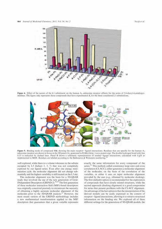
464 Journal of Medicinal Chemistry, 2011, Vol. 54, No. 2 Yaziji et al.
well explored, while there is a volume tolerance in the subsiteoccupied by L3 (helices 2, 3, 7) that was not completelyexplored by our ligand series. Even after one energy mini-mization cycle, the molecular alignment did not change sub-stantially and the highest variability is still located on the L3 site.This molecular alignment was the basis for a 3D-QSAR
study that involved the use of the new generation of Grid-INdependentDescriptors (GRIND-2).50,51 The first generationof these molecular interaction field (MIF)-based descriptorswas originally conceived precisely to circumvent the necessityof obtaining a highly accurate molecular alignment of themolecules prior to the 3D-QSAR analysis.61 However, themost recent version of the GRIND methodology includesa new mathematical transformation applied to the MIFdescriptors that guarantees that a given variable represents
exactly the same information for every compound of theseries.51 This method, called consistency large auto and crosscorrelation (CLACC), either generates a molecular alignmentof the molecules, on the basis of the correlation of thevariables, or either it uses an input molecular alignmentprovided by the user (e.g., obtained by molecular docking).The first (default) option is recommended for the explorationof compounds that have closely related structures, while thesecond approach (docking alignment) is a good compromisefor series that present problems with the CLACC alignment.Anadvantageof the last option is that the interpretationof thederived models can be easily expressed in the context ofreceptor-ligand interactions, allowing it to retrieve structuralinformation on the binding site. We explored all of thesedifferent settings for the generation of 3D-QSARmodels, the
Figure 4. Effect of the nature of the L1 substituent on the human A3 adenosine receptor affinity for the series of 2,6-diaryl-4-amidopyr-imidines. The figure only represents those compounds that have experimental Ki for the three considered L1 substitutions.
Figure 5. Binding mode of compound 11d, showing the main receptor-ligand interactions. Residues that are specific for the human A3
adenosine receptor are shown in boxes in the 3D panel (A), generated in PyMol (http://www.pymol.org). The double hydrogen bond with Asn6.55 is indicated by dashed lines. Panel B shows a schematic representation of residue-ligand interactions, calculated with LigX asimplemented in MOE. Residues are labeled according to the Ballesteros & Weinstein numbering.56

Article Journal of Medicinal Chemistry, 2011, Vol. 54, No. 2 465
results being summarized in Table S1 of the SupportingInformation. The model generated on the basis of a dockingmolecular alignment and the CLACC method for encodingthe descriptors (Model C in Table S1, Supporting Informa-tion) was selected for further interpretation. This 3D-QSARmodel has two latent variables (LV) and presents a satisfac-tory statistical quality,with a fitting parameter of r2=0.86 anda predictive ability of q2=0.67, as obtained by the LOOcross-validation test. The standard error for the correlation and theprediction was 0.31 and 0.48 pKi log units, respectively.
The interpretation of the model highlights the key structuralfeatures for high A3AR affinity. Themost important variables,that is, those with the highest PLS positive coefficients, wereused for themodel interpretation and are depicted inFigure 7.These variables represent, in an ideal case, structural featuresthat are present in the active compounds but absent in theinactive compounds. In this respect, the following features areimportant for the model interpretation:
(i) The hydrophobic interactions at the extracellular tipof the binding site, mainly with residues Ile 6.58 andLeu 7.35, are identified with the O-TIP (optimumdistance at 5.8 A) and the DRY-TIP (6.6 A) correlo-grams. These two hydrophobic residues are a probablesource of A3 specificity: position 6.58 is occupied by athreonine in the other subtypes, while position 7.35 hasalready been related to interspecies selectivity in theA1AR.63 The model suggests that an optimal shape
complementarity is achieved bymolecules with smallerL1 substituents (e.g., acetamides) or, alternatively,molecules bearing larger L1 substituents but smallerL2/L3 substituents (e.g., 13n). The combination of theaforementioned descriptors provides informationabout the interdependence of the size of L1 and L2/L3substituents. This descriptor also accounts for thelack of affinity observed in the aminopyrimidine series(seeTable 1), since these scaffolds donot bear any alkylresidues on the exocyclic nitrogen.
(ii) The optimal pharmacophoric distance between theH-bond acceptor probe, corresponding to the carbo-nyl of the amide, and the shape of the L2 substituent islocated at 16.6 A in the N1-TIP cross-correlogram.Whereas the role of the carbonyl group could behypothesized as a water-mediated interaction withGlu 7.29 in the third extracellular loop (EL3), thisdescriptor mainly identifies the importance of residueIle 5.47, interacting with the L2 substituent. Impor-tantly, Ile 5.47 is occupied by the less bulky valine inthe other AR subtypes, a fact that could be taken intoaccount to improve A3 selectivity.
(iii) Finally, the O-N1 and N1-N1 autocorrelogramsaccount for the differences between the 2-amido- or4-amidopyrimidine series, since these molecular de-scriptors identify the distances between the exocy-clic amide and the N1 in the ring. In the series of
Figure 6. Molecular alignment of the 62 molecules that had the postulated docking pose. This alignment was used as an input for the 3D-QSAR study. (A) side view of the receptor, and (B) view from the extracellular side. The following transmembrane helices (TM) are shown:TM2 (cyan), TM3 (green), TM4 (yellow), TM6 (orange), and TM7 (red). The Connolly surface of the receptor is depicted in gray.
Figure 7. Important variables in the 3D-QSARmodel C (represented for compound 11d). Green dots denote TIP fields, red dots O fields, bluedots N1 fields, and yellow dots DRY fields. (Right) The same representation showing the binding site. The correspondence of the TIP fieldswith the limiting pockets of the receptor for the L1 (Glu 7.29, Ile 7.35) and L3 (Ile 5.47, Trp 6.48), the superposition of the O-NI short distancevariable with Asn 6.55, and the DRY field with Phe 5.29 can be appreciated.

466 Journal of Medicinal Chemistry, 2011, Vol. 54, No. 2 Yaziji et al.
4-amidopyrimidines, optimum distances of 9 and10.6 A for O-N1 and N1-N1 autocorrelograms,respectively, are observed. Conversely, in the series of2-amidopyrimidines, a descriptor in the N1-N1 cor-relogram, which is negatively related with affinity inthe model, identifies the particular location of the N1of this scaffold closer to the carbonyl of the amide(distance 4.6 A).
This last point is intriguing, since the docking model doesnot identify any polar interaction for either the oxygen of theamide group or the nitrogen at position N1 of the pyrimidine(i.e., the nitrogen that varies in position between 2- and4-amidopyrimidines). However, if we compare the bindingmode of the molecular series here reported on the A3ARwiththe experimental binding mode of 4-(2-[7-amino-2-(2-furyl)-[1,2,4]triazolo[2,3-a][1,3,5]triazin-5-ylamino]ethyl)phenol onthe A2AAR, it appears that the N1 in the 4-amidopyrimidinesseries overlies with a nitrogen in the heterocycle of thestandard (N19 according to PDB nomenclature in entry3EML).47 Recently, the group of Jacobson63 noted the im-portance of a polar interaction of this N19 with crystal-lographic water molecules. In order to check if similarinteractions could be achieved in our 4-amidopyrimidines/hA3AR complexes, we performed a computational explora-tion of structural water molecules in the binding site of thehA3ARmodel, as detailed in the methods section. The results(Figure S2, Supporting Information) show an energeticallyfavorable area for a water molecule that overlaps with theN1-N1 descriptor, close to the position of the varying nitro-gen in the 4-amidopytimidines (blue dots at the bottom ofFigure 7). A water-mediated interaction between N1 and Thr7.42, which somehow resembles the H-bond network ZM24-1386(N19)-HOH559-HOH550-His7.43(Nε) in the A2AAR, isthus proposed as an specific polar contact for the 4-amidopyr-imidine series, lacking in the 2-amidopyrimidines. The experi-mental validation of such a different interaction behavior iscurrently being investigated in our laboratory by synthesizingadditional series of unexplored heterocyclic scaffolds.As statedabove, anadvantageof the 3D-QSARmethodology
employed in this study is to place the relevant descriptors inthe context of the binding site. This procedure can not onlydeal with the structural requirements of the hA3AR for highaffinity but also enables a comparison of the hot-spots withthe relative positions in other ARs in order to explore thereasons for selectivity. In an effort to identify these hot-spots,we built a so-called “pseudosequence” based on the dockingresults of this study. This pseudosequence is defined by all ofthe residues of the receptor located at a amaximumdistanceof4.5 A from the most exposed atom of the group of ligandsdocked in the hA3AR (as superimposed in Figure 6). An
alignment of this pseudosequence for the human members ofthe ARs family is shown in Figure 8, in which the variablepositions are clearly identified.In this respect, it is remarkable that positions 5.42 and 6.52,
at the bottom edge of the binding site, are both occupied by aserine in the hA3AR (see Figure 5); in the other three humanARs these positions are occupied by Asn 5.42 and His 6.52,respectively.The less voluminous side chainof a serine in thesepositions would allow the accommodation of bulkier L2substituents in this subsite at the hA3AR, thus offering arationale for the observed receptor selectivity. There are alsoremarkable differences within the ARs family regardingresidues at the top of the binding site: Ile 6.58, interactingwith L1, is replaced by a smaller valine in the other ARs.hA3AR presents a valine at position 5.30 (in the tip of EL2),which replaces a Glu that is conserved in the other three hARsubtypes.According to theA2AARcrystallographic structure,Glu 5.30 hydrogen bonds with the side chain of a His 7.29in EL3, thus closing the top of the binding site while accept-ing an additional hydrogen bond from the amino group of4-(2-[7-amino-2-(2-furyl)[1,2,4]triazolo[2,3-a][1,3,5]triazin-5-ylamino]ethyl)phenol.47 The amino derivatives would bene-fit from this interaction, thus explaining the low selectivityprofile displayed by this group of compounds (Table 1).Interestingly, a recent study of a new series of 2-phenylpyraz-olo[4,3-d]pyrimidin-7-ones already indicated this difference inthe flap regions as being responsible for the A3AR selec-tivitiy.49 Finally, Leu 7.35 provides a specific hydrophobicsubsite for the L1 substituent compared to the more volumi-nous methionine present at this position in the A2AAR andA2BARor thepolarThr7.35 inA1AR. Importantly, sitedirectedmutagenesis studies have identified this position as beingresponsible for the interspecies differences in ligand affinitiesin the A1ARs.62
To further challenge the computational model in terms ofrobustness andpredictive capability, the toleranceofA3ARtosteric factors imposed by the alkyl residue of the amidefunction (L1)was explored.Accordingly, six compoundsweredesigned which combined three new bulky residues [e.g.,CH(Me)2, CH(Et)2, and Cy] on the exocyclic amide groupwith the scaffold of the amines 4d and 4k. The compoundswere docked on the A3AR and queried to the QSAR model,which predicted a good affinity profile for the A3AR (seeTable 3). Figure S2 in the Supporting Information shows howthe most bulky compounds (14c and 14f) optimally accom-modate the cyclohexyl substituent in the hA3AR pocket. Onthe other hand, a superposition with the crystallographicstructure of the hA2AAR shows that steric clashes with theL1 substituent of this receptormight occur, as anticipated by thepseudosequence analysis shown in Figure 8. The compounds
Figure 8. Multiple pseudosequence alignment of human ARs, taking hA3AR residues within 4.5 A of all docked compounds into account.Residue positions are denoted by the Ballesteros &Weinstein numbering,56 and shaded in gray according to their distance toward the dockedcompounds.

Article Journal of Medicinal Chemistry, 2011, Vol. 54, No. 2 467
were then prepared and tested at the four human adenosinereceptor subtypes (Table 4). As predicted by the computa-tional exploration described above, the newer derivativesexhibit potent and selective activity profiles, which unequi-vocally confirms the tolerance of hA3AR to the size of the L1substituent. Excellent agreement is found between predictedand experimental affinity values for the six compoundsdesigned and tested in this part of the study [with an impressivelow standard error of the prediction (SDEP=0.37 log pKi
units)], which further confirms the predictive power of theintegrated computationalmodel reported in this work, that is,combining a molecular alignment from automated dockingwith the prediction of activities on the basis of the 3D-QSARmodel (see Supporting Information, Figure S3C) wasobserved. It isworth noting that on the solely basis of the avai-lable literature39 data or the herein established SAR (Tables 1and 2) the synthesis of compounds 14 would not have beenadvisible. Thus, the modeling exploration enabled us to anti-cipate attractive activity/selectivity profiles for compoundsincorporating hindered fragments at the amide chain (asconsequence of the higher tolerability for the L1 subsite ofthe hA3AR).
Conclusions
A new series of structurally simple and highly potentligands that exhibit remarkable selectivity profiles towardthe A3AR has been identified. A previous series of potentand selective A1AR antagonists was selected, and the subse-quent stepwise structural diversification of these model sub-strates was carried out in order to radically modify theactivity/selectivity profiles while simultaneously providingvaluable structural information on the requirements for itsbinding at the hA3 receptor subtype. Excellent affinity towardthe hA3AR (Kie6 nM) and optimal selectivity profiles (e10%displacement of 0.1μMconcentrations at the otherARs)wereobserved for compounds ISVY133, ISVY130, ISVY074, andISVY167, which incorporate 4-tolyl, 4-methoxyphenyl, and2,4-dimethoxyphenyl moieties at the 2,6-positions of theheterocyclic backbone. The antagonistic behavior of fiverepresentative derivatives of these series was unequivocallyvalidated through functional cAMP experiments. The main
SARs identified were substantiated by an exhaustive molec-ular modeling study that combined a receptor-driven dockingmodel, which was constructed on the basis of the recentlypublished crystal structure of the hA2AAR, and a ligand-based 3D-QSAR model, highlighting the key structural fea-tures required for the optimal interaction with the hA3
receptor subtype in these compounds. The robustness andpredictive capabilities of the model were validated by design-ing novel series of compounds that explore new alkyl residuesat the L1 subsite, which show high affinity and selectivityprofiles for the hA3AR. We must note that these compoundswould not have been synthesized solely on the basis of theavailable SAR data on the literature39 or the qualitative SARestablished on this work (Tables 1-3). On the contrary, theinterest of these compounds was envisioned by the computa-tional modeling exploration, suggesting that the hA3ARshows higher tolerability for the L1 subsite. Further experi-ments are currently in progress in our laboratories to preparenew libaries incorporating nonidentical aryl groups at posi-tions 2,6 and 4,6 obtained by adaptation of the hereindocumented synthetic strategy according to recently pub-lished methodologies.64-66 The biological profile of thesenew derivatives will be published in due course.
Experimental Section
Chemistry. Commercially available starting materials, re-agents, and solvents were purchased (Sigma-Aldrich) and usedwithout further purification. When necessary, solvents weredried by standard techniques and distilled. After being extractedfrom aqueous phases, the organic solvents were dried overanhydrous sodium sulfate. The reactions were monitored bythin-layer chromatography (TLC) with 2.5 mmMerck silica gelGF 254 strips, and the purified compounds each showed a singlespot; unless stated otherwise, UV light and/or iodine vapor wereused for detection of compounds. The Suzuki cross-couplingreactions were performed in coated Kimble vials on a PLS (6� 4)Organic Synthesizer with orbital stirring. Filtration and washingprotocols for supported reagents were performed in a 12-channelvacuummanifold from Aldrich. Purity and identity of all testedcompoundswere established by a combination ofHPLC, elementalanalysis,mass spectrometry, andNMRspectraasdescribedbelow.Purification of isolated products was carried out by columnchromatography (Kieselgel 0.040-0.063 mm, E. Merck) or
Table 4. Structure and Affinity Binding Data for 2,6-Diaryl-4-amidopyrimidines 14 at the Human Adenosine Receptors
Ki (nM) or % at 0.1 μM pKi @ hA3
comp Ar R hA1a hA2A
b hA2Bc hA3
d exp pred
14a 4-OMe-Ph CH(Me)2 5% 1% 3% 12.2 ( 0.9 7.91 8.16
14b CH(Et)2 3% 6% 8% 58.1 ( 4.7 7.24 7.82
14c Cy 5% 1% 1% 32.1 ( 2.4 7.49 7.15
14d 2,4-OMe-Ph CH(Me)2 18% 1% 1% 15.9 ( 1.3 7.8 7.58
14e CH(Et)2 1% 2% 2% 52.4 ( 6.2 7.28 7.57
14f Cy 1% 1% 4% 56.3 ( 6.1 6.59 6.85aDisplacement of specific [3H]DPCPX binding in human CHO cells expressed as Ki ( SEM in nM (n=3) or percentage displacement of specific
binding at a concentration of 0.1 μM (n=2). bDisplacement of specific [3H]4-(2-[7-amino-2-(2-furyl)[1,2,4]triazolo[2,3-a][1,3,5]triazin-5-ylamino]-ethyl)phenol binding in human HeLa cells expressed as Ki ( SEM in nM (n=3) or percentage displacement of specific binding at a concentration of0.1 μM(n=2). cDisplacement of specific [3H]DPCPXbinding in humanHEK-293 cells expressed asKi( SEM in nM(n=3)or percentage displacementof specific binding at a concentration of 0.1 μM(n=2). dDisplacement of specific [3H]NECAbinding in humanHeLa cells expressed asKi( SEM in nM(n=3) or percentage displacement of specific binding at a concentration of 0.1 μM (n=2).

468 Journal of Medicinal Chemistry, 2011, Vol. 54, No. 2 Yaziji et al.
medium pressure liquid chromatography (MPLC) on a Combi-Flash Companion (Teledyne ISCO) with RediSep prepackednormal-phase silica gel (35-60 μm) columns followed by re-crystallization. Melting points were determined on a Gallenkampmelting point apparatus and are uncorrected. TheNMRspectrawere recorded on Bruker AM300 and XM500 spectrometers.Chemical shifts are given as δ values against tetramethylsilaneas internal standard and J values are given in Hz. Massspectra were obtained on a Varian MAT-711 instrument.High resolution mass spectra were obtained on an AutospecMicromass spectrometer. Analytical HPLC was performedon a Agilent 1100 system using an Agilent Zorbax SB-Phenyl,2.1 mm� 150 mm, 5 μm column with gradient elution usingthe mobile phases (A) H2O containing 0.1% CF3COOH and(B)MeCNand a flow rate of 1mL/min. Elemental analyses wereperformed on a Perkin-Elmer 240B apparatus at the Micro-analysis Service of the University of Santiago de Compostela,the elemental composition of the new compounds agreed towithin (0.4% of the calculated value. The purity of all testedcompounds was determined to be >95%. A detailed descriptionof synthetic methodologies as well as analytical and spectros-copic data for all described compounds is included in the Sup-porting Information.
Pharmacology.Radioligand binding competition assays wereperformed in vitro as previously described53 using A1, A2A, A2B,and A3 human adenosine receptors expressed in transfectedCHO (A1AR), HeLa (A2AAR and A3AR), and HEK-293(A2BAR) cells. The experimental conditions used are summar-ized in Table 5. In each instance, aliquots of membranes (15 μgforA1, 10 μg forA2AAR, 18 μg forA2BAR, and 90 μg forA3AR)in buffer A (see Table 5) were incubated for the specified periodat 25 �C with the radioligand (2-35 nM) and six differentconcentrations (ranging from 0.1 nM to 1 μM) of the testmolecule in a final volume of 200 μL. The binding reactionwas stopped by rapid filtration in amultiscreenmanifold system(Milipore Ib�erica, Madrid, Spain). Unbound radioligand wasremoved by washing four times with 250 μL of ice-cold buffer Bfor A1 and A2A receptors, and six times with 250 μL of ice-coldbuffer B for A2BAR and A3AR (see Table 5). Nonspecificbinding was determined using a 50 or 400 μM NECA solutionfor A2AAR and A2BAR and 10 or 100 μM R-PIA solution forA1AR and A3AR, respectively. Radioactivity retained on filterswas determined by liquid scintillation counting using Universol(ICN Biochemicals, Inc.). The binding affinities were deter-mined using [3H]-DPCPX (130 Ci/mmol; GE-Healthcare,Barcelona, Spain) as the radioligand for A1AR and A2BAR,[3H]4-(2-[7-amino-2-(2-furyl)[1,2,4]triazolo[2,3-a][1,3,5]triazin-5-ylamino]ethyl)phenol (21 Ci/mmol; Tocris, Madrid, Spain)for A2AAR and [3H]-NECA (15.3 Ci/mmol; NEN-Perkin-ElmerLife Sciences, Madrid, Spain) for A3AR.
The inhibition constant (Ki) of each compound was calcu-lated by the expression:Ki=IC50 /(1þ (C/KD)); where IC50 is theconcentration of compound that displaces the binding of radio-ligand by 50%, C is the free concentration of radioligand, andKD is the apparent dissociation constant of each radioligand.
The percentage of displacement of specific binding was calcu-lated by the expression:%of displacement=((BT- dpm)*100)/(BT-NSB); where BT is the total binding of the radioligand inthe assay, NSB is the nonspecific binding of the radioligand inthe assay, and dpm are the radioactive measurements obtainedby competing the radioligand binding with a given concentra-tion of the test compound. Unless otherwise specified, resultsshown in the text and tables are expressed as means ( SEM.Significant differences between two means (p<0.05 or p<0.01)weredeterminedbyone-wayanalysis of variance (ANOVA) and/orby Student0s t test for nonpaired data.
Molecular Modeling
Model Building. A homology model of the hA3AR wasbuilt using the recently crystallized hA2AAR as a template.The modeling protocol is adapted from our participation inthe GPCR Dock 2008 competition.44 Briefly, a sequencealignment between the two receptors with Clustal (PAM250substitution matrix, with open and elongation gap penaltiesof 10 and 0.05)67 was provided to Modeller v9.4.68 Fifteeninitial models were obtained using standard parameters. In afirst stage, the best five models were selected on the basis ofProcheck69 geometrical quality and DOPE scoring, andthese were subjected to geometrical improvement by theMolprobity server.70 In a second stage, the best model fromthe previous stepwas subjected to loop optimizationwith theLoopModel routine in Modeler,71 again generating 15 re-fined models. We selected the best model on the basis of acompromise between Procheck stereochemical quality andthe DOPE energetical ranking. The geometry of the loops inthe selected model was refined by partial energy minimiza-tion (i.e., nonloop residues were frozen) using the Polak-Ribiere algorithm (convergence criteria 0.05 kcal/mol 3 A
2)and the OPLS-AA force-field as implemented inMacromodel.72
The general numbering scheme for GPCRs proposed byBallesteros andWeinstein56 was adopted through this work.In essence, every residue is numbered as X.YY, where Xcorresponds to the transmembrane helix (X=[1,7]) and YYis a correlative number in the protein sequence, but taking asa reference position (YY=50) the most conserved residue inthe given helix.
Protein-Ligand Docking. Automated docking explora-tion was performed with GOLD version 3.2.73 Each ligandwas docked 20 times with default (high accuracy) geneticalgorithm (GA) search parameters, using the scoring func-tion Chemscore as implemented in GOLD55 and allowingfull flexibility for the ligand, including flipping of amidebonds. The search sphere was centered on the side chain(CD1) of Ile 7.39, and expanded with a radius of 15 A, thusensuring a generous enough search space comprising the
Table 5. Conditions Used for Radioligand Binding Assays Using A1, A2A, A2B, and A3 Human Adenosine Receptors
A1 A2A 2A2B A3
Buffer A 20 mM Hepes, 100 mMNaCl,
10 mM MgCl2, 2 units/mL
adenosine deaminase
(pH=7.4)
50 mM Tris-HCl,
1 mM EDTA, 10 mM MgCl2,
2 units/mL adenosine
deaminase (pH=7.4)
50 mM Tris-HCl, 1 mM EDTA,
10 mM MgCl2, 0.1 mM
benzamidine, 2 units/mL
adenosine deaminase (pH=6.5)
50 mM Tris-HCl, 1 mM
EDTA, 5 mMMgCl2,
2 units/mL adenosine
deaminase (pH=7.4)
Buffer B 20 mM Hepes, 100 mMNaCl,
10 mM MgCl2, (pH=7.4)
50 mM Tris-HCl, 1 mM EDTA,
10 mM MgCl2 (pH=7.4)
50 mM Tris-HCl (pH= 6.5) 50 mM Tris-HCl
(pH=7.4)
plate GF/C GF/C GF/B GF/B
radioligand [3H]DPCPX nM [3H]ZM2413853 nM [3H]DPCPX35 nM [3H]NECA 30 nM
nonspecific
binding
10 μM (R)-PIA 50 μM NECA 400 μM NECA 100 μM (R)-PIA
incubation 25 �C/60 min 25 �C/30 min 25 �C/30 min 25 �C/180 min

Article Journal of Medicinal Chemistry, 2011, Vol. 54, No. 2 469
antagonist binding site experimentally determined for ade-nosine receptors.46 The criterion for the selection of dockingposes was based on Chemscore ranking and the populationof the solutions (according to a clustering criteria of 1 A).
Geometrical Optimization. Each docking pose was refinedby partial energy minimization of the binding site withMOE.74 The site was selected as any atom within a distanceof 4.5 A around the ligand and OPLS-AA parameters wereused in combination with GBSA model for continuumsolvent representation. The convergence criterion for thesteepest descendent algorithm was set to 0.01 rmsd.
3D-QSAR. The conformations of the compounds ob-tained in the molecular docking step were used to generatea 3D-QSAR model with the software Pentacle v1.1.75 Thissoftware allows the computation of the second generation ofGRid Independent Descriptors (GRIND-2). This family ofmolecular descriptors, which are widely used in QSARstudies, are generated in a three-step fashion: (i) computationof molecular interaction fields (MIF) with different Gridprobes,76 (ii) selection of the most relevant MIF nodes, and(iii) encoding of the descriptors as alignment-independentvectors of node pairs, obtaining the so-called correlograms.61
The advantages of GRIND-2 include the use of AMANDAas a new discretization algorithm for the identification of“hot spots” (most relevant MIF nodes)50 and a new methodfor encoding descriptors into alignment-free vectors calledCLACC.51 This method detects consistency in the computedvariables, ensuring that a given vector on the correlogramcorresponds to the description of same pharmacophoricproperty within the series. In this work, the MIF werecomputed using default values (i.e., GRID probes: DRY,O, N1, TIP; 0.5 A grid step; dynamic parametrization),discretization was carried out with default AMANDA para-meters and theCLACCencodingof the variableswas generatedon the basis of the docking alignment (“use CLACC foralignment”= false) with strict options, meaning that anyvariable that is not consistent in the series is removed(“Remove non-consistent couples”=true). Two rounds offractional factorial design (FFD) were applied for the selec-tion of the most relevant variables in the model. The modelgenerated was “saved for predictions” through the corre-sponding menu option in Pentacle. Thereafter, the designedmolecules were docked in theA3 receptor as explained above,and further imported in the Pentacle software, but using thegenerated model as a template for the prediction of activitieson the A3 receptor.
Water Analysis. The prediction of energetically favorableregions for structural water molecules in the binding site ofthe hA3AR model was carried out with the program Grid,76
using the following parameters: OH2 probe, all programdirectives on their default values except for LEAU=2 andNPLA=2. Further refinement of the position of predictedwater molecules was done with the module FilMap, asimplemented in the Grid software.
Acknowledgment. This work has been developed in theframe of the Red Gallega de Investigaci�on y Desarrollo deMedicamentos (REGID) and was financially supported bythe Fondo Europeo de Desarrollo Social (FEDER) and theGalicianGovernment (Projects 09CSA016234PRandPS09/63).E.S. is the recipient of a Consolidation Group ResearchGrant from the Conselleria de Educaci�on (Xunta de Galicia).E.S., H.G.d.T., and A.C. are researchers of the Isidro Parga
Pondal program (Xunta de Galicia, Spain). J.B. is researcherof the Isabel Barreto program (Xunta deGalicia, Spain).D.R.is recipient of aPFIS grant from the Instituto de SaludCarlos III(Ministerio de Ciencia e Innovaci�on, Spain). We are gratefulto Dr. Manuel Pastor for assistance in the use of the Pentaclesoftware and helpful discussions.
Supporting Information Available: Experimental details forthe synthesis of compounds described, the spectroscopic, spec-trometric, and elemental analysis data of all compounds pre-pared as well as additional molecular modeling details. Thismaterial is available free of charge via the Internet at http://pubs.acs.org.
References
(1) Fredholm, B. B.; Arslan, G.; Halldner, L.; Kull, B.; Shulte, G.;Wasserman,W. Structure and function of adenosine receptors andtheir genes. Naunyn-Schmiedeberg’s Arch. Pharmacol. 2000, 362,364–374.
(2) Murphree, L. J.; Linden, J. Adenosine receptors. EncyclopaediaBiol. Chem. 2004, 1, 34–39.
(3) Jacobson,K.A.; Knutsen, L. J. S. P1 and P2 purine and pyrimidinereceptor ligands. In Purinergic and Pyrimidine Signalling(Handbook of Experimental Pharmacology); Abbrachio, M., Williams,M., Eds.; Springer: Berlin, 2001; Vol. 151/1, pp 129-175.
(4) Fredholm, B. B.; Ijzerman, A. P.; Jacobson, K. A.; Koltz, K. N.;Linden, J. International union of pharmacology XXV. Nomencla-ture and classification of adenosine receptors. Pharmacol. Rev.2001, 53, 527–552.
(5) Baraldi, P. G.; Aghadeh,M.; Tabrizi,M. A.; Gessi, S.; Borea, P. A.Adenosine receptor antagonists: translating medicinal chemistryand pharmacology into clinical utility. Chem. Rev. 2008, 108, 238–263.
(6) Jacobson, K. A.; Gao, Z. Adenosine receptors as therapeutictargets. Nat. Rev. Drug Discovery 2006, 5, 247–264.
(7) Moro, S.; Gao, Z. G.; Jacobson,K.A.; Spalluto,G. Progress in thepursuit of therapeutic adenosine receptor antagonists. Med. Res.Rev. 2006, 26, 131–159.
(8) Zhou, G. L.; Olah, M. E.; Johnson, R. A.; Stiles, G. L.; Clivelli, O.Molecular cloning and characterization of an adenosine receptor:the A3 adenosine receptor. Proc. Natl. Acad. Sci. U. S. A. 1992, 89,7432–7436.
(9) Van Schaick, E. A.; Jacobson, K. A.; Kim, H. O.; IJzerman, A. P.;Danhof,M.Hemodynamic effects and histamine release elicited bythe selective adenosine A3 receptor agonist Cl-IB-MECA in con-scious rats. Eur. J. Pharmacol. 1996, 308, 311–314.
(10) Hannon, J. P.; Pfannkuche,H. J.; Fozard, J. R.A role formast cellsin adenosine A3 receptor-mediated hypotension in the rat. Br. J.Pharmacol. 1995, 115, 945–952.
(11) Fishman, P.; Bar-Yehuda, S. Pharmacology and therapeutic ap-plications of A3 receptor subtypes.Curr. Top. Med. Chem. 2003, 3,463–469.
(12) Muller, C. E. A3 Adenosine receptor antagonists. Mini Rev. MedChem. 2001, 1, 417–427.
(13) Baraldi, P. G.; Tabrizi, M. A.; Fruttarolo, F.; Borevo, A.; Avitabile,B.; Preti, D.; Romagnoli, R.; Merighi, S.; Gessi, S.; Varani, K.;Borea, P. A. Recent developments in the field of A3 adenosinereceptor antagonists. Drug Dev. Res. 2003, 58, 315–329.
(14) Baraldi, P.G.; Fruttarolo, F.; Tabrizi,M.A.;Romagnoli,R.; Preti,D.; Carrion, M.; Iaconinoto, A.; Borea, P. A. Recent improve-ments in the field of A3 adenosine receptor ligands. Exp. Opin.Ther. Pat. 2005, 15, 1507–1519.
(15) Muller, C. E. Medicinal chemistry of adenosine A3 receptorligands. Curr. Top. Med. Chem. 2003, 3, 445–462.
(16) Moro, S.; Deflorian, F. Novel strategies for the design of newpotent A3 antagonists: an update.Curr.Med. Chem. 2006, 13, 639–645.
(17) Baraldi, P. G. A3 ligands, history and perspectives.Med. Res. Rev.2000, 20, 103–128.
(18) VanMuijlwijk-Koezen, J. E.; Timmerman,H.; Ijzerman, A. P. Theadenosine A3 receptor and its ligands. Prog. Med. Chem. 2001, 38,61–113.
(19) Stone, T. W. Purines and neuroprotection. Adv. Exp. Med. Biol.2002, 513, 249–280.
(20) Beaven, M. A.; Ramkumar, V.; Hydar, A. Adenosine A3 receptorsin mast cells. Trends Pharmacol. Sci. 1994, 15, 13–14.

470 Journal of Medicinal Chemistry, 2011, Vol. 54, No. 2 Yaziji et al.
(21) Ramkumar, V.; Stiles, G. L.; Beaven, M. A.; Ali., H. The A3
adenosine receptor is the unique adenosine receptor which facil-itates release of allergicmediators inmast cells. J. Biol. Chem. 1993,268, 16887–16890.
(22) Akkari, R.; Burbiel, J. C.; Hockemeyer, J.; Mueller, C. E. Recentprogress in the development of adenosine receptor ligands asantiinflammatory drugs. Curr. Top. Med. Chem. 2006, 6, 1375–1399.
(23) Von Lubitz, D. K. J. E.; Carter,M. F.; Deutsch, S. I.; Lin, R. C. S.;Mastropaola, J.; Meshulam, Y.; Jacobson, K. A. The effects ofadesnosine receptor stimulation on seizures in mice. Eur. J. Phar-macol. 1995, 275, 23–29.
(24) Nieber, K.; Lewerenz, A.; Hentschel, S.; Vissiennon, Z. AdenosineA1 and A3 receptors: Neuroprotective targets with perspectives.Bioforum 2002, 25, 237–240.
(25) Forsythe, P.; Ennis, M. Adenosine, mast cells and asthma. Inflam-mation Res. 1999, 48, 301–307.
(26) Brown, R. A.; Spina, D.; Page, C. P. Adenosine receptors andasthma. Br. J. Pharmacol. 2008, 153, S446–S456.
(27) Brambilla, R.; Cattabeni, F.; Ceruti, S.; Barbieri, D. Naunyn-Schmiedeberg’s Arch. Pharmacol. 2000, 361, 225–234.
(28) Jacobson, K. A.; Moro, S.; Kim, Y. C.; Li, A. A3 Adenosinereceptors: protective vs damaging effects identified using novelagonists and antagonists. Drug Dev. Res. 1998, 45, 113–124.
(29) Abbracchio,M. P.; Ceruti, S.; Brambilla, R.; Barbiri, D.; Camurri,A.; Franceschi, C.; Giammaroli, A. M.; Jacobson, K. A.; Cattabeni,F.;Malorni,W.AdenosineA3 receptors and viability of astrocytes.Drug Dev. Res. 1998, 45, 379–386.
(30) Baraldi, P. G.; Tabrizi, M. A.; Romagnoli, R.; Fruttarolo, F.;Merighi, S.; Varani, K.; Gessi, S.; Borea, P. A. Pyrazolo[4,3-e]1,2,4-triazolo[1,5-c]pyrimidine ligands, new tools to characterize A3
adenosine receptors in human tumor cell lines. Curr. Med. Chem.2005, 12, 1319–1329.
(31) Merighi, S.; Mirandola, P.; Varani, K.; Gessi, S.; Capitani, S.;Leung, E.; Baraldi, P. G.; Tabrizi, M. A.; Borea, P. A. Pyrazolo-triazolopyrimidine derivatives sensitize melanoma cells to thechemotherapeutic drugs: taxol and vindesine.Biochem. Pharmacol.2003, 66, 739–748.
(32) Cosimelli, B.; Greco, G.; Ehlardo, M.; Novellino, E.; Da Settimo,F.; Taliani, S.; LaMotta, C.; Bellandi, M.; Tuccinardi, T.; Martinelli,A.; Ciampi, O. Derivatives of 4-amino-6-hydroxy-2-mercaptopyr-imidine as novel, potent and selective A3 adenosine antagonists.J. Med. Chem. 2008, 51, 1764–1770.
(33) Van Veldhoven, J. P. D.; Chang, L. C. W.; von Frijtag, J. K.;Mulder-Krieger, T.; Struensee-Link, R.; Beukers, M. W.; Brussee,J.; Ijzerman, A. P. A new generation of adenosine receptor antago-nists: From di- to trisubstituted aminopyrimidines. Bioorg. Med.Chem. 2008, 16, 2741–2752.
(34) Gillespie, R. J.; Bamford, S. J.; Clay, A.; Gaur, S.; Haymes, T.;Jackson, P. S.; Jordan, A. M.; Klenke, B.; Leonardi, S.; Liu, J.;Mansell, H. L.; Ng, S.; Saadi, M.; Simmonite, H.; Stratton, G. C.;Todd, R. S.; Williamson, D. S.; Yule, I. A. Antagonists of thehuman A2A receptor. Part 6: Further optimization of pyrimidine-4-carboxamides. Bioorg. Med. Chem. 2009, 17, 6590–6605.
(35) Zhang, X.; Tellew, J. E.; Luo, Z.; Moorjani, M.; Lin, E.; Lanier,M. C.; Chen, Y.; Williams, J. P.; Saunders, J.; Lechner, S. M.;Markison, S.; Joswig, T.; Petroski, R.; Piercey, J.; Kargo, W.;Malany, S.; Santos, M.; Gross, R. S.; Wen, J.; Jalali, K.; O0Brien,Z.; Stotz, C. E.; Crespo, M. I.; Diaz, J. L.; Slee, D. H. Leadoptimization of 4-acetylamino-2-(3,5-dimethylpyrazol-1-yl)-6-pyr-idylpyrimidines as A2A adenosine receptor antagonists for thetreatment of parkinson0s disease. J. Med. Chem. 2008, 51, 7099–7110.
(36) Vidal, B.; Nueda, A.; Esteve, C.; Domenech, T.; Benito, S.;Reinoso, R. F.; Pont, M.; Calbet, M.; L�opez, R.; Cadavid, M. I.;Loza,M. I.; C�ardenas,A.;Godessart,N.; Beleta, J.;Warrellow,G.;Ryder, H. Discovery and characterization of 40-2(2-furyl)-N-pyridin-3-yl-4,50-bipyrimidin-20-amine (LAS38096), a potent, selective, andefficaciousA2B adenosine receptor antagonist. J.Med.Chem. 2007,50, 2732–2736.
(37) Kuefner-Muehl, U., Scheuplein, S. W., Pohl, G., Gaida, W., Lehr,E., Mierau, J., Meade, C. J. M. Triazines with an adenosineantagonistic effect. PCT Int. Appl. WO 1999011633.
(38) Morizzo, E.; Capelli, F.; Lenzi, O.; Catarzi, D.; Varano, C.;Filachioni, G.; Vicenzi, F.; Varani, K.; Borea, P. A.; Colotta, V.;Moro, S. Scouting human A3 adenosine receptor antagonist bind-ing mode using a molecular simplification approach: from triazo-loquinoxaline to a pyrimidine skeleton as a key study. J. Med.Chem. 2007, 50, 6596–6606.
(39) Chang, L. C. W.; Spanjersberg, R. F.; von Frijtag, J. K.;Mulder-Krieger, T.; van den Hout, G.; Beukers, M. W.; Brussee,J.; Ijzerman,A. P. 2,4,6-Trisubstitutedpyrimidines as a new class of
selective adenosine A1 receptor antagonists. J. Med. Chem. 2004,47, 6529–6540.
(40) Cheong, S. L.;Dolzhenko,A.;Kachler, S.; Paoletta, S.; Federico, S.;Cacciari, B.; Dolzhenko, A.; Koltz, K. N.; Moro, S.; Spalluto, G.;Pastorin, G. The significance of 2-furyl ring substitution with a2-(para-substituted) aryl group in a new series of pyrazolo-triazolo-pyrimidines as potent and highly selective hA3 adenosine receptorsantagonists: new insights into structure-affinity relationshipand receptor-antagonists recognition. J. Med. Chem. 2010, 53,3361–3375.
(41) Priego, E. M.; P�erez-P�erez, M. J.; von Frijtag, J. K.; de Vries, H.;Ijzerman, A. P.; Camarasa, M. J.; Martın-Santamarıa, S. Selectivehuman adenosine A3 antagonists based on pyrido[2,1-f]purine-2,4-diones: novel features of hA3 antagonist binding. ChemMedChem2008, 3, 111–119.
(42) Baraldi, P. G.; Tabrizi,M.A.; Preti, D.; Bovero, A.; Fruttarolo, F.;Romagnoli, R.; Zaid, N. Z.; Moorman, A. R.; Varani, K.; Borea,P. A. New 2-arylpyrazolo[4,3-c]quinoline derivatives as potent andselective human A3 adenosine receptor antagonists. J. Med. Chem.2005, 48, 5001–5008.
(43) Van Muijlwijk-Kozen, J. E.; Timmerman, H.; Vollinga, R. C.;von Drabbe, J. F.; de Groote, M.; Visser, S.; Ijzerman, A. P.Thiazole and thiadiazole analogues as a novel class of adenosinereceptor antagonists. J. Med. Chem. 2001, 44, 749–762.
(44) Michino, M.; Abola, E.; Brooks, C. L.; Dixon, J. S.; Moult, J.;Stevens, R. C. Community-wide assessment of GPCR structuremodelling and ligand docking: GPCR Dock 2008. Nat. Rev. DrugDiscov. 2009, 8, 455–463.
(45) Bondavalli, F.; Botta,M.; Bruno, O.; Ciacci, A.; Corelli, F.; Fossa,P.; Lucacchini, A.; Manetti, F.; Martini, C.; Menozzi, G.; Mosti,L.; Ranise, A.; Schenone, S.; Tafi, A.; Trincavelli, M. L. Synthesis,molecular modeling studies, and pharmacological activity of selec-tive A1 receptor antagonists. J. Med. Chem. 2002, 45, 4875–4887.
(46) Moro, S.; Deflorian, F.; Spalluto, G.; Pastorin, G.; Cacciari,B.; Kim, S.; Jacobson, K. A. Demystifying the three dimensionalstructure of G protein-coupled receptors (GPCRs) with the aid ofmolecular modelling. Chem. Commun. 2003, 24, 2949–2956.
(47) Jaakola, V.; Griffith, M. T.; Hanson, M. A.; Cherezov, V.;Chien, E. Y. T.; Lane, J. R.; Ijzerman, A. P.; y Stevens, R. C.The 2.6 angstrom crystal structure of a human A2A adenosinereceptor bound to an antagonist. Science 2008, 322, 1211–1217.
(48) Kolb, P.; Rosenbaum, D. M.; Irwin, J. J.; Fung, J. J.; Kobilka,B. K.; Shoichet, B. K. Structure-based discovery of beta 2-adre-nergic receptor ligands. Proc. Natl. Acad. Sci. U. S. A. 2009, 106,6843–6848.
(49) Lenzi, O.; Colotta, V.; Catarzi, D.; Varano, F.; Poli, D.; Filacchioni,G.; Varani,K.; Vincenzi, F.; Borea, P.A.; Paoletta, S.;Morizzo, E.;Moro, S. 2-Phenylpyrazolo[4,3-d]pyrimidin-7-one as a new scaffold toobtain potent and selective humanA3 adenosine receptor antagonists:new insights into the receptor-antagonist recognition. J. Med.Chem. 2009, 52, 7640–7652.
(50) Dur�an, A.; Martınez, G. C.; Pastor, M. Development and valida-tion of AMANDA, a new algorithm for selecting highly relevantregions inMolecular Interaction Fields. J. Chem. Inf. Model. 2008,48, 1813–1823.
(51) Duran, A. Ph.D. Thesis, University Pompeu Fabra, Barcelona,2010. http://www.tesisenxarxa.net/TDX-0422110-094351
(52) Yaziji, V.; Coelho, A.; El Maatougui, A.; Brea, J.; Loza, M. I.;Garcıa-Mera, X.; Sotelo, E. Divergent solution-phase diarylpyr-imidine libraries as selective A3 adenosine receptor antagonists.J. Comb. Chem. 2009, 11, 519–522 and references cited there.
(53) Bosch, M. P.; Campos, F.; Niubo, I.; Rosell, G.; Diaz, J. L.;Brea, J.; Loza,M. I.; Guerrero, A. Synthesis and biological activityof new potential agonists for the human adenosine A2A receptor.J. Med. Chem. 2004, 47, 4041–4053.
(54) Leff, P.; Dougall, I. G. Further concerns over Cheng-Prusoffanalysis. Trends Pharmacol. Sci. 1993, 14, 110–112.
(55) Verdonk, M. L.; Cole, J. C.; Hartshorn, M. J.; Murray, C. W.;Taylor, R. D. Improved protein-ligand docking using GOLD.Proteins 2003, 52, 609–623.
(56) Ballesteros, J. A.; Weinstein, H. Integrated methods for the con-struction of three dimensional models and computational probingof structure-function relations in G-protein coupled receptors.Methods Neurosci. 1995, 25, 366–428.
(57) Jaakola, V. P.; Lane, J. R.; Lin, J. Y.; Katritch, V; Ijzerman, A. P.;Stevens, R. C. Ligand binding and subtype selectivity of thehuman A2A adenosine receptor: identification and characteriza-tion of essential amino acid residues. J. Biol. Chem. 2010, 285,13032–13044.
(58) Gao, Z. G.; Chen, A.; Barak, D.; Kim, S. K.; Muller, C. E.;Jacobson, K. A. Identification by site-directed mutagenesisof residues involved in ligand recognition and activation of the

Article Journal of Medicinal Chemistry, 2011, Vol. 54, No. 2 471
human A3 adenosine receptor. J. Biol. Chem. 2002, 277, 19056–19063.
(59) Kim, J.;Wess, J.; vanRhee, A.M.; Sch€oneberg, T.; Jacobson, K.A.Mutagenesis identifies residues involved in ligand recognition inthe human A2A adenosine receptor. J. Biol. Chem. 1995, 270,13987–13997.
(60) Kim, S. K.; Gao, Z. G.; Rompaey, P. V.; Gross, A. S.; Chen, A.;Calenbergh, S. V.; Jacobson, K. A. Comparison of the bindingdomains of A2A agonists and antagonists. J. Med. Chem. 2003, 46,4847–4859.
(61) Pastor,M.; Cruciani,G.;McLay, I.; Pickett, S.; Clementi, S.GRid-INdependent descriptors (GRIND): a novel class of alignment-independent three-dimensional molecular descriptors. J. Med.Chem. 2000, 43, 3233–3243.
(62) Tucker, A. L.; Robeva, A. S.; Taylor, H. E.; Holeton, D.; Bockner,M.; Lynch, K. R.; Linden, J. A1 adenosine receptors. Two aminoacids are responsible for species differences in ligand recognition.J. Biol. Chem. 1994, 269, 27900–27906.
(63) Ivanov,A. A.; Barak,D.; Jacobson,K.A. Evaluation of homologymodeling of G-protein-coupled receptors in light of the A(2A)adenosine receptor crystallographic structure. J.Med. Chem. 2009,52, 3284–3292.
(64) Benderitter, P.; Xavier deAraujo, J.; Schmitta,M.; Bourguignon, J. J.2-Amino-6-iodo-4-tosyloxypyrimidine: a versatile key intermedi-ate for regioselective functionalization of 2-aminopyrimidines in4- and 6-positions. Tetrahedron 2007, 63, 12465–12470.
(65) Gong, B.; Hong, F.; Kohm, C.; Jenkins, S.; Tulinsky, J.; Bhatt, R.;Vries, P.; Singer, J. W.; Klein, P. Synthesis, SAR, and antitumorproperties of diamino-C,Ndiarylpyrimidine positional isomers:inhibitors of lysophosphatidic acid acyltransferase-β. Bioorg.Med. Chem. Lett. 2004, 14, 2303–2308.
(66) Swarbrick, M. E.; Beswick, P. J.; Gleave, R. J.; Green, R. H.;Bingham, S.; Bountra, C.; Carter,M. C.; Chambers, L. J.; Chessell,
I. P.; Clayton, N.M.; Collins, S. D.; Corfield, J. A.; Hartley, C. D.;Kleanthous, S.; Lambeth, P. F.; Lucas, F. S.;Mathews,N.;Naylor,A.; Page, L. W.; Payne, J. J.; Pegg, N. A.; Price, H. S.; Skidmore, J.;Stevens,A. J.; Stocker, R.; Stratton, S. C.; Stuart,A. J.;Wiseman, J.O.Identification of [4-[4-(methylsulfonyl)phenyl]-6-(trifluoromethyl)-2-pyrimidinyl] amines and ethers as potent and selective cyclo-oxygenase-2 inhibitors.Bioorg.Med.Chem.Lett. 2009, 19, 4504–4508.
(67) Thompson, J. D.; Higgins, D. G.; Gibson, T. J. CLUSTAL W:improving the sensitivity of progressive multiple sequence align-ment through sequence weighting, position-specific gap penaltiesand weight matrix choice. Nucleic Acid Res. 1994, 22, 4673–4680.
(68) Sali, A.; Blundell, T. L. Comparative protein modelling by satis-faction of spatial restraints. J. Mol. Biol. 1993, 234, 779–815.
(69) Laskowski, R. A.; MacArthur, M. W.; Moss, D. S.; y Thornton,J.M. PROCHECK: a program to check the stereochemical qualityof protein structures. J. Appl. Crystallogr. 1993, 26, 283–291.
(70) Davis, I. W.; Leaver-Fay, A.; Chen, V. B.; Block, J. N.; Kapral,G. J.; Wang, X.; Murray, L. W.; Arendall, W. B.; Snoeyink, J.;Richardson, J. S.; Richardson, D. C. MolProbity: all-atom con-tacts and structure validation for proteins and nucleic acids.Nucleic Acids Res. 2007, 35, W375–383.
(71) Fiser, A.; Do, R. K.; Sali, A. Modeling of loops in proteinstructures. Protein Sci. 2000, 9, 1753–1773.
(72) Macromodel, version 9.7; Schr€odinger, LLC: New York, NY, 2009.(73) Jones, G.; Willett, P.; Glen, R. C.; Leach, A. R.; Taylor, R.
Development and validation of a genetic algorithm for flexibledocking. J. Mol. Biol. 1997, 267, 727–748.
(74) MOE (Molecular Operative Environment), version 2009.10; Chemi-cal Computing Group Inc. Montreal, Canada; 2009.
(75) Pentacle, version 1.1; Molecular Discovery, Ltd.: Perugia, Italy, 2009.(76) Goodford, P. J. A computational procedure for determining
energetically favorable binding sites of biological importantmacromolecules. J. Med. Chem. 1985, 28, 849–857.


Rodríguez D., Piñeiro Á. and Gutiérrez-de-Terán H.
Molecular Dynamics Simulations Reveal Insights into Key Structural Elements of Adenosine Receptors
Biochemistry, 2011 50(19):4194-208
Paper IVPaper IV

Published: April 11, 2011
r 2011 American Chemical Society 4194 dx.doi.org/10.1021/bi200100t | Biochemistry 2011, 50, 4194–4208
ARTICLE
pubs.acs.org/biochemistry
Molecular Dynamics Simulations Reveal Insights into Key StructuralElements of Adenosine ReceptorsDavid Rodríguez,† �Angel Pi~neiro,‡ and Hugo Guti�errez-de-Ter�an*,†
†Fundaci�on P�ublica Galega de Medicina Xen�omica, Hospital Clínico Universitario de Santiago (CHUS), planta-2, A Choupana,s/n E-15706 Santiago de Compostela, Spain‡Soft Matter andMolecular Biophysics Group, Department of Applied Physics, University of Santiago de Compostela, Campus Vida s/n,E-15782 Santiago de Compostela, Spain
bS Supporting Information
Gprotein-coupled receptors (GPCRs), the largest family ofmembrane receptors in the human genome, are transmem-
branal proteins in charge of the transduction of signals acrosscellular membranes.1 A delicate conformational equilibriumbetween extreme active and inactive forms of these receptors isbehind the signal transduction, which is modulated towardactivation by the binding of their respective natural agonist.2
From a structural point of view, GPCRs share a conservedtopology: a helical bundle consisting of seven transmembrane(TM) helices spanning the membrane, which are connected bythree extracellular (EL) and three intracellular (IL) loops. Recentadvances in structural biology have revealed that the largestdifferences among GPCRs are located in their extracellularhalves, where ligand binding occurs.3 A general phylogeneticclassification of the GPCR superfamily4 shows well-definedclusters, which can be related to the chemical nature of thereceptor's natural ligand.
Adenosine receptors (ARs) make up one family of GPCRs,which mediate the important extracellular role of the nucleosideadenosine. Four AR subtypes exist, namely, A1, A2A, A2B, and A3,
with different signaling functions and tissue distributions. Thepharmacological interest in ARs as drug targets is widely recog-nized because of the variety and importance of the physiologicalfunctions mediated by them, as well as the associatedpathologies.5 Pharmacological applications of AR chemical mod-ulators include several inflammatory processes, where the role ofthe A2AAR and A3AR is generally accepted,6 or several respira-tory pathological events such as allergic asthma, where thenonselective AR antagonist theophylline is one of the maintreatments, although more selective A2BAR antagonists are inclinical trials.7 The use of adenosine itself in the treatment ofcertain arrhythmias and the anti-infarct effect of other adenosineagonists is mediated by the A1AR,
8 while the A2AAR is involvedin vascular diseases such as hypertension or atherosclerosis.9 ARsare also involved in neural regulation in the central nervous system,as exemplified by the well-known AR antagonist caffeine.10
Received: January 21, 2011Revised: April 11, 2011
ABSTRACT: The crystal structure of the human A2A adenosine receptor, a memberof the G protein-coupled receptor (GPCR) family, is used as a starting point for thestructural characterization of the conformational equilibrium around the inactiveconformation of the humanA2 (A2A and A2B) adenosine receptors (ARs). A homologymodel of the closely related A2BAR is reported, and the two receptors were simulatedin their apo form through all-atom molecular dynamics (MD) simulations. Differentconditions were additionally explored in the A2AAR, including the protonation state ofcrucial histidines or the presence of the cocrystallized ligand. Our simulations revealthe role of several conserved residues in the ARs in the conformational equilibrium ofthe receptors. The “ionic lock” absent in the crystal structure of the inactive A2AAR israpidly formed in the two simulated receptors, and a complex network of interactingresidues is presented that further stabilizes this structural element. Notably, theobserved rotameric transition of Trp6.48 (“toggle switch”), which is thought to initiatethe activation process in GPCRs, is accompanied by a concerted rotation of the conserved residue of the A2ARs, His6.52. This newconformation is further stabilized in the two receptors under study by a novel interaction network involving residues intransmembrane (TM) helices TM5 (Asn5.42) and TM3 (Gln3.37), which resemble the conformational changes recently observedin the agonist-bound structure of β-adrenoreceptors. Finally, the interaction between Glu1.39 and His7.43, a pair of conservedresidues in the family of ARs, is found to be weaker than previously thought, and the role of this interaction in the structure anddynamics of the receptor is thoroughly examined. All these findings suggest that, despite the commonalities with other GPCRs, theconformational equilibrium of ARs is also modulated by specific residues of the family.

4195 dx.doi.org/10.1021/bi200100t |Biochemistry 2011, 50, 4194–4208
Biochemistry ARTICLE
The recent discovery of A2AAR�dopamine D2 receptor hetero-dimers in ganglial neurons led to increased interest in the A2AARas a target for Alzheimer's disease.11
Given the high degree of biomedical relevance of the ARfamilymembers, it is not difficult to understand the breakthroughthat meant the release of the hA2AAR crystal structure at aresolution of 2.6 Å.12 The structure represents one of the inactivestates of the receptor with the subtype selective high-affinityantagonist ZM241385 bound to it. As in the case of the humanβ2-adrenergic receptor (hβ2),13 stabilization of the receptor wasachieved by substitution of the third intracellular loop (IL3) withthe T4 bacteriophage lysozyme (T4L fusion protein strategy).The long C-terminus (Ala317�Ser412) was deleted, while the firsttwo residues of the N-terminus and seven residues (149�155) ofthe second extracelluar loop (EL2) were not determined,14 whichprobably indicated the high mobility of these regions. Despitethese limitations, the structure of the A2AAR—ZM241385complex has revealed important particular features of the ARfamily and offers a new scenario for the study of the ARs,considering the high degree of sequence identity within theirmembers. As recently reviewed, the helical bundle orientationand packing is similar among the GPCRs with known structures.3
The major differences are located in the loop regions and withinthe binding site cavity. It is precisely the A2AAR that shows themost divergence: the conformations of EL2 and EL3 areconstrained because of the presence of three extra disulfidebonds, besides the one conserved in all GPCRs between EL2and TM3. Additionally, the binding site not only is more shiftedtoward helices TM6 and TM7, as previously suggested by site-directed mutagenesis and molecular modeling studies of ARs,15
but also includes important direct interactions of residues at EL2with the cocrystallized ligand.
Until very recently, GPCR crystal structures had been deter-mined with either an antagonist or an inverse agonist bound, thusstabilizing an inactive conformation of the receptor. An exceptionto this rule was the opsin, the ligand-free active form ofrhodopsin,16 followed by very recent structures of β-adrenergicreceptors in different activelike conformations.17�19 The eluci-dation of the structure and dynamic pathways, connecting theensemble of conformational states of a GPCR,2 is a question ofmajor interest. According to the currently accepted GPCRactivation theories, two structural elements play a key role inthe activation process: (i) a salt bridge formed by residue Arg3.50(see Materials and Methods for residue numbering), belongingto the (E/D)RYmotif, and Glu6.30 known as an “ionic lock”20,21
and (ii) the “toggle switch”, a term that refers to the rotamerictransition of Trp6.48 in TM6.22 While some authors proposethat a connection should exist between these two events,23
biophysical experiments with the β2-adrenergic receptor suggestthat the disruption of the ionic lock and the activation of therotameric toggle switch are not tightly coupled.24 The ionic lockis present in the inactive crystal structure of bovine rhodopsin butnot in the experimental structure representing a model of theactive form (opsin),16 which has led to the idea that the ruptureof this salt bridge is a switch in the activation process. Whereasthe very recently determined structure of the inactive D3
dopamine receptor (D3DR) shows a formed ionic lock,25 adisrupted TM3�TM6 ionic lock is observed in the rest of theGPCRs crystallized in the inactive form (hA2AAR and tβ1- andhβ2-adrenergic receptors).1 A comparison of active and inactiveforms of (rhod)opsins reveals that the degree of TM6 bending islower in the active conformation. This is accompanied by an
extension of the TM5 and TM6 helical segments in theirintracellular part. Such observations are in agreement with theactivation theories proposed for the family of adrenergicreceptors22 and further confirmed by the very recently deter-mined crystal structure of the nanobody-stabilized active state ofhβ2.19 Interestingly, the partial and full agonist-bound structuresof the β-adrenoreceptors provide new insights into the dynamicrole of the connections amongTM3, TM5, andTM6 in the initialsteps of the activation process.17,18
With the availability of crystal structures, there has been arenaissance of GPCR computational and structural studies.Computational techniques like molecular dynamics (MD)simulations,26�30 protein�protein docking,31 and virtualscreening32 have gone beyond the static crystallographic struc-tures of the β-adrenergic receptors and have allowed for a morecomprehensive exploration of the functionality of this family ofGPCRs. In particular, MD simulations have provided importantinsights into delicate conformational changes such as the ionic lockand its microenvironment in the β-adrenergic receptors.26�29
Additionally, computational studies of rhodopsin employing theLITiCON method,33 prior to the release of the activated opsinstructure, and recent metadynamics simulations of the samesystem34 already predicted the conformational events observedin the rhodopsin activation process.
Two years after the release of the A2AAR structure, some efforthas been spent on this, including the identification of novelchemotypes using structure-based virtual screening,35 the pos-tulation of the stabilization effect of cholesterol molecules in thehA2AAR,
36 and the influence of the saline concentration in theformation of the ionic lock,30 both items studied by MDsimulations on this particular receptor. Despite the importanceof these initial efforts, a deeper structural and dynamic explora-tion of the ARs is clearly needed to gain comprehensive insightinto this particular family of GPCRs. In this work, all-atommolecular dynamics simulations of the ligand-free structures ofthe two human A2 receptors (hA2AAR and hA2BAR) are per-formed in a hydrated membrane model. Moreover, simulationsof the A2AAR evaluating different variables for further exploringthe results of the apo form of the receptor are conducted,including the consideration of the cocrystallized antagonistZM241385. With this approach, conserved motifs in the GPCRsuperfamily, identified by sequence37,38 and recent comparativestructural analysis,3,39 are deeply analyzed, and their role in theconformational equilibrium of ARs is recognized. In addition tothe formation of the ionic lock and the characterization of theconnecting residues among transmembrane helices TM1, TM2,and TM7, we report a novel network of residues connectingTM3, TM5, and TM6 that we propose to be an important stepfor the activation process. The structural role of several particularresidues of the AR family arises, which is discussed in the contextof available experimental data.
’MATERIALS AND METHODS
Residue Numbering. The general GPCR numbering schemeproposed by Ballesteros and Weinstein40 was adopted through-out this work because it allows a proper comparison betweendifferent receptors. Briefly, every residue is labeled asX.YY, wherenumber X corresponds to the TM helix (from 1 to 7) and YY is acorrelative number starting from the most conserved residue ineach helix (to which a YY value of 50 is assigned). Additionally,the absolute receptor sequence number is indicated with a

4196 dx.doi.org/10.1021/bi200100t |Biochemistry 2011, 50, 4194–4208
Biochemistry ARTICLE
superscript when considered necessary (i.e., loop regions),following the corresponding Swiss-Prot sequences (P29274 forA2AAR and P29275 for A2BAR).Molecular Modeling of Adenosine Receptors. The struc-
ture of the A2AAR in a complex with its potent inhibitorZM241385 was retrieved from the Protein Data Bank (PDB)(entry 3EML). The missing regions of the receptor weremodeled by using Modeller version 9.4.41 These include afragment of EL2 (149PKEGKNH155), undetermined in the crystalstructure, and the IL3 segment (209KQMESQPLPGERA221),which was substituted with the T4 bacteriophage lysozyme in thatstructure. The other two missing regions were not considered inthis work, i.e., a small N-terminal fragment (1MP2) and the 95-residue C-terminus. Fifteen initial models of the missing regionswere built. The top five models, according to DOPEHR scoring,were selected for further refinement with the LoopModelroutine.42 Thus, a pool of 10 models per input structure weregenerated. One model per input structure was selected againaccording to DOPEHR scoring, and a final selection among thefive candidates was done by a combination of visual inspectionand quality analysis using Procheck43 andMolprobity.44 The sidechain conformations of all Asn, Gln, and His residues wereassessed with Molprobity44 and PDB2PQR45 prediction servers.The Protein Preparation Wizard utility (Schr€odinger LLC, NewYork, NY) was employed to add hydrogens and initially defineprotonation states of titratable residues and Ser/Thr rotamerassigments. A deeper exploration of the protonation states ofGlu1.39�His7.43 andGlu169 (5.30)�His264 (7.29) residue pairswas performed with MCCE.46 Briefly, this software generatesside chain conformers for the residues under study, performscontinuum electrostatics calculations with DelPhi47 as the Pois-son�Boltzmann equation solver, and performs a final titrationsimulation employing Monte Carlo sampling. Twelve differentrotamers were generated for the aforementioned residues andthose within 0.4 nm of them. A slab of neutral atoms surroundingthe protein was added to mimic the effect of a cellularmembrane.48 Default parameters were employed for the rest ofthe directives.A partial energy minimization of the modeled loops was
performed on the selected structure with Macromodel,49 usingthe Polak-Ribiere Conjugate Gradient (PRCG) with a conver-gence criterion of 0.05, and the OPLS2005 force field50,51 incombination with the GBSA solvation model. The modeledregions were unrestrained, applying a positional harmonic con-straint (2� 104 kJ mol�1 nm�2) to the rest of EL2 and any otherresidue within 2 Å of modeled regions, while the rest of theprotein was completely frozen.The hA2BAR structure was obtained by homology modeling
using Modeller with the hA2AAR as a template. First, a multiple-sequence alignment of multispecies A2AAR and A2BAR receptorswas generated with ClustalX2.0,52 using the PAM250 substitu-tion matrix, and open and elongation gap penalties of 10 and0.05, respectively. In analogy with the A2AAR structure, the firstthree residues of the N-terminus and the last 13 residues of theC-terminus were discarded for the modeling. The modelingprotocol was adapted from that employed by some of us in theGPCRdock2008 competition:53 15 initial models were obtainedusing standard Modeller parameters, selecting the top five modelson the basis of the DOPEHR scoring function to be furtherrefined by means of theMolprobity server.44 The best model wasselected using a compromise between the DOPEHR scoring andthe geometrical quality assessment of Procheck and used as a
starting point for a loop optimization procedure with the Loop-Model routine of Modeller. Only the loop regions with nosecondary structure were subjected to this refinement step (i.e.,the three extracellular loops, excluding the short helical fragment173FENV176 of EL2, and IL3) and 15 models were generated.The final selection was performed using criteria identical to thoseused in the previous step: Procheck stereochemical quality andthe DOPEHR energetic ranking. The addition of hydrogens,refinement, and optimization of the geometry of the loop regionsusing energy minimization procedures were performed as de-scribed above for the hA2AAR.Insertion into the Membrane and Molecular Dynamics
Simulations. An automated protocol for the insertion of theGPCR structures into an explicit membrane model, using acombination of ad hoc Linux shell scripts and GROMACSutilities, was developed. Briefly, the protocol consists of thefollowing steps. (i) The GPCR structure (hA2A or hA2BARs inour case) is introduced into the center of a hexagonal prism-shaped box with the receptor TM helices and the box symmetryaxes parallel to the z direction, with every side boxwalls at aminimumof 2.0 nm and every top and bottom box walls at a minimum of0.8 nm of any atom. (ii) A hydrated POPC gel-phase bilayer withexactly the same dimensions as the box of the previous step, withthe hydrophobic phase parallel to the x�y plane and located athalf of the z dimension, is created from a larger bilayer preequili-brated at 260 K, just by removing the excess of lipid and watermolecules. (iii) The resulting bilayer was copied on the receptorbox. (iv) The water and lipid molecules overlapping any proteinatomwere removed. (v) Cl� ions are introduced to neutralize thetotal charge of the receptor at physiological pH (from 6 to 12,depending on the receptor and protonation state considered forthe histidine residues). The abovementioned gel phase instead ofa liquid-crystalline bilayer was employed to optimize the packingof lipids around the AR model. The final systems consist ofapproximately 14000 water and 200 lipid molecules, in additionto the Cl� ions and the receptor structure.MD simulations were performed by applying the recently
published half-ε double-pairlist method54 to make compatiblethe so-called Berger united atom parameters55,56 used for thelipids with the OPLS-AA force field50,51 employed for thereceptor and ions. Simple point charge (SPC) water molecules57
were included to solvate the systems. All the simulations wereperformed using periodic boundary conditions in the threespatial dimensions with hexagonal prism-shaped boxes. Thisgeometry allows us to optimize the use of the solvent withrespect to the rectangular prism, by saving 13.4% of the volumefor the same distance to the periodic images, and to increase theisotropy of the unavoidable interaction of the receptor with itsperiodic images. A semiisotropic Parrinello-Rahman barostat58,59
was employed tomaintain the pressure independently in the x�yplane and in the z direction at 1 bar with a coupling constant of 2 ps.The isothermal compressibility constant was 4.5 � 10�5 bar�1.The temperature was kept constant at 310 K using a Nose-Hoover thermostat58,59 by coupling independently the lipidmolecules, the protein, and the water�ion groups with acommon period of 0.1 ps. A cutoff of 1.2 nm was employed forthe Lennard-Jones potential. The long-range interactions werecalculated using the particle mesh Ewald method60,61 with a1.2 nm real space cutoff, a 0.15 nm space grid, and a fourth-orderB-spline interpolation scheme to optimize the computationalperformance, giving a PME load of ∼25%. The initial velocitiesof the atoms were randomly assigned to produce a Maxwell

4197 dx.doi.org/10.1021/bi200100t |Biochemistry 2011, 50, 4194–4208
Biochemistry ARTICLE
distribution corresponding to the temperature at which thesimulation will be performed. The equations of motion wereintegrated using the leapfrog method62 with a 2 fs time step. Thebonds lengths and H�O�H angle in water were constrainedusing the SETTLE algorithm,63 while the LINCS algorithm64
was used to constrain bond lengths in the protein and lipidmolecules.All the simulations were performed using GROMACS65,66
version 4.0.5. Simulations of POPC lipid bilayers at differenttemperatures using the same parameters were performed toensure that they reproduce the experimental area per lipid andthe right deuterium order parameters.An equilibration protocol was specifically designed for this
system to avoid potential unfolding or distortion of proteinregions because of a less exhaustive equilibration. This protocol issummarized in Table 1 and includes the following stages: (i)harmonic positional restrictions on all the heavy atoms of theprotein to equilibrate the lipids and water molecules (five steps of1 ns each, decreasing the force constant from 1000 to 200 kJmol�1 nm�2, by 200 kJ mol�1 nm�2 in each step), (ii) harmonicpositional restrictions on the R-carbon atoms of the protein toequilibrate the protein side chains with the surrounding atoms(6 ns using a force constant of 200 kJ mol�1 nm�2), (iii) harmonicpositional restrictions on the R-carbon atoms of the receptortransmembrane regions to equilibrate the loops, avoiding helixperturbations (21 ns using a force constant of 200 kJmol�1 nm�2), and (iv) fully unrestrained simulations [100 nsfor the main simulations (see Table 2)]. After the first fewnanoseconds of the equilibration stage and during the wholeproduction trajectory, the lipids are clearly in the disordered
liquid-crystal phase. By means of the scripted protocols, inde-pendent replicas for several simulations (see Table 2) wereperformed under equivalent conditions. All coordinates werestored every 10 ps for further analysis.MD trajectory analyses were conducted using GROMACS
utilities (see the Supplementary Methods of the SupportingInformation). PyMOL (http://www.pymol.org) was used toprepare all molecular images and to perform protein super-positions (super command, on the basis of the R-carbon traces).Inclusion of Crystallographic Water Molecules and Li-
gand. We performed one simulation of the A2AAR [RW1(see Table 2 and Figure S1 of the Supporting Information)], inwhich selected crystallographic water molecules were explicitlyincluded: residues 502, 504, 505, 509, 512, and 565 according toPDB entry 3EML. Addition of hydrogens and refinement ofassociated hydrogen bonds were performed with the ProteinPreparation Wizard, followed by energy minimization of hydro-gen atoms (rmsd convergence of 3.0 nm, using the OPLS2005force field in Macromodel).The necessary parameters needed for the crystallographic
ligand ZM241385 in the OPLSAA force field were automaticallyobtained with Macromodel49 and translated to the GROMACSsyntax using an ad hoc script. Two MD simulations of theA2AAR�ZM241385 complex [RL1 and RL10 (see Table 2)]were conducted.The same equilibration protocol as in the case of the apo form
of the receptor was employed for these simulations, applying thesame restraints to the ligand and water molecules as in the case ofthe side chains of the receptor.
’RESULTS
Molecular Modeling. On the basis of the high degree ofsequence similarity between the two human A2ARs (59 and 70%for the whole protein and the TM domains, respectively), thecrystallographic structure of the hA2AAR was employed as atemplate to produce a homology model of the hA2BAR. Asexpected, the main topological features of the TM region arecommon to both structures, the most important divergentsequences being located in the longest loops. The topology ofEL2 is influenced by the four additional residues in the A2BAR(37 vs 33 residues in the A2AAR) and by the different arrange-ment of the three disulfide bonds, which were assigned to theA2BAR on the basis of a family sequence alignment (data notshown). Two disulfide bridges are conserved between bothreceptors: the first one connects EL1 and EL2 (residues71�159 in the A2AAR and residues 72�167 in the A2BAR),whereas the second one is the highly conserved disulfide bridgein the GPCR superfamily, located between EL2 (residue 166 inthe A2AAR and residue 171 in theA2BAR) andTM3 (position 3.25).
Table 1. Description of the Equilibration Protocol and Pro-duction Stages Performed in the Simulations of ARModels inPOPC Bilayers
stage
positional harmonic
restrictions comments time (ns)
i on all the receptor
heavy atoms
five steps with decreasing
force constants from 1000
to 200 kJ mol�1 nm�2;
the aim is to relax the
receptor environment
5 � 1
ii on the receptor
R-carbon atoms
to relax the receptor
side chains
6
iii on the receptor
R-carbon atoms
of transmembrane
regions
to relax the loops 21
iv none production trajectory 100a
aValue for the main simulations, see Table 2 for details.
Table 2. Description of the Performed Simulations, Together with the Longitudes of Their Production Phases
code(s) description simulated receptor production time (ns)
R1, R2 receptor with a residue at position 7.29 in its neutral form, replicas 1 and 2 A2AAR, A2BAR 100
R10 , R20 receptor with residue His7.29 in its charged form, replicas 1 and 2 A2AAR 90
R10 0 receptor with both residues His7.29 and His7.43 in its charged form A2AAR 90
RW1 receptor with crystallographic water molecules A2AAR 90
RL1 receptor as in R1, R2, in complex with the crystallographic ligand A2AAR 30a
RL10 receptor as in R10 , R20 , in complex with the crystallographic ligand A2AAR 50aThis MD simulation was not extended until 50 ns because of the high instability of the complex under these conditions.

4198 dx.doi.org/10.1021/bi200100t |Biochemistry 2011, 50, 4194–4208
Biochemistry ARTICLE
However, a third cysteine bridge is present in each receptorbetween nonconserved positions within the family: in the A2AARthis connection is established between EL2 (residue 146) andthe tip of TM3 (position 3.22), offering an additional confor-mational constraint to the loop, while in the A2BAR the bridge isestablished between positions 154 and 166 in EL2. With respectto IL3, the modeled conformations are relatively extended in thetwo receptors, being four residues longer in the A2AAR (17 vs 13residues in the A2BAR). It must be noted that IL3 has beenidentified as an unstructured region in GPCRs;14 thus, the modelquality of this region is very limited because of the absence of thecognate G protein. Finally, a fourth disulfide bridge constrainsparticularly the structure of EL3 in the A2AAR. This bridge,located between positions 259 and 261, is not totally conservedin the AR family, being absent in the A2BAR. An interactionbetween EL3 and EL2 is established through polar interactions ofthe side chains of Glu5.30 in EL2 (conserved in both A2AAR andA2BAR) and position 7.29 in EL3, occupied by His264 in theA2AAR or Asn266 in the A2BAR.Geometrical and Conformational Analysis of MD Simula-
tions. The structures obtained for the two hA2ARs wereemployed as starting conformations for extensive MD simula-tions in an atomistic model of the membrane. Because of thecrystallization conditions of the A2AAR, bound to the ZM241385antagonist, starting conformations of the two receptors should beconsidered to be in the inactive state. Most MD trajectories herediscussed were obtained in the absence of any ligand, in anattempt to observe the initial stages of the activation conforma-tional pathway, which was recently suggested in the case of theβ2-adrenergic receptor.28 This strategy is further supported bythe fact that constitutive activity was described for theA2AAR.
67,68 Furthermore, the influence of the antagonist ligandin the dynamic behavior of the A2AAR was also examined (seebelow). To efficiently sample the conformational space and tocheck for reproducibility of the main structural features, twoindependent replicas with different random seeds were con-ducted for each receptor [from now on simulations R1 or R2 foreither the A2AAR or the A2BAR, respectively (see Table 2)].
Taking into account the fact that the entire A2BAR structure aswell as part of the A2AAR was modeled, we took special careduring the equilibration process, which was sufficiently long toallow the progressive relaxation of solvent molecules, loopresidues, and TM regions. The 100 ns long production stagestarted well after a plateau for the root-mean-square deviation(rmsd) of the loops was reached (see Figure S2 of the SupportingInformation). Unless the contrary is stated, the rest of theanalyses will refer to the unrestrained production phase of eachreplica.The backbone rmsds of the whole receptors fluctuate, as a
function of time, within the values typically obtained for proteincrystal structures, between 0.2 and 0.4 nm (see Figure S2 of theSupporting Information). The root-mean-square fluctuation(rmsf) values per residue (Figure 1) were determined usingthe most representative protein conformation as the referencestructure, which was obtained by cluster analysis of each trajec-tory (see the Supplementary Methods of the Supporting In-formation). The regions with the highest mobility for the A2AARwere found to be IL3, EL2, and IL2. For the A2BAR, EL2 seems tobe by far the most mobile region in the two replicas, followed byEL3 and IL3. The reason for the higher flexibility of EL2 in theA2BAR must be found in the different arrangement of thedisulfide bond in this loop: in this receptor, the disulfide bondoccurs between two cysteine residues located within EL2, whichconsequently has more conformational freedom, whereas in theA2AAR, this bond connects EL2 to the end of TM3, thusrestraining the movement of the loop. The rmsf results fit wellwith the experimental data available for the A2AAR crystalstructure, because it is precisely the unsolved tip of EL2(residues 149�155) showing the largest fluctuations(Figure 1A,B). Also in the extracellular region, EL3 shows ahigher mobility in the A2BAR, likely because of the lack of theparticular disulfide bond occurring within EL3 in the A2AAR.With regard to the intracellular loops, the use of the T4L
fusion protein strategy in the crystallization process accounts forthe highly unstructured nature of IL3 in the GPCRs.14 This is ingood agreement with the rmsf results shown in Figure 1, in
Figure 1. Dynamic properties of simulated A2ARs. (A) Root-mean-square-fluctuation (RMSF) for each receptor under study (A2AAR, top; A2BAR,bottom). In each case, the blue line stands for the R1 replica and the red line accounts for replica R2. Cyan boxes denote the location of the TM helices.(B) For each MD simulation (R1, left; R2, right; A2AAR, top; A2BAR, bottom), the reference structure used for rmsf calculations (see the text) isrepresented in ribbons and colored by the gradient of the estimated β factors (from blue to red, which indicates the highest mobility). (C) Graphicalrepresentation of the PCA. The two extreme projections on the first eigenvector are represented with silver and magenta cartoons. The extracellular partof TM1 and the intracellular side of TM7 are labeled (together with C-terminal helix 8, H8). The rest of the TMs are ordered in a counterclockwisedirection. The results from the different MD simulations are arranged as in panel B.

4199 dx.doi.org/10.1021/bi200100t |Biochemistry 2011, 50, 4194–4208
Biochemistry ARTICLE
particular for the A2AAR, which presents the longest IL3segment. In contrast, the relatively low rmsf of IL2 agrees withthe presence of a R-helical secondary structure in this loop, andthe stabilizing interactions between Tyr3.60 in IL3 and the DRYmotif in the intracellular tip of TM3. Such behavior waspreviously observed in MD studies of β1- and β2-adrenergicreceptors.27
Concerted motions of different domains were identified byprincipal component analysis (PCA) independently performed oneach trajectory (see the Supplementary Methods of the Support-ing Information). The extreme structures of the most representa-tive collectivemotion are shown in Figure 1C, illustrating again theweight of the unstructured loop movement in the proteindynamics. Unexpectedly, the extreme conformations show that,mainly for the A2AAR, TM1 and TM7 slightly separate from eachother during the MD trajectories. As discussed below, this move-ment could be directly related to the disruption of the interaction,initially present in the crystal structure of the A2AAR, between thehighly conserved residues in ARs, Glu1.39 and His7.43.Interactions with the Solvent. The previously presented set
of MD simulations avoids the a priori consideration of any water
molecule derived, e.g., from crystallographic observations. Thiswas done to allow a direct comparison between the crystal-lographic structure of the A2AAR and the homology-derivedmodel of the A2BAR, lacking such information, as it is common inmost of the GPCRs of pharmacological interest. Nevertheless, adetailed analysis of the interaction of water molecules with thereceptor was performed to ensure a correct behavior of thesystem, because water molecules have been assigned a role in thestructure of GPCRs, and even in the receptor activationprocess.69�71 As a first approach, the maximal residence timesin the first hydration shell of every residue (i.e., water molecules amaximum distance of 0.4 nm from the given residue) wereestimated as defined by Freites et al.72 (see Figure 2A). Remark-ably, the cluster of residues connectingTM1,TM2, andTM7,whichinteract with crystallographic water molecules in the hA2AARstructure (see Figure 2B and next section), show residence timeshigher than the average in the two receptors, indicating that theabsence of such crystal water molecules in the starting structurewas successfully restored during the equilibration phase. On theother hand, most of the longest residence times correspond toresidues on the intra- or extracellular edges of the helices, wherewater molecules are not likely to play a structural role.
Figure 2. Water analysis of the trajectories of A2ARs. (A) The residencetime of water molecules is represented as a function of the receptorsequence (top, A2AAR; bottom, A2BAR). Residence time was estimatedas defined in ref 72. Residues Asn1.50, Asp2.50, Asn7.45, and Asn7.49,interacting with water molecules in the crystal structure of the A2AAR,are highlighted with black bars. Dark gray boxes indicate the location ofthe TM helices. (B) Insight into the hydrogen bond network connectinghelices TM1, TM2, TM6, and TM7 based on the crystal structure of theA2AAR. The residues involved are labeled and shown as sticks, withpotential hydrogen bonds depicted as dashed lines and crystallographicwaters as spheres. The corresponding hydrogen bond frequencies arelisted in Table 3.
Table 3. Hydrogen Bond Frequencies in the StructuralMotifs Depicted in Figures 2, 5, and 6
A2A A2B
hydrogen bonda R1 R2 RW1 R1 R2
TM1�TM2�TM6�TM7b (Figure 2)
N1.50�S7.46 81.91% 70.88% 67.93% 70.67% 42.95%
N1.50�D2.50 0.64% 0.84% 3.17% 6.79% 2.86%
D2.50�S7.46 72.96% 99.53% 99.04% 59.05% 48.40%
D2.50�N7.49 20.66% 95.06% 66.18% 68.22% 90.26%
N7.45�N7.49 3.15% 4.33% 0.16% 6.68% 0.00%
N7.45�W6.48 0.00% 0.00% 0.03% 0.00% 0.00%
N1.50�N7.49 0.29% 0.30% 0.00% 0.85% 4.58%
average pairsc 1.80 2.71 2.37 2.12 1.89
Ionic Lockd (Figure 5)
D3.49�R3.50e 100% 100% 99.99% 97.31% 99.80%
E6.30�R3.50e 70.71% 99.38% 81.51% 99.38% 98.86%
D3.49�Y3.60 99.10% 81.50% 99.81% 99.88% 99.92%
D3.49�T2.39 93.06% 99.36% 99.66% 61.25% 99.88%
average pairsc 3.63 3.80 3.81 3.58 3.98
Toggle Switchf (Figure 6)
H6.52�N5.42 8.78% 1.25% 38.80% 12.11% 40.61%
H6.52�N6.55 4.11% 0.01% 0.19% 1.43% 2.20%
Q3.37�C5.46 94.22% 83.56% 24.52% 79.96% 95.46%
Q3.37�N5.42 4.36% 0.01% 3.11% 62.01% 12.59%
average pairsc 1.11 0.85 0.67 1.56 1.51aHydrogen bonding frequency computed with the g_hbond utility inGROMACS, using the default parameters (cutoff accep-tor�donor�hydrogen angle of 30�, cutoff acceptor�donor radius of0.35 nm). b See Figure 2 for a representation of the polar interactionsdepicted here. cThe average number of simultaneous hydrogen bondsper snapshot in the given motif along the MD trajectory. d See Figure 5for a representation of the polar interactions depicted here. e See Figure 6for a representation of the polar interactions depicted here. fTheoccurrence of the salt bridge was analyzed as a regular hydrogen bond.

4200 dx.doi.org/10.1021/bi200100t |Biochemistry 2011, 50, 4194–4208
Biochemistry ARTICLE
To further confirm the feasibility the current protocol ignoringthe crystallographic watermolecules, we provide an additionalMDsimulation of the A2AAR, including selected crystallographic watermolecules [RW1 (see Table 2)]. The results are summarized inTable 3 and in Figures S1 and S2 of the Supporting Information.We can appreciate that the dynamic properties and the behavior ofall structural events analyzed in this work do not significantlydeviate from the behavior observed in the other two MD replicasimulations of the A2AAR. The frequency of hydrogen bonds ofresidues connecting TM1, TM2, TM6, and TM7 is particularlyconserved, being depicted in Table 3 and discussed in more detailin the next section. A supplementary analysis of all the residuesshowing polar contacts with the crystallographic water moleculesin the A2AAR points in the same direction. The bar graphs inFigure S1 of the Supporting Information represent the number oftimes that a residue has been ligated to a water molecule for at least4 ns along the MD trajectories, showing the weak effect that theinclusion of the crystallographic water molecules in the equilibra-tion protocol has on the hydration of the affected residues.The GPCR-Conserved Hydrogen Bond Network among
TM1, TM2, TM6, and TM7. In the crystal structure of thehA2AAR, transmembrane segments TM1, TM2, TM6, andTM7 interact through a GPCR-conserved hydrogen bond net-work (Figure 2B), which is putatively important in maintainingthe architecture of the heptahelical bundle and for the activationmechanism of GPCRs.73,74 This network is formed by thefollowing elements, where the percentage of conservation in
the GPCR class-A family38 is indicated in parentheses. The sidechain of Asn1.50 (100% conserved) interacts with the backboneof Ser7.46 (63%), the side chain of which interacts with Asp2.50(94%). A water molecule mediates hydrogen bond interactionsamong Asp2.50, Asn1.50, and Asn7.49 (75%). Finally, a secondwater molecule connects the last residue with Asn7.45 (67%),which at the same time is directly hydrogen bonding to Trp6.48(71%), the so-called toggle switch residue.The analysis of such a hydrogen bond network, summarized in
Table 3, shows the stability of the interactions among residuesAsn1.50, Ser7.46, Asp2.50, and Asn7.49. All the involved residuepair hydrogen bonds register high frequencies in R1, R2 replicasof both receptors, and in simulation RW1, with an averagenumber of two simultaneous hydrogen bonds. Interestingly,the complexity of this hydrogen bond network was recentlyrevealed by the recent exploration of the protonation state ofAsp2.50 in the β-adrenergic receptors75 or by the influence ofsodium ions in a recent MD simulation of a model of the D2
dopamine receptor.76
It is worth attending to the time evolution of residue Asn7.45.In the crystal structure of the A2AAR, the oxygen in the side chainof this asparagine is hydrogen bonding to the side chain ofTrp6.48. This interaction had been predicted by structuralcomparison of GPCR models with the crystal structure ofrhodopsin, in which the lack of Asn at position 7.45 is supposedto be counterbalanced by crystallographic water moleculesmediating interaction with Trp6.48, which is this way arrested
Figure 3. Interactions of specific residues of ARs. (A) Initial interaction between Glu5.30 in EL2 and the residue in position 7.29 in EL3: His264 inA2AAR (white) or Asn266 in A2BAR (gray). (B) Initial interaction between the side chains of Glu1.39 and His7.43 based on the crystal structure of theA2AAR (white) and the homology-derived model of the A2BAR (gray). (C) Hydrogen bonds between Glu5.30 and His- or Asn7.29 depicted in panel A(gray bars) and between Glu1.39 and His7.43 as depicted in panel B (black bars). Results for whole trajectories are shown; the vertical dotted lineseparates equilibration from the production phase. In the A2AAR, R1 and R2 refer to replicas of the receptor with His
264(7.29) in neutral form; R10 andR20 are receptors with His264(7.29) protonated, and R10 0 presents both His264(7.29) and His7.43 in TM7 protonated. In the case of charged histidines,both Nδ1 and Νε2 atoms are considered for the computation of hydrogen bonds with the counterpart residue.

4201 dx.doi.org/10.1021/bi200100t |Biochemistry 2011, 50, 4194–4208
Biochemistry ARTICLE
in the gaucheþ (gþ) rotamer.70 However, in the crystal struc-tures of the β-adrenergic receptors, the rotamer of Asn7.45appears to be flipped with respect to the crystallographic positionof this residue in the A2AAR, thus avoiding such interaction withTrp6.48. OurMD simulations of both ARs are in agreement withthis last experimental observation, because a change in therotamer of Asn7.45 has already been registered in the equilibra-tion process of all five simulations (Figure S3 of the SupportingInformation), leading to the disruption of the hydrogen bondpostulated in the crystal structure of the A2AAR.Interactions between Specific Residues of the Adenosine
Receptor Family. Besides residues conserved in the rhodopsin-A class of the GPCR superfamily, some particular positions of theAR family members deserve special attention, to elucidate theirpotential structural role.One particular interaction of the A2AAR crystal structure
connects extracellular loops EL2 and EL3 (Figure 3A). In thisstructure, where the receptor is in complex with the antagonistZM241385, Glu5.30 (EL2) accepts two hydrogen bonds: onecoming from the amino group in the ligand and another comingfrom His264 (position 7.29) in EL3 making this loop act as a lidthat closes the binding site stabilizing the complex (seeFigure 4A). Such a role of the ELs in the ligand entrance hasbeen hypothesized in recent molecular modeling of ARs.77 In theA2BAR model presented here, the role of this histidine is playedby Asn266 (see the multiple-sequence alignment of EL3 of thehuman ARs in Figure S4 of the Supporting Information), whichseemed to point out that a polar interaction, rather than a saltbridge, would be conserved between the two loops. Thispossibility was not discarded by the pKa prediction of PDB2PQR,which assigns the neutral form for His7.29 (pKa = 7.03), whichwe subsequently considered in the first instance for our simula-tions (replicas R1 and R2 of the A2AAR). However, suchinteraction is broken in the equilibration phase of two replicassimulated per receptor (Figure 3C), a result that would lead tothe hypothesis that in the apo form of the receptor, thisinteraction is easily broken to allow access of extracellulardiffusible molecules to the binding site. On the other hand,further ionization state calculations with MCCE predictedHis7.29 with a net positive charge, which encouraged us tosimulate two independent replicas of the apo state of the A2AARconsidering this residue charged. As one can see in Figure 3C(R10 and R20), the interaction initially maintained between EL2and EL3 through a salt bridge between His7.29 and Glu5.30 isfrequently observed (61% of the time) in R10, while in R20 thefrequency of this interaction is only slightly higher (6%) than inthe cases where His7.29 was modeled neutral (0% occurrence).Because it is not clear from the simulations whether in the apostate this histidine is positively charged, the next step was toinvestigate whether a charged histidine could favor the closedstate of the extracellular loops as observed in the crystal structureof the hA2AAR in complex with an antagonist (Figure 4A).Subsequent MD simulations of the A2AAR in the presence of theligand were performed. Here, a neutral His7.29 in EL2[simulation RL1 (Table 2)] promotes an opening of the lidformed by EL2 and EL3, resulting in the loss of most receptor�ligand interactions (see Figure 4B, top panel). On the other hand,a charged histidine in that position [simulation RL10 (Table 2)]maintains such a lid atop the ligand and the complex remainsstable for the rest of the simulation (Figure 4B, bottom panel),thus pointing out a preference for the protonated state of thishistidine in the holo form of the receptor.
We also focused on two residues completely conserved in theARs family: a glutamic acid in position 1.39 and a histidine inposition 7.43, which are close in space in the A2AAR crystal-lographic structure and had been previously postulated tointeract through a hydrogen bond.78�80 To initially form sucha hydrogen bond (Figure 3B), the histidine residue was proto-nated at Nδ, as suggested by several of the considered predic-tion tools (Schr€odinger, Molprobity, and PDB2PQR), eventhough the geometry of the hydrogen bond in the crystalstructure is not associated with a strong interaction, taking intoaccount the ideal geometries derived from the study of theBaker group81 [θ angle = 99�; ψ angle = 88�; δHΑ = 0.25 nm(see the original reference for details of these values)]. Accord-ingly, MD simulations with His7.43 in its neutral form showthat this hydrogen bond tends to be transient or even com-pletely broken, only being significantly occupied (27%) in R1 ofthe A2AAR, but with an occupancy of <8% in the remainingsimulations, including both replicas of the A2BAR (Figure 3C).It is worth noting that His7.43 has been experimentally involved
Figure 4. Influence of the protonation state of His264(7.29) in thedynamics of the crystallographic protein�ligand complex. (A) Repre-sentation of the initial structure of the A2AAR (white) in complex withZM241385 (gray). Key residues are labeled. (B) Measurement of theminimum distances between Oε atoms of Glu5.30 and (i) the nitrogenof the ligand’s exocyclic amino group (black lines) and (ii) the Nδ1 andNε2 atoms of His7.29 (gray lines), as depicted by dashed lines in panelA. Results of whole trajectories are shown, with vertical dotted linesindicating the release of the positional restraints for the ligand (11 ns)and the release of restraints for the R-carbon atoms in the loops (32 ns).

4202 dx.doi.org/10.1021/bi200100t |Biochemistry 2011, 50, 4194–4208
Biochemistry ARTICLE
in agonist binding,82 presumably through interaction with thehydroxyl groups of the ribose ring of those ligands. It remainsunclear whether the proposed agonist�receptor interactionshould occur through hydrogen bond acceptance of Nε ofHis7.43 (meanwhile, its Nδ interacts with Glu1.3980) or whetherthis interaction with the agonists promotes a disruption of thehydrogen bond with the adjacent Glu1.39. In Figure 3C, the factthat this interaction is spontaneously broken in the absence of anyligand can be appreciated, thus suggesting an important role of thisresidue in the conformational equilibrium of the receptor,78
probably connected with the structural changes described abovefor TM7 (Figure 1C). Conversely, a salt bridge interaction isstrongly suggested by the pKa calculations performed withMCCEon this pair of residues. This possibility was considered in anindependent MD simulation of the A2AAR, where both His7.29and His7.43 are modeled in their charged form, from now onsimulationR100. Figure 3C shows how the established salt bridge ofa positively charged His7.43 with Glu1.39 is sufficiently strong tomaintain a tight interaction between TM1 and TM7. Accordingly,the separation of TM1 and TM7 is not observed in a PCA analysisof this replica (Figure S5 of the Supporting Information), asopposed towhat is shown in Figure 1when theHis7.43 is modeledin its neutral form. Alternate protonation states, such as a proto-nated form of Glu1.39, were not suggested by any of the pKa
prediction software used.
Ionic Lock. The TM3�TM6 ionic lock, linking the (D/E)RYmotif in TM3 with Glu6.30 in TM6, is broken in the crystalstructure of the A2AAR, which happened in the β1- and β2-adrenergic receptor crystal structures and in contrast to what wasexpected in an inactive form of a GPCR.23 As one can appreciatein Figure 5, the ionic lock is achieved in main simulationsperformed on both A2AAR and A2BAR receptors (replicas R1and R2). In the A2AAR, this interaction is formed at the end of theequilibration phase (R2) or even after 50 ns of the productionphase (R1), while in both replicas of the A2BAR, the lock isformed just after a few nanoseconds of equilibration time. In allfour cases, once the lock is formed it can be considered strongand stable for the rest of the simulation time (see Table 3). It isalso important to note that in all these cases TM3 and TM6approach each other, as measured by a decrease in the distancebetween the R-carbons of Glu6.30 and Arg3.50 (CR�CRdistance, comparing the initial value with the averaged distanceof the last 20 ns, reduced in a magnitude by 0.137 to 0.232 nm,depending on the replica). The whole network of polar interac-tions is depicted in Figure 5B and quantified in Table 3. Suchinteractions involve not only salt bridges between Arg3.50 andboth Asp3.49 and Glu6.30 but also stable hydrogen bonds betweenAsp3.49 and Tyr3.60 in IL2 and Thr2.39 in TM2. This particularnetwork around Asp3.49, with almost four simultaneous inter-actions as an average, might be responsible of the strong salt
Figure 5. Analysis of the ionic lock. (A) Salt bridge between Arg3.50 and Glu6.30, commonly termed the ionic lock, in the A2AAR. The initialconfiguration (gray) and a stable snapshot (magenta) are superimposed. (B) Representative snapshot of the hydrogen bond network in which the ioniclock is contextualized. The residues involved in helices TM2 (cyan), TM3 (green), and TM6 (orange) are labeled and shown as sticks, with potentialhydrogen bonds depicted as dashed lines. The corresponding hydrogen bond frequencies are listed in Table 3. (C) For R1 and R2 of both receptors, thetime evolution of the minimum distance between any Oε atom of Glu6.30 and any Nζ atom of Arg3.50 is colored red (thick line representing thesmoothed version). Similarly, the corresponding distance between the R-carbons of the same residues is represented in black. Results for wholetrajectories are shown; the vertical dotted line separates equilibration from the production phase.

4203 dx.doi.org/10.1021/bi200100t |Biochemistry 2011, 50, 4194–4208
Biochemistry ARTICLE
bridge of this residue with the adjacent Arg3.50, which has beenproposed as the primary constituent of the ionic lock by Vogelet al.21
Conformational Changes Accompanying the ToggleSwitch. The conformational changes associated with the GPCRconserved motif, (6.47)CWxP(6.50), have been thoroughlyexamined in the A2AAR and A2BAR structures (Figure 6). Theχ1 dihedral angles of selected residues were recorded along thedifferent MD simulations. The starting rotameric state ofTrp6.48 in both receptors corresponds to the gþ rotamer(χ1∼�80�), which is associated with the inactive conformation.On the other hand, the toggle switch is considered active whenthis residue adopts a trans (t) conformation; i.e., the χ1 dihedral isin the equivalent regions defined between �120� and �180� orbetween 120� and 180�.22 This rotameric transition might be
concerted with conformational changes in positions 6.52 and6.47, as described in the β2-adrenergic receptor.22 Position 6.52is frequently an aromatic residue in class-A GPCRs (82% Phe),while position 6.47 is a Cys in 74% of the cases.38 Whereas thelast residue is conserved in the two A2AR subtypes, the former isoccupied by a histidine in all ARs members, with the exceptionbeing the A3AR, where a serine is present. In the crystal structureof the A2AAR, both His6.52 and Cys6.47 show approximately thesame χ1 angle as residue Trp6.48 (i.e., gþ conformation). Asdepicted in Figure 6, we observe the transition of the toggleswitch in R1 of the A2AAR (after 24 ns in the production phase)and in the two replicas of the A2BAR (where the change occursjust after the equilibration phase). In all four cases, there is a clearcorrelation between the rotameric states of the toggle switch(Trp6.48) and residue His6.52, because the last also suffers a
Figure 6. Analysis of the toggle switch. (A) Initial phase (gray) and a stable snapshot of the production phase (magenta) of the side chain configurationof residues Cys6.47, Trp6.48, and His6.52, showing the formation of the so-called toggle switch in the A2AAR. (B) Three-dimensional representation ofthe potential hydrogen bonds (dashed lines) between the residues surrounding the toggle switch (sticks), located at helices TM3 (cyan), TM5 (yellow),and TM6 (orange). The corresponding hydrogen bond frequencies are listed in Table 3. (C) A separate panel for each MD simulation represents thetime evolution of the χ1 dihedral angles of the residues involved in the toggle switch (top part): Trp6.48 (black dots), His6.52 (red dots), and Cys6.47(green dots). The bottom part of each panel represents the evolution of the hydrogen bonds of Asn5.42 with His6.52 (blue bars) or with Gln3.37 (redbars). Results of whole trajectories are shown; the vertical dotted line separates equilibration from the production phase.

4204 dx.doi.org/10.1021/bi200100t |Biochemistry 2011, 50, 4194–4208
Biochemistry ARTICLE
transition from the gþ rotamer to the t rotamer only when thetoggle switch changes its conformation. Remarkably, in R2 of theA2BAR, the conformational change in His6.52 clearly precedesthe formation of the toggle switch, suggesting that the rotamericchange of the aromatic residue in position 6.52 (His in the ARsconsidered here) might favor the formation of the toggle switch.In R1 of the A2AAR, the t conformation of His6.52 prevails 59%of the time after the irreversible formation of the toggle switch,which occurs simultaneously with the conformational change ofHis6.52. On the other hand, the toggle switch does not occur inR2 of the A2AAR along the simulated time, and consequently, therotamer of His6.52 stays in the native gþ state. Finally, we haveinvestigated the possible correlation between the toggle switchand the rotameric state of Cys6.47, as previously suggested on thebasis of molecular modeling of the β2-adrenergic receptor.22 Weobserve that, in the A2AAR simulations, Cys6.47 changes itsrotameric state in both R1 and R2 simulations, while the toggleswitch occurs in only R1. On the other hand, Cys6.47 does notchange its conformation in any of the two simulations of theA2BAR, where the toggle switch invariantly occurs. Thus, oursimulations do not support a correlation of the rotameric state ofCys6.47 with the formation of the toggle switch.It is important to note the interactions of His6.52 with Asn5.42
in TM5 through a side chain hydrogen bond, which is somehowconcerted with the rotameric state of the former residue in TM6.The complete hydrogen bond network, which also involvesresidues in TM3, is depicted in Figure 6B and quantitativelyanalyzed in Table 3. It can be appreciated in Figure 6C thatHis6.52 needs to adopt the t rotameric state to form thehydrogen bond with Asn5.42. As a consequence, the frequencyof this hydrogen bond is significant only in the three cases whenthe toggle switch is formed (i.e., all cases except R2 in theA2AAR), although only in one case (R2 of the A2BAR) can thisinteraction be considered stable. It is worth noting that Asn6.55,which is crucial for the ligand binding,12 does not form any stableinteraction with the residues in the vicinity in theMD simulations(performed in the absence of any ligand). In particular, thepossibility of an interaction with His6.52, located almost one R-helix turn below, is not feasible according to the data presented inTable 3. On the other hand, Asn5.42 tends to form a hydrogenbond with Gln3.37 once the toggle switch is formed. Gln3.37 alsostrongly connects to TM5 through a hydrogen bond with themain chain of Cys5.46 (Figure 6B and Table 3). The considera-tion of the crystallographic water molecules on the A2AAR MDsimulation (RW1, with W565 in the vicinity of the aforemen-tioned residues) results in only subtle effects on the identifiednetwork: the interaction between TM6 and TM5 (His6.52 andAsn5.42) is more prominent, whereas the interaction betweenTM5 and TM3 (Cys5.46�Gln3.37) is somehow weakened ascompared to the MD simulations without any crystallographicwaters.
’DISCUSSION
The release of the hA2AAR structure in an inactive conforma-tion offers an excellent starting point for a deep exploration of theconformational and dynamic properties of the adenosine recep-tor family. In this work, we have studied the two human A2
receptors (hA2AAR and hA2BAR) through all-atom MD simula-tions, generating independent replicas for each receptor. Thisschema allows extraction of more robust conclusions than if onlyone replica per receptor were performed, even if the duration of
the simulation would be longer. Overall, a general analysis of theMD trajectories shows good agreement with the expectedbehavior of a GPCR, with higher mobility of the loop regions,while TM segments remain more stable. Interestingly, the maindifferences between the general dynamic behaviors of the A2AARand A2BAR explored can be explained in terms of their particularsequences, loops lengths, and disulfide bridges. The observedenhanced mobility of the extracellular part of the A2BAR couldjustify, to some extent, the lower affinity of this receptor for thenatural ligand, adenosine,83 as a function of the expected higherrate of diffusion of ligand molecules in the extracellular side.Accordingly, the consideration of a positively charged histidine inEL3 of the hA2AAR also reduces the mobility of the extracellularregions of this receptor. The conservation pattern shown inFigure S4 of the Supporting Information denotes that thishistidine is only present in the A1AR and A2AAR, which areprecisely the receptors in the family showing the highest affinityfor adenosine. These observations reinforce the idea that thedifferent composition of the extracellular region surrounding thebinding site is modulating ligand affinity, recently proposed by usin the design of hA3AR ligands,84 as well as by other authors onthe basis of sequence analysis and site-directed mutagenesisstudies.85 Interestingly, the crystal structure of the A2AAR incomplex with an agonist, which has been released while thispaper was being revised,86 shows a disruption of the interactionsbetween EL2 and EL3 to accommodate the bulky substitution atthe N6 exocyclic amino group of the cocrystallized ligand. Wethus find this experimental result in good agreement with thesimulations reported herein, in which the lid formed by theseloops tends to open in the absence of any ligand.
We also investigated the structural role of another relevanthistidine located at position 7.43 in all ARs. The behavior of theinteraction between the Glu1.39�His7.43 conserved pair issomehow surprising. Both in the apo form and in complex withthe antagonist, this interaction fluctuates in the best cases or iseven broken in some replicas. Conversely, when the chargedform of this histidine is considered, the Glu1.39�His7.43 ionicpair is maintained along the whole MD simulation (seeFigure 3C, simulation R100). These results should be contextua-lized with the mutagenesis data available for this particular pair ofresidues. In the A2AAR, a mutation of His7.43 to either Asp orGlu does not produce any detectable changes in either agonist orantagonist binding.87 In this case, pKa calculations predict that atleast one of the two interacting residues should be modeled in itsneutral, protonated form (see Figure S6 of the SupportingInformation), which could thus be used to argue against thepossibility of a salt bridge interaction between Glu1.39 andHis7.43 on the wild-type receptor. However, a polar interactionbetween side chains at these positions may be a minimumcondition for the integrity of the binding site, as a His7.43Alamutant yields no detectable agonist or antagonist binding.88
Finally, it should be noted that a Glu1.39Gln mutant has littleeffect on agonist binding, which is more affected by a His7.43Tyrmutation,78 which led to the idea of position 7.43 interacting withthe ribose moiety,79 recently confirmed by the crystal structure ofthe agonist-bound A2AAR.
86 Altogether, these data support apolar interaction between the aforementioned positions in TM1and TM7, which is presented as a weak, intermittent interactionaccording to our MD simulations (Figure 3C, simulations R1,R2, R10, and R20), allowing a gradual separation of TM1 andTM7 that could be related to the conformational equilibrium ofthe receptor. Alternatively, the hypothesis of a salt bridge

4205 dx.doi.org/10.1021/bi200100t |Biochemistry 2011, 50, 4194–4208
Biochemistry ARTICLE
occurring between these residues implies that no separationbetween these helices occurs along the simulation time, preclud-ing a dynamic role of this pair of residues in the modulation ofreceptor conformations. The reliability of this alternate hypoth-esis should be tested with the expression and pharmacologicalcharacterization of a mutant receptor where His7.43 is replacedwith a positively charged residue (Lys), as modeled in Figure S6of the Supporting Information.
Several important dynamic properties, which prompt novelreceptor interactions not observed in the crystal structure of theinactive A2AAR, are reported and related to the conformationalequilibrium of the ARs. First, the ionic lock, linking the (D/E)RYmotif in TM3 with Glu6.30 in TM6, which is absent in thestarting receptor structures, is spontaneously formed in the MDsimulations presented. The fact that the ionic lock was alsobroken in the crystal structures of the β-adrenergic receptors hasreceived much attention in the recent computational studies ofthis receptor family. The conclusions extracted from indepen-dent microsecond MD simulations26,27 suggest that either theremoval of T4 lysozyme or the absence of the G protein in thecrystal structures might favor the formation of the ionic lock. Theobservation of this event in the initial steps of our MD simula-tions of the ARs, and the subsequent approach of the cytoplasmicpart of the TM3 and TM6 helices, are in agreement with thishypothesis and with recently reported MD simulations of theA2AAR,
36 while other authors have noted that such a conforma-tional change is observed only at high saline concentrations in thesame receptor.30 Our results support the idea of a more complexnetwork of hydrogen bonds and salt bridges stabilizing the ioniclock, where all four polar interactions considered among residuesThr2.39, Asp3.49, Tyr3.60, Arg3.50, and Glu6.30 are quite stablein our simulations (see Table 3). Interestingly, the importance ofthe salt bridge between the conserved Asp3.49 and Arg3.50[within the (D/E)RY motif in TM3] has been identified withinfrared spectroscopy experiments in rhodopsin21 and wassupported by previous experiments with the β2 receptor.20
A second event that we have identified is the spontaneousformation of the toggle switch in TM6, a closer look at whichreveals novel structural elements in the ARs that deserve furtheranalysis. In particular, the rotation of the side chain of Trp6.48 isaccompanied in our simulations by a concerted rotamerictransition of His6.52, a typical residue of the AR family withthe only exception being the A3AR. Interestingly, a histidine isalso present at this position on the ghrelin receptor, where its keyrole in the basal activity of that receptor was postulated andexperimentally verified.89 Moreover, our simulations have re-vealed additional contacts of His6.52 with Phe5.43 and Asn5.42.Available mutagenesis data on the A2AAR indicate that mutationof any of these three residues by an alanine precludes thedetection of ligand binding,88 which is partially reverted by therestoration of aromatic side chains in position 6.52 or 5.43 or apolar amino acid (Ser) at position 5.42. The phenylalanine atposition 5.43 plays a role in the packing between TM5 and TM6,but we did not observe any conformational change for thisresidue in any of the independent MD simulations considered.This result is in contrast with the conclusions extracted from therecent MD simulations of the hA2AAR by Lyman et al.,36 whosuggested that the occurrence of the toggle switch should beassociated with conformational changes in residue Phe182 (5.43).In fact, such an association was motivated by the assignmentof this residue to position 5.47 by the authors, and not toactual position 5.43, thus being incorrectly aligned with the
Phe5.47 that is part of the highly conserved aromatic cluster ofthe aminergic receptor family.90 Remarkably, residues at bothpositions 5.47 and 6.52 are conserved in the A1AR, A2AAR, andA2BAR, with a small valine at position 5.47 favoring the rotamerictransition of His6.52. Conversely, a correlated mutation isobserved in all A3ARs, where the histidine in position 6.52 issubstituted with a Ser while a more bulky Ile substitutes for theotherwise subtype-conserved Val5.47. This evolutionary me-chanism of a correlated mutation in positions 5.47 and 6.52further reinforces the idea of a structural role of this pair ofpositions. The network of interactions that stabilize this micro-environment of the toggle switch in the A2ARs reveals apreviously unobserved dynamic role of polar residues Asn5.42and Gln3.37. Our simulations indicate that the new connectionsestablished among transmembrane helices TM3, TM5, and TM6are crucial for the receptor architecture or even the activationprocess, which might provide further explanation for the site-directed mutagenesis available for Gln3.3791,92 and Asn5.42,88 incontrast with the hypothesis formulated before the release of theA2AAR crystallographic structure that had connected Gln3.37with ligand binding.87 This result is in agreement with theactivation mechanism proposed in light of the new structuresof agonist-bound β-adrenoreceptors, released during the pre-paration of this work.17,18 In that family of GPCRs, the mainvariation in the experimental conformation of the agonist-boundreceptor with respect to previous antagonist-bound structures isthe new interaction between residues at positions 5.43 and 6.55(one helix turn above His6.52), bringing TM5 and TM6 closerby ∼1 Å17 or even 2 Å.18 However, the fully activated structuresof the β2-adrenoreceptor19 and opsin,16 as well as the very recentcrystal structure of the agonist-bound A2AAR,
86 show no changein the side chain conformation of the highly conserved Trp6.48(toggle switch). A recent hypothesis by the Schwartz groupsuggests that the switch of Trp6.48, which is only 70% conservedin the GPCR family, would be one of the many potentialmicroswitches occurring duringGPCR activation and thus wouldbe dispensable in a given GPCR (i.e., β2-adrenoreceptor), whichmight still conserve a tryptophan in this position.93 In otherGPCRs, residue Trp6.48 would change the rotameric state in theinitial steps of the activation process, as previously postulated byseveral authors,22,90 preceding the higher-magnitude conforma-tional changes observed in TM6.16,19 In light of the newexperimental evidence showing an unchanged rotameric stateof Trp6.48 between the agonist- and antagonist-bound confor-mations of the A2AAR, we hypothesize that the role of this switchis transient, promoting an intermediate conformation that wouldfavor the achievement of major movements observed on TM6 inthe active-like conformation of the A2AAR. In fact, a closer look atthe major conformational changes between the two crystalstructures of this receptor reveals that the movement of theintracellular side of TM6 is accompanied by the rotamerictransition of aromatic residues Phe5.62 and Tyr5.68, in theintracellular side of TM5.86 To avoid steric clashes of thesemoving residues with TM6, this helix must visit a more openconformation different from that observed in either antagonist-or agonist-bound crystal structures, a conformation that might bepromoted by the toggle switch and surrounding interactionnetwork described in this work. Notably, the superposition ofthe end point conformation of the A2AAR R1 simulation with theagonist-bound crystal structure shows a remarkable overlay ofTM6 (see Figure S7 of the Supporting Information), which inboth cases is shifted away from the TM bundle on its intracellular

4206 dx.doi.org/10.1021/bi200100t |Biochemistry 2011, 50, 4194–4208
Biochemistry ARTICLE
side (that is, below the conserved proline kink). The observationsreported herein suggest that further site-directed mutagenesisexperiments with the aforementioned positions in helices TM5,TM6, and TM7, especially considering mutations less drasticthan those previously reported, would shed light on the molecularmechanisms of AR activation.
’ASSOCIATED CONTENT
bS Supporting Information. Supplementary Methods de-tailing trajectory analyses; structure of the A2AAR replica withcrystallographic waters (RW1), with a water occupancy analysisof the first hydration shell for the residues in the vicinity of thecrystallographic waters (Figure S1); rmsd analysis of loopequilibration and production trajectories, and comparative rmsfanalysis of RW1 (Figure S2); analysis of the conformationalstates of Asn7.45 (Figure S3); multiple-sequence alignment ofEL3 of human ARs (Figure S4); PCA of the R100 simulation(Figure S5); structural models and short MD simulations ofmutant receptors for position 7.43 (Figure S6); and comparisonof starting and end point structures belonging to the R1 simula-tion with the crystal structure of the agonist-bound A2AAR(Figure S7). This material is available free of charge via theInternet at http://pubs.acs.org.
’AUTHOR INFORMATION
Corresponding Author*E-mail: [email protected]. Phone: þ34 881813873. Fax:þ34981951473.
Funding SourcesFunding from the Xunta de Galicia, Consellería de Sanidade(Grant PS09/63), and the Spanish Ministry of Science andInnovation (2010 ICTS Project) is gratefully acknowledged.�A.P. and H.G.-d.-T. are researchers of the Isidro Parga Pondalprogram (Xunta deGalicia, Spain). D.R. is a recipient of a predoctoralgrant from the Fondo de Investigaci�on Sanitaria (ISCIII, MICINN).
’ACKNOWLEDGMENT
Computations were conducted at the resources of the GalicianSupercomputing Center (CESGA). We thank Pilar Brocos forassistance in the design of automated protocols.We acknowledgereviewer 1 for important suggestions that contributed to improvethis manuscript.
’ABBREVIATIONS
AR, adenosine receptor; GPCR, G protein-coupled receptor; ELand IL, extracellular and intracellular loops, respectively; MD,molecular dynamics; POPC, palmitoyloleoylphosphatidylcho-line; PCA, principal component analysis; rmsd, root-mean-square deviation; rmsf, root-mean-square fluctuation; TM, trans-membrane; hA2AAR and hA2BAR, human receptors; A2A andA2B, adenosine receptors; D3DR, D3 dopamine receptor; tβ1,turkey β1-adrenergic receptor; hβ2, human β2-adrenergic re-ceptor; gþ, gaucheþ; t, trans.
’REFERENCES
(1) Rosenbaum, D. M., Rasmussen, S. G., and Kobilka, B. K. (2009)The structure and function of G-protein-coupled receptors. Nature459, 356–363.
(2) Kenakin, T. (2004) Principles: Receptor theory in pharmacol-ogy. Trends Pharmacol. Sci. 25, 186–192.
(3) Hanson, M. A., and Stevens, R. C. (2009) Discovery of newGPCR biology: One receptor structure at a time. Structure 17, 8–14.
(4) Fredriksson, R., Lagerstr€om, M. C., Lundin, L.-G., and Schi€oth,H. B. (2003) The G-protein-coupled receptors in the human genomeform five main families. Phylogenetic analysis, paralogon groups, andfingerprints. Mol. Pharmacol. 63, 1256–1272.
(5) Jacobson, K. A., and Gao, Z.-G. (2006) Adenosine receptors astherapeutic targets. Nat. Rev. Drug Discovery 5, 247–264.
(6) Ohta, A., and Sitkovsky, M. (2001) Role of G-protein-coupledadenosine receptors in downregulation of inflammation and protectionfrom tissue damage. Nature 414, 916–920.
(7) Kalla, R. V., and Zablocki, J. (2009) Progress in the discovery ofselective, high affinity A(2B) adenosine receptor antagonists as clinicalcandidates. Purinergic Signalling 5, 21–29.
(8) Downey, J. M., Liu, G. S., and Thornton, J. D. (1993) Adenosineand the anti-infarct effects of preconditioning. Cardiovasc. Res. 27, 3–8.
(9) Miller, D. D. (2005) Impact of selective adenosine A2A receptoragonists on cardiac imaging feeling the lightning, waiting on the thunder.J. Am. Coll. Cardiol. 46, 2076–2078.
(10) Fredholm, B. B., Arslan, G., Kull, B., and Svenningsson, P.(1998) Locating the neuronal targets for caffeine. Drug Dev. Res.45, 324–328.
(11) Franco, R., Ferre, S., Torvinen, M., Gines, S., Hillion, J., Ciruela,F., Canela, E. I., Mallol, J., Casado, V., Lluis, C., and Fuxe, K. (2001)Adenosine/dopamine receptor-receptor interactions in the centralnervous system. Drug Dev. Res. 52, 296–302.
(12) Jaakola, V.-P., Griffith, M. T., Hanson, M. A., Cherezov, V.,Chien, E. Y. T., Lane, J. R., Ijzerman, A. P., and Stevens, R. C. (2008) The2.6 angstrom crystal structure of a human A2A adenosine receptorbound to an antagonist. Science 322, 1211–1217.
(13) Cherezov, V., Rosenbaum, D. M., Hanson, M. A., Rasmussen,S. G. F., Thian, F. S., Kobilka, T. S., Choi, H.-J., Kuhn, P., Weis, W. I.,Kobilka, B. K., and Stevens, R. C. (2007) High-resolution crystalstructure of an engineered human β2-adrenergic G protein-coupledreceptor. Science 318, 1258–1265.
(14) Jaakola, V.-P., Prilusky, J., Sussman, J. L., and Goldman, A.(2005) G protein-coupled receptors show unusual patterns of intrinsicunfolding. Protein Eng., Des. Sel. 18, 103–110.
(15) Martinelli, A., and Tuccinardi, T. (2008) Molecular modelingof adenosine receptors: New results and trends. Med. Res. Rev.28, 247–277.
(16) Park, J. H., Scheerer, P., Hofmann, K. P., Choe, H.-W., andErnst, O. P. (2008) Crystal structure of the ligand-free G-protein-coupled receptor opsin. Nature 454, 183–187.
(17) Warne, T., Moukhametzianov, R., Baker, J. G., Nehme, R.,Edwards, P. C., Leslie, A. G., Schertler, G. F., and Tate, C. G. (2011) Thestructural basis for agonist and partial agonist action on a β1-adrenergicreceptor. Nature 469, 241–244.
(18) Rosenbaum, D. M., Zhang, C., Lyons, J. A., Holl, R., Aragao, D.,Arlow, D. H., Rasmussen, S. G., Choi, H. J., Devree, B. T., Sunahara, R. K.,Chae, P. S., Gellman, S. H., Dror, R. O., Shaw, D. E., Weis, W. I., Caffrey,M., Gmeiner, P., and Kobilka, B. K. (2011) Structure and function of anirreversible agonist-β2 adrenoceptor complex. Nature 469, 236–240.
(19) Rasmussen, S. G., Choi, H. J., Fung, J. J., Pardon, E., Casarosa,P., Chae, P. S., Devree, B. T., Rosenbaum, D. M., Thian, F. S., Kobilka,T. S., Schnapp, A., Konetzki, I., Sunahara, R. K., Gellman, S. H., Pautsch,A., Steyaert, J., Weis, W. I., and Kobilka, B. K. (2011) Structure of ananobody-stabilized active state of the β2 adrenoceptor. Nature469, 175–180.
(20) Ballesteros, J. A., Jensen, A. D., Liapakis, G., Rasmussen, S. G.,Shi, L., Gether, U., and Javitch, J. A. (2001) Activation of the β2-adrenergic receptor involves disruption of an ionic lock between thecytoplasmic ends of transmembrane segments 3 and 6. J. Biol. Chem.276, 29171–29177.
(21) Vogel, R., Mahalingam, M., L€udeke, S., Huber, T., Siebert, F.,and Sakmar, T. P. (2008) Functional role of the “ionic lock”: An

4207 dx.doi.org/10.1021/bi200100t |Biochemistry 2011, 50, 4194–4208
Biochemistry ARTICLE
interhelical hydrogen-bond network in family A heptahelical receptors.J. Mol. Biol. 380, 648–655.(22) Shi, L., Liapakis, G., Xu, R., Guarnieri, F., Ballesteros, J. A., and
Javitch, J. A. (2002)β2 adrenergic receptor activation.Modulation of theproline kink in transmembrane 6 by a rotamer toggle switch. J. Biol.Chem. 277, 40989–40996.
(23) Audet, M., and Bouvier, M. (2008) Insights into signaling fromthe β2-adrenergic receptor structure. Nat. Chem. Biol. 4, 397–403.(24) Kobilka, B. K., and Deupi, X. (2007) Conformational complex-
ity of G-protein-coupled receptors. Trends Pharmacol. Sci. 28, 397–406.(25) Chien, E. Y. T., Liu, W., Zhao, Q., Katritch, V., Han, G. W.,
Hanson, M. A., Shi, L., Newman, A. H., Javitch, J. A., Cherezov, V., andStevens, R. C. (2010) Structure of the human dopamine D3 receptor incomplex with a D2/D3 selective antagonist. Science 330, 1091–1095.(26) Vanni, S., Neri, M., Tavernelli, I., and Rothlisberger, U. (2009)
Observation of “ionic lock” formation in molecular dynamics simula-tions of wild-type β1 and β2 adrenergic receptors. Biochemistry48, 4789–4797.
(27) Dror, R. O., Arlow, D. H., Borhani, D. W., Jensen, M. Ø., Piana,S., and Shaw, D. E. (2009) Identification of two distinct inactiveconformations of the β2-adrenergic receptor reconciles structural andbiochemical observations. Proc. Natl. Acad. Sci. U.S.A. 106, 4689–4694.(28) Romo, T. D., Grossfield, A., and Pitman, M. C. (2010)
Concerted interconversion between ionic lock substates of the β2adrenergic receptor revealed by microsecond timescale moleculardynamics. Biophys. J. 98, 76–84.(29) Huber, T., Menon, S., and Sakmar, T. P. (2008) Structural basis
for ligand binding and specificity in adrenergic receptors: Implicationsfor GPCR-targeted drug discovery. Biochemistry 47, 11013–11023.
(30) J�oj�art, B. z., Kiss, R. b., Viskolcz, B. l., Kolossv�ary, I. n., andKeser€u, G. r.M. (2010)Molecular Dynamics Simulation atHigh SodiumChloride Concentration: Toward the Inactive Conformation of theHuman Adenosine A2A Receptor. J. Phys. Chem. Lett. 1, 1008–1013.
(31) Hu, J., Wang, Y., Zhang, X., Lloyd, J. R., Li, J. H., Karpiak, J.,Costanzi, S., and Wess, J. (2010) Structural basis of G protein-coupledreceptor-G protein interactions. Nat. Chem. Biol. 6, 541–548.(32) Kolb, P., Rosenbaum, D. M., Irwin, J. J., Fung, J. J., Kobilka,
B. K., and Shoichet, B. K. (2009) Structure-based discovery of β2-adrenergic receptor ligands. Proc. Natl. Acad. Sci. U.S.A. 106, 6843–6848.
(33) Bhattacharya, S., Hall, S. E., and Vaidehi, N. (2008) Agonist-induced conformational changes in bovine rhodopsin: Insight intoactivation of G-protein-coupled receptors. J. Mol. Biol. 382, 539–555.(34) Provasi, D., and Filizola, M. (2010) Putative active states of a
prototypic g-protein-coupled receptor from biased molecular dynamics.Biophys. J. 98, 2347–2355.(35) Carlsson, J., Yoo, L., Gao, Z.-G., Irwin, J. J., Shoichet, B. K., and
Jacobson, K. A. (2010) Structure-based discovery of A2A adenosinereceptor ligands. J. Med. Chem. 53, 3748–3755.
(36) Lyman, E., Higgs, C., Kim, B., Lupyan, D., Shelley, J. C., Farid,R., and Voth, G. A. (2009) A role for a specific cholesterol interaction instabilizing the Apo configuration of the human A2A adenosine receptor.Structure 17, 1660–1668.
(37) Baldwin, J. M., Schertler, G. F., and Unger, V. M. (1997) An R-carbon template for the transmembrane helices in the rhodopsin familyof G-protein-coupled receptors. J. Mol. Biol. 272, 144–164.(38) Mirzadegan, T., Benko, G., Filipek, S., and Palczewski, K.
(2003) Sequence analyses of G-protein-coupled receptors: Similaritiesto rhodopsin. Biochemistry 42, 2759–2767.(39) Mobarec, J. C., Sanchez, R., and Filizola, M. (2009) Modern
homology modeling of G-protein coupled receptors: Which structuraltemplate to use? J. Med. Chem. 52, 5207–5216.(40) Ballesteros, J. A., and Weinstein, H. (1995) Integrated meth-
ods for the construction of three dimensional models and computa-tional probing of structure-function relations in G-protein coupledreceptors. In Methods in Neuroscience, pp 366�428, Academic Press,San Diego.(41) Sali, A., and Blundell, T. L. (1993) Comparative protein
modelling by satisfaction of spatial restraints. J. Mol. Biol. 234, 779–815.
(42) Fiser, A., Do, R. K., and Sali, A. (2000) Modeling of loops inprotein structures. Protein Sci. 9, 1753–1773.
(43) Laskowski, R. A., MacArthur, M. W., Moss, D. S., and Thornton,J. M. (1993) PROCHECK: A program to check the stereochemical qualityof protein structures. J. Appl. Crystallogr. 26, 283–291.
(44) Davis, I. W., Leaver-Fay, A., Chen, V. B., Block, J. N., Kapral,G. J., Wang, X., Murray, L. W., Arendall, W. B., Snoeyink, J., Richardson,J. S., and Richardson, D. C. (2007) MolProbity: All-atom contacts andstructure validation for proteins and nucleic acids. Nucleic Acids Res.35, W375–W383.
(45) Dolinsky, T. J., Nielsen, J. E., McCammon, J. A., and Baker, N. A.(2004) PDB2PQR: An automated pipeline for the setup of Poisson-Boltzmann electrostatics calculations.Nucleic Acids Res. 32, W665–W667.
(46) Song, Y., Mao, J., and Gunner, M. R. (2009) MCCE2:Improving protein pKa calculations with extensive side chain rotamersampling. J. Comput. Chem. 30, 2231–2247.
(47) Rocchia, W., Sridharan, S., Nicholls, A., Alexov, E., Chiabrera,A., andHonig, B. (2002) Rapid grid-based construction of the molecularsurface and the use of induced surface charge to calculate reaction fieldenergies: Applications to the molecular systems and geometric objects.J. Comput. Chem. 23, 128–137.
(48) Song, Y., Mao, J., and Gunner, M. R. (2003) Calculation ofproton transfers in bacteriorhodopsin bR and M intermediates. Bio-chemistry 42, 9875–9888.
(49) Macromodel (2009) Schr€odinger, LLC, New York.(50) Jorgensen, W. L., Maxwell, D. S., and Tirado-Rives, J. (1996)
Development and testing of the OPLS all-atom force field on conforma-tional energetics and properties of organic liquids. J. Am. Chem. Soc.118, 11225–11236.
(51) Kaminski, G. A., Friesner, R. A., Tirado-Rives, J., and Jorgensen,W. L. (2001) Evaluation and reparametrization of the OPLS-AA forcefield for proteins via comparison with accurate quantum chemicalcalculations on peptides. J. Phys. Chem. B 105, 6474–6487.
(52) Thompson, J. D., Gibson, T. J., Plewniak, F., Jeanmougin, F.,and Higgins, D. G. (1997) The CLUSTAL:X windows interface:Flexible strategies for multiple sequence alignment aided by qualityanalysis tools. Nucleic Acids Res. 25, 4876–4882.
(53) Michino, M., Abola, E., Brooks, C. L., Dixon, J. S., Moult, J., andStevens, R. C. (2009) Community-wide assessment of GPCR structuremodelling and ligand docking: GPCR Dock 2008. Nat. Rev. DrugDiscovery 8, 455–463.
(54) Chakrabarti, N., Neale, C., Payandeh, J., Pai, E. F., and Pom�es,R. (2010) An iris-like mechanism of pore dilation in the CorAmagnesium transport system. Biophys. J. 98, 784–792.
(55) Lindahl, E., and Edholm, O. (2000) Mesoscopic undulationsand thickness fluctuations in lipid bilayers from molecular dynamicssimulations. Biophys. J. 79, 426–433.
(56) Berger, O., Edholm, O., and J€ahnig, F. (1997) Moleculardynamics simulations of a fluid bilayer of dipalmitoylphosphatidylcho-line at full hydration, constant pressure, and constant temperature.Biophys. J. 72, 2002–2013.
(57) Berendsen, H. J. C., Postma, J. P. M., van Gunsteren, W. F., andHermans, J. (1981) Interaction models for Water in Relation to ProteinHydration. In Intermolecular Forces (Pullman, B., Ed.) pp 331�342, D.Reidel Publishing Co., Dordrecht, The Netherlands.
(58) Nose, S., and Klein, M. L. (1983) Constant pressure molecular-dynamics for molecular systems. Mol. Phys. 50, 1055–1076.
(59) Parrinello, M., and Rahman, A. (1981) Polymorphic transitionsin single-crystals: A new molecular-dynamics method. J. Appl. Phys.52, 7182–7190.
(60) Essmann, U., Perera, L., Berkowitz, M. L., Darden, T., Lee, H.,and Pedersen, L. G. (1995) A smooth particle mesh Ewald method.J. Chem. Phys. 103, 8577–8577.
(61) Darden, T., York, D., and Pedersen, L. (1993) Particle meshEwald: An N 3 log(N) method for Ewald sums in large systems. J. Chem.Phys. 98, 10089–10089.
(62) Hockney, R. W., and Eastwood, J. W. (1988) Computer simula-tion using particles, 1st ed., Adam Hilger, Bristol, U.K.

4208 dx.doi.org/10.1021/bi200100t |Biochemistry 2011, 50, 4194–4208
Biochemistry ARTICLE
(63) Miyamoto, S., and Kollman, P. A. (1992) Settle: An analyticalversion of the SHAKE and RATTLE algorithm for rigid water models.J. Comput. Chem. 13, 952–962.(64) Hess, B., Bekker, H., Berendsen, H. J. C., and Fraaije, J. G. E. M.
(1997) LINCS: A linear constraint solver for molecular simulations.J. Comput. Chem. 18, 1463–1472.(65) Hess, B., Kutzner, C., van der Spoel, D., and Lindahl, E. (2008)
GROMACS 4: Algorithms for Highly Efficient, Load-Balanced, andScalable Molecular Simulation. J. Chem. Theory Comput. 4, 435–447.(66) Berendsen, H. J. C., van der Spoel, D., and Vandrunen, R.
(1995) GROMACS: A message-passing parallel molecular-dynamicsimplementation. Comput. Phys. Commun. 91, 43–56.(67) Klinger, M., Kuhn, M., Just, H., Stefan, E., Palmer, T., Freiss-
muth, M., andNanoff, C. (2002) Removal of the carboxy terminus of theA2A-adenosine receptor blunts constitutive activity: Differential effecton cAMP accumulation and MAP kinase stimulation. Naunyn-Schmie-deberg's Arch. Pharmacol. 366, 287–298.
(68) Safhi, M. M., Rutherford, C., Ledent, C., Sands, W. A., andPalmer, T. M. (2010) Priming of signal transducer and activator oftranscription proteins for cytokine-triggered polyubiquitylation and de-gradation by the A 2A adenosine receptor.Mol. Pharmacol. 77, 968–978.(69) Angel, T. E., Chance, M. R., and Palczewski, K. (2009)
Conserved waters mediate structural and functional activation of familyA (rhodopsin-like) G protein-coupled receptors. Proc. Natl. Acad. Sci. U.S.A. 106, 8555–8560.(70) Pardo, L., Deupi, X., D€olker, N., L�opez-Rodríguez, M. L., and
Campillo, M. (2007) The role of internal water molecules in thestructure and function of the rhodopsin family of G protein-coupledreceptors. ChemBioChem 8, 19–24.
(71) Angel, T. E., Gupta, S., Jastrzebska, B., Palczewski, K., andChance, M. R. (2009) Structural waters define a functional channelmediating activation of the GPCR, rhodopsin. Proc. Natl. Acad. Sci. U.S.A. 106, 14367–14372.(72) Freites, J. A., Tobias, D. J., von Heijne, G., and White, S. H.
(2005) Interface connections of a transmembrane voltage sensor. Proc.Natl. Acad. Sci. U.S.A. 102, 15059–15064.(73) Zhou, W., Flanagan, C., Ballesteros, J., Konvicka, K., Davidson,
J., Weinstein, H., Millar, R., and Sealfon, S. (1994) A reciprocal mutationsupports helix 2 and helix 7 proximity in the gonadotropin-releasinghormone receptor. Mol. Pharmacol. 45, 165–170.(74) Liapakis, G., Chan, W. C., Papadokostaki, M., and Javitch, J. A.
(2004) Synergistic contributions of the functional groups of epinephrineto its affinity and efficacy at the β2 adrenergic receptor.Mol. Pharmacol.65, 1181–1190.
(75) Vanni, S., Neri, M., Tavernelli, I., and Rothlisberger, U. (2010)A conserved protonation-induced switch can trigger “ionic-lock” forma-tion in adrenergic receptors. J. Mol. Biol. 397, 1339–1349.(76) Selent, J., Sanz, F., Pastor, M., and De Fabritiis, G. (2010)
Induced effects of sodium ions on dopaminergic G-protein coupledreceptors. PLoS Comput. Biol. 6, e1000884.
(77) Lenzi, O., Colotta, V., Catarzi, D., Varano, F., Poli, D.,Filacchioni, G., Varani, K., Vincenzi, F., Borea, P. A., Paoletta, S.,Morizzo, E., and Moro, S. (2009) 2-Phenylpyrazolo[4,3-d]pyrimidin-7-one as a new scaffold to obtain potent and selective human A3adenosine receptor antagonists: New insights into the receptor-antago-nist recognition. J. Med. Chem. 52, 7640–7652.(78) Gao, Z. G., Jiang, Q., Jacobson, K. A., and Ijzerman, A. P. (2000)
Site-directed mutagenesis studies of human A2A adenosine receptors:Involvement of Glu(13) and His(278) in ligand binding and sodiummodulation. Biochem. Pharmacol. 60, 661–668.(79) Ijzerman, A., van der Wenden, E. M., van Galen, P. J. M., and
Jacobson, K. A. (1994) Molecular modelling of adenosine receptors.The ligand binding site on the rat adenosine A2A receptor. Eur. J.Pharmacol. 268, 95–104.
(80) Ijzerman, A. P., Kunzel, J. K. V. F. D., Kim, J., Qiaoling, J., andJacobson, K. A. (1996) Site-directed mutagenesis of the human adeno-sine A2A receptor. Critical involvement of Glu13 in agonist recognition.Eur. J. Pharmacol. 310, 269–272.
(81) Morozov, A. V., Kortemme, T., Tsemekhman, K., and Baker, D.(2004) Close agreement between the orientation dependence ofhydrogen bonds observed in protein structures and quantummechanicalcalculations. Proc. Natl. Acad. Sci. U.S.A. 101, 6946–6951.
(82) Jiang, Q., Lee, B. X., Glashofer, M., van Rhee, A. M., andJacobson, K. A. (1997) Mutagenesis reveals structure-activity parallelsbetween human A2A adenosine receptors and biogenic amine Gprotein-coupled receptors. J. Med. Chem. 40, 2588–2595.
(83) Fredholm, B. B., Ijzerman, A. P., Jacobson, K. A., Klotz, K. N.,and Linden, J. (2001) International Union of Pharmacology. XXV.Nomenclature and classification of adenosine receptors. Pharmacol. Rev.53, 527–552.
(84) Yaziji, V., Rodriguez, D., Gutierrez-de-Teran, H., Coelho, A.,Caamano, O., Garcia-Mera, X., Brea, J., Loza, M. I., Cadavid, M. I., andSotelo, E. (2011) Pyrimidine derivatives as potent and selective A3adenosine receptor antagonists. J. Med. Chem. 54, 457–471.
(85) Jaakola, V. P., Lane, J. R., Lin, J. Y., Katritch, V., Ijzerman, A. P.,and Stevens, R. C. (2010) Ligand binding and subtype selectivity of thehuman A2A adenosine receptor: Identification and characterization ofessential amino acid residues. J. Biol. Chem. 285, 13032–13044.
(86) Xu, F., Wu, H., Katritch, V., Han, G. W., Jacobson, K. A., Gao,Z. G., Cherezov, V., and Stevens, R. C. (2011) Structure of an Agonist-Bound Human A2A Adenosine Receptor. Science.
(87) Kim, S. K., Gao, Z. G., Rompaey, P. V., Gross, A. S., Chen, A.,Calenbergh, S. V., and Jacobson, K. A. (2003) Modeling the AdenosineReceptors: Comparison of the Binding Domains of A2A Agonists andAntagonists. J. Med. Chem. 46, 4847–4859.
(88) Kim, J., Wess, J., van Rhee, A.M., Sch€oneberg, T., and Jacobson,K. A. (1995) Site-directed Mutagenesis Identifies Residues Involved inLigand Recognition in the Human A2A Adenosine Receptor. J. Biol.Chem. 270, 13987–13997.
(89) Floquet, N., M'Kadmi, C., Perahia, D., Gagne, D., Berge, G.,Marie, J., Baneres, J. L., Galleyrand, J. C., Fehrentz, J. A., and Martinez, J.(2010) Activation of the ghrelin receptor is described by a privilegedcollective motion: A model for constitutive and agonist-induced activa-tion of a sub-class A G-protein coupled receptor (GPCR). J. Mol. Biol.395, 769–784.
(90) Schwartz, T. W., Frimurer, T. M., Holst, B., Rosenkilde, M. M.,and Elling, C. E. (2006) Molecular mechanism of 7TM receptoractivation: A global toggle switch model. Annu. Rev. Pharmacol. Toxicol.46, 481–519.
(91) Rivkees, S. A., Barbhaiya, H., and Ijzerman, A. P. (1999)Identification of the Adenine Binding Site of the Human A1 AdenosineReceptor. J. Biol. Chem. 274, 3617–3621.
(92) Jiang, Q., Van Rhee, A., Kim, J., Yehle, S.,Wess, J., and Jacobson,K. (1996) Hydrophilic side chains in the third and seventh transmem-brane helical domains of human A2A adenosine receptors are requiredfor ligand recognition. Mol. Pharmacol. 50, 512–521.
(93) Nygaard, R., Frimurer, T. M., Holst, B., Rosenkilde, M. M., andSchwartz, T. W. (2009) Ligand binding and micro-switches in 7TMreceptor structures. Trends Pharmacol. Sci. 30, 249–259.


Rodríguez D., Bello X. and Gutiérrez-de-Terán H.
Molecular Modeling of G Protein-Coupled Receptors Through the Web
Molecular Informatics, 2012 31:334-41
Paper VPaper V


DOI: 10.1002/minf.201100162
Molecular Modelling of G Protein-Coupled ReceptorsThrough the WebDavid Rodr�guez,[a] Xabier Bello,[a] and Hugo Guti�rrez-de-Ter�n*[a]
334 � 2012 Wiley-VCH Verlag GmbH&Co. KGaA, Weinheim Mol. Inf. 2012, 31, 334 – 341
Methods Corner

1 Introduction
G protein-coupled receptors (GPCRs) are transmembranalproteins in charge of signal transduction across cellularmembranes. This superfamily of receptors represents ap-proximately the 2% of the genes in the human genome,[1]
and constitute the main drug target (approximately 30% ofthe marketed drugs). The first crystal structure of a GPCR(bovine rhodopsin) was released in 2000,[2] but it was notuntil 2007 that important advances in the receptor stabili-zation[3] have allowed an extraordinary increase of experi-mental crystal structures of other receptors. As a result, rep-resentative members of six GPCR subfamilies, all belongingto class-A (rhodopsin-like) receptors,[1b] are currently depos-ited in the PDB in their inactive conformation. In addition,members of three of these subfamilies (rhodopsin, adeno-sine and b-adrenergic receptors) have also been crystallizedin active-like conformations. This structural information,combined with the conserved topology in the superfamilyconsisting on a seven-transmembrane helical (7TMH)bundle, facilitates the generation of accurate computer-de-rived molecular models of the remaining class-A GPCRs,ready to use in structure-based drug design projects.[4] Inthis scenario, the scientific community is in need of auto-mated methods to elucidate the 3D structure of the majori-ty of the receptors of pharmacological interest. Moreover,there is an increasing demand of molecular dynamics (MD)simulations in this field, given its demonstrated utility incomplementing the knowledge gained with experimentaland modelled structures.[5]
A number of computational procedures have been pro-posed for the modelling of GPCRs, as collected in therecent Critical Assessment of GPCR Structure Modelling andDocking (GPCR Dock) competitions.[6] We here focus onthose methods currently available as web servers as well asin databases of already generated GPCR models, since theyoffer an added value to researchers that, being interestedin GPCR structure, have no prior experience in molecularmodelling.
2 State-of-the-Art
2.1 Molecular Modelling of GPCRs
In the last years several molecular modelling methodolo-gies have been adapted to obtain high quality models ofGPCR structures, and published as software add-ons orweb servers. The selection of the most appropriate protocolbehind each service is a delicate issue, and is conditionedby several factors as it is summarized below:
– Selection of the most appropriate modelling technique :Looking at the recent GPCR Dock competitions,[6] compa-rative modelling techniques emerge as the most popularmethods, although in many cases limited by the availa-bility of an appropriate template with sufficient homolo-gy (see next point). Motivated by this limitation, the so-called topology or first-principles modelling techniqueshave been developed.[7] In essence, these methodsstrongly rely on computational biophysical methods tobuild the different TMH, pack them recognizing the con-served topology of a 7TMH bundle, and again use bio-physical calculations [i.e. , Monte Carlo (MC) or MD simu-lations] to further add loop regions and refine the ob-tained model. Some of these methods have proven theirutility in the pharmaceutical design of GPCR ligands,[8]
but generally speaking their performance is comparableto the template-based methods.[7b]
– Selection of the most appropriate template : this step is acomplex issue that has been explored specifically forGPCRs.[9] Although the number of crystal structures is ex-pected to grow in the forthcoming years, the structuralcoverage of the phylogenetic tree of human GPCRs isstill sparse.[1,10] Frequently the overall sequence identitywith any of the available templates is below the 30%threshold recommended for accurate modelling in mem-brane proteins.[11]
[a] D. Rodr�guez, X. Bello, H. Guti�rrez-de-Ter�nFundaci�n Pfflblica Galega de Medicina Xen�mica (FPGMX),Hospital Cl�nico Universitario de Santiagopl-2, A Choupana s/n, 15706-Santiago de Compostela, Spainphone/fax: +34-981951491/73*e-mail : [email protected]
Abstract : With the recent crystallization of several G Pro-tein-Coupled receptors (GPCRs), homology modelling andall atom molecular dynamics (MD) simulations have proventheir usefulness for exploring the structure and function ofthis superfamily of membrane receptors. Subsequently, au-tomated computational protocols have been implementedas web-based servers in the recent years to produce relia-ble models of GPCRs, providing partial or global solutionsfor the structural characterization and molecular simulation
of GPCRs. These dedicated modelling services represent anattractive tool for the broader community of public re-searchers and pharmaceutical companies, in order to assistin the structure-based drug design of GPCRs. We here col-lect and analyze the existing web servers, among which apreviously unreported service, GPCR-ModSim, offers for thefirst time full atom MD simulations in the pipeline for GPCRmolecular modelling.
Keywords: Database · G Protein-coupled receptor · Molecular dynamics · Structural bioinformatics · Web server
Mol. Inf. 2012, 31, 334 – 341 � 2012 Wiley-VCH Verlag GmbH&Co. KGaA, Weinheim www.molinf.com 335
Molecular Modelling of G Protein-Coupled Receptors through the Web

– Model refinement : given the conserved 7TMH topologyof GPCRs, accurate modelling of the remaining extracel-lular (EL) and intracellular (IL) loops remains elusive.Thus, it is frequent to refine these regions using eitherab initio modelling or dedicated databases for compara-tive loop modelling. Once a full model for the queryGPCR is proposed, MD simulation considering a modelof the lipid bilayer has become a state-of-the-art tech-nique for model refinement,[5] although this is oftentreated as a post-processing step after the modellingstage.
– Automation of the process : The availability of an automat-ed method to model and simulate GPCRs is highly de-manded. Otherwise, the general process (i.e. , sequencealignments, model evaluation and selection, or the prep-aration of MD simulations) is tedious and must be assist-ed by a molecular modeller with additional backgroundin GPCR biology.
2.2 Databases of Computer-Derived Models of GPCRs
There are three dedicated databases of already generatedthree-dimensional molecular models of GPCRs (Table 1).This is quite convenient for researchers with no experienceat all in molecular modelling, since the retrieved structurescan be directly used, with some confidence, for several ex-plorative tasks needed for the design or analysis of bio-chemical experiments. The GPCRDB[12] is a comprehensiveinformation system devoted to GPCRs, which is continuous-ly updated since its first release in 1996. It includes a ho-mology model of every receptor in the database, generatedwith WHATIF and YASARA. The model is offered togetherwith the identification of the template used and the re-gions considered for homology modelling. The GPCR-SSFEdatabase,[13] published in 2010, includes 3D models of the7TMH bundle for every GPCR annotated in the GPCRDB.For each sequence, a careful selection of multiple tem-plates using a specific HMM algorithm[9a] is used as a basisfor the generation of homology models of the 7TMHbundle with Modeller.[14] Alternative models are offered foreach GPCR, depending on the particular selection of tem-plates. Although not strictly required, ad hoc addition ofthe lacking connecting loops and further MD refinement isrecommended by the authors.[13] On the contrary, the data-base TASSER-907-GPCRs[15] offers full-length 3D models forthe subset of the 907 human GPCRs with less than 500aminoacids, as deposited in the GPCRDB in 2006. A thread-ing assembly refinement method, predecessor of I-TASSER(see next point),[16] has been used in the model generation.Although this method allows reasonable modelling of loopregions under a given size,[15] the presence of long loopsegments in some of the deposited structures has led tomismodelled regions (i.e. , long IL2 in aminergic receptorsplaced in the area of the lipid bilayer). The only version ofthis database dates from 2006, when only one crystal GPCR(bovin rhodopsin) was available. Very recently (October2011), the GPCR-ITASSER service has extended the genericthreading modelling server I-TASSER[16] by incorporating theprotein-membrane interactions and the mutagenesis re-straints compiled in the Restraint Database (GPCRRD)[17]
into its knowledge-based force field. With this strategy, theauthors have created and stored structural models of allGPCRs in the human genome.Finally, we should mention comprehensive databases of
protein structures like ModBase,[18] or SWISS-MODEL[19]
since they also include modelled structures for any se-quenced GPCR. The deposited models have been obtainedby homology modelling using Modeller[14] (ModBase) orSwiss-model routines,[19] but in the two cases the automat-ed selection of templates was solely based on sequencehomology, and the allowed decomposition of the sequencein order to build partial models importantly limits their ap-plicability in the GPCR field.
David Rodr�guez has a MSc in Biochem-istry and obtained a MPhil in MolecularMedicine from the University of Santia-go de Compostela in 2009, for thestudy of G Protein-Coupled Receptors(GPCRs) involved in neuropsychiatrydisorders. He is currently a PhD Stu-dent at the FPGMX, focused in the mo-lecular modelling, computer-aidedligand design and computational sim-ulation of GPCRs. Other interests in-clude chemoinformatics and virtualscreening.
Xabier Bello has received his MSc inBiology at the University of Santiagode Compostela in 2008. He is currentlypursuing his PhD at the same universi-ty, on the subject of population andevolutionary genomics of mobile ele-ments. His interests are focused in thefield of bioinformatics, and he is cur-rently working as a bioinformatics as-sistant at the FPGMX.
Hugo Guti�rrez-de-Ter�n received hisPhD in Health and Life Sciences (Uni-versity Pompeu Fabra, 2004). After apostdoctoral period at Uppsala Univer-sity, he joined the FPGMX as a PargaPondal Research Fellow, where he hasstarted the structural bioinformaticsunit in 2008. He has focused on theapplication of molecular modellingand molecular simulation techniquesin drug design projects, with particularemphasis on membrane receptors.
336 www.molinf.com � 2012 Wiley-VCH Verlag GmbH&Co. KGaA, Weinheim Mol. Inf. 2012, 31, 334 – 341
Methods Corner D. Rodr�guez et al.
2.3 Web Services Dedicated to the Computational Modellingof GPCRs
The GPCR-SSFE offers an associated, interactive web serviceto model any GPCR sequence upon demand, using thesame modelling protocol followed in the creation of thedatabase repository. Given the comprehensive coverage ofthis associated database, the interactive service is particu-larly useful to generate models of alternative sequences,i.e. , receptors containing point mutations or artificial chi-mera constructs frequently used in molecular biology. TheGPCR-ITASSER interactive server must be used to generatemodels of any non-human GPCR, which are not included inits associated database, in addition to the other potentialapplications (i.e. , the investigation of alternate sequencesas depicted bellow). It is worth noting that, despite meth-odological differences (i.e. , ab initio vs. comparative model-ling) this procedure is more computationally demandingthat the GPCR-SSFE, and indeed the generation of a singlemodel can take several days. As it occurs with the databas-es of structural models, the general purpose modelling
services such as ModWeb[18] or Swiss-Model[19] can generatereasonable models in easy cases, where a high homologousGPCR template exists, but are of limited applicability inmore complex situations unless guided by expert humanintervention.Attending to the modelling protocols depicted in section
2.1, the web services described above cover to a differentextent the first stages of the GPCR modelling (see Figure 1),that is : i) the selection of the best template(s), ii) the gener-ation of a template-query alignment, iii) the initial model-ling of either the 7TMH region (GPCR-SSFE) or even includ-ing iv) a loop refinement or the EL and IL regions (GPCR-ITASSER). Alternatively, one can also model a full GPCRusing a combination of web servers that specifically per-form some of these steps. One possibility is to use theMEDELLER server[20] which implements a homology-basedcoordinate generation method optimized for membraneproteins, provided that an accurate query-template align-ment has been previously generated by the user (i.e. , asobtained from the GPCRDB[12]). The predicted membraneinsertion of the template protein is used to define the core
Table 1. List of Databases and web services that can be used, to a different extent, for the obtention of three dimensional models ofGPCRs.
Server name URL Alignment Model Loops Dynamics Comments
GPCRDB[a] http://www.gpcr.org/7tm/ Y Y – – Database of GPCR models, basedon multiple sequence alignmentsand generated with WHATIF andYASARA
GPCR-SSFE Data-base[a]
http://www.ssfa-7tmr.de/ssfe/index.php Y Y N N Database of 3D models, and itera-tive model-builder of requested se-quences. Different multiple tem-plate selections.
TASSER 907GPCRs[a]
http://cssb.biology.gatech.edu/skolnick/files/gpcr/gpcr.html
– – – – Database of 3D-models of humanGPCRs. Relatively outdated
GPCR-ITASSER[a] http://zhanglab.ccmb.med.umich.edu/GPCRRD/GPCR ITASSER.html
– Y Y N Generation of GPCR models basedon threading and spatial restraintsfrom GPCRRD.
GPCR-ModSim[a] http://gpcr.usc.es Y Y Y Y All stages of GPCR modelling are in-cluded; allows user intervention:MD equilibration in POPC bilayer.
SwissModel[b] http://swissmodel.expasy.org/ Y Y N N General purpose (not devoted toGPCR) database and iterative ser-vice.
Modbase[b] https://modbase.compbio.ucsf.edu/scgi/mod-web.cgi
Y Y Y N General purpose (not devoted toGPCR) database and iterative ser-vice.
SuperLooper[c] http://bioinf-applied.charite.de/superlooper/ N N Y N Loop modelling for transmembraneproteins. Detection of membranalplanes.
MEDELLER[c] http://portal.stats.ox.ac.uk/userdata/proteins/medeller/gui/home.pl?app=MEDELLER
N Y Y N Performs comparative TM and loopmodelling. User must select andupload template and query-tem-plate alignment.
CHARMM-GUI[c] http://www.charmm-gui.org/ N N N Y Performs membrane insertion andinitial setup for MD simulationswith CHARMM.
[a] GPCR-dedicated web servers; [b] general purpose modelling servers; [c] additional web-based tools that complement some modelling/simulation steps.
Mol. Inf. 2012, 31, 334 – 341 � 2012 Wiley-VCH Verlag GmbH&Co. KGaA, Weinheim www.molinf.com 337
Molecular Modelling of G Protein-Coupled Receptors through the Web

region which is modelled by homology, while the remain-ing loop regions are built on the basis of a loop modellingalgorithm. Further refinement of the generated models isadvised in order to correct local structural differences.[20]
Also interesting is the SuperLooper web server for loopmodelling.[21] This service needs as input a structural modelof the transmembranal bundle the receptor. Thereafter, itdetects the membrane delimiting regions of the providedmodel, to build each of the loop regions as specified bythe user, by means of an internal loop database.[21] Thus, acombination of GPCR-SSFE + SuperLooper can be used asa 2-stepwise protocol to obtain full models of a GPCR[13]
(see Figure 1).Despite the modelling protocol followed, a model assess-
ment and refinement is advised. ProQM[22] is a server formodel quality assessment adapted for TM proteins, withproved utility in GPCRs. It outperforms other consensus-based scoring functions, thus being an important aid todetect regions that need further optimization. Finally, if MDrefinement is attempted, the initial steps for setting up theprotein-membrane system can be simplified with theCHARMM-GUI web service.[23]
2.4 From Sequence to MD Refinement: GPCR-ModSim
Recently, a web-based server that performs for the firsttime the whole pipeline of GPCR modelling, including re-finement with MD simulations was released: GPCR-ModSim(Table 1) offers a simple web interface that performs all thestages of GPCR modelling (see Figure 1) with minimumuser intervention. Structural alignments for inactive- andactive-like GPCR crystal structures were obtained with theCEalign algorithm. The derived multiple sequence align-ments (MSA) were manually checked and stored as “inac-tive” and “active” template profiles. After uploading thequery sequence, the user selects the desired profile (i.e. “in-active” or “active”) and a MSA of templates-query sequen-ces is generated, which can be inspected and manuallyedited through a Jalview applet. The individual sequenceidentity percentages of the query GPCR with each template
are offered for both the whole sequence and the 7TMHbundle. Moreover, sequence identity on each of the 15 re-gions defined in the GPCR conserved topology (i.e. , 7TMHs,3ELs, 3ILs, plus amino and carboxy termini) is separately ex-amined for each query-template combination. This informa-tion and/or additional knowledge of the system (i.e. , SARor site-directed mutagenesis studies), can be used tomodify the server’s default selection of the best templatefor the homology modelling phase (which is done on thebasis of the highest 7TMH sequence identity). Thereafter,up to 10 different models are generated using Modeller9v8,[14] based on the selected template and allowing theuser to restrain additional disulfide bridges (apart from theconserved one between TM3 and EL2 in class-A GPCRs).The models are ranked by DOPE-HR scoring, and the modelwith the lowest (most favoured) value is proposed for fur-ther refinement. However, the user can change this selec-tion after visual inspection or considering the stereochemi-cal evaluation reports provided with each model. The bestinitial model is then (optionally) submitted to a loop refine-ment process, using the LoopModel routine as implement-ed in Modeller.[24,14b] The user defines the precise loop re-gions to be remodelled, and indicates (if any) optional addi-tional disulfide bridges. A Jmol applet aids in the visual in-spection of the models in both modelling steps describedabove. The loop-refined models can be further assessedusing the same indicators than with the initial models (i.e. ,DOPE-HR scoring function, stereochemical quality, visual in-spection) and a final structure is selected for MD refine-ment. The modelled GPCR is then inserted in an atomisticmodel of the cellular membrane, and the system is equili-brated with all-atom MD simulations using GROMACS.[25]
The MD phase is fully automated as adapted from ref. [26]and consists in i) the insertion of the receptor in an hexago-nal prism-shaped simulation box, with TM helices parallelto the z axis, ii) solvation with a pre-equilibrated hydratedPOPC bilayer and iii) neutralization of the system with ex-plicit counter ions, as estimated at physiological pH. A MDequilibration protocol follows for a total simulation time of5 ns, under periodic boundary conditions (see Ref. [26] for
Figure 1. General workflow to obtain a three dimensional model of a GPCR from its sequence. The stages covered by the each of the webservers discussed in the text are indicated. The communication with the user is indicated with arrows.
338 www.molinf.com � 2012 Wiley-VCH Verlag GmbH&Co. KGaA, Weinheim Mol. Inf. 2012, 31, 334 – 341
Methods Corner D. Rodr�guez et al.

details). Finally, the main output files are provided togetherwith several reports and all the necessary files and instruc-tions to extend the MD simulation. The GPCR-ModSimserver stores and organizes the generated data for eachproject (one per query sequence), that the user can easilybrowse and download for 7 days.
3 Recent Applications and Limitations
Despite the increased popularity of biological databases,we have found little examples in the literature about the
utility of the specialized databases of GPCR models. This isunderstandable in the most recent databases, i.e. , GPCR-SSFE,[13] GPCR-ITASSER[17] and even the models as recentlydeposited in the GPCRDB.[12] For the most life-long data-base, the TASSER-907-GPCRs released in 2006,[15] some in-teresting use has been documented: Wang and Duan re-trieved the deposited model of CCR5, and further refined itwith MD simulations, before exploring the binding mode ofsmall molecule inhibitors.[27] Alternatively, the depositedstructures can be taken as a gold standard to comparenewly generated models, as it has happened with differentsphingosine 1-phosphate (S1P) receptor family.[28]
Figure 2. Molecular models of the hH1 receptor, as generated with each dedicated web server using default settings. Each model isaligned on the experimental structure (green, PDB code 3rze) with the corresponding RMSD values and colour codes indicated in the ac-companying table.
Mol. Inf. 2012, 31, 334 – 341 � 2012 Wiley-VCH Verlag GmbH&Co. KGaA, Weinheim www.molinf.com 339
Molecular Modelling of G Protein-Coupled Receptors through the Web

In the two editions of the GPCR Dock competitions, cele-brated in 2008 and 2010,[6] the only available dedicatedweb-server at that time, I-TASSER,[16] was used without fur-ther modifications (see the contributing model of Horstet al. in reference [6a] and KIAS group in reference [6b]), oreven its modelling protocol was adapted in what recentlybecame the GPCR-ITASSER server (see contribution fromUMich-Zhang group in reference [5b]). Indeed, the Zhanggroup has reported one of the best predictions for theCXCR4 structure in complex with the cyclic peptide,[6a] dem-onstrating the utility of this ab initio algorithm in the pre-diction of disordered loop regions. In the GPCR Dock 2008competition, predictions of A2AAR structure by Rodr�guezand Guti�rrez-de-Ter�n were ranked within the top 10% ofall models (codes mod3oak and mod7xmu), by employingthe basic elements of the pipeline that would be latelyused in the initial model building of the GPCR-ModSimserver. This server was recently employed to select the besttemplate for the modelling of the human neuropeptide re-ceptor Y2.[29]
Besides the proven complete automation of the process,the three dedicated web servers are somehow flexible, al-lowing the user to input previous knowledge on thesystem (see Figure 1). In the case of GPCR-SSFE, the flexibili-ty is limited to the generation of several models for eachquery sequence, depending on the selection of the tem-plates for the different TMHs. The GPCR-ITASSER server[17]
automatically selects the template and the experimentaldistance restraints, but the user is referred to the more ge-neric I-TASSER associated server[16] if a fine-tuning of theseparameters is desired. The GPCR-ModSim is the most flexi-ble server, allowing the user to decide on every step in-volved in the modelling pipeline: selection of the template,manual edition of the query-template alignment, selectionof the most appropriate 3D-model, and definition of theloop regions to be refined. The insertion in an atomisticmodel of the cellular membrane and further equilibrationwith MD simulations is an exclusive feature of this server,which can also be used as a stand-alone service by upload-ing a GPCR structure obtained by external methods.
4 Outlook
The generation of accurate molecular models of GPCRs isactually a limiting step to proceed with a deeper structuraland conformational analysis of the majority of human re-ceptors, including structure-based drug design projects.[4,10]
In this sense, the databases of already generated GPCRmodels have a potential interest to users with no experi-ence in macromolecular modelling. The structures deposit-ed in these databases allow simple in silico analysis thatcan complement wet lab experiments, including: i) thethree dimensional location of specific residues, in order toinfer potential roles in protein architecture or even to antic-ipate functional effects of mutations, ii) the location of the
binding site crevice, or iii) the structural alignment of allmembers of a GPCR family. However, it is often recom-mended to refine, or at least check carefully these crudemodels before proceeding to more complex studies,[13]
such as virtual screening or structure-based drug design.A number of web sites exist, offering interesting services
that automate or reduce, to a different extent, the stagesneeded to generate a computational model of a GPCR(Table 1). Three services, namely GPCR-SSFE, GPCR-ITASSERand GPCR-ModSim, emerge as the only GPCR dedicatedweb servers. The three servers can generate accurate 3D-models of the recently crystallized hH1 histamine receptor,as shown in Figure 2, with minimum user intervention.The degree of flexibility of each server is different, as il-
lustrated in Figure 1, and it is worth indicating that someadded human intervention, in particular if additional experi-mental information is provided, can greatly improve thequality of the models.[4] The most flexible server, GPCR-ModSim, is also the only service that offers a stand-aloneprotocol for all modelling stages including full-atom MDequilibration. This is an added value since MD simulationsare becoming a very common technique to further refineexperimental and modelled structures,[5] or even enrich vir-tual screenings.[30] Finally, the expected increase in thenumber of GPCR templates[10] will not only be a benefititself, but will enhance the quality of computer-derivedmodels in the forthcoming years.
Acknowledgements
This research is funded by Xunta de Galicia, Conseller�a deSanidade (PS09/63) and the Spanish Ministry of Science andInnovation (SAF2011-30104). HGT is a Parga Pondal researchfellow (Xunta de Galicia) and DR is recipient of a predoctor-al grant from the Fondo de Investigaci�n Sanitaria (ISCIII).
References
[1] a) R. Fredriksson, M. C. Lagerstrçm, L.-G. Lundin, H. B. Schiçth,Mol. Pharmacol. 2003, 63, 1256–1272; b) The A–F classifica-tion system for GPCRs is adopted, where the superfamily ofGPCRs is divided into 5 classes (A to F), each of which is com-posed by several subfamilies.
[2] K. Palczewski, T. Kumasaka, T. Hori, C. A. Behnke, H. Motoshi-ma, B. A. Fox, I. Le Trong, D. C. Teller, T. Okada, R. E. Stenkamp,M. Yamamoto, M. Miyano, Science 2000, 289, 739–745.
[3] D. M. Rosenbaum, V. Cherezov, M. A. Hanson, S. G. F. Rasmus-sen, F. S. Thian, T. S. Kobilka, H.-J. Choi, X.-J. Yao, W. I. Weis,R. C. Stevens, B. K. Kobilka, Science 2007, 318, 1266–1273.
[4] M. Congreve, C. J. Langmead, J. S. Mason, F. H. Marshall, J.Med. Chem. 2011, 54, 4283–4311.
[5] a) D. M. Rosenbaum, C. Zhang, J. A. Lyons, R. Holl, D. Aragao,D. H. Arlow, S. G. Rasmussen, H. J. Choi, B. T. Devree, R. K. Su-nahara, P. S. Chae, S. H. Gellman, R. O. Dror, D. E. Shaw, W. I.Weis, M. Caffrey, P. Gmeiner, B. K. Kobilka, Nature 2011, 469,236–240; b) R. O. Dror, D. H. Arlow, D. W. Borhani, M. Ø.
340 www.molinf.com � 2012 Wiley-VCH Verlag GmbH&Co. KGaA, Weinheim Mol. Inf. 2012, 31, 334 – 341
Methods Corner D. Rodr�guez et al.

Jensen, S. Piana, D. E. Shaw, Proc. Natl. Acad. Sci. USA 2009,106, 4689–4694.
[6] a) M. Michino, E. Abola, C. L. Brooks, J. S. Dixon, J. Moult, R. C.Stevens, Nat. Rev. Drug Discov. 2009, 8, 455–463; b) I. Kufare-va, M. Rueda, V. Katritch, R. C. Stevens, R. Abagyan, Structure2011, 19, 1108–1126.
[7] a) N. Vaidehi, W. B. Floriano, R. Trabanino, S. E. Hall, P. Freddoli-no, E. J. Choi, G. Zamanakos, W. A. Goddard, Proc. Natl. Acad.Sci. USA 2002, 99, 12622–12627; b) M. Michino, J. Chen, R. C.Stevens, C. L. Brooks, 3rd, Proteins 2010, 78, 2189–2201.
[8] O. M. Becker, D. S. Dhanoa, Y. Marantz, D. Chen, S. Shacham, S.Cheruku, A. Heifetz, P. Mohanty, M. Fichman, A. Sharadendu,R. Nudelman, M. Kauffman, S. Noiman, J. Med. Chem. 2006, 49,3116–3135.
[9] a) C. L. Worth, G. Kleinau, G. Krause, PLoS One 2009, 4, e7011;b) J. C. Mobarec, R. Sanchez, M. Filizola, J. Med. Chem. 2009,52, 5207–5216.
[10] V. Katritch, V. Cherezov, R. C. Stevens, Trends Pharmacol. Sci.2012, 33, 17–27.
[11] L. R. Forrest, C. L. Tang, B. Honig, Biophys. J. 2006, 91, 508–517.
[12] B. Vroling, M. Sanders, C. Baakman, A. Borrmann, S. Verhoeven,J. Klomp, L. Oliveira, J. de Vlieg, G. Vriend, Nucleic Acids Res.2011, 39, D309–319.
[13] C. L. Worth, A. Kreuchwig, G. Kleinau, G. Krause, BMC Bioinfor-matics 2011, 12, 185.
[14] a) A. Sali, T. L. Blundell, J. Mol. Biol. 1993, 234, 779–815; b) Theuser must provide a valid Modeller license key, which can beobtained at http://salilab.org/modeller/registration.shtml.
[15] Y. Zhang, M. E. Devries, J. Skolnick, PLoS Comput. Biol. 2006, 2,e13.
[16] A. Roy, A. Kucukural, Y. Zhang, Nat. Protoc. 2010, 5, 725–738.[17] J. Zhang, Y. Zhang, Bioinformatics 2010, 26, 3004–3005.
[18] U. Pieper, B. M. Webb, D. T. Barkan, D. Schneidman-Duhovny,A. Schlessinger, H. Braberg, Z. Yang, E. C. Meng, E. F. Pettersen,C. C. Huang, R. S. Datta, P. Sampathkumar, M. S. Madhusudhan,K. Sjolander, T. E. Ferrin, S. K. Burley, A. Sali, Nucleic Acids Res.2011, 39, D465–474.
[19] F. Kiefer, K. Arnold, M. Kunzli, L. Bordoli, T. Schwede, NucleicAcids Res. 2009, 37, D387–392.
[20] S. Kelm, J. Shi, C. M. Deane, Bioinformatics 2010, 26, 2833–2840.
[21] P. W. Hildebrand, A. Goede, R. A. Bauer, B. Gruening, J. Ismer, E.Michalsky, R. Preissner, Nucleic Acids Res. 2009, 37, W571–574.
[22] A. Ray, E. Lindahl, B. Wallner, Bioinformatics, 2010, 26, 3067–3074.
[23] S. Jo, T. Kim, W. Im, PLoS One 2007, 2, e880.[24] A. Fiser, R. K. Do, A. Sali, Protein Sci. 2000, 9, 1753–1773.[25] D. van der Spoel, E. Lindahl, B. Hess, G. Groenhof, A. E. Mark,
H. J. C. Berendsen, J. Comput. Chem. 2005, 26, 1701–1718.[26] D. Rodr�guez, A. PiÇeiro, H. Guti�rrez-de-Ter�n, Biochemistry
2011, 50, 4194–4208.[27] T. Wang, Y. Duan, J. Mol. Graph. Model. 2008, 26, 1287–1295.[28] a) S. C. Schurer, S. J. Brown, P. J. Gonzalez-Cabrera, M. T.
Schaeffer, J. Chapman, E. Jo, P. Chase, T. Spicer, P. Hodder, H.Rosen, ACS Chem. Biol. 2008, 3, 486–498; b) T. C. Pham, J. I.Fells, Sr. , D. A. Osborne, E. J. North, M. M. Naor, A. L. Parrill, J.Mol. Graph. Model. 2008, 26, 1189–1201.
[29] H. Fallmar, H. Kerberg, H. Gutierrez-de-Teran, I. Lundell, N.Mohell, D. Larhammar, Neuropeptides 2011, 45, 293–300.
[30] M. Wada, E. Kanamori, H. Nakamura, Y. Fukunishi, J. Chem. Inf.Model. 2011, 51, 2398–2407.
Received: December 8, 2011Accepted: January 6, 2012
Published online: February 8, 2012
Mol. Inf. 2012, 31, 334 – 341 � 2012 Wiley-VCH Verlag GmbH&Co. KGaA, Weinheim www.molinf.com 341
Molecular Modelling of G Protein-Coupled Receptors through the Web

Rodríguez D. and Gutiérrez-de-Terán H.
Characterization of the homodimerization interface and functional hotspots of the CXCR4 chemokine receptor
Proteins, 2012 80(8):1919-28
Paper VIPaper VI


proteinsSTRUCTURE O FUNCTION O BIOINFORMATICS
SHORT COMMUNICATION
Characterization of the homodimerizationinterface and functional hotspots of theCXCR4 chemokine receptorDavid Rodrıguez and Hugo Gutierrez-de-Teran*
Fundacion Publica Galega de Medicina Xenomica, Hospital Clınico Universitario de Santiago, Santiago de Compostela E-15706, Spain
INTRODUCTION
Signal transduction across cellular membranes ismainly controlled by the G protein-coupled receptors(GPCRs). In this family of seven transmembrane helicalproteins, the binding of the extracellular physiologicalligand stabilizes the active conformation of the receptor,leading to the cellular response.1 Chemokine receptorsare GPCRs which mediate the response of the cell to theextracellular levels of chemokines. Upon activation, thesereceptors regulate pathways that induce changes in cyto-skeleton proteins, which are ultimately translated into thebiological phenomenon of chemotaxis. The 20 knownhuman chemokine receptors have been implicated inmany pathological processes related to cell migration,
including inflammation and autoimmune diseases.2 CXC
Receptor 4 (CXCR4) is specifically activated by the che-
mokine CXCL12 (SDF-1),2 and it participates in the
membrane fusion and cell entry processes of HIV-1 in
Additional Supporting Information may be found in the online version of the
article.
Grant sponsor: Xunta de Galicia, Consellerıa de Sanidade; Grant number: PS09/
63; Grant sponsor: Ministerio de Ciencia e Innovacion; Grant numbers: SAF2011-
30104 and ICTS-2009-40.
*Correspondence to: Hugo Gutierrez de Teran, Fundacion Publica Galega de
Medicina Xenomica, Complejo Hospitalario Universitario de Santiago (CHUS),
Planta-2, A Choupana, s/n. E-15706, Santiago de Compostela, Spain.
E-mail: [email protected]
Received 2 February 2012; Revised 3 April 2012; Accepted 13 April 2012
Published online 19 April 2012 in Wiley Online Library (wileyonlinelibrary.com).
DOI: 10.1002/prot.24099
ABSTRACT
The recent crystallographic structures of the human chemokine CXC Receptor 4 (CXCR4) provide experimental evidence of
a human G Protein-Coupled Receptor (GPCR) dimer in atomic detail. The CXCR4 homodimers reveal an unexpected dimeri-
zation mode involving transmembrane helices TM5 and TM6, which is examined here using all-atom molecular dynamics
(MD) simulations in the physiological environment of a lipid bilayer. The bacteriophage T4 lysozyme (T4L), which was fused
to the crystallized protein but absent in our simulations, is found to slightly affect the observed relative position of the pro-
tomers in the two dimers studied here, and consequently some rearrangements of the dimerization interface are proposed.
In addition, the simulations provide further evidence about the role of the two stabilizing single point mutations introduced
to crystallize the receptor. Finally, this work analyzes the structural and dynamic role of key residues involved both in ligand
binding and in the infection process of HIV. In particular, the different side chain conformations of His1133.39 are found to
influence the dynamics of the surrounding functional hotspot region being evaluated both in the presence and in the ab-
sence of the co-crystallized ligand IT1t. The analysis reported here adds valuable knowledge for future structure-based drug
design (SBDD) efforts on this pharmacological target.
Proteins 2012; 80:1919–1928.VVC 2012 Wiley Periodicals, Inc.
Key words: binding site; chemokine; CXCR4; GPCR; homodimer; HIV; hotspot residues; MD simulations.
VVC 2012 WILEY PERIODICALS, INC. PROTEINS 1919

the host cell,3–5 making it a promising target for the
treatment of AIDS. In addition, this receptor has been
associated with up to 23 different types of cancer.6
Following the efforts in GPCR crystallization of the
last decade,7 two high-resolution crystallographic struc-
tures of inactive conformations of CXCR4, in complex
with the antagonists IT1t (PDB code 3ODU) and the
cyclic peptide CVX15 (PDB code 3OE0), have become
available.8 As a major breakthrough, each crystal struc-
ture is a (CXCR4)2 homodimer, where the orientation of
the two protomers of CXCR4 shows a novel dimerization
interface for a human GPCR, with potential biological
relevance on the system.9 The crystal structure with the
small-molecule inhibitor bound (2.5 A resolution) has
two protomers in the crystallographic asymmetric unit,
whereas the structure in complex with the cyclic peptide
(2.9 A resolution) is a biological assembly as defined by
the authors.8 The latter structure presents additional
contacts between protomers in the intracellular side, as
compared to the former. In both cases, the dimerization
interface of both (CXCR4)2 systems is located within
transmembrane (TM) helices TM5 and TM6, in contrast
with previous atomic-force microscopy (AFM) models of
the rhodopsin (Rho) receptor. The interface of the corre-
sponding (Rho)2 homodimers is formed by TM4 and
TM5,10, 11 and had been considered a standard for the
GPCR superfamily of receptors.12 However, TM6 also
has potential as an interacting region for GPCR dimeri-
zation, according to bioinformatics predictions13 or bio-
luminescence resonance energy transfer (BRET) studies
of CXCR4 dimers.14 Both homo- and hetero-dimeriza-
tion of GPCRs has been shown to be of major impor-
tance in the biological function of the whole superfam-
ily,15, 16 including the suggested physiological role for
(CXCR4)2 homodimers.17
Ligand-independent CXCR4 dimerization has been
experimentally observed and characterized by fluores-
cence resonance energy transfer (FRET) and BRET tech-
niques,18, 19 and is one of the phenomena that we will
examine in the context of the new crystal structures of
CXCR4 reported by Wu et al.8 Additional areas that
deserve further exploration related to these crystal struc-
tures include the possible influence of the T4 bacterio-
phage lysozyme (T4L) fused to the intracellular loop (IL)
3 region of the receptors in the dimerization interface,
and the effect of the physiological environment of a lipid
bilayer on the receptor, which contrasts with the condi-
tions used in the crystallization process. It is important
to address these questions to hypothesize a biological sig-
nificance for the experimentally observed dimerization
interface, and extrapolate these findings toward the
design of dual ligands with high therapeutic relevance.20
Here, we approach these problems through a computa-
tional examination of the dimerization interface of the
CXCR4 homodimer by adapting our recently reported
protocol for the molecular dynamics (MD) simulations
of GPCRs21 to the case of receptor dimers. Additionally,
we analyze the dynamics of some residues previously
implicated by mutagenesis studies as relevant for the
infection of HIV. In particular, we focused on a cluster of
residues at TM4 and extracellular loop (EL) 2 that sur-
round His1133.29 in TM3. The influence of the side chain
conformation of His1133.29 in dynamic events is exam-
ined, with possible relevance in the SBDD of low molec-
ular weight compounds, such as the co-crystallized an-
tagonist IT1t.22
MATERIALS ANDMETHODS
Modeling of missing regions and reversal ofstabilizing mutations
The crystal structures of CXCR4 were retrieved from the
Protein Data Bank (PDB codes 3ODU and 3OE0) and fur-
ther refined before MD simulations. In addition, a wild-
type (WT) version of each receptor was built by restoring
the original side chain in each of the two stabilizing muta-
tions present in the corresponding crystal structures by
means of the Mutate tool of Maestro:23 L125W3.41 in
3ODU and 3OE0, and T250P6.36 in 3OE0. In each system,
bound ligands and lipid molecules were deleted. The T4L
was removed from the IL3, missing residues and/or chain
breaks at ILs were rebuilt with Modeller,24 ranked accord-
ing to DOPE-HR scoring,25 and further refined and
evaluated with Molprobity26 and the PDB2PQR server.27
Partial energy minimization followed with Macromodel.23
Selected crystallographic water molecules were included
before the proton addition and energy minimizations of
the modeled regions (for further details see Supplementary
Methods in the Supporting Information).
Molecular dynamics simulations
Membrane insertion and all MD simulations were
performed with the GROMACS software.28 A Python
program was written to automate all the processes, an ad-
aptation of the protocol for MD simulations of GPCRs
previously developed in our group21 to the particular
case of GPCR dimers. Briefly, the process consists of three
steps: (i) each homodimer was introduced into the centre
of an hexagonal prism-shaped simulation box, in a way
that the TM helices were parallel to the z axis, every side
box walls were located at a minimum distance of 35 A
and every top and bottom box wall was at a minimum
distance of 12 A for any protein atom (see Fig. S1 in the
Supporting Information); (ii) a hydrated palmitoyloleoyl-
phosphatidylcholine (POPC) gel-phase bilayer, pre-equili-
brated at 260 K, was adjusted to the dimensions of the
simulation box by removal of excess lipid and water mol-
ecules, including those overlapping with protein atoms;
(iii) Cl2 ions were introduced to neutralize the net posi-
tive charge of the receptor, as estimated at physiological
D. Rodrıguez and H. Gutierrez-de-Teran
1920 PROTEINS

pH. The final systems consist of �100,000 atoms, from
which �73% belong to solvent molecules, �18% to lip-
ids, and �8% to protein atoms. MD simulations were
performed under the OPLSAA force field,29 using the
SPC water model30 and applying the half-e double-pair-
list method31 to ensure compatibility of the lipid parame-
ters adapted from Berger.32 The periodic boundary con-
ditions were implemented with hexagonal prism-shaped
boxes in the NPT ensemble. The simulations were carried
out at a temperature of 310 K using a Nose-Hoover ther-
mostat,33 with independent coupling of the lipid mole-
cules, the protein [together with the ligand in (CXCR4-
IT1t)2-Xray simulation], and the water–ion groups. A cut-
off of 12 A was employed for non-bonded interactions,
applying the particle mesh Ewald (PME) method34 for
electrostatic interactions beyond such cutoff. More details
on the MD parameters adopted for the simulation of
GPCRs are provided in Reference 21. The equilibration
protocol accounts for a total time of 10 ns, and was fol-
lowed by 100 ns unrestrained MD simulations which were
analyzed with several GROMACS utilities (see the Supple-
mentary Methods in the Supporting Information).
The MD simulations of the (CXCR4-IT1t)2-Xray-mut
complex were conducted as described above, with the
following additional parameters and options: one ligand
molecule per protomer was kept, thus maintaining the
2:2 stoichiometry observed in the 3ODU crystal struc-
ture. Two setups were considered to account for the
most probable protonation states of the ligand, as pre-
dicted by pKa calculations35: with a net charge of either
11 or 12, respectively, and varying the protonation
state of the interacting residue Glu2887.39 accordingly
(see Fig. S2 for details). OPLSAA force field parameters
of the ligand IT1t were obtained with Macromodel23
and translated to the GROMACS syntax using an ad hoc
script. Such automated generation of ligand parameters
has been successfully used for recent free energy calcula-
tions.36 During the equilibration phase, the ligand was
restrained as a side chain of the receptor (see Supple-
mentary Methods of the Supporting Information). Unre-
strained MD simulations followed for 50 ns in this sys-
tem.
Molecular superimpositions, trajectory visualizations,
and molecular images were performed with PyMOL
v1.2r3 (http://www.pymol.org). The standard amino acid
sequence number for the human CXCR4 receptor is indi-
cated in normal text, with the Ballesteros and Weinstein
residue numbering for GPCRs37 provided as a super-
script.
RESULTS AND DISCUSSION
Dynamics of the homodimerization interface
We considered four different versions of the (CXCR4)2system, accounting for the two proposed dimerization
models and examined not only the crystallized receptors,
which contain stabilizing mutations, but also the corre-
sponding WT variants. The four systems will be depicted
as (CXCR4)2-Xray-mut (PDB ID 3ODU), (CXCR4)2-as-
sembly-mut (PDB ID 3OE0), (CXCR4)2-Xray-wt, and
(CXCR4)2-assembly-wt as summarized in Table I. The
dimerization interfaces of the parent (CXCR4)2 crystal
structures, located between the TM5 and TM6 regions,
differ mainly in the intracellular half of the receptors,
were the 3ODU (Xray) model presents a smaller contact
surface area than the 3OE0 (assembly) model.8
The geometric analysis of the MD trajectories reveals
that remarkable rearrangements in the dimerization
interface occur in the first part of the simulations (10–40
ns), accounting for the relative approach between proto-
mers, which is followed in all cases by a stable phase that
lasts a minimum of 60 ns, suggesting that the corre-
sponding dimerization interfaces have reached a signifi-
Table IGeometrical Descriptors Obtained in the MD Simulations of the Different (CXCR4)2 Systems
Name PDB code Mutations
RMSD (�)Number ofdistancesa
% Increase of BSAbWhole dimerc Protomer Bc,d Close Open
(CXCR4)2-Xray-mut 3ODU L125W3.41 2.92 (� 0.13) 6.42 (� 0.49) 179 7 74.99%(CXCR4)2-Xray-wt 3ODU – 2.96 (� 0.18) 5.35 (� 0.45) 125 37 30.87%(CXCR4)2-assembly-mut 3OE0 L125W3.41 2.20 (� 0.12) 2.90 (� 0.35) 45 28 12.12%
T250P6.36
(CXCR4)2-assembly-wt 3OE0 – 3.02 (� 0.14) 4.54 (� 0.33) 77 33 10.52%(CXCR4-IT1t)2-Xray-mut
e 3ODU L125W3.41 2.68 (� 0.16) 4.86 (� 0.41) 193 17 54.87%
aNumber of closing and opening distances between residues of different protomers. Only residue pairs with distance variations >2 A along the MD simulations are con-
sidered.bThe percentage of the increase of BSA between protomers is calculated as: iBSA ¼ BSAend 5ns� BSAini 5ns
BSAini 5ns100, where BSAend_5ns and BSAini_5ns correspond to the av-
erage BSA of the final and initial 5 ns of the production phase, respectively.cAverage backbone RMSD (and associated standard error) calculated on the second half of the production phase of each simulation.dAll the snapshots are aligned considering the backbone of the other protomer (A), thus illustrating the relative movements between the two protomers along MD simu-
lations.eSimulation in complex with the crystallographic ligand IT1t, with a production phase of 50 ns (the rest of simulations account for 100 ns).
Characterization of CXCR4 Homodimers
PROTEINS 1921
cant stationary state (see Fig. S3 in the Supporting Infor-
mation). This initial rearrangement occurs to a lower
extent in the (CXCR4)2-assembly model, having the low-
est average root mean squared deviation (RMSD) along
the simulation time (Table I and Supplementary Fig. S3),
consistent with the initial larger contact surface area of
the parent crystal structure. Accordingly, the buried sur-
face area (BSA) of the dimerization interface increases in
all cases from only 10%, in the case of (CXCR4)2-assem-
bly-wt to >70% for (CXCR4)2-Xray-mut (see Table I).
The systems with the stabilizing mutations reach the sta-
ble phase faster than the corresponding WT version (Fig.
S3), which indeed provides structural evidence of the
effect of these mutations in the stabilization of the
homodimer. The root mean square fluctuation (RMSF)
analysis (Fig. 1) reveals remarkable fluctuations in the
TM helices in the WT forms, in particular located
in TM6 and TM7 of (CXCR4)2-Xray-wt, (residues:
240–290), and TM1 (residues: 50–75) of (CXCR4)2-
assembly-wt, again suggesting a reduced stability for the
WT receptor forms.
The location and magnitude of the most important
differences in the dimerization interface has been identi-
fied with the aid of the contact map matrices shown in
Figure 2(A). The (CXCR4)2-Xray structures show the
higher number of closing distances, consistent with the
greater evolution of their BSA (see Table I) and the lower
number of initial contacts of this dimerization model. In
all cases, the relative approach of the protomers is mainly
located in the intracellular side of the dimerization inter-
face [Fig. 2(B)].
Measurement of the relative mobility of protomer B
after an alignment of all MD snapshots to protomer A
(see Table I and Fig. 3) provides evidence of additional
differences in the behavior of the two crystallographic
models of (CXCR4)2. In the Xray (3ODU) structure, the
TM4 from each of the two monomers come close to
each other. This movement involves a relative approach
of the TM5 of one protomer to the TM4 of the other
(see Fig. 2), which is achieved by a relative rotation
between the two protomers (Fig. 3). Remarkably, this
event is not observed in the (CXCR4)2-assembly simula-
tions, where the relative orientation between TM4 and
TM5 remains stable. Conversely, in the last system, the
p-stacking interactions involving Tyr1353.51 (belonging to
the conserved DRY motif) and His1403.56 (at IL2), al-
ready present in the corresponding 3OE0 crystal struc-
ture, drive a relative approach of the intracellular ends of
TM3 and TM5. In addition, a set of novel but stable
hydrogen bonds between protomers is identified with the
MD simulations, which further stabilizes the intracellular
contacts of the (CXCR4)2-assemby structures. These are
located in the region between the corresponding TM3
helices (Tyr1353.51 and His1403.56(IL2)) and between TM4
and TM5 (residues Arg1464.35, Ser2255.63 and Ser2285.66),
as depicted in Table II and Figure 4.
With regards to the extracellular side, all the simulated
systems maintained the initial hydrophobic contacts
between the TM5 residues Leu1945.33, Val1975.36,
Val1985.37, Phe2015.40, Met2055.44, and Leu2105.49, which
validates their proposed role in the dimerization inter-
face.8 Initial hydrogen bonds between protomers in this
area involve symmetric residue pairs, that is, they occur
between one residue of protomer A and another residue
of protomer B, and vice versa (Table II and Fig. 4).
Although originally proposed for the two crystal struc-
tures,8 we have identified that these hydrogen bonds are
only present in the Xray (3ODU) structure, while in the
corresponding assembly (3OE0) structure these residues
are in proximity, but do not show an optimal hydrogen
bond orientation. In any case, the simulations show that
the hydrogen bond formed between residues Trp1955.34
of one protomer and Leu2676.63 of the opposite proto-
mer is present, at least in one form, in all four of the sys-
tems. The hydrogen bond formed by Asn1925.31(EL2) and
Figure 1RMSF of the simulated (CXCR4)2 systems. A: Green lines represent
protomers A, and magenta lines account for protomers B. Location of
TM helices are indicated with gray boxes. B: 3D representation of the
RMSF, with residues’ color increasing from blue to red as a function of
the RMSF values, being the cartoon thickness also proportional to this
property. [Color figure can be viewed in the online issue, which is
available at wileyonlinelibrary.com.]
D. Rodrıguez and H. Gutierrez-de-Teran
1922 PROTEINS

Glu2686.64 is also quite stable, and in (CXCR4)2-Xray-wt
it even occurs simultaneously in its two possible symmet-
rical forms. In addition, a new interaction involving
Asn1925.31(EL2) and the main chain carbonyl of Leu2676.63
is observed in the (CXCR4)2-assemby structures. Con-
versely, hydrogen bonds between residues Leu2666.62 and
Figure 2Contacts between protomers. Closing (red) and opening (blue) distances of the corresponding residue pairs are indicated as (A) Contact maps
(location of TM helices indicated by gray boxes) and (B) in a 3D representation mapped on the corresponding starting structures of each(CXCR4)2 model.
Characterization of CXCR4 Homodimers
PROTEINS 1923

Asn1925.31(EL2) present insignificant occupancies except
for (CXCR4)2-Xray-wt, thus suggesting its irrelevance in
the interaction between protomers despite being suggested
by the crystal structure. In all cases, the relatively low
mean number of simultaneous hydrogen bonds (between
2 and 4, see Table II), suggests that the dimerization is
mainly stabilized by non-polar interactions, as it typically
occurs for protein–protein interactions.38
To evaluate the possible influence of the antagonist in
the dimerization interface, an independent MD simula-
tion was conducted on the (CXCR4-IT1t)2 complex.
Although the consideration of the two most probable
protonation states of the ligand (i.e., with net charge 11
or 12, see Fig. S2), revealed no major effects in the MD
simulations (data not shown), the later setup was selected
for further analysis since it involves a salt bridge with the
Figure 3The backbone RMSD of protomer B, as calculated with all snapshots aligned to the backbone of protomer A (see also Table I). In the case of
(CXCR4)2-Xray-mut, the relative displacement of the protomers after the MD simulation is depicted with arrows on the initial 3D structure. [Color
figure can be viewed in the online issue, which is available at wileyonlinelibrary.com.]
Table IIFrequency (in Percentage of the MD Simulation Time) of Hydrogen Bonds Between Protomers
Residue pair (CXCR4)2-Xray (CXCR4)2-assembly
Protomer A Protomer B mut wt Mut-(IT1t)2 mut wt
Initial hydrogen bondsa
Asn1925.31(EL2) Leu2666.62 0.31% 9.80% 0.08% 0.00% 9.09%Leu2666.62 Asn1925.31(EL2) 2.08% 61.61% 0.20% 0.01% 0.24%Asn1925.31(EL2) Glu2686.64 3.54% 35.23% 3.42% 0.01% 73.86%Glu2686.64 Asn1925.31(EL2) 65.66% 53.28% 30.60% 1.52% 0.45%Trp1955.34 Leu2676.63 0.66% 67.95% 0.34% 93.96% 91.80%Leu2676.63 Trp1955.34 78.80% 2.01% 15.18% 1.81% 3.08%
New hydrogen bonds with >30% occupancyb
Leu2676.63 Asn1925.31(EL2) – – – 39.32% 48.07%Tyr1353.51 Tyr1353.51 – – – 64.33% 10.93%Ser2255.63 Arg1464.35 – – – 53.31% 35.59%Ser2285.66 Arg1464.35 – – – 20.68% 35.47%His1403.56(IL2) Tyr1353.51 – – – 23.24% 59.47%Average number of hydrogen bondsbetween subunitsc
2.12 � 0.83 3.76 � 1.24 2.01 � 1.10 3.93 � 1.49 4.30 � 1.24
aAs observed in the initial structure of (CXCR4)2-Xray dimer.bNew hydrogen bonds with >30% of occupancy are only found in 3OE0 structures. For comparison between wild-type and mutant forms of (CXCR4)2-assembly
dimers, pairs with hydrogen bonds with occupancies below 30% are also indicated (in italics).cComputed for the second half of the production phase.
D. Rodrıguez and H. Gutierrez-de-Teran
1924 PROTEINS

negatively charged Glu2887.39. In this case, a slight stabi-
lizing effect of the ligand in the receptor dimerization
interface (i.e., lower RMSD and smaller increase of BSA
between protomers) as compared to the corresponding
apo simulation of (CXCR4)2-Xray-mut (see Table I) can
be observed, while the average number of hydrogen bonds
remains the same in the two cases, despite the lower fre-
quency in certain hydrogen bond pairs (see Table II). Con-
sequently, the ligand is not significantly altering the evolu-
tion of the dimerization interface but, as we will describe
next, this system provides additional information about
the network of interactions in the binding site.
Events involving key binding site residues
A region of the binding crevice that presents important
conformational rearrangements in one of the simulated
models is located around residue His1133.29, with
Asp1714.60, and Arg1885.27(EL2) in its vicinity (see Fig. 5).
Comparing the two crystal structures of CXCR4 examined
here (3ODU and 3OE0), the side chain of His1133.29 is
found in a flipped state and different tautomeric form.
This difference is likely due to the accommodation of the
Arg2 side chain of the cyclic peptide CVX15 in the 3OE0
structure, which forms a salt bridge with Asp1714.60 and a
hydrogen bond with His1133.29, inducing a more open
conformation of Arg1885.27(EL2) as compared to the recep-
tor in complex with IT1t.8 Consequently, the correspond-
ing tautomeric forms adopted by His1133.29, supported by
MolProbity predictions,26 lead to a different set of polar
interactions: in the 3ODU [(CXCR4)2-Xray] structure, the
histidine coordinates the polar hydrogen in Nd1 oriented
to form a hydrogen bond with the main chain oxygen of
Cys1093.25. On the contrary, in 3OE0 [(CXCR4)2-assem-
bly] structure, His1133.29 is preferably protonated in Ne2so an initial hydrogen bond with the main chain oxygen of
Cys1865.25(EL2) is achieved [see Fig. 5(A)]. This different
initial configuration has an important effect in the salt
bridge initially formed between Asp1714.60 and
Arg1885.27(EL2). The interaction is maintained along the
MD trajectories in the (CXCR4)2-Xray structures, with fre-
quencies typically higher than 90% (see Fig. 5, panels B
and D). However, in the simulations of the apo (CXCR4)2-
assembly structures, the side chain of His1133.29 rotates to
establish a stable hydrogen bond with Asp1714.60 (occu-
pancy higher than 85%), significantly affecting the stability
of the aforementioned salt bridge (reducing its frequency
down to 25% and 63% for mutant and WT forms, respec-
tively) (see Fig. 5, panels C and D). Mutational studies, to-
gether with the proximity of these residues in the starting
structures, suggest an important role for this salt bridge in
receptor function and/or ligand binding; mutants of both
Asp1714.60 and Arg1885.27(EL2) into residues without their
respective original net charges have shown a marked loss
of co-receptor function in HIV infection.39–41 In addi-
tion, mutation of Asp1714.60 into Asn produces an
impaired binding of bi- and monocyclams, which are
potent and selective CXCR4 antagonists with strong antivi-
ral activity against HIV.41, 42 The simulations of the apo
Figure 4Hydrogen bonds between protomer A (green) and B (magenta), as collected
in Table II. A detailed top view of hydrogen bonds at the extracellular side of
the homodimer is on the top panel. The bottom panel depicts the novel
hydrogen bond network observed in the intracellular side of the (CXCR4)2-
assembly simulations, where the initial side chain conformations of the
involved residues are represented in semitransparent white. [Color figure
can be viewed in the online issue, which is available at
wileyonlinelibrary.com.]
Characterization of CXCR4 Homodimers
PROTEINS 1925

(CXCR4)2-assembly system suggest that the rearrangement
caused by the interaction of Asp1714.60 with Arg2 of the
cyclic peptide in the parent crystal structure might be
reverted when the ligand is not present, establishing residue
interactions not previously observed in either of the two
holo crystallographic structures of CXCR4. This observa-
tion is consistent with the idea that residues Asp1714.60 and
Arg1885.27(EL2) are individually important for binding of
peptides and peptidomimetics, rather than the idea that a
salt bridge between them would be key for ligand-inde-
pendent function. On the contrary, it is important to note
that in the simulation of the (CXCR4-IT1t)2-Xray-mut sys-
tem, the behavior of this region is analogous to the corre-
sponding apo simulation [(CXCR4)2-Xray-mut], as can be
observed in Figure 5(D). This suggests that the organic
molecule produces a negligible induced fit effect in these
residues upon binding, and that this particular receptor
conformation is appropriate for evaluating the binding of
small organic molecules exploring the same binding site
region.
CONCLUSIONS
The use of all-atom MD simulations is becoming a rou-
tine technique to assess the increasing repertoire of GPCR
crystal structures.43 We report here all-atom MD simula-
tions of different models of CXCR4 homodimers as revealed
by X-ray crystallography. The results suggest that the dime-
rization model involving TM5 and TM6 is compatible with
the explicit consideration of a cellular membrane, achieving
increased surface contacts between protomers. Notably, no
ligand binding is necessary to stabilize the observed dimeri-
Figure 5Configuration and dynamics of important binding site residues pointed out by mutagenesis data. A: Initial configurations of His1133.29 of 3ODU
(green) and 3OE0 (yellow) structures. Initial (cyan) and final (brown) conformations of the cluster of residues for (CXCR4)2-Xray (B) and
(CXCR4)2-assembly (C) simulations. D: Time evolution of the minimum distance between selected pairs of residues in the four systems: black line,
His1133.29/Cys1093.25 (3ODU, Xray), or His1133.29/Cys1865.25(EL2) (3OE0, assembly); green line, His1133.29/Asp1714.60; red line Asp1714.60/
Arg1885.27(EL2). The corresponding distances for the simulation of the complex (CXCR4-IT1t)2-Xray-mut are shown in thin lines. Upper panels
correspond to protomer A, and lower panels to protomer B. The vertical dotted line separates equilibration from production stages.
D. Rodrıguez and H. Gutierrez-de-Teran
1926 PROTEINS

zation interface, supporting experimental evidences of
ligand-independent homodimerization of CXCR4 recep-
tors.18,19 Indeed, the simulation (CXCR4-IT1t)2-Xray-mut
shows that the behavior of the dimerization interface is
equivalent to that in the corresponding apo form. However,
the orientation between protomers in the 3ODU structures
is not at equilibrium, and an approach of the corresponding
intracellular sides of the receptors accompanied by a relative
rotation of one protomer results in closing distances
between the corresponding TM4 regions of each protomer.
Interestingly, the intracellular region between the two proto-
mers was suggested to be filled by lipid molecules.8 How-
ever, our modeling suggests that no lipids should be present
in this region, which is initially filled by a bubble of solvent
molecules that subsequently dried along the MD simula-
tions. Looking back to the parent crystal structures, one can
appreciate that the dimerization interface of the 3ODU
[(CXCR4)2-Xray] presents a large contact surface between
the two units of the T4L, which is fused on the intracellular
side of each protomer. Consistent with this observation, the
MD simulations presented here clearly indicate that the
T4L, absent in our setup, biased the dimerization mode
observed in this particular crystal structure. Indeed, the as-
sembly model (3OE0), where the corresponding T4Ls do
not interact with each other, is revealed as a more robust
dimerization pose. The biological relevance of the dimeriza-
tion interfaces achieved in the reported simulations, or the
proposal of alternative models for CXCR4 homodimeriza-
tion is an open question that deserves further studies, that
is, performing BRET or FRET assays in conjunction with
site-directed mutagenesis on selected residues to evaluate
their influence in CXCR4 homodimerization. Importantly,
the crystal structures of the l- and j-opioid receptors,44, 45
both released while this manuscript was under review, sig-
nificantly contribute to our understanding of the GPCR oli-
gomerization problem. In those structures, two dimeriza-
tion interfaces are observed; one interface is located along
the TM5-TM6 helices, quite similar to the CXCR4 dimeriza-
tion model discussed here, and another one is between the
TM1-TM2-TM8 helices. The two interfaces are highlighted
as relevant for the formation of higher order oligomers.44
Finally, our simulations suggest that the initial configura-
tion of His1133.29 and Arg1885.27(EL2) might be influenced
by the corresponding ligand present in the crystal structures.
Accordingly, in the structure crystallized with the small mol-
ecule (3ODU), the salt bridge between Asp1714.60 and
Arg1885.27(EL2) remains stable along the MD simulation
time (including the control simulation in complex with the
ligand), whereas in the (CXCR4)2-assembly systems, this
salt bridge is substituted by a strong hydrogen bond between
His1133.29 and Asp1714.60. This dynamic divergence exem-
plifies how important is the chemical nature of the co-crys-
tallized ligands for the selection of a representative receptor
conformation, which would be further used as a template
structure for homology modeling of related receptors, or as
a crucial starting point for SBDD efforts.
ACKNOWLEDGMENTS
Computations were performed at CESGA, initially
assisted by Leonardo Mokarzel-Falcon. D.R. is recipient
of a predoctoral grant from the Fondo de Investigacion
en Salud (ISCIII). H.G.T. is a researcher of the Isidro
Parga Pondal program (Xunta de Galicia, Spain). The
authors are thankful to Dr. Vsevolod Katritch for his crit-
ical reading of the manuscript and to Angela Walker for
proofreading the final text.
REFERENCES
1. Kenakin T. Principles: receptor theory in pharmacology. Trends
Pharmacol Sci 2004;25:186–192.
2. Viola A, Luster AD. Chemokines and their receptors: drug targets
in immunity and inflammation. Annu Rev Pharmacol Toxicol
2008;48:171–197.
3. Feng Y, Broder CC, Kennedy PE, Berger EA. HIV-1 entry cofactor:
functional cDNA cloning of a seven-transmembrane, G protein-
coupled receptor. Science 1996;272:872–877.
4. Brelot A, Heveker N, Montes M, Alizon M. Identification of resi-
dues of CXCR4 critical for human immunodeficiency virus corecep-
tor and chemokine receptor activities. J Biol Chem 2000;275:
23736–23744.
5. Berger EA, Murphy PM, Farber JM. Chemokine receptors as HIV-1
coreceptors: roles in viral entry, tropism, and disease. Annu Rev
Immunol 1999;17:657–700.
6. Balkwill F. The significance of cancer cell expression of the chemo-
kine receptor CXCR4. Semin Cancer Biol 2004;14:171–179.
7. Hanson MA, Stevens RC. Discovery of new GPCR biology: one re-
ceptor structure at a time. Structure 2009;17:8–14.
8. Wu B, Chien EY, Mol CD, Fenalti G, Liu W, Katritch V, Abagyan R,
Brooun A, Wells P, Bi FC, Hamel DJ, Kuhn P, Handel TM, Chere-
zov V, Stevens RC. Structures of the CXCR4 chemokine GPCR with
small-molecule and cyclic peptide antagonists. Science 2010;330:
1066–1071.
9. Selent J, Kaczor AA. Oligomerization of g protein-coupled recep-
tors: computational methods. Curr Med Chem 2011;18:4588–4605.
10. Fotiadis D, Liang Y, Filipek S, Saperstein DA, Engel A, Palczewski
K. Atomic-force microscopy: rhodopsin dimers in native disc mem-
branes. Nature 2003;421:127–128.
11. Liang Y, Fotiadis D, Filipek S, Saperstein DA, Palczewski K, Engel
A. Organization of the G protein-coupled receptors rhodopsin and
opsin in native membranes. J Biol Chem 2003;278:21655–21662.
12. Fanelli F, De Benedetti PG. Inactive and active states and supramo-
lecular organization of GPCRs: insights from computational model-
ing. J Comput Aided Mol Des 2006;20:449–461.
13. Filizola M, Weinstein H. The study of G-protein coupled receptor
oligomerization with computational modeling and bioinformatics.
FEBS J 2005;272:2926–2938.
14. Percherancier Y, Berchiche YA, Slight I, Volkmer-Engert R, Tama-
mura H, Fujii N, Bouvier M, Heveker N. Bioluminescence reso-
nance energy transfer reveals ligand-induced conformational
changes in CXCR4 homo- and heterodimers. J Biol Chem 2005;280:
9895–9903.
15. Dean MK, Higgs C, Smith RE, Bywater RP, Snell CR, Scott PD,
Upton GJ, Howe TJ, Reynolds CA. Dimerization of G-protein-
coupled receptors. J Med Chem 2001;44:4595–4614.
16. George SR, O’Dowd BF, Lee SR. G-protein-coupled receptor
oligomerization and its potential for drug discovery. Nat Rev Drug
Discovery 2002;1:808–820.
17. Wang J, Norcross M. Dimerization of chemokine receptors in living
cells: key to receptor function and novel targets for therapy. Drug
Discov Today 2008;13:625–632.
Characterization of CXCR4 Homodimers
PROTEINS 1927
18. Wang J, He L, Combs CA, Roderiquez G, Norcross MA. Dimeriza-
tion of CXCR4 in living malignant cells: control of cell migration
by a synthetic peptide that reduces homologous CXCR4 interac-
tions. Mol Cancer Ther 2006;5:2474–2483.
19. Babcock GJ, Farzan M, Sodroski J. Ligand-independent dimeriza-
tion of CXCR4, a principal HIV-1 coreceptor. J Biol Chem 2003;
278:3378–3385.
20. Choi WT, Duggineni S, Xu Y, Huang Z, An J. Drug Discovery
Research Targeting the CXC Chemokine Receptor 4 (CXCR4).
J Med Chem 2011.
21. Rodrıguez D, Pineiro A, Gutierrez-de-Teran H. Molecular dynamics
simulations reveal insights into key structural elements of adenosine
receptors. Biochemistry 2011;50:4194–4208.
22. Thoma G, Streiff MB, Kovarik J, Glickman F, Wagner T, Beerli C,
Zerwes HG. Orally bioavailable isothioureas block function of the
chemokine receptor CXCR4 in vitro and in vivo. J Med Chem
2008;51:7915–7920.
23. Schrodinger LLC. Macromodel, version9.7. New York, NY; 2009.
24. Sali A, Blundell TL. Comparative protein modelling by satisfaction
of spatial restraints. J Mol Biol 1993;234:779–815.
25. Shen MY, Sali A. Statistical potential for assessment and prediction
of protein structures. Protein Sci 2006;15:2507–2524.
26. Davis IW, Leaver-Fay A, Chen VB, Block JN, Kapral GJ, Wang X,
Murray LW, Arendall WB, Snoeyink J, Richardson JS, Richardson
DC. MolProbity: all-atom contacts and structure validation for pro-
teins and nucleic acids. Nucleic Acids Res 2007;35:W375–W383.
27. Dolinsky TJ, Nielsen JE, McCammon JA, Baker NA. PDB2PQR: an
automated pipeline for the setup of Poisson-Boltzmann electro-
statics calculations. Nucleic Acids Res 2004;32:W665–W667.
28. Hess B, Kutzner C, van der Spoel D, Lindahl E. GROMACS 4:
Algorithms for highly efficient, load-balanced, and scalable molecu-
lar simulation. J Chem Theory Comput 2008;4:435–447.
29. Kaminski GA, Friesner RA, Tirado-Rives J, Jorgensen WL. Evalua-
tion and reparametrization of the OPLS-AA force field for proteins
via comparison with accurate quantum chemical calculations on
peptides. J Phys Chem B 2001;105:6474–6487.
30. Berendsen HJC, Postma JPM, van Gunsteren WF, Hermans J. Inter-
action models for water in relation to protein hydration. In: Pull-
man B, editor. Intermolecular forces. Dordretch: D. Reidel Publish-
ing Company; 1981. pp 331–342.
31. Chakrabarti N, Neale C, Payandeh J, Pai EF, Pomes R. An iris-like
mechanism of pore dilation in the CorA magnesium transport sys-
tem. Biophys J 2010;98:784–792.
32. Berger O, Edholm O, Jahnig F. Molecular dynamics simulations of
a fluid bilayer of dipalmitoylphosphatidylcholine at full hydration,
constant pressure, and constant temperature. Biophys J 1997;72:
2002–2013.
33. Nose S, Klein ML. Constant pressure molecular-dynamics for mo-
lecular-systems. Mol Phys 1983;50:1055–1076.
34. Darden T, York D, Pedersen L. Particle mesh Ewald: an N log(N)
method for Ewald sums in large systems. J Chem Phys 1993;98:
10089–10089.
35. Marvin 5.7.0; 2011. ChemAxon (http://www.chemaxon.org).
36. Dıaz L, Bujons J, Delgado A, Gutierrez-de-Teran H, Aqvist J. Com-
putational prediction of structure-activity relationships for the
binding of aminocyclitols to beta-glucocerebrosidase. J Chem Inf
Model 2011;51:601–611.
37. Ballesteros JA, Weinstein H. Integrated methods for the construc-
tion of three dimensional models and computational probing of
structure-function relations in G-protein coupled receptors. Meth-
ods neuroscience. San Diego: Academic Press; 1995. pp 366–428.
38. Janin J, Bahadur RP, Chakrabarti P. Protein-protein interaction and
quaternary structure. Q Rev Biophys 2008;41:133–180.
39. Wang ZX, Berson JF, Zhang TY, Cen YH, Sun Y, Sharron M, Lu
ZH, Peiper SC. CXCR4 sequences involved in coreceptor determina-
tion of human immunodeficiency virus type-1 tropism. Unmasking
of activity with M-tropic Env glycoproteins. J Biol Chem 1998;273:
15007–15015.
40. Chabot DJ, Zhang PF, Quinnan GV, Broder CC. Mutagenesis of
CXCR4 identifies important domains for human immunodeficiency
virus type 1 X4 isolate envelope-mediated membrane fusion and
virus entry and reveals cryptic coreceptor activity for R5 isolates.
J Virol 1999;73:6598–6609.
41. Hatse S, Princen K, Gerlach LO, Bridger G, Henson G, De Clercq
E, Schwartz TW, Schols D. Mutation of Asp(171) and Asp(262) of
the chemokine receptor CXCR4 impairs its coreceptor function for
human immunodeficiency virus-1 entry and abrogates the antago-
nistic activity of AMD3100. Mol Pharmacol 2001;60:164–173.
42. Gerlach LO, Skerlj RT, Bridger GJ, Schwartz TW. Molecular interac-
tions of cyclam and bicyclam non-peptide antagonists with the
CXCR4 chemokine receptor. J Biol Chem 2001;276:14153–14160.
43. Rosenbaum DM, Zhang C, Lyons JA, Holl R, Aragao D, Arlow DH,
Rasmussen SG, Choi HJ, Devree BT, Sunahara RK, Chae PS, Gell-
man SH, Dror RO, Shaw DE, Weis WI, Caffrey M, Gmeiner P,
Kobilka BK. Structure and function of an irreversible agonist-
beta(2) adrenoceptor complex. Nature 2011;469:236–240.
44. Manglik A, Kruse AC, Kobilka TS, Thian FS, Mathiesen JM, Suna-
hara RK, Pardo L, Weis WI, Kobilka BK, Granier S. Crystal struc-
ture of the mu-opioid receptor bound to a morphinan antagonist.
Nature 2012.
45. Wu H, Wacker D, Mileni M, Katritch V, Han GW, Vardy E, Liu W,
Thompson AA, Huang XP, Carroll FI, Mascarella SW, Westkaemper
RB, Mosier PD, Roth BL, Cherezov V, Stevens RC. Structure of the
human kappa-opioid receptor in complex with JDTic. Nature 2012.
D. Rodrıguez and H. Gutierrez-de-Teran
1928 PROTEINS
Rodríguez D. and Gutiérrez-de-Terán H.
Adenosine Receptors: a systematic study of ligand binding based on the crystal structure of hA2A receptor.
Poster presentation at the International Workshop In Memoriam of Ángel Ortiz. Madrid, January 26-28 (2009).
Annex I


ADENOSINE RECEPTORS: A SYSTEMATIC STUDY OF LIGAND BINDING BASED ON THE CRYSTAL STRUCTURE OF hA2A RECEPTOR
David Rodríguez, Hugo Gutiérrez-de-Terán *Fundación Pública Galega de Medicina Xenómica – SERGAS
Complexo Hospitalario Universitario de Santiago, E-15706 Santiago de Compostela (Spain)
RedeGalega de Bioinformáticahttp://rgb.cesga.es
AIM OF THE STUDYINTRODUCTIONAdenosine receptors (ARs) are a family of membrane proteins belongingto the superfamily of 7TM G-protein coupled receptors (GPCRs). Four ARsubtypes (A1, A2A, A2B and A3) have been cloned for different species,and they receive much attention as potential drug targets in disorderssuch as schizophrenia, Parkinson, cardiovascular diseases, inflammatoryprocesses or asthma. Even though high sequence identity (82-93%)
1) To perform a compared analysis of the binding site of the different ARs on the basis of the new hA2A structure.
2) To elucidate the binding mode of the different inhibitor scaffolds for ARs, and to determine their biological superimposition.
p g g q y ( )exists between all subtypes, there are remarkable differences in ligandbinding affinity within the family [Fig. 1].
Until very recently, the different structural studies about this family ofreceptors were based on homology models, mainly using the bovinerhodopsin as template, or either ligand based techniques [1]. The latelypublished structure of the hA2A receptor in complex with the potentantagonist ZM241385 (PDB code 3EML) [2] opened the door to a moreconfident structural analysis of important issues such as affinity andselectivity among ARs.
ZM241385 CGS15943 DPCPX
Figure 1. AR antagonists considered in this work
2040 (r) 21 (r) 3.9 (h)0.3 (r) 3.3 (r) 129 (h)87 (g) 112 (h) 56 (h)
10000 (r) 14-90 (h) 3980 (h)
pKi / nM ZM241385 CGS15943 DPCPXA1
A2AA2BA3
1) Sequence and pseudo-sequence alignment of ARs. Full alignment with ClustalX. Pseudo-sequences of binding sites were determined by selecting residues within 5Å of ZM241385 [Fig. 2].
2) Homology modeling of human A1, A2B and A3 ARs using hA2a as template (3EML), performed with MODELLER 9v4 [3], followed by evaluation and refinement with Molprobity [4].
3) A t t d d ki f t i t / AR bi ti ith GOLD
ZM241385 CGS15943 DPCPX
METHODS
L F E M N W L H N H K S T Y IL F E M N W L H N H A L M Y IL F E M N W V H N N K K M N IL F V M S W L S N E V Q L Y I
hA1hA2AhA2BhA3
3.33
ECL2
ECL2
5.38
5.42
6.48
6.51
6.52
6.55
ECL3
ECL3
7.32
7.35
7.37
7.39
Figure 2. Putatively important residues for antagonist binding.
a) Alignment of pseudo-sequences. Weinstein-Ballesteros numbering is indicated.
3) Automated docking of every antagonist / AR combination with GOLD [5].
L F V M S W L S N E V Q L Y IhA3
RESULTSDocking on A2A: solutions overlapped with crystallographic antagonist
binding mode. Biological superimposition of the three ligands is in agreement with previous QSAR studies [6] [Fig. 3].
Comparison of A2A / A2B docking: analogous superimposition was found for both receptors. Slight differences in affinity could be attributed to residue 6.51 [Fig. 2].
b) Superposition of A2A crystal structure and homology models of ARs. Binding site residues and ZMZM241385241385 are in sticks.
Docking of ZM241385 against A1 and A3. Inverted pose was found for A1. A3 binding site is narrower and deeper compared to A2A. These evidences could justify their low affinity [Fig. 4].
Figure 3. Docking of antagonists in hA2A
Receptors colorcode:
AA11 AA22AA AA22BB AA33
Figure 4. ZM241385 binding mode in human A1 and A3. Side chains of pseudo-sequence amino acids are represented. Color codes for ZM241385: docking solutionsdocking solutions crystallographic structure.crystallographic structure.
) R t ti d ki l ti i A It i i t d t t thCrystallographic structure of ZMZM241385241385 is in lines.Color codes:hAhA22AAARARZMZM241385241385CGSCGS1394313943DPCPX DPCPX
a) Representative docking solution in A1. It is inverted respect to the crystallographic pose.
a)
b) b) AA33 docking. The residues that mark the greatest differences respect AA22AAbinding site are shown in sticks and labelled.
FUTURE WORK1) Molecular Dynamics Simulations, including binding free energy prediction, will be performed to optimize ligand-receptor interactions and provide a rationale of the binding affinity [7].
2) Proposal of site-directed mutagenesis of the residues determined to be crucial by the previous studies.
[1] Moro S et al. (2005) Trends in Pharmacol Sci 26:44-51
[2] Jaakola VP et al. (2008) Science 322(5905):1211-7
[3] Sali A et al. (2003) J. Mol. Biol. 234, 779-815
[4] Davis IW et al. (2007) Nucleic Acids Res. 35:W375-W383
[5] Jones G et al. (1997) J. Mol. Biol. 727-748
[6] Gutiérrez-de-Terán H et al. (2003) Presentation at SEQT meeting, Sevilla.
[7] Gutiérrez-de-Terán H et al. (2004) ChemBioChem 5:841-849a

Rodríguez, D.; Sotelo, E.; Bender, A.; and Gutiérrez-de-Terán H.
Discovery of potent and selective adenosine receptor ligands via multi-objective design
Abstract of the oral presentation given at the 243rd ACS National Meeting & Exposition. San Diego (California), March 25-29 (2012)
Annex II


The four Adenosine Receptors (ARs), namely A1, A2A, A2B and A3, are a family
of GPCRs with a well recognized therapeutic potential in disorders such as inflammatory processes, asthma or Parkinson disease.
We present results of a ligand-based virtual screening (LBVS) aimed to obtain novel chemotypes with enough potency and selectivity profiles to be considered lead compounds on each of the four human ARs. Multi-objective selection is performed with the molecular similarity searching tool MOLPRINT 2D, employing curated datasets from the ChEMBL database in order to develop bioactivity models for each AR. These models are then used to screen both public and proprietary databases looking for selective novel compounds towards these receptors.
The virtual hits are being currently pharmacologicaly evaluated for their validation in the four ARs. A comparison of the outcome of this study with recent structure-based VS on ARs is presented.


SUMMARY IN SPANISH
RESUMEN EN CASTELLANO
Tesis doctoral:
“Métodos Computacionales para la Caracterización Estructural y Dinámica de
Receptores Acoplados a Proteína-G: Aplicaciones en el Diseño de Fármacos”
Las siglas indicadas en este resumen se derivan de los términos correspondientes en lengua inglesa, en
concordancia con el texto principal.
Annex III


RESUMEN
i
En la actualidad, la industria farmacéutica se enfrenta a un escenario complejo en la investigación y desarrollo de nuevos fármacos. Los crecientes costes y las dificultades en la obtención de nuevas entidades químicas exigen un rendimiento óptimo en las diferentes etapas necesarias para la aprobación de un medicamento. Las herramientas computacionales están ampliamente integradas en el proceso de desarrollo de nuevos fármacos, tanto en el ámbito académico como en la industria, y su aplicación abarca varias con aplicaciones como i) la realización de cribados virtuales (VS) de grandes quimiotecas, enriqueciendo posteriores ensayos farmacológicos de alto rendimiento sobre dianas de interés, o ii) el diseño racional en la optimización de compuestos cabezas de serie (leads) usando métodos basados en la estructura (SB) del receptor tales como predicciones acoplamiento molecular (docking) o simulaciones de dinámica molecular (MD).
En la presente tesis, se ha empleado una amplia gama de técnicas computacionales para el estudio de varios miembros de la superfamilia de receptores acoplados a proteína-G (GPCRs), un grupo de proteínas de membrana de gran interés farmacológico. El principal afán ha sido la adecuada integración de métodos computacionales de diseño de fármacos basado en receptor (SB) y el ligando (LB), junto con el empleo de técnicas de modelado por homología para la caracterización estructural, y de simulación de MD para el estudio conformacional de estos receptores, un aspecto clave en su función.
Los GPCRs constituyen la principal superfamilia de proteínas integrales de membrana, cuya función es la transducción de señales desde el medio extracelular hacia el citoplasma. El mecanismo canónico de señalización de los GPCRs implica la activación de la proteína-G heterotrimérica intracelular, mediante la disociación de su subunidad . Existen diferentes tipos de proteínas-G, cada una estimulando específicas rutas de señalización bioquímicas. Gracias a esta diversidad de señalización, los GPCRs median una gran variedad de funciones fisiológicas, en parte explicando su condición de principal diana farmacológica, ya que al menos el 30% de los fármacos comercializados a día de hoy se dirigen a un GPCR. Los GPCRs son receptores muy flexibles, explorando diferentes conformaciones cada una con su correspondiente implicación funcional, abarcando desde el estado inactivo al activo. Los ligandos extracelulares pueden influir en este equilibrio a fin de inducir diferentes respuestas biológicas, produciendo un aumento (agonistas completos y parciales), mantenimiento (antagonistas) o reducción (agonista inverso) de la respuesta biológica basal (sin ligando) del receptor.
La mayoría de las familias de GPCRs se caracterizan por unirse a un ligando específico, el agonista natural de un receptor dado. Típicamente, los ligandos de GPCRs se unen al sitio ortostérico, el mismo del agonista natural, situado en un bolsillo profundo e hidrofóbico en la región extracelular del receptor. La gran diversidad química de los ligandos naturales de GPCRs está en línea con la importante variedad de funciones fisiológicas mediadas por estos receptores. Atendiendo al tipo de agonistas naturales y al árbol filogenético basado en su secuencia, los 900 GPCRs humanos se dividen en 5 grupos, del cual el clan A (o rama de receptores similares a rodopsina) es el más poblado y el que cuenta con un mayor interés farmacológico. Los receptores estudiados en la siguiente tesis pertenecen a dicho clan, y se describen en más detalle en los siguientes párrafos:

RESUMEN
ii
i) Receptores involucrados en esquizofrenia. La esquizofrenia es una enfermedad psiquiátrica que afecta significativamente a la calidad de vida de aproximadamente un 1% de la población mundial. A pesar de los constantes avances en la caracterización bioquímica y farmacológica de la misma, los fármacos que se emplean para su tratamiento cuentan con numerosas limitaciones terapéuticas y contraindicaciones. De hecho, casi 50 años después de su descubrimiento, la clozapina se considera el antipsicótico de referencia, a pesar de contar con serios efectos secundarios. La dificultad en el tratamiento de la esquizofrenia deriva de la necesidad de alcanzar la apropiada señalización dopaminérgica en regiones específicas del cerebro, mediante un complejo equilibrio de actividades farmacológicas en un amplio rango de receptores. La mayoría de estas dianas son GPCRs, siendo clave el de serotonina 5-HT2A, y el de dopamina D2, además de familias de los receptores aminérgicos como los y adrenérgicos (ADR), muscarínicos o de histamina. Los antipsicóticos desarrollados recientemente presentan esqueletos químicos similares a la clozapina, por lo que el descubrimiento de moléculas novedosas con capacidad de obtener el perfil farmacológico deseado proporcionaría valiosas herramientas farmacológicas para tratar esta esquiva enfermedad.
ii) Receptores de adenosina. El nucleósido adenosina regula un amplio rango de funciones biológicas a través de la activación de los receptores de adenosina (ARs). Éstos se subdividen en cuatro subtipos: A1, A2A, A2B y A3. Los ARs son dianas farmacológicas altamente relevantes debido a su extenso potencial en una amplia variedad de patologías. Su modulación presenta interesantes indicaciones, y los esfuerzos de diseño de fármacos se han centrado tradicionalmente en el descubrimiento de ligandos ortostérico químicamente similares respecto a productos naturales que se unen a ARs, tanto agonistas (adenosina) como antagonistas (derivados xantínicos tales como la cafeína). Sin embargo, el desarrollo de fármacos novedosos potentes y selectivos de esta familia es todo un reto debido a la similitud estructural de los ARs humanos.
iii) Receptores de quimioquina. Las quimioquinas son proteínas de pequeño tamaño que inducen la quimiotaxis de células somáticas mediante la activación de sus receptores naturales. Hasta 20 miembros de los receptores de quimioquinas se han descrito en humanos. Se asocian con procesos patologías tales como la inflamación y las enfermedades autoinmunes. Uno de los miembros más estudiado de esta familia es el receptor CXC 4 (CXCR4), que participa en el proceso de entrada en la célula huésped por parte del virus VIH-1. Además, CXCR4 se ha implicado en el desarrollo de más de 20 tipos de cáncer, ofreciendo así una amplia gama de posibles aplicaciones terapéuticas.
La estructura tridimensional de alta resolución (típicamente por debajo de 3 Å) de una proteína resuelta mediante técnicas experimentales —como la cristalografía de Rayos-X— supone una valiosísima fuente de información para el diseño de fármacos. Sobre estas estructuras se pueden emplear de técnicas computacionales basadas en estructura del receptor (SB) para predecir la forma de unión de ligandos, y posibles optimizaciones de los mismos. Cuando no existe dicha estructura, se pueden utilizar alternativamente métodos basados en ligando (LB), que relacionan las variaciones de estructura y actividad de grupos de compuestos químicos. Finalmente, también existen métodos de predicción de la estructura del receptor como el modelado por homología. En lo relativo a los GPCRs, la obtención de estructuras de Rayos-X es una tarea extremadamente compleja, ya que son receptores integrales de membrana con flexibilidad inherente. Sin embargo,

RESUMEN
iii
tradicionalmente se ha reconocido su topología global, formada por 7 hélices transmembrana (TM) conectadas por 3 lazos extracelulares y 3 intracelulares mediante distintos métodos biofísicos. Hasta el año 2007 no se desarrollaron revolucionarias metodologías en el área de la cristalografía de GPCRs, permitiendo la resolución de hasta 14 miembros de distintas familias de estos receptores en conformaciones de diferente significación funcional (unidas a agonistas, antagonistas o agonistas inversos). A pesar de estos avances, con un gran impacto en la biología estructural y en el diseño de fármacos mediante técnicas computacionales, la cobertura estructural de toda la superfamilia es aún limitada. De hecho, numerosos receptores de gran interés farmacológico, como el de de oxitocina o los purinérgicos P2Y siguen sin estar caracterizados experimentalmente. De ahí se desprende la gran demanda existente para obtener modelos computacionales de calidad en una gran cantidad de GPCRs.
La resolución de las estructuras cristalográficas de GPCRs ha revelado la importancia de pequeños motivos estructurales en la función de estos receptores, junto con distintas exploraciones computacionales de los mismos. Cabe destacar i) el ionic lock (“cerradura iónica”) formado por un puente salino entre el residuo conservado Arg3.50 y con un Asp/Glu6.30, localizados en la zona intracelular de TM3 y TM6. Esta interacción se ha sido tradicionalmente relacionada con un estado inactivo del receptor. Por su parte, ii) el cambio del rotámero del residuo Trp6.48, o toggle switch (“interruptor de palanca”) —localizado en el extremo inferior del sitio de unión de ligandos de GPCRs— se asocia con su activación. Asimismo, otros motivos estructurales también son descritos y analizados en el contexto de esta tesis.
En cuanto a la topología extracelular de los GPCRs, las estructuras cristalográficas resueltas muestran una gran variabilidad según la familia del receptor. Ésta puede considerarse la puerta de entrada del ligando al sitio de unión. Los tres distintos tipos de receptores estudiados en esta tesis (serotonina, ARs y receptores de quimioquinas) se benefician de la resolución de estructuras cristalográficas de 2ADR, A2AAR y CXCR4 unidos a antagonistas, que muestran los residuos clave para la unión de ligandos. Asimismo, nuevas evidencias experimentales de la dimerización de GPCRs, un aspecto de gran importancia en la función de estos receptores, han sido estudiadas mediante métodos computacionales en esta tesis.
En cuanto a las técnicas empleadas en los trabajos aquí presentados, se pueden dividir en tres grandes bloques: modelado por homología (para la predicción computacional de la estructuras de un receptor), métodos de diseño de fármacos (basados en estructura del receptor o en ligando) y simulaciones de dinámica molecular:
i) Modelado por homología. A pesar de la elevada diversidad de secuencia entre los GPCRs, se pueden obtener modelos computacionales de gran fiabilidad de estos receptores, gracias a la topología de 7TM conservada en la superfamilia. Las nuevas estructuras experimentales proveen moldes de creciente fiabilidad para el modelado de un
Los residuos son numerados de acuerdo con el patrón X.YY, donde la X denota la hélice transmembrana, e YY
es un número correlativo siguiendo la secuencia del receptor, asignando el 50 al residuo más conservado en GPCRs en la hélice correspondiente.
RESUMEN
iv
mayor número de receptores de esta superfamilia. Para la realización de modelos por homología, se empleó el programa MODELLER, además de otras herramientas para evaluar y refinar dichos modelos estructurales.
ii) Métodos de diseño de fármacos asistido por ordenador. Como ya se introdujo anteriormente, pueden ser SB o LB. Respecto a los SB, estudios de docking fueron realizados con el programa GOLD, el cual predice posibles formas de unión de un ligando mediante una búsqueda conformacional con un algoritmo genético, y una función de puntuación empírica (relacionada con la energía libre de unión ligando-receptor). En cuanto a los métodos LB, estudian grupos de ligandos y sus respectivas actividades biológicas en conjunto, para explicar relaciones de estructura-actividad (SAR) o para obtener nuevas entidades químicas con características deseadas. Siguen el principio de similaridad molecular, por el cual compuestos similares deben presentar características parecidas. Los diferentes acercamientos LB emplean distintas descripciones matemáticas de las moléculas (2D ó 3D) y posteriores medidas métricas de su similitud química. En la presente tesis se han utilizado métodos de VS basado en forma de ligando (con la suite de programas OpenEye), relaciones de estructura-actividad cuantitativas (3D-QSAR, con el programa Pentacle), y búsqueda de similaridad basada en ambientes atómicos (con la herramienta MOLPRINT2D). Asimismo, combinaciones complementarias de métodos LB y SB soe han explorado en esta tesis.
iii) Simulaciones de MD. El objetivo principal de esta técnica es reproducir la evolución dinámica de la estructura de biomoléculas para extraer conclusiones funcionales. Dichas simulaciones se basan en una descripción de mecánica molecular del sistema, mediante un campo de fuerza o force field, que evalúa las distintas interacciones entre átomos para calcular la energía potencial del sistema. Posteriormente, el cálculo de las sucesivas coordenadas de los átomos a lo largo del tiempo sigue las leyes de Newton. Gracias a los avances en programación y equipos de supercomputación en esta área, la precisión y las escalas de tiempo accesibles son cada vez mayores.
La principal motivación de esta tesis es proporcionar una metodología computacional para la caracterización de la estructura y dinámica de los miembros seleccionados de GPCRs, dirigidas al esclarecimiento de los mecanismos de activación de miembros de estos receptores, así como la realización de proyectos de descubrimiento y diseño de fármacos. Los resultados obtenidos pueden ser englobados dentro de los siguientes objetivos:
1) Desarrollo y aplicación de una metodología robusta para el modelado por homología de GPCRs, con aplicación en el contexto del diseño de fármacos asistido por ordenador.
En esta tesis se ha realizado modelado por homología de GPCRs con el programa MODELLER, por el cual se emplea una estructura (experimental) como molde, y se deriva la estructura deseada a partir de un alineamiento de secuencia previamente realizado, en este caso con ClustalX2. Además, se desarrolló un protocolo integral incluyendo etapas de refinado y validación de los modelos estructurales.
El anteriormente citado protocolo de modelado computacional se empleó en un primer estudio donde se descubrieron nuevos esqueletos químicos como potenciales antipsicóticos de una quimioteca original de 1.622 compuestos, mediante un VS basado en

RESUMEN
v
forma de ligando. En este caso se empleó la clozapina como compuesto de referencia para el VS, descubriéndose hits farmacológicos con esqueletos purínicos en el rango M sobre el receptor 5-HT2A. Posteriormente se generó un modelo computacional de dicho receptor con la estructura de 2ADR como molde, donde las formas de unión predichas de clozapina y el hit más activo se superponían de forma análoga respecto al método de VS. El apartado SB proporcionó razones estructurales de la relativamente reducida afinidad de los hits.
Una posterior evaluación de la metodología desarrollada en esta tesis tuvo lugar con la participación en el concurso GPCR Dock 2008, donde se sugería a la comunidad científica a predecir computacionalmente la estructura del receptor A2AAR unida al potente antagonista ZM241385 justo antes de hacerse pública su estructura cristalográfica. Empleando de nuevo 2ADR como molde, se generaron 5 modelos por homología del receptor, con las correspondientes formas de unión del compuesto problema. Nuestras predicciones se encuentran por encima de la media, destacando la precisión de los modelos del receptor. Sin embargo, la forma de unión de ZM241385 predicha por nuestro grupo de investigación se encontraba invertida respecto a la cristalográfica, ejemplificando la importancia de incorporar adecuadamente la máxima información experimental posible.
Tras la publicación de la estructura del receptor A2AAR, se realizaron modelos por homología de los otros tres subtipos de adenosina humanos (A1, A2B y A3). Se caracterizaron las principales diferencias estructuralmente en el sitio de unión, además de realizar un estudio sistemático de docking de antagonistas de distintas familias, con distintos perfiles de selectividad. Las diferencias en las formas de unión de dichos ligandos están en línea con los datos experimentales de afinidad, apoyando la metodología empleada.
Sobre la base de este estudio de modelización de los miembros de la familia de ARs, nuestro laboratorio contribuyó diseño computacional y a la racionalización del perfil farmacológico de una novedosa quimiteca de diaril-pirimidinas, potentes y selectivas sobre el receptor A3AR. Se realizó el docking sistemático de los compuestos sobre el anterior modelo de A3AR, obteniendo un modo de unión consenso, posteriormente empleado para generar un modelo 3D-QSAR con una calidad estadística satisfactoria. De este modo se extrajeron las principales características farmacofóricas de la serie, sugiriendo una relación recíproca entre el tamaño de los sustituyentes de los ligandos explorando distintos subsitios lipofílicos del receptor. Una ventaja de esta combinación de metodologías SB y LB es que las descripciones 3D-QSAR se pueden proyectar hacia el sitio de unión, explicando la reducida afinidad de una de las sub-series de compuestos. Además, claves sobre la selectividad de los compuestos pueden detectarse a través del análisis de secuencias de sitios de unión de ARs descritos anteriormente, resaltando sustituciones en las posiciones 5.30, 5.42, 6.52 y 7.35 en este receptor respecto a los otros subtipos de ARs. El protocolo computacional se empleó en el diseño de un conjunto de 6 nuevos ligandos, con excelentes resultados.
La última combinación de métodos SB y LB ha sido realizada en el contexto de un proyecto en progreso, para el descubrimiento de nuevos compuestos novedosos, potentes y selectivos sobre los subtipos de ARs. La quimioteca de la Red Gallega de Descubrimiento de Fármacos fue inicialmente cribada con modelos de bioactividad generados con MOLPRINT2D, los cuales fueron entrenados con datos de actividad de compuestos anotados en la base de datos ChEMBL. Un posterior filtrado por criterios fisicoquímicos y

RESUMEN
vi
de similitud fue realizado antes de la última etapa en la que se evaluaron las formas de unión de los compuestos en modelos por homología de los 4 ARs. De los 40 compuestos ensayados farmacológicamente, se obtuvieron 11 hits, dos de ellos en el rango sub- M en los subtipos A1AR y A2BAR.
2) Estudio de la dinámica y el equilibrio conformacional de GPCRs.
Para alcanzar este objetivo, en el contexto de esta tesis se elaboró un protocolo para configurar simulaciones de dinámica molecular de GPCRs, con un modelo atomístico de la membrana y solvente explícito, empleando herramientas del programa GROMACS. Mediante esta metodología se publicaron dos estudios de dinámica molecular:
- Caracterización del equilibrio conformacional de receptores de adenosina. Partiendo de la estructura cristalográfica del receptor A2AAR, y con el modelo por homología del receptor A2BAR generado previamente, se realizaron simuaciones de MD en diversas condiciones, con extensiones en el rango de los 100 nanosegundos. Los principales eventos estudiados se relacionan con los principales motivos estructurales de GPCRs introducidos anteriormente: se observó una reformación del ionic lock (se hipotetiza que su estado inicial está influenciado por el método de cristalización empleado), y el cambio conformacional del toggle switch hacia una conformación putativamente más activa (soportado por cambios conformacionales concertados de residuos contiguos). Además, se estudió la dinámica de interacciones entre residuos conservados en ARs, ofreciendo explicaciones sobre el equilibrio conformacional de estas regiones en el contexto de la unión de ligandos.
- El protocolo computacional presentado anteriormente fue adaptado para el caso de dímeros de GPCRs, estudiándose la estabilidad de dos de las recientes estructuras cristalográficas de CXCR4 mediante simuaciones de MD, en el mismo rango de tiempo que las anteriores. El modo de dimerización observado permaneció estable, sobre todo para el caso de la estructura unida al derivado peptídico CVX15, por lo que este estudio computacional soporta la relevancia biológica de las interacciones observadas, en concordancia con más recientes estructuras cristalográficas de receptores opioides. Asimismo, también se evaluó la dinámica de una región del sitio de unión de CXCR4, con aplicación en estudios de diseño de compuestos orgánicos dirigidos a esta diana farmacológica.
3) Integración de los protocolos de modelado por homología y de simulación de MD desarrollados en las etapas anteriores, en un flujo de trabajo automático ofrecido como servicio web abierto a la comunidad científica.
Recientemente, se han desarrollado diversos servidores web que ofrecen la predicción de la estructura de proteínas problema, algunos de ellos adaptados al caso de las proteínas de membrana en general, y otros incluso a los GPCRs en particular. En nuestro laboratorio hemos integrado las metodologías para modelado y simulación de GPCRs descritas anteriormente en un único flujo de trabajo automatizado en el servidor GPCR-ModSim (http://gpcr.usc.es). Esto por un lado aumenta la competitividad de nuestro grupo de investigación, además de ofrecer dicho servicio de forma libre a la comunidad científica, por lo que usuarios carentes de un extenso bagaje en el modelado molecular pueden obtener fiables modelos por homología de GPCR. En la publicación de dicho

RESUMEN
vii
servidor, se realizó una comparación con otros servicios análogos, obteniendo excelentes resultados en cuanto a la predicción de la estructura del receptor de histamina H1 justo después de su publicación, además de mostrar el máximo nivel de interacción con el usuario en las distintas etapas para producir modelos por homología (incluyendo un alineamiento de secuencia automatizado); aportando como novedad la posibilidad de realizar simulaciones de MD de GPCRs. El servidor está en constante actualización, tratando de ofrecer soluciones computacionales a la caracterización estructural y dinámica de GPCRs, con aplicaciones en el ámbito bioquímico y farmacológico de estos receptores.
Como conclusión, en la presente tesis se han empleado técnicas del estado-del-arte en el modelado molecular y en el descubrimiento y diseño de fármacos asistido por ordenador en diversos proyectos, con la caracterización estructural de GPCRs como hilo conductor. Los resultados obtenidos cuentan tanto con exitosas combinaciones metodológicas como su aplicación en el estudio de la bioquímica, química médica y farmacología de miembros la superfamilia de los GPCRs.