10.1111-clr.13619.pdf - Minerva Access
35
This is the author manuscript accepted for publication and has undergone full peer review but has not been through the copyediting, typesetting, pagination and proofreading process, which may lead to differences between this version and the Version of Record. Please cite this article as doi: 10.1111/CLR.13619 This article is protected by copyright. All rights reserved PROF. IVAN DARBY (Orcid ID : 0000-0002-6457-5327) Article type : Original Research Alveolar ridge preservation and early implant placement at maxillary central incisor sites: a prospective case series study Stephen T. Chen, BDS, MDSc, PhD, FRACDS 1 Ivan Darby, BDS, PhD, FRACDS (Perio) 2 1 Clinical Associate Professor, Periodontics, Melbourne Dental School, The University of Melbourne, Parkville, VIC, Australia 2 Professor, Periodontics, Melbourne Dental School, The University of Melbourne, Parkville, VIC, Australia Correspondence to: Dr Stephen Chen, 223 Whitehorse Road, Balwyn, VIC 3013, Australia. Email: [email protected] Author Manuscript
-
Upload
khangminh22 -
Category
Documents
-
view
0 -
download
0
Transcript of 10.1111-clr.13619.pdf - Minerva Access

This is the author manuscript accepted for publication and has undergone full peer
review but has not been through the copyediting, typesetting, pagination and
proofreading process, which may lead to differences between this version and the
Version of Record. Please cite this article as doi: 10.1111/CLR.13619
This article is protected by copyright. All rights reserved
PROF. IVAN DARBY (Orcid ID : 0000-0002-6457-5327)
Article type : Original Research
Alveolar ridge preservation and early implant placement at
maxillary central incisor sites: a prospective case series
study
Stephen T. Chen, BDS, MDSc, PhD, FRACDS1
Ivan Darby, BDS, PhD, FRACDS (Perio)2
1Clinical Associate Professor, Periodontics, Melbourne Dental School, The University of
Melbourne, Parkville, VIC, Australia
2Professor, Periodontics, Melbourne Dental School, The University of Melbourne, Parkville,
VIC, Australia
Correspondence to:
Dr Stephen Chen, 223 Whitehorse Road, Balwyn, VIC 3013, Australia. Email:
Auth
or
Manuscript

This article is protected by copyright. All rights reserved
Abstract
Purpose: To assess whether alveolar ridge preservation (ARP) with 90%
deproteinized bovine bone mineral in a 10% collagen matrix (DBBMC) and
resorbable type I/III porcine collagen matrix (CM) maintains sufficient bone volume
for early implant placement 8 to 10 weeks after extraction of maxillary central
incisors.
Materials and Methods: In this case series study of 10 consecutively enrolled
patients, sockets of maxillary single central incisors requiring extraction and early
implant placement were grafted with DBBMC/CM. Ridge dimensions were measured
pre-extraction and just prior to implant placement.
Results: ARP maintained sufficient bone volume for implants to be placed in all
sites. Compared to pre-extraction, there was a significant reduction in the orofacial
dimensions of the ridge (1.4 ± 1.07 mm; 13.2% reduction) and the bone (0.7 ± 0.67
mm; 9.3%) at the coronal mid-facial region. A significant reduction in apicocoronal
height of the crestal bone at midfacial (1.2 ± 0.78 mm) and palatal aspects was
observed. On CBCT, a statistically significant reduction in alveolar ridge area
occurred (10.9 ± 13.42 mm2; 12.2% reduction).
To optimize aesthetic outcomes, 9/10 sites required additional low volume grafting at
the coronal region, whereas 1 site required more extensive grating due to a facial
bone dehiscence. At 1-year, the implant survival rate was 100% and median Pink
Esthetic Score (PES) was 10 (range 9 – 13).
Conclusions: The novel approach of ARP using DBBMC/CM maintains sufficient
bone volume for early implant placement 8.9 ± 0.97 weeks later, with a 100% survival
rate one year after restoration.
Keywords: Extraction, alveolar ridge preservation, bone graft, wound healing,
dehiscence defect, early implant placement, cone beam CT
(Abstract word count: 249 words)
Auth
or
Manuscript

This article is protected by copyright. All rights reserved
Introduction
When a tooth is extracted, resorption of the alveolar bone leads to a reduction in the
dimensions of the ridge. Studies have demonstrated that the resorption occurs
rapidly, with a reduction in more than 50% of the orofacial ridge dimension after a 12
month healing period (Covani, Cornelini, Calvo, Tonelli, & Barone, 2010; Schropp,
Wenzel, Kostopolous, & Karring, 2003) due mainly to the resorption of the facial
socket wall (Araujo & Lindhe, 2005; Araujo, Sukekava, Wennstrom, & Lindhe, 2005).
Resorption of the facial socket wall not only results in an apical and palatal
displacement of the facial bone crest, but is also characterized by the presence of
dehiscence defect in the facial bone in the majority of sites (Chen & Darby, 2017;
Farmer & Darby, 2014). The dimensional change, reduction in bone volume and
formation of dehiscence defects in the facial bone may adversely affect the
subsequent placement of dental implants (Seibert & Salama, 1996). To mitigate the
consequences of post-extraction resorption, grafting of extraction sockets for alveolar
ridge preservation (ALP) has gained increasing attention over the last 10 years. ARP
is defined as a procedure that takes place at the time of extraction, or soon
thereafter, with the aim of minimising resorption of the ridge and maximising bone
formation within the socket (Darby, Chen, & De Poi, 2008). A number of systematic
reviews have concluded that ARP procedures are successful at reducing the extent
of ridge resorption when compared to extraction sockets that are allowed to heal
without intervention (Bassir, Alhareky, Wangsrimongkol, Jia, & Karimbux, 2018;
MacBeth, Trullenque-Eriksson, Donos, & Mardas, 2017; Mardas, Trullenque-
Eriksson, MacBeth, Petrie, & Donos, 2015; Morjaria, Wilson, & Palmer, 2012;
Vignoletti et al., 2012). Furthermore, ARP maintains sufficient bone volume to permit
implants to be subsequently placed (Darby, Chen, & Buser, 2009) with ARP sites
requiring less additional bone augmentation at the time of implant placement
compared to sockets allowed to heal without intervention (Mardas, Chadha, & Donos,
2010). There is, however, a relative lack of data regarding the efficacy of ridge
preservation at sites where aesthetics outcomes are important. In a recent
systematic review, 11 randomised controlled trials, controlled clinical trials and case
series studies were identified that provided data on ridge preservation in single
rooted tooth and premolar extraction sites (Brandam, Malmstrom, Javed, Calvo-
Guirado, & Romanos, 2015). The included studies were heterogeneous in the
distribution of teeth, and included incisors, canines and premolars in both arches.
The merging of data from different tooth sites makes it difficult to evaluate the
Auth
or
Manuscript
This article is protected by copyright. All rights reserved
outcome of ARP at individual tooth types (Araujo, da Silva, de Mendonca, & Lindhe,
2014).
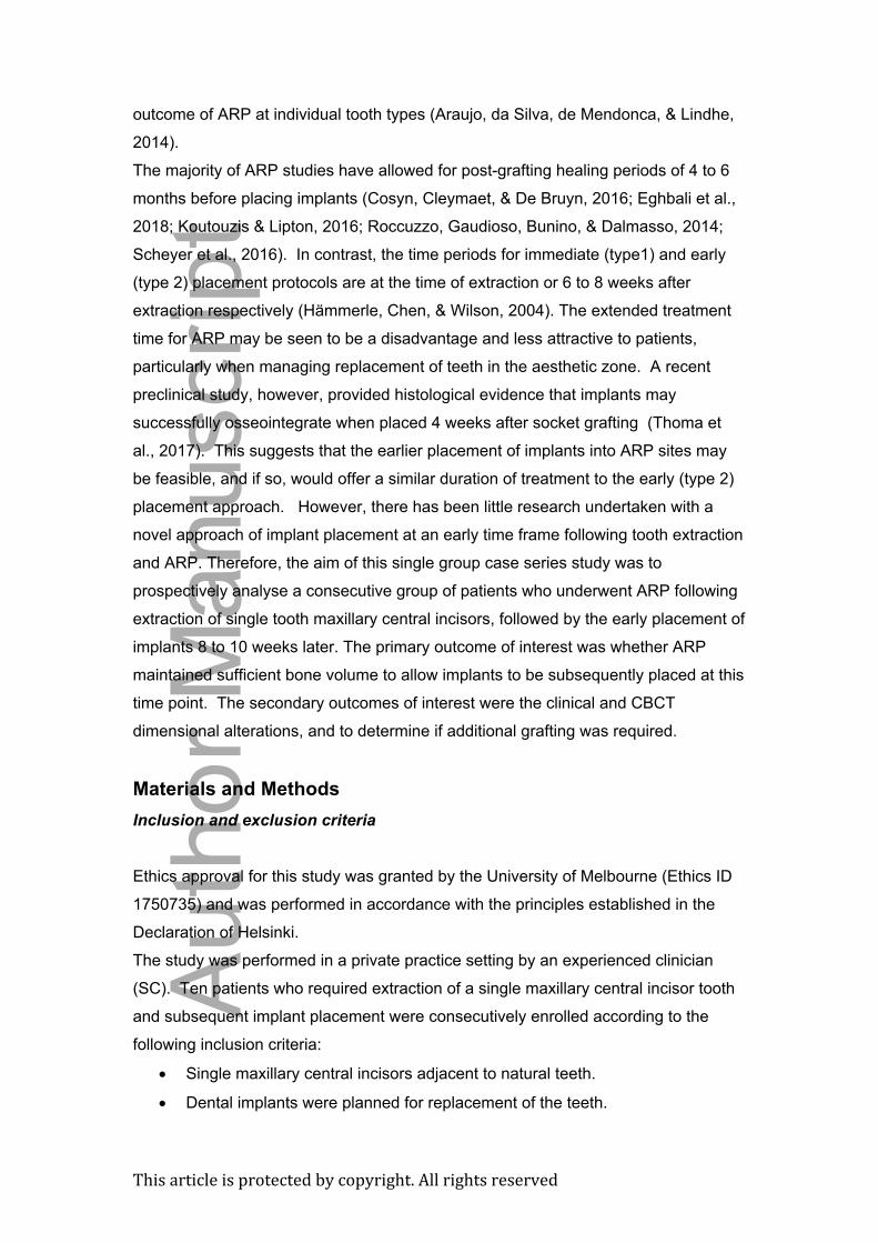
The majority of ARP studies have allowed for post-grafting healing periods of 4 to 6
months before placing implants (Cosyn, Cleymaet, & De Bruyn, 2016; Eghbali et al.,
2018; Koutouzis & Lipton, 2016; Roccuzzo, Gaudioso, Bunino, & Dalmasso, 2014;
Scheyer et al., 2016). In contrast, the time periods for immediate (type1) and early
(type 2) placement protocols are at the time of extraction or 6 to 8 weeks after
extraction respectively (Hämmerle, Chen, & Wilson, 2004). The extended treatment
time for ARP may be seen to be a disadvantage and less attractive to patients,
particularly when managing replacement of teeth in the aesthetic zone. A recent
preclinical study, however, provided histological evidence that implants may
successfully osseointegrate when placed 4 weeks after socket grafting (Thoma et
al., 2017). This suggests that the earlier placement of implants into ARP sites may
be feasible, and if so, would offer a similar duration of treatment to the early (type 2)
placement approach. However, there has been little research undertaken with a
novel approach of implant placement at an early time frame following tooth extraction
and ARP. Therefore, the aim of this single group case series study was to
prospectively analyse a consecutive group of patients who underwent ARP following
extraction of single tooth maxillary central incisors, followed by the early placement of
implants 8 to 10 weeks later. The primary outcome of interest was whether ARP
maintained sufficient bone volume to allow implants to be subsequently placed at this
time point. The secondary outcomes of interest were the clinical and CBCT
dimensional alterations, and to determine if additional grafting was required.
Materials and Methods
Inclusion and exclusion criteria
Ethics approval for this study was granted by the University of Melbourne (Ethics ID
1750735) and was performed in accordance with the principles established in the
Declaration of Helsinki.
The study was performed in a private practice setting by an experienced clinician
(SC). Ten patients who required extraction of a single maxillary central incisor tooth
and subsequent implant placement were consecutively enrolled according to the
following inclusion criteria:
Single maxillary central incisors adjacent to natural teeth.
Dental implants were planned for replacement of the teeth.
Auth
or
Manuscript

This article is protected by copyright. All rights reserved
Periodontal probing pockets < 4 mm.
Intact facial bone wall determined by pre-extraction cone beam computed
tomographic (CBCT) examination (FOV 40 x 40, 80 kV and 6 mA; Morita
Veraviewepocs R100; Kyoto, Japan)
Confirmation of intact socket walls immediately following tooth extraction.
Adjacent natural teeth were periodontally sound with no periodontal
attachment loss.
Adjacent teeth with healthy dental pulps, or if non-vital, had a satisfactory
endodontic status and were free of symptoms.
Patients were excluded if they smoked cigarettes.
Clinical procedures
The periodontal phenotype was classified as thin, medium or thick by measuring the
vertical height of the keratinized gingiva (thin: ≤ 3 mm, medium: 4 to 5 mm, thick: ≥ 6
mm) at the mid-facial aspect of the tooth (Muller, Heinecke, Schaller, & Eger, 2000).
Prior to extraction, removable templates consisting of a stainless-steel wire
positioned at the incisal edges of adjacent teeth and embedded in acrylic resin were
constructed.
A mesiodistal (MD) reference line orthogonal to the apico-coronal axis of the central
incisors was established 5 mm from the mid-facial and mid palatal gingival margins of
the tooth to be extracted (fig 1). The distance of the MD reference line to an acrylic
template was recorded. Following administration of local anesthesia (2% lignocaine
hydrochloride; 1:100,000 adrenaline), the following were recorded:
At the MD reference line, the external orofacial dimensions of the ridge at the
surface of the mucosa at the mesial, midfacial and distal of the tooth to be
extracted was recorded to the nearest millimeter with a surgical caliper
(Salvin Ridge Mapping Caliper, Salvin, USA).
At the same 3 positions, the orofacial dimensions of the alveolar bone were
measured with the same calipers by penetrating the mucosa to the surface of
the bone facially and palatally.
Intrasulcular incisions were made to sever the gingival attachment from the neck of
the tooth circumferentially. The teeth were extracted without flap elevation using
periotomes and fine luxators applied to the palatal aspects of the teeth (fig 2a). Fine
tipped forceps were used with rotational movements only to avoid facially directed
pressure on the socket wall. The sockets were debrided with surgical curettes to
remove soft tissue tags or remnants of granulation tissue. The facial bone walls were
Auth
or
Manuscript

This article is protected by copyright. All rights reserved
examined from the inner aspect of the socket with a periodontal probe to confirm that
they were intact and undamaged.
Immediately following tooth extraction, the distance between the template and the
bone crest at the mesiofacial, midfacial, distofacial, mesiopalatal, midpalatal and
distopalatal aspects of the sockets were recorded.
ARP was then performed using 90% deproteinized bovine bone mineral in a 10%
collagen matrix (DBBMC) (Geistlich Bio-Oss® Collagen, Geistlich Pharma AG,
Wolhusen, Switzerland). The graft was prepared by soaking in sterile saline for
several minutes before filling the socket. The graft was gently packed into the
socket to the level of the facial bone crest with hand instruments (fig 2b). The socket
entrance was closed with a resorbable type I/III porcine collagen matrix (CM)
(Geistlich Mucograft® Seal, Geistlich Pharma AG, Wolhusen, Switzerland) that was
sutured into position (fig 2c). A removable partial denture or vacuum-formed
removable prosthesis was then delivered, with care taken to ensure that the
removable prosthesis did not exert pressure on the crestal or facial region of the
socket. Patients were instructed to use a 0.2% chlorhexidine mouthrinse (Curasept
ADS 220; Curaden AG, Kriens, Switzerland) and to avoid brushing the surgical site
for 2 weeks. Patients returned 2 weeks after surgery for removal of sutures.
The patients were then scheduled for implant surgery 8 to 10 weeks following
extraction and ARP (fig 2d). Pre-operative CBCT were obtained at this time using
the same radiological parameters as described previously. Following administration
of local anesthesia (2% lignocaine hydrochloride; 1:100,000 adrenaline),
measurements of the external soft tissue and bone dimensions of the ridges were
repeated in the same manner as described previously. Two-sided full thickness
mucoperiosteal flaps were raised on the facial aspect with a vertical releasing
incision on the distal aspect of the lateral incisors adjacent to the missing tooth sites
(fig 2e). On the palatal side, flap elevation was performed to only expose the palatal
bone margins. The distances between the template and the bone crest at the
mesiofacial, mid-facial, distofacial, mesiopalatal, mid-palatal and disto- palatal
aspects of the sockets were once again recorded. The implant osteotomies were
then prepared according to the manufacturer’s recommendation (fig 2f), and implants
with a standard 4.1 mm diameter and 10 to 12 mm in length were inserted in the
correct 3-dimensional position (Straumann RC bone level implants; Straumann AG,
Basel, Switzerland) (fig 2g). Additional grafting of the facial aspect of the ridge was
performed if it was determined that additional soft tissue support at the shoulder
region was required for soft tissue support and aesthetics. The additional grafting
was completed grating was using particulate deproteinized bovine bone mineral
Auth
or
Manuscript

This article is protected by copyright. All rights reserved
(DBBM) (Geistlich Bio-Oss®; Geistlich Pharma AG, Wolhusen, Switzerland) and a
resorbable native bilayer collagen barrier membrane (NBCM) (Geistlich Bio-Gide®;
Geistlich Pharma AG, Wolhusen, Switzerland) (fig 2h). Flaps were closed to fully
submerge the implants. After a healing period of 3 months, the coronal regions of
the implants were accessed by removing a plug of soft tissue over the ridge crest
with a 15C scalpel blade to facilitate the connection of transmucosal healing
abutments. Restorative procedures then commenced (fig 2i) and were reviewed
immediately after connection of the prosthesis, and then 1 year later. Soft tissue
aesthetic outcomes at one-year post-restoration were assessed using the Pink
Esthetic Score (Furhauser et al., 2005).
CBCT measurements
The CBCT analysis was performed by one examiner (SC). The DICOM files were
imported into an image analysis software program (OnDemand3D; Cybermed Inc.,
Seoul, Korea). After defining the region of interest (ROI), the 3D mutiplanar
reconstruction (MPR) mode was launched, revealing axial, coronal and sagittal
slices. The software automatically superimposed axial, sagittal and coronal
orientation lines on the images. The images were rotated and adjusted to achieve the
following reproducible reference lines:
In the axial view, a mesiodistal reference line that bisected the root canals of the
teeth adjacent to the site of interest was drawn. The apicocoronal position of this line
was adjusted in the coronal view to be located at the CEJ of the adjacent teeth when
viewed in the sagittal plane. In most cases, this line was also at the level of the CEJ
of the tooth of interest. In the coronal view, the image was rotated so that an
apicocoronal reference was parallel to the tooth of interest and coincident with the
axes of the adjacent teeth when viewed in the sagittal plane.
The planes and reference lines were based on the adjacent teeth rather than on the
tooth to be extracted, thereby allowing the same reference points and planar axes to
be used on the post-extraction/ARP scans (fig 3).
In the sagittal view at the site of interest, orofacial measurements of the ridge
perpendicular to the apicocoronal reference line were then obtained using the linear
measurement tool, at 1 mm increments apical to the mesiodistal reference line from 0
to 10 mm (fig 4a-and 4b).
The area of the alveolar ridge was derived using the area tool of the software. A line
was drawn to connect the facial and palatal bone crests. The line was extended on
the facial aspect to trace the outer surface of the facial bone. The line extended
apically to the horizontal measurement line that coincided with the apex of the root.
Auth
or
Manuscript

This article is protected by copyright. All rights reserved
The line was then extended onto the palatal surface of the bone to coronally meet the
palatal bone crest. Thus, the outline of the alveolar bone was traced as an irregular
shape from which the area was provided as an output by the software program.
These measurements were repeated for the post-extraction/ARP CBCT data.
All measurements were made by one experienced examiner (SC). 3 cases were
measured 1 week apart. The intra-observer reproducibility was assessed by intra-
class correlation coefficient (ICC) to be 0.99. The standard error was 0.49 mm.
Data analysis
Descriptive methods were used to summarize patient demographics. For continuous
data, residuals for individual parameters were plotted to confirm normality of the
distributions. Differences between pretreatment and re-entry data were analysed with
the paired Student’s t-test. The level of significance was set at 0.05. All analyses
were carried out using the statistical package Minitab (Minitab 18, Minitab Inc.,
Philadelphia, USA).
Results
Patient demographics
Over a 14-month recruitment period, a total of 10 consecutive patients who fulfilled
the inclusion criteria were included in the study. All patients who were intended to be
treated received the prescribed treatment with no exclusions. There were 5 female
and 5 male patients, with an average age of 42.9 ± 19.37 years (range 21.7 to 71.7
years). Each patient contributed one maxillary central incisor extraction site. All 10
patients that were enrolled underwent the planned treatment without deviation to the
protocol. The gingival phenotype was thick in 2 patients, medium in 4 patients and
thin in 4 patients.
Clinical outcomes
Extraction, ARP and the subsequent healing proceeded uneventfully in all cases.
Patients returned for the placement of implants after a mean time of 8.9 ± 0.97 weeks
(range 8.0 to 10.6 weeks). At this time, the mucosa was observed to have
completely healed with formation of an epithelized soft tissue barrier at all sites, and
were free of signs of inflammation, infection or scar formation. In 9 cases, a soft
tissue invagination at the crestal region representing about 10 - 20% the size of the
Auth
or
Manuscript

This article is protected by copyright. All rights reserved
original socket size was present. The mucosa appeared less mature within the
invagination. In some cases, particles of exfoliating DBBMC could be seen at the
surface of the invagination without any sign of infection. All sites demonstrated
varying degrees of facial resorption of the ridge in the coronal region.
On flap reflection, the sockets were completely filled with the DBBMC graft. The
superficial region of the graft did not appear to be fully ossified, and was contained
within a fibrous connective tissue matrix. This immature tissue was not removed.
There was sufficient bone volume to allow the placement of implants in the correct 3-
dimensional position in all 10 sites. During preparation of the implant osteotomy, it
was observed that the graft material remained intact and did not dislodge or disperse
from the site.
In one case, however, a dehiscence of the facial bone occurred when the osteotomy
was prepared, indicating a relative lack of orofacial bone dimension in this one case.
All implants were 4.1 mm in diameter (Straumann regular CrossFit RC SLActive
implants; Straumann AG, Switzerland) with lengths of 10 mm (5 sites) and 12 mm (5
sites). Following implant placement, all sites were determined to have insufficient
facial ridge contour for aesthetic outcomes. Additional low volume grafting of the
facial aspect of the ridge was carried for contour augmentation at each site. All
implants were stable and clinically integrated after 3 months. At this time, the
patients returned to their referring dental practitioners who completed restoration of
the implants with single implant crowns 1 to 2 months later.
Intra-operative measurements
Alterations in the dimensions of the ridge between extraction/ARP and surgical re-
entry are presented in table 1.
Pre-treatment, the mean thickness of the facial bone wall was 0.8 ± 0.38 mm
(median 0.7 mm; range 0.5 to 1.7 mm) at 5 mm apical to the CEJ. Only one site had
a facial bone wall thickness of > 1 mm.
There was a significant reduction in the external orofacial dimension of the ridge at
the mid-facial aspect (1.4 ± 1.07 mm; range 0 to 2 mm) which represented a 13.2%
reduction in the original ridge width (p = 0.003). At the mesial and distal aspects, the
reduction observed was not statistically significant. There was also a statistically
significant reduction in the orofacial bone dimension at the mid-facial aspect (0.7 ±
0.67 mm; range 0 to 2 mm; p = 0.010) which amounted to 9.3% of the original
Auth
or
Manuscript

This article is protected by copyright. All rights reserved
orofacial bone width. The corresponding reduction observed on the mesial and distal
aspects was not statistically significant.
Between extraction/ARP and surgical re-entry, there was significant apicocoronal
reduction in height of the crestal bone at the midfacial (1.2 ± 0.78 mm; p = 0.001),
mesiopalatal (0.7 ± 0.67 mm; p = 0.004), midpalatal (1.0 ± 0.81 mm; p = 0.004) and
distopalatal (0.7 ± 0.48 mm; p = 0.001) aspects of the socket.
CBCT analysis
The CBCT assessment of orofacial bone dimensions were measured at 1 mm
increments apical to the CEJ (table 2). Pre-extraction, there were no sites with facial
bone at 1 and 2 mm apical to the CEJ. At 3, 4 and 5 mm apical to the CEJ, a facial
bone wall was observed in 5, 8 and 9 sites respectively. From 6 mm to 10 mm apical
to the CEJ, all 10 sites had facial bone detectable on preoperative CBCT. The
comparison between CBCT at the two time points revealed a significant reduction in
orofacial bone dimensions at 4 to 10 mm apical to the CEJ, ranging from 1.0 to 1.7
mm, or 7.3 to 18.6% reduction in the original orofacial bone width. There was a trend
for a greater reduction in bone width in the coronal half compared to the apical half of
the alveolar ridge.
The total alveolar ridge area showed a statistically significant reduction of 10.9 ±
13.42 mm2, or 12.2% change (range 1.4 to 29.9 mm2; p = 0.030).
Follow-up
At the one-year follow-up post-restoration, the implant survival rate was 100%. One
implant developed mucositis and a fistula on the facial aspect that required
treatment, yielding a one-year success rate of 90%. One implant demonstrated minor
bleeding after probing at the mid-facial aspect. Probing pockets at the implants were
with normal limits (mean 2.4 ± 0.34 mm). At one-year post-restoration, the median
Pink Esthetic Score was 10 (range 9 – 13).
Discussion
In this study, ARP was performed on single maxillary central incisor sockets of 10
consecutively enrolled patients. It was noted that the clinical handling of the graft
material at an early time point of 8 to 10 weeks after extraction/ARP was favourable.
The graft remained intact and did not dislodge or disperse while the osteotomy was
being prepared. All sites retained sufficient bone volume to allow implants to be
placed between 8 to 10 weeks after ARP. These findings are consistent with reports
from previous studies, confirming that ARP is an effective treatment for maintaining
Auth
or
Manuscript

This article is protected by copyright. All rights reserved
sufficient bone volume to facilitate the subsequent placement of implants (Araujo et
al., 2014; Barone et al., 2008; Carmagnola, Adriaens, & Berglundh, 2003; Iasella et
al., 2003; Mardas et al., 2010). However, as reported in previous systematic reviews,
ARP does not prevent dimensional alterations but does limit the extent to which this
occurs (Avila-Ortiz, Elangovan, Kramer, Blanchette, & Dawson, 2014; Bassir et al.,
2018; MacBeth et al., 2017; Mardas et al., 2015; Vignoletti et al., 2012; Willenbacher,
Al-Nawas, Berres, Kammerer, & Schiegnitz, 2016). In the present study, a significant
reduction in external ridge (13.2%) and bone dimensions (9.3%) were observed at
the reference level (5 mm apical to the gingival margin). The reduction in orofacial
bone width on CBCT ranged from 7.3 to 18.6% along the corono-apical height of the
alveolar process, with greater dimensional change in the coronal half compared to
the apical half. These findings are largely in agreement with other studies of ARP
using DBBMC graft (Cosyn et al., 2016; Jung et al., 2013) and similar to a study that
used a combination allograft and xenograft (Serrano, Castellanos, & Botticelli, 2018).
However, a recent study using the same DBBMC graft reported orofacial reduction of
0.62 ± 0.32, 0.40 ± 0.26 and 0.10 ± 0.08 mm at 1 mm, 3 mm and 5 mm apical to the
bone crest 4 months after extraction/ARP (Cardaropoli, Tamagnone, Roffredo, De
Maria, & Gaveglio, 2018). This was much less than that reported in the present
study, where the linear dimensional changes were > 1 mm along the entire
apicocoronal dimension of the alveolar process. The difference between studies may
be attributable to methodical differences in determining the reference planes in the
sagittal CBCT images, with adjacent teeth serving as fixed reference points in the
present study compared to the axis of the extraction socket at the test site by
Cardaropoli and co-workers. Furthermore, the inclusion of maxillary incisors, canines
and premolars by Cardaropoli and co-workers compared to only maxillary central
incisors in the present study may be another reason for the difference between
studies, since post-grafting resorption has reported to be greater at central incisors
and cuspids, compared to lateral incisors and premolars (Cosyn et al., 2016).
It is interesting to note the similarity of the intra-operative reduction in orofacial bone
width of 9.3% at the MD reference level located 5 mm from the gingival margin and
the reduction of 12.6% on CBCT at 5 mm from the CEJ. Although not identical in
location, the reference points are in a similar position at the alveolar ridge. This
suggests that CBCT measurements of ridge dimension change could be a useful
non-invasive surrogate for intra-operative measurements. However, this approach
should be interpreted cautiously, since CBCT scans may distinguish cortical bone
clearly, but less clearly delineate trabecular or woven bone which may form part of
the healing socket wall (Araujo 2014).
Auth
or
Manuscript

This article is protected by copyright. All rights reserved
Although all sites retained sufficient bone volume to allow implants with a standard
4.1 mm diameter and 10 to 12 mm in length to be placed, a dehiscence of the facial
aspect of the implant occurred in one case. This observation is consistent with
reports that the volume of bone preserved with ARP may not always be sufficient to
ensure that implants are completely placed within intact bone walls, thereby requiring
further bone augmentation at the time of implant placement (Barone, Ricci, Tonelli,
Santini, & Covani, 2012; Eskow & Mealey, 2014; Koutouzis & Lipton, 2016). In
contrast, other studies of ARP observed that implant placement could proceed
without the need for further bone augmentation (Cosyn et al., 2016; Iasella et al.,
2003; Pang et al., 2016; Serino, Biancu, Iezzi, & Piattelli, 2003). The contrasting
findings are likely due to the heterogeneity of tooth sites, pre-extraction condition of
the socket walls, biomaterials used for ARP, healing periods, and methodological
differences in recording clinical and radiological dimensional alterations, making it
difficult for meaningful comparisons to be made (Balli et al., 2018). A clinically
significant observation in this study was that additional grafting to further augment the
facial contour of the ridge was required, as the extent of the preservation of bone and
soft tissue volume in the coronal region of the ridge was determined clinically to be
insufficient to ensure satisfactory aesthetic outcomes. The coronal region of the
ridge underwent a 13.2% reduction in external orofacial dimension. The underlying
bone reduction in the same region was 9.3%. This degree of dimensional change is
clinically significant and may adversely affect aesthetic outcomes if not compensated
for. It has been reported that deficient tissue volume in the facial coronal region of
the ridge may result in lack of facial ridge contour which in turn may cause a shadow
at the cervical region of the implant prosthesis (Schneider, Grunder, Ender,
Hammerle, & Jung, 2011). Thus, supplementary augmentation procedures are often
required to compensate for lack of tissue volume. One option is to place a
connective tissue graft (CTG) at the coronal marginal region of the implant, either at
the time the implant is placed (Boardman, Darby, & Chen, 2015; Schneider et al.,
2011) or at the time of connection of the provisional prosthesis after integration of the
implant (Cosyn 2016, Eghbali 2018). In a 5-year report, all 32 implants in 32 ARP
treated patients required a CTG for supplementary augmentation at the time of
provisional crown insertion (Eghbali 2018). This study confirms that lack of tissue
volume should be an expected outcome with ARP in aesthetic areas. In the present
study, however, compensatory augmentation was performed using additional DBBM
at the time of implant placement using the GBR contour augmentation technique
originally described in conjunction with early (type 2) implant placement (Buser et al.,
2008) as it has been shown to effectively maintain bone volume and soft tissue
Auth
or
Manuscript

This article is protected by copyright. All rights reserved
stability on the facial aspect of implants after 10 years (Chappuis et al., 2018).
Although this approach avoids the need for a second surgical site to harvest a CTG,
the disadvantage is the need for additional DBBM and the associated increase in the
cost burden to the patient.
In the present study, CM was placed over the DBBMC to provide an initial barrier and
to subsequently promote soft tissue closure over the socket. The CM undergoes a
substitution process and is replaced with a vascularized collagen-rich connective
tissue (Maiorana et al., 2017). Although fully epithelialized, the soft tissue
invagination observed at 9/10 cases after 8 to 10 weeks of healing indicates that the
mucosa had not fully healed. This is consistent with a clinical study that used a soft
tissue healing index to evaluate the healing extraction sockets with CM placed over
DBBMC in 7 patients (Maiorana et al., 2017). At 4 weeks, there were no sites that
had completely healed, whereas at 8 weeks, 5/7 sites had healed completely. By 10
weeks, all sites had healed completely with full soft tissue closure.
In the present study, all implants were stable 3 months after placement and were
subsequently restored without loss. All implants were integrated 12 months after
restoration. These initial results suggest that early implant placement 8 to 10 weeks
after grafting with DBBMC/CM is viable, and may have a patient-centred benefit in
reducing the duration of the overall treatment. Since the clinical and radiographic
examination was done by one observer (SC), there is a risk of investigator bias which
should be taken in to account when interpreting the results. This was mitigated,
however, by blinding of patient identity during the CBCT analysis. This study is
further limited by the relatively small number of cases and lack of a comparative
group. Longer term studies designed to compare early implant placement after ARP
using DBBM/CM with other placement protocols are required to validate this
approach.
Conclusions
In this study, ARP using DBBMC/CM maintained sufficient bone volume in maxillary
central incisor extraction sites for the subsequent early placement of implants 8 to 10
weeks later. Healing after ARP was characterized by a significant reduction in
orofacial ridge (13.2%) and bone (9.3%) dimensions at the mid-facial coronal region,
and a reduction in apicocoronal height of the crestal bone at the midfacial (1.2 ± 0.78
mm) and palatal aspects of the socket. CBCT data indicated a concomitant reduction
in orofacial bone dimensions at 4 to 10 mm apical to the CEJ ranging from 1.0 to 1.7
mm (representing 7.3 to 18.6% reduction in the original orofacial bone width) and a
Auth
or
Manuscript

This article is protected by copyright. All rights reserved
reduction in total alveolar ridge area of 10.9 ± 13.42 mm2 (12.2 % reduction). Due to
the reduced ridge dimensions in the coronal region, additional low volume grafting
with DBBM/NBCM for contour augmentation was required for aesthetic reasons in
9/10 cases, whereas one case required more extensive grafting due to dehiscence
formation when the osteotomy was prepared. All implants successfully integrated and
were restored with a 100% survival rate one-year after restoration. Acceptable
aesthetic outcomes were achieved (median Pink Esthetic Score 10; range 9 – 13.
The early placement of implants following ARP using DBBMC and CM may be a
viable treatment approach.
Auth
or
Manuscript

This article is protected by copyright. All rights reserved
References
Araujo, M. G., da Silva, J. C., de Mendonca, A. F., & Lindhe, J. (2014). Ridge
alterations following grafting of fresh extraction sockets in man. A randomized
clinical trial. Clin Oral Implants Res. doi:10.1111/clr.12366
Araujo, M. G., & Lindhe, J. (2005). Dimensional ridge alterations following tooth
extraction. An experimental study in the dog. J Clin Periodontol, 32(2), 212-
218.
Araujo, M. G., Sukekava, F., Wennstrom, J. L., & Lindhe, J. (2005). Ridge alterations
following implant placement in fresh extraction sockets: an experimental study
in the dog. J Clin Periodontol, 32(6), 645-652.
Avila-Ortiz, G., Elangovan, S., Kramer, K. W., Blanchette, D., & Dawson, D. V.
(2014). Effect of alveolar ridge preservation after tooth extraction: a
systematic review and meta-analysis. J Dent Res, 93(10), 950-958.
doi:10.1177/0022034514541127
Balli, G., Ioannou, A., Powell, C. A., Angelov, N., Romanos, G. E., & Soldatos, N.
(2018). Ridge Preservation Procedures after Tooth Extractions: A Systematic
Review. Int J Dent, 2018, 8546568. doi:10.1155/2018/8546568
Barone, A., Aldini, N. N., Fini, M., Giardino, R., Calvo Guirado, J. L., & Covani, U.
(2008). Xenograft versus extraction alone for ridge preservation after tooth
removal: a clinical and histomorphometric study. J Periodontol, 79(8), 1370-
1377. doi:10.1902/jop.2008.070628
Barone, A., Ricci, M., Tonelli, P., Santini, S., & Covani, U. (2012). Tissue changes of
extraction sockets in humans: a comparison of spontaneous healing vs. ridge
preservation with secondary soft tissue healing. Clin Oral Implants Res.
doi:10.1111/j.1600-0501.2012.02535.x
Bassir, S. H., Alhareky, M., Wangsrimongkol, B., Jia, Y., & Karimbux, N. (2018).
Systematic Review and Meta-Analysis of Hard Tissue Outcomes of Alveolar
Ridge Preservation. Int J Oral Maxillofac Implants, 33(5), 979-994.
Boardman, N., Darby, I., & Chen, S. (2015). A retrospective evaluation of aesthetic
outcomes for single-tooth implants in the anterior maxilla. Clin Oral Implants
Res. doi:10.1111/clr.12593
Brandam, L., Malmstrom, H., Javed, F., Calvo-Guirado, J. L., & Romanos, G. E.
(2015). Ridge Preservation Techniques in the Anterior Esthetic Zone. Implant
Dent, 24(6), 699-712. doi:10.1097/ID.0000000000000341
Auth
or
Manuscript

This article is protected by copyright. All rights reserved
Buser, D., Bornstein, M. M., Weber, H. P., Grutter, L., Schmid, B., & Belser, U. C.
(2008). Early implant placement with simultaneous guided bone regeneration
following single-tooth extraction in the esthetic zone: a cross-sectional,
retrospective study in 45 subjects with a 2- to 4-year follow-up. J Periodontol,
79(9), 1773-1781. doi:10.1902/jop.2008.080071
Cardaropoli, D., Tamagnone, L., Roffredo, A., De Maria, A., & Gaveglio, L. (2018).
Alveolar Ridge Preservation Using Tridimensional Collagen Matrix and
Deproteinized Bovine Bone Mineral in the Esthetic Area: A CBCT and
Histologic Human Pilot Study. International Journal of Periodontics &
Restorative Dentistry, 38(Suppl), s29-s35. doi:10.11607/prd.3702
Carmagnola, D., Adriaens, P., & Berglundh, T. (2003). Healing of human extraction
sockets filled with Bio-Oss. Clin Oral Implants Res, 14(2), 137-143.
Chappuis, V., Rahman, L., Buser, R., Janner, S. F. M., Belser, U. C., & Buser, D.
(2018). Effectiveness of Contour Augmentation with Guided Bone
Regeneration: 10-Year Results. J Dent Res, 97(3), 266-274.
doi:10.1177/0022034517737755
Chen, S. T., & Darby, I. (2017). The relationship between facial bone wall defects
and dimensional alterations of the ridge following flapless tooth extraction in
the anterior maxilla. Clin Oral Implants Res, 28(8), 931-937.
doi:10.1111/clr.12899
Cosyn, J., Cleymaet, R., & De Bruyn, H. (2016). Predictors of Alveolar Process
Remodeling Following Ridge Preservation in High-Risk Patients. Clin Implant
Dent Relat Res, 18(2), 226-233. doi:10.1111/cid.12249
Covani, U., Cornelini, R., Calvo, J. L., Tonelli, P., & Barone, A. (2010). Bone
remodeling around implants placed in fresh extraction sockets. International
Journal of Periodontics & Restorative Dentistry, 30(6), 601-607.
Darby, I., Chen, S., & De Poi, R. (2008). Ridge preservation: what is it and when
should it be considered. Aust Dent J, 53(1), 11-21. doi:ADJ008 [pii]
10.1111/j.1834-7819.2007.00008.x
Darby, I., Chen, S. T., & Buser, D. (2009). Ridge preservation techniques for implant
therapy. Int J Oral Maxillofac Implants, 24 Suppl, 260-271.
Eghbali, A., Seyssens, L., De Bruyckere, T., Younes, F., Cleymaet, R., & Cosyn, J.
(2018). A 5-year prospective study on the clinical and aesthetic outcomes of
alveolar ridge preservation and connective tissue graft at the buccal aspect of
single implants. J Clin Periodontol, 45(12), 1475-1484.
doi:10.1111/jcpe.13018
Auth
or
Manuscript
This article is protected by copyright. All rights reserved
Eskow, A. J., & Mealey, B. L. (2014). Evaluation of healing following tooth extraction
with ridge preservation using cortical versus cancellous freeze-dried bone
allograft. J Periodontol, 85(4), 514-524. doi:10.1902/jop.2013.130178
Farmer, M., & Darby, I. (2014). Ridge dimensional changes following single-tooth
extraction in the aesthetic zone. Clin Oral Implants Res, 25(2), 272-277.
doi:10.1111/clr.12108
Furhauser, R., Florescu, D., Benesch, T., Haas, R., Mailath, G., & Watzek, G. (2005).
Evaluation of soft tissue around single-tooth implant crowns: the pink esthetic
score. Clin Oral Implants Res, 16(6), 639-644.
Hämmerle, C. H., Chen, S. T., & Wilson, T. G., Jr. (2004). Consensus statements
and recommended clinical procedures regarding the placement of implants in
extraction sockets. Int J Oral Maxillofac Implants, 19 Suppl, 26-28.
Iasella, J. M., Greenwell, H., Miller, R. L., Hill, M., Drisko, C., Bohra, A. A., & Scheetz,
J. P. (2003). Ridge preservation with freeze-dried bone allograft and a
collagen membrane compared to extraction alone for implant site
development: A clinical and histologic study in humans. J Periodontol, 74,
990-999.
Jung, R. E., Philipp, A., Annen, B. M., Signorelli, L., Thoma, D. S., Hammerle, C. H., .
. . Schmidlin, P. (2013). Radiographic evaluation of different techniques for
ridge preservation after tooth extraction: a randomized controlled clinical trial.
J Clin Periodontol, 40(1), 90-98. doi:10.1111/jcpe.12027
Koutouzis, T., & Lipton, D. (2016). Regenerative Needs Following Alveolar Ridge
Preservation Procedures in Compromised and Noncompromised Extraction
Sockets: A Cone Beam Computed Tomography Study. Int J Oral Maxillofac
Implants, 31(4), 849-854. doi:10.11607/jomi.4437
MacBeth, N., Trullenque-Eriksson, A., Donos, N., & Mardas, N. (2017). Hard and soft
tissue changes following alveolar ridge preservation: a systematic review. Clin
Oral Implants Res, 28(8), 982-1004. doi:10.1111/clr.12911
Maiorana, C., Poli, P. P., Deflorian, M., Testori, T., Mandelli, F., Nagursky, H., &
Vinci, R. (2017). Alveolar socket preservation with demineralised bovine bone
mineral and a collagen matrix. J Periodontal Implant Sci, 47(4), 194-210.
doi:10.5051/jpis.2017.47.4.194
Mardas, N., Chadha, V., & Donos, N. (2010). Alveolar ridge preservation with guided
bone regeneration and a synthetic bone substitute or a bovine-derived
xenograft: a randomized, controlled clinical trial. Clin Oral Implants Res,
21(7), 688-698. doi:CLR1918 [pii]
10.1111/j.1600-0501.2010.01918.x
Auth
or
Manuscript

This article is protected by copyright. All rights reserved
Mardas, N., Trullenque-Eriksson, A., MacBeth, N., Petrie, A., & Donos, N. (2015).
Does ridge preservation following tooth extraction improve implant treatment
outcomes: a systematic review: Group 4: Therapeutic concepts & methods.
Clin Oral Implants Res, 26 Suppl 11, 180-201. doi:10.1111/clr.12639
Morjaria, K. R., Wilson, R., & Palmer, R. M. (2012). Bone Healing after Tooth
Extraction with or without an Intervention: A Systematic Review of
Randomized Controlled Trials. Clin Implant Dent Relat Res.
doi:10.1111/j.1708-8208.2012.00450.x
Muller, H. P., Heinecke, A., Schaller, N., & Eger, T. (2000). Masticatory mucosa in
subjects with different periodontal phenotypes. J Clin Periodontol, 27(9), 621-
626.
Pang, C., Ding, Y., Hu, K., Zhou, H., Qin, R., & Hou, R. (2016). Influence of
preservation of the alveolar ridge on delayed implants after extraction of teeth
with different defects in the buccal bone. Br J Oral Maxillofac Surg, 54(2),
176-180. doi:10.1016/j.bjoms.2015.11.025
Roccuzzo, M., Gaudioso, L., Bunino, M., & Dalmasso, P. (2014). Long-term stability
of soft tissues following alveolar ridge preservation: 10-year results of a
prospective study around nonsubmerged implants. International Journal of
Periodontics & Restorative Dentistry, 34(6), 795-804. doi:10.11607/prd.2133
Scheyer, E. T., Heard, R., Janakievski, J., Mandelaris, G., Nevins, M. L., Pickering,
S. R., . . . Nagursky, H. (2016). A randomized, controlled, multicentre clinical
trial of post-extraction alveolar ridge preservation. J Clin Periodontol, 43(12),
1188-1199. doi:10.1111/jcpe.12623
Schneider, D., Grunder, U., Ender, A., Hammerle, C. H., & Jung, R. E. (2011).
Volume gain and stability of peri-implant tissue following bone and soft tissue
augmentation: 1-year results from a prospective cohort study. Clin Oral
Implants Res, 22(1), 28-37.
Schropp, L., Wenzel, A., Kostopolous, L., & Karring, T. (2003). Bone healing and soft
tissue contour changes following single-tooth extraction: A clinical and
radiographic 12-month prospective study. International Journal of
Periodontics & Restorative Dentistry, 23, 313-323.
Seibert, J. S., & Salama, H. (1996). Alveolar ridge preservation and reconstruction.
Periodontol 2000, 11, 69-84.
Serino, G., Biancu, S., Iezzi, G., & Piattelli, A. (2003). Ridge preservation following
tooth extraction using a polylactide and polyglycolide sponge as space filler: a
clinical and histological study in humans. Clin Oral Implants Res, 14(5), 651-
658.
Auth
or
Manuscript

This article is protected by copyright. All rights reserved
Serrano, C. A., Castellanos, P., & Botticelli, D. (2018). Use of Combination of
Allografts and Xenografts for Alveolar Ridge Preservation Procedures: A
Clinical and Histological Case Series. Implant Dent, 27(4), 467-473.
doi:10.1097/ID.0000000000000792
Thoma, D. S., Naenni, N., Benic, G. I., Munoz, F., Hammerle, C. H. F., & Jung, R. E.
(2017). Effect of ridge preservation for early implant placement - is there a
need to remove the biomaterial? J Clin Periodontol, 44(5), 556-565.
doi:10.1111/jcpe.12709
Vignoletti, F., Matesanz, P., Rodrigo, D., Figuero, E., Martin, C., & Sanz, M. (2012).
Surgical protocols for ridge preservation after tooth extraction. A systematic
review. Clin Oral Implants Res, 23 Suppl 5, 22-38. doi:10.1111/j.1600-
0501.2011.02331.x
Willenbacher, M., Al-Nawas, B., Berres, M., Kammerer, P. W., & Schiegnitz, E.
(2016). The Effects of Alveolar Ridge Preservation: A Meta-Analysis. Clin
Implant Dent Relat Res, 18(6), 1248-1268. doi:10.1111/cid.12364
Auth
or
Manuscript

This article is protected by copyright. All rights reserved
Table 1 – Change in orofacial ridge (surface of the soft tissue) and orofacial bone dimensions, and change in apicocoronal position of the bone crest
Mesial
(mean mm ± sd) #
Mid-point
(mean mm ± sd) #
Distal
(mean mm ± sd) #
Change in orofacial ridge (soft tissue
surface) dimension
0.4 ± 0.69 1.4 ± 1.07 * 0.1 ± 0.47 * p = 0.003
Change in orofacial bone dimension 0.1 ± 0.56 0.7 ± 0.67 ** 0.2 ± 0.42 ** p = 0.010
Change in apicocoronal position of the
bone crest
Facial 0.5 ± 0.71 1.2 ± 0.78 *** 0.3 ± 0.48 ** p = 0.001
Palatal 0.7 ± 0.67 ^ 1.0 ± 0.81 ^^ 0.7 ± 0.48 ^^^ ^ p = 0.010
^^ p = 0.004
^^^ p = 0.001
# Positive value indicates a reduction in dimensions
sd = standard deviation
Table 2 – Change in orofacial bone dimensions assessed on CBCT at 1 mm increments from 3 mm apical to the CEJ
Position apical to the CEJ N Mean change #
(mm ± sd)
% change
Change in orofacial bone dimension
(midfacial position)
3 mm 5 1.6 ± 1.34 19.5 p = 0.051 (ns)
4 mm 8 1.7 ± 0.63 18.6 p = 0.000
Auth
or
Manuscript

This article is protected by copyright. All rights reserved
5 mm 9 1.1 ± 0.44 12.6 p = 0.000
6 mm 10 1.6 ± 1.86 17.3 p = 0.027
7 mm 10 1.3 ± 1.55 14.2 p = 0.028
8 mm 10 1.2 ± 1.28 13.3 p = 0.014
9 mm 10 1.0 ± 1.05 10.7 p = 0.014
10 mm 10 1.7 ± 0.69 7.3 p = 0.010
# Positive value indicates a reduction in dimensions
sd = standard deviation
ns = not statistically significant
Auth
or
Manuscript

clr_13619_f1.tif
Thisarticleisprotectedbycopyright.Allrightsreserved
Auth
or
Manuscript

clr_13619_f2a.tiff
Thisarticleisprotectedbycopyright.Allrightsreserved
Auth
or
Manuscript

clr_13619_f2b.tiff
Thisarticleisprotectedbycopyright.Allrightsreserved
Auth
or
Manuscript

clr_13619_f2c.tiff
Thisarticleisprotectedbycopyright.Allrightsreserved
Auth
or
Manuscript

clr_13619_f2d.tiff
Thisarticleisprotectedbycopyright.Allrightsreserved
Auth
or
Manuscript

clr_13619_f2e.tiff
Thisarticleisprotectedbycopyright.Allrightsreserved
Auth
or
Manuscript

clr_13619_f2f.tiff
Thisarticleisprotectedbycopyright.Allrightsreserved
Auth
or
Manuscript

clr_13619_f2g.tiff
Thisarticleisprotectedbycopyright.Allrightsreserved
Auth
or
Manuscript

clr_13619_f2h.tiff
Thisarticleisprotectedbycopyright.Allrightsreserved
Auth
or
Manuscript

clr_13619_f2i.tiff
Thisarticleisprotectedbycopyright.Allrightsreserved
Auth
or
Manuscript

clr_13619_f3.tif
Thisarticleisprotectedbycopyright.Allrightsreserved
Auth
or
Manuscript

clr_13619_f4a.tif
Thisarticleisprotectedbycopyright.Allrightsreserved
Auth
or
Manuscript

clr_13619_f4b.tif
Thisarticleisprotectedbycopyright.Allrightsreserved
Auth
or
Manuscript

Minerva Access is the Institutional Repository of The University of Melbourne
Author/s:Chen, ST;Darby, I
Title:Alveolar ridge preservation and early implant placement at maxillary central incisor sites: Aprospective case series study
Date:2020-06-18
Citation:Chen, S. T. & Darby, I. (2020). Alveolar ridge preservation and early implant placement atmaxillary central incisor sites: A prospective case series study. CLINICAL ORAL IMPLANTSRESEARCH, 31 (9), pp.803-813. https://doi.org/10.1111/clr.13619.
Persistent Link:http://hdl.handle.net/11343/275926