
Applications - HubSpot
232
Technical application notes on cell culture and fermentation Applications
-
Upload
khangminh22 -
Category
Documents
-
view
2 -
download
0
Transcript of Applications - HubSpot

Technical application notes on cell culture and fermentation
Applications

Cell culture
Title Cell line Product Doc. PageIntelligent Control of Chinese Hamster Ovary (CHO) CellCulture Using the BioFlo® 320 Bioprocess Control Station
CHO BioFlo® 320 Bioprocess Control Station
356 5
Perfusion CHO Cell Culture in a BioBLU® 5p Single-use Packed-bed Vessel
CHO BioBLU® 5p Vessel 336 11
Pitched-Blade vs. Spin Filter vs. Packed-bed Basket: CHO Cell Culture Comparison
CHO CelliGen® 310 Bioreactor 320 15
Growing CHO Cells in a New Brunswick™ CelliGen® BLU Benchtop, Stirred-Tank Bioreactor Using Single-use Vessels
CHO CelliGen® BLU Single-use Bioreactor System
312 21
Single-use Scalability: Cho Cell Culture Using 5 to 50 L New Brunswick™ CelliGen® BLU Benchtop, Stirred-Tank Bioreactors
CHO CelliGen® BLU Single-use Bioreactor System
257 25
Hybridoma and CHO Cell Culture Using the New Brunswick™ S41i, An Environmentally-Friendly, “Low Emission” Incubator Shaker
CHO, Hybridoma New Brunswick™ S41i CO2 Incubator Shaker
255 30
CHO Cell Culture with Single-use New Brunswick™ CelliGen® BLU Packed-Bed Fibra-Cel® Basket
CHO CelliGen® BLU Single-use Bioreactor System
254 34
Solving the Aggregation Problem of Human Embryonic Kidney 293 Cells Using the New Brunswick™ S41i CO2 Incubator Shaker
HEK293 New Brunswick™ S41i CO2 Incubator Shaker
339 37
Optimization of HEK293 Cell Culture in a New Brunswick™ CelliGen® 115 Bioreactor for the Production of Recombinant GPCR
HEK293 BioFlo®/CelliGen® 115 Fermentor/Bioreactor
314 46
Cultivation of Human CAP® Cells: Evaluation of Scale-Down Capabilities Using Single-use Bioreactors
Human CAP® BioBLU® 0.3c Single-use Vessel, DASbox® Mini Bioreactor System
291 49
Hybridoma Culture Using New Brunswick™ CelliGen® 310 with Packed-bed Fibra-Cel® Basket Impeller
Hybridoma CelliGen® 310 Bioreactor 258 53
Hypoxic Cell Culture in the New Brunswick™ Galaxy® 170R Incubator: Normal Growth, Morphological Changes
LNCaP New Brunswick™ Galaxy® 170R CO2 Incubator
331 56
Development of a Scale-Down Model for rAAV Viral Vector Production Using a Sf9/BEV System
SF-9 DASbox® Mini Bioreactor System 303 60
Sf-9 Insect Cell Culture Using a New Brunswick™ CelliGen® 310 Bioreactor: Using Headspace Air Overlay for Reduced dCO2
SF-9 CelliGen® 310 Bioreactor 316 64
Insect Cell Culture Using the New Brunswick™ BioFlo®/CelliGen® 115 Benchtop Fermentor/Bioreactor with Spin Filter Assembly
SF-9 BioFlo®/CelliGen® 115 Fermentor/Bioreactor
256 67
Low Oxygen Levels in the New Brunswick™ Galaxy® 170 R CO2 Incubator Enhance the Efficiency of Reprogramming Human Somatic Cells to Pluripotency
Stem Cell New Brunswick™ Galaxy® 170R CO2 Incubator
338 71
Large-scale Production of Human Mesenchymal Stem Cells in BioBLU® 5c Single-use Vessels
Stem Cell BioBLU® 5c Vessel 334 79
Mesenchymal Stem Cell Culture in the New Brunswick™ Galaxy® 170 R CO2 Incubator Under Hypoxic Conditions
Stem Cell New Brunswick™ Galaxy® 170R CO2 Incubator
333 90
Scalable Expansion of Human Pluripotent Stem Cells in Eppendorf BioBLU® 0.3 Single-use Bioreactors
Stem Cell BioBLU® 0.3 Single-use Vessel, DASbox® Mini Bioreactor System
292 99
A Novel Method for the Expansion of Mesenchymal Stem Cells Using a New Brunswick™ S41i CO2 Incubator Shaker
Stem Cell New Brunswick™ S41i CO2 Incubator Shaker
259 103
Which Impeller Is Right for Your Cell Line? A Guide to Impeller Selection for Stirred-Tank Bioreactors
Various Impellers 315 110
An Update on the Advantages of Fibra-Cel® Disks for Cell Culture Various Fibra-Cel® Disks 313 114
Fermentation
Title Cell line Product Doc. PageThe Eppendorf BioFlo® 320 Bioprocess Control Station: An Advanced System for High Density Escherichia coli Fermentation
E. coli BioFlo® 320 Bioprocess Control Station
340 118
High Cell Density Fermentation of Escherichia coli Using the New Brunswick™ BioFlo® 115
E. coli BioFlo®/CelliGen® 115 Fermentor/Bioreactor
335 124
Continuous Separation of E. coli Fermentation Broth Using a CEPA® LE Laboratory Centrifuge System
E. coli CEPA LE Centrifuge 319 128
Table of Contents
Fermentation continued
Title Cell line Product Doc. PageA Comparative Study: Small Scale E. coli Cultivation Using BioBLU® Single-use and Reusable Vessels
E. coli BioBLU® 0.3f Single-use Vessel, DASbox® Mini Bioreactor System
297 130
High Cell Density E. coli Fermentation Using DASGIP® Parallel Bioreactor Systems
E. coli DASGIP® Parallel Bioreactor System
294 134
Scalability of Parallel E. coli Fermentations in BioBLU® f Single-use Bioreactors
E. coli BioBLU® 0.3f Single-use Vessel, DASbox® Mini Bioreactor System
293 138
Amino Acid Fermentation: Evaluation of Scale-Down Capabilities Using DASbox® Mini Bioreactors
E. coli DASbox® Mini Bioreactor System 290 142
Process Development for Silage Inoculants – Optimization of Lactobacillus sp. Fermentation with Parallel Bioreactor Systems
Lactobacillus sp. DASGIP® Parallel Bioreactor System
299 146
Fed-Batch Biofuel Production Process Using a New Brunswick™ BioFlo® 115
Yeast BioFlo®/CelliGen® 115 Fermentor/Bioreactor
318 150
Using Redox Measurements to Control Anaerobic Yeast Fermentation in a New Brunswick™ BioFlo® 310 Fermentor
Yeast BioFlo® 310 Fermentor 317 154
Anaerobic Yeast Fermentation for the Production of Ethanol in a New Brunswick™ BioFlo® 310 Fermentor
Yeast BioFlo® 310 Fermentor 311 157
Publications
Title Cell line Product Doc. PageHybridoma and CHO Cell Culture Using the New Brunswick™ S41i, An Environmentally-Friendly Low Emission Incubator Shaker, Bioprocessing J., Fall 2012
CHO, Hybridoma New Brunswick™ S41i CO2 Incubator Shaker
J113 161
A Comparative Bioreactor Vessel Study: Conventional Reusable Glass & Single-use Disposables for the Production of Alkaline Phosphatase,Bioprocessing J., Spring 2013
CHO CelliGen® BLU Single-use Bioreactor System
J121 165
DoE Bioprocess Development (World Pharma, 2014) E. coli DASware® Design, DASbox® Mini Bioreactor System
174
Successful High Density Escherichia coli Fermentation Using the Eppendorf BioFlo® 320 Advanced Bioprocess Control System,Bioprocessing J., Spring 2015
E. coli BioFlo® 320 Bioprocess Control Station
J141-Li 175
Growing Potential: mAb Production with Fibra-Cel® (European Biotechnology, Vol. 13, 2014)
mAb CelliGen® 310 Bioreactor, Fibra-Cel® Disks
181
Billion-Cell Hypoxic Expansion of Human Mesenchymal Stem Cells in BioBLU® 5c Single-use Vessels (BioProcessing Journal, Summer, 2015)
Stem Cell CelliGen® BLU Single-use Bioreactor System, BioBLU® 5c Single-use Vessel
J142 184
Microcarrier-Based Expansion of Adipose-Derived Mesenchymal Stem Cells in Shake Flasks, Bioprocessing J., Winter 2013
Stem Cell New Brunswick™ S41i CO2 Incubator Shaker
J124 194
Massively Expanding Stem Cell Suspensions (GEN, 2012) Stem Cell DASGIP® Parallel Bioreactor System
201
Efficient Bioprocess Development (World Pharma, 2015) Various DASware® control, DASware® 204
Taking the Strain (EBR, Spring 2014) Vero CelliGen® 310 and 510 Bioreactor
205
Other
Title Product Doc. PageAutomated Bioreactor Sampling – Process Trigger Sampling for Enhancing Microbial Strain Characterization
DASGIP® Parallel Bioreactor System, DASware® control, DASware® analyze
298 210
Isobutanol from Renewable Feedstock - Process Optimization by Integration of Mass Spectrometry to Two 8-fold DASGIP® Parallel Bioreactor Systems
DASGIP® Parallel Bioreactor System, DASware® control, DASware® analyze
295 216
Vero Cell-based Vaccine Production: Rabies and Influenza Cell lines, Media and Bioreactor Options (Review Article)
CelliGen® 310 and 510 Bioreactor
220

Cell Culture

5
Intelligent Control of Chinese Hamster Ovary (CHO) Cell Culture Using the BioFlo® 320 Bioprocess Control Station
Nick Kohlstrom, Stacey Willard, and Ma ShaEppendorf, Inc., Enfield, CT, USA
Corresponding author: [email protected]
APPLICATION NOTE No. 356 I September 2015
Abstract
The recently released BioFlo 320 bioprocess control station offers some of the most intelligent cell culture control mechanisms on the market today. The innovative intelligent software automatically recognizes different type of sensors, from conventional analog sensors to proprietary sensors equipped with Intelligent Sensor Management (ISM®) technology by Mettler-Toledo®; from traditional polarographic sensors to advanced digital and optical sensors. The BioFlo 320 also offers flexible connections to either traditional glass vessels or various BioBLU® single-use vessels without relying on external adaptors. Combined with the seamless integration of biomass sensors from FOGALE nanotech®, the BioFlo 320 presents an intelligent setup with which to conduct and
monitor mammalian cell culture. In this application note, Chinese Hamster Ovary (CHO) batch cell culture runs were conducted to highlight the versatility of this new control station, and as such, various sensors and control strategies were employed. First, the new capability of the control station to automatically detect and integrate sensors with ISM technology was utilized. Sensor health and maintenance was monitored using iSense software (Mettler-Toledo). In addition, the evo 200 (FOGALE nanotech) capacitance-based biomass sensor was also included for in-line growth monitoring. The ease of sensor detection and calibration combined with the elimination of the need for offline cell counting elevates this experiment to “intelligent” cell culture.
Introduction
The BioFlo 320 combines features and benefits from the New Brunswick™ BioFlo/CelliGen® 310 benchtop, autoclavable bioreactor system and the New Brunswick CelliGen BLU bioreactor to create an all-in-one bioprocess system with unique capabilities for intelligent cell culture (Figure 1). The BioFlo 320 can interchangeably control industry-standard autoclavable glass vessels or BioBLU single-use vessels. In addition to increased versatility with respect to vessels, the BioFlo 320 offers the ability to seamlessly connect a wide variety of Mettler-Toledo ISM sensors including dissolved oxygen (DO) and carbon dioxide (DCO2), pH, and redox. As with previous models, the BioFlo 320 supports 4 – 20 mA input/output connection with a multitude of ancillary devices including auxiliary
Figure 1: The left and right-handed BioFlo 320 bioprocess
control stations with magnetic drive glass water-jacketed vessel
(left) and BioBLU single-use vessel (right)
6
APPLICATION NOTE I No. 356 I Page 2
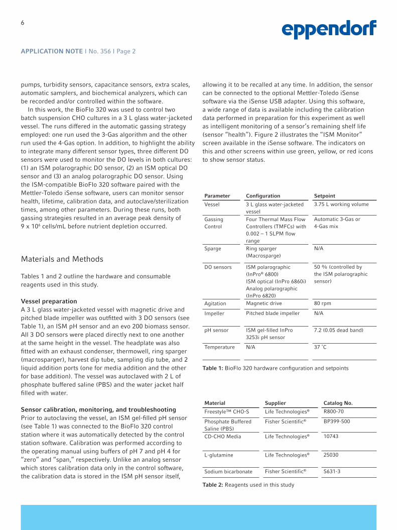
pumps, turbidity sensors, capacitance sensors, extra scales, automatic samplers, and biochemical analyzers, which can be recorded and/or controlled within the software.
In this work, the BioFlo 320 was used to control two batch suspension CHO cultures in a 3 L glass water-jacketed vessel. The runs differed in the automatic gassing strategy employed: one run used the 3-Gas algorithm and the other run used the 4-Gas option. In addition, to highlight the ability to integrate many different sensor types, three different DO sensors were used to monitor the DO levels in both cultures: (1) an ISM polarographic DO sensor, (2) an ISM optical DO sensor and (3) an analog polarographic DO sensor. Using the ISM-compatible BioFlo 320 software paired with the Mettler-Toledo iSense software, users can monitor sensor health, lifetime, calibration data, and autoclave/sterilization times, among other parameters. During these runs, both gassing strategies resulted in an average peak density of 9 x 10⁶ cells/mL before nutrient depletion occurred.
Materials and Methods
Tables 1 and 2 outline the hardware and consumable reagents used in this study.
Vessel preparationA 3 L glass water-jacketed vessel with magnetic drive and pitched blade impeller was outfitted with 3 DO sensors (see Table 1), an ISM pH sensor and an evo 200 biomass sensor. All 3 DO sensors were placed directly next to one another at the same height in the vessel. The headplate was also fitted with an exhaust condenser, thermowell, ring sparger (macrosparger), harvest dip tube, sampling dip tube, and 2 liquid addition ports (one for media addition and the other for base addition). The vessel was autoclaved with 2 L of phosphate buffered saline (PBS) and the water jacket half filled with water.
Sensor calibration, monitoring, and troubleshootingPrior to autoclaving the vessel, an ISM gel-filled pH sensor (see Table 1) was connected to the BioFlo 320 control station where it was automatically detected by the control station software. Calibration was performed according to the operating manual using buffers of pH 7 and pH 4 for “zero” and “span,” respectively. Unlike an analog sensor which stores calibration data only in the control software, the calibration data is stored in the ISM pH sensor itself,
allowing it to be recalled at any time. In addition, the sensor can be connected to the optional Mettler-Toledo iSense software via the iSense USB adapter. Using this software, a wide range of data is available including the calibration data performed in preparation for this experiment as well as intelligent monitoring of a sensor’s remaining shelf life (sensor “health”). Figure 2 illustrates the “ISM Monitor” screen available in the iSense software. The indicators on this and other screens within use green, yellow, or red icons to show sensor status.
Parameter Configuration Setpoint
Vessel 3 L glass water-jacketed vessel
3.75 L working volume
Gassing Control
Four Thermal Mass Flow Controllers (TMFCs) with 0.002 – 1 SLPM flow range
Automatic 3-Gas or 4-Gas mix
Sparge Ring sparger (Macrosparge)
N/A
DO sensors ISM polarographic (InPro® 6800)ISM optical (InPro 6860i)Analog polarographic (InPro 6820)
50 % (controlled by the ISM polarographic sensor)
Agitation Magnetic drive 80 rpm
Impeller Pitched blade impeller N/A
pH sensor ISM gel-filled InPro 3253i pH sensor
7.2 (0.05 dead band)
Temperature N/A 37 ˚C
Table 1: BioFlo 320 hardware configuration and setpoints
Material Supplier Catalog No.
Freestyle™ CHO-S Life Technologies® R800-70
Phosphate Buffered Saline (PBS)
Fisher Scientific® BP399-500
CD-CHO Media Life Technologies® 10743
L-glutamine Life Technologies® 25030
Sodium bicarbonate Fisher Scientific® S631-3
Table 2: Reagents used in this study

7
APPLICATION NOTE I No. 356 I Page 3
A
B
Figure 2: Panels A and B illustrate screenshots from the Mettler-
Toledo iSense software which allows the user to monitor sensor
“health”; calibration data, configuration, and sterilization cycles
can be tracked
Results and Discussion
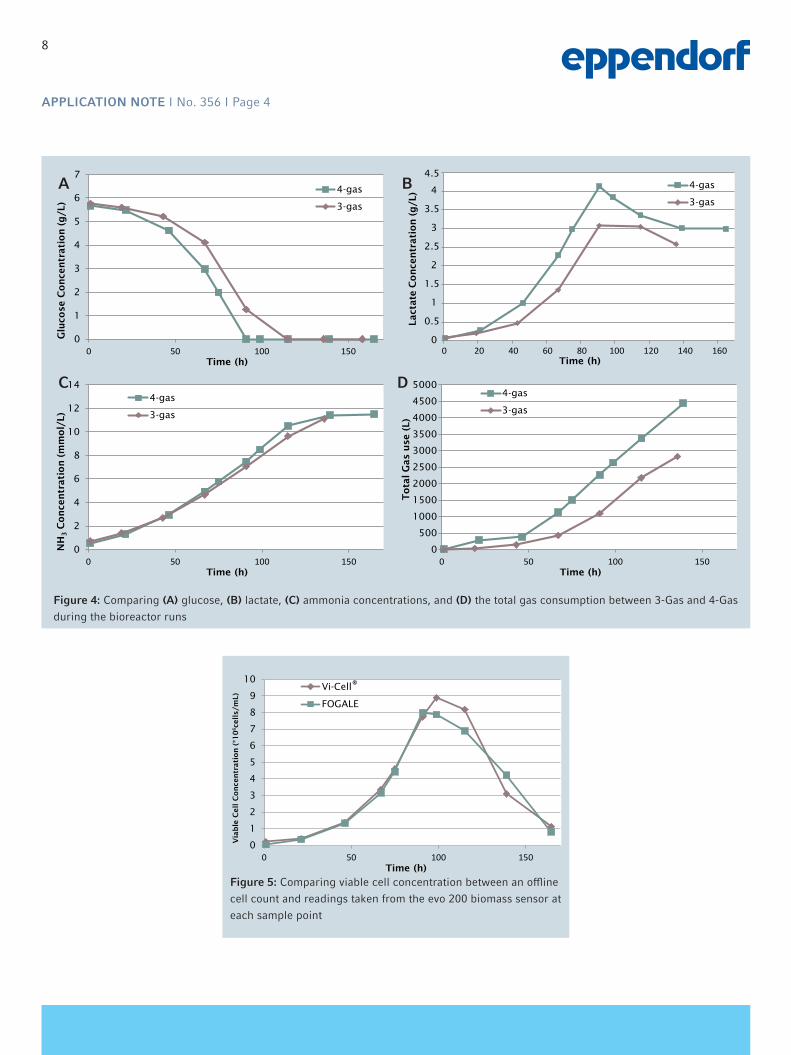
As seen in Figure 3, both the 3-Gas and 4-Gas automatic DO control algorithms allowed the culture to reach similarly high viable cell densities. The 4-Gas experiment reached its peak cell density (8.89 x 10⁶ cells/mL) sooner than the 3-Gas run (9.54 x 10⁶ cells/mL). In addition, Figure 4 shows that glucose consumption and lactate and ammonia accumulation were comparable between the two cultures. Consistent with the cell density trend, the 3-Gas culture consumed glucose slightly slower than the 4-Gas culture. When the glucose was exhausted, the cell growth and viability began to drop. Higher peak densities would have
been possible if glucose and other necessary nutrients had been supplemented using a fed-batch protocol.
The two gassing control algorithms produced comparably healthy cultures, and showed some notable gas consumption differences. The 3-Gas culture consumed more gas overall, as illustrated in Figure 4D. Since the 3-Gas algorithm does not utilize N2 for DO control, there is a possibility for the DO to climb above setpoint at the beginning and end of the run when O2 demand is low. Using 4-Gas control, N2 is available to keep DO at setpoint, which may be beneficial for some sensitive cell types, and for anaerobic cultures. Whether a culture will be healthier with 3-Gas or 4-Gas automatic gassing control will have to be determined empirically for each cell strain.
The evo 200 capacitance biomass sensor was a valuable in-line measure of cell growth during the runs. Figure 5 shows a comparison between the offline viable cell density measurement and the in-line evo 200 capacitance measurement for one run. After calibrating this sensor for a particular cell line and specific culture process, it can be used in place of sampling the bioreactor which would avoid lost volume and reduce the risk of contamination.
Three DO sensors were incorporated into these experiments. The two ISM sensors were automatically detected by the control station, and including the traditional polarographic sensor, all three were able to accurately track and trend DO levels throughout the run. Figure 6 illustrates an example of the DO sensor trends for the 3-Gas experiment. No significant differences were seen between DO measurement by the three sensors.
0
2
4
6
8
10
12
0 20 40 60 80 100 120 140 160
Cell C
on
cen
trati
on
(*1
06
cells/
mL)
Time (h)
4-gas
3-gas
Figure 3: Viable cell densities between the 3-Gas and 4-Gas
experiments; although the 3-Gas run peaked a day after the
4-Gas run, peak cell densities were not significantly different
8
APPLICATION NOTE I No. 356 I Page 4
0
1
2
3
4
5
6
7
0 50 100 150
Glu
cose
Co
nce
ntr
ati
on
(g
/L)
Time (h)
4-gas
3-gas
0
0.5
1
1.5
2
2.5
3
3.5
4
4.5
0 20 40 60 80 100 120 140 160
Lact
ate
Co
nce
ntr
ati
on
(g
/L)
Time (h)
0
2
4
6
8
10
12
14
0 50 100 150
NH
3C
on
cen
trati
on
(m
mo
l/L)
Time (h)
4-gas
3-gas
0
500
1000
1500
2000
2500
3000
3500
4000
4500
5000
0 50 100 150
To
tal G
as
use
(L)
Time (h)
4-gas
3-gas
4-gas
3-gas
Figure 4: Comparing (A) glucose, (B) lactate, (C) ammonia concentrations, and (D) the total gas consumption between 3-Gas and 4-Gas
during the bioreactor runs
A B
C D
0
1
2
3
4
5
6
7
8
9
10
0 50 100 150
Via
ble
Cell C
on
cen
trati
on
(*1
06ce
lls/
mL)
Time (h)
Vi-Cell®
FOGALE
Figure 5: Comparing viable cell concentration between an offline
cell count and readings taken from the evo 200 biomass sensor at
each sample point

9
APPLICATION NOTE I No. 356 I Page 5
Conclusion
With the intelligent upgrades to the BioFlo 320 software and the utilization of intelligent pH/DO sensors, the BioFlo 320 provides advanced process control for CHO cell culture. This method provided similar results using either the 3-Gas or 4-Gas automatic gassing cascades. The setup can be used to meet a host of culture requirements and the upfront knowledge of an ISM sensor’s “health” dramatically reduces operational risk due to potential sensor failure during a cell culture run. In these experiments, the ability to customize the configuration by adding an evo 200 biomass sensor and multiple ISM DO sensors elevated these runs to “intelligent” CHO cell culture. With the addition of an in-line bioanalyzer, sampling of the bioreactor could be eliminated to reduce the risk of sampling-associated contamination, making the BioFlo 320 a superior setup for cell culture and an intelligent choice for bioprocess laboratories worldwide.
Figure 6: This trend was generated by BioCommand® Batch Control which was used to collect data from all control loops through the
runs. In this example, the three DO sensors (red, blue, and pink), sparge air (green) and sparge O2 (maroon) trends are shown during
the 3-Gas run. Note that the three DO trends are superimposed on one another.

10
APPLICATION NOTE I No. 356 I Page 6
Ordering informationDescription Order no. International Order no. North America
BioFlo® 320 Bioprocess Control Station Please Inquire Please Inquire
BioFlo® 320, 3 L vessel bundle, water-jacketed, magnetic drive M1379-0311 M1379-0311
www.eppendorf.com
ISM®, InPro® and Mettler Toledo® are registered trademarks of Mettler-Toledo AG, Switzerland. FOGALE nanotech® is a registered trademark of FOGALE nanotech, France. Vi-Cell® is a registered trademark of Beckman Coulter, Inc., USA. Life Technologies® is a registered trademark of Life Technologies Corporation, USA. Fisher Scientific® is a registered trademark of Fisher Scientific Company, LLC, USA. FreeStyle™ is a trademark of Life Technologies Corporation, USA. Eppendorf®, the Eppendorf logo, and BioBLU® are registered trademarks of Eppendorf AG, Germany. New Brunswick™ is a trademark of Eppendorf AG, Germany. BioFlo®, CelliGen®, and BioCommand®, are registered trademarks of Eppendorf, Inc., USA. US Design Patents are listed on www.eppendorf.com/ip. All rights reserved, including graphics and images. Copyright © 2015 by Eppendorf AG.
Your local distributor: www.eppendorf.com/contactEppendorf AG · 22331 Hamburg · [email protected] · www.eppendorf.com

Perfusion CHO Cell Culture in a BioBLU® 5p Single-Use Packed-Bed Vessel Nick Kohlstrom and Ma ShaEppendorf, Inc., Enfield, CT, USA
Corresponding author: [email protected]
APPLICATION NOTE No. 336 I September 2014
Abstract
The market for humanized monoclonal antibodies (hmAbs), has become a multi-billion dollar industry with the expectation of continued growth. One of the most cost-effective methods for the production of secreted proteins is the packed-bed vessel operated under perfusion conditions. The maximum cell density achieved in a packed-bed vessel is typically much higher than suspension cell culture or microcarrier-based adherent cell culture. The protein harvest can be carried out continuously, providing unparalleled product yield. This poster provides an example of using a BioBLU 5p packed-bed single-use vessel to conduct Chinese hamster ovary cell (CHO) perfusion culture producing a secreted hmAb.
The BioBLU 5p vessel (pre-loaded with Fibra-Cel® disks) was controlled by a New Brunswick™ CelliGen® BLU benchtop bioreactor. The BioBLU 5p vessel was inoculated at an initial cell density of 0.3 x 10⁶ cells/mL. Fourteen days of perfusion cell culture were conducted with a working volume of 3.75 L. Glucose, lactate, and hmAb concentrations were monitored daily. The glucose consumption rate was used to estimate the cell density in the packed-bed vessel. After 12 days, the culture reached a peak cell density of approximately 10 x 106 cells/mL.
Introduction
The New Brunswick CelliGen BLU benchtop bioreactor is a versatile, easy-to-use system with built-in controls and monitoring for agitation, temperature, pH, dissolved oxygen (DO), gassing (with air, oxygen, nitrogen and carbon dioxide) ,and automatic pump control. In addition, the control station can be connected to many other auxiliary devices. The New Brunswick CelliGen BLU benchtop bioreactor is used in conjunction with BioBLU Single-Use Vessels (Eppendorf) allowing for easy scalability while operating in single-use format. Although the single-use bioreactor market has experienced rapid growth in recent years, packed-bed perfusion bioreactor technology has remained predominantly in the traditional glass and stainless steel formats. The single-use packed-bed vessel BioBLU 5p contains Fibra-Cel® which is a solid support growth matrix that is predominantly used for the production of secreted products from cell culture. Since the cells are attached to the Fibra-Cel, it allows for continuous harvest of secreted products without losing
cells over an extended period of time. This makes BioBLU 5p Single-Use Vessels an ideal platform for research and production of secreted proteins or virus from mammalian and insect cell culture.
CHO is a robust cell line that can be cultured to very high cell densities in a packed-bed bioreactor. Using CHO cells to produce recombinant proteins allows proper protein folding and correct post-translational modifications so that the proteins remain biologically active once injected into humans. The cell line has a proven track record in the biopharmaceutical industry [1]. The global market for monoclonal antibodies (mAbs) is expected to reach US $58 billion in 2016 with a variety of new mAbs in the pipeline [2]. In this experiment, an attachment CHO cell line expressing a hmAb was grown using a New Brunswick CelliGen BLU benchtop bioreactor with a BioBLU 5p single-use packed-bed vessel.
11

Parameter SetpointAgitation 100 rpm
Temperature 37 °C
Dissolved oxygen (DO) 50 %
pH 7.1 ± 0.05
Volume (Harvest cascaded to pump)
3.75 L
Gas mix 3-gas automatic gas mixing option
Gas flow control 3 Thermal mass flow controllers (TMFCs) with 0 - 1 SLPM flow range
APPLICATION NOTE I No. 336 I Page 2
Materials and Methods
The B13-24 CHO cell line (ATCC®, CRL-11397™) was adapted to CD CHO media (Life Technologies®, 10743) supplemented with 8 mM L-glutamine (Life Technologies, 25030), 0.125 % heat-inactivated fetal bovine serum (Life Technologies, 10438-034) and 1X penicillin/streptomycin (Life Technologies, 15140-122). The initial culture was conducted on BioCoat™ collagen-coated T-flasks (Corning®, 354485). Cells were inoculated into the BioBLU 5p single-use packed-bed vessel at 0.3 x 10⁶ cells/mL to a total working volume of 3.75 L with the previously described media.
The hardware setup and control loop setpoints used in this study are shown in Table 1. Fresh media was perfused into the vessel as needed to keep the glucose concentration between 1 and 2 g/L. Additional D-(+)-glucose (Sigma-Aldrich®, G5146) was added to the perfusion media as needed to keep the glucose concentration at the desired level without increasing the perfusion rate to a unmanageable level.
The culture’s pH was controlled using automatic CO2 sparging for acid addition and an automatic pump cascade of 1 M sodium bicarbonate (Fisher Scientific®, S631-3) for base addition. Since the cells were attached to the Fibra-Cel packed-bed, bubbles do not interact with the cells which prevents bubble shear. The layer of medium above the packed-bed allows the dilution and mixing of acid or base before allowing them to come in contact with the cells; therefore higher concentrations of acid or base can be used for pH adjustments without adverse effects. Glucose, lactate, and hmAb concentrations were monitored using a Cedex® Bio Analyzer (Roche®).
The approximate amount of glucose consumption per liter per day was calculated by first calculating the average glucose consumption between samples per hour:
Table 1: Parameters and setpoints used for CHO cell growth in the
CelliGen BLU bioreactor
R = V(S1 – S2) + ∆V(PG – S1 + S2
) 2
∆T
> R = Approximate rate of glucose consumption per hour (g/h)
> S1 = Glucose concentration in media sample 1 (g/L) > S2 = Glucose concentration in media sample 2 (g/L) > V = Vessel working volume (L) > PG = Glucose concentration of fresh perfusate (g/L) > ∆V = Perfusion volume between samples (L) > ∆T = Change in time between samples (h)
R was used to calculate the grams of glucose consumption per day by adding the glucose consumed per hour over the 24 hour period. This was then divided by the working volume (3.75 L) to obtain the normalized glucose consumption (g/L/day).
The approximate cell concentration was determined by correlating R with cell growth to obtain a glucose consumption per cell conversion factor. To obtain the conversion factor, CHO cells were cultured in a T-75 flask until 100 % confluence. A precise amount of fresh medium (8 mL) was then added to the flask, the glucose concentration was measured and the cells were incubated for 7.25 h. After the incubation period, the glucose concentration was measured again, the cells were trypsinized from the T-flask and counted on a Vi-Cell® XR automated cell counter (Beckman Coulter®). This information was used to calculate the amount of glucose each cell consumed per hour (~3.92 x 10⁻² ng/cell/h) which was then used to calculate the number of cells in the bioreactor based on the glucose consumption rate. Please note that the conversion factor may be cell line- dependent and may not be applicable to other CHO cells.
12

0
1
2
3
4
5
6
7
0 100 200 300
Co
nce
ntr
ati
on
(g
/L)
Time (h)
GLC
LAC
0
2
4
6
8
10
12
1 2 3 4 5 6 7 8 9 10 11 12 13 14 15
Glu
cose
co
nsu
med
(g
/L)
Day
0
2
4
6
8
10
12
0 100 200 300
Cell
s/m
L (
x 1
06)
Time (h)
00.20.40.60.8
11.21.41.61.8
0 100 200 300
Co
nce
ntr
ati
on
(m
g/L
)
Time (h)
Figure 1: Glucose (GLC) and lactate (LAC)
concentrations throughout the culture
Figure 2: The approximate amount of glucose
consumption (g/L/day)
Figure 3: The approximate cell concentration
Figure 4: The IgG concentration in the bioreactor
over the culture period
APPLICATION NOTE I No. 336 I Page 3
Results and Discussion
Continuous perfusion was used during this experiment to keep glucose levels within a narrow range (Figure 1). The alternative method of fed-batch style non-continuous perfusion can cause large fluctuations in glucose and lactate concentrations which may have an effect on cellular metabolism. Glucose and lactate concentrations were measured multiple times per day during the run. The data were used to adjust the perfusion rate as well as glucose addition rate to keep the glucose level between 1 and 2 g/L where possible.
The cells were attached to the Fibra-Cel and could not be counted directly. Assuming that glucose consumption is proportional to cell growth, glucose consumption was used to calculate the approximate cell number. At the start of the run, glucose consumption steadily increased until day 6 where it began to plateau (Figure 2). Using the conversion factor described above, approximate cell concentrations were determined throughout the run (Figure 3).
Samples were taken throughout the bioreactor run and the IgG concentrations were determined by Cedex Bio Analyzer (Figure 4). These concentrations were measured from samples taken from the vessel and do not include IgG harvested during perfusion.
The cell line used was the only healthy CHO cell line available from ATCC expressing an hmAb. Although this cell line is useful as a model system, the antibody yield is very low. Given a different cell line, much higher cell numbers and antibody production yields are possible.
Conclusion
The New Brunswick CelliGen BLU benchtop bioreactor with BioBLU 5p single-use packed-bed vessel provided precise control and good cell growth throughout the culture period. This combination presents an excellent package for those seeking to produce hmAbs using an attachment CHO cell line. The ability for continuous harvest of secreted products over an extended period of time while maintaining optimal control of cell growth provides great prospects for the antibody market. The cell line and experiments shown in this poster were not optimized and should only be used as an example of the product’s capabilities.
13

APPLICATION NOTE I No. 336 I Page 4
www.eppendorf.com
ATCC® is a registered trademark of American Type Culture Collection, USA. Life Technologies® is a registered trademark of Life Technologies Corporation, USA. Corning® is a registered trademark of Corning Inc., USA. Sigma-Aldrich® is a registered trademark of Sigma-Aldrich Co., USA. Fisher Scientific® is a registered trademark of Fisher Scientific Company, LLC, USA. Cedex® and Roche® are registered trademarks of Roche Diagnostics GmbH, Germany. Vi-CELL® and Beckman Coulter® are registered trademarks of Beckman Coulter, Inc., USA. CRL-11397™ is a trademark of American Type Culture Collection, USA. BioCoat™ is a trademark of Corning Inc., USA. Fibra-Cel® is a registered trademark owned by Celite Corporation and licensed to Eppendorf Inc., USA. Eppendorf®, the Eppendorf logo, and BioBLU® are registered trademarks of Eppendorf AG, Germany. CelliGen® is a registered trademark of Eppendorf Inc., USA. New Brunswick™ is a trademark of Eppendorf AG, Germany. U.S. Design Patents are listed on www.eppendorf.com/ip. All rights reserved, including graphics and images. Copyright © 2016 by Eppendorf AG.
Your local distributor: www.eppendorf.com/contactEppendorf AG · 22331 Hamburg · [email protected] · www.eppendorf.com
References
[1] Jayapal K, Wlaschin K, Hu W-S, Yap M. Recombinant protein therapeutics from CHO cells - 20 years and counting. CHO Consortium 2007; SBE Special Section:40-7.
[2] BCC Research. Antibody drugs: Technologies and Global Markets. 2012; Report code BIO016H.
Ordering information
Description Quantity Order No.
BioBLU® 5p Packed-Bed Vessel, microsparge 1 M1363-0119
BioBLU® 5p Packed-Bed Vessel, microsparge Pack of 4 vessels M1363-0120
BioBLU® 5p Packed-Bed Vessel, macrosparge 1 M1363-0133
BioBLU® 5p Packed-Bed Vessel, macrosparge Pack of 4 vessels M1363-0134
BioBLU® Packed-Bed Vessel Kit, includes heat blanket, RTD, DO probe, optical pH transmitter, needle-free syringes
1 M1363-0108
14

15
Introduction
In the world of bioprocess, there are many tools and methods that can be used to culture mammalian cells, each with their own strengths, weaknesses and purposes. One of the most critical decisions that is made before a bioprocess system purchase is which impeller type is ideal for a particular cell culture. In this application note, three impeller types were compared using CHO cell culture: The pitched-blade impeller, the spin filter with marine-blade impeller and the packed-bed basket impeller. All experiments were performed using a New Brunswick™ CelliGen® 310 benchtop bioreactor.
The pitched-blade impeller has three flat blades set at approximately a 45 ° angle which produces both axial and radial flow. Right handed or left handed blades are options that can be considered depending on which direction you would like your axial flow. Pitched-blade impellers are low shear impellers, designed to gently mix both suspension cells and cells attached to a microcarrier. Typically, these impellers are used for mammalian, insect or other shear-sensitive cell lines, but have also been used in highly viscous fermentation cultures with bacteria and fungi, as well as some biofuel processes. When using a pitched-blade impeller, a culture is typically grown in a batch-style run (no media is added or removed) or fed-batch-style run (a culture is started at a lower working volume and more media is added later during the run). A perfusion-style run (fresh media is continuously added and old media is removed)
is possible, however, unless a filtering device is attached with this system to prevent the cells from being removed, cells will be depleted with the harvested (“waste”) media.
A spin filter is a cylinder-shaped cage that spins with the impeller shaft and is covered with a screen designed to prevent cells
from being collected with the waste media. Typically, underneath the spin filter, a marine blade is attached to the impeller shaft. When attached to the vessel, media is added so it covers the spin filter almost to its top, with a specially designed harvest tube that can reach the media inside the spin filter. When used, this device can keep cells in the vessel while old media is perfused out from inside of the spin filter. The spin filter is offered with two screen sizes, 10 µm openings for suspension cultures and 75 µm openings for microcarrier cultures. The marine-blade impeller attached underneath the spin filter provides gentle mixing but, due to its unidirectional flow, has a lower KLa than the pitched-blade. The spin filter is perfect for cultures that secrete proteins or compounds of interest since the desired product can be collected with the media while the cells are left to continue to produce. This also helps with downstream processing as cells will not have to be removed
Pitched-Blade vs. Spin Filter vs. Packed-bed Basket: CHO Cell Culture ComparisonNick Kohlstrom, Kevin Voll and Rich Mirro, Eppendorf Inc., Enfield, CT, U.S.A.
APPLICATION NOTE No. 320 I November 2013
Abstract
In the following application note, the pitched-blade impeller, the spin filter impeller and the packed-bed basket impeller are discussed, highlighting the uses and advantages for each type. Then examples of actual CHO
cell cultures are given for each impeller type; showing the perfusion capability when using the spin filter or packed-bed basket impeller and the resulting higher cell densities over the pitched-blade impeller.
Figure 1: Pitched-blade impeller (left) and spin filter with marine blade (right)

16
APPLICATION NOTE No. 320 I Page 2
with centrifugation or filtration. It should be noted that at very high density cultures the spin filter may eventually get clogged with cell debris and require cleaning, which can limit run time.
The packed-bed basket impeller, combined with Fibra-Cel® disks, is a system perfect for manufacturing high-yield secreted products from both attachment and suspension cultures with perfusion. Fibra-Cel is a solid supported fiber-mesh matrix microcarrier used predominantly for secreted products with perfusion. Fibra-Cel allows for long-term, high-density cultures without the risk
of clogging. Fibra-Cel can be used for both anchorage-dependent cultures and suspension cultures due to its electrostatically-treated material and woven nature that traps the cells in a single step within 15 - 60 minutes (no need to stop agitation). The basket consists of two horizontally positioned, perforated metal screens that isolate a section in the interior of the vessel that is filled with Fibra-Cel. The impeller consists of a hollow tube (draft tube) with three smaller discharge tubes radiating from the top. When media is filled over the three tubes at the top of the impeller and it is spun, the centrifugal force exerted on the media forces out the liquid, causing a gentle suction at the bottom of the impeller, which brings media from the bottom of the vessel to the top. The media then gently flows through the Fibra-Cel packed-bed from the top to the bottom. Gases are sparged into the vessel through the central draft tube; this method oxygenates the media but prevents bubbles from interacting with the cells growing inside the Fibra-Cel packed-bed, thus, preventing bubble shear.
Eppendorf also offers other impellers for various bioprocess needs. Some impellers offered but not explored in this application note include the Rushton-type impellers; which are ideal for fermentation cultures with bacteria, yeast and fungi that require higher dissolved oxygen level (oxygen transfer rate) but are not sensitive to mechanical shearing damage; and the Cell-Lift impeller; which is an ultra-low-shear impeller that provides uniform circulation for microcarrier cultures and a bubble free environment for the cells.
Materials and methods
Table 1: Materials, media and cellsMaterial Supplier Catalog no.CelliGen® 310 Control Station Eppendorf See ordering
information, page 6
4 TMFC (0 - 1 SLPM) Eppendorf2.5 L water jacketed vessel (with motor)
Eppendorf
2.5 L pH/DO Sensor Kit (with cables) Eppendorf2.5 L Pitched-Blade Impeller Kit Eppendorf2.5 L Spin Filter Impeller Kit (10 µm) Eppendorf2.5 L Basket Impeller Kit EppendorfYSI 2700 Select™ analyzer YSI® Life Science 2700DVi-CELL® XR Beckman
Coulter®731050
Media and cellsFibra-Cel® Disks Eppendorf M1292-9988Freestyle® CHO-S Life
Technologies®R800-70
CD CHO media Gibco® 10743L-glutamine JRH Biosciences® 90114Penicillin/streptomycin 100x Gibco® 15140-122D-(+)-Glucose Sigma-Aldrich® G5146Sodium Bicarbonate Thermo Fisher
Scientific® Chemical
S631-3
Bioreactor conditionsDuring all three of the following CHO bioprocess examples, a CelliGen 310 Bioreactor with four 0-1 Standard Liters Per Minute (SLPM) Thermal Mass Flow Controllers (TMFC) were used. A TMFC is a device that monitors specific gas flow and is used by the cabinet to automatically control the gases flowing into the vessel. The vessel was a 2.5 L glass, water-jacketed vessel with a magnetic drive motor. The water jacket provides uniform temperature distribution with gentle heating and cooling for the culture while the magnetic drive motor provides a sterile vessel environment. All three culture types utilized 3 gas mixing (Air, O2 and CO2) for DO and pH control with a base addition (Pump 2, 0.3 M sodium bicarbonate solution). Table 2 shows all of the settings for each loop used during all three runs. Both the DO and pH were controlled using the cascade parameters seen in Tables 3 and 4.
Figure 2: Packed-bed basket with Fibra-Cel disks
Figure 3: CelliGen 310 with packed-bed basket impeller

17
APPLICATION NOTE No. 320 I Page 3
Table 2: Loop settingsLoop SetpointAgitation See each exampleTemperature 37 °CpH-1 7.20 (Deadband 0.05)pH-2 OffDO-1 50DO-2 OffAir AutoO2 AutoGs3Flo OffCO2 Auto
Table 3: DO-1 cascadeStart setpoint
@ DO start output %
End setpoint
@ DO end output %
Air 0.0 0.0 0.5 60O2 0.0 10 1 100
Table 4: pH-1 cascadeStart setpoint
@ pH start output %
End setpoint
@ pH end output %
Pump 2 0.0 0.0 100 100CO2 0.0 0.0 0.3 -50
Cells were grown in CD CHO media supplemented with 8 mM of L-glutamine and 1 % penicillin/streptomycin and kept at a total working volume of ~1.6 L. Each vessel was inoculated at identical densities of 0.3 x 10⁶ cells/mL. Glucose was added to the perfusion media as needed. Cell counts were performed on the pitched-blade and spin filter reactors using a Vi-CELL®. A YSI® 2700 Biochemical Analyzer was used to determine glucose and lactate concentrations for all three reactors.
Results
Pitched-blade cultureThe pitched-blade reactor was run at an agitation speed of 80 rpm. It was cultured as a batch-style reactor so no media was added or removed throughout the process run. As you can see from Figure 4, viable cell concentration continued to rise until all of the glucose was consumed from the media at which point the cell viability began to drop. Lactate levels increased until the drop in glucose concentrations caused a shift in cellular metabolism which caused the cells to consume lactate.
Figure 4: The pitched-blade viable cell concentration and glucose and lactate concentrations. Viable cell concentration begins to decrease when all the glucose is consumed in the vessel due to it being a batch-style run.
Spin filter cultureThe spin filter reactor was run at an agitation speed of 100 rpm with a 10 µm filter screen. With the spin filter, the culture was run using continuous perfusion. One of the CelliGen 310 cabinet pumps was calibrated and run at varying rates of input as needed to maintain a glucose level above 1 g/L and to keep waste metabolites low. Another pump was cascaded to a level sensor so media was automatically removed from the vessel anytime it reached a volume over 1.6 L. Since the media being removed was from inside the spin filter, the cells were retained outside of the 10 µm spin filter cage. Figure 5 shows that the cells achieved a high density and viability with perfusion using the spin filter. Although the spin filter can achieve 3X, the run ended due to the high cell concentration eventually clogging the spin filter.
Figure 5: The spin filter viable cell concentration and glucose and lactate concentrations. Perfusion prevented glucose from being totally consumed from the vessel and lactate levels from getting too high.

18
APPLICATION NOTE No. 320 I Page 4
Packed-bed basket cultureThe packed-bed basket impeller was run at an agitation speed of 100 rpm and the basket was filled with 70 g of Fibra-Cel disks. This culture, like the spin filter, was run using continuous perfusion using the same methods as described above, except that media was removed from a normal harvest tube, not from inside of the basket. Since all of the cells were trapped in the Fibra-Cel disks and could not be counted using standard methods, the cell number was determined using the amount of glucose consumption. Due to glucose levels being too high during the run, the cells transitioned from a log phase to stationary phase resulting in a plateau in cell growth, as seen in Figure 6. Higher cell numbers were expected.
Figure 6: Packed-Bed Basket results showing calculated viable cells as well as glucose and lactate concentrations. Perfusion prevented
glucose from being totally consumed from the vessel and lactate levels from getting too high.
Discussion
Each impeller and cell culture method results in a different growth pattern and it is necessary to determine what is best for the desired process. When comparing the viable cell growth curves for each of the impellers (Figure 7), it can be seen that each results in a different cell concentration and rate of growth. More importantly, as discussed earlier, some of the impellers/methods allow for perfusion (Packed-bed Basket and Spin filter) resulting in higher and possibly continually sustainable cultures.
The pitched-blade impeller provided a simple way to grow a low-density culture, but it is not possible to grow the culture to a higher density without extra cell separation
Figure 7: A comparison of viable CHO cell concentration for all three impeller experiments. The packed-bed basket impeller provided long term, high-density cell growth. The spin filter also provided high density cell growth compared to the pitched-blade impeller. Since the pitched-blade impeller was run as a batch-style reactor, a lower viable cell density was reached which eventually drops due to all the glucose being consumed in the vessel.
equipment to allow for perfusion. The spin filter resulted in almost 4X the number of cells as the pitched-blade impeller due to its ability to run in perfusion mode. The perfusion process usually does not last as long as the Fibra-Cel basket due to the tendency of clogging at very high cell densities. However, the cost of the spin filter is much less than that of the Fibra-Cel basket. It is reusable and it does not rely on consumable Fibra-Cel disks. The packed-bed basket impeller resulted in 8X the number of cells as the pitched-blade impeller and over 2X the spin filter. The packed-bed impeller culture also grew faster than the spin filter culture which was most likely due to the lack of direct physical agitation and bubble shear on the cells while they are trapped in the Fibra-Cel disks. Table 5 shows a general list of the advantages for each impeller type. Every cell line is different and what will work best for each culture and purpose can vary. Table 6 is a general guide for choosing impellers based on some common cell lines. The CHO cell cultures in this paper were not optimized and are just a general example of what can be expected for each impeller type.
Table 5: Advantages for each impeller typeImpeller AdvantagesPitched-Blade Impeller
> Axial and Radial flow > Simple design > Suspension or Microcarrier attached cultures
Spin Filter Impeller
> Easy to use with perfusion > Capable of higher cell densities
Basket Impeller > Higher cell densities without the risk of clogging > Gentler environment for cells

19
APPLICATION NOTE No. 320 I Page 5
Table 6: A general guide to choosing impellers by cell line
Cell lineRushton,
Rushton-Like Pitched-Blade Marine Blade Spin Filter Cell Lift BasketHumanHEK 293 HeLa HL60 Lncap THP-1 UMSCC HFF KB MRC-5 HybridomaDA4.4 123A 127A GAMMA 67-9-B SP20 PrimateVero COS-7 Rat TumorGH3 9L PC12 Mouse3T3 MC3T3 NS0 HamsterCHO BHK ZebrafishZF4 AB9 InsectSF9 Hi-5 Sf21 BacteriaStreptomyces Bacillus Escherichia coli YeastSaccharomyces cereviseae Baker’s yeast Pichia pastoris Candida albicans AlgaeRed/Green

20
APPLICATION NOTE No. 320 I Page 6
www.eppendorf.com
Your local distributor: www.eppendorf.com/contact Eppendorf AG • 22331 Hamburg • Germany [email protected]
YSI® is a registered trademark of YSI Inc., USA. YSI 2700 Select™ is a trademark of YSI Inc., USA. Vi-CELL® and Beckman Coulter® are registered trademarks of Beckman Coulter, Inc., USA, Gibco® and Life Technologies® are registered trademarks of Life Technologies Corporation, USA. FreeStyle™ is a trademark of Life Technologies Corporation, USA. Thermo Fisher Scientific® is a registered trademark of Thermo Fisher Scientific, Inc., USA. JRH Biosciences® and Sigma-Aldrich® are registered trademarks of Sigma-Aldrich Corporation, USA. Eppendorf® and the Eppendorf logo are registered trademarks of Eppendorf AG, Germany. New Brunswick™ is a trademark of Eppendorf AG, Germany. Fibra-Cel® is a registered trademark owned by Imerys Mineral California, Inc. and licensed to New Brunswick Scientific Co., Inc., USA. CelliGen® and BioFlo® are registered trademarks of New Brunswick Scientific Co., Inc., USA. All rights reserved, including graphics and images. Copyright © 2013 by Eppendorf AG.
References
1. Mirro, R, and K. Voll. 2009. Which Impeller Is Right for Your Cell Line?. BioProcess Int. 7:52-57.
Ordering Information
Product Description International order no. N. America order no.Voltage Option Cabinet voltage M1287-1020 (200V) M1287-1010 (120V)CelliGen® 310 Control Station Cell culture control cabinet M1287-2110 M1287-21104 TMFC (0 - 1 SLPM) Gas flow control M1287-2020 M1287-20202.5 L water jacketed vessel (with motor) Cell culture vessel M1287-0310 M1287-03102.5 L pH/DO Sensor Kit (with cables) pH and Dissolved oxygen sensors M1287-0400 M1287-04002.5 L Pitched-Blade Impeller Kit Pitched-blade impeller M1287-5068 M1287-50682.5 L Spin Filter Impeller Kit (10 µm) Spin Filter Impeller M1287-1125 M1287-11252.5 L Basket Impeller Kit Basket impeller M1287-1140 M1287-1140Fibra-Cel® Disks Microcarrier M1292-9988 M1292-9988
For information on products used in this application note or other sizes and options available please contact your local sales representative.

21
Abstract
The study presents a typical protocol for the setup and operation of the Eppendorf New Brunswick CelliGen BLU single-use, stirred-tank bioreactor, a versatile new benchtop system for the culture of a wide range of mammalian cells. This bioreactor has been designed to provide research and production facilities with a single-use vessel which
combines the benefits of both traditional stirred-tank design and single-use technology, capable of seamless process scale-up. The system can be operated in batch, fed-batch or continuous modes. A procedure for culturing Chinese Hamster Ovarian (CHO) cells in a 5.0 L vessel, using CD CHO serum-free medium in a batch culture is described.
Growing CHO Cells in a New Brunswick™ CelliGen® BLU Benchtop, Stirred-Tank Bioreactor Using Single-Use VesselsGuozheng Wang, Wenying Zhang, Rich Mirro and Vikram Gossain, Eppendorf Inc., Enfield, CT, U.S.A.
Introduction
Historically, stirred-tank fermentors and bioreactors have been the trusted design for culturing all types of submerged cultures including suspension and anchorage-dependent mammalian cells, insect, yeast, plant and microbial cultures. The tried and tested tank design offers scalability and proven reproducibility which is pivotal for cost-saving process development and productivity. In the last decade, there has been an increasing acceptance and use of single-use technologies, due to their convenient operation and low start-up cost. Single-use systems eliminate the need for cleaning and sterilization, reduce validation requirements, provide rapid turn-around between runs, and significantly reduce the risk of cross contamination and microbial contamination because the culture vessel is only used once and then discarded. Although single-use, stirred-tank systems in the 75 – 2000 L scale have been on the market for some time, as have small-scale single-use bags that are gently rocked rather than stirred, until now there has been no single-use stirred-tank system for small-scale work. The new Eppendorf New Brunswick CelliGen BLU fills that void, offering a proven stirred-tank design as well as the benefits of single-use technology in a benchtop system.
Materials and Methods
Single-Use VesselsBioBLU® single-use vessels are offered in 5.0, 14.0 and 50.0 L total volume capacities. The vessels are delivered preassembled with pitched-blade impeller, porous microsparge, and all the necessary tubing, filters, and connectors; and come sterilized, ready for use right out of the package. All components in product contact are made of materials that meet USP Class VI standards and have been tested for leachables and extractables, making these vessels appropriate for cGMP environments. In this protocol, we describe use of a CelliGen BLU with 5.0 L vessel.
APPLICATION NOTE No. 312 I April 2012
Rapid set up, easy operation, and elimination of autoclav-ing and cleaning between runs. These are a few of the many advantages of the new BioBLU 5.0, 14.0 and 50.0 L stirred-tank bioreactors for growth of mammalian cultures.

22
APPLICATION NOTE No. 312 I Page 2
ControllerCelliGen BLU’s compact control station is designed to provide advanced process management and monitoring capability, ranging from three fixed-speed pumps for additions and harvesting, to a powerful controller with 15 in. industrial color touchscreen monitor. Multiple options, including gas flow control, a weight scale, validation packages and more, enable customization to your needs.
The control station used in this protocol was configured with one 2 – 100 cubic centimeters per minute (ccm) Thermal Mass Flow Controller (TMFC) for direct sparging of gases and an integrated gas overlay with 0.1 – 3.0 Standard Liters Per Minute (SLPM) flow rate also regulated by a TMFC. Both the gas flow and gas overlay are capable of 4-gas mixing for automatic pH and Dissolved Oxygen (DO) control. Pumps, temperature control, agitation, as well as all of the other process loops, were controlled and monitored through the powerful Reactor Process Controller (RPC) firmware installed in the controller. DO was monitored using a noninvasive reusable polarographic DO probe; and pH was monitored using a non-invasive optical pH probe and fluorescence sensor.
Inoculum PreparationOne 2.5 mL vial of CHO cells was thawed and used to inoculate a 125 mL shake flask which contained 25 mL of serum-free CD CHO medium (Life Technologies® 10743-029) which was pre-warmed to 37 °C.
On day 4, when the viable cell density reached 1.5 x 10⁶ cells/mL, the cells were transferred into a 500 mL shake flask which contained 100 mL of freshly made, pre-warmed medium and allowed to incubate for 3 additional days at the same conditions as earlier. The cells were then transferred to two 1 L shake flasks, each containing 250 mL of the freshly made medium. The inoculum was grown in the shake flasks until cell density reached 2.0 – 3.0 x 10⁵ cells/mL, with greater than 90 % cell viability, sufficient for the bioreactor inoculation.
Bioreactor Set-Up and InoculationOne day before the cells reached inoculation density, the growth medium was warmed to 37 °C and the DO probe was polarized. For this study, 3.0 L of sterile CD CHO serum-free medium was prepared by pre-warming at 37 °C for 24 hours in a CO2 incubator. During this time, the DO probe was connected to the controller for at least 6 hours to enable polarization, as per the manufacturer’s recommendation.
Once the medium was warmed and the inoculum grown to sufficient starting density, the CelliGen BLU bioreactor vessel was removed from its sterile packaging and the heat blanket supplied with the unit was wrapped around the outside of the vessel. Next, the vessel containing the cell culture medium was connected to one of the bioreactor vessel’s inlet lines using a tube welder. (A tube welder is offered as an optional accessory to the CelliGen BLU. A pre-sterilized medium filter with an attached quick connect or Luer connection can also be used if a tube welder is not available). Since this was a batch process, all of the medium was pumped into the bioreactor vessel. All additional connections to the controller including sparge, overlay, RTD, pH, and agitation were also made.
pH and DO were calibrated through the touchscreen controller, and all process setpoints were entered on the touchscreen using the Control Setpoint values shown on the next page. Once the parameters were at their setpoints, the inoculum flasks were connected to the addition line in a sterile manner using a tube welder and contents were pumped into the bioreactor vessel.
Operational ParametersCultivation of animal cells in an environment optimal for manufacture of desired end products require monitoring and control of a substantial number of physical and chemical parameters. Physical parameters include temperature, fluid flow (gas flow and liquid flow) rates and agitation rates. Chemical parameters include the dissolved oxygen (DO) concentration and pH.
Control SetpointsTemperature 37 °CpH 7.0DO 40 %Agitation 80 rpm
pH Control ParameterspH control was set to Auto mode, which automatically adds base solution or CO2 gas to the system based on culture demands.
Dead-band 0.10PID values Factory set default valuesBase Sodium bicarbonate, 7.5 % solutionBase Solution Transfer tubing
Narrow bore silicone tubing with Luer- connection (1⁄18 in. ID & 1/4 in. OD)
Vessel inlet 1/8 in. inlet tubing in the vessel headplate

23
APPLICATION NOTE No. 312 I Page 3
Dissolved Oxygen (DO) ControlDO control was set to Auto mode, which automatically regulates gas mixing based on culture demand. PID values: factory set default values.
Gas ControlThe gas control was set to 4-gas mode, which automatically maintains DO and pH. The gas flow rate was based on the vessel size.
Up until day 3, gases were introduced into the vessel headspace only through the overlay port at a rate of 0.30 L/min using 4-gas mixing to maintain pH and DO. On day 3, and for the remainder of the run, 5 – 10 ccm of gas were directly sparged into the system using a porous sparger and automatic 4-gas mixing. The overlay gas flow in the vessel headspace was kept at the previous settings.
A built-in sampling device enabled sterile sampling. Daily offline measurements of glucose and lactate concentration were read using a YSI® 2700, and cell density and cell viability was measured using an Automated Cell Counting System (New Brunswick NucleoCounter®).
All data was logged via BioCommand® Batch Control PC-compatible Supervisory Control and Data Acquisition (SCADA) software (New Brunswick).
Results and Discussion
As shown in Figure 1, the CHO cells in this study grew steadily, reaching a maximum viable cell density of 5.55 x 10⁶ cells/mL on day 5.
Cell viability, shown in Figure 2, ranged between 97.1 and97.9 % through Day 5, until the nutrient source, glucose, was depleted from the medium, as shown in Figure 3.
Figure 1. Cell growth over the 7-day run.
Figure 2. Cell viability remained high through day 5.
Figure 3. Glucose consumption and lactate production.

24
APPLICATION NOTE No. 312 I Page 4
As expected, lactate production steadily increased as the available glucose in the medium was consumed. As glucose in the medium became exhausted, consumption of lactate as a secondary carbon source also declined[1].
This data presented here, and in Table 1, demonstrates that the CelliGen BLU bioreactor is an easy-to-use, efficient system for the culture of CHO cells. No effort was made to optimize either the medium or the cell culture process control parameters. This study was only intended to document a general guide to bioreactor setup and operation, and present typical results you could expect to achieve with your mammalian cell line. For protocols on other cell lines, or for additional information on the CelliGen BLU, see eppendorf.com.
DayTotal Viable Viability
[%]Glucose[g/L]
Lactate[g/L][106 cells/mL]
0 0.31 0.30 97.9 5.83 0.231 0.69 0.68 97.1 5.14 0.522 1.42 1.39 97.6 4.711 0.873 2.57 2.51 97.6 3.74 1.274 4.02 3.92 97.5 1.47 2.105 5.70 5.55 97.3 0.59 1.126 5.98 4.52 76.6 0.00 0.327 6.71 3.21 47.8 0.00 0.01
Table 1.
References
[1] A single nutrient feed supports both chemically de-fined NS0 and CHO fed-batch processes: Improved productivity and lactate metabolism. Ma N, Ellet J, Oke-diadi C, Hermes P, McCormick E, Casnocha S. Biotechnol Prog. 2009; 25 (5): 1353-63.
www.eppendorf.com
NucleoCounter® is a registered trademark of ChemoMetec A/S, Denmark. Life Technologies® is a registered trademark of Life Technologies, Inc., USA. YSI® is a registered trademark of YSI Inc. Eppendorf®, the Eppendorf logo and BioBLU® are registered trademarks of Eppendorf AG, Germany. New Brunswick™ is a trademark of Eppendorf AG, Germany. CelliGen® and BioCommand® are registered trademarks of New Brunswick Scientific Co., Inc., USA. All rights reserved, including graphics and images. Copyright © 2013 by Eppendorf AG.
Your local distributor: www.eppendorf.com/contact Eppendorf AG • 22331 Hamburg • Germany [email protected]

25
Abstract
This study illustrates a protocol for the scale up of CHO cells using New Brunswick™ CelliGen BLU stirred-tank bioreactors equipped with 5-Liter (L) and 50-Liter (L) single-use vessels. CelliGen BLU is a versatile benchtop system for the culture of a variety of cell lines. This bioreactor has been designed to provide research and
production facilities with single-use vessels that combine the benefits of both traditional stirred-tank design and single-use technology, capable of seamless process scale-up. Eppendorf has recently launched the CelliGen BLU 50 L system to address larger volume batch demands.
Single-Use Scalability: CHO Cell Culture using 5 to 50 L New Brunswick™ CelliGen® BLU Benchtop, Stirred-Tank Bioreactors
Nick Kohlstrom, Joseph Capone, and Ma Sha, Eppendorf Inc., Enfield, CT, U.S.A.
Introduction
Historically, stirred-tank bioreactors have been the standard for culturing all types of submerged cultures including suspension and anchorage-dependent mammalian, insect, yeast, plant and microbial cultures. This well-tested vessel design offers scalability and reproducibility, which enhance productivity and provide cost savings in process development. In the last decade, there has been an increasing acceptance of single-use technologies, due to their convenient operation and low start-up cost. Single-use systems eliminate the need for cleaning and sterilization, reduce validation requirements, provide rapid turn-around between runs, and significantly reduce the risk of cross contamination and microbial contamination. Until recently, the CelliGen BLU single-use bioreactor system has been limited to 5 L and 14 L sizes. The new 50 L CelliGen BLU vessel is a direct response to customer feedback, accommodating much larger process volumes while maintaining the benefits of single-use technology in the same proven, rigid-walled, stirred-tank design, all in a benchtop platform. All three vessel sizes have the capability to be operated in batch, fed-batch or perfusion style. This protocol describes a cell culture process using Freestyle™ Chinese Hamster Ovarian (CHO-S) cells (Invitrogen® Corp.) starting from the smaller 5 L vessel and finishing up in the larger 50 L vessel.
Materials and Methods
Single-Use VesselsCelliGen BLU single-use vessels are now offered in 5.0 L, 14.0 L, and 50.0 L volumes. The vessels are delivered pre-assembled with a pitched-blade impeller. The vessels have either a porous micro-sparge or a macro-sparge element configuration (selected at time of purchase), and also include all the necessary tubing, filters, and connectors. The vessels come sterilized and ready for use right out of the package. All components in contact with cell culture are made from materials that meet USP Class VI standards and have been tested for leachables and extractables, making these vessels appropriate for cGMP environments. In this protocol, we describe the use of 5 L and 50 L CelliGen BLU vessels with pitched blade impellers and the macrosparge element configurations. The 5 L culture was conducted in a batch style while the 50 L culture was completed as a fed-batch.
APPLICATION NOTE No. 257 I November 2012
The CelliGen BLU 50 L stirred-tank bioreactor offers many advantages for mammalian cell culture: it sets up rapidly, it is easy to operate, and it elimi-nates cleaning and autoclaving between runs

26
APPLICATION NOTE I No. 257 I Page 2
ControllerThe CelliGen BLU compact control station is designed to provide advanced process management and monitoring capability with a powerful Reactor Process Controller (RPC) with 15” LCD color touchscreen monitor. The controller includes three integrated pumps, and other options enable customization to meet a customer’s needs, including high- or low-flow thermal mass flow controllers (TMFC) for gas flow control, scales, and validation packages.
The control station used in this protocol for the 5 L vessel was equipped with three low-flow TMFCs (draw at 0.002-1.0 standard liters per minute [SLPM]) for direct sparging to control gases including air, oxygen, nitrogen, and CO2. The controller was also equipped with an integrated gas overlay function controlled by a single TMFC with a regulated flow of 0.05 - 5.0 SLPM. The control station used for the 50 L vessel utilized the same design with the high gas flow option with TMFCs ranging between 0.04 - 7.5 SLPM. Both the sparge and overlay were capable of 3-gas or 4-gas mixing for automatic pH and Dissolved Oxygen (DO) control. For this protocol, both the 5 L and the 50 L set-ups were operated in 3-gas mode in conjunction with 0.1 SLPM of air as an overlay. The pumps, temperature control, agitation, and all other process loops were controlled and monitored through the RPC firmware installed in the controller. DO and pH were monitored non-invasively; DO was monitored using a traditional, stainless-steel polarographic probe, while pH was monitored using an optical probe with fluorescence sensor.
Inoculum PreparationFor the 5 L bioreactor inoculation, Freestyle Chinese Hamster Ovarian (CHO-S) cells were used to inoculate a 125 mL shake flask which contained 30 mL of serum-free CD CHO medium (Invitrogen) supplemented with 8mM L-glutamine (JRH Biosciences) and 1 % Penicillin-Streptomycin (Invitrogen).
This initial shaker culture was expanded to a one liter shake flask containing 240 mL; the inoculum was then grown until day 4, when the viable cell density reached 3.16 x 10⁶ cells/mL with a viability of 99.3 %, a density sufficient for transfer into the 5 L bioreactor.
Bioreactor Set-Up and Inoculation
Inoculation of 5 L BioreactorThe DO probe was connected to the controller for at least 6 hours for polarization.
On inoculation day, the CelliGen BLU bioreactor vessel was removed from its sterile packaging and the heat blanket was wrapped around the outside of the vessel. Next, the
vessel containing the cell culture medium was connected to one of the bioreactor vessel’s inlet lines using a quick connect. A Luer connection or tube welder can also be used with the CelliGen BLU. All additional connections to the controller including sparge, overlay, RTD, pH, and agitation were also made.
Approximately 2 L of sterile CD CHO serum-free medium was pumped into the vessel and warmed to 37° C. After the growth medium was stabilized at 37° C, the traditional polarographic probe (DO probe) was calibrated to an electronic zero , and then spanned after the agitation was set at 50 rpm and the airflow was set to 100 % at 1 SLPM for ~20 minutes (may vary depending on how long it takes the raw value to stabilize). The optical pH calibration was performed using the pH probe raw data (located on the vessel and packaging; preconfigured for optical pH using fluorescence sensor technology), and an offline sample was taken to re-zero the medium within the bioreactor (after the fluorescence spot was hydrated ~20 mins). DO and pH were calibrated through the touchscreen controller, and all process setpoints were entered on the touchscreen using the Control Setpoint values represented within the following pages below. Once the parameters were at their setpoints, the inoculum flasks were connected to the addition line in a sterile manner using a quick connect and the cells were pumped into the bioreactor vessel for a total volume of ~2 L with an inoculation density of 0.3 x 10⁶ cells/mL.
Inoculation of 50 L BioreactorMedium warming and DO polarization were conducted in a similar fashion to the preparation prior to the inoculation of the 5 L vessel.
Once culture growth within the 5 L bioreactor had achieved sufficient density (4.84 x 10⁶ cell/mL; viability 99.4 %), the 50 L CelliGen BLU vessel was removed from its sterile packaging and the heat blanket was wrapped around the outside of the vessel. Next, the bag containing the cell culture medium was connected to one of the 50 L bioreactor vessel’s inlet lines using a quick connect. Since this portion of the experiment was a fed batch process with starting volume less than 20 L, only the initial 17.8 L of the medium was pumped into the bioreactor vessel. All additional connections to the controller including sparge, overlay, RTD, pH, and agitation were also made. The base pump was also calibrated and primed for use with 20 % Sodium Bicarbonate for pH control.
The polarographic probe on the 50 L vessel was calibrated to an electronic zero once the growth medium was stabilized at 37° C, and then spanned when the agitation was set at 50 rpm and the airflow was set to 100 % at 7.5 SLPM

27
APPLICATION NOTE I No. 257 I Page 3
(~20 minutes to stabilize the raw value). The optical pH calibration was performed using the pH probe raw data, and an offline sample was taken to re-zero the medium within the bioreactor. DO and pH were calibrated through the touchscreen controller, and all process setpoints were entered on the touchscreen using the Control Setpoint values shown on the next page. Once the parameters were at their setpoints, the 5 L harvest line was connected to the inoculation/addition line on the 50 L in a sterile manner using a quick connect, and then the calculated 1.2 L of high density CHO cells were pumped in for a total volume of 19 L with final starting cell density of 0.3 x 10⁶ cells/mL. The 50 L vessel was then fed with an additional 21 liters of pre-warmed Gibco CD CHO serum-free medium on day 5 to support high cell growth and viability.
Operational ParametersCultivation of animal cells for manufacturing of desired end products requires monitoring and controlling of a substantial number of physical and chemical parameters. Physical parameters include temperature, gas flow rates, fluid flow rate, and agitation speed. Chemical parameters include the dissolved oxygen (DO) concentration and pH.
5 L and 50 L Control SetpointsTemperature 37° CpH 7.1DO 50 %Agitation 50 rpm (clockwise)
5 L and 50 L pH Control ParametersBoth vessels’ pH control was set to Auto mode, which automatically adds base solution or CO2 gas to the system based on culture demands. Base addition was utilized for pH control on the 50 L culture due to the higher density expected at end of the 50 L run but was not needed for the 5 L run.
pH Dead-band 0.05PID values factory set default valuesBase (50 L only) Sodium bicarbonate, 20 % solution
5 L and 50 L Dissolved Oxygen (DO) ControlDO control was set to 3-gas Auto mode, which automatically regulates gas mixing based on culture demand. Factory-set default PID values were used.
5 L and 50 L Gas ControlThe gas control was set to 3-gas Auto mode for both bioreactors, automatically maintaining DO and pH. For the 5 L bioreactor, the low flow limit set at 0.002 SLPM with a high flow limit set at 1.00 SLPM, and for the 50 L Bioreactor, the low flow limit was set at 0.04 SLPM with a high flow limit set at 7.5 SLPM. In addition, overlay air flow was supplied to both bioreactors at 0.10 SLPM.
Gases were introduced via the macrosparge element for aeration supplementation to maintain DO and pH and into the vessel headspace through the overlay port during the entire run for both the 5 L and the 50 L bioreactors.
A built-in sampling device enabled sterile sampling. Daily off-line measurements of glucose and lactate concentration were read using an YSI® 2700; cell density and cell viability were measured using an Automated Cell Counting System (Vi-CELL®).
Results and Discussion
All vessel data was logged via BioCommand® Batch Control Supervisory Control and Data Acquisition (SCADA) software (Eppendorf).The bioreactors’ total cell density and viability are shown in Figure 1. CHO cells exhibited steady and consistent growth on both the 5 L and 50 L bioreactors. Cell growth reached a viable cell density of 4.82 x 10⁶ cells/mL on day 4 in the 5 L, and 8.58 x 10⁶ cells/mL on day 8 in the 50 L vessel. Cell viability was maintained around 99 % for the entire culture duration. As expected, lactate production steadily increased as the available glucose in the medium was consumed (Figure 2).
Figure 1. CHO Cell growth and viability plots for the scale-up from 5 L to 50 L CelliGen BLU bioreactors in a combined cell culture process of 8 days. The dip of cell count seen on the 50 L graph represents the single feeding event and cell density dilution resulted from 21 L media addition introduced on day 5.
pH Dead-band………… 0.05 PID values……… …factory set default values Base (50L only) . . . .Sodium bicarbonate, 20 % solution 5L&50L Dissolved Oxygen (DO) Control DO control was set to 3-gas Auto mode, which automatically regulates gas mixing based on culture demand. Factory-set default PID values were used. 5L&50L Gas Control The gas control was set to 3-gas Auto mode for both bioreactors, automatically maintaining DO and pH. For the 5L Bioreactor, the low flow limit set at 0.002 SLPM with a high flow limit set at 1.00 SLPM, and for the 50L Bioreactor, the low flow limit was set at 0.04 SLPM with a high flow limit set at 7.5 SLPM. In addition, overlay air flow was supplied to both bioreactors at 0.10 SLPM. Gases were introduced via the macrosparge element for aeration supplementation to maintain DO and pH and into the vessel headspace through the overlay port during the entire run for both the 5L and the 50L bioreactors. A built-in sampling device enabled sterile sampling. Daily off-line measurements of glucose and lactate concentration were read using an YSI 2700®; cell density and cell viability were measured using an Automated Cell Counting System (Vi-Cell™). 3. Results & Discussions
All vessel data was logged via BioCommand® Batch Control Supervisory Control and Data Acquisition (SCADA) software (Eppendorf).The bioreactors’ total cell density and viability are shown in Figure 1. CHO cells exhibited steady and consistent growth on both the 5L and 50L bioreactors. Cell growth reached a viable cell density of 4.82 x 10⁶ cells/mL on day 4 in the 5L, and 8.58 x 10⁶ cells/mL on day 8 in the 50L vessel. Cell viability was maintained around 99 % for the entire culture duration. As expected, lactate production steadily increased as the available glucose in the medium was consumed (Figure 2).
Figure 1. CHO Cell growth and viability plots for the scale-up from 5L to 50L CelliGen® BLU bioreactors in a combined cell culture process of 8 days. The dip of cell count seen on the 50L graph represents the single feeding event & cell density dilution resulted from 21L media addition introduced on day 5.
0
20
40
60
80
100
0
1
2
3
4
5
6
7
8
9
10
0.0 50.0 100.0 150.0 200.0
% V
iabi
lity
Tota
l cel
ls/m
L (*
10^6
)
Time (hours)
Total cells vs Viability
5L Total cells/mL
50L Total cells/mL
5L % Viability
50L % Viability

28
APPLICATION NOTE I No. 257 I Page 4
Figure 2. Glucose consumption and lactate production in the 5 L and the 50 L vessels.
The data presented here demonstrates that the CelliGen BLU bioreactor is an easy-to-use, efficient system for the scale-up of CHO cell culture up to the 50 L vessel size.
No efforts were made to optimize either the medium or the cell culture process control parameters.
This study was only intended to document a general procedure for CelliGen BLU bioreactor setup and operation, and present typical results one could expect to achieve with mammalian cell line.
Although it is possible to perform such scale-up using a single CelliGen BLU controller, two separate CelliGen BLU systems were used for this application note. For protocols on other cell lines, or for additional information on the CelliGen BLU, see eppendorf.com.
References
1. Productivity Studies Utilizing Recombinant CHO Cells in Stirred-Tank Bioreactors: A Comparative Study Between Pitched-Blade and Packed-Bed Bioreactor Systems. Taylor Hatton, Shaun Barnett, Abby D. Ben-ninghoff, PhD, and Kamal Rashid, PhD. Bioprocessing J., Volume 11, Issue 2 (Summer 2012)
2. A single nutrient feed supports both chemically defined NS0 and CHO fed-batch processes: Improved produc-tivity and lactate metabolism. Ma N, Ellet J, Okediadi C, Hermes P, McCormick E, Casnocha S. Biotechnol Prog. 2009 Jul 27.
Figure 2. Glucose consumption and lactate production in the 5L and the 50L vessels. The data presented here demonstrates that the CelliGen® BLU bioreactor is an easy-to-use, efficient system for the scale-up of CHO cell culture up to the 50L vessel size. No efforts were made to optimize either the medium or the cell culture process control parameters. This study was only intended to document a general procedure for CelliGen BLU bioreactor setup and operation, and present typical results one could expect to achieve with mammalian cell line. Although it is possible to perform such scale-up using a single CelliGen BLU controller, two separate CelliGen BLU systems were used for this application note. For protocols on other cell lines, or for additional information on the CelliGen BLU, see www.nbsc.com/BLU. Order Information
Product Description (Specific Configuration for this test)
Order no. International Order no. North America
CelliGen® BLU Control Station
100-120V,50/60HZ, high flow sparge, overlay w/TMFC, and scale
(M1374-120-HSA)-120V
(M1374-230-HSA)-230V
(M1374-120-HSA)
CelliGen® BLU Control Station
100-120V,50/60HZ, low flow sparge, overlay w/TMFC, and scale
(M1374-120-LSA)-120V
M1374-230-LSA–230V
(M1374-120-LSA)
CelliGen® BLU 5L vessel
Single use 5.0L vessel with macrosparge. Pack of 1
M1363-0121 M1363-0121
0
1
2
3
4
5
6
7
0.0 50.0 100.0 150.0 200.0
Conc
entr
atio
n (g
/L)
Time (hours)
Glucose & Lactate Concentrations 5L Glucose concentration
50L Glucose concentration
5L Lactate concentration
50L Lactate concentration

29
APPLICATION NOTE I No. 257 I Page 5
Ordering Information
Description International order no. North America order no.CelliGen® BLU Control Station 100 - 120V, 50/60Hz, high flow sparge, overlay w/TMFC, and scale
M1374-120-HSA (120V)M1374-230-HSA (230V)
M1374-120-HSA
CelliGen® BLU Control Station100 - 120V, 50/60Hz, low flow sparge, overlay w/TMFC, and scale
M1374-120-LSA (120V)M1374-230-LSA (230V)
M1374-120-LSA
CelliGen® BLU 5L vessel Single-use 5.0L vessel with macrosparge (pack of 1)
M1363-0121 M1363-0121
CelliGen® BLU 14L vessel
Single-use 14.0L vessel with macrosparge (pack of 1)M1363-0122 M1363-0122
CelliGen® BLU 50L vessel
Single-use 50.0L vessel with macrosparge (pack of 1)M1363-0129 M1363-0129
CelliGen® BLU vessel kit
Includes heat blanket, RTD temperature sensor, pH and DO probes, with cables and needle-free syringes
M1363-0105 (5L kit)M1363-0114 (14L kit)M1374-0151 (50L kit 120V) M1374-0150 (50L kit 230V)
M1363-0105 (5L kit)M1363-0114 (14L kit)M1374-0151 (50L kit)
Eppendorf Research® plus, adjustable pipette
Single channel pipette – 1 -10 mL3120 000.089 3120000089
Easypet®
Pipette dispenser – suitable for pipettes from 0.1 to 100 mL 4421 000.013 022230204
www.eppendorf.com
Invitrogen® is a registered trademark and Freestyle™ is a trademark of Invitrogen Corporation. YSI® is a registered trademark of YSI Inc. Vi-CELL® is a registered trademark of Beckman Coulter, Inc. CelliGen® and BioCommand® are registered trademarks of New Brunswick Scientific, Co., Inc, USA. Eppendorf®, the Eppendorf logo, Eppendorf Research® and Easypet® are registered trademarks and New Brunswick™ is a trademark of Eppendorf AG, Germany. All rights reserved, including graphics and images. Copyright © 2012 by Eppendorf AG, Germany.
Your local distributor: www.eppendorf.com/contact Eppendorf AG • 22331 Hamburg • Germany [email protected]

30
Abstract
In this study, the New Brunswick S41i CO2 incubator shak-er’s mammalian cell culture capability was first verified by culturing CHO cells. This was followed by a comparative performance evaluation against two leading incubator shakers on the market. The New Brunswick S41i provided equivalent performance on the growth rate and
viability of mouse hybridoma cells. Comparison of CO2 gas consumption was also conducted. Due to the superior “green” engineering and advanced control of critical pa-rameters, the New Brunswick S41i demonstrated up to 10 times lower gas consumption compared to the competi-tive units while delivering uncompromised performance.
Hybridoma and CHO Cell Culture using the New Brunswick™ S41i, an Environmentally-Friendly, “Low Emission” Incubator ShakerNick Kohlstrom, George Wang, Linette Philip and Ma Sha, Eppendorf Inc., Enfield, CT, U.S.A.
Introduction
Cars aren’t the only source of CO2 emissions; laboratory equipment, such as CO2 incubators, could be releasing over 20,000 liters of CO2 gas per year. Eppendorf® established the epGreen initiative to reduce the environmental impact of our products. Most of the CO2 gas consumed by incubators is released to the environment. Eppendorf’s new incubator shaker, the New Brunswick S41i, releases extremely low amounts of CO2 under normal cell culture conditions without sacrificing performance. This study evaluates the New Brunswick S41i’s performance culturing hybridoma and Chi-nese hamster ovary (CHO) cells. The study also investigates the New Brunswick S41i’s CO2 gas consumption compared to competitive units. The data reveals that the New Bruns-wick S41i consumes 5 to 10 times less CO2 than competi-tors, resulting in a 5 to 10 times smaller carbon footprint. Superior engineering minimizes gas leakage with a tightly sealed inner glass door protected by sturdy outer door, tightly sealed motor drive boots as well as a sealed incuba-tion chamber. The performance evaluation, based on the comparison of cell culture growth rates, cell densities, and percent viabilities, demonstrates the New Brunswick S41i’s in-dustry leading performance. This new CO2 incubator includes a robust New Brunswick triple eccentric drive shaker for ac-curate and stable parameters required to grow non-adherent cells. The shaker drive is optimized for high performance within a humid and carbon dioxide rich environment.
Materials and Methods
Instruments > New Brunswick S41i equipped with high-tem-perature disinfection
> CO2 incubator shaker from competitor 1
> CO2 incubator shaker from competitor 2
> Vi-CELL® analyzer (Beckman Coulter, Germany) > YSI® 2700 analyzer (YSI Life Science, USA) > New Brunswick Galaxy® gas analyzer > Omega® FMA-1608A thermal mass flow-meter (Omega Engineering, USA)
> Eppendorf consumables - Research® plus, single channel pipette - epT.I.P.S®
- Easypet®
Media and cells > DG44 CHO cell (Invitrogen) > EX-CELL® CD CHO serum-free medium for CHO cells (Sigma)
> Hybridoma cell DA4-4; ATCC:HB57 > DMEM (ATCC) > Fetal Bovine Serum 5% (Gibco) > Penicillin-Streptomycin 100x (Gibco)
APPLICATION NOTE No. 255 I July 2012

31
APPLICATION NOTE I No. 255 I Page 2
CHO culture protocolCHO cells were grown in EX-CELL CD CHO serum-free medium supplemented with 1% penicillin-streptomycin antibiotic. Six 250mL Erlenmeyer flasks were each inocu-lated with 60mL of stock culture at a concentration of 3 x 10⁵ cells/mL. All flasks were prepared from the same stock culture. Erlenmeyer flask was placed in six different loca-tions on the shaker platform and the results were averaged. The flasks were incubated at 37°C in a mixture of 5% CO2, 95% air and agitated at 130 RPM (4.69 rcf).
CHO cells were grown for a period of 14 days. A sample was taken on days 3, 5,7,10, 12 and 14 and was analyzed for glucose concentration, cell concentration and viability using YSI 2700 and Beckman Coulter Vi-CELL.
Hybridoma culture protocolDA4-4 hybridoma cells were grown in DMEM medium supplemented with 5% FBS and 1% Penicillin-Streptomy-cin. Six 250mL Erlenmeyer flasks were each inoculated with 45mL of stock culture at a concentration of 2 x 10⁵ cells/mL. All shake flasks were prepared from the same stock culture and were equally distributed in six different locations in the New Brunswick S41i, competitor 1 and competitor 2. The flasks were incubated at 37°C in a mixture of 5% CO2 and 95% air. The units were agitated at 95 RPM (2.52 rcf).
Hybridoma cells were subcultured on day 2 and 4 to a concentration of approximately 2 x 10⁵ cells/mL. A sample was taken every day from each of the flasks and analyzed for glucose concentration, cell concentration, and viability using YSI 2700 and Beckman Coulter Vi-CELL, respectively.
Gas consumptionThe New Brunswick S41i and competitor incubator shakers 1 and 2 were programmed at 37ºC, 95 RPM and 5% CO2 and were allowed to equilibrate for at least 12 hours. Inline CO2 gas pressures were set at the lowest values recom-mended by each manufacturer. An offline gas analyzer was used to verify the CO2 levels within each incubator. A thermal mass flow-meter was used to record volumetric gas consumption over a time period of 48 hours on each unit. Tests were repeated three times and the average values are reported below.
Results
1.) Growth assessment of CHO and hybridoma
Figure 1: Average viable cell concentration and viability of CHO culture in the New Brunswick S41i.
Viable cell density of CHO cells reached a maximum of 4.72 x 10⁶ cells/mL by day 7. The cell viability was maintained at approximately 98% up to day 5 and dropped steadily there-after (Figure 1).
Figure 2: Comparison of average viable cell densities of hybridoma cultures grown in New Brunswick S41i and competitors 1 and 2.
Hybridoma culture protocol DA4-4 hybridoma cells were grown in DMEM medium supplemented with 5% FBS and 1% Penicillin-Streptomycin. Six 250mL shake flask were each inoculated with 45mL of stock culture at a concentration of 0.2 x 10⁶ cells/mL. All shake flasks were prepared from the same stock culture and were equally distributed in six different locations in the New Brunswick S41i, competitor 1 and competitor 2. The flasks were incubated at 37°C in a mixture of 5% CO2 and 95% air. The units were agitated at 95 RPM (2.52 rcf). Hybridoma cells were subcultured on day 2 and 4 to a concentration of approximately 2x10^6 cells/mL. A sample was taken every day from each of the flasks and analyzed for glucose concentration, cell concentration, and viability using YSI 2700 and Beckman Coulter Vi-CELL, respectively. Gas consumption The New Brunswick S41i and competitor shaker incubators 1 and 2 were programmed at 37ºC, 95 RPM and 5% CO2 and were allowed to equilibrate for at least 12 hours. Inline CO2 gas pressures were set at the lowest values recommended by each manufacturer. An offline gas analyzer was used to verify the CO2 levels within each incubator. A thermal mass flow-meter was used to record volumetric gas consumption over a time period of 48 hours on each unit. Tests were repeated multiple times and the average values are reported below. Results 1) Growth assessment of CHO and hybridoma
Viable cell density and % viability of CHO cells
0.00E+00
1.00E+06
2.00E+06
3.00E+06
4.00E+06
5.00E+06
0 2 4 6 8 10 12 14 16Process time (days)
Viab
le c
ell d
ensi
ty (c
ells
/mL)
20
40
60
80
100
Viab
ility
(%)
New Brunswick S41i cell density New Brunswick S41i viability
Figure 1: Average viable cell concentration and viability of CHO culture in the New Brunswick S41i. Viable cell density of CHO cells reached a maximum of 4.72 x 10⁶ cells/mL by day 7. The cell viability was maintained at approximately 98% up to day 5 and dropped steadily thereafter (Figure 1). .
Comparison of viable cell density - hybridoma cells
0.00E+00
5.00E+05
1.00E+06
1.50E+06
2.00E+06
0 1 2 3 4 5 6 7 8Process time (days)
Viab
le c
ell d
ensi
ty (c
ells
/mL)
New Brunswick S41i Competitor 1 Competitor 2
Figure 2: Figure 2: Comparison of average viable cell densities of hybridoma cultures grown in New Brunswick S41i and competitors 1 and 2. In comparison to CHO cells, hybridoma culture was able to maintain a high average viability of approximately 95% through day 6, due to the subculturing of cells during log phase of growth on days 2 and 4. The maximum viable cell density of 1.81 x 10⁶ cells/mL was achieved on day 6 (Figures 2 and 3).

32
APPLICATION NOTE I No. 255 I Page 3
In comparison to CHO cells, hybridoma culture was able to maintain a high average viability of approximately 95% through day 6, due to the subculturing of cells during log phase of growth on days 2 and 4. The maximum viable cell density of 1.81 x 10⁶ cells/mL was achieved on day 6 (Fig-ures 2 and 3).
Figure 3: Comparison of average percentage viabilities of hybridoma cultures grown in New Brunswick S41i and com-petitors 1 and 2.
2.) Measurements of gas consumption
The measurement of CO2 consumption at 5% CO2 setpoint revealed that the competitive units evaluated consumes much higher CO2 gas over the same period as compared to the New Brunswick S41i (Figure 4).
Figure 4: Average CO2 gas consumption of tested units in standard liters (Sltr) over a 24 hour period
Discussion & Conclusion
The rising need to create more eco-friendly products and customer demand for higher efficiency were taken into consideration during the development of the New Bruns-wick S41i. This study validates the performance of the new CO2 incubator with a New Brunswick shaker built inside by growing two cell lines which are very commonplace in research and production. Process and media were not opti-mized for either cell lines in this study.
In conclusion, the results show that the New Brunswick S41i is competent at growing mammalian cells while reduc-ing environmental impact down to a minimum. The New Brunswick S41i combines a robust triple eccentric drive shaker within a CO2 incubator, to provide accurate and stable parameters required for the growth of non-adherent cells.
Comparison of % viability- hybridoma cells
40.0
60.0
80.0
100.0
120.0
0 2 4 6 8process time (in days)
% v
iabi
lity
New Brunswick S41i Competitor 1 Competitor 2
Figure 3: Comparison of average percentage viabilities of hybridoma cultures grown in New Brunswick S41i and competitors 1 and 2. 2) Measurements of gas consumption The measurement of CO2 consumption at 5% CO2 setpoint revealed that the competitive units evaluated consumes much higher CO2 gas over the same period as compared to the New Brunswick S41i (Figure 4).
Daily Gas Usage
Competitor 2
New Brunswick S41i
Competitor 1
0
10
20
30
40
50
60
70
1unit
CO2
cons
umpt
ion
(SLt
r)
New Brunswick S41i Competitor 1 Competitor 2
Figure 4: Average CO2 gas consumption of tested units over a 24 hour period Discussions & Conclusions The rising need to create more eco-friendly products and customer demand for higher efficiency were taken into consideration during the development of the New Brunswick S41i. This study validates the performance of the new CO2 incubator with a New Brunswick shaker built inside by growing two cell lines which are very commonplace in research and production. Process and media were not optimized for either cell lines in this study. In conclusion, the results show that the New Brunswick S41i is competent at growing mammalian cells while reducing environmental impact down to a minimum. The New Brunswick S41i combines a robust quadruple eccentric drive shaker within a CO2 incubator, to provide accurate and stable parameters required for the growth of non-adherent cells. Ordering Info Product Description Order no.
International Order no. North America
New Brunswick S41i CO2 shaker incubator with high temperature disinfection
SI41-230-0100 SI41-120-0100
Galaxy gas analyzer Electronic CO2 gas analyzer
P0628-6150 P0628-6150
eppendorf Research® plus, adjustable
Single channel pipette – 1-10 mL
3120 000.089 3120000089
Easypet® Pipette dispenser – suitable for pipettes from 0.1 to 100 mL.
4421 000.013 022230204

33
APPLICATION NOTE I No. 255 I Page 4
Ordering Information
Description International order no. North America order no.New Brunswick S41i CO2 incubator shaker with high-temperature disinfection
S41I-230-0100 S41I-120-0100
Galaxy gas analyzerElectronic CO2 gas analyzer
P0628-6150 P0628-6150
eppendorf Research® plus, adjustable Single channel pipette – 1-10 mL
3120 000.089 3120000089
Easypet®
Pipette dispenser – suitable for pipettes from 0.1 to 100 mL4421 000.013 022230204
www.eppendorf.com
Eppendorf®, Research®, epT.I.P.S.® and Easypet® are registered trademarks of Eppendorf AG and New Brunswick™ is a trademark of Eppendorf AG, Hamburg, Germany. Galaxy® is a registered trademark of New Brunswick Scientific, Co., Inc., USA. Vi-CELL® is a registered trademark of Beckman Coulter, Inc.YSI® is a registered trademark of YSI Inc. Omega® is a registered trademark of Omega Engineering Inc. EX-CELL® is a registered trademark of Sigma-Aldrich Co. LLC. All rights reserved, including graphics and images. Copyright © 2012 by Eppendorf.
Your local distributor: www.eppendorf.com/contact Eppendorf AG • 22331 Hamburg • Germany [email protected]

34
Abstract
The objective of this study was to compare New Bruns-wick CelliGen BLU single-use packed-bed bioreactor and the traditional glass vessel counterpart used in New Brunswick CelliGen 310. Alkaline phosphatase (ALKP)-secreting Chinese Hamster ovary (CHO) cells were used to measure ALKP production in each bioreactor. Overall,
the results from these comparisons suggest that there is no significant difference between the reusable and single-use FibraCel basket systems for bench-scale production of recombinant proteins. Productivity of cells and col-lection of secreted proteins will not be hindered by the implementation of single-use bioreactor systems.
CHO Cell Culture with Single-Use New Brunswick™ CelliGen® BLU Packed-Bed Fibra-Cel® BasketTaylor Hatton, Shaun Barnett, and Kamal Rashid* *Associate Director, Center for Integrated BioSystems, Utah State University, Logan, UT, U.S.A.
Product Contact: Kevin Voll ([email protected])
Introduction
The packed-bed basket technology, developed by New Brunswick Scientific (acquired by Eppendorf Inc. in 2007), provides a shear free environment for production of animal cells. At present, little information is available on the utility of the New Brunswick CelliGen® BLU single-use bioreactor system for the production of secreted proteins, especially in perfusion mode of operation. Thus, this study was conduct-ed to measure the growth and productivity of alkaline phos-phatase (ALKP)-secreting rCHO. Two packed-bed bioreactor types were used: 5 L New Brunswick CelliGen® BLU single-use vessel and 2.5 L autoclavable glass vessel both operated by New Brunswick CelliGen® 310 console in perfusion mode. The perfusion process provides a homeostatic environment for optimal cell growth similar to that experienced by cells in vivo, where waste products are constantly removed and fresh nutrients are replenished. Cells cultured in packed-bed bioreactors are not exposed to hydrodynamic forces, thus, allowing for maximum cell growth and protein expression1. The objective of this study was to compare the two types of bioreactors to determine if any differences are observed between the productivity of the two bioreactors.
Materials and Methods
Culture proceduresIn order to evaluate the impact of these bioreactor systems on protein production, we utilized a recombinant alkaline phosphatase-secreting CHO cell line (rCHO), a proprietary cell line provided by CDI Bioscience, Inc. (Madison, WI). The rCHO cells were engineered with the IPTG-regulated RP Shift vector so that the rCHO cells stop replicating and shift to protein production when induced with IPTG. Serum free CD-CHO medium (Gibco, Life Technologies, Grand Island, NY) was used throughout these experiments. The media contains 6.3g/L glucose and was supplemented with 8 mM L-glutamine and 100µg/ml of an antibiotic/an-timycotic solution (Invitrogen, Life Technologies). Frozen rCHO cells were thawed and transferred to T-75 flasks with CD-CHO medium and allowed to expand. Once a sufficient number of cells were achieved, sterile disposable spinner flasks were utilized to further expand the cells. Subculture of the cells continued until a sufficient number of viable cells was achieved for use as a seed culture at the density of 5 x 10⁵ cells/ml. Two New Brunswick CelliGen® 310 advanced bench-top stirred-tank bioreactors were utilized to grow the rCHO cells. One of the New Brunswick CelliGen® consoles was connected to an adaptor kit (available from Eppendorf) for use of the New Brunswick CelliGen® BLU single-use vessel.
APPLICATION NOTE No. 254 I July 2012

35
APPLICATION NOTE I No. 254 I Page 2
Table 1: Comparison of perfusion volumes
Perfusion Glass BLU
Day 1 0.5 L 1 L
Day 2 1 L 2 L
Days 3 - 15* 2 L 4 L
* Perfusion occurred every other day.
Table 2: Bioreactor parameters (setpoints)
Parameter Glass BLU
Temperature 37º C (± 0.1° C) 37º C (± 0.1° C)
Agitation 120 rpm (± 5 rpm) 120 rpm (± 5 rpm)
DO 35 % (± 1 %) 35 % (± 1 %)
pH 7.1 (± 0.01) 7.1 (± 0.01)
Gas flow 0.5 slpm 1.5 slpm
Packed-bed basket impeller operated in perfusion modeTwo experimental trials were performed using the packed-bed vessels in perfusion mode: 2.5 L total volume autoclav-able vessel (1.7 L working volume) and a 5 L total volume single-use vessel (3.5 working volume, pre-loaded with 150g of Fibra-Cel® disks). The perfusion process was initi-ated once the cells reached the exponential growth phase as shown in table 1. Both experimental trials had the following parameters shown in table 2.
Biomarkers of cell growth and productivityCell productivity was assessed by measuring activity of the secreted ALKP protein using an enzyme assay (AnaSpec, Freemont, CA) according to the manufacturer’s protocol. For simplicity unit measurements were used in this study. A unit (U) of ALKP activity was defined as the amount of enzyme that hydrolyzes 1µmol of p-nitrophenylphosphate to p-nitrophenol in a total reaction volume of 1 ml in 1 minute at 37° C. The YSI 2700 Select Biochemistry Analyzer (YSI, Inc., Yellow Springs, OH) was utilized to monitor the glucose and lactate levels in the culture media every 24 hr for the duration of each trial.
Results and Discussion
Glucose utilization and lactate productionGlucose is the main energy source for cell proliferation and ALKP production. Thus, glucose levels were expected to directly correlate with ALKP production in each experiment. Because lactate is a secondary energy source, lactate levels were expected to decline following this initial increase and the utilization of glucose in the media. Lactate metabolism is beneficial to the system by reducing a major metabolic by-product from the system2,3. Glucose levels measured at the time of induction (day 3) were nearly 0 g/L in both ex-periments (Fig. 1). Media lactate concentrations increased in response to decreasing glucose availability. The use of lactate as a secondary energy source can also be observed as lactate levels decrease at each 2 L perfusion.
Figure 1. Glucose consumption and lactate production by rCHO cells cultured in two packed-bed bioreactor system. Values shown are the amounts of glucose and lactate measured in the culture media at each media exchange. The time and volume of the media exchange is indicated at each dashed line. Induction of ALKP activ-ity by IPTG began on culture day 5 and continued every two days throughout the remainder of the experiment. Results of two experimental trials are shown (A, reusable; B, single-use).
RESULTS AND DISCUSSION
Glucose utilization and lactate production
Glucose is the main energy source for cell proliferation and ALKP production. Thus, glucose
levels were expected to directly correlate with ALKP production in each experiment. Because lactate is a
secondary energy source, lactate levels were expected to decline following this initial increase and the
utilization of glucose in the media. Lactate metabolism is beneficial to the system by reducing a major
metabolic by-product from the system [2, 3]. Glucose levels measured at the time of induction (day 3) were
nearly 0 g/L in both experiments (Fig. 1). Media lactate concentrations increased in response to decreasing
glucose availability. The use of lactate as a secondary energy source can also be observed as lactate levels
decrease at each 2 L perfusion.
0 1 2 3 4 5 6 7 8 9 10 11 12 13 14 15 16 170.0
1.0
2.0
3.0
4.0
5.0
6.0
Con
cent
raito
n [g
/L]
A 0.5 L 1 L 2 L2 L 2 L 2 L2 L 2 L
0 1 2 3 4 5 6 7 8 9 10 11 12 13 14 15 16 170.0
1.0
2.0
3.0
4.0
5.0
6.0
Glucose
Con
cent
ratio
n [g
/L]
B 1 L 2 L 4 L4 L 4 L4 L 4 L
Lactate
4 L
Days in culture
Figure 1. Glucose consumption and lactate production by rCHO cells cultured in two packed-bed bioreactor system. Values shown are the amounts of glucose and lactate measured in the culture media at each media exchange. The time and volume of the media exchange is indicated at each dashed line. Induction of ALKP activity by IPTG began on culture day 5 and continued every two days throughout the remainder of the experiment. Results of two experimental trials are shown (A, reusable; B, single-use).

36
www.eppendorf.com
Eppendorf® is a registered trademark and New Brunswick™ is a trademark of Eppendorf AG, Germany. CelliGen® is a registered trademark of New Brunswick Scientific Co., Inc., USA. Fibra-Cel® is a registered trademark owned by Celite Corporation and licensed to New Brunswick Scientific Co., Inc., USA. All rights reserved, including graphics and images. Copyright © 2012 by Eppendorf.
Your local distributor: www.eppendorf.com/contact Eppendorf AG • 22331 Hamburg • Germany [email protected]
APPLICATION NOTE I No. 254 I Page 3
Comparison of bioreactor systems for ALKP production
Figure 2. ALKP production by rCHO cells cultured in two packed-bed bioreactor system. (A) ALKP con-centration in culture media measured each day of each experimental trial. IPTG induction of ALKP began on culture day five and continued every two days for the remainder of the experiment. (B) Stacked bar charts show the cumulative production of ALKP throughout the experiment, with each bar representing a perfusion.
The average total ALKP production per experiment trial is shown in Figure 2; overall, there is not a significant differ-ence in ALKP production between the two bioreactor sys-tems. The total amount of ALKP measured after five media exchanges in the reusable vessel was 17.44 U/mL and 16.22 U/mL in the single-use vessel.
In summary, these results demonstrated comparable yields in ALKP production (within the usual biological fluctua-tions) between the two packed-bed bioreactor systems when operated in perfusion. Given the greater productivity of cells cultured in the packed-bed bioreactor and the multitude of advantages of this system operated in perfusion mode, researchers desiring to scale up mammalian cell culture for protein production should strongly consider utilization of the New Brunswick CelliGen® BLU packed-bed, single-use bioreactor system.
Note: This application note study was partially funded by Eppendorf Inc.
References
1. Merten, O.W., P.E. Cruz, C. Rochette, C. Geny-Fiamma, C. Bouquet, D. Goncalves, O. Danos, and M.J.T. Carrondo (2001) Comparison of different bioreactor systems for the production of high titer retroviral vectors. Biotechnology Progress 17(2): p. 326-335.
2. Ma, N.N., J. Ellet, C. Okediadi, P. Hermes, E. McCormick, and S. Casnocha (2009) A single nutrient feed supports both chemically defined NS0 and CHO fed-batch pro-cesses: improved productivity and lactate metabolism. Biotechnology Progress 25(5): p. 1353-1363.
3. Hu, W.S., T.C. Dodge, K.K. Frame, and V.B. Himes (1987) Effect of glucose on the cultivation of mammalian cells. Dev Biol Stand 66: p. 279-90.
Comparison of bioreactor systems for ALKP production
The average total ALKP production per experiment trial is shown in Figure 2; overall, there is not a
significant difference in ALKP production between the two bioreactor systems. The total amount of ALKP
measured after five media exchanges in the reusable vessel was 17.44 U/mL and 16.22 U/mL in the single-
use vessel.
In summary, the results of this study show that there are no significant differences in ALKP
production between the two packed-bed bioreactor systems when operated in perfusion mode. Given the
greater productivity of cells cultured in the packed-bed bioreactor and the multitude of advantages of this
system operated in perfusion mode, researchers desiring to scale up mammalian cell culture for protein
7 9 11 13 15 17 7 9 11 13 15 170
1
2
3
4
Reusable Single-Use
A
Days in culture
ALK
P [U
/mL]
Reusable Single-Use0
5
10
15
20B
ALK
P [U
/mL]
Figure 2. ALKP production by rCHO cells cultured in two packed-bed bioreactor system. (A) ALKP concentration in culture media measured each day of each experimental trial. IPTG induction of ALKP began on culture day five and continued every two days for the remainder of the experiment. (B) Stacked bar charts show the cumulative production of ALKP throughout the experiment, with each bar representing a perfusion.

37
Solving the Aggregation Problem of Human Embryonic Kidney 293 Cells Using the New Brunswick™ S41i CO2 Incubator ShakerAuthorStacey Willard and Ma Sha
Eppendorf, Inc., Enfield, CT, USA
Corresponding author: [email protected]
APPLICATION NOTE No. 339 I January 2015
Abstract
Human embryonic kidney 293 (HEK293) cells are among the most versatile hosts for recombinant protein expression. These cells are capable of expressing large membrane proteins such as G protein-coupled receptors (GPCRs) that are often not properly expressed by even the most popular biopharmaceutical production hosts such as Chinese hamster ovary (CHO) cells. Although an excellent host for protein expression, the issue of cell clumping in large-scale suspension culture of HEK293 cells has limited its use in bioprocess. Recently, many commercial entities have developed specialty media formulations and anti-
clumping reagents to help combat this issue. Here, we show a successful example of the adaptation of a human recombinant protein-expressing HEK293 cell line from serum-supplemented attachment culture to serum-free single cell suspension culture using the New Brunswick S41i CO2 incubator shaker. This adapted HEK293 cell line behaves like a typical aggregationless suspension cell line. We expect that such a cell line can be used in bioprocess applications using the typical batch or fed-batch methods established for conventional CHO cell culture in stirred-tank bioreactors.
Introduction
Within the biopharmaceuticals market, the most utilized model systems for protein production are, by far, mammalian cell lines. Although CHO cells make up the largest portion of these cell lines, some proteins still require a human intracellular environment for proper folding, post-translational modification including glycosylation, and function. For this reason, bioprocess applications involving HEK293 cells have become more relevant [1, 2]. This versatile human cell line benefits from a long history, extensive characterization, and successful protein expression in both transient and stable formats using plasmid and adenoviral vectors. In fact, in the case of the biopharmaceutical drug Xigris (activated Drotrecogin alfa), CHO cells were an inadequate host due to improper glycosylation which rendered the drug unsuitable for human injection. Used in the treatment of sepsis and marketed by Eli Lilly and Company, Xigris was the first biopharmaceutical
generated in HEK293 cells to receive FDA approval [3 – 5].
Recently, a number of studies have begun to re-examine HEK293 cells as a platform for recombinant protein, vaccine, and biosimilar manufacturing [1, 2]. One such example involves the hemophilia treatment, recombinant coagulation factor VIII (rFVIII). Although classically produced in CHO or baby hamster kidney (BHK) cells, multiple recent reports have investigated the feasibility of changing the host cell line to HEK293 [6, 7]. In CHO, the expression levels of rFVIII are low, leading to higher production costs correlating with higher biomass requirements. In addition, possibly owing to the improper protein processing that can occur in non-human host cell lines, the protein is not efficiently secreted. Preliminary studies indicate that using HEK293 as a host increases manufacturing efficiency, reduces

38
APPLICATION NOTE I No. 339 I Page 2
costs, and eliminates the inclusion of immunogenic post-translational modifications [8].
Several problems have plagued large-scale HEK293 cell culture. Among them, one of the biggest hurdles has been that HEK293 cells tend to aggregate in suspension culture, especially at high cell density. Cell clumping in suspension culture is extremely detrimental to a growing population since it restricts the cells inside of the clump from access to sufficient oxygen and necessary nutrients, leading to increased cell death and toxin accumulation. To combat this well-known problem, many specialty media formulations and protocols are now commercially available and a number of anti-clumping reagents have been developed. Here, we aimed to evaluate the effectiveness of various anti-clumping methods using a commercially available human membrane protein-expressing HEK293 cell line. First, we demonstrate that the adaptation of protein-expressing HEK293 cells without cell clumping can be accomplished in a single step in the New Brunswick S41i CO2 incubator shaker. Second, we show that the large 35 x 61 cm shaking platform of the New Brunswick S41i allows for the simultaneous evaluation of a large matrix of cell culture methods with multiple media formulations and varying doses of anti-clumping reagents.
Materials and Methods
Attachment cell culture Untransfected HEK293 cells used for control experiments were obtained from the American Type Culture Collection (ATCC®, CRL-1573™). HEK293 cells expressing hemagglutinin-tagged human Toll-like receptor 4 (hTLR4-HA) were purchased from InvivoGen® (293/hTLR4-HA). 293/hTLR4-HA cells were created by stably transfecting HEK293 cells with a pUNO-hTLR4-HA plasmid. This plasmid encodes the hTLR4 gene fused at the 3’end to the influenza HA tag. The addition of the HA tag was shown to have no deleterious effect on the expression and function of the hTLR4 protein by InvivoGen [9]. Both cell lines, 293 and 293/hTLR4-HA, were initially cultivated in Dulbecco’s modified Eagle medium (DMEM, Life Technologies®, 11960-044) supplemented with 4 mM L-glutamine (Life Technologies, 25030-149), 10 % Heat Inactivated Fetal Bovine Serum (HI-FBS, Life Technologies, 10438-026) and 1 X penicillin/streptomycin (Life Technologies, 15140-122). For the 293/hTLR4-HA cells, the medium was also supplemented with 10 µg/mL blasticidin (InvivoGen, ant-bl-1). All attachment cultures were grown in T-75 flasks and incubated at 37 ˚C
with 5 % CO2 on the static shelf of the New Brunswick S41i CO2 incubator shaker. Standard cell culture techniques were used including passaging using trypsinization with 0.25 % Trypsin-EDTA (Life Technologies, 25200-056).
Figure 1: The New Brunswick S41i CO2 incubator shaker
Suspension cell culture methodologyAdaptation to suspension culture was carried out using multiple methods (Table 1). Each method is comprised of a commercially-available base medium to which standard cell culture supplements such as HI-FBS and L-glutamine were added according to the manufacturer’s adaptation protocol. In order to maintain plasmid stability and expression, blasticidin was added to each media formulation at an initial dose of 5 µg/mL. The manufacturer’s protocol for inoculation and passaging was strictly followed. For example, the 293 SFM II medium protocol recommended a seeding density of 3 x 10⁵ cells/mL, while the EX-CELL 293 medium recommended 6 x 10⁵ cells/mL. These guidelines were followed in all cases. Cells were cultivated in single-use 125 mL vented-cap Erlenmeyer flasks (VWR®, 89095-262) with incubator shaker conditions set at 125 rpm, 8 % CO2, and 37 ˚C. The cells were analyzed periodically (at least every 72 hours) by taking a 1 mL sample and evaluating three parameters: cell density, % viability, and cell clumping. Viable cell density and viability were determined using a Vi-CELL® automated cell counter (Beckman Coulter®) and cell clumping was evaluated by visual inspection under 10 X magnification using an EVOS® FL imaging system (Life

39
APPLICATION NOTE I No. 339 I Page 3
Technologies). As shown in Figure 2, cell clumping was assigned a score
from (–) indicating no clumping to (+++) representing large cell masses, based on the relative size of the cell clumps. Note that once a method was successful in producing a suspension culture that satisfied the three criteria (serum-free, no aggregation, and viable in the presence of
Figure 2: The evaluation of clumping in suspension 293/hTLR4-HA cells:
A: Cells formed large aggregates containing > 50 cells, this culture received an aggregation score of (+++).
B: Successful adaptation in which the culture showed no evidence of cell clumps greater than 2 – 4 cells; this culture was given a (–)
for aggregation. Both images were taken at the same magnification (100 X), scale bars represent 400 µm in both panels.
blasticidin), no more modifications to the other methods were attempted. Therefore, full optimization of the media and conditions was not carried out and we do not know if other formulations would be successful with further experimentation. In addition, although blasticidin selection is required for this cell line, it may not be necessary for other protein-expressing HEK cell lines.
A B
400 µm 400 µm
Table 1: Culture methods used to adapt adherent 293/hTLR4-HA cells to serum-free single cell suspension culture
Method Base Medium Medium order info. Supplements ModificationsSeeding Density (cells/mL)
1 DMEM Life Technologies®/11960-044
• 0 – 10 % HI-FBS• 4 mM L-glutamine• 0 – 5 µg/mL blasticidin
• 0.1 % Pluronic® F-68• 1 X Anti-clumping agent A• 0.5 mg/mL Bovine serum albumin (BSA)
3 x 10⁵
2 CD 293 Life Technologies®/11913-019
• 4 mM L-glutamine• 5 µg/mL blasticidin
N/A 1 x 10⁶
3 293 SFM II Life Technologies®/11686-029
• 4 mM L-glutamine• 0 – 5 µg/mL blasticidin
• 0.5 mg/mL BSA• 0.1 % Pluronic F-68
1 x 10⁶
4 EX-CELL® 293 Sigma-Aldrich®/14571C
• 4 mM L-glutamine• 0 – 5 µg/mL blasticidin
N/A 6 x 10⁵
5 Pro293™
s-CDM™
Lonza®/12-765Q
• 5 – 0 % HI-FBS• 4 mM L-glutamine• 5 µg/mL blasticidin
• 1 X Anti-clumping agent A 5 x 10⁵
6 PeproGrow HEK293
PeproTech®/AF-CD-HEK293
• 5 µg/mL blasticidin N/A 6 x 10⁵

40
APPLICATION NOTE I No. 339 I Page 4
Lysate preparation and western blottingProtein lysates were created by harvesting the cells from confluent T-flasks or from suspension cultures at high density. Lysates were prepared from attachment 293 and 293/hTLR4-HA cells as well as from suspension-adapted 293/hTLR4-HA cells grown in EX-CELL 293 medium in the presence of 5 µg/mL blasticidin. The harvested cells were washed with Dulbecco’s phosphate buffered saline (DPBS, Life Technologies, 14190-144) and resuspended in the lysis buffer formulation recommended by InvivoGen (Table 2). Before use, the lysis buffer was sterile-filtered and Halt™ protease inhibitor single-use cocktail (Thermo Fisher Scientific®, 78430) was added at a final concentration of 1 X. After incubation in lysis buffer on ice for 20 min, the lysate was cleared by centrifugation at maximum speed in an Eppendorf Centrifuge 5430 R with a fixed-angle rotor at 4 ˚C for 20 min. The cleared lysates were stored at -80 ˚C in a New Brunswick Premium U570 freezer to preserve protein integrity until western blotting. The protein concentration of each lysate was determined using the Pierce® BCA protein assay kit (Thermo Fisher Scientific, 23227).
SDS-Polyacrylamide gel electrophoresis (SDS-PAGE) and subsequent immunoblotting were carried out using the following kits from Life Technologies. First, SDS-PAGE was performed using the Bolt™ mini gel system (B4477599) with the accompanying 4 – 12 % Bis-Tris plus gels (BG04125BOX), MOPS buffering system, and PVDF membranes (B0001 and LC2002, respectively). 20 µg of each sample was loaded and after transfer, the membranes were probed with a mouse anti-HA Tag antibody (InvivoGen, ab-hatag) at a 1:1000 dilution. Detection was performed using the WesternBreeze® chromogenic western blot immunodetection kit (Life Technologies, WB7103) with the included anti-mouse secondary antibody, according to the manufacturer’s instructions.
ImmunofluorescenceAttachment 293 and 293/hTLR4-HA cultures were subjected to immunostaining according to the protocol outlined previously [10]. A mouse Anti-HA Tag primary antibody was used at a 1:1000 dilution to detect expression of the hTLR4-HA protein combined with an Alexa Fluor® 594 goat anti-mouse secondary (Life Technologies, A-11005). Samples were counterstained with the nuclear dye, 4’, 6-diamidino-2-phenylindole (DAPI), using ProLong® gold antifade mountant (Life Technologies, P-36931). Cells were imaged as described above.
Reagent Company/Order no.Final concentration
Tris HCl, pH 7.4 Sigma-Aldrich®/T5941
10 mM
NaCl Sigma-Aldrich®/S1679
0.1 M
Ethylenediaminetetraacetic acid (EDTA)
Sigma-Aldrich®/E6760
1 mM
10 % Sodium dodecyl sulfate (SDS)
Life Technologies®/15553-027
0.1 %
Sodium deoxycholate Sigma-Aldrich®/30970
0.5 %
Triton X-100 Sigma-Aldrich®/T8787
1 %
Glycerol Sigma-Aldrich®/G2025
1 %
Table 2: Lysis buffer formulation as recommended by InvivoGen

41
APPLICATION NOTE I No. 339 I Page 5
Results and Discussion
Expression of hTLR4-HA in 293 cellsTo confirm that 293/hTLR4-HA cells expressed the tagged hTLR4 receptor, attachment cells were stained with a mouse antibody raised against the HA tag and detected with a fluorescent anti-mouse secondary antibody. As Figure 3 illustrates, varying levels of expression of hTLR4 were detected in transfected cells, while no signal was found in untransfected cells. These data indicate that the transfected cells may represent a pool of transformants instead of a clonal population of cells. More uniform expression may be obtained if a clonal population was established.
Adaptation of 293/hTLR4-HA to serum-free single cell suspension cultureTo adapt a human membrane protein-expressing 293 cell line to serum-free suspension culture without aggregation, 293/hTLR4-HA cells were subjected to multiple culture methods. Each culture was periodically analyzed for cell viability, density and clumping as described previously. As Table 3 indicates, varying levels of success were documented in each category using the tested media formulations; however, a successful adaptation was only achieved if the cells retained high viability and grew to high densities in the presence of blasticidin with no aggregation under serum-free conditions. If cell clumping was severe (+++), or viability was low, the culture was discontinued and adjustments to the method were made accordingly. For example, DMEM with 0 and 1 % HI-FBS resulted in large cell clumps of over 50 cells in suspension (Figure 2A). Therefore, after 48 h of culture, the formulation was adjusted to contain anti-clumping agents such as Pluronic F-68 and Anti clump A, and a new culture was established from attachment cells. As outlined in Table 3, DMEM was not able to support suspension cell growth without aggregation in this experiment. Another formulation, Pro293 s-CDM, was able to sustain the growth of 293/hTLR4-HA with serum supplementation, however, when serum weaning was complete, the cells did not survive for multiple passages (Figure 4A). Furthermore, it was clear that many cultures seemed to be extremely sensitive to blasticidin selection during the adaptation process as indicated by low viability in the days post-inoculation. Hence, some cultures were allowed to adapt to suspension culture before blasticidin was re-introduced. The most successful adaptation method using this strategy was Method 4 (EX-CELL 293) which, after blasticidin addition,
resulted in virtually no cell clumps and reproducible high cell densities and viabilities without the presence of serum (Figure 4B). This method was deemed successful for the adaptation of this cell line to serum-free suspension culture without aggregation and optimization of the other methods was halted at this stage. Since no further changes to the other methods were attempted, it not clear whether or not other methods would have been found successful in future experiments.
A
B
C
Figure 3: hTLR4-HA expression in untransfected (A) and
transfected (B, C) 293 cells. In all panels, hTLR4-HA is
detected in red and DAPI in blue.
A and B: Photographed at 200 X magnification, scale bar =
200 µm.
Panel C: Photographed at 400 X, scale bar = 50 µm.

42
APPLICATION NOTE I No. 339 I Page 6
0
10
20
30
40
50
60
70
80
90
100
0.0
0.5
1.0
1.5
2.0
2.5
3.0
3.5
4.0
4.5
0 5 10 15 20 25 30 35 40
Via
bilit
y (%
)
Via
ble
Cel
l D
ensi
ty (x10
6ce
lls/
mL)
Time (days)
293 II SFM
Viable Cell Density Viability
0
10
20
30
40
50
60
70
80
90
100
0.0
0.5
1.0
1.5
2.0
2.5
3.0
3.5
4.0
0 10 20 30 40 50 60
Via
bilit
y (%
)
Via
ble
Cel
l D
ensi
ty (x10
6ce
lls/
mL)
Time (days)
EX-CELL 293
Viable Cell Density Viability
Method Base Medium Formulation Aggregation? Viable with Blasticidin? Serum-free?
1 DMEM 0 % HI-FBS1 % HI-FBS1 % HI-FBS, Pluronic F-681 % HI-FBS, Anti-clumping agent A
N/A++++++++
NoNoNoNo
NoNoNoNo
2 CD 293 As recommended +++ N/A Yes
3 293 SFM II No supplement5 µg/mL blasticidin1 % BSA, 5 µg/mL blasticidin
---
N/ANoNo
Yes YesYes
4 EX-CELL® 293 No supplement10 µg/mL blasticidin5 µg/mL blasticidin
---
N/ANoYes
YesYesYes
5 Pro293™
s-CDM™
5 % HI-FBS, 5 µg/mL blasticidin2.5 % HI-FBS, 5 µg/mL blasticidin1 % HI-FBS, 5 µg/mL blasticidin5 µg/mL blasticidin
-----
YesYesYesNoNo
NoNoNoNoYes
6 PeproGroHEK293
As recommended +++ N/A Yes
Table 3: Result of suspension cell culture with the methods and formulations tested
Figure 4: 293/hTLR4-HA cell adaptation to
suspension culture.
A: Cells were able to adapt to suspension
culture without serum, but died upon addition
of blasticidin (yellow line). When blasticidin was
present at the beginning of adaptation in this
method, the cells died rapidly (data not shown).
B: Successful adaptation to single cell serum-free
suspension culture. The yellow line denotes the
addition of blasticidin. Note that after 45 days,
the cells begin to grow reproducibly over multiple
passages at high viability.
A
B

43
APPLICATION NOTE I No. 339 I Page 7
Protein expression after suspension adaptationWhen acclimating a cell line to suspension culture in preparation for scale-up to a stirred-tank bioreactor, it is important to confirm that protein expression was not impacted by the adaptation process. Whole cell lysates from untransfected 293 and 293/hTLR4-HA were analyzed for hTLR4-HA expression by western blotting. As shown in Figure 5, no signal was detected in whole cell lysates from untransfected (293) early or late passage adherent cells. In contrast, early passage adherent 293/hTLR4-HA cells expressed a ~97 kDa HA-tagged protein. Suspension-adapted cells cultured in EX-CELL 293 media with 5 µg/mL blasticidin expressed an identical band of approximately the same intensity. This band closely matches the predicted size of the human TLR4 protein at 95 kDa with ~1 kDa added for the 9 amino acid HA tag. Although not quantitative, these data indicate that the expression of hTLR4-HA was not significantly impacted by the adaptation process.
Figure 5: Post-adaptation hTLR4
expression confirmation by western
blot. Untransfected (293) attachment
cells of early (E; passage 3) and late
(L; passage 21) passage and 293/
hTLR4-HA attachment (A) and post-
adaptation suspension (S) cell lysates
were probed with anti-HA antibody
and detected by the WesternBreeze
chromogenic detection method. 20 µg
of protein was loaded in each lane.
Conclusion
The number of FDA-approved biopharmaceuticals produced in HEK293 cells has been low, partially due to the well-known large-scale suspension culture aggregation issue in bioreactor conditions. In this work, we eliminated the clumping problem in our HEK293 cell line prior to the bioreactor production stage, leveraging commercially available serum-free adaptation methods. We have shown that the adjustment of a membrane protein-expressing HEK293 cell line to clump-free serum-free suspension culture can be accomplished by simultaneously testing multiple adaptation methods in the New Brunswick S41i CO2 incubator shaker. This method cuts down on upstream process development time since it can support adherent and suspension cells simultaneously in the same chamber. Moreover, the large shaking platform can accommodate up to twenty-four 125 mL flasks. By eliminating aggregation before the bioreactor stage, we hope to address one of the major bottlenecks that has limited the bioprocess potential of this cell line.

44
APPLICATION NOTE I No. 339 I Page 8
Literature
[1] Liste-Calleja L, Lecina M, Cairo JJ. HEK293 cell culture media study towards bioprocess optimization: Animal derived component free and animal derived component containing platforms. Journal of Bioscience and Bioengineering 2014; 117(4):471-7.
[2] Dietmair S, Hodson MP, Quek LE, Timmins NE, Gray P, Nielsen LK. A multi-omics analysis of recombinant protein production in Hek293 cells. PloS One 2012; 7(8):e43394.
[3] Doig CJ, Zygun DA, Delaney A, Manns BJ. Drotrecogin alfa (activated; Xigris): An effective and cost-efficient treatment for severe sepsis. Expert Review of Pharmacoeconomics & Outcomes Research 2004; 4(1):15 – 26.
[4] Levi M, De Jonge E, van der Poll T. Recombinant human activated protein C (Xigris). International Journal of Clinical Practice 2002; 56(7):542 – 5.
[5] Larson AM. Xigris: Reducing mortality in adult patients with severe sepsis. Urologic Nursing 2002; 22(3):200-1.[6] Fontes AM, Melo FU, Greene LJ, Faca VM, Lin Y, Gerson SL, Covas DT. Production of human factor VIII-FL in 293T cells
using the bicistronic MGMT(P140K)-retroviral vector. Genetics and Molecular Research : GMR 2012; 11(1):775 – 89.[7] Swiech K, Kamen A, Ansorge S, Durocher Y, Picanco-Castro V, Russo-Carbolante EM, Neto MS, Covas DT. Transient
transfection of serum-free suspension HEK 293 cell culture for efficient production of human rFVIII. BMC Biotechnology 2011; 11:114.
[8] Valentino LA, Negrier C, Kohla G, Tiede A, Liesner R, Hart D, Knaub S. The first recombinant FVIII produced in human cells--An update on its clinical development programme. Haemophilia 2014; 20 Suppl 1:1 – 9.
[9] HEK-293 cells stably transfected with human HA-tagged TLR4a gene. InvivoGen, Inc. 2014 Version # 14G30-MT www.invivogen.com.
[10] Willard S, Philip L, Sha M. Hypoxic Cell Culture in the New Brunswick™ Galaxy® 170R Incubator: Normal Growth, Morphological Changes. 2013. http://www.nbsc.com/files/AA331_Hypoxia_CO2_Incubators.pdf

45
APPLICATION NOTE I No. 339 I Page 9
Ordering informationDescription Order no. International Order no. North America
New Brunswick™ S41i CO2 Incubator Shaker S41I-230-0100 S41I-120-0100
Eppendorf Cell Culture Flask, T-75, Vented filter cap, Tissue culture treated
0030711122 0030711122
Eppendorf Centrifuge 5430 R 5427000410 022620623
www.eppendorf.com
ATCC® is a registered trademark of American Type Culture Collection, USA. InvivoGen® is a registered trademark of InvivoGen, Inc., USA. WesternBreeze®, EVOS®, ProLong®, and Life Technologies® are registered trademarks of Life Technologies Corporation, USA. Alexa Fluor® is a registered trademark of Molecular Probes, Inc., USA. Sigma-Aldrich® is a registered trademark of Sigma-Aldrich Corporation, USA. Lonza® is a registered trademark of Lonza AG Corporation, Switzerland. PeproTech® is a registered trademark of PeproTech, Inc., USA. Pluronic® is a registered trademark of Wyandotte Chemicals Corporation, USA. VWR® is a registered trademark of VWR International, Inc., USA.Vi-CELL® and Beckman Coulter® are registered trademarks of Beckman Coulter, Inc., USA. Pierce® and Thermo Fisher Scientific® is a registered trademark of Thermo Fisher Scientific, Inc., USA. Halt™ is a trademark of Thermo Fisher Scientific, Inc., USA. Bolt™ is a trademark of Life Technologies, Inc., USA. s-CDM™ and Pro293™ is a trademark of Lonza AG Corporation, Switzerland. CRL-1573™ is a trademark of American Type Culture Collection, USA. Eppendorf® and the Eppendorf logo are registered trademarks of Eppendorf AG, Germany. New Brunswick™ is a trademark of Eppendorf AG, Germany. Galaxy® is a registered trademark of Eppendorf, Inc., USA.US Design Patents are listed on www.eppendorf.com/ip. All rights reserved, including graphics and images. Copyright © 2015 by Eppendorf AG.
Your local distributor: www.eppendorf.com/contactEppendorf AG · 22331 Hamburg · [email protected] · www.eppendorf.com
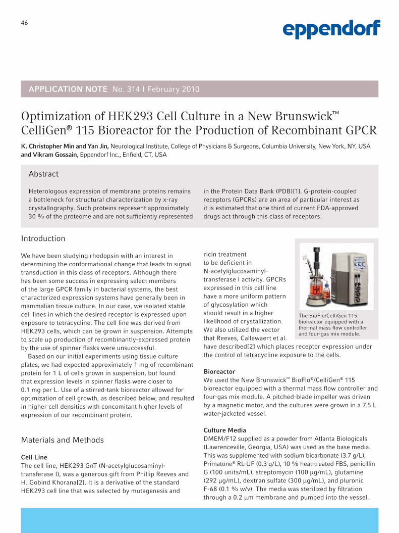
46
Abstract
Heterologous expression of membrane proteins remains a bottleneck for structural characterization by x-ray crystallography. Such proteins represent approximately 30 % of the proteome and are not sufficiently represented
in the Protein Data Bank (PDB)[1]. G-protein-coupled receptors (GPCRs) are an area of particular interest as it is estimated that one third of current FDA-approved drugs act through this class of receptors.
Optimization of HEK293 Cell Culture in a New Brunswick™ CelliGen® 115 Bioreactor for the Production of Recombinant GPCRK. Christopher Min and Yan Jin, Neurological Institute, College of Physicians & Surgeons, Columbia University, New York, NY, USA and Vikram Gossain, Eppendorf Inc., Enfield, CT, USA
Introduction
We have been studying rhodopsin with an interest in determining the conformational change that leads to signal transduction in this class of receptors. Although there has been some success in expressing select members of the large GPCR family in bacterial systems, the best characterized expression systems have generally been in mammalian tissue culture. In our case, we isolated stable cell lines in which the desired receptor is expressed upon exposure to tetracycline. The cell line was derived from HEK293 cells, which can be grown in suspension. Attempts to scale up production of recombinantly-expressed protein by the use of spinner flasks were unsuccessful.
Based on our initial experiments using tissue culture plates, we had expected approximately 1 mg of recombinant protein for 1 L of cells grown in suspension, but found that expression levels in spinner flasks were closer to 0.1 mg per L. Use of a stirred-tank bioreactor allowed for optimization of cell growth, as described below, and resulted in higher cell densities with concomitant higher levels of expression of our recombinant protein.
Materials and Methods
Cell LineThe cell line, HEK293 GnT (N-acetylglucosaminyl-transferase I), was a generous gift from Phillip Reeves and H. Gobind Khorana[2]. It is a derivative of the standard HEK293 cell line that was selected by mutagenesis and
ricin treatment to be deficient in N-acetylglucosaminyl-transferase I activity. GPCRs expressed in this cell line have a more uniform pattern of glycosylation which should result in a higher likelihood of crystallization. We also utilized the vector that Reeves, Callewaert et al. have described[2] which places receptor expression under the control of tetracycline exposure to the cells.
BioreactorWe used the New Brunswick™ BioFlo®/CelliGen® 115 bioreactor equipped with a thermal mass flow controller and four-gas mix module. A pitched-blade impeller was driven by a magnetic motor, and the cultures were grown in a 7.5 L water-jacketed vessel.
Culture MediaDMEM/F12 supplied as a powder from Atlanta Biologicals (Lawrenceville, Georgia, USA) was used as the base media. This was supplemented with sodium bicarbonate (3.7 g/L), Primatone® RL-UF (0.3 g/L), 10 % heat-treated FBS, penicillin G (100 units/mL), streptomycin (100 μg/mL), glutamine (292 μg/mL), dextran sulfate (300 μg/mL), and pluronic F-68 (0.1 % w/v). The media was sterilized by filtration through a 0.2 μm membrane and pumped into the vessel.
APPLICATION NOTE No. 314 I February 2010
The BioFlo/CelliGen 115 bioreactor equipped with a thermal mass flow controller and four-gas mix module.

47
APPLICATION NOTE No. 314 I Page 2
Control SoftwareAll equipment was monitored using New Brunswick BioCommand® software with data logging set at one-minute intervals.
MethodOn the day before inoculation, the 7.5 L bioreactor vessel was filled with 4 L of phosphate buffered saline. The various ports were connected to appropriate tubing for removal of the saline, introduction of media, introduction of cells, and the pumping of the four-gas mix through the sparger. The pH electrode was calibrated and then disconnected and the protective cover was installed. The oxygen probe was examined and also covered by a protective cover. The jacket of the vessel was filled with water, and the assembly was set in autoclave for a 30-minute sterilization cycle. Afterward the vessel was returned to the tissue culture room and allowed to cool overnight. The following day the calibration of the pH electrode and oxygen sensor was checked after allowing the oxygen sensor to charge by the control unit.
A stable cell line which contains the expression cassette for the GPCR, under the control of a cytomegalovirus promoter/tetracycline-responsive promoter was selected using the neo gene. These cells were maintained in tissue culture plates with DMEM/F12 medium supplemented with 10 % fetal bovine serum, G418, and blasticin. For inoculation of a 7.5 L bioreactor vessel, thirty 15 cm plates were grown to approximately 80 % confluence. On the day of inoculation, 4 L of media were prepared and transferred to the vessel with a peristaltic pump after removal of PBS from the vessel.
All setpoints were programmed from BioFlo/CelliGen 115 control station as follows: temperature at 37 °C and a pH of 7.2. Oxygenation was maintained at 50 % using the four-gas mixture of air, nitrogen, oxygen, and carbon dioxide, and the thermal mass flow controller was set to deliver 0.5 L per minute. The pH was maintained by a combination of carbon dioxide and a solution of 7.5 % sodium bicarbonate that was controlled by pump 2. Agitation with the pitched blade impeller was set to 30 rpm.
The cells were recovered from the tissue culture plates by brief trypsinization and resuspension in the culture medium. The cells were pumped into the vessel with an auxiliary peristaltic pump. A small sample was removed and the starting cell density was determined with a hemocytometer.
Over the next five to seven days, the cell density was checked on a daily basis. Once the density reached 0.8 – 1.0 x 10⁶, the culture was supplemented with 40 mL of 20 % (w/v) glucose and 120 mL of 10 % (w/v) Primatone RL-UF. The following day, expression was induced by the addition
of tetracycline (2 µg/mL) and sodium butyrate (5 mM) to the culture (Figure 1). One day later, the cells were recovered from the bioreactor and pelleted by centrifugation. A 1 mL aliquot was reserved for analysis by Western Blot to determine the level of expression (Figure 2).
Figure 1. Growth curve for HEK293 cells in a CelliGen 115 bioreactor 7.5 L vessel. The red arrow indicates the addition of glucose and Prima-tone. The blue arrow indicates the addition of tetracycline and sodium butyrate for the induction of protein expression.
Figure 2. Western Blot with mAb for detection of GPCR protein ex-pressed in HEK293 cells.
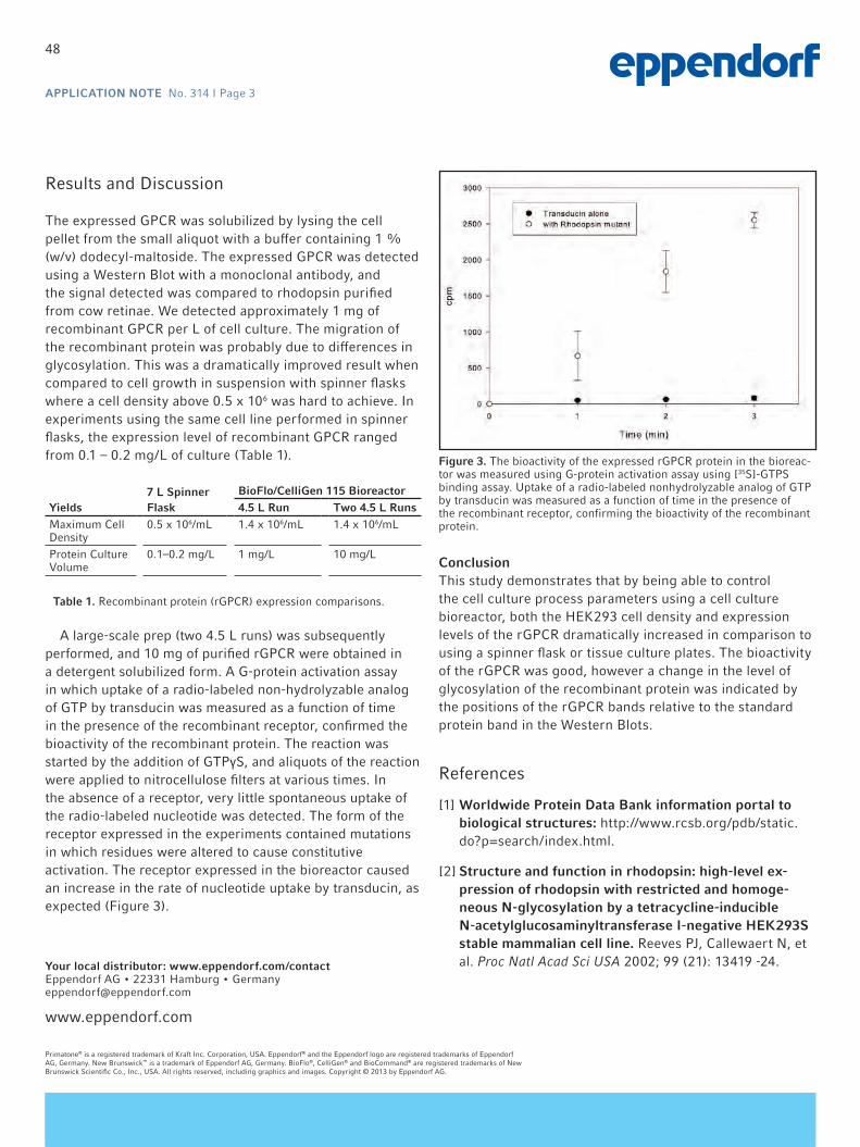
48
APPLICATION NOTE No. 314 I Page 3
Results and Discussion
The expressed GPCR was solubilized by lysing the cell pellet from the small aliquot with a buffer containing 1 % (w/v) dodecyl-maltoside. The expressed GPCR was detected using a Western Blot with a monoclonal antibody, and the signal detected was compared to rhodopsin purified from cow retinae. We detected approximately 1 mg of recombinant GPCR per L of cell culture. The migration of the recombinant protein was probably due to differences in glycosylation. This was a dramatically improved result when compared to cell growth in suspension with spinner flasks where a cell density above 0.5 x 10⁶ was hard to achieve. In experiments using the same cell line performed in spinner flasks, the expression level of recombinant GPCR ranged from 0.1 – 0.2 mg/L of culture (Table 1).
A large-scale prep (two 4.5 L runs) was subsequently performed, and 10 mg of purified rGPCR were obtained in a detergent solubilized form. A G-protein activation assay in which uptake of a radio-labeled non-hydrolyzable analog of GTP by transducin was measured as a function of time in the presence of the recombinant receptor, confirmed the bioactivity of the recombinant protein. The reaction was started by the addition of GTPγS, and aliquots of the reaction were applied to nitrocellulose filters at various times. In the absence of a receptor, very little spontaneous uptake of the radio-labeled nucleotide was detected. The form of the receptor expressed in the experiments contained mutations in which residues were altered to cause constitutive activation. The receptor expressed in the bioreactor caused an increase in the rate of nucleotide uptake by transducin, as expected (Figure 3).
ConclusionThis study demonstrates that by being able to control the cell culture process parameters using a cell culture bioreactor, both the HEK293 cell density and expression levels of the rGPCR dramatically increased in comparison to using a spinner flask or tissue culture plates. The bioactivity of the rGPCR was good, however a change in the level of glycosylation of the recombinant protein was indicated by the positions of the rGPCR bands relative to the standard protein band in the Western Blots.
References
[1] Worldwide Protein Data Bank information portal to biological structures: http://www.rcsb.org/pdb/static.do?p=search/index.html.
[2] Structure and function in rhodopsin: high-level ex-pression of rhodopsin with restricted and homoge-neous N-glycosylation by a tetracycline-inducible N-acetylglucosaminyltransferase I-negative HEK293S stable mammalian cell line. Reeves PJ, Callewaert N, et al. Proc Natl Acad Sci USA 2002; 99 (21): 13419 -24.
Figure 3. The bioactivity of the expressed rGPCR protein in the bioreac-tor was measured using G-protein activation assay using [35S]-GTPS binding assay. Uptake of a radio-labeled nonhydrolyzable analog of GTP by transducin was measured as a function of time in the presence of the recombinant receptor, confirming the bioactivity of the recombinant protein.
Yields7 L Spinner Flask
BioFlo/CelliGen 115 Bioreactor4.5 L Run Two 4.5 L Runs
Maximum Cell Density
0.5 x 10⁶/mL 1.4 x 10⁶/mL 1.4 x 10⁶/mL
Protein Culture Volume
0.1–0.2 mg/L 1 mg/L 10 mg/L
Table 1. Recombinant protein (rGPCR) expression comparisons.
www.eppendorf.com
Primatone® is a registered trademark of Kraft Inc. Corporation, USA. Eppendorf® and the Eppendorf logo are registered trademarks of Eppendorf AG, Germany. New Brunswick™ is a trademark of Eppendorf AG, Germany. BioFlo®, CelliGen® and BioCommand® are registered trademarks of New Brunswick Scientific Co., Inc., USA. All rights reserved, including graphics and images. Copyright © 2013 by Eppendorf AG.
Your local distributor: www.eppendorf.com/contact Eppendorf AG • 22331 Hamburg • Germany [email protected]

49
Cultivation of Human CAP® Cells: Evaluation of Scale-Down Capabili-ties Using Single-use BioreactorsClaudia M. Hüther-Franken1*, Helmut Kewes2 and Dr. Michael Schomberg2 1 Eppendorf AG Bioprocess Center, Juelich, Germany; 2 Cevec Pharmaceuticals GmbH, Cologne, Germany;
* Corresponding author: [email protected]
APPLICATION NOTE No. 291 I June 2012
Abstract
Increasing process complexity coupled with rising cost pressures and rapidly evolving regulatory requirements makes today’s process development efforts a special chal-lenge. The pressure of achieving faster time-to-market for new and innovative biotechnological products has led to the need to optimize every element of the total develop-ment workflow.
The following application note illustrates how the DAS-box® Mini Bioreactor System combined with the BioBLU®
0.3c single-use vessels supports bioprocess development in human cell culture. Scale-down capabilities were inves-tigated by comparison of 500 mL cultures in a DASGIP® Parallel Bioreactor System with 170 mL cultures in the DASbox using the BioBLU 0.3c single-use vessel.
Introduction
Initial bioprocess development involves cell line optimiza-tion, clone selection, and screening for media, feed compo-nents and strategies, and other process conditions. Shake flasks, the most common vessels used in early cell and microbial work, have served the biotechnology industry well over the decades but their limitations for optimizing cell culture or fermentation conditions are well known. Equip-ment used during screening should mimic the physical and mechanical characteristics of production-scale bioreactors to the highest degree possible in order to assure consis-tency throughout development phases. Ideally, these best practices will support the aims of QbD: that quality mea-sures initiated during development are carried forward and manifested in product quality. DASGIP Parallel Bioreactor Systems have the potential to address process consistency and harmonization of unit operations between development and production. Today’s state-of-the-art benchtop systems use sensors and information technology to control, monitor, and record critical process parameters such as temperature, Figure 1: BioBLU® 0.3c Single-Use Bioreactor for cell culture

50
APPLICATION NOTE I No. 291 I Page 2
pH, dissolved oxygen, and agitation. As in production-scale bioreactors, gassing and feeding proceed according to defined settings.
CEVEC® Pharmaceuticals GmbH (Cologne, Germany), a global solution provider focussing on the development of top notch human expression systems with highest ethi-cal standards, has established a master cell bank (MCB) of CAP® cells growing in suspension, tested and certified according to ICH guidelines and European Pharmacopeia. The platform expression technologies CAP and CAP-T are based on specific, amniocyte-derived human cell lines. CAP and CAP-T were designed for stable and transient protein production and achieve highest protein yields with authentic human glycosylation patterns. Simple and reliable protocols allow for the fast generation of customized producer cell lines for pharmaceutically relevant proteins based on the pa-rental permanent CAP cells under controlled and optimized conditions. For the required human cell line screening as well as for media optimization, the small working volumes of 100 – 250 mL make the extendable 4-fold DASbox and the BioBLU 0.3c single-use vessel a perfect fit. Bioprocesses are controlled as precise and effectively as they are in larger scale bioreactors while cell material, media and supple-ments as well as lab space are saved.
Several experiments were carried out aiming at verify-ing the scale-down capabilities from the DASGIP Parallel Bioreactor System, which CEVEC generally uses in process development, to the Mini Bioreactor System DASbox. To overcome the risk of cross-contamination and to reduce time for cleaning, sterilization and assembly they evaluated the novel developed BioBLU 0.3c single-use vessel. Which comes with a magnetic coupled stirrer and pitched blade impeller and holds several short and long dip-tubes as well as two standard PG13.5 ports facilitating full industry stan-dard instrumentation. A specifically designed port including
a gas permeable membrane allows for DO measurement us-ing a reusable probe which can be plugged easily in directly on the bench. Recuperation of liquid from exhaust gas is carried out via a novel liquid-free operated condenser.
Materials and Methods
To evaluate the scale-down capability of the new DASbox Mini Bioreactor System and the usability of the BioBLU 0.3c single-use vessel experimental series with two different systems were carried out and compared. A 4-fold Paral-lel Bioreactor System for cell culture was used in 500 mL scale experiments (PBS). The corresponding small-scale approaches were carried out in a (parallel) DASbox system using single-use vessels with 170 mL (DASbox SU).
The recombinant human CAP cells producing a pharmaceu-tically relevant protein were batch cultivated for 7 d (170 h) in CEVEC‘s serum-free, chemically defined CAP medium supplemented with 40 mM glucose and 6 mM glutamine at 37 °C. Initial viable cell density was 3*105 cells/mL. The DO set-point of 40 % was maintained by a constant stir-rer speed and the oxygen concentration in the inlet gas. Stirrer speed was adjusted to 160 rpm (PBS) and 150 rpm (DASbox SU). The pH value was regulated to 7.1 by addi-tion of 1 M Na2CO3 (feeding, speed rate regulated) and CO2 (submerged gassing). Inlet gas (air, O2, CO2 and N2) was mixed continuously mass flow-controlled. The bioreactors were equipped with pitched blade impellers and liquid-free operated exhaust gas condensers. The pre-cultures were cultivated in 125 mL Erlenmeyer flasks (Corning) with 25 mL working volume using a shaker incubator (37 °C, 5 % CO2) agitating at 185 rpm (Multitron 2, Infors AG). The cells were expanded up to a viable cell density of 3*106 cells/mL in the same medium used for bioreactor runs.
Figure 2: Viable Cell numbers of all experiments with DASGIP Parallel Bioreactor Systems (PBS) and BioBLU 0.3c vessels with average growth rate of 0.02 h-1.
Figure 3: Comparism of metabolic activity by glucose consumption.

51
APPLICATION NOTE I No. 291 I Page 3
The critical process parameters were monitored, controlled and visualized online while additionally offline parameters were added manually for collective analysis and storage in a joint database. Daily samples were taken in place. Viable cell numbers, the concentrations of glucose as well as the target protein were determined via semi-automated trypan blue cell counting (Cedex XS, Roche Innovatis), an automated glucose biosensor (YSI 7100 MBS, YSI Life Sciences) and ELISA, respectively.
Results and Discussion
The highly comparable results shown in figures 2 and 3 prove the reliability of the process control in both indepen-dent experimental series.
The viable cell density increases exponentially within all cultivation studies in a reproducible manner with an average growth rate of 0.02 h-1. The corresponding anti-cyclic glu-cose consumption thereby illustrates the similar metabolism of the different cultures. Cell viabilities ranged in between 90 – 95 % for each sample. As shown in figure 4 the final product yield reached 80 – 121 % in respect to the average protein concentration gained with the Parallel Bioreactor System (PBS) commonly used at CEVEC. No differences in cell growth, metabolic activity and protein expression could be observed using the BioBLU 0.3 c single-use vessels. The results show the successful scale-down from a 500 mL
(PBS) to 170 mL (DASbox SU) bioreactor working volume.
Conclusion
Summarized, the presented results give direct evidence to the scale-down capability of the DASbox Mini Bioreac-tor System used with single-use vessels. This proves the DASbox to be a superior tool for process development with human cell cultures. The small working volumes save mate-rial and consumable costs while utilizing single-use vessels drastically reduce turnover-times and thereby labour costs and development times.
Figure 3: Product yield normalized to the average protein concentration gained using the DASGIP Parallel Bioreactor System (PBS)
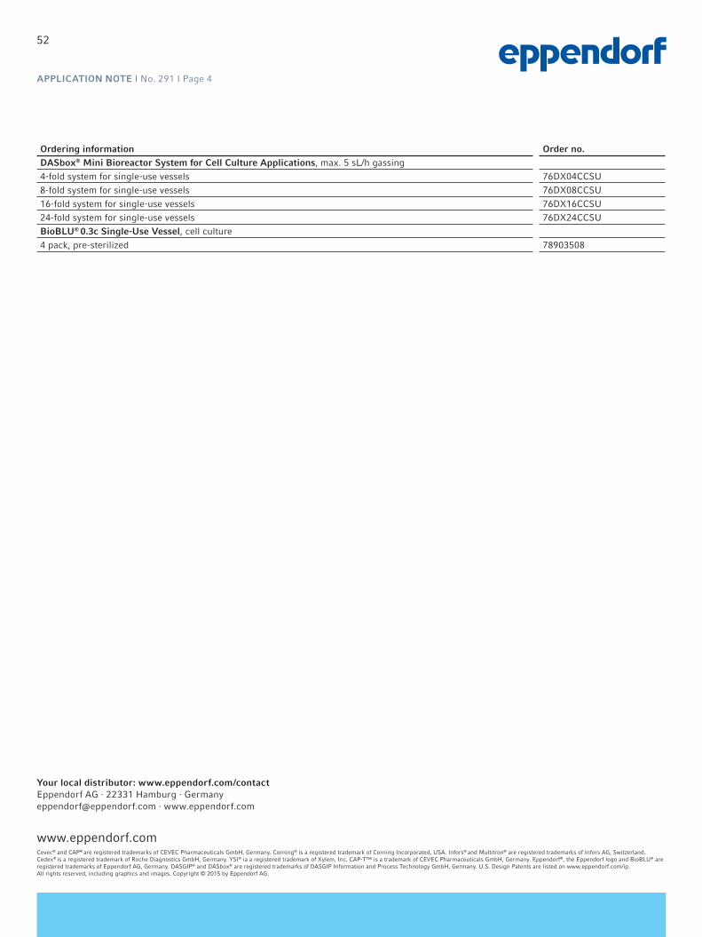
52
APPLICATION NOTE I No. 291 I Page 4
Cevec® and CAP® are registered trademarks of CEVEC Pharmaceuticals GmbH, Germany. Corning® is a registered trademark of Corning Incorporated, USA. Infors® and Multitron® are registered trademarks of Infors AG, Switzerland. Cedex® is a registered trademark of Roche Diagnostics GmbH, Germany. YSI® ia a registered trademark of Xylem, Inc. CAP-T™ is a trademark of CEVEC Pharmaceuticals GmbH, Germany. Eppendorf®, the Eppendorf logo and BioBLU® are registered trademarks of Eppendorf AG, Germany. DASGIP® and DASbox® are registered trademarks of DASGIP Information and Process Technology GmbH, Germany. U.S. Design Patents are listed on www.eppendorf.com/ip. All rights reserved, including graphics and images. Copyright © 2015 by Eppendorf AG.
Ordering information Order no. DASbox® Mini Bioreactor System for Cell Culture Applications, max. 5 sL/h gassing4-fold system for single-use vessels 76DX04CCSU8-fold system for single-use vessels 76DX08CCSU16-fold system for single-use vessels 76DX16CCSU24-fold system for single-use vessels 76DX24CCSUBioBLU® 0.3c Single-Use Vessel, cell culture4 pack, pre-sterilized 78903508
www.eppendorf.com
Your local distributor: www.eppendorf.com/contactEppendorf AG · 22331 Hamburg · [email protected] · www.eppendorf.com
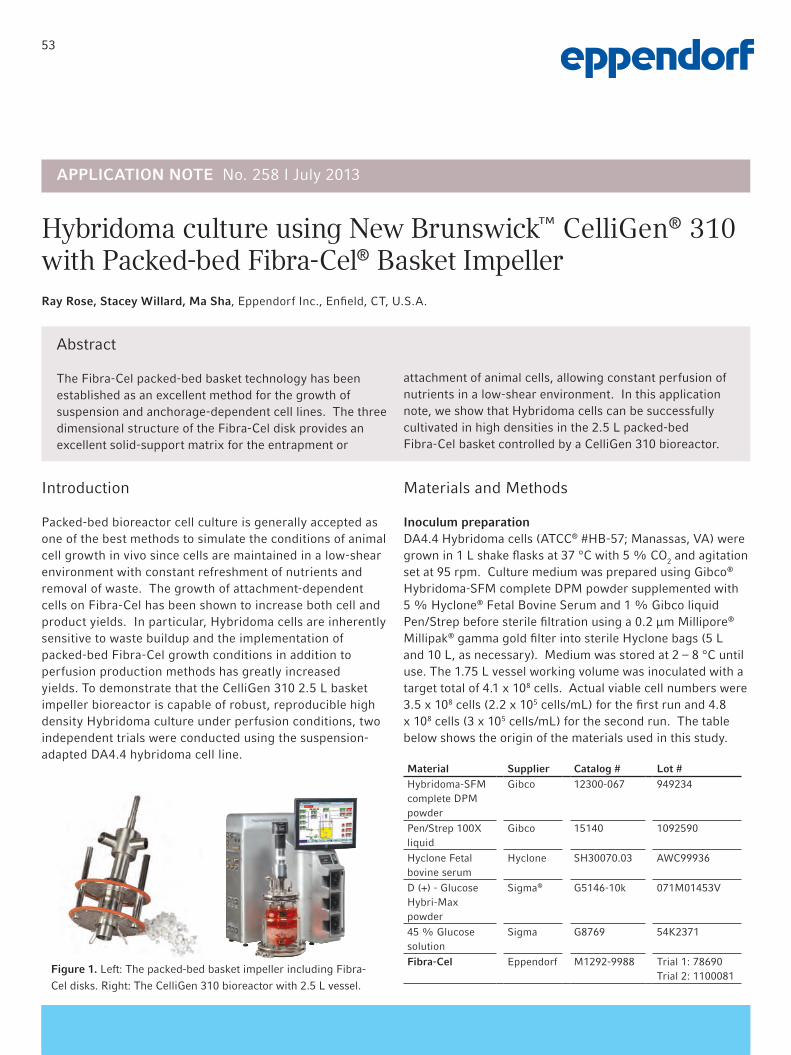
53
Abstract
The Fibra-Cel packed-bed basket technology has been established as an excellent method for the growth of suspension and anchorage-dependent cell lines. The three dimensional structure of the Fibra-Cel disk provides an excellent solid-support matrix for the entrapment or
attachment of animal cells, allowing constant perfusion of nutrients in a low-shear environment. In this application note, we show that Hybridoma cells can be successfully cultivated in high densities in the 2.5 L packed-bed Fibra-Cel basket controlled by a CelliGen 310 bioreactor.
Hybridoma culture using New Brunswick™ CelliGen® 310with Packed-bed Fibra-Cel® Basket ImpellerRay Rose, Stacey Willard, Ma Sha, Eppendorf Inc., Enfield, CT, U.S.A.
Introduction
Packed-bed bioreactor cell culture is generally accepted as one of the best methods to simulate the conditions of animal cell growth in vivo since cells are maintained in a low-shear environment with constant refreshment of nutrients and removal of waste. The growth of attachment-dependent cells on Fibra-Cel has been shown to increase both cell and product yields. In particular, Hybridoma cells are inherently sensitive to waste buildup and the implementation of packed-bed Fibra-Cel growth conditions in addition to perfusion production methods has greatly increased yields. To demonstrate that the CelliGen 310 2.5 L basket impeller bioreactor is capable of robust, reproducible high density Hybridoma culture under perfusion conditions, two independent trials were conducted using the suspension-adapted DA4.4 hybridoma cell line.
Materials and Methods
Inoculum preparationDA4.4 Hybridoma cells (ATCC® #HB-57; Manassas, VA) were grown in 1 L shake flasks at 37 °C with 5 % CO2 and agitation set at 95 rpm. Culture medium was prepared using Gibco® Hybridoma-SFM complete DPM powder supplemented with 5 % Hyclone® Fetal Bovine Serum and 1 % Gibco liquid Pen/Strep before sterile filtration using a 0.2 µm Millipore® Millipak® gamma gold filter into sterile Hyclone bags (5 L and 10 L, as necessary). Medium was stored at 2 – 8 °C until use. The 1.75 L vessel working volume was inoculated with a target total of 4.1 x 10⁸ cells. Actual viable cell numbers were 3.5 x 10⁸ cells (2.2 x 10⁵ cells/mL) for the first run and 4.8 x 10⁸ cells (3 x 10⁵ cells/mL) for the second run. The table below shows the origin of the materials used in this study.
Material Supplier Catalog # Lot #Hybridoma-SFM complete DPM powder
Gibco 12300-067 949234
Pen/Strep 100X liquid
Gibco 15140 1092590
Hyclone Fetal bovine serum
Hyclone SH30070.03 AWC99936
D (+) - Glucose Hybri-Max powder
Sigma® G5146-10k 071M01453V
45 % Glucose solution
Sigma G8769 54K2371
Fibra-Cel Eppendorf M1292-9988 Trial 1: 78690Trial 2: 1100081
APPLICATION NOTE No. 258 I July 2013
Figure 1. Left: The packed-bed basket impeller including Fibra-
Cel disks. Right: The CelliGen 310 bioreactor with 2.5 L vessel.

54
APPLICATION NOTE I No. 258 I Page 2
Bioreactor conditionsFor both runs, hybridoma cells were cultured in the same vessel, using the same CelliGen 310 cabinet for 9 consecutive days, using the basket impeller system packed with 75 g of Fibra-Cel disks.
CelliGen 310 Setpoints
Agitation 80 rpm
Temperature 37 °C
pH 7.15 Dead band 0.04
DO 50 %
Gas supplied 4-gas mix control (N2 off; CO2 fo pH control)
Gas flow conditions 0.4 SLPM
Vessel 2.5 L glass water jacketed
Fibra-Cel 75 g
Perfusion was initiated for each bioreactor on day 3 and continued through day 9. Initially, the main objective was to increase the perfusion rate to maintain a glucose concentration above and near 1 g/L. For the second bioreactor experiment, the perfusion rate was adjusted to match the first bioreactor rate in order to make the two runs as identical as possible. The tables illustrate the experimental parameters and perfusion volumes for both trials.
Day Perfusion volume (L)
3 0.73
4 1.81
5 4.25
6 5.5
7 4.25
8 4.75
9 5
Biochemistry analysisDaily off-line measurements of glucose concentration were performed using a YSI® 2700 analyzer (YSI, Inc., Yellow Springs, OH). The glucose consumption was calculated for each time point and plotted as an average of the two independent trials. Error bars indicate standard error of the mean.
Results and Discussion
As presented in the graph below (Figure 2), the rate of glucose consumption across both trials is indicative of reproducible growth of hybridoma cells in this environment. We conclude that the use of Fibra-Cel in the basket impeller system on the CelliGen 310 is an excellent method for high density hybridoma culture. In a batch run with the CelliGen pitch blade bioreactor, hybridoma cells usually peak at approximately 5 g/day of glucose consumption. The packed-bed basket impeller system presents significantly higher productivity with glucose consumption peaking at, on average, 25 g/day. In addition, if growth conditions are maintained by continued fresh media perfusion and glucose concentration is never allowed to fall below 1 g/L, hybridoma can be continuously cultured in the basket many days after the 9 day window provided in this study; this further increases productivity and decreases overall antibody production costs. No optimization of growth conditions were attempted for either bioreactor run.
30
25
20
15
10
5
0
Glu
cose
con
sum
ptio
n (
g)
Average glucose consumption
0 2 4 6 8 10Day
Figure 2. The glucose consumption was calculated daily for each
bioreactor and the mean is presented. Error bars indicate standard
error of the mean. Comparable consumption was observed across
the two bioreactors.

55
www.eppendorf.com
ATCC® is a registered trademark of American Type Culture Collection Corp., Virginia, USA. Hyclone® is a registered trademark of HyClone Laboratories, Inc., Utah, USA. Fibra-Cel® is a registered trademarks of Imerys Minerals California, Inc., USA and licensed to New Brunswick Scientific Co., Inc, USA. Gibco® is a registered trademark of Life Technologies Corporation, Delaware, USA. Millipak® and Millipore® are registered trademarks of Merck KGaA, Darmstadt, Germany. CelliGen® is a registered trademark of New Brunswick Scientific, USA. Sigma® is a registered trademark of Sigma-Aldrich, Co., LLC, USA. YSI® is a registered trademark of YSI, Inc., Yellow Springs, USA. Eppendorf® and the Eppendorf logo are registered trademarks of Eppendorf AG, Germany. New Brunswick™ is a trademark of Eppendorf AG, Germany.All rights reserved, including graphics and images. Copyright © 2013 by Eppendorf AG, Germany.
Your local distributor: www.eppendorf.com/contact Eppendorf AG • 22331 Hamburg • Germany [email protected]
APPLICATION NOTE I No. 258 I Page 3
Ordering informationDescription North America Order no. International Order no,
New Brunswick™ CelliGen® 310 Bioreactor, 2.5 L System
M1287-1260 (100-120 V) M1287-1264 (200-240 V)
Fibra-Cel® Disks, 250 grams M1292-9988 M1292-9988
2.5 L Basket Impeller Kit M1287-1140 M1287-1140

56
Introduction
Oxygen is a critical regulator of cellular homeostasis and as such, oxygen deprivation is lethal to normal cells. In contrast to normal tissues, solid tumors often have regions of significantly reduced oxygenation due to an inconsistent and disorganized blood supply at the center of the tumor. Tumor O2 deprivation, or hypoxia, can result in gene expression changes in cancer cells. Decades of cancer research have established that tumor cell growth, survival, motility, the recruitment of blood vessels (angiogenesis), energy metabolism, and cellular differentiation are affected by hypoxia[3]. This effect has been documented for many deep tissue tumors such as breast, prostate, ovarian and uterine cancer[2].
A number of human tumor cell lines have been used to model the effects of hypoxia in an effort to target these drug-resistant cells. One such cell line, LNCaP, was derived from a bone metastasis from a terminal prostate cancer patient. These cells have been widely used in prostate cancer drug discovery and are uniquely sensitive to androgen (e.g. testosterone and dihydrotestosterone) levels. Danza and colleagues have shown that LNCaP cells previously stimulated with androgens before growth under hypoxic conditions (2 % O2) grow faster than cells maintained in normoxia (20.9 % O2)
[1].
We chose to use a simplified version of this model to establish a protocol for setting up low-O2 cell culture in a standard tissue culture laboratory. Since standard laboratories do not have access to controlled substances, we did not stimulate the cells with androgen before growth in hypoxic conditions.
We show here that low-O2 cell culture conditions are easy to establish using the New Brunswick Galaxy 170R incubator with 1 - 19 % O2 control. LNCaP cells grew well in both normoxia and hypoxia in this system and morphological changes were noted in low O2 conditions. With available O2 control from 0.1 - 19 %, the Galaxy 170R incubator provides an excellent environment with which to culture cells in a range of gas conditions.
Hypoxic Cell Culture in the New Brunswick™ Galaxy® 170R Incubator: Normal Growth, Morphological ChangesStacey Willard, Linette Philip and Ma Sha, Eppendorf Inc., Enfield, CT, U.S.A.
APPLICATION NOTE No. 331 I October 2013
Abstract
New Brunswick Galaxy 170 series incubators have a wide array of options that allow for not only CO2 and temperature control, but also for O2 concentration control which can be used to create a hypoxic internal environment. Cancer cells are known to be resistant to the toxicity of O2 deprivation. To demonstrate the low
O2 capability of this system, we grew prostate cancer (LNCaP) cells in 2 % O2 and observed their growth and morphology over time. In contrast to the effects of hypoxia on normal cells, LNCaP cells were able to grow normally and displayed morphological changes.
Figure 1: The New Brunswick Galaxy 170R CO2 incubator

57
APPLICATION NOTE No. 331 I Page 2
Materials and Methods
Consumable MaterialsTable 1 details the consumable reagents and materials that were used in this study.
Cell CultureCell culture was carried out using two Galaxy 170 incubators (Eppendorf). First, the Galaxy 170R with High temperature disinfection, 4 split inner doors and 1 - 19 % O2 control was used for culture in hypoxic conditions (Figure 1). Second, the Galaxy 170S with High temperature disinfection and 4 split inner doors was used for normoxic conditions. Setpoints on both units for temperature and CO2 concentration were 37 °C and 5 %, respectively. For hypoxic conditions, the Galaxy 170R was set at 2 % O2 and allowed to stabilize at setpoint for 72 h before cells were introduced into the incubator. To monitor O2 concentration, the Galaxy 170R was connected to a computer using a RS-232/RS-422 converter (Eppendorf). Using BioCommand® SFI (Eppendorf), O2, temperature and CO2 process values were tracked for the course of the experiment. The O2 concentration inside the Galaxy 170R never exceeded 2.1 %.
LNCaP clone FGC cells were acquired from the American Type Culture Collection® (ATCC®, USA #CRL-1740™) and were grown in RPMI medium supplemented with 1 % Penicillin-Streptomycin and 10 % FBS. Cells were grown in T75 flasks until 80 % confluency. At target density, the cells were disassociated from the surface by trypsinization and neutralization with FBS. After pelleting by centrifugation at 120 x g for 3 min, cells were resuspended in complete growth medium and counted using a Vi-CELL® automated cell counter (Vi-CELL XR; Beckman Coulter, Inc., USA #731050). Cells were seeded in quadruplicate 6-well dishes at a density of 300,000 cells/well and two plates were placed
in the same position in both the normoxia and hypoxia incubators.
According to the protocol established by Danza and colleagues, 3 wells from both hypoxic and normoxic conditions were counted every 3 days for 9 days, generating cell concentrations per well for days 3, 6 and 9[1]. At each timepoint, the wells were also photographed using phase contrast microscopy on an Olympus® IX51 inverted microscope equipped with an Infinity2 CCD camera (Lumenera®, Canada) . The data were analyzed and statistical tests including a 2-way analysis of variance (ANOVA) were performed in Microsoft® Excel®.
Fluorescent Marker StainingAfter 6 days in normoxia or hypoxia, two wells were fixed in 4 % paraformaldehyde and stained using the phallotoxin, phalloidin (which selectively binds f-actin) and the double-stranded DNA intercalator, 4’,6-diamidino-2-phenylindole (DAPI; NucBlue® Fixed Cell ReadyProbes® reagent). Staining was performed exactly as the manufacturer recommended. The cells were photographed using an EVOS® LED florescence microscopy system (Life Technologies, USA #AMF4300).
Results and Discussion
LNCaP Cells Grow Normally Under Hypoxic ConditionsAs the graph in Figure 2 details, LNCaP cells grown at 2 % O2 showed growth dynamics indistinguishable from those grown in normal atmospheric O2 (20.9 %). It is possible that we did not see the previously published growth bias in hypoxia because we did not stimulate the cells with androgens prior to exposure to O2 deprivation. Importantly, no differences were seen in the attachment of the cells in hypoxia, as evidenced by the phase contrast micrographs in Figure 3.
Table 1: Materials, media and cellsMaterial Supplier Order no.RPMI-1640 ATCC® 30-2001™
Penicillin-Streptomycin 10,000 U/mL Life Technologies® 15140-122Fetal bovine serum (FBS) Life Technologies® 10437-0281x Dulbecco’s Phosphate buffered saline Life Technologies® 14190-144Trypsin-EDTA HyClone® SV30031.0116 % Paraformaldehyde (w/v) methanol-free Pierce™ 28906rhodamine-conjugated phalloidin Molecular Probes® R415NucBlue® Fixed Cell ReadyProbes® Molecular Probes® R37606T75 T-flasks* Eppendorf 0030 711.1066-well dishes* Eppendorf 0030 720.105
* Currently available in China, India and Italy only

58
APPLICATION NOTE No. 331 I Page 3
Figure 2: Growth and viability of LNCaP cells grown in hypoxia and normoxia. This graph shows the viable cell density and % viable cells in each well. Each data point represents the mean of 3 wells. Error bars indicate standard error of the mean. ANOVA analysis revealed that no significant difference was observed between the two growth conditions.
Figure 3: LNCaP cells grown in hypoxic conditions display normal attachment and homogenous growth. In the top row, LNCaP cells grown in 2 % O2 show similar density as compared to those grown in atmospheric O2 (bottom row). Photos were taken at 100 x magnification and the scale bar in each panel represents 100 µm.
Interestingly, we noted a significant morphology difference between the cells grown in hypoxic and normoxic conditions. Grown in atmospheric O2 conditions, LNCaP cells are known to form large clusters or colonies of cells wherein they display no contact inhibition.
Figure 4: Growth pattern of LNCaP cells in normoxic (A) vs. hypoxic (B) conditions. Grown in atmospheric O2 conditions, LNCaP cells make large 3-dimensional clusters (yellow arrowheads) whereas in hypoxic conditions, cells appear more spread out and do not form large colonies. Photos were collected at 100 x magnification and the scale bar in each panel represents 100 µm.
These colonies are themselves loosely attached to the substrate and can become quite large (see yellow arrowheads in Figure 4A). In contrast, LNCaP cells grown in 2 % O2 do not form such 3-dimensional structures and tend to cover the substrate and grow in more of a packed monolayer (Figure 4B). The cells also appear more spread out and seem to display tighter adherence as evidenced by the length of time required for the cells to detach during enzymatic disassociation. To document these morphological changes, we stained cells grown in both O2 concentrations with rhodamine-conjugated phalloidin and DAPI to visualize the actin cytoskeleton (red, Figure 5) and the cell nucleus (blue, Figure 5). As documented in Figure 5, large clusters of cells are observed in normoxic conditions while a flatter monolayer is seen in hypoxic conditions. Furthermore, the cytoplasm-to-nucleus ratio in the cells grown in 2 % O2 appears to be larger than in those grown in 20.9 % O2, although this observation was not quantified. We conclude that morphological changes have occurred in LNCaP cells grown with O2 deprivation that result in diminished colony forming behavior and a flatter appearance.

www.eppendorf.com
Your local distributor: www.eppendorf.com/contact Eppendorf AG • 22331 Hamburg • Germany [email protected]
59
APPLICATION NOTE No. 331 I Page 4
ATCC® and American Type Culture Collection® are registered trademarks of ATCC, USA. Life Technologies®, Molecular Probes®, ReadyProbes®, NucBlue® and EVOS® are registered trademarks of Life Technologies Corporation, USA. Vi-CELL® is a registered trademark of Beckman Coulter, Inc., USA. Olympus® is a registered trademark of Olympus Corp., Japan. Lumenera® is a registered trademark of Lumenera Corp., Canada. HyClone® is a registered trademark of Thermo Scientific, Inc., USA. Microsoft® and Excel® are registered trademarks of Microsoft Corp., USA. 30-2001™ and CRL-174Q™ are a trademarks of ATCC, USA. Pierce™ is a trademark of Thermo Scientific, Inc., USA. Galaxy® and BioCommand® are registered trademarks of New Brunswick Scientific Co., Inc., USA. New Brunswick™ is a trademark of Eppendorf AG, Germany. Eppendorf®, the Eppendorf logo and Easypet® are registered trademarks of Eppendorf AG, Germany. All rights reserved, including graphics and images. Copyright © 2016 by Eppendorf AG.
Figure 5: Growth characteristics of cells in hypoxia and normoxia. Panels A (fluorescence) and B (phase contrast) show examples of cells grown in 20.9 % O2 for 3 days. The yellow arrowheads denote areas where 3-dimensional colonies are observed, as evidenced by the overlapping cell nuclei (blue) and high concentration of actin (red). Panels C and D show monolayers grown in 2 % O2 where 3-dimensional growth is not seen. In addition, cells grown in hypoxia appear flatter and with larger cytoplasmic volume. The images in this figure were collected at 100 x using an EVOS LED imaging system; scale bars represent 200 µm.
Conclusion
We have shown that LNCaP cells grown in hypoxic conditions display normal growth as compared to those grown in atmospheric O2 concentrations. Growth in 2 % O2 also resulted in morphological changes and changes in growth characteristics such as colony formation. The ease of setup and the tight O2 concentration control displayed by the Galaxy 170R incubator provided the ideal conditions for this experiment.
This study is a demonstration of the low O2 feature that is available on the Galaxy 170 series incubators. Experimental conditions including O2 concentration have not been optimized.
References
[1] Danza, G. D. (2011). Notch signaling modulates hypoxia-induced neuroendocrine differentiation of human pros-tate cancer cells. Mol Cancer Res, 230-238.
[2] Hockel, M. V. (2001). Tumor Hypoxia: Definitions and Current Clinical, Biologic, and Molecular Aspects. J Natl Cancer Inst, 93(4), 266-276.
[3] Kim, Y. L. (2009). Hypoxic Tumor Microenvironment and Cancer Cell Differentiation. Curr Mol Med, 9(4), 425-434.
Ordering Information
DescriptionInternational order no.
N. America order no.
Galaxy® 170R With high temp disinfection 1 - 19 % O2 control Split inner doors, 4
170R230120_B04
170R120120_B04
Galaxy® 170S With high temp disinfection
CO170S-230- 1000
CO170S-120- 1000
8-port RS-232 to USB converter P0460-7750 P0460-7750BioCommand® SFI M1291-1001 M1291-1001Easypet® 3 4430 000.026 4430 000.026Centrifuge 5810 R With 4 x 500 mL rotor, 120 V
022628179 022627082

60
Development of a Scale-Down Model for rAAV Viral Vector Production Using a Sf9/BEV System Simon Fradin1 and Ulrike Becken2*
1 Généthon, Evry, France; 2 Eppendorf AG Bioprocess Center, Juelich, Germany
* Corresponding author: [email protected]
APPLICATION NOTE No. 303 I March 2016
Abstract
Single-gene disorders originate in the absence or loss of function of a protein due to a genetic mutation.Gene therapy is a promising therapeutic approach that delivers a normal version of the gene to affected cells to compensate for its missing or defective counterpart. It often employs viral vectors, such as recombinant Adeno Associated Viruses (rAAVs), to insert the genes. The insect cell line Sf9 provides a suitable host for virus production. Sf9 cells are cultured in suspension, and hence working
volumes can be adapted to changing needs during process development and manufacturing much more easily than for adherent cell cultures. In this study, researchers at Généthon® developed a scale-down model for rAAV viral vector production in Sf9 cells using an Eppendorf DASbox® Mini Bioreactor System. Parallel experimentation in small working volumes allowed time- and cost-efficient evaluation of process performance.
Introduction
Development and use of gene therapy for single-gene disorders requires optimization of the process for large-scale manufacturing of gene therapy vectors. Recombinant Adeno Associated Viruses (rAAV) are promising vector candidates. They are not known to be pathogenic, their DNA integrates into the human genome only very inefficiently, and the virus particles are very robust, to name only a few advantageous attributes. One big challenge on the road to therapeutic use is producing a sufficient amount of the virus for experimentation in large animal models, clinical trials, and actual treatment. A widely used host for rAAV production is the insect cell line Sf9. Cells are transfected with the genes needed to produce functional virus particles using baculovirus expression vector (BEV) systems. A major advantage of the Sf9/BEV system for large-scale virus production is the possibility of growing Sf9 cells in suspension culture. Higher cell densities can be reached than in adherent cell cultures, and the inherent scalability of the system allows for much easier adaption of production volumes during process development and manufacturing. Process development is usually carried out in small culture
sizes, and subsequently scaled up to larger production volumes. It is highly desirable to also establish scale-down models that will mimic the production process in smaller volumes, and which can be used to optimize the process, troubleshoot, and implement changes.
Généthon is a non-profit biotherapy R&D organization created and funded by the Association Française contre les Myopathies, a French organization that supports patients and their families. Its mission is to design gene therapy products for rare diseases, to ensure their pre-clinical and clinical development, as well as the production in order to provide patients with access to these innovative treatments. Généthon is currently sponsoring two gene therapy projects that have reached clinical trial phase, and is involved in several other projects in preclinical or research stages.
This application note describes the successful development of a scale-down model for rAAV production using a Sf9/BEV system. The aim was to reproduce the production performance obtained in a 2 L bioreactor in an Eppendorf DASbox Mini Bioreactor System. It is optimized for parallel process development, meaning that

61
APPLICATION NOTE I No. 303 I Page 2
Material and Methods
Cell cultureThe Généthon research team cultivated Sf9 cells in Sf-900™ III SFM culture medium (Thermo Fisher Scientific® Inc., USA) at 27°C and dissolved oxygen set to 50 %. The reference production system used a glass bioreactor with a working volume of 2 L, and scale-down experiments used an Eppendorf DASbox Mini Bioreactor System (Eppendorf AG, Germany, Fig. 1), with a working volume of 250 mL. Both systems used marine impellers. The agitation speed of the 2 L production system was 180 rpm. Proper culture mixing and oxygen transfer have to be ensured during development of scale-down models. Agitation speeds of 300 rpm, 375 rpm, and 450 rpm were tested in three vessels simultaneously (Table 1).
Cell density, cell viability, and cell diameter were determined offline, using the Cell Viability Analyzer Vi-CELL® XR (Beckman Coulter®, Inc., USA).
Virus production and quantification of production yieldThe researchers at Généthon used Sf9 cells as hosts for the
production of recombinant Adeno Associated Virus serotype 8 (rAAV8). 72 hours after inoculation they co-infected the Sf9 cells with two distinct baculovirus clones, one carrying the genes for the production of the virus capsid and the other carrying the gene of interest. Through their lytic life cycle, the baculoviruses replicate using the Sf9 cell machinery, infect other cells in the culture, and produce the rAAV8 vectors. The culture was harvested 96 hours after infection, and the vectors were released using a detergent treatment. The team determined the rAAV8 vector titer by measuring viral DNA using real-time quantitative PCR. This analysis leads to a viral genome titer (VG), which is interpreted as the number of viral vector particles carrying the gene of interest. Specific productivity (VG/cell) was calculated by normalizing the viral genome titer to the viable cell density at the time of infection.
In parallel processes production yields were compared for cultures agitated at 300 rpm and 400 rpm, respectively. To test for reproducibility, three bioreactor runs were performed for each agitation speed (Table 1).
multiple experiments can be run, monitored, and controlled simultaneously by shared equipment. Processes can be
carried out in working volumes as small as 60 mL, which helps to reduce the cost of media and supplements.
Run Bioreactor 1 Bioreactor 2 Bioreactor 3 Bioreactor 4 Analysis
1 300 rpm 375 rpm 450 rpm - Viable cell density; Cell diameter; Viability
2 300 rpm 300 rpm 400 rpm 400 rpm VG/mL; VG/cell
3 300 rpm 400 rpm - - VG/mL;VG/cell
Table 1: Summary of experimental conditions. Three runs were performed in a DASbox Mini
Bioreactor System. Several agitation speeds were compared within a given run. Cultures were
analyzed for viable cell density, cell diameter, and cell viability (run 1) and viral genome titer per
mL (VG/mL) and viral genome titer per cell (VG/cell), respectively (runs 2 and 3).
Fig. 1: The Eppendorf
DASbox Mini Bioreactor
System used at Généthon.
Results
The study aimed to scale-down rAAV production from a 2 L working volume to a 250 mL working volume, while reproducing production performance. The Généthon
research team first compared growth of non-virus infected Sf9 cells in the 2 L glass vessel and in the 250 mL working volume vessels of the DASbox Mini Bioreactor System.

62
APPLICATION NOTE I No. 303 I Page 3
Conclusion
The results demonstrate the successful scale-down of rAAV production using a Sf9/BEV system. The cell growth and production performance obtained in a 2 L glass vessel were reproduced in an Eppendorf DASbox Mini Bioreactor System with a working volume of 250 mL. This study exemplifies the value of the Eppendorf DASbox Mini Bioreactor System for process scale-down. In the course of scale-down model development, multiple experiments must be performed to test a variety of experimental conditions and to ensure the reproducibility of process performance. The use of a parallel bioreactor system saves time, and ensures maximum
comparability of experimental runs. By using the DASbox, researchers at Généthon shortened development timelines. They state: “The biological timing is impossible to shorten due to the rAAV production kinetics. But the DASbox system shortens the development timelines, by being able to run the same amount of bioreactors in one week whereas it takes two weeks in 2 L glass bioreactors.” Using the DASbox system also helped reduce costs, by facilitating the performance of studies in small-scale bioreactors.
A
1x10⁵
1x10⁶
0 24 48 72
Viab
le c
ells
/ m
L
Time (h)
300 rpm
400 rpm
375 rpm
2 L bioreactor, 180 rpm (n=6)
B
12
14
16
18
0 24 48 72
Cell
diam
eter
(µm
)Time (h)
300 rpm
400 rpm
375 rpm
2 L bioreactor, 180 rpm (n=6)
C
0
20
40
60
80
100
0 24 48 72
Cell
viab
ility
(%)
Time (h)
300 rpm
400 rpm
375 rpm
2 L bioreactor, 180 rpm(n=6)
Fig. 2: Growth performance of Sf9 cells. Sf9 cells were cultivated in a DASbox Mini Bioreactor System and a 2 L bioreactor. Several
agitation speeds were tested. Viable cell density (A), cell diameter (B), and cell viability (C) were determined offline.
Fig. 3: Virus production performance. rAAV was produced using a
Sf9/BEV expression system in a DASbox Mini Bioreactor System at
an agitation speed of 300 rpm and 400 rpm, respectively, and in a
2 L bioreactor at an agitation speed of 180 rpm. Experiments in the
DASbox system were carried out in triplicate, and experiments in
the 2 L bioreactor were carried out in duplicate. Means and standard
deviations are shown. Virus genome titer per mL (VG/mL) (A) and
specific productivity (VG/cell) (B) were determined. 1x10⁹
1x10¹⁰
VG/m
L
1x10²
1x10³
1x10⁴
VG/c
ell
250 mL300 rpm
2 L 180 rpm
250 mL400 rpm
working volumeagitation speed
250 mL300 rpm
2 L 180 rpm
250 mL400 rpm
A B
Growth performance results were comparable for Sf9 cells in the 2 L glass vessel and the vessels of the DASbox system, at agitation speeds of 300 rpm, 375 rpm, and 450 rpm. At 72 hours post-inoculation, for all experimental conditions tested, the viable cell density had increased from 3x10⁵ cells/mL to around 1.5x10⁶ cells/mL (Fig. 2A). The average cell diameter depends on the cell cycle phase, and correlates with infection by baculoviruses. It is routinely determined in Sf9/BEV systems and was around 15 µM for
non-infected cells (Fig. 2B). Close to 100 % of the cells were viable (Fig. 2C). Next, the research team monitored virus production performance, comparing processes carried out in the DASbox system at agitation speeds of 300 rpm and 400 rpm, and in the 2 L glass vessel. Virus genome titers and specific productivities were comparable for all experimental conditions tested. Results from three experimental runs were highly reproducible (Fig. 3).

63
APPLICATION NOTE I No. 303 I Page 4
www.eppendorf.com
Généthon® is a registered trademark of Association Française Contre Les Myopathies, France. Thermo Fisher Scientific® is a registered trademark of Thermo Fisher Scientific Inc., USA. Sf-900™ is a trademark of Thermo Fisher Scientific Inc., USA. Beckman Coulter® and VI-CELL® are registered trademarks of Beckman Coulter, Inc., USA. Eppendorf® and the Eppendorf logo are registered trademarks of Eppendorf AG, Germany. DASbox® is a registered trademark of DASGIP Information and Process Technology GmbH, Germany. U.S. Design Patents are listed on www.eppendorf.com/ip.All rights reserved, including graphics and images. Copyright © 2016 by Eppendorf AG.
Your local distributor: www.eppendorf.com/contactEppendorf AG · 22331 Hamburg · [email protected] · www.eppendorf.com
Ordering informationDescription Order no. DASbox® Mini Bioreactor System for Cell Culture Applications, max. 5 sL/h gassing4-fold system 76DX04CC8-fold system 76DX08CC16-fold system 76DX16CC24-fold system 76DX24CCDASware® control, incl. PC, OS, and licensesfor 4-fold DASbox® system 76DXCS4for 8-fold DASbox® system 76DXCS8for 16-fold DASbox® system 76DXCS16for 24-fold DASbox® system 76DXCS24
DASware® control professional, incl. PC, OS, and licensesfor 4-fold DASbox® system 76DXCSP4for 8-fold DASbox® system 76DXCSP8for 16-fold DASbox® system 76DXCSP16for 24-fold DASbox® system 76DXCSP24

64
Abstract
This study describes a simple procedure for improving insect cell yields in a benchtop cell culture bioreactor. Here, yields of Spodoptera frugiperda (Sf-9) cells were increased by nearly 29 % through monitoring dissolved
carbon dioxide (dCO2) levels in the culture and adding air to the vessel headspace to reduce dCO2. The method can also be used to maximize yields in a wide range of mammalian cell types.
Sf-9 Insect Cell Culture Using a New Brunswick™ CelliGen® 310 Bioreactor: Using Headspace Air Overlay for Reduced dCO2
Vikram Gossain, Guozheng Wang and Wenying Zhang, Eppendorf Inc., Enfield, CT, U.S.A.
Introduction
Producing high yields of protein from insect cells usually requires maintaining high levels of dissolved oxygen (DO) in the culture. However, as cell concentrations grow, they generate an ever-increasing level of dCO2, which can inhibit cell growth. We compared growth of Sf-9 insect cell yields in two runs, first without control of dCO2 levels and then by continuously flowing air into the vessel headspace above the liquid media level to reduce dCO2 concentration. This insect cell culture protocol has not been fully optimized to obtain the highest yields possible, but it is meant to serve as a guide for basic procedures and materials.
Materials and Methods
BioreactorWe used a 2.5 L total volume New Brunswick CelliGen 310 benchtop autoclavable bioreactor with a pitched-blade impeller. The CelliGen 310 is a cGMP-compliant system designed for high-density growth of mammalian, plant, and insect cell lines. It comes standard with a large 15-inch industrial color touchscreen interface with an advanced controller to simplify setup, calibration, and control. The bioreactor includes three built-in pumps, pH/DO and level/foam probes, and one thermal mass flow controller (TMFC) for regulating gas flow. The CelliGen 310 used in this study had a TMFC range of 0.1 – 5 L/min (other flow ranges or choice of multiple TMFCs are also available).
We added three optional accessories. A Mettler-Toledo® dCO2 sensor and transmitter were connected to the CelliGen 310 to measure dCO2 concentration throughout the process. A New Brunswick accessory gas overlay controller with a flow range of 0.1 – 5 L/min (capable of regulating four gases) was used to regulate addition of air to the vessel headspace. And an optional New Brunswick BioCommand® supervisory software package was also used to automatically log data. Additonally, a gas overlay vessel kit that includes neccessary tubes, filters, and fittings is highly recommended.
APPLICATION NOTE No. 316 I July 2008
The benchtop CelliGen 310 bio-reactor is a versatile research tool for optimizing cell growth and production of mammalian, insect, and plant cell lines. Multiple connections, easily ac-cessible from the side and rear, provide ability to integrate data from all your ancillary devices, such as dCO2 sensors, a gas overlay controller, and SCADA software. A large touchscreen interface makes it easy to set up, calibrate, and monitor each run, as well as export data and trend graphs to a PC.

65
APPLICATION NOTE No. 316 I Page 2
Overview1. Autoclave the vessel with phosphate buffer solution (PBS)
for 60 minutes.
2. Remove PBS from the vessel.
3. Add 1.5 L of insect cell media to the vessel.
4. Inoculate 500 mL of insect cell suspension with a starting cell count of 4.0 × 10⁵ cells/mL.
MediumWe used Sf-900 II serum-free media.
InoculumA proprietary Sf-9 cell line was supplied by a leading biotechnology company. The inoculum was cultivated in an Eppendorf New Brunswick open-air Innova® 2000 shaker placed inside an incubator for temperature regulation.
Control Setpoints The following setpoints were keyed into the touchscreen controller prior to inoculation:
> Temperature 28 °C > DO 40 % > Agitation 70 rpm, gradually increased to 100 rpm over the course of the run
DO ControlThe DO probe provided as part of the CelliGen 310 kit was calibrated at 0 % (obtained by briefly disconnecting the cable), then calibrated at 100 % (obtained using 100 rpm agitation and 5 L/m airflow rate). The control was set to 4-gas mode to automatically maintain the DO setpoint by sparging three gases (air, O2 and N2).
pH ControlAlthough pH control capability is built into the CelliGen 310 bioreactor, it is not generally needed for controlling insect cell growth and was therefore not regulated in this study.
Fed-Batch ControlPumps were calibrated using standard supplied tubing to keep track of liquid quantities entering and exiting the vessel. Samples were taken several times a day (as described below) to measure glucose and cell density.
Pump CalibrationTo assure the most accurate flow rate, calibrate the pump each time you change tubing.
1. Use about two feet of tubing for each line attached to a pump head.
2. Insert tubing into the appropriate pump head.
3. Set the pump assignment to “None.”
4. Select “Calibrate” and either “15”, “30,” or “60” seconds as the time interval. Record the quantity of water pumped into a graduated cylinder for the defined time period, and enter that into the “Flow Rate” field, then hit “OK.” Your flow rate will be automatically calculated.
Control ProgramFor these studies, we used one of three built-in pumps. When cells reached a density of 6 × 10⁶ cells/mL, 20 mL/L of Gibco® Yeastolate (Life Technologies® Catalog No. 18200-048) was automatically added by Pump 1. In both runs, O2 was sparged into the liquid media and controlled by the CelliGen 310’s built-in 4-gas controller. In run 1, we added no gas overlay into the vessel headspace. In the second run, all conditions remained the same, but 300 mL/min of air was continuously added to the vessel headspace to reduce dCO2 concentration (control screen shown in Figure 1).
Figure 1: The CelliGen 310’s gas overlay option enables addition of up to four gases into the vessel headspace, as shown in this screen capture.

66
APPLICATION NOTE No. 316 I Page 3
Results and Discussion
As shown in Figure 2, maximum viable cell density in run 1 reached 9.18 × 10⁶ cells/mL on day 9. In run 2, maximum cell density increased by 28.5 %, reaching 11.8 × 10⁶ cells/mL on day 6. This study shows that by reducing dCO2 concentration, maximum cell density not only was significantly improved, but was also achieved at a faster growth rate.
It should be noted that the CelliGen 310 bioreactor can simultaneously control more than 120 process loops (32 loops per vessel, four vessels simultaneously), making it an extremely powerful research tool. It can be operated in batch, fed-batch, or perfusion modes and comes with a choice of four interchangeable vessels (2.5 – 14.0 L) as well as a wide range of specialized impellers to maximize yields. For secreted products, a packed-bed basket option is available to maximize cell productivity regardless of cell type. The system includes multiple analog inputs/outputs for easily integrating data from up to 10 ancillary devices, such as additional TMFCs, sensors, scales, or on-line gas and glucose analyzers for optimized process control. However, we did not take advantage of the full potential of the 310, intending only to provide an easy technique for increasing yields in insect cell culture. This technique is also very useful in maximizing yields in a wide range of mammalian cultures. For system specifications or to request additional information see www.eppendorf.com.
Ordering information
Description Order No.New Brunswick™ CelliGen® 310 120V / 2.5 L System
M1287-1260
New Brunswick™ CelliGen® 310 2.5 L Packed Bed Impeller Kit
M1287-1140
New Brunswick™ CelliGen® 310 Gas Overlay (Rotameter)
M1287-3550
New Brunswick™ CelliGen® 310 Gas Overlay Vessel Kit
M1287-3505
New Brunswick™ Fibra-Cel® disks (50g) M1292-9984New Brunswick™ BioCommand® Batch Control M1326-0010Mettler-Toledo® CO2 Sensor P0720-6480Mettler-Toledo® M400 transmitter P0620-6581
www.eppendorf.com
Mettler-Toledo® is a registered trademark of Mettler-Toledo, Inc., USA. Gibco® is a registered trademark of Life Technologies Corporation, USA. Life Technologies® is a registered trademark of Life Technologies, Inc., USA. Eppendorf® and the Eppendorf logo are registered trademarks of Eppendorf AG, Germany. New Brunswick™ is a trademark of Eppendorf AG, Germany. Fibra-Cel® is a registered trademark owned by Imerys Mineral California, Inc. and licensed to New Brunswick Scientific Co., Inc., USA. CelliGen®, BioCommand® and Innova® are registered trademarks of New Brunswick Scientific Co., Inc., USA. All rights reserved, including graphics and images. Copyright © 2013 by Eppendorf AG.
Your local distributor: www.eppendorf.com/contact Eppendorf AG • 22331 Hamburg • Germany [email protected]
Figure 2: Without air supplementation, run 1 reached a maximum of 9.18 × 10⁶ cells/mL on day 9. When an air overlay was added in run 2 to reduce dCO2 levels, viable cell density increased by nearly 29 %, reach-ing 11.8 × 10⁶ cells/mL on day 6. This shows that when dCO2 levels were reduced, cell density not only increased, but was achieved at a faster growth rate.

Abstract
This application report presents a simple protocol for achieving high-density culture of Spodoptera frugiperda (Sf9) cells using a New Brunswick bench-top, autoclavable stirred-tank reactor with a spin-filter assembly. Factors such as substrate concentration and metabolite buildup can be limiting for culture growth
and viability at high densities. Using the spin filter in a 2 L vessel (0.8 – 2.2 L working volume) attached to the BioFlo/CelliGen 115 cabinet, a cell density of 18.24 x 10⁶ cells with viability over 90 % was achieved, outperforming the batch or fed-batch process.
Insect Cell Culture Using the New Brunswick™ BioFlo®/CelliGen® 115 Benchtop Fermentor/Bioreactor with Spin Filter AssemblyJames Jarvis Eppendorf Inc., Enfield, CT, U.S.A. Corresponding author: [email protected]
Introduction
Stirred-tank bioreactors are widely used for research and industrial applications for cultivating a wide variety of cells types, including insect cells, hybridoma, CHO, BHK21, HEK293, and others; these cultures manufacture viral vaccines and monoclonal antibodies, blood clotting factors, etc.
The BioFlo/CelliGen 115 features an easy-to-use control station with color touch-screen monitor and built-in capability to operate in either fermentation or cell-culture modes. Switching between the operating modes automatically adjusts the control settings. Three fixed-speed pumps, temperature and agitation control, and one rotameter with choice of gas flow ranges are available in the BioFlo/CelliGen 115 systems. Pre-packaged kits for basic or advanced fermentation and advanced cell-culture simplify ordering. All kits include options for direct-drive or magnetic-drive agitation as well as water-jacketed or heat-blanketed vessels in 1, 2, 5, and 10 L sizes. pH/DO and foam/level controllers can be included depending on the selected kit or can be added individually as options. Options for additional rotameters or thermal mass flow controllers (TMFC) are also available.
Spodoptera frugiperda, also known as the Fall Armyworm or Sf9, are insect cells commonly used for the production of proteins of interest in pharmaceutical research due to their unique ability to replicate mammalian post-translational
modifications such as glycosylation. Insect cells produce a variety of proteins utilizing the Baculovirus Expression Vector System (BEVS). Cell lines such as Sf9, Hi-5, Sf21, etc., are proven to express high levels of end products.
Insect cell culture can be achieved by using batch, fed batch or perfusion methods. For this study, the perfusion method was used in conjunction with New Brunswick spin filter assembly.
The spin filter allows for the removal of exhausted media without removing the cells in suspension, making room for fresh media addition, thus achieving and maintaining the highest culture densities possible.
Figure 1: BioFlo/CelliGen 115 systems feature a compact control station capable of either fermentation or cell culture operating modes to accommodate growth of a wide variety of cell types. A built in color touch-screen interface facilitates setpoint control and monitoring. The BioFlo/CelliGen 115 system (left) is equipped with a 2 L water-jacketed vessel with pitched-blade impeller and four rotameters.
APPLICATION NOTE No. 256 I July 2012
67

APPLICATION NOTE I No. 256 I Page 2
Figure 2: Eppendorf impellers: pitched blade, left; spin filter with marine blade, right.
Materials and Methods
BioreactorFor this application, a standard 2 L BioFlo/CelliGen 115 advanced cell culture kit with a magnetic drive and water-jacketed vessel was used. A Suspension-Cell Spin Filter with 10µm screen was used to grow the insect cells in a continuous, high flow rate perfusion mode. In addition, BioCommand® Batch Control software was used to monitor the system and control the feeding schedule.
MediumThis application used an animal component-free chemically-defined ESF-921 medium from Expression Systems (Woodland, CA).
InoculumThe cell stock used was Sf9 cells derived from ATCC® CRL-1711 adapted to a serum free environment, obtained from Expression Systems (Woodland, CA). The inoculum was cultivated in an Eppendorf shaker (New Brunswick Innova® 40, order no. ).
Controller setpointsCalibrate pH probe prior to autoclaving. Enter controller set points prior to inoculation and allow the media equilibrate to prior to proceeding. The DO may remain high after calibration and before inoculation due to the absence of cells consuming it. An initial DO value of > 95 % is acceptable; it will decrease as the cells start to metabolize it. Normal setpoints for Spodoptera cells are controlled by the Primary Control Unit (PCU) and are as follows: Parameter Setpoint
Temperature 28°C
Dissolved oxygen 50 %
pH 6.3
Agitation 100 rpm
Gas control 4-gas mode
Inoculum 4.1 x 10⁵ cells/mL
DO calibrationThe DO probe is calibrated using a standard two-point calibration method: 0 % (often referred to as the zero point) and 100 % (often referred to as span). The zero can be achieved by either disconnecting the DO cable (the electronic zero; used in this process) or by sparging N2 into the media to achieve a level stable near zero. The 100 % calibration point is achieved by bringing the vessel filled with medium to all of the operational setpoints, i.e. agitation, temperature, etc. DO should be calibrated post-autoclave and pre-inoculation after a six hour polarization period. After calibration, the DO may remain around 100 % until after inoculation.
pH calibrationThe pH probe was calibrated prior to the autoclave cycle outside the vessel using a two-point calibration method with standard pH buffers. The pH 7.0 buffer is used to zero the probe and the pH 4.0 or 10.0 can be used as the span (Refer to the BioFlo 115 operating manual).
pH controlThe pH for insect cells normally does not drift much from setpoint, but at higher culture densities the pH may drop. The pH parameters are maintained by the addition of CO2 to lower the pH or an 8 % sodium bicarbonate solution to raise the pH. The dead band was set to 0.1 for this run.
Gas control The BioFlo/CelliGen 115 was set to the 4-gas mode to maintain the DO and pH setpoints automatically. The cascade in 4-gas mode was set to gas flow and the O2 control was set to 4-gas mode.
Continuous feed (perfusion)All pumps were calibrated using the standard, supplied tubing to track liquid quantities entering and exiting the vessel. Samples were taken several times a day to measure the density of the culture as well as nutrient consumption.
Controller setup
Pump 1 Base addition dependent on pH accomplished through the tri-port adaptor in the vessel head plate.
Pump 2 Harvest of spent media accomplished with a level sensor configured to a predetermined level with a dip tube to the interior of the spin-filter. This allows for the extraction of media without the loss of cell density.
Pump 3 Perfusion in of fresh media at a predetermined rate.
68

APPLICATION NOTE I No. 256 I Page 3
Control programFor this study, a BioFlo/CelliGen 115 for the control of the pH, DO, level sensor, and pumps was used; BioCommand Plus software was also used to monitor the culture parameters.
Results and Discussion
Insect cells generally have a high demand for oxygen during protein production. Maximum growth rate and high cell densities are achieved by keeping the DO at a constant set point.
Factors such as substrate concentration and metabolite buildup can be limiting factors; these were made more controllable through the abilities of the BioFlo/CelliGen 115 bioreactor with the Advanced Cell Culture Kit coupled with a spin filter kit.
The BioFlo/CelliGen 115 system allowed for the growth of insect cells to a final density of 18.24 x 10⁶ cells/mL. The inclusion of the TMFCs (thermal mass flow controller) provided the ability to mix the four gases according to culture needs and further enhanced the final culture density.
ConclusionConsidering the above results, we can view the viable cell density of 18.24 x 10⁶ as proof of the fundamental capabilities of the BioFlo/CelliGen 115 system.
The temperature of the system remained steady and was controlled by using un-chilled tap water as the coolant.
DO and pH control remained stable and consistent throughout the experiment.
Overall, the BioFlo/CelliGen 115 system performed extremely well. The BioFlo/CelliGen 115 advanced cell culture system with spin filter assembly is recommended for insect cell culture to achieve high cell densities.
Figure 3: Insect cell Sf9 perfusion culture in a 2 L BioFlo/CelliGen 115 bioreactor with spin filter impeller. The cells were inoculated from 1000 mL shaker flask culture. The inoculum cell density was 4.9 x 10⁵ cells/mL. After two days of the batch process, medium perfusion was started at the rate of 0.5 – 2 L working volumes per day.
0
20
40
60
80
100
0 22.5 44.5 64 72 88 96 112 120
Time (h)
Viab
ility
(%)
02468101214161820
Dens
ity x
106 (c
ells
/mL)
69

www.eppendorf.com
ATCC® is a registered trademark of American Type Culture Collection Corp., USA. Eppendorf® is a registered trademark of Eppendorf AG, Germany. New Brunswick™ is a trademark of Eppendorf AG, Germany. BioFlo®, CelliGen®, Innova®, and BioCommand® are registered trademarks of Eppendorf, Inc., USA. U.S. Design Patents are listed on www.eppendorf.com/ip. All rights reserved, including graphics and images. Copyright © 2016 by Eppendorf AG.
Your local distributor: www.eppendorf.com/contact Eppendorf AG • 22331 Hamburg • Germany [email protected]
APPLICATION NOTE I No. 256 I Page 4
Ordering information Order no. New Brunswick™ BioFlo®/CelliGen® 115 Advanced Cell Culture Kit2 L, 100 - 120 V, Water Jacket, Direct Drive M1369-11122 L, 200 - 240 V, Water Jacket, Direct Drive M1369-1162Impeller2 L Spin Filter Impeller - Suspension M1273-3202SoftwareBioCommand® Batch Control M1326-0010New Brunswick™ Innova® 40230 V/50 Hz, orbit diameter 1.9 cm M1299-0082
230 V/50 Hz, orbit diameter 2.5 cm M1299-0092
70

Low Oxygen Levels in the New Brunswick™ Galaxy® 170 R CO2 Incubator Enhance the Efficiency of Reprogramming Human Somatic Cells to Pluripotency Rick Cohen Rutgers University, Piscataway, NJ, USA
Address correspondence to: Ma Sha
Eppendorf, Inc., Enfield, CT, USA, [email protected]
APPLICATION NOTE No. 338 I October 2014
Abstract
In order to successfully reprogram human somatic cells to the pluripotent state, several variables must be considered and optimized. One of the most overlooked variables is the atmospheric composition in which the cells are cultured, reprogrammed, and expanded as induced Pluripotent Stem Cells (iPSCs). Many researchers optimize the types of starting populations of cells, media composition, growth factors, and small molecules, but fail to address the disadvantageous effects of normal atmospheric percentages of oxygen (O2), which are about 21 %. In this study, we generated iPSCs in either standard
norm-oxy conditions or under low O2 (4 %) conditions in the New Brunswick Galaxy 170 R CO2 Incubator. It is not surprising that, while both conditions were permissive for iPSC generation, the low O2 condition allowed earlier detection of colonies, more mature looking colonies, and a larger and more robust number of colonies by 21 days post-electroporation. It is our finding that controlled O2 conditions should be a vital part of optimized culture systems to generate iPSCs. New Brunswick Galaxy incubators with optional O2 control provide stable O2 levels in a range of 0.1 – 19 %.
Introduction
Pluripotent stem cell research is rapidly expanding. In addition, iPSCs are now being used in drug screening, human embryology, and cancer research. Therefore, an efficient system of generating and culturing these stem cells is under intense investigation.
While advances in media optimization have recently provided several options for xeno-free culture methods [1 – 8], efficient, safe, and easy techniques to generate iPSCs are still evolving. Aside from media and reprogramming methods, another key element in developing robust cultures appears to be the atmosphere in which cells are generated and developed. Shinya Yamanaka, who together with John Gurdan was awarded the Nobel Prize for his work in stem cell research, demonstrated early on that hypoxic conditions, also referred to as low O2 conditions, enhance cellular
reprogramming [9]. Remarkably, since then, the majority of publications involving iPSCs continue to use ambient O2 concentrations with 5 % carbon dioxide (CO2) as the norm.
In order to effectively evaluate emerging methodologies, it is vital to optimize the variables considered above. In this study, we report an optimal reprogramming protocol for human foreskin fibroblasts using electroporation of a single Episomal vector [10 – 12] cultured in a small molecule cocktail-containing media similar to that reported by Yu and colleagues [10]. We aimed to determine whether or not the efficiency of a reprogramming method (in this case, the single Episomal method) could be improved by incorporating low O2 conditions into the protocol. To accomplish this, we used two O2 levels in the New Brunswick Galaxy 170 R CO2 Incubator (Figure 1) to precisely maintain the O2 percentage
71

APPLICATION NOTE I No. 338 I Page 2
at 4 %, and compared our reprogramming results in this condition to standard atmospheric O2. The low O2 conditions allowed more proto-colonies to appear 10 – 12 days following electroporation. By day 21, the difference between norm-oxy and hypoxic conditions was more apparent. While norm-oxy conditions were able to generate iPSC colonies, they were fewer in number and smaller in size when compared to the 4 % low O2 conditions created in the Galaxy series incubator.
Materials and Methods
Table 1 describes the consumable reagents and materials that were used in this study. If no specific instructions are presented here, then the Manufacturer’s instructions were followed as recommended. A more detailed stem cell methodology handbook is available by email request for “Methods” from [email protected].
SolutionsThe dispase stock solution was prepared at 50 mg/mL in DMEM/F12 media, sterilized by 0.22 µm polyethersulfone (PES) filtration, and diluted to 0.5 mg/mL in the same DMEM/F12 media. All small molecules (#1 – 5), except sodium butyrate, were dissolved in DMSO and diluted in ethanol to 2000 X prior to sterilization using a 0.22 µm nylon syringe filter. Neutral buffered paraformaldehyde was made by mixing 40 g of paraformaldehyde powder into 800 mL ddH20, adding drops of 1 N NaOH while mixing until visibly soluble before adding 100 mL of 10 X PBS and 50 mL of 1 M HEPES. After adjusting the pH to 7.4 using 1 N HCl, the solution was filtered and stored at -20 °C.
Fibroblast CultureHuman foreskin fibroblasts were cultured on 10 cm2 uncoated TC-treated dishes in norm-oxy conditions in complete Fibrolife Serum Free medium containing 2 % HI-FBS and 1 % Pen/Strep. Routine expansion until passage 9 was carried out using TrypLE for cell dissociation.
Figure 1: The New Brunswick Galaxy 170 R CO2 Incubator
Material Supplier Catalog No.
DMEM/F12, 270 mOsmo PeproTech® Custom
DMEM/F12, 340 mOsmo PeproTech® Custom
PeproGrow-hESC (Stem Cell Media)
PeproTech® BM-hESC
Recombinant human FGF-basic
PeproTech® 100-18B
PBS-EDTA, high Osmolarity. PeproTech® Custom
KnockOut™ Serum Replacement
Life Technologies® 10828028
TrypLE™ Select Enzyme (1 X), no phenol red
Life Technologies® 12563029
Alexa Fluor® 488 Goat Anti-Rabbit IgG (H+L) Antibody
Life Technologies® A-11008
Alexa Fluor® 594 Goat Anti-Mouse IgG1 (γ1)
Life Technologies® A-21125
Alexa Fluor® 647 Goat Anti-Mouse IgM (µ chain)
Life Technologies® A-21238
ProLong® Gold Antifade Mountant
Life Technologies® P10144
Penicillin-Streptomycin (Pen/Strep: 10,000 U/mL)
Life Technologies® 15140-122
Neon® Transfection System 100 µL Kit
Life Technologies® MPK10096
TRA-1-60 Monoclonal Antibody, Mouse (cl.A)
Life Technologies® 41-1000
Dispase II Life Technologies® 17105-041
4’,6-diamidino-2-phenylindole dihydrochloride (DAPI)
Sigma-Aldrich® D9542
2-Mercaptoethanol Sigma-Aldrich® M3148
Monoclonal Anti-Nanog antibody (mouse)
Sigma-Aldrich® N3038
Fetal Bovine Serum, heat inactivated (HI-FBS)
Sigma-Aldrich® 12203C
Bovine serum albumin (BSA), fatty acid, IgG Free
Sigma-Aldrich® A7030
Table 1 (continued on page 3): Consumable reagents used in this
study.
72

APPLICATION NOTE I No. 338 I Page 3
Amp pBR322 Ori OriP EBNA-1
CAG promoter Oct4-2A-Sox2-2A-KLF4-2A-L-Myc-2A-RFP/Blast WPRE BGH(A)n
ElectroporationPrior to electroporation procedures, target 6-well plates (one for norm-oxy and two for low O2 conditions) were coated with Matrigel for 1 hour at 37 °C and then pre-equilibrated with Reprogramming Media Step 1. This media consisted of complete FibroLife Serum Free Medium, no TGF-beta 1, with 2 % FBS, and the small molecule cocktail described in Table 1. Once 90 % confluence was reached, the fibroblasts were rinsed once with PBS-EDTA and incubated with TrypLE for 2 – 3 min. Prior to detachment from the TC plate, the TrypLE was aspirated and the cells rinsed off the surface using FibroLife media. If the cells detached, then they were collected and diluted 1:1 with cell culture media. If clumps were observed, the solution was passed through a 40 µm cell strainer fitted into a 50 mL conical tube. A 10 µL aliquot was removed and enumerated using a standard bright line hemocytometer during which the remaining sample was centrifuged for 5 min at room temperature (RT) at 1000 rpm (150 x g). The pellet was suspended in PBS to achieve a density of 1 x 10⁶ cells/mL, then 4 mL of the cell suspension were removed and re-centrifuged. The pellet was resuspended in electroporation solution to a density of 1.1 x 10⁶ cells per 100 µL. The cells were mixed in duplicate with 5.5 µg (10 µL) of low endotoxin preparation of pERC-V1 DNA (Figure 2), and then 100 µL of cells/DNA were drawn into an electroporation tip and pulsed according to the Neon Transfection System instructions. Electroporation was conducted at 1700 V, 10 ms, 1 pulse. The 100 µL tip volume was diluted into 900 µL of equilibrated Reprogramming Media Step 1, and then the electroporation process was repeated with duplicate cells/DNA as previously prepared.
Triton™ X-100 solution Sigma-Aldrich® 93443
Phophate buffered saline (PBS), Ca2+/Mg2+ free
Cellgro® 21-040-CM
Matrigel®, hESC qualified Corning® 354277
Costar® 24-well clear TC-treated dishes
Corning® 3524
Costar® 6-well clear TC-treated dishes
Corning® 3506
Falcon® 40 µm cell strainer Corning® 352340
Normal Human Dermal Fibroblasts, Neonatal, Primary
Lifeline Cell Technology®
FC-0001
FibroLife® Serum Free Medium Complete Kit
Lifeline Cell Technology®
LL-0001
PS 48 (small molecule #1) R&D Systems® 4087
A-83-01 (small molecule #2) R&D Systems® 2939
Y-27632 R&D Systems® 1254
Sodium butyrate (small molecule #3)
Fisher Scientific® AAA1107936
Paraformaldehyde, 96 %, extra pure
Fisher Scientific® AC41678-5000
Rabbit Anti-Lin28A [EPR4640] antibody (DyLight® 488)
Epitomics® 4584-1
Goat Serum EMD Millipore® S26-Liter
P53 inhibitor, (small molecule #4), 1 µM final
Proprietary N/A
Epigenetic Modifier (small molecule #5), 50 nM final
Proprietary N/A
Episomal Reprogramming Plasmid, pERC-V1
Stemcellcourse.org pERC-V1
Figure 2: pERC-V1, a novel all-in-one episomal plasmid for cellular reprogramming. This plasmid expresses 5 genes connected by
4 canonical “2A” self-splicing motifs: Oct4, Sox2, KLF4, L-Myc, and a Red fluorescence (RFP)/Blasticidin resistance (Blast) fusion
protein, under the control of the CMV early enhancer/chicken beta actin (CAG) promoter. The RFP/Blast fusion protein allows for easy
visualization and selective enrichment of positively transfected cells.
Table 1 continued: Consumable reagents used in this study. Small
molecules #1 – 5 were used to create the small molecule cocktail
mentioned below.
73

APPLICATION NOTE I No. 338 I Page 4
The second electroporation was immediately pooled with the first, and then 18 identical 100 µL aliquots (500 ng of DNA/100,000 cells) were placed into all wells of the Matrigel-coated and pre-equilibrated 6-well plates.
The day after electroporation, the media was changed to Reprogramming Medium Step 2, which consisted of DMEM/F12, 270 mOsmo, supplemented with 20 % Knock-out Serum replacer, 20 ng/mL FGF-2, and the small molecule cocktail. Some cultures also received 1 % Pen/Strep. This media was refreshed every other day until colonies were visibly distinguished, then changed daily until passaging.
Primary iPSC picking At 21 days post-electroporation, clearly passable colonies were seen in the low O2 cultures whereas smaller, less abundant clones were available in the norm-oxy conditions (Figure 3). Some clones were picked onto new Matrigel-coated 12-well plates using mechanical dissociation with a flamed polished glass-picking tool. Some clones were picked after limited Dispase treatment (0.5 mg/mL for 3 – 5 min). Following both types of initial passaging techniques, replicates were plated in the presence of 2 µM Y-27632. The media in some cultures was changed from Reprogramming Media Step 2 to PeproGrow-hESC media the following day.
iPSC expansion Once the primary selected colonies were of appropriate size, the cultures of presumptive iPSCs were selected and passaged using the Dispase technique, or cleaned of any visible fibroblasts using pick-to-remove techniques and then passaged using PBS/EDTA. Briefly, media was aspirated from the dishes, replaced with 1 – 2 mL of PBS/EDTA, and returned to the incubator for 3 – 5 min. Once the texture of the colonies turned from uniform and smooth to mostly phase bright and rough, the PBS/EDTA was aspirated and cells triturated off the surface by forceful pipetting with 5 mL PeproGrow-hESC media. The supernatant containing the cells was triturated once more and diluted to appropriate density prior to plating (1:10 to 1:100 dilution). For immunostaining techniques, cells were plated at a density of 10,000 to 20,000 cells per well.
Fluorescent marker stainingFollowing 5 to 6 passages, cells were plated onto Matrigel-coated 24-well culture plates and grown to sub-confluent density whereafter they were fixed and stained with three stem cell markers: Nanog (1:1000), Lin28 (1:500), and TRA-1-60 (1:250). These stem cell markers were chosen over other common markers such as Oct4, Sox2, KLF4, and L-Myc because Nanog, Lin28 and TRA-1-60 were not
Phase Contrast RFP
24 h
48 h
Figure 3: Human foreskin fibroblasts post-electroporation.
Fibroblasts begin to express the genes encoded by
pERC-V1 as reflected by the RFP signal within 24 h post-
electroporation. Cell death is seen in both low O2 and
norm-oxy conditions during the culturing in Reprogramming
medium step 1 with 5 µg/mL blasticidin. After 5 – 7 days of
selection, the blasticidin is removed and cells are grown in
Reprogramming medium step 1.
included on the DNA plasmid used to induce pluripotency. Therefore, their expression must be induced by genetic expression changes during adaptation. In addition, these markers are also widely accepted as a combination of epitopes clearly indicating iPSC phenotype. Briefly, the cultures were rinsed once with warmed DMEM/F12, fixed with 4 % neutral buffered paraformaldehyde for 20 min at RT, and rinsed 3 times with PBS. Samples were then treated with Blocking Solution (PBS containing 1 % BSA, 10 % goat serum and 0.1 % Triton X-100) for 1 h, and then incubated overnight at 4 °C with a cocktail of Nanog, Lin28, and TRA-1-60 antibodies diluted in 0.5 X Blocking Solution/PBS. The following day, the samples were rinsed 3 times with PBS and incubated for 1 h at RT with the following pre-clarified fluorescently-conjugated second step antisera diluted in 0.5 X Blocking Solution/PBS: 1:1000 Alexa Fluor 488 goat Anti-rabbit IgG, 1:500 Alexa Fluor 594 goat Anti-mouse IgG1, and 1:500 Alexa Fluor 647 goat Anti-mouse IgM. Samples were rinsed once for 5 min with PBS containing 500 ng/mL DAPI, twice more with PBS, and twice with distilled deionized water prior to drying and mounting under glass coverslips with Prolong Gold.
74

APPLICATION NOTE I No. 338 I Page 5
Results and Discussion
The day after electroporation, fibroblast cultures from low O2 and norm-oxy conditions were observed for RFP fluorescence. Figure 3 shows a representative example of a culture brightly expressing RFP, indicating expression of the stem cell markers carried on the pERC-V1 plasmid (Figure 2). We estimated that roughly 40 % of the cells appeared to express RFP brightly. During the first several days of blasticidin selection, we observed cell death of many non-transfected and some RFP-expressing cells.
By 10 to 12 days (Figure 4), changes in the morphology of the normally spindle shaped fibroblasts were observed as many cells in the low O2 conditions began to become cuboidal in shape and displayed a more compact cytoplasm. Fewer cells, if any, were seen during this time period in the norm-oxy conditions. By 15 – 16 days, morphological changes were very clear in the low O2 cultures, and these alterations were starting to become apparent in the norm-oxy conditions (data not shown).
By 21 days post-electroporation, dozens of large, robustly growing colonies were observed in all three cell lines
assayed. Whereas under norm-oxy conditions, less than 5 colonies were observed, and when present, often they were 1/3 or less in size as compared to the low O2 counterparts (Figure 4). A summary of our assays are detailed in Table 2, and demonstrate that low O2 conditions are far more favorable for generating iPSCs.
In addition to iPSC formation, it was also evident that initial steps to select a stable cell line were more easily
4 % O2 17 % O2
12 d
21 d
Figure 4: Generation of iPSCs
under varying O2 concentrations in
the Galaxy 170 R incubator. After
12 days, early colony formation is
seen in low O2 cultures, whereas
this is not observed as early
in norm-oxy cultures. By 21
days post-electroporation, clear
colonies are seen in both, however
the size and number are more
numerous in low O2 cultures.
This is consistently seen over 3
separate experiments.
Low O2 Norm-Oxy
Proto-Colonies, day 12 + -
Passable Colonies, day 21 +++ +
13 – 21 , n = 3 2 – 4 small, n = 3
Table 2: Outcomes of reprogramming. Over the course of our
investigation it was evident that low O2 culture in the Galaxy 170 R
incubator resulted in the observation of formation of proto-colonies
as early as day 12 post-electroporation. In addition, low O2 culture
produced significantly more robustly growing colonies by day 21.
Following passaging, the colonies grown in both low O2 and
norm-oxy conditions grew in a similar fashion.
75

APPLICATION NOTE I No. 338 I Page 6
accomplished in low O2 conditions. To expand growing colonies, the cells were mechanically picked, with or without prior Dispase treatment, into fresh PeproGrow-hESC medium containing 2 µM Y-27632 and transferred onto fresh Matrigel-coated 6-well plates. In low O2 conditions, many of the colonies attached well, and grew robustly, to form iPSC colonies (Figure 5, right panel) with typical morphology. Whereas, in norm-oxy conditions, many colonies had mixed morphological cell types. After 3 selective passages, seemingly stable iPSCs were obtained from norm-oxy conditions. After 4 to 5 passages, successfully reprogrammed colonies from both atmospheric conditions were seeded onto 24-well plates for standard indirect immunostaining techniques. Colonies replated after Dispase treatment tended to attach better than those that were mechanically picked. When treated with Y-27632, the colonies survived the process at a higher rate and formed colonies within 4 days. By 7 days post-passaging many colonies were observed. However, the morphology observed with low O2 cultures resembled the more purified mature iPSC colonies.
Triple staining of cultures revealed that, as expected, low O2 conditions were favorable for the expansion and development of iPSC colonies with stable typical morphology (Figure 6). Similar staining was observed in norm-oxy conditions (data not shown).
Conclusion
Here we have demonstrated that the Galaxy 170 R with optional O2 control was able to increase the efficiency of the generation of stable iPSC lines using a non-optimized and novel reagent. Without the regulation of O2, this method yielded marginal results and could have been discarded as inefficient. However, as compared to norm-oxy conditions, we were consistently able to observe morphological changes in fibroblast cultures cultivated in low O2 conditions within 12 days post-electroporation, and clearly robust colonies after 3 weeks. While other labs may not use the same reprogramming methodology, or starting cell types, it is clear that the data presented here demonstrates the advantage of maintaining a low O2 environment during reprogramming. Our future goals are to determine whether or not low O2 can also play a role in the differentiation of iPSCs into the three germ layers.
DAPI Lin28
RA-1-60 Nanog
Norm-oxy Low O2
Figure 5: Primary passaging of pERC-V1 reprogrammed iPSCs.
Colonies replated after Dispase treatment tended to attach
better than those mechanically picked. When treated with
Y-27632, colonies survived the process more efficiently and
formed colonies within 4 days. By 7 days post-passaging, many
colonies were observed, however the morphology observed with
low O2 cultures tended to resemble more purified mature iPSC
colonies.
Figure 6: Immunostaining of colonies generated using low O2
culture in the Galaxy 170 R incubator. iPSCs plated on 24-
well dishes were immunostained with a cocktail of antibodies
recognizing Nanog, Lin28, and TRA-1-60. Following indirect
immunostaining procedures, the cells were counterstained
using DAPI and mounted. As expected, those cells cultured in
low O2 conditions have immunostaining patterns resembling
other published iPSC lines.
76

APPLICATION NOTE I No. 338 I Page 7
Literature
[1] Warren L, Ni Y, Wang J, Guo X. Feeder-free derivation of human induced pluripotent stem cells with messenger RNA. Scientific Reports 2012; 2:657.
[2] Wang Q, Mou X, Cao H, Meng Q, Ma Y, Han P, Jiang J, Zhang H, Ma Y. A novel xeno-free and feeder-cell-free system for human pluripotent stem cell culture. Protein & Cell 2012; 3(1):51–9.
[3] Macarthur CC, Fontes A, Ravinder N, Kuninger D, Kaur J, Bailey M, Taliana A, Vemuri MC, Lieu PT. Generation of human-induced pluripotent stem cells by a nonintegrating RNA Sendai virus vector in feeder-free or xeno-free conditions. Stem Cells International 2012; 2012:564–612.
[4] Vaajasaari H, Ilmarinen T, Juuti-Uusitalo K, Rajala K, Onnela N, Narkilahti S, Suuronen R, Hyttinen J, Uusitalo H, Skottman H. Toward the defined and xeno-free differentiation of functional human pluripotent stem cell-derived retinal pigment epithelial cells. Molecular Vision 2011; 17:558–75.
[5] Bergstrom R, Strom S, Holm F, Feki A, Hovatta O. Xeno-free culture of human pluripotent stem cells. Methods in Molecular Biology 2011; 767:125–36.
[6] Swistowski A, Peng J, Liu Q, Mali P, Rao MS, Cheng L, Zeng X. Efficient generation of functional dopaminergic neurons from human induced pluripotent stem cells under defined conditions. Stem Cells 2010; 28(10):1893–904.
[7] Ross PJ, Suhr ST, Rodriguez RM, Chang EA, Wang K, Siripattarapravat K, Ko T, Cibelli JB. Human-induced pluripotent stem cells produced under xeno-free conditions. Stem Cells and Development 2010; 19(8):1221–9.
[8] Rajala K, Lindroos B, Hussein SM, Lappalainen RS, Pekkanen-Mattila M, Inzunza J, Rozell B, Miettinen S, Narkilahti S, Kerkela E, Aalto-Setala K, Otonkoski T, Suuronen R, Hovatta O, Skottman H. A defined and xeno-free culture method enabling the establishment of clinical-grade human embryonic, induced pluripotent and adipose stem cells. PloS one 2010; 5(4):e10246.
[9] Yoshida Y, Takahashi K, Okita K, Ichisaka T, Yamanaka S. Hypoxia enhances the generation of induced pluripotent stem cells. Cell Stem Cell 2009; 5(3):237–41.
[10] Yu J, Chau KF, Vodyanik MA, Jiang J, Jiang Y. Efficient feeder-free episomal reprogramming with small molecules. PloS one 2011; 6(3):e17557.
[11] Chen G, Gulbranson DR, Hou Z, Bolin JM, Ruotti V, Probasco MD, Smuga-Otto K, Howden SE, Diol NR, Propson NE, Wagner R, Lee GO, Antosiewicz-Bourget J, Teng JM, Thomson JA. Chemically defined conditions for human iPSC derivation and culture. Nature Methods 2011; 8(5):424–9.
[12] Yu J, Hu K, Smuga-Otto K, Tian S, Stewart R, Slukvin, II, Thomson JA. Human induced pluripotent stem cells free of vector and transgene sequences. Science 2009; 324(5928):797–801.
77
APPLICATION NOTE I No. 338 I Page 8
www.eppendorf.com
PeproTech® is a registered trademark of PeproTech, Inc., USA. Alexa Fluor®, ProLong®, Neon®, and Life Technologies® are registered trademarks of Life Technologies Corporation, USA. Sigma-Aldrich® is a registered trademark of Sigma-Aldrich Co., USA.Cellgro® is a registered trademark of Mediatech, Inc., USA. Corning®, Falcon®, and Costar® are registered trademarks of Corning Incorporated, USA. Matrigel® is a registered trademark of Collaborative Biomedical Products, Inc., Corporation, USA. FibroLife® and Lifeline Cell Technology® are registered trademarks of Lifeline Cell Technology, LLC, USA. Epitomics® is a registered trademark of Epitomics, Inc., USA. EMD Millipore® is a registered trademark of Merck KGaA, Germany. R&D Systems® is a registered trademark of Techne Corporation, USA. Fisher Scientific® is a registered trademark of Fisher Scientific Company, LLC, USA. DyLight® is a registed trademark of Pierce Biotechnology, Inc.. KnockOut™ and TripLE™ are trademarks of Life Technologies Corporation, USA. Eppendorf® and the Eppendorf logo are registered trademarks of Eppendorf AG, Germany. New Brunswick™ is a trademark of Eppendorf AG, Germany. Galaxy® is a registered trademark of Eppendorf Inc., USA. U.S. Design Patents are listed on www.eppendorf.com/ip. All rights reserved, including graphics and images. Copyright © 2016 by Eppendorf AG.
Your local distributor: www.eppendorf.com/contactEppendorf AG · 22331 Hamburg · [email protected] · www.eppendorf.com
Ordering informationDescription Order no. International Order no. North America
New Brunswick™ Galaxy® 170 RWith high temperature disinfection, O2 control, 1-19 %, 4 Split Inner Doors
CO17334001 CO17234005
78

79
Large-scale Production of Human Mesenchymal Stem Cells in BioBLU® 5c Single-use Vessels Khandaker Siddiquee and Ma ShaEppendorf, Inc., Enfield, CT, USA
Corresponding author: [email protected]
APPLICATION NOTE I May 2014
Abstract
Stem cell-based regenerative medicine has great potential to revolutionize research for human disease treatments. Among the various stem cell platforms, mesenchymal stem cells (MSCs) represent one of the highest potentials. Although successful expansion of MSCs in vitro has been well established, the large scale production of MSCs remains a bottleneck. In this study, we demonstrate a successful large scale bioprocess application of adipose-derived mesenchymal stem cells (AdMSCs) in an industrial single-use vessel at 3.75 liter (L) scale (working volume).
The vessel offers a precision controlled environment for the ideal growth of stem cells under simulated physiological conditions. Stem cells and culture media were monitored, analyzed, and controlled, thus allowing us to produce AdMSCs in large scale quantities while maintaining healthy stem cell properties as evidenced by stem cell marker assays and differentiation assays performed at the end of the culture. Furthermore, every cell culture step from T-flask to shake flask to bioreactor vessel was conducted strictly using single-use consumables.
Introduction
Stem cells are undifferentiated cells which have the capability of self-renewal and the potential to differentiate into a variety of cell types, thus performing a critical role in tissue repair and regeneration. Stem cells can be broadly classified as: embryonic, adult, and induced pluripotent stem cells (iPSCs). Adult stem cells can be further characterized by their tissue of origin, such as: hematopoietic, mammary, intestinal, mesenchymal, endothelial, neural, and hair follicle stem cells. According to recent market reports, mesenchymal stem cells (MSCs) are the most studied stem cells [1 – 3].
Like other adult stem cells, adipose-derived mesenchymal stem cells (AdMSCs) express all of the common stem cell markers and can be differentiated into various types of specialized cells under appropriate growth conditions. AdMSCs have a unique advantage over other MSCs, since
they can be isolated in large quantities from fat tissue and are resistant to apoptosis [2, 4 – 8].
Although MSCs have enormous potential for regenerative medicine, drug screening, and drug discovery, their applications are limited by the quantities required for industrial or clinical applications [8]. Here in this study, we scaled-up AdMSC culture from shake flasks, a method previously developed in our lab [9], into a BioBLU 5c (Eppendorf) single-use vessel. In the vessel, cell samples, and medium can be analyzed throughout the expansion process and the growth process can be tightly controlled (e.g., oxygen, pH, temperature, glucose, glutamine, lactate, ammonia, etc.), thus allowing us to produce AdMSCs in large scale quantities.

80
APPLICATION NOTE I No. 334 I Page 2APPLICATION NOTE I Page 2
Materials and Methods
Initial cell culture in T-flasksAdMSCs were obtained from American Type Culture Collection (ATCC®, PCS-500-011™) at passage 2 and cells were seeded at a density of 5,000 cells/cm2 into a T-75 cm2 flask (USA Scientific®, CC7682-4175) using 15 mL of mesenchymal stem cell basal medium (ATCC, PCS-500-030™). The medium was supplemented with components of the Mesenchymal Stem Cell Growth Kit (ATCC, PCS-500-040™) at the following concentrations: 2 % fetal bovine serum (FBS), 5 ng/mL rh FGF basic, 5 ng/mL rh FGF acidic, 5 ng/mL rh EGF, and 2.4 mM L-alanyl-L-glutamine.
Preparation of microcarrierPrior to the start of the experiment, polystyrene (SoloHill® Engineering, P-221-040) and collagen coated microcarriers (SoloHill Engineering, C102-1521) were prepared according to the manufacturer’s instructions, including sterilization.
Cultivation of cells on microcarriers in shake flasks Cultivation of AdMSCs on microcarriers in shake flask culture was performed as described previously [9].
pH mixing studyIn order to determine the lowest speed of agitation required for sufficient mixing, a pH-based mixing study was performed at various speeds such as: 25, 35, and 55 rpm according to Xing, Kenty, Li, and Lee [10]. Briefly, a pH sensor was calibrated using different standard buffer solutions and placed inside a bioreactor containing PBS buffer. 4 N NaOH at 0.5 % vessel working volume (3.75 L) was added to the bioreactor which created a pH disturbance. The pH value was continuously recorded until reaching a steady state. After each run, the pH value of the bioreactor was brought back to initial pH using 4 N HCl. The homogeneity (H) of pH mixing was calculated and plotted against elapsed time using the following equation:
> H(t) = homogeneity at time t > pH(t) = pH value at time t > pHf = final pH value under the complete homogenized condi-tion
> pHi = initial pH value upon trace (NaOH) addition
Optimization and cultivation of AdMSCs in BioBLU 5c single-use vesselsTwo independent large-scale experiments were performed in BioBLU 5c single-use vessels using two different microcarriers: the first experiment was performed with polystyrene and the
second experiment with collagen coated microcarriers. The New Brunswick™ CelliGen® BLU benchtop bioreactor used for each independent experiment was configured with low-flow thermal mass flow controllers (TMFCs) providing a gas flow range of 0.002 – 1.0 Standard Liters per Minute (SLPM) and an included overlay with a control range of 0.01 – 5.0 SLPM.
For the first experiment, polystyrene microcarriers containing AdMSCs were harvested from shake flask cultures after 15 days of growth and seeded directly into the BioBLU 5c single-use vessel containing 1.5 L of AdMSC complete medium with microcarriers at a concentration of 15 g/L. Following the day of inoculation, another 1.5 L of AdMSC complete growth medium was added to the vessel with microcarriers at a concentration of 45 g/L, to reach the final concentration of microcarriers (30 g/L). The agitation speed was set at 25 rpm. The temperature was set at 37 °C. The pH of the bioreactor was maintained at 7.0 by the controller using automatic addition of CO2 gas and 7.5 % sodium bicarbonate (NaHCO3) solution. During the experiment, the dissolved oxygen (DO) level was set to 10 % and the controller was set to 4-gas mode to automatically maintain the DO setpoint by delivering 4 gases (air, CO2, N2, and O2) through the overlay (vessel head space). The overlay gas flow was maintained at 0.1 SLPM during the first 10 days of the experiment. After 10 days, the overlay gas flow was increased to 0.3 SLPM. A 25 % medium exchange was performed at day 5 and an additional 0.75 L AdMSC complete medium was added at day 11 to reach the maximum working volume of the vessel (3.75 L). Furthermore, a 50 % medium exchange was performed at day 14.
For the second experiment, collagen coated microcarriers containing AdMSCs were harvested from shake flask cultures after 15 days of growth and seeded directly into the BioBLU 5c single-use vessel (Figure 1) containing 3.5 L AdMSC complete medium with collagen coated microcarriers at a concentration of 17 g/L. The initial agitation speed was again set to 25 rpm. After 1 h of incubation, the cell culture volume was adjusted to total 3.75 L with 0.25 L of serum-containing medium to reach a final FBS concentration of 4 % and the targeted level of final concentration of growth supplements (10 ng/mL final concentration of rh FGF basic, rh FGF acidic, and rh EGF, and 2.4 mM final concentration of L-alanyl-L-glutamine). Most of the bioreactor control parameters were the same as the first bioreactor run, except that the agitation speed was increased to 35 rpm after 6 days of cell culture. In addition, the overlay gas flow was increased to 0.3 SLPM and N2 gas was introduced at 0.01 SLPM through the macrosparger to maintain the DO level at 15 %. A 50 % medium exchange was performed at days 4, 8, and 12 with AdMSC complete medium containing 0.1 % Pluronic®-F68 surfactant (Life Technologies®, 24040-032) and 0.5 g/L of glucose was added to the vessel at day 15 to sustain cell growth without additional media exchange.
H(t) =pH(t) - pHipHf - pHi
x 100

81
APPLICATION NOTE I No. 334 I Page 3APPLICATION NOTE I Page 3
Cell counting and metabolite measurementCells on microcarrier beads were counted by NucleoCounter® NC-100™ (ChemoMetec® A/S) according to the manufacturer’s protocol. The supernatants collected during cell counting were used for metabolite measurement using the automated Cedex® Bio Analyzer (Roche®). In addition to the NucleoCounter, a Vi-CELL™ XR (Beckman Coulter®) was also used to count the cells that were collected from T-75 cm2 flasks. Vi-CELL was not used for counting cells from the microcarrier culture due to the risk of jamming the Vi-CELL’s needle sipper with microcarriers.
Stem cell surface marker assaysTo assess the quality of AdMSCs after expansion and to confirm that the stem cell markers were retained during cultivation in the vessel, CD44, CD90, and CD105-specific fluorescent immunoassays were performed using the following procedure: cells on the microcarrier beads were fixed with 4 % paraformaldehyde for 30 min, followed by Dulbecco’s
PBS (DPBS), Ca2+ and Mg2+ free (ATCC, 30-2200™) wash 3 times, and blocked with 5 % FBS at room temperature for 1 h. Immunostaining was performed using BioLegend® FITC-conjugated anti-human CD44 antibody solution containing the nuclear stain 4’, 6-diamidino-2-phenylindole (DAPI, Life Technologies, P36935) for 1 h at room temperature. For immunostaining of the CD90 and CD105 markers, cells were fixed and blocked using the same protocol as described above. The cells were incubated with mouse anti-human CD90 and CD105 antibodies (Abcam®, ab23894 and ab44967) for 1 h and washed 5 times with room temperature DPBS for 5 min each. The cells were further incubated with Alexa-Fluor® 546 and Alexa-Fluor 594 anti-mouse secondary antibodies (Life Technologies, A21123 and A21125) and DAPI solutions at room temperature for 1 h. The cells were washed 5 times with room temperature DPBS for 5 min each and visualized under an EVOS® FL LED-based fluorescence microscope (Life Technologies).
Isolation of cDNA and polymerase chain reaction (PCR) amplification of stem cell markersTotal RNA was isolated from the AdMSCs grown on the microcarrier beads and T-75 cm2 flasks using TRIzol® reagent (Life Technologies, 15596-018). cDNA was synthesized using the High-capacity cDNA Reverse Transcription Kit (Life Technologies, 4374966) in a Mastercycler® pro thermocycler (Eppendorf). The primer sequences and PCR conditions used for the CD45, CD105, and beta actin genes were described previously [11]. The Oct3/4 and Sox2 genes were amplified using primer pair kits from R&D Systems® (RDP-321 and RDP-323). The Human CD44 gene was amplified using forward 5´ AGAAGAAAGCCAGTGCGTCT 3´ and reverse 5´ GGGAGGTGTTGGATGTGAGG 3´ primers, which were designed using the BLAST program with Entrez Gene: 960 human as a template. The following program was used for amplification: Step 1: 94 °C for 4 min; 35 cycles of Step 2: 94 °C for 45 sec, 60 °C for 45 sec, 72 °C for 45 sec; Step 3: 72 °C for 10 min and Step 4: 4 °C hold. All the primers were validated by aligning with respective gene sequences using the BLAST program.
Stem cell differentiation assaysAdMSCs were harvested from the bioreactor into 50 mL conical tubes (USA Scientific, 1500-1200). Once the microcarrier beads settled to the bottom of the tube, the supernatants were removed and cells were washed with DPBS. Afterwards, the microcarrier beads were treated with 5 mL of prewarmed trypsin-EDTA solution (ATCC, PCS-999-003™) at 37 °C for 10 min. During incubation, the tubes were occasionally vortexed for 2 s and then neutralized by adding an equal volume of trypsin neutralizing solution (ATCC, PCS-999-004™). Microcarrier beads were allowed to settle to the bottom of the
Figure 1: New Brunswick CelliGen BLU benchtop bioreactor
combines single-use technology with the trusted performance
and true scalability of a traditional stirred-tank design.
CelliGen BLU has been engineered for high-density animal
cell culture in research or production, using interchangeable,
single-use, stirred-tank BioBLU 5c, 14c, and 50c vessels. A
compact controller enables advanced process management for
research or cGMP manufacturing.

82
APPLICATION NOTE I No. 334 I Page 4APPLICATION NOTE I Page 4
tube and the supernatants were collected as soon as possible. Microcarrier beads were washed 2 – 3 times with DPBS and the supernatant was combined into a 50 mL tube. Following washing, AdMSCs were collected from the bottom of the tube by centrifugation at 120 x g for 5 min and resuspended into 5 mL of mesenchymal stem cell medium. Cells were seeded at a density of 18,000 cells/cm2 into 6-well plates (USA Scientific, CC7682-7506). Differentiations were induced with Adipocyte (ATCC, PCS-500-050™) and Osteocyte (ATCC, PCS-500-052™) Differentiation Toolkits. Following manufacturer’s instructions, differentiated adipocytes were identified by Oil Red O staining (ScienCell™, 0843) and osteocytes were identified with Alizarin Red S staining (ScienCell, 0223). Both were visualized using an Olympus® CK40 inverted microscope equipped with an Infinity2 CCD camera (Lumenera®).
Results and Discussion
From the mixing study, it was found that 100 % homogeneity was achieved by 120 s with agitation at 55 rpm, whereas 90.4 % and 84 % homogeneity were achieved by 35 and 25 rpm agitation, respectively (Figure 2). MSCs are very sensitive to shear force damage; gentle agitation at lower rpm is preferred whenever possible. Since a significant amount (≥ 84 %) of homogeneity was achieved in the BioBLU 5c at 25 or 35 rpm within 2 min, the bioreactor agitation speed was maintained between 25 and 35 rpm during the entire experiment.
AdMSCs were initially expanded under shake flask culture conditions using single-use polycarbonate flasks. Microcarriers containing AdMSCs were collected from these flasks and used to inoculate the BioBLU 5c single-use vessel with an initial cell density of 5,000 cells/mL. For the first experiment, 30 g/L of microcarrier was used in order to explore the maximum microcarrier concentration for AdMSCs cultured under a controlled environment. Although AdMSCs quickly expanded in the bioreactor within 24 h of inoculation, there was a 4 day lag phase in cell growth following the addition of high concentration of microcarriers. This might be due to collisions between microcarriers and shear forces resulting from the ultra-high density microcarrier use. The initial culture also showed that the DO level could not be maintained at the 10 % setpoint. Thus, the overlay gas flow was increased to 0.3 SLPM after 10 days of cell growth. However, the 0.3 SLPM overlay gas flow was still not enough to bring the DO down to the 10 % setpoint. Direct gas sparging was not used in this experiment, but was subsequently used in later experiments. The actual DO fluctuated around 20 % throughout the bioreactor run. After the 50 % medium exchange on day 14, cell growth increased and reached its maximum density of 3.9 x 10⁴ cells/mL by day 18. The final density was ~7-fold higher than the initial cell density (Figure 3).
0
1
2
3
4
Cel
ls/m
L (x
10
4)
Cultivation time (days)
0
0.2
0.4
0.6
0.8
1
0
0.2
0.4
0.6
0.8
1
[Lact
ate
] (g
/L)
[Glu
cose
] (g
/L)
Cultivation time (days)
Glucose Lactate
Figure 3: Growth profile of AdMSCs in BioBLU 5c single-use
vessel with polystyrene microcarrier beads
A: Cell density in single-use vessel
B: Glucose and Lactate concentrations over time
A
B
Figure 2: Homogeneity curves during the pH-based mixing
study at various rpm in a BioBLU 5c single-use vessel
0
20
40
60
80
100
120
0 200 400
% o
f h
om
og
en
eit
y (H
)
Time (sec)
25 rpm
35rpm
55rpm

83
APPLICATION NOTE I No. 334 I Page 5APPLICATION NOTE I Page 5
Since the maximum expected AdMSC density was not achieved from the first bioreactor experiment using polystyrene microcarriers, a second experiment was performed using collagen coated microcarriers. Recent studies have shown that collagen coated microcarriers may support higher MSC density in single-use vessels [12, 13]. In the second experiment, microcarriers containing AdMSCs were collected from shake flasks and inoculated into the bioreactor for a final density of 17,500 cells/mL. Medium exchanges were performed every 4 days during the experiment. The DO was set to a more controllable 15 % and maintained using N2 addition through the overlay. Beginning on day 6, N2 gas was also introduced through the sparger at 0.01 SLPM. Since 100 % DO was calibrated using 100 % air, 15 % DO setpoint represents only ~3 % O2 in the medium, still within the targeted hypoxic physiological conditions (2 – 5 % O2). Furthermore, the agitation speed of the bioreactor was increased to 35 rpm to support the complete suspension of AdMSCs containing microcarriers in the BioBLU vessel. Pluronic-F68 surfactant (0.1 %) was also introduced into the medium to reduce foaming resulting from N2 sparging. Pluronic-F68 is also known to protect cell membranes and reduce the shear force during cell culture agitation [14]. Cell growth steadily increased in the bioreactor from day 6 which was accompanied by an increase in glucose consumption and lactic acid production. Although cells were still metabolically active at day 15 as seen from continued glucose consumption and lactic acid production, the addition of 0.5 g/L glucose at day 15 did not result in a significant increase in cell growth (Figure 4), which indicated that AdMSCs reached a stationary state. This might be due to cell growth being limited by either space for propagation or exhaustion of certain essential nutrients other than glucose. After 16 days of cell culture, AdMSCs in the vessel reached a maximum density of ~2.4 X 10⁵ cells/mL (0.24 million cells/mL), which was about 14-fold higher than initial seeding density.
0
0.5
1
1.5
2
2.5
Cell
s/m
L (
x1
05)
Cultivation time (days)
0
0.2
0.4
0.6
0.8
1
1.2
1.4
0
0.2
0.4
0.6
0.8
1
1.2
[Lact
ate
] (g
/L)
[Glu
cose
] (g
/L)
Cultivation time (days)
Glucose Lactate
Figure 4: Growth profile of AdMSCs in BioBLU 5c single-use
vessel with collagen coated microcarriers
A: Cell density
B: Glucose and Lactate concentrations over time: 50 %
medium exchanged was performed every 4 days and 0.5 g/L
glucose was added at day 15
A
B

84
APPLICATION NOTE I No. 334 I Page 6APPLICATION NOTE I Page 6
To confirm that AdMSCs retained their stem cell properties during expansion in the bioreactor, immunostaining of stem cell surface markers was performed. MSCs express various cell surface markers such as: CD73, CD90, CD105, and CD44 [3 – 5, 7]. Microcarrier beads that contained AdMSCs were characterized based on surface marker expression using CD44, CD90, and CD105-specific antibodies followed by fluorescence imaging. The results revealed that AdMSCs retained stem cell surface markers during the experiment (Figure 5).
A
B
Figure 5: Stem cell marker identification immunoassays for AdMSCs expanded on microcarriers in bioreactor
A: AdMSCs on microcarrier beads are positive for CD 44 stem cell marker, as indicated in green by fluorescence imaging
B: AdMSCs on microcarrier beads are positive for CD90 stem cell marker, as indicated in red by fluorescence imaging
C: AdMSCs on microcarrier beads are positive for CD105 stem cell marker, as indicated in red by fluorescence imaging
C
Red (CD105)
Blue (DAPI)Blue (DAPI)
Blue (DAPI)
Green (CD44)
Red (CD90)

85
APPLICATION NOTE I No. 334 I Page 7APPLICATION NOTE I Page 7
In addition to immunostaining, PCR was also performed to monitor gene expression of additional stem cell markers. PCR data revealed that AdMSCs collected towards the end of the bioreactor culture were positive for CD44, CD90, CD105, Oct3/4, and Sox2 gene expression, whereas they were negative for CD45 gene expression. The post-bioreactor stem
cell marker gene expression was compared to cells cultured on T-75 cm2 fl asks. From the comparison, it was observed that AdMSCs collected from the bioreactor and T-75 cm2 fl asks prior to bioreactor culture had the same stem cell marker gene expression pattern (Figure 6).
A B C
FED
Figure 6: PCR analysis of multipotency markers in AdMSCs cultured in T-fl asks and in BioBLU single-use vessels
For gel A & B: M: DNA ladder; Lane 1: Positive control synthetic DNA sequence for Oct3/4 or Sox2; Lane 2: PCR negative control;
Lane 3: Sample at 0.2 million cells/mL; Lane 4: Sample at 0.24 million cells/mL; Lane 5: Sample from T-75 cm2 fl ask at passage 4;
Lane 6: Sample from T-75 cm2 fl ask at passage 5
For gel C, D, E, & F: M: DNA ladder; Lane 1: PCR negative control; Lane 2: Sample at 0.2 million cells/mL; Lane 3: Sample at 0.24
million cells/mL; Lane 4: Sample from T-75 cm2 fl ask at passage 4; Lane 5: Sample from T-75 cm2 fl ask at passage 5

86
APPLICATION NOTE I No. 334 I Page 8APPLICATION NOTE I Page 8
To further confi rm that the AdMSCs cultured in the bioreactor retained their diff erentiation capacity, adipocyte and osteocyte diff erentiation assays were performed. AdMSCs were collected from the microcarrier beads and seeded into 6-well plates containing either adipocyte or osteocyte diff erentiation media. In the osteocyte diff erentiation medium, cells transformed into long polygonal shaped osteocytes and produced calcium deposits in the extracellular matrix.
On the other hand, when cells were treated with adipocyte diff erentiation medium, cells became oval shaped and accumulated lipid droplets. After 21 days of culture, the plates were stained with Oil Red O or Alizarin Red S staining solutions. Microscopic observation revealed that the AdMSCs from the bioreactor were successfully diff erentiated either into adipocytes or osteocytes (Figure 7).
Since the Vi-CELL could not be used for cell counting in the presence of microcarriers, the NucleoCounter NC-100 was used to conduct daily cell counts throughout the bioreactor run. However, the NC-100 appears to have a smaller dynamic range as compared to Vi-CELL, thus giving inaccurate readings at higher cell densities. In order to provide more accurate cell counts, a comparative study was performed between the NC-100 and the Vi-CELL. For this purpose, AdMSCs were collected from T-75 cm2 fl asks and counted with both the Vi-CELL and the NC-100 counter after a 3-fold dilution. The cell count results indicated that at high cell concentrations, the NC-100 undercounts the cells signifi cantly as compared to the industry standard Vi-CELL. In the high cell concentration range, Vi-CELL reported on average 1.8-fold higher than the NC-100 from the same sample (Figure 8).
Figure 8: Comparison between Vi-CELL and NC-100 for
measuring cell counts from the same AdMSC samples
Figure 7: Diff erentiation assays for AdMSCs expanded on microcarriers in bioreactor
A: Adipogenic diff erentiation formed lipid droplets as indicated by Oil Red O positive staining
B: Osteogenic diff erentiation caused calcium mineralization of extracellular matrix as indicated by Alizarin Red S positive staining
Calcium mineralization as indicated by Alizarin Red S positive staining
B
Lipid droplet as indicated by Oil Red O positive staining
A

87
APPLICATION NOTE I No. 334 I Page 9APPLICATION NOTE I Page 9
A corrected bioreactor cell growth profile was provided based on NC-100 to Vi-CELL correlation using the averaged correction factor of 1.8 (Figure 9). The peak cell density reached ~0.43 million cells/mL in the BioBLU single-use vessel’s 3.75 L maximum working volume, resulting in a total cell number yield of ~1.6 billion cells (1.62 x 10⁹) on day 16. Such a large quantity is necessary for stem cell therapy using MSCs. It was estimated that the average human would require approximately 1 billion cells per treatment dose [15].
Figure 9: Vi-CELL corrected growth profile of AdMSCs in
BioBLU 5c single-use vessel using collagen coated microcarriers
0
1
2
3
4
5
Cell
s/m
L (
x1
05)
Cultivation time (days)
Conclusion
Our study clearly demonstrated the feasibility of using BioBLU 5c single-use vessels for the production of large scale MSCs. The BioBLU 5c single-use vessel has a maximum working volume of 3.75 L, capable of producing large scale MSCs in a single run. In addition, BioBLU 5c is equipped with a pitched-blade impeller which allows stem cells to be cultured under low rpm conditions to avoid shear force damages.
In this study, we have also shown that AdMSCs cultured in BioBLU 5c single-use vessels retained their differentiation and multipotency properties as evident by immunostaining, PCR, and differentiation assays. The above studies validated
the general applicability of the CelliGen BLU benchtop bioreactor and BioBLU single-use vessels for large-scale process optimization and production of stem cells.
Besides the BioBLU 5c, Eppendorf also manufactures BioBLU 14c and BioBLU 50c single-use vessels which are equipped with much larger reaction volumes (up to 40 L working volume). These single-use vessels will allow for the production of larger numbers of MSCs from a single bioreactor run.

88
APPLICATION NOTE I No. 334 I Page 10APPLICATION NOTE I Page 10
Literature
[1] Comley J. Stem cells rapidly gaining traction in research and drug discovery. Drug Discovery World (DDW) 2013; Summer.
[2] Pittenger MF, Mackay AM, Beck SC, Jaiswal RK, Douglas R, Mosca JD, Moorman MA, Simonetti DW, Craig S, Marshak DR. Multilineage potential of adult human mesenchymal stem cells. Science 1999; 284(5411):143-7.
[3] Zhang Y, Khan D, Delling J, Tobiasch E. Mechanisms underlying the osteo- and adipo-differentiation of human mesenchymal stem cells. The Scientific World Journal 2012; 2012:793823.
[4] Caplan AI. Mesenchymal stem cells. Journal of Orthopaedic Research: Official Publication of the Orthopaedic Research Society 1991; 9(5):641-50.
[5] Ertas G, Ural E, Ural D, Aksoy A, Kozdag G, Gacar G, Karaoz E. Comparative analysis of apoptotic resistance of mesenchymal stem cells isolated from human bone marrow and adipose tissue. The Scientific World Journal 2012; 2012:105698.
[6] Mahmoudifar N, Doran PM. Osteogenic differentiation and osteochondral tissue engineering using human adipose-derived stem cells. Biotechnology Progress 2013; 29(1):176-85.
[7] Ra JC, Shin IS, Kim SH, Kang SK, Kang BC, Lee HY, Kim YJ, Jo JY, Yoon EJ, Choi HJ, Kwon E. Safety of intravenous infusion of human adipose tissue-derived mesenchymal stem cells in animals and humans. Stem Cells and Development 2011; 20(8):1297-308.
[8] Schop D, van Dijkhuizen-Radersma R, Borgart E, Janssen FW, Rozemuller H, Prins HJ, de Bruijn JD. Expansion of human mesenchymal stromal cells on microcarriers: growth and metabolism. Journal of Tissue Engineering and Regenerative Medicine 2010; 4(2):131-40.
[9] Siddiquee K, Sha M. Microcarrier-Based Expansion of Adipose-Derived Mesenchymal Stem Cells in Shake Flasks. Bioprocessing Journal 2013; 12(4):32-8.
[10] Xing Z, Kenty BM, Li ZJ, Lee SS. Scale-up analysis for a CHO cell culture process in large-scale bioreactors. Biotechnology and Bioengineering 2009; 103(4):733-46.
[11] Potdar P, Prasannan P. Differentiation of Human Dermal Mesenchymal Stem Cells into Cardiomyocytes by Treatment with 5-Azacytidine: Concept for Regenerative Therapy in Myocardial Infarction. ISRN Stem Cells 2013; 2013:Article ID 687282.
[12] Mesenchymal Stem Cells Expanded on SoloHill Microcarriers in Millipore Cell Ready Bioreactor. SoloHill Application Note 2014; App-007.
[13] Kehoe D, Schnitzler A, Simler J, DiLeo A, Ball A. Scale-up of Human Mesenchymal Stem Cells on Microcarriers in Suspension in a Single-use Bioreactor. BioPharm International 2012; 25(3):28-38.
[14] Murhammer DW, Goochee CF. Sparged animal cell bioreactors: mechanism of cell damage and Pluronic F-68 protection. Biotechnology Progress 1990; 6(5):391-7.
[15] Phillips BW, Lim RY, Tan TT, Rust WL, Crook JM. Efficient expansion of clinical-grade human fibroblasts on microcarriers: cells suitable for ex vivo expansion of clinical-grade hESCs. Journal of Biotechnology 2008; 134(1-2):79-87.

89
APPLICATION NOTE I No. 334 I Page 11APPLICATION NOTE I Page 11
Ordering informationDescription Order no. International Order no. North America
New Brunswick™ CelliGen® BLU Bioreactor Control Station M1374-230-LSA(200 – 240 V)
M1374-120-LSA(100 – 120 V)
BioBLU® 5c Single-use Vessel, Macrosparge Working Volume 1.25 – 3.75 L
M1363-0121 M1363-0121
Mastercycler® Pro Thermal Cycler with Control Panel 6321 000.515(230 V, 50/60 Hz)
950040015(120 V, 50/60 Hz)
www.eppendorf.com
334_ENA_ApNote_MSC. EVOS® and Life Technologies® are registered trademarks of Life Technologies Corp., USA. OLYMPUS® is a registered trademark of OLYMPUS Corp., Japan. SoloHill® is a registered trademark of SoloHill Engineering, Inc., USA. Alexa-Fluor® is a registered trademark of Molecular Probes Inc., USA. USA Scientific® is a registered trademark of USA Scientific, Inc., USA. Abcam® is a registered trademark of Abcam plc., UK. Trizol® is a registered trademark of Molecular Research Center, Inc., USA. ATCC® is a registered trademark of American Type Culture Collection, USA. NucleoCounter® and ChemoMetec® are registered trademarks of ChemoMetec A/S, Denmark. BioLegend® is a registered trademark of BioLegend Inc., USA. Pluronic® is a registered trademark of BASF Corp., USA. Cedex® and Roche® are registered trademarks of Roche Diagnostics GmbH, Germany. Beckman Coulter® is a registered trademark of Beckman Coulter, Inc., USA. R&D Systems® is a registered trademark of Techne Corp., USA. Lumenera® is a registered trademark of Lumenera Corporation, Canada. Vi-CELL™ XR is a trademark of Beckman Coulter, Inc., USA. NC-100™ is a trademark of ChemoMetec A/S, Denmark. ScienCell™ is a trademark of ScienCell Research Laboratories, USA. PCS-500-011™, PCS-500-030™, PCS-999-004™, PCS-500-050™ and PCS-500-052™ are trademarks of American Type Culture Collection, USA.Eppendorf®, the Eppendorf logo, BioBLU® and Mastercycler® are registered trademarks of Eppendorf AG, Germany. New Brunswick™ is a trademark of Eppendorf AG, Germany. CelliGen® is a registered trademark of Eppendorf Inc., USA. U.S. Design Patents are listed on www.eppendorf.com/ip. All rights reserved, including graphics and images. Copyright © 2014 by Eppendorf AG.
Eppendorf North America, Inc. Phone: 800-645-3050 Email: [email protected] Canada Ltd. Phone: 800-263-8715 Email: [email protected]

Mesenchymal Stem Cell Culture in the New Brunswick™ Galaxy® 170 R CO2 Incubator Under Hypoxic Conditions
Pall® SoloHill®, Ann Arbor, MI, USA
Address correspondence to: [email protected]
APPLICATION NOTE No. 333 I November 2014
Abstract
Common laboratory stem cell cultures involve ambient oxygen conditions in contrast to their native environment where they usually reside in 1 – 6 % O2, i.e. under hypoxic conditions. Eppendorf CO2 incubators have a wide array of options that allow for not only carbon dioxide (CO2) and temperature control, but also for oxygen concentration control, which can be used to create a hypoxic internal environment to mimic physiological conditions experienced by stem cells. To demonstrate the low O2 capability of the New Brunswick Galaxy 170 R CO2 Incubator, Human Mesenchymal Stem Cells (hMSCs) were cultured under 3 % O2 conditions. hMSCs were grown on Pall Corporation’s commercially available
SoloHill microcarriers in spinner flasks for seven days while their growth and morphology were analyzed. These cells were shown to grow normally under hypoxic conditions and, in some cases, to reach higher cell densities than under normal O2 (normoxic; ~18 – 21 %) conditions. Following this growth period, the retention of stem cell-like character was confirmed by staining and visualizing several stem cell markers with complimentary antibodies. The differentiation potential, or retention of multipotency, of hMSCs was also confirmed as the cells were able to differentiate into adipocyctes and osteocytes at levels comparable to cells grown on flatware under the same conditions.
Introduction
hMSCs are self-renewing cells that can differentiate into several terminally differentiated cell types. These cells have been isolated from multiple sources such as bone marrow, adipose tissue, and peripheral blood, among other adult tissues [1 – 6]. The interest in these cells lies in their potential to repair damaged tissue and cure disease. Recent research has shown that hMSC growth can be significantly affected by O2 concentrations. The most common belief is that hMSCs should be cultured in an environment that most closely mimics physiological conditions, which have been shown to be between approximately 1 % and 6 % O2
for bone tissue [7 – 8]. Some research has demonstrated that hMSCs cultured at low oxygen, or hypoxic, conditions grow and proliferate to much higher cell densities [9 – 11]. However, there is conflicting research which suggests that hypoxic conditions can negatively impact cell differentiation or reduce cell growth [12 – 15]. In this publication, we demonstrate that low-O2 cell culture conditions are easily established using the New Brunswick Galaxy 170 R CO2
Incubator (Figure 1) with 1 – 9 % O2 control, allowing for the evaluation of hMSC growth on microcarriers in stirred cultures under hypoxic and normoxic conditions.
90
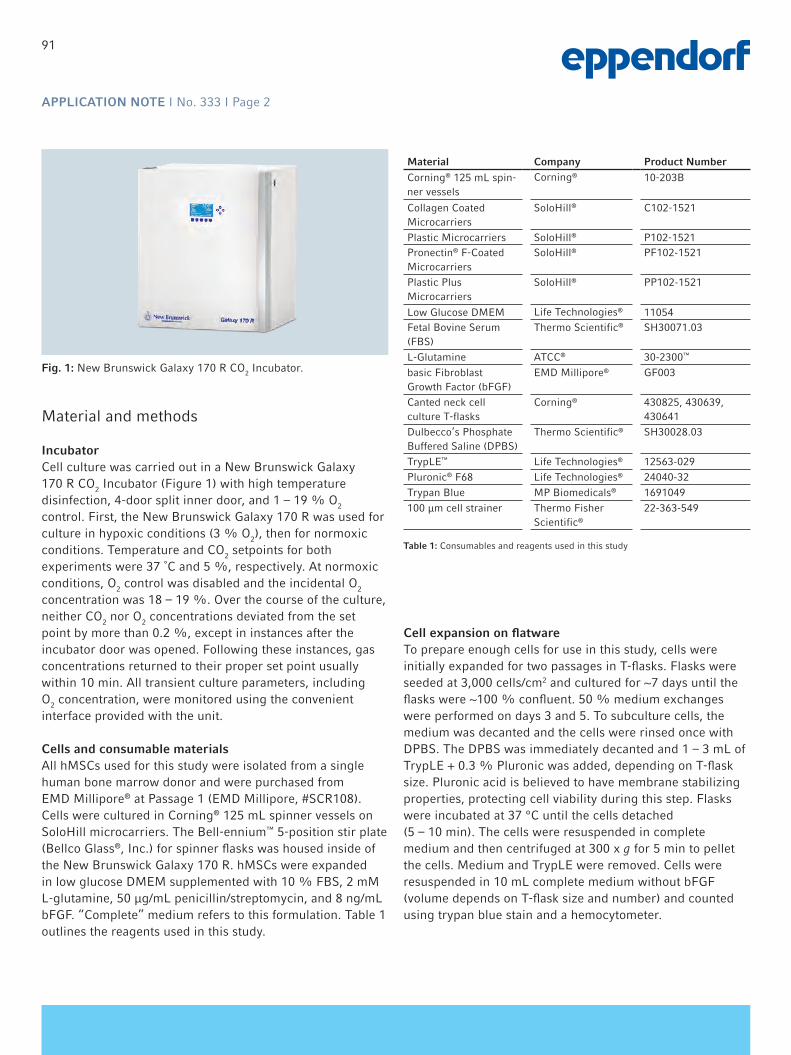
APPLICATION NOTE I No. 333 I Page 2
Fig. 1: New Brunswick Galaxy 170 R CO2 Incubator.
Material and methods
Incubator Cell culture was carried out in a New Brunswick Galaxy 170 R CO2 Incubator (Figure 1) with high temperature disinfection, 4-door split inner door, and 1 – 19 % O2 control. First, the New Brunswick Galaxy 170 R was used for culture in hypoxic conditions (3 % O2), then for normoxic conditions. Temperature and CO2 setpoints for both experiments were 37 ˚C and 5 %, respectively. At normoxic conditions, O2 control was disabled and the incidental O2 concentration was 18 – 19 %. Over the course of the culture, neither CO2 nor O2 concentrations deviated from the set point by more than 0.2 %, except in instances after the incubator door was opened. Following these instances, gas concentrations returned to their proper set point usually within 10 min. All transient culture parameters, including O2 concentration, were monitored using the convenient interface provided with the unit.
Cells and consumable materials All hMSCs used for this study were isolated from a single human bone marrow donor and were purchased from EMD Millipore® at Passage 1 (EMD Millipore, #SCR108). Cells were cultured in Corning® 125 mL spinner vessels on SoloHill microcarriers. The Bell-ennium™ 5-position stir plate (Bellco Glass®, Inc.) for spinner flasks was housed inside of the New Brunswick Galaxy 170 R. hMSCs were expanded in low glucose DMEM supplemented with 10 % FBS, 2 mM L-glutamine, 50 µg/mL penicillin/streptomycin, and 8 ng/mL bFGF. “Complete” medium refers to this formulation. Table 1 outlines the reagents used in this study.
Cell expansion on flatware To prepare enough cells for use in this study, cells were initially expanded for two passages in T-flasks. Flasks were seeded at 3,000 cells/cm2 and cultured for ~7 days until the flasks were ~100 % confluent. 50 % medium exchanges were performed on days 3 and 5. To subculture cells, the medium was decanted and the cells were rinsed once with DPBS. The DPBS was immediately decanted and 1 – 3 mL of TrypLE + 0.3 % Pluronic was added, depending on T-flask size. Pluronic acid is believed to have membrane stabilizing properties, protecting cell viability during this step. Flasks were incubated at 37 °C until the cells detached (5 – 10 min). The cells were resuspended in complete medium and then centrifuged at 300 x g for 5 min to pellet the cells. Medium and TrypLE were removed. Cells were resuspended in 10 mL complete medium without bFGF (volume depends on T-flask size and number) and counted using trypan blue stain and a hemocytometer.
Material Company Product NumberCorning® 125 mL spin-ner vessels
Corning® 10-203B
Collagen Coated Microcarriers
SoloHill® C102-1521
Plastic Microcarriers SoloHill® P102-1521Pronectin® F-CoatedMicrocarriers
SoloHill® PF102-1521
Plastic PlusMicrocarriers
SoloHill® PP102-1521
Low Glucose DMEM Life Technologies® 11054Fetal Bovine Serum (FBS)
Thermo Scientific® SH30071.03
L-Glutamine ATCC® 30-2300™
basic Fibroblast Growth Factor (bFGF)
EMD Millipore® GF003
Canted neck cell culture T-flasks
Corning® 430825, 430639, 430641
Dulbecco’s Phosphate Buffered Saline (DPBS)
Thermo Scientific® SH30028.03
TrypLE™ Life Technologies® 12563-029Pluronic® F68 Life Technologies® 24040-32Trypan Blue MP Biomedicals® 1691049100 µm cell strainer Thermo Fisher
Scientific®
22-363-549
Table 1: Consumables and reagents used in this study
91

APPLICATION NOTE I No. 333 I Page 3
Cell growth in spinner vesselsSpinners were operated at a microcarrier concentration of 5 cm2/mL and a volume of 75 mL. Cells subcultured on flatware (as described previously) were used to seed the spinners at a density of 5,000 cells/cm2. Spinner cultures were performed as described in previous microcarrier protocols [16]. Briefly, for Collagen Coated, Plastic, Pronectin F, and Plastic Plus microcarriers, cells were seeded in medium (acclimated to culture conditions for 20 min prior to seeding) and supplemented with 0.05 % FBS in the absence of bFGF until > 80 % of the cells attached (3 – 4 h). Serum and bFGF were then added to the culture to supplement the medium to the target of 10 % FBS and 8 ng/mL bFGF. Microcarrier cultures were agitated at 40 rpm. 50 % medium exchanges were performed on days 3 and 5. Glucose supplementation to 100 mg/dL was performed on days 4 and 6. Cell counts to quantify growth were performed on days 3, 5, 6, and 7.
Cell counting and harvesting from spinner vesselsCell counts were performed routinely during the culture to monitor the progress of cell growth. In addition, on day 7, the spinners were harvested to determine final cell recoveries. The same protocol was used in both cases, with volumes adjusted appropriately. First, the microcarriers were allowed to settle, then the medium was removed and replaced with DPBS. TrypLE containing 0.3 % Pluronic was added. The mixture was gently pipetted once or twice to thoroughly mix and then incubated at 37 °C for 5 – 10 min (with occasional gentle rocking by hand). Cells and microcarriers were then pipetted to mechanically dissociate the cell clumps into a single-cell suspension. For standard cell counting, the resulting cell slurry was then counted via hemocytometer and trypan blue staining. Following the count, sampled microcarriers were dried and weighed so that the cell count could be adjusted to account for the sampled microcarrier surface area. For cell harvesting, the cell slurry was passed through a 100 µm mesh cell strainer to remove the microcarriers. The cell pool was centrifuged, decanted, and resuspended in medium containing 0.05 % FBS for cell counting.
Stem cell marker visualization and assessment of attachment to microcarriersTo visualize the expression of several stem cell markers onhMSCs, samples were transferred from the spinners into15 mL tubes. Once the microcarriers settled, the medium was removed and the cells/microcarriers were carefully washed with DPBS for 5 min at room temperature. When the cells and microcarriers settled, the DPBS was removed and cells and microcarriers were fixed in 4 % paraformaldehyde for 10 min at room temperature. The paraformaldehyde was
then removed, and the cells were washed in DPBS and storedat 4 °C until use. Non-specific binding was blocked byincubation with 5 % FBS in DPBS for 1 h at roomtemperature. Samples were washed in 500 μL DPBS threetimes for 5 min at room temperature. Samples werethen incubated in 500 μL of the dye/antibody solutions.Dyes and antibodies used were: DAPI (Life Technologies,D3571), phalloidin-FITC (Life Technologies, A12379),FITC anti-human CD44 (BioLegend®, 338803),APC anti-human CD90 (BioLegend, 328113), and Alexa Fluor® 647 anti-human Stro-1 (BioLegend, 340103). Allantibodies were used at 1:1000 except for anti-Stro-1which was used at 1:500.
To assess the distribution of cell attachment across themicrocarrier population, a 2 mL sample was taken fromeach spinner approximately 5 – 6 h after seedingand processed exactly as outlined here. To visualize cellattachment, 500 μL of sample in DPBS was transferred to asingle well of a 24-well plate and incubated with 0.5 μL ofDAPI stain. Cells were visualized under fluorescent light,and the number of cells per microcarrier was tabulated for apopulation of > 50 microcarriers.
Adipogenesis and osteogenesis differentiationTo determine differentiation potential of the hMSCs expanded on microcarriers, cells harvested from spinners were frozen, then thawed, and seeded on 24-well plates at 6,000 cells/cm2. Growth/expansion medium was removed, and 1 mL of either Osteogenesis Induction Medium (EMD Millipore, SCR028) or Adipogenesis Induction Medium (EMD Millipore, SCR020) was added. Induction and maintenance media were changed according to EMD Millipore protocol (as recommended by supplier). Osteocyte differentiation was determined by Alazarin Red S staining and adipocyte differentiation was determined by Oil Red O staining (protocols supplied with EMD Millipore kits).
Results and discussion
AttachmenthMSCs were grown in the New Brunswick Galaxy 170 R for 7 days under normoxic and hypoxic conditions. The attachment distribution was assessed 5 to 6 h after seeding. At the current cell seeding density (5,000 cells/cm2), each microcarrier on average should contain about 4 cells per bead. An even distribution of cells across the microcarrier population leads to the most effective utilization of the available surface area and usually correlates with the highest cell densities if the surface is favorable for growth. Unused microcarriers (microcarrier with no cells bound) can be
92

APPLICATION NOTE I No. 333 I Page 4
Figure 2: Cell densities for hMSC cultures in the New Brunswick Galaxy 170 R incubator under normoxic and hypoxic conditions on various microcarriers. Cell densities were obtained via cell counts. Data points represent the average of n = 3 replicates. Error bars indicate +/– one standard deviation. For easier visualization, data were broken down into 4 graphs: A: Collagen Coated microcarriersB: Plastic microcarriersC: Pronectin F microcarriersD: Plastic Plus microcarriers and flatware controls
an early indication of an inefficient attachment that may not reach its maximum cell density. The results differed slightly depending on the oxygen condition. Under hypoxic conditions, less than 15 % of the microcarrier population was without cells after 5 – 6 h of culture. At normoxic conditions, this number was slightly higher at ~20 % for some microcarrier types; however, there was much higher variability among the replicates for this condition. Attachment did not seem to be affected by the microcarrier type evaluated.
Cell growth in the New Brunswick™ Galaxy® 170 RCell densities of cultures were quantified by using the described cell counting method to generate growth curves (Figure 2). Results obtained were generally consistent with past findings [17] in which cell densities reached 6 – 10 x 104 cells/cm2, with some exceptions. Most notably, cells grew to much higher densities on flatware in this experiment, as previous flatware densities only reached about 4 x 10⁴ cells/cm2. This difference may be due to the 40 % higher densities that were used to seed flatware in this study. In addition, growth on Collagen Coated microcarriers under normoxic conditions was higher than the previously reported range, while growth on Plastic Plus microcarriers under normoxic conditions was lower than the historical range. However, given the large variance among the different spinner replicates in this study (indicated by the error bars in Figure 2) and the variation in seeding densities tested historically, these differences may not be significant. All other conditions tested were comparable to the historical range.
The dependency of cell densities on O2 condition (as indicated by daily cell counts) was variable and depended on the microcarrier type tested. The nature of the growth curve for the normoxic conditions differed from the hypoxic conditions in that normoxic growth lagged behind hypoxic growth earlier in the culture, but then accelerated later in the culture. In the case of Collagen Coated microcarriers, cell densities were similar through day 6, but growth appeared to accelerate under normoxic conditions and the resulting cell density was higher on day 7. The same trend was observed on the flatware controls. For Pronectin F microcarriers, cell densities under hypoxic conditions were higher through day 6, but normoxic growth appeared to accelerate, and cell densities were equivalent on day 7. These trends are difficult to conclude with certainty, as there was a large amount of variation between the replicates, either due to sampling or biological variability. A similar trend was observed on Plastic Plus microcarriers, but cell densities remained lower in normoxic conditions throughout the duration of the culture. On Plastic microcarriers, there were no observed differences in cell densities.
A
B
C
D
0.0
2.0
4.0
6.0
8.0
10.0
12.0
14.0
16.0
18.0
0 2 4 6 8
Cell
Dens
ity (E
4 Ce
lls/c
m²)
Day
Collagen (hypoxic)
Collagen (normoxic)
0.0
2.0
4.0
6.0
8.0
10.0
12.0
14.0
16.0
18.0
0 2 4 6 8
Cell
Dens
ity (E
4 Ce
lls/c
m2)
Day
Pronectin F (hypoxic)
Pronectin F (normoxic)
0.0
2.0
4.0
6.0
8.0
10.0
12.0
14.0
16.0
18.0
0 2 4 6 8
Cell
Dens
ity (E
4 Ce
lls/c
m2)
Day
Plastic Plus (hypoxic)Plastic Plus (normoxic)Flatware Control (Hypoxic)Flatware Control (Normoxic)
0.0
2.0
4.0
6.0
8.0
10.0
12.0
14.0
16.0
18.0
0 2 4 6 8
Cell
Dens
ity (E
4 Ce
lls/c
m2)
Day
Pastic (hypoxic)
Plastic (normoxic)
93

APPLICATION NOTE I No. 333 I Page 5
Figure 3: Harvested day 7 cell densities for hMSC cultures at hypoxic vs. normoxic O2 concentrations in the New Brunswick Galaxy 170 R incubator grown on microcarriers and flatware. Cell densities were obtained via cell counts. Data points represent the average of n = 3 replicates. Error bars indicate +/– one standard deviation.
Cell densities were also dependent on the type of microcarrier tested; while the variability between the replicates made it difficult to make a statistical distinction between growth on Collagen Coated, Plastic, and Pronectin F microcarriers, cells did display more favorable growth on these microcarriers in comparison to Plastic Plus.
Cell harvestAs observed previously [17], hMSCs tend to form bridges between microcarriers late in the culture. This network of bridges leads to microcarrier aggregates that vary in size, which can contribute to variability in daily sampling. The exact mechanism for microcarrier aggregate formation is an area of ongoing investigation, as well as how the average density of cells in these clumps compares to the density of cells on single suspension microcarriers. Therefore, spinners were harvested on day 7, and the resulting cell pool was counted to generate a harvest cell density (Figure 3) as a potential means to gain a more accurate understanding of the true cell density over the entire microcarrier population. This approach also provides insight into how many cells can be harvested and further expanded.
Results from this harvest method suggested that hypoxic conditions were slightly more favorable for generating high cell densities on microcarriers in comparison to normoxic
conditions. This result is supported by the data suggesting attachment under hypoxic conditions was, on average, better than normoxic conditions, as better attachment kinetics often lay the foundation for higher cell densities later in the culture. Daily cell density measurements were also generally similar or higher under hypoxic conditions through day 6 of the culture, suggesting that the apparent accelerated normoxic growth on day 7 might be a sampling or assay artifact. Alternately, the harvest method used for these spinners could be preferentially favorable to cells grown under hypoxic conditions, as hypoxic conditions may be improving cell robustness and making cells more resistant to the stresses of harvesting. Regardless of interpretation, these data suggest that there is some advantage to growing hMSCs on microcarriers under hypoxic conditions. In the case of flatware controls, the data do not support the contention that hypoxic conditions are beneficial for growth. However, the procedure used to harvest flatware was slightly different than the procedure to harvest microcarriers, namely cells do not experience the same magnitude of shear forces. Although cell densities appear higher for normoxic conditions, this difference is not statistically significant and therefore may not represent a true departure from the overall trend.
0.0
2.0
4.0
6.0
8.0
10.0
12.0
14.0
Collagen Plastic Pronectin F Plastic Plus Flatware
Cell
Dens
ity (E
4 ce
lls/c
m2)
Surface
Hypoxic
Normoxic
94

APPLICATION NOTE I No. 333 I Page 6
Figure 4: 3D-stacked Fluorescent DAPI stained images (4 X magnification) of hMSCs on Collagen Coated microcarriers at 3, 5, and 7 days. The similarities in cell morphology between hypoxic and normoxic conditions are representative of other microcarrier types.
Cell morphology, identity and differentiation potentialOn days 3, 5, and 7 of the culture, samples were obtained and stained with DAPI to help visualize the three-dimensional confluency and morphology of cells on microcarriers. A comparison of cells cultured in hypoxic vs. normoxic conditions on Collagen Coated microcarriers (Figure 4) revealed no differences in confluency levels during culture. All cells were of proper morphology, indicating good health and viability of the culture.
Day 7 samples were obtained prior to harvest and stained to verify the retention of stem cell markers as an indication of stem cell identity. Additionally, cells were serially passaged on plastic microcarriers at hypoxic conditions for a total of 6 passages, and samples from passage 6 were compared to cells obtained from passage 1 (Figure 5). Both samples stained positive for CD44, CD90 (not shown), and Stro1, indicating retention of stem cell identity. Results are representative for all microcarrier types after passage 1 at both hypoxic and normoxic conditions (data not shown), but serial passaging was performed only on plastic.
Finally, cells serially passaged 6 times on animal-derived component-free plastic microcarriers under hypoxic conditions were successfully differentiated into adipocyctes and osteocytes following both the first and sixth passage (data not shown, Figure 6). Adipogenesis was indicated by the presence of red–stained lipid droplets and osteogenesis was indicated by a calcification layer and the presence of red–stained calcium phosphate deposits.
Figure 5: hMSCs expanded in the New Brunswick Galaxy 170 R were incubated with anti-CD44 (green), anti-Stro1 (red) and DAPI (blue) at 10 X magnification. Exposure times were adjusted for maximum contrast. High background resulted from lengthy exposure times due to low expression levels. Populations of CD44 expressing cells are indicated by green arrows. Populations of Stro1 expressing cells are indicated by red arrows. Results are representative of all microcarrier types tested.
Figure 6: Cells serially passaged six times on plastic microcarriers in the New Brunswick Galaxy 170 R incubator differentiate into adipocyctes and osteocytes. Bright field images were taken with a Nikon® Ti65 at 20 X (adipocyctes) and 10 X (osteocytes). Arrows indicate lipid droplets and calcium phosphate deposits. Sixth passage undifferentiated samples were stained with hematoxylin solution.
Day 3 Day 5 Day 7
Col
lage
n H
ypox
icC
olla
gen
N
orm
oxic
Plastic, Normoxic,
Passage 1 (Control)
Plastic, Hypoxic,
Passage 1 (Culture start)
Plastic, Hypoxic,
Passage 6 (Culture end)
DA
PI
+ C
D44
DA
PI
+ S
tro1
Control (Undifferentiated)
Adipogenesis Osteogenesis
Fla
twar
e (C
ontr
ol)
Pla
stic
M
icro
carr
iers
Sixth Passage
95
APPLICATION NOTE I No. 333 I Page 7
ConclusionhMSCs grown in the New Brunswick Galaxy 170 R CO2
Incubator on microcarriers under hypoxic conditions display normal growth as compared to those grown at normoxic O2 concentrations. The data presented here suggest that more cells may be harvested from microcarriers grown under hypoxic conditions, indicating that hypoxic conditions favor a slightly better growth rate. Large differences in growth rates between hypoxia and normoxia may not be expected in this experiment, since the cell line was already adapted to normoxic conditions when the cells were harvested from the patient and removed from their native hypoxic environment. Recent reviews indicate that significant changes may happen at the gene expression level during adapation to culture conditions, where O2 level plays an important role by regulating hypoxia-inducible factor-1 (HIF-1)-mediated expression of different genes [18].
The ease of setup and the tight O2 concentration control displayed by the New Brunswick Galaxy 170 R provided
the ideal conditions for this experiment. This study is a demonstration of the low oxygen feature of an advanced incubator and it could be used to successfully conduct stem cell studies under physiological conditions. As recently pointed out by Dr. Elaine Fuchs (Rockefeller University) during her keynote speech at the 2013 annual meeting of the International Society for Stem Cell Research (ISSCR), many in vitro cell culture techniques may produce questionable results since the culture environment is greatly deviated from native physiological conditions. Dr. Fuchs’s group created a mouse embryo-based “biological incubator” in which to study the effects of gene knockout and stem cell transplantation under native physiological conditions instead of in a flask [19,20]. Such principle and practice is enchanting but may not be feasible for universal adaptation by all cell culture labs. However, it is certainly feasible to employ an incubator with precision O2 control to conduct cell culture research under physiological oxygen conditions.
96

APPLICATION NOTE I No. 333 I Page 8
References
[1] Friedenstein AJ, Gorskaja JF, Kulagina NN. Fibroblast precursors in normal and irradiated mouse hematopoietic organs. Experimental Hematology 1976; 4(5):267-74.
[2] Fraser JK, Wulur I, Alfonso Z, Hedrick MH. Fat tissue: an underappreciated source of stem cells for biotechnology. Trends in biotechnology 2006; 24(4):150-4.
[3] Cao C, Dong Y, Dong Y. [Study on culture and in vitro osteogenesis of blood-derived human mesenchymal stem cells]. Chinese Journal of Reparative and Reconstructive Surgery 2005; 19(8):642-7.
[4] Griffiths MJ, Bonnet D, Janes SM. Stem cells of the alveolar epithelium. Lancet 2005; 366(9481):249-60.[5] Beltrami AP, Barlucchi L, Torella D, Baker M, Limana F, Chimenti S, Kasahara H, Rota M, Musso E, Urbanek K, Leri A,
Kajstura J, Nadal-Ginard B, Anversa P. Adult cardiac stem cells are multipotent and support myocardial regeneration. Cell 2003; 114(6):763-76.
[6] Pittenger MF, Mackay AM, Beck SC, Jaiswal RK, Douglas R, Mosca JD, Moorman MA, Simonetti DW, Craig S, Marshak DR. Multilineage potential of adult human mesenchymal stem cells. Science 1999; 284(5411):143-7.
[7] Chow DC, Wenning LA, Miller WM, Papoutsakis ET. Modeling pO(2) distributions in the bone marrow hematopoietic compartment. II. Modified Kroghian models. Biophysical Journal 2001; 81(2):685-96.
[8] Chow DC, Wenning LA, Miller WM, Papoutsakis ET. Modeling pO(2) distributions in the bone marrow hematopoietic compartment. I. Krogh’s model. Biophysical Journal 2001; 81(2):675-84.
[9] Estrada JC, Albo C, Benguria A, Dopazo A, Lopez-Romero P, Carrera-Quintanar L, Roche E, Clemente EP, Enriquez JA, Bernad A, Samper E. Culture of human mesenchymal stem cells at low oxygen tension improves growth and genetic stability by activating glycolysis. Cell Death and Differentiation 2012; 19(5):743-55.
[10] Grayson WL, Zhao F, Bunnell B, Ma T. Hypoxia enhances proliferation and tissue formation of human mesenchymal stem cells. Biochemical and Biophysical Research Communications 2007; 358(3):948-53.
[11] Hung SP, Ho JH, Shih YR, Lo T, Lee OK. Hypoxia promotes proliferation and osteogenic differentiation potentials of human mesenchymal stem cells. Journal of Orthopaedic Research: Official Publication of the Orthopaedic Research Society 2012; 30(2):260-6.
[12] Fehrer C, Brunauer R, Laschober G, Unterluggauer H, Reitinger S, Kloss F, Gully C, Gassner R, Lepperdinger G. Reduced oxygen tension attenuates differentiation capacity of human mesenchymal stem cells and prolongs their lifespan. Aging Cell 2007; 6(6):745-57.
[13] Holzwarth C, Vaegler M, Gieseke F, Pfister SM, Handgretinger R, Kerst G, Muller I. Low physiologic oxygen tensions reduce proliferation and differentiation of human multipotent mesenchymal stromal cells. BMC Cell Biology 2010; 11:11.
[14] Wang DW, Fermor B, Gimble JM, Awad HA, Guilak F. Influence of oxygen on the proliferation and metabolism of adipose derived adult stem cells. Journal of Cellular Physiology 2005; 204(1):184-91.
[15] Zeng HL, Zhong Q, Qin YL, Bu QQ, Han XA, Jia HT, Liu HW. Hypoxia-mimetic agents inhibit proliferation and alter the morphology of human umbilical cord-derived mesenchymal stem cells. BMC Cell Biology 2011; 12:32.
[16] SoloHill Protocols: Protocol for Small Scale Microcarrier Culture. www.pall.com.[17] SoloHill Application Note: Expansion and Characterization Of Mesenchymal Stem Cells on Microcarriers. www.pall.com.[18] Haque N, Rahman MT, Abu Kasim NH, Alabsi AM. Hypoxic culture conditions as a solution for mesenchymal stem cell
based regenerative therapy. The Scientific World Journal 2013; 2013:632972.[19] Beronja S, Livshits G, Williams S, Fuchs E. Rapid functional dissection of genetic networks via tissue-specific
transduction and RNAi in mouse embryos. Nature Medicine 2010; 16(7):821-7.[20] Schober M, Fuchs E. Tumor-initiating stem cells of squamous cell carcinomas and their control by TGF-beta and
integrin/focal adhesion kinase (FAK) signaling. PNAS 2011; 108(26):10544-9.
97

APPLICATION NOTE I No. 333 I Page 9
Ordering informationDescription Order no. international Order no. North America
New Brunswick™ Galaxy® 170 R with high temperature disinfection, 1 – 19 % O2 control, 4-split inner doors
CO17334001 CO17234005
www.eppendorf.com
Pall® and SoloHill® are registered trademarks of Pall Corporation, USA. Pronectin® is a registered trademark of Sanyo Chemical Industries, Ltd., Japan. EMD Millipore® is a registered trademark of Merck KGaA, Germany. Corning® is a registered trademark of Corning, Inc., USA. BellcoGlass® is a registered trademark of BellCoGlass, Inc., USA. Life Technologies® and Pluronic® are registered trademarks of Life Technologies Corporation, USA. Fisher Scientific® is a registered trademark of Fisher Scientific Company, LLC, USA. Nikon® is a registered trademark of Nikon Corporation, Japan. Science® is a registered trademark of AAAS, USA. Thermo Scientific® is a registered trademark of Thermo Scientific, Inc., USA. ATCC® is a registered trademark of ATCC, USA. MP Biomedicals® is a registered trademark of MP Biomedicals, LLC., USA. BioLegend® is a registered trademark of BioLegend, Inc., USA. VWR® is a registered trademark of VWR International, LLC., USA. Bell-ennium™ is trademark of BellcoGlass, Inc., USA. TrypLE™ is a trademark of Life Technologies Corporation, USA. 30-2300™ is a trademark of ATCC. Eppendorf® and the Eppendorf logo are registered trademarks of Eppendorf AG, Germany. New Brunswick™ is a trademark of Eppendorf AG, Germany. Galaxy® is a registered trademark of Eppendorf, Inc., USA. U.S. Design Patents are listed on www.eppendorf.com/ip. All rights reserved, including graphics and images. Copyright © 2016 by Eppendorf AG.
Your local distributor: www.eppendorf.com/contactEppendorf AG · 22331 Hamburg · [email protected] · www.eppendorf.com
98

99
Scalable Expansion of Human Pluripotent Stem Cells in Eppendorf BioBLU® 0.3 Single-use Bioreactors Ruth Olmer1, Christina Kropp1, Claudia Huether-Franken2* and Robert Zweigerdt1
1 Leibniz Research Laboratories for Biotechnology and Artificial Organs (LEBAO), REBIRTH Cluster of Excellence, Hannover Medical School,
Carl-Neuberg-Str. 1, 30625 Hannover, Germany; 2 Eppendorf AG Bioprocess Center, Rudolf-Schulten-Str. 5, 52428 Juelich,
Germany; * Corresponding author: [email protected]
APPLICATION NOTE No. 292 I October 2013
Abstract
The routine application of human pluripotent stem cells and their derivatives in regenerative medicine and inno-vative drug discovery will require the constant supply of high cell numbers in consistent, high quality. Well moni-tored and controlled stirred-tank bioreactors represent suitable systems to establish up-scalable bioprocesses
enabling the required cell production. The following application note describes the successful cultivation of human pluripotent stem cells in suspension culture using Eppendorf BioBLU 0.3 Single-use Vessels in a DASbox® Parallel Mini Bioreactor System.
Introduction
Human pluripotent stem cells (hPSCs), comprising human embryonic stem cells (hESC) and induced pluripotent stem cells (hiPSC), and their derivatives are considered promising cell sources for novel regenerative therapies [1]. Cell thera-pies aim at the replacement of cell or tissue loss induced by degenerative disorders such as cardiovascular and neurode-generative diseases, diabetes and many others, which can-not be healed by currently established, conventional treat-ments. Moreover, specific human cell types derived from hPSCs by differentiation can be utilized for the development of yet unavailable in vitro disease models, novel drug discov-ery strategies and more predictive drug safety assays.
Most of the envisioned clinical and industrial applications will require billions of lineage-specific cells which cannot be produced by conventional surface-adherent 2-dimensional (2D) cultures. Stirred-tank bioreactors, which are widely used in the biopharmaceutical industry for the generation of recombinant proteins expressed in mammalian (tumor) cell lines, provide numerous advantages for process development, as they allow for online monitoring and
control of key process parameters such as pH, oxygen tension and biomass formation. Advanced bioreactor systems which have been developed in a wide range of culture vessels also facilitate the straightforward scale-up
Figure 1: DASbox Mini Bioreactor System with BioBLU 0.3 Single-use Vessels

100
APPLICATION NOTE I No. 292 I Page 2
to larger process dimensions. However, cultivation and differentiation of hPSCs in stirred bioreactors apparently require the adaptation of cell cultivation from the established 2D surface-adherent culture to 3-dimensional (3D) suspension culture. It was recently demonstrated that hPSCs can be successfully grown as free floating, “cell only aggregates” in small-scale suspension [2-4]. Based on this knowhow the transfer to a DASGIP® Parallel Bioreactor System with four individually controlled glass vessels having a working volume of 100 - 250 mL each, was established [5]. Optimization of stirring-controlled aggregate formation from single cell inoculated hiPSCs led to an approximately four-fold cell expansion resulting in 2 x 10⁸ cells per vessel (100 mL) using a fed-batch process. However, with regard to the envisioned clinical application of hPSCs, the possibility to utilize single-use culture vessels, which will support the development of GMP-conform processes, is of great interest. Subsequently, aim of this work was to establish a suspension culture of hiPSCs in a parallel DASbox Mini Bioreactor System equipped with fully instrumented BioBLU 0.3 Single-Use Vessels.
Materials and Methods
Experiments were performed utilizing the cord blood derived hiPSC line hCBiPSC2 [6]. Suspension cultures were initiated by detachment and dissociation of hiPSC monolayer cultures with accutase (Life Technologies). Single cells were suspended in mTeSRTM1 (STEMCELL Technologies, Vancouver, Canada) supplemented with the ROCK inhibitor Y-27623 (10 µM). Each BioBLU 0.3 single-
use vessel was equipped with probes for pO2 and pH. The pH probes were calibrated by two-point calibration. pO2 probe calibration was conducted under process conditions: headspace gassing with 3 sL/h air plus 5% CO2, stirring at 70 rpm utilizing an pitched-blade impeller [5, 7], 37°C in 100 mL mTeSRTM1; after stable pO2 values were reached a slope calibration was performed. For culture inoculation 25 mL of a single-cell suspension were added to achieve a density of 5 x 10⁵ cells /mL in the final 125 mL culture volume. After 48 h the entire medium was replaced daily (batch feeding) excluding cell loss. For cell counting and other analysis a sampling volume of 3.5 mL was harvested daily without medium replacement to prevent culture dilution. This strategy resulted in subsequent culture volume reduction from 125 to approximately 100 mL during the 7 day process duration. Beside pO2 and pH, glucose and lactate concentrations, viable cell counts and the expression of pluripotency markers were monitored. Daily viable cell counts were performed via a trypan blue exclusion assay after cell-aggregate dissociation by collagenase B (Roche) treatment. Pluripotency assessment was performed by flow cytometry analysis specific to SSEA4 and TRA1-60.
Results and Discussion
24 h after inoculation of respective single cell suspensions to BioBLU 0.3 Single-Use Vessels small cell aggregates with an average diameter of 58.1 ± 23.1 µm emerged in the stirred cultures. These aggregates, which showed a highly homogeneous size distribution throughout the process, increased in size over the cultivation period resulting in an average diameter of 139.25 ± 25.37 µm (figure 2) on day 7.
Figure 2: Aggregate formation in stirred suspension culture. After pro-cess inoculation by single cell suspension small cell aggregates arose. Increase in aggregate size was observed during the process. Scale bars: 200 µm.
Figure 3: Growth kinetics of hCBiPSC2 in stirred suspension cul-ture. Cells were seeded at 5 x 10⁵ cells /mL (d0) and cell counts were determined daily. An up to 4-fold increase in cell count was achieved in individual runs over 7 days resulting in up to 2.3 x 10⁶ cells /mL.

101
APPLICATION NOTE I No. 292 I Page 3
A robust ~4-fold increase in viable cell count was achieved in this fed-batch process resulting in an average cell con-centration of 2.1 x 10⁶ cells/mL on day 7 and thus a total cell yield of ~2.1 x 10⁸ cells per vessel (figure 3).
Monitoring the metabolic activity revealed ~47% of glu-cose consumption and accumulation of 7.4 mM lactate at 48 hours. The metabolic activity was also followed by online measurements of pH and pO2. Increasing cell numbers over time resulted in a maximum pH drop to 6.8 (figure 4; as compared to pH 7.4 in fresh medium) and dissolved oxygen
levels decreased to 57% (data not shown). The expression of pluripotency-associated surface markers TRA 1-60 and SSEA4 were determined at the process endpoint to evaluate the quality of the expanded hPSCs. Flow cytometry revealed that the majority of the yielded cell population retained ex-pression of these markers i.e. 84% positivity for TRA 1-60 and 90% for SSEA4 (figure 5) was observed suggesting maintenance of pluripotency in this cultivation process.
ConclusionThis set of experiments demonstrates the successful expan-sion of human pluripotent stem cells applying the DASbox system in combination with BioBLU 0.3 Single-Use Vessels. In a 7 day-lasting expansion process in stirred suspension culture cell yields of up to 2.3 x 10⁸ cells /100 mL were ob-tained, which is in good agreement with our previous data in the DASGIP Parallel Bioreactor System, stirred glass ves-sel system (DS0200TPSS; 100-250 mL working volume) [5]. Notably cells generated by the described process retained expression of established, pluripotency associated cell sur-face markers. The work confirms the general applicability of the culture system for hPSC expansion in stirred suspen-sion and reveals the DASbox system in combination with BioBLU 0.3 Single-Use Vessels to be an excellent platform for further process optimization and future adaptation to lineage-specific hPSC differentiation processes.
Figure 4: Metabolic activity of hCBiPSC2 in stirred suspension culture. The culture medium was replaced daily starting at 48 h thereby resulting respec-tive glucose and lactate concentration patterns. The pH dropped from 7.4 in fresh medium to lowest values of ~6.8 at the end of a 24 h feeding interval on process days 5-7.
Figure 5: Expression of pluripotency-associated surface markers is retained after expansion in stirred suspension cultures. Flow cytometry revealed that the majority of cells that were harvested at the process endpoint (after day 7) expressed pluripotency-associated surface mark-ers TRA 1-60 and SSEA 4 (gray line represents isotype controls).
References1. Zweigerdt, R., Large Scale Production of Stem Cells and Their Derivatives. Adv Biochem Eng Biotechnol, 2009. 114: p. 201-35
2. Olmer, R., et al., Long term expansion of undifferentiated human iPS and ES cells in suspension culture using a defined medium. Stem Cell
Res, 2010. 5(1): p. 51-64.
3. Singh, H., et al., Up-scaling single cell-inoculated suspension culture of human embryonic stem cells. Stem Cell Res, 2010. 4(3): p. 165-79.
4. Zweigerdt, R., et al., Scalable expansion of human pluripotent stem cells in suspension culture. Nat Protoc, 2011. 6(5): p. 689-700.
5. Olmer, R., et al., Suspension culture of human pluripotent stem cells in controlled, stirred bioreactors. Tissue Eng Part C Methods, 2012.
18(10): p. 772-84.
6. Haase, A., et al., Generation of induced pluripotent stem cells from human cord blood. Cell Stem Cell, 2009. 5(4): p. 434-41.
7. Schroeder, M., et al., Differentiation and lineage selection of mouse embryonic stem cells in a stirred bench scale bioreactor with automated
process control. Biotechnol Bioeng, 2005. 92(7): p. 920-33.

102
APPLICATION NOTE I No. 292 I Page 4
Life Technologies® is a registered trademark of Life Technologies Corporation, USA. Roche is a registered trademark of F. Hoffmann-La Roche AG, Switzerland. Eppendorf®, the Eppendorf logo, and BioBLU® are registered trademarks of Eppendorf AG, Germany. DASGIP® and DASbox® are registered trademarks of DASGIP Information and Process Technology GmbH, Germany. U.S. Design Patents are listed on www.eppendorf.com/ip.All rights reserved, including graphics and images. Copyright © 2015 by Eppendorf AG.
Ordering information Order no. DASbox® Mini Bioreactor System for Cell Culture, max. 5 sL/h gassing4-fold system for single-use vessels 76DX04CCSU8-fold system for single-use vessels 76DX08CCSU16-fold system for single-use vessels 76DX16CCSU24-fold system for single-use vessels 76DX24CCSU
www.eppendorf.com
Your local distributor: www.eppendorf.com/contactEppendorf AG · 22331 Hamburg · [email protected] · www.eppendorf.com

Abstract
The expansion of stem cells, including mesenchymal stem cells (MSC), has been successfully demonstrated using microcarrier-based small bioreactors such as spinner flasks. In this study, we explored a simple alternative for microcarrier-based MSC expansion using conventional shake flasks. This method relies on a new type of CO2 incubator with built-in shaking capability, i.e. New Brunswick S41i CO2 Incubator Shaker. The expansion of adipose-derived mesenchymal stem cells (AdMSCs) was compared between shake flasks and spinner flasks using microcarriers. The AdMSCs were seeded at a density of 3x10³ cells/cm2 in both setups, each containing 0.5 g plastic microcarriers and 40 mL of stem cell growth medium.
The cell culture experiments were conducted for 12 days and samples were collected daily for analysis of cell growth, biochemistry and metabolites. Cell density studies revealed that AdMSCs cultured under shake flask conditions achieved excellent growth under 12 day batch-culture conditions.
Lastly, the AdMSCs expanded using the shake flask method remained high quality stem cells, which was evident by CD44 and CD90 stem cell marker assays and their ability to differentiate into either adipocytes or osteocytes.
A Novel Method for the Expansion of Mesenchymal Stem Cells using a New Brunswick™ S41i CO2 Incubator ShakerKhandaker Siddiquee and Ma Sha, Eppendorf Inc., Enfield, CT, U.S.A
Introduction
Stem cells are undifferentiated cells which have the capability of self-renewal and the potential to divide for a long period of time. They have the ability to differentiate into various specialized cells when appropriate growth factors and conditions are provided. Stem cells can be broadly classified as: embryonic, adult, and induced pluripotent stem cells (iPS). Adult stem cells can be further characterized by their tissue of origin, such as: hematopoietic, mammary, intestinal, mesenchymal, endothelial, neural, and hair follicle stem cells. Most of the studies performed on adult stem cells utilize either hematopoietic or adipose-derived mesenchymal stem cells1. Like other adult stem cells, adipose-derived mesenchymal stem cells (AdMSCs) express all of the common stem cell markers and can be differentiated into various types of
APPLICATION NOTE No. 259 I September 2013
specialized cells under appropriate growth conditions. AdMSCs have advantages over other mesenchymal stem cells (MSCs), since they can be isolated in large quantities from fat tissue and are resistant to apoptosis2.
Although MSCs have enormous advantages for regenerative medicine, drug screening and drug discovery, their applications are limited by the quantities required for industrial applications3. In this study, we developed a simple shake flask culture technique to expand MSCs on microcarrier beads which can be used to scale-up into large-scale bioreactors. The microcarrier shake flask culture, which requires both agitation and CO2 gas control, was conducted in the Eppendorf New Brunswick S41i CO2 incubator shaker.
103

APPLICATION NOTE I No. 259 I Page 2
Materials and Methods
Initial cell culture in T-FlasksAdMSCs were obtained from ATCC® (PCS-500-011) at passage 2 and cells were seeded at a density of 5,000 cells /cm2 into a T-75 cm2 flask (Eppendorf) using 15 mL of mesenchymal stem cell basal medium (ATCC) supplemented with 2 % fetal bovine serum, 5ng/mL rh FGF basic, 5ng/mL rh acidic, 5 ng/mL rh EGF and 2.4 mM L-Alanyl-L- Glutamine (ATCC).
Cultivation of cells on microcarriersPrior to start of the experiment, 0.5 g of 125-212 micron polystyrene microcarriers (SoloHill®) (180 cm2 for a 50 mL culture) was transferred into a siliconized (Sigmacoat®; Sigma) 250 mL spinner flask (Corning®) and shake flasks (Schott®, Duran®) along with 25-30 mL of PBS. The flasks were then autoclaved at 121 °C for at least 30 minutes. Microcarriers were allowed to settle to the bottom of the shake/spinner flasks and the autoclaved PBS buffers were carefully aspirated with the electronic pipetting aid easypet® (Eppendorf) equipped with a 25 or 50 mL pipette. The AdMSCs were initially seeded at a density of 3x10³ cells/cm2 into both flasks, each containing 40 mL of basal mesenchymal stem cell medium. For the initial attachment of cells, the agitation speed of the New Brunswick S41i CO2 incubator shaker and rotation speed of the spinner (housed inside of an Eppendorf Galaxy® 170 R
The New Brunswick S41i CO2 Incubator Shaker, designed for both
non-adherent and adherent cell culture applications, combines the
precise temperature and CO2 control of an incubator with the reliable
New Brunswick laboratory shaker drive mechanism. Key features
include sealed inner/outer doors, high-temperature disinfection, and
reduced CO2 consumption compared to competitor models4.
CO2 incubator) were both kept at 50 rpm and incubated for 2 hrs at 37 °C with 5 % CO2. After incubation, the cell culture volume was adjusted to 50 mL total with 10 mL of medium containing serum to reach a final FBS concentration of 4 % and targeted final concentration of growth supplements (10 ng/mL final concentration of rh FGF basic, rh FGF acidic & rh EGF and 4.8 mM final concentration of L-Alanyl-L-Glutamine). Following the addition of FBS and growth supplements, the rotation speed of the spinner and the agitation speed of New Brunswick S41i CO2 incubator shaker were both raised to 70 rpm. After 18 to 24 hrs of incubation, 1 mL of homogeneous samples containing both media and microcarriers were collected for microscopic observations, cell counting as well as biochemistry analysis.
Cell counting Cells on microcarrier beads were counted by hemocytometer. To accomplish this, microcarrier beads were suspended in citric acid solution containing crystal violet (0.1 % crystal violet in 0.1 M citric acid solution) equal to the volume of supernatant removed from the tube. The contents of the tube were incubated for 1 hr or overnight at 37 °C and vortexed for a few seconds to release the stained nuclei. The nuclei were counted with hemocytometer.
Biochemistry and metabolites analysisThe supernatants collected during cell counting were used for biochemistry and metabolite measurements using an automated YSI® 2950 Bio-analyzer.
Stem cell surface marker assayTo assess the quality of AdMSCs after expansion and to confirm that the stem cell markers were retained during the microcarrier-based culture, CD44 and CD90-specific fluorescent immunoassays were performed using the following procedure. 5 mL samples were collected from both the spinner and shake flasks near the end of microcarrier culture. After the microcarriers settled to the bottom, the supernatants were removed and the microcarrier beads containing cells were gently washed 3 times with PBS at room temperature. Cells on the microcarrier beads were then fixed with 4 % paraformaldehyde for 30 minutes followed again by PBS washing 3 times. Cell-containing microcarrier beads were blocked with 5 % FBS at room temperature for 1 hr and immunostained with FITC-conjugated antihuman CD44 (BioLegend®) and APC-conjugated antihuman CD90 (BioLegend®) antibody solutions, also for 1 hr at room temperature. The beads were washed 5 times with room temperature PBS for 5 min and visualized using an EVOS® FL fluorescence microscope.
104

APPLICATION NOTE I No. 259 I Page 3
Stem cell differentiation assaysAdMSCs were harvested from both shake and spinner flasks into 50 mL tubes. Once the microcarrier beads settled to the bottom of the tube, the supernatants were removed and cells were washed with DPBS. Afterwards, the microcarrier beads were treated with 5 mL of prewarmed trypsin-EDTA solution at 37 °C for 10 min. During incubation, the tubes were occasionally vortexed for 2 sec and then neutralized by adding an equal volume of trypsin neutralizing solution. Microcarrier beads were a llowed to settle to the bottom of the tube and the supernatants were collected as soon as possible. Microcarrier beads were washed 2-3 times with DPBS and as much supernatant as possible was collected into a 50 mL tube. Following washing, AdMSCs were collected to bottom of the tube by centrifugation at 120 xg for 5 min and resuspended in 5 mL of mesenchymal stem cell medium. Cells were seed at a density of 18,000 cells/cm2 into a 24-well plate. Adipocytes and osteocyte differentiations were performed on those cells using differentiation assay kits from ATCC. Adipocyte and osteocyte differentiated cells were identified by cell-type specific staining with either Oil red O or Alizarin red S kits (ScienCell®) according to manufacturer instructions and visualized using an OLYMPUS® CK40 microscope.
Results and Discussion
To compare between shake flask and spinner flask cultures, AdMSCs were seeded at a density of 3x10³ cells/cm2 in both systems. Cell culture studiess were conducted for 12 days and samples were collected for cell growth, biochemistry and metabolite analysis daily. Cell growth studies revealed that AdMSCs cultured under shake flask conditions achieved excellent growth during the 12 day batch culture (Figure 1A). Biochemistry and metabolite analysis revealed that glucose concentrations decreased from 1.09 g/L to 0.548 g/L (for shake flask culture) and 0.798 g/L (for spinner culture), whereas lactate concentrations increased from 0.042 g/L to 0.396 g/L (for shake flask culture) and 0.259g/L (for spinner culture) after 12 days of culture (Figure 1B &C).
The higher glucose consumption and lactate production rate seen in the shake flask culture supports the finding that the stem cells grew at a faster rate under the shake flask conditions. Furthermore, during early growth phase (day 4); the amount of ammonium accumulated in spinner flask culture (2.4 mM) was 1.8-fold higher than shake flask culture (1.3 mM) (Figure 1D). It has been shown that even low level of ammonium (1.9 mM) inhibits MSC growth5. The spinner culture has shown ammonium level exceeding 2 mM early and throughout the culture process, which indicates the slower growth by the spinner method could be a result of ammonium toxicity-induced growth inhibition. The fact that spinner culture had elevated ammonium levels early in the culture not seen in the shake flask also indicates possible stem cell damage due to shear force by the spinner rod. The spinner rod was observed to display a “stop & go” motion at low speeds; precise speed control is not possible, especially at low rotation speeds. However, our observations were based on the specific spinner device available at our research facility, the results may not represent typical or average performance from spinner devices available in the market place.
To determine whether or not AdMSCs retained their stem cell properties during their growth under shake flask conditions, immunostaining of stem cell surface markers and differentiation assays were performed. Microcarrier beads that contained AdMSCs were immunostained with stem cell surface marker antibodies such as: FITC-conjugated antihuman CD44 and APC-conjugated antihuman CD90 and revealed that AdMSCs retained stem cell surface markers during growth under shake flask culture condition (Figure 2A&B). For the adipocyte and osteocyte differentiation assays, AdMSCs were collected from the microcarrier beads and seeded into 24 well plates that contained either adipocyte or osteocyte differentiation medium. After 17 days of culture, the plates were stained with Oil Red O or Alizarin Red S staining solutions, respectively. Microscopic observation indicated that most of the AdMSCs from shake flask culture differentiated into either adipocytes or osteocytes successfully (Figure 3A&B).
105

APPLICATION NOTE I No. 259 I Page 4
Figure 1. Analysis of AdMSCs growth and metabolism in shake flask and spinner flasks culture conditions: A) growth; B) glucose
utilization; C) lactate production and D) ammonium production.(u) shake flask and (n) spinner flask.
A. B.
C. D.
106

APPLICATION NOTE I No. 259 I Page 5
CD90 marker (red)
Nucleus (blue)
Microcarrier bead
CD44 marker (green)
Nucleus (blue)
Microcarrier bead
Figure 2. Stem cell marker identification assay for AdMSCs expanded on microcarriers in shake flask. A) AdMSCs on microcarrier beads
are positive for CD90 stem cell marker, as indicated in red by fluorescence imaging. B) AdMSCs on microcarrier beads are positive for CD
44 stem cell marker, as indicated in green by Fluorescence Imaging. Blue color indicates stem cell nuclear staining by DAPI.
Figure 3. Differentiation assays for AdMSCs expanded on microcarriers in shake flask. A) Adipogenic differentiation formed lipid droplets
as indicated by Oil red O positive staining. B) Osteogenic differentiation caused calcium mineralization of extracellular matrix as indicated
by Alizarin Red S positive staining.
A. B.
A. B.
107

APPLICATION NOTE I No. 259 I Page 6
Conclusions
Stem cell expansion using shake flask conditions appears to be a viable and simple alternative to the spinner flask system. This novel method relies on a new type of CO2 incubator with built-in shaking capability, such as the New Brunswick S41i CO2 Incubator Shaker. The New Brunswick S41i reduces shearing, eliminates potential cell damage by the spinner rod, decrease the risk of contamination associated with inserting a magnetic stirrer base into the CO2 incubator and reduces experimental complexity. This method also greatly increases the cell culture capacity whereby a large number of shake flasks can be placed in the New Brunswick S41i simultaneously. In the case of spinner
References
1. Zhang et al. “Mechanisms Underlying the Osteo- and Adipo-Differentiation of Human Mesenchymal Stem Cells”. The Scientific World
Journal (2012).Volume 2012; Article ID793823;
2. Nastran el al. “Osteogenic Differentiation and Osteochondral Tissue Engineering Using Human Adipose-Derived Stem Cells.”
Biotechnology progress (2013) Volume 29.No.1: 176- 185;
3. Schop et al. “Expansion of human mesenchymal stromal cells on microcarriers: growth and metabolism” J. Tissue Eng Regen Med
(2010); Volume 4. No. 4; 131-140.
4. Kohlstrom et al. “Hybridoma and CHO Cell Culture using New Brunswick S41i – An Environmental Friendly, Low Emission Incubator
Shaker”. Eppendorf Application Note AA257, Nov. 2012.
5. Schop D. “Growth and Metabolism of Mesenchymal Stem Cells Cultivated on Microcarriers”, Ph.D. Thesis 2010, University of Twente,
The Netherlands.
flask culture, a typical incubator without active cooling can only handle the heat emitted from a very limited number of magnetic stirrer bases before causing temperature setpoint overshoot, a significant limitation to the scale-up potential of the spinner method. This reinforces the superiority of New Brunswick S41i CO2 Incubator Shaker as an alternative to incubator/spinner based stem cell culture. This method eliminates a scale-up bottleneck while providing the highest quality stem cell culture for inoculation of large scale industrial bioreactors. The shake flask has lower cost and less parts to disassemble, clean, assemble and autoclave.
108

www.eppendorf.com
SoloHill® is a registered trademark of SoloHill Engineering, Inc., Michigan, USA. Sigmacoat® is a registered trademark of Sigma-Aldrich, USA. Duran® is a registered trademark of DURAN Group, GmbH. Corning® is a registered trademark of Corning, Inc., New York, USA. ATCC® is a registered trademark of American Type Culture Collection, Corp., Virginia, USA. YSI® is a registered trademark of YSI, Inc., Yellow Springs, USA. EVOS® is a registered trademark of Life Technologies, New York, USA. BioLegend® is a registered trademark of BioLegend, Inc., California, USA. ScienCell® is a registered trademark of ScienCell Research Laboratories, California, USA. OLYMPUS® is a registered trademark of Olympus Corporation, USA. NucleoCounter® is registered trademark of ChemoMetec, Denmark. Galaxy® is a registered trademark of New Brunswick Scientific, Co., Inc, USA.Eppendorf® and the Eppendorf logo, and Easypet® are registered trademarks of Eppendorf AG, Germany. New Brunswick™ is a trademark of Eppendorf AG, Germany.All rights reserved, including graphics and images. Copyright © 2016 by Eppendorf AG, Germany.
Your local distributor: www.eppendorf.com/contact Eppendorf AG • 22331 Hamburg • Germany [email protected]
APPLICATION NOTE I No. 259 I Page 7
Ordering informationDescription International order no. North America Order no.
New Brunswick™ S41i CO2 Incubator Shaker, 120 V, 60 Hz S41I-120-0100
New Brunswick™ S41i CO2 Incubator Shaker, 230 V, 50 Hz S41I-230-0100
Options Stacking kit P0628-6502 P0628-6502
Additional perforated shelf P0628-6181 P0628-6181
Interchangeable platforms, 612 x 356 mm (24 x 14 in) M1334-9920 M1334-9920
Universal platform M1334-9921 M1334-9921
250 mL Dedicated platform M1334-9922 M1334-9922
500 mL Dedicated platform M1334-9923 M1334-9923
1 L Dedicated platform M1334-9924 M1334-9924
2 L Dedicated platform M1334-9925 M1334-9925
2.8 L Dedicated platform M1334-9926 M1334-9926
4 L Dedicated platform M1334-9927 M1334-9927
Galaxy 170 R CO2 Incubator (High-Temp Disinfection,1-19 % O2 Control)
CO170R-230-1200 CO170R-120-1200
T-75 Tissue Culture Flask5 flasks per bag, 100 flasks per case (Available in China, India and Italy)
0030 711.106
Easypet®
(Electronic pipette controller– suitable for serologiocal pipettes, 0.1 to 100 mL )
4421 000.013 022230204
109

110
Abstract
When growing microbes or animal cells in a stirred-tank reactor, it is critical to choose the impeller type that is best suited to your process. Select the wrong impeller, and you could make chop suey of your filamentous fungi. Pick the right impeller, and you could greatly increase yields of your fussy mammalian cultures such as Chinese hamster ovary
(CHO) and Vero kidney epithelial cells. With a wide range of impeller designs to choose from, how do you tell which is right for your application? Here we describe six commonly used fermentor and bioreactor impellers, explain how they work, and identify which may perform best for culturing certain animal, insect, plant, yeast, and bacterial cell lines.
Which Impeller Is Right for Your Cell Line? A Guide to Impeller Selection for Stirred-Tank BioreactorsRich Mirro and Kevin Voll, Eppendorf Inc., Enfield, CT, U.S.A.
How Blade Orientation Affects Mixing
All impellers are designed to homogenously mix cells, gases, and nutrients throughout the culture vessel. The mixing action evenly distributes oxygen and nutrients to cells for healthy growth, keeps them from settling to the bottom of the vessel, and helps to maintain a uniform culture temperature. Depending on the impeller type you select, mixing will be imparted as a radial flow, axial flow, or a combination of the two. As Figure 1 shows, radial flow occurs when fluid is pushed away from the impeller’s axis toward the vessel wall. Axial flow occurs when fluid is pushed up or down along the axis or shaft of the impeller. The orientation of an impeller (left- or right-handed) and its agitating direction determine whether the direction of axial flow is up or down. A right-handed impeller option will push fluid in an upward direction toward the top of the vessel if agitation is clockwise (as viewed from the top). A left-handed option paired with a clockwise agitation will push fluid down toward the bottom of the vessel. Therefore, when positioning blades on an impeller shaft, it’s important to know which direction your impeller will be turning (clockwise or counterclockwise). To increase mixing action in some applications, one impeller blade may be oriented for up flow while the other is positioned for down flow. Although there is no right or wrong way to position an impeller blade(s), reversing flow direction could improve yields in some instances.
Rushton Impellers for FermentationJ. H. Rushton originally called the impellers he designed flat-bladed turbines. So Rushton impeller is today the most common generic term applied to flat-bladed or disk-turbine impellers (e.g., Figure 2). Their blades are flat and set vertically along an agitation shaft, which produces a unidirectional radial flow. Rushton and Rushton-type impellers are commonly used in fermentations of cell lines that are not considered shear-sensitive, including yeasts, bacteria, and some fungi.
APPLICATION NOTE No. 315 I January 2009
Figure 1: Axial and radial flow models
Figure 2: A six-bladed Rushton-type impeller

111
APPLICATION NOTE No. 315 I Page 2
Pitched-Blade Impellers for Shear-Sensitive CellsThe blades on pitched-blade impellers (Figure 3) are flat and set at ~45° angles, which produces a simultaneous axial and radial flow. This combination provides better overall mixing and creates a higher oxygen mass transfer rate (KLa) than that of unidirectional marine blade impellers. Pitched-blade impellers are low-shear impellers designed to gently mix the contents of a culture without causing cell damage. They are most often used with mammalian, insect, or other shear-sensitive cell lines growing in suspension or with the aid of microcarriers. These impellers are often used in batch or fed-batch cultures, but they can also be used for continuous and perfusion processes. Because of their proficient mixing design, pitched-blade impellers are also widely used in fermentation processes that involve highly viscous cultures, such as filamentous bacteria and fungi, as well as in some anaerobic biofuels processes.
Gentle Marine-blade ImpellersThe leading face of the blades on a marine-blade impeller (Figure 4) can be flat or concave, whereas their back sides are convex. This produces an axial flow. Like pitched-blade impellers, marine-blade impellers are used for applications that require gentle mixing without causing cell damage. Due to the unidirectional flow, however, the KLa values of marine-blade impellers tend to be slightly lower than those of impellers that produce both axial and radial mixing.
Spin FiltersSpin filters are retention devices commonly used to keep cells inside a vessel during continuous or perfusion culture. In New Brunswick™ bioreactors, for example, spin filters with low-shear marine blade impellers are designed for suspension and microcarrier applications. A spin-filter kit consists of a screened cage surrounding an impeller shaft with very small filter pore openings that keep cells isolated outside the cage (Figure 5). Inside that rotating cage, a dip tube is provided for continuous withdrawal of culture broth. A media feed tube outside the cage provides a steady supply of fresh nutrients. Although pore openings
vary from one manufacturer to the next, New Brunswick spin filters come with 10-μm openings for suspension cultures and 75-μm openings for microcarrier cultures. Because of its gentle mixing nature, a spin filter is typically used with microcarrier-dependent cell lines or those that are highly sensitive to shear. These mechanisms are ideal for use in production of secreted proteins because they keep harvested media cell-free, which simplifies purification in downstream processing. Over time, however, the screen material covering a spin-filter cage will become clogged with cell debris and require replacement. Culture run times are limited by this factor.
Special Impellers for Microcarrier CulturesThe New Brunswick™ CelliGen® cell-lift impeller (Figure 6) provides uniform circulation for microcarrier cultures. This is an ultra-low-shear impeller in which flow is caused by three discharge ports located on the impeller shaft. Rotation of those ports creates a low-differential pressure at the base of the impeller tube, lifting microcarriers up through the tube and expelling them out through its ports. This continuous recirculation loop keeps cells uniformly dispersed throughout a vessel. Gases are introduced through a ring sparger, which generates bubbles that pass along the impeller
Figure 3: Pitched-blade impeller, right-handed orientation
Figure 4: Marine-blade impeller, left-handed orientation
Figure 5: Spin filter assembly
Figure 6: Cell-lift impellers for microcarrier culture
Outer screen (10 μm or 75 μm)
Support cage
Marine blade impeller
Dip tube (harvest waste)
Impeller shaft
Foam breaker cage
Discharge ports
Mesh lining (85 µm)
Aeration cage

112
APPLICATION NOTE No. 315 I Page 3
between the exterior of the inner tube and an outer membrane, known as the aeration cage. A mesh lining on the outer membrane of this cage has penetrations that are small enough (85 μm) to ensure that cells growing on the microcarriers cannot pass through. Gas exchange occurs at the membrane–media interface, ensuring that cells remain in a bubble-free environment and are not subjected to shear due to bubble breakage. The bubbles are then expelled through two ports (located at the top of the impeller) into a second screened-in cage. A foam breaker directs air, supplied by a gas overlay, into the cage to break up foam. Cell-lift impellers are typically used in batch and fed-batch processes involving shear-sensitive animal cells. They can also be used for continuous perfusion processes when a decanting column(s) and media feed-in and broth pump-out setup are added.
A General GuideImpeller designs are almost as varied as the types of cell lines they are designed to help grow. Table 1 lists several cell lines commonly used in fermentation and cell culture processes and matches each with the impeller(s) best suited for its growth.
Packed-Bed Basket ImpellersAnother New Brunswick design is the packed-bed basket impeller used in the manufacture of secreted products from either anchorage-dependent or suspension cultures. A basket includes two horizontally positioned, perforated metal screens that extend to the walls of a bioreactor vessel (Figure 7). Enclosed between those screens, a bed of Fibra-Cel® disks serves as a solid support matrix for cell growth.
Cell Line
Rushton and Rushton-Like Impellers
Pitched-Blade Impeller
Marine-Blade Impeller
Spin Filter Impeller
Cell Lift Impeller
Basket Impeller
HumanHEK 293 HeLa HL60 Lncap THP-1 UMSCC HFF KB MRC-5 HybridomaDA4.4 123A 127A GAMMA 67-9-B SP20 PrimateVero COS-7 Rat TumorGH3 9L PC12 Mouse3T3 MC3T3 NS0 HamsterCHO BHK ZebrafishZF4 AB9 InsectSF9 Hi-5 Sf21 YeastSaccharomyces cereviseae Baker’s yeast Pichia pastoris Candida albicans BacteriaStreptomyces Bacillus Echerichia coli AlgaeRed/Green
Table 1: A general guide to choosing impellers by cell line

113
Cells growing in the disk bed become immobilized on or between the disks, where they remain protected from external shear forces throughout each culture run. Media circulates by way of a hollow impeller tube with discharge ports positioned above the basket. As with the cell-lift impeller, rotation of these discharge ports creates a low differential pressure at the base of the impeller tube, which circulates media throughout the system. The medium receives gases through a sparger located at the bottom of the inner tube, which protects cells from being exposed to the gas-iquid interface. This results in low turbulence and low shear stress for the culture. Exceptionally high cell densities are achievable with packed-bed baskets because of a high surface-to-volume ratio for cell growth provided by the disk bed coupled with an ability to use perfusion or medium-replacement techniques. Culture periods in excess of three months have been reported[1][2]. By ensuring that cells remain entrapped in the bed, this system also simplifies protein harvesting from the resulting cell-free media.
References
[1] Bioreactor Design for High Cell Density Culture Us-ing Microcarriers. Wang G, Zhang W. Nanjing Vac-cine Conference,Nanjing, China, 3–4 August 2001. New Brunswick, Edison, NJ.
[2] Modified CelliGen Packed Bed Bioreactors for Hybrid-oma Cell Cultures. Wang G, et al. Cytotechnol. 9, 1992: 41–49.
Metal screens
(to form the basket)
Discharge ports
Packed bed (Fibra-Cel disks not shown)
Harvest dip tube
APPLICATION NOTE No. 315 I Page 4
www.eppendorf.com
Eppendorf® and the Eppendorf logo are registered trademarks of Eppendorf AG, Germany. New Brunswick™ is a trademark of Eppendorf AG, Germany. Fibra-Cel® is a registered trademark owned by Imerys Mineral California, Inc. and licensed to New Brunswick Scientific Co., Inc., USA. CelliGen® is a registered trademark of New Brunswick Scientific Co., Inc., USA. All rights reserved, including graphics and images. Copyright © 2013 by Eppendorf AG.
Your local distributor: www.eppendorf.com/contact Eppendorf AG • 22331 Hamburg • Germany [email protected]
Figure 7: Packed-bed basket impellers for secreted products

Abstract
With a renewed acceptance of perfusion processes in cell culture, both in the laboratory and for production applications [1], New Brunswick™ packed-bed bioreactors using Fibra-Cel disks are seeing an upsurge in interest.
This application note examines Fibra-Cel technology and its many advantages, from increased secreted protein yields to labor savings in applications including rabies vaccine production.
An Update on the Advantages of Fibra-Cel® Disks for Cell CultureRich Mirro Eppendorf Inc., Enfield, CT, USA Corresponding author: [email protected]
What Is Fibra-Cel?
Eppendorf Fibra-Cel is a solid-support growth matrix (Figure 1) for anchorage-dependent and suspension cell cultures. It is used predominantly in perfusion processes for the production of secreted products—such as recombinant proteins and viruses—and it is currently being evaluated for stem cell research [2].
Since the 1980s, scientists around the globe have been using Fibra-Cel to grow a wide range of cell types (see inset, right), including hybridomas and insect cultures. Originally used in New Brunswick™ CelliGen® autoclavable cell culture bioreactors, Fibra-Cel technology has now been successfully scaled up for commercial production in sterilizable-in-place systems as large as 150 liters. BioBLU® packed-bed, single-use vessels containing Fibra-Cel are also available for those who prefer the advantages of a disposable system.
Manufactured according to cGMP guidelines, Fibra-Cel is composed of two layers of nonwoven material—polyester and polypropylene—which are sonicated together, cut into disks, and electrostatically treated to attract cells and facilitate their attachment to the disks. Normally it takes about six hours for cells to attach to microcarriers (with a normal inoculum of 1 × 10⁶ cells/mL), whereas cells can attach within 15 – 60 minutes on Fibra-Cel disks.
Figure 1. Scanning electron micrograph of Fibra-Cel disks (left); mouse–mouse hybridoma DA4.4 immobilized on Fibra-Cel disks during produc-tion at 1 × 10⁸ cells/cm3 of packed-bed volume (right)
Moreover, the growth process for microcarrier cultures can require extended delays for periodic stoppage of stirring to allow time for cells to become attached. By comparison, the Fibra-Cel bed is inoculated (3 × 10⁵ cells/mL of bed volume) in a single step.
APPLICATION NOTE No. 313 I July 2011
Cells successfully used on Fibra-Cel disksHybridoma: 123A, 127A, GAMMA, 67-9-B, DA4-4
Anchorage-dependent: 3T3, MRC-5 and other human fibroblasts, HEK 293, BHK, COS, CHO, stroma cells, hepatocytes
Insect cells: Tn-368, Sf9, rSf9, Hi-5
114

APPLICATION NOTE No. 313 I Page 2
Fibra-Cel in a New Brunswick bioreactor is also advantageous over microcarriers because it enables sustained long-term periods of high-density growth in perfusion mode, without danger of clogging because there are no filters. Perfusion is a mode of cell culture in which a fresh nutrient medium is continuously added to the culture while simultaneously removing the spent medium that contains the product of interest. In a New Brunswick bioreactor, cells growing on or in the Fibra-Cel disk bed are retained within the vessel, inside the packed bed, where they continue the production of the desired product.
The packed bed comprises two horizontally positioned screens that extend to the bioreactor vessel walls. Enclosed between the screens, a bed of Fibra-Cel disks serves as solid support for the growing cells (Figure 2). Cells growing in the disk bed become immobilized on or between the disks, where they remain throughout the culture run, protected from external shear forces. The process is favored for manufacturing because product yields can be increased by as much as tenfold over comparable processes [3]. Once the bioreactor is set up and inoculated, the culture can be maintained to produce proteins for long periods of time thus saving labor, time, and money.
Like the proprietary Eppendorf Cell Lift impeller, rotation of the discharge ports in the proprietary packed-bed impeller creates a low differential pressure at the base of the impeller tube, which circulates the medium throughout the system. The medium receives gases through a sparger located at the bottom of the inner tube, protecting the cells from being exposed to the gas liquid interface. This results in low turbulence and low shear stress on the culture. Exceptionally high cell densities are achievable due to the high surface-to-volume ratio provided by the disk bed, coupled with the ability to use perfusion. In comparison with other cell support systems, it was found that higher titers and cell densities were achieved in trials using Fibra-Cel disks [4].
Figure 2. Basket impeller with Fibra-Cel disks
Other benefitsBecause higher yields are possible, smaller bioreactors can be used to substantially reduce the initial capital expenditure as well as reduce the utilities required for operation (such as electricity, water, and steam if required). In addition, because the cells remain entrapped, the packed bed eliminates the need for cell filtration to separate cells from the end product, thus simplifying harvesting. Last, product recovery and downstream processing can be more easily controlled because users can determine the volume of harvest material that is to be processed at any given time.
115

APPLICATION NOTE No. 313 I Page 3
Commercial productionBioreactors using Fibra-Cel have been used in the production of a variety of commercial products. One example of commercial production includes end products such as EPO, which can now be commercially produced on the bench using Fibra-Cel technology, eliminating the need for labor-intensive and space-consuming roller bottles. A substantial portion of the world’s human rabies vaccine is also produced using Eppendorf Fibra-Cel technology. Additionally, many of our customers are currently using Fibra-Cel in their proprietary processes to produce interferons, monoclonal antibodies, and hormones.
In summary, Fibra-Cel provides benefits in research laboratories as well as in commercial production. Our customers have found that because yields are high, bioreactors containing Fibra-Cel packed beds can outperform much larger-sized bioreactors, thereby achieving commercial-scale production in a bioreactor with a far smaller footprint. Production space requirements are reduced, as are costs associated with labor, start-up, and operations.
For protocols on other cell lines, or for additional information on the Fibra-Cel, see eppendorf.com.
References
[1] Perfusion Bioreactors Are Making a Comeback but Industry Misconceptions Persist. Langer ES. Bioprocessing J. 9, Winter 2010–2011: 49–52.
[2] Expansion of Engrafting Human Hematopoietic Stem/Progenitor Cells in Three-Dimensional Scaffolds with Surface-Immobilized Fibronectin. Feng Q, et al. J. Biomed. Mater. Res. A 78(4) 15 September 2006: 781–791.
[3] Packed-Bed Bioreactors for Mammalian Cell Culture: Bioprocess and Biomedical Applications. Meuwly F, et al. Elsevier Inc., 2006: 46–56.
[4] High Vero Cell Density and Rabies Proliferation on Fibra-Cel Disks Versus Cytodex-1 in Spinner Flask. Hassanzadeh S, et al. Pakistan J. Biol. Sci. 2011: 1–8.
www.eppendorf.com
Gendicine® is a registered trademark and of Shenzhen SiBiono GeneTech Co., Ltd., China. Eppendorf®, the Eppendorf logo and BioBLU® are registered trademarks of Eppendorf AG, Germany. New Brunswick™ is a trademark of Eppendorf AG, Germany. Fibra-Cel® is a registered trademark owned by Imerys Mineral California, Inc. and licensed to New Brunswick Scientific Co., Inc., USA. CelliGen® is a registered trademarks of New Brunswick Scientific Co., Inc., USA. All rights reserved, including graphics and images. Copyright © 2016 by Eppendorf AG.
Your local distributor: www.eppendorf.com/contact Eppendorf AG • 22331 Hamburg • Germany [email protected]
Ordering information
Description Order No.
Fibra-Cel® disks, a solid support growth matrix for mammalian, animal, and insect cells
50 g M1292-9984
250 g M1292-9988
116

Fermentation

The Eppendorf BioFlo® 320 Bioprocess Control Station: An Advanced System for High Density Escherichia coli Fermentation Bin Li, Khandaker Siddiquee, and Ma Sha Eppendorf, Inc., Enfield, CT, USA
Corresponding author: [email protected]
APPLICATION NOTE No. 340 I January 2015
118
Abstract
In this application note, an Escherichia coli (E. coli) fermentation run was conducted using the new Eppendorf BioFlo 320 bioprocess control station. High cell density was achieved at 12 h as determined by a maximum optical density (OD600) measurement of 215.2. The wet cell weight (WCW) and dry cell weight (DCW) were also measured and presented.
Introduction
The newest offering in the Eppendorf bioprocess portfolio, the BioFlo 320 seamlessly combines form and function into one all-inclusive package. The BioFlo 320 is an advanced bioprocess system developed for both microbial fermentation and cell culture applications. A new industrial design, flexibility between autoclavable and single-use vessels, intelligent sensors, Ethernet connectivity, and an improved software package are only a few of the features that set it apart from the competition.
In a previous application note using the BioFlo/CelliGen® 115 benchtop system [1], we successfully cultured E. coli under aerobic conditions with a maximum OD600 value of 140. In this application note, E. coli cultivation achieved an even higher OD600 value of 215.2 using the new BioFlo 320 bioprocess control station (Figure 1). Furthermore, the WCW and DCW were also measured and presented. Figure 1: BioFlo 320 bioprocess control station with
water-jacketed (left) and stainless steel dished-bottom (right)
vessels

APPLICATION NOTE I No. 340 I Page 2
119
Materials and Methods
EquipmentFermentation was performed using an Eppendorf BioFlo 320 bioprocess control station with the configuration outlined in Table 1. The E. coli K12 strain (ATCC®, 10798™) was grown in a 1 L (working volume) stainless steel dished-bottom BioFlo 320 glass vessel, as shown in Figure 1. Glucose concentrations were measured using a Cedex® Bio Analyzer (Roche®). The OD600 was measured with a spectrophotometer. An Eppendorf MiniSpin® plus was used to pellet the cells. A pH sensor (InPro® 3253i/SG/225) and an optical Dissolved Oxygen (DO) sensor (InPro 6860i), both incorporating Intelligent Sensor Management (ISM®) technology from Mettler Toledo®, were used in this experiment. A laboratory oven (LAB-LINE®, L-C series) was used to dry the cell pellets for DCW measurements.
Parameter Configuration
Gas Mix Automatic gas mix
Gas Flow Control Thermal mass flow controller (TMFC) with 0 – 20 standard liters per minute (SLPM) flow range
Vessel 1 L stainless steel dished-bottom glass vessel
Motor Direct drive motor
Impeller Two Rushton impellers
Sparger Ring sparger (Macrosparger)
Table 1: BioFlo 320 hardware configuration
Elapsed Fermentation Time(EFT, h)
5:15 6:15 6:16 7:00 7:01 7:30 9:00 9:30 10:00 10:30 11:00 11:30 12:00
Pump Speed (mL/min) 0.3 0.5 0.2 0.5 0.6 0.9 1.7 2.1 2.7 3.2 3.7 4.5 5.3
MediumE. coli was cultured in chemically defined medium, pH 6.8. The initial fermentation medium was prepared as follows: 150 mL 10 x phosphate/citric acid buffer (133 g/L KH2PO4, 40 g/L (NH4)2HPO4, 17 g/L citric acid) and 1.35 L deionized (DI) water were added to the vessel for sterilization at 121 °C for 20 min. After the medium was cooled to room temperature, the following sterile components were added aseptically to make the complete fermentation medium: 15 mL of 240 g/L MgSO4, 0.34 mL of 20 g/L thiamine, 15 mL of 100 x trace element solution, and 22 mL of 70 % glucose solution. The 100 x trace element solution contained: 10 g/L iron (III) citrate, 0.25 g/L CoCl2
.6H2O, 1.5 g/L MnCl2.4H2O,
0.15 g/L CuCl2.6H2O, 0.3 g/L H3BO3, 0.25 g/L Na2MoO4.2H2O,
1.3 g/L zinc acetate.2H2O, 0.84 g/L EDTA [2, 3].
An additional concentrated feeding medium was prepared separately in a 1 L glass bottle. 67.5 mL of 240 g/L MgSO4, 2.49 mL of 20 g/L thiamine solution, 22.5 mL of 100 x trace element solution, and 70 % glucose solution were added to a final volume of 750 mL.
Inoculum preparation and fermentationThe inoculum was grown in Terrific Broth (TB) medium, prepared as described previously [4]. Two 500 mL baffled shake flasks (VWR®, 30623-210) each containing 100 mL of TB medium were inoculated from a frozen vial of E. coli and incubated at 30 °C, 200 rpm overnight in a New Brunswick™ Innova® 40 benchtop incubator shaker. Cell growth was monitored by offline measurement of the OD600 value. The vessel was inoculated with 75 mL of inoculum (5 % of the initial fermentation medium volume).
Antifoam 204 (Sigma-Aldrich®, A6426) was added only when needed, since it may reduce the oxygen transfer rate (OTR) and possibly lower the final cell density. About 5 mL of 5 g/L antifoam was added between 7 – 12 h of fermentation, as foam accumulation warranted.
Pump 3 was assigned as the feeding pump. The feeding strategy included increasing or decreasing the feeding pump speed based on the glucose concentration. This strategy was designed to maintain glucose concentration below 2 g/L. Table 2 and Figure 2 illustrate the adjustments made to the pump speed over the course of the fermentation. Although a similar feed program can be used for repeated fermentation runs, the feed start time must be adjusted each time according to the growth dynamics of each fermentation.
Cell growth and glucose concentration were monitored offline using 5 mL samples taken according to the following schedule. For OD600 readings, samples were taken every hour and diluted appropriately for accurate measurement. For the determination of glucose concentration, samples were taken every hour before the initiation of feeding, and then every ~30 min after the feeding began. The specific growth rate (μ) was calculated from the fitted OD600 value in Microsoft® Excel®.pH calibration and controlpH calibration was performed outside the vessel using a two-point calibration method and standard buffers. Buffer pH 7.0 was used to set ZERO and pH 4.0 for the SPAN. The pH sensor was calibrated prior to autoclaving the vessel.
Table 2: Pump speed at different EFT during the fed-batch fermentation

APPLICATION NOTE I No. 340 I Page 3
120
Figure 2: EFT-based feeding program as displayed on the en-
hanced software platform
The pH was automatically maintained at 6.8 by adding 25 % (v/v) NH4OH via front mounted peristaltic pump (assigned as “base”). The deadband for pH control was 0.02.
DO sensor calibration and gassing controlSince the BioFlo 320 is compatible with multiple types of DO sensors, an optical sensor was chosen for DO control instead of the traditional polarographic DO sensor. One of the advantages of the optical DO sensor is that it does not require the 6 h polarization time of the polarographic DO sensor, which reduces the turnaround time between fermentation runs. Calibration was performed using a standard two-point calibration method: 0 % (set ZERO) was obtained by running 1200 rpm agitation and 3 SLPM N2 flow until the DO value stabilized. 100 % (set SPAN) was obtained by running 1,200 rpm agitation and 3 SLPM air flow until the DO value stabilized at maximum.
The BioFlo 320 software offers a selection of automatic gassing control cascades that are dependent upon the configuration of the unit. The BioFlo 320 used in this study included the automatic gas mix and four TMFCs with a flow range of 0 – 20 SLPM (Table 1). User-defined DO cascade settings utilizing agitation, air, and oxygen in sequential manner are shown in Figure 3. The DO setpoint was 30 %.WCW and DCW measurementSamples were taken to measure the WCW and DCW. 1 mL of culture sample was added into an Eppendorf microcentrifuge tube and pelleted at 7,500 rpm for 5 min. The supernatant was carefully removed using an Eppendorf Research® pipette and the WCW was measured by calculating the difference in
weight between the tube before and after sample addition. Furthermore, the tube was kept in a heating oven and maintained at 70 – 80 °C until the cell pellet was dry and the DCW measured similarly.
Results and Discussion
Samples were taken periodically to monitor the cell growth (OD600 value) and glucose concentration as described above. Feeding was initiated when the glucose concentration dropped below 2 g/L, which occurred at 5.25 h of cultivation. After starting the feed, the pump rate was adjusted according to the current glucose concentration with the end goal of keeping it at or below 2 g/L (Table 2). As shown in Figure 4A, the OD600 value reached 215.2 within 12 h. The growth curve was also plotted on a log scale to calculate the specific growth rate (µ = 0.54 h-1, Figure 4B).
WCW and DCW were also measured during the fermentation. The results are shown in Table 3 and Figure 5. During the cultivation, both WCWs and DCWs increased proportionally with the increase in the OD600 value.
Figure 3: DO cascade used in this experiment

APPLICATION NOTE I No. 340 I Page 4
0255075100125150175200225
0
1.5
3
4.5
6
7.5
9
10.5
12
0 1 2 3 4 5 6 7 8 9 10 11 12
OD
600
[Glu
cose
] (g
/L)
Time (h)
[Glucose]
OD600
y = 0.54x - 0.90R² = 1.00
-0.7
0.3
1.3
2.3
3.3
4.3
5.3
6.3
0 1 2 3 4 5 6 7 8 9 10 11 12
ln (
OD
600)
Time (h)
050
100150200250300350400450500
0 20 40 60 80 100 120 140 160 180 200 220
Cel
l w
eight
(g/L
)
OD600
Dry cell weight
Wet cell weight
121
A B
Figure 4: Fermentation growth curve and glucose concentration
A: OD600 and glucose concentration over the course of the 12 h fermentation
B: Growth curve plotted on a log scale; a linear trend line was applied in Microsoft Excel, the slope of which was obtained as the
specific growth rate, µ (h-1)
Conclusions
The Eppendorf BioFlo 320 bioprocess control station was able to support high density E. coli growth using a fed-batch fermentation method. An OD600 optical density of 215.2 was reached at 12 h. The wet/dry cell weights were measured at various time points, which were used as records of cell growth along with OD600 values. Although efforts were made to maintain a glucose concentration below 2 g/L, the fermentation was not optimized for medium, growth conditions, or any product yield.
OD600 WCW (g/L) DCW (g/L)
15.6 67.5 ± 4.7 15.2 ± 2.7
30.3 117.2 ± 4.1 24.6 ± 2.0
58.0 214.1 ± 16.3 41.7 ± 2.0
89.8 334.4 ± 19.3 64.6 ± 2.0
111.2 386.5 ± 8.8 72.4 ± 1.7
215.2 453 ± 13.0 110.7 ± 2.4
Table 3: WCW and DCW; average was calculated from 5 samples
with mean +/- one standard deviation
Figure 5: Correlation between OD600 value and cell weight

APPLICATION NOTE I No. 340 I Page 5
122
Literature
[1] Li B, Willard S, Sha M. High Cell Density Fermentation of Escherichia coli Using the New Brunswick BioFlo115. Eppendorf Application Note No. 335, 2014. http://www.nbsc.com/files/335_Li_Ecoli.pdf
[2] Geerlof A. High cell-density fermentation of Escherichia coli. http://www.helmholtz-muenchen.de 2008.[3] Korz DJ, Rinas U, Hellmuth K, Sanders EA, Deckwer WD. Simple fed-batch technique for high cell density cultivation of
Escherichia coli. Journal of Biotechnology 1995; 39(1):59-65.[4] Terrific Broth. Cold Spring Harbor Protocols 2006; 2006(1):pdb.rec8620.

APPLICATION NOTE I No. 340 I Page 6
www.eppendorf.com
ATCC® is a registered trademark of American Type Culture Collection, USA. Cedex® and Roche® are registered trademarks of Roche Diagnostics GmbH, Germany. LAB-LINE® is a registered trademark of LAB-LINE Instruments, Inc., USA. VWR® is a registered trademark of VWR International, Inc., USA. Sigma-Aldrich® is a registered trademark of Sigma-Aldrich Co., USA. 10798™ is a trademark of American Type Culture Collection, USA. InPro®, ISM®, and Mettler Toledo® are registered trademarks of Mettler-Toledo AG, Switzerland. Microsoft® and Excel® are registered trademarks of Microsoft Corporation, USA. The Eppendorf logo, Eppendorf®, Eppendorf Research® plus, and Eppendorf MiniSpin® plus are registered trademarks of Eppendorf AG, Germany. New Brunswick™ is a trademark of Eppendorf AG, Germany. BioFlo®, CelliGen®, and Innova® are registered trademarks of Eppendorf Inc., USA.U.S. Design Patents are listed on www.eppendorf.com/ip. All rights reserved, including graphics and images. Copyright © 2015 by Eppendorf AG.
Your local distributor: www.eppendorf.com/contactEppendorf AG · 22331 Hamburg · [email protected] · www.eppendorf.com
123
Ordering informationDescription Order no. International Order no. North America
BioFlo® 320 Control Station Call Call
BioFlo® 320 1 L Vessel Bundle, Stainless Steel Dished-Bottom M1379-0300 M137-0300
New Brunswick™ Innova® 40 Benchtop Incubator Shaker M1299-0092 M1299-0092
Eppendorf Research® plus, single-channel, fixed, 1000 µL 3120000.062 3121000.120
Eppendorf Safe-Lock Tubes, 1.5 mL 0030120.086 0030120.086
Eppendorf MiniSpin® plus centrifuge 5452000.018 5453000.011

Parameter ConfigurationGas Mix Automatic gas mix option
Gas Flow Control One Thermal mass flow controller (TMFC) with 0 – 20 SLPM flow range
Vessel 2 L working volume heat-blanketed glass ves-sel with baffle assembly
Motor Direct drive motor
Impeller Two rushton impellers
Sparger Ring sparger (Macrosparger)
124
High Cell Density Fermentation of Escherichia coli Using the New Brunswick™ BioFlo® 115 Bin Li, Stacey Willard, and Ma ShaEppendorf, Inc., Enfield, CT, USA
Corresponding author: [email protected]
APPLICATION NOTE No. 335 I July 2014
Abstract
This application note presents a successful example of a high density fermentation of Escherichia coli (E. coli) using the New Brunswick BioFlo 115 benchtop, autoclavable
fermentor from Eppendorf. The highest optical density (OD600) achieved in this study was 140 at 11 hours (h) without optimized fermentation medium and conditions.
Introduction
E. coli is a Gram-negative bacterium that has had a long history in the world of laboratory and industrial processes due to its ease of manipulation and well understood genome. It is widely cultivated under aerobic conditions. High cell density fermentation of E. coli is a powerful technique for the production of recombinant proteins. In this application note, E. coli cultivation achieved a high OD600 value of 140 at 11 h using fed-batch fermentation with the New Brunswick BioFlo 115 benchtop fermentor (Figure 1).
Figure 1: The New Brunswick BioFlo 115 benchtop fermentor
with water-jacketed (left) and heat-blanketed (right) vessels
Materials and Methods
EquipmentFermentation was performed in a New Brunswick BioFlo 115 benchtop fermentor (Eppendorf) with the configuration outlined in Table 1. The E. coli K12 strain (ATCC®, 10798™) was grown in a 2 L working volume New Brunswick BioFlo 115 heat-blanketed glass vessel (Eppendorf). Glucose concentrations were measured using a Cedex® Bio Analyzer (Roche®). The OD600 was measured with a spectrophotometer.
MediumThe initial fermentation medium was prepared as follows: 150 mL 10 X phosphate/citric acid buffer [133 g/L KH2PO4, 40 g/L (NH4)2HPO4, 17 g/L citric acid] and 1.35 L deionized (DI) water were added to the vessel before sterilization at 121 °C for 20 min. After the solution was cooled to room temperature,
Table 1: New Brunswick BioFlo 115 hardware configuration.

Elapsed Fermentation Time (EFT, hours) Pump Output5 1.4 %
6 1.7 %
6.5 2.4 %
7.5 3.5 %
8 4.8 %
8.5 6.3 %
9 7.5 %
9.5 8.5 %
10 10 %
10.5 13 %
125
APPLICATION NOTE I No. 335 I Page 2
the following sterile components were added to make the complete fermentation medium: 15 mL of 240 g/L MgSO4, 0.34 mL of 20 g/L thiamine, 15 mL of 100 X trace element solution, and 22 mL of 70 % glucose solution. The 100 X trace element solution contained: 10 g/L iron (III) citrate, 0.25 g/L CoCl2•6H2O, 1.5 g/L MnCl2•4H2O, 0.15 g/L CuCl2•6H2O, 0.3 g/L H3BO3, 0.25 g/L Na2MoO4•2H2O, 1.3 g/L zinc acetate•2H2O, 0.84 g/L EDTA [1, 2].
An additional concentrated feeding medium was prepared separately in a 1 L glass bottle. 45 mL of 240 g/L MgSO4, 1.66 mL of 20 g/L thiamine solution, 15 mL of 100 X trace element solution, and 70 % glucose solution were added to a final volume of 500 mL.
Inoculum and FermentationThe inoculum was grown in Terrific Broth (TB) medium, prepared as described previously [3]. Two 500 mL baffled shake flasks (VWR®, 30623-210) each containing 100 mL of TB medium were inoculated from a frozen vial of E. coli and incubated at 30 °C, 200 rpm overnight in a New Brunswick Innova® 40 benchtop incubator shaker (Eppendorf). After the overnight culture, the OD600 value was ~9. The vessel was inoculated with 75 mL of inoculum (5 % of the initial working volume).
The initial fermentation temperature was set to 30 °C. Antifoam 204 (Sigma-Aldrich®, A6426) was added only when needed, since it may reduce the oxygen transfer rate (OTR) and possibly lower the final cell density. In this experiment, ~100 µL of 5 g/L antifoam 204 was added at the beginning of the run to prevent foaming and ~4 mL was added between 8 – 11 h of fermentation, as foam accumulation warranted.
Pump 3 was assigned as the feeding pump (maximum speed is 24 mL/min with 0.188 in inner diameter tubing). The feeding strategy included increasing or decreasing the feeding pump speed accordingly, based on the glucose concentration. To achieve high cell density, the target glucose concentration was ≤ 2 g/L. Excessively high glucose concentrations may alter E. coli metabolism and reduce peak cell density. Table 2 illustrates the adjustments made to the pump speed over the course of the fermentation while trying to maintain glucose concentration at or below 2 g/L.
Cell growth and glucose concentration were monitored offline using 5 mL samples taken according to the following schedule. For OD600 readings, samples were taken every hour and diluted appropriately for accurate measurement. For the determination of glucose concentration, samples were taken every hour before the initiation of feeding, and then every ~30 min after the feeding began. The specific growth rate (µ) was calculated from the fitted OD600 value in Microsoft® Excel®.
Table 2: Pump output during the fed-batch fermentation
pH Calibration and ControlpH calibration was done outside the vessel using a two-point calibration method and standard buffers. Buffer pH 7.0 was used to set “ZERO” and pH 4.0 for the “SPAN.” The pH sensor was calibrated prior to autoclaving the vessel. The pH was automatically maintained at 6.8 by adding 25 % (v/v) NH4OH. The deadband for pH control was set to 0.02.
Dissolved oxygen (DO) sensor calibration and gassing con-trolDO sensor calibration was performed using a standard two-point calibration method: 0 % (set “ZERO”) was obtained by disconnecting the sensor from the cabinet and allowing the raw value to stabilize; 100 % (set “SPAN”) was obtained by running 1200 rpm agitation and 3 SLPM air flow until the DO value stabilized at maximum.
The New Brunswick BioFlo 115 Reactor Process Controller (RPC) software offers a selection of automatic gassing control cascades that are dependent upon the configuration of the unit. The New Brunswick BioFlo 115 used in this study included the automatic gas mix option and one TMFC with a flow range of 0 – 20 SLPM (Table 1). Operating in fermentation mode, the automatic DO cascade “Agit/GasFlo/O2” was selected with a DO setpoint of 30 %. User-defined minimum and maximum limits were populated in the cascade screen and are listed in Table 3.
Parameter Value
Agit Casc Low Limit 300 rpm
Agit Casc High Limit 1200 rpm
GasFlo Casc Low Limit 0 SLPM
GasFlo Casc High Limit 3 SLPM
O2 mix Casc Low Limit 0 %
O2 mix Casc High Limit 100 %
Table 3: DO cascade low and high limits

y = 0.58x - 1.01R² = 1.00
-0.7-0.20.30.81.31.82.32.83.33.84.34.85.3
0 1 2 3 4 5 6 7 8 9 10 11
ln (
OD
600)
Time (h)
0
20
40
60
80
100
120
140
02468
101214161820
0 1 2 3 4 5 6 7 8 9 10 11
OD
600
[Glu
cose
] (g
/L)
Time (h)
[Glucose]
OD600
A
B
126
APPLICATION NOTE I No. 335 I Page 3
Results and Discussion
Samples were taken periodically to monitor the cell growth (OD600 value) and glucose concentration. Feeding was initiated when the glucose concentration dropped below 2 g/L, which occurred at 5 h of cultivation. After starting the feed, the pump rate was adjusted according to the current glucose concentration with the end goal of keeping it at or below 2 g/L (Table 2). As shown in Figure 2A, within 11 h, the OD600 value reached 140. The growth curve was also plotted on a log scale to calculate the specific growth rate (µ = 0.58h -1, Figure 2B).
Conclusion
High density E. coli growth in the New Brunswick BioFlo 115 was achieved using a fed-batch fermentation method. An optical density of 140 was reached at 11 h. Although efforts were made to maintain a glucose concentration below 2 g/L, the fermentation was not optimized for medium, growth conditions, or any product yield.
Figure 2: Fermentation growth curve and growth rate calcula-
tion
A: The OD600 and glucose concentration over the course of the
11 h fermentation
B: The growth curve plotted on a log scale; a linear trend line
was applied in Microsoft Excel, the slope of which is equivalent
to the specific growth rate, µ (h-1)
References
[1] Geerlof A. High cell-density fermentation of Escherichia coli. http://www.helmholtz-muenchen.de 2008.[2] Korz D.J., Rinas U., Hellmuth K., Sanders E.A., Deckwer W.D. Simple fed-batch technique for high cell density
cultivation of Escherichia coli. Journal of Biotechnology 1995; 39(1): 59-65.[3] Terrific Broth. Cold Spring Harbor Protocols 2006; 2006(1):pdb.rec8620.

127
APPLICATION NOTE I No. 335 I Page 4
Ordering informationDescription Order no.
New Brunswick™ BioFlo® 115, Configured Master Control Station with TMFC Call for details
New Brunswick™ BioFlo® 115, 2 L Advanced Fermentation Vessel Kit M1369-1602
New Brunswick™ Innova® 40 Benchtop Incubator Shaker M1299-0092
www.eppendorf.com
ATCC® is a registered trademark of American Type Culture Collection, USA. Cedex® and Roche® are registered trademarks of Roche Diagnostics GmbH, Germany. Microsoft® and Excel® are registered trademarks of Microsoft Corporation, USA. VWR® is a registered trademark of VWR International, LLC, USA. Sigma-Aldrich® is a registered trademark of Sigma-Aldrich Co., LLC, USA. 10798™ is a trademark of American Type Culture Collection, USA.
Eppendorf® and the Eppendorf logo are registered trademarks of Eppendorf AG, Germany. New Brunswick™ is a trademark of Eppendorf AG, Germany. BioFlo® and Innova® are registered trademarks of Eppendorf, Inc., USA.U.S. Design Patents are listed on www.eppendorf.com/ip. All rights reserved, including graphics and images. Copyright © 2014 by Eppendorf AG.
Your local distributor: www.eppendorf.com/contactEppendorf AG · 22331 Hamburg · [email protected] · www.eppendorf.com

128
Introduction
The CEPA LE Model, a tubular-bowl continuous centrifuge, is one of a family of separation instruments characterized by their ability to process many times the capacity of their bowl total volume without interruption. This characteristic results from a design that allows continuous feeding of a solid-liquid mixture, while simultaneously expelling the liquid component. The solids, in this application, are the cell mass, and are retained in the bowl.
Clarified liquid is obtained from an exit port while the machine is running. Cell mass is taken from the tubular bowl after the machine is stopped. A removable plastic bowl liner is often used to simplify cell paste removal.
Materials and Methods
FermentationA five liter fermentation was carried out in a New Brunswick BioFlo® benchtop fermentor for the purpose of evaluating the CEPA LE High Speed Centrifuge in a typical E. coli separation. The fermentation broth was determined to contain approximately 6 % wet solids by volume by spinning down a small sample in a laboratory batch centrifuge operated at 2500 rpm for 10 minutes. Dry cell weight was 11.48 grams per liter.
Setup and OperationWhen the fermentation was completed, a Watson-Marlow® peristaltic pump was used to transfer the broth from the fermentor to the centrifuge. A length of silicone flexible tubing was attached to a dip tube in the fermentor vessel, fed through the pump head, and connected to the centrifuge’s inlet nozzle. A second length of tubing was run from the centrifuge’s supernatant outlet port into a collection vessel.
The fermentor was set to maintain temperature at 19 °C. After starting the centrifuge and waiting for it to attain full speed, broth was pumped to the CEPA LE at a rate of 190 mL/min (11.4 L/hr). This value was arbitrarily selected and is near the low end of the system’s range — the CEPA LE has throughput capability up to 30 L/hr. The fermentor agitation was set to a low speed during the transfer to prevent settling and to help maintain temperature uniformity.
Figure 1: The CEPA LE is a benchtop laboratory centrifuge, featuring variable speed contol as standard and a wide array of optional bowls for research, scaleup experiments, and small volume production. The LE is typically used with 2 to 15 liter cultures. Maximum throughput is 30 liters/hour.
Continuous Separation of E. coli Fermentation Broth Using a CEPA® LE Laboratory Centrifuge SystemY. Chen, J. Gerber, G. Hart and J. Capone, Eppendorf Inc., Enfield, CT, U.S.A.
APPLICATION NOTE No. 319 I August 2013
Abstract
In this bioprocess laboratory application, 4.2 liters of E. coli fermentation broth containing 6 % solids by volume were separated by CEPA LE in 22 minutes. The cellular paste
that was collected amounted to 193 grams. Supernatant clarity was excellent, with all samples containing less than our visual detection limit of 0.1 % solids.

129
APPLICATION NOTE No. 319 I Page 2
www.eppendorf.com
Your local distributor: www.eppendorf.com/contact Eppendorf AG • 22331 Hamburg • Germany [email protected]
The centrifuge was configured with a type clarifying cylinder, and a 2 mm inlet nozzle. It was operating at full speed (40,000 rpm) which produces a radial acceleration or G-force of 45,000. The centrifuge and pump operation continued until the liquid in the fermentor fell below the dip tube level.
Clarity MeasurementsSix 10 mL samples of supernatant were taken at 4 minute intervals during the separation process, and the 600 nanometer optical density was measured off-line. The 10 mL samples were spun down in a laboratory centrifuge for 10 minutes at 2500 rpm to get a visual measure of residual cell mass.
Results and Discussion
A total volume of 4.2 L was processed through the centrifuge in 22 minutes yielding 193 grams of wet cellular paste in the CEPA bowl. The 250 mL bowl was approximately 75 % full of paste at the point the processing was complete.
Sample OD Visual1 0.162 < 0.1%2 0.203 < 0.1%3 0.226 < 0.1%4 0.249 < 0.1%5 0.300 < 0.1%6 0.392 < 0.1%
Table 1: Supernatant clarity as indicated by optical density (600 nm) and visual observation of sediment samples taken at four minute intervals.
In addition to separation efficiency, we noted that the time required to carry out the procedures was very short, and handling the system during operation was obvious.
The separation itself took approximately 22 minutes. The entire process from setup through cleaning took less than an hour.
CEPA LE Processing Time in MinutesSetup 5 minAccelerate 2 minProcess 4.2 L 22 minShut down and allocate paste 15 minClean and reassemble 10 min
Table 2: Time taken for different processing steps for the CEPA® LE in minutes.
We determined the LE model to be easy to use, as depicted from the short times for setup and clean up. The ease of handling is partly due to its small size, and partly because of its accessible design.
Predictably, the supernatant OD increased as the separation progressed, but even the last sample showed less than 0.1 % wet cell volume. Visually, this was a barely perceptible amount of cells in the supernatant, which could have been reduced further, either by feeding more slowly, or by exchanging the partially filled rotor for an empty one during the harvesting process.
Tests under various operating conditions could be used to develop a protocol that results in the optimum compromise between process time and supernatant clarity for a specific application. Acceptable residual cell mass depends on several factors, including whether the desired product is in the supernatant or the cells, as well as the post-centrifuge filtration and downstream purification processes, if any. Certainly, this particular process could have been run more quickly or more slowly with a change in clarity. Although not explored here, more complex protocols could be established to optimize the process for the user. One example would be to discharge a high feed rate initially, and then decrease it as the bowl fills to take advantage of the initially higher separation efficiency to improve either speed or clarity with no penalty in the harvest process.
This test showed that the smallest CEPA centrifuge efficiently and conveniently harvested and clarified E. coli broth, making it a highly effective instrument for fermentation applications.
Watson-Marlow® is a registered trademark of Watson-Marlow Limited, USA. CEPA® is a registered trademark of Carl Padberg Zentrifugenbau GmbH, Germany. Eppendorf® and the Eppendorf logo are registered trademarks of Eppendorf AG, Germany. New Brunswick™ is a trademark of Eppendorf AG, Germany. BioFlo® is a registered trademark of New Brunswick Scientific Co., Inc., USA. All rights reserved, including graphics and images. Copyright © 2013 by Eppendorf AG.

130
A Comparative Study: Small Scale E. coli Cultivation Using BioBLU® Single-use and Reusable VesselsClaudia M. Huether-Franken1* and Sebastian Kleebank2
1 Eppendorf AG Bioprocess Center, Rudolf-Schulten-Str. 5, 52428 Juelich, Germany; 2 DASGIP Information and Process Technology GmbH,
Juelich, Germany; *Corresponding author: [email protected]
APPLICATION NOTE No. 297 I October 2014
Abstract
In recent years single-use bioreactors gained more and more importance in animal and human cell culture. With the new line of BioBLU f rigid wall, stirred-tank single-use vessels Eppendorf offers premium solutions for microbial applications. In the following case study, reproducible process control was achieved with parallel operated BioBLU 0.3f single-
use and reusable glass vessels, both used in an Eppendorf DASbox® Mini Bioreactor System. Fermentation of E. coli K12 led to very comparable results thus proving the tested single-use vessels to be an appropriate tool to accelerate microbial process development and shorten time-to-market in all industries related to microbial production processes.
Introduction
Single-use bioreactors are a suitable tool for time and cost effective bioprocessing. Minimized setup times, eliminated cleaning procedures and therefore reduced labor time can sustainably accelerate bioprocess development.
In all biopharmaceutical industries single-use technologies are widely used in mammalian cell culture. With the new BioBLU f line, specifically designed to meet the needs of fermentation, single-use bioreactors make their way to microbial applications in biopharma, food and cosmetics industries. Microbial applications make specific demands on the bioreactor design and functionality. Fermentation processes need much higher kLa values for proper mass transfer and suitable heating and cooling options as well.
This comparative study investigates the functionality and reliability of a BioBLU 0.3f single-use mini bioreactor and an autoclavable DASbox Mini Bioreactor (figure 2) in a small scale E.coli fermentation.
Figure 1: DASbox Mini Bioreactor System for microbial applications equipped with BioBLU 0.3f Single-Use Vessels and autoclavable DASbox Mini Bioreactors with Rushton-type impeller.

131
APPLICATION NOTE I No. 297 I Page 2
Materials and Methods
E. coli K12 (DSM 498) was cultivated in a fully instrumented Eppendorf BioBLU 0.3f single-use mini bioreactor and compared to fermentations in conventional autoclavable glass bioreactors.
The ready-to-use rigid wall stirred-tank single-use bioreactors, specifically designed for microbial applications, are equipped with a 2x Rushton-type impeller, liquid-free peltier exhaust condensation and direct drive for high performance agitation. The overhead-driven autoclavable DASbox Mini Bioreactors included 2x Rushton-type impeller and liquid free peltier exhaust condensers as well.
A 4-fold parallel DASbox Mini Bioreactor System with active heating and cooling capacities was used with DASGIP® Control* Software for precise process control.
Starting with a working volume of 100 mL each, the cultures were grown for 40 h in PAN medium with an initial glucose concentration of 40 g/L and fed with 50 % glucose solution in the fed batch phase.
The temperature was controlled at 37°C and pH was adjusted to 6.8 via 4 % ammonia solution; the cultures were submerged aerated with a constant rate of 1 vvm (6 sL/h or 0.1 sL/min). The dissolved oxygen was maintained at 30 %
with the stirrer speeds ranging from 600 rpm to 2000 rpm which equals to tip speeds of 0.94 m/s to 3.14 m/s. Exhaust concentrations were measured and corresponding oxygen transfer rates (OTR) were automatically calculated using a DASGIP GA4 exhaust analysis module.
Results and Discussion
Highly reproducible OTR values of up to 250 mmol/L/h were observed in the single-use as well as in the glass bioreactors (figure 3). Supporting kLa values of up to 2500 h-1 were determined by static sulfite depletion method (data not shown) demonstrating that the single-use design of the BioBLU 0.3f bioreactors perfectly matches the demands of microbial applications.
The biomass production was determined offline as cell wet weight and revealed comparable growth characteristics in single-use and glass mini bioreactors (figure 4). The maximal biomasses of about 160 g/L achieved in the fermentation are equal to an OD600 of about 100 (data not shown).
Figure 2: BioBLU 0.3f Single-Use Vessel (left) and DASbox Mini Bioreactor (right)
Figure 3: Reproducibility of online calculated oxygen transfer rates (OTR) of parallel process runs at constant temperature of 37 °C using BioBLU 0.3f single-use and reusable DASbox Mini Bioreactors, respectively. T = temperature, PV = process value
*DASGIP Control is now DASware® control 5

132
APPLICATION NOTE I No. 297 I Page 3
Conclusion
This case study proves the BioBLU 0.3f single-use bioreactor addresses the specific needs of an E. coli fermentation especially in regard to mass and heat transfer. OTR values measured in the single-use vessel process runs as well as the final biomasses reached were comparable to those achieved with the conventional autoclavable DASbox glass bioreactors.
The specifically adapted single-use design of the BioBLU 0.3f mini bioreactor supports the high demands of microbial applications. Used with the Eppendorf DASbox this single-use bioreactor is a premium tool for screening, media optimization and as a scale down model for process development including Design of Experiments (DoE) approaches.
Industry interest in adequate single-use bioreactor solutions for fermentation is steadily increasing. With the BioBLU f line of single-use vessels Eppendorf is offering premium solutions for microbial applications. Users in fermentation can now benefit from advanced process control, accelerated process development, reduced costs and shorter time-to-market.
Figure 4: Biomass production. Cell wet weight (cww) of fermentations carried out in BioBLU 0.3f single-use and reusable DASbox Mini Bioreactors, respectively.

133
APPLICATION NOTE I No. 297 I Page 4
Eppendorf®, the Eppendorf logo and BioBLU® are registered trademarks of Eppendorf AG, Germany. DASGIP®, DASbox®, and DASware® are registered trademarks of DASGIP Information and Process Technology GmbH, Germany. U.S. design patents are listed on www.eppendorf.com/ip. All rights reserved, including graphics and images. Copyright © 2015 by Eppendorf AG.
Ordering information Order no. DASbox® Mini Bioreactor System for Microbial Applications, max. 25 sL/h gassing4-fold system 76DX04MB8-fold system 76DX08MB16-fold system 76DX16MB24-fold system 76DX24MB4-fold system for single-use vessels 76DX04MBSU8-fold system for single-use vessels 76DX08MBSU16-fold system for single-use vessels 76DX16MBSU24-fold system for single-use vessels 76DX24MBSUDASbox® GA4 Exhaust Analyzing Module O2 1 - 50 %, CO2 0 - 25 % 76DXGA4O2 1 - 100 %, CO2 0 - 25 % (GA4E) 76DXGA4EDASbox® Vessel Type SR0250ODLS2x Rusthon-type impeller, 60 – 250 mL, overhead drive 76SR0250ODLSBioBLU® 0.3f Single-Use Vessels, microbial4 pack, pre-sterilized 78903509DASbox® Exhaust Condenser, Peltier for 1 vessel 76DXCONDfor 1 single-use vessel 76DXCONDSU
www.eppendorf.com
Your local distributor: www.eppendorf.com/contactEppendorf AG · 22331 Hamburg · [email protected] · www.eppendorf.com

134
High Cell Density E. coli Fermentation Using DASGIP® Parallel Bioreactor SystemsClaudia M. Huether1* and Ute Ehringer2
1 Eppendorf AG Bioprocess Center, Rudolf-Schulten-Str. 5, 52428 Juelich, Germany; 2 BioPharm GmbH, Czernyring 22, 69115 Heidelberg,
Germany; * Corresponding author: [email protected]
APPLICATION NOTE No. 294 I July 2012
Abstract
In contract manufacturing one of the most challenging needs is the adaption of proprietary production processes, their optimization as well as process improve-ments after a transfer from one Contract Manufacturing Organization (CMO) to another. Each single process modification has to be verified with comparable data. The following application note gives an example for such an
adapting procedure achieved by the use of the DASGIP Parallel Bioreactor System. The established process of one CMO included a high cell density fed-batch process, controlled by the key process parameters pH, agitation, temperature and growth dependent glucose feeding. This process was adapted and optimized for the use with another CMO.
Introduction
Biopharm® GmbH, a GLP certified company offering research and development services for CMOs is using a derivative of the E. coli K-12 strain W3110 as their expres-sion platform for prokaryotic production of recombinant, therapeutically proteins. The modified W3110BP strain is a property of Biopharm and is optimized for increased plasmid retention compared to the wild type E. coli W3110 as well as other conventional strains like BL21. Additionally, the Biopharm W3110BP strain is outstandingly capable of fermentation with high cell densities.
Aiming to reduce operation time parallel fermentation pro-cesses were used. The main item was the comparability of two or more parallel processes to show continuous process development by bridging results from one development round to another one.
The most important process parameter in the Biopharm’s fermentation procedure is the dissolved oxygen (DO) con-centration since the DO level determines the set-point from which additional feeding of the culture is needed. Thus, the precise observation and control of the DO level is the crucial factor for efficient fermentation procedures.
Scientists in the Biopharm laboratories are using the Eppen-dorf DASGIP Parallel Bioreactor System for microbial small-
scale process development to get flexibility for their chang-ing needs combined with highest precision and reliability.
Materials and Methods
All experiments were carried out using the cytokine produc-ing recombinant strain E. coli K-12 W3110BP in complex me-dia supplemented with vitamins, trace elements and other
Figure 1: DASGIP® Parallel Bioreactor System for Microbial Applications

135
APPLICATION NOTE I No. 294 I Page 2
additives.
Initial small-scale experiments were performed with the DASGIP Parallel Bioreactor System in 250 mL fermentation vessels which were afterwards replaced by 500 mL vessels to increase biomass production. All key process parameters like pH, agitation and temperature were controlled online as well as the critical DO. The online DO level was used as trig-ger for automated activation of a glucose feeding profile.To proof the scalability properties of the DASGIP Parallel Bioreactor System additional cultivations were run in a 5 L glass bioreactor (3rd party supplier). Temperature, pH and DO were measured online whereas the agitation was manu-ally adapted to the current DO levels. The applied glucose feeding profile was similar to the profile in the 500 mL fermentation approaches.The average cultivation time for all high cell density fermen-tations described in this application note was approximately 28 h.
Results and Discussion
High cell density fermentation was performed successfully. As shown in table 1 all fermentation results were similar with regard to the final biomass production and product formation. The different working volumes of 500 mL in contrast to 5 L did not influence the course of the process, demonstrating the easy scalability of test results gained
with the DASGIP Parallel Bioreactor System.Taking the online measured DO levels into account (figure 2) the vantages of an online controlled agitation as offered by the DASGIP Parallel Bioreactor System are displayed: Constant and precise regulation of the DO by automatically controlled stirring.
Unit 1 Unit 2 5 L
Final optical density
230 240 268
Final bio dry mass [g/L]
56 54 65
Final bio wet mass [g/L]
260 242 275
Final cell num-ber [cells/mL]
9.0 x 10¹⁰ 7.0 x 10¹⁰ 8.8 x 10¹⁰
Final product level [g/L]
2.8 3.0 2.5
All recent improvements which were achieved for the de-scribed fermentation processes were successfully imple-mented into a large scale manufacturing process by a CMO (confidential data, not shown). The parallel set-up and control of independent fermen-
Figure 2: Two parallel fermentations in DASGIP® units (500 mL) compared to a 5 L fermentation
Table 1: Comparison of fermentation results. High cell density fermentations of E. coli K-12 W3110BP were performed in a DASGIP® Parallel Bioreactor System (500 mL, units 1 and 2) as well as in a 5 L bioreactor.

136
APPLICATION NOTE I No. 294 I Page 3
tations guarantees an easy comparability of different ap-proaches. “When comparing e.g. different bacterial host/plasmid combinations for protein production it is advanta-geous to use parallel approaches to avoid environmental influences. Thus, the experimental outcomes can be com-pared directly.”, points out Ute Ehringer, Head of Develop-ment at Biopharm.
The Eppendorf DASGIP Parallel Bioreactor System for microbiology was also used for several other projects at Bio-pharm to accelerate the process development. When search-ing for suitable host/plasmid combinations for new prod-ucts, advanced fermentation processes could be established with short development cycles by the time-saving parallel fermentation approaches.

137
APPLICATION NOTE I No. 294 I Page 4
www.eppendorf.comBiopharm® is a registered trademark of Biopharm Gesellschaft zur biotechnologischen Entwicklung von Pharmaka mbH, Germany. Eppendorf® and the Eppendorf logo are registered trademarks of Eppendorf AG, Germany. DASGIP® is a registered trademark of DASGIP Information and Process Technology GmbH, Germany. U.S. Design Patents are listed on www.eppendorf.com/ip.All rights reserved, including graphics and images. Copyright © 2015 by Eppendorf AG.
Your local distributor: www.eppendorf.com/contactEppendorf AG · 22331 Hamburg · [email protected] · www.eppendorf.com
Ordering information Order no. DASGIP® Parallel Bioreactor System for Microbial Applications, max. 250 sL/h gassing4-fold system with Bioblock 76DG04MBBB8-fold system with Bioblock 76DG08MBBB16-fold system with Bioblock 76DG16MBBB4-fold system, benchtop 76DG04MB8-fold system, benchtop 76DG08MB16-fold system, benchtop 76DG16MB

138
Scalability of Parallel E. coli Fermentations in BioBLU® f Single-use Bioreactors Claudia M. Huether-Franken1*, Anne Niehus1 and Sebastian Kleebank2 1 Eppendorf AG Bioprocess Center, Rudolf-Schulten-Str. 5, 52428 Juelich, Germany; 2 DASGIP Information and Process Technology GmbH,
Juelich, Germany; * Corresponding author: [email protected]
APPLICATION NOTE No. 293 I October 2013
Abstract
Single-use bioreactor solutions have been successfully established in animal and human cell culture in the last years. Now this technology is going to make its way for microbial applications. In the following case study repro-ducible process control was achieved with single-use mini bioreactors and 1 L single-use vessels running in
parallel. Fermentation of E. coli K12 led to highly reproduclible results thus proving the tested rigid wall single-use stirred-tank vessels to be an appropriate tool to accelerate microbial process development and shorten time-to-market in biopharmaceutical industry.
Introduction
Regardless if in cell culture or in microbial applications, single-use bioreactors provide a couple of advantages for time- and cost-effective bioprocessing. Minimal setup times, no need for cleaning procedures and therefore reduced labor time can accelerate bioprocess development rigor-ously. Compared to the use of single-use bioreactors in cell culture, microbial applications make specific demands on bioreactor design and functionality. Fermentation processes need much higher kLa values for proper mass transfer as well as suitable heating and cooling options.
Materials and Methods
To evaluate the reliability of microbial fermentation processes using single-use technology E. coli K12 (DSM 498) was cultivated in a fully instrumented Eppendorf BioBLU 0.3f single-use mini bioreactor and compared to fermentations in BioBLU 1f single-use bioreactors. This rigid wall stirred-tank single-use bioreactors were
specifically designed for microbial applications and are equipped with two (BioBLU 0.3f) and three Rushton-type (BioBLU 1f) impellers, respectively. The BioBLU 1f vessel
Figure 1: BioBLU 0.3f and BioBLU 1f Single-use Vessels for Microbial Applications

139
APPLICATION NOTE I No. 293 I Page 2
carries baffles as well. Both vessel types include a liquid-free Peltier exhaust condensation and magnetic-coupled overhead drive for high performance agitation.
A 4-fold parallel Eppendorf DASbox® Mini Bioreactor Sys-tem was used with BioBLU 0.3f Single-use Vessels and the BioBLU 1f fermentations were carried out using a 4-fold DASGIP® Parallel Bioreactor System with DASGIP Bioblock. Both Systems feature active heating and cooling capacities. DASGIP Control* Software was used for precise process control.
The cultures were grown for 24 h in PAN media with an initial glucose concentration of 40 g/L and fed with 50 % glucose solution in the fed batch phase. The processes were started with working volumes of 0.1 L in BioBLU 0.3f and 0.7 L in BioBLU 1f Single-use Vessels, respectively. The temperature was controlled at 37 °C.
When using the BioBLU 0.3f vessels the pH was adjusted to 6.8 via 4 % ammonia solution. The cultures were
submerged aerated through dip tubes with a constant rate of 6 sL/h (1 vvm). Dissolved oxygen was maintained at 30 % whereas the stirrer speeds ranged from 600 rpm to 2000 rpm which equals to tip speeds of 0.94 m/s to 3.14 m/s. When using the BioBLU 1f vessels the pH was adjusted to 6.8 via 25% ammonia solution. The cultures were submerged aerated through dip tubes with a constant rate of 42 sL/h (1 vvm). Dissolved oxygen was maintained at 30 % whereas the stirrer speeds ranged from 600 rpm to 1600 rpm which equals to tip speeds of 1.35 m/s to 3.59 m/s. Oxygen transfer rates (OTR) were automatically calculated via a DASGIP exhaust analyzer GA4.
Results and Discussion
A two-phase cultivation with automatic feed-start was sucessfully carried out. As shown by the dissolved oxygen the utilization of BioBLU single-use vessels in combina-
Figure 2: Parallel fermentation in BioBLU 0.3f and BioBLU 1f Single-use Vessels. The automatic feed-start was triggered by the glucose depletion in-duced DO peak in all four vessels in parallel. BioBLU 0.3f: 10.54 h ± 0.09 h after inoculation, BioBLU 1f: 11.31 h ± 0.11 h after inoculation. DO = dissolved oxygen concentration, FA = glucose pump rate.
*DASGIP Control is now DASware® control 5.

140
APPLICATION NOTE I No. 293 I Page 3
tion with Eppendorf DASbox or DASGIP Bioblock allows highly parallel and reproducible fermentation (figure 2). Comparing the BioBLU 0.3f and the BioBLU 1f processes proves the capability for seamless scale-up from single-use mini bioreactors to 1 L single-use vessels. kLa values of up to 2500 h-1 in BioBLU 0.3f and up to 4000 h-1 when using BioBLU 1f vessels were determined by static sulfite deple-tion method (data not shown) and demonstrate that these single-use bioreactor designs perfectly match the demands of microbial applications. The biomass production (figure 3) was determined offline as cell wet weight and revealed com-parable growth characteristics in both single-use bioreac-tors. The maximal biomasses of about 160 g/L gained in the fermentation runs correspond to an OD600 of about 100 (data not shown).
Conclusion
This case study shows that the BioBLU f single-use bioreactors address the specific needs of E. coli fermentations especially in regard to mass and heat transfer. The specifically adapted single-use design, featuring Rushton-type impellers, active heating and cooling, and overhead drive enabling high performance agitation,
supports the high demands of microbial applications. Currently, single-use bioreactor technology is mainly used in cell culture. With the introduction of the Eppendorf BioBLU f Single-use Vessels adequate tools to accelerate bioprocess development in microbial applications, even high cell density fermentation, are available now.
Figure 3: Biomass production. Cell wet weight (cww) of fermenta-tions carried out in BioBLU 0.3f and BioBLU 1f Single-use Vessels, respectively.

141
APPLICATION NOTE I No. 293 I Page 4
Eppendorf®, the Eppendorf logo and BioBLU® are registered trademarks of Eppendorf AG, Germany. DASbox®, DASGIP®, and DASware® are registered trademarks of DASGIP Information and Process Technology GmbH, Germany. U.S. Design Patents are listed on www.eppendorf.com/ip.All rights reserved, including graphics and images. Copyright © 2015 by Eppendorf AG.
Ordering information Order no. DASbox® Mini Bioreactor System for Microbial Applications, max 25 sL/h gassing4-fold system for single-use vessels 76DX04MBSU8-fold system for single-use vessels 76DX08MBSU16-fold system for single-use vessels 76DX16MBSU24-fold system for single-use vessels 76DX24MBSUDASGIP® Parallel Bioreactor System for Microbial Applications, max. 250 sL/h gassing4-fold system with Bioblock, for single-use vessels 76DG04MBSU
8-fold system with Bioblock, for single-use vessels 76DG08MBSU16-fold system with Bioblock, for single-use vessels 76DG16MBSUBioBLU® 0.3f Single-Use Vessel, microbial, 4 pack, pre-sterilized 78903509BioBLU® 1f Single-Use Vessel, microbial, 4 pack, pre-sterilized, 3 impellers 78903505
www.eppendorf.com
Your local distributor: www.eppendorf.com/contactEppendorf AG · 22331 Hamburg · [email protected] · www.eppendorf.com

142
Amino Acid Fermentation: Evaluation of Scale-Down Capabili-ties Using DASbox® Mini Bioreactors Christiane Schlottbom1*, Sebastian Kleebank1, Manuela Hauser2 and Stephan Hans2
1 Eppendorf AG Bioprocess Center, Rudolf-Schulten-Str. 5, 52428 Juelich, Germany; 2 Evonik Industries AG, Kantstr. 2, 33790 Halle, Germany;
*Corresponding author: [email protected]
APPLICATION NOTE No. 290 I March 2013
Abstract
Optimization of bioprocesses needs accurate monitor-ing and control while small working volumes are saving media and other resources. Evonik® has established a 2 L process for production of a nutritionally relevant amino acid in E. coli. The following application note descripes how this process was adapted to the smaller working vol-ume of an Eppendorf DASbox Mini Bioreactor System.
Multiple runs were performed with close monitoring of all relevant process parameters and comprehensive evalu-ation of data to prove reliable and reproducible results. The scale-down capabilities of the DASbox system were evaluated by comparing the fermentation results to the data collected at 2 L scale.
Introduction
Rising cost and time pressures in bioprocess development together with rapidly evolving regulatory requirements make process development efforts a special challenge these days. Optimizing every step of the total development work-flow is crucial for maintaining a competitive business.
Advanced miniaturized benchtop bioreactor systems can harmonize operations between development and produc-tion groups while supporting the aims of Quality by Design (QbD). To meet today‘s demands of process development these mini bioreactor systems need to mimic all aspects of large-scale fermentation, and offer comprehensive data and information management tools to support regulatory requirements for both filing support and QbD-driven pro-cess development. In situ sensors as well as an integrated supervisory control and data acquisition (SCADA) are used to control, monitor, and record critical process parameters such as temperature, pH, dissolved oxygen, and agitation. As in production-scale bioreactors, gassing and feeding
proceed according to defined settings. DASGIP® Parallel Bioreactor Systems have the potential to address process
Figure 1: DASbox® Mini Bioreactor System for Microbial Applications

143
APPLICATION NOTE I No. 290 I Page 2
consistency and harmonization of operations between de-velopment and production.
The following application note illustrates how the DASbox Mini Bioreactor System with its working volume range of 60 - 250 mL supports bioprocess development in microbial applications. Scale-down capabilities were investigated by transferring a 2 L fermentation process to a 10x smaller working volume in the DASbox system.
Evonik Industries AG (headquartered in Essen, Germany) is one of the world‘s leading specialty chemicals companies. Its Health & Nutrition Business Unit produces and markets essential amino acids, mainly for animal nutrition and for specialties for the pharmaceuticals industry.
Materials and Methods
To evaluate the reproducibility and scale-down capabilities of the DASbox Mini Bioreactor System (Figure 1) experi-mental series with two different systems were carried out and compared.
Fed-batch fermentation of the amino acid-producing E. coli strain was performed in a standard benchtop bioreactor. The corresponding small-scale approaches were carried out in a DASbox System equipped with four DASGIP Mini Bioreactors.
The E. coli strain was cultivated at 36 °C. During fermen-tation glucose was added according to predefined feed profiles. Both systems used comparable feeding profiles,
the one of the DASbox system being adapted to the smaller working volumes. The DO set-point was maintained by ad-justed agitation speed. The bioreactors were equipped with two Rushton impellers each. The pH value was regulated to a constant value throughout the fermentation process.
The critical process parameters were monitored, controlled and visualized online while additionally, optical density (OD600) and glucose concentration were entered manually for collective analysis and storage in a joint database. Prod-uct concentration was measured at the end of each run.
Results and Discussion
All critical process parameters such as feeding profiles and impeller tip speed as well as pH, DO, and temperature set-points were successfully transferred from the 2 L scale to the DASbox Mini Bioreactor System. The two systems showed similar growth characteristics. Online measured Oxygen Transfer Rates (OTR) resulted in highly comparable curves indicating a successful scale-down (data not shown).
Comparison of parallel fermentation runs performed with the DASbox prove the results to be highly reproducible. OTR values of all four runs again followed highly similar curves. Same was observed for online parameters such as temperature, dissolved oxygen concentration and pH.Fermentation using the DASbox system resulted in product yields comparable to the ones achieved with the larger benchtop system (Figure 2). Again, data obtained from the four individual runs performed with the DASbox Mini
Figure 2: Cell dry weight (X), product concentration (P) and specific product yield (YP/X), each normalized to the 2 L system.

144
APPLICATION NOTE I No. 290 I Page 3
Bioreactor System strongly resembled each other and thus prove its reproducibility.
Conclusion
The results presented in this application note give direct evidence to the scale-down capability of the DASbox Mini Bioreactor System. This proves the DASbox to be an excel-
lent tool for microbial process development. With its small working volumes it helps saving resources without cut-ting back the comprehensive process control of advanced large-scale bioreactor systems. Summarized, the DASbox is a truly parallel mini bioreactor system that provides reliable and reproducible results.
145
APPLICATION NOTE I No. 290 I Page 4
www.eppendorf.comEvonik® is a registered trademark of Evonik Industries AG, Germany. Eppendorf® and the Eppendorf logo are registered trademarks of Eppendorf AG, Germany. DASGIP® and DASbox® are registered trademarks of DASGIP Information and Process Technology GmbH, Germany. U.S. Design Patents are listed on www.eppendorf.com/ip.All rights reserved, including graphics and images. Copyright © 2015 by Eppendorf AG.
Your local distributor: www.eppendorf.com/contactEppendorf AG · 22331 Hamburg · [email protected] · www.eppendorf.com
Ordering information Order no. DASbox® Mini Bioreactor System for Microbial Applications, max. 25 sL/h gassing4-fold system 76DX04MB8-fold system 76DX08MB16-fold system 76DX16MB24-fold system 76DX24MB

146
Process Development for Silage Inoculants – Optimization of Lactobacillus sp. Fermentation with Parallel Bioreactor SystemsAugust Kreici1, Florian Strohmayer1 and Claudia M. Huether-Franken2*1 BIOMIN Holding GmbH, BIOMIN Research Center, Technopark 1, 3430 Tulln, Austria; 2 Eppendorf AG Bioprocess Center, Rudolf-Schulten-Str. 5,
52428 Juelich, Germany; *Corresponding author: [email protected]
APPLICATION NOTE No. 299 I November 2009
Abstract
This application note describes the process development of a Lactobacillus sp. fermentation for use as silage inoculant at BIOMIN® Research Center. To determine the optimum parameters for maximal yields of active
cells an Eppendorf DASGIP® Parallel Bioreactor System was utilized. Sufficient amount of data was generated for statistical evaluations in order to optimize medium composition and growth conditions.
Introduction
Silage is an animal feed ingredient produced by controlled fermentation of crops with high moisture content. The main objective of ensiling is the achievement of anaerobic conditions as quickly as possible, which causes the inhibition of undesirable microorganisms such as clostridia and enterobacteria. Thus, the nutritional value of the original crop is preserved. The optimum conditions can only be guaranteed by quick filling and proper sealing of the silo, in order to provide the necessary conditions for the following fermentation process. The fermentation process can be accelerated and improved by the addition of homofermentative and/or heterofermentative lactic acid bacteria (LAB).
The following application note describes the fermentation process development of Lactobacillus sp. for the application as silage inoculant using an Eppendorf DASGIP Parallel Bioreactor System. In the experiments eight parallel bioreactors were used simultaneously to test growth parameters. All experiments were carried out at the BIOMIN Research Center in Tulln, Austria. The BIOMIN Holding GmbH is a research-oriented company whose objective is
to improve animal health and the economic production of animals. Its core business is the development and manufacturing of innovative and natural feed additives and preservatives to stabilize feed materials.
The main objective of the process development was to determine the optimum parameters leading to a maximum yield of active cells. These parameters were pH, temperature, agitation, consumption of base, the media components
Figure 1: 4-fold DASGIP Parallel Bioreactor System for microbial applications

147
APPLICATION NOTE I No. 299 I Page 2
and their concentrations. The Eppendorf DASGIP Parallel Bioreactor System allows testing of different parameters in parallel fermentations at the same time. Therefore, a suf-ficient amount of data could be generated for statistical evaluations to optimize medium composition and growth conditions.
In this application note the optimization of temperature and pH is described.
Material and Methods
All experiments were carried out using various LAB strains in media containing for example glucose, yeast extract, peptone and salts. The fermentation time depended on the glucose concentration in the medium.
The experiments were carried out with an 8-fold DASGIP Parallel Bioreactor System with 1.5 L vessels. The initial fermentation volumes were 500 to 1000 mL. Subsequently, scale-up experiments were performed in a 20 L lab fermentor to verify the optimized parameters in pilot scale.
Key parameters during fermentation such as pH, temperature, agitation and most importantly base consumption were controlled online and documented with the software DASGIP Control 4.0*. The same parameters were also measured in pilot-scale fermentation allowing to correlate the results of both systems.
Results and Discussion
Fermentation process development could be performed successfully with the DASGIP Parallel Bioreactor System. All shown figures and data were obtained and analyzed with the Eppendorf software DASGIP Control 4.0.*
Figure 2 exemplarily summarizes the results of a parallel fermentation of Lactobacillus sp. for optimizing the growth temperature. Base consumption was used as proportional indicator for the growth-dependent acidification of the fermentation broth. The strain was cultivated at different temperatures ranging from 29 °C to 37 °C and the fermentation was finished after base consumption had stopped. For a detailed insight into the bacterial growth, the colony forming units (CFU) for strains obtained at different temperatures (29 °C, 30 °C, 33 °C, 34 °C and 37 °C) are illustrated in table 1.
* DASGIP Control is now DASware® control 5. Please refer to ordering information on page 4.
Figure 2: Growth progress depending on temperature. NaOH consumption was used as proportional indicator for the growth-dependent acidification.
Temperature [°C] CFU/mL fermentation broth
29 1.16 x 10¹⁰
30 1.18 x 10¹⁰
33 1.32 x 10¹⁰
34 1.59 x 10¹⁰
37 1.74 x 10¹⁰
Table 1: Evaluation of optimum growth temperature. Colony forming units (CFU) of Lactobacillus sp. obtained at different temperatures.
Figure 3: Growth progress depending on pH. NaOH consumption was used as proportional indicator for the growth-dependent acidification.

148
APPLICATION NOTE I No. 299 I Page 3
Considering these values, a fermentation temperature of 37 °C turned out to be the most suitable temperature for cultivation of the used Lactobacillus sp., verifying the results shown in figure 2.
In the next development step the optimum pH value was determined, again by using eight bioreactors in parallel. Figure 3 shows the base consumption during fermentation at different pH values. After fermentation the CFUs in each bioreactor were determined. The optimum was obtained at a pH value of 5.5. This result is in accordance with the base consumption (figure 3), which was also very high for pH 5.5. In table 2 the numbers of CFUs in the fermentors with different pH values are shown.
Finally, the process developed with the DASGIP Parallel Bioreactor System was successfully transferred to 20 L pilot-scale fermentation, demonstrating the reliable scalability properties of the system.
Conclusion
The Eppendorf DASGIP Parallel Bioreactor System is very suitable for fermentation process development with anaerobic microorganisms. The system allows optimization of fermentation processes very effectively as different levels of certain parameters such as pH, redox potential, oxygen concentration and temperature can be tested at the same time. A particular advantage of the Eppendorf DASGIP Parallel Bioreactor System is the simultaneous calibration of pH, redox sensors and pumps which saves a lot of time during preparation of experiments.
The results obtained in the eight bioreactors are comparable among each other and show good reproducibility between different runs. Additionally, results are comparable to larger scale and therefor can be used for the efficient design of scale-up experiments.
Users at BIOMIN especially liked the attractive design of the DASGIP systems: „The whole set-up is very user-friendly, especially the self-explanatory software. Finally, the support from DASGIP service (since 2013 Eppendorf Bioprocess Center) is always on the spot and helps to handle challenges occuring, for example with installation of software updates.“
pH value CFU/mL fermentation broth
3.50 2.85 x 10⁹
4.00 1.18 x 10¹⁰
5.50 1.69 x 10¹⁰
6.00 1.54 x 10¹⁰
7.00 1.02 x 10¹⁰
Table 2: Evaluation of optimum pH. Colony forming units (CFU) of Lactobacillus sp. obtained at different pH values.

149
APPLICATION NOTE I No. 299 I Page 4
www.eppendorf.com
Biomin® is a registered trademark of Erber AG, Austria. Eppendorf® and the Eppendorf logo are registered trademarks of Eppendorf AG, Germany. DASGIP® and DASware® are registered trademarks of DASGIP Information and Process Technology GmbH, Germany. U.S. Design Patents are listed on www.eppendorf.com/ip.All rights reserved, including graphics and images. Copyright © 2015 by Eppendorf AG.
Your local distributor: www.eppendorf.com/contactEppendorf AG · 22331 Hamburg · [email protected] · www.eppendorf.com
Ordering information Order no. DASGIP® Parallel Bioreactor System for Microbial Applications, max. 250 sL/h gassing4-fold system with Bioblock 76DG04MBBB8-fold system with Bioblock 76DG08MBBB16-fold system with Bioblock 76DG16MBBB
4-fold system, benchtop 76DG04MB8-fold system, benchtop 76DG08MB16-fold system, benchtop 76DG16MBDASware® control, incl. PC, OS, and licensesfor 4-fold DASGIP® system 76DGCS4for 8-fold DASGIP® system 76DGCS8for 16-fold DASGIP® system 76DGCS16

150
Fed-Batch Biofuel Production Process Using a New Brunswick™ BioFlo® 115Rivera, Yamakawa, Garcia, Geraldo, Rossell, Bonomi, Brazilian Bioethanol Science and Technology Laboratory, Campinas, SP, Brazil. Filho, School of Chemical Engineering, State University of Campinas, Campinas, SP, Brazil. Capone, Sierra, Sha, Eppendorf Inc., Enfield, CT, U.S.A.
Introduction
The BioFlo 115 features a versatile and easy-to-use control station with color touchscreen monitor and built-in capability to operate in either fermentation or cell-culture mode. Switching between the operation modes automatically adjusts the control settings. Three fixed-speed pumps, temperature control, agitation control, and a rotameter with choice of gas flow ranges are available in BioFlo 115 systems. Pre-packaged kits for Basic or Advanced Fermentation and Advanced Cell Culture simplify the ordering process. Various kits can include options for direct-drive or magnetic-drive agitation, as well as water-jacketed or heat-blanketed vessels in 1 - 10 L range (approximate working volume). Ancillary equipment such as pH/DO and foam/level sensors are either included in kits, or can be added separately as options.
Materials and methods
Fermentation and cell recycling For this application, a BioFlo 115 controller with advanced fermentation kit, direct drive and 2 L water jacketed vessel was used (Figure 1). The total fermentation process consisted of two distinct phases: An initial cell propagation and growth phase using complex medium (yeast extract) followed by sugar cane juice (growth medium) under aerobic conditions and the biofuel production phase using sugar cane juice (alcohol fermentation medium) under anaerobic conditions. The initial cell propagation phase utilized 20.0 g/L of medium substrate (dry mass) under aerobic condition, after that, the sugar cane growth media was added. Cells were recovered for use in the biofuel production phase operated under anaerobic conditions. Cell recycling was conducted through centrifugation. The cells were recovered and diluted with 500 mL of sterile water and transferred back to the bioreactor aseptically via an addition port in the headplate. Sugar cane juice feeding was performed over a four-hour period using peristaltic pump 3 (fixed flow of 6.25 mL/min) up to a final volume of 1.5 L and was maintained for two more hours to ensure uptake of accumulated sugar. Cells were recycled for three consecutive alcoholic fermentations. The cell propagation
APPLICATION NOTE No. 318 I November 2013
Abstract
In Brazil, it is common in the biofuel industry to utilize a biochemical process in which glucose, fructose and sucrose (derived from sugar cane juice and sugar cane molasses) are used to produce biofuel through a fed-batch fermentation process. In this experiment, Saccharomyces cerevisiae is used for biofuel production from sugar cane juice. The fermentation process metabolizes glucose into
ethanol, and is used to produce many biofuel products in large production volumes. This strain of yeast is also widely used in other industrial applications to manufacture enzymes and proteins for beer, wine and bread. In this application note, we show that Saccharomyces cerevisiae can successfully be cultivated in high densities to convert sugar cane into biofuel using a BioFlo 115.

151
APPLICATION NOTE No. 318 I Page 2
phase and the biofuel production phase were both operated under fed-batch mode.
MediumThe initial cell propagation phase used complex medium as follows (per liter of de-mineralized water):
Initial complex medium composition ConcentrationK2SO4 6.60 g/LKH2PO4 3.00 g/LMgSO4 0.50 g/LCaCl2•2H2O 1.00 g/LYeast extract 5.00 g/L
After autoclaving at 121 °C for 15 min, the medium was cooled to room temperature. The carbon source and additional supplements passed through a sterile filter were also added according to the following concentrations:
Filter-sterilized elements ConcentrationUrea 2.30 g/LThiamine 3.00 g/LEDTA 15.00 mg/LZnSO4•7H2O 4.50 mg/LCoCl2•6H2O 0.30 mg/LMnCl2•4H2O 0.84 mg/LCuSO4•5H2O 0.30 mg/LFeSO4•7H2O 3.00 mg/LNaMoO4•2H2O 0.40 mg/LH3BO3 1.00 mg/LKI 0.1 mg/L
The carbon source for growth medium and alcoholic fermentation medium were both formulated with sugar cane juice and sterilized separately at 121 °C for 15 min.
The growth medium contained 129 g/L of total reducing sugar (TRS). The sugar content is shown in the table below:
Sugar cane juice composition ConcentrationSucrose 102.51 g/LGlucose 10.99 g/LFructose 10.01 g/L
The alcoholic fermentation medium contained 171.65 g/L of total reducing sugar (TRS):
Sugar cane juice composition ConcentrationSucrose 133.01 g/LGlucose 16.79 g/LFructose 14.85 g/L
InoculumThe Saccharomyces cerevisiae strain used in this work was an unnamed strain cultivated at the Brazilian Bioethanol Science and Technology Laboratory. It was originated from the department of Food Engineering, State University of Campinas, Brazil. The strain was maintained on agar plates prepared as follows (per liter of de-mineralized water):
Inoculum composition ConcentrationYeast extract 10.00 g/LPeptone 20.00 g/LGlucose 20.00 g/LAgar 20.00 g/L
Before the inoculation, the strain was transferred from agar plate to a liquid complex medium containing the following (per liter of de-mineralized water):
Liquid complex medium composition ConcentrationYeast extract 10.00 g/LPeptone 20.00 g/LGlucose 20.00 g/L
The inoculum was cultured in shake flask for 24 hours using a shaker set to 33 °C at 250 rpm.
Figure 1: BioFlo 115 system used for biofuel production

152
APPLICATION NOTE No. 318 I Page 3
Fermentor control conditions for cell propagation phase
During the cell propagation phase, the Dissolved Oxygen (DO) was controlled to 60 % using the cascade feature through agitation and air control. Temperature was controlled to 33 °C throughout the run and pH was controlled to 5.0 via the acid and base assigned to peristaltic pumps 1 and 2. These parameters were maintained until the biofuel production phase (alcohol fermentation phase) was initiated. When the cell propagation phase was completed, the medium was transferred to a 2 L flask using an external pump (Watson Marlow®). The medium was then placed inside the laminar flow cabinet and transferred into a centrifuge (Beckman Coulter® centrifuge with JLA-9.100 rotor) and was spun down at 8.000 rpm/4 °C for 10 minutes. Setpoints are listed below:
BioFlo® 115 setpointsAgitation Cascaded range at
250 – 600 rpmTemperature 33 °CpH 5.0DO > 60 %Gas (supplied by sparge) Cascaded range at
0.5 – 1.0 SLPM (Air)
Fermentor control conditions for biofuel production phaseDuring the biofuel production phase, the process was changed from aerobic to anaerobic fermentation, and DO was monitored between 0 % and 2.3 %, but not controlled. Agitation was set to auto mode and held at 100 rpm. Temperature controlled to 33 °C throughout the run. pH was controlled to 5.0. These parameters were maintained until the alcoholic fermentation/biofuel production phase was initiated. Gas flow was shut-off and the alcohol fermentation medium was introduced so that the Saccharomyces cerevisiae could start anaerobic fermentation and produce ethanol. Setpoints are listed below:
BioFlo® 115 setpointsAgitation 100 rpmTemperature 33 °CpH 5.0DO (monitored only) 0 – 2.3Gas None
DO calibration
The DO sensor was calibrated using a standard two-point calibration method: 0 % (often referred to as the zero point)and 100 % (often referred to as span). The electronic zero method was performed by disconnecting the sensor from the cabinet, allowing the value to stabilize, and then reconnecting the sensor to the cable. The span was achieved by bringing the vessel filled with medium to all of the operational setpoints to a stable value and then spanning the DO sensor. DO should be calibrated post-autoclave as part of the pre-inoculation setup. The sensor was allowed to polarize for a 6 hour period. After calibration, DO should remain around 100 % until after inoculation.
pH calibrationThe pH sensor was calibrated prior to the autoclave cycle outside the vessel using a two-point calibration method with two standard pH buffers. The pH 7.0 buffer was used to zero the sensor and the pH 4.0 was used as the span.
pH controlThe pH parameters were maintained by adding a sulfuric acid solution (H2SO4) via pump 1 (assigned as “acid”) to lower the pH and adding potassium hydroxide solution (KOH) via pump 2 (assigned as “base”) to raise the pH. The dead-band implemented for pH control was 0.02.
Fed-batch Filter-sterilized media were fed to the vessel through pump 3.
MonitoringSucrose, glucose and fructose concentrations were detected by high-performance liquid chromatography (HPLC) using an Agilent® 1260 Infinity with RI detector through an Aminex® column (HPX-87P, 300 mm x 7.8 mm) at 60 °C. EMD Millipore® Milli-Q® water was used (column flow rate 0.5 mL/min) for the eluent phase. Ethanol concentration was determined by HPLC as well, using a Thermo Fisher Scientific® Dionex® UltiMate® 3000 with RI detector (Shodex RI-101) via an Aminex column (HPX-87H 300 mm x 7.8 mm) at 50 °C. Sulfuric acid, 5 mM at a rate of 0.5 mL/min, was used for the eluent phase. Dry weight mass measurements were carried out in triplicate using an analytical balance. Cell propagation was monitored on line through an Aber® capacitance probe as well as by taking samples for optical density measurements at 600 nm using a spectrophotometer.

153
APPLICATION NOTE No. 318 I Page 4
www.eppendorf.com
Your local distributor: www.eppendorf.com/contact Eppendorf AG • 22331 Hamburg • Germany [email protected]
Results and discussion
Figure 2, right, shows plots of the three key process parameters monitored during the biofuel production phase: total cell mass, X (kg/m3); substrate, S (kg/m3); and ethanol, P (kg/m3). All three are important factors in monitoring ethanol production from metabolized sugar cane juice. The cell concentration profile presented in Figure 2 (A) and the substrate profile presented in Figure 2 (B) illustrate the typical results in a fed-batch configuration. For the first 3 hours of the biofuel production phase, the initial density of yeast cells (44 kg/m3) decreased due to the dilution factor by sugar feeding. In the meantime, the sugar concentration (S) started to accumulate. After necessary sugar feeding, the cell concentration stabilized and the sugar concentration started to decrease and was completely consumed over time. The ethanol, as illustrated in Figure 2 (C), was produced according to available sugars in the medium and the production increased over time to reach a final concentration of approximately 56 kg/m3.
Ordering information
DescriptionN. America Order no.
International Order no.
New Brunswick™ BioFlo® 115 Master Control Station w/Thermal Mass Flow Controller (TMFC)
Call Call
Add-a-Vessel Advanced Fermentation Kit 3 L (Water Jacketed)
M1369-1612 M1369-1612
Beckman Coulter® is a registered trademark of Beckman Coulter, Inc., USA. Watson-Marlow® is a registered trademark of Watson-Marlow Limited, USA. Agilent® is a registered trademark of Agilent Technologies, Inc., USA. Aminex® is a registered trademark of Bio-Rad Laboratories, Inc., USA. Thermo Fisher Scientific® is a registered trademark of Thermo Fisher Scientific, Inc., USA. Dionex® and UltiMate® are registered trademarks of Dionex Corporation, USA. Millipore® and Milli-Q® are registered trademarks of Merck KGAA, Germany. Aber® is a registered trademark of Aber Instruments Limited Company, UK. Eppendorf® and the Eppendorf logo are registered trademarks of Eppendorf AG, Germany. New Brunswick™ is a trademark of Eppendorf AG, Germany. BioFlo®, CelliGen® and BioCommand® are registered trademarks of New Brunswick Scientific Co., Inc., USA. All rights reserved, including graphics and images. Copyright © 2013 by Eppendorf AG.
Figure 2: Experimental (Ferm. 1 (■); Ferm. 2 (▲) and Ferm. 3 (●) for concentration of (A) Cell, (B) Substrate and (C) Ethanol

154
Abstract
In this study, we used Saccharomyces cerevisiae to produce ethanol under anaerobic conditions in a BioFlo 310 fermentor. Initially, the yeast culture was conducted under aerobic conditions; after 24 hours, the culture was switched to anaerobic growth for the production of ethanol. During the fermentation process, redox potential was maintained at -180 mV using a redox sensor and
gas flow control. Toward the end of fermentation, a significant amount of ethanol was produced and the cells retained maximum viability. Our study indicated that with a redox sensor, the BioFlo 310 fermentor was capable of maintaining anaerobic conditions, extremely low oxygen level, during the entire fermentation process.
Using Redox Measurements to Control Anaerobic Yeast Fermentation in a New Brunswick™ BioFlo® 310 FermentorR & D Laboratory, Eppendorf Inc., Enfield, CT, U.S.A.
Introduction
Metabolic activity of microorganisms depends on many factors, including oxidation and reduction reactions, or the “redox potential” of the culture environment. Redox reactions govern metabolism of biologically important nutrients such as carbon, hydrogen, oxygen, nitrogen and sulfur. Measuring their redox potential allows the fermentor operator to monitor the addition of reducing or oxidation agents, while ensuring that the potential is in the proper range for cell growth, especially when the DO level is very low.
Since free electrons never exist in any noteworthy concentration, reduction and oxidation reactions are always coupled together, and can be considered a measure of the ease with which a substance either absorbs or releases electrons. The determination of redox potential is a potentiometric measurement, expressed as millivolts (mV). Practically, however, no electrical current flows through the sample solution during this potential measurement.
Redox sensors are most commonly used to maintain anaerobic conditions in a fermentation broth. They can be used to measure trace amounts (< 1 ppm) of dissolved oxygen, at levels that are too low for the DO sensors in various anaerobic fermentation processes. Glucose-containing feed medium can be treated as a reducing source in oxidation-reduction of the culture medium. When the oxidation capacity is increased, the redox potential level
will elevate to a higher value. On the flip side, its value will become lower when the culture broth has a higher reducing capacity.
Our study used Saccharomyces cerevisiae yeast (ATCC® 20602), because Saccharomyces is widely used in industry (e.g. beer, bread and wine fermentation and ethanol production), as well as in the lab due to its ease of manipulation and growth. Additionally, yeasts are eukaryotic and comparatively similar in structure to human cells. S. cerevisiae metabolizes glucose to ethanol primarily
APPLICATION NOTE No. 317 I November 2008
Figure 1: Redox potential used in the gas mix control to maintain redox potential level at -180 mV.

155
APPLICATION NOTE No. 317 I Page 2
by way of the Embden-Meyerhof pathway. However, a small concentration of oxygen can be provided to the fermenting yeast, as it is a component in the biosynthesis of polyunsaturated fats and lipids. We used redox potential measurements to maintain these special anaerobic fermentation conditions. A trace of air (oxygen) was introduced as an oxidation agent to raise the redox potential level. A short pulse of air was introduced into the vessel when redox potential fell below -180 mV (Figure 2).
Materials and Methods
S. cerevisiae strain ATCC 20602 was grown in a 5 L working volume benchtop Eppendorf New Brunswick BioFlo 310 fermentor. Ethanol production and glucose concentrations were measured with a YSI® 2700 Select™. A Mettler-Toledo® redox sensor was directly connected to the BioFlo 310 controller to track redox potential. A Vi-CELL® XR Cell Viability Analyzer was used to measure cell viability and concentration throughout the entire process.
A seed culture using a 1.0 mL frozen suspension was prepared in a 1 L Erlenmeyer flask containing 250 mL of Becton Dickinson Difco™ YM growth medium. The culture was incubated at 29 °C for 18 hours in an orbital shaker (New Brunswick Innova® 43R) at 240 rpm. The entire inoculum was transferred to the BioFlo 310 fermentor vessel containing 4.75 liters of medium. Medium composition was as follows:
Medium Composition ConcentrationGlucose 10.00 g/LMgSO4•7H2O 0.60 g/L(NH4)2SO4 3.00 g/LKH2PO4 10.00 g/LCaCl2•2H2O 0.14 g/LYeast extract 18.00 g/LSoy Peptone 18.00 g/LNa2HPO4 1.00 g/LThiamine 0.01 g/LTrace metal solution 1.00 mL/LAntifoam 0.5 – 1.00 mL/L
Setpoints were as follows:
Medium Composition ConcentrationTemperature 30.0 °CpH 5.0Aeration rate 2.5 L/min
0.5 VVMAgitation speed 200 – 800 rpm
pH was controlled with a 29 % NH4OH base solution. 50 % glucose was used as feed medium. DO and redox potential were measured during the entire process. DO was cascade-controlled at 30 % via agitation in the growth phase. Glucose feed started at 7 hours of elapsed fermentation time (EFT) after the glucose was close to 1 g/L. New Brunswick BioCommand® software was used to control and log the entire process. The optical density of fermentation broth was measured at 600 nm to monitor cell growth. To
Figure 2: DO and pH controls in the cell growth and production phases
Figure 3: Ethanol production in the fermentation of S. cerevisiae

156
APPLICATION NOTE No. 317 I Page 3
determine concentrations of glucose, ethanol and the dry cell mass, samples were centrifuged and the supernatant and biomass were collected separately. Biomass samples were dried at 80 °C for 48 hours.
Anaerobic Ethanol Production PhaseAfter 24 hours of cell growth, the fermentation process was switched to an anaerobic condition by exposing culture to two simultaneous perturbations: a rapid depletion of oxygen and glucose feeding regulated by redox potential measurement. Nitrogen, instead of air, was used to sparge the fermentor vessel. Gas flow rate was kept at 0.5 VVM (2.5 L/min). On-line redox potential readings of -180 mV triggered a solenoid valve of air supply to maintain the oxidation-reduction level for the ethanol production (Shown in Figure 1). pH was well controlled at 5.0. Cells remained healthy; OD values were maintained around 80 and viability was 85 % at 72 hours EFT. 85 g/L of ethanol were produced in 70 hours as shown in Figure 3.
Conclusion
S. cerevisiae was cultured in an aerobic fermentation, and then switched to an anaerobic process using on-line redox measurements to maintain oxidation-reduction levels for ethanol production. Our study produced 85 g/L of ethanol in 70 hours, while cell viability was maintained at levels as high as 88 %. The study provides a new technique for using redox potentials to monitor and control ethanol production from yeast, but also demonstrates the BioFlo 310 fermentor as a versatile fermentor for aerobic and anaerobic fermentations.
www.eppendorf.com
YSI® is a registered trademark of YSI Inc., USA. YSI 2700 Select™ is a trademark of YSI Inc., USA. Mettler-Toledo® is a registered trademark of Mettler-Toledo, Inc., USA. Vi-CELL® is a registered trademark of Beckman Coulter, Inc. BD Difco™ is a trademark of Becton Dickinson and Co., USA. ATCC® is a registered trademark of American Type Culture Collection, Corp., USA. Eppendorf® and the Eppendorf logo are registered trademarks of Eppendorf AG, Germany. New Brunswick™ is a trademark of Eppendorf AG, Germany. BioFlo®, Innova® and BioCommand® are registered trademarks of New Brunswick Scientific Co., Inc., USA. All rights reserved, including graphics and images. Copyright © 2013 by Eppendorf AG.
Your local distributor: www.eppendorf.com/contact Eppendorf AG • 22331 Hamburg • Germany [email protected]

157
Abstract
Whether used for research or production, the versatile New Brunswick BioFlo 310 fermentor from Eppendorf allows growth of a wide variety of aerobic and anaerobic microorganisms, including bacteria, plant, algae, fungi and yeast. Its advanced controller can regulate up to four
vessels simultaneously, 120 process loops in all. Here we demonstrate one facet of its versatility—a technique for inducing ethanol production in yeast, by switching from an aerobic growth phase to an anaerobic steady-state culture.
Anaerobic Yeast Fermentation for the Production of Ethanol in a New Brunswick™ BioFlo® 310 FermentorYinliang Chen, Jeff Krol, Weimin Huang, Rich Mirro and Vikram Gossain, Eppendorf Inc., Enfield, CT, U.S.A.
Introduction
Saccharomyces cerevisiae is a model eukaryotic organism, often used in research because it is easy to manipulate and culture, and is comparatively similar in structure to human cells. This yeast is also widely used in industrial applications to manufacture enzymes and proteins for beer, wine and bread, and because it metabolizes glucose to ethanol, is also used to produce many biofuel products. We produced ethanol from a S. cerevisiae (American Type Culture Collection® strain 20602) in a 7.5 liter BioFlo 310 fermentor, to demonstrate the flexibility of this advanced fermentation system. In the first phase, we grew the yeast in an aerobic environment, using a dissolved oxygen cascade control strategy to produce a sufficient cell density. Then we pumped in nitrogen gas to create an anaerobic environment for inducing ethanol production, and used reduction and oxidation (redox) potential measurements to monitor any increase in dissolved oxygen levels, which signaled a slowdown in cell growth. Redox potential activates ethanol production and changes the total soluble protein pattern of S. cerevisiae[1]. We used redox potentials to control the oxidation-reduction level by adding feed medium to the vessel when the redox potential value in the fermentor vessel rose above 130 mV.
Materials and Methods
Advanced Control in a Compact PackageThe BioFlo 310 fermentor (Fig. 1) consists of a master control station with built-in controller, 15-inch color touchscreen display, three built-in pumps and 4-gas mixing with a thermal mass flow controller for gas flow control. The
APPLICATION NOTE No. 311 I December 2008
The BioFlo®/CelliGen™ 115 bioreactor equipped with a thermal mass flow controller and four-gas mix module.
Figure 1. BioFlo 310 setup for anaerobic growth. Shown are containers for base solution (1) and feed medium (2), a collection reservoir (3), three pumps (4), the fermentor vessel with pH, dissolved oxygen (DO) and redox (OPR) probes (5), and a nitrogen gas tank (6).

158
APPLICATION NOTE No. 311 I Page 2
BioFlo 310 is available with choice of four interchangeable autoclavable vessels, 2.5, 5.0, 7.5 and 14.0 liter total volume, and includes a pH probe, dissolved oxygen probe and level or foam probe, as well as hoses, sterile sampler and more. The system can be operated in batch, fed-batch and continuous modes for handling a variety of applications, and it meets current good manufacturing practice (cGMP) requirements. Here we used a 7.5 L vessel and operated the system in fed-batch mode. We added a Mettler-Toledo® redox sensor to the vessel and directly connected it to the fermentor controller’s optional redox input. We used New Brunswick’s optional BioCommand® Plus with OPC Control software to automatically control the process and log data throughout the run. Ethanol production and glucose concentrations were measured off-line using a YSI® 2700 Select™ Biochemistry Analyzer, and we used a Beckman Coulter® Vi-Cell® XR Cell Viability Analyzer to measure the cell viability and concentration during the entire process.
Inoculum Preparation and Growth PhaseWe prepared a seed culture in a one-liter Erlenmeyer flask containing 250 mL of Difco™ YM growth medium (Becton Dickinson), using a 1.0 mL frozen suspension. The culture was incubated at 29 °C for 18 hours at 240 rpm in an orbital shaker (New Brunswick model Innova® 43R). Then we transferred the inoculum to the BioFlo 310 vessel containing 4.75 liters of fermentation medium. The fermentor was controlled at 30 °C and pH 5.0. Aeration rate was set at 2.5 L/min[1] (0.5 vessel volumes per minute), and agitation speed was 200 - 800 rpm. We used a 29 % NH4OH base solution to control pH and 50 % glucose as feed medium. Dissolved oxygen and redox potential were measured during the entire process (Fig. 2). Dissolved oxygen was cascade-controlled at 30 % via agitation. The optical
density of the fermentation broth was measured at 600 nm to monitor cell growth. Glucose feed started at 7 hours of elapsed fermentation time (EFT), after the glucose was close to 1 g/L[1]. To determine concentrations of glucose, ethanol and the dry cell mass, we centrifuged the samples and collected the supernatant and biomass separately. Biomass samples were dried at 80 °C for 48 hours. The cell concentration reached 3.26 × 10⁸ cells/mL[1] in 24 hours, and cell viability remained above 96 %. The feed volume was 1,200 mL in 17 hours of fed-batch growth phase, with the feeding rate ramping from 0 to 16 mL/L/H[1]. Although cells were in an aerated fermentation condition, a small quantity of ethanol (20 g/L[1]) was produced before the beginning of anaerobicity.
Figure 2. Time profiles of dissolved oxygen and redox measurements during S. cerevisiae fermentation. Dissolved oxygen was controlled at 30% during the cell growth phase followed by an anaerobic production phase in nitrogen gas.

159
APPLICATION NOTE No. 311 I Page 3
Anaerobic Ethanol Production PhaseAfter 24 hours, we exposed aerobic, glucose-limited cultures grown at a moderate specific growth rate to two simultaneous perturbations: a rapid depletion of oxygen and glucose feeding regulated by redox potential measurement. Nitrogen, instead of air, was used to sparge the fermentor. The gas flow rate was kept at 0.5 VVM (2.5 L/min[1]). The medium feed was controlled by BioCommand OPC software using online redox potential measurement as an on-line input. Redox potential readings of 130 mV triggered a medium feed pump to add carbon source medium to maintain the oxidation-reduction level for the ethanol production (Fig. 3). Cell viability was monitored and measured, validating that cells remained healthy in the anaerobic production phase. Optical density values were maintained around 80 and viability at 88 % at 72 hours elapsed fermentation time. Culture broth pH was well maintained at 5.0.
ConclusionWe cultured yeast in an aerobic fermentation and then switched to an anaerobic process to induce ethanol production, demonstrating how versatile the BioFlo 310 fermentor can be. We produced 85 g/L[1] of ethanol in 70 hours, while cell viability was maintained at levels as high as 88 %, proving this fermentor to be a very powerful and capable research or production instrument.
In addition to being used for a wide variety of fermentation processes, the BioFlo 310 can also be adapted for mammalian or insect culture with the use of optional accessories. Multiple connections are provided for integrating data from ancillary sensors, analyzers, scales or other devices for optimized process control. Eppendorf offers optional validation and training packages, as well
as the services of an in-house lab to assist with process development and scale-up. For more information on this system or on our full range of advanced fermentors and bioreactors for research through production, see www.eppendorf.com or write to us at [email protected].
References
[1] Effect of electrochemical redox reaction on growth and metabolism of Saccharomyces cerevisiae as an environmental factor. Kwan, N.B., Hwang, T.S., Lee, S.H., Ahn, D.H. & Park, D.H. J. Microbiol. Biotechnol. 17, 445–453 (2007).
Figure 3. Use of redox potential to control the medium feed.
www.eppendorf.com
American Type Culture Collection® is a registered trademark of American Type Culture Collection Corporation, USA. Mettler-Toledo® is a registered trademark of Mettler-Toledo, Inc., USA. YSI® is a registered trademark of YSI Inc., USA. YSI 2700 Select™ is a trademark of YSI Inc., USA. Vi-CELL® and Beckman Coulter® are registered trademarks of Beckman Coulter, Inc. Difco™ is a trademark of Becton, Dickinson and Co., USA. Eppendorf® and the Eppendorf logo are registered trademarks of Eppendorf AG, Germany. New Brunswick™ is a trademark of Eppendorf AG, Germany. BioFlo®, BioCommand® and Innova® are registered trademarks of New Brunswick Scientific Co., Inc., USA. All rights reserved, including graphics and images. Copyright © 2013 by Eppendorf AG.
Your local distributor: www.eppendorf.com/contact Eppendorf AG • 22331 Hamburg • Germany [email protected]

Publications

161
Winter 2010/2011 BioProcessing Journal • • www.bioprocessingjournal.com34
Fall 2012 • Volume 11 / Issue 3 • ISSN 1538-8786
BioProcessingTrends & Developments in BioProcess Technology
J O U R N A L
A Production of BioProcess Technology Network
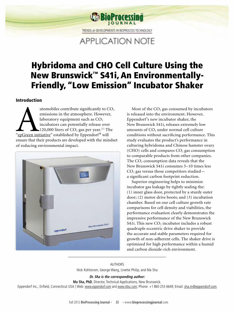
162
Fall 2012 BioProcessing Journal • • www.bioprocessingjournal.com35
TRENDS & DEVELOPMENTS IN BIOPROCESS TECHNOLOGY
Introduction
Automobiles contribute significantly to CO2 emissions in the atmosphere. However, laboratory equipment such as CO2 incubators can potentially release over 120,000 liters of CO2 gas per year.[1] The
“epGreen initiative” established by Eppendorf ® will ensure that their products are developed with the mindset of reducing environmental impact.
Hybridoma and CHO Cell Culture Using the New Brunswick™ S41i, An Environmentally-Friendly, “Low Emission” Incubator Shaker
APPLICATION NOTE
AUTHORS
Nick Kohlstrom, George Wang, Linette Philip, and Ma Sha
Dr. Sha is the corresponding author:Ma Sha, PhD, Director, Technical Applications, New Brunswick.
Eppendorf Inc., Enfield, Connecticut USA | Web: www.eppendorf.com and www.nbsc.com; Phone: +1 860-253-6649; Email: [email protected]
Most of the CO2 gas consumed by incubators is released into the environment. However, Eppendorf ’s new incubator shaker, the New Brunswick S41i, releases extremely low amounts of CO2 under normal cell culture conditions without sacrificing performance. This study evaluates the product’s performance in culturing hybridoma and Chinese hamster ovary (CHO) cells and compares CO2 gas consumption to comparable products from other companies. The CO2 consumption data reveals that the New Brunswick S41i consumes 5–10 times less CO2 gas versus those competitors studied — a significant carbon footprint reduction.
Superior engineering helps to minimize incubator gas leakage by tightly sealing the: (1) inner glass door, protected by a sturdy outer door; (2) motor drive boots; and (3) incubation chamber. Based on our cell culture growth rate comparisons for cell density and viabilities, the performance evaluation clearly demonstrates the impressive performance of the New Brunswick S41i. This new CO2 incubator includes a robust quadruple eccentric drive shaker to provide the accurate and stable parameters required for growth of non-adherent cells. The shaker drive is optimized for high performance within a humid and carbon dioxide-rich environment.

163
Fall 2012 BioProcessing Journal • • www.bioprocessingjournal.com36
Equipment• New Brunswick S41i incubator shaker equipped with high-
temperature disinfection capability• CO2 incubator shaker from competitor 1• CO2 incubator shaker from competitor 2• Vi-CELL® Cell Viability Analyzer (Beckman Coulter, Inc.)• YSI 2700 SELECT™ Biochemistry Analyzer (YSI Inc.)• New Brunswick GALAXY® S series CO2 incubator• Omega® FMA-1608A thermal mass flowmeter
(Omega Engineering, Inc.)• Eppendorf consumables: › Research® plus single-channel pipettes › epT.I.P.S.® pipetting tips › Easypet® pipetting dispensers
Media and Cells• DG44 CHO cells (Invitrogen) • EX-CELL® CD CHO serum-free medium (SFM) for CHO cells
(Sigma-Aldrich Co. LLC)• Hybridoma cell DA4-4 (ATCC® Number HB-57™)• Dulbecco’s Modified Eagle’s Medium (DMEM) (ATCC)• Fetal bovine serum (FBS) 5% (Gibco)• Penicillin Streptomycin (Pen Strep) 100 × (Gibco)
Gas Consumption DeterminationsPrior to cell culture, all incubator shakers were
programmed at 37 °C, 95 rpm and 5% CO2 and were allowed to equilibrate for at least 12 hours. Inline CO2 gas pressures were set at the lowest values recommended by each manufacturer. An offline gas analyzer was used to verify the CO2 levels within each incubator. Thermal mass flowmeters were used to record volumetric gas consumption over a time period of up to 48 hours on each unit. Tests were repeated multiple times and the average values are reported below.
Materials and Methods
Results and Discussion
Growth Assessment ofCHO and Hybridoma Cells
Viable CHO cell density reached a maximum of 4.72 × 10⁶ cells/mL by day 7. The cell viability was maintained at approximately 98% up to day 5 and dropped steadily thereafter (Figure 1).
FIGURE 1. Average viable cell density and viability of CHO culture in the New Brunswick S41i.
CHO Culture ProtocolCHO cells were grown in SFM supplemented with
1% Pen Strep. Six 250 mL Erlenmeyer flasks were inoculated with 60 mL of the same stock culture at a concentration of 0.3 × 10⁶ cells/mL. Two flasks were placed on each of the three incubator shaker platforms (New Brunswick S41i, and competitors 1 and 2). The flasks were incubated at 37 °C in a mixture of 5% CO2, 95% air, and agitated at 130 rpm (4.69 rcf).
CHO cells were grown for a period of 14 days. Samples of 1.5 mL were taken on days 3, 5, 7, 10, 12, and 14, and analyzed with the YSI 2700 SELECT instrument for glucose concentrations. The Vi-Cell determined cell concentrations and viability. Six flasks were cultured per incubator and the results were averaged out.
Hybridoma Culture ProtocolHybridoma cells were grown in DMEM medium
supplemented with 5% FBS and 1% Pen Strep. Six 250 mL Erlenmeyer flasks were each inoculated with 45 mL of stock culture at a concentration of 0.2 × 10⁶ cells/mL. Six flasks were placed on each of the three incubator shaker platforms (New Brunswick S41i, and competitors 1 and 2). The flasks were incubated at 37 °C in a mixture of 5% CO2, 95% air, and agitated at 95 rpm (2.52 rcf).
Hybridoma cells were subcultured on days 2 and 4 to a concentration of approximately 0.2 × 10⁶ cells/mL. A sample of 0.6 mL was taken every day from each of the flasks and analyzed with the YSI 2700 SELECT instrument for glucose concentrations. The Vi-Cell determined cell concentrations and viability. Six flasks were cultured per incubator and the results were averaged out.
approved

164
Fall 2012 BioProcessing Journal • • www.bioprocessingjournal.com37
FIGURE 2. Comparison of average viable cell densities of hybridoma cultures grown in New Brunswick S41i and competitors 1 and 2.
In comparison to CHO cells, hybridoma cultures maintained a high average viability of approximately 95% through day 6 due to the subculturing of cells during log phase of growth on days 2 and 4. A maximum viable cell density of 1.81 × 10⁶ cells/mL was achieved on day 6 (Figures 2 and 3).
FIGURE 3. Comparison of average percentage viabilities of hybridoma cultures grown in New Brunswick S41i and competitors 1 and 2.
REFERENCE [1] Based on calculation of competitor 2 data in Figure 4 (~57 L/24-hr day, 365 days/yr = 20,805 L/yr). Most CO2 are not really “consumed” by cells — they are released into the environment.
NOTESEppendorf®, Research®, epT.I.P.S.®, and Easypet® are registered trademarks of Eppendorf AG and New Brunswick™ is a trademark of Eppendorf AG, Hamburg, Germany.Galaxy® is a registered trademark of New Brunswick Scientific Co., Inc., USA.Vi-CELL® is a registered trademark of Beckman Coulter, Inc.YSI® is a registered trademark of YSI Inc. Omega® is a registered trademark of Omega Engineering Inc. EX-CELL® is a registered trademark of Sigma-Aldrich Co. LLC.
FIGURE 4. Average CO2 gas consumption in standard liter (SLtr) of tested units over a 24-hour period.
Measurements of Gas Consumption
The measurement of CO2 gas consumption at a 5% CO2 setpoint revealed that the competitive units evaluated consume much higher levels of CO2 gas over the same period as compared to the New Brunswick S41i (Figure 4).

165 SPRING 2013 • Volume 12 / Issue 1 • ISSN 1538-8786
A Production of BioProcess Technology Network
Trends & Developments in BioProcess Technology
BioProcessingJ O U R N A L

166
Spring 2013 BioProcessing Journal • • www.bioprocessingjournal.com21
TRENDS & DEVELOPMENTS IN BIOPROCESS TECHNOLOGY
A Comparative Bioreactor Vessel Study:Conventional Reusable Glass and
Single-Use Disposables for the Production of Alkaline Phosphatase
By TAYLOR HATTON, SHAUN BARNETT, MA SHA, and KAMAL RASHID
Abstract
Single-use, stirred-tank bioreactor systems have been used in large-scale production for a number of years. Bench-scale, stirred-tank bioreactors have not been commercially-available for single-use until recently.
The New Brunswick™ CelliGen® BLU pitched-blade bioreactor was introduced in 2009, and the CelliGen BLU packed-bed bioreactor, in 2012.
Little information is currently available on the utility of these bioreactors for bench-scale production of recombinant products. Thus, we designed this study to perform multiple comparisons with these single-use bioreactors and their tradi-tional glass vessel counterparts. The data comparisons included: (1) CelliGen BLU pitched-blade vs. glass pitched-blade; and (2) CelliGen BLU pitched blade in batch mode vs. CelliGen BLU packed-bed in perfusion mode. Chinese hamster ovary (CHO) cells were used to measure alkaline phosphatase (ALKP) pro-duction in each bioreactor. The final measured concentration of ALKP, after eight days of batch-mode culture in the single-use, pitched-blade bioreactor, was 1.6 U/mL compared to 2.1 U/mL in the reusable bioreactor. After six perfusion harvests in the single-use, packed-bed bioreactor, the combined ALKP production was 16.2 U/mL compared to 17.4 U/mL in the reus-able bioreactor in batch mode. Multiple batch culture runs in the pitched-blade bioreactor would be required to match the output of a single run in the packed-bed bioreactor in perfusion mode.
Results demonstrate that there are no significant differences between the reusable and single-use systems for bench-scale production of recombinant proteins. Our results also suggest that the CelliGen BLU packed-bed bioreactor, when operated in perfusion mode, is superior to the CelliGen BLU pitched-blade bioreactor when operated in batch mode, confirming our studies from 2012.[1]
Introduction
Stainless steel, stirred-tank bioreactors have been the trusted and dominant design for decades in scale-up of animal cells. Along with proven reproducibility of these bioreactors come some minor disadvantages, namely, cleaning and maintenance.[2-4] When reusable bioreactors are used for production of biopharmaceuticals, the cleaning process needs to be validated which increases cost in economic terms. In a survey conducted by BioPlan Associates, the primary reason for biopharmaceutical developers increasing their utilization of single-use systems was to eliminate cleaning requirements (90.2 % of respondents).[5]
The New Brunswick CelliGen BLU single-use bioreactor line, available in the pitched-blade and now in packed-bed design, has helped to make single-use, stirred-tank bioreactors readily available commercially to the bench-scale community.
The CelliGen BLU pitched-blade bioreactor incorporates one large plastic pitched-blade impeller which efficiently mixes the media while disrupting the sparger bubbles. The CelliGen BLU packed-bed bioreactor incorporates two horizontal perforated plastic sheets which entrap Fibra-Cel® disks , creating a bed for cells.[6] This design allows for media exchange while eliminating the need for f i ltration, sedimentation, or centrifugation of the cells, as is the case with other bioreactor types.[7] The packed-bed bio reactor, combined with a perfusion mode of operation, is a very useful means of increasing cell growth and productivity of recombinant proteins. Cells cultured in

167
Spring 2013 BioProcessing Journal • • www.bioprocessingjournal.com22
packed-bed bioreactors are not exposed to hydrodynamic forces which allows for maximum cell growth and protein expression.[8]
Reusable packed-bed bioreac tors in per fusion mode have been compared to reusable pitched-blade bioreactors in batch mode [1] and perfusion systems have been compared to fed-batch systems.[9] However, to date, no published studies have compared the productivity of protein-secreting cells in reusable packed-bed and pitched-blade bioreactors to single-use, packed-bed and pitched-blade bioreactors. Therefore, the objective of this study was to perform a multi-comparative study between single-use and reusable bioreactors. We expect the results from these experiments will help aid in the introduction of the single-use (CelliGen BLU) bioreactors as an alternative to reusable packed-bed bioreactors while also highlighting the advantages of the CelliGen BLU packed-bed bioreactor operated in perfusion mode.
Perfusion modes of operation offer many advantages over batch or even fed-batch modes of operation. Systems operated under perfusion mode do not accumulate toxic byproducts, as seen in the batch operations, because the media is removed on a regular basis. Perfusion systems can often be operated at smaller scales. These systems have the ability to increase cell concentrations up to 30 × more than batch systems. Long run times allowed with the packed-bed bioreactor decrease the constant need for re-seeding cells and re-establishing seed cultures. This dramatically reduces setup time and labor over batch systems, advantages that will lead to reduced cost of operation[10, 11] A study performed by Biopharm Services showed that conventional stainless steel bioreactor facilities were the slowest to become cash positive. This was due to the increased capital investment required up front and the longer process of building a stainless steel bioreactor facility.[12]
Materials and Methods
Culture ProceduresIn order to evaluate the impact of these bioreactor
systems on protein secretion by cultured cells, we employed a recombinant ALKP-secreting CHO cell line (rCHO), generously provided by CDI Bioscience, Inc. The rCHO cells were engineered with their IPTG-regulated RP Shift® vector so that the rCHO cells stop replicating and shift to protein production when induced with isopropyl β-D-1-thiogalactopyranoside (IPTG). CD-CHO medium (Gibco, Life Technologies) was used throughout these experiments. The media contained 6.3 g/L glucose and was supplemented with 8 mM L-glutamine and 100 µg/mL of an antibiotic/antimycotic solution (Gibco, Life Technologies). Frozen rCHO cells were thawed and transferred to T-75 flasks
with CD-CHO serum-free medium and allowed to expand. Once a sufficient number of cells were achieved, sterile disposable spinner flasks were utilized to further expand the cells. Cell subculturing continued until a sufficient number of viable cells was achieved for use as a seed culture at a density of approximately 5 × 10⁵ cells/mL. Two New Brunswick CelliGen 310 advanced bench-top, stirred-tank bioreactors incorporating two single-use CelliGen BLU bioreactors were utilized to grow the rCHO cells. A solution of NaHCO3, 8 % w/v (8 g/100 mL), was used to help control the pH inside the bioreactor systems.
Pitched-Blade Impeller Operated in Batch Mode The first bioreactor system utilized the pitched-blade
impeller. The CelliGen BLU pitched-blade bioreactor comes pre-packaged and sterilized with the pitched-blade impeller, tubing, sparger, and filters for easy setup. The vessels are made of materials that meet USP Class VI standards and have been tested for leachables.[13] The blades on the pitched-blade impeller are flat and set at a 45° angle. This blade orientation provides good axial and radial mixture of the media while also increasing the oxygen mass transfer rate and disruption of bubbles released from the sparger. The pitched-blade impeller is designed to minimize the stress of mixing on shear-sensitive cells.[5]
Two experimental trials were performed utilizing the pitched-blade bioreactor. One trial was performed in a reusable (glass) 2.2 L total volume vessel (1.75 L working volume) and a second trial was performed utilizing a single-use CelliGen BLU 5.0 L total volume vessel (3.5 L working volume). For each trial, the bioreactor was allowed to operate until the cell concentration reached approximately 2 × 10⁶ cells/mL at which time the cells were induced with IPTG. Both experimental trials had the following parameters, as shown in Table 1.
Packed-Bed Basket Impeller Operated in Perfusion Mode
The second bioreactor system utilized the packed-bed basket impeller. The CelliGen BLU packed-bed bioreactor
TABLE 1. Bioreactor parameters.
ParameterSetpoint
Glass 2.2 L CelliGen BLU 5.0 L
Temperature 37 °C (± 0.1°C) 37 °C (± 0.1°C)
Agitation 120 rpm (± 5 rpm) 120 rpm (± 5 rpm)
Dissolved O2 35 % (± 1%) 35 % (± 1%)
pH 7.1 (± 0.01) 7.1 (± 0.01)
Gas flow 0.5 slpm 1.5 slpm
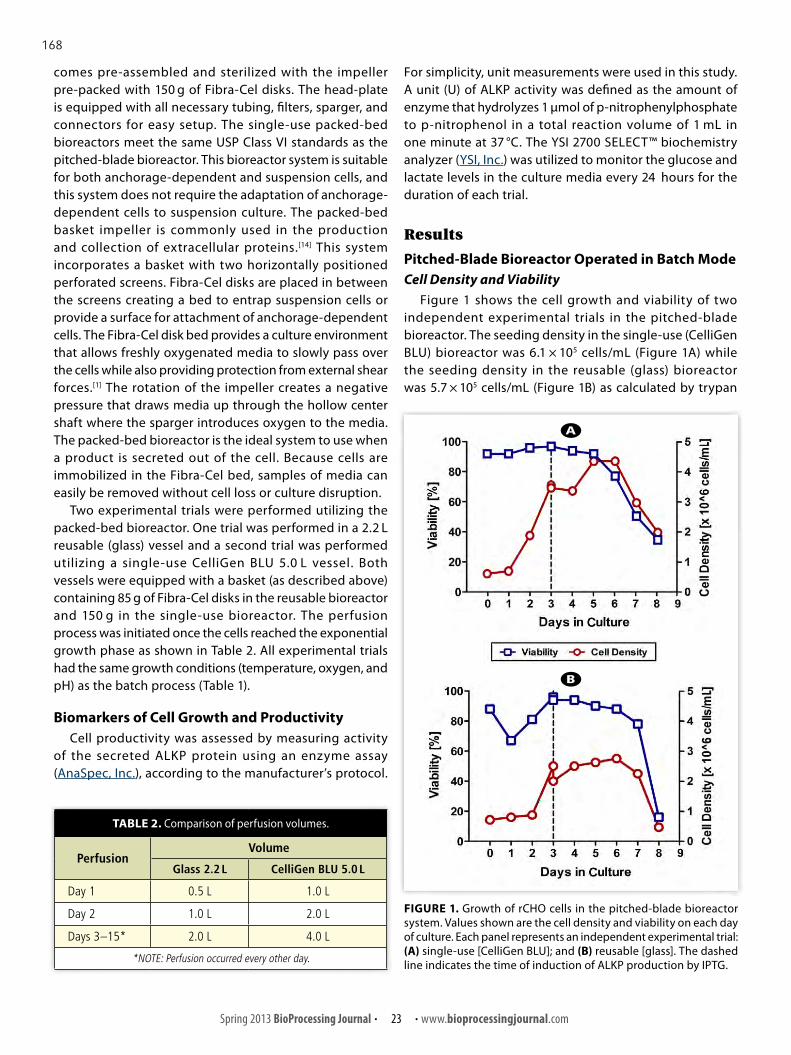
168
Spring 2013 BioProcessing Journal • • www.bioprocessingjournal.com23
comes pre-assembled and sterilized with the impeller pre-packed with 150 g of Fibra-Cel disks. The head-plate is equipped with all necessary tubing, filters, sparger, and connectors for easy setup. The single-use packed-bed bioreactors meet the same USP Class VI standards as the pitched-blade bioreactor. This bioreactor system is suitable for both anchorage-dependent and suspension cells, and this system does not require the adaptation of anchorage-dependent cells to suspension culture. The packed-bed basket impeller is commonly used in the production and collection of extracellular proteins.[14] This system incorporates a basket with two horizontally positioned perforated screens. Fibra-Cel disks are placed in between the screens creating a bed to entrap suspension cells or provide a surface for attachment of anchorage-dependent cells. The Fibra-Cel disk bed provides a culture environment that allows freshly oxygenated media to slowly pass over the cells while also providing protection from external shear forces.[1] The rotation of the impeller creates a negative pressure that draws media up through the hollow center shaft where the sparger introduces oxygen to the media. The packed-bed bioreactor is the ideal system to use when a product is secreted out of the cell. Because cells are immobilized in the Fibra-Cel bed, samples of media can easily be removed without cell loss or culture disruption.
Two experimental trials were performed utilizing the packed-bed bioreactor. One trial was performed in a 2.2 L reusable (glass) vessel and a second trial was performed utilizing a single-use CelliGen BLU 5.0 L vessel. Both vessels were equipped with a basket (as described above) containing 85 g of Fibra-Cel disks in the reusable bioreactor and 150 g in the single-use bioreactor. The perfusion process was initiated once the cells reached the exponential growth phase as shown in Table 2. All experimental trials had the same growth conditions (temperature, oxygen, and pH) as the batch process (Table 1).
Biomarkers of Cell Growth and ProductivityCell productivity was assessed by measuring activity
of the secreted ALKP protein using an enzyme assay (AnaSpec, Inc.), according to the manufacturer’s protocol.
For simplicity, unit measurements were used in this study. A unit (U) of ALKP activity was defined as the amount of enzyme that hydrolyzes 1 µmol of p-nitrophenylphosphate to p-nitrophenol in a total reaction volume of 1 mL in one minute at 37 °C. The YSI 2700 SELECT™ biochemistry analyzer (YSI, Inc.) was utilized to monitor the glucose and lactate levels in the culture media every 24 hours for the duration of each trial.
Results
Pitched-Blade Bioreactor Operated in Batch ModeCell Density and Viability
Figure 1 shows the cell growth and viability of two independent experimental trials in the pitched-blade bioreactor. The seeding density in the single-use (CelliGen BLU) bioreactor was 6.1 × 10⁵ cells/mL (Figure 1A) while the seeding density in the reusable (glass) bioreactor was 5.7 × 10⁵ cells/mL (Figure 1B) as calculated by trypan
TABLE 2. Comparison of perfusion volumes.
PerfusionVolume
Glass 2.2 L CelliGen BLU 5.0 L
Day 1 0.5 L 1.0 L
Day 2 1.0 L 2.0 L
Days 3–15* 2.0 L 4.0 L
*NOTE: Perfusion occurred every other day.
FIGURE 1. Growth of rCHO cells in the pitched-blade bioreactor system. Values shown are the cell density and viability on each day of culture. Each panel represents an independent experimental trial: (A) single-use [CelliGen BLU]; and (B) reusable [glass]. The dashed line indicates the time of induction of ALKP production by IPTG.
A
B

169
Spring 2013 BioProcessing Journal • • www.bioprocessingjournal.com24
FIGURE 3. ALKP production by rCHO cells cultured in the pitched-blade bioreactor system. ALKP concentration in the culture media was measured each day after induction. IPTG induction of ALKP occurred on culture day 3.
FIGURE 2. Glucose consumption and lactate production by rCHO cells cultured in the pitched-blade bioreactor system. Values shown are the concentrations of glucose and lactate in the culture media measured daily. Each panel represents an independent experimental trial: (A) single-use [CelliGen BLU]; and (B) reusable [glass]. The dashed line indicates the time of induction of ALKP production by IPTG.
blue staining utilizing the Countess® cell counter. The maximum cell density observed in the single-use bioreactor (4.4 × 10⁶ cells/mL) was significantly higher than in the reusable bioreactor (2.2 × 10⁶ cells/mL). Cell viability was greater than 90 % for the majority of the experimental trial in the single-use bioreactor while the viability in the reusable bioreactor recovered to greater than 90 % after a few days in culture.
Glucose Utilization and Lactate ProductionGlucose is the main energy source for cell proliferation
and ALKP production. Thus, glucose levels were expected to directly correlate with ALKP production in each experiment. Because lactate is a secondary energy source, lactate levels were expected to decline following this initial increase and the utilization of glucose in the media. Lactate metabolism is beneficial to the system by reducing a major metabolic by-product from the system.[15, 16] Glucose levels measured at the time of induction (day 3) were nearly 0 g/L in both experiments (Figure 2). Media lactate concentrations increased in response to decreasing glucose availability. The observed gradual decrease of lactate near the end of each trial indicates its use as a secondary energy source.
ALKP ProductionFigure 3 shows the concentrations of ALKP measured
daily in two independent experimental trials in the pitched-blade bioreactor. ALKP concentrations increased over the six culture days post-induction. However, a decrease in ALKP activity was observed at the time of harvest in both trials, possibly due to degradation of the ALKP protein at the end of the experiment.[17] Serum-free media was utilized for growth of rCHO cells in this study. Thus, ALKP was susceptible to the action of proteases made by the rCHO cells.[18] As reported above, cell density in the single-use bioreactor was higher than the reusable bioreactor, suggesting that cell proliferation was dominant over ALKP production. This observation likely accounts for the slightly lower amount of ALKP detected in the single-use bioreactor.
Packed-Bed Bioreactor Operated in Perfusion ModeCell Density and Viability: The packed-bed bio-
reactors in both experimental trials were seeded with 5.0 × 10⁵ cells/mL. However, because of the presence of the Fibra-Cel discs, it was not possible to sample the cells directly during culture to determine cell yield and viability. Therefore, cell density and viability were not monitored on a daily basis; rather, the rate of glucose consumption was used as a surrogate to approximate changes in cell density.[19] The growth of cells in the packed-bed bio reactor was estimated using the average glucose consumption
A
B

170
Spring 2013 BioProcessing Journal • • www.bioprocessingjournal.com25
rate. Glucose consumption rates were similar in both the pitched-blade and packed-bed bioreactor systems up to day four (Figure 4). However, after day four of culture, glucose utilization in the packed-bed bioreactor continued to increase exponentially, while the trend for glucose consumption in the pitched-blade bioreactor increased linearly. Increased glucose consumption observed on culture day five suggests that cell density in the packed-bed bioreactor had likely increased.
Glucose Utilization and Lactate Production: Glucose consumption was very similar in both the single-use (Figure 5A) and reusable (Figure 5B) experimental trials. As previously observed with the pitched-blade bioreactor system, media lactate concentrations increased in response to decreasing glucose availability in both trials. The use of lactate as a secondary energy source can also be observed as lactate levels decrease at each 2 and 4 L perfusion.
ALKP Production: Concentrations of ALKP in the two
FIGURE 4 (right). Comparison of glucose uptake by rCHO cells in the pitched-blade and packed-bed bioreactor systems. Values shown are the daily glucose media concentrations consumed in two independent experimental trials: (A) single-use [CelliGen BLU]; and (B) reusable [glass].
FIGURE 5 (left). Glucose consumption and lactate production by rCHO cells cultured in the: (A) pitched-blade; and (B) packed-bed bioreactor systems. Values shown are the amounts of glucose and lactate measured in the culture media at each media exchange. The time and volume of the media exchange is indicated at each dashed line. Induction of ALKP activity by IPTG began on culture day 5 and continued eve r y t wo d ay s th ro u g h o u t th e remainder of the experiment.
A
B
A
B

171
Spring 2013 BioProcessing Journal • • www.bioprocessingjournal.com26
FIGURE 7. Comparison of ALKP production by rCHO cells cultured in the pitched-blade and packed-bed bioreactor systems: (A) final ALKP concentrations in the culture media measured in two independent experimental trials using the pitched-blade bioreactor; (B) cumulative ALKP concentrations in the culture media measured in two independent experimental trials using the packed-bed bioreactor; and (C) ALKP concentrations for two independent experimental trials using either the CelliGen BLU pitched-blade or packed-bed bioreactor systems.
independent experiments utilizing the packed-bed bioreactor are shown in Figure 6. Following initial expansion culture of rCHO cells for five days, ALKP production was induced every two days with a media exchange containing IPTG. We determined previously that continuous culture with IPTG in the media yielded greater production of ALKP as compared to a transient exposure to the inducing agent (data not shown). A modest increase in ALKP production was observed at each media exchange, although the level of ALKP varied by induction day and by experiment trial (Figure 6). The correlation between glucose utilization and ALKP production, as previously observed, supports the inference that the rate of glucose consumption in both experiments was conducive to the production of large amounts of ALKP. The rapid exhaustion of glucose and a presumed high cell density were likely contributing factors to the large amounts of ALKP observed.
Comparison of the Reusable and Single-Use Bioreactor Systems for ALKP Production
A major objective of this study was to compare the single-use to the reusable pitched-blade bioreactor, the single-use to the reusable packed-bed bioreactor, and finally the pitched-blade bioreactor to the packed-bed bioreactor in both vessel types. The total ALKP production per experimental trial is shown in Figure 7. Figure 7A shows the comparison between the two trials performed in the pitched-blade bioreactor. Final ALKP concentrations are very similar in both trials. Figure 7B shows the comparison between the two trials performed in the packed-bed bioreactor. Resembling the pitched-blade bioreactor, final ALKP concentrations in both packed-bed bioreactor types were markedly similar. The total ALKP production in the CelliGen BLU pitched-blade vs. the CelliGen BLU packed-bed bioreactor is shown in Figure 7C. Overall, the packed-bed bioreactor system produced ALKP to a much greater extent (nearly 9-fold greater) compared to the pitched-blade system.
FIGURE 6. ALKP production by rCHO cells cultured in the packed-bed bioreactor system: (A) ALKP concentrations in culture media measured each day in two independent experimental trials using the packed-bed bioreactor. IPTG induction of ALKP began on culture day 5 and continued every two days for the remainder of each experiment; and (B) cumulative production of ALKP throughout each experiment with each bar representing a perfusion.
A
B
A B C

172
Spring 2013 BioProcessing Journal • • www.bioprocessingjournal.com27
Small -scale bioprocessing has b e en re gular ly performed in reusable, stirred-tank bioreactors. Cleaning and sterilization of reusable bioreactors is mandatory between runs. Regular maintenance, careful handling, and storage of the glass vessel are also required. A significant monetary investment in sterilization equipment and manpower is required to maintain and clean reusable bioreactors. Cleaning and maintenance of single-use (disposable) bioreactors is not necessary and therefore implementation of these bioreactors can potentially lower required initial investments. Internal analysis conducted by Eppendorf Inc. indicates that the overall cost of using CelliGen 310 vs. CelliGen BLU is nearly identical (data not shown). The cost of operating the CelliGen 310 is driven by upfront investments including control cabinet, autoclavable vessel(s), probes, and autoclave sterilization hardware, as well as the additional labor costs in vessel set-up, break-down, cleaning, and validation. Conversely, the cost of operating the CelliGen BLU is mostly driven by the investment of the control cabinet and the single-use consumable vessels. The reduction in labor hours, cleaning,
and validation are major driving forces behind single-use products in bioprocessing.
Process performance by each impeller type were similar in both the reusable and single-use bioreactors, indicating that single-use bioreactors can perform all manufacturing operations analogous to reusable bioreactors.
By virtue of its design, when operated in perfusion mode, the packed-bed bioreactor may be used continuously for months. Cell line maintenance is reduced since preparation of new seed cultures is not required. Moreover, initial setup efforts associated with the packed-bed bioreactor is substantially less than would be required using the pitched-blade system which requires multiple, shorter cultures to equal the production of a single culture with the packed-bed system. Although the pitched-blade bioreactor requires less monitoring over the duration of an experiment, as cells are cultured until nutrient depletion has occurred, this approach does require significant initial input in terms of labor and resources. Multiple culture runs, and thus, multiple seed cultures and system preparations, are required to match the output of the packed-bed system.
Discussion
173
Spring 2013 BioProcessing Journal • • www.bioprocessingjournal.com28
[1] Hatton T, Barnett S, Benninghof f AD, Rashid K. Productivity studies utilizing recombinant CHO cells in stirred-tank bioreactors: a comparative study between pitched-blade and packed-bed bioreactor systems. Bioprocessing J, 2012; 11: 29-36.[2] Holzer M. Technologies for downstream processing in biologics. Biopharm Int, 2011; 24: 48-53.[3] Martin J. Single-use technology: balancing the risks and rewards. Biopharm Int, 2011; 24: s19-s21.[4] Bezawada A, Thompson M, Cui W. Use of blast freezers in vaccine manufacture, a case study on freezing characteristics of drug substances in disposable containers. BioProcess Int, 2011, 9: 43-51.[5] Langer ES. CMOs ratchet up use of single-use devices to improve performance. Biopharm Int, 2011; 24: 27-30.[6] Mirro RA, Voll K. Which impeller is right for your cell line? BioProcess Int, 2009, 7: 52-48.[7] Ohashi R, Singh V, Hamel JFP. Per fusion culture in disposable bioreactors–f loating f ilter design to simplif y operations. Genet Eng News, 2001, 21: 40.[8] Merten OW, Cruz PE, Rochette C, Geny-Fiamma C, Bouquet C, Goncalves D, Danos O, Carrondo MJT. Comparison of dif ferent bioreactor systems for the production of high titer retroviral vectors. Biotechnol Progr, 2001; 17: 326-335. http://dx.doi.org/10.1021/bp000162z. PMid:11312711.[9] Lim J, Sinclair A, Shevitz J, Carter JB. An economic comparison of three cell culture techniques: fed-batch, concentrated fed-batch, and concentrated perfusion. Biopharm Int, 2011; 24: 54-60.[10] Langer E. Trends in perfusion bioreactors: the next revolution in bioprocessing? Biopharm Int, 2011, 10: 49-52.[11] Bonham-Carter J, Shevitz J. A brief history of per fusion biomanufacturing. BioProcess Int, 2011; 9: 24-31.[12] Sinclair A, Monge M. Monoclonal antibody manufacturing cost benef its of hybrid disposable systems. BioProcess Int, 2011; 9: 12-17.[13] Wang G, Mirro R, Gossain V. CelliGen® BLU protocol: growing CHO cells in a CelliGen BLU benchtop, stirred-tank bioreactor using single-use vessels. http://www.biotechniques.com/multimedia/archive/00074/New_Brunswick-FP-Cel_74610a.pdf.[14] Kaufman JB, Wang G, Zhang W, Valle MA, Shiloach J. Continuous production and recovery of recombinant Ca2+ binding receptor from HEK 293 cells using perfusion through a packed bed bioreactor. Cytotechnol, 2000; 33: 3-11. http://dx.doi.org/10.1023/A:1008143132056. PMid:19002805, PMCid:3466730.[15] Ma NN, Ellet J, Okediadi C, Hermes P, McCormick E, Casnocha S. A single nutrient feed supports both chemically def ined NS0 and CHO fed-batch processes: improved productivity and lactate metabolism. Biotechnol Progr, 2009; 25: 1353-1363. http://dx.doi.org/10.1002/btpr.238. PMid:19637321.[16] Hu WS, Dodge TC, Frame KK, Himes VB. Ef fect of glucose on the cultivation of mammalian cells. Dev Biol Stand, 1987; 66: 279-290. PMid:3582758.[17] Elliott P, Hohmann A, Spanos J. Protease expression in the supernatant of Chinese hamster ovary cells grown in serum-free culture. Biotechnol Lett, 2003; 25: 1949-1952. http://dx.doi.org/10.1023/B:BILE.0000003992.09492.4b. PMid:14719832.[18] Sandberg H, Lutkemeyer D, Kuprin S, Wrangel M, Almstedt A, Persson P, Ek V, Milkaelsson M. Mapping and partial characterization of proteases expressed by a CHO production cell line. Biotechnol and Bioeng, 2006; 95: 961-971. http://dx.doi.org/10.1002/bit.21057. PMid:16897737.[19] Zhang F, Tian JH, Wang L, He PA, Chen Y. Correlation between cell growth rate and glucose consumption determined by electrochemical monitoring. Sensor Actuat B-Chem, 2011, 156: 416-422. http://dx.doi.org/10.1016/j.snb.2011.04.067.
References
AcknowledgementsThis work was partially funded by Eppendorf and by the Utah Agricultural
Experiment Station, Utah State University, and is approved as journal paper number 8443. Also, the authors would like to thank CDI Bioscience, Inc. for providing the recombinant Chinese hamster ovary cell line for this research project.
In summary, the single-use CelliGen BLU bench-top bioreactor performance was markedly similar to their respective reusable bioreactors. Therefore, significant consideration of single-use bioreactors should be taken into account when small-scale manufacturing of biologics is needed. In both vessel types the packed-bed bioreactor operated in perfusion mode is superior to the pitched-blade bioreactor operated in batch mode for growth of rCHO cells secreting ALKP. However, if a simple batch process is preferred then use of the CelliGen BLU pitched-blade bioreactor should be considered.
Development and ManufacturingServices for Biologics
· Extensive process development capabilities
· End-to-end cGMP manufacturing operations
· FDA- and EMEA-compliant, BSL2 environment
Visit our website: www.aeras-cmo.comContact us: [email protected]
C
M
Y
CM
MY
CY
CMY
K
AERAS_Ad_Border.pdf 1 2/15/13 11:23 AM
Three additional articles published in BioProcessing Journal by USU’s Center for Integrated Biosystems incorporating Eppendorf’s New Brunswick CelliGen products:
Hatton T, Barnett S, Rashid K. CHO cell culture with New Brunswick CelliGen BLU single-use packed-bed Fibra-Cel basket. BioProcess J, 2012; 11(2): 50-52.
Hatton T, Barnett S, Benninghoff AD, Rashid K. Productivity studies utilizing recombinant CHO cells in stirred-tank bioreactors: a comparative study between pitched-blade and packed-bed bioreactor systems. BioProcess J, 2012; 11(2): 29-36.
Parasar P, Barnett S, Wilhelm A, Rashid K, Davies CJ. Large-scale growth of mouse P815 cells expressing a bovine non-classical major histocompatibility complex class I protein utilizing a pitched-blade bioreactor. BioProcess J, 2012; 11(3): 27-34.
About the AuthorsTaylor Hatton1, 2; Shaun Barnett1; Ma Sha, PhD3; and Kamal Rashid, PhD1, 2*
1. Center for Integrated BioSystems, Utah State University, Logan, Utah 2. Department of Animal, Dairy and Veterinary Sciences, Utah State University,
Logan, Utah3. Eppendorf®, Inc., 175 Freshwater Blvd., Enfield, Connecticut 06082 USA*Corresponding author: Dr. Kamal Rashid, Associate Director, Research Professor,
Center for Integrated BioSystems, 4700 Old Main Hill, Logan, Utah 84322 USA Ph: 435-797-2739 | Email: [email protected], Website: biosystems.usu.edu/
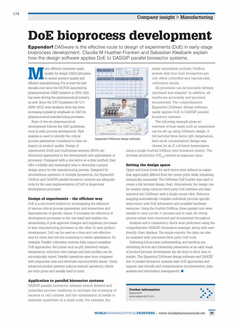
174
37
Company insight > Manufacturing
WorldPharmaceuticalFrontiers | www.worldpharmaceuticals.net
m any different industries apply quality by design (QbD) principles to ensure product quality and
efficient manufacturing. For at least the last decade, ever since the US FDA launched its pharmaceutical cGMP initiative in 2004, QbD has been driving the pharmaceutical industry as well. Since the ICH Guidelines Q8–Q11 (2009–2012) were finalised, there has been increasing regulatory emphasis on QbD for pharmaceutical manufacturing processes.
State-of-the-art pharmaceutical development follows the QbD guidelines even in early process development. Risk analysis is used to identify the critical process parameters considered to have an impact on product quality. Design of experiments (DoE) and multivariate analysis (MVA) are structured approaches to the development and optimisation of processes. Compared with a one-factor-at-a-time method, they offer a reliable and meaningful way to determine a proper design space for the manufacturing process. Designed for simultaneous operation of multiple bioreactors, the Eppendorf DASbox and DASGIP parallel bioreactor systems are adequate tools for the easy implementation of DoE in bioprocess development processes.
design of experiments – the efficient wayDoE is a structured method for investigating the influence of various critical process parameters, and interactions and dependencies of specific values. It increases the efficiency of development processes on the one hand and enables the streamlining of post-approval changes and regulatory processes in later manufacturing processes on the other. In early product development, DoE can be used as a time and cost-effective way for clone and cell-line screening or media optimisation, for example. Parallel cultivation systems fully support seamless DoE approaches. Set points such as pH, dissolved oxygen, temperature, induction time stamps and feed profiles can be automatically varied. Parallel operations save time compared with sequential ones and eliminate reproducibility issues. Using advanced parallel systems reduces manual operations, which are error prone and usually hard to track.
application to parallel bioreactor systems DASGIP parallel bioreactor systems ensure defined and controlled process conditions to facilitate the screening of bacteria or cell cultures, and the optimisation of media or substrate quantities on a small scale. For example, the
easily extendable modular DASbox system with four mini bioreactors per unit offers controlled and reproducible cultivation results.
All processes can be precisely defined, optimised and adapted. In addition, all results are accurately and precisely documented. The comprehensive Eppendorf DASware design software easily applies DoE to DASGIP parallel bioreactor systems.
The following example gives an overview of how easily such an experiment can be set up using DASware design. A full-factorial three-factor (pH, temperature, feed-stock concentration) design was chosen for an E coli batch fermentation
using a single fourfold DASbox mini bioreactor system. The biomass production (OD600) served as response value.
setting the design space Upper and lower levels for each factor were defined as values that appreciably differed from the centre point while remaining biologically reasonable. The DASware DoE builder was used to create a full-factorial design chart. Alternatively, the design can be created using common third-party DoE software and later imported into DASware with a single mouse click. Resource mapping automatically compiles individual, process-specific instructions with DoE information and available hardware resources. Using the fourfold DASbox, three parallel runs were needed to carry out the 11 process runs in total. All critical process values were monitored and documented throughout.
Analysis and a consistency check were performed using the comprehensive DASGIP information manager, along with user-friendly chart displays. Via simple exports, the data can also be analysed with renowned third-party DoE tools.
Gathering full process understanding, and tracking any interfering factors and interacting parameters at an early stage of product/process development are the keys to short time to market. The Eppendorf DASware design software and DASGIP line of parallel bioreactor systems ease DoE approaches and support user-friendly and comprehensive documentation, data analysis and information management. ■
doe bioprocess developmentEppendorf DASware is the effective route to design of experiments (DoE) in early-stage bioprocess development. Claudia M Huether-Franken and Sebastian Kleebank explain how the design software applies DoE to DASGIP parallel bioreactor systems.
Further informationEppendorf www.eppendorf.com
Eppendorf DASware design software.
WPF025_Dasgip.indd 37 25/03/2014 12:25
175
Trends & Developments in BioProcess Technology
A Production of BioProcess Technology Network
SPRING 2015 • Volume 14/Issue 1 • ISSN 1538-8786
BioProcessingJ O U R N A L

176
Spring 2015 BioProcessing Journal www.bioprocessingjournal.com20
APPLICATION NOTEAPPLICATION NOTETRENDS & DEVELOPMENTS IN BIOPROCESS TECHNOLOGY
TECHNOLOGY REVIEWSuccessful High Density
Escherichia coli Fermentation Using the Eppendorf BioFlo® 320 Advanced
Bioprocess Control SystemBy BIN LI AND MA SHA
Abstract
The gram-negative bacterium, Escherichia coli, has a long history in the world of laboratory and industrial processes due to its ease of manipulation and well-
understood genome. It is widely cultured under aerobic conditions. High cell density cultivation of E. coli is a powerful technique for the production of recombinant proteins. Indeed, 30% of the FDA-approved biopharmaceuticals on the market are produced in E. coli.
An Escherichia coli fermentation run conducted using the Eppendorf BioFlo® 320 bioprocess control station achieved high cell density at 12 hours, as determined by a maximum optical density (OD600) measurement of 215.2. The weights of dry and wet cells were also measured.
IntroductionThe BioFlo 320 advanced bioprocess control system from
Eppendorf was developed for both microbial fermentation and cell culture applications. The unit features a new indus-trial design, flexibility between autoclavable and single-use vessels, intelligent sensors, Ethernet connectivity, and an improved software package. In this study, E. coli cultivation using a BioFlo 320 achieved a high optical density (OD600) value of 215.2. Furthermore, the wet cell weights (WCW) and dry cell weights (DCW) were also measured and are presented herein.
Materials and MethodsFermentation was performed using an Eppendorf
BioFlo 320 bioprocess control station with the configu-ration outlined in Table 1. The E. coli K-12 strain (ATCC®, 10798™) was grown in a 1 L (working volume) stainless steel dished-bottom BioFlo 320 glass vessel, as shown in Figure 1. Glucose concentrations were measured using a Cedex® Bio Analyzer (Roche®). The OD600 was measured with an Epoch™ Microplate Spectrophotometer (BioTek®) using
TABLE 1. BioFlo 320 hardware configuration.
Parameter Configuration
Gas Mix Automatic gas mix
Gas Flow Control Thermal mass flow controllers (TMFC) with 0–20 standard liters per minute (SLPM) flow range
Vessel 1 L stainless steel dished-bottom glass vessel
Motor Direct drive motor
Impeller Two Rushton impellers
Sparger Ring sparger (macrosparger)
FIGURE 1. BioFlo 320 bioprocess control station with water- jacketed (left) and stainless steel dish-bottom (right) vessels.

177
Spring 2015 BioProcessing Journal www.bioprocessingjournal.com21
the optional cuvette attachment. An Eppendorf MiniSpin® plus centrifuge was used to pellet the cells. A pH sensor (InPro® 3253i/SG/225) and an optical dissolved oxygen (DO) sensor (InPro 6860i), both incorporating Intelligent Sensor Management (ISM®) technology from Mettler Toledo®, were used in this experiment. A LAB-LINE® L-C series laboratory oven (LAB-LINE Instruments, Inc.) was used to dry the cell pellets for DCW measurements.
MediumE. coli was cultured in chemically defined medium.
The initial fermentation medium was prepared as follows: 150 mL 10× phosphate/citric acid buffer (133 g/L KH2PO4, 40 g/L (NH4)2HPO4, 17 g/L citric acid) and 1.35 L deionized (DI) water were added to the vessel for sterilization at 121°C for 20 minutes. After the medium was cooled to room temperature, the following sterile components were added aseptically to make the complete fermentation medium: 15 mL of 240 g/L MgSO4, 0.34 mL of 20 g/L thiamine, 15 mL of 100× trace element solution, and 22 mL of 70% glucose solution. The 100× trace element solution contained: 10 g/L iron (III) citrate, 0.25 g/L CoCl2·6H2O, 1.5 g/L MnCl2·4H2O, 0.15 g/L CuCl2·6H2O, 0.3 g/L H3BO3, 0.25 g/L Na2MoO4·2H2O, 1.3 g/L zinc acetate·2H2O, 0.84 g/L EDTA.[1, 2]
An additional concentrated feeding medium was pre-pared separately in a 1 L glass bottle: 67.5 mL of 240 g/L MgSO4, 2.49 mL of 20 g/L thiamine solution, 22.5 mL of
100× trace element solution, and 70% glucose solution were added to a final volume of 750 mL.
Inoculum Preparation and FermentationThe inoculum was grown in Terrific Broth (TB) medium,
prepared as described previously.[3] Two 500 mL baffled shake flasks (VWR®) each containing 100 mL of TB medium were inoculated from a frozen vial of E. coli and incubated at 30 °C, 200 rpm overnight in a New Brunswick™ Innova® 40 (Eppendorf) benchtop incubator shaker. Cell growth was monitored by offline measurement of the OD600 value. The vessel was inoculated with 75 mL of inoculum (5% of the initial fermentation medium volume).
Antifoam 204 (Sigma-Aldrich®) was added only when needed, since it may reduce the oxygen transfer rate (OTR) and possibly lower the final cell density. About 5 mL of 5 g/L antifoam was added between 7–12 hours of fermentation, as foam accumulation warranted.
Pump 3 was assigned as the feeding pump. The feeding strategy included increasing or decreasing the feeding pump speed based on the glucose concentration. This strategy was designed to maintain a glucose concentration below 2 g/L. Table 2 and Figure 2 illustrate the adjustments made to the pump speed over the course of the fermentation. Although a similar feed program can be used for repeated fermenta-tion runs, the feed start time must be adjusted each time according to the growth dynamics of each fermentation.
TABLE 2. Pump speed at different elapsed fermentation times (EFTs) during the fed-batch fermentation.
EFT/hour 5:15 6:15 6:16 7:00 7:01 7:30 9:00 9:30 10:00 10:30 11:00 11:30 12:00
Pump Speed (mL/min) 0.3 0.5 0.2 0.5 0.6 0.9 1.7 2.1 2.7 3.2 3.7 4.5 5.3
FIGURE 2. EFT-based feeding program as displayed on the enhanced software platform.
Successful High Density Escherichia coli Fermentation Using the Eppendorf BioFlo® 320 Advanced Bioprocess Control System

178
Spring 2015 BioProcessing Journal www.bioprocessingjournal.com22
FIGURE 3. DO cascade used in this experiment.
Cell growth and glucose concentration were monitored offline using 5 mL samples taken according to the following schedule: For OD600 readings, samples were taken every hour and diluted appropriately for accurate measurement. For the determination of glucose concentration, samples were taken every hour before the initiation of feeding, and then approximately every 30 minutes after the feeding began. The specific growth rate (μ) was calculated from the fitted OD600 value in Microsoft® Excel®.
pH Calibration and ControlpH calibration was performed outside the vessel using
a two-point calibration method and standard buffers. A buffer of pH 7.0 was used to set ZERO and pH 4.0 for the SPAN. The pH sensor was calibrated prior to autoclaving the vessel. The pH was automatically maintained at 6.8 by adding 25% (v/v) NH4OH via front-mounted peristaltic pump (assigned as “base”). The deadband for pH control was 0.02.
DO Sensor Calibration and Gassing ControlSince the BioFlo 320 is compatible with multiple types
of DO sensors, an optical sensor was chosen for DO control instead of the traditional polarographic DO sensor. One of the advantages of the optical DO sensor is that it does not require the six-hour polarization time of the polarographic
DO sensor, which reduces the turnaround time between fermentation runs. Calibration was performed using a standard two-point calibration method: 0% (set ZERO) was obtained by running 1200 rpm agitation and 3 SLPM N2 flow until the DO value stabilized. 100% (set SPAN) was obtained by running 1,200 rpm agitation and 3 SLPM air flow until the DO value stabilized at maximum.
The BioFlo 320 software offers a selection of automatic gassing control cascades that are dependent upon the configuration of the unit. The BioFlo 320 used in this study included the automatic gas mix and four thermal mass flow controllers with a flow range of 0–20 SLPM (Table 1). User-defined DO cascade settings utilizing agitation, air, and oxygen in sequential manner are shown in Figure 3. The DO setpoint was 30%.
WCW and DCW Measurement Samples were taken to measure the WCW and DCW. For
each sample, 1 mL of culture material was added into an Eppendorf microcentrifuge tube and pelleted at 7,500 rpm for five minutes. The supernatant was carefully removed using an Eppendorf Research® pipette and the WCW was measured by calculating the difference in weight between the tube before and after sample addition. Furthermore, the tube was kept in a heating oven and maintained at 70–80°C until the cell pellet was dry and the DCW measured similarly.
Results and Discussion
Successful High Density Escherichia coli Fermentation Using the Eppendorf BioFlo® 320 Advanced Bioprocess Control System
Samples were taken periodically to monitor the cell growth (OD600 value) and glucose concentration as described above. Feeding was initiated when the glucose
concentration dropped below 2 g/L, which occurred at 5.25 hours of cultivation. After starting the feed, the pump rate was adjusted according to the current glucose

179
Spring 2015 BioProcessing Journal www.bioprocessingjournal.com23
FIGURE 4. Fermentation growth curve and glucose concentration: (A) OD600 and glucose concentration over the course of the 12 hour fermentation; and (B) growth curve plotted on a log scale. (A linear trend line was applied in Microsoft Excel, the slope of which was obtained as the specific growth rate, μ [h-1].)
TABLE 3. WCW and DCW. Averages were calculated from five samples with mean ± 1 standard deviation (SD).
OD600 WCW (g/L) DCW (g/L)
15.6
30.3
58.0
89.8
111.2
215.2
67.5 ± 4.7
117.2 ± 4.1
214.1 ± 16.3
334.4 ± 19.3
386.5 ± 8.8
453.0 ± 13.0
15.2 ± 2.7
24.6 ± 2.0
41.7 ± 2.0
64.6 ± 2.0
72.4 ± 1.7
110.7 ± 2.4
B
A
FIGURE 5. Correlation between OD600 value and cell weight.
Successful High Density Escherichia coli Fermentation Using the Eppendorf BioFlo® 320 Advanced Bioprocess Control System
Conclusions
concentration with the end goal of keeping it at or below 2 g/L (Table 2). As shown in Figure 4A, the OD600 value reached 215.2 within 12 hours. The growth curve is plotted on a log scale to calculate the specific growth rate (μ = 0.54 h-1, Figure 4B).
WCW and DCW were also measured during the fermentation, and the results are shown in Table 3 and Figure 5. During the cultivation, both WCWs and DCWs increased proportionally with the increase in the OD600 value.
The Eppendorf BioFlo 320 bioprocess control station was able to support high density E. coli growth using a fed-batch fermentation method. An OD600 optical density of 215.2 was reached at 12 hours. The wet/dry cell weights were measured at various time points, which were used as records of cell growth along with OD600 values. Although efforts were made to maintain a glucose concentration below 2 g/L, the fermentation was not optimized for medium, growth conditions, or any product yield.
Additionally, new developments include single-use vessels for microbial fermentation. Compared to cell culture applications, where single-use cultivation techniques have been established, microbial processes have much higher demands regarding energy input as well as heat and mass transfer. New single-use ves-sels addressing these challenges allow fermentation specialists to benefit from minimized setup times and easy handling, as is the case in cell culture. The Eppendorf line of BioBLU® f single-use vessels is being developed to serve the fermentation market, and will be compatible with the BioFlo 320. These vessels are designed specifically to cultivate bacteria, yeast, and fungi. The combination of high-performance agita-tion drive, innovative cooling strategies, and the use of top-quality materials ensures outstanding process conditions and fully addresses fermentation needs.

180
Spring 2015 BioProcessing Journal www.bioprocessingjournal.com24
Successful High Density Escherichia coli Fermentation Using the Eppendorf BioFlo® 320 Advanced Bioprocess Control System
• 10798™ is a trademark of American Type Culture Collection, USA.• ATCC® is a registered trademark of American Type Culture Collection, USA.• BioFlo®, CelliGen®, and Innova® are registered trademarks of Eppendorf Inc., USA.• BioTek® is a registered trademark of BTI Holdings, Inc., USA. • Cedex® and Roche® are registered trademarks of Roche Diagnostics GmbH, Germany.• Epoch™ is a trademark of BTI Holdings, Inc., USA.• Eppendorf®, Eppendorf Research® plus, Eppendorf MiniSpin® plus, BioBLU®, and the
Eppendorf logo are registered trademarks of Eppendorf AG, Germany. • InPro®, ISM®, and Mettler Toledo® are registered trademarks of Mettler-Toledo AG,
Switzerland.• LAB-LINE® is a registered trademark of LAB-LINE Instruments, Inc., USA.• Microsoft® and Excel® are registered trademarks of Microsoft Corporation, USA.• New Brunswick™ is a trademark of Eppendorf AG, Germany.• Sigma-Aldrich® is a registered trademark of Sigma-Aldrich Co., USA.• VWR® is a registered trademark of VWR International, Inc., USA.
Notes
About the Authors
Bin Li, PhD1, Research Scientist, Fermentation. Ma Sha, PhD 1*, Director, Technical Applications.
1. Eppendorf, Inc., 175 Freshwater Blvd., Enfield, Connecticut 06082 USA
*Dr. Sha is the corresponding author: Phone: +1-860-253-6649 | Email: [email protected]
References
[1] Geerlof A. High cell-density fermentation of Escherichia coli. 2008. http://www.helmholtz-muenchen.de.[2] Korz DJ, Rinas U, Hellmuth K, Sanders EA, Deckwer WD. Simple fed-batch technique for high cell density cultivation of Escherichia coli. J Biotechnol, 1995; 39(1): 59-65. http://dx.doi.org/10.1016/0168-1656(94)00143-Z.[3] Terrif ic Broth. Cold Spring Harbor Protocols 2006; 2006(1):pdb.rec8620.

181
72 European Biotechnology | Autumn Edition | Vol. 13 | 2014SpEciAl
Pict
ures
: Epp
endo
rf/DA
SGIP
Growing potential: mAb production with Fibra-CelBiomanufacturing The market for monoclonal antibodies continues to grow steadily, but there are still bottlenecks in the development of new products, including long development times mostly due to R&D. Advanced bio process equipment that meets the specific demands of mAb-producing hybridoma can accelerate design and production, and reduce overall development costs along the way.
› Claudia M. Huether-Franken, Eppendorf AG, Bioprocess Center, Juelich, Germany; Ray Rose, Stacey Willard and Ma Sha, Eppendorf Inc., Enfield, USA
Recent years have seen a continuous rise in the market for mono clonal anti-body (mAb)-based therapeutics, diag-nostics and imaging modalities. In particular, mouse mAbs produced in hybrid oma cells are experiencing a re-surgence in growth because of the in-creasing demand for immuno diagnostic tests, which identify circulating tumor cells, stem cells and pathogens. For example, the diagnostic used to dif-ferentiate between leukemia subtypes employs hybridoma-generated mAbs to detect B and T cell subsets. Anoth-er imaging technique employed in the diagnosis of prostate cancer hinges on a mAb specific for a human prostate can-cer cell surface marker which was cre-ated using hybridoma technology. In ad-
dition, common pregnancy tests detect the condition with the help of mAbs spe-cific to the β-chain of the pregnancy hor-mone hCG. This growing market seg-ment is predicted to reach US$19.83bn by 2015, and is expected to continue to expand throughout the next decade.
The effective hybridoma
The most common method of produc-ing mAbs for diagnostics and imaging in the biopharmaceutical industries is hybridoma technology. Hybridomas are hybrid cell lines generated from fusing a B cell producing an epitope-specific antibody with a myeloma cell carrying the ability to grow in cell culture and lacking antibody chain synthesis.
Stirred-tank bioreactors are often used in the large-scale production of hybridoma -derived diagnostic mAbs. Besides the culture volume, the advan-tage of bioreactors compared to con-ventional cell culture methods is the automated and precise control of all important culture conditions and proc-ess parameters.
Proven technology with scale-up potential
The Fibra-Cel® technology has been es-tablished as an excellent method for the growth of suspension and anchorage -dependent cell lines. The three dimen-sional structure of the Fibra -Cel disk provides an excellent solid-support ma-trix for the entrapment or attachment of animal cells, allowing constant per-fusion of nutrients in a low-shear en-vironment. It is used predominantly in perfusion processes for the pro duction of secreted products such as recom-binant proteins and viruses. Since the 1980s, scientists around the globe have been using Fibra -Cel to grow a wide range of mammalian and insect cell lines. Recently it was also shown that hybridoma cells such as DADA4.4, 123A, 127A, GAMMA, 67-9-B can be success-fully cultivated on Fibra-Cel disks at high cell densities (see Fig. 1). By im-
Fig. 1: Scanning electron micrograph of Fibra-Cel disks (left); mouse-mouse hybridoma DA4.4 immobilized on Fibra-Cel disks during production at 1 × 10⁸ cells/cm3 of packed-bed volume (right).
The Minicircle CompanyThe Minicircle Company
PlasmidFactory.comPlasmidFactory GmbH & Co. KG | Meisenstraße 96 | D-33607 Bielefeld | Germany | Fon ++49 521 2997350
Made inGermany
Minicircle & Plasmid DNA Service Customized plasmid & minicircle production
InStock service for reporter gene plasmids and minicircles (GFP, lacZ, luc, miniS/MAR etc.)
InStock service for AAV helper & packaging plasmids (e.g. pDG/pDP)
Certifi cated and reproduced quality
High Quality Grade production of plasmid DNA for GMP virus & RNA productions
QC including CGE service for plasmid topology analysis
Stability and storage studies
EuroBioTechNews-Anz.indd 1 08.07.14 16:1772-74_EB-Special_Huether_tg.indd 72 24.09.2014 13:32:58 Uhr

182
73European Biotechnology | Autumn Edition | Vol. 13 | 2014 SpEciAl
proving cell densities, the mAb titers in production processes can be massively increased.
Originally used in autoclavable CelliGen® cell culture bioreactors (Eppendorf), FibraCel technology has now been successfully adapted to sterilizable inplace systems as large as 150 liters, allowing for seamless scaleup to commercial production. With the BioBLU® packedbed, singleuse vessels (Eppendorf), FibraCel technology is also available for those who prefer the advantages of disposable systems.
Increasing productivity fivefold
Scientists at the Eppendorf Research & Development Lab in Enfield, CT, USA have been evaluating DA4.4 hybridoma cell cultures on FibraCel disks. To demonstrate that the proprietary packedbed basket impeller is capa
ble of robust, reproducible highdensity hybrid oma culture under perfusion conditions, two independent trials were conducted using the suspensionadapted DA4.4 hybridoma cell line in a CelliGen 310 bioreactor (Fig. 2). This packedbed impeller creates a low differential pressure at the base of the impeller tube, which circulates the medium throughout the system. The medium receives gases through a sparger located at the bottom of the inner tube, protecting the cells from exposure to the gas liquid interface. This results in low turbulence and low shear stress on the culture.
In preparing the inoculum, DA4.4 hybrid oma cells were grown in 1 L shake flasks at 37 °C with 5% O2 and agitation set at 95 rpm. The culture medium was prepared using Gibco® HybridomaSFM complete DPM powder supplemented with 5% Hyclone® Fetal Bovine Serum and 1% Gibco liquid
Pen/Strep. For bioreactor cultivation, the 1.75 L vessel working volume was inoculated with a target total of 4.1 x 10⁸ cells. Actual viable cell numbers were 3.5 x 10⁸ cells (2.2 x 10⁵ cells/mL) for the first run and 4.8 x 10⁸ cells (3 x 10⁵ cells/mL) for the second run. For both runs, hybridoma cells were cultured in CelliGen 310 bioreactors for nine consecutive days, using the basket impeller system packed with 75 g of FibraCel disks. Perfusion was initiated for each bioreactor on day 3 and continued through day 9. Initially, the main objective was to increase the perfusion rate to maintain a glucose concentration at or above 1 g/L. For the second bioreactor experiment, the perfusion rate was adjusted to match the first bioreactor rate in order to make the two runs as similar as possible. Daily offline measurements of glucose concentration were performed from both bioreactors, and the glucose consumption
The Minicircle CompanyThe Minicircle Company
PlasmidFactory.comPlasmidFactory GmbH & Co. KG | Meisenstraße 96 | D-33607 Bielefeld | Germany | Fon ++49 521 2997350
Made inGermany
Minicircle & Plasmid DNA Service Customized plasmid & minicircle production
InStock service for reporter gene plasmids and minicircles (GFP, lacZ, luc, miniS/MAR etc.)
InStock service for AAV helper & packaging plasmids (e.g. pDG/pDP)
Certifi cated and reproduced quality
High Quality Grade production of plasmid DNA for GMP virus & RNA productions
QC including CGE service for plasmid topology analysis
Stability and storage studies
EuroBioTechNews-Anz.indd 1 08.07.14 16:1772-74_EB-Special_Huether_tg.indd 73 24.09.2014 13:33:04 Uhr

183
74 European Biotechnology | Autumn Edition | Vol. 13 | 2014SpEciAl
Pict
ures
: Epp
endo
rf/DA
SGIP
was calculated for each time point and plotted as an average of the two inde-pendent runs.
The performed experiments have demonstrated that the implementation of packed-bed Fibra-Cel growth condi-tions in addition to perfusion produc-tion methods greatly increase yields of hybridoma cells, which are inherently sensitive to waste buildup. Fig. 2 shows the rate of glucose consumption across both trials. Comparable consumption was observed, indicating reproduci-ble growth performance of hybridoma cells in this environment. One conclu-sion is that the use of Fibra-Cel in the basket impeller system on the CelliGen 310 is an excellent method for high-density hybridoma culture. In batch runs with common pitched blade im-pellers, hybrid oma cells usually peak at approximately 5 g/day of glucose
consumption (data not shown). The packed-bed basket impeller system presents significantly higher produc-tivity, with glucose consumption peak-ing at an average 25 g/day. In addition, if growth conditions are maintained by continued fresh media perfusion and glucose concentration is never allowed to fall below 1 g/L, hybrid oma can be continuously cultured in the basket many days after the nine-day window observed in this study. This further in-creases productivity and decreases overall antibody production costs. No optimisation of growth conditions were attempted for either bioreactor run.
Conclusions
In summary, Fibra-Cel provides ben-efits in research laboratories as well as for commercial production of mAbs.
Fig. 2: Experimental setup and results; cultivation parameters, perfusion volumes and equipment used (left and upper right). Average glucose consumption of both runs as an indicator of cell productivity (lower right). Error bars indicate standard error of the mean.
Because higher yields can be achieved, smaller bioreactors can be used to substantially reduce initial capital ex-penditure, as well as to reduce the utilities required for operation (such as electricity, water, and steam if re-quired). In addition, because the cells remain entrapped, the packed bed eliminates the need for cell filtration to separate cells from the end prod-uct, thus simplifying harvesting. Final-ly, product recovery and downstream processing can be more easily control-led because users can determine the volume of harvest material that is to be processed at any given time.
ContACtClaudia M. Huether-FrankenEppendorf AG Bioprocess Center [email protected]
72-74_EB-Special_Huether_tg.indd 74 25.09.2014 13:15:36 Uhr

184
Trends & Developments in BioProcess Technology
A Production of BioProcess Technology Network
SUMMER 2015 • Volume 14/ Issue 2 • ISSN 1538-8786
BioProcessingJ O U R N A L

185
Summer 2015 BioProcessing Journal www.bioprocessingjournal.com6
APPLICATION NOTEAPPLICATION NOTETRENDS & DEVELOPMENTS IN BIOPROCESS TECHNOLOGY
TECHNOLOGY REVIEW
IntroductionStem cells are undifferentiated cells which have the
capability of self-renewal and the potential to differenti-ate into a variety of cell types; thus performing a critical role in tissue repair and regeneration. Broadly classified as embryonic, adult, and induced pluripotent stem cells (iPSCs) they can be further characterized by their tissue of origin, including hematopoietic, mammary, intestinal, mesenchymal, endothelial, neural, and hair follicular stem cells. There are numerous peer-reviewed, published articles available based on mesenchymal stem cell (MSC) studies.[1-3] MEDLINE® contains thousands of journal citations and abstracts on the subject from around the world.
Like other adult stem cells, adipose-derived mesenchy-mal stem cells (AdMSCs) express all of the common stem cell markers and will undergo differentiation into various types of specialized elements under appropriate growth conditions. AdMSCs have a unique advantage over other MSCs since they can be isolated in large quantities from body fat and are resistant to apoptosis.[2, 4-8]
MSCs have important implications in the treatment of myeloablative chemotherapeutic protocols. Hematopoietic stem cell (HSC) transplantation, although a routine treat-ment after chemotherapy, is not entirely successful in the prevention of post-treatment neutropenia and thrombocytopenia.[9] In human trials, supplementation of HSCs with autologous stem cells has resulted in positive outcomes. However, this strategy absolutely requires an ample source of MSCs.[10]
Another important application for MSCs is in the treat-ment of neurodegenerative disorders such as amyotrophic lateral sclerosis (ALS). In a critical review by Thomsen et al.[11] of current clinical trials using either mesenchymal or neural stem cells to treat ALS patients, the authors wrote that com-prehensive pre-clinical trials will be required to establish
Billion-Cell Hypoxic Expansion of Human Mesenchymal Stem Cells in
BioBLU® 5c Single-Use VesselsBy KHANDAKER SIDDIQUEE and MA SHA
Abstract
Stem cell-based regenerative medicine has great potential to advance the thera-peutic treatment of human diseases. Among the various stem cell platforms,
mesenchymal stem cells (MSCs) represent one of the most promising options. Currently, there are over 500 clinical trials based on MSCs registered at the NIH’s ClinicalTrials.gov website. Although successful expansion of MSCs in vitro has been well-established, higher-yield, billion-cell expan-sion of MSCs remains a bottleneck.
In this study, we successfully dem on strated large-scale culture of human adipose-derived mesenchymal stem cells (AdMSCs) in an industrial, single-use vessel at 3.75 L scale. The vessel offers a precision-controlled environment for the ideal growth of stem cells under simulated hypoxic physiological conditions. Stem cells and their culture media were monitored, analyzed, and controlled, allowing production of AdMSCs in substantial quantities. At the same time, the spe-cific properties of the stem cell were maintained, as evidenced by stem cell marker assays and dif-ferentiation assays performed at the conclusion of the run.
Finally, because all steps in the platform were conducted employing single-use consumables, this study demonstrates the fact that the process can be conveniently scaled up to industrial levels of production without having to rely on stainless steel culture facilities.
Summer 2015 BioProcessing Journal • Page 1 of 9 • www.bioprocessingjournal.com

186
Summer 2015 BioProcessing Journal www.bioprocessingjournal.com7Summer 2015 BioProcessing Journal www.bioprocessingjournal.com6
the safety and efficacy of the procedure. This line of attack will require a reliable production protocol for clinical-scale quantities of stem cells.
Although MSCs have enormous potential for regen-erative medicine, drug screening, and drug discovery, their exploitation has been hindered due to insufficient production quantities required for industrial or cellular therapy applications.[8] In this report, we scaled-up AdMSC
culture from shaker flasks, a methodology previously developed in our lab[10], into an industrial-scale, single-use bioreactor. In the bioreactor, cell samples and media are analyzed and closely regulated throughout the expansion process. Moreover, growth parameters (e.g., oxygen, pH, temperature, glucose, glutamine, lactate, ammonia, etc.) can be tightly controlled, thus allowing the production of AdMSCs in large quantities.
Initial Cell Culture in T-FlasksAdMSCs were obtained from ATCC® at passage 2 and cells
were seeded at a density of 5 × 10³ cells/cm2 into a T-75 cm2 flask (USA Scientific®) using 15 mL of MSC basal medium (ATCC). The medium was supplemented with ATCC MSC growth kit components at the following concentrations: 2% fetal bovine serum (FBS); 5 ng/mL each: rhFGF-basic (recombinant human fibroblast growth factor)-basic, rhFGF-acidic, and rhEGF (recombinant human epidermal growth factor); and 2.4 mM L-alanyl-L-glutamine.
Single-Use Culture Vessel and Bioreactor ControllerThe BioBLU 5c single-use culture vessel (Eppendorf)
was used with the CelliGen® BLU bioreactor controller (Eppendorf). The vessel has a maximum working volume of 3.75 L and incorporates a pitched blade impeller. This benchtop bioreactor was configured with low-flow thermal mass flow controllers (TMFCs) providing a gas flow range of 0.002–1.0 standard liters per minute (SLPM) and an included gas overlay with a control range of 0.01–5.0 SLPM.
Preparation of MicrocarriersPrior to the start of the experiment, polystyrene and
collagen-coated microcarriers (SoloHill®, Pall® Life Sciences) were prepared and sterilized according to the manufac-turer’s instructions. Cultivation of cells on microcarriers was performed as described previously.[12]
pH Mixing StudyIn order to determine the lowest speed of agitation
required for sufficient mixing, the effects of pH were evaluated in a mixing study performed at various speeds including 25, 35, and 55 rpm.[13] Briefly, a pH sensor (Mettler-Toledo®) was calibrated using different standard buffer solutions and placed inside a bioreactor containing 3.75 L of phosphate-buffered saline (PBS; Life Technologies®). 18.75 mL of 4 N NaOH (0.5% vessel working volume) was added to the bioreactor to create a temporary pH fluctua-tion. The pH value was continuously recorded until a steady state was achieved. After each run, the pH value of the bioreactor was brought back to the initial pH using 4 N HCl.
Billion-Cell Hypoxic Expansion of Human Mesenchymal Stem Cells in BioBLU 5c Single-Use Vessels
The homogeneity (H) of pH mixing was calculated and plotted against elapsed time using the following equation:
Optimization and Cultivation of AdMSCs in Single-Use Vessels
Two independent, large-scale experiments were per-formed in single-use vessels using two different types of microcarriers.
For the first experiment, polystyrene microcarriers containing AdMSCs were harvested from single-use poly-carbonate flask (Corning®) cultures after 15 days of growth and seeded directly into the BioBLU 5c single-use vessel containing 1.5 L of AdMSC complete growth medium with microcarriers at a concentration of 15 g/L. The day after inoculation, another 1.5 L of AdMSC complete growth medium was added to the vessel with microcarriers at a concentration of 45 g/L to reach the final concentration of microcarriers (30 g/L). The agitation speed was set at 25 rpm and the temperature, 37 °C. The bioreactor pH was maintained at 7.0 by the controller using the auto-matic addition of CO2 gas and 7.5% NaHCO3 solution. During the experiment, the dissolved oxygen (DO) level was set to 10 % and the controller was set to 4-gas mode to automatically maintain the DO setpoint by delivering four gas choices (air, CO2, N2, and O2) through the overlay (vessel head space) instead of the sparger. The overlay gas flow was maintained at 0.1 SLPM during the first ten days of the experiment. After ten days, the overlay gas flow was increased to 0.3 SLPM. A 25% medium exchange was performed at day 5 and an additional 0.75 L AdMSC com-plete medium was added at day 11 to reach the maximum working volume of the vessel (3.75 L). Furthermore, a 50 %
Materials and Methods
H(t) = × 100pH(t) – pH(i)pH( f ) – pH(i)
H(t) = homogeneity at time, t pH(t) = pH value at time, t pH( f ) = final pH value under the complete homogenized condition pH(i) = initial pH value upon trace (NaOH) addition
Summer 2015 BioProcessing Journal • Page 2 of 9 • www.bioprocessingjournal.com

187
Summer 2015 BioProcessing Journal www.bioprocessingjournal.com8
Billion-Cell Hypoxic Expansion of Human Mesenchymal Stem Cells in BioBLU 5c Single-Use Vessels
medium exchange was performed at day 14.For the second experiment, collagen-coated microcar-
riers containing AdMSCs were harvested from single-use polycarbonate flask cultures after 15 days of growth and seeded directly into the BioBLU 5c single-use ves-sel (Figure 1) containing 3.5 L AdMSC complete medium with collagen-coated microcarriers at a concentration of 17 g/L. The initial agitation speed was again set to 25 rpm. After one hour of incubation, the cell culture volume was adjusted to a total working volume of 3.75 L. The work-ing volume included targeted final concentration levels of: 0.25 L serum-containing medium (total FBS 4%), and growth supplements (10 ng/mL each of rhFGF-basic, rhFGF-acidic, and rhEGF; and 2.4 mM L-alanyl-L-glutamine). The bioreactor control parameters were the same as the first bioreactor run except that the agitation speed was increased to 35 rpm after six days of cell culture. In addition, the overlay gas flow was increased to 0.3 SLPM and N2 gas was introduced at 0.01 SLPM through the macrosparger to maintain the DO level at 15%. A 50% medium exchange was performed at days 4, 8, and 12 with AdMSC com-plete medium containing 0.1% Pluronic® F-68 surfactant (Life Technologies). On day 15, 0.5 g/L of glucose was added to the vessel to sustain cell growth without additional media exchange.
Cell Counting and Metabolite MeasurementCells on microcarrier beads were counted by the
NucleoCounter® NC-100™ (ChemoMetec® A/S) accord-ing to the manufacturer’s protocol. The supernatants collected during cell counting were used for metabolite measurement with the automated Cedex® Bio Analyzer (Roche®). In addition to the NucleoCounter, a Vi-CELL™ XR (Beckman Coulter®) was also used to count the cells that were collected from the T-75 cm2 flasks used in the initial cell culture. However, the Vi-CELL was not used for count-ing cells from the microcarrier cultures due to the risk of jamming the Vi-CELL’s needle sipper with microcarriers.
Stem Cell Surface Marker AssaysTo assess the quality of AdMSCs after expansion, and to
confirm that the stem cell markers were retained during cultivation in the vessel, CD44, CD90, and CD105-specific fluorescent immunoassays were performed using the fol-lowing procedure: Cells on the microcarrier beads were fixed with 4% paraformaldehyde for 30 minutes followed by three washes with ATCC’s Dulbecco’s PBS (DPBS), Ca2+ and Mg2+ free, and subsequently blocked with 5% FBS at room temperature for one hour. Immunostaining was performed using BioLegend® FITC-conjugated anti-human CD44 antibody solution containing the nuclear stain 4’, 6-diamidino-2-phenylindole (DAPI; Life
Technologies) for one hour at room temperature. For immunostaining of the CD90 and CD105 markers, cells were fixed and blocked using the same protocol as described above. The cells were incubated with mouse anti-human CD90 and CD105 antibodies (Abcam®) for one hour and washed five times with room-temperature DPBS for five minutes each. The cells were further incubated with Alexa Fluor® 546 and 594 anti-mouse secondary antibodies (Life Technologies) and DAPI solutions at room temperature for one hour. The cells were washed five times with room-temperature DPBS for five minutes each and visualized under an EVOS® FL LED-based fluo-rescence microscope (Life Technologies).
Isolation of cDNA and Polymerase Chain Reaction (PCR) Amplification of Stem Cell Markers
Total RNA was isolated from the AdMSCs grown in the T-75 cm2 flasks and on the microcarrier beads using TRIzol® reagent (Life Technologies). cDNA was synthe-sized using the high-capacity cDNA Reverse Transcription Kit (Life Technologies) in a Mastercycler® pro thermo-cycler (Eppendorf ). The primer sequences and PCR conditions used for the CD45, CD105, and beta actin genes were described previously. [14] The Oct3/4 and Sox2 genes were amplified using primer pair kits from R&D Systems®. The human CD44 gene was amplified using forward 5’ AGAAGAAAGCCAGTGCGTCT 3’ and reverse 5’ GGGAGGTGTTGGATGTGAGG 3’ primers, which were designed using the BLAST® program with Entrez gene: 960 human as a template. All the primers were validated by aligning with respective gene sequences using the BLAST program.
FIGURE 1. Homogeneity curves during the pH-based mix-ing study at various rpms in a BioBLU 5c single-use vessel.
Summer 2015 BioProcessing Journal • Page 3 of 9 • www.bioprocessingjournal.com

188
Summer 2015 BioProcessing Journal www.bioprocessingjournal.com9
FIGURE 2. Growth profile of AdMSCs in a BioBLU 5c single-use vessel with polystyrene microcarrier beads. (A) cell density; and (B) glucose and lactate concentrations over time.
Billion-Cell Hypoxic Expansion of Human Mesenchymal Stem Cells in BioBLU 5c Single-Use Vessels
Stem Cell Differentiation AssaysAdMSCs were harvested from the bioreactor into 50 mL
conical tubes (USA Scientific). Once the microcarrier beads settled to the bottom of the tube, the supernatants were removed and cells were washed with DPBS. Afterward, the microcarrier beads were treated with 5 mL of pre-warmed trypsin-EDTA solution (ATCC) at 37 °C for ten minutes. During incubation, the tubes were occasionally vortexed for two seconds and then neutralized by adding an equal volume of trypsin-neutralizing solution (ATCC). Microcarrier beads were allowed to settle to the bottom of the tube and the supernatants were collected as soon as possible.
Microcarrier beads were washed 2–3 times with DPBS
and the supernatant was combined into a 50 mL tube. Following washing, AdMSCs were collected from the bot-tom of the tube by centrifugation at 120 ×g for five minutes and resuspended into 5 mL of MSC complete medium. Cells were seeded at a density of 1.8 ×10⁴ cells/cm2 into 6-well plates (USA Scientific). Differentiations were induced with Adipocyte (ATCC) and Osteocyte (ATCC) Differentiation Toolkits. Following manufacturer’s instructions, differ-entiated adipocytes were identified by oil red O staining (ScienCell™) and osteocytes were identified with alizarin red S staining (ScienCell). Both were visualized using an Olympus® CK40 inverted microscope equipped with an Infinity2 CCD camera (Lumenera®).
Optimization of Bioprocess Parameters Using Polystyrene Microcarriers
From the mixing study, it was found that 100 % homoge-neity was achieved in two minutes with agitation at 55 rpm; whereas 90.4 % and 84 .0% homogeneity were achieved by 35 and 25 rpm agitation, respectively (Figure 1). MSCs are very sensitive to shear force damage, so gentle agita-tion at a lower rpm is preferred whenever possible. Since a significant amount (≥84.0%) of homogeneity was achieved in the bioreactor at 25 or 35 rpm within two minutes, the bioreactor agitation speed was maintained between 25 and 35 rpm during the entire experiment. AdMSCs were initially expanded under shake flask culture conditions using single-use polycarbonate flasks. Microcarriers containing AdMSCs were collected from these flasks and used to inoculate the BioBLU 5c single-use vessel with an initial cell density of 5×10³ cells/mL. For the first experiment, 30 g/L of microcar-rier was used in order to explore the maximum microcarrier
concentration for AdMSCs cultured under a controlled environment. Although AdMSCs quickly expanded in the bioreactor within 24 hours of inoculation, there was a four-day lag phase in cell growth following the addition of a high concentration of microcarriers. This might have been due to collisions between microcarriers and shear forces resulting from their ultra-high density. The initial culture also showed that the DO level could not be maintained at the 10% set-point. Thus, the overlay gas flow was increased to 0.3 SLPM after ten days of cell growth. However, the 0.3 SLPM overlay gas flow was still not enough to bring the DO down to the 10 % setpoint. Direct gas sparging was not used in this experiment, but was subsequently used in later experi-ments. The actual DO fluctuated around 20% throughout the bioreactor run. After the 50 % medium exchange on day 14, cell growth increased and reached its maximum density of 3.9 ×10⁴ cells/mL by day 18. The final density was ~7-fold higher than the initial cell density (Figure 2).
Results and Discussion
B A
Summer 2015 BioProcessing Journal • Page 4 of 9 • www.bioprocessingjournal.com

189
Summer 2015 BioProcessing Journal www.bioprocessingjournal.com10
FIGURE 3. Growth profile of AdMSCs in a BioBLU 5c single-use vessel with collagen-coated microcarriers. (A) cell density; (B) glucose and lactate concentrations over time: 50 % medium exchange was performed every four days and 0.5 g/L glucose was added at day 15.
Billion-Cell Hypoxic Expansion of Human Mesenchymal Stem Cells in BioBLU 5c Single-Use Vessels
the vessel reached a maximum density of ~2.4 ×10⁵ cells/mL, about 14-fold higher than the initial seeding density.
Corrections of NucleoCounter Cell Measurement Since the Vi-CELL could not be used for cell counting in
the presence of microcarriers, the NucleoCounter NC-100 was used to conduct daily cell counts throughout the bioreactor run. However, the NC-100 appeared to have a smaller dynamic range than the Vi-CELL, thus giving inaccurate readings at the higher cell densities. In order to provide more precise cell counts, a comparative study was performed between the NC-100 and the Vi-CELL. For this purpose, AdMSCs were collected from T-75 cm2 flasks and counted with both the Vi-CELL and the NC-100 counter after a 3-fold dilution. The cell count results indicated that, at high cell concentrations, the NC-100 undercounted the cells significantly, as compared to the Vi-CELL (an industry standard). In the high cell concentration range, Vi-CELL reported on average, 1.8-fold higher than the NC-100 from the same sample (Figure 4).
Optimization of Bioprocess Parameters Using Collagen-Coated Microcarriers
Since the maximum expected AdMSC density was not achieved from the first bioreactor experiment using poly-styrene microcarriers, a second experiment was performed using collagen-coated microcarriers. Recent studies have shown that collagen-coated microcarriers may support higher MSC density in single-use vessels.[15, 16] In the second experiment, microcarriers containing AdMSCs were col-lected from shake flasks and inoculated into the bioreactor to a final density of 1.75 ×10⁴ cells/mL. Medium exchanges were performed every four days during the experiment. The DO was set to a more controllable 15% and maintained using the addition of N2 through the overlay. Beginning on day 6, additional N2 gas was introduced through the sparger at 0.01 SLPM. Since 100% DO was calibrated using 100% air, 15% DO represents ~3% O2 in the medium, which is within the targeted hypoxic physiological conditions (2–5% O2). Furthermore, the agitation speed of the bioreactor was increased to 35 rpm to support the complete suspension of AdMSC-containing microcarriers in the BioBLU vessel. Pluronic-F68 surfactant (0.1%) was also introduced into the medium to reduce foaming, a result of N2 sparging. Pluronic-F68 is also known to protect cell membranes and reduce the shear force during cell culture agitation. [16] Cell growth steadily increased in the bioreactor from day 6, which was accompanied by an increase in glucose con-sumption and lactic acid production. Although cells were still metabolically active at day 15, as seen from continued glucose consumption and lactic acid production, the addi-tion of 0.5 g/L glucose at day 15 did not result in a significant increase in cell growth (Figure 3), which indicated that the AdMSCs had reached a stationary state. This might have been due to cell growth being limited by either space for propagation or exhaustion of certain essential nutrients other than glucose. After 16 days of cell culture, AdMSCs in
FIGURE 4. Comparison between Vi-CELL and NC-100 for measuring cell counts from the same AdMSC samples.
B A
Summer 2015 BioProcessing Journal • Page 5 of 9 • www.bioprocessingjournal.com

190
Summer 2015 BioProcessing Journal www.bioprocessingjournal.com11
Billion-Cell Hypoxic Expansion of Human Mesenchymal Stem Cells in BioBLU 5c Single-Use Vessels
A corrected bioreactor cell growth prof i le was provided, based on NC-100 to Vi-CELL correlation, using the averaged correction factor of 1.8-fold (Figure 5). The peak cell density reached ~ 4.3 × 10⁵ cells/mL in the BioBLU 5c single-use vessel’s 3.75 L maximum working volume, resulting in a total cell number yield of ~1.6 bil-lion cells (1.6 ×10⁹) on day 16.
Immunostaining of Stem Cell Markers
To confirm that AdMSCs retained their stem cell properties during expansion in the bioreactor, immunos-taining of stem cell surface markers was performed. MSCs express vari-ous cell sur face markers such as CD73, CD90, CD105, and CD44. [3-5, 7] Microcarrier beads containing AdMSCs were characterized based on surface marker expression using CD44, CD90, and CD105-specific antibodies fol-lowed by fluorescence imaging. The results revealed that AdMSCs retained stem cell surface markers during the experiment (Figure 6).
PCR Analysis of AdMSCsIn addition to immunostaining,
PCR was also performed to monitor gene expression of additional stem cell markers. This approach revealed
FIGURE 5. Corrected cell growth pro-file of AdMSCs in a BioBLU 5c single-use bioreactor with collagen-coated microcarriers.
FIGURE 6. Stem cell marker identifi-cation immunoass ays for AdMSCs expanded on collagen-coated micro-carriers in bioreactor. (A) AdMSCs on microcarrier beads are positive for CD44 stem cell marker, as indicated in green by fluorescence imaging; (B) AdMSCs on microcarrier beads are positive for CD90 stem cell marker, as indicated in red by fluorescence imaging; and (C) AdMSCs on microcarrier beads are positive for CD105 stem cell marker, as indicated in red by fluorescence imaging.
A
200 µm
B
200 µm
C
200 µm
Blue (DAPI)
Green (CD44)
Red (CD105)
Blue (DAPI)
Red (CD90)
Blue (DAPI)
Summer 2015 BioProcessing Journal • Page 6 of 9 • www.bioprocessingjournal.com

191
Summer 2015 BioProcessing Journal www.bioprocessingjournal.com12
Assessment of AdMSC Differentiated FunctionsTo further confirm that the AdMSCs cultured in the
bioreactor retained their differentiation capacity, adipo-cyte and osteocyte differentiation assays were performed. AdMSCs were collected from the microcarrier beads and seeded into 6-well plates containing either adipocyte or osteocyte differentiation media. In the osteocyte dif-ferentiation medium, the cells transformed into long polygonal-shaped osteocytes and produced calcium
FIGURE 7. PCR analysis of multipotency markers in AdMSCs cultured in T-flasks and in BioBLU single-use vessels. For gels (A) and (B), M: DNA ladder; Lane 1: positive control synthetic DNA sequence for Oct3/4 or Sox2; Lane 2: PCR negative control; Lane 3: sample at 0.20 million cells/mL; Lane 4: sample at 0.24 million cells/mL; Lane 5: sample from T-75 cm2 flask at passage 4; and Lane 6: sample from T-75 cm2 flask at passage 5. For gels (C), (D), (E), and (F), M: DNA ladder; Lane 1: PCR negative control; Lane 2: sample at 0.20 million cells/mL; Lane 3: sample at 0.24 million cells/mL; Lane 4: sample from T-75 cm2 flask at passage 4; Lane 5: sample from T-75 cm2 flask at passage 5.
that AdMSCs collected towards the end of the bioreactor culture were positive for CD44, CD90, CD105, Oct3/4, and Sox2 gene expression, whereas they were negative for CD45 gene expression. The post-bioreactor stem cell marker gene expression was compared to cells cultured in T-75 cm2 flasks. From the comparison, it was observed that AdMSCs collected from the T-75 cm2 flasks (used in the initial culture) and the BioBLU 5c expansion bioreactor had the same stem cell marker gene expression pattern (Figure 7).
M 1 2 3 4 5 6
M 1 2 3 4 5
M 1 2 3 4 5 M 1 2 3 4 5 6
M 1 2 3 4 5 M 1 2 3 4 5
Oct3/4
CD105 CD44 CD45
Beta actinSox2
Positive control 320 bp
486 bp
Positive control 320 bp
591 bp 417 bp
770 bp
377 bp
A B C
D E F
Billion-Cell Hypoxic Expansion of Human Mesenchymal Stem Cells in BioBLU 5c Single-Use Vessels
Summer 2015 BioProcessing Journal • Page 7 of 9 • www.bioprocessingjournal.com

192
Summer 2015 BioProcessing Journal www.bioprocessingjournal.com13
Calcium mineralization, as indicated by alizarin red S positive staining
FIGURE 8. Differentiation assays for AdMSCs expanded on microcarriers in bioreactor. (A) adipogenic differentiation formed lipid droplets, as indicated by oil red O positive staining; and (B) osteogenic differen-tiation caused calcium mineralization of extracellular matrix, as indicated by alizarin red S positive staining.
B
deposits in the extracellular matrix. On the other hand, when cells were treated with adipocyte differentiation medium, cells became oval-shaped and accumulated lipid droplets. After 21 days of culture, the plates were
stained with oil red O or alizarin red S staining solutions. Microscopic observation revealed that the AdMSCs from the bioreactor were successfully differentiated either into adipocytes or osteocytes (Figure 8).
Conclusions
Lipid droplets, as indicated by oil red O
positive staining
A
Billion-Cell Hypoxic Expansion of Human Mesenchymal Stem Cells in BioBLU 5c Single-Use Vessels
The lack of reliable technology for large-scale produc-tion of mesenchymal stem cells had been a major drawback to their evaluation in clinical studies. Nienow et al.[17] have addressed this challenge through the use of microcarri-ers. These researchers were able to achieve high levels of expression for several mesenchymal markers. Using this approach, they were able to grow the cells to numbers as high as 1.75 ×10⁸ total cells; however, it is still far below the billion-cell scale.
It was estimated that the average human would require approximately one billion cells per treatment dose for cer-tain cell therapies.[18] Our study demonstrates the feasibility of employing single-use bioreactors for the large-scale production of MSCs. The smaller BioBLU 5c single-use vessel used in this study has a maximum working volume of 3.75 L
and is capable of producing over one billion (1.62 ×10⁹) MSCs. The larger BioBLU 50c, with a maximum working volume of 40 L, can be used to produce much greater quantities of MSCs using the same CelliGen BLU control-ler. In addition, the single-use vessel is equipped with a large, pitched-blade impeller that provides stem cells with ample mixing under low rpm conditions to avoid potential shear force damages. In this study, we have also shown that AdMSCs cultured in single-use vessels retained their differentiation and multipotency properties, as evidenced by immunostaining, PCR, and differentiation assays. Our study results validated the general applicability of single-use bioreactor technology for process optimization and large-scale production of stem cells in numbers appropriate for the cellular therapy market.
[1] Comley J. Stem cells rapidly gaining traction in research and drug discovery. Drug Discov World (DDW), 2013; Summer.[2] Pi t tenger MF, Mackay AM, Beck SC, Jais wal RK, Douglas R, Mosc a JD, Moorman MA, Simonetti DW, Craig S, Marshak DR. Multilineage potential of adult human mesenchymal stem cells. Science, 1999; 284 (5411): 143-7. http://dx.doi.org/10.1126/science.284.5411.143. PMid:10102814.[3] Zhang Y, Khan D, Delling J, Tobiasch E. Mechanisms underlying the osteo-
and adipo - dif ferentiat ion of human mesenchymal s tem cells . Sci World J, 2012; 2012: 793823. http://dx.doi.org/10.1100/2012/793823. PMid:22500143, PMCid:PMC3317548.[4] Caplan AI. Mesenchymal stem cells. J Orthop Res: Off icial Publication of the Or thopaedic Research Societ y, 1991; 9(5):641-50. ht tp://dx.doi.org/10.1002/jor.1100090504. PMid:1870029.[5] Ertas G, Ural E, Ural D, Aksoy A, Kozdag G, Gacar G, Karaoz E. Comparative
References
Summer 2015 BioProcessing Journal • Page 8 of 9 • www.bioprocessingjournal.com

193
Summer 2015 BioProcessing Journal www.bioprocessingjournal.com14
Billion-Cell Hypoxic Expansion of Human Mesenchymal Stem Cells in BioBLU 5c Single-Use Vessels
About the Authors
Khandaker Siddiquee, PhD, and Ma Sha, PhD 1*, Director, Technical Applications
1. Eppendorf, Inc., 175 Freshwater Blvd., Enfield, Connecticut 06082 USA
*Dr. Sha is the corresponding author: Phone: +1-860-253-6649 | Email: [email protected]
Notes
• Abcam® is a registered trademark of Abcam plc., UK. • Alexa-Fluor®, TRIzol®, EVOS®, and Life Technologies® are trademarks of Thermo Fisher
Scientif ic Inc., USA. • ATCC® is a registered trademark of American Type Culture Collection, USA.• BioBLU®, CelliGen®, Mastercycler®, and Eppendorf® are registered trademarks of
Eppendorf AG, Germany. U.S. Design Patents are listed on www.eppendorf.com/ip. All rights reserved, including graphics and images. Copyright © 2015 by Eppendorf AG.
• BioLegend® is a registered trademark of BioLegend Inc., USA. • BLAST® is a registered trademark of the National Library of Medicine, USA.• Cedex® and Roche® are registered trademarks of Roche Diagnostics GmbH, Germany. • Corning® is a registered trademark of Corning Incorporated.• Lumenera® is a registered trademark of Lumenera Corporation, Canada. • MEDLINE® is a registered trademark of the National Library of Medicine, USA.• Mettler-Toledo® is a registered trademark of Mettler-Toledo International Inc.• NucleoCounter®, NC-100™, and ChemoMetec® are trademarks of ChemoMetec A/S, Denmark. • Olympus® is a registered trademark of Olympus Corp., Japan. • Pluronic® is a registered trademark of BASF Corporation, USA. • R&D Systems® is a registered trademark of Bio-Techne, USA. • SoloHill® and Pall® are registered trademarks of Pall Corporation, USA. • ScienCell™ is a trademark of ScienCell Research Laboratories, USA. • USA Scientif ic® is a registered trademark of USA Scientif ic, Inc., USA.• Vi-CELL® and Beckman Coulter® are registered trademarks of Beckman Coulter, Inc., USA.
analysis of apoptotic resistance of mesenchymal stem cells isolated from human bone marrow and adipose tissue. Sci World J, 2012; 2012: 105698. http://dx.doi.org/10.1100/2012/105698. PMid:22619573, PMCid:PMC3349309.[6] Mahmoudifar N, Doran PM. Osteogenic dif ferentiation and osteochondral tis-sue engineering using human adipose derived stem cells. Biotechnol Progr, 2013; 29(1):176 - 85. http://dx.doi.org/10.1002/btpr.1663. PMid:23125060.[7] Ra JC, Shin IS, Kim SH, Kang SK, Kang BC, Lee HY, Kim YJ, Jo JY, Yoon EJ, Choi HJ, Kwon E. Safety of intravenous infusion of human adipose tissue-derived mesen-chymal stem cells in animals and humans. Stem Cells Dev, 2011; 20 (8):1297-308. http://dx.doi.org/10.1089/scd.2010.0466. PMid:21303266.[8] Schop D, van Dijkhuizen-Radersma R, Borgart E, Janssen FW, Rozemuller H, Prins HJ, de Bruijn JD. Expansion of human mesenchymal stromal cells on microcar-riers: growth and metabolism. J Tissue Eng Regen Med, 2010; 4 (2):131- 40. http://dx.doi.org/10.1002/term.224. PMid:19842106.[9] Koç ON, Gerson SL, Cooper BW, Dyhouse SM, Hayneswor th SE, Caplan AI, Lazarus HM. Rapid hematopoietic recovery after coinfusion of autologous-blood stem cells and culture-expanded marrow mesenchymal stem cells in advanced breast cancer patients receiving high-dose chemotherapy. J Clin Oncol, 2000; Jan; 18(2): 307-16. PMid:10637244.[10] Siddiquee K, Sha M. Microcarrier-based expansion of adipose-derived mes-enchymal stem cells in shake f lasks. Bioprocess J, 2013; 12(4): 32-8. http://dx.doi.org/10.12665/J124.Sha.[11] Thomsen GM, Gowing G, Svendsen S, Svendsen CN. The past, present and future of stem cell clinical trials for ALS. Exp Neurol, 2014 Mar 6. pii: S0014-4886(14)00073-9. [Epub ahead of print] Review. http://dx.doi.org/10.1016/j.
expneurol.2014.02.021. [ 12] M e s e n c h y m a l s t e m c e l l s e x p a n d e d o n S o l o H i l l m i c r o c a r r i e r s i n Millipore Cell Ready Bioreactor. SoloHill Application Note 2014; App-007. http://w w w.pall.com/pdf s/Biophar maceutic als/MSC s _ E xpanded _ Microc ar r ier s _CellReady_Bioreacto_USD2978_AN.pdf. [13] Xing Z, Kenty BM, Li ZJ, Lee SS. Scale-up analysis for a CHO cell culture process in large-scale bioreactors. Biotechnol Bioeng, 2009; 103 (4): 733- 46. http://dx.doi.org/10.1002/bit.22287. PMid:19280669.[14] Potdar P, Prasannan P. Dif ferentiation of human dermal mesenchymal stem cells into cardiomyocytes by treatment with 5-azacytidine: concept for regenera-tive therapy in myocardial infarction. ISRN Stem Cells, 2013; Article ID 687282.[15] Kehoe D, Schnitzler A, Simler J, DiLeo A, Ball A. Scale-up of human mes-enchymal stem cells on microcarriers in suspension in a single-use bioreactor. BioPharm Int, 2012; 25 (3): 28-38.[16] Murhammer DW, Goochee CF. Sparged animal cell bioreactors: mechanism of cell damage and Pluronic F-68 protection. Biotechnol Progr, 1990; 6 (5): 391-7. http://dx.doi.org/10.1021/bp00005a012. PMid:1366875.[17] Nienow AW, Rafiq QA, Coopman K, Hewitt, CJ. A potentially scalable method for harvesting of hMSCs from microcarriers. Biochem Eng J, 2014; 85: 79-88. http://dx.doi.org/10.1016/j.bej.2014.02.005.[18] Phillips BW, Lim RY, Tan T T, Rust WL, Crook JM. Ef f icient expansion of clinical-grade human fibroblasts on microcarriers: cells suitable for ex vivo expan-sion of clinical-grade hESCs. J Biotechnol, 2008; 134 (1-2): 79-87. http://dx.doi.org/10.1016/j.jbiotec.2007.12.007. PMid:18261818.
Summer 2015 BioProcessing Journal • Page 9 of 9 • www.bioprocessingjournal.com

194
Trends & Developments in BioProcess Technology
A Production of BioProcess Technology Network
WINTER 2013 • Volume 12/ Issue 4 • ISSN 1538-8786
BioProcessingJ O U R N A L

195
Winter 2013 BioProcessing Journal www.bioprocessingjournal.com11
TRENDS & DEVELOPMENTS IN BIOPROCESS TECHNOLOGY
Microcarrier-Based Expansion of Adipose-Derived Mesenchymal
Stem Cells in Shake FlasksBy KHANDAKER SIDDIQUEE and MA SHA
Abstract
The expansion of stem cells, including mesenchymal stem cells (MSCs), has been successfully demonstrated using micro-carrier-based small bioreactors such as
spinner flasks. In this study, we explored a simple alternative for microcarrier-based MSC expansion using conventional shake flasks. This method relies on a new type of shaker with built-in CO2 gas con-trol capability, the New Brunswick™ S41i incubator shaker. The expansion of adipose-derived mesen-chymal stem cells (AdMSCs) was compared between shake and spinner flasks containing microcarriers. The AdMSCs were seeded at a density of 3 × 10³ cells/cm2 in both setups, each containing 0.5 g of plastic microcarriers and 50 mL of stem cell growth medium.
The cell culture experiments were conducted over 12 days with samples collected daily for cell growth, bio chemistry, and metabolite analysis. The study revealed that AdMSCs cultured under shake flask conditions achieved excellent growth under 12-day batch-culture conditions.
Finally, the AdMSCs expanded using the shake f lask method retained high quality stem cell characteristics, as indicated by CD44 and CD90 stem cell marker assays, and the ability of these cells to differentiate into either adipocytes or osteocytes.
IntroductionStem cells are undifferentiated cells that have the
capability of self-renewal and the potential to divide for a long period of time. They have the ability to differentiate into various specialized cells when appropriate growth factors and conditions are present. Stem cells can be broadly classified as either embryonic, adult, or induced pluripotent stem cells (iPSCs). Adult stem cells can be further characterized by their tissue of origin, for example, hematopoietic, mammary, intestinal, mesenchymal, endothelial, neural, and hair follicle stem cells.[1,2] Many recent studies performed using adult stem cells utilized either hematopoietic or adipose-derived mesenchymal stem cells (AdMSCs). Like other adult stem cells, AdMSCs express all of the common stem cell markers and can be differentiated into various types of specialized cells under appropriate growth conditions. AdMSCs have a unique advantage over hematopoietic stem cells since they can be isolated in large quantities from fat tissue and are resistant to apoptosis.[2-7]
MSCs have enormous potential for regenerative medicine, drug screening, and drug discovery, but their applications are limited by the large quantities of cells required for industrial or clinical applications.[4] In an effort toward addressing the cell availability issue, we demonstrated a simple shake flask technique to culture AdMSCs on microcarrier beads. The method can be quickly expanded into liter-scale, thus allowing potential scale-up into large-scale industrial bioreactors. We expanded the AdMSCs on microcarrier beads using shake flasks and, at the same time, we also cultivated them on identical microcarrier beads using spinner flasks. After expansion, we performed stem cell marker immunoassays and differentiation assays on AdMSCs obtained via the shake flask method. Our study identified several advantages of the shake flask method over the spinner flask for the growth of AdMSCs.

196
Winter 2013 BioProcessing Journal www.bioprocessingjournal.com12
Initial Cell Culture in T-FlasksAdMSCs were obtained from ATCC® (PCS-500-011™)
at passage 2 and cells were seeded at a density of 5×10³ cells/cm2 into a T-75 cm2 flask (Eppendorf®) using 15 mL of MSC basal medium (ATCC PCS-500-030™). The medium was supplemented with ATCC products: 2% fetal bovine serum (FBS); 5 ng/mL each: rh FGF-basic, rh FGF-acidic, and rh EGF; and 2.4 mM L-alanyl-L-glutamine.
Cultivation of Cells on MicrocarriersPrior to the start of the experiment, 0.5 g of 125–212 µm
SoloHill® Engineering polystyrene microcarriers (180 cm2 for a 50 mL culture) were transferred into siliconized Corning® 250 mL glass spinner flasks (Sigma-Aldrich) and 250 mL glass baffled shake flasks (DURAN®) along with 25–30 mL of phosphate-buffered saline (PBS). The flasks were then autoclaved at 121°C for 30 minutes. Microcarriers were allowed to settle to the bottom of each flask (shake and spinner) and the autoclaved PBS buffer was carefully aspirated using an Easypet® electronic pipette controller (Eppendorf) equipped with 25 or 50 mL pipettes. The AdMSCs were initially seeded at a density of 3 × 10³ cells/cm2 into both types of flasks, each containing 40 mL of MSC basal medium.
For the initial cell attachment incubation, the agitation speed of the New Brunswick™ S41i incubator shaker and rotation speed of the spinner (HI 313 autospeed magnetic stirrer, HANNA instruments housed inside of a New Brunswick Galaxy® 170 R CO2 incubator) were both kept at 50 rpm for two hours at 37 °C with 5% CO2. After incubation, the cell culture volumes were adjusted to 50 mL each with 10 mL of serum-containing medium to reach a f inal FBS concentration of 4 %. The targeted concentrations of growth supplementation were finalized at 10 ng/mL each: rh FGF-basic, rh FGF-acidic, and rh EGF; and 4.8 mM L-alanyl-L-glutamine. Following the addition of FBS and growth supplements, the rpm for both setups were raised to 70 rpm. Each day of incubation, a 1 mL sample (homogenous mix of microcarriers in solution) were collected for microscopic observations, cell counting, and biochemistry analysis.
Cell Counting Following supernatant removal, the microcarrier
beads with attached cells were resuspended in a citric acid solution (0.1 M) containing crystal violet (0.1%). The contents of the tube were incubated overnight at 37 °C and vortexed for a few seconds to release the cells and stained nuclei from the beads. The nuclei were counted using a Bright-Line Hemacytometer (Hausser Scientific).
Materials and Methods
Biochemistry and Metabolite AnalysisThe supernatants collected during cell counting were used
for biochemistry and metabolite measurements using the YSI® 2950 Biochemistry Analyzer (YSI Inc. Life Sciences).
Stem Cell Surface Marker AssaysTo assess the quality of AdMSCs after expansion and to
confirm that the stem cell markers were retained during the microcarrier-based shake flask culture, CD44 and CD90-specific fluorescent immunoassays were performed. Samples were collected from the shake flasks (5 mL) on day 12. After the microcarriers settled to the bottom of the tubes, supernatants were removed and the microcarrier beads were gently washed three times with PBS at room temperature. Cells on the microcarrier beads were then fixed with 4% paraformaldehyde for 30 minutes and washed three more times in PBS. Cell-containing microcarrier beads were blocked with 5% FBS at room temperature for one hour and immunostained with BioLegend® FITC-conjugated antihuman CD44 and APC-conjugated antihuman CD90 antibody solutions for one hour, also at room temperature. The beads were washed five times (for five minutes each) with room-temperature PBS and visualized using an EVOS® FL LED-based fluorescence microscope (Life Technologies™).
Stem Cell Differentiation AssaysAdMSCs were harvested from shake flasks into 50 mL conical
tubes (USA Scientific). Once the beads had settled at the bottoms of the tubes, supernatant was transferred to separate tubes. The microcarrier beads with attached cells were washed with Dulbecco’s phosphate buffered saline (DPBS) and then treated with 5 mL of pre-warmed trypsin-EDTA solution at 37°C for ten minutes. During incubation, the tubes were vortexed for two seconds intermittently to aid in cell release. Following incubation, equal volumes (5 mL) of trypsin-neutralizing solution (ATCC PCS-999-004™) were added to each tube. Once the microcarrier beads had settled to the bottom of the tubes, supernatants were again collected. Microcarrier beads were washed three times with DPBS. Supernatants were collected each time and combined with the initial supernatant. AdMSCs were pelleted in the tube bottoms by centrifugation at 120× g for five minutes and then resuspended with 5 mL of MSC basal medium.
Cells were seeded at a density of 1.8×10⁴ cells/cm2 into a 24-well plate. Differentiations were induced with Adipocyte (ATCC PCS-500-050™) and Osteocyte (ATCC PCS-500-052™) Differentiation Toolkits. Following manufacturer’s instructions, differentiated adipocytes were identified by oil red O staining (ScienCell™) and osteocytes were identified with alizarin red S staining (ScienCell). Both were visualized using an Olympus® CK40 inverted microscope.

197
Winter 2013 BioProcessing Journal www.bioprocessingjournal.com14
Results and Discussion
FIGURE 1. Analysis of AdMSCs growth and metabo-lism in shake flask and spinner flask culture condi-tions. (A) growth; (B) glucose utilization; (C) lactate production; and (D) ammonium production.
Cel
l num
bers
(104 )
/mL
6
5
4
3
2
1
01 2 3 4 5 6 7 8 9 10 11 12
Day
Glu
cose
co
ncen
trat
ions
(g/L
)
1.2
1.0
0.8
0.6
0.4
0.2
01 2 3 4 5 6 7 8 9 10 11 12
Day
0.450.400.350.300.250.200.150.100.05
01 2 3 4 5 6 7 8 9 10 11 12
Day
Lact
ate
conc
entr
atio
ns (g
/L)
Am
mon
ium
co
ncen
trat
ions
(g/L
)
3.5
3.0
2.5
2.0
1.5
1.0
0.5
01 2 3 4 5 6 7 8 9 10 11 12
Day
B
A
C
D
Our cell culture study using the shake flask method as well as the spinner flask method was conducted over a 12-day period. The study revealed that AdMSCs cultured under shake flask conditions achieved excellent growth during the 12-day batch culture (Figure 1A).
Biochemistry analysis revealed that glucose concentrations decreased from 1.09 g/L to 0.548 g/L for shake f lask culture and to 0.798 g/L for spinner culture. In contrast, lactate concentrations increased from 0.042 g/L to 0.396 g/L for shake flask culture and 0.259 g/L for spinner culture after 12 days (Figure 1, B and C). The higher glucose consumption and lactate production rate seen in the shake flask culture supports the finding that the stem cells grew at a faster rate than the spinner cultures.
Furthermore, during early growth phase (day 4), the amount of ammonium accumulating in spinner flask culture (2.4 mM) was 1.8-fold higher than with the shake flask culture (1.3 mM) (Figure 1D). It has been shown that even low levels of ammonium (1.9 mM) inhibit MSC growth.[8] The spinner culture has shown ammonium levels exceeding 1.9 mM early and throughout the culture process. This finding indicates that the slower growth exhibited by the spinner method could be a result of ammonium toxicity-induced growth inhibition. The fact that the spinner culture had elevated ammonium levels early may also indicate possible stem cell damage due to shear force by the spinner rod. The spinner rod displayed a “stop and go” motion at low speeds — precise speed control may not be possible with certain laboratory spinner devices, especially at low rotation speeds. However, our observations were based on the specific spinner device utilized by our research facility. Our results may not represent typical or average

198
Winter 2013 BioProcessing Journal www.bioprocessingjournal.com15
performance from the various spinner devices available in the marketplace.
MSCs express various cell surface markers such as CD73, CD90, CD105, and CD44.[1, 2, 9] To determine whether or not AdMSCs retained their stem cell properties during growth under shake flask conditions, immunostaining of stem cell surface markers and differentiation assays were performed. Microcarrier beads containing AdMSCs
were immunostained with FITC-conjugated antihuman CD44 and APC-conjugated anti-human CD90 stem cell surface marker antibodies. The results revealed that AdMSCs retained stem cell surface markers during growth under shake flask culture conditions (Figure 2, A and B). To evaluate if the AdMSCs grown under the shake flask method retained their pluripotency capacity, adipocyte and osteocyte differentiation assays were performed. For
FIGURE 2. Stem cell marker identification assay for AdMSCs expanded on micro-carriers in shake flasks. (A) AdMSCs on microcarrier beads are positive for CD90 stem cell marker, as indicated in red by fluorescence imaging. (B) AdMSCs on microcarrier beads are positive for CD44 stem cell marker, as indicated in green by fluorescence imaging. The blue color indicates stem cell nuclear staining by 4’,6-diamidino-2-phenylindole (DAPI).
A
200 µm
CD90 marker (red)
Nucleus (blue)
Microcarrier bead
B
200 µm
CD44 marker (green)
Nucleus (blue)
Microcarrier bead
these two differentiation assays, AdMSCs were collected from the microcarrier beads and seeded into 24-well plates that either contained adipocyte or osteocyte differentiation media. During the treatment of cells with osteocyte differentiation medium, cells transformed into long polygonal shaped osteocytes and produced calcium
deposition onto the extracellular matrix. And then when cells were treated with adipocyte differentiation medium, cells became oval shaped and accumulated lipid droplets. Non-treated AdMSCs did not change their phenotypic properties, verified by their resistance to staining (Figure 3).
After 17 days of 24-well plate culture, adipocyte-
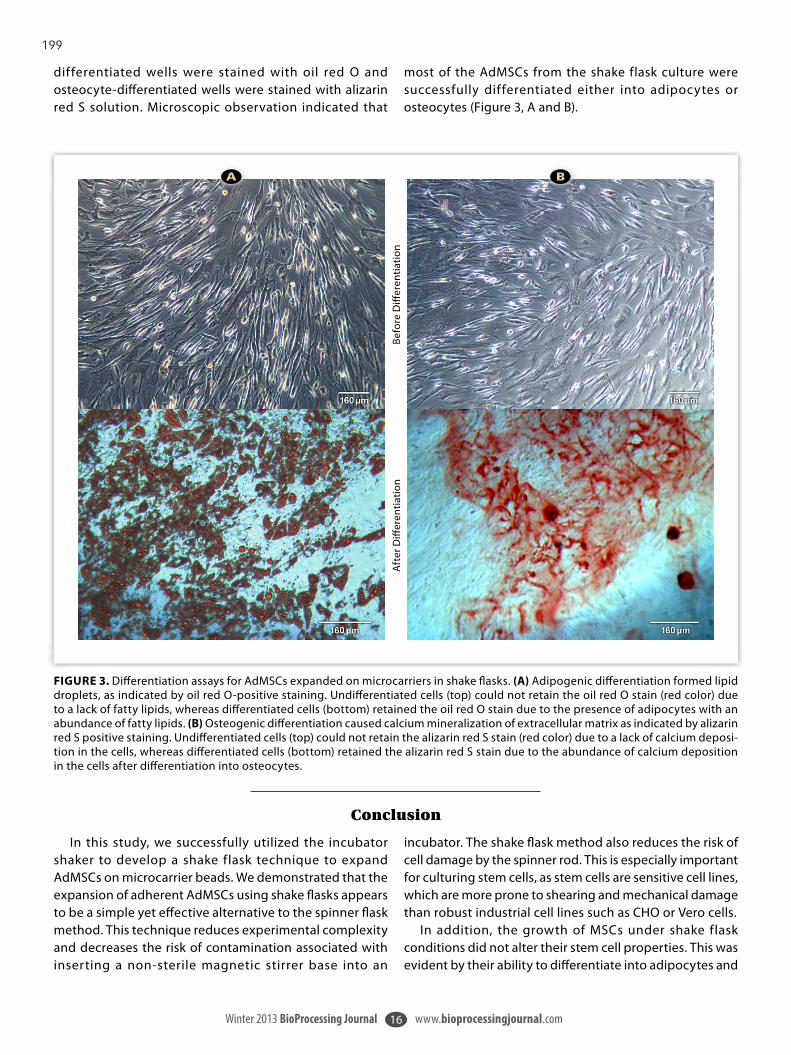
199
Winter 2013 BioProcessing Journal www.bioprocessingjournal.com16
FIGURE 3. Differentiation assays for AdMSCs expanded on microcarriers in shake flasks. (A) Adipogenic differentiation formed lipid droplets, as indicated by oil red O-positive staining. Undifferentiated cells (top) could not retain the oil red O stain (red color) due to a lack of fatty lipids, whereas differentiated cells (bottom) retained the oil red O stain due to the presence of adipocytes with an abundance of fatty lipids. (B) Osteogenic differentiation caused calcium mineralization of extracellular matrix as indicated by alizarin red S positive staining. Undifferentiated cells (top) could not retain the alizarin red S stain (red color) due to a lack of calcium deposi-tion in the cells, whereas differentiated cells (bottom) retained the alizarin red S stain due to the abundance of calcium deposition in the cells after differentiation into osteocytes.
B A
Befo
re D
iffer
entia
tion
Aft
er D
iffer
entia
tion
dif ferentiated wells were stained with oil red O and osteocyte-differentiated wells were stained with alizarin red S solution. Microscopic observation indicated that
most of the AdMSCs from the shake flask culture were successfully dif ferentiated either into adipocytes or osteocytes (Figure 3, A and B).
In this study, we successfully utilized the incubator shaker to develop a shake flask technique to expand AdMSCs on microcarrier beads. We demonstrated that the expansion of adherent AdMSCs using shake flasks appears to be a simple yet effective alternative to the spinner flask method. This technique reduces experimental complexity and decreases the risk of contamination associated with inserting a non-sterile magnetic stirrer base into an
incubator. The shake flask method also reduces the risk of cell damage by the spinner rod. This is especially important for culturing stem cells, as stem cells are sensitive cell lines, which are more prone to shearing and mechanical damage than robust industrial cell lines such as CHO or Vero cells.
In addition, the growth of MSCs under shake flask conditions did not alter their stem cell properties. This was evident by their ability to differentiate into adipocytes and
Conclusion

200
Winter 2013 BioProcessing Journal www.bioprocessingjournal.com17
osteocytes. Furthermore, we found that the S41i incubator shaker was able to accommodate more shake flasks than the standard CO2 incubator equipped with a stir plate for spinner flasks. In the case of the spinner flask, the use of multiple or large magnetic stirrer bases can potentially generate
excessive heat. This could lead to temperature control issues, especially for incubators that do not have refrigeration capabilities. We believe that the shake flask method provided an important improvement for the future scale-up potential of stem cells into large-scale industrial bioreactors.
[1] Zhang Y, Khan D, Delling J, Tobiasch E. Mechanisms underlying the osteo- and adipo-dif ferentiation of human mesenchymal stem cells. ScientificWorldJournal, 2012; (2012): 793823. http://dx.doi.org/10.1100/2012/793823.[2] Pittenger MF, Mackay AM, Beck SC, Rama K, Jaiswal RK, Douglas R, Mosca JD, Moorman MA, Simonetti DW, Craig S, Marshak DR. Multilineage potential of adult human mesenchymal stem cells. Science, 1999; 284 (5411): 143-147. http://dx.doi.org/10.1126/science.284.5411.143. PMID: 10102814.[3] Capla A. Mesenchymal stem cells. J Orthop Res, 1991; 9 (5): 641-50. http://dx.doi.org/10.1002/jor.1100090504.[4] Schop D, van Dijkhuizen-Radersma R, Borgart E, Janssen FW, Rozemuller H, Prins H-J, de Bruijn JD. Expansion of human mesenchymal stromal cells on microcar-riers: growth and metabolism. J Tissue Eng Regen Med, 2010; 4 (2): 131-140. http://dx.doi.org/10.1002/term.224. PMID: 19842106. [5] Ra JC, Shin IS, Kim SH, Kang S, Kang BC, Lee HY, Kim YJ, Jo JY, Yoon EJ, Choi J, Kwon E. Safety of intravenous infusion of human adipose tissue-derived mesen-
chymal stem cells in animals and humans. Stem Cells Dev, 2011; 20 (8): 1297-1308. http://dx.doi.org/10.1089/scd.2010.0466. PMID: 21303266.[6] Nastran M, Doran P. Osteogenic dif ferentiation and osteochondral tissue engineering using human adipose-derived stem cells. Biotechnol Prog, 2013; 29 (1) 176-185. http://dx.doi.org/10.1002/btpr.1663.[7] Ertaş G, Ural E, Ural D, Aksoy A, Kozdağ G, Gacar G, Karaöz E. Comparative analysis of apoptotic resistance of mesenchymal stem cells isolated from human bone marrow and adipose tissue. ScientificWorldJournal, 2012; (2012): 105698. http://dx.doi.org/10.1100/2012/105698. Epub 2012 Apr 24. [8] Schop D. Growth and metabolism of mesenchymal stem cells cultivated on microcarriers. PhD Thesis 2010, University of Twente, The Netherlands. [9] Dominic i M, Le Blanc K, Mullae I . Minimal cr iter ia for def ining mult i -potent mesenchymal s t romal cel ls . The inter nat ional Soc iet y for Cellular Therapy position statement. Cytotherapy, 2006; 8 (10): 315-317. http://dx.doi.org/10.1080/14653240600855905. PMID: 16923606.
References
About the Authors
Khandaker Siddiquee, PhD,1 is a Senior Research Scientist. Ma Sha, PhD, 1* is Director, Technical Applications.
1. Eppendorf Inc., 175 Freshwater Blvd., Enfield, Connecticut 06082 USA
*Dr. Sha is the corresponding author. Phone: +1-860-253-6649 | Email: [email protected]
Notes
ATCC® is a registered trademark of American Type Culture Collection.BioLegend® is a registered trademark of BioLegend Inc.Corning® is a registered trademark of Corning Inc.DURAN® is registered trademark of DURAN Group GmbH.Eppendorf®: the Eppendorf logo, and Easypet® are registered trademarks, and New Brunswick™ is a trademark, of Eppendorf AG, Germany.
EVOS® is a registered trademark of Life Technologies Corp.Galaxy® is a registered trademark of New Brunswick Scientif ic, Co., Inc., USA.OLYMPUS® is a registered trademark of OLYMPUS Corp.ScienCell™ is a trademark of ScienCell Research Laboratories.SoloHill® is a registered trademark of SoloHill Engineering, Inc.YSI® is a registered trademark of YSI Inc.
201
R E P R I N T F R O M
Ruth Olmer, Ph.D., Sebastian Selzer, and Robert Zweigerdt, Ph.D.
Human pluripotent stem cells (hPSCs) and their derivatives have gained in-creased importance for industrial ap-plications in recent years. They have a great potential for therapeutic applica-tions as well.
In vitro assays and novel regen-erative therapies will require large cell quantities produced under defined conditions.
In conventional mammalian cell cul-ture, the utilization of bioreactors is well established, e.g., for the production of recombinant therapeutic proteins, vac-cines, and antibodies. Established pro-tocols are used for process development and manufacturing of mammalian cells in 100–1,000-L scale and beyond.
Cell cultivation in stirred tank bio-reactors allows for tight control and online monitoring of all relevant pro-cess parameters such as temperature, agitation, pH or dissolved oxygen, and scaleup.
This know-how can serve as a ba-sis for creating processes to cover the demand on hPSCs, including human induced pluripotent stem cells (hiP-
SCs) and humen embryonic stem cells (hESCs).
There already have been investi-gations on cultivating adherence-de-pendent stem cells in suspension cul-ture. These studies mainly focused on the modification of matrix-attached hESC cultivation on microcarriers, a method that is widely used in conven-tional mammalian cell culture.
However, culture heterogeneity due to the preference of undifferentiated hPSCs to stick to each other rather than to prescreened types of microcarriers might be challenging. Recent studies in our labs and elsewhere have now dem-onstrated the potential of cultivating undiffentiated human ESCs and iPSCs as cell-only-aggregates in suspension.
More recently, we have demon-strated the feasibility of translating this approach into stirred tank reac-tors, paving the way for the envisioned mass production of pluripotent stem cells and their derivatives. Key features of this technology include utilization of a fully defined serum-free culture medium, single cell-based inoculation, and significant long-term expansion of hPSCs in easy-to-scaleup suspension independent of extracellular matrices
or scaffolds. The method was successfully ap-
plied to several human hPSC lines and cynomolgus monkey ES cells as well. In a previous step, transfer from static suspension in culture dishes to stirred spinner and shaken Erlenmeyer flasks was also enabled.
Such dynamic cultivation of cell-only-aggregates turned out to be robust regarding the reproducibility of cell ex-pansion, karyotype stability, and over-all preservation of the stem cells’ plu-ripotency. Somewhat lower expansion rates in dynamic conditions further suggested a high potential for culture optimization by applying a more con-trolled environment.
To improve culture monitoring and control capabilities and to pave the way for larger-scale cultivation, the method was transferred to a stirred tank bioreactor system.
Studies were carried out in a Das-gip parallel bioreactor system consist-ing of four 250 mL cultivation vessels
Massively Expanding Stem Cell Suspensions
Tools | Technologies | Trends November 15, 2012volume 32, Number 20
Bioprocessing TUTORIAL
Achieving Optimal Cultivation and Maintaining Pluripotency and Differentiation Potential Counts
Ruth Olmer, Ph.D., is senior scientist and Robert Zweigerdt, Ph.D., is principal investigator, both at Hannover Medical School, Leibniz Research Laboratories for Biotechnology and Artificial Organs supported by the REBIRTH cluster of Excellence. Sebastian Selzer ([email protected]) is engineer for hardware development at Dasgip. Web: www.dasgip.com.

202
(100–250 mL working volume) in-cluding an integrated Dasgip control unit and software. Allowance was made for independent monitoring and control of temperature, pH, oxygen tension, and stirring conditions.
To test and ensure reproducibil-ity, all approaches were performed in four independent experimental re-peats in a culture volume of 125 mL
each. Utilizing cord blood endothe-lial cell-derived hiPSCs, preliminary experiments showed that the inocu-lation density and the agitation mode were highly critical for successful process initiation in impeller-stirred bioreactors.
Experiments revealed that 5x10⁵ hPSCs/mL was an efficient inocula-tion density in the stirred bioreactor
setting. Subsequently, experiments were carried out to compare impeller designs and stirring speed modulation aiming at optimal control of cell ag-gregation and homogeneity of aggre-gate size distribution.
Impeller Design
Axial 8-blade pitched impellers and modified stirring bars were designed
November 15, 2012 | genengnews.com | Genetic Engineering and Biotechnology News
Figure 2. Aggregate formation and distribution depending on impeller designs: The impeller and time-dependent average of the aggregate diameter represents 2–3 independent bioreactor runs.
Figure 1. Impact of stirrer design and agitation speed on microcarrier distribution:The stained microcarriers were stirred in 100 ml buffer for 10 min at 30, 40, 50, and 60 rpm. Pictures were taken immediately thereafter from the top of the vessels.

203
and tested for comparative agitation studies (Figure 1). The basic design of the pitched blade impeller was previ-ously developed and successfully ap-plied to control single cell-inoculated mouse ESC aggregate formation in a 2-L bioreactor scale.
In the context of the 250 mL vessels inoculated with 125 mL culture vol-ume, impeller dimensions were down-scaled to ensure similar geometries of the reactor-impeller design. Impeller variants also differed in blade size and ankle as outlined in Figure 1.
To initially evaluate suitability of the agitation system, cell-free assessment of microcarrier distribution in the bioreac-tor was tested providing a meaningful and cost-efficient substitute of cell ag-
gregates. All impeller designs and stir-ring bars were analyzed at agitation speeds varying between 30–60 revolu-tions per minute (rpm; Figure 1). These experiments revealed that all three 8-blade 40 mm impeller variants as well as a 60 mm stirring bar resulted in ho-mogeneous carrier distribution at 40 or 60 rpm, respectively. Having the blade impellers’ applicability for the common use with bioreactors in mind, only these devices were chosen for the subsequent cell culture experiments.
Undifferentiated hiPSC cultures (expanded in conventional 2D culture) were dissociated and inoculated as sin-gle cell suspensions at 4–5 x 10⁵ cells/mL in a total volume of 125 mL and stirred at 60 rpm. To visualize the im-
pact of the three different 8-blade im-peller design variants, aggregate diam-eter analysis and size distribution was carried out by light microscopy.
Notably, all approaches resulted in successful aggregate formation from single cells whereby minor variability between the three impeller designs was detected (Figure 2). Growth kinetics and metabolic activity revealed robust and reproducible hiPSC expansion in the stirred bioreactor system.
Evaluation of viable cell numbers in eight independent bioreactor runs over the time revealed, on average, a robust, about four-fold expansion of the inocu-lated hiPSCs after seven days of cultiva-tion (Figure 3). Monitoring the meta-bolic activity by determining glucose, lactate, and amino acid concentrations as well as pH and dissolved oxygen lev-els confirmed efficiency and high repro-ducibility of the culture system.
Importantly, further analysis proved maintenance of pluripotency-associ-ated marker expression of suspension culture expanded cells, and functional assessment confirmed their multilin-eage differentiation in vitro.
Conclusions
In summary, advanced parallel bio-reactor systems are highly suitable for the transfer of single cell inoculated human pluripotent stem cell suspen-sion to fully controlled cultivation. The precise process control, detailed online monitoring as well as evaluation and optimization of complex, multifacto-rial culture parameters will further al-low significant process optimization in ongoing studies.
Capable of operating with even small working volumes of 125 mL (while the functionality and geometry is similar to larger-scale stirred bioreactors), these reactor systems are matching demands of process development and optimiza-tion for human stem cell cultivation. The yield of 2 x 10⁸ pluripotent cells in a single process run in the 125 mL scale shown in our previous study has proven the technology to be suitable to enable the mass expansion of human pluripotent stem cells.
Genetic Engineering and Biotechnology News | genengnews.com | November 15 2012
Figure 3. Growth kinetics and metabolic activity of hiPSCs suspension cultures: up to 5.5 fold increase in cell numbers was achieved in individual runs over seven days. The dashed lines on glucose and lactate concentration, pH, and po2 graphs illustrate medium exchange.

204
Company insight > Drug delivery
WORLDPHARMACEUTICALFRONTIERS | www.worldpharmaceuticals.net 33
B ioprocess development requires software that not only monitors and controls process runs, but also integrates design, execution and evaluation of
experiments. In this way, the software supports initiatives to implement QbD (quality by design) principles, ensuring product quality and successful scale-up to manufacturing. There are several points to consider when selecting SCADA (supervisory control and data acquisition) software and associated data and information management.
1. Number of bioreactors Parallel operation of bioreactors saves valuable time and accelerates process development. Advanced control software should be able to control multiple vessels individually, up to 24, for example, at a time through incorporated batch management. Ideally, it also features parallel cleaning and calibration procedures.
2. Vessel flexibility Laboratories that use both autoclavable and single-use bioreactors of various sizes call for flexibility in their control software. Advanced packages allow control of different vessels setups in one and the same experimental run.
3. Software knowledge of scientistsAn easy-to-use interface can be the difference between good and bad software. In bioprocess control, integrated recipe management with templates for fermentation and cell culture further aid beginners. Experienced users should be able to maximise their processes, through user-defined scripting, for example.
4. Control strategies and levels of automation Profile and equation-based feedback control provide the ultimate flexibility in bioprocess control. Feed rates based on offline analyser data, oxygen transfer rates (OTR)-based feed control, or temperature shifts that are triggered by cell performance are only a few examples used to automate a process.
5. Third-party laboratory equipment Real-time analytical data deliver vast insight into the status of a cultivation. Integration of autosampling and analytical devices, such as nutrient analysers, HPLC, mass spectrometry and Raman spectroscopy, enables feedback control and enhanced automation.
6. External alarm notification and remote control Alarm warnings via e-mail or text message enhance process safety. Progressive bioprocess control systems also enable users to
manage process settings remotely via PC or mobile devices. Users can react immediately to changing process conditions, even from outside the laboratory.
7. Statistical approaches Design of experiments (DoE) is a structured approach for investigating the influence of critical process parameters and how they interact. Parallel bioreactor systems and easy-to-execute DoE setups streamline process development and facilitate regulatory processes. Advanced control integrates with common DoE tools.
8. Reporting and analysis Analysis of bioprocess data can be time-consuming. Automated Microsoft Excel reporting, which is based on user-defined templates, saves manual workload and makes complex analyses easier. A powerful chart creation tool, with an unlimited number of data tracks and axes and an export function to Excel, is a plus. When it comes to comparison of historical and current runs, users benefit from intuitive queries and editable query templates.
9. Cross-system analysis If multiple bioreactor systems, or even multiple sites, need to be evaluated and compared, tools are required to go beyond the possibilities of standard SCADA software. Sophisticated solutions enable mixed-system control and cross-platform analysis of run-time data between systems made by different manufacturers.
10. Data storage and management Documentation demands can require a central database with managed access. If the bioreactor system is integrated with legacy corporate historians, communication via OPC (object linking and embedding for process control) must be installed. This allows company-wide access to all relevant bioprocess data.
Summary Efficient bioprocess control software facilitates the implementation of QbD principles in pharmaceutical development and offers flexibility. Sophisticated solutions for interconnectivity of benchtop equipment and bioprocess data and information management provide further tools to integrate processes, reduce the manual workload and accelerate process development. ■
Efficient bioprocess developmentEppendorf’s bioprocess marketing and communications manager Christiane Schlottbom and product and application specialist Stephan Zelle list the 10 main factors to consider when choosing a bioreactor control software.
Further informationEppendorfwww.eppendorf.com

205
Vaccine Production
Euro
pean
Bio
phar
mac
eutic
al R
evie
w A
pril
2014
issu
e. ©
Sam
edan
Ltd
. 201
4
K John Morrow, Jr, at Newport Biotech and Ma Sha at Eppendorf, Inc
Taking the StrainMammalian cell culture systems are starting to dominate the production of vaccines for viral diseases such as rabies. Making use of existing biopharma infrastructure, these advances might even spell the end for the use of hens’ eggs in future vaccine development
Today, the rapidly expanding demand for vaccine products for viral diseases such as rabies has necessitated the development of more sophisticated production techniques based around cell culture systems. This article reviews vaccine production strategies, with a focus on rabies, looking specifically at the use of the Vero cell line – used worldwide and approved by the US Food and Drug Administration – as well as media technology and the bioreactor options available.
Growing Demand
Viral diseases, including rabies, are worldwide challenges for the international biomedical community. The World Health Organization (WHO) notes that over 32,000 rabies-related deaths were reported in 1998, while annual deaths worldwide from the virus grew to 55,000 by 2006 (1,2).
Rabies is often transmitted to humans from infected domestic animals. Dogs
infected with rabies can become extremely aggressive and attacks on humans are widespread, especially in certain Asian countries where using unleashed dogs for home security is common. It is spread through the saliva of infected animals and bites can be fatal.
Since the 18th century, vaccination has proven to be the most successful – and perhaps the only – route to the total elimination of viral diseases such as rabies. From the early work of pioneers such as Jenner and Pasteur, vaccination was put on a sound scientific footing. Demand for vaccine products has continued to increase ever since, prompting the advances in production technology that we see today.
Viral Cultivation
The expanding vaccine requirements have led to techniques for growing large quantities of antigenic proteins. Traditionally, viruses have been grown in embryonated hens’ eggs, but
numerous shortcomings compromise their utility. These include a bottleneck in the availability of high-quality, pathogen-free eggs, as well as low titers of emerging viruses (3).
A major concern is that, when viruses are cultivated through extended passages in hens’ eggs, there is an evolutionary process in the amnion or allantoic cavity of the egg, resulting in the selection of a virus subpopulation, antigenically and biochemically distinct from the original inoculum (4). Because of these and other factors, permanent cell lines are coming to dominate the field as an alternative method.
Mammalian cell culture systems provide much shorter lead times; a more controlled production process that takes advantage of closed-system bioreactors; a reduced risk of microbial contamination; and the opportunity to cultivate viral stocks without significant egg passage-dependent antigenic changes (5). These systems provide

206
VP-SFM (Invitrogen), EX-CELL Vero SFM (SAFC Biosciences), Provero-1 (Lonza) and HyQ SFM4MegaVir (HyClone). EX-CELL Vero SFM gave one of the highest cell densities, demonstrating that the use of serum-free media has become routine for Vero cell cultivation. Comparable results were observed with a commercial serum-free medium MDSS2N (AXCEVIR-VeroTM by Axcell Biotechnologies).
Rabies Strategies
The Brazilian group led by Frazatti-Gallina has been active in the field of rabies vaccine production (13). Using Vero cells adhered to microcarriers, and cultivated in a bioreactor with
a flexible and scalable platform that can make use of existing biopharma infrastructure for vaccine production, and could replace egg-based vaccines in the foreseeable future (6).
Cell Line Options
In the past few years, several continuous cell lines have been approved by regulatory authorities for virus production. These include the Spodoptera frugiperda insect cell line from Protein Sciences (7), Madin-Darby canine kidney (MDCK), the PER.C6 cell line, designed for growth to high densities (8), and the widely used Vero line.
It is important to note that certain cell lines may provide an environment favouring selection of viral subpopulations, and these types may be inappropriate for vaccine production. In light of this, various lines of investigation support the Vero cell line as the candidate of choice for viral vaccine production, including:
● Efficiency of primary virus isolation and replication to high-infectivity titers
● Genetic stability of the haemagglutinin molecule, while maintaining the antigenic properties of human-derived viruses
● Similarities in the pattern of protein synthesis and morphological changes between virus-infected Vero and MDCK cells (9)
Media Alternatives
There is a variety of different Vero isolates available from commercial
suppliers, but all are quite similar, and their nutritional needs are comparable (10). The search for the ideal mammalian cell culture medium began in the 1950s, with the holy grail being an economical, protein-free, serum-free medium that would provide strong growth support and have the property of scalability to large volumes, up to thousands of litres, while coming in at an affordable price.
Serum provides a protective function to cultured cells, and binds toxins and other contaminating materials. As such, serum-free media must be extremely carefully formulated (11). Albumin can be substituted for serum, but it may impede the downstream steps of purification (12).
Chen et al tested five different serum-free media, combined with Cytodex 1 microcarriers (12). The following were evaluated: OptiPro SFM (Invitrogen),
Sour
ce: E
ppen
dorf,
Inc
Figure 1: Fluorescent image of confluent Vero cells; DAPI-stained nuclei appear blue, and actin filaments stained with rhodamine-conjugated phalloidin appear red
It is important to note that certain cell lines may provide an environment favouring selection of viral subpopulations, and these types may be inappropriate for vaccine production

207
Important Step
The combination of advances reviewed here provides strong support for the use of cell culture systems for virus production of vaccines. The fact that Vero cells have been approved for clinical products represents an important step on the road to technologies that do not rely upon hens’ eggs for generation of adequate quantities of viruses.
Advances in culture media enable the elimination of serum, thus driving the rapid and efficient purification of proteins. The use of carrier beads adds to the efficiency of culture technology, allowing greatly increased cell densities to be reached. Improvements in bioreactor design, combined with these various technological advances, results in a greatly improved and more functional production train.
References1. World survey of rabies number 34 (for
the year 1998), WHO, 2000. Visit: www.who.int/emc/diseases/zoo/wsr98/html version/wsr 98 index.html
2. Kaur M, Rai A and Bhatnagar R, Rabies DNA vaccine: no impact of MHC class I and class II targeting sequences on immune response and protection against lethal challenge, Vaccine 27(15): pp2,128-2,137, 2009
3. Genzel Y and Reichl U, Continuous cell lines as a production system for influenza vaccines, Expert Rev Vaccines 8(12): pp1,681-1,692, 2009
4. Oxford JS et al, Serological studies with influenza A(H1N1) viruses cultivated in eggs or in a canine kidney cell line, Bull World Health Organ 65(2): pp181-187, 1987
5. Doroshenko A and Halperin SA, Trivalent MDCK cell culture-derived influenza vaccine, Optaflu (Novartis Vaccines), Expert Rev Vaccines 8: pp679-688, 2009
6. Montomoli E et al, Cell culture-derived influenza vaccines from Vero cells: a new horizon for vaccine production, Expert Rev Vaccines 11(5): pp587-94, 2012
7. Visit: www.proteinsciences.com/ BEVS.htm
8. Visit: www.crucell.com/Technology%20-%20Cell%20Technology
serum-free medium, they generated an effective rabies vaccine. With the aid of tangential filtration, they purified the rabies virus by chromatography and inactivated it using beta-propiolactone.
Their protocol states that 350cm2 T-flasks were harvested and inoculated into a 3.7-litre CelliGen bioreactor, at a proportion of 16 cells per microcarrier (Cytodex 3-GE), yielding an initial seeding of 2.5×10⁵ cell/ml. The cells were grown in serum-free MDSS2 medium.
The serum-free VP-SFM medium, according to the manufacturer, was developed for Vero, BHK-21 and Chinese hamster ovary cell growth. This medium drives the adherence of the Vero cells to the microcarriers. After four days of cultivation in VP-SFM medium, the cells were infected with Pasteur vaccins (PV) strain rabies virus (multiplicity of infection = 0.08). The harvests of the culture supernatant were carried out three days after the virus inoculation and four times thereafter at 24-hour intervals. During this period, culture conditions were maintained at 60rpm at a pH of 7.15 and 5 per cent dissolved oxygen. Only the temperature varied, from 36.5ºC in the cellular growth phase of the culture to 34ºC after virus inoculation. In the course of the programme, seven batches of virus suspensions were produced in the bioreactor (16 litre per cycle) at a mean viral titer of 104. FFD50/0.05ml.
The effectiveness of the preparation was demonstrated by immunising mice with three doses of the new vaccine, and comparing it with the commercial Verorab and human diploid cell rabies vaccine. Mean titers of neutralising antibodies of 10.3-34.6, 6.54 and 9.36 IU/ml were found, respectively.
The choice of the serum-free medium was fortunate. In this case, the amount of contaminating DNA was very low and tolerable – less than 22.8pg per dose of vaccine. The authors argue that this protocol is especially applicable in the developing world, where rabies is a constant hazard and a major public health problem.
Increasing Yield
Yu et al sought methods to increase yield in Vero cell culture systems over that obtained using roller bottles (14). In a recent review, they summarised the production technology developed over the course of the last seven years. They have adopted the 30-litre BioFlo 4500 fermentor/bioreactor. The cells were cultivated in media containing 10 per cent serum, first grown as a monolayer, and when the cell density reached 1.0-1.2×10⁶ cells/ml, they were transferred to the bioreactor containing 25g/litre of Cytodex-1 for perfusion culture. The virus preparations, also cultured in roller bottles, were infected with the PV2061 virus strain, harvested and transferred to the bioreactors.
Purified Vaccine
Wang et al have described a purified Vero cell rabies vaccine that has been widely produced in China, referred to as ChengDa (Liaoning ChengDa Biological) (15,16). It is grown on a Vero cell line utilising the PV2061 strain, inactivated with beta-propiolactone, lyophilised, and reconstituted in 0.5ml of physiological saline. It fulfils WHO recommendations for potency.
The process used for ChengDa was developed by Aycardi (17). A single bioreactor was capable of producing one million doses of rabies vaccine per year. The method uses ultra-high-density microcarrier cell cultures adapted to a 30-litre CelliGen bioreactor equipped with a patented cell lift impeller, specifically configured for a perfusion system to feed the growth media into the bioreactor. A specially designed decanting column was used to prevent perfusion loss of microcarrier and keep the cells in high concentration. The system delivers high oxygen transfer, high nutrient level and low shear stress, allowing cell growth of up to 1.2x10⁷ million cells/ml under continuous perfusion for up to 20 days.

208
16. Visit: bariconsultants.com/ projects.html
17. Aycardi E, Producing human rabies vaccines at low cost, Genetic Engineering News 22(8), 5th April 2002
9. Govorkova EA et al, African green monkey kidney (Vero) cells provide an alternative host cell system for influenza A and B viruses, J Virol 70(8): pp5,519-5,524, 1996
10. Ammerman NC, Beier-Sexton M and Azad AF, Growth and maintenance of Vero cell lines, Current Protocols in Microbiology, Appendix 4E, November 2008
11. Chen A et al, Serum-free microcarrier based production of replication deficient influenza vaccine candidate virus lacking NS1 using Vero cells, BMC Biotechnol, 11th August 2011
12. Visit: www.invitria.com/cell-culture-products-services/serum-albumin.html
13. Frazatti-Gallina NM et al, Vero-cell rabies vaccine produced using serum-free medium, Vaccine 23(4): pp511-517, 2004
14. Yu P et al, Production and evaluation of a chromatographically purified Vero cell rabies vaccine in China using microcarrier technology, Hum Vaccine Immunother 8(9): pp1,230-1235, 2012
15. Wang C et al, Promising rabies vaccine for postexposure prophylaxis in developing countries, a purified Vero cell vaccine produced in China, Clin and Vaccine Immuno 17(4): p688, 2010
K John Morrow, Jr, is President and Chief Executive Officer of Newport Biotech Consultants. He has extensive experience in the academic community, including faculty positions at the University of Kansas and the Texas Tech University Health Sciences Center, where he was a lead or co-investigator on $2.5 million grant funding. John's employment in the private sector includes research and consulting work with Meridian Bioscience, Inc in Cincinnati, Ohio. He obtained his PhD from the University of Washington, and did postdoctoral work at the Universitá di Pavia, Italy, and the Fox Chase Cancer Center in Philadelphia. Email: [email protected]
Ma Sha is Director of Technical Applications for Eppendorf, Inc. He has extensive experience across multiple fields within the biotech industry, including mass spectometry, cell culture consumables, immunodiagnostics and bioprocess. Ma obtained his PhD from the City University of New York, completed postdoctoral work at Harvard Medical School and The Rockefeller University, and was the principal inventor of four US and several European patents. Most recently, he led the effort to obtain state approval to conduct biosafety level 2 research on induced pluripotent stem cells at the Eppendorf laboratory in Enfield, Connecticut. Email: [email protected]
About the authors

Other

210
Automated Bioreactor Sampling –Process Trigger Sampling for Enhancing Microbial Strain CharacterizationMatthew J. Maurer1, Jeffrey M. Skerker1,2,3, Adam P. Arkin1,2,3, William Miller4, Michael Biksacky4*, Claudia M. Huether-Franken5*, and Karl
Rix5
1 UC Berkeley Energy Biosciences Institute, Berkeley, CA, USA; 2 UC Berkeley Department of Bioengineering, Berkeley, CA, USA; 3 Physical
Biosciences Division, Lawrence Berkeley National Laboratory, Berkeley, CA, USA; 4 Flownamics® Analytical Instruments, Inc., Madison, WI,
USA; 5 Eppendorf AG Bioprocess Center, Juelich, Germany
* Corresponding authors: [email protected], [email protected]
APPLICATION NOTE No. 298 I January 2015
Abstract
This application note describes the integration of a Flownamics Seg-Flow® 4800 Automated On-line Sampling System with Eppendorf DASGIP® Parallel Bioreactor Systems as implemented at the Energy Biosciences Institute in Berkeley, California. The
automated process trigger sampling technology enabled the researchers to rapidly characterize process events, parameters and stress responses that impact yeast strain gene regulation and, ultimately, biofuel productivity.
Introduction
Scientists at the Energy Biosciences Institute (EBI) conduct research in a variety of areas in bioenergy development. The Quantitative Engineering of Industrial Yeast program at the EBI focuses on a thorough, systems-level understanding of bacterial and yeast metabolism, gene regulation, and stress response for elucidating principles to help rationally engineer bacteria and yeasts for improved biofuel production from lignocellulosic sources [1].
In order to accomplish their goals and objectives, researchers in the Quantitative Engineering of Industrial Yeast program have implemented automated processes, including the use of an integrated parallel bioreactor system and automated bioreactor sampling system, to conduct experiments for optimizing yeast strain characterization and selection.
Materials and Methods
Incorporating tools such as parallel bioreactor systems and automated bioreactor sampling technologies can significantly reduce project timelines and increase the efficiency of the microbial strain characterization and selection process.
DASGIP® Parallel Bioreactor SystemsEppendorf DASGIP Parallel Bioreactor Systems allow for advanced screening of bacteria, yeasts and/or fungi. The multi-bioreactor/vessel design enables parallel experimentation intended to accelerate process development and increase throughput. Multiple bioreactor vessels are controlled via shared equipment and a single computer system, enabling the experimenter to test multiple conditions side-by-side or by allowing multiple independent

211
APPLICATION NOTE I No. 298 I Page 2
experiments to be run simultaneously using the shared equipment resources. Additionally, the DASGIP Parallel Bioreactor System’s modular design provides ease of setup and maintenance, while offering the same control strategies and precision as larger scale production plants to achieve a reproducible and scalable process (figure 1) [2].
The DASGIP Control* software and associated hardware provides high precision monitoring and control units designed for small working volumes, high information output and easy comparative data analysis. The Eppendorf software DASware® analyze, utilizes the platform-independent Object Linking and Embedding for Process Control (OPC) communication protocol for enabling bidirectional communication between the DASGIP system and third-party analytical devices, including automated bioreactor sampling systems.
Seg-Flow® Automated On-line Sampling SystemThe Seg-Flow 4800 Automated On-line Sampling System (Seg-Flow System) is a liquid and data management device designed to withdraw samples from up to eight bioreactors and deliver them to up to four analytical instruments and/or fraction collectors. This functionality enables real-time analysis and sample collection from parallel bioreactor systems. The Seg-Flow System’s patented “segmented on-line sampling” technology allows a wide range of sample volumes to be obtained and rapidly delivered to distances up to 7.6 meters (25 feet) from the bioreactor.
The FlowWeb™ software platform, which controls all the Seg-Flow System functions, provides seamless connectivity with various third-party analyzers for enabling real-time analysis of important culture process parameters such as nutrients, metabolites and various cell measurements. Upon completion of the analysis, the Seg-Flow system acquires and processes the analyzer data. The FlowWeb OPC software suite communicates the analyzer data into any OPC-enabled supervisory control and data acquisition (SCADA) system, which expands real-time monitoring capabilities for bioprocess cultures. Figure 2 shows the Seg-Flow configuration used by EBI for conducting automated on-line
fraction collection for their microbial strain characterization evaluation.
Process trigger samplingThe Seg-Flow System is capable of performing automated sampling and analysis during planned or unplanned process events in response to an external SCADA or other bioprocess management system such as the DASGIP Control/DASware software platform. This is achieved through OPC connectivity.
The process events used to activate, or trigger, the Seg-Flow System are user-defined. Examples of process events include pH or dissolved oxygen excursions, culture induction, feeding or other in-process control actions. The process events used to trigger the Seg-Flow system require OPC data tag configuration and must be programmed into the host SCADA/bioprocess management system. When the process event is detected by the bioreactor station, the data trigger is communicated to the SCADA system to commence the remote activation of the Seg-Flow system (figure 3).
Once the Seg-Flow system is activated, a sample is
Fig. 3: Architecture for the Seg-Flow 4800 process trigger sampling function. The process event or “trigger” is user-defined and is programmed in the bioreactor’s OPC-enabled SCADA or bioprocess management system, which remotely controls the Seg-Flow system.
Analyzer
Fraction
Collector
HPLC
DASware control
Data
Samples
Fig. 1: DASGIP Parallel Bioreactor System for biofuel development
Fig. 2: Seg-Flow 4800 Automated On-line Sampling System with FlowFraction™ 400 Fraction Collector
*DASGIP Control is now DASware control 5. Please refer to ordering information on page 6.
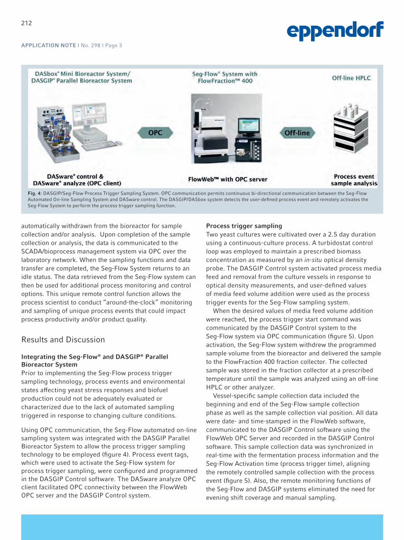
212
APPLICATION NOTE I No. 298 I Page 3
automatically withdrawn from the bioreactor for sample collection and/or analysis. Upon completion of the sample collection or analysis, the data is communicated to the SCADA/bioprocess management system via OPC over the laboratory network. When the sampling functions and data transfer are completed, the Seg-Flow System returns to an idle status. The data retrieved from the Seg-Flow system can then be used for additional process monitoring and control options. This unique remote control function allows the process scientist to conduct “around-the-clock” monitoring and sampling of unique process events that could impact process productivity and/or product quality.
Results and Discussion
Integrating the Seg-Flow® and DASGIP® Parallel Bioreactor SystemPrior to implementing the Seg-Flow process trigger sampling technology, process events and environmental states affecting yeast stress responses and biofuel production could not be adequately evaluated or characterized due to the lack of automated sampling triggered in response to changing culture conditions.
Using OPC communication, the Seg-Flow automated on-line sampling system was integrated with the DASGIP Parallel Bioreactor System to allow the process trigger sampling technology to be employed (figure 4). Process event tags, which were used to activate the Seg-Flow system for process trigger sampling, were configured and programmed in the DASGIP Control software. The DASware analyze OPC client facilitated OPC connectivity between the FlowWeb OPC server and the DASGIP Control system.
Process trigger sampling Two yeast cultures were cultivated over a 2.5 day duration using a continuous-culture process. A turbidostat control loop was employed to maintain a prescribed biomass concentration as measured by an in-situ optical density probe. The DASGIP Control system activated process media feed and removal from the culture vessels in response to optical density measurements, and user-defined values of media feed volume addition were used as the process trigger events for the Seg-Flow sampling system.
When the desired values of media feed volume addition were reached, the process trigger start command was communicated by the DASGIP Control system to the Seg-Flow system via OPC communication (figure 5). Upon activation, the Seg-Flow system withdrew the programmed sample volume from the bioreactor and delivered the sample to the FlowFraction 400 fraction collector. The collected sample was stored in the fraction collector at a prescribed temperature until the sample was analyzed using an off-line HPLC or other analyzer.
Vessel-specific sample collection data included the beginning and end of the Seg-Flow sample collection phase as well as the sample collection vial position. All data were date- and time-stamped in the FlowWeb software, communicated to the DASGIP Control software using the FlowWeb OPC Server and recorded in the DASGIP Control software. This sample collection data was synchronized in real-time with the fermentation process information and the Seg-Flow Activation time (process trigger time), aligning the remotely controlled sample collection with the process event (figure 5). Also, the remote monitoring functions of the Seg-Flow and DASGIP systems eliminated the need for evening shift coverage and manual sampling.
Fig. 4: DASGIP/Seg-Flow Process Trigger Sampling System. OPC communication permits continuous bi-directional communication between the Seg-Flow Automated On-line Sampling System and DASware control. The DASGIP/DASbox system detects the user-defined process event and remotely activates the Seg-Flow System to perform the process trigger sampling function.

213
APPLICATION NOTE I No. 298 I Page 4
Fig. 5: Process Trigger Sampling Data using DASware Plant Overview Function. Plot displays (A) time of Seg-Flow activation by the DASGIP controller (vessel 1 = green, vessel 2 = red); (B) time and duration of Seg-Flow sample collection (vessel 1 = orange, vessel 2 = blue); (C) time of sample deposition into vial and vial position (vessel 1 = magenta, vessel 2 = green); (D) culture density data from biomass probe (vessel 1 = purple, vessel 2 = blue) and (E) cumulative media addition (vessel 1 = black, vessel 2 = magenta).
Conclusion
Coupling the DASGIP Parallel Bioreactor and Seg-Flow automated on-line sampling technologies enabled EBI’s Microbial Characterization Facility research staff to implement remote-controlled, automated process trigger sampling as an integral part of its yeast strain characterization activities. By integrating this functionality into their high-throughput screening and selection process, EBI research scientists are better able to rapidly characterize process events, parameters and stress responses that impact yeast strain gene regulation and, ultimately, biofuel productivity.

214
APPLICATION NOTE I No. 298 I Page 5
Literature
[1] Energy Biosciences Institute. Biofuels Production Research Programs. Programs and Projects. 24 March 2014.
http://www.energybiosciencesinstitute.org/research/biofuels#1.
[2] Knocke, C., Vogt, J. Biofuels - Challenges & Chances: How Biofuel Development can Benefit from Advanced Process Technology. Eng. Life Sci.
9-2 (2009): 96-99.

215
APPLICATION NOTE I No. 298 I Page 6
www.eppendorf.com
This work described was supported in part by the U.S. Department of Energy. While this document is believed to contain correct information, neither the United States Government nor any agency thereof, nor the Regents of the University of California, nor any of their employees, makes any warranty, express or implied, or assumes any legal responsibility for the accuracy, completeness, or usefulness of any information, apparatus, product, or process disclosed, or represents that its use would not infringe privately owned rights. The views and opinions of authors expressed herein do not necessarily state or reflect those of the United States Government or any agency thereof or the Regents of the University of California. The Regents of the University of California, University of California, Berkeley, including its Energy Biosciences Institute (EBI), and Lawrence Berkeley National Laboratory are not affiliated with Flownamics Analytical Instruments, Inc., Eppendorf AG, or DASGIP Information and Process Technology GmbH and do not support, endorse, or sponsor Flownamics Analytical Instruments, Inc., Eppendorf AG, or DASGIP Information and Process Technology GmbH, or their products/services.
Flownamics® and Seg-Flow® are registered trademarks of Flownamics Analytical Instruments, Inc., USA. FlowWeb™ and FlowFraction™ are trademarks of Flownamics Analytical Instruments, Inc., USA. Eppendorf® and the Eppendorf logo are registered trademarks of Eppendorf AG, Germany. DASGIP®, DASbox®, and DASware® are registered trademarks of DASGIP Information and Process Technology GmbH, Germany. U.S. Design Patents are listed on www.eppendorf.com/ip. All rights reserved, including graphics and images. Copyright © 2015 by Eppendorf AG.
Your local distributor: www.eppendorf.com/contactEppendorf AG · 22331 Hamburg · [email protected] · www.eppendorf.com
Ordering informationDescription Order no. DASGIP® Parallel Bioreactor System for Microbial Applications, max. 250 sL/h gassing4-fold system with Bioblock 76DG04MBBB8-fold system with Bioblock 76DG08MBBB16-fold system with Bioblock 76DG16MBBB4-fold system, benchtop 76DG04MB8-fold system, benchtop 76DG08MB16-fold system, benchtop 76DG16MBDASware® control, incl. PC, OS, and licensesfor 4-fold DASGIP® system 76DGCS4for 8-fold DASGIP® system 76DGCS8DASware® control professional, incl. PC, OS, and licensesfor 4-fold DASGIP® system 76DGCSP4for 8-fold DASGIP® system 76DGCSP8DASware® analyze, OPC client professional incl. 1x tunneller lic. (OPC DA e.g. for ext. analyzer with autosampler)for 4 vessels 76DWANA4Pfor 8 vessels 76DWANA8Pfor 12 vessels 76DWANA12P
For Information on the Seg-Flow 4800 Automated On-line Sampling System, FlowFraction 400 Fraction Collector and the FlowWeb Software Plat-form please see www.flownamics.com.

216
Isobutanol from Renewable Feedstock—Process Optimization by Integration of Mass Spectrometry to Two 8-fold DASGIP® Parallel Bioreactor SystemsJames Glenn1 and Claudia M. Huether2*1 Gevo Inc., 345 Inverness Dr S C-310 Englewood, CO 80112, USA; 2 Eppendorf AG Bioprocess Center, Rudolf-Schulten-Str. 5, 52428 Juelich,
Germany; * Corresponding author: [email protected]
APPLICATION NOTE No. 295 I July 2011
Abstract
This application note describes the integration of a Thermo Scientific® Prima® dB Mass Spectrometer (MS) with DASGIP Parallel Bioreactor Control Systems
implemented at Gevo®, Inc. in Englewood, Colorado. The availability of real-time MS data will aid in maximizing cell growth and isobutanol production.
Introduction
Isobutanol has broad market applications as a solvent and a gasoline blendstock that can help refiners meet their renew-able fuel and clean air obligations. It can also be further processed using well-known chemical processes into jet fuel and feedstocks for the production of synthetic rubber, plastics, and polyesters. Isobutanol is an ideal platform mol-ecule that can be made inexpensively using fermentation. The ability to automate the data analysis would increase production and reduce costs.
Gevo, a leading renewable chemicals and advanced biofuels company is developing biobased alternatives to petroleum-based products using a combination of synthetic biology and chemistry. Gevo plans to produce isobutanol, a versatile platform chemical for the liquid fuels and petrochemical market.
The main objective of implementing OPC communication
Figure 1: Isobutanol - a versatile platform Chemical

217
APPLICATION NOTE I No. 295 I Page 2
between the Thermo Scientific Mass Spectrometer and the DASGIP Control* Software during a fermentation run was to optimize growth and isobutanol production through automation. The system previously in place at Gevo required that manual data calculations had to be performed by merg-ing the bioreactor runtime data with the MS data to assess the fermentation performance.
Materials and Methods
Corn mash was used as a substrate for the production of isobutanol by fermentation. The fermentation process was carried out using two DASGIP Parallel Bioreactor Systems with eight vessels each. The working volume in all 16 biore-actors was 1L, respectively.
OPC communication was implemented between the Thermo Scientific Prima dB Mass Spectrometer and the DASGIP Control Software to provide real-time exhaust results. Script calculations were used to take the MS data as inputs and generate meaningful metrics to automatically analyze key fermentation operating values and quickly make process control changes.
Results and Discussion
By integrating the Thermo Scientific Prima dB Mass Spec-trometer with the DASGIP Parallel Bioreactor System the calculation of key fermentation operating values was suc-cessfully automated. This automation streamlined the work-flow and allowed for data-driven control decisions using the real-time exhaust based analytical results.
Before Automation:Without integration of the MS and the DASGIP Parallel Bio-reactor System using OPC, calculation of key fermentation operating values was time-consuming and labor-intensive.
Optimized by MS Integration:Using OPC communication the real-time MS results were sent to the two DASGIP Control systems. Within the control system, the fermentation runtime data and the MS results were charted and transferred to the data historian with syn-chronized time stamps.
Figure 2: Schematic of the MS integration setup. Implementing OPC communication between the Thermo Scientific Prima dB Mass Spectrometer and the DASGIP Control Software (now DASware control) provided continuous exhaust results that were synchronized with fermentation data and viewed in real-time. The isobutanol production rates could be calculated online and were then available for data-driven control decisions.
*DASGIP Control is now DASware® control 5.

218
APPLICATION NOTE I No. 295 I Page 3
Key fermentation operating values were calculated online from combined fermentation and MS runtime data, charted and sent to the data historian and were then available for
data-driven control decisions. Set-up and script calculations were stored in a user-editable recipe.
Conclusion
With its comprehensive data management functions the DASGIP Parallel Bioreactor System allowed the seamless integration of the Thermo Scientific Prima dB Mass Spec-trometer.
The most important success criterion was the ability to calculate isobutanol production rates in real-time giving in-stant feed back on the quality of run. The availability of the exhaustbased analytical results made data-driven control decisions possible. A secondary success criterion was the fermentation runtime data and MS data was logged with synchronized time stamps to allow for post-run analysis if needed.
Figure 3: MS integration: data display and charts. The DASGIP Control bioreactor view showed online bioreactor runtime data including real-time MS results. Editable scripting allowed for online calculation of production rate and graphic display in defined charts.

219
APPLICATION NOTE I No. 295 I Page 4
www.eppendorf.comGevo® is a registered trademark of Gevo Inc., USA. Thermo Scientific® and Prima® are registered trademarks of Thermo Fischer Scientific Inc. Corporation, USA. Eppendorf® and the Eppendorf logo are registered trademarks of Eppendorf AG, Germany. DASGIP® and DASware® are registered trademarks of DASGIP Information and Process Technology GmbH, Germany. U.S. Design Patents are listed on www.eppendorf.com/ip.All rights reserved, including graphics and images. Copyright © 2015 by Eppendorf AG.
Your local distributor: www.eppendorf.com/contactEppendorf AG · 22331 Hamburg · [email protected] · www.eppendorf.com
Ordering information Order no. DASGIP® Parallel Bioreactor System for Microbial Applications, max. 250 sL/h gassing4-fold system with Bioblock 76DG04MBBB8-fold system with Bioblock 76DG08MBBB16-fold system with Bioblock 76DG16MBBB4-fold system, benchtop 76DG04MB8-fold system, benchtop 76DG08MB16-fold system, benchtop 76DG16MBDASware® control, incl. PC, OS, and licensesfor 4-fold DASGIP® system 76DGCS4for 8-fold DASGIP® system 76DGCS8for 16-fold DASGIP® system 76DGCS16DASware® analyze, OPC client professional incl. 1x tunneller licence (OPC DA e.g. for ext. analyzer with autosampler)for 4 vessels 76DWANA4Pfor 8 vessels 76DWANA8P

220
www.eppendorf.com
Eppendorf, Inc. 175 Freshwater Boulevard Tel.: +1 860 253 6700 Enfield, CT 06082-4444, USA [email protected]
Vero Cell-based Vaccine Production: Rabies and Influenza Cell lines, Media and Bioreactor Options Review Article, Dec. 2013
Authors K. John Morrow Jr. Ph.D, Newport Biotech, Newport, KY, USA Ma Sha Ph.D, Eppendorf, Inc., Enfield, CT, USA

221
2 www.eppendorf.com
Table of Contents 1. Abstract .......................................................................................................................................................................... 3
2. Introduction ...................................................................................................................................................................... 3
3. Biological Systems for Viral Cultivation ............................................................................................................................ 4
4. Cell Line Options .............................................................................................................................................................. 4
5. Media Alternatives ........................................................................................................................................................... 5
6. Rabies Virus Cultivation Strategies .................................................................................................................................. 6
7. Influenza Virus Cultivation Strategies .............................................................................................................................. 7
8. Conclusions ..................................................................................................................................................................... 8
9. Acknowledgments ............................................................................................................................................................ 8
10. References ....................................................................................................................................................................... 9

222
3 www.eppendorf.com
1. Abstract We review strategies for optimizing vaccine production with examples given for rabies and influenza using cell culture systems. The Vero cell line is one of the most satisfactory based on its stability and well-documented performance in quality and quantity of viral yield. It has received FDA approval and is used throughout the world. Cell culture media technology has advanced dramatically in recent years, and a number of serum free and protein free options are available through commercial suppliers. Because serum tends to bind toxins and contaminants, its elimination calls for careful monitoring of culture conditions in order to achieve optimal performance.
Improvements in microcarriers have been important additions to the range of possible choices for optimizing in vitro production systems. With a series of bioreactor options available, we can foresee the elimination of hens eggs for virus production.
2. Introduction
Figure 1. Fluorescent image of confluent Vero cells; DAPI-stained nuclei appear blue, and actin filaments stained with rhodamine-conjugated phalloidin appear red (Eppendorf Inc.).
Viral diseases, including rabies and influenza, are worldwide challenges to the international biomedical community. WHO notes that in 1998 over 32,000 deaths due to rabies were reported, while influenza has been responsible for millions of deaths worldwide over the course of the last century.1

223
4 www.eppendorf.com
Rabies is often transmitted to humans from infected domestic animals. Dogs infected with rabies can become extremely aggressive and attacks on humans are widespread, especially in certain Asia countries where using unleashed dogs for home security is common. The virus is spread through their saliva and bites by infected animals can be fatal. In China, the disease is referred to as “Kuang Quan Bing” ( in Chinese), i.e. “Mad Dog Disease”. The annual number of deaths worldwide caused by rabies had grown to 55,000 by 2006.2
Influenza is a second worldwide scourge. The CDC Influenza Division reported an estimated range of deaths between 151,700 and 575,400 individuals resulting from the 2009 H1N1 virus infection during the first year the virus circulated.3
These figures, however pale in comparison to reports of a half a million deaths every year throughout the world due to influenza.3 Annual deaths in the United States top 36,000 with 114,000 hospitalizations accompanied by a staggering cost of $600 million in health care and an additional $1 billion in economic costs.4 Anti-viral drugs are employed for acute treatment, but vaccination remains far and away the most effective approach for combating viral illnesses.
Moreover, there is a constant, underlying concern regarding the possibility of the emergence of a truly deadly influenza strain, on a level with the 1918 influenza outbreak, the “Spanish Flu" that caused ~50 million deaths worldwide. For this reason, existing technologies are being relentlessly evaluated and upgraded with the aim of avoiding a devastating pandemic.5
Since the 18th century, vaccination has proven to be the most successful (and perhaps the only) route to the total elimination of viral diseases. The history of smallpox is well known, as is the introduction of the use of cowpox virus from lesions in infected animals by Jenner in 1796.6 Despite his work and that of others, smallpox epidemics continued throughout the 19th century, due to improperly applied or non-existent vaccination regimes. The work of Pasteur and others toward the end of the 19th century put vaccination on a sound scientific footing.7
3. Biological Systems for Viral Cultivation Today throughout the world there is a rapidly expanding demand for vaccine products. These growing requirements have necessitated the development of a range of techniques for growing large quantities of antigenic proteins. Traditionally, viruses have been grown in embryonated hen’s eggs, but numerous shortcomings compromise their utility. These include a bottleneck in the availability of high quality, pathogen-free eggs, as well as low titers of emerging viruses.8 A major concern is the fact that when viruses are cultivated through extended passages in hens eggs, there is an evolutionary process in the amnion or allantoic cavity of the egg resulting in the selection of a virus subpopulation, antigenically and biochemically distinct from the original innoculum.9 Because of these and other factors, permanent cell lines are coming to dominate the field. As an alternative to egg-based vaccine production, the advantages of mammalian cell culture systems have been widely recognized. Cultured cells provide much shorter lead times, a more controlled production process that takes advantage of closed-system bioreactors, a reduced risk of microbial contamination, and the opportunity to cultivate viral stocks without significant egg passage-dependent antigenic changes.10
A WHO conference some years ago enunciated concern regarding the rapid emergence of pandemic viral strains. It was concluded that insufficient time would be available to generate the large quantity of high quality, fertile hens’ eggs that would be required to the demands of a worldwide pandemic.11 In the intervening years, the situation has only exacerbated. Thus the cell culture alternative provides a flexible and scalable platform that can make use of existing biopharmaceutical infrastructure for Influenza vaccine production. Indeed, Montomoli et al12 argue that because of these inherent limitations, cell culture will replace egg-based vaccines within the foreseeable future.
4. Cell Line Options In the past few years, several continuous cell lines have been approved by regulatory authorities for influenza virus production, such as the Spodoptera frugiperda insect cell line (Protein Sciences13), the Madin-Darby canine kidney (MDCK) and the Vero line, one of the most widely used. A fourth alternative is the PER.C6® cell line,14 designed for growth to high densities. This property means that much more biological product can be harvested from much smaller bioreactors. The manufacturers claim that the PER.C6 cells, infected with virus for manufacturing purposes, produce at

224
5 www.eppendorf.com
least 10 times more virus per ml than other FDA approved cell lines. It should be noted that PER.C6 is a proprietary cell line, and licensing costs, obtained from Crucell, may be substantial.
It is important to be aware that certain cell lines may provide an environment favoring selection of viral subpopulations, and these types may be inappropriate for vaccine production. Anez et al attempted15 production of Dengue virus vaccine candidates using FRhL-2 diploid fetal rhesus monkey lung cells. However, passage in this cell line resulted in the accumulation of a mutational variant which was responsible for reduced infectivity and immunity in Rhesus monkeys. This phenomenon was not observed in viruses passaged in the Vero cell line. Other lines of investigation support the Vero cell line as the candidate of choice for viral vaccine production, including: efficiency of primary virus isolation and replication tohigh infectivity titers; genetic stability of the hemagglutinin molecule, while maintaining the antigenic properties of human-derived viruses; and similarities in the pattern of protein synthesis and morphological changes between virus-infected Vero and MDCK cells.16 Vero is the only cell line that has received worldwide regulatory acceptance.17
There are claims that head to head comparison of growth performance in lab-scale bioreactors (stirred tank, wave bioreactor) resulted in lower yield for Vero cells as opposed to the MDCK line, although both displayed comparable productivity in small scale systems. However, this observation is applicable only under the specific conditions and specific cell lines employed in this study. Given the regulatory acceptance of the Vero cell line as well as the abundance of vaccines already successfully developed (Table 1), Vero remains one of the most attractive platforms for cell based viral vaccine production.
5. Media Alternatives There are a variety of different Vero isolates available from commercial suppliers (Vero, Vero 76, Vero E6, Vero B4), but all are quite similar, and their nutritional needs are comparable.18 The search for the ideal mammalian cell culture medium began in the 1950s, with the holy grail being an economical, protein-free, serum-free medium that would provide strong growth support and have the property of scalability to large volumes, up to thousands of liters, while coming in at an affordable price.
Anti-viral Vero Cell-Based Vaccines Study (year) Disease Vaccine Type Genus Wang et al (2008) Chikungunya
Fever Live attentuated Alphavirus
Howard et al (2008) Chikungunya Fever
Inactivated Alphavirus
Guirakhoo et al (2004) Blaney et al (2007) Blaney et al (2008)
Dengue Fever Live attenuated or live chimeric
Flavivirus
Tauber et al (2007) Tauber et al (2008) Srivastava et al (2001)
Japanese encephalitis Inactivated Flavivirus
Kuzuhar et al (2003) Guirakhoo et al (1999) Monath et al (2003)
Japanese encephalitis Live attenuated or live chimeric
Flavivirus
Vesikari et al (2006) Ruis-Palacios et al (2006)
Viral gastroenteritis Live attenuated Rotavirus
Montagnon (1989) Montagnon (1989)
Polio Live attenuated Inactivated
Picornovirus
Montagnon (1989) Rabies Inactivated Lyssavirus Aycardi E (2002) Rabies Inactivated Lyssavirus Kistner et al (2007)19 Ross River fever Inactivated Alphavirus Spruth et al (2006) Qu et al (2005) Qin et al (2006)
Severe acute respiratory syndrome
Inactivated Cornovirus
Monath et al (2004) Smallpox Live attenuated Orthopoxvirus

225
6 www.eppendorf.com
Lim et al (2008) West Nile Encephalitis Inactivated Flavivirus Monath et al (2006) West Nile Encephalitis Live attenuated Flavivirus Kistner et al (1998) Ehrlich et al (2008)
Influenza Inactivated Orthomyxovirus
Bonnie and William (2009)
Influenza Inactivated Orthomyxovirus
Chan and Tambyah (2012)
Influenza Inactivated Orthomyxovirus
Table 1. Anti-viral vaccines using Vero cell culture production technologies. Modified from Barrett et al23.
Serum provides a protective function to cultured cells and binds toxins and other contaminating materials. Thus serum-free 20 media must be extremely carefully formulated. Albumin can be substituted for serum, but it may impede the downstream steps of purification.21
Chen et al21 have tested five different serum free media, combined with Cytodex 1 microcarriers. The following were evaluated: OptiPro SFM (Invitrogen®), VPSFM (Invitrogen), EX-CELL™ Vero SFM (SAFC Biosciences®), Provero-1 (Lonza®) and HyQ SFM4MegaVir (HyClone®). The EX-CELL Vero SFM gave one of the highest cell densities, demonstrating that the use of serum free media has become routine for Vero cell cultivation. Comparable results were observed with a commercial serum-free medium MDSS2N (manufactured under the name AXCEVIR-VeroTM by Axcell Biotechnologies). In this case, Vero cells were compared with MDCK cells grown in T-flasks and microcarrier cultures.
6. Rabies Virus Cultivation Strategies The Brazilian group led by Frazatti-Gallina22 has been active in the field of Rabies vaccine production. Using Vero cells adhered to microcarriers, and cultivated in a bioreactor with serum-free medium, they generated an effective rabies vaccine. With the aid of tangential filtration, they purified the Rabies virus by chromatography and inactivated it using beta-propiolactone.
Their protocol states that 350 cm2 T-flasks were harvested and inoculated into a 3.7 liter New Brunswick™ CelliGen®
bioreactor, at a proportion of 16 cells per microcarrier (Cytodex® 3-GE), yielding an initial seeding of 2.5 × 10⁵ cell/ml. The cells were grown in serum-free MDSS2 medium (Axcell Biotechnologies).
The serum-free VP-SFM medium, according to the manufacturer, was developed for Vero, BHK-21 and CHO cell growth. This medium drives the adherence of the Vero cells to the microcarriers. After 4 days of cultivation in VP-SFM medium, the cells were infected with PV rabies virus (multiplicity of infection = 0.08). The harvests of the culture supernatant were carried out 3 days after the virus inoculation and four times thereafter at intervals of 24 h. During this period, culture conditions were maintained at 60 rpm at a pH of 7.15 and 5% dissolved oxygen. Only the temperature varied from 36.5 oCin the cellular growth phase of the culture to 34oC after virus inoculation. In the course of the program, seven batches of virus suspensions were produced in the bioreactor (16L per cycle) at a mean viral titer of 104. FFD50/0.05 ml.
The effectiveness of the preparation was demonstrated by immunizing mice with three doses of the new vaccine (seven batches), comparing it with the commercial Verorab and HDCV (Rabies vaccine). Mean titers of neutralizing antibodies of 10.3-34.6, 6.54 and 9.36 IU/ml were found, respectively.
The choice of the serum-free medium was fortunate. In this case the amount of contaminating DNA was very low, and tolerable, less than 22.8 pg per dose of vaccine. The authors argue that this protocol is especially applicable in the developing world, where rabies is a constant hazard and a major public health problem.
Yu et al23 sought methods to increase yield in Vero cell culture systems over that obtained using roller bottles. In a recent review, they summarized the production technology developed over the course of the last seven years. They have adopted the 30 L New Brunswick BioFlo® 4500 Fermentor/Bioreactor. The cells were cultivated in media containing 10% serum, first grown as a monolayer, and when the cell density reached 1.0–1.2 × 10⁶ cells/ml, they were transferred to the bioreactor containing 25 g/L of Cytodex-1 for perfusion culture. The virus preparations, also cultured in roller bottles, were infected with the PV2061 virus strain, harvested and transferred to the bioreactors.

226
7 www.eppendorf.com
Wang24 et al have described a purified Vero cell rabies vaccine that has been widely produced in China, which currently is responsible for almost two-thirds of the total rabies vaccines used in Asia. The most successful offering used in China is a purified Vero cell vaccine, referred to as ChengDa (Liaoning ChengDa Biological Co., Ltd., Shengyang, China25). It is grown on a Vero cell line utilizing the L. Pasteur 2061 strain of rabies virus, inactivated with β-propiolactone, lyophilized, and reconstituted in 0.5 ml of physiological saline. It fulfills the WHO recommendations for potency.
The process used at ChengDa was developed by Aycardi.26 A single New Brunswick bioreactor was capable of producing one million dose of rabies vaccine per year. The method uses ultra-high density microcarrier cell cultures adapted to a 30 L New Brunswick CelliGen bioreactor equipped with a patented Cell Lift Impeller (Figure 2), specifically configured for a perfusion system to feed the growth media into the bioreactor. A specially designed decanting column (New Brunswick Scientific) was used to prevent perfusion loss of microcarrier and keep the cells in high concentration. The system delivers high oxygen transfer, high nutrient level and low shear stress, thus allowing cell growth up to 1.2 x 10⁷ million cells/ml) under continuous perfusion for up to 20 days.
ChengDa was licensed by the Health Ministry of China and the State Food and Drug Administration of China (SFDA) in 2002 and has been marketed throughout the country since that time. Although not approved for sale in the United States, purified Vero cell rabies vaccine is permitted for use by US citizens if available in a destination country, according to the CDC24.
Figure 2. New Brunswick Cell Lift Impeller (Eppendorf Inc). Patented design consists of three discharge ports located on the impeller shaft to provide uniform circulation without traditional spinning blades for conducing microcarrier cultures under ultralow-shear conditions. The flow is driven by centrifugal force, the rotation of the three ports creates a low-differential pressure at the base of the impeller shaft, lifting microcarriers up through the hollow shaft and expelling them out through its ports (The discharge ports must be submerged during operation). Bubble-sheer is eliminated by the Cell Lift impeller, which utilizes a ring sparger generating bubbles only within the aeration cage, so that the oxygenation works without any bubbles coming into contact with the cells.
7. Influenza Virus Cultivation Strategies The application of Vero cells for the propagation of influenza virus in animal-derived component free (ADCF) media was extensively described by Wallace et al in their US patent27 (no. 7,534,596 B2). The patent application includes the steps of attaching ADCF-adapted cells to a microcarrier (SoloHill® Engineering Inc.) and infecting the cells with vaccine media, producing virus within the cells and harvesting of the virus. The influenza viruses produced by this method achieved higher titer than that of the egg produced vaccine (Table 2.).
Production System
Panama H1N1 Titers (log10 TCID50/mL)
Egg 7.8 Vero: Serum-containing 7.9 Vero: Serum-free ADCF 8.0
Table 2. Comparing egg-based influenza production with Vero-cell–based production using Hillex II microcarriers (SoloHill Engineering).
A method for microcarrier-based expansion of cells from a 0.2 L spinner culture to a 2L and 10 L bioreactor culture was developed (Figure 3). A New Brunswick CelliGen 310 bioreactor with a 5 L vessel was used for the 2 L culture stage. The vessel was equipped with a ring sparger, spin filter, 3-segmented pitched blade impeller (up-pumping), and 4-gas control

227
8 www.eppendorf.com
at 100 mL/min (Air, N2, CO2, and O2). This expansion strategy couple with the demonstration of viral productivity represents an ideal closed system platform for vaccine production.
Figure 3. Vero-based expansion on microcarriers; seed train of Vero cells cultured on Hillex II micocarrier beads (SoloHill Engineering).Left: Diagram detailing bioreactor based expansion scheme; Right: Scale-up from Spinner flask to industrial bioreactors.
A similar method using Vero cell line for influenza vaccine production was demonstrated by Chen et al4. Using Cytodex 1 microcarrier beads, these investigators were able to achieve cell densities of 2.6 x 10⁶/ml in serum free, protein free medium. These findings were obtained using a 250 ml Bellco microcarrier spinner flask equipped with a paddle impeller, inoculated with 2.5 105/ml Vero cells in 5% CO2 atmosphere. In a subsequent expansion phase, starting from an initial number of 5 x 10⁵ /ml, the cells were expanded in a 3L bioreactor. After 24 hours the cells had adhered to the microcarriers and the virus was added together with fresh medium. Using these procedures, the authors were able to obtain high virus titers up to 10 Log10 TCID50/ml. They conclude that their approach could serve as a basis for large scale commercial production of influenza virus.
In 2011, Baxter International Inc. announced the approval for PREFLUCEL, the first Vero Cell based seasonal influenza vaccine, available for 13 participating European Union countries, including Germany, Spain, UK and the Scandinavian countries. Preflucel is comprised of purified, inactivated split influenza virions, manufactured using Baxter’s adaptation of the Vero cell platform.
Although not approved for sale in the United States, data from a U.S. Phase III study with over 7,200 healthy individualshas shown that Preflucel provided 78.5% protective efficacy against subsequent culture-confirmed influenza infection, and robust immune responses against the three viral strains contained in the vaccine.
8. Conclusions The combination of advances reviewed here provides strong support for the use of cell culture systems for virus production for vaccines. The fact that Vero cells have been approved for clinical products represents an important step on the road to technologies that do not rely upon hen’s eggs for generation of adequate quantities of viruses. Advances in culture media enable the elimination of serum, thus driving the rapid and efficient purification of proteins. Whereas serum-containing media may continue to occupy a default position, it is now generally recognized that serum-free media are now the optimal choice. The use of carrier beads adds to the efficiency of culture technology, allowing greatly increased cell densities to be reached. Finally, improvements in bioreactor design combined with these various technological advances results in a greatly improved and more functional production train.
9. Acknowledgments Our thanks to Ms. Alain Fairbank (VP, Sales & Marketing) and Dr. Mark Szczypka, President of SoloHill Engineering, Inc. for their assistance and content support.

228
9 www.eppendorf.com
10. References 1 World Health Organization. World survey of rabies number 34 (for the year 1998). Geneva: WHO; 2000.
http://www.who.int/emc/diseases/zoo/wsr98/HTML version/wsr 98 index.html, accessed: August 2002.
2 Kaur M, Rai A, Bhatnagar R. 2009. Rabies DNA vaccine: no impact of MHC class I and class II targeting sequences on immune response and protection against lethal challenge. Vaccine.27(15):2128-37. doi: 10.1016/j.vaccine.2009.01.128. Epub 2009 Feb 6.
3 http://www.cdc.gov/flu/spotlights/pandemic-global-estimates.htm
3 Chen A, Poh SL, Dietzsch C, Roethl E, Yan ML, Ng SK. 2011. Serum-free microcarrier based production of replication deficient influenza vaccine candidate virus lacking NS1 using Vero cells. BMC Biotechnol.11:81-97.
4 James T. Matthews. 2006. Egg -Based Production of Influenza Vaccine: 30 Years of Commercial Experience. In: The Bridge, editor, George Bugliarello. (National Academy of Sciences publication: USPS 551-240).
5 Gottlieb T, Hassin S and Ben-Yedidia T. 2013. Preparedness ahead of pandemic outbreaks. Bioprocess International 11(9)s:20-25.
6 Behbehani AM. 1983. The smallpox story: life and death of an old disease. Microbiol Rev. 47(4):455-509.
7 Pasteur L, Chamberland, R. 1881. Summary report of the experiments conducted at Pouilly-le-Fort, near Melun, on the anthrax vaccination. (reprinted) Yale J Biol Med. 2002 Jan-Feb;75(1):59-62.
8 Genzel Y, Reichl U. 2009. Continuous cell lines as a production system for influenza vaccines. Expert Rev Vaccines.8(12):1681-92.
9 Oxford JS, Corcoran T, Knott R, Bates J, Bartolomei O, Major D, Newman RW, Yates P, Robertson J, Webster RG, et al. 1987. Serological studies with influenza A(H1N1) viruses cultivated in eggs or in a canine kidney cell line (MDCK). Bull World Health Organ. 65(2):181-7.
10 Doroshenko A, Halperin SA. 2009. Trivalent MDCK cell culture-derived influenza vaccine Optaflu (Novartis Vaccines). Expert review of vaccines. 8: 679–688.
11 Cox N et al. 1995. Cell culture as a substrate for the production of influenza vaccines: memorandum from a WHO meeting. Bull World Health Organ. 73(4):431-5.
12 Montomoli E, Khadang B, Piccirella S, Trombetta C, Mennitto E, Manini I, Stanzani V, Lapini G. 2012. Cell culture-derived influenza vaccines from Vero cells: a new horizon for vaccine production. Expert Rev Vaccines. 11(5):587-94.
13 http://www.proteinsciences.com/BEVS.htm
14 http://www.crucell.com/Technology%20-%20Cell%20Technology
15 Añez G, Men R, Eckels KH, Lai CJ. 2009. Passage of dengue virus type 4 vaccine candidates in fetal rhesus lung cells selects heparin-sensitive variants that result in loss of infectivity and immunogenicity in rhesus macaques. J Virol. 83(20):10384-94.
16 Govorkova, EA, Murti, G, Meignier, B, de Taisne, C and Webster, RG. 1996. African green monkey kidney (Vero) cells provide an alternative host cell system for influenza A and B viruses. J Virol. 70(8): 5519–5524.
17 Khadang B, Lapini G, Manini I, Mennitto E, Montomoli E and Piccirella S. 2012. Cell culture-derived influenza vaccines from Vero cells: a new horizon for vaccine production. Expert Review of Vaccines 11(5):587.

229
10 www.eppendorf.com
18 Ammerman,NC, Beier-Sexton, M, and Azad, AF. 2008. Growth and Maintenance of Vero Cell Lines. Curr Protoc Microbiol. November; APPENDIX: Appendix–4E.
19 Kistner O, Barrett PN, Mundt W, Reiter M, Schober-Bendixen S, et al. 1999. A novel mammalian cell (Vero) derived influenza virus vaccine: development, characterization and industrial scale production. Wiener klinische Wochenschrift 111: 207–214.
20 Chen A, Poh SL, Dietzsch C, Roethl E, Yan ML, Ng SK. 2011. Serum-free microcarrier based production of replication deficient influenza vaccine candidate virus lacking NS1 using Vero cells. BMC Biotechnol. Aug 11;11:81.
21 In Vitria website: http://www.invitria.com/cell-culture-products-services/serum-albumin.html.
22 Frazatti-Gallina NM, Mourão-Fuches RM, Paoli RL, Silva ML, Miyaki C, Valentini EJ, Raw I, Higashi HG. 2004. Vero-cell rabies vaccine produced using serum-free medium. Vaccine. 23(4):511-7.
23 Yu P, Huang Y, Zhang Y, Tang Q, Liang G. 2012. Production and evaluation of a chromatographically purified Vero cell rabies vaccine (PVRV) in China using microcarrier technology. Hum Vaccin Immunother. Sep;8(9):1230-5.
24 http://wwwnc.cdc.gov/travel/yellowbook/2014/chapter-3-infectious-diseases-related-to-travel/rabies
24 Wang C, Zhang X, Song Q and Tang K. 2010. Promising Rabies Vaccine for Postexposure Prophylaxis in Developing Countries, a Purified Vero Cell Vaccine Produced in China. Clinical and Vaccine Immunology. 17(4):688.
25 http://bariconsultants.com/projects.html
26 Aycardi E. 2002. Producing human rabies vaccines at low cost. Genetic Engineering News (GEN). April 15, Vol. 22 #8.
27 Wallace B. and Hillegas W. 2009. Method and device for producing vaccine. US patent 7,534,596 B2, May 19, 2009.
Your local distributor: www.eppendorf.com/contact Eppendorf AG • 22331 Hamburg • Germany [email protected]
www.eppendorf.comPER.C6® is a registered trademark of Crucell Holland B.V., Netherlands. SoloHill® is a registered trademark of SoloHill Engineering, Inc., USA. Cytodex® is a registered trademark of GE Healthcare Bio-Sciences AB, LLC, Sweden. HyClone® is a registered trademark of Hyclone Laboratories, Inc., USA. Lonza® is a registered trademark of Lonza AG, Switzerland. Invitrogen® is a registered trademark of Invitrogen Corporation, USA. SAFC Biosciences® is a registered trademark of Sigma-Aldrich Co., LLC, USA. Eppendorf® and the Eppendorf logo are registered trademarks of Eppendorf AG, Germany. New Brunswick™ is a trademark of Eppendorf AG, Germany. BioFlo® and CelliGen® are registered trademarks of New Brunswick Scientific Co., Inc., USA. All rights reserved, including graphics and images. Copyright © 2013 by Eppendorf AG, Germany.

Product Cell line Doc. Title PageBioBLU® 0.3 Single-use Vessel Stem Cell 292 Scalable Expansion of Human Pluripotent Stem Cells in Eppendorf
BioBLU® 0.3 Single-use Bioreactors99
BioBLU® 0.3c Single-use Vessel Human CAP® 291 Cultivation of Human CAP® Cells: Evaluation of Scale-Down Capabilities Using Single-use Bioreactors
49
BioBLU® 0.3f Single-use Vessel E. coli 297 A Comparative Study: Small Scale E. coli Cultivation Using BioBLU® Single-use and Reusable Vessels
130
E. coli 293 Scalability of Parallel E. coli Fermentations in BioBLU® f Single-use Bioreactors
138
BioBLU® 5c Single-use Vessel Stem Cell J142 Billion-Cell Hypoxic Expansion of Human Mesenchymal Stem Cells in BioBLU® 5c Single-use Vessels (BioProcessing Journal, Summer, 2015)
184
Stem Cell 334 Large-scale Production of Human Mesenchymal Stem Cells in BioBLU® 5c Single-use Vessels
79
BioBLU® 5p Vessel CHO 336 Perfusion CHO Cell Culture in a BioBLU® 5p Single-use Packed-bed Vessel
11
BioFlo® 320 Bioprocess Control Station
CHO 356 Intelligent Control of Chinese Hamster Ovary (CHO) CellCulture Using the BioFlo® 320 Bioprocess Control Station
5
E. coli 340 The Eppendorf BioFlo® 320 Bioprocess Control Station: An Advanced System for High Density Escherichia coli Fermentation
118
E. coli J141-Li Successful High Density Escherichia coli Fermentation Using the Eppendorf BioFlo® 320 Advanced Bioprocess Control System,Bioprocessing J., Spring 2 015
175
CEPA® LE Centrifuge E. coli 319 Continuous Separation of E. coli Fermentation Broth Using a CEPA® LE Laboratory Centrifuge System
128
DASbox® Mini Bioreactor System E. coli DoE Bioprocess Development (World Pharma, 2014) 174
E. coli 297 A Comparative Study: Small Scale E. coli Cultivation Using BioBLU® Single-use and Reusable Vessels
130
E. coli 293 Scalability of Parallel E. coli Fermentations in BioBLU® f Single-use Bioreactors
138
E. coli 290 Amino Acid Fermentation: Evaluation of Scale-Down Capabilities Using DASbox® Mini Bioreactors
142
Human CAP® 291 Cultivation of Human CAP® Cells: Evaluation of Scale-Down Capabilities Using Single-use Bioreactors
49
SF-9 303 Development of a Scale-Down Model for rAAV Viral Vector Production Using a Sf9/BEV System
60
Stem Cell 292 Scalable Expansion of Human Pluripotent Stem Cells in Eppendorf BioBLU® 0.3 Single-use Bioreactors
99
DASGIP® Parallel Bioreactor System
E. coli 294 High Cell Density E. coli Fermentation Using DASGIP® Parallel Bioreactor Systems
134
Lactobacillus sp. 299 Process Development for Silage Inoculants – Optimization of Lactobacillus sp. Fermentation with Parallel Bioreactor Systems
146
Stem Cell Massively Expanding Stem Cell Suspensions (GEN, 2012) 201
298 Automated Bioreactor Sampling – Process Trigger Sampling for Enhancing Microbial Strain Characterization
210
295 Isobutanol from Renewable Feedstock - Process Optimization by Integration of Mass Spectrometry to Two 8-fold DASGIP® Parallel Bioreactor Systems
216
DASware® control, DASware® analyze
298 Automated Bioreactor Sampling – Process Trigger Sampling for Enhancing Microbial Strain Characterization
210
295 Isobutanol from Renewable Feedstock - Process Optimization by Integration of Mass Spectrometry to Two 8-fold DASGIP® Parallel Bioreactor Systems
216
Various Efficient Bioprocess Development (World Pharma, 2015) 204
DASware® design E. coli DoE Bioprocess Development (World Pharma, 2014) 174
Fibra-Cel® Disks mAb Growing Potential: mAb Production with Fibra-Cel® (European Biotechnology, Vol. 13, 2014)
181
Various 313 An Update on the Advantages of Fibra-Cel® Disks for Cell Culture 114
Product Index

Product Cell line Doc. Title PageImpellers Various 315 Which Impeller Is Right for Your Cell Line? A Guide to Impeller
Selection for Stirred-Tank Bioreactors110
New Brunswick™ BioFlo®/Celli-Gen® 115 Fermentor/Bioreactor
E. coli 335 High Cell Density Fermentation of Escherichia coli Using the New Brunswick™ BioFlo® 115
124
HEK293 314 Optimization of HEK293 Cell Culture in a New Brunswick™ CelliGen® 115 Bioreactor for the Production of Recombinant GPCR
46
SF-9 256 Insect Cell Culture Using the New Brunswick™ BioFlo®/CelliGen® 115 Benchtop Fermentor/Bioreactor with Spin Filter Assembly
67
Yeast 318 Fed-Batch Biofuel Production Process Using a New Brunswick™ BioFlo® 115
150
New Brunswick™ BioFlo® 310 Fermentor
Yeast 317 Using Redox Measurements to Control Anaerobic Yeast Fermenta-tion in a New Brunswick™ BioFlo® 310 Fermentor
154
Yeast 311 Anaerobic Yeast Fermentation for the Production of Ethanol in a New Brunswick™ BioFlo® 310 Fermentor
157
New Brunswick™ CelliGen® 310 Bioreactor
CHO 320 Pitched-Blade vs. Spin Filter vs. Packed-bed Basket: CHO Cell Culture Comparison
15
Hybridoma 258 Hybridoma Culture Using New Brunswick™ CelliGen® 310 with Packed-bed Fibra-Cel® Basket Impeller
53
SF-9 316 Sf-9 Insect Cell Culture Using a New Brunswick™ CelliGen® 310 Bioreactor: Using Headspace Air Overlay for Reduced dCO2
64
mAb Growing Potential: mAb Production with Fibra-Cel® (European Biotechnology, Vol. 13, 2014)
181
New Brunswick™ CelliGen® 310 and 510 Bioreactor
VERO Taking the Strain (EBR, Spring 2014) 205
Vero Cell-based Vaccine Production: Rabies and Influenza Cell lines, Media and Bioreactor Options (Review Article)
220
New Brunswick™ CelliGen® BLU Single-use Bioreactor System
CHO 312 Growing CHO Cells in a New Brunswick™ CelliGen® BLU Benchtop, Stirred-Tank Bioreactor Using Single-use Vessels
21
CHO 257 Single-use Scalability: CHO Cell Culture Using 5 to 50 L New Brunswick™ CelliGen® BLU Benchtop, Stirred-Tank Bioreactors
25
CHO 254 CHO Cell Culture with Single-use New Brunswick™ CelliGen® BLU Packed-Bed Fibra-Cel® Basket
34
CHO J121 A Comparative Bioreactor Vessel Study: Conventional Reusable Glass & Single-use Disposables for the Production of Alkaline Phosphatase, Bioprocessing J., Spring 2013
165
Stem Cell J142 Billion-Cell Hypoxic Expansion of Human Mesenchymal Stem Cells in BioBLU® 5c Single-use Vessels (BioProcessing Journal, Summer, 2015)
184
New Brunswick™ Galaxy® 170R CO2 Incubator
LNCaP 331 Hypoxic Cell Culture in the New Brunswick™ Galaxy® 170R Incubator: Normal Growth, Morphological Changes
56
Stem Cell 338 Low Oxygen Levels in the New Brunswick™ Galaxy® 170 R CO2 Incubator Enhance the Efficiency of Reprogramming Human Somatic Cells to Pluripotency
71
Stem Cell 333 Mesenchymal Stem Cell Culture in the New Brunswick™ Galaxy® 170 R CO2 Incubator Under Hypoxic Conditions
90
New Brunswick™ S41i CO2 Incubator Shaker
CHO, Hybridoma 255 Hybridoma and CHO Cell Culture Using the New Brunswick™ S41i, An Environmentally-Friendly, “Low Emission” Incubator Shaker
30
CHO, Hybridoma J113 Hybridoma and CHO Cell Culture Using the New Brunswick™ S41i, An Environmentally-Friendly Low Emission Incubator Shaker, Bioprocessing J., Fall 2012
161
HEK293 339 Solving the Aggregation Problem of Human Embryonic Kidney 293 Cells Using the New Brunswick™ S41i CO2 Incubator Shaker
37
Stem Cell 259 A Novel Method for the Expansion of Mesenchymal Stem Cells Using a New Brunswick™ S41i CO2 Incubator Shaker
103
Stem Cell J124 Microcarrier-Based Expansion of Adipose-Derived Mesenchymal Stem Cells in Shake Flasks, Bioprocessing J., Winter 2013
194

www.eppendorf.com
Your local distributor: www.eppendorf.com/contact Eppendorf AG • 22331 Hamburg • Germany [email protected]
Eppendorf®, the Eppendorf logo and DASGIP® are registered trademarks of Eppendorf AG, Germany. New Brunswick™ is a trademark of Eppendorf AG, Germany. All rights reserved, including graphics and images. Order no. B-1003. Copyright © 2015 by Eppendorf AG.