APIC Implementation Guide: Infection Preventionist's Guide to ...
95
-
Upload
khangminh22 -
Category
Documents
-
view
1 -
download
0
Transcript of APIC Implementation Guide: Infection Preventionist's Guide to ...

ImpGuide_OR_Cover_wSpine17x11_3.indd 1 5/11/2018 1:43:14 PM

APIC Implementation Guide: Infection Preventionist’s Guide to the OR
About APIC
The Association for Professionals in Infection Control and Epidemiology (APIC) is the leading professional association for infection preventionists (IPs) with more than 15,000 members. Our mission is to create a safer world through the prevention of infection. APIC advances its mission through patient safety, implementation science, competencies and certification, advocacy, and data standardization.

About the Sponsor
For more than 100 years, the Johnson & Johnson Medical Devices Companies (JJMDC) have focused on advancing technologies, products, and services to enhance patient care and bring greater precision to every aspect of surgery. As an APIC Strategic Partner and a proud sponsor of APIC’S Infection Preventionist’s Guide to the OR, JJMDC remains committed to providing the infection prevention community with the knowledge and tools to address risks for infection in the perioperative setting. Thank you to APIC, the authors, and the contributors for developing this comprehensive resource to help bring us closer to a world without infection.
About the Implementation Guide Series
APIC Implementation Guides help infection preventionists apply current scientific knowledge and best practices to achieve targeted outcomes and enhance patient safety. This series reflects APIC’s commitment to implementation science and focus on the utilization of infection prevention research. Topic-specific information is presented in an easy-to-understand and use format that includes numerous examples and tools.
Visit www.apic.org/implementationguides to learn more and to access all of the titles in the Implementation Guide series.
Printed in the United States of America First edition, December 2015
ISBN: 978-1-933013-76-3
All inquiries about this guide or other APIC products and services may be directed addressed to:
APIC 1400 Crystal Drive, Suite 900 Arlington, VA 22202 Phone: 202-789-1890 Fax: 202-789-1899 Email: [email protected] Web: www.apic.org
© 2018, Association for Professionals in Infection Control and Epidemiology, Inc. (APIC).
Disclaimer
All rights reserved. Use of this APIC Guide does not grant any right of ownership or license to any user.
The Association for Professionals in Infection Control and Epidemiology, its affiliates, directors, officers, and/or agents (collectively, “APIC”) provides this Guide solely for the purpose of providing information to APIC members and the general public. The material presented in this Guide has been prepared in good faith with the goal of providing accurate and authoritative information regarding the subject matter covered. However, APIC makes no representation or warranty of any kind regarding any information, apparatus, product, or process discussed in this Guide and any linked or referenced materials contained therein, and APIC assumes no liability therefore.
WITHOUT LIMITING THE GENERALITY OF THE FOREGOING, THE INFORMATION AND MATERIALS PROVIDED IN THIS GUIDE ARE PROVIDED ON AN “AS-IS” BASIS AND MAY INCLUDE ERRORS, OMISSIONS, OR OTHER INACCURACIES. THE USER ASSUMES THE SOLE RISK OF MAKING USE AND/OR RELYING ON THE INFORMATION AND MATERIALS PROVIDED IN THIS GUIDE. APIC MAKES NO REPRESENTATIONS OR WARRANTIES ABOUT THE SUITABILITY, COMPLETENESS, TIMELINESS, RELIABILITY, LEGALITY, UTILITY OR ACCURACY OF THE INFORMATION AND MATERIALS PROVIDED IN THIS GUIDE OR ANY PRODUCTS, SERVICES, AND TECHNIQUES DESCRIBED IN THIS GUIDE. ALL SUCH INFORMATION AND MATERIALS ARE PROVIDED WITHOUT WARRANTY OF ANY KIND, INCLUDING, WITHOUT LIMITATION, ALL IMPLIED WARRANTIES AND CONDITIONS OF MERCHANTABILITY, FITNESS FOR A PARTICULAR PURPOSE, TITLE, AND NON-INFRINGEMENT.
IN NO EVENT SHALL APIC BE LIABLE FOR ANY INDIRECT, PUNITIVE, INCIDENTAL, SPECIAL, OR CONSEQUENTIAL DAMAGES ARISING OUT OF OR IN ANY WAY CONNECTED WITH THE USE OF THIS GUIDE OR FOR THE USE OF ANY PRODUCTS, SERVICES, OR TECHNIQUES DESCRIBED IN THIS GUIDE, WHETHER BASED IN CONTRACT, TORT, STRICT LIABILITY, OR OTHERWISE.

TABLE OF CONTENTSGuide Overview ........................................................................7
Purpose
Introduction
References
SECTION 1. IP Role in Perioperative Settings ..................... 9
Building Partnerships with Perioperative Teams
Engaging Surgeon and Perioperative Leaders in Use of SSI Data
Figure 1: Quality Metrics Data Systems Compared
Acting as a Change Agent to Support Surgical Infection Prevention
Application of Regulatory and Accreditation Requirements to Perioperative Care
The IP Role in Policy Review and Surveys
Implementation Strategies for the IP
Tools and Resources
References
SECTION 2. Preventing Surgical Site Infections ................ 18
Sterile Technique
OR Attire
Statement on Head and Ear Coverings
Causes and Prevention of SSIs
Figure 2: Comparison of SSI Prevention Guidelines 2016-2017
Preventing Other Healthcare-Associated Infections
Implementation Strategies for the IP
Tools and Resources
References
SECTION 3. Understanding the OR Environment ............. 30
Standards for HVAC
OR Air and Infection Risk: Air Quality
Air Flow and Air Pressure
OR Temperature
Handling Humidity Issues
Figure 3: Risk Assessment for Lower OR Humidity Levels
HVAC Variance and Risk Assessment
Foot Traffic and Door Openings
Figure 4: Traffic Restriction by Area/Unit
OR Design Features for Cleanability
Implementation Strategies for the IP
Tools and Resources
References
SECTION 4. The Sterile Processing Department,
High-Level Disinfection, and Sterilization ......................... 42
The SPD Environment and Basics
Figure 5: Attire and PPE Requirements for the Sterile Processing Department
Point-of-Use Precleaning
Decontamination
Instrument/Item Prep
Verification of Cleaning
Assembly/Packaging
Sterilization
Loading and Running the Sterilizer
Immediate-Use Steam Sterilization (IUSS)
Cooling and Recordkeeping
Sterile Storage
Monitoring the Sterilization Process
Loaned Instrument Considerations
Special Considerations: Toxic Anterior Segment Syndrome (TASS) and Creutzfeldt-Jakob Disease (CJD)
High-Level Disinfection
Figure 6: High-Level Disinfectants and Kill Times
Flexible Endoscope Reprocessing
Impact of Biofilms on High-Level Disinfection of Flexible Endoscopes
Length of Storage of Endoscopes
Other Semi-Critical Devices
Implementation Strategies for the IP
Tools and Resources
References
SECTION 5. Epidemiology and Pathogenesis
of Surgical Site Infections .................................................... 59
The Scope and Cost of SSIs
Procedure-Specific Risks
Figure 7: SSI Rates for Open and Robotic Procedures
Primary Organisms Associated with SSIs
Implementation Strategies for the IP
Tools and Resources
References
SECTION 6. SSI Surveillance: Definitions, Methods,
Outcomes, and Reporting .................................................... 63
SSI Definitions
SSI Wound Class
Duration of Surgical Procedure
Classification of Wound Closure
Figure 8: Wound-Closure Types
SSI Case Finding
Implementation Strategies for the IP
Tools and Resources
References
SECTION 7. Audit and Feedback ......................................... 68
IP Observations of Surgical Cases
Environmental Infection Prevention Audit in the OR
Figure 9: Cleaning Frequencies For OR and Procedure Rooms
Implementation Strategies for the IP
Tools and Resources
References
SECTION 8. Risk Management ..............................................73
Root-Cause Analysis for SSI
Sterilizer Failure and Recall of Instruments
Recall of Contaminated Products and Tissue Implants
Assessing Patient Risk after Gaps in Sterilization Processes
Figure 10: Protocol for Exposure Investigation after a Failure of Disinfection and Sterilization Procedures
Operating Room Noise
The Role of Communication
Implementation Strategies for the IP
Tools and Resources
References
SECTION 9. Connecting the Dots: Turning Data
into Action .............................................................................. 80
Getting Started: Transmitting Outcome Data—Not Infections
Engagement and Leader Champions
Figure 11: 4 E’s: An Action-oriented Implementation Model
Using Outcome Data to Improve Performance: From SSI Rates to SIRs
Figure 12: SSI Data as Reported to NHSN for Two Procedure Types
Figure 13: Four Steps to Determining Statistical Significance
Figure 14: Macro Snapshot of SIRs by Select Procedure Groups
Figure 15: Analysis for a Group of Hospitals
Figure 16: Real-Time Analysis through the NHSN Component Bundles: The Sum is Greater Than the Parts
Figure 17: Core Elements of SSI Prevention Bundles
Figure 18: Procedure-specific SSI Prevention Bundle Elements
Figure 19: Use of Enhanced Recovery after Surgery (ERAS) Protocols
Figure 20: Perioperative Environment-Related Interventions
Bundle Implementation: Two Case Studies: Hospital Corporation of America (HCA), Michigan Surgical Quality Collaborative (MSQC)
Figure 21: MSQC Study: SSI Rate by Number of Bundle Elements Employed
Implementation Strategies for the IP
Tools and Resources
References

Acknowledgments
Accomplishing this comprehensive update required input and expertise from a broad array of experts from practice and research settings. The Association for Professionals in Infection Control and Epidemiology gratefully acknowledges the following individuals for their valuable contributions:
Scientific Advisory Group
Linda Greene, RN, MPS, CIC, FAPIC Highland Hospital, Affiliate University of Rochester Medical Center Rochester, NY
Russell Olmsted, MPH, CIC, FAPIC Trinity Health Livonia, MI
Amber Wood, MSN, RN, CNOR, CIC, FAPIC Association of perioperative Registered Nurses Denver, CO
Managing Editor
Sue Barnes, RN, CIC, FAPIC Independent Clinical Infection Prevention Consultant San Mateo, CA
Contributing Authors
Sue Barnes
Charles Edmiston, Jr., PhD, CIC, FIDSA, FSHEA, FAPIC Department of Surgery (Division of Vascular Surgery), Medical College of Wisconsin Milwaukee, WI
Linda Greene
Heather Hohenberger, MSN, RN, CIC, CNOR, CPHQ, FAPIC Indiana University Health Hospitals (Arnett, White, Frankfort) Lafayette, IN
Kathleen McMullen, MPH, CIC, FAPIC Christian Hospital and Northwest Healthcare Florissant, MO
Frank Myers III, MA, CIC, FAPIC University of California San Diego Health San Diego, CA
Russell Olmsted
Jennifer Spivey, MSN, RN, CNOR, CIC, FAPIC St. Vincent, Inc. Indianapolis, IN
Amber Wood
Reviewers
Kathy Arias, MS, MT(ASCP), SM(AAM), CIC, FAPIC Arias Infection Control Consulting, LLC Crownsville, MD
Ramona Conner, MSN, RN, CNOR, FAAN Association of perioperative Registered Nurses Denver, CO
Michelle Farber, RN, CIC Infection Preventionist Consultant McGregor, MN
APIC Editorial and Production Services
Charu Malik, PhD Vice President, Education, Research, and Special Projects Washington, DC
Colin Richardson Associate Director, Special Projects and Development Washington, DC
Cover Design
Sarah Vickers, Art Director, APIC Washington, DC
Text Design and Layout
Project Design Company Washington, DC
Printing
HBP Alexandria, VA

APIC: IP’s Guide to the OR
Declarations of Conflicts of Interest
Only individuals who have made declarations of potential conflicts have been listed here.
△Sue Barnes reports consulting for: Aerobiotix, AORN, Applied Silver, Clorox, Denise Graham and Associates, Infection Prevention Partners, Infection Control Today, Elyptol, IDSA, Ethicon, Nanosonics, and Nozin
△Charles Edmiston reports consultation work for the Wisconsin Division of Public Health, membership to the Scientific Committee-Aerobiotix, and membership to Speaker’s Bureau-Ethicon
△Linda Greene reports honorarium from New Jersey Healthcare Association, Florida Healthcare Association for work on educational activities; honorarium for participation in Steering Committee, Duke Network (federal SSI reduction grant)
△Russell Olmsted reports personal fees from Ethicon, Inc., Premier Inc., and Medscape Public Health
△Amber Wood reports being employed by AORN as Senior Perioperative Practice Specialist

Guide Overview | 7
APIC: IP’s Guide to the OR APIC: IP’s Guide to the OR
GUIDE OVERVIEW
Purpose
The purpose of this guide is to prepare and support Infection Preventionists (IPs) as they engage and collaborate with the entire Surgery Suite, Sterile Processing department (SPD), critical support services—such as Environmental Services, Facility Engineering and Value Analysis—and supply chain in efforts to eliminate preventable surgical site infections (SSIs) and other healthcare-associated infections (HAIs). This engagement will, of course, primarily involve perioperative nurses and those technicians, surgeons, and anesthesia professionals who provide and oversee perioperative care.
Introduction
Effective infection prevention in the perioperative setting calls for expertise in teamwork, relationship development, and communication. In addition, an understanding of aseptic technique, procedure-specific SSI risk, disinfection and sterilization, and effective methods for case finding; expertise in the analysis and communication of outcomes; and knowledge of improvement science are essential to the mission of preventing SSI.
IPs advocate for optimal surgical infection prevention through dissemination of both process and outcome data and of current and evolving evidence related to products and practices designed to reduce surgical infection risk, as well as sharing findings from case observations and infection surveillance reports. Development of trust and collaboration with the perioperative team is important to the IP’s success in the OR as collaboration is in every other department.
According to the Surgical Site Infection Guidelines published in 2017 by the American College of Surgeons (ACS) and Surgical Infection Society (SIS), SSIs are the most common and costly type of healthcare-associated infection (HAI), accounting for 20 percent of all HAIs.1 Surgical site infections occur in an estimated
2 to 5 percent of patients undergoing inpatient surgery. Annual incidence of SSIs in the United States is between 160,000 and 300,000, and annual attributable cost ranges from $3.5 billion to $10 billion. On average, an SSI increases a hospital length of stay by 9.7 days. In addition, the Guidelines reports that approximately 50 percent of SSIs are preventable when evidence-based prevention strategies are employed.1
The Centers for Disease Control and Prevention Guideline for the Prevention of Surgical Site Infection, 2017 points to the rising need for such strategies: “The human and financial costs of treating SSIs are increasing. The number of surgical procedures performed in the United States continues to rise, on patients with increasingly complex comorbidities.”2
A key responsibility of the IP is to support the perioperative team in applying the most current and evidence-based surgical infection prevention strategies, as well as in tracking and communicating targeted surgical infection rates. This guide is for new IPs or IPs who wish to improve their understanding of and collaboration with perioperative team members in the goal of SSI prevention. All efforts have been made to highlight key concepts and strategies in guidelines most recent to the publication date, including those from the CDC, World Health Organization (WHO),3 Wisconsin

Guide Overview | 8
APIC: IP’s Guide to the OR
Division of Public Health (WDPH),4 and the American College of Surgeons (ACS)/Surgical Infection Society (SIS). In addition, we have presented the key practices relevant to HAI prevention including SSI recommended by the Association for Professionals in Infection Control and Epidemiology and the Association of periOperative Registered Nurses.5,6
It is our aim that this guide will help IPs apply science to advance SSI prevention practice and improve patient
outcomes. Development of this Implementation Guide has been a team effort by expert Infection Preventionists with a passion for ensuring collaboration between Infection Prevention and Control and Perioperative departments and professionals with a shared goal of surgical site infection prevention. We appreciate the great assistance of APIC staff members, in particular Charu Malik, PhD, Vice President, Education, Research, and Special Projects, APIC.
REFERENCES
1. Ban KA, Minei JP, Laronga C, Harbrecht BG, Jensen EH, Fry DE, Itani KMF, Dellinger EP, Ko CY, Duane TM. American College of Surgeons/Surgical Infection Society surgical site infection guidelines-2016 update. Surg Infect (Larchmt). 2017 May/Jun;18(4):379-382.
2. Berríos-Torres, S et al. for the Healthcare Infection Control Practices Advisory Committee. Centers for Disease Control and Prevention guideline for the prevention of surgical site infection, 2017. JAMA Surg August 2017;152(8):784-791.
3. World Health Organization. WHO Global Guidelines for the Prevention of Surgical Site Infection. Geneva [Switzerland]: World Health Organization; 2016. Available from: https://www.ncbi.nlm.nih.gov/books/NBK401132/
4. Edmiston CE, Borlaug, G, Davis, JP, Gould, JC, Roskos, M, Seabrook, GR. (2017, January). Wisconsin Division of Public Health supplemental guidance for the prevention of SSI: an evidence based perspective. Wisconsin Division of Public Health website. January 2017. https://www.dhs.wisconsin.gov/publications/p01715.pdf. Accessed April 30, 2018.
5. Association for Professionals in Infection Control and Epidemiology. APIC Implementation Guides: Prevention of Central Line-Associated Bloodstream Infections, Hand Hygiene, Preventing Catheter-Associated Urinary Tract Infections, Elimination of Orthopedic Surgical Site Infections, Prevention of Mediastinitis Surgical Site Infections Following Cardiac Surgery. APIC website. Available at: https://www.APIC.org. Accessed April 30, 2018.
6. Association of periOperative Registered Nurses. AORN Guidelines for Perioperative Practice. Denver, CO: AORN, Inc.; 2017. Available from: https://www.AORN.org.

APIC: IP’s Guide to the OR APIC: IP’s Guide to the OR
9
SECTION1
IP ROLE IN PERIOPERATIVE SETTINGS
Building Partnerships with Perioperative Teams
Efforts to improve collaboration and partnership among multidisciplinary teams has been shown to reduce adverse patient outcomes, including surgical site infections (SSIs) and other types of healthcare-associated infections (HAIs).1, 2 In the domain of perioperative care, this partnership should involve the Infection Prevention and Control (IPC) department and all perioperative team members. Increasingly in recent years, Infection Preventionists (IPs) have become trusted advisors and core stakeholders in the perioperative team. Infection prevention and control is central to all patient care, but particularly to care provided in the Perioperative Department. Knowledge-sharing between the IPC and Perioperative departments ensures continual performance improvement and the safest patient care.1
The perioperative team includes surgeons, nurses, nonlicensed technicians, Environmental Services technicians, sterile processing technicians, anesthesia providers, nurse educator(s), and others who collaborate to ensure safe patient care throughout the perioperative experience. Hereafter, the terms operating room (OR) and Surgery Suite will be used interchangeably. The Surgery Suite typically comprises a number of ORs with zones encompassing areas that are unrestricted (family-visitor waiting and other areas used by healthcare personnel), semi-restricted (corridor outside individual
ORs under oversight of a control desk), and restricted (such as inside an OR).
Other essential partners to the perioperative team include clinical engineers; value analysis professionals, who oversee selection, distribution and stewardship of patient care supplies, products and devices; and facility engineers, who operate mechanical systems of heating, ventilation, and air conditioning (HVAC) and the water-distribution network. In general, the surgeon is the leader of any perioperative team. As a result, the relationship between IPs and surgeons is especially important to a successful partnership between Perioperative and IPC departments.3 Another perioperative team member important for IPs to connect with is the OR nurse-educator, who is fully versed on policies and procedures governing daily work practice by the team in the Surgery Suite. Surgical subspecialty nurse coordinators are another important source of information and expertise; they can provide assistance to the IP during case observations as well as help determine why infections might be occurring in a given surgical subspecialty.
The operating room and its myriad players can feel like a foreign environment to an IP whose training and work experience have been outside the domain of perioperative care. Cloaked in interwoven traditions, dogma, diverse cultural identities, and evidence-based practices, the OR can present simultaneously as a state-of-the-art enclave where heroic lifesaving technologies

APIC: IP’s Guide to the OR
Section 1 IP Role in Perioperative Settings | 10
improve patient outcome and an intimidating environment to outsiders unfamiliar with this “turf.” IPs who thrive in the OR environment exhibit a genuine curiosity about individual practices among the various surgical team members, a respect for the diversity of personalities that co-exist within the cloistered enclave, and a willingness to integrate themselves into the daily OR routine. By embracing this strategy, an IP can function as an “agent of change” and a valued resource by all perioperative team members.
Any IP who enters the OR environment will, in the short-term, be on a steep learning curve: becoming familiar with the processes and procedures that encompass aseptic technique sterile instrument reprocessing, terminal room cleaning during room change-over, appropriate surgical attire, and management of patients with multidrug-resistant organisms (MDRO) as well as learning about innovative surgical procedures and technologies that play a role in improving patient outcomes.
It is important to recognize that both worlds—perioperative services and infection prevention—are clinically dynamic. Just as surgical techniques, instruments, and procedures are constantly evolving, so too is the body of knowledge and evidence regarding products and practices designed to reduce the risk of SSIs, central line-associated and catheter-related bloodstream infections (CLABSI, CRBSI), catheter-associated urinary tract infections (CAUTI), ventilator-associated pneumonia (VAP) and healthcare-associated pneumonia (HAP). Within their specific areas of expertise, surgeons, anesthesia providers, perioperative nurses, and ancillary staff should serve as clinical expert resources for IPs. Similarly, and also with regard to their areas of expertise, IPs should serve in the same role for the perioperative team.3 Indeed, sharing their expert clinical knowledge best mitigates surgical infection risks. Knowledge-sharing can also lead to a more robust exchange of ideas, which can further foster a collaborative relationship, improving patient safety.4
One challenge to building such collaborative partnerships lies in the physical and environmental
separateness of the Surgery Suite. This is sometimes referred to as a “silo effect.” Over time, developing respect and trust among IPs and perioperative team members can counter this silo effect. In addition, knowledge-sharing can enhance prevention efforts for surgical, bloodstream, urinary, and respiratory tract infections, thereby improving patient outcomes.5 Key values that Infection Preventionists bring to the operating room a command of HAI prevention and control literature, grading of scientific evidence, evaluation and selection of products and devices to support SSI prevention, implementation science, and subject-matter expertise to identify selective interventional strategies that should be included in surgical care bundles. In addition, IPs can keep surgical leadership and perioperative staff apprised of SSI data and trends, which are best offered as opportunities for collaborative resolution and performance improvement.
Finally, understanding specific surgical procedures and innovative technologies can be complex. Fortunately, most surgeons are natural teachers and enjoy describing in great detail how their surgeries are performed. Surgeons can be especially helpful in determining root causes of a surgical infection, identifying what risk factors were present, naming possible exacerbating co-morbidities, and developing strategies that might be beneficial in preventing further adverse events. This process of briefings and debriefings offers an excellent opportunity for improving teamwork and opening up lines of communication.
Engaging Surgeon and Perioperative Leaders in Use of SSI Data
Surgical site infection has emerged as a leading outcome measure of surgical quality. For instance, SSI data from the Centers for Disease Control and Prevention’s National Healthcare Safety Network (NHSN) for colon procedures and abdominal hysterectomy are being used by the National Quality Forum and have been incorporated into the Centers for Medicare & Medicaid Services (CMS) Hospital Inpatient Quality Reporting program. These data are reported publicly on the CMS Hospital Compare

APIC: IP’s Guide to the OR APIC: IP’s Guide to the OR
Section 1 IP Role in Perioperative Settings | 11
website and are tied to payment determinations in the CMS Hospital Acquired Conditions (HAC) and Value-Based Purchasing programs.6
Surgeons and others have questioned the accuracy of the NHSN data and the sufficiency of the risk adjustment methods. An audit of this process by the New York State Department of Health, published in 2009, found a 10.9 percent false-positive rate and a 39.6 percent false- negative rate for colon surgery.7
Many hospitals use the American College of Surgeons National Surgical Quality Improvement Program (NSQIP) in addition to the mandated NHSN reporting system. NSQIP uses hierarchical multivariable logistic regression modeling for hospital performance adjustment; it accounts for clusters of patients within hospitals, in theory reducing false-positive rates through multiple sampling. In a recent analysis of 16 hospitals, 11 of which were academic centers, the mean colon SSI rates were 5.7 percent for NHSN and 13.5 percent for NSQIP. The authors concluded that colon SSI rates for NHSN and NSQIP could not be used interchangeably to evaluate hospital performance, because in most cases the NSQIP rate would result in the hospital being deemed an outlier.8
A second study, by Memorial Sloan Kettering Cancer Center, suggests that NSQIP SSI rates, when used in conjunction with an in-house surgical secondary event (SSE) database, resulted in excellent concordance.9 The authors reported that while the programs are complementary, the SSE program is a prospective real-time collection database that facilitates real-time intervention in response to adverse outcomes.
A separate study, published in 2016, queried three databases: Vizient, a large, member-driven, performance-improvement company, NHSN, and NSQIP. The study compared SSI rates following surgery for gynecologic malignancy. The Vizient database included only those cases that occurred during the same hospital admission and had the broadest inclusion criteria. The authors reported a wide variation in the rates of deep incisional and organ-space SSIs among the three databases (p<0.001).10 These findings, while suggesting a significant level of disharmony among current reporting systems, should come as no surprise to anyone knowledgeable about the pitfalls of SSI surveillance.
It is important for IPs at hospitals that participate in NSQIP to understand the similarities and differences between both systems and to collaborate and utilize information in identifying improvement opportunities.
FIGURE 1
QUALITY METRICS DATA SYSTEMS COMPARED
National Healthcare Safety Network (NHSN) National Surgical Quality Improvement Program (NSQIP)
Data are used for public reporting Data are used for internal quality
Provides comparison data based upon U.S. experience in hospitals reporting to National Healthcare Safety Network (NHSN)
Provides comparison data based upon other National Surgical Quality Improvement Program (NSQIP) hospitals
Standardized SSI definitions per CDC NHSN Standardized SSI definitions similar to CDC NHSN
100 percent of denominators of eligible proceduresUses sampling methodology: 40 cases per 8-day cycle minimum (some hospitals may elect to review all cases)
Variety of case-finding methodologies Standardized case-finding methodologies
Review potential SSI for 30 days postsurgery; 90 days with implantables
All cases followed for 30 days, including orthopedic joint and implantables
Used for reporting and calculating SSI rates and standardized infection ratios. Analytical functions are available to the user.
In addition to SSI data, provides information on other complications such as respiratory, cardiac; mortality numbers

APIC: IP’s Guide to the OR
Section 1 IP Role in Perioperative Settings | 12
As a part of the multidisciplinary team, IPs deliver meaningful data to the perioperative team, including communicating any trends noted during HAI surveillance. This is true not only for SSI data, but also CLABSIs and CAUTIs, which may be traced back to the OR if the devices were placed, “accessed, and manipulated” in the perioperative department. However, since NHSN definitions require that any HAI be attributed to the unit where the patient first meets infection criteria, and not to the location of probable causation, teasing out the location of causation involves additional work on the part of the IPC department. Otherwise, it may be common to hear phrases from the perioperative team such as: “I never see infection data, so I guess we don’t have a problem,” “HAI data are too broad—it doesn’t show me if we have a problem in the OR,” or “The data we get never show the infection attributable to the OR.”
Perioperative staff have an intrinsic desire to provide safe patient care, and they rely on the infection prevention team to communicate meaningful infection data to identify improvement opportunities. Aseptic skill during placement of invasive devices such as vascular access and urinary catheters can lessen risk of infection once the patient leaves the perioperative care environment.
The introduction of the standardized infection ratio (SIR) in the NHSN surveillance reporting system opened a new door for infection-data reporting in healthcare. As are the NSQIP data reporting structures, the SIR is a statistical measurement used to report and track the observed number of infections compared with the expected number of infections.11,12 While the NSQIP database calculates rates using a risk-adjusted model, the SIR uses a logistic regression model and specific exclusion criteria. The SSI/SIR data commonly shared with the perioperative team currently are for colorectal and abdominal hysterectomy procedures. With the July 2017 changes in NHSN calculations and definitions, SSI/SIRs can now be reported for a broader range of NHSN–defined surgical procedure groups.
One important technique to engage the surgeon and perioperative team in understanding and using findings
from SSI surveillance is to include these team members in clinical case reviews if a patient develops an SSI or other HAI following a surgical procedure. For example, if a urinary catheter was inserted intra-operatively and the patient develops a CAUTI within 3 to 7 days after insertion, the team/individual who inserted the catheter should be a part of the review team. Another approach that can encourage perioperative engagement: The IP can provide SSI data stratified by teams that oversee care of various NHSN procedure groups, such as joint arthroplasty, spine. SSI data stratified by procedure groups is the operating room equivalent of nursing-unit-based HAI data. Providing surgical-specialty-specific SSI data shows the IPC department’s investment in ensuring improvement in patient care and can support the relationship between the OR team and the IPC department.
Acting as a Change Agent to Support Surgical Infection Prevention
The primary role of the IP in the operating room is to support all efforts to optimize SSI prevention. This work may involve providing evidence updates and information on current practices as well new products. It also commonly involves offering to provide an outside view via direct observation of cases. In addition, this work may include summarizing both best practices and opportunities to improve infection prevention efforts, based on a comparison with the most recent SSI Prevention Guidelines and practices recommended by APIC and the Association of periOperative Registered Nurses (AORN).13-18
To effectively execute the role, an IP must be able to engage with the perioperative team, as the complexity inherent in the OR requires effective inter-professional collaboration.19 Indeed, an effective partnership between the Perioperative and IPC departments is critical for evaluation and introduction of new infection prevention products and practices in the real world.20 The blended insights of the two departments foster performance improvement by hardwiring new products and practices demonstrated as effective in peer-reviewed literature.21,22

APIC: IP’s Guide to the OR APIC: IP’s Guide to the OR
Section 1 IP Role in Perioperative Settings | 13
IPs may also benefit from leveraging change-management strategies. For example, one simple change-management strategy is to identify a local champion for any recommended change in product or practice.23 Another strategy is use of a compelling patient story to engage the team in support of the change.24
A more sophisticated example is Kotter’s 8-Step Change Model, developed by Harvard professor and entrepreneur John Kotter. It includes the following steps: create urgency, create a powerful coalition, create a vision for change, communicate the vision, remove obstacles, create short-term wins, build on the change, and anchor the change in corporate culture.25 Another useful tool is the Agency for Healthcare Research and Quality’s Toolkit to Promote Safe Surgery.26
Application of Regulatory and Accreditation Requirements to Perioperative Care
The IPC department in every healthcare facility providing surgical services should include perioperative care in the annual infection control plan. During accreditation and regulatory surveys, the IP may be consulted as an advisor to the perioperative or regulatory/accreditation team. While not all IPC departments have a formal linkage to the Quality department, IPs must be knowledgeable about the differences between regulation and accreditation as well as ensure that the infection prevention and control program is well integrated with the facility’s quality assurance and performance improvement (QAPI) program and initiatives. And of course, infection prevention leadership is critical for response to findings identified by accreditation/regulatory surveyors, including assessment of annual infection prevention and control plan review.
The perioperative area is regulated by federal, state, and local government agencies as a subset of the healthcare facility.27 Federal regulatory agencies include the Occupational Safety and Health Administration (OSHA), the Environmental Protection Agency (EPA) and the Department of Health and Human Services (HHS). Selected agencies under HHS include the CDC,
the Centers for Medicare & Medicaid Services (CMS), and the Food and Drug Administration (FDA). OSHA regulations associated with infection prevention in the perioperative space include reporting of occupational sharps injuries, because these can lead to possible exposure to blood-borne pathogens.
The FDA is responsible for monitoring the safety of medications and accountable for regulating surgical instruments, medical devices, biologics, blood products, safety notices involving medical devices, and implantable product recalls due to issues including contamination. Resources for federal regulations can be found in the Federal Register or the Code of Federal Regulations. The most pertinent section for IPs is the CMS Conditions of Participation (CoP), and for ambulatory surgery centers, Conditions for Coverage (CfCs).28 (See Tools and Resources: CMS CoP Infection Control Worksheet.) CMS also publishes interpretive guidelines with details on survey process, expectations, and enforcement of CoPs.29
All healthcare facilities, regardless of payer mix, are required to meet federal, state, and local regulations. Healthcare organizations receiving reimbursement for services from CMS are required to undergo a process known as certification. Certification determines whether a healthcare facility meets regulatory standards using the rules in place per the CoPs.
CoP Subpart C: Basic Hospital Functions include:
△482.21—Condition of participation: Quality assessment and performance improvement program
△§482.25—Condition of participation: Pharmaceutical services
△§482.26—Condition of participation: Radiologic services
△§482.41—Condition of participation: Physical environment
△§482.42—Condition of participation: Infection control › The Interpretive Guidelines offer additional details, importantly: “…The hospital’s program for prevention, control, and investigation of infections

APIC: IP’s Guide to the OR
Section 1 IP Role in Perioperative Settings | 14
and communicable diseases should be conducted in accordance with nationally recognized infection control practices or guidelines, as well as applicable regulations of federal or state agencies. Examples of organizations that promulgate nationally recognized infection and communicable disease control guidelines and/or recommendations include the CDC, APIC, AORN, and the Society for Healthcare Epidemiology of America. The U.S. Occupational Health and Safety Administration also issues federal regulations applicable to infection control practices…”
△§482.45—Condition of participation: Organ, tissue, and eye procurement
CoP Subpart D: Optional Hospital Services include:
△§482.51—Condition of participation: Surgical services
△§482.52—Condition of participation: Anesthesia services
Surgical Services CoPs specific to the infection prevention and control include:
△§482.21(a)(2)—The hospital must measure, analyze, and track quality indicators, including adverse patient events and other aspects of performance that assess processes of care, hospital service, and operations. Perioperative areas may use SSI data as a quality indicator meeting this rule. IPs play an integral part in maintaining compliance since the IPC department is responsible for infection surveillance and reporting and for triggering the multidisciplinary team for SSI case review as needed.
△§482.41(c) (2)—Facilities, supplies, and equipment must be maintained to ensure an acceptable level of safety and quality. Sterile supplies and instrumentation are frequently stored in surgical and procedural areas. One topic commonly included in
an IPC department’s Environmental OR checklist is proper storage of sterile supplies and equipment.
△§482.42(a)(1)—Determine whether the hospital has an active, hospital-wide infection control program reflecting the infection control officer responsibilities. The IP is responsible for conducting active surveillance for HAIs, mitigating the risks for development of HAIs, and routinely evaluating the infection prevention plan.
△§482.51—Surgical services: Surveyors will perform tracer activities to validate [the following:] surgical area is accessed by authorized personnel only, there are appropriate traffic patterns, and proper surgical attire is worn; maintenance of aseptic technique; environmental cleaning between each patient; terminal cleaning; processes for high-level disinfection and sterilization of reusable instrumentation; appropriate storage and handling of sterile supplies; temperature, humidity, and air-pressure monitoring; and proper testing of equipment used for patient care.30
Of note are the following interpretive guidelines in the CMS State Operations Manual for Surgical Services:
△“…If the hospital provides surgical services, the services must be well-organized and provided in accordance with acceptable standards of practice. If outpatient surgical services are offered, the services must be consistent in quality with inpatient care in accordance with the complexity of services offered.
△“Surgical services must be consistent with needs and resources. Policies governing surgical care must be designed to assure the achievement and maintenance of high standards of medical practice and patient care. … Policies and procedures must be written, implemented, and enforced. Surgical Services’ policies must be in accordance with acceptable standards of medical practice and surgical patient care …”31

APIC: IP’s Guide to the OR APIC: IP’s Guide to the OR
Section 1 IP Role in Perioperative Settings | 15
IMPLEMENTATION STRATEGIES FOR THE IP
△AttendperioperativestaffmeetingstoimproveconnectionsanddevelopdeepermutualunderstandingbetweenInfectionPreventionandControlandPerioperativedepartments
△InviteperioperativenurseexecutivesandchiefsofanesthesiaandsurgerytoparticipateindevelopingoneormoresharedgoalsduringtheannualIPRiskAssessmentandProgramPlanningprocess.ThisLeadershipTriadteamtypicallyoverseesprovisionofsurgicalcareandisanessentialunitwithwhichtheIPshoulddeveloparelationshipandensurerobust,ongoing,communicationandcollaboration.
△OfferperioperativeleadershipanopportunitytoreviewSSIandotherHAIreportspriortointernalandexternalpublication.
△Inviteperioperativeteamrepresentativetopresentbestpractices,performanceimprovementprojectresults,ornewproductinformationtotheInfectionPreventionandControlCommittee.
△Serveasaresourceforprobleminvestigation,riskmitigation,andresponsewheninfectionsurveillancedatasuggestapossibleclusteroroutbreak.TheIPcanalsoassistwithstrategiesanddecision-makingintheeventofunanticipatedalterationordisruptioninHVACorwaterquality,orofwaterintrusion.TheIPshouldbeknowledgeableregardingtheCMSwater-managementrequirements.
△EnsurethattheIPCdepartmenthasaseatontheSurgeryQualityofCareCommittee,andviceversa.
△WorkcollaborativelywithORnursingstaffandsurgeonstoreducetraffic.
△Identifyaperioperativeteamchampion(e.g.,surgeon,nurse,oranesthesiaprovider)foranynewproductorpractice.
△Useapatientstorytoengageachampionorthewholedepartment.SeeToolsandResourcesforacollectionofpatientstories.
△ConsideruseofKotter’s 8-Step Change Modelwhensuggestingpractice/productchanges(seeToolsandResources).
△Includetheperioperativespaceintheannualinfectioncontrolplan.
△Bepreparedtorespondtoaccreditationsurveyorsregardingperioperativeservicesduringtheinfectioncontrolandpreventionplanreview,howcurrentpracticemeetsregulatorystandards,anyprocess-improvementworkinreducingSSIs,anddocumentationofroutinetraceractivityintheperioperativespace.
△BefamiliarwiththedetailsoftheCMSCoPsandstatelicensureruleswhereperioperativeservicesandinfectionpreventionoverlap.
The IP Role in Policy Review and Surveys
Guidelinesfromrelevantprofessionalorganizations,suchasAORN,AmericanCollegeofSurgeons(ACS)
andthefederalHealthcareInfectionControlPracticesAdvisoryCommittee(HICPAC),areoftenusedasa
basisforfacilitypoliciesandprocedures.Surveyorstypicallywillreviewfacilitypoliciestoassesswhether
practicesobservedalignwithpolicies.Ifthereisvariationfrompolicies,thisoftencanleadtocitationsor
requirementsforimprovementfromCMSoraccreditingorganizations.Itisessentialtocarefullyreviewpolicies
onrequirementsappliedtothesurgeonandperioperativeteam.Therefore,theIPcanadvisethePerioperative
CaredepartmentregardingrelevantguidelinesandrecommendationsrelatedtoSSIpreventionthatshouldbe
citedinsupportofthesepoliciesandtheirperiodicreviewandrevision.
TheIPisavitalmemberofthefacilitysurveyteamduringcertificationorlicensuresurveys.Asacontentexpert
inevidence-basedpractices,theIPcanprovideinputduringperioperativeservicestraceractivities,respond
tosurveyors’questionsdirectly,anddevelopactionplanswhengapsarediscovered.(SeeToolsandResources:
TracerToolsforORandSPD.)
APIC: IP’s Guide to the OR
Section 1 IP Role in Perioperative Settings | 16
TOOLS AND RESOURCES
△CMS Conditions of Participation (CoP) Infection Control Worksheet—Itemsandquestionsforinterviews,andreviewforon-sitesurveytodeterminecompliancewithConditionsofParticipation;48pages.Provided by APIC https://bit.ly/2jNGXt1
△“Partnering with Perioperative Colleagues to Prevent Infections”—2017AORN JournalguesteditorialaddressingsurgicalteamandInfectionPreventionistpartnershiponSSIprevention,includingattire,precautions,aseptictechnique,andenvironment.Provided by APIC https://bit.ly/2rDxJmy
△Collection of Patient Stories—First-personaccountscollectedbytheConsumersUnionofConsumerReports;canbeusedtochampionchangeormakethecaseforprograms.https://safepatientproject.org/stories
△Kotter’s 8-Step Change Model—Scalableandadaptableprocessforsuccessfullyleadingchange;developedbyHarvardBusinessSchoolprofessor.https://www.mindtools.com/pages/article/newPPM_82.htm
△CMS Conditions of Participation—StateOperationsManual,AppendixA,outliningsurveyprotocol,regulations,andinterpretiveguidelinesforhospitals;2008.https://go.cms.gov/2dbxAzT
△CMS Water Management Directive—MemoonCoPrequirementsandregulationsreLegionellaandotheropportunisticwaterbornepathogens.https://go.cms.gov/2r3ue6B
REFERENCES
1. Epstein NE. Multidisciplinary in-hospital teams improve patient outcomes: A review. Surg Neurol Int 2014 Aug 28; 5(Suppl 7):S295303.
2. Dellinger EP. Teamwork and collaboration for prevention of surgical site infections. Surg Infect (Larchmt) 2016 Apr; 17(2):198-202.
3. Singer SJ, Molina G, Li Z, et al. Relationship between operating room teamwork, contextual factors, and safety checklist performance. J Am Coll Surg 2016 Oct; 223(4):568-580.
4. Chan TM, Gottlieb M, Quinn A, London K, Conlon LW, Ankel F. Academic primer series: five key papers for consulting clinician educators. West J Emerg Med 2017 Feb;18(2):311-317.
5. Epstein NE. Multidisciplinary in-hospital teams improve patient outcomes: A review. Surg Neurol Int 2014 Aug 28; 5(Suppl 7):S295303.
6. Centers for Medicare and Medicaid Services. HHS Medicaid program; payment adjusted for provider-preventable conditions. Final rule. Fed Regist 2011;76:32816-32838.
7. Haley VB, Van Antwerpen C, Tserenpuntsag B, et al. Use of administrative data in efficient auditing of hospital acquired surgical site infections, New York State 2009-2010. Infect Control Hosp Epidemiol 2012; 33:565-571.
8. Ju MH, Ko CY, Hall BL, Bosk CL, Bilimoria KY, Wick EC. A comparison of 2 surgical infection monitoring systems. JAMA Surg 2015;150:51-57.
9. Selby LV, Sjoberg DD, Cassella D, Sovel M, Weiser MR, Sepkowitz K, et al. Comparing surgical infections in the national improvement project and an institutional database. J Surg Res 2015;196:416-420.
10. Taylor JS, Marten CA, Potts KA, Cloutier LM, Cain KE, Fenton SL, et al. What is the real rate of infection? J Oncology Practice 2016;12:e878-e881.
11. American College of Surgeons (July 27, 2017). About NSQIP. American College of Surgeons website. July 27, 2017. https://www.facs.org/quality-programs/acs-nsqip/about. Accessed April 30, 2018.
12. Centers for Disease Control and Prevention. The NHSN standardized infection ratio (SIR): A guide to the SIR. CDC website. July 2017. https://www.cdc.gov/nhsn/pdfs/ps-analysis-resources/nhsn-sir-guide.pdf. Accessed April 30, 2018.
13. Berríos-Torres, S et al. for the Healthcare Infection Control Practices Advisory Committee. Centers for Disease Control and Prevention guideline for the prevention of surgical site infection, 2017. JAMA Surg August 2017;152(8):784-791.
14. Edmiston C. et al. Wisconsin Division of Public Health supplemental guidance for the prevention of SSI: an evidence based perspective. Wisconsin Division of Public Health website. January 2017. https://www.dhs.wisconsin.gov/publications/p01715.pdf. Accessed April 30, 2018.
15. World Health Organization. WHO Global Guidelines for the Prevention of Surgical Site Infection. Geneva [Switzerland]: World Health Organization; 2016. Available from: https://www.ncbi.nlm.nih.gov/books/NBK401132/

APIC: IP’s Guide to the OR APIC: IP’s Guide to the OR
Section 1 IP Role in Perioperative Settings | 17
16. Ban KA, Minei JP, Laronga C, Harbrecht BG, Jensen EH, Fry DE, Itani KMF, Dellinger EP, Ko CY, Duane TM. American College of Surgeons/Surgical Infection Society surgical site infection guidelines-2016 update. Surg Infect (Larchmt) 2017 May/Jun;18(4):379-382.
17. Association of periOperative Registered Nurses. AORN guidelines for perioperative practice. Denver, CO: AORN, Inc.; 2017. Available from: www.AORN.org
18. Association for Professionals in Infection Control and Epidemiology. APIC Implementation Guides: Prevention of Central Line-Associated Bloodstream Infections, Hand Hygiene, Preventing Catheter-Associated Urinary Tract Infections, Elimination of Orthopedic Surgical Site Infections, Prevention of Mediastinitis Surgical Site Infections. Following Cardiac Surgery. APIC website. Available at: https://www.APIC.org. Accessed April 30, 2018
19. Jones NJ, Villavaso CD. An inter-professional team approach to decreasing surgical site infection after coronary artery bypass graft surgery. Crit Care Nurs Clin North Am 2017 Mar;29(1):1-13.
20. Bohnenkamp S, Pelton N, Rishel CJ, Kurtin S. Implementing evidence-based practice using an interprofessional team approach: part two. Oncol Nurs Forum 2014 Sep;41(5):548-50.
21. Grossman S, Bautista C. Collaboration yields cost-effective, evidence-based nursing protocols. Orthop Nurs 2002 May-Jun;21(3):30-6.
22. Lassiter S. Preventing infection: collaboration between surgical team members and infection preventionists. AORN J 2011 Feb;93(2):287-90.
23. Gagliardi AR, Eskicioglu C, McKenzie M, Fenech D, Nathens A, McLeod R. Identifying opportunities for quality improvement in surgical site infection prevention. Am J Infect Control 2009 Jun;37(5):398-402.
24. Houston TK, Cherrington A, Coley HL, Robinson KM, Trobaugh JA, Williams JH, Foster PH, Ford DE, Gerber BS, Shewchuk RM, Allison JJ. The art and science of patient storytelling- harnessing narrative communication for behavioral interventions: the ACCE project. J Health Commun 2011 Aug;16(7):686-97.
25. Burden M. Using a change model to reduce the risk of surgical site infection. Br J Nurs 2016 Sep 22;25(17):949-955.
26. Agency for Healthcare Research and Quality. AHRQ toolkit to promote safe surgery. AHRQ website. 2017. https://www.ahrq.gov/professionals/quality-patient-safety/hais/tools/surgery/index.html Accessed April 9, 2018.
27. Brown, D.S. Q Solutions: Regulation, Accreditation, and Continuous Readiness (3rd ed.). Glenview, OH: NAHQ; 2013.
28. Center for Medicare & Medicaid Services. CMS Conditions of Participation. CMS website. 2008. https://www.cms.gov/Regulations-and-Guidance/Guidance/Transmittals/downloads/R37SOMA.pdf. Accessed April 30, 2018.
29. Centers for Medicare & Medicaid Services. State operations manual, appendix A - survey protocol, regulations and interpretive guidelines for hospitals. Rev. 151, 11-20-15. CMS website. 2015. https://www.cms.gov/Regulations-and-Guidance/Guidance/Manuals/Internet-Only-Manuals-IOMs-Items/CMS1201984.html. Accessed April 30, 2018.
30. Brown, D.S. Q Solutions: Regulation, Accreditation, and Continuous Readiness (3rd ed.). Glenview, OH: NAHQ; 2013.
31. Centers for Medicare & Medicaid Services. State operations manual, appendix A - survey protocol, regulations and interpretive guidelines for hospitals. Rev. 151, 11-20-15. CMS website. 2015. https://www.cms.gov/Regulations-and-Guidance/Guidance/Manuals/Internet-Only-Manuals-IOMs-Items/CMS1201984.html. Accessed April 30, 2018.

APIC: IP’s Guide to the OR
18
SECTION2
PREVENTING SURGICAL SITE INFECTIONS
Sterile Technique1-4
Sterile technique is the foundation for prevention of surgical site infections (SSIs). The following list contains the primary principles of sterile technique.
△Hand hygiene should be performed before and after patient contact, before performing a clean or sterile task, after risk for blood or body fluid exposure, after contact with patient surroundings, when hands are visibly soiled, before and after eating, and after using the restroom.
△Surgical hand antisepsis should be performed before donning sterile gowns and gloves for operative and other invasive procedures.
△Personnel should wear a clean surgical mask that covers the mouth and nose and is secured in a manner to prevent venting when open sterile supplies are present and when preparing, performing, or assisting with surgery and other invasive procedures.
△Preoperative patient skin antisepsis minimizes the number of microorganisms on the patient’s skin prior to incision and should be performed. Hair removal at the surgical site should be performed only in select clinical situations. Skin antiseptic products should be purchased in single-use containers. Only preoperative skin antiseptics that meet Food and Drug Administration (FDA) requirements should be used. The four most recently
published SSI prevention guidelines concur that skin preparation solutions containing alcohol plus another antiseptic (e.g., iodine, chlorhexidine) provide the most effective immediate and sustained antimicrobial effect.5-8 Correct application and drying of the product is required to ensure optimal antimicrobial efficacy and mitigate risk of fires associated with alcohol-containing skin preparations.
△A sterile field should be prepared for patients undergoing surgical or other invasive procedures. Perioperative team members should place sterile drapes on the patient, furniture, and equipment in the sterile field and should handle them in a manner that prevents contamination. Only the top surface of a sterile, draped area should be considered sterile. Items that fall below the sterile area should be considered contaminated.
△Only sterile items should come in contact with the sterile field. Perioperative team members should inspect sterile items for proper processing,
Important terms
Sterile technique: Theuseofspecificactionsand
activitiestopreventcontaminationandmaintain
sterilityofidentifiedareasduringasurgicalor
otherinvasiveprocedure.
Aseptic technique: Methodsbywhich
contaminationwithmicroorganismsisprevented.

Section 2 Preventing Surgical Site Infections | 19
APIC: IP’s Guide to the OR APIC: IP’s Guide to the OR
packaging, and package integrity immediately before presentation to the sterile field. Prior to opening, the package or rigid sterilization container should be inspected for seal integrity, holes, or intact filter (rigid containers). Sterile packs should also be inspected for expiration dates; manufacturers have shelf-life parameters for specific sterile wraps and dust covers of hospital sterilized packs. Humidity levels in sterile storage supplies areas also affect shelf life of sterile items. Packs should also be checked for dampness because high humidity levels can compromise integrity. Chemical indicators (external and internal) should be inspected for appropriate change indicating exposure to sterilization conditions.
△Medication Management is an important activity in the perioperative setting. Infectious outbreak–related perioperative breaches have occurred in medication preparation or administration in the perioperative setting. Medications and solutions are to be visually inspected immediately before transfer to the sterile field and are not to be used if the expiration date has passed or if there is any indication that the medication or solution has been compromised (e.g., discoloration, particulate formation). Stoppers should not be removed from vials for the purpose of pouring medications unless specifically designed by the manufacturer for such for removal and pouring. Sterile transfer devices (e.g., sterile vial spike, filter straw, plastic catheter) should be used.
△The sterile field should:
› Be prepared in the location where it will be used, and should not be moved
› Be prepared as close as possible to the time of use
› Never be left unattended. Once the sterile field is opened, a member of the perioperative team should monitor at all times.
△Scrubbed team members should remain close to the sterile field and touch only sterile areas or items. Unscrubbed personnel should face the sterile field on approach, should not walk between sterile fields or scrubbed persons, and should maintain a distance of at least 12 inches from the sterile field and scrubbed persons at all times.
△The number and movement of individuals involved in an operative or other invasive procedure should be kept to a minimum.
△All members of the perioperative team are responsible for monitoring the sterile field. Breaks in sterile technique should be immediately communicated and remedied.
△Any reprocessed instruments on which tissue (bone, hair, etc.) or debris is found should be considered contaminated and immediately removed from the sterile field, then returned for repeat reprocessing to the Sterile Processing department (SPD). The perioperative team should conduct a risk assessment to determine any further corrective action.
The role of the Infection Preventionist (IP) relative to sterile technique should include observation during cases and collaborating with perioperative leadership in planning performance improvement efforts when trends in breaches of sterile technique are noted.
OR Attire
Personnel entering the semi-restricted and restricted (operating room, or OR) areas of the Surgery Suite should wear clean surgical attire, laundered in a healthcare-accredited laundry in accordance with facility policy. Personnel should don clean scrub attire daily. Scrub attire that has been penetrated by blood, body fluids, or other potentially infectious materials must be removed immediately or as soon as possible and replaced with clean attire. When extensive contamination of the body occurs, the healthcare worker should take a shower or bath before donning fresh attire. Personnel should change into street clothes whenever they go outside of the building. The Occupational Safety and Health Administration (OSHA) requires that PPE not permit blood, body fluids, or other potentially infectious materials to pass through or reach the employee’s clothing, skin, eyes, or other mucous membranes under normal conditions of use.
When in the restricted areas, personnel who are nonscrubbed should cover their arms completely, such as with a long-sleeved scrub top or jacket. While

Section 2 Preventing Surgical Site Infections | 20
APIC: IP’s Guide to the OR
preparing and packaging items in the clean assembly section of the sterile processing area, the perioperative or sterile processing team member should wear scrub attire that covers the arms.
Surgical masks worn in the perioperative setting serve two purposes. First, they help protect the patient and environment from microbial contamination by organisms carried in the provider’s mouth or nose. Second, they provide protection for the wearer from exposure to blood, body fluids, or other potentially infectious materials. Surgical masks in combination with eye-protection devices such as goggles, glasses with solid side shields, or chin-length face shields must be worn whenever splashes, spray, spatter, or droplets of blood, body fluids, or other potentially infectious materials may be generated, and eye, nose, or mouth contamination can be reasonably anticipated. A mask should be worn where open sterile supplies are present. The surgical mask should cover the mouth and nose
and be secured in a manner that prevents venting at the sides of the mask. Masks should be changed when soiled or wet and should be removed and discarded by handling only the mask ties. Masks should not be left dangling around the neck. Hand hygiene should be performed after removal of masks.9
As stated earlier in this Implementation Guide, practice issues where consensus is lacking and peer-reviewed evidence is not definitive should be addressed by the Surgery Leadership Triad (perioperative nurse executive leader and chiefs of surgery and anesthesia) in consultation with the IPC team. A notable, recent example of this involves the element of attire related to covering scalp and facial hair. Currently, the Association of periOperative Registered Nurses (AORN) publication, Guideline for Surgical Attire, and the American College of Surgeons (ACS) Statement on surgical attire differ in the amount of hair covering recommended.
A Statement from the Meeting of ACS, AORN, ASA, APIC, AST, and TJC
TheAmericanCollegeofSurgeons(ACS),theAmericanSocietyofAnesthesiologists(ASA),theAssociation
ofperi-OperativeRegisteredNurses(AORN),theAssociationforProfessionalsinInfectionControl
andEpidemiology(APIC),theAssociationofSurgicalTechnologists(AST),theCouncilonSurgicaland
PerioperativeSafety(CSPS);andTheJointCommission(TJC)metonFebruary27,2018,toreviewanddiscuss
theliteraturerelatedtorecommendationsforoperatingroom(OR)attire,specificallyearandhaircovering.
Overthepasttwoyears,asrecommendationswereimplemented,itbecameincreasinglyapparentthat
inpractice,coveringtheearsisnotpracticalforsurgeonsandanesthesiologistsandinmanycases
counterproductivetotheirabilitytoperformoptimallyintheOR.Furthermore,inreassessingthestrength
oftheevidenceforthisnarrowlydefinedrecommendation,thegroupconcludedthefollowing:
△Evidence-basedrecommendationsonsurgicalattiredevelopedforperioperativepoliciesandproceduresarebestcreatedcollaboratively,withamulti-disciplinaryteamrepresentingsurgery,anesthesia,nursing,andinfectionprevention.
△Therequirementforearcoverageisnotsupportedbysufficientevidence.
△Atpresent,availablescientificevidencedoesnotdemonstrateanyassociationbetweenthetypeofhatorextentofhaircoverageandSSIrates.Onerecentstudy*onheadcoverings(disposablebouffantorskullcap,clothcap),identifiedthatthecommonlyavailabledisposablebouffanthatistheleasteffectivebarriertotransmissionofparticles.
△Otherissuesregardingareasofsurgicalattireneedfurtherevaluation.
* Markel TA, Gormley T, Greeley D, Ostojic J, Wise A, Rajala J, Bharadwaj R, Wagner J. Hats Off: A Study of Different Operating Room Headgear Assessed by Environmental Quality Indicators. JACS 225(5): 573-581, 2017.

Section 2 Preventing Surgical Site Infections | 21
APIC: IP’s Guide to the OR APIC: IP’s Guide to the OR
On February 27, 2018, APIC met with the ACS, AORN, the American Society of Anesthesiologists, the Association of Surgical Technologists, and The Joint Commission to discuss the state of the evidence on surgical attire, specifically ear and head coverings. The group concluded through its review that at present, the evidence does not indicate the ideal amount of hair and scalp coverage or best type of material for the head covering, nor does the evidence indicate any association with head covering and SSI rates. Furthermore, there is insufficient evidence to require ear covering.
The type of head covering will vary by individual practitioners in the amount of hair and scalp covered. Regulatory and accreditation agencies have cited providers for noncompliance. However, it is important to clarify if this noncompliance tracks to the facility’s policies or to the cited source. The IPs will find themselves at the nexus of this issue, as the primary rationale for covering scalp and facial hair is to prevent contamination of the surgical site. Issues tangential to a proscriptive requirement of all who enter the restricted area include enforcement of facility policy against need for a high-functioning collaborative perioperative team. The pathway toward consensus is for the IP to participate in deliberations with the Surgery Leadership Triad and advise on development and revision of policy and work practices.
The Centers for Medicare & Medicaid Services released the following Interpretive Guidance in its Surgical Services Conditions of Participation (CoP) number 42 CFR 482.5110:
△“If the hospital provides surgical services, the services must be well organized and provided in accordance with acceptable standards of practice. If outpatient surgical services are offered the services must be consistent in quality with inpatient care in accordance with the complexity of services offered…
△“Acceptable standards of practice include maintaining compliance with applicable federal and state laws, regulations, and guidelines governing surgical services or surgical service locations, as well as any standards and recommendations promoted by or established by nationally recognized
professional organizations (e.g., the American Medical Association, American College of Surgeons, Association of periOperative Registered Nurses, Association for Professionals in Infection Control and Epidemiology, etc.).”
Note that the language above does not prescribe that only one of organizations listed in the examples be used to establish acceptable standards and policies. Therefore, the Leadership Triad is in the best position to identify findings on which to base such policies.
The AORN Guideline for Sterile Technique recommends: “Scrubbed team members should wear two pairs of surgical gloves, one over the other, during surgical and other invasive procedures with the potential for exposure to blood, body fluids, or other potentially infectious materials. When double gloves are worn, a perforation indicator system should be used.”11 Use of an indicator glove under a standard glove can help identify when glove perforation has occurred. The 2016 SSI prevention guidelines from the American College of Surgeons and Surgical Infection Society (ACS/SIS) concurs with this recommendation, not only for the protection of the surgical team, but for the patient as well.12
The benefits of double-gloving were first documented in 1992 by a surgical team at the Medical College of Wisconsin. The team found that by double-gloving, surgeons were protected from blood contamination in the OR.13 In addition to this benefit, several other studies have documented the efficacy of double-gloving in preventing transference of bacteria from surgeons’ hands through microperforations in the gloves to the wound bed.
Mechanical stressors within the OR can lead to glove perforation and glove-barrier failure. Glove failure rates of 22 to 61 percent have been observed during various types of surgical procedures.14 While glove perforation during laparoscopic surgery appears to occur at a low frequency, glove perforation during orthopedic procedures approaches 50 percent, increasing the risk of blood exposure of the surgical team and of transference of bacteria from surgical team to the patient wound.15,16 Double-gloving by anesthesia providers as well can

Section 2 Preventing Surgical Site Infections | 22
APIC: IP’s Guide to the OR
help protect the patient by reducing environmental contamination, because frequent contact with upper airway secretions and blood and body fluids can lead to the potential contamination of anesthesia provider gloves and subsequently their surroundings (e.g., the laryngoscope, anesthesia machine, keyboards, stopcocks, and IV tubing). This contamination can involve both skin commensal microbial populations and MDROs. A recent study has suggested that when an anesthesia provider wears two sets of gloves during laryngoscopy and intubation and removes the outer set immediately after intubation, contamination of the intraoperative environment can be significantly reduced (p<0.001).17
In addition to double-gloving, care must be taken to prevent percutaneous injury from other sources. Successful strategies to prevent such injuries may include blunt-tip suture needles, neutral zone, and engineered sharps injury prevention.
Causes and Prevention of SSIs
The fundamental strategy for preventing SSIs involves reducing the vulnerability of the surgical wound to contamination. This can be accomplished by selective evidence-based practices, such as skin antisepsis. Other strategies include administering a prophylactic antibiotic for certain cases per guidelines, use of an innovative wound protector to reduce wound-edge contamination during abdominal procedures, use of isolation technique (changing of gloves and instrument sets prior to skin closure for extensive intra-abdominal procedures), and enhancing the immune integrity of the patient through normothermia, glycemic control, and smoking cessation.18-21
Effective SSI risk reduction should be viewed as a four-pronged approach, mitigating risk in the pre-admission, pre-operative, intra-operative, and postoperative periods.
In the pre-admission period, a minimum of two (night before, morning of surgery) showers/cleansings using a standardized process with 4 percent chlorhexidine gluconate (CHG) aqueous soap or 2 percent CHG-impregnated, no-rinse cloths has been shown to be an effective risk reduction strategy when combined with a number of other SSI prevention strategies.22 While the surface of the skin can never be rendered sterile, use of a standardized evidence-based antiseptic preadmission shower/bath will result in several log reductions of typical Gram-positive and Gram-negative surgical wound pathogens, including methicillin-resistant Staphylococcus aureus (MRSA) and other drug-resistant bacteria.23
The 2017 guidance from AORN and the federal Healthcare Infection Control Practices Advisory Committee (HICPAC) recommend use of soap or an antiseptic for pre-operative cleansing. The rationale for this option is absence of definitive evidence that antiseptic cleansing as a single intervention can lessen risk of SSIs.
SSI RISK FACTORS
AnSSIcanoccurthroughoneormoreofthese
interrelatedriskfactors:
△Microbial-relatedfactors,whichcenterprimarilyaroundbacterialvirulenceandantimicrobialresistance
△Host-relatedfactors,includingmultiplecomorbidities(e.g.,obesity,diabetes,historyofcorticosteroidtherapy)
△Intra-operativeriskfactors,whichincludeperioperativeteamfactors,operativetechnique,organizationalandmanagementfactors,andtheoperatingroomenvironment
△Postoperativecare-relatedfactors,suchasinadequatepostoperativewoundmanagement,whichcanadverselyimpactoutcomesonceapatientleavestheoperatingroom.

Section 2 Preventing Surgical Site Infections | 23
APIC: IP’s Guide to the OR APIC: IP’s Guide to the OR
Intra-operative contamination leading to a postoperative SSI can occur by a variety of mechanisms, including:24
△Dispersion of microbial aerosols within the vicinity of the surgical wound during the intra-operative period (exacerbated by excessive room traffic, which can disrupt microbes)
△Contaminated OR air, alteration in OR air differential (positive pressure), reduced velocity (air changes), or excessive humidity
△Contamination of the wound bed by endogenous host flora originating from the sebaceous glands at the time of surgical incision
△Insertion of a contaminated biomedical device or use of surgical instruments that have been inadequately cleaned or sterilized
△Contamination of the fascial or subcuticular tissues by the hands of surgical team members during bowel manipulation/resection
△Failure to adequately irrigate the surgical wound prior to closure or use of contaminated irrigation solution
△Failure to deliver the correct weight-based antimicrobial prophylaxis or neglecting to re- dose the patient during surgical procedures lasting more than 3 hours
△Any inadvertent break in aseptic technique by a member of the surgical team
The mechanistic risk of infection in the postoperative period can be associated with failure to adequately manage the surgical wound, leading to possible wound contamination and/or dehiscence. The sterile dressing should remain intact for 48 hours if there is no evidence of infection. In the absence of excessive discharge, members of the postoperative care team should refrain from repeatedly lifting the edges of the dressing to observe the incision within the first 48 hours, since this can lead to possible wound contamination and delayed healing. There is some, but not conclusive, evidence of advantage of antimicrobial dressings over regular occlusive or gauze dressings. However, further studies are warranted as new wound care technologies become available.
Successful prevention of SSIs involves many moving parts and people—starting with the patient—including the surgical team and the consultative assistance of the IP and the Infection Prevention and Control department. By keeping pace with the constantly evolving products and practices designed to prevent surgical infections, the IP and the IPC department can provide this information to the perioperative team during the surgical case observation process, during committee meetings, and during informal interactions with surgical team members.

Section 2 Preventing Surgical Site Infections | 24 Section 2 Preventing Surgical Site Infections | 25
APIC: IP’s Guide to the OR APIC: IP’s Guide to the OR
FIGURE 2
COMPARISON OF SSI PREVENTION
GUIDELINES 2016-2017
Thefourmostrecentevidence-
basedSSIpreventionguidelines
comefromtheCDC,theWisconsin
DivisionofPublicHealth,the
WorldHealthOrganization,and
theAmericanCollegeofSurgeons/
SurgicalInfectionSociety.
KEY
CDC:IA, IB, IC, II, NR(norecommendation);with IAasstrongest
recommendation.
WisconsinDPH:Yesmeanssupport,No meansdon’tsupport,andNR;
withYesasstrongest.
WHO:Strong, Conditional,andNR;withStrongasstrongest.
ACS/SIS:Yesmeanssupport,Nomeansdon’tsupport,andNR;with
Yes asstrongest.
Green shading highlights where these
guidelines concur.
Intervention Details and rationale
2016-2017 Evidence-Based SSI Guidelines
CDC
HICPAC
Wisconsin
DPHWHO ACS/SIS
Surgical hand
scrub
Scrub with either alcohol-based brushless product or
antimicrobial solution with brush, to reduce resident and
transient hand flora.
Strong
Surgical team attire
Wear long sleeves, masks, gloves to cover all skin and hair to
reduce shedding of skin scales, hair, and respiratory aerosols to
reduce risk of wound contamination.
Yes
Double-glove for surgeon to reduce risk of migration of
bacterial through microperforationsNR Yes
OR air quality
Consistent engineering controls (positive pressure, 20 ACH,
humidity, temperature, HEPA) and traffic control to reduce risk
of implant contamination
Yes Yes
Laminar air flow NR
Blood loss
preventionEliminate the immunosuppressive effect of blood transfusion NR
Balanced
approach
Glycemic control
Improve tissue granulocytic cell function and wound healing
by maintaining a mean perioperative blood glucose level >200
mg/di in diabetic and nondiabetic surgical patients
IA
Yes,
alternative
Ha1c <6.7
Conditional Yes
Normothermia Diminish blood loss, increase O2 tissue perfusion IA Yes Conditional Yes
Nutritional supportEnhance nutritional status with oral or enteral multiple nutrient-
enhanced nutritional formulas.Conditional
Prophylactic
antibiotic (PAB)
PAB for clean contaminated, contaminated and dirty cases,
in addition to high-risk clean cases within 60 minutes prior to
incision to ensure bactericidal concentration of the agents is
established in the serum and tissues when the incision is made
IB Yes
Strong
(within 120
minutes of
incision)
Yes
Adjust the PAB dose based on the patient’s weight in obese
and morbidly obese patientsIB Yes Yes
Re-dose for prolonged cases to ensure adequate tissue
concentrationNR Yes Yes
Administer NO further doses after incision is closed IA Yes Strong Yes
Oral antibiotic /
mechanical bowel
prep
Bowel prep prior to colorectal surgical procedures both oral
antibiotic and mechanical to reduce wound contaminationYes Strong Yes
Pre-op patient
bathing
For reduction of resident and transient skin flora IB Yes Conditional No
With bland soap IB Conditional
With CHG Yes
Intervention Details and rationale
2016-2017 Evidence-Based SSI Guidelines
CDC
HICPAC
Wisconsin
DPHWHO ACS/SIS
Nasal screening
and decolonization
Pre-op nasal screen for MSSA, MRSA Yes StrongOrtho and
cardiac
Mupirocin x 5 days Yes Strong Yes
Antiseptic (alcohol or PVI) immediately pre-op Yes
Hair removal
by clippingRemove hair only when necessary—and by clipper vs. razor Strong Yes
Alcohol-based skin
antiseptic agent
Use skin prep containing alcohol plus PVI or CHG IA Yes Strong Yes
Alcohol plus iodine or alcohol plus CHG IA Yes
Alcohol plus iodine
Alcohol plus CHG
Mucous membrane
surgical prep
Prep mucous membranes (e.g., genital) to ensure
bactericidal effectYes
CHG
PVI
Supplemental O2
For patients with normal pulmonary function undergoing
general anesthesia with endotracheal intubation, administer
an increased fraction of inspired oxygen (FiO2) both
intraoperatively and post-extubation in the immediate
postoperative period. To optimize tissue oxygen delivery,
maintain perioperative normothermia and adequate volume
replacement; data strongest for colorectal surgery.
IA Yes Strong Yes
Minimally invasive
surgeryTo minimal incision size and reduced operative time Yes
Wound edge
protector
To protect subcuticular tissue from contamination for
abdominal proceduresYes Conditional Yes
Surgical irrigation
prior to closure
To eliminate any contaminants introduced during case Yes
Antiseptic (PVI, chlorhexidine) irrigation II (PVI)
Yes, PVI –
0.85%; CHG
– 0.05%
Conditional
Antibiotic irrigation NR Do not use Do not use
Sterile glove
change prior to
wound closure
To reduce risk of sound contamination, for colorectal, selective
OB/GYN, orthopedic and other device-related proceduresYes NR Yes
Dedicated sterile
instrument tray for
wound closure
To reduce wound contamination, for colorectal, selective OB/
GYN, orthopedic and other device related procedures to reduce
risk of instrument contamination for fascia and skin closure
Yes NR Yes
Antimicrobial
(triclosan) suturesTo reduce SSI risk in selective surgical patients II
Yes, multiple
evidence-
based meta-
analysis
documents
as IA
Yes, use
for the
prevention
of SSI
regardless
of surgical
procedure
Yes, clean
and clean-
contaminated
abdominal
procedures
Aseptic post-op
dressing
To reduce post-operative contamination of would prior to
wound edges healingNR Yes Conditional
Aseptic Yes
Antiseptic-impregnated Do not use
Negative-pressure wound dressing Conditional Yes
Sources: Centers for Disease Control and Prevention5, Wisconsin Division of Public Health6, World Health Organization7, American College of Surgeons/Surgical Infection Society8

Section 2 Preventing Surgical Site Infections | 26
APIC: IP’s Guide to the OR
The appendix from the 2017 guidelines from the Centers for Disease Control and Prevention (CDC) also provides a review of specific recommendations for orthopedic surgery, as well as re-addressing 1999 recommendations such as airflow and ventilation.
Preventing Other Healthcare- Associated Infections
Adverse outcomes after a surgical procedure can include not only SSIs but also CAUTI, CLABSI, and postoperative pneumonia. APIC Implementation Guides provide a prescribed evidence-based approach to the prevention of these HAI.25 Additionally, collaborative strategies have proven to be effective in reducing both SSIs and device-associated HAIs. Three key examples are The Joint Commission’s Surgical Care Improvement Project (SCIP), Enhanced Recovery After Surgery (ERAS), and Fast-Track Surgery (FTS) protocols.26
Care should be taken when patients require a urinary catheter. In surgery, as elsewhere in healthcare, a CAUTI can develop as a result of contamination during insertion or while the catheter is in place. Prevention focuses on aseptic insertion technique as well as measures to eliminate contamination while the catheter is in place, such as keeping the urine bag off the floor and below the level of the bladder. There is evidence that awareness of
and prompt removal of urinary catheters can mitigate risk of urinary tract infection.27
Peripheral and central catheter-associated bloodstream infections are also a risk for those who require a vascular access device.27 Infection can result from introduction of organisms during catheter insertion or during maintenance of the device while in place. Prevention focuses on aseptic technique during insertion and measures to eliminate contamination while the catheter is in place. In the operating room, this includes use of closed (versus open) stop cocks, scrubbing the hub prior to injection of any medication, and/or use of hub protector/disinfection caps.28,29
Postoperative pneumonia, meanwhile, is a risk for any patient who receives general anesthesia. This is due to the process of intubation and extubation, and the associated risk of aspiration of secretions containing oral bacteria into the lungs. Prevention focuses on hand hygiene for anesthesia providers performing intubation and extubation and while providing ventilation during the surgical procedure. Prevention may also include preoperative oral rinsing with an antiseptic solution, such as chlorhexidine, and postoperative incentive spirometry and early ambulation.30 These collaborative approaches to perioperative care have been proven to reduce lengths of stay and postoperative HAIs.

Section 2 Preventing Surgical Site Infections | 27
APIC: IP’s Guide to the OR APIC: IP’s Guide to the OR
IMPLEMENTATION STRATEGIES FOR THE IP
△Offertoprovideinfectionprevention–relatededucational/in-servicesessionsforstaff.
△CoordinatewiththeperioperativeteamforIPstoobservesurgicalcases.
△Collaboratetodevelopanobservationchecklist(withsurgical,nursing,andanesthesiainput)toensureagreementandengagementregardingtheobservationprocess,includingwhenandhowobservationsummariesshouldbeshared.
△Regularlyengageperioperativeteammembers,includingsurgeons,togetinputonthegreatestopportunitiesforreducingSSIsandotherHAIs.
△IncludeperioperativestaffinclinicalcasereviewswhenapatientdevelopsanSSI.
△ProvideactionableinfectiondatatoORteamsandcollaborateonroot-causeanalysis.
△ConsiderdevelopinganInfectionPreventionResourceNurseprogramtosupportcareer-ladderdevelopmentforperioperativenursesinterestedinlearningmoreaboutSSIprevention.
△FormallyrecognizeandacknowledgebestpracticesinSSIpreventiononthepartofperioperativeteamsandteammembers(e.g.,alettertothelocalexecutiveteam).
△LookbeyondtheobviouscausesofSSIs(e.g.,inadequateantimicrobialprophylaxis,obesity,diabetes,hyperglycemia,hypothermia,andsurgicaltechnique),remaininginquisitiveinordertodeterminetherootcauseofeachsurgicalinfection.
△Identifyaperioperativeteamchampion(e.g.,surgeon,nurse,oranesthesiaprovider)foranynewproductorpractice.
△Useapatientstorytoengageachampion,orthewholedepartment.SeeToolsandResourcesforacollection of patient stories.

Section 2 Preventing Surgical Site Infections | 28
APIC: IP’s Guide to the OR
TOOLS AND RESOURCES
△APIC Implementation Guides—Practical,evidence-basedstrategiesforsurveillanceandtheeliminationofinfection,eachwithonlinetoolsandresources;12publications.https://bit.ly/2wwIGw1
△7S SSI Prevention Bundle—Aseven-stepapproachtopreventingsurgicalsiteinfections,developedbyIPconsultantMaureenSpencer,RN,BSN,MEd,CIC,FAPIC.https://bit.ly/2Kgb73w
△HAI Prevention Plus Measures Toolkit (SSI Prevention chapter) ANIPresourcewhenassessing/expandinginfectionpreventionprograms,with“plusmeasures,”orthosesupportedbylessthancategory1evidence,developedbySueBarnes,RN,CIC,FAPIC,independentclinicalconsultant,infectionpreventionandcontrol.https://bit.ly/2wxX4Er
△“Centers for Disease Control and Prevention Guideline for the Prevention of Surgical Site Infection, 2017”—Updatedevidence-basedrecommendationsforpreventionofSSIfromtheCDC.Provided by APIChttps://bit.ly/2IgUPXh
△“Global Guidelines for the Prevention of Surgical Site Infection”—WHOguidelinesforthepreventionofsurgicalsiteinfection,publishedNovember2016.Provided by APIC https://bit.ly/2wxm4M2
△American College of Surgeons and Surgical Infection Society: Surgical Site Infection Guidelines, 2016 Update—Fulltextofarticlewithguidelinesforprevention,detection,andmanagementofsurgicalsiteinfections,published2016.Provided by APIChttps://bit.ly/2rDlzdx
△“Wisconsin Division of Public Health Supplemental Guidance for the Prevention of Surgical Site Infections: An Evidence-Based Perspective”—25-pagebookofpreventionrecommendations,published2017.Provided by APIChttps://bit.ly/2IcpuJp
△2017 HICPAC-CDC Guideline for Prevention of Surgical Site Infection: What the IP Needs to Know—Open-access,three-pagearticlefromAPIC’sPreventionStrategistmagazine,breakingdowntheguidelinesandsummarizingtoppointsandactionitemsforIPs.Provided by APIChttps://bit.ly/2wFT2d6
△APIC Position Paper on safe injection practices—Recommendationsforinjection,infusion,medicationvial,andpoint-of-caretestingpractices;2016.https://bit.ly/2KfuCcs
△ACS Paper on sharps safety—StatementfromtheAmericanCollegeofSurgeonsonpracticesforsharpsinjuryreduction;2016.https://bit.ly/2rGT5Rd
△CDC safe injection practices—Collectionofguidelines,slidepresentations,FAQs,andpublicationsoninjectionsafety.https://www.cdc.gov/injectionsafety/index.html
△SSI Template Review—Checklistforreviewingasurgicalsiteinfectionincident,fromHighlandHospital,UniversityofRochesterMedicalCenter.Provided by APIC https://bit.ly/2ryQGrG
REFERENCES:
1. Rothrock, J. Alexander’s Care of the Patient in Surgery, 14th ed. St. Louis, MO: Elsevier Mosby; 2011.
2. Association of periOperative Registered Nurses. AORN Guidelines for Perioperative Practice. Denver, CO: AORN, Inc.; 2017. Available from: https://www.AORN.org.
3. Phillips, NF, Berry, EC. Berry & Kohn’s Operating Room Technique, 11th ed. St. Louis, MO: Elsevier; 2007.
4. Dalstrom DJ, et al. Time-dependent contamination of opened sterile operating-room trays. J Bone Joint Surg Am 2008;90:1022-5.
5. Berríos-Torres, S et al. for the Healthcare Infection Control Practices Advisory Committee. Centers for Disease Control and Prevention guideline for the prevention of surgical site infection, 2017. JAMA Surg August 2017;152(8):784-791.
6. Edmiston C. et al. Wisconsin Division of Public Health supplemental guidance for the prevention of SSI: an evidence based perspective. Wisconsin Division of Public Health website. January 2017. https://www.dhs.wisconsin.gov/publications/p01715.pdf. Accessed April 30, 2018.
7. World Health Organization. WHO Global Guidelines for the Prevention of Surgical Site Infection. Geneva [Switzerland]: World Health Organization; 2016. Available from: https://www.ncbi.nlm.nih.gov/books/NBK401132/

Section 2 Preventing Surgical Site Infections | 29
APIC: IP’s Guide to the OR APIC: IP’s Guide to the OR
8. Ban KA, Minei JP, Laronga C, Harbrecht BG, Jensen EH, Fry DE, Itani KMF, Dellinger EP, Ko CY, Duane TM. American College of Surgeons/Surgical Infection Society surgical site infection guidelines-2016 update. Surg Infect (Larchmt) 2017 May/Jun;18(4):379-382.
9. Association of periOperative Registered Nurses. AORN Guideline for Surgical Attire. In: Association of periOperative Registered Nurses. AORN Guidelines for Perioperative Practice. Denver, CO: AORN, Inc.; 2017. p. 105-128.
10. Center for Medicare & Medicaid Services. Interpretive guidance from CMS in its surgical services CoP # 42 CFR 482.51. CMS website. 2017. https://www.cms.gov/Regulations-and-Guidance/Guidance/Manuals/downloads/som107ap_a_hospitals.pdf. Accessed April 30, 2018.
11. Association of periOperative Registered Nurses. Sterile technique. In: Association of periOperative Registered Nurses. AORN Guidelines for Perioperative Practice. Denver, CO: AORN, Inc.; 2017.
12. Ban KA, Minei JP, Laronga C, Harbrecht BG, Jensen EH, Fry DE, et al. American College of Surgeons and Surgical Infection Society: surgical site infection guidelines-2016 update. J Am Coll Surg 2017;224:59-74.
13. Birnbach DJ, Rosen LF, Fitzpatrick M, Carling P, Arheart KL, Munoz-Price S. Double gloves: a randomized trial to evaluate a simple strategy to reduce contamination in the operating room. Anesth Analg 2015 Apr;120(4):848-52.
14. Quebbeman EJ, Telford GL, Wadsworth K, Hubbard S, Goodman H, Gottlieb MS. Double gloving: protecting surgeons from blood contamination in the operating room. Arch Surg 1992;127:213-217.
15. Thomas-Copeland J. Do surgical personnel really need to double glove? AORN J 2009; 89:322-328.
16. Yinusa W, Li YH, Chow W, Ho WY, Leong JC. Glove punctures in orthopedic surgery. Int Orthop 2004;28:36-39.
17. Birnbach DJ, Rosen LF, Fitzpatrick M, Carling P, Arheart KL, Munoz-Price S. Double gloves: a randomized trial to evaluate a simple strategy to reduce contamination in the operating room. Anesth Analg 2015 Apr;120(4):848-52.
18. Anderson DJ, Podgorny K, Berríos-Torres SI, Bratzler D, Dellinger EP, Greene, L, et al. Strategies to prevent surgical site infections in acute care hospitals: 2014 update. Infect Control Hosp Epidemiol 2014 35: 605-627.
19. Ban KA, Minei JP, Laronga C, Harbrecht BG, Jensen EH, Fry DE, et al. American College of Surgeons and Surgical Infection Society: surgical site infection guidelines-2016 update. J Am Coll Surg 2017;224:59-74.
20. Edmiston C. et al. Wisconsin Division of Public Health supplemental guidance for the prevention of SSI: an evidence based perspective. Wisconsin Division of Public Health website. January 2017. https://www.dhs.wisconsin.gov/publications/p01715.pdf. Accessed April 30, 2018.
21. Thomsen T, Tønnesen H, Møller AM. Effect of preoperative smoking cessation interventions on postoperative complications and smoking cessation. British Journal of Surgery 2009;96:451-461.
22. Edmiston CE, Krepel C, Spencer M, Lee CJ, Malinowski M, Brown KR, Rossi PR, Lewis BL, Seabrook GR. Evidence for preadmission showering regimen to achieve maximal antiseptic skin surface concentrations of chlorhexidine gluconate, 4% in surgical patients. JAMA Surg 2015;150:1027-332.
23. Edmiston CE, Krepel CJ, Spencer, M, Ferraz AA, Seabrook GR, Lee CJ, Lewis BD, Brown KR, Rossi PR, Malinowski M, Edmiston SE, Ferraz EM, Leaper D. Preadmission application of 2% chlorhexidine gluconate (CHG): enhancing patient compliance while maximizing skin surface concentrations. Infect Control Hosp Epidemiol 2016;37:254-259.
24. Edmiston CE, Seabrook GR, Cambria RA, Brown KR, Lewis BD, Sommers JR, Krepel CJ, Wilson PJ, Sinski S, Towne JB. Molecular epidemiology of microbial contamination in the operating room environment: is there a risk for infections? Surgery 2005;138:572-588.
25. Association for Professionals in Infection Control and Epidemiology. APIC Implementation Guides: Prevention of Central Line-Associated Bloodstream Infections, Hand Hygiene, Preventing Catheter-Associated Urinary Tract Infections, Elimination of Orthopedic Surgical Site Infections, Prevention of Mediastinitis Surgical Site Infections Following Cardiac Surgery. APIC website. Available at: https://www.APIC.org. Accessed April 30, 2018
26. Grant MC, Yang D, Wu CL, Makary MA, Wick EC. Impact of enhanced recovery after surgery and fast track surgery pathways on healthcare-associated infections: results from a systematic review and meta-analysis. Ann Surg 2017 Jan;265(1):68-79.
27. Okrainec A, et al. Compliance with urinary catheter removal guidelines leads to improved outcome in enhanced recovery after surgery patients. J Gastrointest Surg (2017) 21:1309–1317.
28. Cole DC1, Baslanti TO, Gravenstein NL. Leaving more than your fingerprint on the intravenous line: a prospective study on propofol anesthesia and implications of stopcock contamination. Anesth Analg 2015 Apr;120(4):861-7.
29. Kamboj M1, Blair R1, Bell N1, Son C1, Huang YT2, Dowling M3, Lipitz-Snyderman A4, Eagan J1, Sepkowitz K1. Use of disinfection cap to reduce central-line-associated bloodstream infection and blood culture contamination among hematology-oncology patients. Infect Control Hosp Epidemiol 2015 Dec;36(12):1401-8.
30. Lin YJ1, Xu L2, Huang XZ2, Jiang F2, Li SL2, Lin F2, Ye QY2, Chen ML2, Lin JL2. Reduced occurrence of ventilator-associated pneumonia after cardiac surgery using preoperative 0.2% chlorhexidine oral rinse: results from a single-centre single-blinded randomized trial. J Hosp Infect 2015 Dec;91(4):362-6.

APIC: IP’s Guide to the OR
30
SECTION3
UNDERSTANDING THE OR ENVIRONMENT
The operating room (OR) is a space designed for conducting surgical procedures. Typically, a number of ORs are organized into a suite of three major zones: unrestricted, semi-restricted, and restricted. Surgical procedures are routinely conducted in many different spaces in acute care and outpatient settings. Surgery occurs in areas including intensive care units, trauma rooms, interventional radiology suites, MRI rooms, and physician offices. Additionally, for maximum capacity, facilities may license operating room space. These spaces (e.g., the cardiac catheterization lab) are never, however, used as operating room suites.
This Implementation Guide addresses only those spaces designed to be and functioning as operating rooms. This includes not only ORs but also a newer generation called “hybrid operating rooms,” which combine the operating room with another service (e.g., MRI). Hybrid operating rooms allow for more complex surgeries and provide access to useful technologies while maintaining the protective environment of the OR. As far as possible, operating/surgical cystoscopy rooms, operating rooms class b & c, and delivery rooms licensed for cesarean sections are regarded as equivalent. In addition, this section focuses on elements in the OR that have a direct impact on infection risk.
The operating room environment has been identified by some as a risk factor for infection for patients undergoing surgical procedures.1 However, a review of the literature finds that in such events, certain elements
of the infrastructure were not functioning according to design. It is a persistent challenge that among facilities investigating outbreaks or elevated surgical site infection (SSI) rates, the operating room is routinely considered as a potential source, despite the paucity of evidence suggesting that it is a risk factor when compared to all other variables.2
Many years of experience and thought have gone into the design of operating rooms in order to optimize surgical team functioning and patient outcomes. Knowledge and practices have been codified in a series of building codes and design standards, which direct how new operating rooms are built.3 For example, if an operating room has a design standard to operate between 65° and 72°F, the operating room heating, ventilation, and air conditioning (HVAC) system must be built to maintain a temperature in that range. Additionally, a design standard or building code for new construction or renovation applies only to the edition and year enforced by the Authority Having Jurisdiction (AHJ) in the state in which this OR/Surgery Suite is located. This means, for instance, that an operating room built in 1970 and remodeled in 1994 need comply only with the building code for 1994, unless regulations explicitly state that all operating rooms must be upgraded to any newer standards. A given operating room cannot be held to the newest code and standards. However, once the operating room or Surgery Suite is built and commissioned for use, it transitions to the domain of operational aspects.

Section 3 Understanding the OR Environment | 31
APIC: IP’s Guide to the OR APIC: IP’s Guide to the OR
When there is a variance in the HVAC performance parameters, the Association of periOperative Registered Nurses (AORN) recommends that the perioperative team, including the IP, perform a risk assessment to determine whether any corrective measures should be taken.4 Design standards are not developed using the same level of scientific review as practice guidelines. Expert experience and economics, however, play a large role.
Design standards for operating rooms address several factors: those related to air, such as air-pressure relationships, temperature, and relative humidity; those of the space itself, such as traffic and door and ceiling design; and cleanability. The HVAC standards cover the source of the air, filtration and flow of the air, and the relationship between the air supplied to the room and the air exhausted from the room. Ranges for temperature and humidity also are provided. Structural design that impacts traffic patterns and plumbing (e.g., clinical flush sinks) are determined. Cleanable surfaces are essential to an operating room, and standards around this speak not only to the surface design but also to the materials used in the space.5-7
Standards for HVAC
Air quality, air flow, air pressure, temperature, and humidity in the rooms that make up the Surgery Suite are interrelated—and all tied to the function and design of the HVAC system.
For the OR and the Sterile Processing department (SPD), the standard for HVAC system design and construction most often cited by surveyors is ANSI/ASHRAE/ASHE Standard 170-2013: Ventilation of Health Care Facilities, known as ASHRAE 170.8 These standards were developed by the American National Standards Institute (ANSI), the American Society of Heating, Refrigerating, and Air-Conditioning Engineers (ASHRAE), and the American Society for Healthcare Engineering (ASHE). The guidance was incorporated as part of the 2014 Facility Guidelines Institute (FGI) Guidelines for Design and Construction of Hospitals and Outpatient Facilities.9
For maintaining temperature and humidity levels once a space has been occupied, the most-often cited clinical practice guidelines are the AORN Guideline for a Safe Environment of Care, Part 210 and the Association for the Advancement of Medical Instrumentation (AAMI) guide ST79: Comprehensive Guide to Steam Sterilization and Sterility Assurance in Health Care Facilities.11
OR Air and Infection Risk: Air Quality
Air is known to contain particles on which microorganisms can reside. There is evidence to suggest that these airborne contaminants can cause SSIs.12,13 Recent studies using a model prototype that mimics conditions during surgery on a human have found notable differences between ORs in deposition and clearance of particles and bacteria. Interestingly, these studies demonstrated that the mayo stand or back table used to stage sterile instruments was contaminated with high levels of particulates and microbes during the procedure. Such investigations provide important perspective regarding sources of airborne contamination in the OR.14
The air source in the OR must, therefore, be as free as possible from microorganisms. The established standard for operating rooms requires 20 air changes/hour, of which at least four come from outdoor air.15
Fungi are routinely present in air from the outside environment, so this source air is filtered and conditioned prior to supplying the OR. The filters for supply air are rated on a minimum efficiency reporting value scale, or MERV. The higher the MERV, the more efficient the filter. Recommended MERV ratings for the OR are included in the FGI guidelines.16
When a HEPA (high-efficiency particulate air) filter, typically of MERV rating 17 to 20, is used, air is often referred to as “ultra-clean.” In published studies, ultra-clean air has been reported to achieve less than 10 colony-forming units per meter cubed (CFU/m3) of bacteria. In order to determine whether a given operating room ventilation system is achieving a certain range of CFU, air sampling must be performed.

Section 3 Understanding the OR Environment | 32
APIC: IP’s Guide to the OR
Unfortunately, the United States has not established a standard method for testing OR air quality.17
In today’s operating rooms, implants are being placed by those in many surgical specialties, including plastics, spine, general (e.g., hernia mesh), OB-GYN (e.g., pelvic floor mesh), and cardiac (e.g., pacemakers, stents, valves, and IADs). While orthopedic surgeons are typically sensitive to the potential patient risk posed by implants, other surgical services are often less informed. The risk of contamination by bacteria-laden particles increases during implant procedures because the patient’s immune system is primarily focused on the implant (foreign body) rather than on any bacteria. In addition, there is the matter of bacteria possibly contaminating the implant and adhering to it. Bacteria multiply more slowly on implant surfaces, which further slows the body’s recognition of the contamination.18 This phenomenon has been reported regarding vascular grafts, where infection symptoms can be delayed for up to months after the surgery.19,20
Consequently, SSIs involving an implant can result from even a minimal microbial inoculum.21,22 In nonimplant surgeries, an SSI typically results from bacterial contamination of not less than 100,000 CFU; but with an implant, the inoculum resulting in an SSI can be as low as 100 CFU.18,23,24
At present, microbial contamination of air in the OR is an under-appreciated factor in selective biomedical device-associated infections. The stringent air quality standards enforced among the pharmaceutical and computer industries may prove useful as models. A recent report has suggested that future consideration be given to research into OR air quality by testing the feasibility of HVAC-implemented designs according to ASHRAE 170 using both simulated surgical procedures and equipment that mirrors activity during a typical device-related surgical procedure, such as a total joint arthroplasty.25-27
Air Flow and Air Pressure
Air flow plays a role in air quality. ORs are typically designed so that air flows into the top area of the room and is exhausted at the bottom of the room. The
rationale for this design is to have clean air enter high, and contaminated air exit by the feet, thus moving clean, filtered air toward the operative field. If the design were reversed, clean air from the floor, likely contaminated, would be directed upward to be exhausted, and could potentially contaminate the surgical field. When this directional airflow meets certain specifications, it is considered “unidirectional airflow.”
Unidirectional airflow also requires that air move at the same speed and in the same direction, with no or minimal crossover of air streams (a condition also called “laminar”). HEPA refers to the efficiency of air filtration of circulating particulates. Laminar airflow (LAF) specifications include “a canopy of approximately 18 inches high and approximately 10 feet square attached to the ceiling [and] containing a full diffuser to focus and direct the filtered air plenum downward over the wound site and the immediate surgical team [at] a downward air speed of approximately 75 feet per minute with a circulation rate of approximately 450 changes per hour.” By contrast, turbulent airflow creates swirls and eddies that deposit particles on surfaces randomly and unpredictably.28,29
Another important factor in OR air and SSI infection risk is air pressure. The standard air-pressure differential for operating rooms is positive pressure: A positive pressure environment means air flows out of the OR and into surrounding rooms. This is generally accomplished by supplying more air to a room than is exhausted from the room, forcing supplemental air out.
The standard approach to measuring a room’s air-pressure status is to measure supply against exhaust. This approach does not account, however, for the surrounding area’s pressure, which, if insufficient, can result in air flowing back into a positive-pressure room. The logic of positive pressure reducing SSIs is as follows: If air can carry bacteria and fungi, then maintaining a pressure difference that prevents additional bacteria from entering the room should reduce SSI risk. This logic is also put into place in “clean room” environments such as compounding pharmacies. The 1999 SSI prevention guideline from the Centers for Disease Control and

Section 3 Understanding the OR Environment | 33
APIC: IP’s Guide to the OR APIC: IP’s Guide to the OR
Prevention (CDC) lists the recommendation as a category IB and cites only one paper.30
Positive pressure is addressed in the 2017 CDC guideline supplemental with a statement that the issue was readdressed as a re-emphasis of several 1999 recommendations and asserting the importance of OR ventilation. In addition, a comprehensive literature review in the 600-page supplemental addresses laminar flow. The CDC Environmental Infection Control Guidelines repeats the single paper as the sole citation on patient outcomes.31 That paper looked solely at orthopedic procedures. While the authors saw a reduction of SSIs as the purity of the air in the OR increased, they had simultaneously made other changes to their surgical approach, such as increased use of prophylactic antibiotics to prevent SSIs in orthopedic surgery.32 There is a need for much more rigorous studies on the current recommendations regarding air quality in the OR environment.33
Thus, while national design guidelines agree that ORs should be positive-pressure environments, no outcome data support that such interventions provide any reductions in SSI rates. This should not be surprising, given that the patient’s own microbiome and the surgeon’s microbiome would be expected to comprise the vast majority of organisms present at the surgical field during surgery. When quantitative counts of the patient’s own microbiome and the surgeons are conducted, one can easily see the numbers of pathogens measured in the air are dwarfed by comparison.
That isn’t to say that airflow patterns are completely irrelevant to surgical infection risk. Indeed, one recent outbreak demonstrated that contaminated air does pose a risk to patients undergoing surgery.34 In an M. chimaera outbreak in open-heart surgical patients, several factors associated with OR air appear to have led to the SSI cases. The first: The specific make and model of the heater/cooler machines were contaminated before arriving at the facility. Second, the cooling fan blew directly over the surgical field machine exhaust containing the pathogen. This compounded the risk:
The normal protection of the surgical field was disrupted by the fan’s exhaust air, meaning that the room’s ventilation design was disturbed by the machine; and the air did not flow from outside the room but from inside the room, so a positive-pressure environment offered no protection to the patients.35 These incidents make clear that issues such as local air disruptions caused by equipment cooling fans should be more carefully considered in the OR environment.
Efficacy of complete air changes is also dependent on avoiding blockage of return air grills in the OR. Often, equipment placed in front of such grills results in inadvertent disruption of proper functioning.
When a patient is known to have active tuberculosis (TB), OR ventilation systems must be adjusted. Indeed, operating rooms have been identified as an area where tuberculosis can be acquired if air pressure is positive instead of negative as directed by CDC.36 To mitigate the risk of TB from known or suspected cases, some institutions require OR use of a portable HEPA filter. One study demonstrated that the room airflow was disrupted by the HEPA filter, thereby potentially increasing the SSI risk to the patient—and, additionally, providing no protection for those outside the OR.37
An HVAC system requires significant consumption of energy during the course of the scheduled surgery shifts. The ASHRAE 170 standard does permit “set-back” of the HVAC when the OR is not in use. This technique reduces energy consumption but maintains the proper pressure relationship, i.e., the OR has positive pressure with respect to the adjacent areas and corridor. The ASHRAE 170 standard includes the following guidance:
△“…Design of the ventilation system shall provide air movement that is generally from clean to less-clean areas. If any form of variable-air-volume or load-shedding system is used for energy conservation, it shall not compromise the pressure balancing relationships or the minimum air changes required…”

Section 3 Understanding the OR Environment | 34
APIC: IP’s Guide to the OR
OR Temperature
Temperature in the OR is another factor considered when discussing infection risk. Although temperature variations are sometimes cited as infection control failures during accreditation surveys, evidence suggesting a relationship between ambient room temperature in the OR and infections is weak to nonexistent. Overt sweating by the surgical team with dispersal onto the operating field would of course increase the risk of wound contamination and infection. The temperature point at which a surgeon sweats is driven by a number of variables including but not limited to physical fitness, weight, age, the amount of exertion during the procedure, heat given off by surgical lights, the body’s ability to dissipate heat, and hormonal state. Design standards for operating rooms recommend a temperature of 68° to 75° F, but also allow for flexibility.38
The ASHRAE 170 standard states: “Surgeons or surgical procedures may require room temperatures, ventilation rates, humidity ranges, and/or distribution methods that exceed the minimum indicated ranges.” A suggested reason for lower OR temperatures is the common misconception that lower temperatures retard microbial growth. This ignores that some bacteria are psychrophilic (prefer cold), some are thermophilic (prefer higher temperatures), and some are mesophilic (prefer normal temperature ranges). Most pathogenic bacteria are mesophilic, but some within this category can replicate well at temperatures within the range of 68° to 75°.39
Cooling patients during long surgeries in which critical organs like the heart or brain are at risk of hypoxia has been shown to improve outcomes. Such cooling is generally done using cooling devices or blankets. For burn patients with more than 20 percent of skin surface damaged, hypothermia is a known risk. In most cases, mechanical devices are used to warm the patient. However, if the patient’s wounds and surgery are extensive enough, it may not be possible to employ such devices. In such cases, OR room temperatures can be set to exceed 100°F. The impact on the surgeon and team of extremely high room temperatures can be mitigated by the use of cooling vests.
Handling Humidity Issues
Temperature and humidity combined can pose a real infection risk to patients. Cold air can enter the OR through the HVAC system at temperatures well below the design temperatures so that it can cool the room to an acceptable range. However, if such cool air combines with high humidity, condensation can result. Condensation can form on walls and other surfaces, increasing contamination risk. Current guidance prescribes 20 to 60 percent relative humidity for operating rooms.40 Issues around humidity largely echo those around temperature; thus, the Joint Interim Guidance addresses both. Low humidity may impair some bacteria’s ability to reproduce over long periods of time, though this has not been shown to be relevant in regard to SSI. Low humidity can, however, increase fire risk, which can be a true threat in the OR.
To reiterate, although humidity poses no infection risk to the patient, it can pose other risks. Low and high humidity can impair the accuracy of measurements taken using electrical conductivity. Over a long period of time, low humidity can deteriorate the wrap used on sterile items and compromise the sterility. Although such deterioration would not be caused by a 1-minute variance outside the recommended humidity ranges, the exact duration needed to compromise the wrap is not known. As sterile wrap during its journey to a healthcare facility is exposed numerous times to areas of uncontrolled humidity, that variance time is generally considered to be more than the 1-minute variance.
Attempts to mitigate humidity variance using mobile dehumidifiers creates a greater risk of infection than the variance itself. Dehumidifiers and humidifiers disrupt airflow patterns and, like air conditioners, draw air in much closer to the ground, releasing air at roughly the same height as the sterile field. Moreover, these devices frequently are not filtering the released air, leading to increased circulating contaminants and associated surgical infection risk. Pressure differentials can be assessed using a tissue test: Hold a tissue at the edge of the OR door and observe how it moves; this will reveal whether air pressure

Section 3 Understanding the OR Environment | 35
APIC: IP’s Guide to the OR APIC: IP’s Guide to the OR
is positive or negative. Operating rooms should have positive pressure (i.e., the OR should be positive relative to the exterior hallway) and SPD decontamination process rooms should have negative pressure.
The American Hospital Association in collaboration with ASHE and the Association for Healthcare Resource and Materials Management (AHRMM) released New Guidance on Humidity Levels in the Operating Room in early 2015.41 This communication had two goals: Ensure patients are protected through the safe and effective use of equipment and products during surgery; and eliminate the potential waste of resources used for installation, energy, and ongoing maintenance that do not improve patient outcomes. It reads in part: “At the request of a number of healthcare delivery organizations, ASHRAE investigated and revised its international standard for HVAC design parameters in 2010 (Addendum D to the 2008 version). The environmental room humidity (RH) for anesthetizing locations, including operating rooms, was changed to expand the minimum end of the range from 30 percent to 20 percent RH. The upper limit remains at 60 percent RH. The 2012 edition of National Fire Protection Association (NFPA) 99 eliminated direct references to humidity requirements for anesthetizing
locations and cross-referenced the 2008 ASHRAE Standard 170 Ventilation of Health Care Facilities, with Addendum D, and the 2013 version of the standard has also been incorporated into the 2014 edition of the FGI Guidelines for Design and Construction of Hospitals and Outpatient Facilities. The [ASHE] and [AORN] also support the ASHRAE standard, as does The Joint Commission. Use of a 20 percent rather than a 30 percent minimum RH is becoming increasingly desirable from a facilities management perspective.”
The Joint Commission FAQ states: “Organizations should determine the appropriate temperature and humidity (and ventilation) parameters based on the design criteria at the time of construction (see also the note in EC.02.05.01 EP 15). For new, renovated, or altered spaces, organizations that use The Joint Commission for deemed status purposes must use ASHRAE 170-2008 as referenced in NFPA 99-2012, Chapter 9, effective July 5, 2016. This document is included in the 2010 FGI Guidelines for Design and Construction of Health Care Facilities. Organizations that do not use The Joint Commission for deemed status purposes would use ASHRAE 170-2013 as referenced in the 2014 FGI Guidelines for new, renovated, or altered spaces.”42
FIGURE 3
RISK ASSESSMENT FOR LOWER OR HUMIDITY LEVELS43
FollowingarequestionsproposedforriskassessmentinpreparationforlowerORhumiditylevels.43
1. What is the desired minimum humidity range in the OR? What is the actual level of humidity the HVAC system is able to achieve and maintain in a variety of weather conditions?
2. Have you assessed humidity-level data over a sufficient time to know whether, when, and for how long the humidity falls below 30 percent due to environmental conditions with all seasonal variations? The method of assessment should be conducted in consultation with facilities engineers.
3. Have you determined what the information for use (IFU) says about humidity levels for each item in the existing inventory of supplies and equipment used in the OR?
4. What are the likely risks of using equipment that calls for a humidity level of 30 percent or higher (which may be especially prevalent with older electro-medical equipment)? What are the potential impacts on performance?
5. Request data from manufacturers documenting the variance of time (excursion data) that products can be out of range before their package integrity or performances are impacted. Learn and understand how integrity and performance are affected when supplies and equipment are stored and used out of range. Note: These data may not be available from all manufacturers as of the date of this communication.
6. For any planned new supplies and equipment, what are the anticipated recommended humidity levels for storage and use?
7. Using all of the available information, have you done an overall assessment to determine whether the benefits of lowering the humidity-level threshold below 30 percent override the potential risks?
8. If the decision is made to maintain humidity levels below 30 percent, consider moving supplies that call for humidity levels of 30 percent or higher to a humidity-controlled closet. Note: Supplies that currently require minimum RH levels of 30 percent or higher are used throughout a healthcare facility (e.g., EKG electrodes). While this risk assessment is specific to the OR, the same process should be considered for other areas where RH levels are going below 30 percent by design or effect.

Section 3 Understanding the OR Environment | 36
APIC: IP’s Guide to the OR
HVAC Variance and Risk Assessment
Newer HVAC monitoring technologies, including systems that monitor the air and humidity in the OR minute by minute, have resulted in an increase in regulatory agencies’ citations for variances outside a hospital’s own defined OR temperature ranges. In response to this, a joint HVAC task force was convened on April 29, 2015, for the purpose of achieving consistency among the professional guidelines related to HVAC parameters for the operating room, SPD, and endoscopy suites in U.S. healthcare facilities. The task force included representatives from APIC and AORN as well as those from AAMI, ASHRAE, ASHE, FGI, The Joint Commission, Centers for Medicare and Medicaid Services (CMS), Kaiser Permanente, and International Association of Healthcare Central Service Materiel Management (IAHCSMM). It met in Annapolis, Maryland, to work toward the goal of harmonizing the HVAC guidance in the various standards and guidelines.
The task force recognized that achieving consensus among task force members, drafting proposed changes to the various guidelines, gaining approval from the respective organizations, and final publication of revised guidelines all take time. Therefore, on September 21, 2015, the task force issued Joint Interim Guidance: HVAC in the Operating Room and Sterile Processing Department. The guidance was intended to inform regulatory bodies of the quality of data around infections and temperature and to assist hospitals in creating policies that result in fewer citations.44 Healthcare organizations are challenged to meet a number of conflicting and sometimes unclear HVAC standards and guidelines established by a variety of professional organizations. Misunderstandings about critical differences between building and engineering design standards and clinical practice guidelines have led to a great deal of confusion and even conflict in the healthcare community.
The Joint Interim Guidance recommendations include a facility risk assessment and establishment of acceptable duration of temperature variation (and humidity, see
below) from the prescribed range. A small variance for a short period of time may not be of clinical concern, whereas a large variance for a longer period may have clinical significance. Therefore, a multidisciplinary team should perform a risk assessment when any component of the HVAC falls outside the prescribed range.
The AORN guideline provides recommendations that may be taken based on risk assessment:
△Reschedule or redirect procedures to areas of the Surgery Suite where the HVAC system is functioning within parameters
△Delay elective procedures
△Limit surgical procedures to emergency procedures only
△Close the affected OR
△Take no action
To restore the Surgery Suite to full functionality after the HVAC system variance has been corrected, measures may include:
△Terminal cleaning when there is evidence of contamination on surfaces
△Reprocessing or discarding any supplies with packaging that may have been compromised
△Inventorying discarded, damaged supplies for insurance-claim purposes and to obtain replacements
The Joint Interim Guidance was created not only to avoid unnecessary citations, but also to avoid temperature and humidity variations that put patients at risk.44 For example, there have been reports of facilities bringing portable air conditioning units into the operating room in order to maintain temperatures within design standards. Such disruption of the airflow and differentials in the OR can put patients at risk of infection.
Perioperative team members in collaboration with multidisciplinary team members, including IPs, should perform a risk assessment if any of the HVAC parameters fall out of range for an extended period. There is no association claimed between low humidity and surgical site infections. However high-humidity

Section 3 Understanding the OR Environment | 37
APIC: IP’s Guide to the OR APIC: IP’s Guide to the OR
levels that are out of range for extended periods may require IP intervention. Action steps include:
1. Assemble the multidisciplinary team (OR nurse, IP, facility engineer, SPD representative, surgeon, etc.) and carry out a risk assessment.
2. Based upon risk assessment, actions may include movement of cases to unaffected areas, delay of elective procedures, limiting cases to emergencies, closing the OR, or no action.
3. Once restorative measures have been taken, actions may include terminal cleaning if evidence of contamination on surfaces, discarding compromised supplies, and ensuring inventory of damaged supplies or equipment for claim purposes.
Foot Traffic and Door Openings
The operating room is not a sterile environment. The microbial burden in the ambient air is complex and diverse, often including the presence of multidrug-resistant organisms (MDROs) within the immediate vicinity of the surgical wound. The rationale for minimizing traffic in the OR includes the conclusion in the 1999 CDC SSI Prevention guideline that “the microbial level in operating room air is directly proportional to the number of people moving about in the room; therefore, efforts should be made to minimize personnel traffic during operations.”45 Findings from a 2012 study in orthopedic trauma implant surgery were consistent with this conclusion, reporting a positive correlation between airborne microbial recovery (CFU/m3) and increased traffic flow, exacerbating the risk of implant contamination.46
The impact of multiple door openings was also studied in cardiac surgery; investigators noted a trend among patients who developed SSIs toward increased frequency of door openings.47 In addition, a recent quality initiative (QI) looked at the volume of OR traffic that occurred in selective surgical services, reporting in baseline analysis that average door openings ranged from 33 per hour in general surgery to a high of 54 per hour in cardiac surgery.48
A 2005 study looked at multiple nasopharyngeal cultures taken from members of the surgery team (surgeons, anesthesiologist, nurses, residents, and fellows) over the course of 75 peripheral vascular procedures. Using pulse-field gel electrophoresis (PFGE), the authors observed on multiple occasions direct clonality between isolates recovered from ambient air sampling and strains cultured from members of the surgical team, including the circulating nurses who exited and entered the room several times during cases.49
The 2017 update of the CDC SSI Prevention guidelines, however, assesses the impact of OR traffic on bacterial counts in air and not on infection rates. For this reason, limitation of traffic was given only a category II recommendation (suggested for implementation and supported by suggestive clinical or epidemiological studies or theoretical rationale).50 Currently, only small, uncontrolled studies support that reduced OR traffic may reduce SSI risk.51 New technology, however, makes it possible to automate counts of entries and exits, allowing for more accurate readings. Where such counts have been done, findings clearly challenge accepted opinion. A large, multi-center study of many surgeries, adjusted for patient acuity (using NHSNs model), found no association between increased room entries and SSIs.52
When addressing quality of air, disruption of airflow, control of ventilation, and maintenance of positive airflow in the OR suite, it is important to include a discussion of traffic patterns. Research supports the idea that disruption of airflow during the surgical procedure may increase risk of surgical site microbial contamination.53
Decreasing the number of times OR doors are opened during a procedure has also been the focus of published improvement projects focusing in part on improved communication of ORs with the front desk. The AORN Guideline for a Safe Environment of Care, Part 2 addresses the designation of level of restriction and traffic patterns based on environmental controls for infection prevention.54
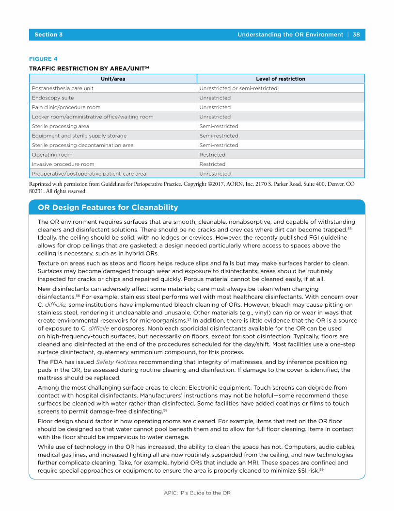
Section 3 Understanding the OR Environment | 38
APIC: IP’s Guide to the OR
FIGURE 4
TRAFFIC RESTRICTION BY AREA/UNIT54
Unit/area Level of restriction
Postanesthesia care unit Unrestricted or semi-restricted
Endoscopy suite Unrestricted
Pain clinic/procedure room Unrestricted
Locker room/administrative office/waiting room Unrestricted
Sterile processing area Semi-restricted
Equipment and sterile supply storage Semi-restricted
Sterile processing decontamination area Semi-restricted
Operating room Restricted
Invasive procedure room Restricted
Preoperative/postoperative patient-care area Unrestricted
Reprinted with permission from Guidelines for Perioperative Practice. Copyright ©2017, AORN, Inc, 2170 S. Parker Road, Suite 400, Denver, CO 80231. All rights reserved.
OR Design Features for Cleanability
TheORenvironmentrequiressurfacesthataresmooth,cleanable,nonabsorptive,andcapableofwithstanding
cleanersanddisinfectantsolutions.Thereshouldbenocracksandcreviceswheredirtcanbecometrapped.55
Ideally,theceilingshouldbesolid,withnoledgesorcrevices.However,therecentlypublishedFGIguideline
allowsfordropceilingsthataregasketed;adesignneededparticularlywhereaccesstospacesabovethe
ceilingisnecessary,suchasinhybridORs.
Textureonareassuchasstepsandfloorshelpsreduceslipsandfallsbutmaymakesurfaceshardertoclean.
Surfacesmaybecomedamagedthroughwearandexposuretodisinfectants;areasshouldberoutinely
inspectedforcracksorchipsandrepairedquickly.Porousmaterialcannotbecleanedeasily,ifatall.
Newdisinfectantscanadverselyaffectsomematerials;caremustalwaysbetakenwhenchanging
disinfectants.56Forexample,stainlesssteelperformswellwithmosthealthcaredisinfectants.Withconcernover
C.difficile, someinstitutionshaveimplementedbleachcleaningofORs.However,bleachmaycausepittingon
stainlesssteel,renderingituncleanableandunusable.Othermaterials(e.g.,vinyl)canriporwearinwaysthat
createenvironmentalreservoirsformicroorganisms.57Inaddition,thereislittleevidencethattheORisasource
ofexposuretoC. difficileendospores.NonbleachsporicidaldisinfectantsavailablefortheORcanbeused
onhigh-frequency-touchsurfaces,butnecessarilyonfloors,exceptforspotdisinfection.Typically,floorsare
cleanedanddisinfectedattheendoftheproceduresscheduledfortheday/shift.Mostfacilitiesuseaone-step
surfacedisinfectant,quaternaryammoniumcompound,forthisprocess.
TheFDAhasissuedSafety Noticesrecommendingthatintegrityofmattresses,andbyinferencepositioning
padsintheOR,beassessedduringroutinecleaninganddisinfection.Ifdamagetothecoverisidentified,the
mattressshouldbereplaced.
Amongthemostchallengingsurfaceareastoclean:Electronicequipment.Touchscreenscandegradefrom
contactwithhospitaldisinfectants.Manufacturers’instructionsmaynotbehelpful—somerecommendthese
surfacesbecleanedwithwaterratherthandisinfected.Somefacilitieshaveaddedcoatingsorfilmstotouch
screenstopermitdamage-freedisinfecting.58
Floordesignshouldfactorinhowoperatingroomsarecleaned.Forexample,itemsthatrestontheORfloor
shouldbedesignedsothatwatercannotpoolbeneaththemandtoallowforfullfloorcleaning.Itemsincontact
withthefloorshouldbeimpervioustowaterdamage.
WhileuseoftechnologyintheORhasincreased,theabilitytocleanthespacehasnot.Computers,audiocables,
medicalgaslines,andincreasedlightingallarenowroutinelysuspendedfromtheceiling,andnewtechnologies
furthercomplicatecleaning.Take,forexample,hybridORsthatincludeanMRI.Thesespacesareconfinedand
requirespecialapproachesorequipmenttoensuretheareaisproperlycleanedtominimizeSSIrisk.59

Section 3 Understanding the OR Environment | 39
APIC: IP’s Guide to the OR APIC: IP’s Guide to the OR
IMPLEMENTATION STRATEGIES FOR THE IP
△IncorporateassessmentofenvironmentalfactorsinroutineORsitevisits.
△EnsureenvironmentalfactorsareincludedinSSIroot-causeanalysistemplate.
△Usecleaninggrids.
TOOLS AND RESOURCES
△FGI Guideline for OR Design 2018—FacilityGuidelinesInstitutepublicationwithdefinitionsofnewsurgicalspaces,sizes,andrequirementsforscrubsinksandhand-washingfacilities;availableforpurchase.https://bit.ly/2I8unTR
△Joint Interim Guidance: HVAC in the Operating Room and Sterile Processing Department—3-page2015guidancepreparedbyAmericanSocietyofHeating,RefrigeratingandAir-ConditioningEngineers(ASHRAE),theAmericanSocietyforHealthcareEngineering(ASHE),theAssociationfortheAdvancementofMedicalInstrumentation(AAMI),theAssociationforProfessionalsinInfectionControlandEpidemiology(APIC),theAssociationofperiOperativeRegisteredNurses(AORN),andtheFacilityGuidelinesInstitute(FGI).https://bit.ly/2IapVUu
REFERENCES
1. Spagnolo AM, Ottria G, Amicizia D, Perdelli F, Cristina ML. Operating theatre quality and prevention of surgical site infections. J Prev Med Hyg 2013 Sep;54(3):131-7.
2. Cristina M, et al. Operating room environment and surgical site infections in arthroplasty Procedures. J Prev Med Hyg 2016;57(3):E142-E148.
3. Mora M, Mahnert A, Koskinen K, et al. Microorganisms in confined habitats: microbial monitoring and control of intensive care units, operating rooms, cleanrooms and the international space station. Front Microb 2016;7:1573.
4. Burlingame B, Davidson J, Denholm B, Fearon M, Giarrizzo-Wilson S, Johnstone EM, Link T, Ogg MJ, Spruce L, Spry C, Van Wicklin SA, Wood A. Guideline for safe environment of care, part 2. In: Conner R, ed. AORN Guidelines for Perioperative Practice, Vol. 1. AORN website. doi:10.6015/ psrp.17.01.269. 2018. Accessed April 30, 2018.
5. Sehulster LM, Chinn RYW, Arduino MJ, Carpenter J, Donlan R, Ashford D, Besser R, Fields B, McNeil MM, Whitney C, Wong S, Juranek D, Cleveland J. Guidelines for environmental infection control in health-care facilities. Recommendations from CDC and the Healthcare Infection Control Practices Advisory Committee (HICPAC). Chicago IL; American Society for Healthcare Engineering/American Hospital Association; 2004.
6. Memarzadeh F. The environment of care and healthcare-associated infections: an engineering perspective. Chicago, IL: ASHE; 2011.
7. Ayliffe GA. Role of the environment of the operating suite in surgical wound infection. Rev Infect Dis 1991;13(Suppl 10):S800-4.
8. American National Standards Institute/ American Society of Heating, Refrigerating, and Air-Conditioning Engineers / American Society of Health Care Engineering. Standard 170-2013—Ventilation of health care facilities.
9. Facility Guidelines Institute. 2014 Guidelines for Design and Construction of Hospitals and Outpatient Facilities. Available from: https://www.fgiguidelines.org/guidelines/2014-fgi-guidelines/
10. Burlingame B, Davidson J, Denholm B, Fearon M, Giarrizzo-Wilson S, Johnstone EM, Link T, Ogg MJ, Spruce L, Spry C, Van Wicklin SA, Wood A. Guideline for safe environment of care, part 2. In: Conner R, ed. AORN Guidelines for Perioperative Practice, Vol. 1. AORN website. doi:10.6015/ psrp.17.01.269. 2018. Accessed April 30, 2018.
11. American National Standards Institute/Association for the Advancement of Medical Instrumentation. ST 79:2017—Comprehensive guide to steam sterilization and sterility assurance in health care facilities. ANSI website. 2017. http://my.aami.org/aamiresources/previewfiles/1709_ST79Preview.pdf. Accessed April 30, 2018.
12. Fridkin SK, Kremer FB, Bland LA, Padhye A, McNeil MM, Jarvis WR. Acremonium kiliense endophthalmitis that occurred after cataract extraction in an ambulatory surgical center and was traced to an environmental reservoir. Clin Infect Dis 1996 Feb;22(2):222-7
13. Williamson D, Howden B, Stinear, T. Mycobacterium chimaera spread from heating and cooling units in heart surgery. N Engl J Med 2017; 376: 600-602.
14. Spagnolo A, et al. Operating theatre quality and prevention of surgical site infections. J Prev Med Hyg 2013;54(3):131-137.

Section 3 Understanding the OR Environment | 40
APIC: IP’s Guide to the OR
15. American National Standards Institute/ American Society of Heating, Refrigerating, and Air-Conditioning Engineers / American Society of Health Care Engineering. Standard 170-2013—Ventilation of health care facilities.
16. Facility Guidelines Institute. 2014 Guidelines for Design and Construction of Hospitals and Outpatient Facilities. Available from: https://www.fgiguidelines.org/guidelines/2014-fgi-guidelines/
17. Evans RP. Current concepts for clean air and total joint arthroplasty: laminar airflow and ultraviolet radiation: a systematic review. Clin Orthop Related Res 2011;469(4):945-953.
18. Edmiston CE. Prosthetic device infections in surgery. In: Nichols, R.L. and Nyhus, L.M. (eds). Update Surgical Sepsis. Philadelphia: J.B. Lippincott Co.; 1993.
19. Hasanadka R, Seabrook GR, Edmiston CE. Vascular graft infections. In: Rello J, Vanes J, Kollef M, eds. Critical Care Infectious Diseases, 2nd ed. Boston: Kluwer Academic Publishers; 2001.
20. Edmiston CE, McBride A, Leaper D. Surgical site infections associated with microbial biofilms. In: Donelli C (ed). Biofilm-Based Healthcare-Associated Infections, Advances in Experimental Medicine and Biology series (AEMB), Berlin [Germany]: Springer; 2015.
21. Sawan SP, Manivannan G. In: Sawan SP, Manivannan G, eds. Antimicrobial/Anti-Infective Materials: Principles, Applications and Devices. Lancaster, LA: Technomic Publishing Company, Inc., 2000.
22. Petrova OE, Sauer K. Sticky situations: key components that control bacterial surface attachment. J Bacteriol 2012;194:2413-2425.
23. Zimmerli W, Trampuz A, Ochsner PE. Prosthetic-joint infections. N Eng J Med 2004; 35:1645–1654.
24. Gomez-Alonso, A et al. Study of the efficacy of Coated VICRYL Plus® Antibacterial suture in two animal models of general surgery. J Infect 2007 January;54:82-88.
25. Parvizi J, Barnes S, Shohat N, Edmiston CE. Environment of care: Is it time to reassess microbial contamination of the operating room air as a risk factor for surgical site infection in total joint arthroplasty? Am J Infect Control 2012;45:1267-1272
26. Gormley T, et al. Cost-benefit analysis of different air change rates in an operating room environment. Am J Infect Control 2017 Dec 1;45(12):1318-1323.
27. Gormley T, et al. Methodology for analyzing environmental quality indicators in a dynamic operating room environment. Am J Infect Control 2017 April 1;45(4):354-9.
28. Facility Guidelines Institute. 2014 Guidelines for Design and Construction of Hospitals and Outpatient Facilities. Available from: https://www.fgiguidelines.org/guidelines/2014-fgi-guidelines/
29. Memarzedeh F. Role of air changes per hour (ACH) in possible transmission of airborne infections. Build Simul 2012;5:15-28.
30. Mangram A, Horan T, Pearson M, Silver L, Jarvis W. Guideline for Prevention of Surgical Site Infection, 1999. Infect Control Hosp Epidemiol 1999;20(4):247-280. doi:10.1086/501620
31. Sehulster LM, Chinn RYW, Arduino MJ, Carpenter J, Donlan R, Ashford D, Besser R, Fields B, McNeil MM, Whitney C, Wong S, Juranek D, Cleveland J. Guidelines for environmental infection control in health-care facilities. Recommendations from CDC and the Healthcare Infection Control Practices Advisory Committee (HICPAC). Chicago IL; American Society for Healthcare Engineering/American Hospital Association; 2004
32. Lidwell OM. Clean air at operation and subsequent sepsis in the joint. Clin Orthop 1986;211:91-102
33. Pittet D, Ducel G. Infectious risk factors related to operating rooms. Infect Control Hosp Epidemiol 1994;15:456–462.
34. Perkins KM, Lawsin A, Hasan NA, et al. Notes from the Field. Mycobacterium chimaera contamination of heater-cooler devices used in cardiac surgery — United States. MMWR 2016;65:1117-1118. DOI: https://www.cdc.gov/mmwr/volumes/65/wr/mm6540a6.htm.
35. Sommerstein R, Rüegg C, Kohler P, Bloemberg G, Kuster SP, Sax H. Transmission of Mycobacterium chimaera from heater-cooler units during cardiac surgery despite an ultraclean air ventilation system. Emerg Infect Dis 2016 Jun;22(6)1008-1013.
36. Centers for Disease Control and Prevention. Guidelines for Preventing the Transmission of Mycobacterium tuberculosis in Health-Care Settings, 2005. MMWR 2005;54(No. RR-17)
37. Olmsted RN. Pilot study of directional airflow and containment of airborne particles in the size of Mycobacterium tuberculosis in an operating room. Am J Infect Control 2008;36(4):260-267
38. American National Standards Institute/ American Society of Heating, Refrigerating, and Air-Conditioning Engineers / American Society of Health Care Engineering. Standard 170-2013—Ventilation of health care facilities.
39. Memarzadeh F. The environment of care and healthcare-associated infections: an engineering perspective. Chicago, IL: ASHE; 2011.
40. American Society of Heating, Refrigerating and Air-Conditioning Engineers (ASHRAE), American Society for Healthcare Engineering (ASHE), Association for the Advancement of Medical Instrumentation (AAMI), Association for Professionals in Infection Control and Epidemiology (APIC), Association of periOperative Registered Nurses (AORN), Facility Guidelines Institute (FGI). Joint interim guidance: HVAC in the operating room and sterile processing department. September 21, 2015. http://apic.org/Resource_/TinyMceFileManager/Implementation_Guides/PosStat-Safety-HVAC-Interim-Guidance.pdf. Accessed April 30, 2018.
41. American National Standards Institute/ American Society of Heating, Refrigerating, and Air-Conditioning Engineers / American Society of Health Care Engineering. Standard 170-2013—Ventilation of health care facilities.
42. The Joint Commission. Temperature and Humidity - Monitoring Requirements for Sterile Supply Storage Areas. The Joint Commission website. n.d. https://www.jointcommission.org/standards_information/jcfaqdetails.aspx?StandardsFAQId=1498. Accessed April 30, 2018

Section 3 Understanding the OR Environment | 41
APIC: IP’s Guide to the OR APIC: IP’s Guide to the OR
43. Association of Surgical Technologists. Guidelines for best practices for humidity in the operating room. AST website. Approved April 10, 2015, revised June 2017. http://www.ast.org/uploadedFiles/Main_Site/Content/About_Us/ASTGuidelinesHumidityintheOR.pdf Accessed April 30, 2018.
44. American Society of Heating, Refrigerating and Air-Conditioning Engineers (ASHRAE), American Society for Healthcare Engineering (ASHE), Association for the Advancement of Medical Instrumentation (AAMI), Association for Professionals in Infection Control and Epidemiology (APIC), Association of periOperative Registered Nurses (AORN), Facility Guidelines Institute (FGI). Joint interim guidance: HVAC in the operating room and sterile processing department. September 21, 2015. http://apic.org/Resource_/TinyMceFileManager/Implementation_Guides/PosStat-Safety-HVAC-Interim-Guidance.pdf. Accessed April 30, 2018.
45. Mangram A, Horan T, Pearson M, Silver L, Jarvis W. Guideline for Prevention of Surgical Site Infection, 1999. Infect Control Hosp Epidemiol 1999;20(4):247-280. doi:10.1086/501620
46. Andersson, AE. Traffic flow in the operating room: an explorative and descriptive study on air quality during orthopedic trauma implant surgery. J Infect Control 2012;40(8):750-755.
47. Sastry S, Rahman R, Yassin MH. Cardiac implantable electronic device infection: from an infection prevention perspective. Adv Prev Med 2015:357087.
48. Lynch R, et al. Measurement of Foot Traffic in the Operating Room: Implications for Infection Control. Am J Med Qual 2009 January-February;(24-1):45-52
49. Edmiston CE Jr, Seabrook GR, Cambria RA, Brown KR, Lewis BD, Sommers JR, Krepel CJ, Wilson PJ, Sinski S, Towne JB. Molecular epidemiology of microbial contamination in the operating room environment: Is there a risk for infection? Surgery 2005 Oct;138(4):573-9.
50. Berríos-Torres S, et al. for the Healthcare Infection Control Practices Advisory Committee Centers for Disease Control and Prevention Guideline for the Prevention of Surgical Site Infection, 2017. JAMA Surg August 2017;152(8):784-791.
51. American College of Surgeons. Minimizing operating room traffic may improve patient safety by lowering rates of surgical site infections. ACS website. July 18, 2016. https://www.facs.org/media/press-releases/2016/nsqip-ssi-071816. Accessed April 30, 2018.
52. Abeles, SR, Myers F, El-Kareh R, Marmor, R, Kovryga Kornick, M, Gohil, SK, Cohen, SH, Mahr, N, Nichols, A, Garritson, S, Liu, C, Rubin, Z, Trout, A, Allyn, PR, Torriani, FJ. (2016, May). No Significant Correlation between OR Traffic and Surgical Site Infections in a Multicenter Study. Poster session presented at Society for Healthcare Epidemiology of America Conference, Atlanta, GA.
53. Spagnolo AM, Ottria G, Amicizia D, Perdelli F, Cristina ML. Operating theatre quality and prevention of surgical site infections. J Prev Med Hyg 2013 Sep;54(3):131-7.
54. Burlingame B, Davidson J, Denholm B, Fearon M, Giarrizzo-Wilson S, Johnstone EM, Link T, Ogg MJ, Spruce L, Spry C, Van Wicklin SA, Wood A. Guideline for safe environment of care, part 2. In: Conner R, ed. AORN Guidelines for Perioperative Practice, Vol. 1. AORN website. doi:10.6015/ psrp.17.01.269. 2018. Accessed April 30, 2018.
55. Lybert L. Porous and Non-Porous Hard Surfaces. Health Facilities Manag July 6, 2016.
56. Rutala WA, Weber DJ. Selection of the Ideal Disinfectant. Infect Cont Hosp Epidemiol 2014;35(7); 855-865.
57. Bradbury S, et al. Potential bloodborne pathogen exposure from occult mattress damage. Am J Infect Cont 2014;42(4);421-422.
58. Pal S, Juyal D, Adekhandi S, et al. Mobile phones: Reservoirs for the transmission of nosocomial pathogens. Adv Biomed Res 2015;4:144.
59. Mora M, Mahnert A, Koskinen K, et al. Microorganisms in confined habitats: microbial monitoring and control of intensive care units, operating rooms, cleanrooms and the international space station. Front Microb 2016;7:1573.

APIC: IP’s Guide to the OR
42
SECTION4
THE STERILE PROCESSING DEPARTMENT, HIGH-LEVEL DISINFECTION, AND STERILIZATION
The SPD Environment and Basics
The Sterile Processing department (SPD) is defined as a service within the hospital in which medical/surgical supplies and equipment, both sterile and nonsterile, are cleaned, prepared, processed, stored, and issued for patient care. The effectiveness of an SPD relies on expert execution of processes, facility design, resources (including equipment and personnel), education and training, quality control, and documentation of processes. Professional guidelines, including those from the Association of periOperative Registered Nurses (AORN) and the Association for the Advancement of Medical Instrumentation (AAMI), direct SPD protocols.1-5 The SPD leadership should have access to these guidelines, be familiar with their contents, and be able to speak to discrepancies between their facility’s process and these recommendations. Every facility should maintain policies for all elements of decontamination, disinfection, sterilization, and storage as performed within SPD.
All SPD staff should have easy access to manufacturer’s IFUs for every instrument processed as well as for machines, products, solutions, and chemicals used. Indeed, the importance of the IFUs cannot be overstated. When any questions arise, these documents should be consulted to ensure compliance. IF IFUs are
not followed, a risk assessment should be completed to justify the practice.
The major areas of the SPD are decontamination, high-level disinfecting (HLD), assembly/packaging, sterilization, and sterile storage. SPD should be considered a semi-restricted space. Surgical attire is recommended in all these areas, but personal protection equipment (PPE) is required only in the decontamination area, where exposure to blood and other potentially infectious materials (OPIM) is likely. In addition, jewelry on hands and wrists is prohibited.
Ideally, SPD areas should be separated via physical barriers. This may not be possible in all facilities, however. The highest-priority separation is that between the decontamination area and “clean” spaces (e.g., assembly/packaging, sterilization). Isolating the decontamination area decreases the chances for cross-contamination to other areas of the SPD. The traffic flow of items through SPD should be from contaminated areas to increasingly cleaner ones.
Surfaces in the department—including countertops, cabinets, floors, doors, walls, and ceilings—should be smooth, made of cleanable materials, and durable enough to withstand frequent use and application of disinfectant solutions. Lighting should be adequate to facilitate the attention to detail required for

Section 4 The Sterile Processing Department, High-Level Disinfection, and Sterilization | 43
APIC: IP’s Guide to the OR APIC: IP’s Guide to the OR
instrument reprocessing. Hand hygiene stations, with soap and water and waterless alcohol hand sanitizer, should be readily available throughout the department. All personnel should comply with the facility’s hand hygiene policies.
The AAMI and AORN guidelines provide clinical practice recommendations for SPD.1,3 ANSI/ASHRAE/
ASHE Standard 170-2013 provides guidance regarding HVAC parameters, including temperature and humidity for the various areas within the SPD. Facility Guidelines Institute (FGI) Guidelines for Design and Construction of Hospitals and Outpatient Facilities, 2014 provides standards to be used when constructing or completing major renovations in SPD.6-7
FIGURE 5
ATTIRE AND PPE REQUIREMENTS FOR THE STERILE PROCESSING DEPARTMENT5
Work Area Scrubs Head Covers Gloves* Gowns#Eye
Protection+
Masks or Face
Shields±
Decontamination X X X X X X
Preparation and Packaging X X
Sterilization Processing X X
Sterile Storage X X
*Gloves should be waterproof, general-purpose utility, or heavy-duty.#Gowns must be liquid-resistant with sleeves.+Eye protection includes goggles/eyeglasses with side shields or chin-length face shields.±Masks should be fluid-resistant.
Other protective equipment (such as shoe covers) may be worn as needed. The type and characteristics will depend on the task and degree of anticipated exposure.
Source: Occupational Safety & Health Administration. US Department of Labor. Toxic and hazardous substances. Appendix A: bloodborne pathogens. 29 CFR 1910.1030. Fed Regist 1991;56(235):64004-64182. Effective December 6, 1991. http://www.osha.gov/pls/oshaweb/owadisp.show_document?p_table=standards&p_id=10051 Accessed April 25, 2012
Reprinted with permission from Guideline for Sterilization. Copyright (c)2017, AORN, Inc, 2170 S. Parker Road, Suite 400, Denver, CO 80231. All rights reserved.
Point-of-Use Precleaning
Instruments should be cleaned and decontaminated as soon as possible after use. To prevent biofilm formation, preparation for decontamination of instruments should begin at the point of use. Point-of-use preparation for decontamination can be accomplished by removing gross soil, flushing or suctioning lumens, and immersion of instruments in sterile water. During the procedure, the scrub person should remove gross soil from instruments by wiping the surfaces with a sterile surgical sponge moistened with sterile water. Instruments that cannot be cleaned immediately should be treated with an instrument cleaner according to the device and the instrument cleaner manufacturers’ written IFU.
Biofilm can form on many types of surfaces, including medical and surgical instruments. The risk of formation is greatest for surfaces that are moist and in regular contact with water. Biofilm may contain multiple types of microorganisms, including bacteria, yeast and fungi. Biofilm is created as bacterial colonies grow, secreting a protective gel that is very difficult to penetrate with detergent and water. Bacteria within biofilms can be up to 1,000 times more resistant to cleaning agents than are their counterparts outside a biofilm colony. Consequently, it is easier to clean devices before biofilm forms.8 Biofilm development on endoscopes can result from failure to adequately wipe down the scope and/or thoroughly flush the internal channels with an
Section 4 The Sterile Processing Department, High-Level Disinfection, and Sterilization | 44
APIC: IP’s Guide to the OR
enzymatic detergent at the point of use; a failure to adequately brush all internal surfaces prior to HLD or sterilization; or internal surface defects within the lumens of the various scope channels.9-11 Such defects can be caused by wear and tear over time or be minute structural flaws emerging during manufacturing.12 Flaws in instrument design can increase risk of biofilm formation, as well. For example, cleaning the side-viewing duodenoscopes is challenging: A design flaw provides a place under the elevator mechanism or within the sheath housing the elevator cable where organic debris can be trapped. See Tools and Resources for the Food and Drug Administration guidance document on culturing and sampling of these scopes.
Contaminated instruments should be transported as soon as possible to the decontamination room of the SPD for reprocessing. Spray enzymatic products cannot be applied during a surgical case, but may be applied after the surgical case, and prior to transporting. Hinged instruments should be in an open position. Soiled instruments must be transported to the decontamination area in a closed container or enclosed transport cart. The container or cart must be:
△Leakproof
△Puncture resistant
△Large enough to contain all contents
△Labeled with a fluorescent orange or red label containing a biohazard legend
Both precleaning at point of use and thorough cleaning in SPD is critical before high-level disinfection or sterilization. If soiled material becomes dried onto the item, the process of soil removal becomes much more difficult. Precleaning at point of use is critical to preventing this, especially if the item will sit for more than 1 hour prior to comprehensive SPD cleaning. Include SPD staff in design of the precleaning process, so they are prepared to properly treat the instruments afterward.13-15
Decontamination
Decontamination refers to cleaning or removal of foreign material, such as soil, tissue, and blood.16 This process reduces the bioburden on an item prior to disinfection or sterilization. Decontamination is typically accomplished using water with detergents and/or enzymatic products. Manual decontamination has two essential components: friction and fluidics. Friction means rubbing or scrubbing the soiled area with a brush. Fluidics is the use of fluids under pressure to remove soil and debris from internal channels after brushing, or when the design does not allow the passage of a brush through a channel. It is important to note that neither of these processes ensures complete cleaning of intricate devices.
Automated cleaning is a good adjunct to manual cleaning. Two common types are ultrasonic cleaning, and washers or washer/disinfectors. Ultrasonic cleaning uses a process of cavitation to loosen soil, which can be particularly useful for delicate instruments. Automated washers and washer/disinfectors can be likened to a residential dishwasher, where large volumes of water and disinfectants are applied, often in conjunction with high-heat drying cycles.17,18
The correct flow of instruments and items within the decontamination area is critical to prevent cross-transmission. The area should have a dedicated entry door for contaminated instruments, separate from the door through which reprocessed instruments exit. Pass-through windows are commonly employed to make it easier to get manually cleaned instruments from the decontamination area or room into the assembly and packing area prior to sterilization. All doors and pass-through windows should remain closed when not in use. In addition, the ventilation system should be designed so that air flows into the decontamination area (i.e., negative pressure).
Personal protective equipment is required for much of the work done in the decontamination area. This should include a liquid-resistant gown with sleeves, utility gloves that extend beyond the gown sleeve,

Section 4 The Sterile Processing Department, High-Level Disinfection, and Sterilization | 45
APIC: IP’s Guide to the OR APIC: IP’s Guide to the OR
and a fluid-resistant mask and eye protection. Personal protective equipment should be readily available in the area, and there should be a dedicated place to put on and remove PPE. Convenient access to regular trash containers, regulated waste (blood and body substances) containers, and hampers for soiled linens (including reusable PPE) are also needed. Emergency eye wash and shower stations should be easily accessible within 10 seconds of contact with any hazardous chemical. Finally, care should be taken to prevent sharps injuries, and food and drink should not be kept in this area.19,20
Sinks in the decontamination area ideally have three sections: for soaking, cleaning, and rinsing. Sinks should be large enough to accommodate trays and baskets of instruments, and deep enough to allow enough water and/or detergent to cover the instruments. Evaluate the decontamination area for ergonomic quality: Look at height and placement of sinks, attached counters or workspaces, adequate space to perform the required tasks, and anti-fatigue flooring or mats.
Instrument/Item Prep
As instruments enter the SPD, additional pre-treatment (or other methods to ensure bioburden remains moist) is advisable if instruments will not be immediately cleaned. At the beginning of processing, instruments should be separated based on cleaning method (e.g., hand wash/delicate). Instruments should be disassembled as much as possible and jointed or channeled instruments should be opened. It is important to ensure that the solution used for presoaking and cleaning is approved by the device or instrument manufacturer. In addition, the solution manufacturer’s requirements should be followed, including dilution, temperature, and contact time. If an automatic dilution device is used, it should be routinely verified and calibrated. The solution should be clean before use and may require changing after every use. Thorough rinsing after presoaking and cleaning is also necessary to ensure removal of all solutions.
Any brushes that are used should be designed for that specific purpose. Instruments that require brushing (such as those with lumens) will have instructions on
how to obtain the correct size and type of brush. It is critical to obtain the correct brush for each item, and for SPD personnel to be able to speak to the process of determining which brush to use. Brushes should be checked for visible soil and damage after each use, and reusable brushes should be reprocessed and dried for storage.
If ultrasonic cleaning is used, it is important to ensure that gross soil is removed prior to placing instruments in the machine. The cleaning solution that is used should be labeled for ultrasonic use, and it should be changed after each load. As always, the manufacturer’s recommendations must be followed for proper loading of instruments and items, for daily cleaning of the machine, for performance verification, and for degassing of the machine.
When automated washers are used, it should be as the last step of the cleaning process in decontamination. Manufacturer’s IFUs must be followed, including how to load items (avoidance of overloading is critical), cycle selection, and how to maintain all working parts of the equipment, including any connectors. IPs should be mindful that water quality is an important issue in sterilizers and automated washers. Water can contain contaminants that are organic or inorganic, and microorganisms can survive and even multiply in water. Poor water quality can reduce the efficiency and life of equipment and can contribute to infections and toxic reactions.
Reusable containers, such as rigid container systems and transportation carts, should be regularly inspected for damage, including breaks in gaskets and malfunctioning latching mechanisms. Processing of rigid containers (for cleaning and disinfection) and transport carts should follow the manufacturer IFUs. In general, filters and filter holders, valves, and interior baskets should be removed and processed separately.
Following cleaning, items are typically rinsed in critical water (although each manufacturer’s IFU should be consulted). Items should be inspected for damage at the end of the cleaning process prior to assembly and packaging.21
Section 4 The Sterile Processing Department, High-Level Disinfection, and Sterilization | 46
APIC: IP’s Guide to the OR
Verification of Cleaning
Verification of cleaning starts with a visual inspection of the device for damage or remaining visible soil. Magnifiers can assist with this process. Methods for verification of soil removal, such as ATP testing or microbiologic sampling, may be used. If used, the processes for testing should be approved by the facility and rigorously followed. Verification of cleaning also includes monitoring of the parameters of the cleaning process. For example, automated washers and ultrasonic machines often provide a digital readout or printout. These records should be reviewed to ensure all parameters are met. In addition, the documentation should be maintained. There are a variety of additional verification methods, such as use of coupons contaminated with proteins, which should be used only with facility-approved testing procedures.
All mechanical equipment should be properly maintained according to manufacturers’ IFUs, along with documentation of routine preventive, and other types of maintenance. After non-routine maintenance, cleaning verification should be completed prior to use.16-20
Assembly/Packaging
In the assembly and packaging area, clean instruments should be thoroughly dried, inspected, and assembled into packaging for sterilization. If there is concern about the cleanliness of any item, or contamination of an item occurs in this area, the item should be returned to decontamination for repeat cleaning. The ventilation of this and the other clean areas (i.e., sterilization, sterile storage) should be designed so that air flows out of the overall area (positive pressure), although pressure gradients between these areas can be neutral.
Assembly requires that each instrument be positioned in the packaging to allow the sterilant to contact all surfaces. Every item and instrument should be opened, unlatched, and disassembled to allow contact to all surfaces. Items that could hold water should be placed on their sides, and heavy items should be placed below lighter and more delicate ones. In the end, a set should not weigh more than 25 pounds. Packages should be
well labeled. Labeling should be done with a material designed for this use, and labels should remain securely attached to the package. The label should indicate: sterilizer used, the cycle number, load control record number, date of sterilization, and the worker responsible for prep and packaging. The outside of opaque container systems should have a short description of the contents, and a full description of all contents should be attainable via the load control record number. The expiration date or statement of event-based expiration should also be visible on the external container.
Policies and procedures regarding selection and use of packaging systems should include:
△Pre-purchase evaluation
△Assembly of devices within packaging systems
△Weight limitations
△Product testing
△Labeling
△Placement and positioning of packages within the sterilizer
△Storage requirements pre- and post-sterilization
△Shelf life
△Use of internal and external sterilization monitors
△Wrapping requirements and technique
△Use of peel pouches
△Maintenance of packaging materials, peel pouches, rigid container systems, and heat sealers
Instruments and other items that are prepared for sterilization must be packaged so that their sterility can be maintained to the point of use. Materials and techniques used for packaging must allow the sterilant to contact the device during the sterilization process as well as protect the device from contamination during storage and handling before it is used. Types of packaging may include textiles or nonwoven disposable materials, pouch packaging, or rigid container systems.
Section 4 The Sterile Processing Department, High-Level Disinfection, and Sterilization | 47
APIC: IP’s Guide to the OR APIC: IP’s Guide to the OR
The package should be closed in a way that any tampering would be obvious. Closures should not inhibit sterilization or package shape and integrity. Paper-plastic pouches (commonly called “peel packs”) should be used for small items that are used as singular items (and generally not as part of a surgical procedure). Ensure the items fit completely inside the pouch, and that the seals of the pouch are smoothly closed. Double pouching may be allowable, based on the pouch manufacturer’s IFUs.
Odd-shape or heavy items may require items to be packaged by wrapping the item with the textile or nonwoven disposable material referenced above. However, this method should be minimized whenever possible due to the risk of integrity-failure associated with wrapped instruments. Every wrapper should be examined for holes and tears prior to use. Reusable wraps are permissible, assuming they are well maintained and laundered between uses. The material should completely cover the item enclosed, and it should be applied to allow sterility maintenance while opening (e.g., envelope-fold or square-fold technique). The packaging material selected must also permit the device to be removed aseptically. 16-20
Sterilization
Critical items that enter sterile tissue or the vascular system must be sterile. Examples of items that require sterilization include surgical instruments and ultrasound probes used in sterile body cavities.
There are three common sterilization processes:
△Heat: Steam sterilization is the preferred method.
△Gas: Items that are heat sensitive are typically sterilized by ethylene oxide or hydrogen peroxide vapor.
△Liquid Chemical: The same chemicals used in high-level disinfection can render an item sterile, but the contact time is much longer (3 to 12 hours). The disadvantage of the liquid chemical sterilization process is that items cannot be wrapped to maintain sterility (and must be rinsed).19,20
Loading and Running the Sterilizer
Items should be carefully loaded into the sterilizer. The sterilizer manufacturer’s IFU for loading must be closely followed. Different sterilizers require different load configurations and have specific specifications. Peel pouches should stand on edge with the paper of one package facing the plastic of the next. Holding racks are recommended to assist with proper loading of peel packs. Instrument sets should be placed level. Rigid containers should be placed lower on the rack than wrapped items or peel pouches. All items and packs loaded in the sterilizer at the same time should require the same cycle parameters. There are several “standard” cycles recommended by manufacturers. All SPD staff should be able to speak to the sterilizer parameters as well as the need for any adjustments among the parameters used. It’s important to recognize that some items require nonstandard or extended cycles. Validation of these types of cycles is extremely difficult, and use of these items should be evaluated by the facility using a risk assessment.19,20
Immediate-Use Steam Sterilization (IUSS)
Immediate-use steam sterilization (IUSS), historically referred to as flashing or flash sterilization, should be used only when no other method is possible or when there is insufficient time to process by the preferred wrapped or container method intended for terminal sterilization. It can be used, for instance, if there is a hole in a wrap immediately prior to surgery for nonimplantable instruments, if specialized equipment is needed for back-to-back surgical cases, or if a specialized instrument is dropped during an operation. It should not serve as a substitute for sufficient instrument inventory. Instruments sterilized through IUSS cannot be stored for future use; they must be used immediately. Terminal sterilization of reusable surgical instrumentation is always the preferred method of preparing instruments for surgery.
The IUSS method can be used under the following conditions:
△Device manufacturer’s written instructions include instructions for IUSS
Section 4 The Sterile Processing Department, High-Level Disinfection, and Sterilization | 48
APIC: IP’s Guide to the OR
△Manufacturer’s written instructions for cleaning, cycle type, exposure times, temperature settings, and drying times (if recommended) are available and followed
△Items are placed in a containment device that has been validated for IUSS, cleared by the FDA for this purpose, and works in a manner that allows steam to contact all instrument surfaces
△Containment device manufacturer’s written IFUs are followed
△Measures are taken to prevent contamination during transfer to the sterile field
△Items subjected to IUSS are used immediately and not stored for later use or held from one procedure to another
IUSS is the process of sterilization of a surgical instrument through the use of a shorter, abbreviated cycle, with no drying time before the instrument is used for patient care. The instrument must first be cleaned according to IFUs, then placed in an autoclave designated for IUSS. The same monitoring and documentation of sterilization processes are required no matter where the sterilizer is located.
IUSS cycles last generally either 3 or 10 minutes, depending on the nature of the device being sterilized and the type of cycle indicated, with minimal or no dry time and no cool down. This makes the cycle-time shorter than that for wrapped or terminally sterilized items. Time constraints may result in pressure on personnel to eliminate or modify one or more steps in the cleaning and sterilization process, which could result in retained contamination and associated SSI risk.
IUSS should not be performed on the following devices:22
△Implants, except in a documented emergency situation when no other option is available
△Postprocedure decontamination of instruments used on patients who may have Creutzfeldt-Jakob disease or similar disorders
△Devices or loads that have not been validated with the specific cycle employed
△Devices that are sold sterile and intended for single-use only22
Cooling and Recordkeeping
After sterilization, items should be cooled to room temperature before they are handled, in order to prevent condensation. ANSI/AAMI ST79, Comprehensive Guide to Steam Sterilization and Sterility Assurance in Health Care Facilities, states that a 30-minute minimum cool-down time is recommended; however, it could require 2 hours or more for sterilized items to cool enough prior to being handled. Items should be cooled in the sterilizer (or on the cart used in the sterilizer), and not transferred to other racks or shelves for cooling. There should be a dedicated area to allow for holding of items away from traffic, air conditioning, and cooling vents.19,20
After cooling items, inspect them to ensure that packaging is intact, external indicators of sterilization are there, and no moisture is present. If there is any failure on these points or other concern with integrity of packaging, then the items should be returned to decontamination and the process restarted. Recordkeeping is necessary for every run of the sterilizer. Recordkeeping should include the sterilization date, sterilizer number, the cycle number (sequential number of times the sterilizer is run in a day), the load control record numbers (which point to a detailed list of all items run in that sterilization cycle), the exposure time and temperature (if printouts are provided by the sterilizer, they should be used for this documentation), the name of the person operating the sterilizer, and the results of any testing. Results of testing can include biological testing, Bowie-Dick testing, and response of a chemical indicator (CI) placed in a process-challenge device. If there are negative or questionable chemical indicator results, these should also be recorded. These records should then be retained for the duration established by facility policy.

Section 4 The Sterile Processing Department, High-Level Disinfection, and Sterilization | 49
APIC: IP’s Guide to the OR APIC: IP’s Guide to the OR
All sterilization equipment should be properly maintained according to IFUs, with documentation of routine preventive and all other types of maintenance. After nonroutine maintenance, sterilization verification should be completed prior to instrument use.19,20
Sterile Storage
As its name indicates, the sterile storage area is used for storage of clean and sterile items. The sterile storage area should be designed to protect items from damage and contamination. Open or wire shelving is appropriate, unless the area has high levels of traffic, in which case closed carts are preferable. Items should be at least 8 to 10 inches from the floor, and all storage shelves should have solid bottoms to protect items from dust and debris. Items should be at least 18 inches below ceiling or sprinkler heads and 2 inches from exterior walls.
In general, there should be plenty of space and minimal stacking to ensure package integrity. (Refer to the wrapping manufacturer’s instructions for allowance of stacking of wrapped instruments.) Sterile items should not be stored near water sources.
External shipping containers and web-based cardboard should be considered contaminated. Contents should be removed before transporting to the clean areas of the SPD or OR.
Shelf life of sterilized items is event-related: It is dependent on packaging material, storage conditions, transport, and handling. An event must occur to compromise package content sterility. Events that may compromise the sterility of a package include:
△Multiple instances of handling that leads to seal breakage or loss of package integrity
△Moisture penetration
△Exposure to airborne contaminants
Use the FIFO technique: First In, First Out. “Older” items should be used prior to those freshly sterilized. Even though event-related sterility principles are used, it is still necessary to ensure stock is arranged on shelves so that the oldest stock is used first. These stock rotation
principles should apply to all areas where sterile stock is stored, as well as to commercial products as well.
In addition, extreme care should be taken to maintain package integrity whenever an item has been handled or transported. Containers should remain level when stored, handled, and transported.16,17
Monitoring the Sterilization Process
Physical, chemical, and biological monitors should be used to oversee the sterilization processes. They should be used for routine load release, routine sterilizer efficacy testing, and sterilizer qualification testing (e.g., after installation, relocation, malfunction, major repair, and sterilization process failure). These monitors are used in a variety of ways and places and will be evaluated by sterilization personnel at the end of the process as well as by any healthcare workers who open the item at the point of use. If there is concern about any process monitor, the item should not be used, management should be notified, and an investigation should be undertaken to remedy any problems.
The first type of monitor is a physical monitor. Physical monitors include a sterilizer’s time, temperature, and pressure. The sterilizer will produce this information, usually via printout or electronic display. The sterilizer operator should verify that the physical monitor indicates that the necessary parameters have been met and should retain the cycle information as required by the facility policy.
The second type of monitor is a chemical indicator. A chemical indicator is a device used to monitor the attainment of one or more critical parameters required for sterilization. A characteristic color or other visual change indicates a defined level of exposure, based on the type of the chemical indicator used.22
There are six types of chemical indicators. Type 1 is a process indicator, which is placed on the outside of individual containers or packs and provides a visual indication that an item has been exposed to the sterilization process. The Bowie-Dick is an example of a Type 2 indicator, which tests for air leaks, adequate air

Section 4 The Sterile Processing Department, High-Level Disinfection, and Sterilization | 50
APIC: IP’s Guide to the OR
removal, and steam penetration in steam sterilization. Bowie-Dick tests should be run daily for all steam sterilizers. Types 3 and 4 test only some of the process variables and have little use in healthcare. Type 5, also referred to as an integrating indicator, is designed to react to all of the critical process variables. Type 6 is an emulating indicator, and it reacts to all critical process variables for specific sterilization processes.
A type 5 or 6 chemical indicator should be placed inside each container or package. Multiple indicators may be placed in packages, depending on the manufacturer’s IFUs, and evaluated by the staff member who opens the item at the point of use. These indicators are specific to the sterilization cycle and should be selected based on the cycle required for the items being sterilized. A type 5 or 6 indicator will also be placed within a process-control device that is evaluated at the end of the sterilization load (prior to releasing the items for use).
The final type of monitor is a biological indicator. Biological indicators are composed of very hearty bacterial spores and provide clear indication that the sterilization cycle was adequate to kill organisms. Biological indicator should be selected specific to the sterilization method:
△Steam sterilizers: Geobacillus stearothermophilus
△Ethylene oxide sterilizers: Bacillus atrophaeus
△Low-temperature hydrogen peroxide gas plasma sterilizers: Geobacillus stearothermophilus
△Hydrogen-peroxide vapor sterilizer: Geobacillus stearothermophilus
△Ozone sterilizers: Geobacillus stearothermophilus
△Dry-heat sterilizers: Bacillus atrophaeus
The biological indicator is placed within a process-control device and incubated after the sterilization cycle is complete. After the manufacturer’s recommended incubation time has passed, the indicator is evaluated for growth (showing whether the sterilization process has succeeded or failed). A biological indicator should be used for routine load release, routine sterilizer efficacy monitoring, sterilizer qualification testing, and periodic product quality assurance testing. A biological indicator should be run with every sterilization load that contains an implant. When used for implant loads, the biological incubation should be completed prior to use of the implant on a patient.23-27
Loaned Instrument Considerations
Sterility assurance related to loaned instruments should begin at the point at which the healthcare organization assumes responsibility for the items. If an item was reprocessed at another institution prior to delivery, the item should be considered contaminated and delivered directly to the SPD for decontamination and sterilization. A deadline for receipt by SPD should be made—usually 24 to 48 hours prior to the case—to allow proper processing. Late receipt of instruments should not result in IUSS. A record of loaned instruments should be kept, and the facility should have access to the IFUs for each loaned instrument.28-32

Section 4 The Sterile Processing Department, High-Level Disinfection, and Sterilization | 51
APIC: IP’s Guide to the OR APIC: IP’s Guide to the OR
Special Considerations
Toxic Anterior Segment Syndrome (TASS)
TASSisarareophthalmologicconditionassociatedwithinadequateorimproperinstrumentprocessing.Itis
causedwhenachemicalagententerstheanteriorchamberoftheeye,causinganinflammatoryreaction.Itis
commonlyassociatedwithcataractsurgery,butitcanoccursubsequenttoanyanterioreyesurgery.33,34IfTASS
isnottreatedproperly,theinflammatoryresponsecanresultinseverevisualimpairment.Therootcauseof
TASShasbeenfoundtobeassociatedwithselectiveelementsofinstrumentreprocessingincluding:34
△Residualdetergentleftoninstruments
△Insufficientrinsingofinstruments
△Driedorganicdebrisandresiduesofophthalmicviscoelasticmaterialremainingoninstruments
△Insufficientlydriedlumens
TheAmericanSocietyofCataractandRefractiveSurgeryhaspublishedRecommendedPracticesforCleaning
andSterilizingIntraocularSurgeryInstruments.35
Creutzfeldt-Jakob Disease (CJD)
CJDisarare,degenerative,fatalbraindisordercausedbyanabnormal“infectious”protein.Itbelongstoa
familyofhumanandanimaldiseasesknownasthetransmissiblespongiformencephalopathies(TSEs).Prions
aredifficulttokillandareresistanttocommonstrategiesfordisinfectionandsterilization.Instrumentsthat
havecomeincontactwithselectivetissuesfromhigh-risksurgicalpatients(e.g.,brain,spinalcord,andeyes)
requiresegregation(quarantine)fromothersurgicalinstrumentsandmustbeadequatelydecontaminatedto
reducethetransmissionoftheseparticlestootherpatients.Neurosurgicalinstrumentsposethegreatestrisk,
becausetheycanbecontaminatedwithalargeburdenofinfectiousproteins.36TheWorldHealthOrganization
(WHO)haspublishedguidelinesfortheinactivationoftheseresistantparticles.Ifdisposableinstrumentsare
notused,WHOrecommendsoneofthefollowingreprocessingprotocols:37
△Immersetheinstrumentinacoveredpancontaining1Nsodiumhydroxide(NaOH)andheatinagravitydisplacementautoclaveat121°Cfor30minutes;clean;rinseinwater;andsubjecttoroutinesterilization.
△Immersetheinstrumentin1NNaOHorsodiumhypochlorite(20,000ppmavailablechlorine)for1hour;transferinstrumentstowater;heatinagravitydisplacementautoclaveat121°Cfor1hour;clean;andsubjecttoroutinesterilization.
△Immersetheinstrumentin1NNaOHorsodiumhypochlorite(20,000ppmavailablechlorine)for1hour;removeandrinseinwater;transfertoanopenpanandheatinagravitydisplacement(121°C)orporousload(134°C)autoclavefor1hour;clean;andsubjecttoroutinesterilization.
△Heat-sensitivereusableinstrumentsandsurfacesthatcomeincontactwithhigh-infectivityandlow-infectivitytissuescanbedecontaminatedbyfloodingwithorsoakingin2NNaOHorundilutedsodiumhypochloritefor1hourandthenrinsedwithwater.Tominimizedryingoftissuesandbodyfluidsonthesurgicalinstruments,thedevicesshouldbekeptmoistuntilcleanedanddecontaminated.
InsuspectedorconfirmedcasesofCJD,alldisposableinstruments,materials,andwastesthatcomeincontact
withhigh-infectivitytissues(e.g.,brain,spinalcord,andeyes)andlow-infectivitytissues(e.g.,cerebrospinal
fluid,kidneys,liver,lungs,lymphnodes,spleen,andplacenta)ofsuspectedorconfirmedTSEpatientsshould
bedisposedofbyincineration. Managementofsuspectedorconfirmedcasesrequiresclosecommunication
amongthesurgeon,ORnursing,andtheIPtoensurethatacomprehensiveCJDpolicyisdevelopedandin
placeformanagingsuspectedorconfirmedcases.

Section 4 The Sterile Processing Department, High-Level Disinfection, and Sterilization | 52
APIC: IP’s Guide to the OR
High-Level Disinfection
The high-level disinfection (HLD) process involves elimination of all microorganisms (mycobacteria, fungi, viruses, and bacteria) in or on a device, with the exception of high numbers of bacterial spores. Devices processed by HLD include endoscopes (GI, bronchoscopes, and laryngoscopes), laryngoscope blades,
endocavitary probes (rectal and vaginal), respiratory therapy, and anesthesia equipment. SPD staff may be responsible for performing high-level disinfection on medical devices used in hospital or outpatient clinics, emergency rooms, OB/GYN clinics, operating rooms, special procedures rooms, and endoscopy suites. HLD can involve heat, pasteurization (65° to 77°C for 30 minutes), or more typically, chemical disinfection.
FIGURE 6
HIGH-LEVEL DISINFECTANTS AND KILL TIMES
Thefollowingtablelistscommonchemicalsusedforhigh-leveldisinfectionalongwithestimatedkilltimes.38,39
HLD Method Duration
>2 percent glutaraldehyde 20 to 45 minutes
0.55 percent ortho-phthalaldehyde (OPA) 12 minutes
1.12 percent GLUT/1.93 percent phenol 20 minutes
7.5 percent hydrogen peroxide (HP) 30 minutes
7.35 percent HP/0.23 percent peracetic acid (PA) 15 minutes
1.0 percent HP/0.08 percent PA 25 minutes
400 to 500 ppm chlorine 10 minutes
2.0 percent HP 8 minutes
3.4 percent GLUT/26 percent isopropanol 10 minutes
While these chemical disinfectants have proven to be effective for HLD, several of the selective agents have been documented to cause skin, eye, and respiratory irritation; contact dermatitis; and anaphylactic irritation in bladder cancer patients. All require the use of personal protective equipment (PPE)i.e., long-sleeve impervious gown, protective eyewear, mask, gloves, and cap).
The determination of level of disinfection or sterilization required for any instrument is determined by the Spaulding criteria, a classification scheme using the degree of infection risk and the intended use of the object to determine how items should be reprocessed. Spaulding criteria uses three categories:
△Critical: Items that enter sterile tissue, which must be sterilized
△Semicritical: Items that contact mucous membranes, which require at least high-level disinfection
△Noncritical: Items that come in contact with intact skin, which require low-level disinfection38,39
As noncritical (low-level) disinfection rarely applies to items used in the OR, it will not be discussed further in this guide.
Flexible Endoscope Reprocessing
Flexible endoscopes, categorized as semi-critical items, require high-level disinfection at a minimum. They can be processed manually or by using an automated endoscope reprocessor (AER).40 For manual reprocessing at point of use, the flexible endoscope must be immediately wiped down and placed in a basin with detergent or enzymatic cleaner, which must then be suctioned through all channels until the fluid is clear. The scope must then be placed in a covered container and transported to the reprocessing area. In the reprocessing area, the endoscope must be leak-tested; manually brushed on all internal and external surfaces; all channels flushed with detergent cleaning fluid; thoroughly rinsed in clean water; then immersed with accessories in high-level disinfectant, to fill all channels

Section 4 The Sterile Processing Department, High-Level Disinfection, and Sterilization | 53
APIC: IP’s Guide to the OR APIC: IP’s Guide to the OR
and ports. Following HLD the scope and channels must be thoroughly rinsed, purged with air, flushed with 70 to 80 percent alcohol (refer to manufacturer IFU), and stored in a vertical position in a well-ventilated and dust-free area/cabinet. Wet or inadequately dried endoscopes pose an increased risk of contamination and have been associated with transmission of waterborne microorganisms and infection; it is important that scopes be thoroughly dried.
The 2018 Guidelines for Perioperative Practice recommend that scopes be stored in a drying cabinet. Drying cabinets for endoscope storage are defined as a cabinet equipped with a drying system that continually blows pressurized, HEPA-filtered air through the endoscope’s channels and while also circulating HEPA-filtered air around the exterior of the endoscopes. If a drying cabinet is not available, the guidelines recommend that scopes be stored in a cabinet that is HEPA filtered with positive pressure.
The distal end of the endoscope should not touch the floor or bottom of the storage cabinet, nor should the distal end rest on a soft surface such as a towel or synthetic “bumper” since these surfaces can become contaminated over time.41,42
For automated endoscope reprocessing, the scope must also be wiped down at point of use with an enzymatic detergent, which must then be suctioned through all channels, after which the scope must be transported in closed container to the cleaning/decontamination area in the SPD. Prior to placing in the AER, the scope must be leak-tested (some AERs include this step during reprocessing) and manually brushed and cleaned. The HLD solution concentration must be checked and recorded, and the scope and accessories connected to the AER. Following the HLD cycle, the scope channel(s) must be flushed with alcohol and compressed air, and the scope must be stored in a vertical position to facilitate drying and prevent tip contamination. The automated reprocessing steps must follow all manufacturer’s recommendations, including validating HLD concentration and cycle time.
When transporting a “dirty” scope from the procedure area, the instrument must be safely moved to the cleaning
area/decontamination room in a closed container and labeled as biohazardous. An advantage of automated reprocessing of endoscopes is reduced operator exposure to chemicals, and reduced operator error. However, there have been several documented cross transmissions of multidrug-resistant pathogens following reprocessing with AER.43,44 These failures in HLD were found to be related to improper manual cleaning, improper use of AER, inability or failure to clean the suction channel, and mechanical or design flaws in the flexible endoscope.
Impact of Biofilms on High-Level Disinfection of Flexible Endoscopes
As with other instruments, biofilm formation is a challenge in the reprocessing of flexible endoscopes.45
Biofilm development on or in flexible endoscopes can be associated with the following:
△Failure to adequately wipe down the scope and/or thoroughly flush the channels with an enzymatic detergent at the point of use
△Failure to adequate brush all internal and external surfaces prior to HLD
△Development of internal surface defects within the lumens of the various scope channels
△Design flaws in the endoscope46-48
Biofilm-forming Gram-negative bacteria such as Pseudomonas aeruginosa and other multidrug-resistant pathogens have been implicated in biliary tract infection following ERCP.49 Endoscope contamination involving mature biofilms are difficult to resolve, even following high-level disinfection, and can result in patient infection. Consequently, enhanced guidelines emphasize the importance of adequate precleaning at point of use, incorporating the following steps:50
△Prepare fresh cleaning solution for each endoscope
△Wipe exterior surfaces with a soft, lint-free cloth or sponge saturated with the solution
△Suction the cleaning solution through the channels of the device by placing the distal end in the cleaning solution

Section 4 The Sterile Processing Department, High-Level Disinfection, and Sterilization | 54
APIC: IP’s Guide to the OR
△Alternate suctioning the channels with cleaning solution and air, finishing with air
△Discard the cleaning solution and cloth or sponge after each endoscope
△Visually inspect the endoscope for damage
This visual inspection is critical to determine if any residual debris remains on the surface of the device or if damage has occurred to the external or internal surfaces. A recent study documented microbial recovery from 60 percent of endoscopes following use of automated reprocessing machines. In the study, visual inspection via borescope documented internal surface irregularities that could sequester microbial contamination following HLD. It was the authors’ opinion that rigorous reprocessing practices may not be adequate to ensure that “patient-ready” endoscopes are free of residual contamination, underscoring the need for both visual inspections and a biochemical-based verification test to ensure more comprehensive reprocessing.51 However, the use of a borescope is not currently recommended by any regulatory or professional guidelines. It is important to recognize that endoscopic equipment has a finite life expectancy, and continuous wear and tear will compromise the internal/external integrity of the device. In light of current outbreaks, a systematic maintenance schedule and/or strategy for replacement of aged equipment should be implemented to decrease the risk of endoscope-associated bacterial transmission.52
Length of Storage of Endoscopes
How long can endoscopes be stored after reprocessing? Insufficient data exist. Concern about possible microbial colonization has led to various recommendations for reprocessing intervals among institutions, with many as short as 5 days. The Multisociety Guideline on Reprocessing Flexible GI Endoscopes (https://www.ncbi.nlm.nih.gov/pubmed/28069113) notes that this is an unresolved issue: “Although reuse of endoscopes within 10 to 14 days of high-level disinfection appears to be
safe, data are insufficient to provide a maximal duration for use of appropriately cleaned, reprocessed, dried, and stored flexible endoscopes.”
A 2015 article in Gastrointestinal Endoscopy aimed to demonstrate whether duodenoscopes, gastroscopes, and colonoscopes could be stored for as long as 21 days without microbial colonization by potential pathogens. In light of conflicting recommendations, organizations should do a thorough risk assessment including careful consideration of turnover and risk of potential contamination. Through this process, many organizations have set specific time frames, usually reprocessing within 14 to 21 days. Scopes should be labeled with the date they underwent HLD.
Other Semi-Critical Devices
Vaginal and rectal (endocavitary) probes and laryngoscopes are designated as semi- critical devices, requiring at least high-level disinfection between patient uses. The precleaning, cleaning and disinfecting process must be performed per the device manufacturer’s recommendations and current published guidelines.53 Human papilloma virus (HPV) has been reported to contaminate both endovaginal and endorectal ultrasound probes.54,55
Guidelines from the American Institute of Ultrasound in Medicine recommends the following steps to reduce the risk of microbial cross-transmission between patients: single-use sheath or condom applied to the probe prior to use; thorough cleaning after each use; HLD after cleaning.56 When determining which method and solution to use for HLD, it is important to note that current studies conclude that glutaraldehyde and orthophthaladehyde (OPA) demonstrate little virucidal activity against HPV types 16 and 18.57 Sonication of the probe in an H2O2 system has been demonstrated to provide strong virucidal activity resulting in a 5.2 to 7.4 log10 reduction in viral loads for HPV types 16 and 18. Furthermore, a sonicated H2O2 system provides HLD of both the probe and probe handle.57

Section 4 The Sterile Processing Department, High-Level Disinfection, and Sterilization | 55
APIC: IP’s Guide to the OR APIC: IP’s Guide to the OR
IMPLEMENTATION STRATEGIES FOR THE IP
△ReviewandbecomefamiliarwithSPDpoliciesandrelatedprofessionalguidelines(AORN,AAMI).
△ReviewandbecomefamiliarwithANSI/ASHRAE/ASHEStandard170-2013regardingHVAC,temperature,andhumidityparametersforSPD.
△ReviewandbecomefamiliarwithFGIguidelinesfordesignofSPD.
△RequestSPDmanager’sinputduringtheannualIPCriskassessmentprocessrelativetoSPDprocesses,anyrecalls,and/orissuesrelatedtoinadequateinventoryorinstrumentreprocessing.
△UseachecklisttohelpguidesitevisitstotheSPDandotherareaswhereinstrumentsusedforinvasiveproceduresarereprocessed;reviewandbefamiliarwiththeseprocessestosupportbestpracticesduringsitevisits.AsampleendoscopyreviewchecklistisinToolsandResourcesforthissection.
△CollaboratewithperioperativeleadershiptoadvocateforminimizingIUSSbyreviewingreasonsforuseofIUSSandpromotingadequateinventoryofsurgicalinstrumentationwiththesupportoftheInfectionControlCommittee.
△Promotemeasuresthathelpensurethatallpersonnelthatusereusablemedicaldevicesandthatinstrumentsadheretothefacility’sprotocolsforreprocessingandhandlingtheseitems.
TOOLS AND RESOURCES
△CDC Guideline for Disinfection and Sterilization in Healthcare Facilities—Linkto161-pageprintdocument,summary,andupdates.https://bit.ly/2FcAEHI
△Point-of-Use Pre-Cleaning Surgical Instruments—Step-by-stepinstructionsandreferencesonpropercareandhandlingofinstrumentsanddeviceswhichmustbetransportedforsterilizationorhigh-leveldisinfection,fromHighlandHospital,UniversityofRochesterMedicalCenter.Provided by APIC https://bit.ly/2I81AyJ
△“AORN Guideline Summary: Cleaning and Care of Surgical Instruments”—Comprehensivelistofrecommendationsandkeypointsforactionforcleaningandcareofsurgicalinstruments.Provided by APIChttps://bit.ly/2IeFKWk
△“AORN Guideline Summary: Processing Flexible Endoscopes”—Recommendationsandkeypointsforperioperative,endoscopy,andsterile-processingpersonnelforprocessingalltypesofreusableflexibleendoscopesandaccessories.Provided by APIC https://bit.ly/2IbJmwx
△SPD Tracer Observation Form—TheJointCommissionkeysurveyassessmenttoolfortheSterileProcessingDepartment,withquestionsoninstruments,practices,training.Canalsobeadapted.https://bit.ly/2KcP0Lg
△FDA guidance on scopes—DuodenoscopeSurveillanceSampling&Culturing:ReducingtheRiskofInfection;57-pagereportwithprotocols;2018.Provided by APIC https://bit.ly/2G8w6m5
△BSWH Endoscopy Audit Tool—Excelscoringtoolforhigh-leveldisinfectionofGIendoscopes,createdbyTeriMauldin,MBA,BSN,RN,ACM,CIC,Manager,InfectionPreventionandControlCentralizedSurveillance,BaylorScott&WhiteHealth.Provided by APIC https://bit.ly/2rGVfjN

Section 4 The Sterile Processing Department, High-Level Disinfection, and Sterilization | 56
APIC: IP’s Guide to the OR
REFERENCES
1. American National Standards Institute/Association for the Advancement of Medical Instrumentation. ST 79:2017—Comprehensive guide to steam sterilization and sterility assurance in health care facilities. ANSI website. 2017. http://my.aami.org/aamiresources/previewfiles/1709_ST79Preview.pdf. Accessed April 30, 2018.
2. American National Standards Institute/Association for the Advancement of Medical Instrumentation. ST 58:2013—Chemical sterilization and high-level disinfection in health care facilities. ANSI website. 2013. http://my.aami.org/aamiresources/previewfiles/ST58_1308_preview.pdf Accessed April 30, 2018.
3. Rutala WA, Weber DJ. Healthcare Infection Control Practice Advisory Committee (HICPAC). Guidelines for disinfection and sterilization in healthcare facilities. CDC website. 2008. https://www.cdc.gov/infectioncontrol/pdf/guidelines/disinfection-guidelines.pdf Accessed April 30, 2018.
4. Association of periOperative Registered Nurses. Guideline for cleaning and care of surgical instruments. AORN website. January 2017. http://www.aornstandards.org/content/1/SEC36.extract. Accessed April 30, 2018.
5. Association of periOperative Registered Nurses. Guideline for sterilization. AORN website. January 2017. http://www.aornstandards.org/content/1/SEC38.extract. Accessed April 30, 2018.
6. American National Standards Institute, American Society of Heating, Refrigerating and Air-Conditioning Engineers, American Society for Healthcare Engineering. ANSI website. 2017.
7. Facilities Guideline Institute. Guidelines for design and construction of hospitals and outpatient facilities, 2018. FGI website. 2018. Available at: https://www.fgiguidelines.org/guidelines/purchase-the-guidelines/read-only-copy/
8. Roberts CG. The role of biofilms in reprocessing medical devices. Am J Infect Control 2013;41:S77-80.
9. Epstein L, Hunter JC, Arwady MA, Stein L, Gribogiannis M, et al. New Delhi metallo-beta- lactamase producing carbapenem-resistant Escherichia coli associated with exposure to duodenoscopes. JAMA 2014;312:1447-1455.
10. Smith Z, Oh Y, Saeian K, Edmiston CE, Khan AH, Massey BT, Dua KS. Transmission of carbapenem resistant Enterobacteriaceae during ERCP: time to revisit the current reprocessing guidelines. In Press: Gastrointestinal Endoscopy 2015;81:1041-5.
11. Ofstead CL, Wetzler HP, Heymann OL, Johnson EA, Eiland JE, Shaw MJ. Longitudinal assessment of reprocessing effectiveness for colonoscopes and gastroscopes: results of visual inspections, biochemical markers, and microbial cultures. A, J Infect Control 2017;45:e26-e33.
12. Kenters N, Huijskens EGW, Meier C, Voss A. Infectious diseases linked to cross- contamination of flexible endoscopes. Endoscopy Int Open 2015;3(4):E259- E265.
13. American National Standards Institute/Association for the Advancement of Medical Instrumentation. ST 58:2013—Chemical sterilization and high-level disinfection in health care facilities. ANSI website. 2013. http://my.aami.org/aamiresources/previewfiles/ST58_1308_preview.pdf Accessed April 30, 2018.
14. Rutala WA, Weber DJ. Healthcare Infection Control Practice Advisory Committee (HICPAC). Guidelines for disinfection and sterilization in healthcare facilities. CDC website. 2008. https://www.cdc.gov/infectioncontrol/pdf/guidelines/disinfection-guidelines.pdf Accessed April 30, 2018.
15. Association of periOperative Registered Nurses. Guideline for cleaning and care of surgical instruments. AORN website. January 2017. http://www.aornstandards.org/content/1/SEC36.extract. Accessed April 30, 2018.
16. Jinadatha C, Bridges A. Cleaning, Disinfection, and Sterilization. In: APIC Text Online, 2018. http://text.apic.org/toc/basic- principles-of-infection-prevention-practice/cleaning-disinfection-and-sterilization. Accessed April 2018. Subscription required.
17. Rutala WA, Weber DJ. Healthcare Infection Control Practice Advisory Committee (HICPAC). Guidelines for disinfection and sterilization in healthcare facilities. CDC website. 2008. https://www.cdc.gov/infectioncontrol/pdf/guidelines/ disinfection-guidelines.pdf Accessed April 30, 2018.
18. Association of periOperative Registered Nurses. Guideline for cleaning and care of surgical instruments. In: AORN. Guidelines for Perioperative Practice. Denver, CO: AORN, Inc.; 2017. p. 815-850.
19. American National Standards Institute/Association for the Advancement of Medical Instrumentation. ST 79:2017—Comprehensive guide to steam sterilization and sterility assurance in health care facilities. ANSI website. 2017. http://my.aami.org/aamiresources/previewfiles/1709_ST79Preview.pdf. Accessed April 30, 2018.
20. American National Standards Institute/Association for the Advancement of Medical Instrumentation. ST 58:2013—Chemical sterilization and high-level disinfection in health care facilities. ANSI website. 2013. http://my.aami.org/aamiresources/previewfiles/ST58_1308_preview.pdf Accessed April 30, 2018.
21. Association for the Advancement of Medical Instrumentation. Technical information report 34: water for the reprocessing of medical devices. Atlanta, GA: Association for the Advancement of Medical Instrumentation; 2014.
22. Association of PeriOperative Registered Nurses. AORN guideline for sterilization and guideline for packaging. In: Guidelines for Perioperative Practice. Denver, CO: AORN, Inc.; 2017. p. 865-892.
23. American National Standards Institute/Association for the Advancement of Medical Instrumentation. ST 79:2017—Comprehensive guide to steam sterilization and sterility assurance in health care facilities. ANSI website. 2017. http://my.aami.org/aamiresources/previewfiles/1709_ST79Preview.pdf. Accessed April 30, 2018.

Section 4 The Sterile Processing Department, High-Level Disinfection, and Sterilization | 57
APIC: IP’s Guide to the OR APIC: IP’s Guide to the OR
24. American National Standards Institute/Association for the Advancement of Medical Instrumentation. ST 58:2013—Chemical sterilization and high-level disinfection in health care facilities. ANSI website. 2013. http://my.aami.org/aamiresources/previewfiles/ST58_1308_preview.pdf Accessed April 30, 2018.
25. Rutala WA, Weber DJ. Disinfection and sterilization: An overview. Am J Infect Control 2013; 41:S2-S5
26. Association of periOperative Registered Nurses. AORN Guideline for Cleaning and Care of Surgical Instruments. In: Guidelines for Perioperative Practice. Denver, CO: AORN, Inc.; 2017. p. 815-850.
27. Association of PeriOperative Registered Nurses. AORN guideline for sterilization. In: Guidelines for Perioperative Practice. Denver, CO: AORN, Inc.; 2017. p. 865-892.
28. American National Standards Institute/Association for the Advancement of Medical Instrumentation. ST 79:2017—Comprehensive guide to steam sterilization and sterility assurance in health care facilities. ANSI website. 2017. http://my.aami.org/aamiresources/previewfiles/1709_ST79Preview.pdf. Accessed April 30, 2018.
29. American National Standards Institute/Association for the Advancement of Medical Instrumentation. ST 58:2013—Chemical sterilization and high-level disinfection in health care facilities. ANSI website. 2013. http://my.aami.org/aamiresources/previewfiles/ST58_1308_preview.pdf Accessed April 30, 2018.
30. Rutala WA, Weber DJ. Healthcare Infection Control Practice Advisory Committee (HICPAC). Guidelines for disinfection and sterilization in healthcare facilities. CDC website. 2008. https://www.cdc.gov/infectioncontrol/pdf/guidelines/ disinfection-guidelines.pdf Accessed April 30, 2018.
31. Association of PeriOperative Registered Nurses. AORN guideline for cleaning and care of surgical instruments. In: Guidelines for Perioperative Practice. Denver, CO: AORN, Inc.; 2017. p. 815-850.
32. Association of PeriOperative Registered Nurses. AORN guideline for sterilization. In: Guidelines for Perioperative Practice. Denver, CO: AORN, Inc.; 2017. p. 865-892.
33. Cetinkaya S, Dadaci Z, Aksoy H, Acir NO, Yener HI, Kadioglu E. Toxic anterior-segment syndrome (TASS). Clinical Ophthalmology (Auckland, NZ). 2014;8:2065-2069.
34. Johnston J. Toxic anterior segment syndrome—more than sterility meets the eye. AORN J 2006 Dec;84(6):969-84.
35. American Society of Cataract and Refractive Surgery, American Society of Ophthalmic Registered Nurses. Recommended practices for cleaning and sterilizing intraocular surgery instruments. J Cataract Refract Surg 2007;33:1095-1100.
36. Belay ED, Blase J, Sehulster LM, Maddox RA, Schonberger LB. Management of neurosurgical instruments and patients exposed to Creutzfeldt-Jakob Disease. Infection control and hospital epidemiology. 2013;34(12):1272-1280.
37. World Health Organization. WHO infection control guidelines for transmissible spongiform encephalopathies: Report of a WHO consultation, Geneva, Switzerland, March 23-26, 1999. http://www.who.int/csr/resources/publications/bse/whocdscsraph2003.pdf. Accessed April 30, 2018.
38. Jinadatha C, Bridges A. Cleaning, Disinfection, and Sterilization. In: APIC Text Online, 2018. http://text.apic.org/toc/basic- principles-of-infection-prevention-practice/cleaning-disinfection-and-sterilization. Accessed April 2018. Subscription required.
39. Rutala WA, Weber DJ. Healthcare Infection Control Practice Advisory Committee (HICPAC). Guidelines for disinfection and sterilization in healthcare facilities. CDC website. 2008. https://www.cdc.gov/infectioncontrol/pdf/guidelines/ disinfection-guidelines.pdf Accessed April 30, 2018.
40. Rutala WA, Weber DJ. Healthcare Infection Control Practice Advisory Committee (HICPAC). Guidelines for disinfection and sterilization in healthcare facilities. CDC website. 2008. https://www.cdc.gov/infectioncontrol/pdf/guidelines/ disinfection-guidelines.pdf Accessed April 30, 2018.
41. Society of Gastroenterology Nurses and Associates. Standards of infection prevention in reprocessing flexible endoscopes. Chicago, IL: SGNA; 2015. p. 1-31.
42. Association of PeriOperative Registered Nurses. AORN guideline for processing flexible endoscopes. In: Guidelines for Perioperative Practice. Denver, CO: AORN, Inc.; 2017. p. 717-800.
43. Epstein L, Hunter JC, Arwady MA, Stein L, Gribogiannis M, et al. New Delhi metallo-beta-lactamase producing carbapenem-resistant Escherichia coli associated with exposure to duodenoscopes. JAMA 2014;312:1447-1455.
44. Smith Z, Oh Y, Saeian K, Edmiston CE, Khan AH, Massey BT, Dua KS. Transmission of carbapenem resistant Enterobacteriaceae during ERCP: time to revisit the current reprocessing guidelines. In Press: Gastrointest Endoscopy 2015;81:1041-5.
45. Roberts CG. The role of biofilms in reprocessing medical devices. Am J Infect Control 2013;41:S77-80.
46. Putnam K. Preventing biofilm formation in flexible endoscopes. AORN J 2016;103:7-8.
47. Smith Z, Oh Y, Saeian K, Edmiston CE, Khan AH, Massey BT, Dua KS. Transmission of carbapenem resistant Enterobacteriaceae during ERCP: time to revisit the current reprocessing guidelines. In Press: Gastrointest Endoscopy 2015;81:1041-5.
48. Ofstead CL, Wetzler HP, Heymann OL, Johnson EA, Eiland JE, Shaw MJ. Longitudinal assessment of reprocessing effectiveness for colonoscopes and gastroscopes: Results of visual inspections, biochemical markers, and microbial cultures. A, J Infect Control 2017;45:e26-e33.
49. Nelson DB, Muscarella LF. Current issues in endoscope reprocessing and infection control during gastrointestinal endoscopy. World J of Gastroenterol 2006;12(25):3953-3964.

Section 4 The Sterile Processing Department, High-Level Disinfection, and Sterilization | 58
APIC: IP’s Guide to the OR
50. Centers for Disease Control and Prevention. Interim protocol for healthcare facilities regarding surveillance for bacterial contamination of duodenoscopes after reprocessing. Atlanta, GA: Department of Health and Human Services; 2015. p. 1-5.
51. Qiu L, Zhou Z, Liu Q, Ni Y, Zhao F, Cheng H. Investigating the failure of repeated standard cleaning and disinfection of a Pseudomonas aeruginosa-infected pancreatic and biliary endoscope. Am J Infect Control 2015;43:e43-e46.
52. Smith Z, Oh Y, Saeian K, Edmiston CE, Khan AH, Massey BT, Dua KS. Transmission of carbapenem resistant Enterobacteriaceae during ERCP: time to revisit the current reprocessing guidelines. In Press: Gastrointest Endoscopy 2015;81:1041-5.
53. Rutala WA, Weber DJ. Healthcare Infection Control Practice Advisory Committee (HICPAC). Guidelines for disinfection and sterilization in healthcare facilities. CDC website. 2008. https://www.cdc.gov/infectioncontrol/pdf/guidelines/ disinfection-guidelines.pdf Accessed April 30, 2018.
54. Casalegno JS, Carval KLB, Eibach D, Valdeyron ML, Lamblin G, Jacquemoud H, et al. High risk HPV contamination of endocavity vaginal ultrasound probes: an underestimated route of nosocomial infection? PLOS one 2012;7:e48137.
55. Combs CA, Fishman A. A proposal to reduce the risk of transmission of human papilloma virus via transvaginal ultrasound. Am J Obstet Gynecol 2016;215:63-67.
56. American Institute of Ultrasound Medicine. Guidelines for cleaning and preparing external- and internal-use ultrasound probes between patients, safe handling, and use of ultrasound coupling gel. AIUM website. May 16, 2017. http://www.aium.org/officialStatements/57. Accessed April 30, 2018.
57. Ryndock E, Robison R, Meyers C. Susceptibility of HPV17 and 18 to high-level disinfectants indicated for semi-critical ultrasound probes. J Med Virology 2016;88:1076-1080.

APIC: IP’s Guide to the OR APIC: IP’s Guide to the OR
59
SECTION5
EPIDEMIOLOGY AND PATHOGENESIS OF SURGICAL SITE INFECTIONS
The Scope and Cost of SSIs
In the United States, surgical site infections (SSIs) are a common postoperative complication associated with significant morbidity and mortality. A multi-state point-prevalence survey published in 2014 ranked SSI as the number one healthcare-associated infection (HAI) occurring in U.S. acute care facilities.1 Approximately 160,000 to 300,000 SSIs occur annually in the United States.2 It is estimated that there is a 2- to 11-fold increased risk of mortality associated with SSIs. This risk is modulated by the selective surgical procedure; preoperative, intra-operative, and postoperative care processes provided; and comorbidity patient risk factors. The national costs incurred are significant: SSIs now rank as the costliest of all HAIs. Indeed, the annual fiscal burden has been estimated at between $4 billion and $10 billion.3
It has been suggested that 60 percent of SSIs could be prevented if practitioners and healthcare institutions consistently employed all evidence-based infection prevention practices.4 In 2015, the Hospital-Acquired Condition (HAC) Reduction Program, which was mandated by the Affordable Care Act, required that Centers for Medicare & Medicaid Services (CMS) reduce payments by 1 percent for hospitals that rank among the lowest-performing 25 percent with regard to HACs. In 2016, SSIs after abdominal hysterectomies and colon-related surgical procedures were added to the list of HACs, and there is an expectation that SSIs after other surgical procedures will be added to
the program in future years. As a result, a failure to mitigate SSI risk will have a significant impact on future hospital reimbursements.5
Procedure-Specific Risks
The National Healthcare Safety Network (NHSN) calculates the surgical site infection standardized infection ratio (SSI SIR) for facilities that enroll in NHSN as acute care or critical access hospitals. Three SSI SIR models are available for inpatient adult procedures and two models for inpatient pediatric procedures.6 Studies support the continual enhancement of procedure-specific risk adjustment to make more effective inter-hospital comparisons.
In one study, for example, the overall SSI rate for colon procedures was 19.2 percent, but specific procedures ranged from 7.6 percent (lap colectomy) to 52.5 percent (open colectomy with or without other bowel surgery plus colostomy).7 In addition, the study showed that colon surgeries combined with other surgeries had a higher infection rate (29.3 percent) compared to the overall rate. Moreover, teaching hospitals have been found to perform a disproportionately greater number of surgical procedure types.7
Although it has long been known that emergency procedures are associated with a higher SSI rate than scheduled surgeries, one study suggests that seven types of procedures represent the majority of costs and patient harms (including SSI) among all emergency general surgery (EGS) procedures.8 The study examined

Section 5 Epidemiology and Pathogenesis of Surgical Site Infections | 60
APIC: IP’s Guide to the OR
a large sample of EGS procedures, representing approximately 500,000 cases per year. The authors concluded that partial colectomy, small-bowel resection, cholecystectomy, operative management of peptic ulcer disease, lysis of peritoneal adhesions, appendectomy, and laparotomy make up 80 percent of all EGS procedures, 80.3 percent of deaths, 78.9 percent of complications, and 80.2 percent of inpatient costs nationwide.
There is no separate NHSN category for robotic surgical procedures at the present time. The NHSN SSI module
directs that “robotic assistance is considered equivalent to use of a scope for NHSN SSI surveillance.” However, it is difficult to understand how the two can be considered in the same category. Although robot-assisted surgery has evolved over the years for the purpose of providing a less invasive and safer approach to a number of surgical procedures, there are mixed reviews re safety. Studies offer a variety of conclusions, including that there is not enough evidence of benefits to support its significantly higher costs.9
FIGURE 7
SSI RATES FOR OPEN AND ROBOTIC PROCEDURES
Astudythatanalyzedoutcomesfrom273robot-assistedproceduresfounda5.9percentSSIrate.10Thestudyalso
foundthatthepatientswithSSIshadalongerdurationofprocedurethanthosewithoutanSSI.Thestudycompared
nationallyreportedSSIsafteropenprocedurestoSSIsafterroboticprocedures.Itconcludedthattheincreased
incidenceofSSIsaftersometypesofrobot-assistedsurgeriescomparedwithratesaftertraditionalopensurgeries
mayberelatedtothelearningcurveassociatedwithuseoftherobot.10
Open SSI rate Robotic SSI rate
Prostate and GU 0.85/100 procedures 5.74/100 procedures
GYN 1.72/100 procedures 10.00/100 procedures
COLO 5.88/100 procedures 33.33/100 procedures
Herniorrhaphy 1.62/100 procedures 37.50/100 procedures
Primary Organisms Associated with SSIs
Most sources agree that the most common organisms found to cause SSIs include Staphylococcus aureus, coagulase-negative staphylococci, Pseudomonas, Enterococcus spp., and Escherichia coli.11,12 Some studies suggest pathogen specificity based on procedure. For instance, one recent study retrospectively analyzed more than 900 head and facial plastic surgery SSI cases and found that the majority of causative organisms were Pseudomonas aeruginosa and Klebsiella pneumoniae
(54 percent).13 Meanwhile, a retrospective cohort comparative study of 389 instrumented spine surgery procedures concluded that Pseudomonas was the most common organism causing SSIs.14 Possible reservoirs for these organisms include the skin, nose, and hair of patients and surgical team members, a contaminated environment, contaminated instruments and equipment (e.g., ortho-pneumatic tourniquets), and bowel contamination. Introduction of the organisms can occur during surgery or after surgery before the wound has healed.
Section 5 Epidemiology and Pathogenesis of Surgical Site Infections | 61
APIC: IP’s Guide to the OR APIC: IP’s Guide to the OR
IMPLEMENTATION STRATEGIES FOR THE IP
△Compareyourfacility/organizationSSIpreventionpracticeswithTable1;meetwithyourperioperativenurseexecutivetosharethecomparisonandevidencesupportinganypracticesnotcurrentlyinplace.SuggestthatyouscheduleameetingwiththemedicaldirectoroftheORtopresentthesameopportunityforimprovementinSSIprevention.
△OffertoleadamultidisciplinaryPerioperativeTracerbasedonanactualorfictionalizedSSIcaseduringastaffmeetingorothertime.SeeToolsandResources:Operating Room Tracer Observation Form.
△KnowtheepidemiologyofSSIsinyourorganizationandprovideinformationonthistotheORpersonnelandsurgeons(i.e.,identifyandcollect/reportdataonorganismsassociatedwithSSIsinyourfacility).
TOOLS AND RESOURCES
△CDC/NHSN Classifications for SSI Diagram—Visualaidusingcross-sectionofabdominalwalltohelpdetermineSSIclassification.Provided by APIChttps://bit.ly/2I8ghlq
△Article on procedure-specific SSI incidence—“Procedure-specificSurgicalSiteIncidenceVarieswidelywithinCertainNationalHealthcareSafetyNetworkSurgicalGroups,”SaeedMJ,DubberkeER,FraserVJ,OlsenMA.Procedure-specificsurgicalsiteinfectionincidencevarieswidelywithincertainNationalHealthcareSafetyNetworksurgerygroups.AmJInfectControl2015June;43(6):617-23.doi:10.1016/j.ajic.2015.02.012. Provided by APIC https://bit.ly/2I65ZlI
△Operating Room Tracer Observation Form—TheJointCommissionskeysurveyassessmenttooltocheckORequipmentuseandstorage,attire,space,andprocesses.Provided by APIC https://bit.ly/2wyjvJL
△NHSN procedure manual—37-pagemoduleonhandlinganSSIevent;2018.Provided by APIChttps://bit.ly/2rA9IwQ
REFERENCES
1. Magill SS, Edwards JR, Bamberg W, et al. Multistate point-prevalence survey of health care–associated infections. NEJM 2014;370(13):1198-1208. doi:10.1056/NEJMoa1306801.
2. Anderson DJ, Podgorny K, and Berrios-Torres SI, et al. Strategies to prevent surgical site infections in acute care hospitals: 2014 update. Infect Control Hosp Epidemiol 2014; 35:605-627.
3. Jenks PJ, Laurent M, McQuarry S, Watkins R. Clinical and economic burden of surgical site infection (SSI) and predicted financial consequences of elimination of SSI from an English hospital. J Hosp Infect 2014 Jan;86(1):24-33.
4. Berríos-Torres, S et al. for the Healthcare Infection Control Practices Advisory Committee. Centers for Disease Control and Prevention guideline for the prevention of surgical site infection, 2017. JAMA Surg August 2017;152(8):784-791.
5. Kandilov A, Coomer N, Dalton K. The impact of hospital-acquired conditions on medicare program payments. Medicare Medicaid Res Rev 2014; 4(4): mmrr2014-004-04- a01.
6. Centers of Disease Control and Prevention. NHSN procedure manual, surgical site infection event. CDC website. January 2018. https://www.cdc.gov/nhsn/pdfs/pscmanual/9pscssicurrent.pdf. Accessed April 30, 2018.
7. Saeed MJ, Dubberke ER, Fraser VJ, Olsen MA. Procedure-specific surgical site infection incidence varies widely within certain national healthcare safety network surgery groups. Am J Infect Control. 2015;43:617-623.
8. John W. Scott, MD, MPH; Olubode A. Olufajo, MD, MPH; Gabriel A. Brat, MD; John A. Rose, MD, MPH; Cheryl K. Zogg, MSPH, MHS; Adil H. Haider, MD, MPH; Ali Salim, MD; Joaquim M. Havens, MD. Use of national burden to define operative emergency general surgery. JAMA Surg 2016;151(6):e160480.
9. Broholm M, Onsberg Hansen I, Rosenberg J. Limited evidence for robot-assisted surgery: a systematic review and meta-analysis of randomized controlled trials. Surg Laparosc Endosc Percutan Tech 2016 Apr;26(2):117-23.

Section 5 Epidemiology and Pathogenesis of Surgical Site Infections | 62
APIC: IP’s Guide to the OR
10. Hermsen ED, Hinze T, Sayles H, Sholtz L, Rupp ME. Incidence of surgical site infection associated with robotic surgery. Infect Control Hosp Epidemiol 2010 Aug;31(8):822-7.
11. Owens CD, Stoessel K. Surgical site infections: epidemiology, microbiology and prevention. J Hosp Infect 2008 Nov;70 Suppl 2:3-10.
12. Johns Hopkins Medicine. Health library: Surgical site infections. Johns Hopkins Medicine website. n.d. http://www.hopkinsmedicine.org/healthlibrary/conditions/surgical_care/surgical_site_infections_134,144/. Accessed April 30, 2018.
13. Wang G, Zhang S. The risk factors, etiology, and drug resistance of infection after plastic surgery, and corresponding measures. Minerva Chir 2017 Feb 23.
14. Hey HW, Thiam DW, Koh ZS, Thambiah JS, Kumar N, Lau LL, Liu KG, Wong HK. Is intraoperative local vancomycin powder the answer to surgical site infections in spine surgery? Spine (Phila Pa 1976) 2017 Feb 15;42(4):267-274.

APIC: IP’s Guide to the OR APIC: IP’s Guide to the OR
63
SECTION6
SSI SURVEILLANCE: DEFINITIONS, METHODS, OUTCOMES, AND REPORTING
Surgical site infection (SSI) surveillance is a critical component of any infection prevention and control program. Whether SSI surveillance is manual or automated, its goal is to quantify surgical infection risk, assess outcomes in patients undergoing specific operations, and identify and implement measures to reduce risk of infection. This information is typically shared on a regular basis with stakeholders—including surgeons, perioperative nurses, and executives—as a means of assessing the effectiveness of infection prevention efforts, as well as to identify opportunities for improvement.
SSI Definitions
The Centers for Disease Control and Prevention (CDC) has developed and maintains the surveillance definition for SSI events, protocols for reporting SSI events to the National Healthcare Safety Network (NHSN), and training materials for enrolled NHSN users.1 IPs can face challenges in collecting procedure denominator data and SSI event data if surveillance is performed manually. Fortunately, as electronic medical records are increasingly used in place of paper records, automated electronic capture of denominator data, as well as screening for infections, can be accomplished using “homegrown” tools (e.g., Excel) or infection-surveillance software.
SSI Wound Class
Surgical wound classification (also called the wound class) is an assessment of the degree of contamination of a surgical wound and is intended to assist in stratifying the patient’s risk for developing an infection. Wounds are categorized in one of four classes:
△Class I, Clean: An uninfected operative wound in which no inflammation is encountered and the respiratory, alimentary, genital, or uninfected urinary tracts are not entered. In addition, clean wounds are primarily closed and, if necessary, drained with closed drainage. Most operative incisional wounds that follow nonpenetrating (blunt) trauma should be included in this category.
△Class II, Clean-Contaminated: Operative wounds in which the respiratory, alimentary, genital, or urinary tracts are entered under controlled conditions and without unusual contamination. Operations involving the biliary tract, appendix, vagina, and oropharynx are included in this category, provided no evidence of infection or major break in technique is encountered.
△Class III, Contaminated: Open, fresh, accidental wounds. This category also includes operations with major breaks in sterile technique (e.g., open cardiac massage) or gross spillage from the gastrointestinal tract and incisions in which acute, nonpurulent

Section 6 SSI Surveillance: Definitions, Methods, Outcomes, and Reporting | 64
APIC: IP’s Guide to the OR
inflammation is encountered, including necrotic tissue without evidence of purulent drainage (e.g., dry gangrene).
△Class IV, Dirty or Infected: Includes old traumatic wounds with retained devitalized tissue and those that involve existing clinical infection or perforated viscera. This definition suggests that the organisms causing postoperative infection were present in the operative field before the operation.2,3
The literature has demonstrated that there are interprofessional variations in wound classification. One study of 11 pediatric facilities found that documentation of wound classification by registered nurse (RN) circulators and surgeons was in agreement for only 56 percent of procedures. The researchers found that nurses underclassified wound classification in 84 percent of the procedures.2 A second study, of pediatric general surgery procedures, found similar results when comparing wound classifications by RN circulators, surgeons, and National Surgical Quality Improvement Program (NSQIP) reviewers.3 The researchers observed 52 percent agreement among the classifications, and RN circulators were most likely to underclassify the wounds.
Perioperative personnel must validate the wound classification data by comparing the SSI denominator data source (often the RN circulator’s documentation on the intra-operative record through an electronic health record) with the surgeon’s postoperative note. The reliability of the wound classification documentation may be improved by intra-operative use of a standardized decision tool, with the perioperative nurse assigning the wound classification at the end of the procedure in consultation with the surgeon and including the wound classification in the surgical debrief to facilitate team communication.
Duration of Surgical Procedure
The duration of the operative procedure is defined by the Association of Anesthesia Clinical Directors (AACD) as the interval in hours and minutes between the procedure/surgery start time and the procedure/surgery finish time.
△Procedure/Surgery Start Time: Time when the procedure is begun (e.g., incision for a surgical procedure).
△Procedure/Surgery Finish: Time when all instrument and sponge counts are completed and verified as correct, all postoperative radiologic studies to be done in the operating room are complete, wound is closed and all dressings and drains are secure, and the physicians/surgeons have completed all procedure-related activities on the patient. Perioperative personnel entering data for these times in the data source should follow the AACD definitions. This is typically done by the RN circulator documenting the times in the intra-operative record. Although the AACD defines the start times for positioning and prepping the patient for the procedure, the AACD definitions do not clearly address whether injection of local anesthesia prior to the surgical procedure is included in this definition. Because of this, the RN circulator may document the start time as the injection of local anesthesia before the incision has been made. The end of the surgical procedure is a busy time for the RN circulator, which makes real-time documentation challenging. Documenting surgery start-times earlier than the actual incision time and surgery finish-times later may falsely lengthen the time that a wound was open, which may reduce the accuracy of SSI risk adjustment.

Section 6 SSI Surveillance: Definitions, Methods, Outcomes, and Reporting | 65
APIC: IP’s Guide to the OR APIC: IP’s Guide to the OR
Classification of Wound Closure
Classificationofthetypeofsurgicalwoundclosurehasbeenthetopicofoneofthemostfrequentlyasked
questionsregardingSSIeventprotocol.Woundclosureisclassifiedaseitherprimaryornonprimary:
△Primary closure: Closureoftheskinlayerduringtheoriginalsurgery,regardlessofthepresenceofwires,wicks,drains,orotherdevicesorobjectsextrudingthroughtheincision.Thiscategoryincludessurgerieswheretheskinisclosedbyanymeans.Ifanyportionoftheincisionisclosedattheskinlevel,byanymanner,adesignationofprimaryclosureshouldbeassignedtothesurgery.
△Nonprimary closure: Closureofthesurgicalwoundinawaythatleavestheskinlayercompletelyopenfollowingsurgery.Forsurgerieswithnonprimaryclosure,thedeep-tissuelayersmaybeclosedbysomemeans(withtheskinlevelleftopen),orboththedeepandsuperficiallayersmaybeleftcompletelyopen.Anexampleofasurgerywithnonprimaryclosurewouldbealaparotomyinwhichtheincisionwasclosedtothelevelofthedeep-tissuelayers(sometimescalledfasciallayersordeepfascia)buttheskinlevelwasleftopen.Anotherexamplewouldbeanopenabdominalcaseinwhichtheabdomenisleftcompletelyopenaftersurgery.Woundswithnonprimaryclosuremayormaynotbedescribedas“packed”withgauzeoranothermaterialandmayormaynotbecoveredwithplasticdressing,woundvacuum,orothersyntheticdevicesormaterials.2,3
Wound-closuredocumentationischallengingandrequirescollaborationamongtheperioperativeteam,who
arevisualizingthewound.Theelectronichealthrecordmayneedtobemodifiedtocapturethisdataelement
inafieldthatcanbeabstractedforthedenominatordatareport.ProvidingavisualtooltotheperioperativeRN
documentingthisdataelementmayfacilitatethecorrectclassificationofwound-closuretype.
FIGURE 8
WOUND-CLOSURE TYPES
Avisualcanhelpinclassificationofwound-closuretypes,afrequenttopicinSSIeventprotocol.
Source: Centers for Disease Control and Prevention, National Healthcare Safety Network. Surveillance for surgical site infection (SSI) event resources and procedure-associated module. CDC website. January 2018. https://www.cdc.gov/nhsn/acute-care-hospital/ssi/index.html and http://www.cdc.gov/nhsn/pdfs/pscmanual/9pscssicurrent.pdf.

Section 6 SSI Surveillance: Definitions, Methods, Outcomes, and Reporting | 66
APIC: IP’s Guide to the OR
SSI Case Finding
Detection of SSIs is challenging in part because infections generally develop after discharge. This is always true for patients undergoing outpatient surgical procedures. The patient may present with an infection to their primary physician, at urgent care, or at another hospital, and they may or may not report the infection to the surgeon or healthcare facility where the surgery was done. In the event that a patient receives postoperative care at the hospital where the surgery took place, the IP will have a better opportunity to capture all surgical infections that occur.
According to the CDC, post-discharge surveillance may include:
△Review of medical records or surgery-clinic patent records (including Admission, Readmission, Emergency department notes, and OR logs; lab, x-ray, and other diagnostic test reports; and nurse and physician notes)
△Visiting the ICU and wards to talk to primary- care staff
△Mail or telephone surgeon surveys
△Mail or telephone patient surveys (although patients may have difficulty assessing their infections)
These processes do not take into account systems that completely or partially automate infection detection
using electronic medical records and algorithms. The more accurate and comprehensive methods for case-finding will provide a more robust SSI surveillance program and improve identification of outbreaks. Alternatively, taking a bare-bones (e.g., surgeon self-report) approach to SSI case-finding may lead to missed SSI events, a failure to identify outbreaks in a timely fashion, and potential regulatory consequences upon validation of SSI data by the Centers for Medicaid & Medicare Services (CMS). Automated and semi-automated infection-detection products are now available. Infection-surveillance data mining software programs typically automate certain aspects of infection surveillance, such as denominator data collation. These systems are being used with more frequency as electronic medical records replace paper-based records in most healthcare facilities. These programs, as well as homegrown fully automated surgical-infection detection tools, can reduce burden the on valuable IP resources that might otherwise be spent in front of a computer. The results of peer-reviewed studies have demonstrated that adopting electronic surveillance software yields considerable dividends in IP staff time relating to data collection and case-finding while maintaining high levels of sensitivity and specificity. This has the potential to enable reinvestment of Infection Preventionist (IP) time in infection prevention activities, to make more efficient use of often constrained expert IP resources.4

Section 6 SSI Surveillance: Definitions, Methods, Outcomes, and Reporting | 67
APIC: IP’s Guide to the OR APIC: IP’s Guide to the OR
IMPLEMENTATION STRATEGIES FOR THE IP
△Provideavisualtool,suchasFigure8,totheperioperativeRNdocumentingthisdataelementtofacilitatethecorrectclassificationofwoundclosuretype.
△Whenpossible,automatesurgicaldenominatorcollationandsurgicalinfectioncase-findingandreportingtoNHSN,inordertodedicatethemajorityofIPresourcesininfectionpreventionandcontrolactivities.
△Considerdevelopingabusinesscasesupportinginstallationofinfectioncontroldata-miningsoftwareorITdevelopmentofhomegrowntooltocollatedenominatordataandscreenmedicalrecordsforsurgicalinfectiontriggers.
TOOLS AND RESOURCES
△NHSN procedure manual—37-pagemoduleonhandlinganSSIevent;2018.https://bit.ly/2rA9IwQ
△APIC HAI Cost Calculator—InsertdataforcustomreportoncostsassociatedwithHAIsandsavingsrealizedbypreventingthem;availableforopenaccessfromAPIC.Provided by APIC https://bit.ly/2rKFuat
△Days between Infections Calculator—Exceltoolgivesnumberofdaysfreeofinfections;fromKaiserPermanente.Provided by APIC https://bit.ly/2KfIOlE
△AHRQ Toolkit for Safe Surgery—AgencyforHealthcareResearchandQualitytoolkitbasedontheorganization’sComprehensiveUnit-basedSafetyProgram(CUSP);twocomplementaryguidesand15instructionalmodules.https://bit.ly/2wAUzkG
△APIC Text: Surveillance Chapter—GuidanceforimplementinganSSIsurveillanceprogram,andmajorcomponentsofaneffectivesurveillanceprogram;subscriptionrequired.https://bit.ly/2wwQwpr
REFERENCES
1. Centers for Disease Control and Prevention, National Healthcare Safety Network. Surveillance for surgical site infection (SSI) event resources and procedure-associated module. CDC website. January 2018. https://www.cdc.gov/nhsn/acute-care-hospital/ssi/index.html and http://www.cdc.gov/nhsn/pdfs/pscmanual/9pscssicurrent.pdf. Accessed April 30, 2018.
2. Levy SM, Lally KP, Blakely ML, et al. Surgical wound misclassification: a multicenter evaluation. J Am Coll Surg 2015;220(3):323-329.
3. Snyder RA, Johnson L, Tice J, et al. Wound classification in pediatric general surgery: significant variation exists among providers. J Am Coll Surg 2013;217(5):819-826.
4. Russo PL, Shaban RZ, MacBeth D, Carter A, Mitchell BG. The impact of electronic healthcare associated infection surveillance software on infection prevention resources: a systematic review of the literature. J Hosp Infect 2017 Sep 8;pii:S0195-6701(17)30491-7.

APIC: IP’s Guide to the OR
68
SECTION7
AUDIT AND FEEDBACK
IP Observations of Surgical Cases
Case observation by the Infection Preventionist (IP) can help identify areas to improve surgical site infection (SSI) prevention and afford an opportunity for knowledge-sharing between Infection Prevention and Control (IPC) and Perioperative departments relative to SSI prevention. A critical first step is to ensure support of the perioperative team regarding the plan for IP case observations, including agreement on what will be observed and the method and process for sharing information after the observations. In some instances, it may be convenient to have an immediate debrief with the perioperative manager after each case observation. In others, a written summary and/or presentation at the OR committee meeting may be preferable. Advance planning is important: When all parties are not included in the process, auditing can have a negative impact on the function of the team.1
To guide the IP’s observations, develop a checklist that supports the goals of both teams (perioperative and infection prevention). The list can include items not directly related to SSI prevention, but which nonetheless assist the perioperative team—such as assessment of Universal Protocol for preventing wrong site, wrong procedure, and wrong person surgery. Findings from observations guided by an infection prevention checklist can encourage the sharing of critical case-related information, flag knowledge gaps, help prioritize findings based on risk, and enhance team cohesion.1
Entering the domain of the operating room (OR) for the first time can be daunting for the novice IP. Setting the stage with some foundational steps, such as touring the OR and observing an entire surgical patient journey, can support success in subsequent case observations. Surgical case observations can be enriching for the IP and can provide a valuable opportunity for IPs to partner with perioperative colleagues in identifying opportunities to reduce surgical and device-associated infection risks.2
A number of useful checklists that include infection prevention measures have been created by organizations including The Joint Commission (TJC), the America Hospital Association (AHA), and the Health Research Educational Trust (HRET).3 Any one of these might be a useful starting point for development of an infection prevention observation checklist, which can serve to support compliance with standards and evidence-based practices.4 The checklist can be developed for use with a mobile device or simply as a paper tool on a clipboard.
Following are categories that might be included. See Tools and Resources at the end of this section for a sample OR Observation checklist.
△Attire
△Environment (including traffic during cases, i.e., the number of people in the room and number of door openings)
△Skin prep application per product
△Sterile technique

Section 7 Audit and Feedback | 69
APIC: IP’s Guide to the OR APIC: IP’s Guide to the OR
△Hand hygiene by nonscrubbed members of the team
△Instrument handling and reprocessing
△Catheter-associated bloodstream infection prevention practices
△Catheter-associated urinary tract infection prevention practices
△Healthcare-associated pneumonia prevention practices
Alternative or additional checklists might be considered for focused observations addressing specific workstreams such as surgical instrument reprocessing or specific prevention measures such as operating room traffic, compliance with surgical attire, or anesthesia practices. Before undertaking a focused assessment, the IP should review any related perioperative policies and procedures. A request for a focused assessment sometimes results from a concern by perioperative nursing leadership, physicians, or another staff member, or rises after an adverse event. The facility’s Risk Management department may also request a focused assessment after a root-cause analysis is performed and infection risk is identified. Alternatively, the IPC department may initiate a recommendation for a focused assessment in response to an upward trend in surgical infection for a particular procedure type. Real-time auditing and sharing observations can support an environment of continuous learning, compliance, and synergy, which has been reported to result in a safer OR.5
Through the relationships built with members of the multidisciplinary team, the IP acts as a trusted consultant, advisor, and teammate to those with direct accountability for practice changes impacting patient outcomes. Ensuring changes in practice is the responsibility of the perioperative management and leadership, who have direct responsibility and accountability for those staff working within the perioperative area.
Environmental Infection Prevention Audit in the OR
An environmental infection prevention audit in the OR should be designed to identify issues or concerns that could create patient infection risk. The audit might be focused only on infection prevention issues/concerns, or it could be a component of a more comprehensive safety audit. Either way, it should optimally be multidisciplinary. Members in addition to an IP may include perioperative leadership, perioperative staff, Sterile Processing department (SPD) staff, and representatives from facilities maintenance, Environmental Services (EVS), quality/regulatory/accreditation, and executive leadership.
The IP can provide input on principles of surgical infection prevention such as adequacy of hand-sanitizer dispensers, ensuring that supplies are stored behind closed doors, traffic control and closure of operating room doors during procedures, etc. EVS representatives can weigh in regarding prescribed procedures for operating room cleaning6-9.

Section 7 Audit and Feedback | 70
APIC: IP’s Guide to the OR
FIGURE 9
CLEANING FREQUENCIES FOR OR AND PROCEDURE ROOMS10
EnhancedEvery patient, if used If soiledEvery patient
Reprinted with permission from Guidelines for Perioperative Practice. Copyright © 2017, AORN, Inc, 2170 S. Parker Road, Suite 400, Denver, CO 80231. All rights reserved.
Facilities and maintenance, meanwhile, can identify issues related to the building and infrastructure. Quality/regulatory/accreditation’s role would involve communicating any concerns with federal, state, or local regulatory compliance. The executive leader can help remove barriers as needed. The benefit in having multiple parties performing environmental rounds is the synergy created when the team works together.
Documentation of the environmental audit should be completed in a short period of time (e.g., 2 days) after rounding within the perioperative area. The documentation in the environmental audit is meant to be a useful feedback tool for continuous process improvement of the perioperative practice.

Section 7 Audit and Feedback | 71
APIC: IP’s Guide to the OR APIC: IP’s Guide to the OR
IMPLEMENTATION STRATEGIES FOR THE IP
△Workwithrepresentativesfromperioperativenursing,surgeons,andanesthesiaproviderstocollaborativelycreatetheORcase-observationchecklist.
△ConsiderlimitingtheORcase-observationchecklisttoitemsthatcanbeobservedduringacase,tosimplifytheprocess.
△Askforinput:TheperioperativeteammayhaveitemstoincludeinthechecklistbeyondSSIprevention(e.g.,theteammayhavesuggestionshelpfulforotherperformance-improvementprojects).
△Whendevelopingthechecklist,collaboratewithperioperativeleaderstoleverageclinicalsupportofferedbyvendors,suchasinformationregardingproperuseofproductsforskinprep.
△Framethesurgicalcase-observationprojectasamethodforidentifyingbestpracticesandleveraginganoutsidepairofeyestoidentifyopportunitiesforimprovement.
△Communicatewithperioperativeleadersinadvanceofanycaseobservationtodeterminethepreferredmethodfordebriefing/reportingobservations.
△Whenplanningtheobservationproject,considerinvitingperioperativestaff(includingsurgeonsandanesthesiaproviders)toidentifyproceduresascandidatesforobservation—particularlythosethathavethehighestriskforinfectionandmorbidityfromSSI/HAI.
△Prepareforobservation,suchasbywatchingaYouTubevideoinadvanceofthetypeofcasescheduled,withaneyetowardconsideringanyprocedure-specificinfectionrisk.
△Changeintoscrubsbeforeobservingcase(i.e.,nobunnysuits).
△Plantoattendthemorninghuddleifpossible,forintroductiontotheteambeforeenteringtheOR.
△ShareamobilenumberwiththeORmanagerpriortocaseobservation,inordertocommunicateifneededduringthecase(i.e.,toavoidleavingtheroom).
△Consideraskingtheperioperativemanagerforanintroductiontotheperioperativeteamintheroomonthedayoftheobservation—introduceyourself—tosharethepurposeoftheobservationandreiteratethatyouintendtoremainsilent(unlessquestionsareasked)toavoidcausingdistraction.
△Inordertoobserveallaspectsofthecase,takeyourpositionintheORintimetoseetheinstrumentset(s)openedandtheroompreppedandtoidentifyanout-of-the-wayobservationspot.
△Donotleavetheroomduringthesurgicalcasetoavoidunnecessarydooropening(s)andassociatedincreasedinfectionrisk.
△Prepareawrittensummaryofobservations,includingbestpracticesidentifiedandanyperceivedopportunitiesforimprovement,tosharewiththeperioperativeteam.
△Beforeyourobservation,reviewperioperativedepartmentalpoliciesandinfectionpreventionpoliciespertinenttotheoperatingroom.Thisisalsoagoodopportunitytoreviewthemostrecentevidence-basedbestpracticeguidelines,whichshouldbereflectedintheperioperativepolicies.Evidence-basedguidelineswouldincludethosefromtheAssociationforProfessionalsinInfectionControlandEpidemiology(APIC),theAssociationofperiOperativeRegisteredNurses(AORN),theAssociationfortheAdvancementofMedicalInstrumentation(AAMI),theSocietyofGastroenterologyNursesandAssociates(SGNA),andtheInternationalAssociationofHealthcareCentralServiceMaterielManagement(IAHCSMM).

Section 7 Audit and Feedback | 72
APIC: IP’s Guide to the OR
TOOLS AND RESOURCES
△OR Case Observation Tool for IPs—ChecklistforSterileProcessingprocessesandpractices.Provided by APIC https://bit.ly/2IeLgbu
△OR Cleaning Guidelines—Chartoutliningthewho,what,andwhenofcleaningresponsibilities,fromHighlandHospital,UniversityofRochesterMedicalCenterProvided by APIC https://bit.ly/2jQZhRW
REFERENCES
1. Oliveira AC, Gama CS. [Evaluation of adherence to measures for the prevention of surgical site infections by the surgical team]. Rev Esc Enferm USP 2015 Oct;49(5):767-74.
2. Russ S, Rout S, Sevdalis N, Moorthy K, Darzi A, Vincent C. Do safety checklists improve teamwork and communication in the operating room? A systematic review. Ann Surg 2013 Dec;258(6):856-71.
3. American Hospital Association/Health Research & Educational Trust. Surgical site infections change package. Health Research & Educational Trust/Hospital Improvement Innovation Network website. April 17, 2017. http://www.hret-hiin.org/Resources/ssi/17/surgical-site-infections-change-package.pdf. Accessed April 30, 2018.
4. Anderson, D.J. et al. Strategies to prevent surgical site infections in acute care hospitals: 2014 update. Infect Control Hosp Epidemiol 2014;35(6),605-627.
5. Russ S, Rout S, Sevdalis N, Moorthy K, Darzi A, Vincent C. Do safety checklists improve teamwork and communication in the operating room? A systematic review. Ann Surg 2013 Dec;258(6):856-71.
6. Munoz-Price LS, Birnbach DJ, Lubarsky DA, et al. Decreasing operating room environmental pathogen contamination through improved cleaning practice. Infect Control Hosp Epidemiol 2012;33(9):897-904.
7. Jefferson J, Whelan R, Dick B, Carling P. A novel technique for identifying opportunities to improve environmental hygiene in the operating room. AORN J 2011;93(3):358-364.
8. Dancer SJ. The role of environmental cleaning in the control of hospital-acquired infection. J Hosp Infect 2009;73(4):378-385.
9. Otter JA, Yezli S, French GL. The role played by contaminated surfaces in the transmission of nosocomial pathogens. Infect Control Hosp Epidemiol 2011;32(7):687-699.
10. Association of periOperative Registered Nurses. AORN guideline for environmental cleaning. In: Guidelines for perioperative practice. Denver, CO: AORN, Inc.; 2017. p. 7-28.
APIC: IP’s Guide to the OR APIC: IP’s Guide to the OR
73
SECTION8
RISK MANAGEMENT
Risk management is defined as the process by which an organization introduces specific measures to minimize or eliminate unacceptable risks associated with its operations. Risk mitigation measures can be directed toward reducing the severity of risk consequences, reducing the probability of a risk materializing, or reducing an organization’s exposure to that risk.
In the perioperative setting, numerous aspects of surgical care can involve infection risk.1 To help mitigate these risks, an Infection Preventionist (IP) can collaborate with the Risk Management, Perioperative, and Sterile Processing departments to perform root-cause analysis (RCA) after surgical infection as well as to assess patient infection risk after human, system, and mechanical failures related to the patient’s surgical journey.
Whether facing an operating room hallway flooded from a burst ceiling water pipe, out-of-control temperature and humidity variations, or contaminated instruments opened on the sterile field, an IP must be able to provide expert input during root-cause analysis and development of a risk mitigation plan to reduce the risk of recurrence. The IP will need expertise in researching the topic at hand using key evidence-based guidelines and peer-reviewed literature. This section will address the critical-thinking skills required to understand the issues and provide sound evidence-based practice recommendations to mitigate infection risk.
Root-Cause Analysis for SSI
Surgical site infection (SSI) is one of the most significant complications a patient can experience. SSI can result in morbidity, functional dependence, and death. Surgical site infections are also associated with additional costs—those resulting from extended hospital stay as well as increased total healthcare, social, and labor costs.2 Many hospitals routinely perform a root-cause analysis in response to every SSI. What is required by the Centers for Medicare & Medicaid Services (CMS) and accrediting agencies is an RCA or similar analysis for events including any SSI that results in permanent disability or death.3 The Risk Management or Quality department typically coordinates any RCA. For RCAs involving contaminated instruments or implants and/or surgical and other healthcare associated infections (HAIs), IPs must be included.
The goal of the RCA is to identify any preventable system issue that may have contributed to the infection and plan for remediation strategies to prevent recurrence. Depending on the findings of the RCA and local protocols, a decision regarding disclosure to the patient and their family is made. Each facility will operationalize disclosure and transparency within the scope of its organizational guidelines. The Infection Prevention and Control (IPC) department is encouraged to collaborate with the perioperative team to compare evidence-based SSI prevention measures during all

Section 8 Risk Management | 74
APIC: IP’s Guide to the OR
periods of the surgical journey (preoperative, intra-operative, and postoperative) to the care provided to the patient in question, to determine any causes and contributing factors. Steps to the RCA process include: 1) identify and define the effect (infection); 2) identify the main causes contributing to the infection; 3) identify factors that may contribute (becoming more detailed with identification of more factors); 4) make recommendations to prevent future occurrences, and widely share.4
Sterilizer Failure and Recall of Instruments
The Perioperative and Sterile Processing departments should have well-defined policies based on national standards to provide direction in the event of a failed mechanical or chemical indicator in a sterilizer. Multiple types of human and equipment errors can result in a sterilization failure. The AAMI ST-79 Comprehensive Guide to Steam Sterilization and Sterility Assurance in Healthcare Facilities data provide some examples5,6:
△Improper use or placement of biological indicator (BI) or interpretation of BI results, incorrect documentation of BI, or improper chemical indicator selection for the load
△Improper choice of packages, wraps, or containers for sterilization
△Improper loading of the sterilizer, such as stacking items improperly, using improper tray weights, or stacking packages of improper densities
△Equipment failure, including poor steam quality, wet steam, improper pressure, clogged ventilation lines, or improper calibrations and steam pressure
An experienced, knowledgeable sterile processing professional will review the sterilizer chart or printout at the end of the sterilization cycle, as well as examine results of other indicators that have been used to monitor the sterilization process. A recalled load should be quarantined until the results of the BI testing are available. When documented medical exceptions dictate (e.g., the need for trauma-related orthopedic
screw-plate sets), it could be necessary to release an implantable device before the BI results are known. In this case, the release of the device before the BI results are known should be documented; the BI result obtained later should also be documented. It is critical that this documentation be fully traceable to the patient. Emergency situations should be defined in written policy and procedures developed in consultation with IPC, the surgeon, and Risk Management. Steps should be taken to reduce the frequency of emergency release of implantable items. For example, periodic reviews of the exception forms and implant logs could reveal consistent patterns of events that are leading to the need for emergency release and that could be corrected.7-9
In the event of a faulty sterilizer, the underlying problem must be corrected. After a major repair of any type of steam sterilizer or utilities connected to the sterilizer, three consecutive test cycles with a Process Challenge Device (PCD) containing a BI should be run, one right after the other, in an otherwise empty chamber for sterilizers larger than 2 cubic feet and for IUSS cycles, and in a fully loaded chamber for table-top sterilizers. After a major repair to a dynamic-air-removal sterilizer, three immediately consecutive Bowie-Dick test cycles should then be run in an empty chamber and the test sheets examined. The test results should be obtained (i.e., the BI should be incubated according to the BI manufacturer’s written IFUs) and be determined to be satisfactory before the sterilizer is returned to service.6 Infection Preventionists should work with the Sterile Processing department (SPD) to ensure any such events are reported on a regular basis to the Infection Control Committee.
Recall of Contaminated Products and Tissue Implants
Notices of product recalls and alerts from the Food and Drug Administration (FDA) and the Centers for Disease Control and Prevention (CDC) provide information regarding contamination of pharmaceuticals, medical devices, and hospital supplies. Typically, a hospital has a department or committee responsible for oversight

Section 8 Risk Management | 75
APIC: IP’s Guide to the OR APIC: IP’s Guide to the OR
of product evaluation. Such a committee usually also coordinates oversight of any required product recall. A representative from the IPC department should be included on the Product Evaluation Committee; it is important for IPs to be aware of any contaminated products that may have been used on the local patient population in order to monitor for resulting HAI. For instance, a recall was announced by the FDA on Jan 5, 2011, by Triad Group, of all lots of alcohol prep pads, swabs, and swabsticks. The recall was due to contamination with Bacillus cereus; the recalled material resulted in at least one case of sepsis in 2012.10
Infection Preventionists can receive information regarding relevant recalls and alerts from the local Product Evaluation Committee, as well as from FDA and APIC online groups and notifications. The Joint Commission’s Patient Care Standards of performance provides guidance on tissue implants and the traceability of records.11 These standards guide facilities in tracing a chain of events or creating an audit trail for both reporting and investigational purposes. Records should permit bidirectional tracing of any tissue in order to:
△Report potential disease transmission to the recipient, after notification by the donor source facility
△Report adverse patient reactions to the donor source facility
△Investigate the chain of events (e.g., who handled the tissue, how it was transported, how it was stored and processed, and the dates and times of these activities).
For any tissue or medical implant device designated as requiring tracking under the FDA’s Medical Device Rule, the manufacturer of that product is required to
provide implant device tracking cards.12 The cards must be completed by the implanting facility and returned to the manufacturer. If an implantable device does not have implant tracking documents in its packaging, the device is not required to be logged or tracked by the FDA.
Notification of an adverse event triggering a tissue-implant recall may be received by the donor facility or by the tissue/implant recipient. For example, the donor facility may notify an organization of a suspected infectious disease associated with a particular tissue source. The organization would then need to promptly identify and notify all recipients and quarantine any implicated tissue not yet implanted. Alternatively, a patient’s physician might notify the IP of a postoperative infection associated with the tissue implant. Procedures and records should allow the organization to determine the tissue’s unique identifier and enable reporting of the event to the source facility. In addition, records should facilitate an investigation to determine whether the postoperative infection is related to the organization’s storage or handling processes (e.g., use of sterile reconstitution supplies, operating room procedures, storage temperatures, expiration dates, and so forth).12
Assessing Patient Risk after Gaps in Sterilization Processes
Errors and gaps in disinfection and sterilization processes can result in patient infection. Failure to follow evidence-based guidelines or mechanical failure can cause such events. The risk of patient harm after such an error or gap is identified must be assessed, and so must the need for patient notification. This process is typically multidisciplinary and should involve the IPC department. The 2007 peer-reviewed article by William Rutala cited in the references is a good source of guidance on how to assess patient risk after these events.13

Section 8 Risk Management | 76
APIC: IP’s Guide to the OR
FIGURE 10
PROTOCOL FOR EXPOSURE INVESTIGATION AFTER A FAILURE OF DISINFECTION
AND STERILIZATION PROCEDURES
1. Confirm disinfection or sterilization reprocessing failure
2. Impound any improperly disinfected/sterilized items
3. Do not use the questionable disinfection/sterilization unit (e.g., sterilizer, automated endoscope reprocessor) until proper functioning can be assured
4. Inform key stakeholders
5. Conduct a complete and thorough evaluation of the cause of the disinfection/sterilization failure
6. Prepare a line listing of potentially exposed patients
7. Assess whether disinfection/sterilization failure increases patient risk for infection
8. Inform expanded list of stakeholders of the reprocessing issue
9. Develop a hypothesis for the disinfection/sterilization failure and initiate corrective action
10. Develop a method to assess potential adverse patient events
11. Consider notification of state and federal authorities
12. Consider patient notification
13. Develop long-term follow-up plan
14. Perform after-action report
Source: Rutala W, Weber D. How to Assess Risk of Disease Transmission to Patients When There Is a Failure to Follow Recommended Disinfection and Sterilization Guidelines. Infect Cont Hosp Epidemiol February 2007;(28)2.
Operating Room Noise
Florence Nightingale wrote in 1959 that “unnecessary noise is the most cruel absence of care which can be inflicted either on sick or on well.”14 More recently, the role noise plays both in the operating room (OR) and in development of postoperative complications has received increased attention. Noise levels in the operating room have been found to exceed the limits established by federal regulatory agencies, exceeding in some cases 40 dBA.15 Excessive noise in the OR has been associated with increased stress, fatigue, distraction, and ineffective communication, all of which can lead to medical errors. A 2015 publication has suggested excessive noise can lead to miscommunication of critical information in what is viewed as a “high-stakes environment.”16
Many of the tasks that occur during the course of surgery are performed in conjunction with other team members, and complex tasks require a higher level of focused attention that can be influenced by noise
distraction. Investigators in the United Kingdom have published a paper concluding that excessive noise levels result in a lapse in concentration that can adversely impact compliance to fastidious aseptic technique, especially during wound closure. The authors hypothesize that poor concentration caused by high levels of noise may affect one’s ability to perform adequate aseptic closure, increasing the risk of the patient developing an SSI.17
As we delve further into the continually changing tasks, technology, and people that make up the operating room environment, we are likely to encounter a myriad of new risk factors with the potential to play a role in the etiology of SSIs. While we tend to focus on the obvious players (e.g., inadequate antimicrobial prophylaxis, skin decolonization, patient obesity, diabetes, hyperglycemia, hypothermia, and surgical technique), an inquisitive mind can be a person’s greatest resource when the root cause of an SSI is not immediately apparent.

Section 8 Risk Management | 77
APIC: IP’s Guide to the OR APIC: IP’s Guide to the OR
IMPLEMENTATION STRATEGIES FOR THE IP
△WorkwiththeSPDtoensurethatsterilizerfailuresandinstrumentrecallsareincludedaspartofaroutinereporttotheInfectionControlCommitteeandthatnotificationoftheInfectionPreventionandControldepartmentisimmediate,inordertoidentifyanyassociatedHAI.
△Participateinthemultidisciplinaryassessmentofpatientriskandofneedforpatientnotificationafteranyfailureindisinfectionorsterilizationofinstruments.
△BepartofthelocalProductEvaluationorValueAnalysiscommitteetoprovideconsultationduringfollow-upinresponsetoproductrecallsbecauseofcontamination.
△EmploytheSBARtoolwhencommunicatingcriticalinformationwiththeORteam.
△Joinonlinegroupsandnotificationliststoensureyoureceiverapidnotificationofproductrecallsandcontamination:
› http://www.fda.gov/Safety/MedWatch/default.htm
› http://community.apic.org/home
The Role of Communication
Theabilitytoinfluence,serveasarolemodel,demonstrateaccountabilityandintegrity,andcommunicate
thevalueofinfectionpreventiontoadiverseaudienceareallrequiredforIPleadershipcompetency.
Communication(verbalandwritten)hasbeensuggestedasthemostcriticalelementofsuccessfulleadership,
andcertainlythisistrueintheOR.Effectivecommunicationmusttakeintoaccounttheinformationalneeds
oftheaudience,theirculturalbackground,andtheirknowledgeofthesubject.Beingconcise,accurate,and
timelyincommunicatingcriticalinformationisaskilltaughtinconjunctionwiththescienceofsafety—andit
isavaluablecompetencyforIPs.Theartofinfluenceandpersuasionisalsodirectlylinkedtocommunication
competencies.18
TheInstituteforHealthcareImprovement(IHI)offersagoodtooltofacilitatecommunicationofcritical
information:theSBAR.19Ithasbeenshowntobepowerfulinimprovingtheeffectivenessofcommunication
inhealthcare.ItiseasytouseandeasilyadaptabletotheIP’sneedtoprovideclearandconcisemessaging.
FollowingarethekeycomponentsoftheSBAR:
△S: Situation—Clearlyand brieflydefinethesituationorproblem.
△B: Background—Provideclear,relevantbackgroundinformationthatrelatestothesituation.
△A: Assessment—Astatementofyourprofessionalconclusion:Whatdidyoufind/Whatdoyouthink?(Addevidence-basedresourceshere.)
△R: Recommendation—Actionrequested/recommendation:Whatyouwant.

Section 8 Risk Management | 78
APIC: IP’s Guide to the OR
TOOLS AND RESOURCES
△“Steam Sterilization Process Failures and Recalls: Taking the Correct Actions”—Self-StudySeriessponsoredby3M.2009self-studyarticleandquizincludingactionsformonitorfailure,checklist,anddecisiontreeforinvestigations;byRoseSeaveyRN,BS,MBA,CNOR,CRCST,CSPDT.Provided by APIChttps://bit.ly/2I9axaX
△“Guidance for Performing Root Cause Analysis (RCA) with Performance Improvement Projects (PIPs)—CMSQAPI12-pageguideforperformingroot-causeanalysis,including7-stepprocess,templatesforPerformanceImprovementProjectsandcorrectiveaction.Provided by APIC https://bit.ly/2wweJwj
△“How to Assess Risk of Disease Transmission to Patients When There Is a Failure to Follow Recommended Disinfection and Sterilization Guidelines”—2007 Infection Control and Hospital EpidemiologyarticlefromWilliamA.Rutala,PhD,MPH,withinvestigationprotocol,samplecommunicationstools,andguidelinesforassessingandinterpretingrisk.Provided by APIC https://bit.ly/2KhgOyf
△Listservs for notification of product recalls due to contamination—FDAMedWatchAdverseEventReportingProgram;APICMyAPICcommunityforumshome.https://bit.ly/2sTaOXb
△SBAR Tool—Guidetoframeworkforcommunicationbetweenmembersofthehealthcareteamaboutapatient’scondition;fromInstituteforHealthcareImprovement;log-inrequiredforaccess.https://bit.ly/2iZNwMg
△“Infection prevention in anesthesia practice: A tool to assess risk and compliance”—2013articleinAJIC,byS.Dolan,etal,includesanesthesiainfectionpreventionassessmenttooldesignedcoveringissuessuchaspolicies,handhygiene,environment,workflow,anddisinfection;APICmemberscanaccessthroughAJICwebsite,https://bit.ly/2KfL9wW
REFERENCES
1. InvestorWords. Risk mitigation defined. www.investorwords.com/19332/risk_mitigation.html. Accessed April 30, 2018.
2. Pyrek, K. Infection prevention and control, state of industry report. Infect Control Today June 2017.
3. Sentinel Event Alert January 22, 2003 Issue 28 - January 22, 2003 Infection control related sentinel events https://www.jointcommission.org/assets/1/18/SEA_28.pdf
4. National Center for Patient Safety. VHA National Patient Safety Improvement Handbook. VHA website. March 2011. https://www.va.gov/vhapublications/ViewPublication.asp?pub_ID=2389. Accessed April 30, 2018. Spath PL, ed. Error Reduction in Health Care: A Systems Approach to Improving Patient Safety, 2nd ed. San Francisco: Jossey-Bass; 2011. Stratis Health and Minnesota Department of Health. Adverse Health Events Measurement Guide. Stratis Health website. Revised December 2015. http://www.stratishealth.org/documents/MN_AE_Health_Events_Measurement_Guide.pdf. Accessed April 30, 2018.
5. American National Standards Institute/Association for the Advancement of Medical Instrumentation. ST 58:2013—Chemical sterilization and high-level disinfection in health care facilities. ANSI website. 2013.
6. American National Standards Institute/Association for the Advancement of Medical Instrumentation. ST 79:2017—Comprehensive guide to steam sterilization and sterility assurance in health care facilities. ANSI website. 2017.
7. Tissue Implants, Patient Care Standards Chapter 2017, https://www.jointcommission.org/standards_information/jcfaqdetails.aspx?StandardsFAQId=797.
8. Stoulzenberger, T, Immediate-Use Steam Sterilization: Best Practices. Infect Control Today 2011 Dec;15(12):40-44.
9. American Society of Heating, Refrigerating and Air-Conditioning Engineers (ASHRAE), American Society for Healthcare Engineering (ASHE), Association for the Advancement of Medical Instrumentation (AAMI), Association for Professionals in Infection Control and Epidemiology (APIC), Association of periOperative Registered Nurses (AORN), Facility Guidelines Institute (FGI). Joint interim guidance: HVAC in the operating room and sterile processing department. September 21, 2015. http://apic.org/Resource_/TinyMceFileManager/Implementation_Guides/PosStat-Safety-HVAC-Interim-Guidance.pdf. Accessed April 30, 2018.

Section 8 Risk Management | 79
APIC: IP’s Guide to the OR APIC: IP’s Guide to the OR
10. Dolan SA, Littlehorn C, Glodé MP, Dowell E, Xavier K, Nyquist AC, Todd JK. Association of Bacillus cereus infection with contaminated alcohol prep pads. Infect Control Hosp Epidemiol 2012 Jul;33(7):666-71.
11. Tissue Implants, Patient Care Standards Chapter 2017, https://www.jointcommission.org/standards_information/jcfaqdetails.aspx?StandardsFAQId=797.
12. U.S. Food and Drug Administration, Center for Devices and Radiological Health. Overview of medical devices. FDA website. March 27, 2018. https://www.fda.gov/MedicalDevices/DeviceRegulationandGuidance/Overview/default.htm. Accessed April 30, 2018.
13. Rutala WA, Weber DJ. How to assess risk of disease transmission to patients when there is a failure to follow recommended disinfection and sterilization guidelines. Infect Control Hosp Epidemiol 2007 Feb;28(2).
14. Nightingale F. What it is, and what it is not. Notes on Nursing, First American Edition. New York: D. Appleton and Company; 1860.
15. Katz J. Noise in the Operating Room. Anesthesiology 2014 Oct;121(4).
16. Dholakia S, Jeans JP, Kalid U, Dholakia S, D’Souza C, Nemeth K. The association of noise and surgical-site infection in day case hernia repair. Surgery 2015;157:1153-1156.
17. Way JT, Long A, Weihlings J, Ritchie R, Jones R, Bush M, et al. Effect of noise on auditory processing in the operating room. J Am Coll Surg 2013;216:933-938.
18. Spitzberg BH. (Re)introducing communication competence to the health professions. J Public Health Res 2013;2(3):e23.
19. Institute for Healthcare Improvement. The SBAR Toolkit. IHI website. n.d. http://www.ihi.org/resources/Pages/Tools/SBARToolkit.aspx. Accessed April 30, 2018.

APIC: IP’s Guide to the OR
80
SECTION9
CONNECTING THE DOTS: TURNING DATA INTO ACTION
In her book There is Magic in Collaboration, Terri Goodman, PhD, RN, CNOR writes: “patient safety is a team effort … no single caregiver or department can ensure safe and effective outcomes independently. Relationships based on mutual understanding and respect also pave the way for effective collaborative efforts and improved patient outcomes.”1
Her words provide an excellent foundation for this section of the Implementation Guide. This section will review the use of outcome data—as well as the related and important elements of performance improvement, structure, and process—in preventing surgical complications.
The Association of periOperative Registered Nurses (AORN) published in 2017 a position on patient safety that identifies the essential components for providing a safe perioperative environment.2 These include patient-centeredness, teamwork, verbal and written communication, infection prevention, sharps injury prevention, fall prevention, safe patient handling and movement, correct cleaning and care of instruments, a safe environment of care, fire safety, and appropriate staffing levels.
The practice of sharing findings from infection surveillance and case observations with the surgeon
and perioperative team so that these can be used to improve performance, safety, and quality of care has power and efficacy. The Infection Preventionist (IP) is the provider of process and outcome data to the perioperative team. This team, in turn, is supported by a broad range of colleagues, including Sterile Processing department (SPD) personnel, Environmental Services technicians, the facility engineer (who oversees HVAC operations), and the clinical engineer.
Getting Started: Transmitting Outcome Data—Not Infections
Relationships between the IP and the perioperative team are best organized under a Leadership Triad made up of the surgeon, the perioperative nursing leader, and the anesthesia provider. This triad provides a governance structure that can facilitate understanding of surgical site infection (SSI) risk, support any needed structural and process changes, and share the overall mission and vision relative to prevention of SSIs. One expert observed that “personnel in the operating room (OR) suite are role models for a true culture of safety.” Quite literally, the OR suite is an environment where infection prevention is a way of life.3

Section 9 Connecting the Dots: Turning Data into Action | 81
APIC: IP’s Guide to the OR APIC: IP’s Guide to the OR
Engagement and Leader Champions
FIGURE 11
4 E’s: AN ACTION-ORIENTED IMPLEMENTATION MODEL
Akeystudy,“ImplementationScience:HowtoJumpstartInfectionPrevention,”describesthisconceptualmodel
forsupportingpreventionofhealthcare-associatedinfections(HAIs).4
Source: Agency for Healthcare Research and Quality. http://www.ahrq.gov/professionals/education/curriculum-tools/clabsitools/index.html
The first element is to “Engage” the surgeon, the perioperative care team, and the anesthesia provider in understanding SSI risk, associated morbidity, and current performance, and enlist their support in optimizing prevention strategies and tactics. Having champions for infection prevention is essential to successful collaboration under the 4E’s and other performance improvement models. Success in prevention of catheter-associated urinary tract infections (CAUTI) has been demonstrated using this model.5 The American College of Surgeons National Surgical Quality Improvement Program (NSQIP) has also demonstrated the efficacy of a multifacility collaborative in improving surgical care, as led by surgeon and perioperative nursing champions.6
Using Outcome Data to Improve Performance: From SSI Rates to SIRs
Once surgical leaders and team members are engaged, the IP can work with them to develop an agreed-upon process to share findings from the SSI surveillance program. The Society for Healthcare Epidemiology of America recommends the following for any surveillance program7:
△Provide ongoing feedback of SSI rates to surgical and perioperative personnel and leadership.
△Routinely audit and provide confidential feedback on SSI rates and adherence to process measures to individual surgeons, the surgical division and/or department chiefs, and hospital leadership.

Section 9 Connecting the Dots: Turning Data into Action | 82
APIC: IP’s Guide to the OR
△For each type of procedure performed, provide risk-adjusted rates of SSI.
△Anonymously benchmark procedure-specific risk-adjusted rates of SSI among surgeon peers.
Providing surgeon-specific SSI rates or standardized infection ratios (SIRs) can be very effective in motivating practice changes. However, allowing for confidential feedback and anonymous benchmarking is also critical.8 Feedback must be given in a collaborative manner, including with support from surgeon leaders. Outcome data can also be shared with the perioperative nursing
team. Sharing outcomes with the broader perioperative team should begin with higher-level, summary outcomes (e.g., SIRs).
The SIR is the outcome metric currently used by the National Healthcare Safety Network (NHSN).9 Similar to an “observed/expected” ratio used by other collaboratives, “[t]he SIR is a risk-adjusted summary measure that compares observed number of SSIs to the predicted or expected number of SSIs. The predicted number of infections is calculated by a logistic regression model developed by NHSN.”
FIGURE 12
SSI DATA AS REPORTED TO NHSN FOR TWO PROCEDURE TYPES
UsingtheexampleofcolonandabdominalhysterectomySIRs,thischartcomparesratestopredictedevents,
showing,forinstance,thatforcolonsurgery(COLO),thefacilityisreportingmorethan80percentfewercolon
SSIsthanpredicted.
Source: Reports to NHSN displayed by the Centers for Medicare and Medicaid Services (CMS) on the Hospital Compare website. https://www.medicare.gov/hospitalcompare/Data/About.html

Section 9 Connecting the Dots: Turning Data into Action | 83
APIC: IP’s Guide to the OR APIC: IP’s Guide to the OR
STEP 1. Enter number of SSIs observed, number expected and the SIR, 1.198.
FIGURE 13
FOUR STEPS TO DETERMINING STATISTICAL SIGNIFICANCE
AnotheruseoftheSIRmetricistocompareaparticularSIRto1.0(SIRof1=numberofactualinfectionsisthesame
asthenumberofpredictedinfections).Forexample,iftheSIRforSSIsfollowingC-sectionis1.2.Thequestionis
whetherthisnumbercomparedto1.0isstatisticallysignificant.Thefollowingfiguresillustratethisanalysis.10
STEP 3. Next is interpretation. While the SIR was higher than expected at 1.198, the p-value is not less
than 0.05 (so is not statistically different from 1.0). The 95-percent confidence interval (0.67,
1.976) includes 1.0, which also confirms that the SIR is not significantly different from 1.0.
STEP 2. Run the analysis (which yields the table below).
Compare Single SIR to 1When comparing a standardized infection ratio, the hypothesis is that the SIR is not different from one. To perform a hypothesis test and calcluate a p-value, enter the number of observed events and the number of expected events. The SIR will be displayed automatically. Press calculate.
Data Source #1
Group Labels: CSEC SIR
Number Observed: 15
Number Expected: 1252
Standardized Infection Ratio: 1.198
National Healthcare Safety NetworkAs of: March 7, 2011 at 3:57 PM
CSEC SIR Number
Observed
CSEC SIR Number
Expected
SIR SIR p-value
SIR95CI
15 12.52 1.198 0.2800 0.67, 1.976
p-value = 0.2800

Section 9 Connecting the Dots: Turning Data into Action | 84
APIC: IP’s Guide to the OR
STEP 4. Follow-up: While understanding that the SIR is not statistically significantly high, the OB team
may request monthly SSI rates to monitor the impact of interventions they have employed to
prevent SSIs following C-sections. The following chart and result is an example of what might
be what is subsequently shared:
“The interventions implemented in December 2007 (larger dose and better timing of
preoperative antibiotic prophylaxis & competency training on application of preoperative
skin prep), resulted in a 36 percent reduction in the incidence of SSIs.”
CONTROL CHART - MONTHLY SSI RATES TRENDED OVER TIME
Source: Edwards JR. Analytic methods for assessing intervention effectiveness. APIC Annual Conference, Portland, OR, Session 2402. June 15, 2017.

Section 9 Connecting the Dots: Turning Data into Action | 85
APIC: IP’s Guide to the OR APIC: IP’s Guide to the OR
FIGURE 14
MACRO SNAPSHOT OF SIRS BY SELECT PROCEDURE GROUPS
Standardizedinfectionratioscanbeusedmorebroadlytodeterminetheperformanceofallhealthcarefacilities
enrolledintheNHSN.Thefollowingtableprovidesthe2014SIRsfor10NHSNsurgicalproceduregroupscomparedto
the2008baselinedata,aswellastheoverallperformanceofthegreaterthan2,500hospitalsthatsubmitteddata.The
SIRforhiparthroplastyin2014,forexample,was0.78,or22percentlowercomparedtoperformanceatthebaseline.
PROCEDURECATEGORY
# HOSPITALSREPORTING
# PROCEDURESREPORTED
2014 NAT’L SIRVS. NAT’L BASELINE
2014 NAT’L SIR
Hip arthroplasty 1,928 291,628green down arrow
22% 0.78
Knee arthroplasty 1,907 417,937green down arrow
41% 0.59
Colon surgery 3,377 300,526green down arrow
2% 0.98
Rectal surgery 329 6,561green down arrow
40% 0.60
Abdominal hysterectomy 3,225 307,648green down arrow
17% 0.83
Vaginal hysterectomy 822 30,961white down arrow
14% 0.86
Coronary artery bypass graft 755 117,972green down arrow
45% 0.55
Other cardiac surgery 379 44,713green down arrow
58% 0.42
Peripheral vascular bypass surgery 295 8,755green down arrow
30% 0.70
Abdominal aortic aneurysm repair 273 2,121green down arrow
72% 0.28
These 10 procedures combined 3,618 1,528,822green down arrow
17% 0.83
Source: CDC National and State Healthcare Associated Infections Progress Report, 2016. https://www.cdc.gov/hai/surveillance/progress-report/index.html

Section 9 Connecting the Dots: Turning Data into Action | 86
APIC: IP’s Guide to the OR
FIGURE 15
ANALYSIS FOR A GROUP OF HOSPITALS
Moregranularanalysisfromthemacro-levelviewcanbeperformedforagroupofhospitalsusingeitherSSIrates
orSIRs.TheMichiganSurgicalQualityCollaborative(MSQC)isoneexample,whereperformanceacrossalarge
numberofhospitalswassharedwiththeaimofimprovingperformanceamongallparticipants.Thefollowingfigure
illustratesthepredicted/expected(P/E)SSIratioforabdominalhysterectomyacrossthe53facilities.Thenumberis
similartobutnotidenticaltothatseenintheNHSNdata.11
ThechartshowsthatonlytwohospitalshadP/Eratiosstatisticallyhigherthanaratioof1.0.However,application
oftheCentersforMedicare&MedicaidServices’Hospital-AcquiredConditionReductionProgramwouldresultin
penaltiesforthegroupoftenhospitalsintheupperquartile(N=12)despitethelackofastatisticallyhigherSSIratio
whencomparedtoallfacilities.Theprogramresultsina1percentreductionpaymenttohospitalswiththehighest
SIRs.Thebottomquartileofhospitalswillalwaysreceiveapenalty.Thisuseofoutcomedatahighlightstheneedfor
additionalriskadjustmentandsharedunderstandingofoutcomecomparisonsamongalargegroupoffacilities.
Analysis of SSI Rates after Hysterectomy by P/E Ratio
Source: Michigan Surgical Quality Collaborative (MSQC) in Morgan DM, et al. Surgical site infection following hysterectomy: adjusted rankings in a regional collaborative. Am J Obstet Gynecol 2016;214:259.e1-8

Section 9 Connecting the Dots: Turning Data into Action | 87
APIC: IP’s Guide to the OR APIC: IP’s Guide to the OR
FIGURE 16
REAL-TIME ANALYSIS THROUGH THE NHSN COMPONENT
OnemethodoftrackingSSIsfortworeportableprocedures(colonsurgeryandhysterectomy)andevaluating
currentperformanceisthroughtheuseoftheNHSNPatientSafetyAnalysisQuickReferenceGuides,including
theGuidetotheSIR,athttps://www.cdc.gov/nhsn/ps-analysis-resources/reference-guides.html.Thisoptionis
availabletohospitalsandcanbeusedperiodicallytoassessperformance.
Source: CDC/NHSN Patient Safety Analysis Quick Reference Guides, https://www.cdc.gov/nhsn/ps-analysis-resources/reference-guides.html
Bundles: The Sum is Greater Than the Parts
When outcome data show action must be taken to reduce SSIs, the first approach often considered is implementing a bundle. However, there are a myriad of SSI prevention bundles, composed of varying combinations of interventions. Some bundles are generic and can be used for all surgical procedures, some are specific to a certain type of procedure, and some seem to include every possible intervention.
Bundles are often based on a facility-specific risk assessment and gap analysis of best practices for SSI prevention. Because SSIs are multifactorial, it can be difficult to determine the exact source of the infection problem. Consequently, the tendency is to implement interventions for every deficient practice observed. This approach may lead to including in the bundle interventions that have low compliance, are not supported by evidence, are not measurable, and/or are not directly tied to patient care. Creating a bundle based on practices that are unsupported, controversial, unmeasurable, or simply a laundry list of tasks will undermine the success of the bundle. Incorporating elements supported by high-quality evidence, with high reliability, implementation science, change management, human factors, and efforts to enhance perioperative team communication will create a strong foundation for bundle implementation success.
The concept of bundling practices to improve care originated in 2001 with an initiative by the Voluntary Hospital Association and the Institute for Healthcare Improvement (IHI) to improve patient care in the intensive care unit setting.12 The IHI defines a bundle as “a small set of evidence-based interventions for a defined patient segment/population and care setting that, when implemented together, will result in significantly better outcomes than when implemented individually.” When designing a care bundle, the IHI recommends following these guidelines for a successful outcome13:
△Select three to five interventions supported by strong evidence and clinician agreement for the patient population of focus. This prevents the team from being derailed with debate over the validity of bundle elements.
△Select bundle elements that are relatively independent of one another; if one of the elements of care is not implemented for the patient, that element will not affect whether other bundle elements are implemented.
△Design the bundle for a defined patient population and patient care teams that physically work together in the same location (e.g., preoperative unit, operating room, postoperative unit). When a bundle element crosses perioperative phases of care, develop a bundle for each location and design standardized hand-off communication tools.

Section 9 Connecting the Dots: Turning Data into Action | 88
APIC: IP’s Guide to the OR
△Involve an interdisciplinary perioperative team (e.g., surgeon, anesthesia provider, surgical assistants, perioperative RN, scrub person, pharmacy) in the creation of the bundle to facilitate communication and teamwork. Teams may be specific to a service line (e.g., orthopedic, cardiac, colorectal) when developing procedure-specific bundles.
△Design descriptive bundle elements that allow for local customization and clinical judgment, including an “opt out” choice when an intervention is clinically inappropriate.
△Develop bundle elements that can be measured using an all-or-none method (i.e., yes or no), with a goal of 95 percent or higher compliance.
The IHI recommends designing the bundle around the patient and direct patient interventions. However,
a bundle is not intended to be a comprehensive care protocol, nor should it become a checklist. Common core elements of SSI bundles that apply to most surgical procedures, are centered on direct patient care interventions, and are supported by strong evidence include: antimicrobial prophylaxis, glycemic control, normothermia, increased fraction of inspired oxygenation, patient skin antisepsis with an alcohol-based antiseptic, and preoperative patient bathing. Other common bundle elements with lower levels of supporting evidence include: Staphylococcus aureus screening and decolonization, use of sterile technique interventions (e.g., use of closing tray), wound dressings (e.g., antimicrobial or antiseptic- impregnated, standardized technique), postoperative patient education on wound care, and hair-removal methods as part of skin preparation (e.g., clippers, leave hair in place).14
FIGURE 17
CORE ELEMENTS OF SSI PREVENTION BUNDLES
*Strong recommendations from CDC-HICPAC SSI Guideline.
Sources: Berríos-Torres S, et al. for the Healthcare Infection Control Practices Advisory Committee Centers for Disease Control and Prevention Guideline for the Prevention of Surgical Site Infection, 2017. JAMA Surg August 2017;152(8):784-791; Summary of SSI bundle posters presented at APIC Annual Conference 2016.
Antimicrobial Prophylaxis*
Normothermia*
S aureus Screening & Decolonization
Preoperative Bathing*
Sterile Technique Wound DressingsPostoperative
Wound EducationHair Removal
Oxygenation*
Patient Skin Antisepsis
Glycemic Control*

Section 9 Connecting the Dots: Turning Data into Action | 89
APIC: IP’s Guide to the OR APIC: IP’s Guide to the OR
FIGURE 18
PROCEDURE-SPECIFIC SSI PREVENTION BUNDLE ELEMENTS
Surgicalpatientsmaybenefitfromprocedure-specificbundlesthatincludeinterventionsforpreventionofSSIs
basedonthesurgicaltechniqueandanatomicallocation.Forexample,useofmechanicalandchemicalbowel
preparationisaspecificevidence-basedinterventionforcolorectalsurgerythatwouldnotbeapplicableto
orthopedicorcardiacsurgicalpatients.
Total Joint Colorectal Cardiac Cesarean Birth
△Nasalantiseptic
△Surgicalhelmetsystems*
△Laminarflow*
△Woundprotector
△Boweltechnique
△Closinginstruments
△Changinggown/glovesforclosing
△Mechanicalandchemicalbowelprep
△Triclosancoatedsuture
△Changinggown/glovesforclosing
△Mechanicalandchemicalbowelprep
△Triclosancoatedsuture
△Changinggown/glovesforclosing
△Mechanicalandchemicalbowelprep
△Triclosancoatedsuture
△Changinggown/glovesforclosing
△Mechanicalandchemicalbowelprep
△Triclosancoatedsuture
△Nasalantisepticprotector
△CHGmouthwash
△Disposabletelemetryleads
△Closurewithsutureratherthanstaples
△Vaginalprepwithchlorhexidineorpovidone-iodine
△Placentaremovalwithtractioninsteadofmanualextraction
*Not direct patient care intervention, related to perioperative environment.
Source: Summary of SSI bundle posters presented at APIC Annual Conference 2016.
While bundles are not intended to be comprehensive care protocols, common bundle elements may already be included in a patient care protocol. When designing
a bundle, assess elements already included in other protocols, to avoid duplication of efforts and confusion.

Section 9 Connecting the Dots: Turning Data into Action | 90
APIC: IP’s Guide to the OR
FIGURE 19
USE OF ENHANCED RECOVERY AFTER SURGERY (ERAS) PROTOCOLS
EnhancedRecoveryAfterSurgery(ERAS)isanevidence-basedprotocolforimprovingseveralsurgicalpatient
outcomes,includingpreventionofSSIs.15TheERASguidelinesareprocedure-specificandincludeinterventions
throughouttheperioperativephases(i.e.,preoperative,intra-operative,postoperative).Forexample,theERAS
protocolforelectivecolorectalsurgeryincludeselementsforpreventionofSSIs:antimicrobialprophylaxis
timing,skinpreparation,modificationsofsurgicalaccess(e.g.,minimallyinvasiveversusopen),normothermia,and
eliminationofperitonealcavitydrainsafteranastomosis.
Enhanced Recovery After Surgery (ERAS) protocol for elective colorectal surgery15
Postoperative
△Nauseaandvomiting
△Eliminatingdrains*
△Removeurinarycatheter
△Preventileus
△Multimodalanalgesia
△Nutrition
△Glucose
△Earlyambulation
Preoperative
△Fastingandcarbohydrateneeds
△Thromboembolismprophylaxis
△Antimicrobialprophylaxis*
△Skinpreparation*
Intraoperative
△Anestheticprotocol
△Surgicalaccess*
△Avoidnasogastricintubation
△Normothermia*
△Fluidmanagement
Source: Hohenberger, H. & Delahanty, K. 2015. Patient-centered care – Enhanced recovery after surgery and population health management. AORN Journal. 102(6):578-583.
Interventions for prevention of SSIs may also be embedded in a comprehensive safe surgery checklist designed to improve surgical patient outcomes, particularly for preventing wrong-site surgery. The World Health Organization Guideline for Safe Surgery includes antibiotic prophylaxis within 1 hour before incision and confirmation of sterilization indicators in the surgical time-out procedure.16
The IHI does not recommend including in the bundle general processes such as hand hygiene or environmental cleansing, because these mixed measures are not patient interventions, they are difficult to track, and they are not linked back to the individual patient.
*Protocol elements for prevention of surgical site infection.

Section 9 Connecting the Dots: Turning Data into Action | 91
APIC: IP’s Guide to the OR APIC: IP’s Guide to the OR
FIGURE 20
PERIOPERATIVE ENVIRONMENT-RELATED INTERVENTIONS17
Intheperioperativesetting,theenvironmentisoftenconsideredasapotentialfactorforSSIdevelopment,with
varyingdegreesofevidencetosupportthisconcern.However,theevidenceforenvironmentalfactorsisnotstrongly
linkedtoSSIandmayunderminecomplianceiftheteamdoesnotagreeonthevalidityofbundleelements.
HVAC Settings
(temperature, humidity,
air exchanges)
Environment
Traffic in OR
Surgical AttireEnvironmental Cleaning
Source: SSI bundle posters presented at APIC Annual Conference 2016
Bundle Implementation: Two Case Studies
Successfully turning data into action will require more than implementing a bundle. Successful implementation of a bundle will be part of a larger approach to improving patient outcomes, one that includes redesign of work processes, strategies to enhance communication, and an infrastructure of vigilance. Use of a bundle can enhance communication and teamwork, elements found independently to improve patient outcomes. Successful implementation of an SSI bundle should go hand-in-hand with team training and cultivation of a culture of safety. Including human-factors engineering and change management in the implementation plan will further support successful implementation. Furthermore, ensuring that the team has the resources they need to implement the bundle is vital. And of course, including frontline staff in the bundle-development process will provide the team with critical feedback early on regarding potential barriers to adoption and resources needed for success.
Hospital Corporation of America (HCA)18
A five-year, quasi-experimental study conducted in 20 HCA-affiliated hospitals in the United States evaluated the effectiveness of an SSI bundle for patients undergoing cardiac, hip, and knee surgery. The primary outcome was complex SSIs (i.e., deep incisional, organ space), and adherence to the bundle was monitored. The SSI bundle elements included:
△Screening patient nares in the preoperative clinic for methicillin-resistant Staphylococcus aureus (MRSA) and methicillin-susceptible Staphylococcus aureus (MSSA)
△When tests were positive for MRSA or MSSA, decolonizing patient nares and skin with
△CHG bathing daily for 5 days and mupirocin intranasally twice daily for 5 days
△Targeted antimicrobial prophylaxis with Vancomycin for MRSA-positive patients
The researchers reported bundle adherence of 83 percent (39 percent full, 44 percent partial) after a 3-month phase-in period. The bundle was found to be associated with a statistically significant decrease in complex S aureus SSIs for hip and knee arthroplasties and cardiac operations.

Section 9 Connecting the Dots: Turning Data into Action | 92
APIC: IP’s Guide to the OR
Michigan Surgical Quality Collaborative (MSQC)19
A retrospective cohort study of 24 hospitals voluntarily participating in the Michigan Surgical Quality Collaborative evaluated the effectiveness of an SSI prevention bundle for patients undergoing colorectal surgery. The primary outcome metric was SSI. The SSI bundle elements included:
△Appropriate selection of IV prophylactic antibiotics
△Postoperative normothermia (temperature >98.6°F)
△Oral antibiotics with mechanical bowel prep
△Postoperative day-one glucose ≤140 mg/dl
△Minimally invasive surgery
△Short operative duration, < 100 minutes from incision to closure
Compliance with the MSQC bundle was associated with SSI rates nearly 80 percent lower than baseline values. The researchers found that patients who received all six bundle measures had a risk-adjusted SSI rate of 2.5 percent. Interestingly, as with other HAI-prevention bundles, individual components were not as effective as consistent use of the composite of all elements. Study author Leaper and team conclude that failure to document the benefit of an evidence-based surgical-care bundle is directly associated with poor compliance.20
FIGURE 21
MSQC STUDY: SSI RATE BY NUMBER OF BUNDLE ELEMENTS EMPLOYED
Source: Michigan Surgical Quality Collaborative (MSQC) in Morgan DM, et al. Surgical site infection following hysterectomy: adjusted rankings in a regional collaborative. Am J Obstet Gynecol 2016;214:259.e1-8
While numerous surgical-care bundles have been published representing multiple surgical disciplines, colorectal bundles have documented the greatest risk-reduction benefit. A recent meta-analysis reported that
the statistical advantage of using an evidence-based surgical-care bundle to reduce the risk of colorectal infection is p<0.005.21
Risk-stratified surgical site infection (SSI) rate as a function of the number of the SSI prevention measures followed (appropriate Surgical Care Improvement Project-2 antibiotics, postoperative normothermia, oral antibiotics with bowel preparation, perioperative glycemic control, minimally invasive surgery, and short operative duration).

Section 9 Connecting the Dots: Turning Data into Action | 93
APIC: IP’s Guide to the OR APIC: IP’s Guide to the OR
IMPLEMENTATION STRATEGIES FOR THE IP
△ProvideongoingfeedbackofSSIratestosurgicalandperioperativepersonnelandleadership.
△RoutinelyauditandprovideconfidentialfeedbackonSSIratesandadherencetoprocessmeasurestoindividualsurgeons,thesurgicaldivisionand/ordepartmentchiefs,andhospitalleadership.
△Foreachtypeofprocedureperformed,providerisk-adjustedratesofSSI.
△Anonymouslybenchmarkprocedure-specificrisk-adjustedratesofSSIamongsurgeonpeers.
△SharedatainSIRformataswellasratesandbepreparedtohelpperioperativeteammembersunderstandboth.
TOOLS AND RESOURCES
△“Advanced Analysis in National Healthcare Safety Network (NHSN) Surgical Site Infection (SSI)”—NHSN/CDCinstructionsonriskadjustmentforSSI/SIRcalculations,includingcheckingriskfactors,interpretingandusingSIRs,ensuringdataquality;resourcelinks.Provided by APIC https://bit.ly/2KWeCNx
△Colorectal SSI Prevention Bundle—Listofguidelinesandrecommendationstocheckatofficevisit,pre-op,intraoperative,andpostoperativetoimproveSSIpreventionincolorectalsurgery,developedbyHighlandHospital,UniversityofRochesterMedicalCenter.Provided by APIC https://bit.ly/2Ix0M64
△“CDC Guideline for the Prevention of SSI, 2017”—CDCHICPACGapAnalysisprovideschecklistofguidelinesfromCDCandHealthcareInfectionControlPracticesAdvisoryCommitteefordifferentbundlesandforJointArthroplasty.Provided by APIC https://bit.ly/2IgUPXh
REFERENCES
1. Goodman T. There’s magic in collaboration. AORN J 2017;106:5-7.
2. Association of PeriOperative Registered Nurses. AORN position statement on patient safety. AORN J 2017;105:501-2.
3. Mayfield J. Infection prevention and perioperative professionals: a crucial partnership. AORN J 2014;100:435-8.
4. Saint S, et al. Implementation science: how to jumpstart infection prevention. Infect Control Hosp Epidemiol 2010;31(Suppl 1):S14-17.
5. Fakih MG, et al. Engaging health care workers to prevent catheter-associated urinary tract infection and avert patient harm. Am J Infect Control 2014;42:S223-S229.
6. Tepas JJ 3rd, et al. Macro vs micro level surgical quality improvement: a regional collaborative demonstrates the case for a national NSQIP initiative. J Am Coll Surg 2014;218:599-604.
7. Anderson DJ, et al. Strategies to prevent surgical site infections. Infect Control Hosp Epidemiol 2014;35(6):605-627.
8. Maruthappu M, et al. The impact of feedback of surgical outcome data on surgical performance: a systematic review. World J Surg 2015 Apr;39(4):879-89.
9. Mu Y, et al. Improving risk-adjusted measures of surgical site infection for the national healthcare safety network. Infect Control Hosp Epidemiol 2011;32:970-986.
10. Edwards JR. Analytic methods for assessing intervention effectiveness. APIC Annual Conference, Portland, OR, Session 2402. June 15, 2017.
11. Morgan DM, et al. Surgical site infection following hysterectomy: adjusted rankings in a regional collaborative. Am J Obstet Gynecol 2016;214:259.e1-8.
12. Resar R, Griffin FA, Haraden C, Nolan TW. Using care bundles to improve health care quality. IHI Innovation Series white paper. Cambridge, Massachusetts: Institute for Healthcare Improvement; 2012.
13. Summary of SSI bundle posters presented at APIC Annual Conference 2016.
14. Berríos-Torres, S et al. for the Healthcare Infection Control Practices Advisory Committee. Centers for Disease Control and Prevention guideline for the prevention of surgical site infection, 2017. JAMA Surg August 2017;152(8):784-791.
15. Hohenberger H, Delahanty, K. Patient-centered care – enhanced recovery after surgery and population health management. AORN J 2015;102(6):578-583.
16. World Health Organization. WHO Global Guidelines for the Prevention of Surgical Site Infection. Geneva [Switzerland]: World Health Organization; 2016. Available from: https://www.ncbi.nlm.nih.gov/books/NBK401132/
Section 9 Connecting the Dots: Turning Data into Action | 94
APIC: IP’s Guide to the OR
17. SSI bundle posters presented at APIC Annual Conference 2016.
18. Schweizer ML, Chiang H, Septimus E, et al. Association of a bundled intervention with surgical site infections among patients undergoing cardiac, hip, or knee surgery. JAMA 2015;313(21):2162-2171.
19. Waits SA, et al. Developing an argument for bundled interventions to reduce surgical site infections in colorectal surgery. Surgery 2014;155:602-6.
20. Leaper DJ, Tanner J, Kiernan M, Assadian O, Edmiston CE Jr. Surgical site infection: poor compliance with guidelines and care bundles. Int Wound J 2015;12:357-362.
21. Tanner J, Padley W, Assadian O, Leaper D, Kiernan M, Edmiston C. Do surgical care bundles reduce the risk of surgical site infections in patients undergoing colorectal surgery? A systematic review and cohort meta-analysis of 8,515 patients. Surgery 2015;158:66-77.

Section 9 Connecting the Dots: Turning Data into Action | 96
APIC: IP’s Guide to the OR
ImpGuide_OR_Cover_wSpine17x11_3.indd 1 5/11/2018 1:43:14 PM