APATITE(CaOH) IN THE FOSSIL BAT GUANO DEPOSIT FROM THE "DRY" CIOCLOVINA CAVE, SUREANU MOUNTAINS,...
16
463 The Canadian Mineralogist Vol. 46, pp. 000 (2008) DOI : 10.3749/canmin.46.2.000 APATITE-(CaOH) IN THE FOSSIL BAT GUANO DEPOSIT FROM THE “DRY” CIOCLOVINA CAVE, ˛ SUREANU MOUNTAINS, ROMANIA DELIA-GEORGETA DUMITRAŞ AND ŞTEFAN MARINCEA § Department NII, Geological Institute of Romania, 1 Caransebeş Street, Bucharest, RO–012271 Romania ESSAÏD BILAL Département GENERIC, Centre SPIN, Ecole Nationale Supérieure des Mines de Saint Etienne, 158, Cours Fauriel, Saint Etienne, Cedex 2, F–42023, France FRÉDÉRIC HATERT Laboratoire de Minéralogie, Université de Liège, Sart-Tilman, Bâtiment B-18, B–4000 Liège, Belgium ABSTRACT Apatite-(CaOH) is the most abundant phosphate in the deposit of fossil bat guano in the “dry” Cioclovina Cave, Şureanu Mountains, South Carpathians, Romania. Initial deposits, of both biogenic and authigenic origin, were chemically equilibrated during diagenesis. Individual crystals are tabular, roughly hexagonal, platy on (0001) and usually between 3 and 15 mm across and up to 1 mm thick. The mean indices of refraction measured for 10 representative samples are 1.645(2) and v 1.653(1). The mean measured density [D m = 3.17(2) g/cm 3 ] is in good agreement with the individual calculated values. The unit-cell parameters calcu- lated as average of 20 sets of values are a 9.436(13), c 6.868(8) Å. The mineral is Ca-deficient, carbonate- and sulfate-bearing. Less than 2.26% of the phosphate groups are protonated, and less than 4.66% are replaced by sulfate. The cumulative incorpora- tion of other cations in the Ca sites accounts for only 0.71 to 4.07% (mean 2.07%). Both unit-cell parameters and thermal behavior are characteristic for a hydrous A-type carbonated apatite-(CaOH), with molecular H 2 O and carbonate substituting for hydroxyl in the structural channels. The multiplicity of the bands in the infrared absorption spectrum (3n 3 + 1n 1 + 3n 4 + 2n 2 ) is consistent with a C 6 point symmetry of the phosphate anion. Whitlockite obtained by thermal breakdown at 1000°C is sulfate-bearing. The authigenesis of the Cioclovina apatite-(CaOH) involved a reaction between calcium carbonate from the moonmilk flows or the cave floor and phosphoric solutions derived from guano, with brushite or an X-ray amorphous phase as a precursor. Keywords: apatite-(CaOH), physical properties, crystal chemistry, unit-cell parameters, thermal behavior, infrared absorption data, authigenesis, “dry” Cioclovina Cave, Romania. SOMMAIRE L’apatite-(CaOH) est le phosphate le plus abondant du dépôt fossile de guano de chauve-souris de la grotte «sèche» de Cioclo- vina, monts Şureanu, Carpates Méridionales, en Roumanie. Les dépôts initiaux, d’origine aussi bien biogène qu’authigène, ont été portés à l’équilibre chimique durant la diagenèse. Les individus cristallins sont tabulaires, vaguement hexagonaux, aplatis selon (0001). En général, ils ont entre 3 et 15 mm de diamètre et une épaisseur jusqu’à 1 mm. Les indices de réfraction moyens, mésures sur 10 échantillons représentatifs, sont 1,645(2) et v 1,653(1). La densité mesurée moyenne [D m = 3.17(2) g/cm 3 ] concorde bien avec les valeurs de densité calculées. Les paramètres réticulaires, obtenus comme moyenne de 20 déterminations de valeurs individuelles, sont a 9.436(13) et c 6.868(8) Å. Le minéral est déficitaire en Ca et contient du carbonate et du sulfate. Jusqu’à 2.26% des groupes phosphate sont protonés et jusqu’à 4.66% sont remplacés par des groupes sulfate. Le remplacement du Ca par d’autres cations est mineur, ne comptant que pour 0.71 à 4.07% des sites occupés normalement par Ca (2.07% en moyenne). Les paramètres de la maille, ainsi que le comportement thermique, sont caractéristiques d’une apatite-(CaOH) carbonatée de type A, contenant de l’eau moléculaire et du carbonate en tant que remplaçants de l’hydroxyle dans les tunnels structuraux. La multiplicité des bandes dans le spectre d’absorption en infrarouge (3n 3 + 1n 1 + 3n 4 + 2n 2 ) concorde avec une symétrie ponctuelle C 6 de l’anion phosphate. La whitlockite obtenue après la décomposition thermique à 1000°C de l’apatite-(CaOH) contient du § E-mail address: [email protected] 463_vol_46-2_art_15.indd 463 08/04/08 12:05:12
-
Upload
mines-st-etienne -
Category
Documents
-
view
1 -
download
0
Transcript of APATITE(CaOH) IN THE FOSSIL BAT GUANO DEPOSIT FROM THE "DRY" CIOCLOVINA CAVE, SUREANU MOUNTAINS,...

463
The Canadian Mineralogist Vol. 46, pp. 000 (2008) DOI : 10.3749/canmin.46.2.000
APATITE-(CaOH) IN THE FOSSIL BAT GUANO DEPOSIT FROM THE “DRY” CIOCLOVINA CAVE, S̨UREANU MOUNTAINS, ROMANIA
Delia-GeorGeta DUMitraŞ anD Ştefan MarinCea§
Department NII, Geological Institute of Romania, 1 Caransebeş Street, Bucharest, RO–012271 Romania
essaïD Bilal
Département GENERIC, Centre SPIN, Ecole Nationale Supérieure des Mines de Saint Etienne, 158, Cours Fauriel, Saint Etienne, Cedex 2, F–42023, France
fréDériC Hatert
Laboratoire de Minéralogie, Université de Liège, Sart-Tilman, Bâtiment B-18, B–4000 Liège, Belgium
aBstraCt
Apatite-(CaOH) is the most abundant phosphate in the deposit of fossil bat guano in the “dry” Cioclovina Cave, Şureanu Mountains, South Carpathians, Romania. Initial deposits, of both biogenic and authigenic origin, were chemically equilibrated during diagenesis. Individual crystals are tabular, roughly hexagonal, platy on (0001) and usually between 3 and 15 mm across and up to 1 mm thick. The mean indices of refraction measured for 10 representative samples are 1.645(2) and v 1.653(1). The mean measured density [Dm = 3.17(2) g/cm3] is in good agreement with the individual calculated values. The unit-cell parameters calcu-lated as average of 20 sets of values are a 9.436(13), c 6.868(8) Å. The mineral is Ca-deficient, carbonate- and sulfate-bearing. Less than 2.26% of the phosphate groups are protonated, and less than 4.66% are replaced by sulfate. The cumulative incorpora-tion of other cations in the Ca sites accounts for only 0.71 to 4.07% (mean 2.07%). Both unit-cell parameters and thermal behavior are characteristic for a hydrous A-type carbonated apatite-(CaOH), with molecular H2O and carbonate substituting for hydroxyl in the structural channels. The multiplicity of the bands in the infrared absorption spectrum (3n3 + 1n1 + 3n4 + 2n2) is consistent with a C6 point symmetry of the phosphate anion. Whitlockite obtained by thermal breakdown at 1000°C is sulfate-bearing. The authigenesis of the Cioclovina apatite-(CaOH) involved a reaction between calcium carbonate from the moonmilk flows or the cave floor and phosphoric solutions derived from guano, with brushite or an X-ray amorphous phase as a precursor.
Keywords: apatite-(CaOH), physical properties, crystal chemistry, unit-cell parameters, thermal behavior, infrared absorption data, authigenesis, “dry” Cioclovina Cave, Romania.
soMMaire
L’apatite-(CaOH) est le phosphate le plus abondant du dépôt fossile de guano de chauve-souris de la grotte «sèche» de Cioclo-vina, monts Şureanu, Carpates Méridionales, en Roumanie. Les dépôts initiaux, d’origine aussi bien biogène qu’authigène, ont été portés à l’équilibre chimique durant la diagenèse. Les individus cristallins sont tabulaires, vaguement hexagonaux, aplatis selon (0001). En général, ils ont entre 3 et 15 mm de diamètre et une épaisseur jusqu’à 1 mm. Les indices de réfraction moyens, mésures sur 10 échantillons représentatifs, sont 1,645(2) et v 1,653(1). La densité mesurée moyenne [Dm = 3.17(2) g/cm3] concorde bien avec les valeurs de densité calculées. Les paramètres réticulaires, obtenus comme moyenne de 20 déterminations de valeurs individuelles, sont a 9.436(13) et c 6.868(8) Å. Le minéral est déficitaire en Ca et contient du carbonate et du sulfate. Jusqu’à 2.26% des groupes phosphate sont protonés et jusqu’à 4.66% sont remplacés par des groupes sulfate. Le remplacement du Ca par d’autres cations est mineur, ne comptant que pour 0.71 à 4.07% des sites occupés normalement par Ca (2.07% en moyenne). Les paramètres de la maille, ainsi que le comportement thermique, sont caractéristiques d’une apatite-(CaOH) carbonatée de type A, contenant de l’eau moléculaire et du carbonate en tant que remplaçants de l’hydroxyle dans les tunnels structuraux. La multiplicité des bandes dans le spectre d’absorption en infrarouge (3n3 + 1n1 + 3n4 + 2n2) concorde avec une symétrie ponctuelle C6 de l’anion phosphate. La whitlockite obtenue après la décomposition thermique à 1000°C de l’apatite-(CaOH) contient du
§ E-mail address: [email protected]
463_vol_46-2_art_15.indd 463 08/04/08 12:05:12
464 tHe CanaDian MineraloGist
sulfate. Si authigène, l’apatite-(CaOH) de Cioclovina est issue des réactions entre le carbonate de calcium des coulées de “lait de lune” ou du socle de la grotte et les solutions phosphatées dérivées du guano, ayant la brushite ou des phases amorphes aux rayons X comme précurseurs.
Mots-clés: apatite-(CaOH), propriétés physiques, chimie cristalline, paramètres de maille, comportement thermique, données d’absorption infrarouge, authigenèse, grotte «sèche» de Cioclovina, Roumanie.
phosphate association displays all the characteristics of a “dry” system, favored by a mean temperature of about 10°C, with small variations over the seasons, and a relatively low humidity (80–88%). The mined part of the cave (Fig. 1) accounts for about 800 m out of a total length of 2002.5 m known so far (Tomuş 1999).
Apatite-(CaOH) is the most abundant calcium phos-phate in the fossil deposit of bat guano from Cioclovina; it makes up to 70% by volume of the phosphate mass. The mineral is most abundant near the cave floor, which is visible where the carbonate substratum was not disturbed by mining.
Apatite-(CaOH) from Cioclovina is commonly found in intimate association with quartz, illite 2M1, X-ray-amorphous iron sesquioxides and, in some cases, with brushite or ardealite. Apatite-(CaOH) is the earliest formed of the phosphate phases. Other associated minerals in the fossil guano deposit include taranakite, monetite, crandallite, tinsleyite, leucophos-phite, variscite, francoanellite, gypsum, bassanite, calcite, vaterite, aragonite, hematite, goethite, birnes-site, romanèchite, todorokite and kaolinite. Onac et al. (2002, 2005) and Onac & White (2003) reported the presence of some exotic mineral species, such as berlinite, burbankite, churchite, chlorellestadite, foggite, paratacamite, collinsite and sampleite, but some of these species were probably misidentified (Marincea & Dumitraş 2005).
analytiCal ProCeDUres
The mineral was investigated using scanning electron microscopy (SEM), combined with energy-dispersion spectroscopy (SEM–EDS), optical study, wet-chemical analysis, wavelength-dispersive electron-microprobe analysis (EMPA), inductively coupled plasma – atomic emission spectrometry (ICP–AES), ion chromatography, X-ray powder diffraction (XRD), thermally assisted X-ray powder diffraction, thermal analysis and infrared absorption spectrometry. Most of the analytical facilities, procedures and experi-mental details are similar to those already described by Marincea et al. (2002), Marincea & Dumitraş (2003) and Dumitraş et al. (2004). We will focus on the tech-niques not previously used.
Wavelength-dispersive electron-microprobe anal-yses were carried out on a fully automated CAMECA SX–50 apparatus, using the same technique, standards, operating conditions and corrections as described by Marincea et al. (2002).
introDUCtion
Apatite-(CaOH), ideally Ca5(PO4)3(OH), a mineral investigated by mineralogists, crystallographers and solid-state physicists over many years, is of great interest in material science, mainly because of its use in biomedical applications (e.g., bone and teeth implants). Despite the wealth of documented research on apatite-(CaOH), and its notorious abundance in the fossil bat guano deposits in the cave environment, it is perhaps surprising that there are very few reports of integrated mineralogical data on apatite-(CaOH) from caves (e.g., Balenzano et al. 1974, Fiore & Laviano 1991).
Normally, apatite-(CaOH) from caves is carbonate-bearing. A description using a specific term like “carbonate–hydroxylapatite” is undesirable, as this is an invalid species (Burke 2008). The use of terms such as “dahlite” (e.g., Diaconu & Medeşan 1975) has caused a considerable amount of confusion in the past.
As part of a wider attempt to better characterize the phosphate minerals from the type locality of ardealite (Schadler 1932), we document the occurrence and some fundamental chemical, crystallographic and physical aspects of apatite-(CaOH) from the “dry” Cioclovina Cave, in the Şureanu Mountains, Romania. Our aims are twofold: (1) to present mineralogical data on the mineral, and (2) to investigate the chemical peculiarities of apatite-(CaOH) from a “dry” cave hosting a fossil bat guano deposit, as well as the relations between apatite-(CaOH) and the other phases.
GeoloGiCal settinG
The “dry” Cioclovina Cave has received consider-able attention over the years (e.g., Schadler 1929, 1932, Constantinescu et al. 1999, Marincea et al. 2002, Onac et al. 2002, 2005, Marincea & Dumitraş 2003, 2005, Onac & White 2003, Dumitraş et al. 2004). Descrip-tions of the geological setting were presented by all these authors. The cave is located in the upper basin of Luncanilor Valley, at the base of the western slope of the Şureanu Mountains, in the Southern Carpathians, at about 16 km east–southeast of the city of Haţeg. It is developed in Tithonian – Neocomian reef limestones, which form a ridge that penetrates into the Haţeg Basin.
The huge fossil guano deposit inside the cave was mined during the first half of the last century. This resulted in magnificent exposures of the phosphate layers and greatly facilitated the investigation. The
463_vol_46-2_art_15.indd 464 08/04/08 12:05:12

aPatite-(CaoH) in fossil Bat GUano DePosit, roMania 465
For all the bulk analyses, the mineral was sepa-rated by hand-picking with a preliminary screen on the basis of the mineral association in order to choose only samples devoid of brushite and ardealite. The final purity was checked by XRD. X-ray-amorphous, carbonate-bearing “moonmilk” was identified in many of the samples by infrared spectrometry.
The wet-chemical analysis was carried out on about 1 g of carefully hand-picked sample, using combined gravimetric, titrimetric and atomic absorption tech-niques, as described by Iosof & Neacşu (1980). The presence of S was checked by ion chromatography. The amount of CO2 should normally be in excess because of the presence in many samples of the X-ray amorphous “moonmilk”. However, a test for the CO2 content was carried out on about 50 mg of high-purity powder tested by infrared spectrometry, using a conventional volumetric method coupled with a passage through KOH of the evolved gas, modified from that described by Shapiro & Brannock (1962).
The ICP–AES analyses were carried out following the method described by Marincea et al. (2002), using a Jobin–Ivon 138 Ultrace spectrometer. The sulfate anion was determined from the same solutions as used for ICP–AES analysis using a Dionex DX–500 ion chro-matograph with standard AG 11–HC and AS 11–HC columns. The estimated accuracy of the analytical data is Å1% of the amount present for each of the major cations.
X-ray powder-diffraction (XRD) analysis was performed on a Siemens D–5000 Kristalloflex automated diffractometer equipped with a graphite diffracted-beam monochromator (CuKa radiation, l = 1.54056 Å). The analytical conditions were the same as described by
Dumitraş & Marincea (2004). The unit-cell parameters were calculated by least-squares refinement of the XRD data, using the computer program of Appleman & Evans (1973) modified by Benoit (1987). Synthetic silicon (NBS 640b) was used as an external standard in order to verify the accuracy of the measurements.
The same operating conditions were maintained during thermally assisted diffractometry, which was carried out using a Brucker (AXS) D8 Advance diffrac-tometer, under a constant helium flow (flow rate 20 mL/min). The powders were deposited on a stainless steel support [Ni–Cr–Fe, a 3.5545(5) Å at 25°C]. The heating rate was of 10°C/min, and each record was made 10 minutes after the desired temperature was attained. Three lines belonging to the support (those at ~2.05, 1.78 and 1.26 Å) were used to correct for shift of the main lines.
The thermal analysis was accomplished with a double furnace SETARAM TAG 24 thermobalance coupled with a DSC 111 thermal analyzer, with simul-taneous registration of thermogravimetric (TGA), differential thermogravimetric (DTG) and differential scanning calorimetric (DSC) curves. The heating rate was 10°C/min under a constant flow of argon (flow rate 30mL/min).
The infrared absorption spectra were recorded using both a SPECORD M–80 spectrometer in the frequency range between 250 and 4000 cm–1 (Marincea & Dumitraş 2003), and a Fourier-transform THERMO NICOLET NEXUS spectrometer, in the frequency range between 400 and 4000 cm–1. In both cases, we used standard pressed disk techniques and KBr pellets.
Luminescence tests were performed using a portable Vetter ultraviolet lamp, with 254 and 366 nm filters.
fiG. 1. Sketch of the “dry” Cioclovina Cave (the mined area) showing the location of apatite-(CaOH) samples used for this study (marked with stars). Redrawn from Constantinescu et al. (1999).
463_vol_46-2_art_15.indd 465 08/04/08 12:05:13

466 tHe CanaDian MineraloGist
MoDe of oCCUrrenCe anD MorPHoloGy of Crystals
Macroscopically, apatite-(CaOH) from Cioclovina generally forms porous multilayered crusts or mamillary encrustations. The layered crusts attain 3 cm thick. They are usually deposited directly on the carbonate floor of the cave. The mineral also occurs as nodules in the fossil guano mass or in fossil bone fragments surrounded by guano. Veins of apatite-(CaOH) may also transect the fossil guano mass and locally the detrital sequences of terra rossa type.
Under the microscope, in transmitted light, the mineral, very fine-grained, has invariably a lamellar appearance and appears as rosette-like to radiating aggregates of tabular crystals. Individual crusts, bunches of crystals and radiating aggregates do not exceed 3 mm in thickness, and most are typically much smaller. Diffuse alteration of some apatite-(CaOH) crusts spreads out from microscopic fractures, which are marked by extremely fine veins of brushite or iron sesquioxides.
The SEM study shows that the masses macroscopi-cally perceived as crusts are composed of randomly oriented or compact radiating aggregates of lamellar crystals, roughly hexagonal and flattened on (0001). The individual crystals are usually between 3 and 15 mm across, and up to 1 mm thick. Five modes of occurrence may be distinguished: (1) Aggregates of subspherical shape or isolated, nearly perfect spheres, generally varying in diameter between 10 and 100 mm. These aggregates (Fig. 2A) consist of very fine, densely packed radiating crystals and seem to be formed by the crystallization of an amorphous material. (2) Networks of reticulate aggregates (Fig. 2B), probably representing fragments of fossil spongy bones or other tissues. (3) Rosette-like radial aggregates with diameters up to 500 mm, built of blade-like, roughly hexagonal crystals (Fig. 2C). These could also result from the recrystallization of the initial gels and are quite common. Illite (Fig. 2D) and quartz were commonly found as inclusions in this kind of aggregates and apparently served as seeds for crystallization during the transition of an amorphous Ca phosphate toward apatite-(CaOH). (4) Aggregates of randomly oriented interlocking or subparallel platy crystals, having locally rosette-like nuclei (Fig. 2E). This kind of aggregate constitutes the major amount of the mass of layered crusts deposited directly on the carbonate substrate. (5) Aggregates of tabular hexagonal crystals intergrown on (0001), that are apparently relics of cortical (“compact”) fossil bones affected by advanced recrystallization after “digestion” by the guano mass (Fig. 2F). The crystals composing these aggregates have larger dimensions (up to 100 mm across and 5 mm thick) and are the only ones to have distin-guishable crystal forms: in addition to the basal pinacoid {0001}, only {100} prisms could be identified.
PHysiCal ProPerties
Some of the samples fluoresce slightly in short wave UV light (l = 254 nm), the response color being a light blue-violet. None of them fluoresces in long wave (366 nm) ultraviolet radiation.
Because of the small size of the crystals, no direct measurement of the indices of refraction could be made. For this reason, they were measured on thin, platy bundles of crystals that show hexagonal peripheral faces in monochromatic light (l = 589 nm) by immersion in Cargille oils. The values were determined as minimum and maximum values for separates from 10 selected samples (the first ten in Table 1), assuming that nmin = and nmax = v. The indices of refraction obtained were = 1.645(2) and v = 1.653(1), resulting in a mean index of refraction n = (2v + )/3 of 1.6503 for use in Gladstone–Dale calculations.
The mean density of the same representative samples of carefully hand-picked apatite-(CaOH) from Cioclo-vina was measured by the sink-float method at 25°C, using a mixture of methylene iodide and toluene as immersion liquid. The average of five determinations yielded Dm = 3.17(2) g/cm3, which compares well with the calculated densities in Table 1, based on the chemical formulae (Table 2) and unit-cell parameters (Table 1), accepting Z = 1 (PDF 86–1201).
Gladstone–Dale calculations with the constants of Mandarino (1981) gave compatibility indices indicating excellent compatibilities in all cases (Table 1).
CHeMiCal Data
Preliminary SEM–EDS analyses showed Ca and P as principal elements, with minor S, Mg, Fe, Mn, K and Na.
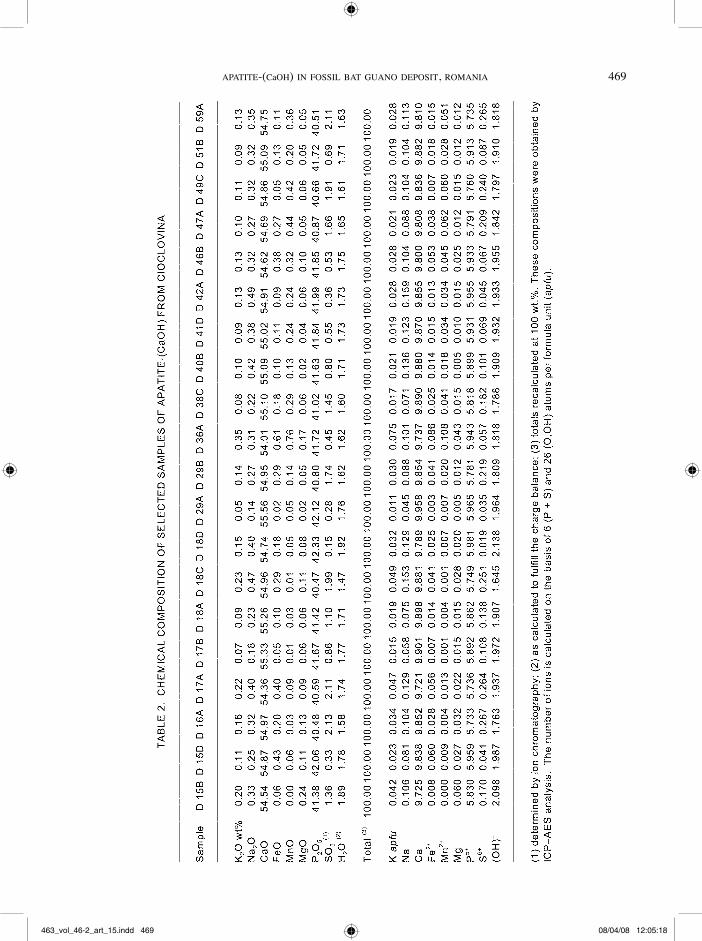
Results of ICP–AES analyses of 20 selected samples are presented in Table 2. The H2O contents were calcu-lated so as to fulfill charge balance, and the totals were recalculated to 100 wt.%. The amount of CO2 was ignored. The formulae were normalized on the basis of 6 (S + P) and 26 (O,OH) per formula unit (pfu), as recommended by Fiore & Laviano (1991). This basis is appropriate for emphasizing the Ca-deficiency of our samples, that probably belong to a series of general formula Ca10–x(HPO4)x(PO4)6–x(OH)2–x, where 0 ≤ x ≤ 1 (Berry 1967, Elliott 1994) and presumably also contain (CO3)2– and (SO4)2– groups.
A few remarks must be drawn on the basis of the results in Table 2: (1) No significant compositional differences occur between the various textural types of apatite-(CaOH). (2) As expected, all but six samples in Table 2 are Ca-oversaturated, showing seven-fold plus nine-fold cation totals higher than the theoretical value of 10 apfu. If we assume that the CO2 content in each sample is 0.81 wt.%, as determined by wet-chemical analysis of the purest sample, we must consider
463_vol_46-2_art_15.indd 466 08/04/08 12:05:13

aPatite-(CaoH) in fossil Bat GUano DePosit, roMania 467
fiG. 2. SEM photomicrographs of typical aggregates of apatite-(CaOH). A. Densely packed aggregate of subspherical shape. B. Reticulate network of apatite-(CaOH) aggregates, probably a fossil fragment of spongy bone or of another tissue. C. Rosette-like radial aggregates of apatite-(CaOH). Individual crystals, roughly hexagonal, are clearly visible. D. Rosette-like aggregates of apatite-(CaOH) overgrown on platy crystals of illite. E. Aggregates of randomly oriented interlocking and subparallel platy crystals of apatite-(CaOH). The arrow indicates a perfectly shaped crystal. F. Fractured aggregate of tabular crystals of apatite-(CaOH) intergrown on (0001), representing probably a fragment of a fossil cortical bone.
463_vol_46-2_art_15.indd 467 08/04/08 12:05:14

468 tHe CanaDian MineraloGist
that part of the Ca (about 0.02 apfu in equivalent) is consumed for charge-balancing carbonate. In this case, all our samples are in reality Ca-deficient, which suggests that (HPO4)2– groups partially replace (PO4)3–. However, apatite-(CaOH) samples in Table 2 contain less than 2.26% of the protonated phosphate groups, the maximum value being recorded in Sample D 18 D. (3) Sulfate groups, already reported in apatite-(CaOH) from caves (e.g., Fiore & Laviano 1991), also substitute for phosphate in apatite-(CaOH) from Cioclovina: S accounts for 0.32% to 4.66% of the tetrahedral sites normally occupied by P. Consequently, we could imagine that coupled substitutions such as (PO4)3– + (OH)– 7 (SO4)2– + O2– or (PO4)3– + (OH)– 7 (SO4)2– + (CO3)2– play an important role. (4) The substitutions in the cation sites normally occupied by Ca are less important. The main substitution is Na-for-Ca, but the cumulative substitution in these sites accounts for only 0.71 to 4.07% (mean 2.07%). Other potential cations substituting for Ca were evaluated with the ICP–AES results, but were found as minor elements, as shown by the results in Table 3.
As in the case of brushite (Dumitraş et al. 2004), apatite-(CaOH) from Cioclovina contains less Ba than Sr, which is quite normal in apatite-(CaOH) in fossil bone [see Trueman & Tuross (2002) and refer-
ences therein], but also encountered in the authigenic samples. The REE contents are relatively low. On the basis of results of partial analyses in Table 3, we can state that apatite-(CaOH) from Cioclovina is enriched in the light REE relative to the heavy REE + Y; all but three samples respect this rule, which also parallels the behavior of brushite (Dumitraş et al. 2004). In addi-tion to these elements, trace concentrations of some transitional metals (i.e., Cu, V, Cr and Ni) were also identified. The higher concentration of these elements in two of the analyzed samples (D 36A and D 38C B), is probably due to admixed X-ray amorphous phases and correlates with higher contents of manganese and iron.
Attempts to check the accuracy of the conclusions drawn from the results of bulk ICP–AES analyses were done using EMPA. We chose for study a fragment of fossil bone engulfed in the guano mass, which consists of pure apatite-(CaOH). Representative results for this sample (D 40 A, equivalent of D 40 B in Table 2) are summarized in Table 4. All iron was considered to be divalent, and the amount of H2O was calculated to fulfill charge-balance conditions. A parallel wet-chemical analysis was carried out on the same sample (Table 4). A few conclusions may be drawn: (1) The propor-tions of halogens in the EMP analyses are probably
463_vol_46-2_art_15.indd 468 08/04/08 12:05:15
aPatite-(CaoH) in fossil Bat GUano DePosit, roMania 469
463_vol_46-2_art_15.indd 469 08/04/08 12:05:18

470 tHe CanaDian MineraloGist
overestimated owing to analytical interferences with the araldite used for fixing the samples on the support; it does not seem possible that fluorine and chlorine occupy up to 34.77% and 6.44%, respectively, of the positions normally occupied by hydroxyl, even if the analyzed apatite-(CaOH) represents a bone fragment. The wet-chemical analysis in Table 4 seems more accu-rate on this point. (2) EMP analyses reveal insignificant variations in composition among different micromounts. (3) The chemical peculiarities established by ICP–AES analyses are confirmed: up to 2.32% of the tetrahedral positions are occupied by sulfur, and the incorpora-tion of Na, K, Mn, Fe, Mg in the Ca sites is minor, summing up to 2.11%. (4) Because the EMP analysis does not offer any indication for the incorporation of (CO3)2– into the apatite-(CaOH) lattice, it is difficult to evaluate the deficiency in Ca of the analyzed sample; accepting, however, a CO2 content of 0.81 wt.% as determined by wet-chemical analysis, all but one EMP datassets in Table 4 are representative of Ca-deficient apatite-(CaOH).
X-ray PowDer Data
X-ray powder diffractometry was carried out on about 100 powders of apatite-(CaOH) from Cioclovina in order to refine the cell parameters. The primary patterns are available upon request from the first author. the index of crystallinity calculated by the method recommended by Simpson (1964), involving the ratio between the total width of the diffraction peaks (211) and (112) at their half height and the greatest height
463_vol_46-2_art_15.indd 470 08/04/08 12:05:19

aPatite-(CaoH) in fossil Bat GUano DePosit, roMania 471
of the two peaks, vary between 0.037 and 0.158. The indices of crystallinity of the samples whose chemical analyses are given in Table 2 are reported in Table 1. Only about 12% of the analyzed samples have high indices of crystallinity, indicating that as a whole they are poorly crystalline (Simpson 1964). The unit-cell parameters were successfully refined based on a hexagonal P63/m cell, after indexing the patterns in analogy with PDF 86–1199, 86–1201 and 86–1203. Sets of 36 to 60 reflections in the 2u range between 10 and 70° (CuKa, l = 1.54056 Å) for which unam-biguous indexing was possible were used to refine the cell dimensions of 20 representative samples, which are summarized in Table 1. The unit-cell parameters calculated as an average of these 20 datasets of values are a 9.436(13), and c 6.868(8) Å, where the errors in the brackets correspond to the standard deviations of the mean (2s) for each set of data. These values, as well as the individual values in Table 1, and particularly c, differ significantly from those determined by various authors for synthetic apatite-(CaOH) of stoichiometric compo-sition [e.g., a = 9.4176(5), c 6.8814(5) Å according to Elliott (1994), a 9.421(2), c 6.882(2) Å according to Brunet et al. (1999), and a 9.4302(5), c 6.8911(2) Å according to Dorozkhin & Epple (2002)].
The unit-cell dimensions of carbonate-bearing apatite-(CaOH) depend, to a first approximation, on the incorporation of (CO3)2– in the structure (Elliott 1994). From this point of view, our samples generally behave like the A-type carbonated apatite-(CaOH), which are characterized by increased a unit-cell parameters due to carbonate substituting for hydroxyl group in the struc-tural channels (Elliott 1994). The presence of carbonate in the analyzed samples could explain why the c value, 6.867(5) Å, does not differ significantly from that deter-mined by Neweseley (1963) for a synthetic “carbonate–hydroxylapatite”. As apatite-(CaOH) samples from Cioclovina have uniformly low chlorine and fluorine contents, the occupancy by halogens of the X sites in the structure (notation after Hughes et al. 1989, and Brunet et al. 1999), could not explain the change in lattice parameters.
tHerMal DeCoMPosition
The differential thermal analysis and differential thermogravimetric curves of apatite-(CaOH) are gener-ally smooth and featureless until 1000°C, in spite of the losses of H2O and CO2 recorded on the thermogra-vimetric curve (Fiore & Laviano 1991, Ivanova et al. 2001). This was the reason for recording the thermal curves of apatite-(CaOH) from Cioclovina using a DSC–TG–TGA coupling, in spite of the lower limits of working temperature (up to 600°C). The thermal curves recorded for a representative sample of authi-genic apatite-(CaOH) from Cioclovina (D 301) are given in Figure 3. On the basis of previous studies on the thermal decomposition of the Ca-deficient carbon-
ated apatite-(CaOH) (Elliott 1994, Ivanova et al. 2001), we can presume that the main effects on the DTG and DSC curves are due to the following causes: (1) the loss of surface-bound H2O (–1.43 wt.% on the TGA curve), marked by an endothermic effect at ~97°C, (2) the loss of channel H2O (–3.61 wt.% cumulative on the TGA curve), marked by a small endothermic effect at ~198°C, and (3) the start of the carbonate migration (see below), marked by a small endothermic effect on the DTG curve at ~520°C, accompanied by an exothermic effect on the DSC curve. The loss of the lattice H2O is continuous, whereas the loss of carbonate starts after
fiG. 3. Thermal curves recorded for a apatite-(CaOH) sample from Cioclovina: differential thermogravimetric (top), differential scanning calorimetric (middle) and thermo-gravimetric (bottom).
463_vol_46-2_art_15.indd 471 08/04/08 12:05:20

472 tHe CanaDian MineraloGist
520°C. The TGA curve gradually declines, the total loss-in-weight at 600°C being of 6.62 wt.%.
An XRD study of the breakdown product at 600°C, after cooling, reveals that only a phase with the apatite-(CaOH) pattern persists, without any supplementary crystalline associated phase. According to Brunet et al. (1999), the breakdown of Ca-deficient apatite-(CaOH) at high temperature must, however, result in the growth of b-Ca3(PO4)2 according to the reaction:
Ca10–x(HPO4)x(PO4)6–x(OH)2–x ! (1–x) Ca10(PO4)6(OH)2 + 3x Ca3(PO4)2 + x H2O.
In fact, no traces of a Ca3(PO4)2 phase were detected, even after heating of many selected powdered samples of apatite-(CaOH) from Cioclovina at 600°C in a resis-tance furnace; these were held at this temperature for 24 hours in order to favor the loss of H2O, then cooled down to 25°C. The breakdown products, analyzed by XRD, gave patterns similar with those of the starting materials. This finding apparently disagrees with our results indicating a substantial loss of H2O, with the results of Berry (1967), which indicate the dehydration of Ca-deficient apatite-(CaOH) at 600°C, and also with the results of the thermal expansion measurements of Brunet et al. (1999), which indicate a breakdown at about 550°C. In fact, it is more reasonable to accept that the loss of H2O due to the breakdown of the protonated phosphate in our sample can be explained by another reaction, proposed by Mortier et al. (1989):
Ca10–x(HPO4)x(PO4)6–x(OH)2–x ! Ca10–x(P2O7)x–z(PO4)6–2x+2z(OH)2(1–z) + (x +z) H2O.
A weak and broad band centered at ~735 cm–1 in the FTIR spectra of the heated samples (not given, but available upon request from the first author) can be assigned to (P2O7)4– groups, indicating the polymeriza-tion of some of the structural orthophosphate groups, without structural breakdown.
As proven by the differential weighting, the mass loss continues until 1000°C, involving a continuous
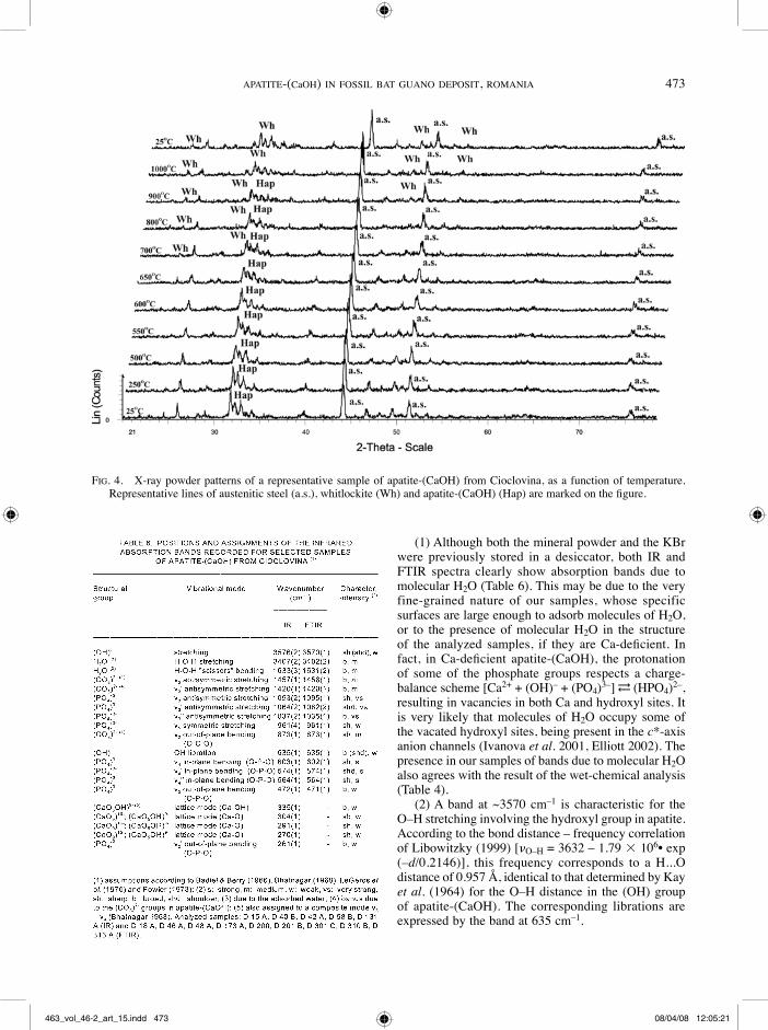
dehydration and the expelling of carbonate: the total loss in mass of sample D 301 heated to 1000°C is 7.43 wt.%. X-ray-assisted heating of this sample shows, however, that a phase with a apatite-(CaOH) pattern still persist at 900°C, allowing the refinement of its unit-cell parameters (Fig. 4, Table 5). Taking into account the dehydration, this phase must be an “oxyapatite”, whose crystal structure is identical with that of apatite-(CaOH) (Alberius-Henning et al. 1999), generating a similar XRD pattern (PDF 01–089–6495). This inference agrees with the hypothesis that by heating the (OH)– ions in the apatite-(CaOH), anion channels are replaced by O2– through the scheme 2 (OH)– ! O2– + □, where □ is a vacancy, without any disruption of other struc-tural constituents (Alberius-Henning et al. 1999).
As expected, and in agreement with experimental studies on Ca-deficient carbonated apatite-(CaOH) (Ivanova et al. 2001) and with studies of the thermal expansion of stoichiometric apatite-(CaOH) (Brunet et al. 1999), both a and c unit-cell parameters generally define an increasing trend with increasing temperature (Table 5). Two contractions of the cell can be observed, however: (1) at up to 250°C, owing to the loss of the channel H2O, and (2) at 550–600°C, owing to the loss of carbonate (cf. Ivanova et al. 2001). The increase of a and the striking decrease of c between 600 and 650°C agree with the changes in the location of (CO3)2– from (OH)– to (PO4)3– sites, in a process contrary to that inferred by Ivanova et al. (2001) in the B-type carbon-ated apatite-(CaOH).
Whitlockite occurs as breakdown product at 700°C, the transformation being complete at 1000°C. The resulting whitlockite [a 10.438(8), c 37.51(7) Å at 1000°C and a 10.430(6), c 37.47(7) Å after cooling at 25°C] is, as proven by ion chromatography, sulfate-bearing, indicating that sulfate was not (entirely) expelled at 1000°C.
infrareD aBsorPtion Data
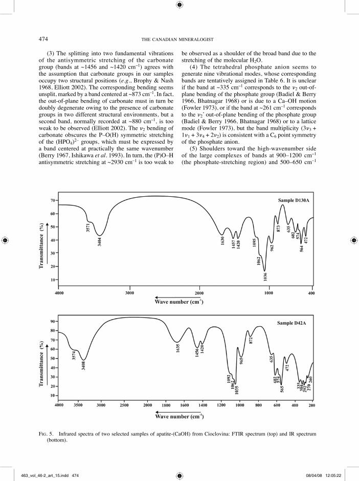
Figure 5 displays the infrared absorption spectra obtained for two representative samples of apatite-(CaOH) from Cioclovina with and without a Fourier-transform spectrometer, whereas Table 6, give the wavenumbers, characters and intensities of the infrared absorption bands as well as attempts to assign the bands on the basis of the works of Baddiel & Berry (1966), Bhatnagar (1968), LeGeros et al. (1970) and Fowler (1973). The positions of the bands in Table 6 were established as the mean of the wavenumbers recorded for similar bands in five (IR) and, respectively, nine (FTIR) different spectra of representative samples, with standard deviations given into brackets. The primary records are available on request from the first author. As the band assignment is generally agreed upon, only the following remarks are considered important:
463_vol_46-2_art_15.indd 472 08/04/08 12:05:20
aPatite-(CaoH) in fossil Bat GUano DePosit, roMania 473
(1) Although both the mineral powder and the KBr were previously stored in a desiccator, both IR and FTIR spectra clearly show absorption bands due to molecular H2O (Table 6). This may be due to the very fine-grained nature of our samples, whose specific surfaces are large enough to adsorb molecules of H2O, or to the presence of molecular H2O in the structure of the analyzed samples, if they are Ca-deficient. In fact, in Ca-deficient apatite-(CaOH), the protonation of some of the phosphate groups respects a charge-balance scheme [Ca2+ + (OH)– + (PO4)3–] !← (HPO4)2–, resulting in vacancies in both Ca and hydroxyl sites. It is very likely that molecules of H2O occupy some of the vacated hydroxyl sites, being present in the c*-axis anion channels (ivanova et al. 2001, Elliott 2002). The presence in our samples of bands due to molecular H2O also agrees with the result of the wet-chemical analysis (Table 4).
(2) A band at ~3570 cm–1 is characteristic for the O–H stretching involving the hydroxyl group in apatite. According to the bond distance – frequency correlation of Libowitzky (1999) [nO–H = 3632 – 1.79 3 106• exp (–d/0.2146)], this frequency corresponds to a H...O distance of 0.957 Å, identical to that determined by Kay et al. (1964) for the O–H distance in the (OH) group of apatite-(CaOH). The corresponding librations are expressed by the band at 635 cm–1.
fiG. 4. X-ray powder patterns of a representative sample of apatite-(CaOH) from Cioclovina, as a function of temperature. Representative lines of austenitic steel (a.s.), whitlockite (Wh) and apatite-(CaOH) (Hap) are marked on the figure.
463_vol_46-2_art_15.indd 473 08/04/08 12:05:21
474 tHe CanaDian MineraloGist
(3) The splitting into two fundamental vibrations of the antisymmetric stretching of the carbonate group (bands at ~1456 and ~1420 cm–1) agrees with the assumption that carbonate groups in our samples occupy two structural positions (e.g., Brophy & Nash 1968, Elliott 2002). The corresponding bending seems unsplit, marked by a band centered at ~873 cm–1. In fact, the out-of-plane bending of carbonate must in turn be doubly degenerate owing to the presence of carbonate groups in two different structural environments, but a second band, normally recorded at ~880 cm–1, is too weak to be observed (Elliott 2002). The n2 bending of carbonate obscures the P–O(H) symmetric stretching of the (HPO4)2– groups, which must be expressed by a band centered at practically the same wavenumber (Berry 1967, Ishikawa et al. 1993). In turn, the (P)O–H antisymmetric stretching at ~2930 cm–1 is too weak to
be observed as a shoulder of the broad band due to the stretching of the molecular H2O.
(4) The tetrahedral phosphate anion seems to generate nine vibrational modes, whose corresponding bands are tentatively assigned in Table 6. It is unclear if the band at ~335 cm–1 corresponds to the n2 out-of-plane bending of the phosphate group (Badiel & Berry 1966, Bhatnagar 1968) or is due to a Ca–OH motion (Fowler 1973), or if the band at ~261 cm–1 corresponds to the n2’ out-of-plane bending of the phosphate group (Badiel & Berry 1966, Bhatnagar 1968) or to a lattice mode (Fowler 1973), but the band multiplicity (3n3 + 1n1 + 3n4 + 2n2) is consistent with a C6 point symmetry of the phosphate anion.
(5) Shoulders toward the high-wavenumber side of the large complexes of bands at 900–1200 cm–1 (the phosphate-stretching region) and 500–650 cm–1
fiG. 5. Infrared spectra of two selected samples of apatite-(CaOH) from Cioclovina: FTIR spectrum (top) and IR spectrum (bottom).
463_vol_46-2_art_15.indd 474 08/04/08 12:05:22

aPatite-(CaoH) in fossil Bat GUano DePosit, roMania 475
(phosphate in-plane bending region), potentially due to the presence of sulfate groups in the structure, are too poorly resolved. Such bands were reported by Baumer et al. (1990) in sulfate-substituted fluorapatite samples, but are not evident in our case.
GenetiC ConsiDerations
Textural evidence, as well as parallels with other similar deposits, show that apatite-(CaOH) from Cioclovina formed by two complementary processes, namely: (1) by authigenesis, as reaction products between carbonate (basement, boulders or moonmilk flows) and acid solutions derived from guano, and (2) by sedimentation followed by diagenesis of organic relics such as bones, teeth and other tissues, in one of the richest deposits of fossil fragments in Europe. Both authigenic and biogenic apatite-(CaOH) from Cioclovina are carbonate- and sulfate-bearing and Ca-deficient. The lack of major physical, crystallo-graphic and chemical differences between the two kinds of apatite-(CaOH) from Cioclovina is an argument in favor of re-equilibration during diagenesis.
An XRD analysis coupled with SEM–EDS, as well as textural evidence, show that the authigenic apatite-(CaOH) had a gel-like, X-ray-amorphous precursor. The nature of this precursor (“ACP phase”) is unclear, but we presume that it is identical to that observed during the growth of synthetic apatite-(CaOH), having a composition like Ca3(PO4)2•nH2O (Francis & Webb 1971, Abbona & Franchini-Angela 1990). This phase is unstable and normally transforms into the more stable phases octacalcium phosphate [Ca4H(PO4)3•2.5H2O] and apatite-(CaOH) (Abbona & Franchini-Angela 1990). As shown by Christoffersen et al. (1990), the octacalcium phosphate is the intermediate phase in the formation of apatite-(CaOH) via heterogeneous nucle-ation at increased supersaturation, which could explain the presence at Cioclovina of the spherical aggregates of densely matted material apparently crystallized from gels. The authigenic crystallization of apatite-(CaOH) by direct precipitation, as well as the recrystallization of biogenic fragments during diagenesis, were controlled by values of pH higher than 6.93 (Elliott et al. 1959) or 6.2 (Simpson 1964), this pH value essentially depending on the saturation of the mother solution. At low ranges of pH, the calcium phosphate expecxted to precipitate is brushite (Elliott et al. 1959, Simpson 1964). As proved by the frequency of the inclusions of quartz and illite in apatite-(CaOH) aggregates, the nucleation of Ca phos-phates and the subsequent growth of apatite-(CaOH) seed crystals were stimulated by the pre-existence of silicate nuclei.
The contents of REE + Y reported previously (Table 3) are in the range of 2.95 to 47.11 ppm (average 12.79 ppm), even lower than those recorded by us in ten selected samples of carbonates from the limestone
support and from the solidified flows of moonmilk on the walls, which range from 21.00 to 34.19 ppm (average 25.28 ppm). The classical hypothesis of the formation of authigenic apatite-(CaOH) by direct reac-tion between the phosphate solutions derived from the guano mass and the carbonates in the cave is conse-quently fully confirmed.
The coexistence of apatite-(CaOH) with brushite is essentially a function of the local variations of pH. The action of magnesium as local inhibitor of apatite-(CaOH) crystallization (Abbona & Franchini-Angela 1990) is excluded since the magnesium phosphates are lacking in the fossil guano, and in the surrounding limestones the level of concentration of magnesium is low. The inhibitor of the crystal growth of apatite-(CaOH) must in fact be the local excess of carbonate ions resulting from the breakdown of carbonates due to the acidity of solutions derived from the guano mass. This may explain the extensive development of brushite in the vicinity of the gel-like aggregates of Ca phosphate preceding apatite-(CaOH). The excess of sulfate in the system and the low values of pH (up to 5.5) destabi-lize both brushite and apatite-(CaOH), favoring the crystallization of ardealite or even gypsum (Rinaudo & Abbona 1988). This environment could explain the local occurrence of sprays and crusts of ardealite and gypsum directly on apatite-(CaOH).
aCknowleDGeMents
This research was supported by the cooperative ANCS – DRI research program under project 8/2005 between the Romanian and the Walloon governments, by two MIRA grants awarded by the Rhône-Alpes Region in France to the first two authors, and by the CERES project No. 4–153/04.11.2004 of the Roma-nian Ministry of Education and Research. Support from Ecole Nationale Supérieure des Mines de Saint-Etienne (ENSMSE, France) is gratefully appreciated. F.H. acknowledges the FNRS (Belgium) for the award of a position of Chargé de Recherches in 2004–2006 and for grant 1.5.112.02. The authors are indebted to Dr. Jacques Moutte (ENSMSE) for assistance with ICP–AES analysis, to Mr. Olivier Valfort (ENSMSE) for technical assistance during the thermally assisted X-ray powder work, to Mrs. Gabriela Stelea (Geological Institute of Romania, Bucharest) and Mr. Pierre Lefèvre (University of Liège, Belgium) for recording the infrared absorption spectra, to Miss Véronique Bourgier for recording the thermal curves, and to Mr. Jean-Pierre Poyet (ENSMSE) for the ion-chromatographic analysis. Field assistance by Mr. Bogdan Tomuş is gratefully acknowledged. The quality of this contribution was improved after reviews by Dr. Clive Trueman and an anonymous referee. Robert F. Martin and Associate Editor Louis Raimbault are gratefully acknowledged for handling the manuscript.
463_vol_46-2_art_15.indd 475 08/04/08 12:05:22
476 tHe CanaDian MineraloGist
referenCes
aBBona, f. & franCHini-anGela, M. (1990): Crystallization of calcium and magnesium phosphates from solutions of low concentration. J. Crystal Growth 104, 661-671.
alBeriUs-HenninG, P., lanDa-Canovas, a., larsson, a.-k. & liDin, s. (1999): Elucidation of the crystal structure of oxyapatite by high-resolution electron microscopy. Acta Crystallogr. B 55, 170-176.
aPPleMan, D.e. & evans, H.T., Jr. (1973): Indexing and least-squares refinement of powder diffraction data. U.S. Geol. Surv., Comput. Contrib. 20 (NTIS Doc. PB-216).
BaDDiel, C.B. & Berry, e.e. (1966): Spectra structure cor-relations in hydroxy and fluorapatite. Spectrochim. Acta 22, 1407-1416.
Balenzano, f., Dell’anna, l. & Di Piero, M. (1974): Ricerche mineralogiche su alcuni fosfati rinvenuti nelle grotte di Castellana (Bari): strengite aluminifera, vivianite, taranakite, brushite e idrossiapatite. Rend. Soc. Ital. Min-eral. Petrogr. 30, 543-573.
BaUMer, a., CarUBa, r. & GanteaUMe, M. (1990): Car-bonate-fluorapatite: mise en évidence de la substitution 2PO4
3– ! SiO44– + SO4
2– par spectrométrie infrarouge. Eur. J. Mineral. 2, 297-304.
Benoit, P.H. (1987): Adaptation to microcomputer of the Appleman–Evans program for indexing and least-squares refinement of powder-diffraction data for unit-cell dimen-sions. Am. Mineral. 72, 1018-1019.
Berry, e.e. (1967): The structure and composition of some calcium-deficient apatites. J. Inorg. Nucl. Chem. 29, 317-327.
BHatnaGar, v.M. (1968): Infrared spectra of hydroxyapatite and fluorapatite. Bull. Soc. Chim. France, 1771-1773.
BroPHy, G.P. & nasH, t.J. (1968): Compositional, infrared and X-ray analysis of fossil bone. Am. Mineral. 53, 445-454.
BrUnet, f., allan, D.r., reDfern, a.t.s., anGel, r.J., MiletiCH, r., reiCHMann, H.J., serGent, J. & HanflanD, M. (1999): Compressibility and thermal expansivity of synthetic apatites, Ca5(PO4)3X with X = OH, F and Cl. Eur. J. Mineral. 11, 1023-1035.
BUrke, e.a.J. (2008): Tidying up mineral names: an IMA–CNMNC scheme for suffixes, hyphens and diacritical marks. Mineral. Rec. 39, 131-135.
CHristoffersen, M.r., CHristoffersen, M.r. & kiBalCzyC, w. (1990): Apparent solubilities of two amorphous calcium phosphates and of octacalcium phosphate in the tempera-ture range 30–42°C. J. Crystal Growth 106, 349-354.
ConstantinesCU, e., MarinCea, Ş. & CrăCiun, C. (1999): Crandallite in the phosphate association from Cioclovina cave (Şureanu Mts., Romania). In Scientific works by Emil
Constantinescu. Mineralogy in the system of Earth sci-ences. Imperial College Press, London, U.K. (1-5).
DiaConu, G. & MeDeşan, a. (1975): Spéléothèmes de dahllite dans la grotte “Peştera Muierii”, Baia de Fier – Roumanie. Trav. Inst. Spéol. “Emile Racovitza” 14, 149-156.
DorozHkin, s.v. & ePPle M. (2002): Die biologische und medizinische Bedeutung von Calciumphosphaten. Angew. Chem. 114, 3260-3277.
DuMitraş D.G. & MarinCea, Ş. (2004):
DuMitraş D.G., MarinCea, Ş. & fransolet, a.M. (2004): Brushite in the bat guano deposit from the “dry” Cioclo-vina Cave (Şureanu Mountains, Romania). Neues Jahrb. Mineral., Monatsh., 45-64.
elliot, J.s., sHarP, r.f. & lewis, l. (1959): The effect of the molar Ca/P ratio upon the crystallization of brushite and apatite. J. Phys. Chem. 63, 725-726.
elliott, J.C. (1994): Structure and Chemistry of the Apa-tites and the Other Calcium Orthophosphates. Elsevier, Amsterdam, The Netherlands.
elliott, J.C. (2002): Calcium phosphate biominerals. In Phosphates – Geochemical, Geobiological and Materials Importance (M.L. Kohn, J. Rakovan & J.M. Hughes, eds.). Rev. Mineral. Geochem. 48, 427-454.
fiore, s. & laviano, r. (1991): Brushite, hydroxylapatite, and taranakite from Apulian caves (southern Italy): new mineralogical data. Am. Mineral. 76, 1722-1727.
fowler, B.o. (1973): Infrared studies of apatites. I. Vibra-tional assignments for calcium, strontium and barium hydroxyapatites utilizing isotopic substitution. Inorg. Chem. 13, 194-207.
franCis, M.D. & weBB, n.C. (1971): Hydroxyapatite forma-tion from a hydrated calcium monohydrogen phosphate precursor. Calc. Tiss. Res. 6, 335-342.
HUGHes, J.M., CaMeron, M. & Crowley, k.D. (1989): Structural variations in natural F, OH and Cl apatites. Am. Mineral. 74, 870-876.
iosof, V. & neaCşu, V. (1980): Analysis of silicate rocks by atomic absorption spectrometry. Rev. Roum. Chim. 25, 589-597.
isHikawa, k., DUCHeyne, P. & raDin, s. (1993): Determina-tion of the Ca/P ratio in calcium-deficient hydroxyapatite using X-ray diffraction analysis. J. Mater. Sci., Mater. Med. 4, 165-168.
ivanova, t.i., frank-kaMenetskaya o.v., kaltsov, v. & UGolkov, l. (2001): Crystal structure of calcium-deficient carbonated hydroxylapatite. Thermal decomposition. J. Solid State Chem. 160, 340-349.
kay, M.i., yoUnG, r.a. & Posner, a.s. (1964): Crystal struc-ture of hydroxylapatite. Nature 204, 1050-1052.
463_vol_46-2_art_15.indd 476 08/04/08 12:05:23
aPatite-(CaoH) in fossil Bat GUano DePosit, roMania 477
leGeros, r.z., leGeros, J.P., traUtz, o.r. & klein, e. (1970): Spectral properties of carbonate in carbonate-containing apatites. Develop. Appl. Spectrosc. 7 B, 3-12.
liBowitzky, e. (1999): Correlation of O–H stretching fre-quencies and O–H...O hydrogen bond lengths in minerals. Monatsh. Chem. 130, 1047-1059.
ManDarino, J.a. (1981): The Gladstone–Dale relationship. IV. The compatibility concept and its application. Can. Mineral. 19, 441-450.
MarinCea, Ş. & DuMitraş, D. (2003): The occurrence of taranakite in the “dry” Cioclovina Cave (Sureanu Mountains, Romania). Neues Jahrb. Mineral., Monatsh., 127-144.
MarinCea, Ş. & DuMitraş, D. (2005): First reported sedimen-tary occurrence of berlinite (AlPO4) in phosphate-bearing sediments from Cioclovina Cave, Romania – Comment. Am. Mineral. 90, 1203-1208.
MarinCea, Ş., DuMitraş, D. & Gibert, r. (2002): Tinsley-ite in the “dry” Cioclovina Cave (Sureanu Mountains, Romania): the second world occurrence. Eur. J. Mineral. 14, 157-164.
Mortier, a., leMaitre, J. & roUXHet, P.G. (1989): Temperature-programmed characterization of synthetic calcium-deficient phosphate apatites. Thermochim. Acta 143, 265-282.
neweseley, H. (1963): Kristallchemische und micromorphol-ogische Untersuchungen an Carbonat-Apatiten. Monatsh. Chem. 94, 270-280.
onaC, B.P., BreBan, r., kearns, J. & taMas, t. (2002): Unusual minerals related to phosphate deposits in Cioclo-vina Cave, Sureanu Mountains (Romania). Theor. Appl. Karst. 15, 27-34.
onaC, B.P., ettinGer, k., kearns, J & Balasz, i.i. (2005): A modern, guano-related occurrence of foggite, CaAl(PO4)(OH)2•H2O and churchite-(Y), YPO4•2H2O in Cioclovina Cave, Romania. Mineral. Petrol. 85, 291-302.
onaC, B.P. & wHite, w.B. (2003): First reported sedimentary occurrence of berlinite (AlPO4) in phosphate-bearing sedi-ments from Cioclovina Cave, Romania. Am. Mineral. 88, 1395-1397.
rinaUDo, C. & aBBona, f. (1988): A contribution to the study of the crystal chemistry of calcium sulfate phosphate hydrate. Mineral. Petrogr. Acta 31, 95-105.
sCHaDler, J. (1929): Mineralogische-petrographische Char-acteristik der Phosphat-Ablagerung in the Cioclovinahöhle bei Pui. Pub. Muz. Hunedoara 5(27), 1-3.
sCHaDler, J. (1932): Ardealit , ein neues Mineral CaHPO4•CaSO4+4H2O. Zb. Mineral. A, 40-41.
sHaPiro, l. & BrannoCk, w.w. (1962): Rapid analysis of silicate, carbonate and phosphate rocks. U. S. Geol. Surv. Bull. 1144-A (A 14-15, A 49-51).
siMPson, D.r. (1964): The nature of alkali carbonate apatites. Am. Mineral. 49, 363-376.
toMUş, r.B. (1999): The Karst Complex Ciclovina. The Basin 2063. Proteus, Hunedoara, Romania (in Romanian).
trUeMan, C.n. & tUross, n. (2002): Trace elements in recent and fossil bone apatite. In Phosphates – Geochemical, Geobiological and Materials Importance (M.L. Kohn, J. Rakovan & J.M. Hughes, eds.). Rev. Mineral. Geochem. 48, 489-522.
Received February 27, 2007, revised manuscript accepted January 15, 2008.
463_vol_46-2_art_15.indd 477 08/04/08 12:05:23
463_vol_46-2_art_15.indd 478 08/04/08 12:05:23