Gene Expression Profiling of Childhood Adrenocortical Tumors
Upload
hms-harvardCategory
view
2download
0
Antitumorigenic and Antiinsulinogenic Effects ofCalcitriol on Insulinoma Cells and Solid �-Cell Tumors
FRANCESCA GALBIATI, LUCA POLASTRI, SILVIA GREGORI, MASSIMO FRESCHI,MARA CASORATI, UGO CAVALLARO, PAOLO FIORINA, FEDERICO BERTUZZI,ALESSANDRO ZERBI, GUIDO POZZA, LUCIANO ADORINI, FRANCO FOLLI,GERHARD CHRISTOFORI, AND ALBERTO M. DAVALLI
Departments of Medicine (F.G., L.P., M.C., P.F., F.B., G.P., A.M.D.), Pathology (M.F.), and Surgery (A.Z.), Unit forMetabolic Diseases (F.F.), and Universita Vita-Salute (G.P.), San Raffaele Scientific Institute, Milan 20132, Italy; RocheMilano Ricerche (S.G., L.A.), Milan, Italy; and Research Institute of Molecular Pathology (U.C., G.C.), ViennaA-1030, Austria
Malignant insulinoma is a rare form of cancer with a poorprognosis because of metastatic dissemination and untreat-able hypoglycemia. Effective chemotherapy of patients whoare not cured by surgery is needed. Calcitriol has known an-ticancer properties on different neoplastic cell lines, but nodata are available regarding its activity on tumorigenic pan-creatic �-cells. We analyzed the in vitro effects of calcitriol onthe murine insulinoma cell line �TC3 and primary cultures ofhuman isolated islets and benign insulinoma. The effect of invivo calcitriol administration on insulinoma of recombinantinsulin/Simian virus 40 oncogene-expressing transgenic micewas also investigated. In �TC3, calcitriol induced growth in-hibition; apoptosis; down-regulation of insulin gene expres-
sion; and nongenomic activation of the MAPK pathway. MAPKkinase inhibitor (UO126) and staurosporine reduced calcit-riol-mediated �TC3 death, and down-regulation of insulingene transcription was prevented by staurosporine but notUO126. Calcitriol significantly decreased insulin release andmRNA levels of human islets and insulinoma cells. Finally,recombinant insulin/Simian virus 40 oncogene-expressingtransgenic mice treated with calcitriol showed reduced insu-linoma volumes because of increased apoptosis of adenoma-tous cells. Together, these findings provide the rationale fortesting the efficacy of calcitriol in the treatment of patientswith solid �-cell tumors. (Endocrinology 143: 4018–4030, 2002)
CALCITRIOL, THE HORMONALLY active form of vi-tamin D3 [1,25-(OH)2D3], regulates the transcription of
a variety of genes, with important effects on calcium ho-meostasis, regulation of the immune response, and celldifferentiation (1, 2). Over the past decade, it has becomeevident that calcitriol and its analogs also exhibit antiprolif-erative activities in a variety of tumorigenic cell lines (3–9),raising the possibility that calcitriol, besides its classical usesin secondary hyperparathyroidism and psoriasis, might beemployed in the treatment of malignancies (10). Malignantinsulinoma is a rare cancer with a poor prognosis and areported 5-yr survival rate of 35% (11). In contrast to benign�-cell adenomas, which are usually cured by surgery, ma-lignant insulinomas are often diagnosed when metastaticdissemination has already occurred so that surgical resectionis rarely successful (12). Several cytotoxic agents have beenemployed for chemotherapy of malignant insulinomas butwith disappointing results (13). In addition to metastaticdissemination, untreatable hypoglycemia, consequent to un-regulated insulin release by neoplastic �-cells, is a life-threat-ening condition in these patients. In contrast to the hypo-
glycemia associated with benign insulinomas, which is wellcontrolled by inhibitors of insulin release such as diazoxideand certain calcium channel antagonists, malignant insuli-nomas are usually unresponsive to these drugs. Moreover,somatostatin and its long-acting analogs are effective in con-trolling hormone-mediated symptoms and growth of func-tionally active neuroendocrine tumors of the gastrointestinaltract but are usually ineffective in the treatment of malignantinsulinomas (14).
The presence of nuclear vitamin D receptors (VDRs) in thepancreatic �-cell has been demonstrated (15, 16), but theprecise role of calcitriol in islet cell physiology is still unclear.Vitamin D-deficient rats show an impaired insulin responseto a glucose challenge (17, 18), and adverse effects of vitaminD deficiency on insulin secretion have also been reported inman (19, 20). These effects can be corrected by calcitrioladministration (19, 20), and the beneficial effects of vitaminD may actually depend on its capability to raise plasmacalcium concentrations (21). The beneficial effects of calcitriolon insulin secretion have been documented by a large seriesof in vitro studies, mostly on islets isolated from vitaminD-depleted rats. Conversely, an inhibitory effect of long-termcalcitriol exposure on �-cell growth and insulin secretion hasbeen reported in islets from vitamin D-repleted rats and therat insulinoma cell line RIN-38 (22). However, surprisinglylittle is known about the effects that calcitriol exerts on isletsisolated from normal animals.
Studies on the relationship between vitamin D and pan-
Abbreviations: BrdU, 5�-Bromo-2�deoxyuridine; DAPI, 4�,6�-diamino-2-phenylindole; FCS, fetal calf serum; LDH, lactate dehydro-genase; MEK, MAPK kinase; MTT, 3-(4,5-dimethylthiazol-2-yl)-2,5-di-phenyltetrazolium bromide; PDX-1, pancreatic duodenal homeobox-1;PKC, protein kinase C; RIP1Tag2, recombinant insulin/Simian virus 40oncogene-expressing transgenic mice; TUNEL, terminal deoxynucleo-tidyl transferase-mediated deoxyuridine triphosphate nick end-label-ing; VDR, vitamin D receptor.
0013-7227/02/$15.00/0 Endocrinology 143(10):4018–4030Printed in U.S.A. Copyright © 2002 by The Endocrine Society
doi: 10.1210/en.2002-220200
4018

creatic �-cells have become even more appealing after thedemonstration that calcitriol and its analogs can delay andreduce the onset of diabetes in the nonobese diabetic mouse(23, 24) and that a polymorphism within the vitamin D re-ceptor gene may play a role in the pathogenesis of either type1 (insulin-dependent) and type 2 diabetes mellitus (25–27).
Despite its known anticancer properties, no studies havebeen previously conducted to test the antineoplastic activityof calcitriol on tumorigenic �-cells. In the present study, wehave examined the effects of calcitriol in vitro on the mouse�-cell line �TC3 (28) and in vivo in the recombinant insulin/Simian virus 40 oncogene-expressing transgenic mice(RIP1Tag2) from which �TC3 cells were derived (29).RIP1Tag2 mice inheritably develop insulinomas with a pre-cise multistep model of tumorigenesis. Islets are essentiallynormal in newborn mice and become hyperplastic in youngadult mice (4–6 wk). By the age of 9 wk, 50% of the islets arecomposed of proliferating �-cells (29, 30), but only a fewislets progress to histologically distinct adenomas, which arepresent by 12 wk of age in all mice of this lineage. Beforebecoming adenomas, hyperplastic islets start to produce an-giogenic factors that are necessary for the transition fromhyperplasia to neoplasia (31). If left untreated, RIP1Tag2mice die of hypoglycemia 12–16 wk after birth; however, ifglucose is added to their drinking water, they survive longenough to show the progression of a few benign insulinomasinto invasive tumors. The transition from well-differentiatedadenomas to carcinomas coincides with the lack of the ad-hesion molecule E-cadherin in adenomatous cells (32). More-over, neural cell adhesion molecule-deficient RIP1Tag2 micedevelop metastases that are never observed in normalRIP1Tag2 mice (33).
Our results show that calcitriol has a profound antineo-plastic effect on insulinoma cells, both in vitro and in vivo. Inaddition, calcitriol significantly decreases gene expression,total content, and release of insulin both in mouse and humaninsulinoma cells as well as in isolated human islets. Theimplications of these findings for future trials aimed at de-termining the efficacy of calcitriol or its analogs in the treat-ment of patients with solid �-cell tumors are discussed.
Materials and MethodsCell lines and cultures
�TC3 cells were provided by Shimon Efrat (Sackler School of Med-icine, Tel Aviv University, Tel Aviv, Israel); �TC1 cells from DouglasHanahan (Department of Biochemistry and Biophysics, University ofCalifornia, San Francisco, CA), and pituitary GH3 from the AmericanType Culture Collection (Manassas, VA). �TC3 cells were grown inRPMI 1640 (Life Technologies, Inc., Rockville, MD) supplemented with10% fetal calf serum (FCS), 2 mm l-glutamine, and 100 IU/ml strepto-mycin/penicillin. The �TC1 and GH3 cells were cultured in DMEM (LifeTechnologies, Inc.) containing 25 mm glucose and supplemented with10% FCS, 2 mm glutamine, and 100 IU/ml streptomycin/penicillin.Cultures were performed under standard humidified conditions of 5%CO2 and 95% air at 37 C.
Preparation and culture of human insulinoma cells
Insulinoma tissue was obtained from a 34-yr-old male who wasadmitted to the hospital for hypoglycemia and hyperinsulinism. At theoperating field, the adenoma appeared as a nodule of 15 mm in diameter,easily excisable from the surrounding pancreatic parenchyma. Afterremoval, the tumor was divided into two parts; a part was sent to the
Pathology Department for routine histology that, according to the cri-terion for the classification of the endocrine tumors (34), confirmed theclinical diagnosis of benign insulinoma. The other part was taken to thelaboratory in which it was minced into small fragments and then dis-persed into a suspension of cell clusters by vigorous pipetting. Afterwashing, insulinoma cells were plated in DMEM/F12 medium contain-ing 2 mm l-glutamine, 33.2 mm glucose, 9.6 �g/ml putrescine, 6.3 ng/mlprogesterone, 5.2 ng/ml sodium selenite, 0.025 mg/ml insulin, 0.1mg/ml transferrin, and 2 �g/ml heparin (sodium salt, grade II, Sigma,St. Louis, MO) onto 100 � 20-mm plastic culture dishes and allowed torecover for 3–5 d before being used for the experiments. Mediumchanges were performed every 48 h.
Isolation and culture of human islets
Human pancreatic islets were isolated from the pancreases of cadav-eric multiorgan donors by using the procedure already described indetail (35). The islets used in this study were isolated from eight differentorgans. After the isolation, the islets were purified from the contaminantexocrine tissue by centrifugation on Ficoll gradients, obtaining a finalpurity that ranged from 60% to 80%, as assayed by dithizone staining(35). Islets were then cultured in RPMI 1640 tissue culture medium. Aftera 48-h stabilization culture, old medium was replaced with fresh me-dium containing vehicle or increasing calcitriol (kindly provided byHoffmann-La Roche Inc., Nutley, NJ) concentrations (10, 100, and 1000nm), and islets were cultured for an additional 48 h until used for insulinsecretory studies or RNA extraction.
Cell growth in vitro
Cell proliferation was determined in �TC3, GH3, and �TC1 cell lines.The cells were seeded at a density of 9 � 104 cells per well onto 24-multiwell culture plates. After allowing the cells to attach and growthfor 48 h, cells were treated with vehicle (ethanol, final concentration0.04%) or increasing calcitriol concentrations (10, 100, and 1000 nm)dissolved in ethanol. After 48 and 72 h of vehicle or calcitriol exposure,cells were harvested and counted in duplicate with a Coulter CounterZM (Beckman Coulter, Inc., Fullerton, CA).
Cell cycle experiments
Analysis of cell cycle was performed on �TC3 and GH3 cells that wereplated at the density of 1.8 � 106 cells into 60-cm2 tissue culture platesand allowed to attach and grow in standard medium for 48 h. On thethird day, cells were refed with fresh medium with vehicle or calcitriol(10, 100, and 1000 nm) and cultured for an additional 48 h. Beforeharvesting, cells were pulse labeled with 10 �m 5�-bromo-2�deoxyuri-dine (BrdU, a thymidine analog that is incorporated into newly syn-thesized DNA by the cells entering and progressing through the S phaseof the cell cycle) for 15 h. Cell cycle distribution was determined by theBrdU flow kit (PharMingen, San Diego, CA). The incorporated BrdU wasstained with specific anti-BrdU fluorescent antibodies, and positive cellswere detected and counted by flow cytometry. The phase of cell cycleand DNA synthetic activities of the cells were determined by analyzingthe correlated expression of total DNA (7-AAD) and incorporated BrdUlevels. Different region gates were applied and the percentage of the cellsin the different cell cycle compartments was measured according to thekit’s manufacturer instructions.
3-(4,5-dimethylthiazol-2-yl)-2,5-diphenyltetrazoliumbromide (MTT) assay
The number of viable cells was assayed using the MTT (Sigma)method. Vital dye staining was performed on �TC3, �TC1, and GH3 cellsthat were seeded at a density of 3 � 104 cells per well onto 96-well cultureplates and allowed to attach and grow for 48 h in standard medium. Onthe third day, cells were washed and refed with fresh medium contain-ing vehicle or increasing concentrations of calcitriol (10, 100, and 1000nm). Number of viable cells was measured after 48 and 72 h as previouslydescribed (36).
Galbiati et al. • Calcitriol Effects on Tumorigenic �-Cells Endocrinology, October 2002, 143(10):4018–4030 4019

Detection of apoptosis and quantification offragmented DNA
Detection of apoptosis and quantification of fragmented DNA wasperformed in �TC3 cells and primary cultures of the benign insulinoma.The �TC3 cells were seeded at a density of 2 � 106 onto 25-cm2 tissueculture flasks and allowed to attach and grow for 48 h. On the third day,old medium was replaced with fresh medium containing vehicle orincreasing calcitriol concentrations. After 48 h cells were harvested,lysed in 100 �l lysis buffer (10 mm Tris-Hcl; 10 mm EDTA; 0.5% TritonX-100, pH 8.0), and centrifuged at 10,500 rpm for 5 min. Supernatantscontaining fragmented (soluble) DNA were collected and pellets con-taining insoluble DNA were further extracted; both DNA fractions werethen treated in sequence with Rnase and proteinase-K for 1 h andprecipitated with isopropanol. Pellets were then dissolved in Tris/EDTA and DNA concentrations were measured by spectrophotometer.Fragmented DNA was calculated as 100% � soluble DNA/total DNA.DNA ladders were visualized by agarose/ethidium bromide gel elec-trophoresis. Apoptosis was also detected by immunofluorescence stain-ing of �TC3 cells and human islet treated with calcitriol 10, 100, and 1,000nm. Cells monolayers were fixed in freshly prepared 4% paraformal-dehyde (in 0.05 m PBS, pH 7.4, Life Technologies, Inc.) for 10 min at roomtemperature and then immunostained for insulin (mouse antihumaninsulin, diluted 1:100, Signet Laboratories, Dedham, MA) and 4�,6�-diamino-2-phenylindole (DAPI, 1 mg/ml in 1� PBS, diluted 1:50,Sigma) for determination of nuclear morphology.
Primary cultures of human insulinoma cells were treated with 0.05%trypsin in 0.05 mm EDTA at 37 C for about 5 min. Detached cells werewashed twice, seeded again at a density of 5 � 104 cells onto a chamberslide with a cover (Nalge Nunc International, Rochester, NY), and al-lowed to form cell monolayers for 48–72 h in standard medium. On theday of the experiments, the old medium was discarded and replacedwith fresh medium containing vehicle or calcitriol (100 nm and 1000 nm)and culture continued for additional 48 h. Detection of apoptosis wasperformed by fluorescence microscopy as described for �TC3 and hu-man islets. Monolayers were immunostained for insulin for pancreaticduodenal homeobox-1 (PDX-1) (rabbit antihuman, diluted 1:2000) andnuclear morphology by DAPI. The number of insulin-positive cellsshowing condensed or fragmented nuclei was counted and expressed asa percentage of total insulin-positive cells.
Detection of necrosis
The levels of lactate dehydrogenase (LDH) released from the cytosolof the cells undergoing necrosis were measured in the culture media of�TC3 cultured in absence and presence of calcitriol (1000 nm) by theClinical Core Laboratory of the Hospital.
Northern blot analysis
Insulin gene expression in �TC3 cells, human insulinoma cells, andhuman islets was analyzed after 48 h of calcitriol exposure by Northernblotting as previously described (37). Relative expression levels of in-sulin and 18S rRNA were determined by densitometric analysis. TotalRNA extracted from 1.5 � 103 isolated islets and 5 � 105 insulinoma cellswas submitted to the same protocol described for �TC3 cells.
Insulin release and content
Basal insulin release was measured in �TC3 cells, human insulinomacells, and human islets. The �TC3 cells were seeded at a density of 3 �104 cells per well onto 96-multiwell cultures plates and cultured in RPMI1640 medium in absence and presence of calcitriol (10, 100, and 1000 nm)for 48 h. On the third day, the old medium was aspirated off and cellswere washed twice with PBS before being incubated for 1 h in plainRPMI 1640 medium (11 mm glucose). After 1 h, the medium was col-lected and stored at �20 C, and the cells were harvested and extractedfor the measurement of total protein and insulin content. Experimentsof insulin release by primary cultures of human insulinoma cells wereperformed on cultures derived from 1 � 104 cells seeded onto 96-welltissue culture plates. For insulin secretory studies with isolated humanislets, 25 islets were handpicked and plated in 96 multiwell cultureplates. Human islets and human insulinoma cells were submitted to the
same experimental culture and insulin secretory protocols previouslydescribed for �TC3 cells. Insulin content of media and cell extracts weremeasured by RIA (INSIK-5, DiaSorin, Inc., Saluggia, Italy) using rat orhuman insulin standards as appropriate and were normalized to totalprotein content. Insulin release and content of cells exposed to calcitriolwere then expressed as percentage of control (vehicle-treated) cells.
ERK1 and ERK2 phosphorylation
Phosphorylation of ERK proteins was analyzed in �TC3 cells andhuman islets. After 48 h of culture in standard medium, �TC3 cells orhuman islets were serum starved for 3 h and then alternatively exposedfor 20 min to plain medium (without FCS, plus 0.2% albumin), mediumplus 10% FCS, or plain medium supplemented with calcitriol (10, 100,and 1,000 nm) in the presence or absence of the MAPK kinase (MEK)inhibitor (UO126 at 2 nm) (38) or the protein kinase C (PKC) inhibitor(staurosporine at 5 nm). After incubation, �TC3 cells and islets wereresuspended in lysis buffer (30 mm Tris HCl, 5 mm EGTA, 5 mm EDTA,250 mm sucrose, 1% Triton X-100, 1 mm sodium fluoride, 2 mm sodiumorthovanadate, 1 mm phenylmethylsulfonyl fluoride, and 1 �g/ml apro-tinin). After 1 h at 4 C, lysates were centrifuged at 13,000 rpm for 5 min,and the extracted proteins were analyzed by immunoblotting with an-tisera against anti-P-ERK1/2 and anti-ERK1/2 (antimouse and antigoat1:1,000, respectively, Santa Cruz Biotechnology, Santa Cruz, CA).
PKC activity
After 5 min of exposure to 100 nm calcitriol, the �TC3 cells wereharvested and extracted in lysis buffer (HEPES-Na 50 mm, Na Cl 250mm, MgCl2 5 mm Triton 1%, glycerol 10%, phenylmethylsulfonyl flu-oride 10 �g/ml, leupeptin 10 �g/ml, and aprotinin 10 �g/ml). PKCactivity in cell lysates was measured by using PepTag assay for non-radioactive detection of PKC (Promega Corp., Madison, WI). Thismethod uses a fluorescent peptide substrate that, when phosphorylatedby PKC, alters its net ionic charge from �1 to �1. Phosphorylated andnonphosphorylated substrates are rapidly separated on a agarose geland quantification of the phosphorylated peptide is performed byspectrofluorimetry.
Caspase-3 activity
The activity of the protease caspase-3 was measured in �TC3 cellscultured at a density of 25 � 104 cells per well onto 12-well culture plates.Cells were exposed for 48 h to calcitriol (10, 100, and 1000 nm) in absenceor presence of 50 �m caspase inhibitor Z-VAD-FMK. The �TC3 cells werethen harvested and caspase-3 activity in whole-cell lysates was deter-mined by using the CaspACE assay system (Promega Corp.) followingthe manufacturer’s instructions. This assay provides a colorimetric sub-strate (Ac-DEVD) labeled with the chromophore p-nitroaniline (p-NA),which is released from the substrate on cleavage by caspase-3. Freep-nitroaniline produces a yellow color that is measured by a photometerat 405 nm. The amount of yellow color produced is proportional to theamount of DEVDase activity present in the sample.
Animal studies
All animal experiments described in this article were conducted inaccord with accepted standards of humane animal care. The in vivoantitumorigenic activity of calcitriol was tested in RIP1Tag2 mice at theage of 8–9 wk. A group of mice (n � 6) was treated with calcitriol, givenby gastric gavage at the dose of 5 �g/kg body weight every other dayfor 3 wk. A second group (n � 7) was treated with vehicle only. Micewere monitored twice weekly for body weight and random (from 0800to 0900 h) blood glucose levels. Because of the development of severehypoglycemia (blood glucose � 50 mg/dl), 35% glucose was added tothe drinking water, starting from the second week of treatment. On theday they were killed, the mice were injected with BrdU (Sigma, 100mg/kg body weight ip) 6 h before being killed with an overdose ofsodium amobarbital. Before death blood samples were drawn for mea-surement of insulin and calcium levels.
4020 Endocrinology, October 2002, 143(10):4018–4030 Galbiati et al. • Calcitriol Effects on Tumorigenic �-Cells
Pancreas histology and morphometric analysis of islet andinsulinoma volumes
Pancreases of RIP1Tag2 mice treated with calcitriol or vehicle wereretrieved and fixed overnight in Bouin’s solution. The following day thepancreases were extensively washed under tap water and were thenimmersed in 10% buffered formalin until embedded in paraffin. Isletsand insulinomas were identified by immunostaining 3- to 5-�m-thicksections for insulin (anti-pig insulin, I8510, Sigma). The volumes of theislets and insulinomas were measured on insulin-stained sections byusing a computerized image analysis system (Quinn 2, Leica Corp.,Wetzlar, Germany). The volumes of normal islets (longest diameter �300 �m), hyperplastic islets (longest diameter � 300 and � 600 �m),insulinomas (all insulin-positive masses with longest diameter � 600�m), and total �-cells (all insulin-positive cells) were measured indi-vidually. The number of �-cells undergoing replication or apoptosis wasmeasured in insulinomas by counting the cells stained positively forBrdU and terminal deoxynucleotidyl transferase-mediated deoxyuri-dine triphosphate nick end- labeling (TUNEL), respectively. Immuno-staining with anti-BrdU antibodies (cell proliferation kit, RPN20, Am-ersham Pharmacia Biotech, Buckinghamshire, UK) and by TUNEL wasperformed as previously described (39), and number of positive cellswas expressed as the percentage of total counted �-cells.
Statistical analysis
In vitro studies consisted of a minimum of three independent exper-iments, each carried out at least in duplicate. The experiments performedon human insulinoma tissue (n � 1) were performed at least in triplicate.Data were expressed as means � se. Statistical analysis was performedby using the unpaired t test for pairwise comparisons or one- or two-wayANOVA (Tukey post hoc test), as appropriate. Statistical significance wasconsidered at P � 0.05.
ResultsCalcitriol inhibits �TC3 cell growth
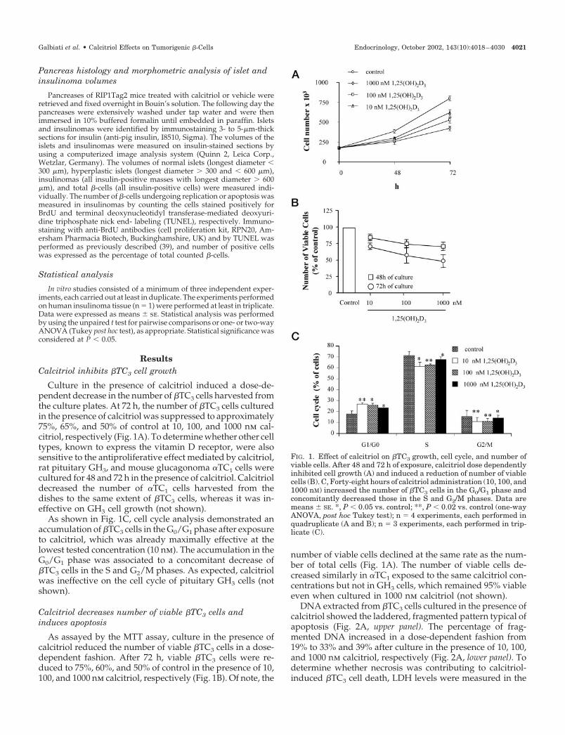
Culture in the presence of calcitriol induced a dose-de-pendent decrease in the number of �TC3 cells harvested fromthe culture plates. At 72 h, the number of �TC3 cells culturedin the presence of calcitriol was suppressed to approximately75%, 65%, and 50% of control at 10, 100, and 1000 nm cal-citriol, respectively (Fig. 1A). To determine whether other celltypes, known to express the vitamin D receptor, were alsosensitive to the antiproliferative effect mediated by calcitriol,rat pituitary GH3, and mouse glucagonoma �TC1 cells werecultured for 48 and 72 h in the presence of calcitriol. Calcitrioldecreased the number of �TC1 cells harvested from thedishes to the same extent of �TC3 cells, whereas it was in-effective on GH3 cell growth (not shown).
As shown in Fig. 1C, cell cycle analysis demonstrated anaccumulation of �TC3 cells in the G0/G1 phase after exposureto calcitriol, which was already maximally effective at thelowest tested concentration (10 nm). The accumulation in theG0/G1 phase was associated to a concomitant decrease of�TC3 cells in the S and G2/M phases. As expected, calcitriolwas ineffective on the cell cycle of pituitary GH3 cells (notshown).
Calcitriol decreases number of viable �TC3 cells andinduces apoptosis
As assayed by the MTT assay, culture in the presence ofcalcitriol reduced the number of viable �TC3 cells in a dose-dependent fashion. After 72 h, viable �TC3 cells were re-duced to 75%, 60%, and 50% of control in the presence of 10,100, and 1000 nm calcitriol, respectively (Fig. 1B). Of note, the
number of viable cells declined at the same rate as the num-ber of total cells (Fig. 1A). The number of viable cells de-creased similarly in �TC1 exposed to the same calcitriol con-centrations but not in GH3 cells, which remained 95% viableeven when cultured in 1000 nm calcitriol (not shown).
DNA extracted from �TC3 cells cultured in the presence ofcalcitriol showed the laddered, fragmented pattern typical ofapoptosis (Fig. 2A, upper panel). The percentage of frag-mented DNA increased in a dose-dependent fashion from19% to 33% and 39% after culture in the presence of 10, 100,and 1000 nm calcitriol, respectively (Fig. 2A, lower panel). Todetermine whether necrosis was contributing to calcitriol-induced �TC3 cell death, LDH levels were measured in the
FIG. 1. Effect of calcitriol on �TC3 growth, cell cycle, and number ofviable cells. After 48 and 72 h of exposure, calcitriol dose dependentlyinhibited cell growth (A) and induced a reduction of number of viablecells (B). C, Forty-eight hours of calcitriol administration (10, 100, and1000 nM) increased the number of �TC3 cells in the G0/G1 phase andconcomitantly decreased those in the S and G2/M phases. Data aremeans � SE. *, P � 0.05 vs. control; **, P � 0.02 vs. control (one-wayANOVA, post hoc Tukey test); n � 4 experiments, each performed inquadruplicate (A and B); n � 3 experiments, each performed in trip-licate (C).
Galbiati et al. • Calcitriol Effects on Tumorigenic �-Cells Endocrinology, October 2002, 143(10):4018–4030 4021

FIG. 2. Calcitriol-induced apoptosis in �TC3 cells. A, Exposure to calcitriol induced �TC3 cell apoptosis, as shown by the typical ladder-like DNAappearance (top). Quantification of fragmented DNA showed a dose-dependent effect of calcitriol (bottom; means � SE, n � 3 experiments, eachperformed in triplicate; *, P � 0.01 vs. control; **, P � 0.05 vs. control and P � 0.01 vs. 100 and 1000 nM, one-way ANOVA, post hoc Tukeytest). B, Immunofluorescence for insulin (green) and DAPI (blue) showing a dose-dependent reduction in �TC3 cell number and nuclearcondensation and fragmentation of calcitriol-treated cells. C, Apoptosis was associated with a dose-dependent increase in caspase-3 activity,which was prevented by 50 �M caspase-3 inhibitor Z-VAD-FMK (means � SE, n � 4 experiments, each performed in duplicate; *, P � 0.02; **,P � 0.01 vs. control one-way ANOVA, post hoc Tukey test).
4022 Endocrinology, October 2002, 143(10):4018–4030 Galbiati et al. • Calcitriol Effects on Tumorigenic �-Cells

culture media. The similar LDH levels detected in the me-dium containing calcitriol and control medium excluded thispossibility (not shown). Furthermore, calcitriol-induced apo-ptosis was confirmed by immunofluorescence staining withinsulin and DAPI performed in �TC3 cells exposed or not tocalcitriol (Fig. 2B). Nuclear staining with DAPI confirmed thedose-dependent decrease in the number of �TC3 cells ex-posed to calcitriol. Also, the number of condensed and frag-mented nuclei, a morphologic feature typical of apoptosis,increased after calcitriol exposure. To identify the cellularpathways involved in calcitriol-induced �TC3 apoptosis,caspase-3 activity was measured after 48 and 72 h of exposureto increasing calcitriol concentrations and was comparedwith the activity of control cells. At 48 h, calcitriol inducedan increase in caspase-3 activity to 216%, 263%, and 310% ofcontrol cells at 10, 100, and 1000 nm, respectively (Fig. 2C).Calcitriol-induced increase in caspase-3 activity was selec-tively abolished by the caspase inhibitor (Z-VAD-FMK) (Fig.2C). Caspase activity remained slightly, albeit significantly,higher than in control cells, even after 72 h (not shown).
Calcitriol induces ERK1 and ERK2 phosphorylation via anongenomic pathway
It has been previously shown that calcitriol can induce arapid activation of the MAPK pathway in certain cell types(40) and that, in wild-type p53-retaining cells (such as �TC3),inhibition of MAPK efficiently abrogates p53-induced celldeath (41). Thus, we explored whether calcitriol could acti-vate ERK1/2 in �TC3 cells and ERK inhibition was capableto prevent in the proapoptotic effect of calcitriol on thesecells. To do so, proteins extracted from �TC3 cells that hadbeen exposed for 20 min to increasing calcitriol concentra-tions were analyzed by immunoblotting with antiseraagainst unphosphorylated and phosphorylated ERK1 andERK2. Calcitriol induced an increase in phosphorylatedERK1/ERK2 to 136%, 150%, 159%, and 184% of control cellsafter treatment with 1, 10, 100, and 1000 nm calcitriol, re-spectively (Fig. 3A). Calcitriol-induced ERK phosphoryla-tion was significantly prevented by the MEK inhibitor UO126and, to a lesser extent, the PKC inhibitor staurosporine (Fig.3B), which were both ineffective alone (Fig. 3A).
Inhibition of MEK and PKC partially prevents calcitriol-induced decrease in number of viable �TC3 cells
UO126 and staurosporine not only prevented calcitriol-induced ERK1/2 phosphorylation but also reduced calcitriol-mediated decrease in the number of viable �TC3 cells. At theconcentration of 2 nm, UO126 increased the number of viable�TC3 cells exposed to 1000 nm calcitriol from 68% to 90% ofcontrol cells (Fig. 3C). At 5 nm, UO126 lost its protective effectand became cytotoxic (not shown). Calcitriol-induced celldeath was also reduced significantly by 5 nm staurosporinethat, albeit less potent than UO126, increased the number ofviable �TC3 cells exposed to 1000 nm calcitriol from 70% toabout 82% of control (Fig. 3D). Noteworthy, calcitriol-medi-ated �TC3 cell death was unaffected by forskolin (1–10 �m),wortmannin (100 nm), and N-methylarginine (1 �m) (notshown). Calcitriol induced a significant increase in PKC ac-tivity of �TC3 exposed for 5 min to 100 nm hormone con-
centration (to 164% of control cells). Calcitriol-induced PKCactivation was completely prevented by coadministration ofstaurosporine (5 nm), which was ineffective when adminis-tered alone (Fig. 3E).
Calcitriol decreases insulin gene expression, insulin content,and release in �TC3 cells
Insulin gene expression, insulin content, and basal insulinrelease were measured in �TC3 cells cultured in the presenceof increasing calcitriol concentrations. These experimentswere carried out to determine whether, besides its antitu-morigenic activity, calcitriol has also antiinsuligenic effectson mouse insulinoma cells. Indeed, calcitriol dose depen-dently inhibited insulin mRNA levels in �TC3. After 48 h,insulin mRNA levels decreased to 75%, 70%, and 46% ofcontrol cells in the presence of 10, 100, and 1000 nm calcitriol,respectively (Fig. 4A). Down-regulation of insulin geneexpression was completely prevented by staurosporine and,to a lesser extent, by the phosphatidylinositol 3- kinase in-hibitor wortmannin and forskolin. In contrast, UO126 andN-methylarginine were ineffective in this regard (Fig. 4B). Alltested inhibitors did not change the insulin mRNA levelswhen administered alone (Fig. 4C). Reduced insulin genetranscription was associated with a significant decrease in�TC3 insulin content (Fig. 4D) and release (Fig. 4E).
Calcitriol decreases insulin gene expression and insulin secretionbut fails to induce apoptosis in primary cultures of benign humaninsulinoma cells
Calcitriol also exerted a clear antiinsulinogenic effect onprimary cultures derived from a human insulinoma. InsulinmRNA levels of human insulinoma cells exposed to 1000 nmcalcitriol decreased to 70% of control (Fig. 5A). Unstimulatedinsulin release by human insulinoma cells cultured in thepresence of 100 and 1000 nm calcitriol also decreased signif-icantly to 75% of control cells (Fig. 5B). The MTT and DNAfragmentation assays were not performed on primary cul-ture of human insulinoma because they were composed alsoof non-�-cells. Insulinoma cells, identified by insulin andPDX-1 staining, were indeed only about 30–40% of total cellpopulation (Fig. 5C). Because of that, detection of humaninsulinoma cells undergoing apoptosis was performed byimmunocytochemistry of fixed monolayers stained for in-sulin and DAPI (Fig. 5D). Human insulinoma cells wereinsensitive to calcitriol-induced cytotoxicity as indicated bythe extremely low number of insulin- positive cells showingnuclear condensation or fragmentation (approximately0.5–1% in either vehicle and calcitriol-cultured cultures).
Calcitriol induces ERK1 and ERK2 phosphorylation in humanpancreatic islets and decreases insulin gene expression, insulincontent, and release
Aimed at determining whether nontumorigenic human �cells were also sensitive to nongenomic calcitriol effects, is-lets were exposed for 20 min to calcitriol, and levels of un-phosphorylated and phosphorylated ERK1/2 were deter-mined by immunoblotting. As shown in Fig. 6A, calcitriolinduced a significant increase in phosphorylated ERK1/2 toabout 120%, 135%, and 140% of control cells in the presenceof 10, 100, and 1000 nm calcitriol, respectively. As in �TC3 and
Galbiati et al. • Calcitriol Effects on Tumorigenic �-Cells Endocrinology, October 2002, 143(10):4018–4030 4023

FIG. 3. Calcitriol signal transduction pathway in �TC3 cells. A, Calcitriol induced a rapid phosphorylation (within 20 min) of ERK1 (42 kDa)and ERK2 (44 kDa) in a dose-dependent fashion. B, This effect, quantitatively similar to that induced by serum alone, was prevented by UO126(MEK inhibitor) and staurosporine (PKC inhibitor), which had no effect when administered alone (A) (means � SE, n � 4 experiments, eachperformed in triplicate, one-way ANOVA, post hoc Tukey test; #, P � 0.01 vs. control without FCS; **, P � 0.01 vs. calcitriol alone; *, P � 0.05vs. calcitriol alone). C and D, Calcitriol-induced reduction in number of viable cells (assayed by the MTT method) was prevented by UO126 and,to a lesser extent, staurosporine, which were both ineffective when given alone. (means � SE, n � 4 experiments, each performed in 16 replicates,one-way ANOVA, post hoc Tukey test, *, P � 0.01 vs. control; **, P � 0.01 vs. control; P � 0.05 vs. calcitriol alone). E, After 5 min of exposure,calcitriol induced a significant increase in PKC activity that was completely abolished by staurosporine (5 nM), which was ineffective whenadministered alone (means � SE, n � 3 experiments, each performed triplicate, one-way ANOVA, post hoc Tukey test; *, P � 0.01 vs. control).
4024 Endocrinology, October 2002, 143(10):4018–4030 Galbiati et al. • Calcitriol Effects on Tumorigenic �-Cells

human insulinoma cells, calcitriol induced a significant de-crease in mRNA levels, content, and release of insulin inhuman islets despite the lack of a clear dose-dependent effect
(Fig. 6, B–D). Immunofluorescence experiments showed norelevant differences in nuclear morphology of human isletcells cultured in absence or presence of 10 and 1000 nm
FIG. 4. Effect of calcitriol on insulin gene expression, insulin content, and release in �TC3 cells. A, Calcitriol induced a dose-dependent decreasein insulin gene expression (means � SE, n � 4 experiments, each performed in duplicate, one-way ANOVA, post hoc Tukey test; *, P � 0.01vs. control; **, P � 0.01 vs. 10 nmol/liter). B, Down-regulation of insulin gene expression was prevented by staurosporine, wortmannin, andforskolin but not by UO126 and N-methylarginine. C, All of these inhibitors were ineffective when administered alone (means � SE, n � 3experiments, each performed in duplicate, one-way ANOVA, post hoc Tukey test; *, P � 0.01 vs. control). D and E, Calcitriol significantlydecreased content and release of insulin in �TC3 cells (means � SE, n � 7 experiments, each performed in triplicate, one-way ANOVA, posthoc Tukey test; **, P � 0.03 vs. control; #, P � 0.01 vs. control).
Galbiati et al. • Calcitriol Effects on Tumorigenic �-Cells Endocrinology, October 2002, 143(10):4018–4030 4025

calcitriol, but insulin immunoreactivity was weaker in hu-man islets exposed to calcitriol (data not shown). Togetherthese data demonstrate that the antiinsulinogenic activity ofcalcitriol is not limited to the neoplastic phenotype but ispresent also in normal �-cells.
Calcitriol treatment reduces the volume of solid �-celltumors by increasing apoptotic cell death ofadenomatous cells
Insulinoma-bearing RIP1Tag2 mice were treated for 3 wkwith calcitriol to ascertain whether the antitumorigenic ac-tivity detected in vitro was confirmed in vivo. At the end oftreatment, the number of insulinomas present in the pan-creases harvested from calcitriol-treated mice was lower thanthat of mice treated with vehicle, even though the differencedid not reach statistical significance (6.3 � 1.3 vs. 9.2 � 1.6,respectively, NS). Moreover, morphometric analysis of pan-creatic sections stained for insulin showed that treatmentwith calcitriol did not affect the volume of the normal isletpopulation (Fig. 7A). The volume of the hyperplastic isletsdecreased after calcitriol administration but not significantly(Fig. 7A). In contrast, the effects of calcitriol were particularlyremarkable when considering the global �-cell populationand the insulinomas, whose volumes were significantly re-
duced by calcitriol (Fig. 7B). Reduction of insulinoma vol-umes was a consequence of increased adenoma cell deathrather than reduced replication. As shown in Fig. 7C, about5% of insulinoma cells of calcitriol-treated mice were under-going apoptosis, a 3-fold increase, compared with the apo-ptotic rate detected in the insulinomas of vehicle-treatedmice. In contrast, cell replication remained similar in theinsulinomas of the two groups (BrdU� cells 14.3% � 2.1% vs.13.4% � 1.6% in calcitriol- vs. vehicle-treated, respectively).
Despite the significant reduction in insulinoma volumes,treatment with calcitriol did not affect the glycemic levels ofRIP1Tag2 mice. As expected, placebo-treated mice gainedweight during the treatment (from 22.8 � 1.2 to 25.8 � 1.3 g,P � 0.01), but calcitriol-treated mice lost weight (from 22.4 �1.3 to 19.8 � 1.1 g, P � 0.05). Such loss of body weight waslikely consequent to the anorexia induced by hypercalcemiathat, at this dosage of calcitriol, is usually observed in mice(9.4 � 0.1 vs. 10.6 � 0.3 mg/dl, vehicle vs. calcitriol-treatedmice, respectively, P � 0.01). To determine whether calcitriolhad in vivo antiinsulinogenic activity, circulating insulin lev-els were measured in RIP1Tag2 mice before and after treat-ment with calcitriol or vehicle. Results showed a significantreduction in insulin levels of calcitriol-treated mice (from33 � 6 to 14 � 2 �U/ml; P � 0.05), but insulinemia ofvehicle-treated mice remained stable in the 30 �U/ml range.
FIG. 5. Effect of calcitriol on primarycultures of a human benign insulinoma.A, Calcitriol (1000 nM) induced a de-crease of insulin mRNA levels in benignhuman insulinoma cells to approxi-mately 70% of vehicle-treated cells (n �1 experiment performed in triplicate; *,P � 0.05 vs. control). B, Basal release ofinsulin by human insulinoma cells wassignificantly decreased by calcitriol,with 100 nM being already maximallyeffective (means � SE, n � 3, each per-formed in triplicate, one-way ANOVA,post hoc Tukey test; *, P � 0.05 vs. con-trol). C, Immunofluorescence for PDX-1(upper panel) and DAPI (lower panel)showing that insulinoma PDX-1-posi-tive cells accounted for only 30–40% ofthe total cell population. D, Immuno-fluorescence for insulin (upper panel)and DAPI (lower panel) did not showmorphological evidence of nuclear con-densation suggestive of ongoing apo-ptosis in human insulinoma cells. Twoinsulin-positive cells undergoing repli-cation are evident on the bottom right.
4026 Endocrinology, October 2002, 143(10):4018–4030 Galbiati et al. • Calcitriol Effects on Tumorigenic �-Cells

Discussion
Within the last two decades, a number of studies haveshown that the VDR is present not only in classical targettissue such as bone, kidney, and intestine but also in severalother nonclassical tissues, including the pancreatic �-cell.Those studies demonstrated that calcitriol and analogs areinvolved in a multitude of cellular processes including po-tent antiproliferative and prodifferentiating effects on cancercell lines in vitro and marked antitumoral effects in vivo (42).However, no studies have been conducted to address theefficacy of calcitriol in the treatment of �-cell tumors. In thisstudy, we analyzed in vitro and in vivo the effects of a pro-longed exposure to calcitriol on the proliferation, viability,and insulinogenic properties of mouse tumorigenic �-cellsand �-cell tumors. We also explored the effects exerted bycalcitriol on primary cultures of a human benign insulinomaand isolated human islets.
The results showed that calcitriol induces a wide spectrumof negative effects on mouse insulinoma cells ranging frominduction of growth arrest and apoptosis, down-regulationof insulin gene transcription, and reduction of solid �-cell
tumors. Calcitriol also induced growth inhibition and apo-ptosis of �TC1 glucagonoma cells but not pituitary adenomaGH3 cells, which also express the VDR (not shown) (43). Theantiproliferative effects that calcitriol induced in vitro on�TC3 cells were due to a combination of decreased replica-tion and increased cell death. Calcitriol induced an increasein the number of �TC3 cells in the G0/G1 phase of the cellcycle and a concomitant decrease of cells population in theS and G2/M phases. This effect of calcitriol, previously de-scribed to occur in human prostate cancer cells (44) and incolonic adenocarcinoma CaCo-2 cells (45), was already max-imal at the concentration of 10 nm in �TC3 cells.
Another mechanism by which calcitriol inhibited �TC3
growth was the induction of apoptosis. Apoptosis was as-sociated with an increase in caspase-3 activity, indicating theinvolvement of general apoptotic pathways. Experiments arecurrently ongoing to explore the mechanisms responsible forthe induction of �TC3 cell apoptosis by calcitriol. In partic-ular, it will be interesting to determine the role exerted byBcl-2, which blocks either Apaf-1 activation (46) and p53-induced cell death (47) and whether Bcl-2 overexpression
FIG. 6. Effect of calcitriol on human pancreatic islets. A, After 20 min of exposure, calcitriol induced a dose-dependent phosphorylation of ERK1/2. (means � SE, n � 3 experiments, each performed in triplicate, one-way ANOVA, post hoc Tukey test; *, P � 0.05 vs. control, **, P � 0.01vs. control). B–D, In human islets calcitriol significantly reduced gene expression, content, and release of insulin (means � SE, n � 4 experiments,each performed in triplicate, one-way ANOVA, post hoc Tukey test; *, P � 0.05 vs. control; ** P � 0.01 vs. control).
Galbiati et al. • Calcitriol Effects on Tumorigenic �-Cells Endocrinology, October 2002, 143(10):4018–4030 4027

might prevent �TC3 cells apoptosis, as previously shown inprostate cancer cells exposed to a vitamin D3 analog (46).
Calcitriol may generate biological responses via regulationof gene transcription but also via nongenomic pathways.Indeed, calcitriol increases the intracellular level of manysecond messengers including cAMP, inositols, calcium, andceramides that, in turn, activate a variety of protein kinases,including PKC, Raf, MAPK, and Src kinases (48–51). Fur-thermore, Raf 1 and MEK, upstream effectors of MAPK, arestimulated by calcitriol-induced PKC activation (52). We hereprovide evidence that, in pancreatic �-cells, calcitriol acti-vates the MAPK cascade via a nongenomic pathway. Thisoccurs in mouse insulinoma cells as well as in human isletsas shown by the rapid phosphorylation of ERK1 and ERK2 oncalcitriol treatment. ERK1/2 phosphorylation was effica-
ciously prevented by UO126 and staurosporine, indicatingthat activation of PKC is upstream of MAPK activation. Note-worthy, MAPK activation appears to contribute to calcitriol-induced cytotoxicity because MEK inhibition with UO126partially prevented this effect in �TC3 cells.
Rather unexpectedly, calcitriol decreased insulin gene ex-pression, insulin content, and insulin release of all the �-celltypes analyzed in this study, including malignant cells(�TC3), benign cells (human insulinoma), and nontumori-genic cells (isolated islets). These data contrast with the vastmajority of the studies on the effects of vitamin D on theendocrine pancreas, which consistently found a beneficialeffect of calcitriol on �-cell function. It should be noted,however, that almost all of these studies were performed insituations of vitamin D depletion or islet cell damage. In
FIG. 7. Calcitriol treatment exerts in vivo antineoplastic effect on solid �-cell tumors. A, RIP1Tag2 mice were treated with calcitriol (5 �g/kgorally 3 times/wk, n � 6) or vehicle (n � 7). Morphometric analysis of the harvested pancreas showed no difference in the volumes of the normaland hyperplastic islet populations between calcitriol- and vehicle-treated mice. B, In contrast, a significant reduction of insulinomas and total�-cell mass volumes are evident in calcitriol-treated mice (means � SE, *, P � 0.02 vs. vehicle treated). C, A significant increase (3-fold) inthe percentage of cells undergoing apoptosis (TUNEL�) was detected in the insulinomas of calcitriol-treated mice (means � SE, *, P � 0.05 vs.calcitriol-treated mice). D, Representative sections of pancreases harvested from vehicle (upper panels) and calcitriol-treated (bottom panels)mice stained for insulin and TUNEL, showing the reduced insulinoma volumes and the increased number of apoptotic cells rate present in thelatter group. Treatment with calcitriol did not induce in vivo a decrease in the rate of replication of insulinoma cells.
4028 Endocrinology, October 2002, 143(10):4018–4030 Galbiati et al. • Calcitriol Effects on Tumorigenic �-Cells

contrast to the antitumorigenic, the antiinsulinogenic activityexerted by calcitriol did not involve the MAPK pathway.Indeed, down-regulation of insulin gene transcription wasnot prevented by UO126 but was completely abolished bystaurosporine, thereby indicating that this particular effect ofcalcitriol involves PKC but is independent from MAPK ac-tivation. The evidence that in �TC3 cells, UO126 efficaciouslyprevented calcitriol-induced cytotoxicity but failed to reducethe down-regulation of the insulin gene, confirms that thelatter phenomenon was specific and not the result of anadmixture of viable and dying cells. That the antitumorigenicand the antiinsulinogenic effects exerted by calcitriol on �TC3cells are mediated by distinct intracellular pathways is fur-ther supported by the evidence that wortmannin and fors-kolin significantly reduced the down-regulation of the in-sulin gene but were completely ineffective in the preventionof calcitriol-induced cytotoxicity. It is now clear that theinsulin receptor signaling pathway is active in pancreatic�-cells (53) and plays an important role in �-cell homeostasis(54–58). Interestingly, staurosporine, which prevents PKC-dependent activation of insulin receptor substrate-1 (59), andwortmannin, which inhibits phosphatidylinositol 3-kinase,were both effective in preventing calcitriol-induced down-regulation of insulin mRNA. Perhaps calcitriol may inducethe overexpression of insulin receptor substrate-1, which, inpancreatic �-cells, determines reduced mRNA levels, bio-synthesis, and content of insulin (60, 61).
Antitumorigenic activity of calcitriol was analyzed in vivoin transgenic RIP1Tag2 mice, which represent an extremelywell-characterized model of �-cell tumorigenesis. Threeweeks of calcitriol treatment significantly altered �-cell tu-morigenesis in these mice. Although the volumes of thenormal islet populations were similar in calcitriol- and ve-hicle-treated mice, those of the insulinomas were signifi-cantly reduced in the former group. This evidence is con-sistent with the results obtained in vitro, which showed thatcalcitriol cytotoxicity is limited to the �-cells that show amalignant phenotype. Noteworthy, the decrease in insuli-noma volumes was associated to a 3-fold increase in apo-ptotic cell death, but adenoma cell replication remained sim-ilar in the two groups of mice. Other mechanisms, however,may have contributed to the in vivo antitumorigenic effect ofcalcitriol. Besides growth inhibition, calcitriol and analogshave been reported to exert their antitumorigenic activity viainhibition of angiogenesis (62) and by decreasing the inva-siveness of cancer cells (63). Perhaps calcitriol may act atdifferent sites of the multistep tumorigenic process describedin RIP1Tag2 mice. Inhibition of angiogenesis may reduce invivo the number of islets undergoing the angiogenic switchand the following transition to adenomas, but overexpres-sion of certain adhesion molecules may hinder the transitionfrom adenomas to carcinomas. Despite the reduced insuli-noma volumes, glycemic levels of calcitriol- and vehicle-treated mice remained similar. This apparent paradox maydepend on the anorexia resulted from calcitriol-induced hy-percalcemia. To compensate for hypoglycemia, vehicle-treated mice became hyperphagic and gained weight; in-stead, calcitriol-treated mice became anorectic and lostweight. Thus, the maintenance of similar blood glucose lev-els, despite a completely different feeding behavior, may be
considered a remarkable clinical achievement. Moreover, cir-culating insulin levels decreased significantly in calcitriol-treated mice but remained stable in vehicle-treated mice.
In conclusion, we found that calcitriol exerts a potent an-titumorigenic and antiinsulinogenic effect on actively repli-cating murine insulinoma �-cells in vitro as well as in vivo. Atthe doses used in this study, calcitriol shows also a clearantiinsulinogenic effect on human �-cells from either normalislets or benign insulinoma. These data provide the rationalefor testing the efficacy of calcitriol in the clinical managementof patients with �-cell tumors. Calcitriol might be particu-larly helpful in patients with malignant insulinomas who dieof metastatic cancer dissemination and untreatable hypogly-cemia in which calcitriol might exert antineoplastic and an-tiinsulinogenic effects. The availability of calcitriol analogs(64), characterized by a lower calcemic effect, is particularlyappealing because they will probably consent to increase thedose of drug that can be safely given to the patient, therebyincreasing its biological effect on the neoplastic �-cells.
Acknowledgments
Received February 20, 2002. Accepted June 13, 2002.Address all correspondence and requests for reprints to: Alberto M.
Davalli, Department of Medicine San Raffaele Scientific Institute, ViaOlgettina 60, 20132 Milan, Italy. E-mail: [email protected].
This work was supported by a postdoctoral fellowship grant from theMinistry of Health of Italy (projects RF98.52 and RF98.50 to F.G., L.P.,and M.C.) and Telethon-Italy Grant A118 (to A.M.D.) and Grant E739 (toF.B.F.).
References
1. Bouillon R, Okamura WH, Norman AW 1995 Structure-function relationshipsin the vitamin D endocrine system. Endocr Rev 16:200–257
2. Jones G, Strugnell SA, DeLuca HF 1998 Current understanding of the mo-lecular actions of vitamin D. Physiol. Rev 78:1193–1231
3. Otoshi T, Iwata H, Kitano M, Nishizawa Y, Morii H, Yano Y, Otani S,Fukushima S 1995 Inhibition of intestinal tumor development in rat multi-organ carcinogenesis and aberrant crypt foci in rat colon carcinogenesis by22-oxa-calcitriol, a synthetic analogue of 1�, 25-dihydroxyvitamin D3. Carci-nogenesis 16:2091–2097
4. Kawa S, Yoshizawa K, Tokoo M, Imai H, Oguchi H, Kiyosawa K, HommaT, Nikaido T, Furihata K 1996 Inhibitory effect of 220-oxa-1,25-dihydroxyvi-tamin D3 on the proliferation of pancreatic cancer cell lines. Gastroenterology110:1605–1613
5. Baudet C, Chevalier G, Chassevent A, Canova C, Filmon R, Larra F, BrachetP, Wion D 1996 1,25-Dihydroxyvitamin D3 induces programmed cell death ina rat glioma cell line. J Neurosci Res 46:540–550
6. Danielsson C, Fehsel K, Polly P, Carlberg C 1998 Differential apoptoticresponse of human melanoma cells to 1�, 25-dihydroxyvitamin D3 and itsanalogues. Cell Death Differ 5:946–952
7. VanWeelden K, Flanagan L, Binderup L, Tenniswood M, Welsh J 1998Apoptotic regression of MCF-7 xenografts in nude mice treated with vitaminD3 analog, EB1089. Endocrinology 139:2102–2110
8. Feldman D, Zhao XY, Krishnan AV 2000 Vitamin D and prostate cancer.Endocrinology 141:5–9
9. Hershberger PA, Modzelewski RA, Shurin ZR, Rueger RM, Trump DL,Johnson CS 1999 1,25-Dihydroxycholecalciferol (1,25-D3) inhibits the growthof squamous cell carcinoma and down-modulates p21 (waf1/Cip1) in vitro andin vivo. Cancer Res 59:2644–2649
10. Smith DC, Johnson CS, Freeman CC, Muindi J, Wilson JW, Trump DL 1999A phase I trial of calcitriol (1,25-dihydroxycholecalciferol) in patients withadvanced malignancy. Clin Cancer Res 5:1339–1345
11. Pavelic K, Hrascan R, Kapitanovic S, Karapandza N, Vranes Z, Belicza M,Kruslin B, Cabrijan T 1995 Multiple genetic alterations in malignant meta-static insulinomas. J Pathol 177:395–400
12. Ruszniewski P, Malka D 2000 Hepatic arterial chemoembolization in themanagement of advanced digestive endocrine tumors. Digestion 62(Suppl1):79–83
13. Schott M, Scherbaum WA, Feldkamp J 2000 Drug therapy of endocrineneoplasms. Part II: malignant gastrinomas, insulinomas, glucagonomas, car-cinoids and other tumors. Med Klin 95:81–84
Galbiati et al. • Calcitriol Effects on Tumorigenic �-Cells Endocrinology, October 2002, 143(10):4018–4030 4029

14. Arnold R, Simon B, Wied M 2000 Treatment of neuroendocrine GEP tumorswith somatostatin analogues: a review. Digestion 62(Suppl 1):84–91
15. Ishida H, Norman AW 1988 Demonstration of high affinity receptor for 1,25-dihydroxyvitamin D3 in rat pancreas. Mol Cell Endocrinol 60:109–117
16. Johnson JA, Grande JP, Roche PC, Kumar R 1994 Immunohistochemicallocalization of the 1,25(OH)2D3 receptor and calbindin D28K in human and ratpancreas. Am J Physiol 267:E356–E360
17. Norman AW, Frankel BJ, Heldt AM, Grodsky GM 1980 Vitamin D deficiencyinhibits pancreatic secretion of insulin. Science 209:823–825
18. Chertow BS, Sivitz WI, Baranetsky NG, Clark SA, Waite A, DeLuca HF 1983Cellular mechanisms of insulin release: the effects of vitamin D deficiency andrepletion on rat insulin secretion. Endocrinology 113:1511–1518
19. Gedik O, Akalin S 1986 Effects of vitamin D deficiency and repletion oninsulin and glucagon secretion in man. Diabetologia 29:142–145
20. Mak RH 1992 Intravenous 1, 25 dihydroxycholecalciferol corrects glucoseintolerance in hemodialysis patients. Kidney Int 41:1049–1054
21. Chertow BS, Sivitz WI, Baranetsky Cordle MB, DeLuca HF 1986 Islet insulinrelease and net calcium retention in vitro in vitamin D-deficient rats. Diabetes35:771–775
22. Lee S, Clark SA, Gill RK, Christakos S 1994 1, 25-dihydroxyvitamin D3 andpancreatic �-cell function: vitamin D receptors, gene expression, and insulinsecretion. Endocrinology 134:1602–1610
23. Mathieu C, Waer M, Laureys J, Rutgeerts O, Bouillon R 1994 Prevention ofautoimmune diabetes in NOD mice by 1,25-dihydroxyvitamin D3. Diabeto-logia 37:552–558
24. Gregori S, Giarratona N, Smiroldo S, Uskokkovic M, Adorini L 2002 A 1�,25-dihydroxivitamin D3 analog enhances regulatory T-cells and arrests auto-immune diabetes in NOD mice. Diabetes 51:1367–1374
25. Mcdermott MF, Ramachandran A, Ogunkolade BW, Aganna E, Curtis D,Boucher BJ, Snehalatha C, Hitman GA 1997 Allelic variations in the vitaminD receptor influences susceptibility to IDDM in indian Asians. Diabetologia40:971–975
26. Hitman GA, Mannan N, McDermott MF, Aganna E, Ogunkolade BW, HalesCN, Boucher BJ 1998 Vitamin D receptor polymorphisms influence insulinsecretion in Bangladeshi Asians. Diabetes 47:688–690
27. Speer G, Cseh K, Winker G, Vargha P, Braun E, Takacs I, Lakato P 2001Vitamin D and estrogen receptor gene polymorphisms in type 2 diabetesmellitus and in android type obesity. Eur J Endocrinol 144:385–389
28. Efrat S, Linde S, Kofod H, Spector D, Delannoy M, Grant S, Hanahan D,Baekkeskov S 1988 Beta-cell lines derived from transgenic mice expressing ahybrid insulin gene-oncogene. Proc Natl Acad Sci USA 85:9037–9041
29. Hanahan D 1985 Heritable formation of pancreatic beta-cell tumors in trans-genic mice expressing recombinant insulin/simian virus 40 oncogenes. Nature315:115–122
30. Teitelman G, Alpert S, Hanahan D 1988 Proliferation, senescence, and neo-plastic progression of � cells in hyperplasic pancreatic islets. Cell 52:97–105
31. Folkman J, Watson K, Ingber D, Hanahan D 1989 Induction of angiogenesisduring the transition from hyperplasia to neoplasia. Nature 339:58–61
32. Perl AK, Wilgenbus P, Dahl U, Semb H, Christofori G 1998 A causal role forE-cadherin in the transition from adenoma to carcinoma. Nature 392:190–193
33. Perl AK, Dahl U, Wilgenbus P, Cremer H, Semb H, Chrisofori G 1999Reduced expression of neural cell adhesion molecule induces metastatic dis-semination of pancreatic � tumor cells. Nat Med 5:286–291
34. Solcia E, Rindi G, Palotti D, La Rosa S, Capella C, Fiocca R 1999 Clinico-pathological profile as a basis for classification on the endocrine tumors of thegastroenteropancreatic tract. Ann Oncol 10:S9–S15
35. Socci C, Falqui L, Davalli AM, Ricordi C, Braghi S, Bertuzzi F, Maffi P, SecchiA, Gavazzi F, Freschi M, Magistretti P, Socci S, Vignali A, Di Carlo V, PozzaG 1991 Fresh human islet transplantation to replace pancreatic endocrinefunction in type I diabetic patients. Report of six cases. Acta Diabetol 28:151–157
36. Mosmann T 1983 Rapid colorimetric assay for cellular growth and survival:application to proliferation and cytotoxicity assays. J Immunol Methods 65:55–63
37. Davalli AM, Galbiati F, Bertuzzi F, Polastri L, Pontiroli AE, Perego L, FreschiM, Pozza G, Folli F, Meoni C 2000 Insulin-secreting pituitary GH3 cells: apotential �-cell surrogate for diabetes cell therapy. Cell Transplant 8:841–851
38. Favata MF, Horiuchi KY, Manos EJ, Daulerio AJ, Stradley DA, Feeser WS,Van Dyk DE, Pittis WJ, Earl RA, Hobbs F, Copeland RA, Magolda RL,Scherle PA, Trzaskas JM 1998 Identification of a novel inhibitor of mitogen-activated protein kinase. J Biol Chem 273:18623–18632
39. Davalli AM, Ogawa Y, Scaglia L, Wu YJ, Hollister J, Bonner-Weir S, WeirGC 1995 Function, mass, and replication of porcine and rat islets transplantedinto diabetic nude mice. Diabetes 44:104–111
40. Song X, Bishop JE, Okamura WH, Norman AW 1998 Stimulation of phos-phorylation of mitogen-activated protein kinase by 1�,25-dihydroxyvitaminD3 in promyelocytic NB4 leukemia cells: a structure-function study. Endo-crinology 139:457–465
41. Ryan KM, Ernst MK, Rice NR, Vousden KH 2000 Role of NF-�B in p-53-mediated programmed cell death. Nature 404:892–897
42. Verstuyf A, Segaert S, Verlinden L, Casteels K, Bouillon R, Mathieu C 1998Recent developments in the use of vitamin D analogues. Curr Opin NephrolHypertens 7:397–403
43. Atley LM, Lefroy N, Wark JD 1995 1, 25-Dihydroxyvitamin D3-induced up-regulation of the thyrotropin-releasing hormone receptor in clonal rat pituitaryGH3 cells. J Endocrinol 147:397–404
44. Zhuang SH, Burnstein KL 1998 Antiproliferative effect of 1alpha, 25-dihy-droxyvitamin D3 in human prostate cancer cell line LNCaP involves reductionof cyclin-dependent kinase 2 activity and persistent G1 accumulation. Endo-crinology 139:1197–1207
45. Scaglione-Sewell BA, Bissonnette M, Skarosi S, Abraham C, Brasitus TA2000 A vitamin D3 analog induces a G1-phase arrest in CaCo-2 cells byinhibiting Cdk2 and Cdk6: roles of cyclin E, p21waf1, and p27kip1. Endocri-nology 141:3931–3939
46. Hausmann G, O’Reilly LA, van Driel R, Beaumont JG, Strasser A, AdamsJM, Huang DC 2000 Pro-apoptotic apoptosis protease-activating factor 1(Apaf-1) has a cytoplasmic localization distinct from Bcl-2 or Bclx-(L). J CellBiol 149:623–634
47. Chiou SK, Rao L, White E 1994 Bcl-2 blocks p53-dependent apoptosis. Mol CellBiol 14:2556–2563 0
48. Blutt SE, McDonnel TJ, Polek TC, Weigel NL 2000 Calcitriol-induced apo-ptosis in LNCaP cells is blocked by overexpression of Bcl-2. Endocrinology141:10–17
49. Hmama Z, Nandan D, Sly L, Knutson KL, Henera-Velit P, Reiner NE 19991�,25-Dihydroxyvitamin D3-induced myeloid cell differentiation is regulatedby a vitamin D receptor-phosphatidylinositol 3-kinase signaling complex. JExp Med 190:1583–1594
50. Nemere Z, Schwartz Z, Pedrozo H, Sylvia VL, Dean DD, Boyan BD 1998Identification of a membrane receptor for 1�,25-dihydroxyvitamin D3 whichmediates rapid activation of protein kinase C. J Bone Miner Res 13:1353–1359
51. Baran DT 1994 Nongenomic actions of the steroid hormone 1 �,25-dihy-droxyvitamin D3. J Cell Biochem 56:303–306
52. Marcinkowska E, Wiedlocha A, Radzikowski C 1997 1,25-DihydroxyvitaminD3 induced activation and subsequent nuclear translocation of MAPK is up-stream regulated by PKC in HL-60 cells. Biochem Biophys Res Comun 24:419–426
53. Xu G, Marshall CA, Lin TA, Kwon G, Munivenkatappa RB, Hill JR, Law-rence JC, McDaniel ML 1998 Insulin mediates glucose-stimulated phosphor-ylation of PHAS-I by pancreatic � cells. An insulin-receptor mechanism forautoregulation of protein synthesis by translation. J Biol Chem 273:4485–4491
54. Kahn BB 1998 Type 2 diabetes: when insulin secretion fails to compensate forinsulin resistance. Cell 92:593–596
55. Rothenberg PL, Willison LD, Simon J, Wolf BA 1995 Glucose-induced insulinreceptor tyrosine phosphorylation in insulin-secreting beta-cells. Diabetes 44:802–809
56. Harbeck MC, Louie DC, Howland J, Wolf BA, Rothenberg PL 1996 Expres-sion of insulin receptor mRNA and insulin receptor substrate 1 in pancreaticislet �-cells. Diabetes 45:711–717
57. Xu GG, Rothenberg PL 1998 Insulin receptor signaling in the �-cell influencesinsulin gene expression and insulin content: evidence for autocrine �-cellregulation. Diabetes 47:1243–1252
58. Leibiger IB, Leibiger B, Moede, T, Berggren PO 1998 Exocytosis of insulinpromotes insulin gene transcription via the insulin receptor/PI-3 kinase/p70s6 kinase and CaM kinase pathways. Mol Cell 1:933–938
59. DeVente JE, Carey JO, Bryant WO, Pettit GJ, Ways DK 1996 Transcriptionalregulation of insulin receptor substrate 1 by protein kinase C. J Biol Chem271:32276–32280
60. Xu GG, Gao ZY, Borge PD, Wolf BA 1999 Insulin receptor substrate 1-inducedinhibition of endoplasmic reticulum Ca2� uptake in �-cells. Autocrine regu-lation of intracellular Ca2� homeostasis and insulin secretion. J Biol Chem274:18067–18074
61. Porzio O, Federici M, Hribal MT, Lauro D, Accili D, Lauro R, Borboni P, SestiG 1999 The Gly972-Arg amino acid polymorphism in IRS-1 impairs insulinsecretion in pancreatic � cells. J Clin Invest 104:357–364
62. Mantell DJ, Mawer EB, Bundred NJ, Canfield AE 1997 The effects of 1,25-dihydroxyvitamin D3 on angiogenesis in vitro. In: Norman AW, BouillonR, Thomasset M, eds. Vitamin D. Chemistry, biology and clinical applicationsof the steroid hormone. Riverside, CA: University of California; 471–472
63. Schwartz GG, Lokeshwar BL, Selzer MG, Block NL, Binderup L 1997 1�,25-(OH)2 vitamin D and EB1089 inhibit prostate cancer metastases in vivo. In:Norman AW, Bouillon R, Thomasset M, eds. Vitamin D. Chemistry, biologyand clinical applications of the steroid hormone. Riverside, CA: University ofCalifornia; 489–490
64. Guyton KZ, Kensler TW, Posner GH 2001 Cancer chemoprevention usingnatural vitamin D and synthetic analogs. Annu Rev Pharmacol Toxicol 41:421–442
4030 Endocrinology, October 2002, 143(10):4018–4030 Galbiati et al. • Calcitriol Effects on Tumorigenic �-Cells
Copyright © 2022 FDOKUMEN