Antimicrobial natural products: an update on future antibiotic drug candidates
17
Antimicrobial natural products: an update on future antibiotic drug candidates Muhammad Saleem, * a Mamona Nazir, a Muhammad Shaiq Ali, b Hidayat Hussain, c Yong Sup Lee, d Naheed Riaz a and Abdul Jabbar a Received 13th August 2009 First published as an Advance Article on the web 25th November 2009 DOI: 10.1039/b916096e Covering: 2000 to 2008 Over the last decade, it has become clear that antimicrobial drugs are losing their effectiveness due to the evolution of pathogen resistance. There is therefore a continuing need to search for new antibiotics, especially as new drugs only rarely reach the market. Natural products are both fundamental sources of new chemical diversity and integral components of today’s pharmaceutical compendium, and the aim of this review is to explore and highlight the diverse natural products that have potential to lead to more effective and less toxic antimicrobial drugs. Although more than 300 natural metabolites with antimicrobial activity have been reported in the period 2000–2008, this review will describe only those with potentially useful antimicrobial activity, viz. with MICs in the range 0.02–10 mg mL 1 . A total of 145 compounds from 13 structural classes are discussed, and over 100 references are cited. 1 Introduction 2 Alkaloids 3 Acetylenes 4 Coumarins 5 Flavonoids and isoflavonoids 6 Iridoids 7 Lignans 8 Macrolides 9 Phenolics (other than flavonoids and lignans) 10 Polypeptides 11 Quinones 12 Steroidal saponins 13 Terpenoids 14 Xanthones 15 Miscellaneous compounds 16 Conclusions and future prospects 17 References 1 Introduction Antibiotics were considered to be ‘miracle drugs’ when they first became available half a century ago, but their popularity rapidly led to overuse. Over the last decade, it has become clear that antibiotics are losing their effectiveness as pathogens evolve resistance against them, a problem compounded by the fact that new drugs only rarely reach the market. Moreover, bacteria can acquire drug resistance in a multitude of ways, so getting around the resistance problem is not a straightforward matter. To address these issues, pharmaceutical companies have recently revived efforts to develop new antibiotics, 1 which is now a matter of urgency due to the appearance of antibiotic- resistant strains such as methicillin-resistant Staphylococcus aureus (MRSA). Natural products are both a fundamental source of new chemical diversity and an integral component of today’s phar- maceutical compendium. However, many currently available antifungal and antibacterial agents have undesirable toxicity, and the widespread use of these drugs has led to rapid develop- ment of drug-resistant strains, which are the leading cause of failure in both clinical and agricultural applications. Several thousand microbial products have so far been discovered, and many of these with potential for medicinal use await investigation. 2 The aim of this review is not only to list the natural molecules that are future antimicrobial candidates, but also to explore the diverse sources that may provide more effective and less toxic antimicrobial compounds. This review covers the literature between 2000 and 2008, and covers only those metabolites that have potentially useful antimicrobial activity, namely those with minimum inhibitory concentrations (MICs) in the range 0.02–10 mg mL 1 . These compounds, which total 145 from 13 structural classes, are arranged according to class and then source. 2 Alkaloids Probably the first medicinally important alkaloid reported was morphine isolated from Papver somniferum. Relatives of many a Department of Chemistry, Baghdad-ul-Jadeed Campus, The Islamia University of Bahawalpur, 63000 Bahawalpur, Pakistan b H.E.J. Research Institute of Chemistry, International Center for Chemical and Biological Sciences (ICCBS), University of Karachi, 75270 Karachi, Pakistan c Department of Chemistry, Universit € at Paderborn, Warburger Str. 100, 33098 Paderborn, Germany d Department of Pharmaceutical Science, College of Pharmacy, Kyung-Hee University, Dongdaemun-Ku, 1, Hoegi-Dong, Seoul 130-701, Korea 238 | Nat. Prod. Rep., 2010, 27, 238–254 This journal is ª The Royal Society of Chemistry 2010 REVIEW www.rsc.org/npr | Natural Product Reports Downloaded by Universidade de Lisboa on 16 November 2010 Published on 25 November 2009 on http://pubs.rsc.org | doi:10.1039/B916096E View Online
-
Upload
bogoragriculturaluniversity -
Category
Documents
-
view
3 -
download
0
Transcript of Antimicrobial natural products: an update on future antibiotic drug candidates

REVIEW www.rsc.org/npr | Natural Product Reports
Dow
nloa
ded
by U
nive
rsid
ade
de L
isbo
a on
16
Nov
embe
r 20
10Pu
blis
hed
on 2
5 N
ovem
ber
2009
on
http
://pu
bs.r
sc.o
rg |
doi:1
0.10
39/B
9160
96E
View Online
Antimicrobial natural products: an update on future antibiotic drugcandidates
Muhammad Saleem,*a Mamona Nazir,a Muhammad Shaiq Ali,b Hidayat Hussain,c Yong Sup Lee,d
Naheed Riaza and Abdul Jabbara
Received 13th August 2009
First published as an Advance Article on the web 25th November 2009
DOI: 10.1039/b916096e
Covering: 2000 to 2008
Over the last decade, it has become clear that antimicrobial drugs are losing their effectiveness due
to the evolution of pathogen resistance. There is therefore a continuing need to search for new
antibiotics, especially as new drugs only rarely reach the market. Natural products are both
fundamental sources of new chemical diversity and integral components of today’s pharmaceutical
compendium, and the aim of this review is to explore and highlight the diverse natural products that
have potential to lead to more effective and less toxic antimicrobial drugs. Although more than 300
natural metabolites with antimicrobial activity have been reported in the period 2000–2008, this review
will describe only those with potentially useful antimicrobial activity, viz. with MICs in the range
0.02–10 mg mL�1. A total of 145 compounds from 13 structural classes are discussed, and over 100
references are cited.
1 Introduction
2 Alkaloids
3 Acetylenes
4 Coumarins
5 Flavonoids and isoflavonoids
6 Iridoids
7 Lignans
8 Macrolides
9 Phenolics (other than flavonoids and lignans)
10 Polypeptides
11 Quinones
12 Steroidal saponins
13 Terpenoids
14 Xanthones
15 Miscellaneous compounds
16 Conclusions and future prospects
17 References
1 Introduction
Antibiotics were considered to be ‘miracle drugs’ when they first
became available half a century ago, but their popularity
rapidly led to overuse. Over the last decade, it has become clear
that antibiotics are losing their effectiveness as pathogens evolve
aDepartment of Chemistry, Baghdad-ul-Jadeed Campus, The IslamiaUniversity of Bahawalpur, 63000 Bahawalpur, PakistanbH.E.J. Research Institute of Chemistry, International Center for Chemicaland Biological Sciences (ICCBS), University of Karachi, 75270 Karachi,PakistancDepartment of Chemistry, Universit€at Paderborn, Warburger Str. 100,33098 Paderborn, GermanydDepartment of Pharmaceutical Science, College of Pharmacy, Kyung-HeeUniversity, Dongdaemun-Ku, 1, Hoegi-Dong, Seoul 130-701, Korea
238 | Nat. Prod. Rep., 2010, 27, 238–254
resistance against them, a problem compounded by the fact that
new drugs only rarely reach the market. Moreover, bacteria can
acquire drug resistance in a multitude of ways, so getting
around the resistance problem is not a straightforward matter.
To address these issues, pharmaceutical companies have
recently revived efforts to develop new antibiotics,1 which is
now a matter of urgency due to the appearance of antibiotic-
resistant strains such as methicillin-resistant Staphylococcus
aureus (MRSA).
Natural products are both a fundamental source of new
chemical diversity and an integral component of today’s phar-
maceutical compendium. However, many currently available
antifungal and antibacterial agents have undesirable toxicity,
and the widespread use of these drugs has led to rapid develop-
ment of drug-resistant strains, which are the leading cause of
failure in both clinical and agricultural applications.
Several thousand microbial products have so far been
discovered, and many of these with potential for medicinal use
await investigation.2 The aim of this review is not only to list the
natural molecules that are future antimicrobial candidates, but
also to explore the diverse sources that may provide more
effective and less toxic antimicrobial compounds. This review
covers the literature between 2000 and 2008, and covers only
those metabolites that have potentially useful antimicrobial
activity, namely those with minimum inhibitory concentrations
(MICs) in the range 0.02–10 mg mL�1. These compounds, which
total 145 from 13 structural classes, are arranged according to
class and then source.
2 Alkaloids
Probably the first medicinally important alkaloid reported was
morphine isolated from Papver somniferum. Relatives of many
This journal is ª The Royal Society of Chemistry 2010

Dow
nloa
ded
by U
nive
rsid
ade
de L
isbo
a on
16
Nov
embe
r 20
10Pu
blis
hed
on 2
5 N
ovem
ber
2009
on
http
://pu
bs.r
sc.o
rg |
doi:1
0.10
39/B
9160
96E
View Online
plant families are known to produce antimicrobial alkaloids,3
and several alkaloids from natural sources are reported to
possess potent antimicrobial properties and could therefore be
a good substitute for existing drugs.
Canthin-6-one (1) is known from Allium neapolitanum and
Zanthoxylum chiloperone var. angustifolium, and exhibited
a broad spectrum of activities against Aspergillus fumigatus,
A. niger, A. terreus, Candida albicans, C. tropicalis, C. glabrata,
Cryptococcus neoformans, Geotrichum candidum, Saccharo-
myces cerevisiae, Trichosporon beigelii, Trichosporon cutaneum
and Trichophyton mentagrophytes var. interdigitale, with MIC
values between 1.66 and 10.12 mg mL�1. 5-Methoxycanthin-6-
one (2) was active against only T. mentagrophytes var. inter-
digitale, with an MIC value of 3.075 mg mL�1.4 In addition,
compound 1 and 8-hydroxycanthin-6-one (3) from Allium
neapolitanum inhibited Mycobacterium smegmatis ATCC
14468, M. smegmatis mc22700, M. phlei ATCC 11758, Staph-
ylococcus aureus 1199B and S. aureus XU212 (all with an MIC
of 8.0 mg mL�1), whereas compound 3 was more active against
M. smegmatis mc22700, with an MIC of 2.0 mg mL�1.5 The
activity of 3 was greater against M. smegmatis mc22700 than
M. smegmatis ATCC 14468, suggesting that the C-8 hydroxyl
substituent may play a role in the inhibition of fatty acid
synthetase I (FASI). Sephadex LH-20 was utilized for isolation
of these compounds and structures were elucidated by 1D and
2D NMR techniques.
Muhammad Saleem
Muhammad Saleem graduated
from The Islamia University of
Bahawalpur, Pakistan, in 1996,
and received his Ph.D. in 2001
from the HEJ Research Institute
of Chemistry, University of
Karachi, Pakistan. During his
Ph.D., he worked on secondary
metabolites of higher plants and
marine algae under the supervi-
sion of Prof. Muhammad Shaiq
Ali. He then joined the Pakistan
Council of Scientific and Indus-
trial Research Laboratories
(PCSIR) Karachi and worked
on applied pharmaceutical and cosmeceutical aspects of natural
products. In 2003, Dr. Saleem carried out postdoctoral research at
the Korea Institute of Science and Technology, Seoul, with
Professor Yong Sup Lee. In 2004 he was appointed a visiting
scientist at the same institute and worked on the development of
new drug candidates from natural products. In 2005, he joined the
Institute of Organic and Biomolecular Chemistry, University of
G€ottingen, Germany, where he searched for new drug candidates
from microbial sources under the guidance of Prof. Dr. H. Laatsch.
He returned to the Chemistry Department of the Islamia Univer-
sity of Bahawalpur in 2007, where is currently an Assistant
Professor. His research interests are the development of new drug
candidates, including novel antibiotics, anti-cancer compounds,
neuroprotectants and anti-virals, from natural products.
This journal is ª The Royal Society of Chemistry 2010
In an experiment to determine the mechanism of action of
canthin-6-one, Lagoutte et al. observed the accumulation of the
derivatives of these compounds within lipid droplets, and that
the relative amount of unsaturated alkyl chain fatty acids is
enhanced, which they propose is due to the stimulation of
desaturase enzyme systems.6
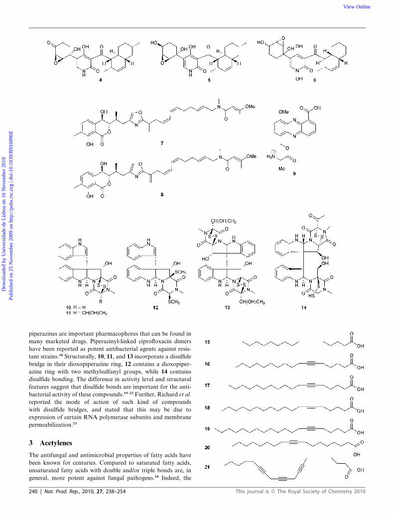
A cytotoxic compound, YM-215343 (4), found in the bacterial
culture of Phoma sp. exhibited antifungal activity against the
pathogenic fungi C. albicans, C. neoformans and A. fumigatus,
with MIC values of 2–16 mg mL�1. The structure of 4 was
determined by several spectroscopic experiments, and found to
be closely related to apiosporamide (5) and fischerin (6).7 The
mode of action of this compound has not been studied. Two
alkaloids with an oxazole moiety, ajudazols A (7) and B (8), were
identified as antimicrobial agents from Chondromyces crocatus.
Both showed their potential against Micrococcus luteus (MIC
12.5 mg mL�1). Ajudazol A (7) also showed minor activity against
a few fungi and Gram-positive bacteria. It is suggested that these
compounds block the electron flow in submitochondial particles
(SMPs).8 It is well known that the oxazole nucleus is rarely found
in nature, but several simple substituted oxazoles have been
isolated from plants and/or synthesized that exhibit antimicro-
bial activities.9
Phenazine antibiotics are synthesized by a number of bacteria
from diverse genera including Streptomyces, Pseudomonas,
Pelagiobacter and Vibrio.10–12 These microbes produce a range of
phenazine compounds that differ widely in antibiotic properties,
according to the nature and position of side groups attached to
the phenazine nucleus.13 For example D-alanylgriseoluteic acid
(AGA, 9) is a potent antimicrobial phenazine compound
produced by Pantoea agglomerans (Erwinia herbicola) Eh1087
and isolated from the culture supernatant.14 Susceptibility tests
against a range of microbes indicated that 9 exhibited a broad
spectrum of antimicrobial activity, particularly against Gram-
positive pathogens (many pneumococcal and multi-drug resis-
tant isolates), with MICs of 0.06–0.75 mg mL�1). It was further
established that 9 induced an SOS response in Escherichia coli
and slightly increased the frequency of GC–AT transition
mutations. The potency and broad-spectrum activity of 9
warrant further investigation, and in future it could have an
application as a topical agent.15
Zheng et al. isolated five epidithiodioxopiperazines, bionectins
A (10), B (11) and C (12) and verticillins D (13) and G (14), from
the mycelium of liquid fermentation cultures of the fungus
Bionectra byssicola F120. Compounds 10, 11 and 13 exhibited
antibacterial activity against S. aureus including methicillin-
resistant S. aureus (MRSA) and quinolone-resistant S. aureus
(QRSA), with MIC values of 10–30 mg mL�1, while 12 showed no
antibacterial activity even at 100 mg mL�1.16 Verticillin G (14)
was the most active and inhibited the growth of S. aureus
including methicillin-resistant and quinolone-resistant S. aureus,
with an MIC of 3–10 mg mL�1.17 Piperazines and substituted
Nat. Prod. Rep., 2010, 27, 238–254 | 239
Dow
nloa
ded
by U
nive
rsid
ade
de L
isbo
a on
16
Nov
embe
r 20
10Pu
blis
hed
on 2
5 N
ovem
ber
2009
on
http
://pu
bs.r
sc.o
rg |
doi:1
0.10
39/B
9160
96E
View Online
piperazines are important pharmacophores that can be found in
many marketed drugs. Piperazinyl-linked ciprofloxacin dimers
have been reported as potent antibacterial agents against resis-
tant strains.18 Structurally, 10, 11, and 13 incorporate a disulfide
bridge in their dioxopiperazine ring, 12 contains a dioxopiper-
azine ring with two methylsulfanyl groups, while 14 contains
disulfide bonding. The difference in activity level and structural
features suggest that disulfide bonds are important for the anti-
bacterial activity of these compounds.19–22 Further, Richard et al.
reported the mode of action of such kind of compounds
with disulfide bridges, and stated that this may be due to
expression of certain RNA polymerase subunits and membrane
permeabilization.23
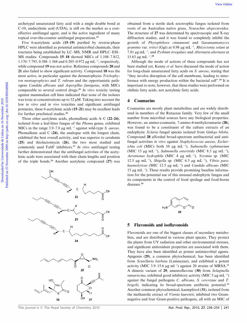
3 Acetylenes
The antifungal and antimicrobial properties of fatty acids have
been known for centuries. Compared to saturated fatty acids,
unsaturated fatty acids with double and/or triple bonds are, in
general, more potent against fungal pathogens.24 Indeed, the
240 | Nat. Prod. Rep., 2010, 27, 238–254 This journal is ª The Royal Society of Chemistry 2010
Dow
nloa
ded
by U
nive
rsid
ade
de L
isbo
a on
16
Nov
embe
r 20
10Pu
blis
hed
on 2
5 N
ovem
ber
2009
on
http
://pu
bs.r
sc.o
rg |
doi:1
0.10
39/B
9160
96E
View Online
archetypal unsaturated fatty acid with a single double bond at
C-10, undecylenic acid (UDA), is still on the market as a cost-
effective antifungal agent, and is the active ingredient of many
topical over-the-counter antifungal preparations.25
Five 6-acetylenic acids (15–19) purified by reverse-phase
HPLC were identified as potential antimicrobial chemicals, their
tructures being established by LC–MS, NMR and HPLC–ESI–
MS studies. Compounds 15–18 showed MICs of 1.108–7.812,
1.170–7.793, 0.588–1.568 and 0.205–0.972 mg mL�1, respectively,
while compound 19 was not active. Reference compounds 20 and
21 also failed to show significant activity. Compound 18 was the
most active, in particular against the dermatophytes Trichophy-
ton mentagrophytes and T. rubrum and the opportunistic path-
ogens Candida albicans and Aspergillus fumigatus, with MICs
comparable to several control drugs.26 In vitro toxicity testing
against mammalian cell lines indicated that none of the isolates
was toxic at concentrations up to 32 mM. Taking into account the
low in vitro and in vivo toxicities and significant antifungal
potencies, these 6-acetylenic acids (15–21) may be excellent leads
for further preclinical studies.26
Three other acetylenic acids, phomallenic acids A–C (22–24),
isolated from a leaf-litter fungus of the Phoma genus, exhibited
MICs in the range 3.9–7.8 mg mL�1 against wild-type S. aureus.
Phomallenic acid C (24), the analogue with the longest chain,
exhibited the best overall activity, and was superior to cerulenin
(25) and thiolactomycin (26), the two most studied and
commonly used FabF inhibitors.27 In vitro antifungal testing
further demonstrated that the antifungal activities of the acety-
lenic acids were associated with their chain lengths and position
of the triple bonds.26 Another acetylenic compound (27) was
This journal is ª The Royal Society of Chemistry 2010
obtained from a sterile dark ectotrophic fungus isolated from
roots of an Australian native grass, Neurachne alopecuroidea.
The structure of 27 was determined by spectroscopic and X-ray
diffraction studies, and it was found to completely inhibit the
growth of Phytophthora cinnamomi and Gaeumannomyces
graminis var. tritici (Ggt) at 0.98 mg mL�1, Rhizoctonia solani at
7.81 mg mL�1, and Pythium irregulare and Alternaria alternata at
15.63 mg mL�1.28
Although the mode of actions of these compounds has not
been studied yet, Kenny et al. have discussed the mode of action
for long-chain unsaturated fatty acids on S. aureus, stating that
‘‘they involve disruption of the cell membrane, leading to inter-
ference with energy production within the bacterial cell’’.29 It is
important to note, however, that these studies were performed on
olefinic fatty acids, not acetylenic fatty acids.
4 Coumarins
Coumarins are mostly plant metabolites and are widely distrib-
uted in members of the Rutaceae family. Very few of the small
number from microbial sources have any biological properties.
However, an amino-coumarin, 7-amino-4-methylcoumarin (28),
was found to be a constituent of the culture extracts of an
endophytic Xylaria fungal species isolated from Ginkgo biloba.
Compound 28 afforded broad-spectrum antibacterial and anti-
fungal activities in vitro against Staphylococcus aureus, Escher-
ichia coli (MICs both 10 mg mL�1), Salmonella typhimurium
(MIC 15 mg mL�1), Salmonella enteritidis (MIC 8.5 mg mL�1),
Aeromonas hydrophila (MIC 4 mg mL�1), Yersinia sp. (MIC
12.5 mg mL�1), Shigella sp. (MIC 6.3 mg mL�1), Vibrio para-
haemolyticus (MIC 12.5 mg mL�1) and Candida albicans (MIC
15 mg mL�1). These results provide promising baseline informa-
tion for the potential use of this unusual endophytic fungus and
its components in the control of food spoilage and food-borne
diseases.30
5 Flavonoids and isoflavonoids
Flavonoids are one of the biggest classes of secondary metabo-
lites, and are distributed in various plant species. They protect
the plants from UV radiation and other environmental stresses,
and significant antioxidant properties are associated with them.
They have also been identified as potent antimicrobial agents.
Apigenin (29), a common phytochemical, has been identified
from Scutellaria barbata (Lamiaceae), and exhibited a potent
activity (MIC 3.9–15.6 mg ml�1) against 20 strains of MRSA.31
A dimeric variant of 29, amentoflavone (30) from Selaginella
tamariscina, exhibited good inhibitory activity (MIC 5 mg mL�1)
against the fungal pathogens C. albicans, S. cerevisiae and T.
beigelii, indicating its broad-spectrum antibiotic potential.32
Another common phytochemical, kaempferol (31), isolated from
the methanolic extract of Vismia laurentii, inhibited two Gram-
negative and four Gram-positive pathogens, all with an MIC of
Nat. Prod. Rep., 2010, 27, 238–254 | 241

Dow
nloa
ded
by U
nive
rsid
ade
de L
isbo
a on
16
Nov
embe
r 20
10Pu
blis
hed
on 2
5 N
ovem
ber
2009
on
http
://pu
bs.r
sc.o
rg |
doi:1
0.10
39/B
9160
96E
View Online
2.4 mg mL�1, and showed activity against Candida glabrata
(MIC 4.8–9.7 mg mL�1).33
The methanolic extract from the root bark of Newbouldia
laevis produced chrysoeriol (32), which has broad-spectrum in
vitro activity against four Gram-positive and ten Gram-negative
bacterial species, as well as three Candida species (MIC
1.2–9.76 mg mL�1).34 Flavone glycoside 33, from the leaves of
Vitex negundo, is also known as an antimicrobial agent.
Compound 33 showed promising activity against Trichophyton
mentagrophytes and C. neoformans (MICs both 6.25 mg mL�1)
when compared to the standard antifungal drug fluconazole
(MIC 2.0 mg mL�1).35 A dibenzyloxyflavone 34 from Helichrysum
gymnocomum was found to have high activity against Crypto-
coccus neoformans ATCC 90112, with an MIC of 7.8 mg mL�1.36
Laburnetin (35), isolated from the methanolic extract of Ficus
chlamydocarpa, exhibited potent inhibitory activity against
Mycobacterium smegmatis and M. tuberculosis, with MIC values
of 0.61 and 4.88 mg mL�1 respectively.37
242 | Nat. Prod. Rep., 2010, 27, 238–254
The efficacy of flavonoids against such a variety of pathogens
can be attributed to cell-wall permeability and the porins in the
outer membrane present in microorganisms – it seems likely that
the compounds may block the charges of the amino acids in
porins.38 A comparison of the above-discussed properties reveals
that the flavonoids having free hydroxyl groups at C-5 and C-7 in
ring A are more active, a conclusion supported by studies of their
synthetic analogues.39 The activity of flavonoids may also be due
to their ability to complex with extracellular and soluble proteins
and then with bacterial cell walls. The same authors also suggest
that the more lipophilic flavonoids may also disrupt microbial
membranes.40
6 Iridoids
Iridoids are widely distributed in dicotyledonous plant families
such as the Apocynaceae, Scrophulariaceae, Diervillaceae,
Lamiaceae, Loganiaceae and Rubiaceae. They are known for their
biological activities, and recent developments have revealed that
they also possess antimicrobial properties.41 The iridoid glycosides
36 and 37 were purified from an alcoholic extract of leaves of Vitex
negundo; 37 showed promising activity against Trichophyton
mentagrophytes and Cryptococcus neoformans (MIC for both
6.25 mg mL�1) while 36 inhibited the growth of C. neoformans and
T. mentagrophytes (MIC for both 12.5 mg mL�1).35
7 Lignans
A lignan, (+)-lyoniresinol-3a-O-b-D-glucopyranoside (38), iso-
lated from the root bark of Lycium chinense, exhibited potent
antimicrobial activity against Staphylococcus aureus (MIC
2.5–5.0 mg mL�1), and three human-pathogenic fungi, Candida
albicans, Saccharomyces cerevisiae and Trichosporon beigelii
(MICs 5.0, 5.0 and 10.0 mg mL�1, respectively), without having
any hemolytic effect on human erythrocytes. It induced the
accumulation of intracellular trehalose on C. albicans as stress
response to the drug, and disrupted the dimorphic transition that
forms pseudo-hyphae caused by the pathogenesis, indicating 38
to be a potential lead compound for the development of antibi-
otic agents.42 Another lignan, a resveratrol trimer with an
ortho-quinone nucleus, hopeanolin (39), was isolated and
This journal is ª The Royal Society of Chemistry 2010
Dow
nloa
ded
by U
nive
rsid
ade
de L
isbo
a on
16
Nov
embe
r 20
10Pu
blis
hed
on 2
5 N
ovem
ber
2009
on
http
://pu
bs.r
sc.o
rg |
doi:1
0.10
39/B
9160
96E
View Online
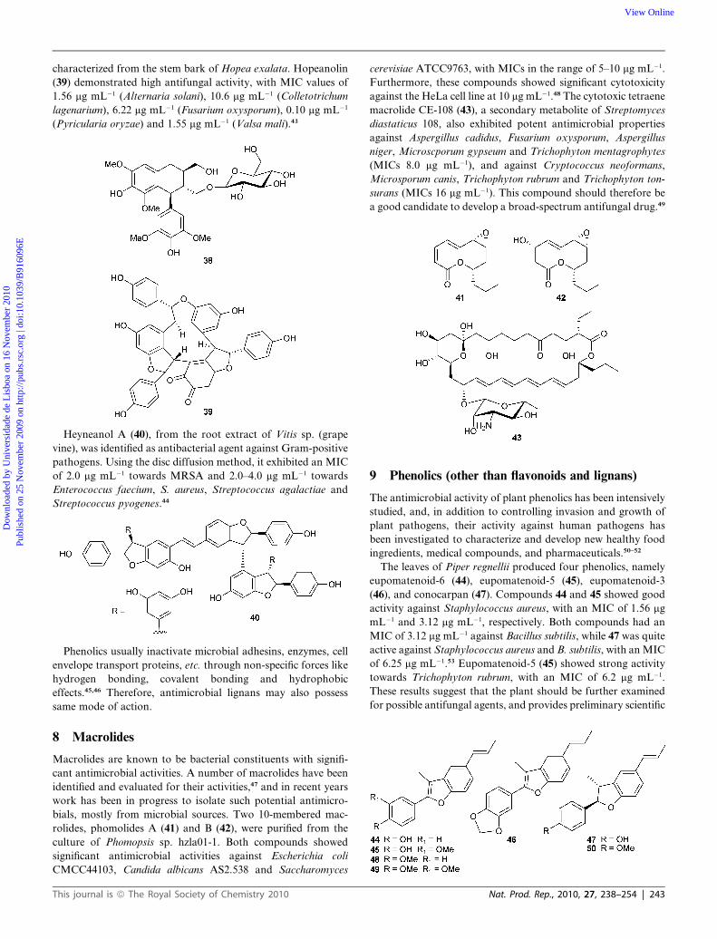
characterized from the stem bark of Hopea exalata. Hopeanolin
(39) demonstrated high antifungal activity, with MIC values of
1.56 mg mL�1 (Alternaria solani), 10.6 mg mL�1 (Colletotrichum
lagenarium), 6.22 mg mL�1 (Fusarium oxysporum), 0.10 mg mL�1
(Pyricularia oryzae) and 1.55 mg mL�1 (Valsa mali).43
Heyneanol A (40), from the root extract of Vitis sp. (grape
vine), was identified as antibacterial agent against Gram-positive
pathogens. Using the disc diffusion method, it exhibited an MIC
of 2.0 mg mL�1 towards MRSA and 2.0–4.0 mg mL�1 towards
Enterococcus faecium, S. aureus, Streptococcus agalactiae and
Streptococcus pyogenes.44
Phenolics usually inactivate microbial adhesins, enzymes, cell
envelope transport proteins, etc. through non-specific forces like
hydrogen bonding, covalent bonding and hydrophobic
effects.45,46 Therefore, antimicrobial lignans may also possess
same mode of action.
8 Macrolides
Macrolides are known to be bacterial constituents with signifi-
cant antimicrobial activities. A number of macrolides have been
identified and evaluated for their activities,47 and in recent years
work has been in progress to isolate such potential antimicro-
bials, mostly from microbial sources. Two 10-membered mac-
rolides, phomolides A (41) and B (42), were purified from the
culture of Phomopsis sp. hzla01-1. Both compounds showed
significant antimicrobial activities against Escherichia coli
CMCC44103, Candida albicans AS2.538 and Saccharomyces
This journal is ª The Royal Society of Chemistry 2010
cerevisiae ATCC9763, with MICs in the range of 5–10 mg mL�1.
Furthermore, these compounds showed significant cytotoxicity
against the HeLa cell line at 10 mg mL�1.48 The cytotoxic tetraene
macrolide CE-108 (43), a secondary metabolite of Streptomyces
diastaticus 108, also exhibited potent antimicrobial properties
against Aspergillus cadidus, Fusarium oxysporum, Aspergillus
niger, Microscporum gypseum and Trichophyton mentagrophytes
(MICs 8.0 mg mL�1), and against Cryptococcus neoformans,
Microsporum canis, Trichophyton rubrum and Trichophyton ton-
surans (MICs 16 mg mL�1). This compound should therefore be
a good candidate to develop a broad-spectrum antifungal drug.49
9 Phenolics (other than flavonoids and lignans)
The antimicrobial activity of plant phenolics has been intensively
studied, and, in addition to controlling invasion and growth of
plant pathogens, their activity against human pathogens has
been investigated to characterize and develop new healthy food
ingredients, medical compounds, and pharmaceuticals.50–52
The leaves of Piper regnellii produced four phenolics, namely
eupomatenoid-6 (44), eupomatenoid-5 (45), eupomatenoid-3
(46), and conocarpan (47). Compounds 44 and 45 showed good
activity against Staphylococcus aureus, with an MIC of 1.56 mg
mL�1 and 3.12 mg mL�1, respectively. Both compounds had an
MIC of 3.12 mg mL�1 against Bacillus subtilis, while 47 was quite
active against Staphylococcus aureus and B. subtilis, with an MIC
of 6.25 mg mL�1.53 Eupomatenoid-5 (45) showed strong activity
towards Trichophyton rubrum, with an MIC of 6.2 mg mL�1.
These results suggest that the plant should be further examined
for possible antifungal agents, and provides preliminary scientific
Nat. Prod. Rep., 2010, 27, 238–254 | 243

Dow
nloa
ded
by U
nive
rsid
ade
de L
isbo
a on
16
Nov
embe
r 20
10Pu
blis
hed
on 2
5 N
ovem
ber
2009
on
http
://pu
bs.r
sc.o
rg |
doi:1
0.10
39/B
9160
96E
View Online
validation for the traditional medicinal use of this plant.54 The
low (or lack of) activity of 48–50 and structural comparison of
44–50 and their activity levels indicates that phenolic functions
are important for the compound to be antimicrobial, as was
observed for flavonoids.
Other antimicrobial phenolics include pterocarpans, ery-
braedin B (51), erybraedin A (52), phaseollin (53), erythrabyssin
II (54), erystagallin A (55), erythrabissin-1 (56) and erycrista-
gallin (57) isolated from the stems of Erythrina subumbrans
(Leguminosae). Compounds 52 and 54 exhibited the highest
degree of activity against Streptococcus strains, with an MIC
range of 0.78–1.56 mg mL�1, whereas 57 exhibited the highest
degree of activity against Staphylococcus strains, including drug-
resistant strains (MRSA and VRSA), with an MIC range of
0.39–1.56 mg mL�1. Compounds 52, 54, 55 and 57 were also
reported to be more active against several strains of Strepto-
coccus and Staphylococcus than the standard antibiotics vanco-
mycin and oxacillin.55 Compound 57 showed the highest level of
activity against S. aureus strains resistant to vancomycin and
oxacillin, with an MIC range of 0.39–1.56 mg mL�1. These
compounds may prove to be potent phytochemical agents for
antibacterial activity, especially against MRSA and VRSA.55
The variation in activity and structural differences in these kinds
244 | Nat. Prod. Rep., 2010, 27, 238–254
of compounds shows that the dimethylallyl and phenolic units
may play important role – for example, 51 and 53 have a pyran
ring and are inactive.
Kanzonols are well-known phyto-phenolics possessing anti-
microbial activities,56 and kanzonol C (58) has significant bio-
logical activities. Kanzonol C 58 and its analogues,
isobavachalcone (59), stipulin (60) and 4-hydroxylonchocarpin
(61), were isolated from the extract of the twigs of Dorstenia
barteri, and showed broad-spectrum inhibitory activities against
both Gram-positive and Gram-negative bacteria and fungi, MIC
values being determined using the micro-well dilution method.
Compound 59 was highly active against Enterobacter cloacae,
Streptococcus faecalis, Staphylococcus aureus, Bacillus stear-
othermophilus, Candida albicans and C. glabrata (MICs 0.3 mg
mL�1), Enterobacter aerogens, Morganella morganii, Shigella
flexneri, Bacillus cereus, B. megaterium and B. subtilis (MICs 0.6
mg mL�1), Proteus mirabilis, Proteus vulgaris, Microsporum
audorium and Trichophyton rubrum (MICs 1.2 mg mL�1).
Compound 61 inhibited growth of E. cloacae, M. morganii,
B. megaterium and B. stearothermophilus (MICs 1.2 mg mL�1),
E. aerogens, S. flexneri, S. faecalis, S. aureus, B. cereus, B. sub-
tilis, C. albicans, C. glabrata and T. rubrum (MICs 4.9 mg mL�1)
and M. audorium (MIC 9.8 mg mL�1). Compound 58 showed
This journal is ª The Royal Society of Chemistry 2010

Dow
nloa
ded
by U
nive
rsid
ade
de L
isbo
a on
16
Nov
embe
r 20
10Pu
blis
hed
on 2
5 N
ovem
ber
2009
on
http
://pu
bs.r
sc.o
rg |
doi:1
0.10
39/B
9160
96E
View Online
moderate activity against E. aerogens, E. cloacae, M. morganii,
S. flexneri, S. faecalis, B. megaterium, B. stearothermophilus,
C. albicans and C. glabrata (MICs 4.9 mg mL�1), P. mirabilis,
P. vulgaris, B. cereus, B. subtilis and M. audorium (MIC, 9.8 mg
mL�1). The structure–activity relationship was not discussed,
but compound 60 (from the same source), which differs from 58
and 59 in the position of isoprene unit, was found to be inac-
tive.56 Curcumin (62), a similar compound, has been reported to
induce filamentation in B. subtilis 168, suggesting that it inhibits
bacterial cytokinesis. Further, 62 strongly inhibited the forma-
tion of the cytokinetic Z-ring in the protein structure of
B. subtilis.57 On that basis, the site of action of the kanzonols
can be predicted, but further studies of these antimicrobials are
needed.
The structural similarities and antimicrobial activities of two
compounds from Helichrysum gymnocomum, acylphloroglucinol
(63) and its analogue 64, have been examined. Compound 63
afforded high activity against Cryptococcus neoformans ATCC
90112 (MIC 7.8 mg mL�1), while 64 was active against S. aureus
ATCC 12600 (MIC 6.8 mg mL�1), Enterococcus faecalis ATCC
29212, methicillin- and gentamycin-resistant S. aureus ATCC
33592, B. cereus ATCC 11778 (MICs 7.8 mg mL�1), and S. epi-
dermidis ATCC 2223 (MIC 9.8 mg mL�1).36 The natural
compounds grandinol (65), jensenone (66) were synthesized and
their synthetic analogue (67) also showed high antimicrobial
activities. The comparative data revealed that phloroglucinols
are known for their various bioactivities and their acyl deriva-
tives possess antimicrobial activities, therefore, they can be good
candidates for further clinical studies to develop new antimi-
crobial dugs.58
Six isolates of mushroom Merulius incarnatus, 5-alkylresorci-
nols (68–73), were characterized by sophisticated NMR tech-
niques. Compound 68 is the first 5-alkylresorcinol derivative
that contains a conjugated trans–cis double-bond system.
Compounds 68–73 were found to inhibit MRSA, with IC50
values of 2.5–15.0 mg mL�1.59
Psoracorylifols A–E (74–78) are phenolics isolated from
a well-known traditional Chinese medicine, the seeds of Psoralea
corylifolia. The structures of 74–78, including their absolute
configurations, were established on the basis of spectral methods
and biosynthetic reasoning, with that for 74 being confirmed by
single-crystal X-ray diffraction. Psoracorylifols D (77) and E (78)
have an unprecedented carbon skeleton. All five compounds
This journal is ª The Royal Society of Chemistry 2010
showed significant inhibitory activity against two strains of
Helicobacter pylori (SS1 and ATCC 43504), with MICs of
12.5–25 mg mL�1. It is remarkable that psoracorylifols A–E
(74–78) are 5–10 times stronger than the standard drug metro-
nidazole, which is a critical ingredient for combination therapies
of H. pylori and Clostridium difficile infections. Therefore,
compounds of this kind could be substitutes for current H. pylori
and C. difficile anti-infective drugs,60 although their mode of
action remains unstudied.
The stem bark of Irvingia gabonensis produced 3,30,40-tri-O-
methylellagic acid (79) and 3,4-di-O-methylellagic acid (80). The
lowest MIC values for 79 and 80 (both 9.76 mg mL�1) were
observed against E. coli, Proteus vulgaris and B. subtilis, while 80
exhibited high activity against P. aeruginosa (MIC 4.8 mg
mL�1).61 Again, compounds with more free hydroxyl groups
(like 80) are more active.
Nat. Prod. Rep., 2010, 27, 238–254 | 245
Dow
nloa
ded
by U
nive
rsid
ade
de L
isbo
a on
16
Nov
embe
r 20
10Pu
blis
hed
on 2
5 N
ovem
ber
2009
on
http
://pu
bs.r
sc.o
rg |
doi:1
0.10
39/B
9160
96E
View Online
10 Polypeptides
Polypeptides are well-known for their pharmaceutical uses, and
have earned recognition as antimicrobial agents, for example in
pharmaceutical applications, as well as for preservation, cleaning
and disinfection. Polypeptides have in particular been used to
treat textiles or laundry (e.g., in detergents), and for odour
reduction by reducing microbial growth.62 Most of these poly-
peptides are of microbial origin, and in recent years many
polypeptides have been discovered to be antimicrobial agents.
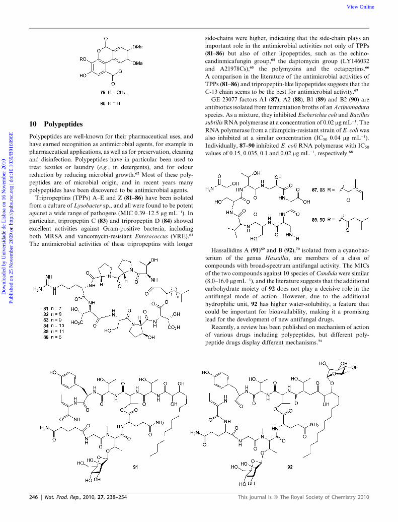
Tripropeptins (TPPs) A–E and Z (81–86) have been isolated
from a culture of Lysobacter sp., and all were found to be potent
against a wide range of pathogens (MIC 0.39–12.5 mg mL�1). In
particular, tripropeptin C (83) and tripropeptin D (84) showed
excellent activities against Gram-positive bacteria, including
both MRSA and vancomycin-resistant Enterococcus (VRE).63
The antimicrobial activities of these tripropeptins with longer
246 | Nat. Prod. Rep., 2010, 27, 238–254
side-chains were higher, indicating that the side-chain plays an
important role in the antimicrobial activities not only of TPPs
(81–86) but also of other lipopeptides, such as the echino-
candinmicafungin group,64 the daptomycin group (LY146032
and A21978Cs),65 the polymyxins and the octapeptins.66
A comparison in the literature of the antimicrobial activities of
TPPs (81–86) and tripropeptin-like lipopeptides suggests that the
C-13 chain seems to be the best for antimicrobial activity.67
GE 23077 factors A1 (87), A2 (88), B1 (89) and B2 (90) are
antibiotics isolated from fermentation broths of an Actinomadura
species. As a mixture, they inhibited Escherichia coli and Bacillus
subtilis RNA polymerase at a concentration of 0.02 mg mL�1. The
RNA polymerase from a rifampicin-resistant strain of E. coli was
also inhibited at a similar concentration (IC50 0.04 mg mL�1).
Individually, 87–90 inhibited E. coli RNA polymerase with IC50
values of 0.15, 0.035, 0.1 and 0.02 mg mL�1, respectively.68
Hassallidins A (91)69 and B (92),70 isolated from a cyanobac-
terium of the genus Hassallia, are members of a class of
compounds with broad-spectrum antifungal activity. The MICs
of the two compounds against 10 species of Candida were similar
(8.0–16.0 mg mL�1), and the literature suggests that the additional
carbohydrate moiety of 92 does not play a decisive role in the
antifungal mode of action. However, due to the additional
hydrophilic unit, 92 has higher water-solubility, a feature that
could be important for bioavailability, making it a promising
lead for the development of new antifungal drugs.
Recently, a review has been published on mechanism of action
of various drugs including polypeptides, but different poly-
peptide drugs display different mechanisms.71
This journal is ª The Royal Society of Chemistry 2010

Dow
nloa
ded
by U
nive
rsid
ade
de L
isbo
a on
16
Nov
embe
r 20
10Pu
blis
hed
on 2
5 N
ovem
ber
2009
on
http
://pu
bs.r
sc.o
rg |
doi:1
0.10
39/B
9160
96E
View Online
11 Quinones
Quinones from natural sources have a variety of biological
activities, and are particularly well-known for their bactericidal
properties.72,73
3,4-Dihydroxy-1-methoxyanthraquinone-2-carboxaldehyde (93)
and damnacanthal (94), were identified from the aerial part of
Saprosma fragrans as antifungal compounds with MIC values
as follows: 93: 12.5 mg mL�1 (Trichophyton mentagrophytes);
94: 12.5, 6.25 and 1.56 mg mL�1 (Cryptococcus neoformans,
Sporothrix schenckii and T. mentagrophytes, respectively).74
Another anthraquinone containing a naphthaquinone moiety,
named newbouldiaquinone A (95), is a constituent of the roots of
Newbouldia laevis (Bignoniaceae). It possesses broad-spectrum
antimicrobial activity, and is reported to be 13- and 24-fold more
active against Candida glabrata and Enterobacter aerogens,
respectively, than the standard antibiotics nystatin and
gentamycin.75 Against Gram-negative bacteria it showed high
potential, with MICs in the range 0.31–9.76 mg mL�1.75
The root bark of Newbouldia laevis contains newbouldiaquinone
(96), 2-acetylfuro-1,4-naphthoquinone (97), 2-hydroxy-3-methoxy-
9,10-dioxo-9,10-dihydroanthracene-1-carbaldehyde (98) and
This journal is ª The Royal Society of Chemistry 2010
lapachol (99). Compounds 96–99 have broad-spectrum in vitro
antimicrobial activity against six Gram-positive and twelve Gram-
negative bacterial species, as well as Candida strains, the MIC
values being in the range 0.076–9.76 mg mL�1.35
Kuete et al. identified four antimicrobial quinones (100–104)
from the extract of Vismia laurentii, these being active against
Gram-negative bacteria and two Candida species. Compound 100
inhibits the growth of Streptococcus faecalis and Morganella
morganii (both MICs 4.8 mg mL�1) and Pseudomonas aeruginosa
and Shigella flexneri (both MICs 2.4 mg mL�1). Compound 101 had
MICs of 4.8 mg mL�1 against M. morganii and S. faecalis, and
2.4 mg mL�1 against P. aeruginosa, S. flexneri, B. subtilis, C. albi-
cans and C. glabrata. Compound 102 was only active against
Gram-positive and fungal pathogens (B. megaterium, B. subtilis,
C. albicans and C. glabrata) with MICs of 2.4 mg mL�1, while
compound 103 had MICs of 4.8 mg mL�1 against P. aeruginosa,
S. flexneri and B. subtilis, and 9.7 mg mL�1 against M. morganii.
Compound 104 was highly active against B. subtilis (MIC 1.2 mg
mL�1), and also showed significant activity against S. dysenteriae,
S. faecalis and B. stearothermophilus (MICs 4.8 mg mL�1), and
B. cereus (2.4 mg mL�1). The latter also had moderate activity (MIC
19.5 mg mL�1) against M. morganii, P. vulgaris and S. aureus.33
Nat. Prod. Rep., 2010, 27, 238–254 | 247

Dow
nloa
ded
by U
nive
rsid
ade
de L
isbo
a on
16
Nov
embe
r 20
10Pu
blis
hed
on 2
5 N
ovem
ber
2009
on
http
://pu
bs.r
sc.o
rg |
doi:1
0.10
39/B
9160
96E
View Online
Omicron-naphthoquinone, 9-methyl-3-(4-methyl-3-pentenyl)-
2,3-dihydronaphtho[1,8-bc]pyran-7,8-dione (105), is a constit-
uent of the Australian plant Eremophila serrulata, and exhibits
antimicrobial activity against Staphylococcus aureus ATCC
25923, Streptococcus pneumoniae ATCC 49619 and Strepto-
coccus pyogenes ATCC 10389, with MICs of 7.8 mg mL�1.76
7-epi-Clusianone (106), identified from the antimicrobial
extract of the fruit of Rheedia brasiliensis, showed high anti-
bacterial activity at low concentrations (MIC 1.25–2.5 mg mL�1)
against Streptococcus mutans. This may lead to the use of 106 as
a new agent to control S. mutans biofilms; however, more studies
are needed to further elucidate the mechanism of action.77
Anthracycline antibiotic mutactimycin C (107) was purified by
reverse-phase HPLC from a soil isolate (Saccharothrix sp.) that
inhibited two Gram-positive bacteria (Microccocus leteus and
Klebsiella pneumoniae) in vitro with MICs of 5.0 mg mL�1.78
Decaspirone A (108), related to the palmarumycin class of
compounds, was isolated from cultures of the freshwater aquatic
fungal species Decaisnella thyridioides, and its structure was
established by X-ray crystallography. It displayed MIC values of
approximately 10 and 5 mg mL�1 against Aspergillus flavus and
Fusarium verticillioides, respectively.79
An unidentified endophytic fungus of the order Pleosporales
was isolated from Anthyllis vulneraria (Fabaceae), and produced
pleosporone (109) and phaeosphenone (110), following isolation
by silica gel and Sephadex LH-20 chromatography followed by
reverse-phase HPLC. In a two-plate whole-cell differential
sensitivity screening assay using an antisense-sensitized S. aureus
strain,80,81 109 exhibited significant activity towards Strepto-
coccus pneumoniae and Haemophilus influenzae (MICs of 4.0 and
1.0 mg mL�1, respectively) and Bacillus subtilis and E. coli (MICs
of 8.0 and 16 mg mL�1, respectively).82 Compound 110 showed
broad-spectrum antibacterial activity against Gram-positive
bacteria, with MIC values ranging from 8 to 64 mg mL�1. It
showed the highest activity for S. pneumoniae and C. albicans
(MICs of 8.0 mg mL�1), and modest selectivity for the inhibition
of RNA synthesis over DNA and protein synthesis in S. aureus.83
248 | Nat. Prod. Rep., 2010, 27, 238–254
Ribosomal protein S4 (RPSD), a part of the ribosomal small
subunit, is one of the proteins that is a part of the ribosomal
machinery and is a potential new target for the discovery of
antibacterial agents. Therefore, 109 and 110 could be good leads
for antibacterial drug development. Another endophytic fungus,
Chloridium sp., produces javanicin (111) under both liquid and
solid media culture conditions. This highly functionalized
naphthaquinone (111), the structure of which was established by
X-ray crystallography, was found to have strong antibacterial
activity against Pseudomonas fluorescens and P. aeruginosa
(MICs 2.0 mg mL�1), Rhizoctonia solani and Verticillium dahlae
(MICs 10 mg mL�1), and antifungal activity against Cercospora
arachidicola (MIC 5.0 mg mL�1).84
Protein synthesis is one of the best antibacterial targets, and
studies in this area have led to the development of a number of
highly successful clinical drugs. In addition to providing a source
of stable free radicals, quinones are known to complex irrevers-
ibly with nucleophilic amino acids in proteins,85 often leading to
inactivation of the protein and loss of function. For that reason,
the potential range of quinone antimicrobial effects is great.
Probable targets in the microbial cell are surface-exposed adhe-
sins, cell-wall polypeptides, and membrane-bound enzymes.
Quinones may also render substrates unavailable to the micro-
organism. The above-discussed results reveal that quinones
exhibit a range of antimicrobial efficacies, and the reasons for
this may lie in the substituent present on the benzene ring and
perhaps in solubility differences.
12 Steroidal saponins
Many plant extracts containing steroidal saponins have been
reported to exhibit antimicrobial activities,86 and this also applies to
the purified steroidal saponins.87 Tigogenin 3-O-b-D-xylopyranosyl-
(1/2)-[b-D-xylopyranosyl (1/3)]-b-D-glucopyranosyl-(1/4)-[a-
L-rhamnopyranosyl-(1/2)]-b-D-galactopyranoside (TTS-12, 112)
and tigogenin 3-O-b-D-glucopyranosyl-(1/2)-[b-D-xylopyranosyl-
(1/3)]-b-D-glucopyranosyl-(1/4)-b-D-galactopyranoside (TTS-15,
This journal is ª The Royal Society of Chemistry 2010

Dow
nloa
ded
by U
nive
rsid
ade
de L
isbo
a on
16
Nov
embe
r 20
10Pu
blis
hed
on 2
5 N
ovem
ber
2009
on
http
://pu
bs.r
sc.o
rg |
doi:1
0.10
39/B
9160
96E
View Online
113) were obtained from the extract of Tribulus terrestris, and
their in vivo activity in a Candida albicans vaginal infection model
studied. Both 112 and 113 were very active against C. albicans
(MIC80 10 and 2.3 mg mL�1) and Cryptococcus neoformans
(MIC80 1.7 and 6.7 mg mL�1). Phase-contrast microscopy and
transmission electron microscopy showed that 112 has significant
in vitro and in vivo antifungal activity, weakening the virulence of
C. albicans and killing fungi through destroying the cell
membrane.88 Another cytotoxic steroidal saponin, dioscin (114),
isolated from the rhizomes of Smilacina atropurpurea, was found
to be active against C. albicans and C. glabrata (minimum
fungicidal concentration (MFC) # 5.0 mg mL�1).89
Aginoside (115) and (25R)-5a-spirostan-3b,6b-diol-3-O-{b-D-
glucopyranosyl-(1/2)-O-[b-D-xylopyranosyl-(1/3)]-O-b-D-glu-
copyranosyl-(1/4)-b-D-galactopyranoside} (116) were found in
the flower of Allium leucanthum. In an in vitro experiment, 115
and 116 showed antifungal activity against seven Candida strains
with an MFC ranging from 6.25 to 12.5 mg mL�1.90
CAY-1 (117), a saponin from Capsicum frutescens, showed
antifungal activity and is reported to be active against 16
different fungal strains, including Candida spp. and Aspergillus
fumigatus, with MICs ranging from 4.0 to 16 mg mL�1. It was
This journal is ª The Royal Society of Chemistry 2010
especially active against Cryptococcus neoformans, exhibiting
90% inhibition at 1.0 mg mL�1. However, it had no significant
cytotoxicity against 55 mammalian cell lines at concentrations up
to 100 mg mL�1. Importantly, CAY-1 (117) appears to act by
disrupting the membrane integrity of fungal cells.91 b-Sitosterol-
3-O-b-D-glucopyranoside (118), a very common phytochemical,
was obtained from the root bark of Newbouldia laevis, and dis-
played broad-spectrum in vitro antimicrobial activity against
three Gram-positive and nine Gram-negative bacterial species, as
well as three Candida species (MICs 0.61–9.76 mg mL�1).34
In general, the mechanism of action of steroids is not yet clear,
but their antimicrobial properties might be due to their ability to
disrupt membrane integrity.
13 Terpenoids
Terpenoids are a class of secondary metabolites made of isoprene
units. The important phytochemicals in essential oils are
mixtures of mono- and sesquiterpenoids, and are known for their
antimicrobial properties – 60% of essential oil derivatives
examined to date are inhibitory to fungi, while 30% inhibit
bacteria.92 Many higher terpenoids are also reported to have
Nat. Prod. Rep., 2010, 27, 238–254 | 249
Dow
nloa
ded
by U
nive
rsid
ade
de L
isbo
a on
16
Nov
embe
r 20
10Pu
blis
hed
on 2
5 N
ovem
ber
2009
on
http
://pu
bs.r
sc.o
rg |
doi:1
0.10
39/B
9160
96E
View Online
antimicrobial properties. These facts indicate the antimicrobial
potential of this important class of compounds. The sesquiter-
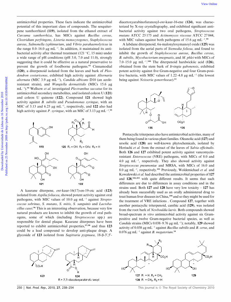
pene xanthorrhizol (119), isolated from the ethanol extract of
Curcuma xanthorrhiza, has MICs against Bacillus cereus,
Clostridium perfringens, Listeria monocytogenes, Staphylococcus
aureus, Salmonella typhimurium, and Vibrio parahaemolyticus in
the range 8.0–16.0 mg mL�1. In addition, it maintained its anti-
bacterial activity after thermal treatment (121 �C, 15 min) under
a wide range of pH conditions (pH 3.0, 7.0 and 11.0), strongly
suggesting that it could be effective as a natural preservative to
prevent the growth of foodborne pathogens.93 Cinnamodial
(120), a diterpenoid isolated from the leaves and bark of Pleo-
dendron costaricense, exhibited high activity against Alternaria
alternata (MIC 3.9 mg mL�1), Candida albicans D10 (an azole-
resistant strain), and Wangiella dermatitidis (MICs 15.6 mg
mL�1).94 Wellsow et al. investigated Plectranthus saccatus for its
antimicrobial secondary metabolites, and isolated coleon U (121)
and coleon U quinone (122). Compound 121 showed high
activity against B. subtilis and Pseudomonas syringae, with an
MIC of 3.13 and 6.25 mg mL�1, respectively, and 122 also had
high activity against P. syringae, with an MIC of 3.13 mg mL�1.95
A kaurane diterpene, ent-kaur-16(17)-en-19-oic acid (123)
isolated from Aspilia foliacea, showed potent activity against oral
pathogens, with MIC values of 10.0 mg mL�1 against Strepto-
coccus sobrinus, S. mutans, S. mitis, S. sanguinis and Lactoba-
cillus casei.96 This is an interesting observation, because very few
natural products are known to inhibit the growth of oral path-
ogens, some of which (including Streptococcus spp.) are
responsible for dental plaque. Kaurane diterpenes have been
reported to exhibit antimicrobial properties,97,98 and thus 123
could be a lead compound to develop anti-plaque drugs. A
glycoside of 123 isolated from Sagittaria pygmaea, 18-b-30,50-
250 | Nat. Prod. Rep., 2010, 27, 238–254
diacetoxyarabinofuranosyl-ent-kaur-16-ene (124), was charac-
terized by X-ray crystallography, and exhibited significant anti-
bacterial activity against two oral pathogens, Streptococcus
mutans ATCC 25 175 and Actinomyces viscosus ATCC 27 044,
with MIC values against both pathogens of 15.6 mg mL�1.99
A labdane diterpenoid, 6a-malonyloxymanoyl oxide (125) was
isolated from the aerial parts of Stemodia foliosa, and found to
inhibit the growth of Staphylococcus aureus, Bacillus cereus,
B. subtilis, Mycobacterium smegmatis, and M. phlei with MICs of
7.0–15.0 mg mL�1.100 The diterpenoid hardwickiic acid (126),
obtained from the stem bark of Irvingia gabonensis, exhibited
potent activity against five Gram-negative and four Gram-posi-
tive bacteria, with MIC values of 1.22–4.8 mg mL�1 (the lowest
being against Neisseria gonorrhoeae).61
Pentacyclic triterpenes also have antimicrobial activities, many of
them being found in various plant families. Oleanolic acid (127) and
ursolic acid (128) are well-known phytochemicals, isolated by
Horiuchi et al. from the extract of the leaves of Salvia officinalis.
Both 126 and 127 exhibited potent activity against vancomycin-
resistant Enterococcus (VRE) pathogens, with MICs of 8.0 and
4.0 mg mL�1, respectively. They also showed activity against
Streptococcus pneumoniae and MRSA, with MICs of 16.0 and
8.0 mg mL�1, respectively.101 Previously, Woldemichael et al. and
Kowalewski et al. had described the antimicrobial properties of 127
and 128,102,103 with quite different results. It seems that such
differences are due to differences in assay conditions and in the
strains used. Both 127 and 128 have very low toxicity – 127 has
already been successfully used as an orally administered drug to
treat human liver diseases in China,104 and so they might be used for
the treatment of VRE infections . Compound 127, together with
another pentacyclic triterpenoid, canthic acid (129), was isolated
from the root bark of Newbouldia laevis. Both compounds showed
broad-spectrum in vitro antimicrobial activity against six Gram-
positive and twelve Gram-negative bacterial species, as well as
Candida strains (MICs 0.038–9.76 mg mL�1); notably, 129 showed
activity of 0.038 mg mL�1 against Bacillus subtilis and B. cerus, and
0.076 mg mL�1 against B. megaterium.34
This journal is ª The Royal Society of Chemistry 2010

Dow
nloa
ded
by U
nive
rsid
ade
de L
isbo
a on
16
Nov
embe
r 20
10Pu
blis
hed
on 2
5 N
ovem
ber
2009
on
http
://pu
bs.r
sc.o
rg |
doi:1
0.10
39/B
9160
96E
View Online
Fridelin (130), a constituent of Vismia laurentii, exhibited
broad-spectrum antimicrobial character against six Gram-
negative, four Gram-positive and two fungal strains, with MICs
of 2.4–9.7 mg mL�1.33 A diterpene, xeniolide I (131), has been
isolated from the Kenyan soft coral Xenia novaebrittanniae, and
possesses antibacterial activity at a concentration of 1.25 mg
mL�1 against Escherichia coli and Bacillus subtilis.105
The mechanism of action of terpenes is not fully understood
but is speculated to involve membrane disruption by these lipo-
philic compounds. Mendoza et al. found that increasing the
hydrophilicity of kaurene diterpenoids by introduction of a 3b-
OH group drastically reduced their antimicrobial activity.106
14 Xanthones
Xanthones related to phytoalexins are thought to be produced as
a result of microbial infection in higher plants as a consequence
of a passive defense system. Biological evaluation revealed that
these compounds have fewer side-effects and a low tendency to
result in pathogen resistance compared with conventional anti-
biotics and synthetic antibacterial agents; natural xanthones are
known for their potent MRSA-inhibitory properties.107
Two tetraoxygenated xanthones, mangostanin (132) and a-
mangostin (133), were isolated from the crude hexane extract of
the fruits of Garcinia cowa. Compound 132 is a strong inhibitory
agent against the growth of Staphylococcus aureus (penicillin-
sensitive strain ATCC 25923 and methicillin-resistant strain
MRSA SK1), with MIC values of 4.0 mg mL�1, whereas
compound 133 inhibited both strains of S. aureus with MIC
values of 8.0 mg mL�1.108 Boonsri et al. isolated formoxanthone A
This journal is ª The Royal Society of Chemistry 2010
(134), formoxanthone C (135), macluraxanthone (136), xanthone
V1 (137) and gerontoxanthone I (138) as potential antibiotics
from the roots of Cratoxylum formosum. Compounds 135 and
136 were quite active against B. subtilis (both MICs 4.6 mg mL�1),
S. aureus (MIC 2.3 and 4.6 mg mL�1, respectively), S. faecalis
(MIC 18.7 and 2.3 mg mL�1, respectively) and S. typhi (MIC 4.6
and 9.3 mg mL�1, respectively). Compound 137 was particularly
active, with an MIC for all the tested pathogens of 1.1 mg mL�1
(except P. aeruginosa, MIC 9.3 mg mL�1), while 138 inhibited the
pathogens with MIC values of 1.1–4.6 mg mL�1. All these
compounds were also reported to have cytotoxic properties.109
Xanthones 139 and 140 are the active constituents of Vismia
laurentii. Compound 139 inhibited the growth of Bacillus subtilis
and Candida glabrata with an MIC value of 1.2 mg mL�1, whereas
140 showed the same degree of activity against Bacillus stear-
othermophilus, and significant activity against B. subtilis (MIC
4.8 mg mL�1).33
15 Miscellaneous compounds
The cytotoxic compound aculeatin D (141) was isolated as minor
constituent from the rhizomes of Amomum aculeatum, and
showed very potent activity against two Plasmodium falciparum
strains (MIC 0.42 mg mL�1), as well as against Trypanosoma
brucei rhodesiense (MIC 0.20 mg mL�1) and Trypanosoma cruzi
(MIC 0.49 mg mL�1). The same compound was less potent as an
antibacterial, with moderate activity against Bacillus cereus
(MIC 16 mg mL�1), Escherichia coli (MIC 16 mg mL�1) and
Staphylococcus epidermidis (MIC 8 mg mL�1) .110 Platensimycin
(142) is a novel broad-spectrum Gram-positive antibiotic
produced by Streptomyces platensis, and was discovered by
Nat. Prod. Rep., 2010, 27, 238–254 | 251
Dow
nloa
ded
by U
nive
rsid
ade
de L
isbo
a on
16
Nov
embe
r 20
10Pu
blis
hed
on 2
5 N
ovem
ber
2009
on
http
://pu
bs.r
sc.o
rg |
doi:1
0.10
39/B
9160
96E
View Online
a target-based whole-cell screening strategy using an antisense
differential sensitivity assay. It was isolated originally by a three-
step isolation method using Amberchrome, Sephadex LH-20,
and reverse-phase HPLC chromatographies. The structure was
elucidated by 2D NMR methods and confirmed by X-ray crys-
tallographic analysis of a bromo derivative. It inhibits bacterial
growth, with IC50 values of 48 and 160 nM for S. aureus and E.
coli, respectively. This compound also inhibited phospholipid
synthesis (IC50 0.1 mg mL�1), and exhibited MIC values of 0.1–
1.0 mg mL�1 against MRSA.111 Mechanistically, it exerts its
activity by a novel mode of action that involves specific binding
with the acyl enzyme intermediate of the key condensing enzyme
FabF of the fatty acid synthesis pathway. Fatty acids are
essential for survival of bacteria and are synthesized by a series of
enzymes including the elongation enzymes, b-ketoacyl acyl
carrier protein synthase I/II (FabF/B). Inhibition of fatty acid
synthesis is therefore a target for the discovery and development
of antibacterial agents, and 142 could be a candidate for anti-
bacterial drug development.111
Phenylacetic acid (143) and sodium phenylacetate (144) were
isolated from Streptomyces humidus strain S5-55, both being
identified by NMR, EI-MS and ICP-MS. Compounds 143 and
144 completely inhibited the growth of Pythium ultimum, Phy-
tophthora capsici, Rhizoctonia solani, Saccharomyces cerevisiae,
and Pseudomonas syringae pv. syringae at concentrations from
10 to 50 mg mL�1. They were also as effective as the commercial
fungicide metalaxyl in inhibiting spore germination and hyphal
growth of P. capsici.112 A derivative of benzofuran (145) was
purified from a culture of Phomopsis sp. hzla01-1, and showed
significant antimicrobial activity against Escherichia coli
CMCC44103, Candida albicans AS2.538 and Saccharomyces
cerevisiae ATCC9763, with MICs of 5–10 mg mL�1, and strongest
activity against Bacillus subtilis CMCC63501, with an MIC of
1.25 mg mL�1. None of the three compounds showed significant
cytotoxicity against the HeLa cell line at 10 mg mL�1.48
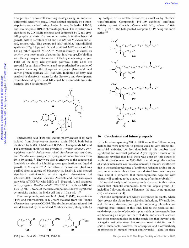
Four compounds, rotiorinols A (146), C (147), (�)-rotiorin
(148) and rubrorotiorin (149), were isolated from the fungus
Chaetomium cupreum CC3003. The absolute configuration of 146
was determined by the modified Mosher method, along with X-
252 | Nat. Prod. Rep., 2010, 27, 238–254
ray analysis of its acetate derivative, as well as by chemical
transformation. Compounds 146–149 exhibited antifungal
activity against Candida albicans with IC50 values of 0.6–
24.3 mg mL�1, the halogenated compound 149 being the most
active.113
16 Conclusions and future prospects
In the literature spanning 2000 to 2008, more than 300 secondary
metabolites were reported to possess weak to very strong anti-
microbial activities, but less than half of this number have
significant antimicrobial potential. A year-by-year review of the
literature revealed that little work was done on this aspect of
antibiotic development in 2000–2004, and although the number
of studies in this area continues to increase, it remains insufficient
due to the rapid appearance of antibiotic-resistant strains. In the
past, most antimicrobials have been derived from microorgan-
isms, and it is expected that microorganisms, together with
plants, will continue to be a good source of antimicrobials.114
Numerical analysis of the compounds discussed in this review
shows that phenolic compounds form the largest group (47,
including 7 flavonoids and 3 lignans), the next being quinones
(19) and alkaloids (14).
Phenolic compounds are widely distributed in plants, where
they protect the plants from microbial infections, UV-radiation
and chemical stressors, and plants containing phenolics are
receiving great interest at this time. Due to the potential anti-
oxidative properties of phenolics, plants rich in these metabolites
are becoming an important part of diets, and current research
into these compounds has led to the conclusion that they not only
act against oxidative stress, but are also potent anti-infectives. In
spite of these facts, however, the absorption and bioavailability
of phenolics in humans remain controversial – data on these
This journal is ª The Royal Society of Chemistry 2010

Dow
nloa
ded
by U
nive
rsid
ade
de L
isbo
a on
16
Nov
embe
r 20
10Pu
blis
hed
on 2
5 N
ovem
ber
2009
on
http
://pu
bs.r
sc.o
rg |
doi:1
0.10
39/B
9160
96E
View Online
aspects of phenolics are scarce, and highlight the need for
extensive investigations of the action of the gastrointestinal
tract upon phenolics, and their subsequent absorption and
metabolism.
Quinones are the other significant group of secondary
metabolites with potentially useful activities. The reason for the
wide range of antimicrobial effects may lie in the fact that besides
providing a source of stable free radicals, quinones are known to
complex irreversibly with nucleophilic amino acids in proteins,
resulting in loss of protein function.115 On the other hand, the
possible toxic effects of quinones obtained from natural sources
needs to be thoroughly examined before they can be assessed as
potential drugs. In the literature reviewed, 14 alkaloids were
shown to possess antimicrobial properties, but no toxicity studies
were reported.
Some diverse and unusual structures isolated from natural
sources may also be potential drug candidates. For example,
platensimycin (142), identified from Streptomyces platensis as
a novel broad-spectrum Gram-positive antibiotic, and rubror-
otiorin (149), isolated from the fungus Chaetomium cupreum
CC3003, both show strong antifungal properties.
In conclusion, the results summarised in this review emphasise
the potential of antimicrobial natural products, and highlight the
need to continue to explore natural sources, using the most
sophisticated techniques for metabolite purification, character-
ization and assessment that are at our disposal.
17 References
1 C. F. Amabile-Cuevas, Am. Sci., 2003, 91, 138–149.2 V. Behal, Fol. Microbiol., 2001, 46, 363–370.3 E. Omulokoli, B. Khan and S. C. Chhabra, J. Ethnopharmacol.,
1997, 56, 133–137.4 T. C�eline, G. Jean-Charles, D. Philippe, F. Christophe, H. Reynald,
F. Maria-Elena, R. A. Antonieta and F. Alain, Phytother. Res.,2003, 17, 678–680.
5 G. O’Donnell and S. Gibbons, Phytother. Res., 2007, 21, 653–7.6 D. Lagoutte, V. Nicolas, E. Poupon, A. Fournet, R. Hocquemiller,
D. Libong, P. Chaminade and P. M. Loiseau, Biomed.Pharmacother., 2008, 62, 99–103.
7 M. Shibazaki, M. Taniguchi, T. Yokoi, K. Nagai, M. Watanabe,K. Suzuki and T. Yamamoto, J. Antibiot., 2004, 57, 379–382.
8 B. Kunze, R. Jansen, G. Hofle and H. A. Reichenbach, J. Antibiot.,2004, 57, 151–155.
9 W. Huang, J. Pei, B. Chen, W. Pei and X. Ye, Tetrahedron, 1996, 52,10131–10136.
10 A. Sato, S. Takahashi, T. Ogita, M. Sugano and K. Kodama, Annu.Rep. Sankyo Res. Lab., 1995, 47, 1–58.
11 J. M. Turner and A. J. Messenger, Adv Microb Physiol., 1986, 27,211–75.
12 J. R. Kerr, Infect. Dis. Rev., 2000, 2, 184–94.13 C. Maul and et al, J. Antibiot., 1999, 52, 1124–1134; K. Yagishita, J.
Antibiot., 1960, 13, 83.14 S. R. Giddens, Y. Feng and H. K. Mahanty, Mol. Microbiol., 2002,
45, 769–83.15 S. R. Giddens and D. C. Bean, Int. J. Antimicrob. Agents, 2007, 29,
93–97.16 C. Zheng, C.-J. Kim, K. S. Bae, Y.-H. Kim and W.-G. Kim, J. Nat.
Prod., 2006, 69, 1816–1819.17 C. J. Zheng, S. H. Park, H. Koshino, Y. H. Kim and W. G. Kim, J.
Antibiot., 2007, 60, 61–64.18 P. Chaudhary, R. Kumar, A. K. Verma, D. Singh, V. Yadav,
A. K. Chhillar, G. L. Sharma and R. Chandra, Bioorg. Med.Chem., 2006, 14, 1819–1826.
19 C. Takahashi, A. Numata, Y. Ito, E. Matsumura, H. Araki,H. Iwaki and K. Kushida, J. Chem. Soc., Perkin Trans. 1, 1994,1859–1864.
This journal is ª The Royal Society of Chemistry 2010
20 J.-Y. Dong, H.-P. He, Y.-M. Shen and K.-O. Zhang, J. Nat. Prod.,2005, 68, 1510–1513.
21 B. K. Joshi, J. B. Gloer and D. T. Wicklow, J. Nat. Prod., 1999, 62,730–733.
22 T. Yamada, C. Iwamoto, N. Yamagaki, T. Yamanouchi,K. Minoura, T. Yamori, Y. Uehara, T. Andoh, K. Umemura andA. Numata, Tetrahedron, 2002, 58, 479–487.
23 C. Richard, R. Canon, K. Naghmouchi, D. Bertrand, H. Pr�evostand D. Drider, Food Microbiology, 2006, 23, 175–183.
24 H. Gershon and L. Shanks, Can. J. Microbiol., 1978, 24, 593–597.25 S. J. Jurenka, Undecylenic acid – a monograph, Altern. Med. Rev.,
2002, http://www.highbeam.com/Alternative_Medicine_Review/publications.aspx?date_200202.
26 X. C. Li, M. R. Jacobs, S. I. Khan, M. K. Ashfaq, K. S. Babu,A. K. Agarwal, H. N. Elsohly, S. P. Manly and A. M. Clark,Antimicrob. Agents Chemother., 2008, 52, 2442–2448.
27 J. G. Ondeyka, D. L. Zink, K. Young, R. Painter, S. Kodali,A. Galgoci, J. Collado, J. R. Tormo, A. Basilio, F. Vicente,J. Wang and S. B. Singh, J. Nat. Prod., 2006, 69, 377–380.
28 H.-J. Kim, F. Vinale, E. L. Ghisalberti, C. M. Worth,K. Sivasithamparam, B. W. Skelton and A. H. White,Phytochemistry, 2006, 67, 2277–2280.
29 J. G. Kenny, D. Ward, E. Josefsson, I.-M. Jonsson, J. Hinds,H. H. Rees, J. A. Lindsay, A. Tarkowski and M. J. Horsburgh,PLoS One, 2009, 4, 1–29.
30 X. Liu, M. Dong, X. Chen, M. Jian, X. Lv and J. Zhou, Appl.Microbiol. Biotechnol., 2008, 78, 241–247.
31 Y. Sato, S. Suzaki, T. Nishikawa, M. Kihara, H. Shibata andT. Higuti, J. Ethnopharmacology, 2000, 72, 483–488.
32 H. J. Jung, W. S. Sung, S.-H. Yeo, H. S. Kim, I.-S. Lee,E.-R. Woo and D. G. Lee, Arch. Pharmacal. Res., 2006, 29,746–751.
33 V. Kuete, J. R. Nguemeving, V. P. Beng, A. G. Azebaze, F. X. Etoa,M. Meyer, B. Bodo and A. E. Nkengfack, J. Ethnopharmacol., 2007,109, 372–379.
34 V. Kuete, K. O. Eyong, G. N. Folefoc, V. P. Beng, H. Hussain,K. Krohn and A. E. Nkengfack, Pharmazie, 2007, 62, 552–556.
35 B. Sathiamoorthy, P. Gupta, M. Kumar, A. K. Chaturvedi,P. K. Shukla and R. Maurya, Bioorg. Med. Chem. Lett., 2007, 17,239–242.
36 S. E. Drewes and S. F. van Vuuren, Phytochemistry, 2008, 69, 1745–1749.37 V. Kuete, B. Ngameni, C. C. Simo, P. K. Tankeu, B. T. Ngadjui,
J. I. Meyer, N. Lall and J. R. Kuiate, J. Ethnopharmacol., 2008,120, 17–24.
38 M. A. Alvarez, N. B. Debattista and N. B. Pappano, Fol. Microbiol.,2008, 53, 23–28.
39 B. Sherif, G. Abdel, W. Louise, H. Z. Zidan, M. A. Hussein,C. W. Keevil and C. D. B. Richard, Bioorg. Med. Chem. Lett.,2008, 18, 518–522.
40 H. Tsuchiya, M. Sato, T. Miyazaki, S. Fujiwara, S. Tanigaki,M. Ohyama, T. Tanaka and M. Iinuma, J. Ethnopharmacol., 1996,50, 27–34.
41 R. Tundis, M. R. Loizzo, F. Menichini, G. A. Statti andF. Menichini, Recent Developments. Mini Reviews in MedicinalChemistry, 2008, 8, 399–420.
42 D. G. Lee, H. J. Jung and E.-R. Woo, Arch. Pharmacal. Res., 2005,28, 1031–1036.
43 H. M. Ge, B. Huang, S. H. Tan, D. H. Shi, Y. C. Song andR. X. Tan, J. Nat. Prod., 2006, 69, 1800–1802.
44 S. C. Peng, C. Y. Cheng, F. Sheu and C. H. Su, J. Appl. Microbiol.,2008, 105, 485–491.
45 E. Haslam, J. Nat. Prod., 1996, 59, 205–215.46 J. L. Stern, A. E. Hagerman, P. D. Steinberg and P. K. Mason,
J. Chem. Ecol., 1996, 22, 1887–1899.47 C. Jaruchoktaweechai, J. Nat. Prod., 2000, 63, 984–986; T. Nagao,
J. Antibiot., 2001, 54, 333–339.48 X. Du, C. Lu, Y. Li, Z. Zheng, W. Su and Y. Shen, J. Antibiot., 2008,
61, 250–253.49 F. J. Perez-zuniga, E. M. Seco, T. Cuesta, F. Degenhardt, J. Rohr,
C. Vallin, Y. Iznaga, E. M. Perez, L. Gonzalez and F. Malpartida,J. Antibiot., 2004, 57, 197–204.
50 R. Puupponen-Pimi€a, L. Nohynek, H. L. Alakomi and K. M. Oksman-Caldentey, Appl. Microbiol. Biotechnol., 2005, 67, 8–18.
51 H. M. A. Cavanagh, M. Hipwell and J. M. Wilkinson, J. Med. Food,2003, 6, 57–61.
Nat. Prod. Rep., 2010, 27, 238–254 | 253
Dow
nloa
ded
by U
nive
rsid
ade
de L
isbo
a on
16
Nov
embe
r 20
10Pu
blis
hed
on 2
5 N
ovem
ber
2009
on
http
://pu
bs.r
sc.o
rg |
doi:1
0.10
39/B
9160
96E
View Online
52 K. M. Chung, T. Y. Wong, C. I. Wei, Y. W. Huang and Y. Lin, Crit.Rev. Food Sci. Nutr., 1998, 38, 421–464.
53 G. L. Pessini, B. P. Dias Filho, C. V. Nakamura andD. A. G. Cortez, Mem. Inst. Oswaldo Cruz, Rio de Janeiro, 2003,98, 1115–1120.
54 A. M. Koroishi, S. R. Foss, D. A. Cortez, T. Ueda-Nakamura,C. V. Nakamura and B. P. Dias Filho, J. Ethnopharmacol., 2008,117, 270–277.
55 T. Rukachaisirikul, P. Innok, N. Aroonrerk, W. Boonamnuaylap,S. Limrangsun, C. Boonyon, U. Woonjina and A. Suksamrarn,J. Ethnopharmacol., 2007, 110, 171–175.
56 A. T. Mbaveng, B. Ngameni, V. Kuete, I. K. Simo, P. Ambassa,R. Roy, M. Bezabih, F. X. Etoa, B. T. Ngadjui, B. M. Abegaz,J. I. Meyer, N. Lall and V. P. Beng, J. Ethnopharmacol., 2008,116, 483–489.
57 D. Rai, J. K. Singh, N. Roy and D. Panda, Biochem. J., 2008, 410,147–155.
58 S. B. Bharate, S. I. Khan, N. A. Yunus, S. K. Chauthe, M. R. Jacob,B. L. Tekwani, I. A. Khan and I. P. Singh, Bioorg. Med. Chem.,2007, 15, 87–96.
59 W. Jin and J. K. Zjawiony, J. Nat. Prod., 2006, 69, 704–706.60 S. Yin, C.-Q. Fan, L. Dong and J.-M. Yue, Tetrahedron, 2006, 62,
2569–2575.61 V. Kuete, G. F. Wabo, B. Ngameni, A. T. Mbaveng, R. Metuno,
F. X. Etoa, B. T. Ngadjui, V. P. Beng, J. I. Meyer and N. Lall,J. Ethnopharmacol., 2007, 114, 54–60.
62 Y. Debbie and G. Erik, ‘Use of polypeptides having antimicriobialactivity’, US Pat., IPC8 class: AA61K3816FI; USPC class: 514 12.
63 H. Hashizume, M. Igarashi, S. Hattori, M. Hori, M. Hamada andT. Takeuchi, J. Antibiot., 2001, 54, 1054–1059.
64 M. Hino, A. Fujie, T. Iwamoto, Y. Hori, M. Hashimoto,Y. Tsurumi, K. Sakamoto, S. Takase and S. Hashimoto, J. Ind.Microbiol. Biotechnol., 2001, 27, 157–162.
65 H. L. Jeremy and E. J. A. Lea, Biochim. Biophys. Acta, 1986, 859,219–226.
66 R. S. Daniel, K. S. Rosenthal and P. E. Swanson, Ann. Rev.Biochem., 1977, 46, 723–763.
67 H. Hashizume, S. Hattori, M. Igarashi and Y. Akamatsu,J. Antibiot., 2004, 57, 394–399.
68 I. Ciciliato, E. Corti, E. Sarubbi, S. Stefanelli, L. Gastaldo,N. Montanini, M. Kurz, D. Losi, F. Marinelli and E. Selva,J. Antibiot., 2004, 57, 210–217.
69 T. Neuhof, P. Schmieder, K. Preussel, R. Dieckmann, H. Pham,F. Bartl and H. von D€ohren, J. Nat. Prod., 2005, 68, 695–700.
70 T. Neuhof, P. Schmieder, M. Seibold, K. Preussel and H. vonD€ohren, Bioorg. Med. Chem. Lett., 2006, 16, 4220–4222.
71 A. M. S. Mayer, A. D. Rodrı́guez, R. G. S. Berlinck andM. T. Hamann, Biochim. Biophys. Acta, Gen. Subj., 2009, 1790,283–308.
72 N. D. Malkina and et al, J. Antibiot., 1994, 47, 342–348.73 S. Omura, Y. Iwai, K. Hinotozawa, H. Tanaka, Y. Takahashi and
A. Nakagawa, J. Antibiot., 1982, 35, 1425.74 D. N. Singh, N. Verma, S. Raghuwanshi, P. K. Shukla and
D. K. Kulshreshtha, Bioorg. Med. Chem. Lett., 2006, 16, 4512–4514.
75 K. O. Eyong, G. N. Folefoc, V. Kuete, V. P. Beng, K. Krohn,H. Hussain, A. E. Nkengfack, M. Saeftel, S. R. Sarite andA. Hoerauf, Phytochemistry, 2006, 67, 605–609.
76 C. P. Ndi, S. J. Semple, H. J. Griesser, S. M. Pyke and M. D. Barton,Phytochemistry, 2007, 68, 2684–2690.
77 L. S. Almeida, R. M. Murata, R. Yatsuda, M. H. Dos Santos,T. J. Nagem, S. M. Alencar, H. Koo and P. L. Rosalen,Phytomedicine, 2008, 15, 886–891.
78 A. Zitouni, H. Boudjella, F. Mathieu, N. Sabaou and A. Lebrihi,J. Antibiot., 2004, 57, 367–372.
79 P. Jiao, D. C. Swenson, J. B. Gloer, J. Campbell and C. A. Shearer,J. Nat. Prod., 2006, 69, 1667–1671.
80 K. Young, H. Jayasuriya, J. G. Ondeyka, K. Herath, C. Zhang,S. Kodali, A. Galgoci, R. Painter, V. Brown-Driver,R. Yamamoto, L. L. Silver, Y. Zheng, J. I. Ventura, J. Sigmund,S. Ha, A. Basilio, F. Vicente, J. R. Tormo, F. Pelaez,P. Youngman, D. Cully, J. F. Barrett, D. Schmatz, S. B. Singhand J. Wang, Antimicrob. Agents Chemother., 2006, 50, 519–526.
81 S. B. Singh, J. W. Phillips and J. Wang, Curr. Opin. Drug DiscoveryDev., 2007, 10, 160–166.
254 | Nat. Prod. Rep., 2010, 27, 238–254
82 C. Zhang, J. G. Ondeyka, D. L. Zink, A. Basilio, F. Vicente,J. Collado, G. Platas, J. Huber, K. Dorso, M. Motyl, K. Byrneand S. B. Singh, Bioorg. Med. Chem., 2009, 17, 2162–2166.
83 C. Zhang, J. G. Ondeyka, D. L. Zink, A. Basilio, F. Vicente,J. Collado, G. Platas, J. Huber, K. Dorso, M. Motyl, K. Byrneand S. B. Singh, J. Nat. Prod., 2008, 71, 1304–1317.
84 R. N. Kharwar, V. C. Verma, A. Kumar, S. K. Gond, J. K. Harper,W. M. Hess, E. Lobkovosky, C. Ma, Y. Ren and G. A. Strobel, Curr.Microbiol., 2009, 58, 233–238.
85 J. L. Stern, A. E. Hagerman, P. D. Steinberg and P. K. Mason,J. Chem. Ecol., 1996, 22, 1887–1899.
86 U. Abbaso1u and S. T€urk€oz, Pharm. Biol., 1995, 33, 293–296.87 K. Krishnan, R. T. Ramalingam and K. G. Venkatesan, J. Appl.
Biol. Sci., 2008, 2, 109–112.88 J.-D. Zhang, Z. Xu, Y.-B. Cao, H.-S. Chen, L. Yan, M.-M. An, P.-
H. Gao, Y. Wang, X.-M. Jia and Y.-Y. Jiang, J. Ethnopharmacol.,2006, 103, 76–84.
89 Y. Zhang, H.-Z. Li, Y.-J. Zhan, M. R. Jacob, S. I. Khan, X.-C. Liand C.-R. Yang, Steroids, 2006, 71, 712–719.
90 L. Mskhiladze, J. Kutchukhidze, D. Chincharadze, F. Delmas,R. Elias and A. Favel, Georgian Med. News, 2008, 154, 39–43.
91 S. Renault, A. J. De Lucca, S. Boue, J. M. Bland, C. B. Vigo andC. P. Selitrennikoff, Med. Mycol., 2003, 41, 75–81.
92 S. C. Chaurasia and K. K. Vyas, J. Res. Indian Med. YogaHomeopath., 1977, 24–26.
93 L. Y. Lee, J. S. Shim, Y. Rukayadi and J. K. Hwang, J. Food Prot.,2008, 71, 1926–1930.
94 V. T. Amiguet, P. Petit, C. A. Ta, R. Nu~nez, P. S�anchez-Vindas,L. P. Alvarez, M. L. Smith, J. T. Arnason and T. Durst, J. Nat.Prod., 2006, 69, 1005–1009.
95 J. Wellsow, R. J. Grayer, N. C. Veitch, T. Kokubun, R. Lelli, G. C. Kiteand M. S. J. Simmonds, Phytochemistry, 2006, 67, 1818–1825.
96 S. R. Ambrosio, N. A. Furtado, D. C. de Oliveira, F. B. da Costa,C. H. Martins, T. C. de Carvalho, T. S. Porto andR. C. Veneziani, Z. Naturforsch. C, 2008, 63, 326–330.
97 E. L. Ghisalberti, Fitoterapia, 1997, 68, 303–325.98 R. Slimestad, A. Marston, S. Mavi and K. Hostettmann, Planta
Med., 1995, 61, 562–563.99 Y. Shi, B. Yu, I. D. Williams, H. H. Sung, Q. Zhang, J. Y. Liang,
N. Y. Ip and Z. D. Min, Planta Med., 2007, 73, 84–90.100 L. L. da Silva, M. S. Nascimento, A. J. Cavalheiro, D. H. Silva,
I. Castro-Gamboa, M. Furlan and V. S. Bolzani, J. Nat. Prod.,2008, 71, 1291–1293.
101 K. Horiuchi, S. Shiota, T. Hatano, T. Yoshida, T. Kuroda andT. Tsuchiya, Biol. Pharm. Bull., 2007, 30, 1147–1149.
102 G. M. Woldemichael, M. P. Singh, W. M. Maiese andB. N. Timmermann, Z. Naturforsch. C, 2003, 58, 70–75.
103 Z. Kowalewski, M. Kortus, W. Kedzia and H. Koniar, Arch.Immunol. Ther. Exp., 1976, 24, 115–119.
104 J. Liu, J. Ethnopharmacol., 1995, 49, 57–68.105 A. Bishara, A. Rudi, I. Goldberg, Y. Benayahu and Y. Kashman,
Tetrahedron, 2006, 62, 12092–12097.106 L. Mendoza, M. Wilkens and A. Urzua, J. Ethnopharmacol., 1997,
58, 85–88.107 D. E. Townsend, N. Ashdown, S. Bolton, J. Bradley, G. Duckworth,
E. C. Moorhouse and W. B. Grubb, J. Hosp. Infect., 1987, 9, 60–71.108 K. Panthong, W. Pongcharoen, S. Phongpaichit and C. T. Walter,
Phytochemistry, 2006, 67, 999–1004.109 S. Boonsri, C. Karalai, C. Ponglimanont, A. Kanjana-opas and
K. Chantrapromma, Phytochemistry, 2006, 67, 723–727.110 J. Heilmann, R. Brun, S. Mayr, T. Rali and O. Sticher,
Phytochemistry, 2001, 57, 1281–1285.111 S. B. Singh, H. Jayasuriya, J. G. Ondeyka, K. B. Herath, C. Zhang,
D. L. Zink, N. N. Tsou, R. G. Ball, A. Basilio, O. Genilloud,M. T. Diez, F. Vicente, F. Pelaez, K. Young and J. Wang, J. Am.Chem. Soc., 2006, 128, 11916–11920.
112 B. K. Hwang, S. W. Lim, B. S. Kim, J. Y. Lee and S. S. Moon, Appl.Environ. Microbiol., 2001, 67, 3739–3745.
113 S. Kanokmedhakul, K. Kanokmedhakul, P. Nasomjai,S. Louangsysouphanh, K. Soytong, M. Isobe, P. Kongsaeree,S. Prabpai and A. Suksamrarnr, J. Nat. Prod., 2006, 69, 891–895.
114 G. M. Cragg, D. J. Newman and K. M. Snader, Nat. Prod. DrugDiscovery Dev., 1997, 60, 52–60.
115 J. L. Stern, A. E. Hagerman, P. D. Steinberg and P. K. Mason,J. Chem. Ecol., 1996, 22, 1887–1899.
This journal is ª The Royal Society of Chemistry 2010