Antibiotic Resistance in Blood Culture Samples from Patients Referred to Razi Laboratory of Rasht,...
9
Advances in Infectious Diseases, 2014, 4, 165-172 Published Online September 2014 in SciRes. http://www.scirp.org/journal/aid http://dx.doi.org/10.4236/aid.2014.43023 How to cite this paper: Saeidynia, F., Keihanian, F. and Saeidynia, A. (2014) Antibiotic Resistance in Blood Culture Samples from Patients Referred to Razi Laboratory of Rasht, 2006-2011. Advances in Infectious Diseases, 4, 165-172. http://dx.doi.org/10.4236/aid.2014.43023 Antibiotic Resistance in Blood Culture Samples from Patients Referred to Razi Laboratory of Rasht, 2006-2011 Fatemeh Saeidynia 1 , Faeze Keihanian 2* , Amin Saeidynia 2 1 Guilan University of Medical Sciences, Rasht, Iran 2 Scientific & Research Club of Student Basij, Guilan University of Medical Sciences, Rasht, Iran Email: [email protected] , * [email protected] Received 29 June 2014; revised 29 July 2014; accepted 18 August 2014 Copyright © 2014 by authors and Scientific Research Publishing Inc. This work is licensed under the Creative Commons Attribution International License (CC BY). http://creativecommons.org/licenses/by/4.0/ Abstract Introduction: Irregular wide antibiotic administration by physicians and over the counter use of them had led to inappropriate or non-essential choice of antibiotic. If blood related diseases like sepsis and bacteremia, have not been diagnosed and treated as soon as possible, they can make high complications and mortalities. This study was conducted to survey of epidemiological changes about frequency of micro-organisms in blood cultures in Razi laboratory of Rasht during 2006 to 2011. Material & Methods: This is a descriptive retrospective study performed by infor- mation database of Razi laboratory. For all of positive samples addressed for blood culture, an- ti-biograms had been done by method of Kirby & Bauer and agar diffusion. Last, formation or de- formation of growth zone was evaluated and on the basis of standard table, results were classified to three categories of sensitive, intermediate and resistance. All of common standard antibiotic disks had been used in this survey. Results: In all cases which addressed for culture (466 cases) 43 subjects (9.22%) were positive and one of them was contaminated. The most frequent bacteria separated from the blood cultures were respectively: coagulase negative Staphylococcus, Escheri- chia coli, Pseudomonas aeruginosa, Klebsiella and coagulase positive Staphylococcus. Relation be- tween type of bacteria and gender of patients was significant (P = 0.001) in which in female cases it was the most frequent. The most resistance in all of applied antibiotics in coagulase negative Staphylococcus group was 60.2%, in gram negative non non-fermentative bacillus group was 43.16% and in gram negative intestinal bacteria group was 39.25%. Discussion & Conclusion: Although there are similar results in the pattern of resistance in comparison of other studies, pattern of this study is unique and exclussive for Rasht city and it is based on hygienic condition of Guilan prov- ince. Because of being respective and variables were not considerd by opinions of researchers of this study, it is necessary to conduct a prospective and continous study in this wide level. * Corresponding author.
Transcript of Antibiotic Resistance in Blood Culture Samples from Patients Referred to Razi Laboratory of Rasht,...
Advances in Infectious Diseases, 2014, 4, 165-172 Published Online September 2014 in SciRes. http://www.scirp.org/journal/aid http://dx.doi.org/10.4236/aid.2014.43023
How to cite this paper: Saeidynia, F., Keihanian, F. and Saeidynia, A. (2014) Antibiotic Resistance in Blood Culture Samples from Patients Referred to Razi Laboratory of Rasht, 2006-2011. Advances in Infectious Diseases, 4, 165-172. http://dx.doi.org/10.4236/aid.2014.43023
Antibiotic Resistance in Blood Culture Samples from Patients Referred to Razi Laboratory of Rasht, 2006-2011 Fatemeh Saeidynia1, Faeze Keihanian2*, Amin Saeidynia2 1Guilan University of Medical Sciences, Rasht, Iran 2Scientific & Research Club of Student Basij, Guilan University of Medical Sciences, Rasht, Iran Email: [email protected], *[email protected] Received 29 June 2014; revised 29 July 2014; accepted 18 August 2014
Copyright © 2014 by authors and Scientific Research Publishing Inc. This work is licensed under the Creative Commons Attribution International License (CC BY). http://creativecommons.org/licenses/by/4.0/
Abstract Introduction: Irregular wide antibiotic administration by physicians and over the counter use of them had led to inappropriate or non-essential choice of antibiotic. If blood related diseases like sepsis and bacteremia, have not been diagnosed and treated as soon as possible, they can make high complications and mortalities. This study was conducted to survey of epidemiological changes about frequency of micro-organisms in blood cultures in Razi laboratory of Rasht during 2006 to 2011. Material & Methods: This is a descriptive retrospective study performed by infor-mation database of Razi laboratory. For all of positive samples addressed for blood culture, an-ti-biograms had been done by method of Kirby & Bauer and agar diffusion. Last, formation or de-formation of growth zone was evaluated and on the basis of standard table, results were classified to three categories of sensitive, intermediate and resistance. All of common standard antibiotic disks had been used in this survey. Results: In all cases which addressed for culture (466 cases) 43 subjects (9.22%) were positive and one of them was contaminated. The most frequent bacteria separated from the blood cultures were respectively: coagulase negative Staphylococcus, Escheri-chia coli, Pseudomonas aeruginosa, Klebsiella and coagulase positive Staphylococcus. Relation be-tween type of bacteria and gender of patients was significant (P = 0.001) in which in female cases it was the most frequent. The most resistance in all of applied antibiotics in coagulase negative Staphylococcus group was 60.2%, in gram negative non non-fermentative bacillus group was 43.16% and in gram negative intestinal bacteria group was 39.25%. Discussion & Conclusion: Although there are similar results in the pattern of resistance in comparison of other studies, pattern of this study is unique and exclussive for Rasht city and it is based on hygienic condition of Guilan prov-ince. Because of being respective and variables were not considerd by opinions of researchers of this study, it is necessary to conduct a prospective and continous study in this wide level.
*Corresponding author.
F. Saeidynia et al.
166
Keywords Pattern of Resistance, Antibiotic, Rasht, North of Iran
1. Introduction Antibiotic therapy is the use of chemical agents against micro organisms that affect on death or inhibition of their growth [1]. Antibiotics are used for more than fifty years to treat infections quickly and effectively and till now, many changes in the types of consumable antibiotics, sensitive and bacteria resistance are happening. It could be due to some reasons, including long-term use of antibiotics [2] the rise of emerging and reemerging in-fectious diseases, empirical use of antibiotics by patients over the counter (access to the antibiotics without a prescription) [3] or indiscriminate prescribing of antibiotics by doctors which leads to the selection of inappro-priate or unnecessary antibiotics. Increasing bacterial resistance to antibiotics, in addition to treatment failure, imposes an economic burden to patients [4]. Loss of appropriate treatment and rapid diagnosis in blood disord-ers such as sepsis and bacteremia, will cause high mortality and morbidity. Blood culture reports positive in 30%-60% of cases in which gram-negative bacteria, gram positive and fungi are respectively the most common causes. In these cases it must be made at least two blood cultures. However, because of the urgency in blood disorders and requirement to immediate action, should not be kept waiting for an answer and culture and the empiric treatment should immediately begin [5] [6]. Bacteremia and dissemination to different parts of the mi-croorganisms can cause organ dysfunction and patient mortality [7] [8]. Annually worldwide, about 200,000 cases of bacteremia occur, and about 20 and 50 percent of them lead to death [9] [10]. Due to the growing trend of increased number of bacteria resistant to treatment, because of lack of knowledge about the side effects of unnecessary use of antibiotics, the need for study of bacterial resistance to antibiotics used in any clinical labor-atory antibiogram test, regularly, seems to be required [11]. According to Razi laboratory of Rasht city, which is one of the accredited laboratories and reference, this study was performed to epidemiological changes in the abundance of microorganisms and their antibiogram sensitivity changes in blood cultures during 2006 to 2011. Awareness of the resistance and sensitivity in identifying and using appropriate empiric therapy can be effective.
2. Materials and Methods This was a retrospective study which performed by using data recorded in the computer and register system of Razi laboratory on blood culture.
In the sample of patients who had blood culture, blood culture bottles were sent to the laboratory and it was kept for 7 weeks in 37 degree in incubator and usually three blood cultures is taken from each patient. Of course sometimes we had avail or two of blood culture vials. Maintained during after the first 24 hours, a sub-culture sample was done on Mackanky and blood agar settings and after negative culture result, second sub-culture sample was taken after 72 hours of arrival and again after negative result of cultures after 10 days, final sub- culture has done. In each of the above steps, the result of positive cultures were obtained from colonies then di-agnostic and biochemical tests were taken, finally the type of bacteria determined.
In this study, all the samples that were sent for blood culture. All positive blood cultures in the laboratory were examined and all it took antibiotic disc method agar diffusion method of Kirby & Bauer. First, a suspen-sion of isolated colonies standard McFarland preparation and a sterile swab along the flame from bacterial sus-pensions and withdrawals and the level of Muller Hinton culture medium agar culture dense was done on the surface of plate then antibiotic discs with sterile forceps, placed on the plates. After 24 h incubation at 37˚C, the formation or non-formation of growth zone investigated based on the results of a standard table which is classi-fied in three forms: susceptible, resistant or intermediate. Antibiotics used in this study consisted of: Amikacin, Amoxicillin, Carbenicillin, Cephalothin, Chloramphenicol, Ciprofloxacin, Cefoxitin, Sulfamethoxazole–Tri- methoprim, Erythromycin, Gentamicin, Kanamycin, Nalidixic acid, Oxacillin, Penicillin, Rifampin, Tetracycline, Tobramycin, Vancomycin, Imipenem, Cefotaxime, Clindamycin, Cefixime, Ofloxacin, Piperacillin, Ceftazidime, Cefuroxime, Cefepime, Ticarcillin, Ceftriaxone, Streptomycin, Azithromycin, Norfloxacin.
F. Saeidynia et al.
167
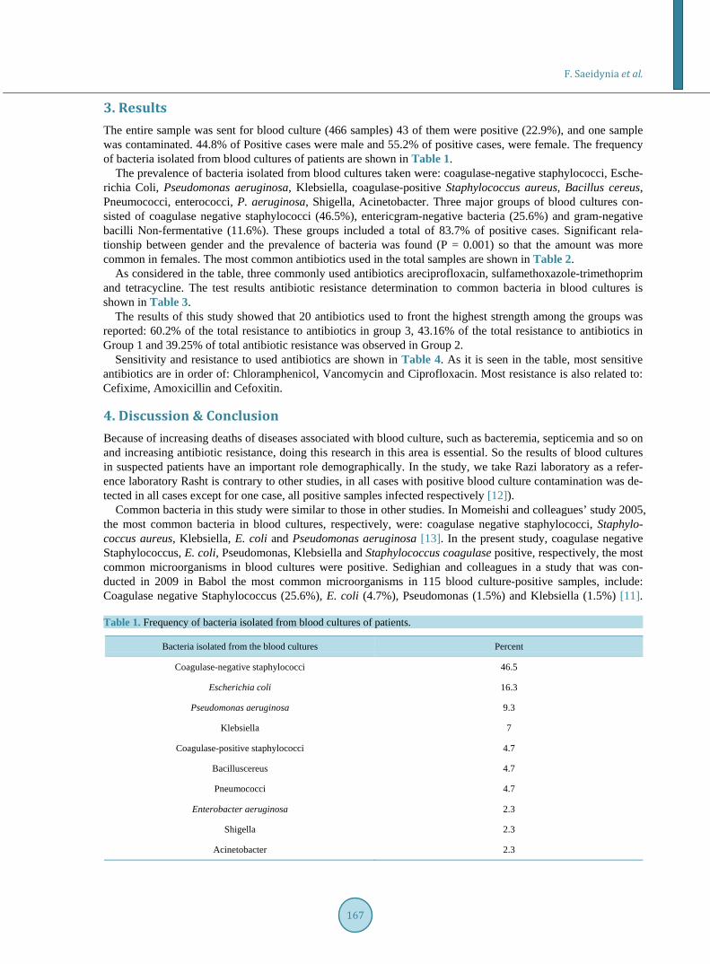
3. Results The entire sample was sent for blood culture (466 samples) 43 of them were positive (22.9%), and one sample was contaminated. 44.8% of Positive cases were male and 55.2% of positive cases, were female. The frequency of bacteria isolated from blood cultures of patients are shown in Table 1.
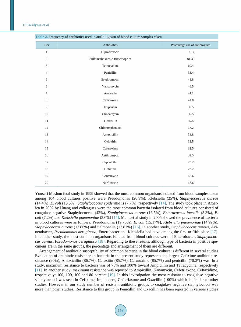
The prevalence of bacteria isolated from blood cultures taken were: coagulase-negative staphylococci, Esche-richia Coli, Pseudomonas aeruginosa, Klebsiella, coagulase-positive Staphylococcus aureus, Bacillus cereus, Pneumococci, enterococci, P. aeruginosa, Shigella, Acinetobacter. Three major groups of blood cultures con-sisted of coagulase negative staphylococci (46.5%), entericgram-negative bacteria (25.6%) and gram-negative bacilli Non-fermentative (11.6%). These groups included a total of 83.7% of positive cases. Significant rela-tionship between gender and the prevalence of bacteria was found (P = 0.001) so that the amount was more common in females. The most common antibiotics used in the total samples are shown in Table 2.
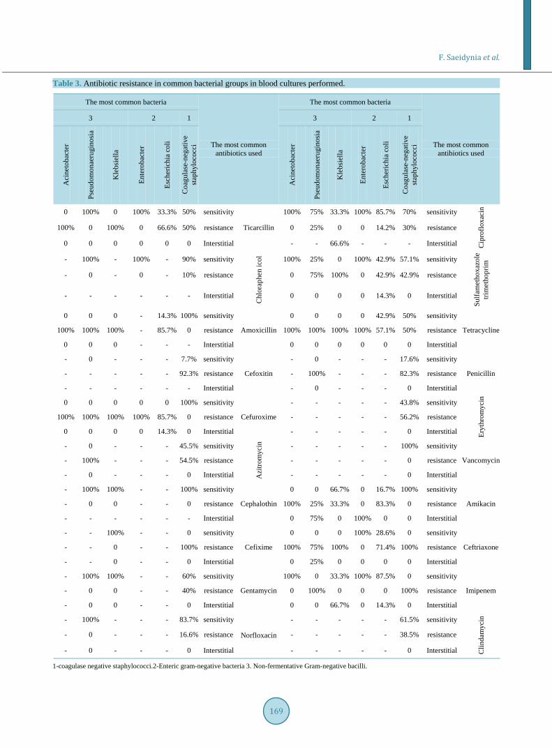
As considered in the table, three commonly used antibiotics areciprofloxacin, sulfamethoxazole-trimethoprim and tetracycline. The test results antibiotic resistance determination to common bacteria in blood cultures is shown in Table 3.
The results of this study showed that 20 antibiotics used to front the highest strength among the groups was reported: 60.2% of the total resistance to antibiotics in group 3, 43.16% of the total resistance to antibiotics in Group 1 and 39.25% of total antibiotic resistance was observed in Group 2.
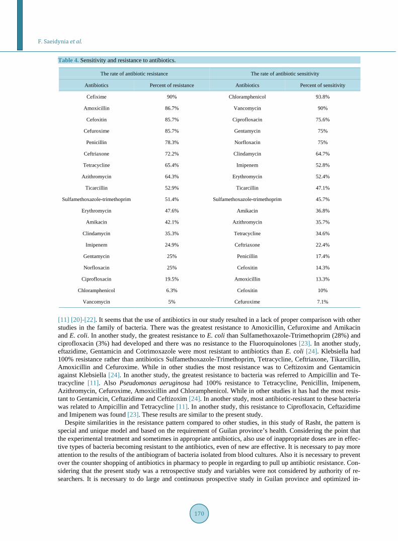
Sensitivity and resistance to used antibiotics are shown in Table 4. As it is seen in the table, most sensitive antibiotics are in order of: Chloramphenicol, Vancomycin and Ciprofloxacin. Most resistance is also related to: Cefixime, Amoxicillin and Cefoxitin.
4. Discussion & Conclusion Because of increasing deaths of diseases associated with blood culture, such as bacteremia, septicemia and so on and increasing antibiotic resistance, doing this research in this area is essential. So the results of blood cultures in suspected patients have an important role demographically. In the study, we take Razi laboratory as a refer-ence laboratory Rasht is contrary to other studies, in all cases with positive blood culture contamination was de-tected in all cases except for one case, all positive samples infected respectively [12]).
Common bacteria in this study were similar to those in other studies. In Momeishi and colleagues’ study 2005, the most common bacteria in blood cultures, respectively, were: coagulase negative staphylococci, Staphylo-coccus aureus, Klebsiella, E. coli and Pseudomonas aeruginosa [13]. In the present study, coagulase negative Staphylococcus, E. coli, Pseudomonas, Klebsiella and Staphylococcus coagulase positive, respectively, the most common microorganisms in blood cultures were positive. Sedighian and colleagues in a study that was con-ducted in 2009 in Babol the most common microorganisms in 115 blood culture-positive samples, include: Coagulase negative Staphylococcus (25.6%), E. coli (4.7%), Pseudomonas (1.5%) and Klebsiella (1.5%) [11]. Table 1. Frequency of bacteria isolated from blood cultures of patients.
Bacteria isolated from the blood cultures Percent
Coagulase-negative staphylococci 46.5
Escherichia coli 16.3
Pseudomonas aeruginosa 9.3
Klebsiella 7
Coagulase-positive staphylococci 4.7
Bacilluscereus 4.7
Pneumococci 4.7
Enterobacter aeruginosa 2.3
Shigella 2.3
Acinetobacter 2.3
F. Saeidynia et al.
168
Table 2. Frequency of antibiotics used in antibiogram of blood culture samples taken.
Tier Antibiotics Percentage use of antibiogram
1 Ciprofloxacin 95.3
2 Sulfamethoxazole-trimethoprim 81.39
3 Tetracycline 60.4
4 Penicillin 53.4
5 Erythromycin 48.8
6 Vancomycin 46.5
7 Amikacin 44.1
8 Ceftriaxone 41.8
9 Imipenem 39.5
10 Clindamycin 39.5
11 Ticarcillin 39.5
12 Chloramphenicol 37.2
13 Amoxicillin 34.8
14 Cefoxitin 32.5
15 Cefuroxime 32.5
16 Azithromycin 32.5
17 Cephalothin 23.2
18 Cefixime 23.2
19 Gentamycin 18.6
20 Norfloxacin 18.6
Yousefi Mashou fetal study in 1999 showed that the most common organisms isolated from blood samples taken among 104 blood cultures positive were Pseudomonas (26.9%), Klebsiella (25%), Staphylococcus aureus (14.4%), E. coli (13.5%), Staphylococcus epidermid is (7.7%), respectively [14]. The study took place in Amer-ica in 2002 by Huang and colleagues were the most common bacteria isolated from blood cultures consisted of coagulase-negative Staphylococcus (42%), Staphylococcus aureus (16.5%), Enterococcus faecalis (8.3%), E. coli (7.2%) and Klebsiella pneumoniae (3.6%) [15]. Mahtaet al study in 2005 showed the prevalence of bacteria in blood cultures were as follows: Pseudomonas (19.75%), E. coli (15.17%), Klebsiella pneumoniae (14.99%), Staphylococcus aureus (13.86%) and Salmonella (12.87%) [16]. In another study, Staphylococcus aureus, Aci-netobacter, Pseudomonas aeruginosa, Enterobacter and Klebsiella had have among the first to fifth place [17]. In another study, the most common organisms isolated from blood cultures were of Enterobacter, Staphylococ-cus aureus, Pseudomonas aeruginosa [18]. Regarding to these results, although type of bacteria in positive spe-cimens are in the same groups, the percentage and arrangement of them are different.
Arrangement of antibiotic susceptibility of common bacteria in the blood culture is different in several studies. Evaluation of antibiotic resistance in bacteria in the present study represents the largest Cefixime antibiotic re-sistance (90%), Amoxicillin (86.7%), Cefoxitin (85.7%), Cefuroxime (85.7%) and penicillin (78.3%) was. In a study, maximum resistance to bacteria was of 75% and 100% toward Ampicillin and Tetracycline, respectively [11]. In another study, maximum resistance was reported to Ampicillin, Kanamycin, Cefetriaxone, Ceftazidime, respectively: 100, 100, 100 and 80 percent [19]. In this investigation the most resistant to coagulase negative staphylococci was seen in Cefixime, Impipenem, Cefteriaxone and Oxacillin (100%) which is similar to other studies. However in our study number of resistant antibiotic groups to coagulase negative staphylococci was more than other studies. Resistance to this group in Penicillin and Oxacillin has been reported in various studies
F. Saeidynia et al.
169
Table 3. Antibiotic resistance in common bacterial groups in blood cultures performed.
The most common antibiotics used
The most common bacteria
The most common antibiotics used
The most common bacteria
1 2 3 1 2 3
Coa
gula
se-n
egat
ive
st
aphy
loco
cci
Esch
eric
hia
coli
Ente
roba
cter
Kle
bsie
lla
Pseu
dom
onae
rugi
nosi
a
Aci
neto
bact
er
Coa
gula
se-n
egat
ive
st
aphy
loco
cci
Esch
eric
hia
coli
Ente
roba
cter
Kle
bsie
lla
Pseu
dom
onae
rugi
nosi
a
Aci
neto
bact
er
Cip
roflo
xaci
n sensitivity 70% 85.7% 100% 33.3% 75% 100%
Ticarcillin
sensitivity 50% 33.3% 100% 0 100% 0
resistance 30% 14.2% 0 0 25% 0 resistance 50% 66.6% 0 100% 0 100%
Interstitial - - - 66.6% - - Interstitial 0 0 0 0 0 0
Sulfa
met
hoxa
zole
tri
met
hopr
im sensitivity 57.1% 42.9% 100% 0 25% 100%
Chl
orap
hen
icol
sensitivity 90% - 100% - 100% -
resistance 42.9% 42.9% 0 100% 75% 0 resistance 10% - 0 - 0 -
Interstitial 0 14.3% 0 0 0 0 Interstitial - - - - - -
Tetracycline
sensitivity 50% 42.9% 0 0 0 0
Amoxicillin
sensitivity 100% 14.3% - 0 0 0
resistance 50% 57.1% 100% 100% 100% 100% resistance 0 85.7% - 100% 100% 100%
Interstitial 0 0 0 0 0 0 Interstitial - - - 0 0 0
Penicillin
sensitivity 17.6% - - - 0 -
Cefoxitin
sensitivity 7.7% - - - 0 -
resistance 82.3% - - - 100% - resistance 92.3% - - - - -
Interstitial 0 - - - 0 - Interstitial - - - - - -
Eryt
hrom
ycin
sensitivity 43.8% - - - - -
Cefuroxime
sensitivity 100% 0 0 0 0 0
resistance 56.2% - - - - - resistance 0 85.7% 100% 100% 100% 100%
Interstitial 0 - - - - - Interstitial 0 14.3% 0 0 0 0
Vancomycin
sensitivity 100% - - - - -
Azi
trom
ycin
sensitivity 45.5% - - - 0 -
resistance 0 - - - - - resistance 54.5% - - - 100% -
Interstitial 0 - - - - - Interstitial 0 - - - 0 -
Amikacin
sensitivity 100% 16.7% 0 66.7% 0 0
Cephalothin
sensitivity 100% - - 100% 100% -
resistance 0 83.3% 0 33.3% 25% 100% resistance 0 - - 0 0 -
Interstitial 0 0 100% 0 75% 0 Interstitial - - - - - -
Ceftriaxone
sensitivity 0 28.6% 100% 0 0 0
Cefixime
sensitivity 0 - - 100% - -
resistance 100% 71.4% 0 100% 75% 100% resistance 100% - - 0 - -
Interstitial 0 0 0 0 25% 0 Interstitial 0 - - 0 - -
Imipenem
sensitivity 0 87.5% 100% 33.3% 0 100%
Gentamycin
sensitivity 60% - - 100% 100% -
resistance 100% 0 0 0 100% 0 resistance 40% - - 0 0 -
Interstitial 0 14.3% 0 66.7% 0 0 Interstitial 0 - - 0 0 -
Clin
dam
ycin
sensitivity 61.5% - - - - -
Norfloxacin
sensitivity 83.7% - - - 100% -
resistance 38.5% - - - - - resistance 16.6% - - - 0 -
Interstitial 0 - - - - - Interstitial 0 - - - 0 -
1-coagulase negative staphylococci.2-Enteric gram-negative bacteria 3. Non-fermentative Gram-negative bacilli.
F. Saeidynia et al.
170
Table 4. Sensitivity and resistance to antibiotics.
The rate of antibiotic sensitivity The rate of antibiotic resistance
Percent of sensitivity Antibiotics Percent of resistance Antibiotics
93.8% Chloramphenicol 90% Cefixime
90% Vancomycin 86.7% Amoxicillin
75.6% Ciprofloxacin 85.7% Cefoxitin
75% Gentamycin 85.7% Cefuroxime
75% Norfloxacin 78.3% Penicillin
64.7% Clindamycin 72.2% Ceftriaxone
52.8% Imipenem 65.4% Tetracycline
52.4% Erythromycin 64.3% Azithromycin
47.1% Ticarcillin 52.9% Ticarcillin
45.7% Sulfamethoxazole-trimethoprim 51.4% Sulfamethoxazole-trimethoprim
36.8% Amikacin 47.6% Erythromycin
35.7% Azithromycin 42.1% Amikacin
34.6% Tetracycline 35.3% Clindamycin
22.4% Ceftriaxone 24.9% Imipenem
17.4% Penicillin 25% Gentamycin
14.3% Cefoxitin 25% Norfloxacin
13.3% Amoxicillin 19.5% Ciprofloxacin
10% Cefoxitin 6.3% Chloramphenicol
7.1% Cefuroxime 5% Vancomycin
[11] [20]-[22]. It seems that the use of antibiotics in our study resulted in a lack of proper comparison with other studies in the family of bacteria. There was the greatest resistance to Amoxicillin, Cefuroxime and Amikacin and E. coli. In another study, the greatest resistance to E. coli than Sulfamethoxazole-Ttrimethoprim (28%) and ciprofloxacin (3%) had developed and there was no resistance to the Fluoroquinolones [23]. In another study, eftazidime, Gentamicin and Cotrimoxazole were most resistant to antibiotics than E. coli [24]. Klebsiella had 100% resistance rather than antibiotics Sulfamethoxazole-Trimethoprim, Tetracycline, Ceftriaxone, Tikarcillin, Amoxicillin and Cefuroxime. While in other studies the most resistance was to Ceftizoxim and Gentamicin against Klebsiella [24]. In another study, the greatest resistance to bacteria was referred to Ampicillin and Te-tracycline [11]. Also Pseudomonas aeruginosa had 100% resistance to Tetracycline, Penicillin, Imipenem, Azithromycin, Cefuroxime, Amoxicillin and Chloramphenicol. While in other studies it has had the most resis-tant to Gentamicin, Ceftazidime and Ceftizoxim [24]. In another study, most antibiotic-resistant to these bacteria was related to Ampicillin and Tetracycline [11]. In another study, this resistance to Ciprofloxacin, Ceftazidime and Imipenem was found [23]. These results are similar to the present study.
Despite similarities in the resistance pattern compared to other studies, in this study of Rasht, the pattern is special and unique model and based on the requirement of Guilan province’s health. Considering the point that the experimental treatment and sometimes in appropriate antibiotics, also use of inappropriate doses are in effec-tive types of bacteria becoming resistant to the antibiotics, even of new are effective. It is necessary to pay more attention to the results of the antibiogram of bacteria isolated from blood cultures. Also it is necessary to prevent over the counter shopping of antibiotics in pharmacy to people in regarding to pull up antibiotic resistance. Con-sidering that the present study was a retrospective study and variables were not considered by authority of re-searchers. It is necessary to do large and continuous prospective study in Guilan province and optimized in-
F. Saeidynia et al.
171
forming through labs, especially reference labs to doctors and hospitals should be exposed to all these pheno-mena with more knowledge to confront to consider these issues in their empiric treatments. Also it is recom-mended to use resistance pattern and susceptibility from the same patterns in reference laboratory to be more comprehensive. Also registration system and similar network of provincial between all of laboratories including public and private under the province deputy of hygienic can better inform all doctors to facilitate and it has been a remarkable help at similar information.
Acknowledgements
At the end we thanks from Razi laboratory management in Rasht city, all laboratory colleagues, especially Mr Doctor Mesbah who helped us in this research.
References [1] Mahon, C.R., Manuselis, G. and Lehman, D.C. (2007) Text Book of Diagnostic Microbiology. 3th Edition, Saunders,
USA, 321, 373, 383, 527. [2] Michael, B. and Richard, P. (2010) Organization for Infection Control. In: Mandell, G.L., Bennett, J.E. and Dolin, R.,
Eds., Principles and Practice of Infectious Diseases, 7th Edition, Churchill Livingstone, Philadelphia, 3669-3676. [3] Naderi, H.R., Bakhshaei, M., Ghazvini, K., Zamaniyan, A. and Haghighi, J. (2006) The Prevalence of Nasopharyngeal
Carriers Moraksella Kataralys in Healthy Children under 6 Years in Kindergartens of Mashhad City and Determination Antibiotic Resistance Pattern of Isolated in Moraksellla Kataralys. Journal of the Ear, Throat, Nose and Throat Iran, 18, 169-173.
[4] Masomi, J., Yadegari, D. and Mozoni, Sh. (2005) More Appropriate Antimicrobial Agents for Antibiogram. Iranian Journal of Infectious Disease and Tropical Medicine, 10, 53-58.
[5] Longo, D.L, Kasper, D.L, Jameson, J.L, Fauci, A.S, Hauser, S.L. and Loscalzo, J. (2011) Harrison, Sepsis and Septic Shock: Principles of Internal Medicine. 18th Edition, McGraw-Hill co., Boston.
[6] Karimishahidi, M., Dabbagh Mohammadi, G.H., Mohajer Irvani, B. and Meigooie, A. (2002) Evaluation of Inpatient Patients’ Blood Cultures of Common Microorganisms. Changes in the Epidemiology and Antibiogram Amir Alam Hospital, 79-1377. Journal of Medicine Tehran University of Medical Sciences, 60, 31-37.
[7] Pirzade, T. and Nahaei, M.R. (2002) Bacteria Isolated from Blood Cultures at the Educational and Therapeutic Center Imam Khomeini, Iran. Journal of Tabriz University of Medical Sciences, 56, 40-45.
[8] Spencer, R.C. (1991) Anaerobic Bacteremia. In: Duerden Brian, I. and Drasar, B.S., Eds., Anaerobic in Human Dis-ease, Edward Arnold, England, 324-342.
[9] Forbes, B.A., Sahm, D.F. and Weissfeld, A. (2007) Bloodstream Infections. In: Baily & Scotts Diagnostic Microbiolo-gy, 20th Edition, Mosby Company, USA, 778.
[10] Ghafoori, A., Eslami, Z. and Saffari, F. (2009) The Prevalence of Aerobic and Anaerobic Bacteremia in Patients with Cancer and Their Antibiotic Resistance Patterns. Journal of Infectious Diseases and Tropical Medicine, Infectious Diseases Specialist Association, 14, 19-26.
[11] Sedighian, F., Saneii, A., Allaodolehi, H., Arashi, M. and Rekabpoor, Kh. (2008) Resistance to isolated microorgan-isms in Yahyanejad hospital, Babol. Medical Laboratory Journal, 2, 29-35.
[12] Saderi, H., Karimi, A. and Looni, M. (2009) The Frequency of Bacteria Isolated from Blood Cultures and Their Anti-biotic Susceptibility Patterns in a University Hospital in Tehran. Iranian South Medical Journal, 12, 142-148. (in Per-sian)
[13] Mamishi, S., Pourakbari, B., Ashtiani, M.H., et al. (2005) Frequency of Isolation and Antimicrobial Susceptibility of Bacteria Isolated from Blood Stream Infections at Children’s Medical Center, Tehran, Iran, 1996-2000. International Journal of Antimicrobial Agents, 26, 373-379. http://dx.doi.org/10.1016/j.ijantimicag.2005.08.004
[14] Yosefi, M.R. (1999) Bacteriology of Neonatal Septicemia and Determination of Their Antibiotic Resistance in Hama-dan in 1998 to 1999 (Persian). Iranian South Medical of Journal, 2, 136-143.
[15] Huang, S.S., Labus, B.J., Samuel, M.C., et al. (2002) Antibiotic Resistance Patterns of Bacterial Isolates from Blood in San Francisco County, California, 1996-1999. Emerging Infectious Diseases, 8, 195-201. http://dx.doi.org/10.3201/eid0802.010102
[16] Mehta, M., Dutta, P. and Gupta, V. (2005) Antimicrobial Susceptibility Pattern of Blood Isolates from a Teaching Hospital in North India. Japanese Journal of Infectious Diseases, 58, 174-176.
[17] Yinnon, A.M., Schlesinger, Y., Gabbay, D. and Rudensky, B. (1997) Analysis of 5 Years of Bacteraemias: Importance of Stratification of Microbial Susceptibilities by Source of Patients. Journal of Infection, 35, 17-23.
F. Saeidynia et al.
172
http://dx.doi.org/10.1016/S0163-4453(97)90857-4 [18] Shehabi, A.A. and Baadran, I. (1996) Microbial Infection and Antibiotic Resistance Patterns among Jordanian Inten-
sive Care Patients. Eastern Mediterranean Health Journal, 3, 515-520. [19] Habibian, R., Imani, R. and Khoshdel, A. (2012) Changes of Trend of Antibiotic Susceptibility in Isolated Bacteria
from Culture of ICU Patients of Shahrekord Ayatollah Kashanani Hospital, I.R. Iran. Journal of Shahrekord University of Medical Sciences, 14, 77-83.
[20] Aminzadeh, Z., Vahdani, P. and Khosravi, Z. (2005) Sensitivity and Resistance of Microorganisms in Loghman Hakim Hospital, Tehran, 1381. Iranian Journal of Infectious Disease and Tropical Medicine, 10, 47-52.
[21] Sader, H.S., Jones, R.N., Dowzicky, M.J. and Fritsche, T.R. (2005) Antimicrobial Activity of Tetracycline Tested against Nosocomial Bacterial Pathogens from Patients Hospitalized in the Intensive Care Unit. Diagnostic Microbiol-ogy and Infectious Disease, 52, 203-208. http://dx.doi.org/10.1016/j.diagmicrobio.2005.05.002
[22] Tavakoli, A., Yazdani, R. and Bokaeian, M. (2001) Relative Frequency Study of Coagulase Positive Staphylococci Resistance to Beta Lactam Antibiotics Using Iodometric and Acidometric Assays. Tabib-E-Shargh, 1, 1-7.
[23] Huang, S.S., Labus, J.B., Samuel, M.C., Wan, D.T. and Reingold, A.L. (2002) Antibiotic Resistance Patterns of Bac-terial Isolates from Blood in San Francisco County, California, 1996-1999. Emerging Infectious Diseases, 8, 195-201. http://dx.doi.org/10.3201/eid0802.010102
[24] Mohamad, A.A., Yadegari, D., Hoseinimoghadam, M.M., Shadnoosh, M., Farahanchi, N., Rafiazade, R. and Danesh-var, S. (2007) Antibiotic Resistance Pattern of in Gram-Negative Microorganisms Nosocomial during the Years 2000-2004 in Anbiya Hospital Tehran. Researcher (Research Journal Martyr Beheshti University of Medical Sciences), 11, 123-127.