Characterisation of algogenic organic matter extracted from cyanobacteria, green algae and diatoms
Ancient DNA derived from alkenone-biosynthesizing
haptophytes and other algae in Holocene sediments
from the Black Sea
Marco J. L. Coolen,1,2 Arjan Boere,1 Ben Abbas,1 Marianne Baas,1
Stuart G. Wakeham,3 and Jaap S. Sinninghe Damste1
Received 28 June 2005; revised 14 October 2005; accepted 10 November 2005; published 18 February 2006.
[1] Holocene sea surface temperatures (SST) of the Black Sea have been reconstructed using sedimentary C37
unsaturated alkenones assumed to be derived from the coccolithophorid haptophyte Emiliania huxleyi, whosefossil coccoliths are an important constituent of the unit I sediments. However, alkenones can also bebiosynthesized by haptophyte species that do not produce microscopic recognizable coccoliths. A species-specific identification of haptophytes is important in such U37
K0-based past SST reconstructions since different
species have different alkenone-SST calibrations. We showed that 18S rDNA of E. huxleyi made up only a verysmall percentage (less than 0.8%) of the total eukaryotic 18S rDNAwithin the up to 3600-year-old fossil recordobtained from the depocenter (>2000 m) of the Black Sea. The predominant fossil 18S rDNAwas derived fromdinoflagellates (Gymnodinium spp.), which are predominant members of the summer phytoplankton bloom inthe modern Black Sea. Using a polymerase chain reaction/denaturing gradient gel electrophoresis methodselective for haptophytes, we recovered substantial numbers of a preserved 458-base-pair (bp)-long 18S rDNAfragment of E. huxleyi from the Holocene Black Sea sediments. Additional fossil haptophyte sequences were notdetected, indicating that the E. huxleyi alkenone-SST calibration can be applied for at least the last �3600 years.The ancient E. huxleyi DNA was well protected against degradation since the DNA/alkenone ratio did notsignificantly decrease throughout the whole sediment core and 20% of �2700-year-old fossil E. huxleyi DNAwas still up to 23,000 base pairs long. We showed that fossil DNA offers great potential to study the Holocenepaleoecology and paleoenvironment of anoxic deep-sea settings in unprecedented detail.
Citation: Coolen, M. J. L., A. Boere, B. Abbas, M. Baas, S. G. Wakeham, and J. S. Sinninghe Damste (2006), Ancient DNA derived
from alkenone-biosynthesizing haptophytes and other algae in Holocene sediments from the Black Sea, Paleoceanography, 21, PA1005,
doi:10.1029/2005PA001188.
1. Introduction
[2] Organic components in sediments, such as lipids andpigments, derived from specific organisms (i.e., biomarkers)form an archive of the past species composition of the watercolumn and hence can be used to reconstruct the physicaland chemical conditions caused by climate change[Brassell, 1993]. For example, long-chain (C37, C38 andC39) unsaturated methyl and ethyl ketones (alkenones) havebeen found to be characteristic of haptophyte microalgae,including the cosmopolitan coccolithophorid Emilianiahuxleyi [Volkman et al., 1980], which first appeared in thelate Pleistocene [Marlowe et al., 1990]. This species isconsidered to be the dominant source of alkenones in mostcontemporary marine sediments. Alkenones are of greatinterest to paleoceanographers because of the strong empir-ical relationship between the degree of unsaturation in
alkenones and growth temperature, which forms the basisfor their use as molecular proxies of past sea surfacetemperatures (SST) [Bendle and Rosell-Mele, 2004;Brassell et al., 1986; Conte et al., 2001; Goni et al.,2004; Prahl and Wakeham, 1987; Rosell-Mele, 1998; Sachset al., 2000; Sikes et al., 2005; Volkman et al., 1995]. Apartfrom E. huxleyi, other haptophyte algae also biosynthesizealkenones and these algae often possess different relation-ships between the degree of unsaturation in alkenones andgrowth temperature [Prahl and Wakeham, 1987; Versteeghet al., 2001; Volkman et al., 1995] with implications forpaleoceanographic interpretations. If these other species arecoccolithophorids, preserved fossil coccoliths may indicatethe species identity [Villanueva et al., 2002] but there is alsoa group of alkenone-producing haptophytes that does notproduce coccoliths.[3] We recently showed for the Holocene sediments of
postglacial Ace Lake (Vestfold Hills, Antarctica) by analy-sis of 18S rDNA of ancient haptophyte species that thesedimentary alkenones were derived from haptophytesrelated to noncoccolithophorid Isochrysis species [Coolenet al., 2004]. This combined lipid-DNA biomarker approachalso showed that Holocene salinity variations caused majorchanges in the abundance of different haptophyte species,
PALEOCEANOGRAPHY, VOL. 21, PA1005, doi:10.1029/2005PA001188, 2006
1Department of Marine Biogeochemistry and Toxicology, RoyalNetherlands Institute for Sea Research, Den Burg, Netherlands.
2Now at Department of Marine Chemistry and Geochemistry, WoodsHole Oceanographic Institution, Woods Hole, Massachusetts, USA.
3Skidaway Institute of Oceanography, Savannah, Georgia, USA.
Copyright 2006 by the American Geophysical Union.0883-8305/06/2005PA001188$12.00
PA1005 1 of 17
each with different alkenone distributions. This studyshowed that potentially the analysis of fossil DNA canreveal important paleoceanographic information. However,in lakes most of the DNA of phototrophic organisms isalready degraded before the decaying cells reached thesediment, although the portion of DNA buried within theHolocene sediments was well preserved and protectedagainst further degradation [Coolen and Overmann, 1998;Coolen et al., 2006]. These sulfidic lakes are relativelyshallow (<25 m) and, therefore the DNA of decaying cellsexperience only a short travel distance and residence timebefore the cellular remains reach the sediment. This posesthe question whether fossil DNA of water column dwellingalgae may survive in the Holocene record of anoxic deep-sea environments, where cells have to travel a far greaterdistance and experience a longer residence time before theyreach the sediment.[4] To answer this question, we have chosen to analyze
sediments of the Black Sea because the >2000-m-deepwaters below �70 m as well as the organic carbon-richsediments are sulfidic and therefore are expected to provideexcellent preservation conditions for fossil DNA. Xu et al.[2001] reported a predominance of C37:2 and C37:3 alke-nones, indicative of E. huxleyi throughout the coccolith-bearing unit I sediments of the Black Sea. The latter studyalso described a novel C36:2 ethyl ketone (hexatriaconta-16E,21E)-dien-3-one) which was found only in traceamounts in unit I sediments, but it was the most abundantalkenone in the older sediment layers of unit II where fossilcoccoliths derived of E. huxleyi were absent. Since unit IIsediments were deposited before E. huxleyi started to colo-nize the photic zone of the Black Sea [Hay, 1988], a differentbiological precursor for this compound was proposed. Onthe basis of these findings, we focused our study on whetherfossil DNA evidence confirmed that E. huxleyi was indeedthe only biological source of the alkenones or whetheradditional noncoccolithophorid haptophytes known to bio-synthesize alkenones such as Isochrysis galbana [Marloweet al., 1984; Rontani et al., 2004], thrived in the photic zoneof the Black Sea during the Holocene. Fossil partial 18SrDNA fragments of ancient haptophytes were amplified bypolymerase chain reaction (PCR) using general primers forthe domain Eukarya [Dıez et al., 2001] as well as primersselective for haptophytes [Coolen et al., 2004]. All ampli-cons were analyzed by denaturing gradient gel electropho-resis (DGGE) [Coolen et al., 2004; Muyzer et al., 1993].[5] Since DNA of alkenone-biosynthesizing haptophytes
was expected to be far less stable compared to thesealkenone lipids, we also determined the haptophyte DNA/alkenone ratio in the sediment record and compared it to thehaptophyte DNA/alkenone ratio found in the extant watercolumn as well as in a culture of E. huxleyi. Fully hydratedfossil DNA in sulfidic lake sediments has been shown to beprone to fragmentation [Coolen and Overmann, 1998].Thus we also studied the extent and onset of fragmentationof fossil DNA from the Black Sea deep-sea sediment recordbased on the quantitative distribution of fossil 458-bp-longE. huxleyi 18S rDNA by means of quantitative real-timePCR (Q-PCR) in various size classes of the extracted DNA.Since we expected to find shortened fossil DNA fragments,
we decided that DGGE, which allows only the analysis ofup to 600-bp-long fragments [Muyzer et al., 1993], was themethod of choice for the analysis of short ancient DNAfragments as this method is fast and far less laboriouscompared to the screening of a clone library.
2. Experimental Setup
2.1. Setting
[6] On the basis of stratigraphic analyses, we now knowthat the Black Sea was originally a freshwater lake butbecause of the postglacial rise of the global sea level, aseawater connection across the shallow sill of the Bosporuswas established at least 7150 years ago. The first evidenceof seawater intrusion dates earlier, however, some 9800years ago [Arthur and Dean, 1998; Jones and Gagnon,1994]. After the Bosporus connection, a stable pycnoclinedeveloped in the Black Sea between the brackish surfacewater, influenced by the large riverine inflow, and the moresaline bottom water of Mediterranean origin. Owing to thestable stratification, anoxia developed below the pycno-cline, and from �7500 years ago an organic-rich sapropelstarted to accumulate. The coccolithophorid, Emilianiahuxleyi, is believed to have invaded the Black Sea �3450years ago, and since then a varved coccolith ooze has beendeposited [Arthur and Dean, 1998; Calvert et al., 1987;Hay et al., 1991; Xu et al., 2001].[7] In the chronological description of late Pleistocene
and Holocene Black Sea sediments, three depositionalperiods are recognized [Arthur and Dean, 1998; Ross andDegens, 1974]: the modern coccolith ooze (unit I), themarine sapropel (unit II), and the deep limnic sediment(unit III). Unit I, the uppermost unit, is a laminated, organiccarbon rich (3–7% total organic carbon, TOC), coccolithmarl, with abundant E. huxleyi coccoliths [Arthur et al.,1994]. The deepest section of unit I documents the firstinvasion of E. huxleyi and is briefly interrupted by atransition sapropel (TS) before returning to the depositionof coccolith marl that has continued to the present-day [Hay,1988]. The Holocene sapropel of unit II has a lowercarbonate content of approximately 16% but is richer inorganic matter (5–20% TOC) considered to be mostly ofmarine origin [Hay, 1988]. The accumulation of organiccarbon-rich sediments may be due to enhanced preservationduring anoxia [Arthur and Dean, 1998; Wilkin et al., 1997],enhanced primary productivity during the time of sapropelformation [Calvert et al., 1987], lower sedimentation rate ofother bulk components [Calvert and Karlin, 1998], or to acombination of these factors.
2.2. Sampling
[8] Particulate organic matter (POM) for phylogeneticanalysis of the extant haptophyte communities as well asthe alkenone composition, was collected on GFF filtersfrom specific water depths of the photic zone at differentlocations in the Black Sea (Figure 1). Filters with POM ofstation 5 (Central Basin (43�0603300N, 34�0006100E) at depthsof 10, 30, and 62 m) and station 7 (42�4409300N,37�3000000E) (Eastern Basin at a depth of 30 m) werecollected during the R/V Knorr cruise K172/8 in May
PA1005 COOLEN ET AL.: ANCIENT DNA IN HOLOCENE BLACK SEA SEDIMENTS
2 of 17
PA1005
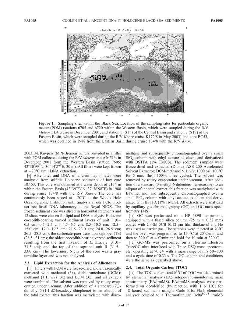
2003. M. Kuypers (MPI-Bremen) kindly provided us a filterwith POM collected during the R/V Meteor cruise M51/4 inDecember 2001 from the Western Basin (station 7605;42�3009900N, 30�1402700E; 30 m). All filters were kept frozenat �20�C until DNA extraction.[9] Alkenones and DNA of ancient haptophytes were
analyzed from sulfidic Holocene sediments of box coreBC 53. This core was obtained at a water depth of 2154 mwithin the Eastern Basin (42�3907300N, 37�3609800E) in 1988during cruise 134/8 with the R/V Knorr. The core hascontinuously been stored at �20�C at the Woods HoleOceanographic Institution until analysis at our PCR prod-uct-free fossil DNA laboratory at the Royal NIOZ. Thefrozen sediment core was sliced in horizontal fragments and12 slices were chosen for lipid and DNA analysis: Holocenecoccolith-bearing varved sediment layers of unit I (0–0.5 cm; 0.5–2.5 cm; 4.5–6.5 cm; 8.5–10.5 cm; 12.5–15.0 cm; 17.0–19.5 cm; 21.5–23.0 cm; 24.0–26.5 cm;26.5–28.5 cm); the carbonate-poor transition sapropel (TS)(28.5–31 cm); the oldest coccolith-bearing varved sedimentresulting from the first invasion of E. huxleyi (31.0–31.5 cm); and the top of the sapropel unit II (31.5–33.0 cm). The lowermost 6 cm of the core was a grayturbidite layer and was not analyzed.
2.3. Lipid Extraction for the Analysis of Alkenones
[10] Filters with POM were freeze-dried and ultrasonicallyextracted with methanol (3x), dichloromethane (DCM)/methanol (1:1, v/v) (3x) and DCM (3x), and all extractswere combined. The solvent was removed by rotary evap-oration under vacuum. After addition of a standard (2,3-dimethyl-5-(1,1-d2-hexadecyl)thiophene) to an aliquot ofthe total extract, this fraction was methylated with diazo-
methane and subsequently chromatographed over a smallSiO2 column with ethyl acetate as eluent and derivatizedwith BSTFA (1% TMCS). The sediment samples werefreeze-dried and extracted (Dionex ASE 200 AcceleratedSolvent Extractor; DCM/methanol 9:1, v/v; 1000 psi; 100�Cfor 5 min; flush 100%; three cycles). The solvent wasremoved by rotary evaporation under vacuum. After addi-tion of a standard (3-methyl-6-dideutero-heneicosane) to analiquot of the total extract, this fraction was methylated withBF3/methanol and subsequently chromatographed over asmall SiO2 column with ethyl acetate as eluent and deriv-atized with BSTFA (1% TMCS). All extracts were analyzedby capillary gas chromatography (GC) and GC-mass spec-trometry (MS).[11] GC was performed on a HP 5890 instrument,
equipped with a fused silica column (25 m � 0.32 mm)coated with CP-Sil 5CB (0.12 mm film thickness) and Hewas used as carrier gas. The samples were injected at 70�Cand the oven was programmed to 130�C at 20�C/min andthen to 320�C at 4�C/min and hold for 10 min at 320�C.[12] GC-MS was performed on a Thermo Electron
TraceGC ultra interfaced with Trace DSQ mass spectrom-eter operating at 70 eV with a mass range of m/z 50–800and a cycle time of 0.33 s. The GC column and conditionswere the same as described above.
2.4. Total Organic Carbon (TOC)
[13] The TOC content and d13C of TOC was determinedby elemental analysis (EA)/isotope-ratio-monitoring massspectrometry (EA/irmMS). EA/irmMS analyses were per-formed on decalcified (by reaction with 1 N HCl for18 hours) sediments using a Carlo Erba Flash elementalanalyzer coupled to a Thermofinnigan DeltaPLUS irmMS
Figure 1. Sampling sites within the Black Sea. Location of the sampling sites for particulate organicmatter (POM) (stations 6705 and 6720 within the Western Basin, which were sampled during the R/VMeteor 51/4 cruise in December 2001, and station 5 (ST5) of the Central Basin and station 7 (ST7) of theEastern Basin, which were sampled during the R/V Knorr cruise K172/8 in May 2003) and core BC53,which was obtained in 1988 from the Eastern Basin during cruise 134/8 with the R/V Knorr.
PA1005 COOLEN ET AL.: ANCIENT DNA IN HOLOCENE BLACK SEA SEDIMENTS
3 of 17
PA1005
system. The TOC content (as a percentage) and d13C ofTOC were determined using external standards of knowncarbon content and d13C.
2.5. Extraction of Total DNA
[14] Total DNAwas extracted from sections of GFF filtersand from 0.25 g of the 12 selected sediment sections usingthe UltraClean
TM
Soil DNA Kit Mega Prep following thedescriptions of the manufacturer (Mobio, Carlsbad, Califor-nia). The sections of the filters used for DNA extractioncontained POM from various amounts of filtered water:station 7605 (30 m (4 L)); station 5 (10 m (64 L), 30 m(94 L), 62 m (187 L)); and station 7 (30 m (55 L)). Prior toextraction, the filters were sliced with a sterile scalpel inorder to enhance the extraction efficiency. We preferred touse this extraction kit for fossil DNAwork since all reagentsand disposable tubes are free of DNA, which substantiallyreduced the chance of contamination with foreign DNA.[15] Because our study relied on the analysis of fossil 18S
rDNA derived from ancient haptophytes by PCR amplifi-cation, it was of utmost importance to prevent any contam-ination of the sediment samples by foreign DNA. Thereforethe DNA extractions were performed in our PCR product-free clean lab and inside a sterilized HEPA-filtered laminarflow bench. Prior to the extractions, the laboratory wascleaned with 6 wt % sodium hypochlorite and the laminarflow bench was sterilized by UV for 4 hours followed bysterilization of all surfaces with RNase Away (MolecularBio Products, San Diego, California), a sterilizing agentwhich also destroys DNA. At all times, two layers ofdisposable gloves were worn and the second layer of gloveswas replaced before vials with reagents from the MoBio kitwere opened. Separate pipets and sterile DNA free filter tipswere used in order to prevent the introduction of foreignDNA via aerosols during pipetting. As a control for con-tamination during DNA extraction, a parallel sample with-out sediment was subjected to the whole extraction andpurification procedure (extraction control). The concentra-tion of DNA for each extracted sediment sample wasquantified with the fluorescent dye PicoGreen (MoBiTec,Gottingen, Germany). A subsample of the total DNAextracts from various sediment depths was subjected toagarose gel electrophoresis to determine the quality andfragment length of the DNA throughout the core. Undiluted,as well as 2, 5, 10, 20, and 50 times diluted DNA extractswere subjected to quantitative PCR (Q-PCR) reactions inorder to determine whether PCR-inhibiting coextractedimpurities within the DNA extracts were present.[16] Extensive precautions against contamination with
foreign DNA were also performed during the pipetting ofPCR reagents. PCR reactions were prepared in thecleaned PCR product-free clean room inside a separatesterilized (UV + RNase Away) PCR work station. Sep-arate pipets and filter tips were used for postextractionPCR. PCR ingredients and sterile disposable tubes wereonly opened inside the bench. Disposable gloves wereworn at all times.[17] Additional precautions against contamination with
foreign (modern) DNAwere performed during other experi-ments as described in the material and methods section.
2.6. Amplification of Ancient 18S rDNA
[18] All PCR reactions were performed in an iCycler(Biorad, Hercules, California). All reactions involved initialdenaturing (5 min at 95�C), followed by 35–38 cyclesincluding denaturing (30 s at 94�C), 40 s of primer anneal-ing at temperatures described below, and primer extension(40 s at 72�C). A final extension was performed at 72�C. Inan initial attempt to detect fossil 18S rDNA of haptophytes,the most abundant eukaryotic partial 18S rDNA was ampli-fied using a primer combination as described previously[Dıez et al., 2001]. In addition, partial 18S rDNA solelyfound in haptophyte algae was selectively amplified usingprimers selective for haptophytes [Coolen et al., 2004]. Theannealing temperature was set to 64�C (eukaryotes) and62.5�C (haptophyte-specific PCR). A 40-bp-long GC clamp[Muyzer et al., 1993] was attached to the 50-end of primerEuk 563r (eukaryotes) and Prym-887r (haptophyte-specificPCR), to prevent complete melting of the PCR productsduring DGGE. For other purposes, the same combinationswithout GC clamp were used. Each amplification reactioncontained 0.25 mM of each deoxynucleotide (dNTP)(Amersham-Biosciences, Piscataway, New Jersey), 8 mg ofBovine Serum Albumin (BSA), 2 mL of 10 � PicoMaxx
TM
reaction buffer (Stratagene, LaJolla, California), 1 unit ofPicoMaxx
TM
high-fidelity PCR system and 0.2 mM ofprimers (Thermo-Electron, Ulm, Germany). The reactionmixtures were adjusted to a final volume of 20 mL withDNA and DNase free, sterile water (Sigma, Saint Louis,Missouri). Each PCR amplification series included threereactions without DNA template, which served as a controlfor contaminations during the pipetting of the reactionmixture components. A fourth reaction with 0.4 mL of theextraction control was amplified by PCR as a control forcontamination during the extraction of DNA from thesediment samples. A fifth reaction containing 4 � 106
copies of complete length 18S rDNA of the chlorophyteTetraselmis was used to monitor the specificity of the PCRreactions.
2.7. Denaturing Gradient Gel Electrophoresis
[19] The PCR-amplified partial 18S rDNA of eukar-yotes (603 bp including the 40 bp-long GC clamp) orhaptophytes (498 bp including the 40-bp-long GC clamp),were separated by DGGE [Muyzer et al., 1993] using theconditions as described by [Coolen et al., 2004] with theslight modification that electrophoresis proceeded for5 hours at 200 V and 60�C. Afterward, the gel wasstained for 10 min by covering the gel with 4 mL of 1xTAE buffer (pH 8.3) containing 2 mL concentratedSybrGold (Molecular Probes, Eugene, Oregon). In orderto prevent DNA damage by UV, we used a Dark Reader(Clare Chemicals Research Inc., Dolores, Colorado)which uses visible light instead of UV in order tovisualize the SybrGold-stained DNA. DGGE fragmentswere sliced from the gel with a sterile scalpel and theDNA of each fragment was eluted in 50–75 mL sterile10 mM Tris-HCl (pH 8.0) by incubation for 48 hours at2�C. One mL of the eluted 18S rDNA fragments (ap-proximately 107 copies) were reamplified using 25 cycles,and the primer combinations listed above, but this time
PA1005 COOLEN ET AL.: ANCIENT DNA IN HOLOCENE BLACK SEA SEDIMENTS
4 of 17
PA1005
without GC clamp, in order to generate template DNAfor the subsequent cycle sequencing reactions.
2.8. Sequencing of DGGE Fragments
[20] Primers and dNTPs were removed using the QIA-quick PCR Purification Spin Kit (Qiagen, Hilden, Germany)and the amount of DNAwas quantified with the fluorescentdye PicoGreen (MoBiTec, Germany). Cycle sequencingreactions were performed as described by [Coolen et al.,2004].
2.9. Phylogenetic Analysis
[21] Sequence data were compiled using ARB software[Ludwig et al., 2004] and aligned with complete lengthsequences of closest relatives obtained from the GenBankdatabase [Benson et al., 2004] using the ARB FastAlignerutility. Matrices of similarity, distance and phylogeneticallycorrected distance values were generated using the neighborjoining and maximum parsimony option in ARB. Sequen-ces obtained in this study have been deposited in theGenBank sequence database under accession numbersDQ234281 to DQ234297.
2.10. Quantitative Real-Time PCR
[22] Real time PCR was performed in an iCycler system(Biorad) in order to study the relative quantitative distribu-tion of fossil 18S rDNA copies of eukaryotes and specifi-cally haptophyte algae. To quantify the 18S rDNA copynumbers, the PCR conditions and primers (without GCclamp) were used as described above. Accumulation ofnewly amplified double stranded rDNAwas followed onlineas the increase in fluorescence because of the binding of thefluorescent dye SybrGreen. Reaction mixtures (20 mL)contained 1 unit of Picomaxx
TM
high-fidelity DNA polymer-ase, 2 mL of 10x Picomaxx PCR buffer (both Stratagene),0.25 mM of each dNTP, 8 mg of BSA, 0.2 mM of primers,50,000 times diluted SYBRgreen (Molecular Probes) (opti-mized concentration), a final concentration of 10 nMfluorescein, 3 mM of MgCl2 (optimized concentration)and ultra pure sterile water (Sigma). Even after 45 cycles,all control reactions stayed negative which also indicatedthat the formation of primer dimers was negligible. Never-theless, the fluorescent signal was read in each cycle duringan additional step holding the temperature at 80�C for 25 sin order to maximize the chance that a-specific productssuch as primer dimers were melted and not quantified.Known amounts of template DNA (total DNA extractsranging between 1 and 25 ng) from each sample was addedto each Q-PCR reaction.[23] Calibration of the samples was performed as follows.
Genomic DNA of E. huxleyi strain Oslo Fjord served as atemplate to generate 563-bp-long PCR fragments using theprimers for the eukaryotal domain and to generate 458-bp-long PCR fragments using the haptophyte specific primerset. Primers and salts were removed from these PCRproducts using Qiaquick Spin Columns (Qiagen) and theexact DNA concentrations were determined fluorometri-cally (Picogreen, Molecular Probes) in order to calculatethe number of 18S rDNA copies.mL�1 of purified PCRproducts. For the calibration of the samples, between 3.10�2
and 3.107 copies of the 563-bp-long fragment (quantifica-
tion of most predominant eukarya) and between 1.10�2 and1.107 copies of the 458-bp-long fragment (quantification ofhaptophytes) were subjected to Q-PCR along with thesamples.[24] The quantification of the number of 18S rDNA
copies of E. huxleyi or the domain Eukarya was repeatedfrom duplicate total DNA extracts and the second quan-tification series were calibrated with freshly preparedstandards.[25] Control reactions were performed and included a
reaction without DNA as a control for contamination duringpipetting. A second reaction contained 0.4 mL of DNAextracted with the MoBio kit but without addition ofsediment as a control for contamination with foreign DNAduring the DNA extraction procedure. A third reactioncontained 1 � 108 copies of the complete 18S rDNA ofthe chlorophyte Tetraselmis as a control for the specificityof the haptophyte selective amplification reactions.[26] In order to prevent any contamination of the pristine
sediments, fossil DNA extracts, or PCR reagents with PCRproducts via aerosols, the calibration reactions were pre-pared in a PCR workstation located in a spatially separatedpost-PCR laboratory where no ancient DNA analysis wasperformed.
2.11. Natural Fragmentation of Fossil DNA ofE. huxleyi
[27] Nucleic acids were extracted from 4 g of wet sedi-ment of varved, coccolith-bearing unit I sediment layers:2.5–4.5 cm, 8.5–10.5 cm, 17.5–19.0, and the oldest unit Ilayer located just above the transition sapropel (24–26.5 cm). For this experiment, the previously describedsimultaneous DNA and RNA extraction method [Hurt etal., 2001] was used since this method allowed the extractionof up to 40-kb-long DNA fragments, whereas only up to10-kb-long fragments can be recovered with the UltraCleanSoil DNA Kit (MoBio). A grinding step was included in theextraction method used [Hurt et al., 2001] for efficient celllysis. The grinding was performed in a similar way for allsamples; equal amounts of sterile, heat-sterilized sand(5 hours at 450�C) was added to the sediments and grindingoccurred in a sterile, baked mortar with liquid nitrogen (thenitrogen was transported in a heat-sterilized container(5 hours at 450�C)). The samples were ground with a pestleusing 30 circular movements. This grinding procedure wasrepeated twice for all samples.[28] A fraction of the total DNA extracts was subjected to
agarose gel electrophoresis (33 min at 90 V). In order tominimize contamination with foreign DNA, the sterile 1.5%(w/vol) agarose gel was run with sterile 1x TAE pH 8.0buffer. The electrophoresis chamber was first treated withRNAse Away (Molecular Bio Products) and rinsed withnucleic acid and Nuclease free water (Sigma) and exposedwith UV for 30 min prior to use. Afterward, the gel wasstained for 20 min in a sterile chamber with 1x SybrGold(Molecular Probes) in sterile 1x TAE pH 8.0. In order toprevent DNA damage by UV, we used the Dark Reader(Clare Chemical Research, Inc.) to visualize the SybrGold-stained DNA. Using a sterile set of a scalpel and tweezers,sedimentary DNA size classes (23–40 kb, 4.4–23 kb, 2.2–
PA1005 COOLEN ET AL.: ANCIENT DNA IN HOLOCENE BLACK SEA SEDIMENTS
5 of 17
PA1005
4.4 kb, 0.7–2.2 kb and less than 0.7 kb) were sliced fromthe gel and the DNA was eluted using a Centrilutorelectrophoresis system (Millipore) following the proceduresaccording to the manufacturer.[29] Cross contamination was prevented by leaving one
lane between each sample during initial gel electrophoresisand by slicing the largest DNA size class first, followed byslicing the shorter DNA size classes. In between slicing,the scalpel and tweezers were heat-sterilized using a gasflame. The exact fragment lengths of the isolated DNAsize classes were again subjected to agarose gel electro-phoresis to determine their exact sizes using fragment sizerulers (phage l � HindIII digest and Position MolecularMass Standard (PMMS, Biorad)) on each side of theagarose gel.[30] The 0.4 mL of the total extracts as well as of each
individual fragment was subjected to Q-PCR using themethod selective for haptophytes as described earlier. Theconcentration of partial 458-bp-long amplification productsof the fossil 18S rDNA of E. huxleyi found per fossil DNAsize class was calculated (copies (ng dry wt sediment)�1)and represented the fragmentation of ancient DNA ofE. huxleyi in the fossil record. The Q-PCR products werereamplified (10 cycles) using primers (including the GCclamp) selective for haptophytes for subsequent DGGEanalysis. Sequence analysis of the resulting DGGE bandswas used to verify whether solely E. huxleyi was quantifiedin each DNA size class.[31] As a control for (cross) contamination during this
experiment, pieces of ‘‘empty’’ gel were excised along withthe various DNA-containing fractions and subjected to allsteps performed during this experiment. Along with thesamples, 0.4 mL of the controls were subjected to Q-PCRusing the primers and conditions selective for haptophytes.[32] In order to estimate the amount of DNA which was
sheared because of grinding compared to natural shearing ofthe ancient DNA, the above experiment was also performedusing an aliquot (4 g) of wet sediment 24–26.5 cm(1) without shearing and (2) to which 107 intact cells ofE. huxleyi were added. The aliquot of this extract which wassubjected to Q-PCR contained besides the natural amount ofsedimentary E. huxleyi DNA, the DNA of 2.105 addedE. huxleyi cells.[33] In order to prevent any cross contamination of
modern DNA of E. huxleyi, the addition of E. huxleyi cellsto the sediment aliquot was performed in the post-PCRlaboratory and after all ancient DNA experiments at pristinesediments were completed.
3. Results
3.1. Calibration of Sediment Ages
[34] The lithology of core BC53 was determined in 1988by X-radiography. The upper 28.5 cm represented thecoccolith-bearing varved sediment layers of unit I, followedby the presence of an olive green sapropelic layer defined asthe transition sapropel between 28.5 and 31.0 cm. Thetransition sapropel was separated from the upper 2 cm ofolive green sapropel of unit II by a thin layer of coccolith-bearing sediment at 31.0–31.5 cm. This layer was depositedduring the first invasion of E. huxleyi. AMS radiocarbon
(14C) studies revealed that the first invasion of E. huxleyioccurred �3450 years before present (BP) [Hay et al.,1991].
3.2. TOC, D13C of TOC, and Total DNAConcentrations
[35] The TOC content varied between 11 and 20% in thecoccolith-bearing sediment layers of unit I, dropping to 10%in the transition sapropel (TS) and then increasing to 20%within the upper layer of the sapropel of unit II (Figure 2a).The d13C values of the bulk organic matter varied between�25.3 and �24% in unit I. d13C in the TS was slightlymore depleted compared to the coccolith layers but slightlyenriched in the unit II sapropel layer (Figure 2b). The totalDNA concentration was highest in the top sediment layer(20 mg (g dry sediment)�1), then declined to a relativelystable concentration of �8 mg (g dry sediment)�1 through-out the remaining part of the core (Figure 2c).
3.3. The 18S rDNA-Based Identification andQuantification of Eukaryotes
[36] The PCR/DGGE analysis selective for eukaryotic18S rDNA (Figure 3) resulted in the identification of 13unique phylotypes throughout the sediment core (Figure 4).Despite the abundance of morphological remains (cocco-liths) of E. huxleyi in Black Sea unit I sediments, none ofthe sequenced predominant eukaryotic DGGE fragmentsrepresented E. huxleyi (Figures 3 and 4).[37] The most predominant DGGE fragment 9, which was
found throughout unit I and in the upper part of unit II,melted at the same position as the DGGE fragment resultingfrom the Sargasso Sea strain of E. huxleyi. However,sequence analysis revealed that the closest cultivated rela-tive of fragment 9 and the related fragment 11 weredinoflagellates of the genus Gymnodinium (94–97.2%homology). Six out of the 13 recovered eukaryotic phylo-types represented dinoflagellates. DGGE fragment 12a, 12band 13 were related to a separate cluster of Gymnodiniumspecies (94.6–97.4% homology), whereas the closest rela-tive of fragment 8 was Dinophysis norvegica (95.0%homology). Sequence 12 was present in all analyzed sedi-ment layers, whereas sequence 13 was absent from the unitII sapropel layer as well as the transition sapropel. DGGEfragment 1 was the only sequence unique to the unit IIsapropel and dropped below detection limit soon afterthe interval representing the first invasion of E. huxleyi.Sequence 1 clusters within a novel lineage of fungi/metazoa with sequences obtained from the oxygenatedmarine surface waters of the Bay of Fundy with clonems313 as its closest relative [Savin et al., 2004] (99.8%sequence homology).[38] DGGE fragments 2 and 5 represented ciliates. DGGE
fragment 2 was only abundant between 8.5 and 15 cm andits closest cultivated relative was Cryptocaryon irritans.Strombidium purpureum was the closest cultivated relativeof DGGE fragment 5 and appeared only in the sedimentlayer which was deposited during the first invasion ofE. huxleyi. DGGE fragment 4 first appeared in the transitionsapropel and younger sediment layers of unit I and itsclosest relative was the cercozoa Cryothecomonas longipes(98% sequence homology). The faint DGGE fragment 3
PA1005 COOLEN ET AL.: ANCIENT DNA IN HOLOCENE BLACK SEA SEDIMENTS
6 of 17
PA1005
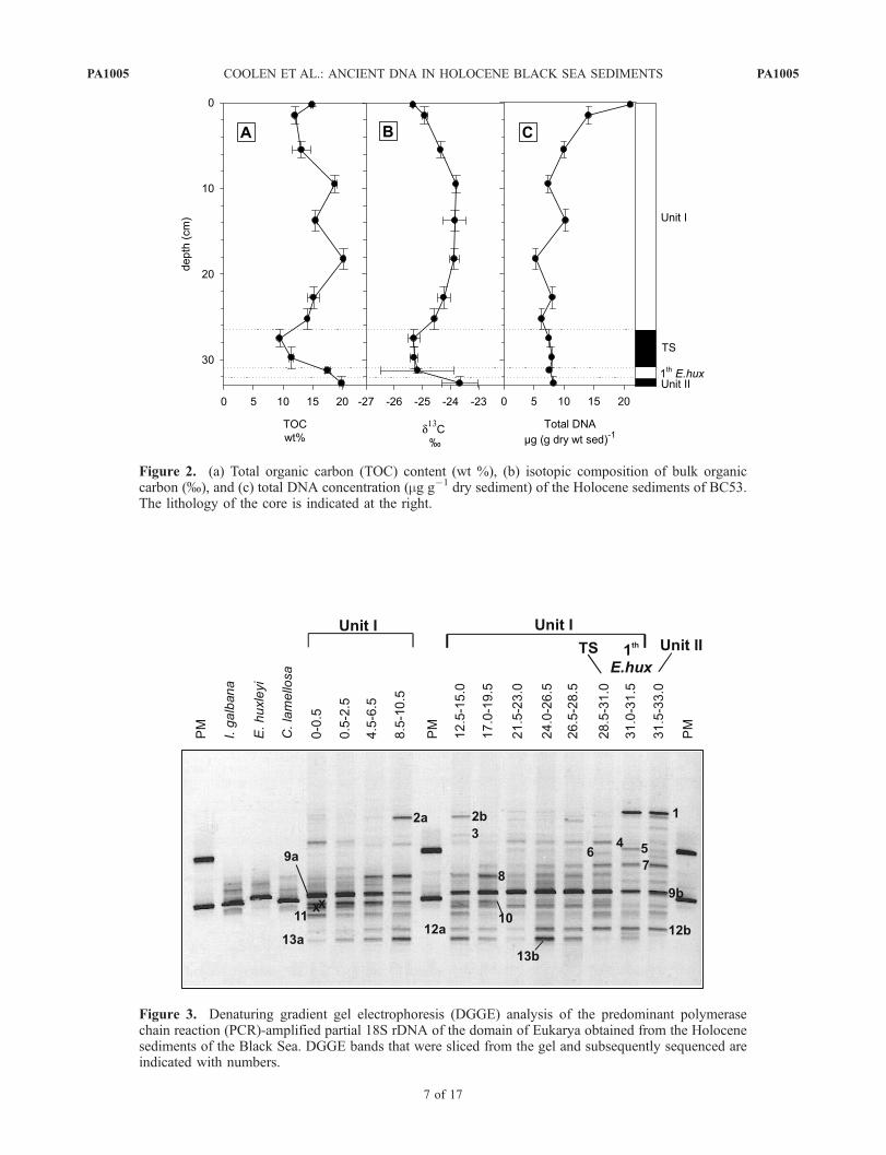
Figure 2. (a) Total organic carbon (TOC) content (wt %), (b) isotopic composition of bulk organiccarbon (%), and (c) total DNA concentration (mg g�1 dry sediment) of the Holocene sediments of BC53.The lithology of the core is indicated at the right.
Figure 3. Denaturing gradient gel electrophoresis (DGGE) analysis of the predominant polymerasechain reaction (PCR)-amplified partial 18S rDNA of the domain of Eukarya obtained from the Holocenesediments of the Black Sea. DGGE bands that were sliced from the gel and subsequently sequenced areindicated with numbers.
PA1005 COOLEN ET AL.: ANCIENT DNA IN HOLOCENE BLACK SEA SEDIMENTS
7 of 17
PA1005

Figure 4. Phylogenetic tree showing the relationship of predominant 18S rDNA sequences ofeukaryotes retrieved from the water column (POM) and Holocene sediment layers of the Black Sea(white text in black rectangles) to reference sequences obtained from the GenBank database. The lattersequences were determined from the DGGE analysis shown in Figure 3. DGGE bands with identicalmelting positions within the gel appeared to contain identical sequences except for sequences 12a and12b. As a result, phylotype BS_DGGE Euk 9 was found throughout the fossil record.
PA1005 COOLEN ET AL.: ANCIENT DNA IN HOLOCENE BLACK SEA SEDIMENTS
8 of 17
PA1005
appeared only in one sediment layer and its closest relativewas Cryothecomonas aestivales (95% sequence similarity).All attempts to sequence the DGGE fragments just aboveDGGE fragment 11, indicated with a cross, failed.[39] Q-PCR with the same primer combination selective
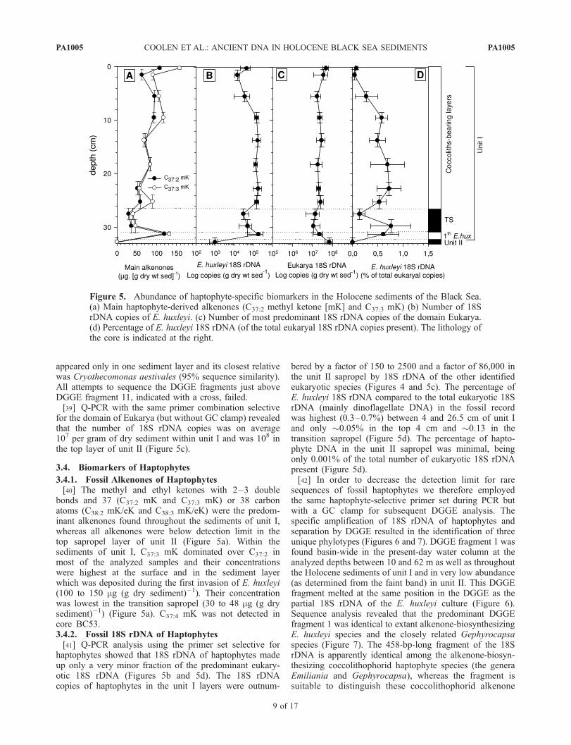
for the domain of Eukarya (but without GC clamp) revealedthat the number of 18S rDNA copies was on average107 per gram of dry sediment within unit I and was 108 inthe top layer of unit II (Figure 5c).
3.4. Biomarkers of Haptophytes
3.4.1. Fossil Alkenones of Haptophytes[40] The methyl and ethyl ketones with 2–3 double
bonds and 37 (C37:2 mK and C37:3 mK) or 38 carbonatoms (C38:2 mK/eK and C38:3 mK/eK) were the predom-inant alkenones found throughout the sediments of unit I,whereas all alkenones were below detection limit in thetop sapropel layer of unit II (Figure 5a). Within thesediments of unit I, C37:3 mK dominated over C37:2 inmost of the analyzed samples and their concentrationswere highest at the surface and in the sediment layerwhich was deposited during the first invasion of E. huxleyi(100 to 150 mg (g dry sediment)�1). Their concentrationwas lowest in the transition sapropel (30 to 48 mg (g drysediment)�1) (Figure 5a). C37:4 mK was not detected incore BC53.3.4.2. Fossil 18S rDNA of Haptophytes[41] Q-PCR analysis using the primer set selective for
haptophytes showed that 18S rDNA of haptophytes madeup only a very minor fraction of the predominant eukary-otic 18S rDNA (Figures 5b and 5d). The 18S rDNAcopies of haptophytes in the unit I layers were outnum-
bered by a factor of 150 to 2500 and a factor of 86,000 inthe unit II sapropel by 18S rDNA of the other identifiedeukaryotic species (Figures 4 and 5c). The percentage ofE. huxleyi 18S rDNA compared to the total eukaryotic 18SrDNA (mainly dinoflagellate DNA) in the fossil recordwas highest (0.3–0.7%) between 4 and 26.5 cm of unit Iand only �0.05% in the top 4 cm and �0.13 in thetransition sapropel (Figure 5d). The percentage of hapto-phyte DNA in the unit II sapropel was minimal, beingonly 0.001% of the total number of eukaryotic 18S rDNApresent (Figure 5d).[42] In order to decrease the detection limit for rare
sequences of fossil haptophytes we therefore employedthe same haptophyte-selective primer set during PCR butwith a GC clamp for subsequent DGGE analysis. Thespecific amplification of 18S rDNA of haptophytes andseparation by DGGE resulted in the identification of threeunique phylotypes (Figures 6 and 7). DGGE fragment 1 wasfound basin-wide in the present-day water column at theanalyzed depths between 10 and 62 m as well as throughoutthe Holocene sediments of unit I and in very low abundance(as determined from the faint band) in unit II. This DGGEfragment melted at the same position in the DGGE as thepartial 18S rDNA of the E. huxleyi culture (Figure 6).Sequence analysis revealed that the predominant DGGEfragment 1 was identical to extant alkenone-biosynthesizingE. huxleyi species and the closely related Gephyrocapsaspecies (Figure 7). The 458-bp-long fragment of the 18SrDNA is apparently identical among the alkenone-biosyn-thesizing coccolithophorid haptophyte species (the generaEmiliania and Gephyrocapsa), whereas the fragment issuitable to distinguish these coccolithophorid alkenone
Figure 5. Abundance of haptophyte-specific biomarkers in the Holocene sediments of the Black Sea.(a) Main haptophyte-derived alkenones (C37:2 methyl ketone [mK] and C37:3 mK) (b) Number of 18SrDNA copies of E. huxleyi. (c) Number of most predominant 18S rDNA copies of the domain Eukarya.(d) Percentage of E. huxleyi 18S rDNA (of the total eukaryal 18S rDNA copies present). The lithology ofthe core is indicated at the right.
PA1005 COOLEN ET AL.: ANCIENT DNA IN HOLOCENE BLACK SEA SEDIMENTS
9 of 17
PA1005
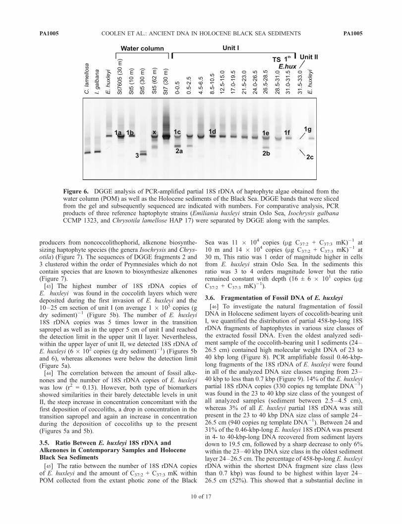
producers from noncoccolithophorid, alkenone biosynthe-sizing haptophyte species (the genera Isochrysis and Chrys-otila) (Figure 7). The sequences of DGGE fragments 2 and3 clustered within the order of Prymnesiales which do notcontain species that are known to biosynthesize alkenones(Figure 7).[43] The highest number of 18S rDNA copies of
E. huxleyi was found in the coccolith layers which weredeposited during the first invasion of E. huxleyi and the10–25 cm section of unit I (on average 1 � 105 copies (gdry sediment)�1 (Figure 5b). The number of E. huxleyi18S rDNA copies was 5 times lower in the transitionsapropel as well as in the upper 5 cm of unit I and reachedthe detection limit in the upper unit II layer. Nevertheless,within the upper layer of unit II, we detected 18S rDNA ofE. huxleyi (6 � 102 copies (g dry sediment)�1) (Figures 5band 6), whereas alkenones were below the detection limit(Figure 5a).[44] The correlation between the amount of fossil alke-
nones and the number of 18S rDNA copies of E. huxleyiwas low (r2 = 0.13). However, both type of biomarkersshowed similarities in their barely detectable levels in unitII, the steep increase in concentration concomitant with thefirst deposition of coccoliths, a drop in concentration in thetransition sapropel and again an increase in concentrationduring the deposition of coccoliths up to the present(Figures 5a and 5b).
3.5. Ratio Between E. huxleyi 18S rDNA andAlkenones in Contemporary Samples and HoloceneBlack Sea Sediments
[45] The ratio between the number of 18S rDNA copiesof E. huxleyi and the amount of C37:2 + C37:3 mK withinPOM collected from the extant photic zone of the Black
Sea was 11 � 104 copies (mg C37:2 + C37:3 mK)�1 at10 m and 14 � 104 copies (mg C37:2 + C37:3 mK)�1 at30 m, This ratio was 1 order of magnitude higher in cellsfrom E. huxleyi strain Oslo Sea. In the sediments thisratio was 3 to 4 orders magnitude lower but the ratioremained constant with depth (16 ± 6 � 101 copies (mgC37:2 + C37:3 mK)�1).
3.6. Fragmentation of Fossil DNA of E. huxleyi
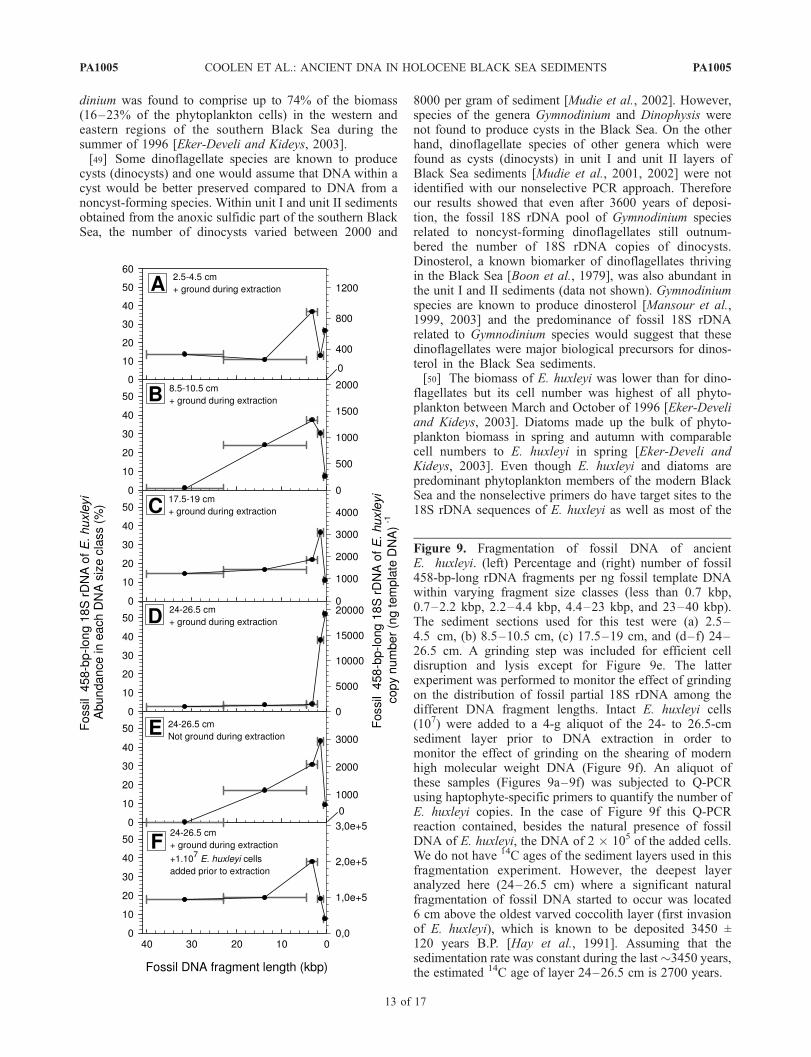
[46] To investigate the natural fragmentation of fossilDNA in Holocene sediment layers of coccolith-bearing unitI, we quantified the distribution of partial 458-bp-long 18SrDNA fragments of haptophytes in various size classes ofthe extracted fossil DNA. Even the oldest analyzed sedi-ment sample of the coccolith-bearing unit I sediments (24–26.5 cm) contained high molecular weight DNA of 23 to40 kbp long (Figure 8). PCR amplifiable fossil 0.46-kbp-long fragments of the 18S rDNA of E. huxleyi were foundin all of the analyzed DNA size classes ranging from 23–40 kbp to less than 0.7 kbp (Figure 9). 14% of the E. huxleyipartial 18S rDNA copies (330 copies ng template DNA�1)was found in the 23 to 40 kbp size class of the youngest ofall analyzed samples (sediment between 2.5–4.5 cm),whereas 3% of all E. huxleyi partial 18S rDNA was stillpresent in the 23 to 40 kbp DNA size class of sample 24–26.5 cm (940 copies ng template DNA�1). Between 24 and31% of the 0.46-kbp-long E. huxleyi 18S rDNAwas presentin 4- to 40-kbp-long DNA recovered from sediment layersdown to 19.5 cm, followed by a sharp decrease to only 6%within the 23–40 kbp DNA size class in the oldest sedimentlayer 24–26.5 cm. The percentage of 458-bp-long E. huxleyirDNA within the shortest DNA fragment size class (lessthan 0.7 kbp) was found to be highest within layer 24–26.5 cm (52%). This showed that a substantial decline in
Figure 6. DGGE analysis of PCR-amplified partial 18S rDNA of haptophyte algae obtained from thewater column (POM) as well as the Holocene sediments of the Black Sea. DGGE bands that were slicedfrom the gel and subsequently sequenced are indicated with numbers. For comparative analysis, PCRproducts of three reference haptophyte strains (Emiliania huxleyi strain Oslo Sea, Isochrysis galbanaCCMP 1323, and Chrysotila lamellose HAP 17) were separated by DGGE along with the samples.
PA1005 COOLEN ET AL.: ANCIENT DNA IN HOLOCENE BLACK SEA SEDIMENTS
10 of 17
PA1005

Figure 7. Phylogenetic tree showing the relationship of 18S rDNA sequences of haptophyte algaeretrieved from the water column (POM) and Holocene sediment layers of the Black Sea (white text inblack rectangles) to reference sequences obtained from the GenBank database. The grey box indicates thealkenone-biosynthesizing haptophytes of the order Isochrysidales. The haptophyte sequences from theBlack Sea were determined from the DGGE represented in Figure 6. DGGE bands with identical meltingpositions within the gel appeared to contain identical sequences. For example, sequences BS_DGGEHAP 1a and 1b of the extant water column were identical to sequences found in the fossil record(BS_DGGE HAP 1c–1g).
PA1005 COOLEN ET AL.: ANCIENT DNA IN HOLOCENE BLACK SEA SEDIMENTS
11 of 17
PA1005
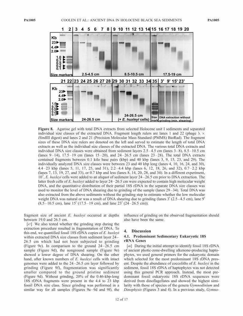
fragment size of ancient E. huxleyi occurred at depthsbetween 19.0 and 26.5 cm.[47] We also tested whether the grinding step during the
extraction procedure resulted in fragmentation of DNA. Tothis end, we quantified fossil 18S rDNA copies of E. huxleyiwithin extracted DNA size classes from sediment layer 24–26.5 cm which had not been subjected to grinding(Figure 9e). In comparison to the ground 24–26.5 cmsample (Figure 9d), the nonground sample (Figure 9e)showed a lower degree of DNA shearing. On the otherhand, after known numbers of E. huxleyi cells with intactgenomes were added to the 24–26.5 cm layer followed bygrinding (Figure 9f), fragmentation was significantlysmaller compared to the ground pristine sediment(Figure 9d). Without grinding, 20% of the 0.46-kbp-long18S rDNA fragments were present in the 4.4 to 23 kbpfossil DNA size class. Since grinding was performed in asimilar way for all samples (Figures 9a–9d and 9f), the
influence of grinding on the observed fragmentation shouldalso have been the same.
4. Discussion
4.1. Predominant Sedimentary Eukaryotic 18SrRNA Genes
[48] During the initial attempt to identify fossil 18S rDNAof ancient photic-zone-dwelling alkenone-producing hapto-phytes, we used general primers for the eukaryotic domainwhich selected for the most predominant 18S rDNA pres-ent. Despite the abundance of coccoliths of E. huxleyi in thesediment, fossil 18S rDNA of haptophytes was not detectedusing this general PCR approach. Instead, the most pre-dominant fossil eukaryotic 18S rDNA sequences werederived from dinoflagellates and showed the highest simi-larity with those of species of the genera Gymnodinium andDinophysis (Figures 3 and 4). In a previous study, Gymno-
Figure 8. Agarose gel with total DNA extracts from selected Holocene unit I sediments and separatedindividual size classes of the extracted DNA. Fragment length rulers are lanes 1 and 22 (phage l �HindIII digest) and lanes 2 and 21 (Precision Molecular Mass Standard (PMMS) BioRad). The fragmentsizes of these DNA size rulers are denoted on the left and served to estimate the length of total DNAextracts as well as the individual size classes of the extracted DNA. The various total DNA extracts andindividual DNA size classes were obtained from sediment layers 2.5–4.5 cm (lanes 3–8), 8.5–10.5 cm(lanes 9–14), 17.5–19 cm (lanes 15–20), and 24–26.5 cm (lanes 23–28). The total DNA extractscontained fragments between 0.1 kilo base pairs (kbp) and 40 kbp (lanes 3, 9, 15, 23, and 29). Theindividually analyzed DNA size classes were between 23 and 40 kbp long (lanes 4, 10, 16, 24, and 30),4.4–23 kbp (lanes 5, 11, 17, 25, and 31), 2.2–4.4 kbp (lanes 6, 12, 18, 26, and 32), 0.7–2.2 kbp(lanes 7, 13, 19, 27, and 33), or 0.7 kbp and less (lanes 8, 14, 20, 28, and 34). In a different experiment,107, E. huxleyi cells were added to an aliquot of sediment layer 24–26.5 cm prior to DNA extraction. Thelatter fresh cells of E. huxleyi added to layer 24–26.5 cm were expected to contain high molecular weightDNA, and the quantitative distribution of their partial 18S rDNA in the separate DNA size classes wasused to monitor the level of DNA shearing due to grinding of the sample (lanes 29–34). Total DNAwasalso extracted from the above sediments without the grinding step to estimate whether the low molecularweight DNAwas natural or was a result of DNA shearing due to grinding (lanes 30 (2.5–4.5 cm), lane 90
(8.5–10.5 cm), lane 150 (17.5–19 cm), and lane 230 (24–26.5 cm)).
PA1005 COOLEN ET AL.: ANCIENT DNA IN HOLOCENE BLACK SEA SEDIMENTS
12 of 17
PA1005
dinium was found to comprise up to 74% of the biomass(16–23% of the phytoplankton cells) in the western andeastern regions of the southern Black Sea during thesummer of 1996 [Eker-Develi and Kideys, 2003].[49] Some dinoflagellate species are known to produce
cysts (dinocysts) and one would assume that DNA within acyst would be better preserved compared to DNA from anoncyst-forming species. Within unit I and unit II sedimentsobtained from the anoxic sulfidic part of the southern BlackSea, the number of dinocysts varied between 2000 and
8000 per gram of sediment [Mudie et al., 2002]. However,species of the genera Gymnodinium and Dinophysis werenot found to produce cysts in the Black Sea. On the otherhand, dinoflagellate species of other genera which werefound as cysts (dinocysts) in unit I and unit II layers ofBlack Sea sediments [Mudie et al., 2001, 2002] were notidentified with our nonselective PCR approach. Thereforeour results showed that even after 3600 years of deposi-tion, the fossil 18S rDNA pool of Gymnodinium speciesrelated to noncyst-forming dinoflagellates still outnum-bered the number of 18S rDNA copies of dinocysts.Dinosterol, a known biomarker of dinoflagellates thrivingin the Black Sea [Boon et al., 1979], was also abundant inthe unit I and II sediments (data not shown). Gymnodiniumspecies are known to produce dinosterol [Mansour et al.,1999, 2003] and the predominance of fossil 18S rDNArelated to Gymnodinium species would suggest that thesedinoflagellates were major biological precursors for dinos-terol in the Black Sea sediments.[50] The biomass of E. huxleyi was lower than for dino-
flagellates but its cell number was highest of all phyto-plankton between March and October of 1996 [Eker-Develiand Kideys, 2003]. Diatoms made up the bulk of phyto-plankton biomass in spring and autumn with comparablecell numbers to E. huxleyi in spring [Eker-Develi andKideys, 2003]. Even though E. huxleyi and diatoms arepredominant phytoplankton members of the modern BlackSea and the nonselective primers do have target sites to the18S rDNA sequences of E. huxleyi as well as most of the
Figure 9. Fragmentation of fossil DNA of ancientE. huxleyi. (left) Percentage and (right) number of fossil458-bp-long rDNA fragments per ng fossil template DNAwithin varying fragment size classes (less than 0.7 kbp,0.7–2.2 kbp, 2.2–4.4 kbp, 4.4–23 kbp, and 23–40 kbp).The sediment sections used for this test were (a) 2.5–4.5 cm, (b) 8.5–10.5 cm, (c) 17.5–19 cm, and (d–f) 24–26.5 cm. A grinding step was included for efficient celldisruption and lysis except for Figure 9e. The latterexperiment was performed to monitor the effect of grindingon the distribution of fossil partial 18S rDNA among thedifferent DNA fragment lengths. Intact E. huxleyi cells(107) were added to a 4-g aliquot of the 24- to 26.5-cmsediment layer prior to DNA extraction in order tomonitor the effect of grinding on the shearing of modernhigh molecular weight DNA (Figure 9f). An aliquot ofthese samples (Figures 9a–9f) was subjected to Q-PCRusing haptophyte-specific primers to quantify the number ofE. huxleyi copies. In the case of Figure 9f this Q-PCRreaction contained, besides the natural presence of fossilDNA of E. huxleyi, the DNA of 2 � 105 of the added cells.We do not have 14C ages of the sediment layers used in thisfragmentation experiment. However, the deepest layeranalyzed here (24–26.5 cm) where a significant naturalfragmentation of fossil DNA started to occur was located6 cm above the oldest varved coccolith layer (first invasionof E. huxleyi), which is known to be deposited 3450 ±120 years B.P. [Hay et al., 1991]. Assuming that thesedimentation rate was constant during the last�3450 years,the estimated 14C age of layer 24–26.5 cm is 2700 years.
PA1005 COOLEN ET AL.: ANCIENT DNA IN HOLOCENE BLACK SEA SEDIMENTS
13 of 17
PA1005
known diatom sequences, none of the identified 18S rDNAsequences were found to be haptophytes or diatoms(Figures 3 and 4). This suggests that there are unknownmechanisms that cause a species- or group-specific preser-vation of fossil DNA.[51] The remaining sequences which were retrieved with
the nonselective primers for the eukaryotic domain wererelated to heterotrophs such as ciliates, cercozoa (represent-ing faint DGGE bands) and a sequence related to a novellineage of fungi/metazoan (Figure 3). The latter sequence(DGGE band 1) showed 99.8% sequence homology withclone ms313 obtained from oxygenated water of the Bay ofFundy [Savin et al., 2004] (Figure 4). The latter sequencewas predominant only in the unit II layer and disappearedsoon after E. huxleyi started to colonize the photic zone.Whether the biological precursor of the sequence was alsoadapted to thrive in the sapropel layer of unit II is unlikelyas the TOC concentration did not vary greatly between thesapropel and the oldest coccolith layer or the transitionsapropel (Figure 2) where 18S rDNA of this species wasbelow the detection limit (Figure 3). Most likely, this not-yet-described species was deposited during times of sapro-pel (unit II) deposition. Its abrupt absence after E. huxleyistarted to colonize the Black Sea points toward less favor-able conditions possibly related to a changing salinity.Fungal remains (spores) have indeed been described fromsouthern Black Sea sediments and found to be highest in thesapropel. The presence of fungal spores was associated witha greater freshwater/terrestrial input at the times of sapropeldeposition [Mudie et al., 2002].[52] These results showed that fossil DNA of various
ancient photic zone associated microorganisms survived inthe Holocene sulfidic sediment record of the Black Sea.However, the mechanisms controlling the deposition ofcells and the preservation of their fossil DNA is not similarfor every cell type. We, furthermore, conclude that nonse-lective primers for, for example, the domain Eukarya cannotbe used to identify specific and less abundant sequences ofancient phytoplankton.
4.2. Fossil DNA of Haptophytes in the HoloceneBlack Sea Sediments
[53] Since our initial attempts to identify fossil DNA ofhaptophytes from Holocene Black Sea sediments using anonselective PCR for 18S rDNA of Eukarya failed, we triedto lower the detection limit by using a PCR approachselective for haptophytes [Coolen et al., 2004]. Usingselective primers during (Q)-PCR, we were indeed able tolower the detection limit of 18S rDNA of haptophytes toaround 0.1% of the total 18S rDNA pool in unit I sedimentsand even to 0.001% of the total 18S rDNA pool in unit II.Using this selective and sensitive approach, a phylotypewith 100% sequence similarity to sequences of extantE. huxleyi strains was detected in all analyzed coccolith-bearing layers of unit I, concomitant with the presence ofdiunsaturated and triunsaturated C37 to C39 methyl and ethylketones believed to be derived from E. huxleyi. The amountof fossil 18S rDNA of E. huxleyi showed a comparableconcentration profile as for the fossil C37:2 mK and C37:3
mK (Figure 5).
[54] Despite the fact that alkenone concentrations werebelow the detection limit and coccoliths of E. huxleyi havenot been reported from unit II sediments, our highlysensitive PCR-based approach resulted also in the detectionof trace amounts of 18S rDNA of E. huxleyi in the upper2 cm of the sapropel unit II (Figures 6 and 7). This indicatedthat low numbers of E. huxleyi started to colonize the BlackSea prior to the deposition of the oldest, very well distin-guishable varved coccolith-bearing layer, and that cocco-liths in the unit II sapropel were perhaps prone todissolution which often occurs in TOC-rich sediments[Van Os et al., 1994]. Using Q-PCR, we here reached theabsolute detection limit with 200 copies of E. huxleyi 18SrDNA per gram dry sediment. This amount was equal toonly �1–5 copies in the Q-PCR reaction for which 38cycles were needed to generate visible amounts of PCRproducts. A contamination with modern E. huxleyi can beruled out as no visible PCR products were generated evenafter 45 cycles from the various control reactions. Inaddition, haptophyte sequence 2 which was detected alsofrom the unit II layer, only appeared in the upper layers ofsediment, and therefore a cross contamination duringsampling of adjacent sediment layers is also not likely.[55] Vertical transport of exogenous DNA (e.g., by
groundwater seepage over time) has been proposed to biasthe true age of ancient DNA in the fossil record [Hoehler,2005; Inagaki et al., 2005]. However, DNA is known to bestrongly adsorbed by the sediment matrix [Khanna andStotzky, 1992; Lorenz and Wackernagel, 1987; Paget et al.,1992]. Our present combined DNA and lipid analysisshowed that PCR-amplified 18S rDNA of E. huxleyi aswell as its alkenones were barely detectable from the top2 cm of the unit II sediment, whereas this layer was locatedjust below the oldest varved coccolith-bearing layer of coreBC 53 where both the DNA and alkenones of E. huxleyibecame abundant. This indicated that DNA of E. huxleyiwas indeed strongly adsorbed to the sediment matrix andthat vertical transport of nucleic acids via pore water wasnegligible. Also during our previous studies dealing withfossil DNA in sulfidic sediments, we never found PCRamplifiable rDNA of, for example, haptophytes or photo-synthetic sulfur bacteria in sediment layers where theirspecific biomarkers (respectively alkenones or carotenoids)were absent, whereas their rDNA could be amplified fromlayers with a concomitant presence of the specific lipidbiomarkers [Coolen et al., 2004; Coolen et al., 2006;Coolen and Overmann, 1998]. This showed that in thevarious investigated sediments the contamination withvertically transported DNA was negligible.[56] Even though 18S rDNA of E. huxleyi, which is not
known to form resting stages, made up a relatively smallnumber compared to the total 18S rDNA pool, the aboveresults showed that the detection limit for ancient DNA inthe fossil record can be lowered significantly by usinggroup-specific primers during PCR. This study providesstrong evidence that DNA of ancient photosynthetic algaesurvived degradation in a Holocene deep-sea sedimentrecord with an overlaying sulfidic water column of morethan 2000 m and was archived in the sedimentary record.This shows that even in deep-sea environments, the analysis
PA1005 COOLEN ET AL.: ANCIENT DNA IN HOLOCENE BLACK SEA SEDIMENTS
14 of 17
PA1005
of fossil DNA is a promising tool to identify ancient watercolumn derived microorganisms at the species level, andhence to reconstruct ancient paleoenvironments in unpre-cedented detail.[57] Using our selective and sensitive DNA approach to
analyze fossil 18S rDNA of ancient haptophytes, we foundthat no alkenone biosynthesizing haptophyte other thanE. huxleyi colonized the Black Seas photic zone duringthe last 3600 years of deposition. This showed that for thistime frame no species-specific calibration of the U37
K0-based
SST proxy [Prahl and Wakeham, 1987; Versteegh et al.,2001; Volkman et al., 1995] is required for the Black Sea.Unfortunately, core BC53 which we used for our analyses,did not penetrate deeper into the unit II sapropel, where Xuet al. [2001] reported a novel C36:2-ethyl ketone (hexatria-conta-16E,21E)-dien-3-one) as an abundant alkenone. Sinceunit II sediments were deposited before E. huxleyi started tocolonize the photic zone of the Black Sea [Hay, 1988], theauthors assumed a possibly different biological precursorfor this compound [Xu et al., 2001], which could have beentraced by our fossil DNA approach.
4.3. Fate of Fossil DNA of E. huxleyi in the HoloceneBlack Sea Sediment Record
[58] The fossil 18S rDNA/alkenone ratio of E. huxleyiwas found to be 4 orders of magnitude lower than incultured cells of E. huxleyi (strain Oslo Sea) and 3 ordersof magnitude lower compared to particulate organic matter(POM) from the photic zone of the Black Sea. Remarkably,this ratio did not increase with increasing burial in thesediment (Figure 5). This indicated that haptophyte DNAwas degraded much faster than the alkenones during thesenescence of cells and subsequent transport to the sedimentbut that the DNA was somehow protected from furtherdegradation in the late Holocene sediments. The lowerDNA/alkenone ratio in POM than in intact cells seems toindicate that in the POM there is already a fraction ofpartially degraded haptophyte cell material.[59] In previous work, we analyzed the 16S rDNA/iso-
renieratene ratio of obligate anoxygenic photosyntheticgreen sulfur bacteria (GSB) [Pfennig, 1989] within POMcollected from various depths of the sulfidic part of thewater column of the stratified Antarctic Ace Lake andcompared this ratio with the ratio found in the Holocenesediment record [Coolen et al., 2006]. Even in the presenceof 8 mM H2S, a strong decline within this ratio wasobserved within cells thriving in the photic sulfidic chemo-cline and within cells collected from the dark monimolimn-ion where light for anoxygenic photosynthesis was absent.However, as observed for the DNA to alkenone ratio in ourpresent study, the ratio of DNA to isorenieratene of the GSBdid not further decrease in the dark, sulfidic, and up to9-kyr-old sediment layers of Ace Lake. Therefore theintracellular 16S rDNA of the GSB was degraded mainlywithin the water column before the cells became buriedwithin the sediment record [Coolen et al., 2006]. During theresidence time in the dark sulfidic waters, the DNA in thesedecaying GSB was most likely attacked by intracellularnucleases and the presence of H2S did not play a substantialrole in protecting the intracellular DNA from being
degraded. However, once adsorbed by the sediment matrix,extracellular DNA has been shown to be far less prone toattack by nucleases [Khanna and Stotzky, 1992; Lorenz andWackernagel, 1987; Paget et al., 1992]. Likely, nucleasesmight also be trapped in the sediment matrix and cannotreach and attack a substantial part of the adsorbed DNA. Ourdata from the Black Sea are in good agreement with this idea.[60] Even fossil DNA in sulfidic sediments is, however,
not likely to completely escape degradation. Coolen andOvermann [1998] studied fossil 16S rDNA of MahoneyLake’s predominant obligate anoxyphototrophic purple sul-fur bacterium (PSB) Amoebobacter purpureus. Theyshowed that even in the presence of extremely high andlethal in situ levels of H2S of up to 60 mM, Holocenesediments older than 4 ka only contained up to 600-bp-longfragments and that the oldest (10-kyr-old) sediment onlycontained up to 400-bp-long fragments of the 16S rDNA.This revealed substantial fragmentation of fossil DNA and,because of the absence of high molecular weight (HMW)DNA indicative for the absence of intact and metabolicallyactive prokaryotic cells, hydrolysis was deemed the mostlikely route for the observed fragmentation [Coolen andOvermann, 1998]. The up to 2.7-kyr-old Black Sea sedi-ments contained in addition to short DNA fragments, alsoHMW DNA (Figure 8) and this indicated the presence ofextant prokaryotes that most likely participated or perhapsstill participate in the microbial degradation of fossil DNA.Nevertheless, in the 0–19 cm section of the sediment the458-bp-long fossil E. huxleyi 18S rDNA occurred for asubstantial part in the high molecular weight DNA size class(Figure 9). However, after 2700 years of deposition, 80% ofthe 458-bp-long fossil E. huxleyi 18S rDNA occurred in theextracted DNA size classes smaller than 4.4 kbp (Figure 9e),signifying a substantial fragmentation. Therefore the de-gree and onset of fragmentation of fossil DNA seemscomparable in the Mahoney Lake and the Black Seasedimentary record, despite a possible microbial degrada-tion of fossil DNA, and a lower H2S concentration in theBlack Sea.[61] An important question following from these findings
is at what point natural fragmentation of fossil DNA startsto limit the PCR amplification of genes since this woulddetermine the ultimate sedimentary age at which the poten-tially useful method of fossil DNA analysis for paleoenvir-onmental work can be applied. We found that the ratiobetween the total number of 458-bp-long fragments ofE. huxleyi and the concentration of C37 alkenones did notfurther decrease with increasing sediment depth and age.This demonstrated that in the Black Sea sedimentaryrecord, natural fragmentation in the first 30 cm of the record,spanning approximately 2700 years of deposition, did notyet limit the PCR amplification efficiency. In the Holocenesulfidic Ace Lake sedimentary record, the fossil 499-bp-long16S rDNA of ancient GSB was even less prone to diageneticalteration relative to its specific carotenoid chlorobactene asevidenced from the substantially increased fossil GSB-16SrDNA/chlorobactene ratios with increasing sediment depth[Coolen et al., 2006]. This was due to rapid transformationof the carotenoid chlorobactene, which is much more proneto diagenetic reactions than the alkenones in the Black Sea
PA1005 COOLEN ET AL.: ANCIENT DNA IN HOLOCENE BLACK SEA SEDIMENTS
15 of 17
PA1005
core. Therefore, from the previous study performed on AceLake [Coolen et al., 2006] as well as our present study, itseems that in a lacustrine as well as in deep-sea Holocenesulfidic sediments, 450- to 500-bp-long fossil DNA frag-ments can be used for qualitative species-specific phyloge-netic analysis and the quantitative analysis of ancient watercolumn derived species.[62] However, it is likely that the ancient DNA will
become even more fragmented in older sediments and thiswill ultimately significantly decrease the PCR amplificationefficiency. On the basis of the degree of fragmentationobserved from Holocene sulfidic sediments, we expect thatsediments older than Pleistocene cannot be used forancient DNA-based phylogenetic studies. However, inorder to determine this limitation, additional DNA frag-mentation experiments on older sulfidic sediments shouldbe performed.
5. Conclusion
[63] The 18S rDNA of the coccolithophorid, alkenone-biosynthesizing haptophyte E. huxleyi which colonized theBlack Seas photic zone during the late Holocene, made uponly a very small percentage of the total eukaryotic 18SrDNA within the fossil record (between 0.03 and 0.8% inthe coccolith-bearing unit I and only 0.001% in the unit IIsapropel). The predominant fossil 18S rDNA sequences, asrevealed by Q-PCR using nonselective primers for thedomain Eukarya, were derived from dinoflagellates relatedto Gymnodinium species which are predominant membersof the summer phytoplankton bloom in the modern BlackSea. However, using a PCR method, selective for 18SrDNA of haptophyte species, we recovered preserved458-bp-long 18S rDNA fragments of Emiliania huxleyifrom Holocene sulfidic sediments from the depocenter ofthe Black Sea. Additional 18S rDNA sequences related toother alkenone biosynthesizing haptophytes were notdetected, indicating that the E. huxleyi alkenone-SST cali-bration can be applied for at least the last 3600 years for theBlack Sea. The ratio between fossil haptophyte 18S rDNAand alkenones, which are known to survive for long timeperiods in sediments, was 3 orders of magnitude lowercompared to this ratio found in the extant water column ofthe Black Sea and 4 orders of magnitude lower compared tothis ratio found in a E. huxleyi culture.
[64] During an experiment in which we quantified thenumber of 458-bp-long fossil 18S rDNA of E. huxleyi inindividual size classes of the extracted fossil DNA, it wasshown that significant fragmentation of ancient haptophyteDNA occurred only after �2700 years of deposition.However, even after this time period, 200 copies of thepartial E. huxleyi 18S rDNA per ng of fossil DNA (i.e., 3%of the total E. huxleyi 18S rDNA copies) were still presentin the 23- to 40-kbp-long DNA fragment class and 20% ofthe copies were found in the 4.4- to 23-kbp-long DNAfragment size class. The presence of long DNA fragments ofE. huxleyi and the fact that the haptophyte DNA to alkenoneratio did not significantly decrease with increasing sedimentdepth and age, indicated that the fossil DNA was wellprotected against degradation for at least 2700 years. Inthe unit II sapropel located just below the oldest coccolith-bearing sediment layer, biomarkers for E. huxleyi (alke-nones, and 18S rDNA) were below or just at the detectionlimit. This indicated that the DNA of E. huxleyi wasadsorbed by the sediment matrix and that vertical migrationof DNA, which would otherwise have biased the true age ofthe ancient DNA, was very restricted. Toxic levels of H2S,and a strong adsorption of the DNA to the sediment matrixas indicated by the low recovery of the DNA of E. huxleyi,are the most likely factors resulting in the excellent preser-vation of the fossil DNA of E. huxleyi.[65] This work showed for the first time that, in addition
to traditional proxies, fossil DNA of ancient photic-zone-dwelling microorganisms offers great potential to study thepaleoecology and paleoenvironment in deep-sea settings inunprecedented detail. We are currently employing similartechniques to study the fate of DNA of other groups ofmicroorganisms in the Holocene Black Sea sediments.
[66] Acknowledgments. We would like to thank Marcel Kuypers(Max Planck Institute for Marine Microbiology, Bremen, Germany) forproviding a filter with POM collected from 30 m water depth at the westernpart of the Black Sea (station 6705). We thank Anna Noordeloos (RoyalNIOZ) for providing a culture of E. huxleyi strain from Oslo Sea and Jean-Francois Rontani (Centre d’Oceanologie de Marseille, France) for provid-ing a culture of Chrysotila lamellosa HAP 17. In addition, we would like toacknowledge Judith van Bleijswijk of the Royal NIOZ for assistance withphylogenetic analysis. This work was supported by a grant from theNetherlands Organization for Scientific Research (NWO) (Open Competi-tion Program 813.13.001 to M.J.L.C.) and NSF grant OCE0117824 toS.G.W., which we greatly appreciate.
PA1005 COOLEN ET AL.: ANCIENT DNA IN HOLOCENE BLACK SEA SEDIMENTS
16 of 17
PA1005
ReferencesArthur, M. A., and W. E. Dean (1998), Organic-matter production and preservation and evolu-tion of anoxia in the Holocene Black Sea,Paleoceanography, 13(4), 395–411.
Arthur, M. A., W. A. Dean, E. D. Neff, B. J. Hay,J. King, and G. Jones (1994), Varve calibrationrecords of carbonate and organic carbon accu-mulation over the last 2000 years in the BlackSea, Global Biogeochem. Cycles, 8(2), 195–217.
Bendle, J., and A. Rosell-Mele (2004), Distribu-tions of U37
K and U370K in the surface waters
and sediments of the Nordic Seas: Implica-tions for paleoceanography, Geochem. Geo-phys. Geosyst., 5, Q11013, doi:10.1029/2004GC000741.
Benson, D. A., I. Karsch-Mizrachi, D. J. Lipman,J. Ostell, and D. L. Wheeler (2004), GenBank:Update, Nucleic Acids Research, 32 (databaseissue), D23–D26.
Boon, J. J., W. I. C. Rijpstra, F. De Lange, J. W.de Leeuw, M. Yoshioka, and Y. Shimizu(1979), Black sea sterol—A molecular fossilfor dinoflagellate blooms, Nature, 277, 125–127.
Brassell, S. C. (1993), Applications of bio-markers for delineating marine paleocli-matic fluctuations during the Pleistocene,in Organic Geochemistry: Principles andApplications, edited by M. H. Engel andS. A. Macko, pp. 699–738, Springer, NewYork.
Brassell, S. C., G. Eglinton, I. T. Marlowe,U. Pflaumann, and M. Sarnthein (1986), Mo-lecular stratigraphy: A new tool for climaticassessment, Nature, 320, 129–133.
Calvert, S. E., and R. E. Karlin (1998), Organiccarbon accumulation in the Holocene sapropelof the Black Sea, Geology, 26, 107–110.
Calvert, S. E., J. S. Vogel, and J. R. Southon(1987), Carbon accumulation rates and the ori-gin of the Holocene sapropel in the Black Sea,Geology, 15, 918–921.
Conte, M. H., J. C. Weber, L. L. King, and S. G.Wakeham (2001), The alkenone temperaturesignal in western North Atlantic surfacewaters, Geochim. Cosmochim. Acta, 65(23),4275–4287.

PA1005 COOLEN ET AL.: ANCIENT DNA IN HOLOCENE BLACK SEA SEDIMENTS
17 of 17
PA1005
Coolen, M. J. L., and J. Overmann (1998), Ana-lysis of subfossil molecular remains of purplesulfur bacteria in a lake sediment, Appl. En-viron. Microbiol., 64(11), 4513–4521.
Coolen, M. J. L., G. Muyzer, W. I. C. Rijpstra,S. Schouten, J. K. Volkman, and J. S.Sinninghe Damste (2004), Combined DNAand lipid analyses of sediments reveal changesin Holocene haptophyte and diatom popula-tions in an Antarctic lake, Earth Planet. Sci.Lett., 223, 225–239.
Coolen, M. J. L., G. Muyzer, S. Schouten, J. K.Volkman, and J. S. Sinninghe Damste (2006),Sulfur and methane cycling during the Holo-cene in Ace Lake (Antarctica) revealed by li-pid and DNA stratigraphy, in Past and PresentMarine Water Column Anoxia, NATO Sci.Ser., Ser. IV, vol. 64, edited by L. N. Neretin,pp. 41–65, Springer, New York.
Dıez, B., C. Pedros-Alio, T. L. Marsh, andR. Massana (2001), Application of denaturinggradient gel electrophoresis (DGGE) to studythe diversity of marine picoeukaryotic assem-blages and comparison of DGGE with othermolecular techniques, Appl. Environ. Micro-biol., 67(7), 2942–2951.
Eker-Develi, E., and A. E. Kideys (2003), Dis-tribution of phytoplankton in the southernBlack Sea in summer 1996, spring and autumn1998, J. Mar. Syst., 39(3–4), 203–211.
Goni, M. A., M. P. Woodworth, H. L. Aceves,R. C. Thunell, E. Tappa, D. Black, F. Muller-Karger, Y. Astor, and R. Varela (2004), Gen-eration, transport, and preservation of thealkenone-based U37
K sea surface temperatureindex in the water column and sediments ofthe Cariaco Basin (Venezuela), Global Biogeo-chem. Cycles., 18(2), 1–21.
Hay, B. J. (1988), Sediment accumulation in thecentral western Black Sea over the past 5100years, Paleoceanography, 3(4), 491–508.
Hay, B. J., M. A. Arthur, W. A. Dean, E. D. Neff,and S. Honjo (1991), Sediment deposition inthe late Holocene abyssal Black Sea with cli-matic and chronological implications, DeepSea Res., Part A, 38(2), 1211–1235.
Hoehler, T. M. (2005), Cretaceous Park? A com-mentary on microbial paleomics, Astrobiology,5(2), 95–99.
Hurt, R. A., X. Qiu, L. Wu, Y. Roh, A. V.Palumbo, J. M. Tiedje, and J. Zhou (2001),Simultaneous recovery of RNA and DNAfrom soils and sediments, Appl. Environ. Mi-crobiol., 67(10), 4495–4503.
Inagaki, F., H. Okada, A. I. Tsapin, and K. H.Nealson (2005), Research paper: Microbialsurvival—The paleome: A sedimentarygenetic record of past microbial communities,Astrobiology, 5(2), 141–153.
Jones, G. A., and A. R. Gagnon (1994), Radio-carbon chronology of Black-Sea sediments,Deep Sea Res., Part I, 41(3), 531–557.
Khanna, M., and G. Stotzky (1992), Transforma-tion of Bacillus subtilis by DNA bound onmontmorillonite and effect of DNase on thetransforming ability of bound DNA, Appl. En-viron. Microbiol., 58(6), 1930–1939.
Lorenz, M. G., and W. Wackernagel (1987), Ad-sorption of DNA to sand and variable degrada-
tion rates of adsorbed DNA, Appl. Environ.Microbiol., 53(12), 2948–2952.
Ludwig, W., et al. (2004), ARB: A software en-vironment for sequence data, Nucleic AcidsRes., 32(4), 1363–1371.
Mansour, M. P., J. K. Volkman, A. E. Jackson,and S. I. Blackburn (1999), The fatty acid andsterol composition of five marine dinoflagel-lates, J. Phycol., 35(4), 710–720.
Mansour, M. P., J. K. Volkman, and S. I.Blackburn (2003), The effect of growth phaseon the lipid class, fatty acid and sterol compo-sition in the marine dinoflagellate, Gymnodi-nium sp in batch culture, Phytochemistry,63(2), 145–153.
Marlowe, I. T., J. C. Green, A. C. Neal, S. C.Brassell, G. Eglinton, and P. A. Course (1984),Long-chain (N-C37–C39) alkenones in thePrymnesiophyceae—Distribution of alkenonesand other lipids and their taxonomic signifi-cance, Br. Phycol. J., 19(3), 203–216.
Marlowe, I. T., S. C. Brassell, G. Eglinton, andJ. C. Green (1990), Long-chain alkenones andalkyl alkenoates and the fossil coccolith recordof marine-sediments, Chem. Geol., 88(3 –4),349–375.
Mudie, P. J., A. E. Aksu, and D. Yasar (2001),Late Quaternary dinoflagellate cysts from theBlack, Marmara and Aegean seas: Variationsin assemblages, morphology and paleosalinity,Mar. Micropaleontol., 43(1–2), 155–178.
Mudie, P. J., A. Rochon, A. E. Aksu, andH. Gillespie (2002), Dinoflagellate cysts,freshwater algae and fungal spores as salinityindicators in Late Quaternary cores from Mar-mara and Black seas, Mar. Geol., 190(1–2),203–231.
Muyzer, G., E. C. De Waal, and A. G.Uitterlinden (1993), Profiling of complex mi-crobial populations by denaturing gradient gel-electrophoresis analysis of polymerase chainreaction-amplified genes coding for 16s rRNA,Appl. Environ. Microbiol., 59(3), 695 –700.
Paget, E., L. J. Monrozier, and P. Simonet(1992), Adsorption of DNA on clay-minerals:Protection against DNase and influence ongene transfer, FEMS Microbiol. Lett., 97(1–2), 31–39.
Pfennig, N. (1989), Ecology of phototrophic pur-ple and green sulfur bacteria, in AutotrophicBacteria, edited by H. G. Schlegel andB. Bowien, pp. 97–116, Springer, New York.
Prahl, F. G., and S. G. Wakeham (1987), Calibra-tion of unsaturation patterns in long-chain ke-tone compositions for palaeotemperatureassessment, Nature, 330, 367–369.
Rontani, J. F., B. Beker, and J. K. Volkman(2004), Long-chain alkenones and relatedcompounds in the benthic haptophyte Chryso-tila lamellosa Anand HAP 17, Phytochemistry,65(1), 117–126.
Rosell-Mele, A. (1998), Interhemispheric apprai-sal of the value of alkenone indices as tempera-ture and salinity proxies in high-latitudelocations, Paleoceanography, 13(6), 694–703.
Ross, D. A., and E. T. Degens (1974), Recentsediments of Black Sea, AAPG Bull., 20,183–199.
Sachs, J. P., R. R. Schneider, T. I. Eglinton, K. H.Freeman, G. Ganssen, J. F. McManus, andD. W. Oppo (2000), Alkenones as paleoceano-graphic proxies, Geochem. Geophys. Geosyst.,1(11), doi:10.1029/2000GC000059.
Savin, M. C., J. L. Martin, M. LeGresley,M. Giewat, and J. Rooney-Varga (2004),Plankton diversity in the Bay of Fundy as mea-sured by morphological and molecular meth-ods, Microb. Ecol., 48(1), 51–65.
Sikes, E. L., T. O’Leary, S. D. Nodder, and J. K.Volkman (2005), Alkenone temperature re-cords and biomarker flux at the subtropicalfront on the chatham rise, SW Pacific Ocean,Deep Sea Res., Part I, 52(5), 721–748.
Van Os, B. J. H., L. J. Lourens, F. J. Hilgen, G. J.De Lange, and L. Beaufort (1994), The forma-tion of Pliocene sapropels and carbonate cyclesin the Mediterranean: Diagenesis, dilution, andproductivity, Paleoceanography, 9(4), 601–617.
Versteegh, G. J. M., R. Riegman, J. W. deLeeuw, and J. H. F. Jansen (2001), U37
K0values
for Isochrysis galbana as a function of culturetemperature, light intensity and nutrient con-centrations, Org. Geochem., 32, 785–794.
Villanueva, J., J. A. Flores, and J. O. Grimalt(2002), A detailed comparison of the U37
K0and
coccolith records over the past 290 kyears:Implications to the alkenone paleotemperaturemethod, Org. Geochem., 33(8), 897–905.
Volkman, J. K., G. Eglinton, E. D. S. Corner, andT. E. V. Forsberg (1980), Long-chain alkenesand alkenones in the marine coccolithophoridEmiliania-Huxleyi, Phytochemistry, 19(12),2619–2622.
Volkman, J. K., S. M. Barrett, S. I. Blackburn,and E. L. Sikes (1995), Alkenones in Gephyr-ocapsa oceanica: Implications for studies ofpaleoclimate, Geochim. Cosmochim. Acta,59(3), 513–520.
Wilkin, R. T., M. A. Arthur, and W. A. Dean(1997), History of water-column anoxia inthe Black Sea indicated by pyrite framboid sizedistributions, Earth Planet. Sci. Lett., 148,517–525.
Xu, L., C. M. Reddy, J. W. Farrington, G. S.Frysinger, R. B. Gaines, C. G. Johnson, R. K.Nelson, and T. I. Eglinton (2001), Identifica-tion of a novel alkenone in Black Sea sedi-ments, Org. Geochem., 32(5), 633–645.
�������������������������B. Abbas, M. Baas, A. Boere, and J. S.
Sinninghe Damste, Department of Marine Bio-geochemistry and Toxicology, Royal Nether-lands Institute for Sea Research, P.O. Box 59,1790 AB Den Burg, Netherlands.M. J. L. Coolen, Department of Marine
Chemistry and Geochemistry, Woods HoleOceanographic Institution, 360 Woods HoleRoad, Woods Hole, MA 02543, USA.([email protected])S. G. Wakeham, Skidaway Institute of Ocea-
nography, 10 Ocean Science Circle, Savannah,GA 31411, USA.
Copyright © 2022 FDOKUMEN