Analysis of the DNA-binding activities of Myc/Max/Mad network complexes during induced...
12
Analysis of the DNA-binding activities of Myc/Max/Mad network complexes during induced dierentiation of U-937 monoblasts and F9 teratocarcinoma cells Lars-Gunnar Larsson 1,2 , Fuad Bahram 1 , Hannelore Burkhardt 2 and Bernhard Lu¨scher 2 1 Laboratory of Tumor Biology, Department of Pathology, University of Uppsala, S-75185 Sweden; 2 Institut fu ¨r Molekularbiologie, Medizinische Hochschule Hannover, 30623 Hannover, Germany The bHLHZip protein Max interacts with both the Myc and Mad family proteins forming heterodimers which specifically bind certain E-box DNA recognition se- quences, thereby regulating transcription. Whereas Myc proteins actively promote cell proliferation, Mad com- plexes have the opposite function. Although the main regulation of this network seems to be the control of myc- and mad family gene expression, regulation at the level of DNA-binding and transactivation may also be in operation. Few studies on the DNA-binding activity of native Myc : Max or Max : Mad complexes have been reported mainly due to technical diculties. To overcome these problems we have developed a specific and sensitive solid phase DNA-binding assay based on partial purification of native Myc, Max and Mad1 complexes by immunological methods. Using this technique we report that the DNA-binding activity of c-Myc-contain- ing complexes is reduced during induced dierentiation of U-937 monoblasts and F9 embryonic teratocarcinoma cells. In contrast, the DNA-binding of Mad1-containing complexes increases during monocytic dierentiation. In general, the DNA-binding activity of c-Myc and Mad1 correlate with their expression. However, our studies of early kinetics of TPA-induced dierentiation of U-937 cells as well as of late events during F9 dierentiation suggest that post-translational regulation of Myc and Max DNA-binding may also occur. The solid phase DNA-binding assay may thus provide a tool to study the regulation of DNA-binding in more detail. Keywords: cell growth control; c-Myc; Mad1; oncoprotein Introduction The Myc family of oncoproteins is considered to play a central role in the control of cell proliferation, dierentiation and apoptosis. The expression of the c- myc gene is tightly controlled at the transcriptional and post-transcriptional level and usually closely mirrors the proliferative state of the cell. The c-myc gene is transcribed at a low level in resting cells but is activated as an early response gene after mitogenic stimulation by a variety of growth factors. Furthermore, the expression of c-myc is down-regulated in many cell systems as cells enter a dierentiation pathway, thereby exiting the cell cycle. Oncogenic events such as translocations, amplifi- cations, retroviral transductions or insertions and point mutations which aect regulatory regions of the c-myc gene result in deregulated expression and may lead to uncontrolled proliferation, block of dierentiation, induction of apoptosis and inability to exit the cell cycle (for review see Marcu et al., 1992; Henriksson and Lu¨scher, 1996). Myc proteins belong to the basic region/helix – loop – helix/leucine zipper (bHLHZip) family of transcription factors and form heterodimers with the bHLHZip protein Max (Blackwood and Eisenman, 1991; Pre- ndergast et al., 1991; Blackwood et al., 1992). This interaction is mediated by the HLHZip domains of the two proteins and enables the dimer to specifically recognise the DNA sequence CACGTG or related DNA motifs referred to as Myc E-boxes (Blackwell et al., 1990; Prendergast and Zi, 1991). In contrast to Max, c-Myc contains a transactivation domain at its N- terminus and the Myc : Max complex is thereby capable of activating transcription from artificial promoters containing CACGTG-motifs (Amati et al., 1992; Kretzner et al., 1992; Amin et al., 1993; Gu et al., 1993). Since c-Myc can not homodimerize it is dependent on Max to function as a transcription factor. In addition, the interaction with Max is crucial for the biological activities of c-Myc including stimulation of growth, block of dierentiation and induction of apoptosis (Prender- gast et al., 1992; Amati et al., 1993a,b) and it is therefore believed that c-Myc : Max heterodimers mod- ulate these functions by directly regulating specific target genes (for review see Henriksson and Lu¨scher, 1996). In addition to c-Myc, Max binds at least five other bHLHZip proteins, Mad1, Mxi1 (Mad2), Mad3 and Mad4, collectively referred to as the Mad family (Ayer et al., 1993; Zervos et al., 1993; Hurlin et al., 1995a) and Mnt/Rox (Hurlin et al., 1997; Meroni et al., 1997). These proteins bind as heterodimers with Max to the same Myc E-box DNA recognition sequences as Myc : Max but, in contrast to c-Myc, repress transcription from promoters containing these motifs. Furthermore, transfection of mad genes inhibit the ability of c-Myc to transform primary fibroblasts together with activated Ras in cotransfor- mation assays (Lahoz et al., 1994; Cerni et al., 1995; Chin et al., 1995; Hurlin et al., 1995a; Koskinen et al., 1995; Va¨strik et al., 1995) and arrest growth of several cell types in the G1 phase of the cell cycle (Chen et al., 1995; Roussel et al., 1996; A Menkel, J Mertsching and BL, unpublished data). These results thus suggest that the Mad proteins act as antagonists to Myc. The described anti-Myc-activities of Mad and Mnt/Rox seem to be dependent on a conserved domain in these proteins which binds mammalian homologues of the Correspondence: L-G Larsson Received 26 February 1997; revised 25 June 1997; accepted 4 July 1997 Oncogene (1997) 15, 737 – 748 1997 Stockton Press All rights reserved 0950 – 9232/97 $12.00
Transcript of Analysis of the DNA-binding activities of Myc/Max/Mad network complexes during induced...

Analysis of the DNA-binding activities of Myc/Max/Mad networkcomplexes during induced di�erentiation of U-937 monoblasts and F9teratocarcinoma cells
Lars-Gunnar Larsson1,2, Fuad Bahram1, Hannelore Burkhardt2 and Bernhard LuÈ scher2
1Laboratory of Tumor Biology, Department of Pathology, University of Uppsala, S-75185 Sweden; 2Institut fuÈr Molekularbiologie,Medizinische Hochschule Hannover, 30623 Hannover, Germany
The bHLHZip protein Max interacts with both the Mycand Mad family proteins forming heterodimers whichspeci®cally bind certain E-box DNA recognition se-quences, thereby regulating transcription. Whereas Mycproteins actively promote cell proliferation, Mad com-plexes have the opposite function. Although the mainregulation of this network seems to be the control ofmyc- and mad family gene expression, regulation at thelevel of DNA-binding and transactivation may also be inoperation. Few studies on the DNA-binding activity ofnative Myc :Max or Max :Mad complexes have beenreported mainly due to technical di�culties. To overcomethese problems we have developed a speci®c and sensitivesolid phase DNA-binding assay based on partialpuri®cation of native Myc, Max and Mad1 complexesby immunological methods. Using this technique wereport that the DNA-binding activity of c-Myc-contain-ing complexes is reduced during induced di�erentiationof U-937 monoblasts and F9 embryonic teratocarcinomacells. In contrast, the DNA-binding of Mad1-containingcomplexes increases during monocytic di�erentiation. Ingeneral, the DNA-binding activity of c-Myc and Mad1correlate with their expression. However, our studies ofearly kinetics of TPA-induced di�erentiation of U-937cells as well as of late events during F9 di�erentiationsuggest that post-translational regulation of Myc andMax DNA-binding may also occur. The solid phaseDNA-binding assay may thus provide a tool to study theregulation of DNA-binding in more detail.
Keywords: cell growth control; c-Myc; Mad1;oncoprotein
Introduction
The Myc family of oncoproteins is considered to play acentral role in the control of cell proliferation,di�erentiation and apoptosis. The expression of the c-myc gene is tightly controlled at the transcriptional andpost-transcriptional level and usually closely mirrors theproliferative state of the cell. The c-myc gene istranscribed at a low level in resting cells but is activatedas an early response gene after mitogenic stimulation bya variety of growth factors. Furthermore, the expressionof c-myc is down-regulated in many cell systems as cellsenter a di�erentiation pathway, thereby exiting the cellcycle. Oncogenic events such as translocations, amplifi-
cations, retroviral transductions or insertions and pointmutations which a�ect regulatory regions of the c-mycgene result in deregulated expression and may lead touncontrolled proliferation, block of di�erentiation,induction of apoptosis and inability to exit the cellcycle (for review see Marcu et al., 1992; Henriksson andLuÈ scher, 1996).Myc proteins belong to the basic region/helix ± loop ±
helix/leucine zipper (bHLHZip) family of transcriptionfactors and form heterodimers with the bHLHZipprotein Max (Blackwood and Eisenman, 1991; Pre-ndergast et al., 1991; Blackwood et al., 1992). Thisinteraction is mediated by the HLHZip domains of thetwo proteins and enables the dimer to speci®callyrecognise the DNA sequence CACGTG or relatedDNA motifs referred to as Myc E-boxes (Blackwell etal., 1990; Prendergast and Zi�, 1991). In contrast toMax,c-Myc contains a transactivation domain at its N-terminus and the Myc :Max complex is thereby capableof activating transcription from arti®cial promoterscontaining CACGTG-motifs (Amati et al., 1992;Kretzner et al., 1992; Amin et al., 1993; Gu et al.,1993). Since c-Myc can not homodimerize it is dependentonMax to function as a transcription factor. In addition,the interaction with Max is crucial for the biologicalactivities of c-Myc including stimulation of growth, blockof di�erentiation and induction of apoptosis (Prender-gast et al., 1992; Amati et al., 1993a,b) and it istherefore believed that c-Myc :Max heterodimers mod-ulate these functions by directly regulating speci®c targetgenes (for review see Henriksson and LuÈ scher, 1996).In addition to c-Myc, Max binds at least ®ve
other bHLHZip proteins, Mad1, Mxi1 (Mad2),Mad3 and Mad4, collectively referred to as the Madfamily (Ayer et al., 1993; Zervos et al., 1993; Hurlin etal., 1995a) and Mnt/Rox (Hurlin et al., 1997; Meroniet al., 1997). These proteins bind as heterodimers withMax to the same Myc E-box DNA recognitionsequences as Myc :Max but, in contrast to c-Myc,repress transcription from promoters containing thesemotifs. Furthermore, transfection of mad genesinhibit the ability of c-Myc to transform primary®broblasts together with activated Ras in cotransfor-mation assays (Lahoz et al., 1994; Cerni et al., 1995;Chin et al., 1995; Hurlin et al., 1995a; Koskinen et al.,1995; VaÈ strik et al., 1995) and arrest growth of severalcell types in the G1 phase of the cell cycle (Chen et al.,1995; Roussel et al., 1996; A Menkel, J Mertsching andBL, unpublished data). These results thus suggest thatthe Mad proteins act as antagonists to Myc. Thedescribed anti-Myc-activities of Mad and Mnt/Roxseem to be dependent on a conserved domain in theseproteins which binds mammalian homologues of the
Correspondence: L-G LarssonReceived 26 February 1997; revised 25 June 1997; accepted 4 July1997
Oncogene (1997) 15, 737 ± 748 1997 Stockton Press All rights reserved 0950 ± 9232/97 $12.00

yeast repressor protein Sin3 (Ayer et al., 1995;Schreiber-Agus et al., 1995). The expression of mad1and mxi1 is induced during in vitro di�erentiation ofmyeloid cell lines (Ayer et al., 1993; Zervos et al., 1993;Larsson et al., 1994) and keratinocytes (Hurlin et al.,1995b) resulting in a shift in the ratio of Max-containing heterodimers from predominantly c-Myc :Max in proliferating, undi�erentiated cells toMad :Max in di�erentiated, non-proliferating cells(Ayer et al., 1993; Hurlin et al., 1995b). Studies ofneuronal and epithelial tissues during embryonicdevelopment or in adults suggested that the madfamily genes, in contrast to the myc family genes, arepreferentially expressed in resting, di�erentiated cells(Hurlin et al., 1995a; VaÈ strik et al., 1995). Theseobservations have lead to the hypothesis that Max andthe Myc and Mad family proteins form a networkwhich may function as a molecular switch duringgrowth and di�erentiation.These data may suggest that the main regulation of
the Myc/Max/Mad network lies in the signal-mediatedcontrol of the expression of myc and mad family genes,whereas the expression of max seems to be lessvariable. It is still an open question whether post-translational regulation of the members of the networkalso occurs in response to external signals. Such aregulation could operate for instance by regulating theinteraction of Max with its partners, the DNA-bindingactivity, the transactivating/transrepressing functions ofthe complexes or the turnover of the components.Studies on the DNA-binding activity of endogenous
Myc, Max and Mad proteins from cell extracts have sofar been hampered by methodological problems. It isvery di�cult to detect DNA-binding complexes ofthese proteins using conventional DNA-binding assayssuch as electrophoretic mobility shift assays (EMSA)unless they are highly overexpressed, primarily due totheir low abundance and the presence of otherpredominating proteins binding to the same DNArecognition sequence (Littlewood et al., 1992; Cerni etal., 1995; our unpublished results). In an attempt toaddress the important issue of possible post-transla-tional regulation of the Myc network we havedeveloped a solid phase DNA-binding assay based onpartial puri®cation of native protein complexes usingimmunological methods. We have applied this assay tostudy the DNA-binding activity of Myc, Max andMad1 during induced di�erentiation of U-937 mono-blasts (Nilsson et al., 1980) and F9 teratocarcinomacells (Strickland and Mahdavi, 1978).
Results
Development of a solid phase DNA-binding assay forMyc and Max immunocomplexes
Initial attempts to study the DNA-binding activity ofMyc :Max heterodimers from nuclear or total U-937cell extracts by EMSA were unsuccessful (data notshown). In order to circumvent these problems wedeveloped an alternative strategy as outlined in Figure1. In short c-Myc :Max complexes were immunopreci-pitated from low stringency cell lysates and, whilebound to protein A-Sepharose beads, were analysed forDNA-binding. To con®rm that our lysis conditions
and the antisera used preserved the interation betweenMyc and Max, we lysed 35S-labelled U-937 myc-6(Figure 2a) and U-937-GTB (Figure 2b) cells in highstringency AB-bu�er or low stringency L-bu�er. Thelysates were immunoprecipitated with pan-Myc or Maxantisera and analysed on SDS ±PAGE. Figure 2a andb show that Myc was coimmunoprecipitated with Maxantiserum and vice versa Max with pan-Myc antiserumunder low but not high stringency conditions. U-937-myc-6 cells contain in addition to p62 c-Myc also p57v-Myc (Larrson et al., 1988). Since Myc may bedi�cult to detect in anti-Max immunoprecipitates dueto the high background, we also washed the pellet inAB-bu�er to release Myc and then reimmunoprecipi-tated with pan-Myc antiserum. As shown in Figure 2athe anti-Max immunoprecipitates clearly contained c-and v-Myc.We then used the same mild lysis conditions and
antisera to perform the solid phase DNA-binding assay(Figure 1) using unlabelled U-937-myc-6, BK3A andManca cell lysates (Figure 3a,b,c, respectively).Immunoprecipitates with various antisera were incu-bated with 32P-labelled CMD (containing theCACGTG Myc/Max binding site) and CMM (con-taining a mutated version of the sequence) oligonucleo-tides (Figure 3). Substantial amounts of CMDoligonucleotide bound to anti-Myc and anti-Maximmunoprecipitates (corresponding to approximately
Figure 1 Scheme of the solid phase DNA-binding assay. For adetailed description of the di�erent steps see Materials andmethods
DNA-binding activity of c-Myc, Max and Mad1L-G Larsson et al
738

15 and 30% of the oligonucleotide, respectively, Figure3a), whereas only a small amount of mutatedoligonucleotides were bound (Figure 3a,b). Thespeci®city of the interaction was demonstrated byemploying control antisera and by performing competi-tion experiments (Figure 3a,b,c). These results thussuggested that native Myc and Max immunocomplexescan e�ciently and speci®cally recognise a Myc/MaxDNA-binding site using this assay. The quantitativedi�erence in CMD binding when using Max and Mycantisera (Figure 3a) may re¯ect the higher amount ofMax in the U-937 cells (unpublished observation) orthe e�ciency of the antisera. The Max antiserumshould in addition to Myc :Max heterodimers alsoprecipitate Max :Max homodimers and potentiallyother Max-containing DNA-binding complexes. Thusit is well possible that the Max-containing immuno-complexes have a higher total DNA-binding capacitythan the Myc complexes in these cells.In order to optimise the conditions for the DNA-
binding assay further we tested di�erent concentrationsof salmon sperm DNA as a non-speci®c competitor and
the CMD concentration. From these experiments wefound that 1 ± 2.5 ng of labelled oligonucleotide with100 ng of salmon sperm DNA per reaction was optimal(data not shown). In addition the amount of antibodywas titrated to ensure su�cient excess of antibody(exempli®ed in Figure 3c). Also the amount of cell lysatewas titrated to determine the lower detection limits(Figure 3d). Max DNA-binding activity could readily bedetected using as few as 250 000 cells. Finally, we testeddi�erent bu�er conditions for lysis of the cells (Figure3e). 0.5% SDS abolished the binding of both anti-Maxand anti-Myc immunocomplexes as expected. Additionof 0.5% desoxycholate (DOC) only partially inhibitedthe binding. This is consistent with the relativeine�ciency of DOC to disrupt c-Myc :Max complexesin U-937 cell extracts (data not shown). Other whole cellextraction procedures such as Frackelton- and Tris lysis-bu�er (see Materials and methods) were comparable tothe L-bu�er which we have used most frequently.To summarize the methodological part of the work
we have shown that the described technique is asensitive assay for measuring speci®c DNA-binding ofMyc- and Max-containing complexes. The assay workswell for several di�erent antisera as well as for extractsfrom di�erent types of cells, is saturable regardingoligonucleotide and antisera concentrations and islinear regarding amounts of cell extracts.
DNA-binding activity of Myc and Max during induceddi�erentiation of U-937 cells
Our conclusion from the data shown above was thatthe solid phase DNA-binding assay could be a usefulmethod for studying the potential regulation of Mycand Max DNA-binding activities during a dynamicprocess such as cell di�erentiation. We ®rst employedthe U-937 di�erentiation model (Nilsson et al., 1980).U-937-1 cells were induced by a phorbol ester (TPA),vitamin D3 (VitD3), retinoic acid (RA) or interferon(IFN)-g for 3 days. Growth and di�erentiation werestudied by cell counting and FACS analysis of thedi�erentiation marker CD11c. In agreement with ourprevious report (Larsson et al., 1994). TPA, RA andVitD3, and to a lesser extent IFN-g, inhibited cellgrowth and induced the cell surface expression ofCD11c (Table 1). The somewhat less e�cient overallresponse to VitD3 as compared to TPA and RA by 3days of induction is due to the somewhat slowerkinetics of VitD3-induced di�erentiation (data notshown). In addition we quantitated the expression ofc-myc and max mRNA by Northern blot followed byimager analysis and normalised it to the levels of actinmRNA (Table 1). Whereas c-myc mRNA was down-regulated to various degrees, which correlated well withthe proliferative activity and inversely with thedi�erentiation of the cells, little change in maxexpression was observed (Table 1).Total cell extracts prepared from the untreated and
the induced cells were immunoprecipitated with Mycand Max antisera and analysed for speci®c DNA-binding activity using 32P-labelled CMD oligonucleo-tide as above. Table 1 shows that the DNA-bindingactivity of anti-Myc immunocomplexes was substan-tially reduced after TPA, RA and VitD3 treatment, butwas less a�ected by IFN-g treatment. This corre-sponded very well to the observed reduction in c-myc
α-Max
High str
U-937-myc-6
U-9
37 G
TB
U-937-GTBHigh str
α-M
yc
α-M
ax
α-M
yc
α-M
ax
NR
S
— 68
— 43
— 29
— 18
c-Myc
— v-Myc
Max
94 —
68 —
43 —
29 —
18 —
KDa
KDa
a
b
2nd Ab – – – – – α-Myc
High str wash
— — — —
Low str
Low strα-Myc1st Ab
Figure 2 Myc :Max in vivo interactions in U-937 cells. U-937-GTB and U-937-myc-6 cells were labelled with 35S-methionineand lysed in high stringency AB-bu�er or low stringency L-bu�eras indicated in the ®gure. The lysates were immunoprecipitatedwith R2 Myc, 3811E Max or normal rabbit antiserum, collectedon protein A Sepharose and washed under high or low stringencyconditions followed by SDS±PAGE analysis as described inMaterials and methods. Coimmunoprecipitated c- and v-Mycproteins were visualized in (a) by washing the low stringency anti-Max immunoprecipitate under high stringency conditionsfollowed by immunoprecipitation of the supernatant with Mycantiserum. In (b) coimmunoprecipitated Max and c-Myc fromlabelled U-937-GTB lysates are visible directly in anti-Myc andanti-Max immunoprecipitates, respectively
DNA-binding activity of c-Myc, Max and Mad1L-G Larsson et al
739

mRNA expression. The DNA-binding activity of anti-Max complexes showed a di�erent pattern. A smallreduction was observed after TPA treatment, whereasthe DNA-binding slightly increased after VitD3, RAand IFN-g treatment.We next studied DNA binding activities during the
early phase of TPA-induced di�erentiation. In bothcontrol cells (U-937-GTB) and in v-Myc-expressingcells (U-937-myc-6) Myc-dependent DNA-binding wasrapidly downregulated (Figure 4a). This was occurringfaster than the downregulation of c-Myc expression
measured both by metabolic labelling and Westernblotting (Figure 4b and data not shown) and despitethe continuous expression of v-Myc in the U-937-myc-6 cells. This early phase of TPA-induction in U-937-myc-6 as well as in U-937-GTB cells coincides withgrowth inhibition and onset of di�erentiation (Larssonet al., 1988). To investigate whether the rapid decline inMyc DNA-binding could be due to reduced c- or v-Myc :Max dimerization, the portion of Myc coimmu-noprecipitated with Max was compared with the totalamount of synthesized Myc (Figure 4b, right panel).
Figure 3 Optimization of the solid phase DNA-binding assay. (a) L-bu�er lysates from U-937-myc-6 cells (26106 per reaction)were immunoprecipitated with R2 Myc, 3811E Max or normal rabbit antiserum, collected on protein A Sepharose and washedunder low stringency conditions. The immunoprecipitates were incubated with 0.5 ng 32P-labelled CMD oligonucleotide (containinga Myc/Max binding site) or the mutated version CMM in gelshift-bu�er containing 100 ng ssDNA. The speci®city of the DNA-binding was determined by competing with a 30- or 100-fold molar excess of unlabelled CMD and CMM oligonucleotides asindicated. (b) Lysates from BK3A cells were immunoprecipitated with 8711E and 91-1 anti-Max, 5042 and a-Bp35 anti-Myc orrabbit anti-mouse serum and DNA-binding was assayed as above using 0.4 ng of the labelled oligonucleotides CMD, the mutatedversions CMM and CMM-1 and E-pal (an E-box derived from the E cadherin promoter) as indicated. (c) The amount of 91-3 Maxantiserum required for e�cient immunoprecipitation of lysates from 106 Manca cells was titrated as indicated and assayed forDNA-binding to 1.4 ng labelled CMD oligonucleotide as described above. The speci®city of the DNA-binding was veri®ed by usingpreimmune serum (91-0) and 91-3 antiserum pre blocked with the cognate peptide as indicated. (d) For determination of thesensitivity of the assay, the indicated number of U-937-GTB cells were lysed, immunoprecipitated with 5 ml of 91-3 Max antiserumand assayed for DNA-binding to 1 ng of labelled CMD oligonucleotide. (e) For studies of the e�ect of di�erent lysis conditions, theDNA-binding assay was performed using 26106 U-937-GTB cells lysed in L-bu�er with or without 0.5% SDS and/or 0.5% DOC,Frackelton (F) bu�er or Tris lysis (TL) bu�er and immunoprecipitated with R2 anti-Myc, 91-3 anti-Max or normal rabbit serum asindicated. 1 ng of CMD was used
Table 1 DNA-building of c-myc- and Max-containing immunocomplexes during induced di�erentiation of U-937 cells
Inducer
Cell number
(6106/ml)
CD11c expression
(% positive cells)
c-myc mRNA
(% of
untreated cells)
c-Myc DNA-binding
(% of
untreated cells)
max mRNA
(% of
untreated cells)
Max DNA-binding
(% of
untreated cells)
UntreatedTPAVitD3
RAIFN-g
1.400.160.620.391.07
1186578923
10019327.568
10023 (+3)37 (+0.5)17 (+1)63 (+13)
10064125109125
10081 (+3)153 (+1)130 (+2)114 (+14)
U-937-1 cells (0.16106 per ml) were induced to di�erentiate by the indicated agents for 3 days after which the cells were counted and theexpression of the monocytic di�erentiation marker CD11c was measured by FACS analysis. For Northern blot analysis of c-myc and maxmRNA expression, 15 mg of total RNA isolated from untreated and di�erentiated cells was separated on a formaldehyde-containing agarose gel,blotted to nitrocellulose and hybridized to 32P-labelled human c-myc and max probes as described previously (Larsson et al., 1994). Theexpression was quanti®ed by an image analyser. The results of the quanti®cation are presented as percent of the signal obtained from untreatedcells. For analysis of the DNA-binding of c-Myc and Max the cells were lysed in L-bu�er and an equal amount of lysate (100 mg) wasimmunoprecipitated and analysed for DNA-binding as described in the legend to Figure 3 using the X anti-Myc and 91-4 anti-Max sera, 1 ng oflabelled CMD oligonucleotide and 100 ng of ssDNA. The DNA-binding is presented as percent of the radioactivity bound to complexes fromuntreated cells
DNA-binding activity of c-Myc, Max and Mad1L-G Larsson et al
740

120
100
80
60
40
20
0
DN
A-b
ind
ing
(%
of
un
trea
ted
cel
ls)
α-Myc α-Max α-Myc α-Max
0 hr
30'
1 hr
2 hr
4 hr
24 hr
aU-937-GTB U-937-myc-6
TPA
Untreated 1 4 24 hr
b
α-M
yc IP
α-M
ax c
oIP
α-M
yc IP
α-M
ax c
oIP
α-M
yc IP
α-M
ax c
oIP
α-M
yc IP
α-M
ax c
oIPUn
trea
ted
1 2 4 24 hr
TPAα-Myc IP
— c-Myc —
v-Myc —
Un
trea
ted
0.5 1 2 4 24 hr
TPA
Un
trea
ted
TPA
1 4 24 hr
— —
— — Max
c
Figure 4 Kinetic analysis of Myc and Max DNA-binding activity during TPA-induced U-937 di�erentiation. (a) DNA-binding.U-937-GTB and U-937-myc-6 cells were induced by TPA and harvested at the indicated time points. Equal amounts of lysate(100 mg) were analysed for DNA-binding as described in the legend to Table 1. (b) Synthesis of c- and v-Myc and complexformation with Max. The cells were induced by TPA for the indicated times, labelled with 35S-methionine, harvested and lysed inAB-bu�er or L-bu�er. Lysates containing an equal amount of TCA-precipitable radioactivity were immunoprecipitated by X Myc(AB-bu�er lysates) and C17 Max (L-bu�er lysates) antisera. Myc coimmunoprecipitated with Max was reprecipitated with X Mycantiserum as described in the legend to Figure 2. The proteins were then analysed by SDS±PAGE. (c) Untreated or TPA-inducedU-937-GTB cells (left panel) labelled with 35S-methionine and unlabelled U-937-myc-6 cells (right panel) were lysed in AB-bu�erand immunoprecipitated with C17 Max antiserum followed by SDS±PAGE. The unlabelled proteins (right panel) were thensubjected to Western blot analysis using C17 antiserum
DNA-binding activity of c-Myc, Max and Mad1L-G Larsson et al
741

Equivalent ratios of total Myc versus Myc coimmuno-precipitated with Max were observed for each timepoint suggesting that TPA-treatment results in aposttranslational inhibition of Myc-speci®c DNA-binding. By contrast little di�erence in the DNA-binding of Max complexes and in the synthesis rateand steady state level of Max could be observed(Figure 4a,c).
DNA-binding activity of Mad1 during induceddi�erentiation of myeloid cells
We and others have previously shown that mad1expression is induced during in vitro di�erentiation ofU-937 and other myeloid cells (Ayer and Eisenman,
1993; Zervos et al., 1993; Larsson et al., 1994). Thesolid phase DNA-binding assay was therefore em-ployed to study the DNA-binding activity of Mad1during this process. We ®rst determined whetherinteractions between Mad1 and Max could be detectedin U-937 cells. Lysates from untreated and TPAtreated 35S-labelled U-397-1 cells were immunoprecipi-tated under high and low stringency conditions usingC-19 Mad1 and C-17 max antisera, respectively. Thepellet from the low stringency Max immunoprecipita-tion was subjected to a high stringency wash in orderto release bound Mad1 and then reprecipitated withMad1 antiserum. The synthesis of Mad1 increasedafter TPA treatment as expected and was coimmuno-precipitated with Max (Figure 5a). Small amounts of
35S-labelling TPA: – + – + 1st Ab: α-Mad α-Max
2nd Ab: α-Mad
46 —
KDa
Mad1 —
30 —
a
Max westen TPA: – + – + 1st Ab: α-Max α-Mad
2nd Ab: α-Max
KDa
29 —
Max
— — 19 —
b
c d
Figure 5 DNA-binding of Mad1-containing immunocomplexes during induced di�erentiation of myeloid cells. (a and b)Max :Mad1 in vivo interactions in U-937 cells. In a, untreated U-937-1 cells and cells induced by TPA for 2.5 h were labelled with35S-methionine, harvested, lysed in high or low stringency bu�er and immunoprecipitated with C19 Mad1 or C17 Max antisera.Coimmunoprecipitated Mad1 was visualized in anti-Max immunoprecipitates using Mad1 antiserum as described in the legend toFigure 2. In b, untreated or TPA-stimulated (4 h) unlabelled U-937-myc-6 cells were lysed in low stringency bu�er,immunoprecipitated with C19 Mad1 or C17 Max antisera and separated on SDS±PAGE. Max proteins immunoprecipitatedwith anti-Max or coimmunoprecipitated with anti-Mad1 sera were analysed by Western blotting as described in Materials andmethods. (c) DNA-binding of Mad1 in U-937-1 cells. The DNA-binding assay was performed as described in the legend to Table 1using L-bu�er lysates from 26106 untreated and TPA-stimulated cells, in combination with C19 and 266-4 Mad1 antisera, 266-0preimmune serum and 2 ng of labelled CMD oligonucleotide. (d) DNA-binding of Mad1 during di�erentiation of U-937-1, HL-60and ML-1 myeloid cells. The cells were induced to di�erentiate by TPA for 48 h. Frackelton-bu�er lysates from 1.56106 cells wereimmunoprecipitated by 266-4 Mad1 antiserum and analysed for DNA-binding using 2.5 ng of CMD oligonucleotide
DNA-binding activity of c-Myc, Max and Mad1L-G Larsson et al
742
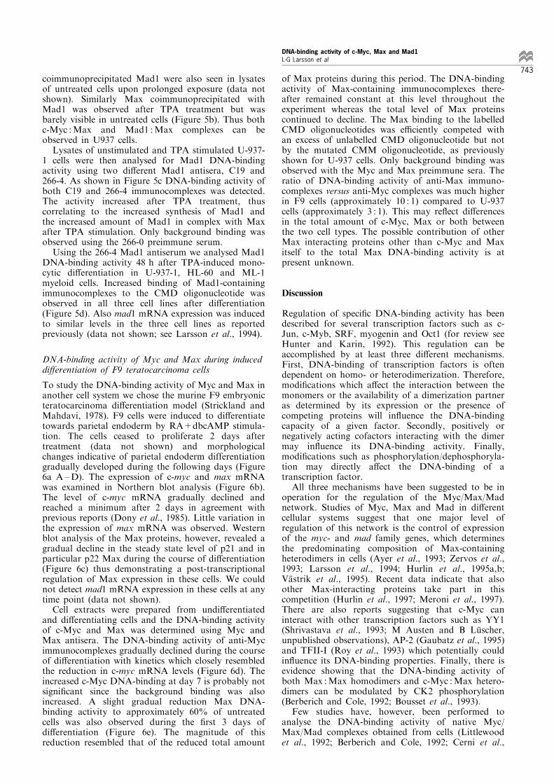
coimmunoprecipitated Mad1 were also seen in lysatesof untreated cells upon prolonged exposure (data notshown). Similarly Max coimmunoprecipitated withMad1 was observed after TPA treatment but wasbarely visible in untreated cells (Figure 5b). Thus bothc-Myc :Max and Mad1 :Max complexes can beobserved in U937 cells.Lysates of unstimulated and TPA stimulated U-937-
1 cells were then analysed for Mad1 DNA-bindingactivity using two di�erent Mad1 antisera, C19 and266-4. As shown in Figure 5c DNA-binding activity ofboth C19 and 266-4 immunocomplexes was detected.The activity increased after TPA treatment, thuscorrelating to the increased synthesis of Mad1 andthe increased amount of Mad1 in complex with Maxafter TPA stimulation. Only background binding wasobserved using the 266-0 preimmune serum.Using the 266-4 Mad1 antiserum we analysed Mad1
DNA-binding activity 48 h after TPA-induced mono-cytic di�erentiation in U-937-1, HL-60 and ML-1myeloid cells. Increased binding of Mad1-containingimmunocomplexes to the CMD oligonucleotide wasobserved in all three cell lines after di�erentiation(Figure 5d). Also mad1 mRNA expression was inducedto similar levels in the three cell lines as reportedpreviously (data not shown; see Larsson et al., 1994).
DNA-binding activity of Myc and Max during induceddi�erentiation of F9 teratocarcinoma cells
To study the DNA-binding activity of Myc and Max inanother cell system we chose the murine F9 embryonicteratocarcinoma di�erentiation model (Strickland andMahdavi, 1978). F9 cells were induced to di�erentiatetowards parietal endoderm by RA+dbcAMP stimula-tion. The cells ceased to proliferate 2 days aftertreatment (data not shown) and morphologicalchanges indicative of parietal endoderm differentiationgradually developed during the following days (Figure6a A ±D). The expression of c-myc and max mRNAwas examined in Northern blot analysis (Figure 6b).The level of c-myc mRNA gradually declined andreached a minimum after 2 days in agreement withprevious reports (Dony et al., 1985). Little variation inthe expression of max mRNA was observed. Westernblot analysis of the Max proteins, however, revealed agradual decline in the steady state level of p21 and inparticular p22 Max during the course of di�erentiation(Figure 6c) thus demonstrating a post-transcriptionalregulation of Max expression in these cells. We couldnot detect mad1 mRNA expression in these cells at anytime point (data not shown).Cell extracts were prepared from undi�erentiated
and di�erentiating cells and the DNA-binding activityof c-Myc and Max was determined using Myc andMax antisera. The DNA-binding activity of anti-Mycimmunocomplexes gradually declined during the courseof di�erentiation with kinetics which closely resembledthe reduction in c-myc mRNA levels (Figure 6d). Theincreased c-Myc DNA-binding at day 7 is probably notsigni®cant since the background binding was alsoincreased. A slight gradual reduction Max DNA-binding activity to approximately 60% of untreatedcells was also observed during the ®rst 3 days ofdi�erentiation (Figure 6e). The magnitude of thisreduction resembled that of the reduced total amount
of Max proteins during this period. The DNA-bindingactivity of Max-containing immunocomplexes there-after remained constant at this level throughout theexperiment whereas the total level of Max proteinscontinued to decline. The Max binding to the labelledCMD oligonucleotides was e�ciently competed withan excess of unlabelled CMD oligonucleotide but notby the mutated CMM oligonucleotide, as previouslyshown for U-937 cells. Only background binding wasobserved with the Myc and Max preimmune sera. Theratio of DNA-binding activity of anti-Max immuno-complexes versus anti-Myc complexes was much higherin F9 cells (approximately 10 : 1) compared to U-937cells (approximately 3 : 1). This may re¯ect di�erencesin the total amount of c-Myc, Max or both betweenthe two cell types. The possible contribution of otherMax interacting proteins other than c-Myc and Maxitself to the total Max DNA-binding activity is atpresent unknown.
Discussion
Regulation of speci®c DNA-binding activity has beendescribed for several transcription factors such as c-Jun, c-Myb, SRF, myogenin and Oct1 (for review seeHunter and Karin, 1992). This regulation can beaccomplished by at least three di�erent mechanisms.First, DNA-binding of transcription factors is oftendependent on homo- or heterodimerization. Therefore,modi®cations which a�ect the interaction between themonomers or the availability of a dimerization partneras determined by its expression or the presence ofcompeting proteins will in¯uence the DNA-bindingcapacity of a given factor. Secondly, positively ornegatively acting cofactors interacting with the dimermay in¯uence its DNA-binding activity. Finally,modi®cations such as phosphorylation/dephosphoryla-tion may directly a�ect the DNA-binding of atranscription factor.All three mechanisms have been suggested to be in
operation for the regulation of the Myc/Max/Madnetwork. Studies of Myc, Max and Mad in di�erentcellular systems suggest that one major level ofregulation of this network is the control of expressionof the myc- and mad family genes, which determinesthe predominating composition of Max-containingheterodimers in cells (Ayer et al., 1993; Zervos et al.,1993; Larsson et al., 1994; Hurlin et al., 1995a,b;VaÈ strik et al., 1995). Recent data indicate that alsoother Max-interacting proteins take part in thiscompetition (Hurlin et al., 1997; Meroni et al., 1997).There are also reports suggesting that c-Myc caninteract with other transcription factors such as YY1(Shrivastava et al., 1993; M Austen and B LuÈ scher,unpublished observations), AP-2 (Gaubatz et al., 1995)and TFII-I (Roy et al., 1993) which potentially couldin¯uence its DNA-binding properties. Finally, there isevidence showing that the DNA-binding activity ofboth Max :Max homodimers and c-Myc :Max hetero-dimers can be modulated by CK2 phosphorylation(Berberich and Cole, 1992; Bousset et al., 1993).Few studies have, however, been performed to
analyse the DNA-binding activity of native Myc/Max/Mad complexes obtained from cells (Littlewoodet al., 1992; Berberich and Cole, 1992; Cerni et al.,
DNA-binding activity of c-Myc, Max and Mad1L-G Larsson et al
743

a
b
c
c-myc —
max —
actin —
Un
trea
ted
1 2 3 4 5 days
RA+dbcAMP
Un
trea
ted
1 3 5 7 9 days
RA+dbcAMP
KDa
d e
Figure 6 DNA-binding of c-Myc- and Max-containing immunocomplexes during induced di�erentiation of F9 cells. (a) F9 cellswere induced to di�erentiate by the addition of RA+dbcAMP and the morphological development of parietal endoderm-like cellsfollowed under light microscope after (A) 0 day (B) 3 days (C) 5 days and (D) 9 days of treatment. (b) Northern blot analysis of c-myc and max expression. F9 cells were induced to di�erentiate by RA+dbcAMP for the indicated time points and the expression ofc-myc and max mRNA was determined by Northern blot analysis. Expression of actin mRNA was analysed as a reference gene. (c)Western blot analysis of Max expression. F9 cells were induced as above for the indicated time points, harvested and lysed in L-bu�er. Equal amounts of extracts were subjected to Western blot analysis using 3811E Max antiserum. (d and e) DNA-binding of c-Myc (d) and Max (e). F9 cells were induced to di�erentiate as above for the indicated time points. The cells were lysed in L-bu�erand subjected to the DNA-binding assay as described in the legend to Table 1 using 88-6 Myc and 91-6 Max antisera and 1 ng oflabelled CMD oligonucleotide. The speci®city of the DNA-binding was determined by using preimmune sera (88-0 and 91-0) and bycompeting with a 50-fold molar excess of unlabelled CMD and mutated CMM oligonucleotides as indicated
DNA-binding activity of c-Myc, Max and Mad1L-G Larsson et al
744

1995), in particular in response to external signals.These previous studies have been performed by EMSAusing cells which highly overexpress the studiedproteins. The EMSA technique has, however, notbeen very successful to study DNA-binding of Myc,Max and Mad in cells which express more physiolo-gical levels of the proteins. The main problem has beenthe often low abundance of the proteins, the di�cultyto extract the proteins in standard Dignam extractsand the high background of other unrelated proteinsbinding the CACGTG motif. In order to overcomethese limitations we now report the development of analternative DNA-binding assay. This technique is basedon a solid phase assay using partially puri®ed nativeMyc/Max/Mad complexes isolated by immunologicalmethods. A similar approach has been used to studyDNA-binding of the SV40 large T antigen (McKay,1981). In this study cell extracts of infected cells wereincubated with labelled DNA probes after which largeT antigen/DNA complexes were immunoprecipitatedand the speci®c DNA-binding was measured. Thisassay system has also been applied to Myc :Maxcomplexes derived either from in vitro transcriptions/translations in rabbit reticulocyte lysate or from HeLacell extracts (Littlewood et al., 1992). This worksuggested that principally DNA-binding of Myc :Maxand Max :Max complexes can be measured whenbound to antibodies.Our results now show that the solid phase DNA-
binding assay for immunocomplexes of Myc/Max/Madfamily proteins is very sensitive and can be used tostudy the DNA-binding activity of non-overexpressed,endogenous Myc, Max and Mad1 complexes in as fewas 250 000 cells, which is an advantage when theamounts of cells are limiting. Since the major part ofMyc E-box binding activity in whole cell extracts is dueto proteins other than those of the Myc/Max/Madfamily, the assay described here avoids competition forDNA by such proteins. We have also shown that thebinding of the immunocomplexes to the CACGTG-containing oligonucleotide is very speci®c. It isdetectable with a number of di�erent Myc, Max andMad1 antisera, whereas preimmune or normal rabbitsera give only background binding. The binding is alsoabolished by pre-incubating the antisera with peptideor by competing with excess unlabelled oligonucleotidebut not by oligonucleotides containing mutatedversions of the Myc/Max binding site (Figure 3 anddata not shown). The major drawback with thetechnique is that it only gives information on thetotal binding of Myc, Max or Mad1 containingcomplexes. Although we have shown that anti-Mycand anti-Mad1 immunocomplexes contain Max andvice versa, it is not possible to distinguish whether thereare di�erent types of complexes formed or whethertheir composition varies. It may be possible to solvethese problems by competing away the antibodies bypeptide followed by analysis of the complexes byEMSA. We are currently working on these modifica-tions of the technique. Another concern is thepossibility that certain antibodies may change theconformation of the complexes or disrupt binding ofpotential interacting factors, thereby a�ecting theDNA-binding properties of the proteins. Such e�ectscan be analysed using Cos-7 cell overexpressed proteinsin combination with gel shifts (A Sommer, K Bousset
and BL, unpublished data). This obviously has to bestudied further.Despite these disadvantages we believe that this
technique can give valuable information on the DNA-binding activity of the proteins of the Myc/Max/Madnetwork under various conditions. We have applied thisassay to studies of Myc, Max and Mad1 DNA-bindingactivity during induced di�erentiation using twodi�erent model systems; monocytic di�erentiation ofU-937 monoblasts (Table 1, Figures 4 and 5) (Nilsson etal., 1980) and di�erentiation of F9 embryonic terato-carcinoma cells towards parietal endoderm (Figure 6)(Strickland and Mahdavi, 1978). Our results show thatchanges in DNA-binding activity of these complexesindeed can be observed in both systems duringdi�erentiation. The DNA-binding of c-Myc-containingcomplexes is gradually reduced after stimulation in U-937 and F9 cells. In general, this reduction seems tocorrelate well with the level of expression of c-mycmRNA and protein. Also the increased Mad1 DNA-binding observed after TPA stimulation of U-937, HL-60 and ML-1 cells correlates with the increased mad1mRNA and protein expression and the increasedformation of Mad1 :Max heterodimers.However we have found at least two situations in
which the protein expression did not strictly correlatewith the measured DNA-binding capacity. First, inU937 cells c-Myc levels are initially slightly enhancedearly during di�erentiation whereas consistently DNA-binding was rapidly reduced. This was also observed inU-937-myc-6 cells which continuously express v-Myc.Together these ®ndings suggest that c-Myc :MaxDNA-binding is regulated by additional means thanexpression levels. This seems not to be due to reducedMyc :Max dimer formation since the ratio of total Mycand Myc in complex with Max was constant duringdi�erentiation but rather an e�ect on the speci®cactivity of DNA-binding. Phosphorylation of Max atthe N-terminal CK2 sites e�ects DNA-binding(Berberich and Cole, 1992; Bousset et al., 1993) and,in addition, c-Myc is also phosphorylated by CK2 nearthe basic region (LuÈ scher et al., 1989). Thus alteredstoichiometry of phosphorylation at these sites coulda�ect DNA-binding. Experiments to address this are inprogress. Another possibility is that the DNA-bindingactivity of Myc :Max is modulated by other interactingproteins which are regulated by TPA. Second, thesteady state levels of Max proteins decline graduallyduring F9 di�erentiation, still the DNA-bindingactivity of Max complexes, although reduced, stabi-lises at a quite high level. At present we do not knowthe composition of these complexes. The Max DNA-binding activity observed in all instances will representthe sum of the contribution of Max :Max homodimersand all types of Max-containing heterodimers in thecell that can be immunoprecipitated with the Maxantiserum used. Since at present we only have access toantiserum towards c-Myc and Mad1 (which is notexpressed in F9) we have not been able to analyse thecontribution of other known Max-interacting proteins.Possible interpretations of the F9 Max DNA-bindingdata is that new types of Max heterodimers with stronga�nity to DNA or Max :Max homodimers withincreased a�nity form during di�erentiation, whichcould compensate for the reduced total level of Max inthe cell.
DNA-binding activity of c-Myc, Max and Mad1L-G Larsson et al
745

The reduced total level of Max proteins, but notmax mRNA, points at a post-transcriptional regulationof Max expression not previously documented. It alsochallenges the generally held view that Max levels arefairly constant in cells. Changes in max expression,however, have been observed during di�erentiationalso in other cell types. Induced erythroid differentia-tion of MEL and K-652 cells (Dunn et al., 1994;Delgado et al., 1995) and TPA-induced monocyticdi�erentiation of HL-60 and to a lesser extent ML-1and U-937 myeloid cells are accompanied by a reducedexpression of max (Larsson et al., 1994). The ratiobetween total Max and Myc DNA-binding activity ismuch higher (10 : 1) in F9 cells compared to other celltypes we have studied (2-3 : 1). This may indicate thatearly embryonic cells express a very high level of Maxwhich is later down-modulated during the develop-ment.Our data on the DNA-binding activity of Myc, Max
and Mad1 so far seem to con®rm the view that themain regulation of the network is at the level ofexpression of its components, since most of theobserved changes in DNA-binding correlate withdi�erences in gene expression. However, our studiesof early kinetics of TPA-induced di�erentiation of U-937 cells as well as of late events during F9di�erentiation suggest that post-translational regula-tion of DNA-binding may also occur. Additionalstudies will be required to elucidate the relevantmechanisms.
Materials and methods
Cell culture and di�erentiation assays
The cells were cultured in RPMI-1640 medium (U-937,HL-60 and ML-1) or DMEM medium (F9 and Manca)supplemented with 10% fetal calf serum and antibioticsBK3A cells were grown in DMEM supplemented with 5%calf serum, 1% chicken serum and 10% tryptone broth. U-937 (Nilsson et al., 1980), HL-60 (Collins et al., 1977) andML-1 (Han and Minowada, 1980) are human myeloid celllines, Manca (Nishikori et al., 1984) is a human Burkitt'slymphoma cell line, BK3A (Hihara et al., 1974) is achicken bursal lymphoma cell line and F9 (Strickland andMahdavi, 1978) is a murine embryonic teratocarcinomacell line. The U-937 clones, U-937-myc-6, containing theOK10 v-myc gene, the parental clone U-937-GTB and theU-937-1 clone have been described previously (Larsson etal., 1988, 1994). For in vitro di�erentiation of U-937, HL-60 and ML-1, exponentially growing cells (105/ml) wereinduced in medium containing 1.661078 M TPA (Sigma, StLouis, MO), 1077 M VitD3 (a generous gift from Hofmann-La Roche), 1076 M RA (Sigma) or 100 U/ml IFN-g(generously provided by Dr GR Adolf, Ernst-BoehringerInstitute, Vienna, Austria). F9 cells were induced todi�erentiate in medium containing 1076 M RA and 1 mM
dbcAMP. Immuno¯uorescence studies of CD11c, the a-subunit of the gp105.95 adhesion molecule which is amarker of monocytic di�erentiation (Miller et al., 1986),were performed using the mAb LeuM5 (Becton &Dickinson, Mountain View, CA) as described previously(OÈ berg et al., 1991).
Immunoprecipitation and Western blot analysis
56106 cells were labelled for 40 min in 1 ml of methionine-free RPMI-1640 containing 0.15 mCi of 35S-methionine.
For high stringency immunoprecipitations cells were lysedin AB bu�er (20 mM Tris pH 7.4, 50 mM NaCl, 1 mM
EDTA, 0.5% NP-40, 0.5% sodium desoxycholate, 0.5%SDS, 0.5% aprotinin, 0.5 mM phenylmethylsulphonyl¯uoride (PMSF)), sonicated, cleared by centrifugationand immunoprecipitated with antibodies as described(LuÈ scher and Eisenman, 1988). An equal number ofTCA-precipitable counts were used for each sample. Theimmunocomplexes were collected with protein A-SepharoseCL4B (Sigma) and washed twice with RIPA bu�er (10 mM
Tris, pH 7.4, 150 mM NaCl, 1% NP-40, 1% desoxycholate,0.1% SDS, 0.5% aprotinin), once with high salt bu�er (2 M
NaCl, 10 mM NaCl pH 7.4, 1% NP-40, 0.5% desoxycho-late) and ®nally with RIPA (LuÈ scher and Eisenman, 1988).For low stringency immunoprecipitations the cells weresuspended in L bu�er (PBS, 1% NP-40, 0.5 mM PMSF,0.5% aprotinin, 10 mM NaF, 50 mM b-glycerophosphate,0.5 mg/ml pepstatin, 100 mM orthovanadate, 0.5 mM
benzamidine, 2.5 mg/ml leupeptin, 1 mM DTT), vortexedbrie¯y, lysed for 10 min on ice, centrifuged and immuno-precipitated (Blackwood et al., 1992). The beads were thenwashed four times in L bu�er. The SDS ± PAGE sampleswere analysed on 10 ± 15% polyacrylamide gels underreducing conditions.
For Western blot analysis unlabelled cells were lysed underlow stringency conditions. The protein concentration wasmeasured using a Biorad assay (Biorad Laboratories,Richmond, CA) and an equal amount of proteins wasimmunoprecipitated and washed as above. The immunocom-plexes were resolved on a polyacrylamide gel and electro-blotted (Biorad) to a nitrocellulose ®lter. The ®lter wasincubated with the antibody in blocking bu�er (PBS, 0.5%NP-40, 5% milk powder). After washing (PBS, 0.5%desoxycholate, 0.5% NP-40) the blot was developed byenhanced chemiluminescence (Amersham, Buckinghamshire,UK) using horseradish peroxidase (HRP) conjugated anti-rabbit Ig (Amersham).
Solid phase DNA-binding assays
Cells were lysed under low stringency conditions in L-,Frackelton (10 mM Tris pH 7.05, 50 mM NaCl, 30 mM
NaPPi, 50 mM NaF, 5 mM ZnCl2, 100 mM Na3VO4, 1%Triton X-100, 1 mM PMSF) or Tris (0.1 M Tris pH 8.0,0.15 M NaCl, 5 mM EDTA, 1% NP-40, 1 mM PMSF)lysis bu�er. Equal amounts of protein were incubated onice for 2 h with the antibody. The immunocomplexeswere collected by protein A-Sepharose beads, washedthree times in lysis bu�er and once in gelshift bu�er(20 mM HEPES pH 7.3, 50 mM KCl, 3 mM MgCl2, 1 mM
EDTA, 8% glycerol, 0.1% aprotinin, 1 mM b-mercap-toethanol) at 48C. The samples were then incubated with1 ng of 32P-labelled oligonucleotide in 30 ml of gelshiftbu�er containing 100 ng of salmon sperm DNA for25 min at 258C. The immunocomplexes were then washedthree times in gel shift bu�er at 48C and the amount ofbound oligonucleotide was measured in a scintillationcounter.
The oligonucleotides used were CMD (which contains anoptimal Myc/Max binding site, underlined), CMM andCMM-1 (which contain mutated Myc/Max binding sites) orE-pal (which contains an E-box sequence from the promoterof the E-cadherin gene) obtained from W Birchmeier.
CMD 5'-TCAGACCACGTGGTCGGGCMM 5'-TCAGACCAGCTGGTCGGGCMM-1 5'-TCAGACACCGTGGTCGGGE-pal 5 '-GATCCGGCTGCCACCTGCAGGTGCGTCCCG
The oligonucleotides were end-labelled by T4 polynucleo-tide kinase and then puri®ed on a 8% polyacrylamide gel.
DNA-binding activity of c-Myc, Max and Mad1L-G Larsson et al
746

Rabbit antisera
Myc antisera: R2 (pan-Myc, kindly provided by DrRosemary Watt), X (pan-Myc, kindly provided by Dr GEvan), 5042 (a chicken Myc-speci®c antiserum kindlyprovided by Dr S Hann and Dr RN Eisenman) and a-Bp35 (pan-Myc, kindly provided by Dr K Alitalo), 88(generated by immunising with a bacterially producedMyc-GST fusion protein containing the 262 N-terminalamino acids of c-Myc).
Max antisera: 3811E and 8711E (kindly provided by DrElizabeth Blackwood). Antiserum 91 was made against asynthetic peptide (aa 119 ± 151 of p21 Max).
Mad1 antisera: 266 and C-19 (Santa Cruz Biotechnology,Santa Cruz, CA). The 266 antiserum was generated byimmunising with a bacterially produced full-length Mad-GSTfusion protein. For the following immunisations a His-taggedfull-length Mad protein and a His-tagged C-terminal Mad-protein comprising amino acids 141 ± 221 were used.
Northern blot analysis
The procedure for preparation of total RNA, for theNorthern blot analysis and the probes used has been
described previously (Larsson et al., 1988, 1994). Afterhybridization and washing the ®lters were analysed on Fujibas image analyser.
AcknowledgementsWe thank Drs K Alitalo, W Birchmeier, E Blackwood, REisenman, G Evan, S Hann and R Watt for reagents. Wealso thank Hofmann-La Roche and Dr GR Adolf, Ernst-Boehringer Institute, Vienna for generous gifts of VitD3
and IFN-g, respectively. We appreciate the critical com-ments on the manuscript by A Sommer. This work wassupported by grants from the Swedish Cancer Society, theChildren Cancer Foundation of Sweden, the Lovisa andThielman, the Selander and the Wiberg Foundations andan EMBO fellowship to L-GL and the Deutsche For-schungsgemeinschaft, the Deutsche Krebshilfe and theFonds der Chemischen Industrie to BL.
References
Amati B, Dalton S, Brooks MW, Littlewood TD, Evan GIand Land H. (1992). Nature, 359, 423 ± 426.
Amati B, Brooks MW, Levy N, Littlewood TD, Evan GI andLand H. (1993a). Cell, 72, 233 ± 245.
Amati B, Littlewood TD, Evan GI and Land H. (1993b).EMBO J., 12, 5083 ± 5087.
Amin C, Wagner AJ and Hay N. (1993).Mol. Cell. Biol., 13,383 ± 390.
Ayer DE, Kretzner L and Eisenman RN. (1993). Cell, 72,211 ± 222.
Ayer DE and Eisenman RN. (1993). Genes Dev., 7, 2110 ±2119.
Ayer DE, Lawrence QA and Eisenman RN. (1995). Cell, 80,767 ± 776.
Berberich SJ and Cole MD. (1992). Genes Dev., 6, 166 ± 176.Blackwell TK, Kretzner L, Blackwood EM, Eisenman RNand Weintraub H. (1990). Science, 250, 1149 ± 1151.
Blackwood EM and Eisenman RN. (1991). Science, 251,1211 ± 1217.
Blackwood EM, LuÈ scher B and Eisenman RN. (1992). GenesDev., 6, 71 ± 80.
Bousset K, Henriksson M, LuÈ scher-Firzla� JM, Litch®eldDW and LuÈ scher B. (1993). Oncogene, 8, 3211 ± 3220.
Cerni C, Bousset K, Seelos C, Burkhardt H, Henriksson Mand LuÈ scher B. (1995). Oncogene, 11, 587 ± 596.
Chen J, Willingham T, Margraf LR, Schreiber-Agus N,DePinho RA and Nisen PD. (1995). Nat. Med., 1, 638 ±643.
Chin L, Schreiber-Agus N, Pellicer I, Chen K, Lee HW,Dudast M, Cordon-Cardo C and DePinho RA. (1995).Proc. Natl. Acad. Sci. USA, 92, 8488 ± 8492.
Collins SJ, Gallo RC and Gallagher RE. (1977). Nature, 270,347 ± 349.
Delgado MD, Lerga A, Canelles M, Gomez-Casares MT andLeon J. (1995). Oncogene, 10, 1659 ± 1665.
Dony C, Kessel M and Gruss P. (1985). Nature, 317, 636 ±639.
Dunn BK, Cogliati T, Cultraro CM, Bar-Ner M and Segal S.(1994). Cell Growth Di�er., 5, 847 ± 854.
Gaubatz S, Imhof A, Dosch R, Werner O, Mitchell P,Buettner R and Eilers M. (1995). EMBO J., 14, 1508 ±1519.
Gu W, Cechova K, Tassi V and Dalla-Favera R. (1993).Proc. Natl. Acad. Sci. USA, 90, 2935 ± 2939.
Han T and Minowada J. (1980). J. Clin. Lab. Immunol., 4,169 ± 173.
Henriksson M and LuÈ scher B. (1996). Adv. Cancer Res., 68,109 ± 182.
Hihara H, Shimizu T and Yamamoto H. (1974). Natl. Inst.Anim. Health Q. Tokyo, 14, 163 ± 173.
Hunter T and Karin M. (1992). Cell, 70, 375 ± 387.Hurlin PJ, Queva C, Koskinen PJ, Steingrimsson E, AyerDE, Copeland NG, Jenkins NA and Eisenman RN.(1995a). EMBO J., 14, 5646 ± 5659.
Hurlin PJ, Foley KP, Ayer DE, Eisenman RN, Hanahan Dand Arbeit JM. (1995b). Oncogene, 11, 2487 ± 2501.
Hurlin PJ, Que va C and Eisenman RN. (1997). Genes Dev.,11, 44 ± 58.
Koskinen PJ, Ayer DE and Eisenman RN. (1995). CellGrowth Di�er., 6, 623 ± 629.
Kretzner L, Blackwood EM and Eisenman RN. (1992).Nature, 359, 426 ± 429.
Lahoz EG, Xu L, Schreiber-Agus N and DePinho RA.(1994). Proc. Natl. Acad. Sci. USA, 91, 5503 ± 5507.
Larsson LG, Ivhed I, Gidlund M, Pettersson U, VennstromB and Nilsson K. (1988). Proc. Natl. Acad. Sci. USA, 85,2638 ± 2642.
Larsson LG, Pettersson M, OÈ berg F, Nilsson K and LuÈ scherB. (1994). Oncogene, 9, 1247 ± 1252.
Littlewood TD, Amati B, Land H and Evan GI. (1992).Oncogene, 7, 1783 ± 1792.
LuÈ scher B and Eisenman RN. (1988). Mol. Cell. Biol., 8,2504 ± 2512.
LuÈ scher B, Kuenzel EA, Krebs EG and Eisenman RN.(1989). EMBO J., 8, 1111 ± 1119.
Marcu KB, Bossone SA and Patel AJ. (1992). Annu. Rev.Biochem., 61, 809 ± 860.
Meroni G, Reymond A, Alcalay M, Borsani G, Tanigami A,Tonlorenzi R, Lo Nigro C, Messali S, Zollow M,Ledbetter DH, Brent R, Ballabio A and Carrozzo R.(1997). EMBO J., 16, 2892 ± 2906.
McKay RDG. (1981). J. Mol. Biol., 145, 471 ± 488.Nilsson K, Andersson LC, Gahmberg CG and Forsbeck K.(1980). International Symposium on New Trends in HumanImmunology and Cancer Immunotherapy. Doin Editeurs:Paris, pp. 271.
DNA-binding activity of c-Myc, Max and Mad1L-G Larsson et al
747

Nishikori M, Hansen H, Jhanwar S, Fried J, Sordillo P,Koziner B, Lloyd K and Clarkson B. (1984). CancerGenet. Cytogenet., 12, 39 ± 50.
OÈ berg F, Larsson L-G, Anton R and Nilsson K. (1991).Proc. Natl. Acad. Sci. USA, 88, 5567 ± 5571.
Prendergast GC and Zi� EB. (1991). Science, 251, 186 ± 189.Prendergast GC, Lawe D and Zi� EB. (1991). Cell, 65, 395 ±407.
Prendergast GC, Hopewell R, Gorham BJ and Zi� EB.(1992). Genes Dev., 6, 2429 ± 2439.
Roussel MF, Ashmun RA, Sherr CJ, Eisenman RN and AyerDE. (1996). Mol. Cell. Biol., 16, 2796 ± 2801.
Roy AL, Carruthers C, Gutjahr T and Roeder RG. (1993).Nature, 365, 359 ± 361.
Schreiber-Agus N, Chin L, Chen K, Torres R, Rao G, GuidaP, Skoultchi AI and DePinho RA. (1995). Cell, 80, 777 ±786.
Shrivastava A, Saleque S, Kalpana GV, Artandi S, Go� SPand Calame K. (1993). Science, 262, 1889 ± 1892.
Strickland S and Mahdavi V. (1978). Cell, 15, 393 ± 403.Vastrik I, Kaipainen A, Penttila TL, Lymboussakis A,Alitalo R, Parvinen M and Alitalo K. (1995). J. Cell. Biol.,128, 1197 ± 1208.
Zervos AS, Gyuris J and Brent R. (1993). Cell, 72, 223 ± 232.
DNA-binding activity of c-Myc, Max and Mad1L-G Larsson et al
748


![E=F9;mklge]j ZYjge]l]j L`ak lae] alÌk h]jkgfYd2 ^jge [gfkme]j lg [g%[j]Ylgj](https://static.fdokumen.com/doc/165x107/631789cb7451843eec0ab6f2/ef9mklgej-zyjgelj-lak-lae-alik-hjkgfyd2-jge-gfkmej-lg-gjylgj.jpg)