Mad dogs, Englishmen and apoptosis: The role of cell death in UV-induced skin cancer
11
Apoptosis 2003; 8: 315–325 C 2003 Kluwer Academic Publishers Mad dogs, Englishmen and apoptosis: The role of cell death in UV-induced skin cancer E. Guzman, J. L. Langowski and L. Owen-Schaub Department of Biomedical Sciences, The University of California-Riverside, Riverside, California 92521, USA (E. Guzman, J. L. Langowski, L. Owen-Schaub); The University of Texas-Houston, Graduate School of Biomedical Sciences, Houston, Texas 77030, USA (E. Guzman, J. L. Langowski) Apoptosis plays a critical role in the development and pro- gression of ultraviolet-induced skin cancers. In particular, Fas and Fas ligand (FasL) interactions are known to con- trol the development of “sunburn cells” or apoptotic ker- atinocytes in the UV-exposed epidermis. In the absence of functional Fas/FasL signaling, UV-induced apoptosis is diminished and mutations rapidly accumulate. UV- induced suppression of host immunity, a process regu- lating skin cancer outgrowth, is also controlled through Fas/FasL interactions. Other death receptors, such as the receptor for tumor necrosis factor, may also contribute to UV-induced carcinogenesis and progression. Under- standing the involvement of cell death in cancers caused by exposure to sunlight may provide novel approaches for prevention and therapy of these ever-increasing ma- lignancies. Keywords: apoptosis; Fas; Fas ligand; immune suppression; UV-induced skin cancer; ultraviolet In tropical climes there are certain times of day When all the citizens retire to tear their clothes off and perspire It’s one of the rules that the greatest fools obey, Because the sun is much too sultry And one must avoid its ultry-violet ray ..... But mad dogs and Englishmen go out in the midday sun. Noel Coward, Mad Dogs and Englishmen Skin cancer The incidence of skin cancer in the United States con- tinues to increase at the alarming rate of approximately 1.3 million new cases annually. Apoptosis plays an impor- tant role in the host response to ultraviolet light (UV), the principal carcinogen responsible for skin cancer induction. UV exposure induces “sunburn cells” in the epidermis by a process of keratinocyte apoptosis. The apoptotic response Correspondence to: Laurie Owen-Schaub, Department of Biomedical Sciences, 1274 Webber Hall, Riverside, California 92521, USA. Tel.: (909) 787-2583; Fax: (909) 787-2438; e-mail: [email protected] is crucial for the elimination of DNA-damaged cells that will develop genetic mutations initiating the carcino- genic cascade. However, because UV-induced apoptosis also induces cytokine release and eliminates non-mutated, DNA-damaged skin, the post-UV environment is favor- able for the clonal expansion of mutated cells—providing a physically unrestricted space for expansion and an im- mune response that “ignores” such malignant and prema- lignant cells. This review focuses on the mechanisms of UV-induced apoptosis in the development and progres- sion of skin cancer. Malignancies arising in the skin are broadly divided into two categories: melanoma and non-melanoma skin cancers. Non-melanoma skin cancers (NMSC) occur in sun-exposed sites and include basal cell and squamous cell carcinomas. Although BCC and SCC account for 40% of the total cancers diagnosed in the United States, their as- sociated mortality remains low (an estimated 2,200 deaths annually). Basal cell carcinomas (BCC), the most common type of skin cancers, are slow growing, non-metastatic tumors resembling ulcers with rolled edges. Cells giv- ing rise to BCC originate from interfollicular basal cells, keratinocytes in hair follicles, or sebaceous glands. Be- cause these cells are found in a deeper zone of the skin, it is speculated that the ultraviolet wavelength responsible for their development may differ from that causing other NMSC. Squamous cell carcinomas (SCC) resemble warts and metastasize more frequently than BCC, accounting for the bulk of mortality from NMSC. SCC originate from cells in the surface of the skin usually following the development of actinic keratoses (thickening of the skin). The events controlling the progression of actinic keratoses (AK) to SCC remain poorly defined, occurring in an exceptionally small proportion of AK. Melanoma, a malignant proliferation of melanocytes, accounts for ap- proximately 54,000 new cases of skin cancer annually in the U.S. While the incidence is considerably lower than NMSC, melanoma is highly lethal (approximately 7,600 deaths annually) and tends to metastasize to both local and distant sites. Melanomas arise from dysplastic nevi, Apoptosis · Vol 8 · No 4 · 2003 315
-
Upload
independent -
Category
Documents
-
view
3 -
download
0
Transcript of Mad dogs, Englishmen and apoptosis: The role of cell death in UV-induced skin cancer

Apoptosis 2003; 8: 315–325C© 2003 Kluwer Academic Publishers
Mad dogs, Englishmen and apoptosis: The role of celldeath in UV-induced skin cancer
E. Guzman, J. L. Langowski and L. Owen-Schaub
Department of Biomedical Sciences, The University of California-Riverside, Riverside, California 92521, USA(E. Guzman, J. L. Langowski, L. Owen-Schaub); The University of Texas-Houston, Graduate School of BiomedicalSciences, Houston, Texas 77030, USA (E. Guzman, J. L. Langowski)
Apoptosis plays a critical role in the development and pro-gression of ultraviolet-induced skin cancers. In particular,Fas and Fas ligand (FasL) interactions are known to con-trol the development of “sunburn cells” or apoptotic ker-atinocytes in the UV-exposed epidermis. In the absenceof functional Fas/FasL signaling, UV-induced apoptosis isdiminished and mutations rapidly accumulate. UV-induced suppression of host immunity, a process regu-lating skin cancer outgrowth, is also controlled throughFas/FasL interactions. Other death receptors, such as thereceptor for tumor necrosis factor, may also contributeto UV-induced carcinogenesis and progression. Under-standing the involvement of cell death in cancers causedby exposure to sunlight may provide novel approachesfor prevention and therapy of these ever-increasing ma-lignancies.
Keywords: apoptosis; Fas; Fas ligand; immune suppression;UV-induced skin cancer; ultraviolet
In tropical climes there are certain times of dayWhen all the citizens retire to tear their clothes off and perspireIt’s one of the rules that the greatest fools obey,Because the sun is much too sultryAnd one must avoid its ultry-violet ray.....But mad dogs and Englishmen go out in the midday sun.
Noel Coward, Mad Dogs and Englishmen
Skin cancer
The incidence of skin cancer in the United States con-tinues to increase at the alarming rate of approximately1.3 million new cases annually. Apoptosis plays an impor-tant role in the host response to ultraviolet light (UV), theprincipal carcinogen responsible for skin cancer induction.UV exposure induces “sunburn cells” in the epidermis by aprocess of keratinocyte apoptosis. The apoptotic response
Correspondence to: Laurie Owen-Schaub, Department ofBiomedical Sciences, 1274 Webber Hall, Riverside, California92521, USA. Tel.: (909) 787-2583; Fax: (909) 787-2438; e-mail:[email protected]
is crucial for the elimination of DNA-damaged cells thatwill develop genetic mutations initiating the carcino-genic cascade. However, because UV-induced apoptosisalso induces cytokine release and eliminates non-mutated,DNA-damaged skin, the post-UV environment is favor-able for the clonal expansion of mutated cells—providinga physically unrestricted space for expansion and an im-mune response that “ignores” such malignant and prema-lignant cells. This review focuses on the mechanisms ofUV-induced apoptosis in the development and progres-sion of skin cancer.
Malignancies arising in the skin are broadly dividedinto two categories: melanoma and non-melanoma skincancers. Non-melanoma skin cancers (NMSC) occur insun-exposed sites and include basal cell and squamous cellcarcinomas. Although BCC and SCC account for 40% ofthe total cancers diagnosed in the United States, their as-sociated mortality remains low (an estimated 2,200 deathsannually). Basal cell carcinomas (BCC), the most commontype of skin cancers, are slow growing, non-metastatictumors resembling ulcers with rolled edges. Cells giv-ing rise to BCC originate from interfollicular basal cells,keratinocytes in hair follicles, or sebaceous glands. Be-cause these cells are found in a deeper zone of the skin, itis speculated that the ultraviolet wavelength responsiblefor their development may differ from that causing otherNMSC. Squamous cell carcinomas (SCC) resemble wartsand metastasize more frequently than BCC, accountingfor the bulk of mortality from NMSC. SCC originatefrom cells in the surface of the skin usually followingthe development of actinic keratoses (thickening of theskin). The events controlling the progression of actinickeratoses (AK) to SCC remain poorly defined, occurringin an exceptionally small proportion of AK. Melanoma, amalignant proliferation of melanocytes, accounts for ap-proximately 54,000 new cases of skin cancer annually inthe U.S. While the incidence is considerably lower thanNMSC, melanoma is highly lethal (approximately 7,600deaths annually) and tends to metastasize to both localand distant sites. Melanomas arise from dysplastic nevi,
Apoptosis · Vol 8 · No 4 · 2003 315

E. Guzman et al.
which are irregularly shaped and colored moles, both onsun-exposed and non-sun exposed sites (soles of the feetand buttocks).
UV in sunlight is the principal carcinogen for skin can-cer development, acting as both an inducer and promoter.1
Exposure to UV results in DNA damage causing muta-tions in critical genes such as p53 and inducing a transientstate of immune suppression in the host. Mutations andUV-induced immune suppression operate conjointly inskin tumor development.1 High cumulative sun expo-sure has been associated with NMSC development whileacute sunburn has been correlated with the risk of devel-oping melanoma. Individuals developing NMSC have anincreased risk of developing new skin cancers, particu-larly in the first year after initial diagnosis.2 In addition,skin cancer patients have an increased overall risk of non-cutaneous malignancies.3
Ultraviolet radiation and DNA damage
The ultraviolet region in sunlight is sub-divided accord-ing to wavelength: UV-A (320–400 nm), UV-B (280–320 nm), and UV-C (200–280 nm). UV-C is absorbed bythe stratospheric ozone layer, such that terrestrial sunlightcontains only UV-A and UV-B. UV-A readily penetratesboth the epidermis and dermal regions of the skin, butis weakly absorbed by biomolecules. UV-A is thought toexert its effects principally through the generation of re-active oxygen intermediates.4 UV-B, on the other hand,has a lower level of skin penetration, but is readily ab-sorbed by DNA and proteins in the epidermal layer. Ex-posure to UV-B is closely associated with the develop-ment of actinic keratoses as well as basal and squamouscell carcinomas.5 The biologic effects of UV-B (comparedto UV-A) has been attributed to its heightened DNA andprotein absorption spectra resulting in edema, erythema,and mutagenesis.6,7
A direct linkage between UV exposure and DNA dam-age is powerfully demonstrated by the high skin cancerfrequency in individuals with the autosomal recessive dis-ease Xeroderma Pigmentosum (XP), a genetic defect inDNA damage repair.8 Median onset of skin cancer de-velopment in the United States occurs at 50–60 years ofage while XP patients show a median onset within thefirst decade of life.9 Absorption of UV-B energy by DNAresults in two main types of lesions known as pyrimidinedimers and (6-4) photoproducts [pyrimidine (6-4) pyrim-idone]. Pyrimidine dimers form when the double bondsof the C-4 and C-5 carbon atoms of two adjoining pyrim-idines become saturated resulting in a four-memberedring structure. Formation of (6-4) photoproducts occurswhen the C-6 position of a 5′ pyrimidine is covalentlybonded to the C-4 position of a neighboring 3′ pyrimi-dine. The nature of the DNA site, flanking residues, and
UV wavelength contribute to lesion formation, althougha clear preference for CPD formation exists.
CPDs comprise 70–80% of all UV photoproductswhereas (6-4) photoproducts form 10–50% lessfrequently.10,11 Interestingly, despite their lower rate ofoccurrence, (6-4) photoproducts are repaired at much fas-ter rate than CPDs, likely because of their overt cellularcytotoxicity.12 Some investigators have reported that (6-4) photoproducts are more mutagenic than CPDs,12 whileothers have found that CPDs are the major contributor tomammalian mutagenesis.13 Although the relative role ofCPD and (6-4) photoproduct formation in carcinogenesisis somewhat controversial, their role in the disruption ofDNA synthesis leading to mutagenesis is not.
Because the majority of terrestrial organisms live insunlight, multiple adaptations have evolved to repair UV-induced photoproducts. These include nucleotide excisionrepair, base excision repair and, in some species, a light-activated photolyase enzyme. If unrepaired, photoprod-ucts can lead to the incorporation of base mismatches bymeans of the “the A-rule” where adenine is inserted op-posite the non-informational base. Mutations occur mostoften when cytosine is a component of the photoproductsince insertion of adenine opposite thymine is a correctevent. For this reason, most CPDs that form between twothymine bases are non-mutagenic. However, if a cytosineis involved, after DNA replication, a T will be insertedin the strand opposite the A resulting in a so-called UV“signature mutation”. The footprint signature UV muta-tions are C to T or CC to TT changes. These mutations arefound at very high frequencies in certain codons or “hotspots” of genes, such as the tumor suppressor gene p53,and are frequently observed in skin cancers.5,14
In addition to mutations, UV-induced DNA damageexerts local effects on epidermal cell survival and systemiceffects on host immunity. Previous reports using DNArepair enzymes, such as the bacteriophage T4endonuclease V (T4N5) or liposomes containing pho-tolyase, have documented that the repair or UV-inducedepidermal DNA damage can diminish CPD formation,reduce swelling and inflammation, block apoptotic deathof keratinocytes, and prevent UV-induced immunesuppression.15–19 Figure 1 shows a model where bothDNA damage-induced apoptosis and immune suppres-sion play a role in the development of UV-induced skintumors.
The tumor suppressor p53and skin cancer
Failure to repair DNA damage leads to mutations. Agene commonly mutated after chronic UV exposure isthe tumor suppressor p53.5,14,20 The p53 gene encodesa transcription factor that is functionally inactivated by
316 Apoptosis · Vol 8 · No 4 · 2003

The role of cell death in UV-induced skin cancer
Figure 1. Model for UV-induced skin cancer showing a role for both DNA damage-induced apoptosis and immune suppression for tumoroutgrowth. See text for details. (a) UV induces DNA damage (shown as white stars) which can lead to mutant clones (shown as darkcells) if not eliminated. DNA damage-induced apoptosis of normal keratinocytes (white cells) can lead to the expansion of mutant clones.(b) UV-induced mutant clones must expand across the epidermal stem cell compartment to form a tumor. Apoptosis of normal skin mayincrease such expansion. Mutant clones may also be cleared by the immune system or become “imprisoned”. Controlling the fate ofmutant clones may determine the rate of UV-induced carcinogenesis.
binding to the cytosolic factor mdm-2. Environmentaltriggers such as UV21 activate p53 by inducing releasefrom mdm-2 increasing transcription and stabilizationof the protein, and enhancing DNA binding and tran-scription through protein modification (phosphorylationand acetylation). p53 controls expression of proteins reg-ulating cell cycle (p21, Mdm-2), DNA repair (Gadd45,XPC), and apoptosis (Fas, Bax). After UV exposure, p53increases cyclin-dependent kinase p21Waft/CiPl resultingin G1 cell cycle arrest to allow for DNA damage repair.22
If UV-induced DNA lesions are excessive or poorly re-paired, the damaged cell can undergo p53-induced apop-tosis. Cumulative UV exposure is associated with p53mutations and loss-of-function. Mutations in p53 lead todefective repair of UV damage23 and a decreased apoptoticresponse. As UV-induced death is a mechanism to “erase”heavily damaged cells, changes that result in decreasedapoptosis are associated with the increased accumulationof pre-malignant cells in sun-exposed sites.24,25 As ex-pected from these findings, experimental studies usingtransgenic or knockout mice containing p53 mutations
or deletions, respectively, show a greatly enhanced sensi-tivity to UV carcinogenesis.26,27 Interestingly, p53 mu-tations appear to be an early event in UV-induced skincarcinogenesis28 and are present in greater than 90% ofhuman squamous cell carcinomas and 56% of human basalcell carcinomas.28,29
UV-induced apoptosis
UV exposure can lead to the induction of apoptosis bymultiple overlapping and non-overlapping pathways(Figure 2). Broadly defined, these pathways involve ei-ther an extracellular (death receptor) or intracellular (Bax,JNK) trigger for cell death initiated by UV-induced re-active oxygen intermediates, protein crosslinking, cel-lular stress, or DNA damage. Reactive oxygen species(ROS) induced by UV30 cause lipid peroxidation of mem-branes and the upregulation of c -jun whose N-terminalkinases (JNK) lead to apoptosis through the mitochon-drial pathway. Moreover, UV-induced ROS can activatep53 to initiate an apoptotic cascade.31 Several reports have
Apoptosis · Vol 8 · No 4 · 2003 317
E. Guzman et al.
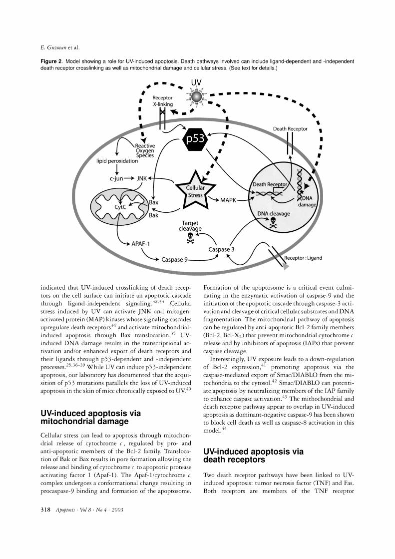
Figure 2. Model showing a role for UV-induced apoptosis. Death pathways involved can include ligand-dependent and -independentdeath receptor crosslinking as well as mitochondrial damage and cellular stress. (See text for details.)
indicated that UV-induced crosslinking of death recep-tors on the cell surface can initiate an apoptotic cascadethrough ligand-independent signaling.32,33 Cellularstress induced by UV can activate JNK and mitogen-activated protein (MAP) kinases whose signaling cascadesupregulate death receptors34 and activate mitochondrial-induced apoptosis through Bax translocation.35 UV-induced DNA damage results in the transcriptional ac-tivation and/or enhanced export of death receptors andtheir ligands through p53-dependent and -independentprocesses.25,36–39 While UV can induce p53-independentapoptosis, our laboratory has documented that the acqui-sition of p53 mutations parallels the loss of UV-inducedapoptosis in the skin of mice chronically exposed to UV.40
UV-induced apoptosis viamitochondrial damage
Cellular stress can lead to apoptosis through mitochon-drial release of cytochrome c , regulated by pro- andanti-apoptotic members of the Bcl-2 family. Transloca-tion of Bak or Bax results in pore formation allowing therelease and binding of cytochrome c to apoptotic proteaseactivating factor 1 (Apaf-1). The Apaf-1/cytochrome ccomplex undergoes a conformational change resulting inprocaspase-9 binding and formation of the apoptosome.
Formation of the apoptosome is a critical event culmi-nating in the enzymatic activation of caspase-9 and theinitiation of the apoptotic cascade through caspase-3 acti-vation and cleavage of critical cellular substrates and DNAfragmentation. The mitochondrial pathway of apoptosiscan be regulated by anti-apoptotic Bcl-2 family members(Bcl-2, Bcl-XL) that prevent mitochondrial cytochrome crelease and by inhibitors of apoptosis (IAPs) that preventcaspase cleavage.
Interestingly, UV exposure leads to a down-regulationof Bcl-2 expression,41 promoting apoptosis via thecaspase-mediated export of Smac/DIABLO from the mi-tochondria to the cytosol.42 Smac/DIABLO can potenti-ate apoptosis by neutralizing members of the IAP familyto enhance caspase activation.43 The mithochondrial anddeath receptor pathway appear to overlap in UV-inducedapoptosis as dominant-negative caspase-9 has been shownto block cell death as well as caspase-8 activation in thismodel.44
UV-induced apoptosis viadeath receptors
Two death receptor pathways have been linked to UV-induced apoptosis: tumor necrosis factor (TNF) and Fas.Both receptors are members of the TNF receptor
318 Apoptosis · Vol 8 · No 4 · 2003

The role of cell death in UV-induced skin cancer
superfamily and type I membrane proteins. Upon bind-ing their cognate ligand, TNF or Fas ligand (FasL), re-spectively, these receptors signal apoptosis. UV exposureinduces TNF, FasL, and receptor upregulation.25,37 Thesubsequent receptor:ligand interactions induce “sunburncell” formation, illustrated by the fact that mice deficientin either ligand or receptors for TNF or Fas show a remark-able decrease in apoptotic keratinocyte formation follow-ing UV exposure.25,45 Both TNF receptor and Fas can beactivated in a ligand-independent manner via UV-inducedcrosslinking of cell-surface receptors.32,33 The death re-ceptor pathway intersects with the cell stress and mito-chondrial pathways for apoptosis through p53. In thisregard, p53 has been shown to transcriptionally activateFas and Bax and repress Bcl-2.39,46,47 UV-induced p53mutations significantly diminish death receptor-inducedapoptosis,24 thought to be a critical underlying mech-anism for enhanced tumor development in the absencewild-type p53.26–29 Because Fas and TNF signaling playcentral roles in both the apoptotic response and host adap-tive responses to UV (discussed below), we will discusstheir regulation and signaling in some detail.
Fas regulation and signaling
The Fas receptor is constitutively expressed in most tis-sues, while FasL is considerably more restricted with con-stitutive expression found in the anterior chamber of theeye, the testis, the lung, and the large and small intestine.48
Both Fas and FasL are upregulated following genotoxicstress and DNA damage.25,36 Our laboratory has docu-mented that UV light is a powerful inducer of both Fasand FasL in exposed skin.25 Further, we39 and others49
have observed that Fas is a potent transcriptional target forp53. Functional inactivation of p53 through UV-inducedmutagenesis is a likely factor in the process of NMSC skincarcinogenesis and may involve the suppression of bothFas and FasL expression. In this respect, Fas expression isattenuated or absent in basal cell and squamous cell carci-nomas where p53 mutations are numerous50,51 and FasLinduction is attenuated in skin chronically exposed to UVin a manner consistent with the onset of detectable p53mutations.40 Melanomas, which typically contain few p53mutations, also show decreased Fas expression52,53 result-ing from active transcriptional repression.54
Apoptotic Fas/FasL interactions can be controlled at thelevel of the membrane by insufficient cell-surface receptordensity (discussed above), or the production of soluble Fas(sFas) or decoy receptors that bind ligand, but fail to sig-nal. sFas, unlike other members of the TNF superfamily,arises as a result of alternative mRNA splicing rather thanproteolytic cleavage.55,56 Multiple isoforms of sFas resultfrom the splicing of exon 6, exon 4, or exons 3, 4 and6, removing the transmembrane anchor region. Fas iso-forms share in common only a small 49 amino acid region
in the ligand binding domain. Secreted by the producingcell, sFas is thought to attenuate cell-surface Fas signalingthrough ligand competition. sFas is increased in a numberof malignancies, including melanoma,57,58 where elevatedlevels have been shown to be associated with poor progno-sis, tumor recurrence, and reduced survival. Decoy recep-tors, transmembrane receptors that bind FasL but fail tosignal, are found on normal keratinocytes and are down-regulated after UV.59 To date, it is not known whetherthese receptors are increased in malignant cells.
The interaction of Fas with FasL initiates a signal trans-duction cascade resulting in apoptosis.60 The Fas extracel-lular region contains three cysteine rich domains involvedin FasL binding,61 while the intracellular region containsan 80-amino acid region known as the death domain.The death domain is a protein-protein association motiffound also in signal-transducing adaptor proteins such asthe Fas associated death domain (FADD), TNF associateddeath domain (TRADD), TNF-Receptor associated fac-tors (TRAF2), and receptor interacting protein (RIP).60
Ligand binding to Fas results in receptor trimerizationand FADD recruitment to the death domain62 which re-cruits and activates pro-caspase-8 initiating the apoptoticcascade.63 Activation of caspase-8 is an essential link con-necting Fas-induced apoptosis to the mitochondrial path-way of cell death. The pro-apoptotic Bcl-2 family member,Bid, is cleaved by caspase-8 to form truncated Bid (tBid)that translocates into the mitochondrial membrane initi-ating cytochrome c loss. The interaction of cytochrome cwith Apaf-1 induces caspase-9 activity, potentiating cellu-lar demise. Caspase-8 activation can be inhibited by FLIP,a caspase 8 homologue lacking catalytic activity.64 Ele-vated FLIP levels have been observed in melanoma.64,65
Fas signaling has also been shown to be inhibited bysomatic mutations in the death domain preventing for-mation of an apoptotic signal. Somatic mutations in Fashave been reported in melanoma66 and squamous cellcarcinoma.67 The IAPs that bind and inhibit caspases-3,-6, -7, and -968 have also been shown to block Fas apop-totic signaling. In summary, Fas-induced apoptosis canbe regulated at multiple levels including receptor expres-sion, alternative mRNA splicing, mutations, expression ofanti-apoptotic proteins, and a failure to upregulate FasL.Skin tumors arising from UV exposure frequently demon-strate impairment in Fas apoptotic signaling, a summaryof these alterations is shown in Table 1.
TNF receptor regulation and signaling
TNF signals through two receptors, TNF-R1 and TNF-R2. Most cells express TNF-R1 constitutively, whereasTNF-R2 is expressed more selectively (notably in lym-phocytes). The majority of the biologic effects induced byTNF (cytotoxicity, antiviral activity, induction of
Apoptosis · Vol 8 · No 4 · 2003 319

E. Guzman et al.
Table 1. Potential mechanisms underlying the impairment of Fasapoptotic signaling in UV-induced tumors
Non-Melanoma SkinMelanoma Cancer (NMSC)
Loss of Fas expression 52, 53∗ 50, 51
Transcriptional repression 54 N.R.+
Somatic mutations 66 67
c-FLIP 64, 65 N.R.
sFas production 57, 58 N.R.
Bcl-2 family members 100 101, 102
IAPs (survivin) 98 99
Failure to upregulate FasL N.R. 40
∗Denotes reference number containing information.+N.R.: not reported.
superoxide dismutase, fibroblast proliferation) appear tobe initiated through the binding of TNF-R1.69 Signal-ing through TNF-R2, on the other hand, appears to beinvolved in the proliferation of thymocytes and cyto-toxic cells.70 Binding of TNF to TNF-R1 leads to re-cruitment of the adaptor protein TRADD, which sub-sequently recruits RIP, TRAF2, or FADD. Assembly ofsignal-transducing proteins leads to downstream activa-tion of caspases as well as nuclear factor kappa (NFκB) andc -Jun regulating cell growth and death, inflammation,and stress responses.69,70 TNF is upregulated by UV andsuch upregulation has been shown to induce keratinocyteapoptosis through activation of TNF-R1.37,45,71 Poly-morphisms in the TNF gene may regulate the strengthof the UV response.72,73 TNF production plays a role inUV-induced skin carcinogenesis, as experimental animalslacking TNF are significantly more resistant to tumordevelopment than their wild-type counterparts.74 TNF isknown to be involved in the induction of UV-induced im-mune suppression (see below) and the production of thepro-inflammatory cytokines IL-1 and IL-6.71–73 Mice de-ficient in TNF receptor expression fail to develop alteredimmune responses following UV75,76 perhaps explainingtheir increased resistance to UV-induced carcinogenesis.Taken together, these findings suggest that UV-inducedTNF production and response is associated with increasedskin cancer risk.
UV-induced immune suppression
A paradox of skin carcinogenesis is the immunogenecityof UV-induced tumors; most are easily rejected whentransplanted into immunocompetent animals.77 Immun-odeficient or UV-irradiated mice, however, are unable toreject transplanted UV-induced tumors such that thesetumors grow unhindered. These results suggest that (1)UV-induced tumor outgrowth is controlled by the im-mune system; and (2) UV exposure induces a transient
state of immune suppression that is critical for the devel-opment of skin tumors.78
Sensitivity to UV-induced immune suppression hasbeen proposed as a risk factor for sunlight-induced hu-man skin cancer. While approximately 40% of the adultCaucasian population is classified as “UV-sensitive” show-ing immune suppression to contact sensitizers applied onUV-exposed skin, >92% of patients with a history of basalcell or squamous cell carcinomas and 100% of patientswith a diagnosis of malignant melanoma are classifiedas “UV-sensitive”.79,80 Further evidence for the associa-tion of UV-induced immune suppression and tumorige-nesis comes from patients receiving immunosuppressants(cyclosporine or FK506), where a sharply increased inci-dence of skin tumors in sun-exposed sites is observed.81–83
Using contact hypersensitivity (CHS) and delayed typehypersensitivity (DTH) responses to study UV-inducedimmune suppression, differences in underlying mecha-nisms have been described.84,85 Such distinctions may re-flect the nature of the antigen, the route of administrationof the antigen, or the dose of UV.
Experimental mouse models have been used to under-stand the basis of sensitivity and resistance to UV-inducedapoptosis, immune suppression, and carcinogenesis. Miceare typically exposed to between 5–30 KJ/m2 UV, de-pending on sensitivity, to mimic a minimum erythemaldose (MED, the time taken to produce skin redness understandard sunlamp conditions). Different strains of labora-tory mice have different susceptibilities to UV-inducedsuppression.86 High susceptibility is seen in C57B1/6mice (50% suppression with a UV dose of 0.7–2.3 KJ/m2);intermediate susceptibility is seen in C3H and DBAmice (50% suppression observed using a dose of4.7–6.9 KJ/m2); and low susceptibility is seen in Balb/cmice (50% suppression induced only at UV doses between9.6–12.3 KJ/m2). In humans, these UV doses translate toexposure lengths of 15–20 min sun exposure (midday,summer months), a dose easily achievable during normalactivities.
Effects of UV on the immune response are transient,with maximum effects observed 3–7 days following a sin-gle, acute exposure. Multiple, tandem exposures, how-ever, may result in a state of chronically reduced immunefunction. UV-induced DNA damage appears to be theinitiating factor for systemic immune suppression.15–19
Such damage results in the upregulation of a number ofcytokines as well as apoptosis-inducing ligands includingFasL and TNF.25,37,71 UV-induced suppression is antigen-specific and can be adoptively transferred to naı̈ve, unir-radiated mice by T cells in the spleen.
Our laboratory is interested in UV-induced immunesuppression mediated by Fas and FasL interactions. Wehave documented that gld mice bearing a point muta-tion in FasL (rendering it non-functional), demonstrate noUV-induced immune suppression of either CHS or DTH
320 Apoptosis · Vol 8 · No 4 · 2003
The role of cell death in UV-induced skin cancer
immune responses and cannot transfer suppression to anaı̈ve host.87 We have shown that FasL is expressed in skindraining lymph nodes of UV-exposed animals87 and arecurrently investigating the possibility that DNA-damaged, FasL+ Langerhans cells, migrating from UV-exposed skin participate in the generation of T regulatorycells. Indeed, FasL+, T regulatory cells have been de-scribed in other experimental systems involving immunesuppression.88 FasL-induced apoptosis may also contri-bute to the production of immunosuppressive cytokines(such as IL-10) produced after UV.89,90 The requirementfor FasL in UV-induced immune suppression is unam-biguous, although additional work will be required todetermine precisely where Fas/FasL interactions occur andthe role they play in regulating antigen presentation, Tcell responses, and the UV-induced cytokine milieu.
Regulators of apoptosis and skincarcinogenesis
UV-induced apoptosis is a beneficial event that allowsthe host to eliminate cells containing DNA damage andthwart the accumulation of genetic mutations. Disrup-tion of apoptotic pathways can promote UV-induced skincarcinogenesis by allowing DNA damage to persist andmutations to develop. Multiple reports using a varietyof experimental systems have validated this premise. Forexample, transgenic mice expressing Bcl-2 selectively inkeratinocytes demonstrate an increased sensitivity to skintumor induction,91 while Bax knockout mice show a sig-nificantly increased UV-induced tumor incidence.92 Micedeficient in wild-type p53 show an increased incidence ofUV-induced skin cancers owing to the joint effects of p53on apoptosis and cell cycle arrest and DNA repair.26,27
Interestingly, mice heterozygous for wild-type p53 alsoshow an increased incidence of UV-induced skin tumors93
as well as an increased incidence of internal cancers(lymphomas),94 suggesting that UV can cooperate withthe loss of p53 for the generation of non-cutaneous ma-lignancies as well. Bax has recently been implicated inthe repair of UV-induced DNA damage,95 suggestingthat this pro-apoptotic protein may also protect againstUV-induced skin carcinogenesis through the inductionof apoptosis and DNA repair. Transgenic mice express-ing survivin, an IAP, under the control of a keratin-14promoter have been shown to be resistant to UV-inducedapoptosis in the skin.96 Although additional studies willbe required to document a causative role for this pro-tein in UV-induced skin cancers, survivin has alreadybeen reported to promote chemical-induced skin tumorprogression97 and to be expressed in sentinel lymph nodesof melanoma patients where it has been correlated withpoor patient outcome and disease recurrence.98 Indeed,there are numerous reports of elevated anti-apoptotic gene
expression in UV-induced skin cancers including survivinin non-melanoma skin cancer,99 Bcl-2 in melanoma,100
bcl-2 in non-melanoma skin cancers,101,102 FLIP inmelanoma,65 and sFas in melanoma.57,58
Death receptors UV-induced skincarcinogenesis and the imprisonedclone model
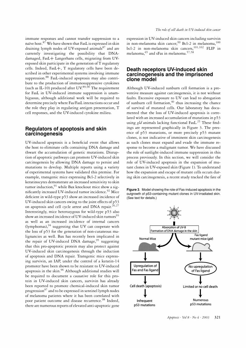
Although UV-induced sunburn cell formation is a pre-ventive measure against carcinogenesis, it is not withoutfaults. Excessive exposure to UV can lead to abrogationof sunburn cell formation,40 thus increasing the chanceof survival of mutated cells. Our laboratory has docu-mented that the loss of UV-induced apoptosis is corre-lated with an increased accumulation of mutations in p53using gld animals lacking functional FasL.25 These find-ings are represented graphically in Figure 3. The pres-ence of p53 mutations, or more precisely p53 mutantclones, is not indicative of imminent skin carcinogenesisas such clones must expand and evade the immune re-sponse to become a malignant tumor. We have discussedthe role of sunlight-induced immune suppression in thisprocess previously. In this section, we will consider therole of UV-induced apoptosis in the expansion of mu-tant clones in UV-exposed skin (Figure 1). To understandhow the expansion and escape of mutant cells occurs dur-ing skin carcinogenesis, a recent study tracked the fate of
Figure 3. Model showing the role of Fas-induced apoptosis in theoutgrowth of p53-containing mutant clones in UV-irradiated skin.(See text for details.)
Apoptosis · Vol 8 · No 4 · 2003 321

E. Guzman et al.
keratinocyte clones with mutations in the p53 gene.103
Generally, such clones expand when skin is chronicallyexposed to UV irradiation and regress after cessation ofirradiation. However, in about 20% of the mutant clones,regression does not occur after the host is no longer ex-posed to UV. In fact, some mutant keratinocyte clonescontinue proliferating without expanding overall clonesize to become “imprisoned clones” with densely packedcells containing very little cytoplasm (Figure 1b). Thesemutant clones are imprisoned by normal keratinocytes. Itappears that these imprisoned clones are contained withinindividual epidermal stem cell compartments until UVexposure occurs. Expansion beyond an epidermal stemcell compartment is likely a critical step in carcinogen-esis. Which of the myriad of UV effects might permitthis “get out of jail free” card? Based on our knowledge ofUV-induced apoptosis and the loss of apoptosis sensitivityobserved in UV-induced tumors, it appears highly likelythat death receptor interactions play an important role.For example, we have documented that the ability to up-regulate FasL can be lost during repeated UV exposures.40
If imprisoned clones have lost this ability as well, sunburnof normal adjacent skin would result in apoptosis creatingspace for the mutant clone to expand. With continued UVexposure at close intervals, the imprisoned clone might beable to overtake its own stem cell compartment, and per-haps, expand to neighboring ones. Death receptor inducedapoptosis that is initially beneficial to the UV-irradiatedhost could conceivably become a detriment contribut-ing to the outgrowth of mutant keratinocyte clones (Fig-ure 1a). UV-induced immune suppression would likelyalso enhance imprisoned clone outgrowth by preventingimmunologic rejection (Figure 1b). There are at least threefates of UV-induced mutant clones depending on the sta-tus of the host immune response. First, clones may persistif the host is immune suppressed or immunologically ig-norant, second, the clones may be cleared by an intacthost immune response, and third, the clones may becomeimprisoned as a result of immune surveillance and/or thefailure to induce apoptosis in surrounding normal tissuesfollowing chronic UV exposure. Thus, both apoptosis andUV-induced immune suppression likely contribute to thegenesis and outgrowth skin cancers. Additional studieswill be required to fully characterize the role of death re-ceptor interactions in UV-induced expansion and regres-sion of mutant keratinocyte clones and explore the in-volvement of this pathway in the outgrowth of malignantmelanocytes.
Conclusion
Death receptor-induced apoptosis initiated by UV expo-sure plays a prominent role in the elimination of DNA-damaged skin and the generation of UV-induced immune
suppression. In the skin, such apoptosis protects againstthe development of sunlight-induced cancers by erasingDNA-damaged cells rather than relying on DNA repairfor damage correction. Death receptors are also involved insuppression of immune function required for skin tumoroutgrowth. The effects of the death receptors are contra-dictory in overall host protection against UV-induced tu-mors, imploring that additional studies address the selec-tive enhancement and elimination, respectively, of deathreceptor function at the level of the skin and the systemicimmune response.
References
1. Fisher MS, Kripke ML. Systemic alteration induced in mice byultraviolet light irradiation and its relationship to ultravioletcarcinogenesis. Proc Natl Acad Sci USA 1977; 74: 1688–1692.
2. Frankel DH, Hanusa BH, Zitelli JA. New primary non-melanoma skin cancer in patients with a history of squamouscell carcinoma of the skin. Implications and recommendationsfor follow-up. J Am Acad Dermatol 1992; 26: 720–726.
3. Kahn HS, Tatham LM, Patel AV, Thun MJ, Heath Jr. CW. In-creased cancer mortality following a history of nonmelanomaskin cancer. Jama 1998; 280: 910–912.
4. Tyrrell RM. Ultraviolet radiation and free radical damage toskin. Biochem Soc Symp 1995; 61: 47–53.
5. Ziegler A, Leffell DJ, Kunala S, et al. Mutation hotspots dueto sunlight in the p53 gene of nonmelanoma skin cancers.Proc Natl Acad Sci USA 1993; 90: 4216–4220.
6. Parrish JA, Jaenicke KF, Anderson RR. Erythema andmelanogenesis action spectra of normal human skin. PhotochemPhotobiol 1982; 36: 187–191.
7. Drobetsky EA, Turcotte J, Chateauneuf A. A role for ultra-violet A in solar mutagenesis. Proc Natl Acad Sci USA 1995;92: 2350–2354.
8. Cleaver JE, Bootsma D. Xeroderma pigmentosum: Biochemi-cal and genetic characteristics. Annu Rev Genet 1975; 9: 19–38.
9. Kraemer KH, Lee MM, Scotto J. Xeroderma pigmentosum:Cutaneous, ocular, and neurological abnormalities in 830published cases. Arch Dermatol 1987; 123: 241–250.
10. Brash DE. UV mutagenic photoproducts in Escherichia coliand human cells: A molecular genetics perspective on humanskin cancer. Photochem Photobiol 1988; 48: 59–66.
11. Cadet J, Anselmino C, Douki T, Voituriez L. Photochemistryof nucleic acids in cells. J Photochem Photobiol B 1992; 15:277–298.
12. Tang MS, Hrncir J, Mitchell D, Ross J, Clarkson J. The rela-tive cytotoxicity and mutagenicity of cyclobutane pyrimidinedimers and (6-4) photoproducts in Escherichia coli cells. Mu-tat Res 1986; 161: 9–17.
13. You YH, Lee DH, Yoon YH, Nakajima S, Yasui A, PfeiferGP. Cyclobutane pyrimidine dimers are responsible for thevast majority of mutations induced by UVB irradiation inmammalian cells. J Biol Chem 2001; 276: 44688–44694.
14. Kanjilal S, Perceall WE, Cummings KK, Kripke ML,Ananthaswamy HN. High frequency of p53 mutations inultraviolet radiation-induced murine skin tumors: Evidenceof strand bias and tumor heterogeneity. Cancer Res 1993; 53:2961–2964.
15. Kripke ML, Cox PA, Alas LG, Yarosh DB. Pyrimidinedimers in DNA initiate systemic immunosuppression in UV-irradiated mice. Proc Natl Acad Sci USA 1992; 89: 7516–7520.
322 Apoptosis · Vol 8 · No 4 · 2003

The role of cell death in UV-induced skin cancer
16. Vink AA, Strickland FM, Bucana C, et al. Localization ofDNA damage and its role in altered antigen-presenting cellfunction in ultraviolet-irradiated mice. J Exp Med 1996; 183:1491–1500.
17. Wolf P, Yarosh DB, Kripke ML. Effects of sunscreens and aDNA excision repair enzyme on ultraviolet radiation-inducedinflammation, immune suppression, and cyclobutane pyrim-idine dimer formation in mice. J Invest Dermatol 1993; 101:523–527.
18. Wolf P, Cox P, Yarosh DB, Kripke ML. Sunscreens and T4N5liposomes differ in their ability to protect against ultraviolet-induced sunburn cell formation, alterations of dendritic epi-dermal cells, and local suppression of contact hypersensitivity.J Invest Dermatol 1995; 104: 287–292.
19. Vink AA, Moodycliffe AM, Shreedhar V, et al. The inhibi-tion of antigen-presenting activity of dendritic cells resultingfrom UV irradiation of murine skin is restored by in vitro pho-torepair of cyclobutane pyrimidine dimers. Proc Natl Acad SciUSA 1997; 94: 5255–5260.
20. Brash DE, Rudolph JA, Simon JA, et al. A role for sun-light in skin cancer: UV-induced p53 mutations in squamouscell carcinoma. Proc Natl Acad Sci USA 1992; 88: 10124–10128.
21. Zhan Q, Carrier F, Fornance Jr. AJ. Induction of cellular p53activity by DNA-damaging agents and growth arrest. MolCell Biol 1993; 13: 4242–4250.
22. Levine AJ. p53, the cellular gatekeeper for growth and divi-sion. Cell 1997; 88: 323–331.
23. Smith ML, Chen IT, Zhan Q, O’Connor PM, Fornace Jr. AJ.Involvement of the p53 tumor suppressor in repair of u.v.-typeDNA damage. Oncogene 1995; 10: 1053–1059.
24. Ziegler A, Jonason AS, Leffell DJ, et al. Sunburn and p53 inthe onset of skin cancer. Nature 1994; 372: 773–776.
25. Hill LL, Ouhtit A, Loughlin SM, Kripke ML, AnanthaswamyHN, Owen-Schaub LB. Fas ligand: A sensor for DNA damagecritical in skin cancer etiology. Science 1999; 285: 898–900.
26. Li G, Ho VC, Berean K, Tron VA. Ultraviolet radiation in-duction of squamous cell carcinomas in p53 transgenic mice.Cancer Res 1995; 55: 2070–2074.
27. Li G, Tron V, Ho V. Induction of squamous cell carcinoma inp53-deficient mice after ultraviolet irradiation. J Invest Der-matol 1998; 110: 72–75.
28. Campbell C, Quinn AG, Ro YS, Angus B, Rees JL. p53 mu-tations are common and early events that precede tumor inva-sion in squamous cell neoplasia of the skin. J Invest Dermatol1993; 100: 746–748.
29. Lacour JP. Carcinogenesis of basal cell carcinomas: Geneticsand molecular mechanisms. Br J Dermatol 2002; 146(Suppl61): 17–19.
30. Ichiki H, Sakurada H, Kamo N, Takahashi TA, Sekiguchi S.Generation of active oxygens, cell deformation and membranepotential changes upon UV-B irradiation in human bloodcells. Biol Pharm Bull 1994; 17: 1065–1069.
31. Renzing J, Hansen S, Lane DP. Oxidative stress is involved inthe activation of p53. J Cell Sci 1996; 109: 1105–1112.
32. Aragane Y, Kulms D, Metze D, et al. Ultraviolet light in-duces apoptosis via direct activation of CD95 (Fas/APO-1)independently of its ligand CD95L. J Cell Biol 1998; 140:171–182.
33. Sheikh MS, Antinore MJ, Huang Y, Fournance Hr AJ.Ultraviolet-irradiation-induced apoptosis is mediated via lig-and independent activation of tumor necrosis factor recep-tor 1. Oncogene 1998; 17: 2555–2563.
34. Zanke BW, Boudreau K, Rubie E, et al. The stress-activatedprotein kinase pathway mediates cell death following injury
induced by cis-platinum, UV irradiation or heat. Curr Biol1996; 6: 606–613.
35. Tournier C, Hess P, Yang DD, et al. Requirement of JNK forstress-induced activation of the cytochrome c-mediated deathpathway. Science 2000; 288: 870–874.
36. Muller M, Wilder S, Bannasch D, et al. p53 activates the CD95(APO-1/Fas) gene in response to DNA damage by anticancerdrugs. J Exp Med 1998; 188: 2033–2045.
37. Kibitel J, Hejmadi V, Alas L, O’Connor A, Sutherland BM,Yarosh DB. UV-DNA damage in mouse and human cells in-duces the expression of tumor necrosis factor alpha. PhotochemPhotobiol 1998; 67: 541–546.
38. Bennett M, MacDonald K, Chan SW, Luzio J, Simari R,Weissberg P. Cell surface trafficking of Fas: A rapid mech-anism of p53-mediated apoptosis. Science 1998; 282: 290–293.
39. Owen-Schaub LB, Zhang W, Cusack JC, et al. Wild-type human p53 and a temperature-sensitive mutant in-duce Fas/APO-1 expression. Mol Cell Biol 1995; 15: 3032–3040.
40. Ouhtit A, Gorny A, Muller HK, Hill LL, Owen-Schaub L,Ananthaswamy HN. Loss of Fas-ligand expression in mousekeratinocytes during UV carcinogenesis. Am J Pathol 2000;157: 1975–1981.
41. Washio F, Ueda M, Ito A, Ichihashi M. Higher susceptibilityto apoptosis following ultraviolet B irradiation of xerodermapigmentosum fibroblasts is accompanied by upregulation ofp53 and downregulation of bcl-2. Br J Dermatol 1999; 140:1031–1037.
42. Adrain C, Creagh EM, Martin SJ. Apoptosis-associated releaseof Smac/DlABLO from mitochondria requires active caspasesand is blocked by Bcl-2. Embo J 2001; 20: 6627–6636.
43. Ekert PG, Silke J, Hawkins CJ, Verhagen AM, Vaux DL.DIABLO promotes apoptosis by removing MIHA/XIAP fromprocessed caspase 9. J Cell Biol 2001; 152: 483–490.
44. Sitailo L, Tibudan SS, Denning MF. Activation of caspase-9 isrequired for UV-induced apoptosis of human keratinocytes. JBiol Chem 2002; 277: 19346–19352.
45. Zhuang L, Wang B, Shinder GA, Shivji GM, Mak TW, SauderDN. TNF receptor p55 plays a pivotal role in murine ker-atinocyte apoptosis induced by ultraviolet B irradiation. JImmunol 1999; 162: 1440–1447.
46. Miyasita T, Reed JC. Tumor suppressor p53 is a direct tran-scriptional activator of the human bax gene. Cell 1995; 80:293–299.
47. Miyashita T, Harigai M, Hanada M, Reed JC. Identificationof a p53 dependent negative response element in the bcl-2gene. Cancer Res 1994; 54: 3131–3135.
48. French LE, Hahne M, Viard I, et al. Fas and Fas ligand in em-bryos and adult mice: Ligand expression in several immune-privileged tissues and coexpression in adult tissues character-ized by apoptotic cell turnover. J Cell Biol 1996; 133: 335–343.
49. Munsch D, Watanabe-Fukunaga R, Bourdon JC, et al. Hu-man and mouse Fas (APO-1/CD95) death receptor genes eachcontain a p53-responsive element that is activated by p53mutants unable to induce apoptosis. J Biol Chem 2000; 275:3867–3872.
50. Filipowicz E, Adegboyega P, Sanchez RL, Gatalica Z. Expres-sion of CD95 (Fas) in sun-exposed human skin and cutaneouscarcinomas. Cancer 2002; 94: 814–819.
51. Gutierrez-Steil C, Wrone-Smith T, Sun X, Krueger JG, CovenT, Nickoloff BJ. Sunlight-induced basal cell carcinoma tumorcells and ultraviolet-B-irradiated psoriatic plaques express Fasligand (CD95L). J Clin Invest 1998; 101: 33–39.
Apoptosis · Vol 8 · No 4 · 2003 323

E. Guzman et al.
52. Bullani RR, Wehrli P, Viard-Leveugle I, et al. Frequentdownregulation of Fas (CD95) expression and function inmelanoma. Melanoma Res 2002; 12: 263–270.
53. Soubrane C, Mouawad R, Antoine EC, Verola O, Gil-DelgadoM, Khayat D. A comparative study of Fas and Fas-ligandexpression during melanoma progression. Br J Dermatol 2000;143: 307–312.
54. Ivanov VN, Bhoumik A, Krasilnikov M, et al. Cooperationbetween STAT3 and c-jun suppresses Fas transcription. Molec-ular Cell 2001; 7: 517–528.
55. Cheng J, Zhou T, Liu C, et al. Protection from Fas-mediatedapoptosis by a soluble form of the Fas molecule. Science 1994;263: 1759–1762.
56. Casino I, Fiucci G, Papoff G, Ruberti G. Three functionalforms of the human apoptosis-inducing Fas molecule areproduced by alternative splicing. J Immunol 1995; 154: 2706–2713.
57. Redondo P, Solano T, Vazquez B, Bauza A, Idoate M. Fasand Fas ligand: Expression and soluble circulating levels incutaneous malignant melanoma. British J Dermatol 2002; 147:80–86.
58. Ugurel S, Rappl G, Tilgen W, Reinhold U. Increased solubleCD95 (sFas/CD95) serum level correlates with poor prognosisin melanoma patients. Clin Cancer Res 2001; 7: 1282–1286.
59. Maeda T, Hao C, Tron VA. Ultraviolet light (UV) regulationof TNF family decoy receptors DcR2 and DcR3 in humankeratinocytes. J Cutan Med Surg 2001; 5: 294–298.
60. Nagata S. Apoptosis by death factor. Cell 1997; 88: 355–365.
61. Orlinick JR, Elkon KB, Chao MV. Separate domains of thehuman fas ligand dictate self-association and receptor bind-ing. J Biol Chem 1997; 272: 32221–32229.
62. Chinnaiyan A, O’Rourke K, Tewari M, Dixit VM. FADD,a novel death domain-containing protein, interacts with thedeath domain of Fas and initiates apoptosis. Cell 1995; 81:505–512.
63. Salvesen GS, Dixit VM. Caspases: Intracellular signaling byproteolysis. Cell 1997; 91: 443–446.
64. Irmler M, Thome M, Hahne M, et al. Inhibition of deathreceptor signals by cellular FLIP. Nature 1997; 388: 190–195.
65. Bullani RR, Huard B, Viard-Leveugle I, et al. Selective expres-sion of FLIP in malignant melanocytic skin lesions. J InvestDermatol 2001; 117: 360–364.
66. Shin MS, Park WS, Kim SY, et al. Alterations of Fas (Apo-1/CD95) gene in cutaneous malignant melanoma. Am J Pathol1999; 154: 1785–1791.
67. Lee SH, Shin MS, Kim HS, et al. Somatic mutations of Fas(Apo-1/CD95) gene in cutaneous squamous cell carcinomaarising from a burn scar. J Invest Dermatol 2000; 114: 122–126.
68. Deveraux QL, Reed JC. IAP family proteins—Suppressors ofapoptosis. Genes Dev 1999; 13: 239–252.
69. Chen G, Goeddel DV. TNF-R1 signaling: A beautiful path-way. Science 2002; 296: 1634–1635.
70. Gupta S. A decision between life and death during TNF-alpha-induced signaling. J Clin Immunol 2002; 22: 185–194.
71. Kock A, Schwartz T, Kirnbauer R, et al. Human keratinocytesare a source for human tumor necrosis factor alpha: Evidencefor synthesis and release upon stimulation with endotoxin orultraviolet light. J Exp Med 1990; 172: 1609–1614.
72. Yoshikawa T, Streilein JW. Genetic basis of the effects ofultraviolet light B on cutaneous immunity. Evidence thatpolymorphism at the Tnfa and Lps loci governs susceptibility.Immunogenetics 1990; 32: 398–405.
73. Niizeki H, Naruse T, Hecker KH, et al. Polymorphisms inthe tumor necrosis factor (TNF) genes are associated withsusceptibility to effects of ultraviolet-B radiation on inductionof contact hypersensitivity. Tissue Antigens 2001; 58: 369–378.
74. Moore RJ, Owens DM, Stamp G, et al. Mice deficient in tumornecrosis factor-alpha are resistant to skin carcinogenesis. NatMed 1999; 5: 828–831.
75. Kondo S, Wang B, Fujisawa H, et al. Effect of gene-targetedmutation in TNF receptor (p55) on contact hypersensitiv-ity and ultraviolet B-induced immunosuppression. J Immunol1995; 155: 3801–3805.
76. Amerio P, Toto P, Feliciani C, et al. Rethinking the role of tu-mour necrosis factor-alpha in ultraviolet (UV) B-induced im-munosuppression: Altered immune response in UV-irradiatedTNFR1R2 gene-targeted mutant mice. Br J Dermatol 2001;144: 952–957.
77. Kripke ML. Antigenicity of murine skin tumors induced byultraviolet light. J Natl Cancer Inst 1974; 53: 1333–1336.
78. Kripke ML. Ultraviolet radiation and immunology: Some-thing new under the sun—Presidential address. Cancer Res1994; 54: 6102–6105.
79. Yoshikawa T, Rae V, Bruins-Slot W, Van den Berg JW, TaylorJR, Streilien JW. Susceptibility to effects of UVB radiation asa risk factor for skin cancer in humans. J Invest Dermatol 1990;95: 530–536.
80. Streilien JW, Taylor JR, Vincek V, et al. Relationship be-tween ultraviolet radiation-induced immunosuppression andcarcinogenesis (Review). J Invest Dermatol 1994; 103: 107S–111S.
81. Cockburn IT, Krupp P. The risk of neoplasms in patientstreated with cyclosporine A. J Autoimmun 1989; 2: 723–731.
82. Kinlen LJ, Sheil AG, Peto J, Doll R. Collaborative UnitedKingdom Australasian study of cancer in patients treatedwith immunosuppressive drugs. Br Med J 1979; 2: 1461–1466.
83. Boyle J, MacKie RM, Briggs JD, Junor BJ, Atichison TC.Cancer, warts, and sunshine in renal transplant patients. Acase-control study. Lancet 1984; 1: 702–705.
84. Kim TY, Kripke ML, Ullrich SE. Immunosuppression by fac-tors released from UV-irradiated epidermal cells: Selectiveeffects on the generation of contact and delayed hypersensitiv-ity after exposure to UVA or UVB radiation. J Invest Dermatol1990; 94: 26–32.
85. Moodycliffe AM, Bucana CD, Kripke ML, Norval M, Ull-rich SE. Differential effects of a monoclonal antibody to cis-urocanic acid on the suppression of delayed and contact hyper-sensitivity following ultraviolet irradiation. J Immunol 1996;157: 2891–2899.
86. Noonan FP, Hoffman HA. Susceptibility to immunosuppres-sion by ultraviolet B radiation in the mouse. Immunogenetics1994; 39: 29–39.
87. Hill LL, Shreedhar VK, Kripke ML, Owen-Schaub LB. Acritical role for Fas ligand in the active suppression of systemicimmune responses by ultraviolet radiation. J Exp Med 1999;189: 1285–1294.
88. Watanabe T, Yoshida M, Shirai Y, et al. Administration of anantigen at a high dose generates regulatory CD4+ T cellsexpressing CD95 ligand and secreting IL-4 in the liver. JImmunol 2002; 168: 2188–2199.
89. Rivas JM, Ullrich SE. The role of IL-4, IL-10, and TNF-alphain the immune suppression induced by ultraviolet radiation.J Leukoc Biol 1994; 56: 769–775.
90. Nishigori C, Yarosh DB, Ullrich SE, et al. Evidence thatDNA damage triggers interleukin 10 cytokine production in
324 Apoptosis · Vol 8 · No 4 · 2003

The role of cell death in UV-induced skin cancer
UV-irradiated murine keratinocytes. Proc Natl Acad Sci USA1996; 93: 10354–10359.
91. Rodriguez-Villanueva J, Greenhalgh D, Wang XJ, et al. Hu-man keratin-1.bcl-2 transgenic mice aberrantly express ker-atin 6, exhibit reduced sensitivity to keratinocyte cell deathinduction and are susceptible to skin tumor formation. Onco-gene 1998; 16: 853–863.
92. Cho SH, Delehedde M, Rodriguez-Villanueva J, Brisbay S,McDonnell TJ. Bax gene disruption alters the epidermal re-sponse to ultraviolet irradiation and in vivo induced skin car-cinogenesis. Int J Mol Med 2001; 7: 235–241.
93. Jiang W, Ananthaswamy HN, Muller HK, Kripke ML. p53protects against skin cancer induction by UV-B radiation.Oncogene 1999; 18: 4247–4253.
94. Jiang W, Ananthaswamy HN, Muller HK, et al. UV irra-diation augments lymphoid malignancies in mice with onefunctional copy of wild-type p53. Proc Natl Acad Sci USA2001; 98: 9790–9795.
95. Cho S, O’Connor SL, McDonnell TJ. Evidence that nucleotideexcision repair is attenuated in bax-deficient mammalian cellsfollowing ultraviolet irradiation. Exp Cell Res 2002; 278: 158–165.
96. Grossman D, Kim PJ, Blanc-Brude OP, et al. Transgenicexpression of survivin in keratinocytes counteracts UV-Binduced apoptosis and cooperates with loss of p53. J ClinInvest 2001; 108: 991–999.
97. Allen SM, Florell SR, Hanks AN, et al. Survivin expressionin mouse skin prevents papilloma regression and promoteschemical-induced tumor progression. Cancer Res 2003; 63:567–572.
98. Gradilone A, Gazzaniga P, Ribuffo D, et al. Survivin, bcl-2,bax, and bcl-X gene expression in sentinel lymph nodes frommelanoma patients. J Clin Oncol 2003; 21: 306–312.
99. Lo Muzio L, Staibano S, Pannone G, et al. Expression of theapoptosis inhibitor survivin in aggressive squamous cell car-cinoma. Exp Mol Pathol 2002; 70: 249–254.
100. Kanter-Lewensohn L, Hedblad MA, Wejde J, Larsson O.Immunohistochemical markers for distinguishing Spitz nevifrom malignant melanomas. Mod Pathol 1997; 10: 917–920.
101. Morales-Ducret CR, van de Rijn M, LeBrun DP, Smoller BR.bcl-2 expression in primary malignancies of the skin. ArchDermatol 1995; 131: 909–912.
102. Cerroni L, Kerl H. Aberrant bcl-2 protein expression pro-vides a possible mechanism of neoplastic cell growth in cu-taneous basal-cell carcinoma. J Cutan Pathol 1994; 21: 398–403.
103. Zhang W, Remenyik E, Zelterman D, Brash DE, WikonkalNM. Escaping the stem cell compartment: Sustained UVBexposure allows p53-mutant keratinocytes to colonize adja-cent epidermal proliferating units without incurring addi-tional mutations. Proc Natl Acad Sci USA 2001; 98: 13948–13953.
Apoptosis · Vol 8 · No 4 · 2003 325