![NO Q RSTUVWXYZW [V \Z]]^XYZW _``Y[VWXV]]V VX ...](https://static.fdokumen.com/doc/165x107/633db03fa7b2edc6000ec873/no-q-rstuvwxyzw-v-zxyzw-yvwxvv-vx-.jpg)
Analysis of FK506, timcodar (VX-853) and FKBP51 and FKBP52 chaperones in control of glucocorticoid...
11
ORIGINAL ARTICLE Analysis of FK506, timcodar (VX-853) and FKBP51 and FKBP52 chaperones in control of glucocorticoid receptor activity and phosphorylation Terry D. Hinds Jr 1,2,a , Lance A. Stechschulte 1,a , Fadel Elkhairi 3,b & Edwin R. Sanchez 1 1 Center for Diabetes and Endocrine Research,Department of Physiology & Pharmacology, University of Toledo College of Medicine, Toledo, Ohio, 43614 2 Center for Hypertension and Personalized Medicine,Department of Physiology & Pharmacology, University of Toledo College of Medicine, Toledo, Ohio, 43614 3 Department of Urology, University of Toledo College of Medicine, Toledo, Ohio, 43614 Keywords Ligands, neuroprotection, nuclear hormone receptors, posttranslational modification, regenerative medicine, steroids Correspondence Edwin R. Sanchez, Department of Physiology and Pharmacology, 3000 Arlington Avenue, Mailstop 1008, University of Toledo College of Medicine, Toledo, OH 43614. Tel: 419.383.4182; Fax: 419.383.2871; E-mail: [email protected] Present address b Department of Urology, Central Ohio Urol- ogy Group, 941 Chatham Ln Suite 110/210, Columbus, Ohio, 43221 Funding Information This work was supported in part by the Stranahan Endowment for Oncologic Research, Department of Urology UTCOM awarded to E. R. S., and by a National Institutes of Health grant [DK70127] (E. R. S.). L. A. S. was supported in part by a Center for Diabetes and Endocrine Research Hiss Foundation Fellowship. T. D. H. was supported by a National Institutes of Health PRIDE grant [HL106365]. Received: 11 June 2014; Revised: 30 June 2014; Accepted: 1 July 2014 Pharma Res Per, 2(6), 2014, e00076, doi: 10.1002/prp2.76 doi: 10.1002/prp2.76 a These authors contributed equally to this work. Abstract The immunosuppressive ligand FK506 and the FK506-binding protein FKBP52 are stimulatory to glucocorticoid receptor (GR) activity. Here, we explore the underlying mechanism by comparing GR activity and phosphorylation status in response to FK506 and the novel nonimmunosuppressive ligand timcodar (VX- 853) and in the presence and absence of FKBP52 and the closely related protein FKBP51. Using mouse embryonic fibroblast cells (MEFs) deficient knockout (KO) in FKBP51 or FKBP52, we show decreased GR activity at endogenous genes in 52KO cells, but increased activity in 51KO cells. In 52KO cells, ele- vated phosphorylation occurred at inhibitory serine 212 and decreased phos- phorylation at the stimulatory S220 residue. In contrast, 51KO cells showed increased GR phosphorylation at the stimulatory residues S220 and S234. In wild-type (WT) MEF cells, timcodar, like FK506, potentiated dexamethasone- induced GR transcriptional activity at two endogenous genes. Using 52KO and 51KO MEF cells, FK506 potentiated GR activity in 51KO cells but could not do so in 52KO cells, suggesting FKBP52 as the major target of FK506 action. Like FK506, timcodar potentiated GR in 51KO cells, but it also increased GR activity in 52KO cells. Knock-down of FKBP51 in the 52KO cells showed that the latter effect of timcodar required FKBP51. Thus, timcodar appears to have a dual specificity for FKBP51 and FKBP52. This work demonstrates phosphorylation as an important mechanism in FKBP control of GR and identifies the first non- immunosuppressive macrolide capable of targeting GR action. Abbreviations FKBP, FK506-binding protein; GR, glucocorticoid receptor; PPIase, peptidyl-prolyl cis-trans isomerase; TPR, tetratricopeptide repeat. ª 2014 The Authors. Pharmacology Research & Perspectives published by John Wiley & Sons Ltd, British Pharmacological Society and American Society for Pharmacology and Experimental Therapeutics. This is an open access article under the terms of the Creative Commons Attribution License, which permits use, distribution and reproduction in any medium, provided the original work is properly cited. 2014 | Vol. 2 | Iss. 6 | e00076 Page 1
-
Upload
independent -
Category
Documents
-
view
1 -
download
0
Transcript of Analysis of FK506, timcodar (VX-853) and FKBP51 and FKBP52 chaperones in control of glucocorticoid...
ORIGINAL ARTICLE
Analysis of FK506, timcodar (VX-853) and FKBP51 andFKBP52 chaperones in control of glucocorticoid receptoractivity and phosphorylationTerry D. Hinds Jr1,2,a, Lance A. Stechschulte1,a, Fadel Elkhairi3,b & Edwin R. Sanchez1
1Center for Diabetes and Endocrine Research,Department of Physiology & Pharmacology, University of Toledo College of Medicine, Toledo, Ohio,
436142Center for Hypertension and Personalized Medicine,Department of Physiology & Pharmacology, University of Toledo College of Medicine, Toledo,
Ohio, 436143Department of Urology, University of Toledo College of Medicine, Toledo, Ohio, 43614
Keywords
Ligands, neuroprotection, nuclear hormone
receptors, posttranslational modification,
regenerative medicine, steroids
Correspondence
Edwin R. Sanchez, Department of Physiology
and Pharmacology, 3000 Arlington Avenue,
Mailstop 1008, University of Toledo College
of Medicine, Toledo, OH 43614. Tel:
419.383.4182; Fax: 419.383.2871; E-mail:
Present addressbDepartment of Urology, Central Ohio Urol-
ogy Group, 941 Chatham Ln Suite 110/210,
Columbus, Ohio, 43221
Funding Information
This work was supported in part by the
Stranahan Endowment for Oncologic
Research, Department of Urology UTCOM
awarded to E. R. S., and by a National
Institutes of Health grant [DK70127] (E. R.
S.). L. A. S. was supported in part by a
Center for Diabetes and Endocrine Research
Hiss Foundation Fellowship. T. D. H. was
supported by a National Institutes of Health
PRIDE grant [HL106365].
Received: 11 June 2014; Revised: 30 June
2014; Accepted: 1 July 2014
Pharma Res Per, 2(6), 2014, e00076,
doi: 10.1002/prp2.76
doi: 10.1002/prp2.76
aThese authors contributed equally to this
work.
Abstract
The immunosuppressive ligand FK506 and the FK506-binding protein FKBP52
are stimulatory to glucocorticoid receptor (GR) activity. Here, we explore the
underlying mechanism by comparing GR activity and phosphorylation status in
response to FK506 and the novel nonimmunosuppressive ligand timcodar (VX-
853) and in the presence and absence of FKBP52 and the closely related protein
FKBP51. Using mouse embryonic fibroblast cells (MEFs) deficient knockout
(KO) in FKBP51 or FKBP52, we show decreased GR activity at endogenous
genes in 52KO cells, but increased activity in 51KO cells. In 52KO cells, ele-
vated phosphorylation occurred at inhibitory serine 212 and decreased phos-
phorylation at the stimulatory S220 residue. In contrast, 51KO cells showed
increased GR phosphorylation at the stimulatory residues S220 and S234. In
wild-type (WT) MEF cells, timcodar, like FK506, potentiated dexamethasone-
induced GR transcriptional activity at two endogenous genes. Using 52KO and
51KO MEF cells, FK506 potentiated GR activity in 51KO cells but could not do
so in 52KO cells, suggesting FKBP52 as the major target of FK506 action. Like
FK506, timcodar potentiated GR in 51KO cells, but it also increased GR activity
in 52KO cells. Knock-down of FKBP51 in the 52KO cells showed that the latter
effect of timcodar required FKBP51. Thus, timcodar appears to have a dual
specificity for FKBP51 and FKBP52. This work demonstrates phosphorylation
as an important mechanism in FKBP control of GR and identifies the first non-
immunosuppressive macrolide capable of targeting GR action.
Abbreviations
FKBP, FK506-binding protein; GR, glucocorticoid receptor; PPIase, peptidyl-prolyl
cis-trans isomerase; TPR, tetratricopeptide repeat.
ª 2014 The Authors. Pharmacology Research & Perspectives published by John Wiley & Sons Ltd,
British Pharmacological Society and American Society for Pharmacology and Experimental Therapeutics.
This is an open access article under the terms of the Creative Commons Attribution License,
which permits use, distribution and reproduction in any medium, provided the original work is properly cited.
2014 | Vol. 2 | Iss. 6 | e00076Page 1
Introduction
The macrolide analog FK506 is an immunosuppressive
drug that targets FK506-binding proteins (FKBPs). The
FKBPs range in molecular weight from 12 to 135 kDa
and share a common peptidyl-prolyl isomerase (PPIase)
function (MacMillan 2013). Yet, their roles in signaling
pathways are as diverse as their size (Harding 2003).
FK506 has been shown to suppress the immune system
by inhibiting the PPIase function of small molecular
weight FKBPs, such as FKBP12. FK506-bound FKBP12
inhibits calcineurin, resulting in increased phosphoryla-
tion of the transcription factor NF/AT (nuclear factor of
activated T cells), preventing activation of cytokine genes
and suppressing the immune system (Clipstone and Crab-
tree 1992; Snyder and Sabatini 1995). FK506 can also
bind the large molecular weight (LMW) immunophilins,
FKBP52, and FKBP51, which do not play significant roles
in immunosuppression (Yem et al. 1992; Smith et al.
1993a). Instead, FK506-bound LMW FKBPs have impor-
tant neurotrophic actions, protecting nerves against dam-
age and promoting regeneration (Gold and Villafranca
2003). Importantly, FK506 was shown to be neuroprotec-
tive in FKBP12 knockout (KO) mice (Gold et al. 1999)
and derivatives of FK506 unable to bind FKBP12 can
induce nerve regeneration (Gold 1997; Gold and Villafr-
anca 2003; Gold et al. 2005). These effects have been
attributed to FKBP52 (Gold et al. 1999; Price et al. 2005)
and underscore the need for macrolide analogs that are
selective for LMW FKBPs. One such candidate is timco-
dar (VX-853), a macrolide analog and non-FKBP12
ligand, whose structure was first reported by Grossman
and coworkers (Mullin et al. 2004). Timcodar has been
shown to provide neural protection and improves nerve
function in several rodent models of experimentally
induced neuropathy (Cole et al. 2000; Babine et al. 2005).
However, timcodar has not been shown to target the
LMW FKBPs.
FKBP51 and FKBP52 are also important regulators of
nuclear receptors (Sanchez 2012; Storer et al. 2011).
Unlike FKBP12, FKBP51 and FKBP52 contain three tet-
ratricopeptide repeat domains which allow them to bind
to the chaperone heat shock protein 90 (Hsp90) and ste-
roid receptor complexes (Owens-Grillo et al. 1996).
FKBP52 and FKBP51 were discovered as components of
progesterone receptor complexes (Tai et al. 1986; Smith
et al. 1993b) and are now known to bind glucocorticoid
receptor (GR), androgen, estrogen, and mineralocorticoid
receptors (Tai et al. 1986; Sanchez 1990; Bruner et al.
1997). Interestingly, hormone-free GR has a higher affin-
ity for FKBP51 and exchange for FKBP52 occurs upon
glucocorticoid binding (Davies et al. 2002, 2005). In
addition, FKBP52 is a positive but gene-specific regulator
of GR (Wolf et al. 2009), while FKBP51 has an inhibi-
tory effect on GR (Reynolds et al. 1999; Denny et al.
2000). Exposure of cells to FK506 increased (potentiated)
the dexamethasone-induced GR response, suggesting that
FK506 not only targets FKBP12 but also the larger
FKBPs within GR complexes (Ning and Sanchez 1993).
Although the mechanism by which FK506 potentiates
GR is not fully understood, some progress has been
made. Treatment of cell lysates and intact cells with
FK506 increase GR hormone-binding affinity (Ning and
Sanchez 1995; Davies et al. 2005), a response that can at
least partially explain the increase in GR activity, espe-
cially at low-hormone concentrations. However, potentia-
tion of GR still occurs at saturating concentrations of
hormone, suggesting the existence of additional mecha-
nisms. Interestingly, this effect is not controlled by the
PPIase function of FKBP52, as enzymatically dead
mutants of FKBP52 can still bind FK506 and potentiate
GR (Riggs et al. 2007).
A potential mechanism for FK506 or FKBP control of
GR activity is phosphorylation. The N-terminal region of
GR contains several serines (S) whose phosphorylation is
known to affect GR transcriptional activity (Bodwell et al.
1998; Ismaili and Garabedian 2004; Weigel and Moore
2007). Increased phosphorylation of S203 (murine S212)
leads to perinuclear localization and inhibition of human
GR. (Wang et al. 2002). Phosphorylation of hS211
(mS220) is acutely upregulated in response to hormone
and causes increased nuclear localization and activity
(Bodwell et al. 1995; Webster et al. 1997; Wang et al.
2002). Similarly, increased phosphorylation of hS226
(m234) increases GR activity (Webster et al. 1997; Wang
et al. 2007). Thus, site-specific phosphorylation of GR
represents an important modulatory mechanism. Indeed,
we have shown that another GR cochaperone, protein
phosphatase 5 (PP5), decreases GR activity by dephos-
phorylating the receptor at all three serines (Hinds et al.
2011). In this work, we investigated the possibility that
FKBP51 and FKBP52 control GR by indirectly affecting
its phosphorylation status. In addition, the ability of tim-
codar to potentiate GR activity is also tested by compar-
ing it to FK506 and determining whether the ligands
specifically target FKBP52 or FKBP51 and alter phosphor-
ylation of the receptor.
Materials and Methods
Materials
Dexamethasone (Dex), 4-(2-hydroxyethyl)-1-piperazi-
neethanesulfonic acid (HEPES), dulbecco’s modified
eagle’s medium (DMEM), powdered medium, Tris, ethy-
lenediaminetetraacetic acid (EDTA), phosphate buffered
2014 | Vol. 2 | Iss. 6 | e00076Page 2
ª 2014 The Authors. Pharmacology Research & Perspectives published by John Wiley & Sons Ltd,
British Pharmacological Society and American Society for Pharmacology and Experimental Therapeutics.
FKBP and Timcodar Control of GR Activity T. D. Hinds et al.

saline (PBS), sodium orthovanadate, sodium fluoride,
protease inhibitor cocktail, dextran coated charcoal, and
sodium chloride were all obtained from Sigma (St. Louis,
MO). Iron-supplemented bovine calf serum was from
Hyclone Laboratories Inc. (Logan, UT). Immobilon-FL
polyvinylidenefluoride membrane was obtained from
Millipore Corporation (Bedford, MA). Puromycin was
from Fisher Scientific (Pittsburgh, PA). FK506 was from
Cell Signaling Technology, Inc. (Boston, MA). VX-853 (as
timcodar dimesylate) was a gift from Dr. Bruce Gold
(Oregon Health and Science University).
Cell lines and hormone treatment
Mouse embryonic fibroblasts (MEF) were isolated from
wild-type (WT), FKBP51 knockout (51KO), and FKBP52
knockout (52KO) E13.5 embryos, as previously described
(Yong et al. 2007). Knockdown of FKBP51 in 52KO MEF
cells was achieved by lentiviral infection with shRNA
(GGTGAAGATATCACTACGAAGAAAGACAG) to mouse
FKBP51, as previously described (Hinds et al. 2010). A
control 52KO knockdown cell line was made using scram-
bled shRNA not specific to any known RNA sequence. Cells
were routinely cultured in DMEM containing 10% bovine
calf serum with 1% penicillin-streptomycin. All of the
experiments were carried out on cells at or near confluence
with serum that was prestripped of endogenous steroids by
1% (w/v) dextran-coated charcoal. Replicate plates of cells
were pretreated for 2 h at 37°C with either FK506, timco-
dar (VX-853), or DMSO, followed by incubation with etha-
nol or Dex for an additional 1 h.
Quantitative real-time PCR analysis
Total RNA was extracted from mouse tissues using 5-Prime
PerfectPure RNA Tissue Kit (Fisher Scientific Company,
LLC). Total RNA was read on a NanoDrop 2000 spectro-
photometer (Thermo Fisher Scientific, Wilmington, DE)
and cDNA was synthesized using iScript cDNA Synthesis kit
(Bio-Rad, Hercules, CA). PCR amplification of the cDNA
was performed by quantitative real-time PCR using qPCR
Core kit for SYBR Green I (Applied Biosystems, Carlsbad,
CA). The thermocycling glucocorticoid-inducible leucine
zipper (GILZ) and serum- and glucocorticoid-regulated
kinase (SGK) protocol consisted of 5 min at 95°C, 40 cycles
of 15 sec at 95°C, and 30 sec at 60°C and finished with a
melting curve ranging from 60 to 95°C to allow distinction
of specific products. Primers were designed specific to each
gene using Primer Express 3.0 software (Applied Biosys-
tems). Normalization was performed in separate reactions
with primers to 18S mRNA (TTCGAACGTCTGCCCTAT-
CAA and ATGGTAGGCACGGCGACTA). All primer
sequences were uploaded to the primer database at http://
www.primerfinder.com/.
Whole cell extraction
Cells were washed and collected in 1X PBS followed by
centrifugation at 1500g for 10 min. The supernatant was
discarded and the pellet was resuspended in 1X PBS. After
a short spin at 20,800g for 5 min at 4°C the pellet was rap-
idly frozen on dry ice ethanol mix and stored at –80°C for
overnight. The frozen pellet was then resuspended in 3
volumes of cold whole cell extract buffer (20 mmol/L
HEPES, 25% glycerol, 0.42 mol/L NaCl, 0.2 mmol/L
EDTA, pH 7.4) with protease and phosphatase inhibitors
(sodium orthovanadate and sodium fluoride) and incu-
bated on ice for 10 min. The samples were centrifuged at
100,000g for 5 min at 4°C. Protein levels were measured
spectrophotometrically by a Spectra Max Plus spectropho-
tometer (Molecular Devices Corp., Sunnyvale, CA). The
supernatants were used immediately for Western analysis.
Gel electrophoresis and western blotting
Protein samples were resolved by SDS polyacrylamide gel
electrophoresis and electrophoretically transferred to
Immobilon-FL membranes. Membranes were blocked at
room temperature for 1 h in TBS (10 mmol/L Tris-HCl
[pH 7.4], 150 mmol/L NaCl) containing 3% BSA plus
phosphatase inhibitors. Incubation with primary antibody
was done overnight at 4°C. After three washes in TBST (tris
buffered saline plus 0.1% Tween 20), membranes were
incubated with infrared anti-rabbit (IRDye 800, green),
anti-mouse (IRDye 680, red), or anti-goat (IRDye 800,
green) secondary antibodies (LI-COR Biosciences, Lincoln,
NE) at 1:15,000 dilution in TBS for 2 h at 4°C. Immunore-
activity was visualized and quantified by infrared scanning
in the Odyssey system (LI-COR Biosciences). Antibodies
against FKBP51 (sc-11518), FKBP52 (sc-1803), GAPDH
(sc-32233), and FiGR (sc-12763) a monoclonal antibody
against GR were obtained from Santa Cruz Biotechnologies
(Santa Cruz, CA). Phospho-GR S212, S220, and S234 anti-
bodies were made as previously described (Wang et al.
2002) and provided as a gift by Dr. Michael Garabedian
(New York University).
Statistical analysis
Data were analyzed with Prism 5 (GraphPad Software,
San Diego, CA) using ANOVA combined with Tukey’s
posttest to compare pairs of group means, or unpaired t
tests. P values of 0.05 or smaller were considered statisti-
cally significant.
ª 2014 The Authors. Pharmacology Research & Perspectives published by John Wiley & Sons Ltd,British Pharmacological Society and American Society for Pharmacology and Experimental Therapeutics.
2014 | Vol. 2 | Iss. 6 | e00076Page 3
T. D. Hinds et al. FKBP and Timcodar Control of GR Activity

Results
FKBP52 and FKBP51 reciprocally regulate GRactivity and phosphorylation
FKBP52 and FKBP51 have differential effects on the gene
regulatory activities of GR (Denny et al. 2000; Wolf et al.
2009), but the mechanism is unresolved. Here, the mecha-
nism is explored by utilizing MEFs derived from FKBP51
and FKBP52 knock-out mice, 51KO and 52KO MEFs,
respectively. The results of Figure 1A show Western blot
analysis of each FKBP in the KO cell lines. Although an
apparent reduction in FKBP51 is seen in the 52KO cells,
quantitation of four independent samples demonstrated no
significant reduction (P = 0.3359) in the 52KO cells
(0.8227 � 0.1388 SEM) compared to WT cells (1.000 �0.0973 SEM). Figure 1B shows real-time PCR (qRT-PCR)
results measuring GR activity at two endogenous genes,
GILZ and SGK. As previously shown (Wolf et al. 2009),
52KO MEFs have significantly reduced Dex-induced GR
activity at both genes compared to WT cells. However,
51KO MEFs have increased GR activity at both genes com-
pared to WT MEF cells. Under basal conditions 51KO cells
have an increased gene expression of SGK, whereas
FKBP52KO cells have a decreased expression. There was no
change in basal expression in either cell line for GILZ.
Phosphorylation of GR is known to regulate its tran-
scriptional activity (Bodwell et al. 1998). To determine
the effect of FKBP52 and FKBP51 on the phosphorylation
of GR, we used antibodies specific to three phospho-
serines, S212, S220, and S234 (analogous to human GR
S203, S211, and S226), within the transactivation func-
tion-1 (TAF-1) domain. WT, 52KO, and 51KO cells were
treated with Dex for one hour. Immunoblotting (Fig. 1C)
showed no change of S212 phosphorylation in 51KO cells
under basal and Dex conditions. However, 52KO cells
had a significant increase in Dex-induced S212 phosphor-
ylation. The major Dex-induced site of GR is serine 220
(Wang et al. 2002). Dex treatment of WT cells resulted in
an increase in S220 phosphorylation and a significantly
higher response in 51KO cells compared to WT. In con-
trast, 52KO cells had reduced phosphorylation of S220
compared to WT. Treatment of WT or 52KO MEFs with
Dex did not enhance phosphorylation at S234. However,
there was a significant increase in S234 phosphorylation
in 51KO cells.
A B
C
Figure 1. FKBP51 and FKBP52 reciprocally control GR activity and phosphorylation. (A) Western blot analysis of whole cell extracts from WT,
FKBP51-KO, and FKBP52-KO MEF cells demonstrating a complete lack of FKBP51 and FKBP52 in the KO cells. GAPDH was used as loading
control. (B) qRT-PCR analysis of SGK and GILZ expression in WT, FKBP51-KO, and FKBP52-KO MEF cells following treatment with 100 lmol/L Dex
for 2 h. Transcript expression was normalized to 18S mRNA. Data represent the mean � SEM of three independent experiments, assayed in
duplicate. *versus WT control, †KO versus WT. (C) Whole cell extracts of WT, FKBP51-KO and FKBP52-KO MEF cells treated with or without Dex
for 1 h were analyzed by Western blotting with antibodies specific to phospho-serines 212, 220, and 234 of mouse GR. FiGR antibody was used
to detect total GR. Extracts from COS-7 cells lacking GR were used as negative controls (neg ctrl). Quantitation of GR bands was performed by
infrared spectrophotometry. Phospho-GR (pGR) signals were normalized to total GR at each condition. Data represent the mean � SEM of three
independent experiments. Significant differences in transcript expression or protein levels are indicated as follows: *P < 0.05; **P < 0.01;
***P < 0.001. The same parameters apply to † symbol.
2014 | Vol. 2 | Iss. 6 | e00076Page 4
ª 2014 The Authors. Pharmacology Research & Perspectives published by John Wiley & Sons Ltd,
British Pharmacological Society and American Society for Pharmacology and Experimental Therapeutics.
FKBP and Timcodar Control of GR Activity T. D. Hinds et al.
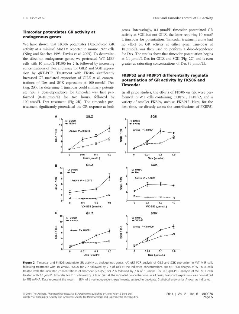
Timcodar potentiates GR activity atendogenous genes
We have shown that FK506 potentiates Dex-induced GR
activity at a minimal MMTV reporter in mouse L929 cells
(Ning and Sanchez 1993; Davies et al. 2005). To determine
the effect on endogenous genes, we pretreated WT MEF
cells with 10 lmol/L FK506 for 2 h, followed by increasing
concentrations of Dex and assay for GILZ and SGK expres-
sion by qRT-PCR. Treatment with FK506 significantly
increased GR-mediated expression of GILZ at all concen-
trations of Dex and SGK expression at 100 nmol/L Dex
(Fig. 2A). To determine if timcodar could similarly potenti-
ate GR, a dose-dependence for timcodar was first per-
formed (0–10 lmol/L) for two hours, followed by
100 nmol/L Dex treatment (Fig. 2B). The timcodar pre-
treatment significantly potentiated the GR response at both
genes. Interestingly, 0.1 lmol/L timcodar potentiated GR
activity at SGK but not GILZ, the latter requiring 10 lmol/
L timcodar for potentiation. Timcodar treatment alone had
no effect on GR activity at either gene. Timcodar at
10 lmol/L was then used to perform a dose-dependence
for Dex. The results show that timcodar potentiation begins
at 0.1 lmol/L Dex for GILZ and SGK (Fig. 2C) and is even
greater at saturating concentrations of Dex (1 lmol/L).
FKBP52 and FKBP51 differentially regulatepotentiation of GR activity by FK506 andTimcodar
In all prior studies, the effects of FK506 on GR were per-
formed in WT cells containing FKBP51, FKBP52, and a
variety of smaller FKBPs, such as FKBP12. Here, for the
first time, we directly assess the contributions of FKBP51
A
B
C
Figure 2. Timcodar and FK506 potentiate GR activity at endogenous genes. (A) qRT-PCR analysis of GILZ and SGK expression in WT MEF cells
following treatment with 10 lmol/L FK506 for 2 h followed by 2 h of Dex at the indicated concentrations. (B) qRT-PCR analysis of WT MEF cells
treated with the indicated concentrations of timcodar (VX-853) for 2 h followed by 2 h of 1 lmol/L Dex. (C) qRT-PCR analysis of WT MEF cells
treated with 10 lmol/L timcodar for 2 h followed by 2 h of Dex at the indicated concentrations. In all cases, transcript expression was normalized
to 18S mRNA. Data represent the mean � SEM of three independent experiments, assayed in duplicate. Statistical analysis by Anova, as indicated.
ª 2014 The Authors. Pharmacology Research & Perspectives published by John Wiley & Sons Ltd,British Pharmacological Society and American Society for Pharmacology and Experimental Therapeutics.
2014 | Vol. 2 | Iss. 6 | e00076Page 5
T. D. Hinds et al. FKBP and Timcodar Control of GR Activity
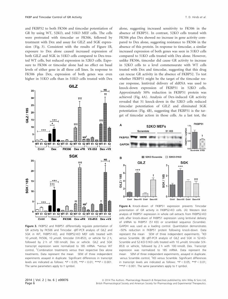
and FKBP52 to both FK506 and timcodar potentiation of
GR by using WT, 52KO, and 51KO MEF cells. The cells
were pretreated with timcodar or FK506, followed by
treatment with Dex and assay for GILZ and SGK expres-
sion (Fig. 3). Consistent with the results of Figure 1B,
exposure to Dex alone caused increased expression of
both GILZ and SGK in 51KO cells compared to Dex-trea-
ted WT cells, but reduced expression in 52KO cells. Expo-
sure to FK506 or timcodar alone had no effect on basal
levels of either gene in all three cell lines. In response to
FK506 plus Dex, expression of both genes was even
higher in 51KO cells than in 51KO cells treated with Dex
alone, suggesting increased sensitivity to FK506 in the
absence of FKBP51. In contrast, 52KO cells treated with
FK506 plus Dex showed no increase in gene activity com-
pared to Dex alone, suggesting resistance to FK506 in the
absence of this protein. In response to timcodar, a similar
increased expression of both genes was seen in 51KO cells
compared to 51KO cells treated with Dex alone. However,
unlike FK506, timcodar did cause GR activity to increase
in 52KO cells to a level commensurate with WT cells
treated with Dex and timcodar, suggesting that this drug
can rescue GR activity in the absence of FKBP52. To test
whether FKBP51 might be the target of the timcodar res-
cue response, lentiviral delivery of shRNA was used to
knock-down expression of FKBP51 in 52KO cells.
Approximately 50% reduction in FKBP51 protein was
achieved (Fig. 4A). Analysis of Dex-induced GR activity
revealed that 51 knock-down in the 52KO cells reduced
timcodar potentiation of GILZ and eliminated SGK
potentiation (Fig. 4B), suggesting that FKBP51 is the tar-
get of timcodar action in those cells. As a last test, the
Figure 3. FKBP52 and FKBP51 differentially regulate potentiation of
GR activity by FK506 and Timcodar. qRT-PCR analysis of GILZ and
SGK in WT, FKBP51-KO, and FKBP52-KO MEF cells treated with
10 lmol/L FK506, 10 lmol/L timcodar (VX-853), or vehicle for 2 h,
followed by 2 h of 100 nmol/L Dex or vehicle. GILZ and SGK
transcript expression were normalized to 18S mRNA. *versus WT
control, †Combination treatments versus their respective Dex alone
treatments. Data represent the mean � SEM of three independent
experiments assayed in duplicate. Significant differences in transcript
levels are indicated as follows: *P < 0.05; **P < 0.01; ***P < 0.001.
The same parameters apply to † symbol.
A
B
Figure 4. Knock-down of FKBP51 expression prevents Timcodar
potentiation of GR activity in FKBP52-KO cells. (A) Western blot
analysis of FKBP51 expression in whole cell extracts from FKBP52-KO
cells after knock-down of FKBP51 expression using lentiviral delivery
of shRNA to FKBP51 (51 KD) or scrambled sequence (Scramble).
GAPDH was used as a loading control. Quantitation demonstrates
~50% reduction in FKBP51 protein following knock-down. Data
represent the mean � SEM of three independent experiments. †KD
versus Scramble. (B) qRT-PCR analysis of GILZ and SGK in 52-KO-
Scramble and 52-KO-51KD cells treated with 10 lmol/L timcodar (VX-
853) or vehicle, followed by 2 h with 100 nmol/L Dex. Transcript
expression was normalized to 18S mRNA. Data represent the
mean � SEM of three independent experiments, assayed in duplicate.
versus Scramble control, †KD versus Scramble. Significant differences
in transcript levels are indicated as follows: *P < 0.05; **P < 0.01;
***P < 0.001. The same parameters apply to † symbol.
2014 | Vol. 2 | Iss. 6 | e00076Page 6
ª 2014 The Authors. Pharmacology Research & Perspectives published by John Wiley & Sons Ltd,
British Pharmacological Society and American Society for Pharmacology and Experimental Therapeutics.
FKBP and Timcodar Control of GR Activity T. D. Hinds et al.
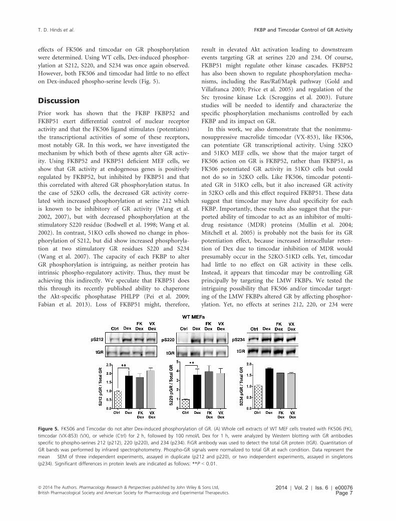
effects of FK506 and timcodar on GR phosphorylation
were determined. Using WT cells, Dex-induced phosphor-
ylation at S212, S220, and S234 was once again observed.
However, both FK506 and timcodar had little to no effect
on Dex-induced phospho-serine levels (Fig. 5).
Discussion
Prior work has shown that the FKBP FKBP52 and
FKBP51 exert differential control of nuclear receptor
activity and that the FK506 ligand stimulates (potentiates)
the transcriptional activities of some of these receptors,
most notably GR. In this work, we have investigated the
mechanism by which both of these agents alter GR activ-
ity. Using FKBP52 and FKBP51 deficient MEF cells, we
show that GR activity at endogenous genes is positively
regulated by FKBP52, but inhibited by FKBP51 and that
this correlated with altered GR phosphorylation status. In
the case of 52KO cells, the decreased GR activity corre-
lated with increased phosphorylation at serine 212 which
is known to be inhibitory of GR activity (Wang et al.
2002, 2007), but with decreased phosphorylation at the
stimulatory S220 residue (Bodwell et al. 1998; Wang et al.
2002). In contrast, 51KO cells showed no change in phos-
phorylation of S212, but did show increased phosphoryla-
tion at two stimulatory GR residues S220 and S234
(Wang et al. 2007). The capacity of each FKBP to alter
GR phosphorylation is intriguing, as neither protein has
intrinsic phospho-regulatory activity. Thus, they must be
achieving this indirectly. We speculate that FKBP51 does
this through its recently published ability to chaperone
the Akt-specific phosphatase PHLPP (Pei et al. 2009;
Fabian et al. 2013). Loss of FKBP51 might, therefore,
result in elevated Akt activation leading to downstream
events targeting GR at serines 220 and 234. Of course,
FKBP51 might regulate other kinase cascades. FKBP52
has also been shown to regulate phosphorylation mecha-
nisms, including the Ras/Raf/Mapk pathway (Gold and
Villafranca 2003; Price et al. 2005) and regulation of the
Src tyrosine kinase Lck (Scroggins et al. 2003). Future
studies will be needed to identify and characterize the
specific phosphorylation mechanisms controlled by each
FKBP and its impact on GR.
In this work, we also demonstrate that the nonimmu-
nosuppressive macrolide timcodar (VX-853), like FK506,
can potentiate GR transcriptional activity. Using 52KO
and 51KO MEF cells, we show that the major target of
FK506 action on GR is FKBP52, rather than FKBP51, as
FK506 potentiated GR activity in 51KO cells but could
not do so in 52KO cells. Like FK506, timcodar potenti-
ated GR in 51KO cells, but it also increased GR activity
in 52KO cells and this effect required FKBP51. These data
suggest that timcodar may have dual specificity for each
FKBP. Importantly, these results also suggest that the pur-
ported ability of timcodar to act as an inhibitor of multi-
drug resistance (MDR) proteins (Mullin et al. 2004;
Mitchell et al. 2005) is probably not the basis for its GR
potentiation effect, because increased intracellular reten-
tion of Dex due to timcodar inhibition of MDR would
presumably occur in the 52KO-51KD cells. Yet, timcodar
had little to no effect on GR activity in these cells.
Instead, it appears that timcodar may be controlling GR
principally by targeting the LMW FKBPs. We tested the
intriguing possibility that FK506 and/or timcodar target-
ing of the LMW FKBPs altered GR by affecting phosphor-
ylation. Yet, no effects at serines 212, 220, or 234 were
Figure 5. FK506 and Timcodar do not alter Dex-induced phosphorylation of GR. (A) Whole cell extracts of WT MEF cells treated with FK506 (FK),
timcodar (VX-853) (VX), or vehicle (Ctrl) for 2 h, followed by 100 nmol/L Dex for 1 h, were analyzed by Western blotting with GR antibodies
specific to phospho-serines 212 (p212), 220 (p220), and 234 (p234). FiGR antibody was used to detect the total GR protein (tGR). Quantitation of
GR bands was performed by infrared spectrophotometry. Phospho-GR signals were normalized to total GR at each condition. Data represent the
mean � SEM of three independent experiments, assayed in duplicate (p212 and p220), or two independent experiments, assayed in singletons
(p234). Significant differences in protein levels are indicated as follows: **P < 0.01.
ª 2014 The Authors. Pharmacology Research & Perspectives published by John Wiley & Sons Ltd,British Pharmacological Society and American Society for Pharmacology and Experimental Therapeutics.
2014 | Vol. 2 | Iss. 6 | e00076Page 7
T. D. Hinds et al. FKBP and Timcodar Control of GR Activity
seen with either ligand. Thus, it appears that unliganded
FKBP51 and FKBP52 can affect phosphorylation pathways
in a way that is unaffected by FK506 or timcodar.
Although it is possible that FK506 or timcodar are target-
ing other phospho-residues on the GR, our results are
consistent with several published facts. First, Smith and
colleagues have shown that FKBP52 stimulation of GR is
completely independent of its PPIase activity, as enzymat-
ically null mutants still potentiated GR activity (Riggs
et al. 2007). Thus, the PPIase function of LMW FKBPs
may also be inconsequential to regulation of phosphoryla-
tion cascades. Second, we have shown that FK506
increases GR hormone-binding affinity (Ning and Sanchez
1995; Davies et al. 2005), yet loss of FKBP52 had no
effect on this function (Wolf et al. 2009), further indicat-
ing that FK506 binding to FKBP52 is a gain-of-function
phenomenon when it comes to control of the hormone-
binding function. Taken as a whole, these observations
suggest that FKBPs and their cognate ligands can exert a
multifactorial control over GR transcriptional activity
through FKBP-dependent regulation of phosphorylation
and FK506-dependent regulation of the hormone-binding
event.
This is the first study to demonstrate that timcodar can
potentiate GR activity, similar to FK506. However, unlike
FK506, the timcodar mechanism cannot involve the
FKBP12/calcineurin pathway responsible for immunosup-
pression. Indeed, in this work we also show that GR
potentiation by FK506 is independent of FKBP12/calci-
neurin, as FK506 stimulation of GR was lost in FKBP52-
KO cells. Although several non-FKBP12 ligands have been
shown to be useful in neuroregeneration, without the
immunosuppression that occurs with FK506 (Gold et al.
1999), drugs that increase nerve regeneration by specifi-
cally targeting the LMW FKBPs have not been developed.
Timcodar can now be viewed as such a candidate and
may serve as the basis for drug design targeting the neu-
roprotective properties of LMW FKBPs. It is interesting
to note that timcodar dimesylate has already been shown
to be an orally bioavailable ligand protective against
experimentally induced neuropathies in several rodent
models, such as pyridoxine-induced nerve damage and
streptozotocin-induced type 1 diabetes (Cole et al. 2000).
However, timcodar dimesylate has not been effective in
two preclinical human trials utilizing capsaicin-induced
nerve injury (Polydefkis et al. 2006) or intracutaneous
axotomy (Hahn et al. 2006). This discrepancy may be
attributable to many factors, such as use of healthy indi-
viduals in the human trials, the nature of the neurotrau-
ma, or an inability of timcodar to target human FKBP
cognates. Indeed, Hausch and colleagues have shown that
timcodar does not bind purified FK1 (PPIase domain)
fragments of human FKBP51 or FKBP52 (Gopalakrishnan
et al. 2012), suggesting that timcodar is inactive in human
FKBPs, or that it targets other regions of the proteins,
such as the FK2/PPIase-like domain. Further and more
complete in vitro binding studies using mouse LMW
FKBPs, as well as the development of timcodar derivatives
will be necessary to address these issues.
Another potential benefit of our study is reflected in
the ability of timcodar to enhance glucocorticoid signal-
ing in the absence of FKBP52 by targeting FKBP51.
Timcodar may, therefore, be a potential therapy for dis-
ease states in which FKBP52 is suppressed or absent. Our
laboratory and others have shown that KO of FKBP52 in
mice results in several pathological conditions, such as
altered signaling of androgen receptor leading to hypospa-
dias and reduced male fertility (Cheung-Flynn et al. 2005;
Yong et al. 2007), reduced progesterone receptor activity
leading to defective uterine implantation (Tranguch et al.
2005; Yang et al. 2006), and reduced GR activity lead-
ing to increased susceptibility to diet-induced steatosis
(Warrier et al. 2010). Finally, a study by Manabe et al.
(2002) demonstrated that FKBP52, but not FKBP12, was
downregulated in the pathogenesis of early-stage amyo-
trophic lateral sclerosis.
In summary, these studies identify an important new
mechanism by which FKBP51 and FKBP52 regulate ste-
roid receptor action, namely, phosphorylation. Although
this mechanism is likely to be indirect (through regula-
tion of phosphorylation cascades), its further investiga-
tion may uncover the basis for FKBP reciprocal
modulation of important glucocorticoid-mediated cellular
responses, such as apoptosis, immunosuppression, and
control of lipid and carbohydrate metabolism. Indeed,
our other finding in this work that timcodar, like FK506,
can potentiate GR activity, suggests that these com-
pounds may be useful in combination therapy with
glucocorticoids for the purposes of immunosuppression,
such as in organ transplantation, or for treatment of
inflammatory conditions. At the very least, FK506 and/or
timcodar may allow for lower effective glucocorticoid
dosing, thus helping to ameliorate the steroid’s many
side-effects.
Acknowledgements
The authors are especially grateful to Bruce G. Gold for
providing timcodar (VX-853) and for guidance during the
initial phases of experimental design. The authors are also
indebted to Michael J. Garabedian (New York University
School of Medicine) for the gift of phospho-specific anti-
bodies to GR. We also thank Steven Selman (Chair,
Department of Urology, University of Toledo College of
Medicine) for critical reading of the manuscript and for
technical and financial support.
2014 | Vol. 2 | Iss. 6 | e00076Page 8
ª 2014 The Authors. Pharmacology Research & Perspectives published by John Wiley & Sons Ltd,
British Pharmacological Society and American Society for Pharmacology and Experimental Therapeutics.
FKBP and Timcodar Control of GR Activity T. D. Hinds et al.
Disclosures
None declared.
References
Babine RE, Villafranca JE, Gold BG (2005). FKBP
immunophilin patents for neurological disorders. Expert Opin
Ther Pat 15: 555–573.
Bodwell JE, Hu JM, Orti E, Munck A (1995).
Hormone-induced hyperphosphorylation of specific
phosphorylated sites in the mouse glucocorticoid receptor. J
Steroid Biochem Mol Biol 52: 135–140.
Bodwell JE, Webster JC, Jewell CM, Cidlowski JA, Hu JM,
Munck A (1998). Glucocorticoid receptor phosphorylation:
overview, function and cell cycle-dependence. J Steroid
Biochem Mol Biol 65: 91–99.
Bruner KL, Derfoul A, Robertson NM, Guerriero G,
Fernandes-Alnemri T, Alnemri ES, et al. (1997). The
unliganded mineralocorticoid receptor is associated with heat
shock proteins 70 and 90 and the immunophilin FKBP-52.
Recept Signal Transduct 7: 85–98.
Cheung-Flynn J, Prapapanich V, Cox MB, Riggs DL,
Suarez-Quian C, Smith DF (2005). Physiological role for the
cochaperone FKBP52 in androgen receptor signaling. Mol
Endocrinol 19: 1654–1666.
Clipstone NA, Crabtree GR (1992). Identification of
calcineurin as a key signalling enzyme in T-lymphocyte
activation. Nature 357: 695–697.
Cole DG, Ogenstad S, Chaturvedi P, (2000). Pharmacological
activities of neurophilin ligands. Pp. 109–116 in B. G. Gold, G.
Fischer and T. Herdegen, eds. Immunophilins in the brain
FKBP-ligands: novel strategies for the treatment of
neurodegenerative disorders. Prous Science, Barcelona, Spain.
Davies TH, Ning YM, Sanchez ER (2002). A new first step in
activation of steroid receptors: hormone-induced switching of
FKBP51 and FKBP52 immunophilins. J Biol Chem 277: 4597–
4600.
Davies TH, Ning YM, Sanchez ER (2005). Differential control
of glucocorticoid receptor hormone-binding function by
tetratricopeptide repeat (TPR) proteins and the
immunosuppressive ligand FK506. Biochemistry 44: 2030–2038.
Denny WB, Valentine DL, Reynolds PD, Smith DF, Scammell
JG (2000). Squirrel monkey immunophilin FKBP51 is a potent
inhibitor of glucocorticoid receptor binding. Endocrinology
141: 4107–4113.
Fabian AK, Marz A, Neimanis S, Biondi RM, Kozany C,
Hausch F (2013). InterAKTions with FKBPs–mutational and
pharmacological exploration. PLoS ONE 8: e57508.
Gold BG (1997). FK506 and the role of immunophilins in
nerve regeneration. Mol Neurobiol 15: 285–306.
Gold BG, Villafranca JE (2003). Neuroimmunophilin ligands:
the development of novel neuroregenerative/neuroprotective
compounds. Curr Top Med Chem 3: 1368–1375.
Gold BG, Densmore V, Shou W, Matzuk MM, Gordon HS
(1999). Immunophilin FK506-binding protein 52 (not
FK506-binding protein 12) mediates the neurotrophic action
of FK506. J Pharmacol Exp Ther 289: 1202–1210.
Gold BG, Armistead DM, Wang MS (2005).
Non-FK506-binding protein-12 neuroimmunophilin ligands
increase neurite elongation and accelerate nerve regeneration. J
Neurosci Res 80: 56–65.
Gopalakrishnan R, Kozany C, Gaali S, Kress C, Hoogeland B,
Bracher A, et al. (2012). Evaluation of synthetic FK506
analogues as ligands for the FK506-binding proteins 51 and
52. J Med Chem 55: 4114–4122.
Hahn K, Sirdofsky M, Brown A, Ebenezer G, Hauer P, Miller
C, et al. (2006). Collateral sprouting of human epidermal
nerve fibers following intracutaneous axotomy. J Peripher Nerv
Syst 11: 142–147.
Harding MW (2003). Immunophilins, mTOR, and
pharmacodynamic strategies for a targeted cancer therapy. Clin
Cancer Res 9: 2882–2886.
Hinds TDJ, Ramakrishnan S, Cash HA, Stechschulte LA,
Heinrich G, Najjar SM, et al. (2010). Discovery of
glucocorticoid receptor-beta in mice with a role in
metabolism. Mol Endocrinol 24: 1715–1727.
Hinds TDJ, Stechschulte LA, Cash HA, Whisler D, Banerjee A,
Yong W, et al. (2011). Protein phosphatase 5 mediates lipid
metabolism through reciprocal control of glucocorticoid
receptor and peroxisome proliferator-activated
receptor-gamma (PPARgamma). J Biol Chem 286:
42911–42922.
Ismaili N, Garabedian MJ (2004). Modulation of
glucocorticoid receptor function via phosphorylation. Ann N Y
Acad Sci 1024: 86–101.
MacMillan D (2013). FK506 binding proteins: cellular
regulators of intracellular Ca2+ signalling. Eur J Pharmacol
700: 181–193.
Manabe Y, Warita H, Murakami T, Shiote M, Hayashi T,
Omori N, et al. (2002). Early decrease of the
immunophilin FKBP 52 in the spinal cord of a transgenic
model for amyotrophic lateral sclerosis. Brain Res 935:
124–128.
Mitchell AM, Tom M, Mortimer RH (2005). Thyroid
hormone export from cells: contribution of P-glycoprotein. J
Endocrinol 185: 93–98.
Mullin S, Mani N, Grossman TH (2004). Inhibition of
antibiotic efflux in bacteria by the novel multidrug resistance
inhibitors biricodar (VX-710) and timcodar (VX-853).
Antimicrob Agents Chemother 48: 4171–4176.
ª 2014 The Authors. Pharmacology Research & Perspectives published by John Wiley & Sons Ltd,British Pharmacological Society and American Society for Pharmacology and Experimental Therapeutics.
2014 | Vol. 2 | Iss. 6 | e00076Page 9
T. D. Hinds et al. FKBP and Timcodar Control of GR Activity
Ning YM, Sanchez ER (1993). Potentiation of glucocorticoid
receptor-mediated gene expression by the immunophilin
ligands FK506 and rapamycin. J Biol Chem 268: 6073–6076.
Ning YM, Sanchez ER (1995). Stabilization in vitro of the
untransformed glucocorticoid receptor complex of S49
lymphocytes by the immunophilin ligand FK506. J Steroid
Biochem Mol Biol 52: 187–194.
Owens-Grillo JK, Czar MJ, Hutchison KA, Hoffmann K,
Perdew GH, Pratt WB (1996). A model of protein targeting
mediated by immunophilins and other proteins that bind to
hsp90 via tetratricopeptide repeat domains. J Biol Chem 271:
13468–13475.
Pei H, Li L, Fridley BL, Jenkins GD, Kalari KR, Lingle W,
et al. (2009). FKBP51 affects cancer cell response to
chemotherapy by negatively regulating Akt. Cancer Cell 16:
259–266.
Polydefkis M, Sirdofsky M, Hauer P, Petty BG, Murinson B,
McArthur JC (2006). Factors influencing nerve regeneration in
a trial of timcodar dimesylate. Neurology 66: 259–261.
Price RD, Yamaji T, Yamamoto H, Higashi Y, Hanaoka K,
Yamazaki S, et al. (2005). FK1706, a novel
non-immunosuppressive immunophilin: neurotrophic activity
and mechanism of action. Eur J Pharmacol 509: 11–19.
Reynolds PD, Ruan Y, Smith DF, Scammell JG (1999).
Glucocorticoid resistance in the squirrel monkey is associated
with overexpression of the immunophilin FKBP51. J Clin
Endocrinol Metab 84: 663–669.
Riggs DL, Cox MB, Tardif HL, Hessling M, Buchner J, Smith
DF (2007). Noncatalytic role of the FKBP52 peptidyl-prolyl
isomerase domain in the regulation of steroid hormone
signaling. Mol Cell Biol 27: 8658–8669.
Sanchez ER (1990). Hsp56: a novel heat shock protein
associated with untransformed steroid receptor complexes. J
Biol Chem 265: 22067–22070.
Sanchez ER (2012). Chaperoning steroidal physiology: lessons
from mouse genetic models of Hsp90 and its cochaperones.
Biochim Biophys Acta 1823: 722–729.
Scroggins BT, Prince T, Shao J, Uma S, Huang W, Guo Y,
et al. (2003). High affinity binding of Hsp90 is triggered by
multiple discrete segments of its kinase clients. Biochemistry
42: 12550–12561.
Smith DF, Albers MW, Schreiber SL, Leach KL, Deibel MR Jr
(1993a). FKBP54, a novel FK506-binding protein in avian
progesterone receptor complexes and HeLa extracts. J Biol
Chem 268: 24270–24273.
Smith DF, Baggenstoss BA, Marion TN, Rimerman RA (1993b).
Two FKBP-related proteins are associated with progesterone
receptor complexes. J Biol Chem 268: 18365–18371.
Snyder SH, Sabatini DM (1995). Immunophilins and the
nervous system. Nat Med 1: 32–37.
Storer CL, Dickey CA, Galigniana MD, Rein T, Cox MB
(2011). FKBP51 and FKBP52 in signaling and disease. Trends
Endocrinol Metab 22: 481–490.
Tai PK, Maeda Y, Nakao K, Wakim NG, Duhring JL, Faber LE
(1986). A 59-kilodalton protein associated with progestin,
estrogen, androgen, and glucocorticoid receptors. Biochemistry
25: 5269–5275.
Tranguch S, Cheung-Flynn J, Daikoku T, Prapapanich V, Cox
MB, Xie H, et al. (2005). Cochaperone immunophilin FKBP52
is critical to uterine receptivity for embryo implantation. Proc
Natl Acad Sci USA 102: 14326–14331.
Wang Z, Frederick J, Garabedian MJ (2002). Deciphering the
phosphorylation “code” of the glucocorticoid receptor in vivo.
J Biol Chem 277: 26573–26580.
Wang Z, Chen W, Kono E, Dang T, Garabedian MJ
(2007). Modulation of glucocorticoid receptor
phosphorylation and transcriptional activity by a
C-terminal-associated protein phosphatase. Mol Endocrinol
21: 625–634.
Warrier M, Hinds TDJ, Ledford KJ, Cash HA, Patel PR,
Bowman TA, et al. (2010). Susceptibility to diet-induced
hepatic steatosis and glucocorticoid resistance in
FK506-binding protein 52-deficient mice. Endocrinology 151:
3225–3236.
Webster JC, Jewell CM, Bodwell JE, Munck A, Sar
M, Cidlowski JA (1997). Mouse glucocorticoid
receptor phosphorylation status influences multiple
functions of the receptor protein. J Biol Chem 272:
9287–9293.
Weigel NL, Moore NL (2007). Steroid receptor
phosphorylation: a key modulator of multiple receptor
functions. Mol Endocrinol 21: 2311–2319.
Wolf IM, Periyasamy S, Hinds TD Jr, Yong W, Shou W,
Sanchez ER (2009). Targeted ablation reveals a novel role of
FKBP52 in gene-specific regulation of glucocorticoid receptor
transcriptional activity. J Steroid Biochem Mol Biol 113:
36–45.
Yang Z, Wolf IM, Chen H, Periyasamy S, Chen Z, Yong
W, et al. (2006). FK506-binding protein 52 is essential to
uterine reproductive physiology controlled by the
progesterone receptor a isoform. Mol Endocrinol 20:
2682–2694.
Yem AW, Tomasselli AG, Heinrikson RL, Zurcher-Neely H,
Ruff VA, Johnson RA, et al. (1992). The Hsp56 component of
steroid receptor complexes binds to immobilized FK506 and
shows homology to FKBP-12 and FKBP-13. J Biol Chem 267:
2868–2871.
Yong W, Yang Z, Periyasamy S, Chen H, Yucel S, Li W, et al.
(2007). Essential role for Co-chaperone Fkbp52 but not
Fkbp51 in androgen receptor-mediated signaling and
physiology. J Biol Chem 282: 5026–5036.
2014 | Vol. 2 | Iss. 6 | e00076Page 10
ª 2014 The Authors. Pharmacology Research & Perspectives published by John Wiley & Sons Ltd,
British Pharmacological Society and American Society for Pharmacology and Experimental Therapeutics.
FKBP and Timcodar Control of GR Activity T. D. Hinds et al.
Supporting Information
Additional Supporting Information may be found in the
online version of this article:
Figure S1. Comparison of FK506 and VX-853 chemical
structures.
Figure S2. Demonstration of no significant change in
FKBP51 expression in 52KO MEF cells. These data show
Western blots of FKBP51 from three independent experi-
ments in which rescue (ResQ) expression of Flag-tagged
FKBP51 and Flag-tagged FKBP52 was being tested in each
cell line. Normal (unrescued) WT, 51KO, and 52KO
MEFs cells were used as controls. Blots of Flag-tagged
proteins not shown, as rescue expressions were not suc-
cessful and were not relevant to the manuscript. Quanti-
tation of FKBP51 from these blots (WT, 51KO, and
52KO lanes only) and from the blot of Figure 1A were
compiled and reported in the text under Results.
ª 2014 The Authors. Pharmacology Research & Perspectives published by John Wiley & Sons Ltd,British Pharmacological Society and American Society for Pharmacology and Experimental Therapeutics.
2014 | Vol. 2 | Iss. 6 | e00076Page 11
T. D. Hinds et al. FKBP and Timcodar Control of GR Activity