Analysis of facial soft tissue changes with aging and their effects on facial morphology: A forensic...
11
ORIGINAL ARTICLE Analysis of facial soft tissue changes with aging and their effects on facial morphology: A forensic perspective Manavpreet Kaur a , Rakesh K. Garg a, * , Sanjeev Singla b a Department of Forensic Science, Punjabi University, Patiala 147002, India b Central Forensic Institute, Bhopal, Madhya Pradesh, India Received 5 December 2013; revised 7 May 2014; accepted 8 July 2014 Available online 11 August 2014 KEYWORDS Forensic; Aging; Soft-tissue thickness; Rhytids; Senescence; Face Abstract While working on the concept of employing human faces as a biometric tool for personal identification, it is common to come across certain hindrances such as illumination, pose variation and facial hair. But, dealing with the aging process of an individual has been generally overlooked until recently. This relatively untouched aspect may enable us more insight while tackling identifi- cation problems. As the face matures, it changes some of its most enduring properties (e.g., shape of the cranium) and acquires new attributes (e.g., wrinkles). These changes are the basis of information about the aging of the face. The human brain can analyze the face and estimate the approximate age of an individual, though this estimation is not accurate. The perception of age in the human brain is still a subject of research. This article takes the phenomenon of ‘‘aging of face’’ into consideration and following the same, this study has been carried out to analyze the kind of changes occurring in the facial soft tissue thickness with progression of age and can be used along with other biological markers for personal identification or in developing automatic facial age estimation. The data can be used as an additional feature for corroboration or authentication in individualization. This pre- liminary study will help in forensic investigation although a database needs to be generated on other populations. ª 2015 The International Association of Law and Forensic Sciences (IALFS). Production and hosting by Elsevier B.V. All rights reserved. 1. Introduction The features of a human face, facial components, as well as the human face taken as a whole, act as a biometric tool for purposes of individual human identification. With the advancements in technology and with the introduction of bio- metric systems for the purpose of identification, automated face recognition (FR) systems have been created to match indi- vidual faces from print and digital photographs and video to the faces of an individual with the reference image in a com- puter database or with the photograph of the suspect. One of the main problems which is addressed with the facial recog- nition systems is the change occurring in the face with time or * Corresponding author. Tel.: +91 175 3046271/3046272. E-mail address: [email protected] (R.K. Garg). Peer review under responsibility of The International Association of Law and Forensic Sciences (IALFS). Egyptian Journal of Forensic Sciences (2015) 5, 46–56 HOSTED BY Contents lists available at ScienceDirect Egyptian Journal of Forensic Sciences journal homepage: http://www.journals.elsevier.com/egyptian-journal-of-forensic-sciences http://dx.doi.org/10.1016/j.ejfs.2014.07.006 2090-536X ª 2015 The International Association of Law and Forensic Sciences (IALFS). Production and hosting by Elsevier B.V. All rights reserved.
-
Upload
independent -
Category
Documents
-
view
2 -
download
0
Transcript of Analysis of facial soft tissue changes with aging and their effects on facial morphology: A forensic...
Egyptian Journal of Forensic Sciences (2015) 5, 46–56
HO ST E D BY Contents lists available at ScienceDirect
Egyptian Journal of Forensic Sciences
journal homepage: http://www.journals.elsevier.com/egyptian-journal-of-forensic-sciences
ORIGINAL ARTICLE
Analysis of facial soft tissue changes with aging and
their effects on facial morphology: A forensic
perspective
* Corresponding author. Tel.: +91 175 3046271/3046272.
E-mail address: [email protected] (R.K. Garg).
Peer review under responsibility of The International Association of
Law and Forensic Sciences (IALFS).
http://dx.doi.org/10.1016/j.ejfs.2014.07.0062090-536X ª 2015 The International Association of Law and Forensic Sciences (IALFS). Production and hosting by Elsevier B.V. Areserved.
Manavpreet Kaura, Rakesh K. Garg
a,*, Sanjeev Singlab
a Department of Forensic Science, Punjabi University, Patiala 147002, Indiab Central Forensic Institute, Bhopal, Madhya Pradesh, India
Received 5 December 2013; revised 7 May 2014; accepted 8 July 2014
Available online 11 August 2014
KEYWORDS
Forensic;
Aging;
Soft-tissue thickness;
Rhytids;
Senescence;
Face
Abstract While working on the concept of employing human faces as a biometric tool for personal
identification, it is common to come across certain hindrances such as illumination, pose variation
and facial hair. But, dealing with the aging process of an individual has been generally overlooked
until recently. This relatively untouched aspect may enable us more insight while tackling identifi-
cation problems. As the face matures, it changes some of its most enduring properties (e.g., shape of
the cranium) and acquires new attributes (e.g., wrinkles). These changes are the basis of information
about the aging of the face. The human brain can analyze the face and estimate the approximate age
of an individual, though this estimation is not accurate. The perception of age in the human brain is
still a subject of research. This article takes the phenomenon of ‘‘aging of face’’ into consideration
and following the same, this study has been carried out to analyze the kind of changes occurring in
the facial soft tissue thickness with progression of age and can be used along with other biological
markers for personal identification or in developing automatic facial age estimation. The data can
be used as an additional feature for corroboration or authentication in individualization. This pre-
liminary study will help in forensic investigation although a database needs to be generated on other
populations.ª 2015 The International Association of Law and Forensic Sciences (IALFS). Production and hosting by
Elsevier B.V. All rights reserved.
1. Introduction
The features of a human face, facial components, as well as thehuman face taken as a whole, act as a biometric tool for
purposes of individual human identification. With the
advancements in technology and with the introduction of bio-metric systems for the purpose of identification, automatedface recognition (FR) systems have been created to match indi-
vidual faces from print and digital photographs and video tothe faces of an individual with the reference image in a com-puter database or with the photograph of the suspect. Oneof the main problems which is addressed with the facial recog-
nition systems is the change occurring in the face with time or
ll rights

Analysis of facial soft tissue changes with aging and their effects on facial morphology: A forensic perspective 47
with aging. As the personal identity credentials such as pass-ports, driving licenses and ration cards are valid for years.The primary problem that arise with automated facial recogni-
tion systems is to match the two photographs of the same indi-vidual from different age groups. Several researchers havehighlighted this temporal performance degradation.1–3 To
meet this challenge, the approach which needs to be followedis to research the adult age-related craniofacial morphologicalchanges to better understand the process of aging individuals
from a facial image, or rather, develop workable artificialage progression techniques to anticipate how an individual’sface may appear after several years have passed.
The appearance of a human face is affected considerably by
aging as shown in a diagrammatical representation in Fig. 1.Albert et al. and Rhodes. Have reported that facial aging ismainly attributed to bone movement and growth, and skin
related deformations associated with the introduction of wrin-kles and the reduction of muscle strength.4,5 Usually bonegrowth takes place during childhood whereas during adult-
hood the most intense age-related deformations are linked withtexture changes. The observation of aging-related features onfaces allows humans to estimate the age of other persons just
by looking at their face. However, researchers studying theprocess of age estimation by humans have concluded thathumans are not so accurate in age estimation and hence thepossibility of developing automatic facial age estimation meth-
ods poses an attractive area of research.5
As adults age, myriad changes occur diachronically withinthe craniofacial complex. Notable soft tissue modifications
can be seen across each decade of adult life that passes.Subtle hard tissue or bony changes slightly alter the overallshape of the human face, mainly in the dentoalveolar region.
These age-related changes affect the accuracy and efficacy ofbiometric techniques concerning automated facial recognition.A lot of researchers worldwide are working on a wide variety
of aspects related to face aging, facial age progression or syn-thetic facial aging, and facial expression, with the goal ofimproving computer automated face recognition systems. Inthis study, we provide an overview of intrinsic and extrinsic
factors that affect human adult craniofacial aging and thedetails of the effects of changing facial soft tissue thicknesswith age and its impact on the overall appearance of the face.
This study will ultimately help in establishing personal identi-fication in forensic cases.
Upper third of the face
Lower third of the face
Middle third of the face
Figure 1 Typical aging skin changes.
2. Materials and methods
2.1. Collection of samples
Four hundred photographs of 400 individuals (200 males and200 females) from four different age groups were collected
from the individuals belonging to a Punjabi Sikh population.The photographs of individuals from age groups: 30–40 years,40–50 years, 50–60 years and 60+ years were collected. The
individuals with head and neck trauma or other pathologicalconditions that could distort the normal facial structures wereexcluded from the present study. Before collecting the samples,the participants were informed about the purpose of collection
of the photographs and their written consent was alsoobtained. The participants in the present study were selectedfrom a residential area in the vicinity of Punjabi University.
The medical history of the participants was checked beforetaking their photographs and the participants who had under-gone some treatment to enhance their looks or appearance
were also excluded from the study.
2.2. Analysis of samples
Various features like – changes in skin texture, appearance ofrhytids and other morphological changes were noticed andrecorded in the form of a table. Their characteristic appearancewas categorized as – none, minimal, fair, marked and
prominent.
� None (Showing 0% signs of aging) – The characteristics
which were totally absent in an individual were classifiedas none.� Minimal (Showing 10–20% signs of aging) – The character-
istics which were present in the form of fine lines were clas-sified as minimal.� Fair (Showing 20–40% signs of aging) – The characteristicswhich were present in the form of deep lines were classified
as fair.� Marked (Showing 50–70% signs of aging) – The character-istics which were present in the form of deep grooves were
classified as marked.� Prominent (Showing 70–100% signs of aging) – The charac-teristics which were present in the form of deep groves and
folds were classified as prominent.
The various features considered for the present study were:
� Transverse forehead rhytids: These are caused by lifting ofthe brows which happens in our normal daily facial anima-tion. Over a lifetime, this results in thinning of the tissues
beneath these lines and ultimately results in wrinkles evenwhen we are not animating. Forehead lines can also becaused by a subconscious effort to raise the brows con-
stantly if they droop with age.� Glabellar frown lines: These are vertical or slightly diagonallines in between the brows that form when we frown or
scowl. Initially they only form on animation, but with timethey can be nearly permanent and very difficult to remove.Frown lines are caused by a very powerful muscle thatmoves the brows toward one another. The stronger the
muscles, the deeper these wrinkles are.

48 M. Kaur et al.
� Brow droop: This refers to either the overall lowering of the
eyebrow position, or the flattening of eyebrows from a gen-tle curve to a flat position.� Flattening and lowering: The tissue of the forehead drifts
inferiorly, creating wrinkles and drooping of the eyebrowsdownward and giving them a flatter appearance.� Brow ridges prominence: Due to the diminution in the fatand with the thinning of underlying tissues, the eyes flatten
and in turn make brow ridges appear prominent.� Sunken temples: A deeper area outside the eye, which maybe accentuated by a very prominent brow bone, sunken
temples are caused often by an age-related loss of volumein this area.� Lateral canthal rhytids: Lateral canthal rhytids are perpen-
dicular to the direction of the lateral muscle fibers and tothe orbital portion of the orbicularis oculi which runs invertical direction around the lateral canthus. These arecaused by the contraction of the lateral side of the orbital
portion of the orbicularis oculi. These rhytids are the resultof in folding and pleating of overlying skin which radiatesaway from the lateral canthus.
� Upper and under eye bags: With aging, the tissues aroundthe eyes, including some muscles supporting the eyelidsweaken, the fat that helps support the eyes then migrates
forward into the lower eyelids and takes the form of bags.� Lower eyelid sunken or hollow: Due to the gradual loss of fatand reduction in underlying muscle fibers, the eyelids
appear sunken or hollow.� Upper eyelid muscle reduction: With growing age there is amarked change in the muscle strength.� Eyelid skin elasticity: The skin loses elasticity with aging
and the stages of the same were observed in the presentstudy.� Dark circles: Due to the loosening of the skin which occurs
because of the reduction in fat and with the muscle fiberreduction, the skin appears darker. It is a characteristic fea-ture in the lower eyelid.
� Eyelid folds: With the reduction in fat and muscle fibers, theskin becomes loose and this loose skin of upper and lowereyelids creates folds.� Ptosis: The drooping or sagging of the eyelid is known as
ptosis.� Nose elongation, tip movement and dorsal hump: With aging,the nasal skin thickens and the ligaments and structures
that hold the tip in its vernal position start to weaken,resulting in dropping of the tip. Along with this, the boneunder the tip and above the teeth (maxilla) loses volume
and that further drops the tip. When the tip drops, it givesthe appearance as if the nose developed a bulge and thatbulge is called dorsal hump.
� Bulbous nose: The nasal tip does drop during the aging pro-cess and becomes more bulbous due to loose cartilage andmuscle reduction around the nose.� Reduction in fat deposition: The effects of aging on morpho-
logical appearance of cheeks are clearly visible because ofthe reduction in fat deposition with growing age.� Malar fat pad shifting and nasolabial folds: The associated
ligaments holding the malar fat pad in place weaken anda nasolabial fold develops along with the malar fat padshift.
� Sagging chin: Skin loosening and muscle loss are normal
effects of aging. When these muscles begin to slacken, andwhen the skin loses tautness and elasticity the chances ofdeveloping a double chin increases. This appearance of a
double chin is called sagging chin.� Cheek hollowness: With aging, there is significant atrophy ofsubcutaneous fat, leading to a loss of volume and decreasedsupport for facial features, as well as a loss of skin elasticity
in turn making the cheeks appear hollow.� Ear elongation, loosening of the ear lobe and an increase in
ear width: With aging, the muscles become loose and carti-
lages become lax in turn affecting the morphologicalappearance of the ear.� Vertical/perioral rhytids: The perioral region ages with time
because of volume loss and bone loss in the maxilla and alsobecause of gravity pulling the cheeks and jowls down. Theupper lip lengthens, the philtrum flattens, Cupid’s bow dis-appears, and the vermilion thins, leaving a flat, thin profile
instead of the nice curvature seen in youth. The upper inci-sors are no longer visible in repose and the vertical rhytidsappear and gradually intensify with time.
� Ornamental groove: These are lines forming at the cornersof the mouth, developing mainly in the 40’s and deepeningas age increases.
� Buccomandibular crease: This is a crease that forms on theside of the cheek above the lower jaw and is oriented verti-cally. It appears in the late 30’s and gradually elevates.
� Lip elongation: Due to the weakening of muscle strength,loss of fat deposition and shifting of the malar pad, the lipsbend downward and elongate with time.� Lip thinning: There is less visibility of the red portion of the
lips when seen from the front with the mouth barely open.Older people, usually have a slight lengthening of the upperlip that causes the red part of the lip to roll backward a bit
and makes it less visible. Older people can also lose a bit ofactual volume in the lips as well.� Jowls: This refers to a small focal accumulation of fat in the
lower cheek overlying the jaw bone.� Marionette lines: These are long vertical lines that laterallycircumscribe the chin. Marionette lines appear with advanc-ing age and some people never get them, depending on
facial structure and anatomy. They tend to appear as theligaments around the mouth and chin relax and begin toloosen and sag, and fatty tissues of the cheek deflate and
descend during the aging process.
The various intrinsic and extrinsic factors causing facial
aging are:
� Intrinsic face aging factors - Intrinsic aging is caused by
internal biological factors. Facial aging is mainly due tothe natural changes that occur as soft tissues lose their elas-ticity, muscle tone, and volume6 as well as the facial boneshape modifications resulting from the lifelong and ongoing
process of cranium remodeling.7,8 Additional factors whichaffect these changes are an individual’s biological sex, eth-nicity, and idiosyncratic features (i.e., features purely
unique to the individual such as hyperdynamic facialexpressions).
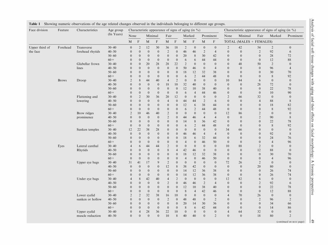
Table 1 Showing numeric observations of the age related changes observed in the individuals belonging to different age groups.
Face division Feature Characteristics Age group
(In Years)
Characteristic appearance of signs of aging (in %) Characteristic appearance of signs of aging (in %)
None Minimal Fair Marked Prominent None Minimal Fair Marked Prominent
M F M F M F M F M F TOTAL (MALES + FEMALES)
Upper third of
the face
Forehead Transverse
forehead rhytids
30–40 0 2 12 30 36 18 2 0 0 0 2 42 54 2 0
40–50 0 0 0 0 2 0 46 46 2 4 0 0 2 92 6
50–60 0 0 0 0 0 0 20 8 30 42 0 0 0 28 72
60+ 0 0 0 0 0 0 6 6 44 44 0 0 0 12 88
Glabellar frown
lines
30–40 0 0 20 28 28 22 2 0 0 0 0 48 50 2 0
40–50 0 0 0 0 0 0 50 46 0 4 0 0 0 96 4
50–60 0 0 0 0 0 0 18 12 32 38 0 0 0 30 70
60+ 0 0 0 0 0 0 6 2 44 48 0 0 0 8 92
Brows Droop 30–40 2 8 44 40 4 2 0 0 0 0 10 84 6 0 0
40–50 0 0 0 0 18 10 32 40 0 0 0 0 28 72 0
50–60 0 0 0 0 0 0 12 10 38 40 0 0 0 22 78
60+ 0 0 0 0 0 0 6 4 44 46 0 0 0 10 90
Flattening and
lowering
30–40 0 2 30 36 20 12 0 0 0 0 2 66 32 0 0
40–50 0 0 0 0 4 0 44 44 2 6 0 0 4 88 8
50–60 0 0 0 0 0 0 12 6 38 44 0 0 0 18 82
60+ 0 0 0 0 0 0 6 2 44 48 0 0 0 8 92
Brow ridges
prominence
30–40 2 10 46 40 2 0 0 0 0 0 12 86 2 0 0
40–50 0 0 0 0 2 0 44 46 4 4 0 0 2 90 8
50–60 0 0 0 0 0 0 14 8 36 42 0 0 0 22 78
60+ 0 0 0 0 0 0 6 2 44 48 0 0 0 8 92
Sunken temples 30–40 12 22 38 28 0 0 0 0 0 0 34 66 0 0 0
40–50 0 0 0 0 0 0 46 46 4 4 0 0 0 92 8
50–60 0 0 0 0 0 0 18 6 32 44 0 0 0 24 76
60+ 0 0 0 0 0 0 4 0 46 50 0 0 0 4 96
Eyes Lateral canthal
Rhytids
30–40 4 6 44 44 2 0 0 0 0 0 10 88 2 0 0
40–50 0 0 0 0 8 4 42 46 0 0 0 0 12 88 0
50–60 0 0 0 0 0 0 18 12 32 38 0 0 0 30 70
60+ 0 0 0 0 0 0 4 0 46 50 0 0 0 4 96
Upper eye bags 30–40 31 41 17 9 2 0 0 0 0 0 72 26 2 0 0
40–50 0 0 0 0 12 8 38 42 0 0 0 0 20 80 0
50–60 0 0 0 0 0 0 14 12 36 38 0 0 0 26 74
60+ 0 0 0 0 0 0 14 12 36 38 0 0 0 26 74
Under eye bags 30–40 4 8 42 40 4 2 0 0 0 0 12 82 6 0 0
40–50 0 0 0 0 2 0 46 46 2 4 0 0 2 92 6
50–60 0 0 0 0 0 0 12 10 38 40 0 0 0 22 78
60+ 0 0 0 0 0 0 8 4 42 46 0 0 0 12 88
Lower eyelid
sunken or hollow
30–40 2 2 32 38 16 10 0 0 0 0 4 70 26 0 0
40–50 0 0 0 0 2 0 48 48 0 2 0 0 2 96 2
50–60 0 0 0 0 0 0 20 14 30 36 0 0 0 34 66
60+ 0 0 0 0 0 0 8 6 42 44 0 0 0 14 86
Upper eyelid
muscle reduction
30–40 0 4 28 36 22 10 0 0 0 0 4 64 32 0 0
40–50 0 0 0 0 10 8 40 40 0 2 0 0 18 80 2
(continued on next page)
Analysis
offacia
lsofttissu
echanges
with
agingandtheir
effectsonfacia
lmorphology:A
foren
sicpersp
ective
49
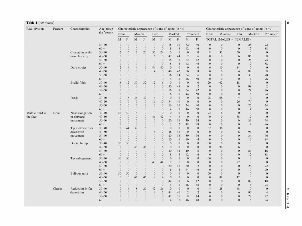
Table 1 (continued)
Face division Feature Characteristics Age group
(In Years)
Characteristic appearance of signs of aging (in %) Characteristic appearance of signs of aging (in %)
None Minimal Fair Marked Prominent None Minimal Fair Marked Prominent
M F M F M F M F M F TOTAL (MALES + FEMALES)
50–60 0 0 0 0 0 0 18 10 32 40 0 0 0 28 72
60+ 0 0 0 0 0 0 8 4 42 46 0 0 0 12 88
Change in eyelid
skin elasticity
30–40 2 6 12 20 36 24 0 0 0 0 8 32 60 0 0
40–50 0 0 0 0 6 0 42 44 2 6 0 0 6 86 8
50–60 0 0 0 0 0 0 18 8 32 42 0 0 0 26 74
60+ 0 0 0 0 0 0 8 4 42 46 0 0 0 12 88
Dark circles 30–40 2 4 4 6 44 40 0 0 0 0 6 10 84 0 0
40–50 0 0 0 0 4 0 46 42 0 8 0 0 4 88 8
50–60 0 0 0 0 0 0 16 14 34 36 0 0 0 30 70
60+ 0 0 0 0 0 0 4 0 46 50 0 0 0 4 96
Eyelid folds 30–40 8 12 30 32 12 6 0 0 0 0 20 62 18 0 0
40–50 0 0 0 0 0 0 50 48 0 2 0 0 0 98 2
50–60 0 0 0 0 0 0 16 8 34 42 0 0 0 24 76
60+ 0 0 0 0 0 0 6 0 44 50 0 0 0 6 94
Ptosis 30–40 10 18 36 32 4 0 0 0 0 0 28 68 4 0 0
40–50 0 0 0 0 16 10 34 40 0 0 0 0 26 74 0
50–60 0 0 0 0 0 0 16 10 34 40 0 0 0 26 74
60+ 0 0 0 0 0 0 4 0 46 50 0 0 0 4 96
Middle third of
the face
Nose Nose elongation
or forward
movement
30–40 42 50 8 0 0 0 0 0 0 0 92 8 0 0 0
40–50 0 0 0 0 46 42 4 8 0 0 0 0 88 12 0
50–60 0 0 0 0 0 0 20 16 30 34 0 0 0 36 64
60+ 0 0 0 0 0 0 2 2 48 48 0 0 0 4 96
Tip movement or
downward
movement
30–40 18 44 32 6 0 0 0 0 0 0 62 38 0 0 0
40–50 0 0 0 0 4 2 46 48 0 0 0 0 6 94 0
50–60 0 0 0 0 0 0 20 14 30 36 0 0 0 34 66
60+ 0 0 0 0 0 0 10 6 40 44 0 0 0 16 84
Dorsal hump 30–40 50 50 0 0 0 0 0 0 0 0 100 0 0 0 0
40–50 0 0 48 46 2 4 0 0 0 0 0 94 6 0 0
50–60 0 0 0 0 0 0 40 44 10 6 0 0 0 84 16
60+ 0 0 0 0 0 0 8 4 42 46 0 0 0 12 88
Tip enlargement 30–40 50 50 0 0 0 0 0 0 0 0 100 0 0 0 0
40–50 0 0 0 0 48 44 2 6 0 0 0 0 92 8 0
50–60 0 0 0 0 0 0 20 18 30 32 0 0 0 28 72
60+ 0 0 0 0 0 0 6 4 44 46 0 0 0 10 90
Bulbous nose 30–40 50 50 0 0 0 0 0 0 0 0 100 0 0 0 0
40–50 0 0 42 46 8 4 0 0 0 0 0 88 12 0 0
50–60 0 0 0 0 0 0 44 38 6 12 0 0 0 82 18
60+ 0 0 0 0 0 0 4 2 46 48 0 0 0 6 94
Cheeks Reduction in fat
deposition
30–40 0 4 8 20 42 26 0 0 0 0 4 28 68 0 0
40–50 0 0 0 0 4 2 44 46 2 2 0 0 6 90 4
50–60 0 0 0 0 0 0 42 36 8 14 0 0 0 78 22
60+ 0 0 0 0 0 0 4 2 46 48 0 0 0 6 94
50
M.Kauret
al.
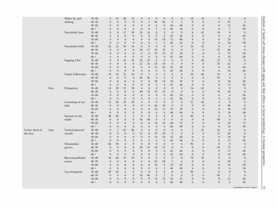
Malar fat pad
shifting
30–40 6 18 44 32 0 0 0 0 0 0 24 76 0 0 0
40–50 0 0 0 0 4 2 46 46 0 2 0 0 6 92 2
50–60 0 0 0 0 0 0 6 6 44 44 0 0 0 12 88
60+ 0 0 0 0 0 0 2 0 48 50 0 0 0 2 98
Nasolabial lines 30–40 0 4 12 30 38 16 0 0 0 0 4 42 54 0 0
40–50 0 0 0 0 8 0 0 4 42 46 0 0 8 4 88
50–60 0 0 0 0 0 0 18 10 32 40 0 0 0 28 72
60+ 0 0 0 0 0 0 2 8 48 42 0 0 0 10 90
Nasolabial folds 30–40 26 32 24 18 0 0 0 0 0 0 58 42 0 0 0
40–50 0 0 0 0 20 12 30 38 0 0 0 0 32 68 0
50–60 0 0 0 0 0 0 18 14 32 36 0 0 0 32 68
60+ 0 0 0 0 0 0 8 6 42 44 0 0 0 14 86
Sagging Chin 30–40 0 4 16 28 34 18 0 0 0 0 4 44 52 0 0
40–50 0 0 0 0 22 16 28 34 0 0 0 0 38 62 0
50–60 0 0 0 0 0 0 12 20 38 30 0 0 0 32 68
60+ 0 0 0 0 0 0 8 6 42 44 0 0 0 14 86
Cheek hollowness 30–40 10 14 32 34 8 2 0 0 0 0 24 66 10 0 0
40–50 0 0 0 0 46 46 4 4 0 0 0 0 92 8 0
50–60 0 0 0 0 0 0 20 14 30 36 0 0 0 34 66
60+ 0 0 0 0 0 0 8 4 42 46 0 0 0 12 88
Ears Elongation 30–40 14 20 32 30 4 0 0 0 0 0 34 62 4 0 0
40–50 0 0 0 0 40 34 10 16 0 0 0 0 74 26 0
50–60 0 0 0 0 0 0 4 0 46 50 0 0 0 4 96
60+ 0 0 0 0 0 0 8 4 42 46 0 0 0 12 88
Loosening of ear
lobe
30–40 12 20 34 30 4 0 0 0 0 0 32 64 4 0 0
40–50 0 0 0 0 4 0 46 50 0 0 0 0 4 96 0
50–60 0 0 0 0 0 0 18 12 32 38 0 0 0 30 70
60+ 0 0 0 0 0 0 6 4 44 46 0 0 0 10 90
Increase in ear
width
30–40 48 48 2 2 0 0 0 0 0 0 96 4 0 0 0
40–50 0 0 0 0 50 44 0 6 0 0 0 0 94 6 0
50–60 0 0 0 0 0 0 10 18 40 32 0 0 0 28 72
60+ 0 0 0 0 0 0 4 6 46 44 0 0 0 10 90
Lower third of
the face
Lips Vertical/perioral
rhytids
30–40 0 2 42 40 8 8 0 0 0 0 2 82 16 0 0
40–50 0 0 0 0 12 0 38 50 0 0 0 0 12 88 0
50–60 0 0 0 0 0 0 14 10 36 40 0 0 0 24 76
60+ 0 0 0 0 0 0 6 6 44 44 0 0 0 12 88
Ornamental
groove
30–40 46 50 4 0 0 0 0 0 0 0 96 4 0 0 0
40–50 0 0 0 0 20 18 30 32 0 0 0 0 28 72 0
50–60 0 0 0 0 0 0 8 4 42 46 0 0 0 12 88
60+ 0 0 0 0 0 0 8 6 42 44 0 0 0 14 86
Buccomandibular
crease
30–40 34 40 16 10 0 0 0 0 0 0 74 26 0 0 0
40–50 0 0 0 0 6 0 42 44 2 6 0 0 6 86 8
50–60 0 0 0 0 0 0 28 8 22 42 0 0 0 36 64
60+ 0 0 0 0 0 0 4 2 46 48 0 0 0 6 94
Lip elongation 30–40 48 50 2 0 0 0 0 0 0 0 98 2 0 0 0
40–50 0 0 0 0 50 48 0 2 0 0 0 0 98 2 0
50–60 0 0 0 0 0 0 14 18 36 32 0 0 0 32 68
60+ 0 0 0 0 0 0 4 2 46 48 0 0 0 6 94
(continued on next page)
Analysis
offacia
lsofttissu
echanges
with
agingandtheir
effectsonfacia
lmorphology:A
foren
sicpersp
ective
51

Table
1(continued)
Face
division
Feature
Characteristics
Agegroup
(InYears)
Characteristicappearance
ofsignsofaging(in%
)Characteristicappearance
ofsignsofaging(in%
)
None
Minim
al
Fair
Marked
Prominent
None
Minim
al
Fair
Marked
Prominent
MF
MF
MF
MF
MF
TOTAL
(MALES+
FEMALES)
Lip
thinning
30–40
44
48
62
00
00
00
92
80
00
40–50
00
00
20
48
48
02
00
296
2
50–60
00
00
00
22
628
44
00
028
72
60+
00
00
00
22
48
48
00
04
96
Chin
Jowls
30–40
44
48
62
00
00
00
92
26
00
40–50
00
00
44
44
64
02
00
88
10
2
50–60
00
00
00
12
10
38
40
00
022
78
60+
00
00
00
44
46
46
00
08
92
Marionette
lines
30–40
618
36
32
80
00
00
24
68
80
0
40–50
00
00
40
46
44
06
00
490
6
50–60
00
00
00
62
44
48
00
08
92
60+
00
00
00
44
46
46
00
08
92
M:Males.
F:Fem
ales.
52 M. Kaur et al.
� Extrinsic face aging factors - Extrinsic factors regulating
facial aging are due to lifestyle such as diet, drug use, and/orsmoking9 but the main cause of skin aging is exposure tosolar ultraviolet rays and the same is known as photo-
aging.10
� Body composition - The muscle fibers and the fat depositionare generally reckoned as body composition changes withaging in adulthood. With growing age, the muscle fiber
atrophy and fat evanesces. It has been found that theappendages become thinner while abdominal girth expandsdue to a general weakening of the abdominal muscles.11 The
ways in which muscle loss affects the aging face are entirelyempathized in the present study as there is a death of liter-ature specifically addressing the effects of aging and weight
loss in and around the face. However, it is often noted thatweight gain in the face typically results in a more vernalappearance but this concept has no scientific basis. It maybe speculated that the facial fat buoys and fills out the facial
folds and creases. Indeed several websites contain annota-tions that weight loss leads to marked or pronouncedappearance of rhytids and wrinkles.12 Generally, fat stored
in the face shifts position as age progresses. Fat from theupper cheeks diminishes and the face takes on a more hol-low appearance. Fat that droops toward the jawline creates
the appearance of jowls.13
3. Results
For the purpose of convenient description of changes observedin the different regions of the face, it was divided into three
regions – upper third of the face, middle third of the faceand lower third of the face as shown in Fig. 1. The character-istic appearance of various changes in morphological featuresin four different age groups classified as – none, minimal, fair,
marked and prominent are shown in Table 1. The characteris-tic appearance of different morphological changes in differentparts of the face with age are described below.
3.1. Soft tissue age changes: Upper third of the face
The observations for this portion of the face were made on the
characteristic aging signs on and around the forehead and eyeswhich includes – transverse forehead rhytids and glabellarfrown lines on the forehead, eyelid droop, flattening and low-
ering of eyebrows, brow ridge prominence, sunken temples,changes in the eyelids, ptosis and dark circles around the eyes.All the age related changes were found to be minimal in theindividuals of age group 30–35 years and fair in 35–40 years
age group. Marked changes were observed in individuals 40–50 years of age and all these changes were prominent in indi-viduals 50–60 years of age.
3.2. Soft tissue age changes: Middle third of the face
Observations for this portion of the face were made on the
characteristic aging signs on and around the nose, cheeksand ears. The nasolabial folds develop along with the malarfat pad shift; which was found to be minimal and fair in indi-
viduals of 30–40 years of age, marked in 40–50’s and promi-nent in 50’s to 60 years of individuals. This shifting of malar
Analysis of facial soft tissue changes with aging and their effects on facial morphology: A forensic perspective 53
fat pad and reduction of fat makes cheeks appear hollow. Thenasolabial lines begin to form in the 20–30’s, the lines deepeninto the 30’s and gradually the folds appear, the folds increase
in depth in the 40–50’s and continue to deepen into the 60’sand beyond. Nose elongation and tip movement was foundto be minimal in 30–40 years of age and was found to be grad-
ually increasing with growing age. Bulbous nose was a charac-teristic feature in individuals of 50–60 years of age. In somecases, the dorsal hump in the nose was also observed in indi-
viduals belonging to 50’s and beyond this age group. The earswere found to be elongated, the width also seemed to haveincreased with age and the ear lobe was found to be loose withaging.
3.3. Soft tissue age changes: Lower third of the face
The observations for this portion of the face were made on the
characteristic aging signs on and around the lips and chin. Thevertical rhytids were found to be minimal in 30’s and graduallyincrease till senescence. The ornamental groove, or lines form-
ing at the corners of the mouth, develop mainly in the 40’s anddeepen as age increases. A buccomandibular crease may arisein some individuals; this is a crease that forms at the side of
the cheek above the lower jaw and is oriented vertically. Itappears late in the 30’s and gradually elevates. The ornamentalgroove, or lines forming at the corners of the mouth, developmainly in the 40’s and deepen as age increases. Jowls, along
with a sagging chin (i.e., fat deposition below the jawline), tendto occur in the 50’s or late 40’s and become more pronouncedin the 60’s and beyond. The lips were found to elongate with
age and become thinner.In the early 30s, the females showed less signs of aging as
compared to males. After the 40s, females showed a sudden
rise in the signs of aging because of hormonal changes. Theaging processing in the males was found to be gradual as com-pared to females. The details of the observations of signs of
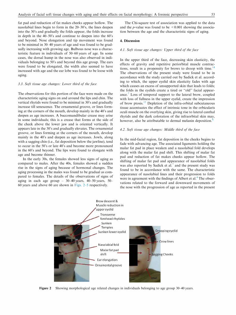
aging in each age group – 30–40 years, 40–50 years, 50–60 years and above 60 are shown in Figs. 2–5 respectively.
Figure 2 Showing morphological age related changes
The Chi-square test of association was applied to the dataand the p-value was found to be <0.001 showing the associa-tion between the age and the characteristic signs of aging.
4. Discussion
4.1. Soft tissue age changes: Upper third of the face
In the upper third of the face, decreasing skin elasticity, the
effects of gravity and repetitive periorbital muscle contrac-tions, result in a propensity for brows to droop with time.14
The observations of the present study were found to be in
accordance with the study carried out by Sadick et al. accord-ing to which, the upper eyelid skin elasticity fades with agewhich causes an excess of unsupported skin that leads to folds;
the folds in the eyelids create a tired or ‘‘old’’ facial appear-ance.7 Loss of temporal support to the lateral brow, coupledwith loss of fullness in the upper eyelid, create the impressionof brow ptosis.15 Depletion of the infra-orbital subcutaneous
tissue accentuates the effect of intrinsic tone in the orbicularisoculi muscle on the overlying skin, giving rise to lateral canthalrhytids and the dark coloration of the infraorbital skin may,
however, also be attributable to dermal melanin deposition.6
4.2. Soft tissue age changes: Middle third of the face
In the mid-facial region, fat deposition in the cheeks begins tofade with advancing age. The associated ligaments holding themalar fat pad in place weaken and a nasolabial fold develops
along with the malar fat pad shift. This shifting of malar fatpad and reduction of fat makes cheeks appear hollow. Theshifting of malar fat pad and appearance of nasolabial foldswas also reported by Sadick et al.7 and the present study was
found to be in accordance with the same. The characteristicappearance of nasolabial lines and their progression to foldswere in agreement with the findings of Albert et al.4 The obser-
vations related to the forward and downward movements ofthe nose with the progression of age as reported in the present
in individuals belonging to age group 30–40 years.
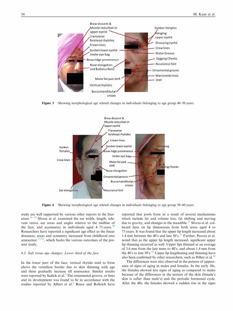
Figure 3 Showing morphological age related changes in individuals belonging to age group 40–50 years.
Figure 4 Showing morphological age related changes in individuals belonging to age group 50–60 years.
54 M. Kaur et al.
study are well supported by various other reports in the liter-ature.16–19 Sforza et al. examined the ear width, length, rele-
vant ratios, ear areas and angles relative to the midline ofthe face, and asymmetry in individuals aged 4–73 years.20
Researchers have reported a significant age effect as the linear
distances, areas and symmetry increased from childhood intosenescence 17,18, which backs the various outcomes of the pre-sent study.
4.3. Soft tissue age changes: Lower third of the face
In the lower part of the face, vertical rhytids tend to formabove the vermilion border due to skin thinning with age
and these gradually increase till senescence. Similar resultswere reported by Sadick et al.7 The ornamental groove, or linesand its development was found to be in accordance with the
studies reported by Albert et al.4 Reece and Rohrich have
reported that jowls form as a result of several mechanismswhich include fat and volume loss, fat shifting and moving
due to gravity, and changes in the mandible.21 Sforza et al. col-lected data on lip dimensions from both sexes aged 4 to73 years. It was found that the upper lip length increased about
1.4 mm between the 40’s and late 50’s.22 Further, Pecora et al.noted that as the upper lip length increased, significant upperlip thinning occurred as well. Upper lips thinned at an average
of 3.6 mm from the late teens to 40’s, and about 1.4 mm fromthe 40’s to late 50’s.23 Upper lip lengthening and thinning havealso been confirmed by other researchers, such as Iblher et al.24
The differences were also observed in the pattern of appear-
ance of signs of aging in males and females. In the early 30s,the females showed less signs of aging as compared to malesbecause of the differences in the texture of the skin (female’s
skin is softer than male’s) and the periodic hormonal cycle.After the 40s, the females showed a sudden rise in the signs
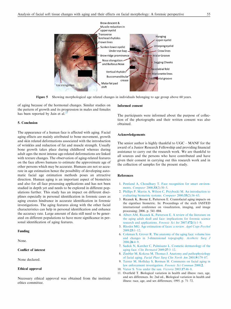
Figure 5 Showing morphological age related changes in individuals belonging to age group above 60 years.
Analysis of facial soft tissue changes with aging and their effects on facial morphology: A forensic perspective 55
of aging because of the hormonal changes. Similar studies on
the pattern of growth and its progression in males and femaleshas been reported by Jain et al.25
5. Conclusion
The appearance of a human face is affected with aging. Facialaging effects are mainly attributed to bone movement, growth
and skin related deformations associated with the introductionof wrinkles and reduction of fat and muscle strength. Usuallybone growth takes place during childhood whereas during
adult ages the most intense age-related deformations are linkedwith texture changes. The observation of aging-related featureson the face allows humans to estimate the approximate age ofother persons which may be accurate. Humans are not so accu-
rate in age estimation hence the possibility of developing auto-matic facial age estimation methods poses an attractivedirection. Human aging is an important aspect of biometrics
and also for all face processing applications and has not beenstudied in depth yet and needs to be explored in different pop-ulations further. This study has an impact on different disci-
plines especially in personal identification in forensic cases asaging creates hindrance in accurate identification in forensicinvestigations. The aging features along with the other facial
characteristics can help in personal identification and enhancethe accuracy rate. Large amount of data still need to be gener-ated on different populations to have more significance in per-sonal identification of aging features.
Funding
None.
Conflict of interest
None declared.
Ethical approval
Necessary ethical approval was obtained from the institute
ethics committee.
Informed consent
The participants were informed about the purpose of collec-tion of the photographs and their written consent was alsoobtained.
Acknowledgements
The senior author is highly thankful to UGC – MANF for theaward of a Junior Research Fellowship and providing financialassistance to carry out the research work. We are thankful toall sources and the persons who have contributed and have
given their consent in carrying out this research work and inthe collection of samples for the present study.
References
1. Pentland A, Choudhury T. Face recognition for smart environ-
ments. Computer 2000;33(2):50–5.
2. Phillips P, Martin A, Wilson C, Przybocki M. An introduction to
evaluating biometric systems. Computer 2000;33(2):56–63.
3. Ricanek K, Boone E, Patterson E. Craniofacial aging impacts on
the eigenface biometric. In: Proceedings of the sixth IASTED
international conference on visualization, imaging, and image
processing; 2006. p. 541–004.
4. Albert AM, Ricanek K, Patterson E. A review of the literature on
the aging adult skull and face: implications for forensic science
research and applications. Forensic Sci Int 2007;172(1):1–9.
5. Rhodes MG. Age estimation of faces: a review. Appl Cogn Psychol
2009;23:1–12.
6. Coleman S, Grover R. The anatomy of the aging face: volume loss
and changes in 3-dimensional topography. Aesthetic Surg J
2006;26:4–9.
7. Sadick N, Karcher C, Palmisano L. Cosmetic dermatology of the
aging face. Clin Dermatol 2009;27:3–12.
8. Zimbler M, Kokosa M, Thomas J. Anatomy and pathophysiology
of facial aging. Facial Plast Surg Clin North Am 2001;9:179–87.
9. Taister M, Holliday S, Borman H. Comments on facial aging in
law enforcement investigation. Forensic Sci Commun 2000;2.
10. Victor S. You under the sun. Victoria 2003;17:46–8.
11. Overfield T. Biological variation in health and illness: race, age,
and sex differences. In: 2nd ed., Biological variation in health and
illness: race, age, and sex differences; 1995. p. 71–72.
56 M. Kaur et al.
12. Peertrainer. What to do about wrinkly facial skin after weight loss;
2009. URL: www.peertrainer.com/LoungeCommunity/Thread.
aspx?ForumI.
13. Lipotherapy; 2009. URL: http://www.queensparkclinic.co.uk/
lipotherapy-fat-treaments-c56.html.
14. Sforza C, Grandi G, Catti F, Tommasi DG, Ugolini A, Ferrario
VF. Age- and sex-related changes in the soft tissues of the orbital
region. Forensic Sci Int 2009;185(1–3), 115.e1-115.e8.
15. Coleman SR. Structural fat grafting. St. Louis, MO: Quality
Medical Publishing; 2004.
16. Bishara S, Jakobsen J, Hession T, Treder J. Soft tissue profile
changes from 5 to 45 years of age. Am J Orthod Dentofacial
Orthop 1998;114:698–706.
17. Sarnas K, Solow B. Early adult changes in the skeletal and soft
tissue profile. Eur J Orthod 1980;2:1–12.
18. West K, McNamara J. Changes in the craniofacial complex from
adolescence to midadulthood: a cephalometric study. Am J Orthod
Dentofacial Orthop 1999;115:521–32.
19. Sforza C, Grandi G, De Menezes M, Tartaglia GM, Ferrario VF.
Age- and sex-related changes in the normal human external nose.
Forensic Sci Int 2009;204:205e1–9.
20. Sforza C, Grandi G, Binelli M, Tommasi D, Rosati R, Ferrario V.
Age and sex-related changes in the normal human ear. Forensic Sci
Int 2009;187:1–7.
21. Reece EM, Rohrich R. The aesthetic jaw line: management of the
aging jowl. Aesthetics Surg J 2008;28:668–74.
22. Sforza C, Grandi G, Binelli M, Dolci C, De Menezes M, Ferrario
V. Age and sex-related changes in three-dimensional lip morphol-
ogy. Forensic Sci Int 2010;200:1–7.
23. Pecora N, Baccetti T, McNamara J. The aging craniofacial
complex: a longitudinal cephalometric study from late adolescence
to late adulthood. Am J Orthod Dentofacial Orthop
2008;134:496–505.
24. Iblher N, Kloepper J, Penna V, Bartholomae J, Stark G. Changes
in the upper lip – A photometric and MRI based study (on a quest
to find the right rejuvenation approach). J Plast Reconstr Aesthet
Surg 2008;61:1170–6.
25. Jain AK, Bolle R, Pankanti S. Biometrics: personal identification
in networked security. In: Jain AK, Bolle R, Pankanti S,
editors. Kluwer Academic Publishers; 1999.