Analysis of conserved residues in the β pat-3 cytoplasmic tail reveals important functions of...
20
Analysis of conserved residues in the βpat-3 cytoplasmic tail reveals important functions of integrin in multiple tissues Xiaojian Xu 1 , Jeong H. Ahn 1 , Phebe Tam 1 , Eun Jeong Yu 1 , Sushil Batra 1 , Erin J. Cram 2,* , and Myeongwoo Lee 1,* 1 Department of Biology, Baylor University, One Bear Place 97388, Waco, TX 76798 2 Department of Biology, Northeastern University, 360 Huntington Ave, Boston, MA 02115, U.S.A Abstract Integrin cytoplasmic tails contain motifs that link extracelluar information to cell behavior such as cell migration and contraction. To investigate the cell functions mediated by the conserved motifs, we created mutations in the Caenorhabditis elegans βpat-3 cytoplasmic tail. The β1D ( 799 FK 800 ), NPXY, tryptophan ( 784 W), and threonine ( 797 TT 798 ) motifs were disrupted to identify their functions in vivo. Animals expressing integrins with disrupted NPXY motifs were viable, but displayed distal tip cell migration and ovulation defects. The conserved threonines were required for gonad migration and contraction as well as tail morphogenesis, whereas disruption of the β1D and tryptophan motifs produced only mild defects. To abolish multiple conserved motifs, a β1C- like variant, which results in a frameshift, was constructed. βpat-3(β1C) transgenic animals showed cold-sensitive larval arrests and defective muscle structure and gonad migration and contraction. Our study suggests that the conserved NPXY and TT motifs play important roles in tissue-specific function of integrin. Keywords Integrin; conserved tyrosine; DTC migration; cytoplasmic tail; gonad; ovulation; frameshift mutations; cold-sensitive; beta 1C integrin; NPXY motif Introduction Integrins are heterodimeric transmembrane receptors for extracellular matrix (ECM). In mammals, there are more than 20 different integrin molecules, which are composed of combinations of α and β genes (Hynes, 1992; Hynes, 2002). Integrins bind ECM molecules such as collagens, fibronectin, and laminin and assemble a cytoplasmic protein complex known as a focal adhesion, which includes both structural and signaling components. This integrin-mediated linkage is crucial for the proper control of cell behaviors such as cell migration, adhesion, differentiation, and death (Giancotti, 1997; Giancotti and Ruoslahti, 1999). Caenorhabditis elegans is an excellent model system with which to study the developmental function of integrins because it only possesses two integrin heterodimers composed of one α (INA-1 or PAT-2) and one β (PAT-3) integrin subunit (Kramer, 2005). Null alleles of αpat-2 and βpat-3 result in the paralyzed and arrested at two-fold embryonic lethality phenotype (Pat) (Williams and Waterston, 1994), and loss of αina-1 results in larval lethal (Baum and * All correspondence should be addressed to: Erin J. Cram, Ph.D., Tel) 617-373-7533, [email protected] or Myeongwoo Lee, Ph.D., TEL) 254-710-2135, [email protected]. NIH Public Access Author Manuscript Dev Dyn. Author manuscript; available in PMC 2011 April 25. Published in final edited form as: Dev Dyn. 2010 March ; 239(3): 763–772. doi:10.1002/dvdy.22205. NIH-PA Author Manuscript NIH-PA Author Manuscript NIH-PA Author Manuscript
-
Upload
independent -
Category
Documents
-
view
2 -
download
0
Transcript of Analysis of conserved residues in the β pat-3 cytoplasmic tail reveals important functions of...

Analysis of conserved residues in the βpat-3 cytoplasmic tailreveals important functions of integrin in multiple tissues
Xiaojian Xu1, Jeong H. Ahn1, Phebe Tam1, Eun Jeong Yu1, Sushil Batra1, Erin J. Cram2,*,and Myeongwoo Lee1,*
1 Department of Biology, Baylor University, One Bear Place 97388, Waco, TX 767982 Department of Biology, Northeastern University, 360 Huntington Ave, Boston, MA 02115, U.S.A
AbstractIntegrin cytoplasmic tails contain motifs that link extracelluar information to cell behavior such ascell migration and contraction. To investigate the cell functions mediated by the conserved motifs,we created mutations in the Caenorhabditis elegans βpat-3 cytoplasmic tail. The β1D (799FK800),NPXY, tryptophan (784W), and threonine (797TT798) motifs were disrupted to identify theirfunctions in vivo. Animals expressing integrins with disrupted NPXY motifs were viable, butdisplayed distal tip cell migration and ovulation defects. The conserved threonines were requiredfor gonad migration and contraction as well as tail morphogenesis, whereas disruption of the β1Dand tryptophan motifs produced only mild defects. To abolish multiple conserved motifs, a β1C-like variant, which results in a frameshift, was constructed. βpat-3(β1C) transgenic animalsshowed cold-sensitive larval arrests and defective muscle structure and gonad migration andcontraction. Our study suggests that the conserved NPXY and TT motifs play important roles intissue-specific function of integrin.
KeywordsIntegrin; conserved tyrosine; DTC migration; cytoplasmic tail; gonad; ovulation; frameshiftmutations; cold-sensitive; beta 1C integrin; NPXY motif
IntroductionIntegrins are heterodimeric transmembrane receptors for extracellular matrix (ECM). Inmammals, there are more than 20 different integrin molecules, which are composed ofcombinations of α and β genes (Hynes, 1992; Hynes, 2002). Integrins bind ECM moleculessuch as collagens, fibronectin, and laminin and assemble a cytoplasmic protein complexknown as a focal adhesion, which includes both structural and signaling components. Thisintegrin-mediated linkage is crucial for the proper control of cell behaviors such as cellmigration, adhesion, differentiation, and death (Giancotti, 1997; Giancotti and Ruoslahti,1999).
Caenorhabditis elegans is an excellent model system with which to study the developmentalfunction of integrins because it only possesses two integrin heterodimers composed of one α(INA-1 or PAT-2) and one β (PAT-3) integrin subunit (Kramer, 2005). Null alleles of αpat-2and βpat-3 result in the paralyzed and arrested at two-fold embryonic lethality phenotype(Pat) (Williams and Waterston, 1994), and loss of αina-1 results in larval lethal (Baum and
*All correspondence should be addressed to: Erin J. Cram, Ph.D., Tel) 617-373-7533, [email protected] or Myeongwoo Lee, Ph.D.,TEL) 254-710-2135, [email protected].
NIH Public AccessAuthor ManuscriptDev Dyn. Author manuscript; available in PMC 2011 April 25.
Published in final edited form as:Dev Dyn. 2010 March ; 239(3): 763–772. doi:10.1002/dvdy.22205.
NIH
-PA Author Manuscript
NIH
-PA Author Manuscript
NIH
-PA Author Manuscript
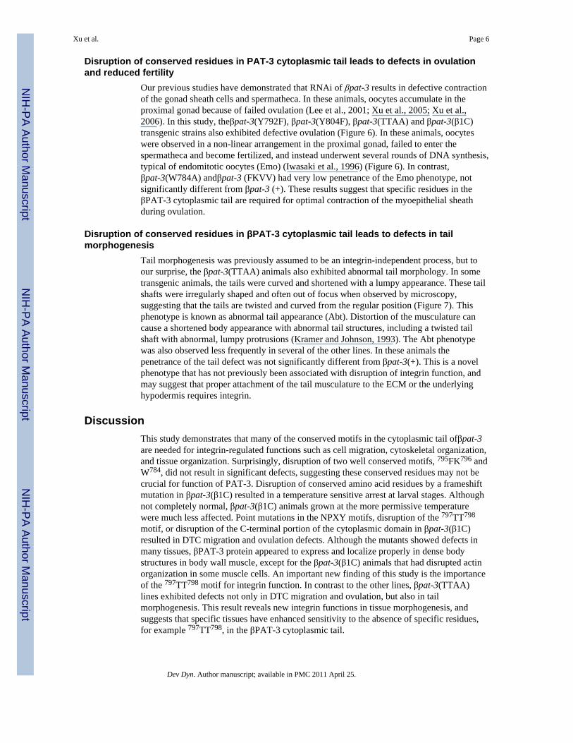
Garriga, 1997). βPAT-3 integrin likely forms a heterodimer with αPAT-2 or αINA-1 andcontrols many cellular events during C. elegans development. βPAT-3 integrin is expressedin the body wall and sex muscles, embryonic pharynx, spermatheca, uterus, somatic gonad,and neurons (Gettner et al., 1995). βPAT-3/αPAT-2 heterodimers are expressed in manytissues including muscles and somatic gonad whereas βPAT-3/αINA-1 pairs are localizedmainly to migratory cells such as distal tip cells (DTC) and neurons (Lee et al., 2001; Cramet al., 2003; Meighan and Schwarzbauer, 2007).
In body wall muscle cells, integrins are localized in dense bodies (Z band analog) and M-lines, anchoring sarcomeres to the adjacent basement membrane (BM), a specialized ECMencompassing muscles and gonad, and play essential roles in stabilizing the muscleattachment structures (Moerman and Williams, 2006). Genetic screens have isolated severalβpat-3 mutations including null and reduction-of-function alleles (Williams and Waterston,1994; Gettner et al., 1995). However, all available βpat-3 mutations are within in theextracellular domain, limiting the analysis of its intracellular function (Gettner, 1994).
Functional analysis of βpat-3 integrin using a dominant negative HA-βtail revealed thatinhibition of βpat-3 cytoplasmic tail function caused gonad migration and ovulation defects,suggesting an important role for βpat-3 integrin in cell migration and contraction (Lee et al.,2001; Lee et al., 2005). In addition, RNA mediated interference (RNAi) of βpat-3 results indisorganized F-actin in muscle, defective DTC migration (Cram et al., 2003; Cram et al.,2006) and contractile defects of the myoepithelial cells of the somatic gonad, demonstratingthe essential role of cell-ECM interaction in organization and function of these tissues (Leeet al., 2005; Sherwood et al., 2005; Xu et al., 2006).
βPAT-3 integrin is well conserved relative to human β1 integrin (61% protein sequencesimilarity) and shares many conserved motifs (Figure 1). Essential residues such as anaspartic acid (768D in βPAT-3) that forms a salt bridge to the α partner, a conserved domain(761K to 775F) that mediates focal adhesion kinase (FAK) or paxillin binding, and the talinbinding (784W to 800K) sites are all conserved in βPAT-3 (Figure 1). The two NPXY (asp-pro-X-tyr) motifs are also conserved in βPAT-3 tail (Lee et al., 2001). Tyrosinephosphorylation of NPXY leads to integrin activation and promotes interactions withdownstream signaling molecules including talin, ICAP-1, and kindlins (Calderwood et al.,2002;Zawistowski et al., 2002;Calderwood, 2004;Oxley et al., 2008;Harburger et al., 2009).Functional studies of NPXY motifs in mouse β3 integrin showed that platelets displayeddefective chemotaxis when one or both Y was changed to F (Law et al., 1999). The Y toalanine (A) mutation in mouse β1 or β3 integrin abolishes integrin functions and results inembryonic lethality (Chen et al., 2006). Disrupting the two NPXY motifs impaired theproliferation and survival of mouse embryonic fibroblasts (MEF) (Hirsch et al., 2002).
In a previous study, we determined the role of the conserved NPXY motifs in thecytoplasmic domain of βPAT-3. Two rescued lines, βpat-3(Y804F) and βpat-3(YYFF),rescued the embryonic lethality of βpat-3(st564) and generated viable and fertile progeny(Lee et al., 2001). These transgenic animals exhibited mild defects in distal tip cellmigration. Most commonly, the DTC turned away from the dorsal surface and returned tothe ventral side, perhaps because of defective adhesion to the dorsal body wall basementmembrane (Lee et al., 2001). Immunofluorescence staining of rescued animals with MH25,a monoclonal antibody against βPAT-3, demonstrated normal localization and distributionof dense bodies (Francis and Waterston, 1991; Lee et al., 2001). Muscle filamentous actinstaining in the rescued NPXY lines was also indistinguishable from βpat-3 (+) animals (Leeet al., 2001). These data suggested that the NPXY motif may play an important role in cellmigration but is not required for muscle cell organization.
Xu et al. Page 2
Dev Dyn. Author manuscript; available in PMC 2011 April 25.
NIH
-PA Author Manuscript
NIH
-PA Author Manuscript
NIH
-PA Author Manuscript

In this study we have extended our analysis of conserved motifs, βpat-3(TTAA),βpat-3(W784A), βpat-3(FKVV), and a splice mutant βpat-3(β1C), in the cytoplasmic tail ofβPAT-3 and compared them to our previous results targeting the NPXY motifs. All of ourtransgenic rescue constructs can rescue the embryonic lethality of the βpat-3(st564) nullallele (Williams and Waterston, 1994), but displayed multiple larval and adult phenotypescaused by defective integrin function. Disruption of specific integrin motifs results in defectsin DTC migration, ovulation, and muscle cell function. Importantly, we identify a novel rolefor integrin in hermaphrodite tail morphogenesis.
ResultsTargeting conserved motifs in the β integrin cytoplasmic tail
To study the role of the conserved amino acid residues and motifsessential for integrinfunction in embryonic and post-embryonic development, C. elegans strains with pointmutations in conserved motifs in the cytoplasmic domain of βpat-3 were established (Figure1A). All constructs were created by PCR mutagenesis, purified, and injected into balancedβpat-3(st564) heterozygous animals to produce rescued transgenic lines in which animalsare dependent on the transgene for survival.
There are two NPXY motifs that include the tyrosines Y792 and Y804 in βPAT-3 tail (Chenet al., 1990; Lee et al., 2001). Tyrosine phosphorylation of the NPXY motif is thought toactivate integrin and promote interactions with downstream signaling molecules(Calderwood et al., 2002). This motif has been shown to be important for integrin functionin fly (Zusman et al., 1990; Zusman et al., 1993; Jannuzi et al., 2002; Jannuzi et al., 2004),worm (Lee et al., 2001), mouse (Czuchra et al., 2006), and mammalian tissue culture (Sakaiet al., 1998; Kaapa et al., 1999). We have previously demonstrated that disruption of theNPXY motifs, either singly (Y804F) or in combination (YYFF) leads to defects in therescued transgenic lines including DTC migration defects (Lee et al., 2001). In order tofurther characterize the importance of phosophorylation in the NPXY motif, we generated apoint mutation in Y792 for this study, and compare the effects of disruption of these NPXYmotifs to a new set of point mutations targeting conserved residues in the βPAT-3cytoplasmic domain.
The three new motifs targeted for analysis are a 799FK800 motif, a tryptophanmotif 784WDT786, and a threonine motif 797TT798. In order to determine the in vivoimportance of these sequences, we have generated rescued transgenic lines expressingβpat-3 with point mutations disrupting these residues. An identical sequence to βPAT-3(799FKNPXY804)is found in the muscle integrin β1D (Figure 1B), a splice isoform ofmammalian β1 integrin (Belkin et al., 1997; Belkin and Retta, 1998), whereas the sequenceis 790VVNPXY795 in human β1A, a splice isoform abundant in focal adhesions (Argraves etal., 1987), of β1 integrin. We generated rescued transgenic lines expressing 799VVNPXY804,and designated this line βpat-3 (FKVV). The tryptophan (W) residue at the position 775 ofhuman β1 integrin has been identified as necessary for integrin-mediated protein kinase B/Akt survival signaling (Pankov et al., 2003). Similar to human β1, βPAT-3 possessesthe 784WDT786 motif upstream of the first NPXY. The 784W residue was mutated to alanine(A) and used to establish rescued transgenic nematodes, designated βpat-3 (W784A). Inmouse GD25 cells, mutations in the 788TT789 motif of β1 integrin caused defective cellattachment to fibronectin and induced a shift to inactive conformation in the extracellulardomain (Wennerberg et al., 1998), suggesting that these residues play important roles in theinside-out integrin signaling by relaying conformational change of cytoplasmic tail to theextracellular domain causing high affinity ligand binding (Tanentzapf and Brown, 2006).The threonines (797TT798) of βpat-3 were mutated to alanine to produce 797AA798 mutantdesignatedβpat-3(TTAA) (Figure 1).
Xu et al. Page 3
Dev Dyn. Author manuscript; available in PMC 2011 April 25.
NIH
-PA Author Manuscript
NIH
-PA Author Manuscript
NIH
-PA Author Manuscript

In addition, we have analyzed a nematode strain expressing βPAT-3 with significantlydisrupted cytoplasmic tail sequences. In mammals, β1C integrin is an alternatively splicedform of the β1 subfamily that contains a unique 42-amino acid sequence in the cytoplasmictail (Figure 1A). The β1C variant inhibits the growth of prostate cancer epithelial cells(Fornaro et al., 1998), endothelial cells (Meredith et al., 1995), and fibroblasts (Fornaro etal., 1995; Fornaro et al., 2000), is usually expressed in non-proliferative epithelium, anddownregulated in prostate adenocarcinoma and proliferating breast carcinoma (Manzotti etal., 2000). We designed a mutant βpat-3 gene, pPAT3-β1C, carrying a mutation (ag to aa) inthe splice acceptor sequence of pPAT3(+) intron 7, which creates a frameshift in the ORF ofβpat-3 gene. Although the splicing defect in βpat-3(β1C) is not predicted to result in ahomologous sequence to that found in the human β1C variant, in both cases all conservedmotifs are disrupted. Inferred protein sequences of the cytoplasmic tails of all rescueconstructs are indicated in Figure 1A. Rescued lines, able to rescue the embryonic lethalityof st564, were established for each construct.
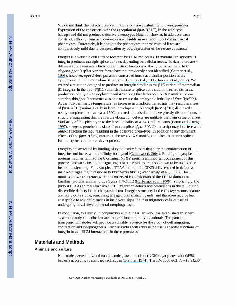
Severe disruption of the βpat-3 cytoplasmic tail leads to defective muscle structureBecause integrin is localized to body wall muscles and involved in assembly andstabilization of contractile structures (Kramer, 1997; Kramer, 2005), we determined thelocalization of βPAT-3 and arrangement of dense bodies in the rescued lines usingimmunofluorescence. In the βpat-3 (+) animals, βPAT-3 is localized to dotted andcontinuous lines along the length of muscle cells, typical of dense bodies (Figure 2).Immunostaining was performed on all rescued lines, and all were indistinguishable fromwild type. Representative staining of βpat-3(Y792F) and βpat-3(β1C) is shown in Figure 2.The actin cytoskeleton was also visualized in mutant lines by staining with rhodamine-conjugated phalloidin. All of the rescued lines showed a normal staining pattern of densebodies and actin filaments except for the frameshift line βpat-3 (β1C) (Figure 3). These datasuggest that the function of 799FK800, 784WDT786, and 797TT798 motifs are dispensable fordense body organization and for organization of the actin cytoskeleton in muscle cells. Thisresult is somewhat unexpected because FK motif is specifically found in a muscle specificsplice form of mammalian integrin β1D (Belkin et al., 1997; Belkin and Retta, 1998), andthe TT motif plays an important role in integrin activation and matrix binding in mouse cellculture (Wennerberg et al., 1998).
Consistent with genetic studies of βpat-3 (Lee et al., 2001; Lee et al., 2005), significantdisruption of βPAT-3 structure does result in muscle defects. βpat-3 (β1C) transgenicanimals were stained with MH25 and phalloidin to localize βpat-3 and visualize the actincytoskeleton. The majority of muscle cells appeared to have normal βpat-3 localization anda regular pattern of filamentous actin staining. However, in some muscle cells, the actincytoskeleton were clumped irregularly (Figure 2), suggesting that expression of thetransgene can cause cytoskeletal defects. In C. elegans, disruption of splice junctions oftenresults in temperature sensitive alleles, because splicing occurs inefficiently at 15° C (Aroianet al., 1990; Aroian and Sternberg, 1991; Aroian et al., 1993). Therefore, we investigated thecold sensitivity of the βpat-3(β1C) lines. At 15°C, a significant proportion of the animalswere paralyzed and arrested after hatching, whereas at 23°C, the animals hatched and movednormally (Table 1). Arrested L1 larvae were stained with phalloidin and appeared to havemuscle striations, although the actin seemed disorganized compared to normal L1 larvae(Figure 4).
To determine if βpat-3(β1C) can act in a dominant negative fashion at 15°C, the transgenewas crossed into the wildtype (N2) background. The resulting βpat-3(β1C)/+ animalsshowed significant larval arrest at 15°C (58% larval arrest, N=76). A lower percentage ofβpat-3(+)/+ heterozygotes displayed larval defects at the cold temperature (22% larvalarrest, N=41). We then investigated the molecular basis of the cold sensitive phenotype of
Xu et al. Page 4
Dev Dyn. Author manuscript; available in PMC 2011 April 25.
NIH
-PA Author Manuscript
NIH
-PA Author Manuscript
NIH
-PA Author Manuscript

βpat-3(β1C). RT-PCR was conducted on total RNA extracted from wild type (N2) andβpat-3(β1C) animals. Two different species of cDNA were detected and sequenced (Figure4). RT-PCR conducted on rescued βpat-3(+) animals revealed correctly spliced transcriptsindistinguishable from N2 (data not shown). The mutant, incorrectly spliced cDNAcontained 53 bp of sequence derived from intron 7, whereas the smaller one contained nointron sequences. Therefore, βpat-3 (β1C) animals express at least two βpat-3 splicevariants. Quantitative PCR (qPCR) analysis of pat-3 transcripts indicates that in βpat-3(β1C)animals raised at 15° C, pat-3 levels are elevated six-fold as compared to pat-3 levels inanimals raised at 23° C. This increase may be due to an increase in the unspliced form,because neither N2 nor rescued pat-3 (+) animals show any increase in pat-3 transcript at thenon-permissive temperature. Taken together, these results suggest that βpat-3(β1C) tailproduces two different species of βPAT-3 protein, one of which, presumably the mutant one,causes cold sensitivity and disrupts integrin functions.
Conserved residues in βpat-3 cytoplasmic tail are important for DTC migrationDuring C. elegans development, the two hermaphrodite gonad arms elongate to form amirror-image U-shaped structure (Hubbard and Greenstein, 2000). This elongation andmigration is navigated by a pair of distal tip cells (DTC), specialized leader cells at the distaltip of each gonad arm (Montell, 1999; Nishiwaki, 1999; Hubbard and Greenstein, 2000).The NPXY motifs have previously been shown to be required for correct DTC pathfinding,and potentially adhesion, during DTC migration along the dorsal basement membrane (Leeet al., 2001) (Figure 5). As expected, βpat-3(Y804F) and βpat-3(YYFF) exhibited milddefects in DTC migration, and were significantly more affected than βpat-3 (+). In many ofthese animals, distal gonad arms wandered out of the regular trajectory and collapsed towardthe ventral side. Defects also included DTC migration from the dorsal to the ventral sidenear the proximal gonad, suggesting either abnormal pathfinding or loss of contacts with thedorsal body wall (Figure 5C). Disruption of the first NPXY motif in the βpat-3(Y792F)animals produced a milder effect on DTC migration than seen in the βpat-3(Y792F) andβpat-3(YYFF) lines. Although defects were consistently observed, the penetrance of defectswas not significantly different from βpat-3(+) lines. The βpat-3(β1C) animals grown at 15°Cshowed DTC migration defects that were much more severe than βpat-3(β1C) animalsgrown at room temperature (Figure 5B, Table 1). DTC migration in βpat-3(β1C) at 15°Cwas also significantly more disrupted than any of the NPXF lines (Figure 5B, Table 1).
Disruption of the TT motif in the βpat-3 tail also resulted in DTC migration defects. Theβpat-3(TTAA) rescued lines had a much higher penetrance of DTC migration defects thanβpat-3(+) (Table 1, Figure 4). In contrast, the βpat-3(FKVV) and βpat-3(W784A) transgeniclines exhibited mild DTC migration defects not significantly different from those observedin βpat-3 (+) rescued animals. These results indicate that the 797TT798 residues are importantfor proper pathfinding of the DTC, possibly more so than phosphorylation of the NPXYmotif, or presence of the FK or WDT motifs. The DTC migration defects in βpat-3(TTAA)animals include incorrect pathfinding along the dorsal surface and supernumerary turns(Figure 5, Table 1). In cell culture, β1A integrin lacking the TT motif remained inactive anddid not contribute to matrix binding or matrix remodeling (Wennerberg et al., 1998). Ourresults suggest that similarly, the TT motif may be required for the integrin-mediatedextracellular matrix interaction important for correct DTC migration. Taken together, ouranalysis suggests that, while not essential for viability, multiple motifs within the βPAT-3tail are required for optimal control of DTC migration.
Xu et al. Page 5
Dev Dyn. Author manuscript; available in PMC 2011 April 25.
NIH
-PA Author Manuscript
NIH
-PA Author Manuscript
NIH
-PA Author Manuscript
Disruption of conserved residues in PAT-3 cytoplasmic tail leads to defects in ovulationand reduced fertility
Our previous studies have demonstrated that RNAi of βpat-3 results in defective contractionof the gonad sheath cells and spermatheca. In these animals, oocytes accumulate in theproximal gonad because of failed ovulation (Lee et al., 2001; Xu et al., 2005; Xu et al.,2006). In this study, theβpat-3(Y792F), βpat-3(Y804F), βpat-3(TTAA) and βpat-3(β1C)transgenic strains also exhibited defective ovulation (Figure 6). In these animals, oocyteswere observed in a non-linear arrangement in the proximal gonad, failed to enter thespermatheca and become fertilized, and instead underwent several rounds of DNA synthesis,typical of endomitotic oocytes (Emo) (Iwasaki et al., 1996) (Figure 6). In contrast,βpat-3(W784A) andβpat-3 (FKVV) had very low penetrance of the Emo phenotype, notsignificantly different from βpat-3 (+). These results suggest that specific residues in theβPAT-3 cytoplasmic tail are required for optimal contraction of the myoepithelial sheathduring ovulation.
Disruption of conserved residues in βPAT-3 cytoplasmic tail leads to defects in tailmorphogenesis
Tail morphogenesis was previously assumed to be an integrin-independent process, but toour surprise, the βpat-3(TTAA) animals also exhibited abnormal tail morphology. In sometransgenic animals, the tails were curved and shortened with a lumpy appearance. These tailshafts were irregularly shaped and often out of focus when observed by microscopy,suggesting that the tails are twisted and curved from the regular position (Figure 7). Thisphenotype is known as abnormal tail appearance (Abt). Distortion of the musculature cancause a shortened body appearance with abnormal tail structures, including a twisted tailshaft with abnormal, lumpy protrusions (Kramer and Johnson, 1993). The Abt phenotypewas also observed less frequently in several of the other lines. In these animals thepenetrance of the tail defect was not significantly different from βpat-3(+). This is a novelphenotype that has not previously been associated with disruption of integrin function, andmay suggest that proper attachment of the tail musculature to the ECM or the underlyinghypodermis requires integrin.
DiscussionThis study demonstrates that many of the conserved motifs in the cytoplasmic tail ofβpat-3are needed for integrin-regulated functions such as cell migration, cytoskeletal organization,and tissue organization. Surprisingly, disruption of two well conserved motifs, 795FK796 andW784, did not result in significant defects, suggesting these conserved residues may not becrucial for function of PAT-3. Disruption of conserved amino acid residues by a frameshiftmutation in βpat-3(β1C) resulted in a temperature sensitive arrest at larval stages. Althoughnot completely normal, βpat-3(β1C) animals grown at the more permissive temperaturewere much less affected. Point mutations in the NPXY motifs, disruption of the 797TT798
motif, or disruption of the C-terminal portion of the cytoplasmic domain in βpat-3(β1C)resulted in DTC migration and ovulation defects. Although the mutants showed defects inmany tissues, βPAT-3 protein appeared to express and localize properly in dense bodystructures in body wall muscle, except for the βpat-3(β1C) animals that had disrupted actinorganization in some muscle cells. An important new finding of this study is the importanceof the 797TT798 motif for integrin function. In contrast to the other lines, βpat-3(TTAA)lines exhibited defects not only in DTC migration and ovulation, but also in tailmorphogenesis. This result reveals new integrin functions in tissue morphogenesis, andsuggests that specific tissues have enhanced sensitivity to the absence of specific residues,for example 797TT798, in the βPAT-3 cytoplasmic tail.
Xu et al. Page 6
Dev Dyn. Author manuscript; available in PMC 2011 April 25.
NIH
-PA Author Manuscript
NIH
-PA Author Manuscript
NIH
-PA Author Manuscript
We do not think the defects observed in this study are attributable to overexpression.Expression of the constructs, with the exception of βpat-3(β1C), in the wild typebackground did not produce defective phenotypes (data not shown). In addition, eachconstruct, although similarly overexpressed, yields an overlapping but distinct set ofphenotypes. Conversely, it is possible the phenotypes in these rescued lines arecomparatively mild due to compensation by overexpression of the rescue constructs.
Integrin is a versatile cell surface receptor for ECM molecules. In mammalian systems,β1integrin produces multiple splice variants depending on cellular needs. To date, there are 4different splice variants which confer distinct functions to the cytoplasmic tails. In C.elegans, βpat-3 splice variant forms have not previously been identified (Gettner et al.,1995), however, βpat-3 does possess a conserved intron at a similar position in thecytoplasmic tail of mammalian β1 integrin (Gettner et al., 1995; Jannuzi et al., 2002). Wecreated a mutation designed to produce an integrin similar to the β1C variant of mammalianβ1 integrin. In the βpat-3(β1C) animals, failure to splice out a small intron results in theproduction of a βpat-3 cytoplasmic tail 42 aa long that lacks both NPXY motifs. To oursurprise, this βpat-3 construct was able to rescue the embryonic lethality of βpat-3(st564).At the non-permissive temperature, an increase in unspliced transcripts may result in arrestof βpat-3(β1C) animals early in larval development. Although βpat-3(β1C) displayed anearly complete larval arrest at 15°C, arrested animals did not have grossly disrupted musclestructure, suggesting that the muscle elongation defects are unlikely the main cause of arrest.Similarity of this phenotype to the larval lethality of αina-1 null mutants (Baum and Garriga,1997), suggests proteins translated from unspliced βpat-3(β1C) transcript may interfere withαina-1 function thereby resulting in the observed phenotype. In addition to any dominanteffects of the βpat-3(β1C) construct, the two NPXY motifs, abolished in the non-splicedform, may be required for development.
Integrins are activated by binding of cytoplasmic factors that alter the conformation ofintegrins and increase their affinity for ligand (Calderwood, 2004). Binding of cytoplasmicproteins, such as talin, to the C-terminal NPXY motif is an important component of thisprocess, known as inside-out signaling. The TT residues are also known to be involved ininside-out signaling. For example, a TTAA mutation in GD25 cells resulted in defectiveinside-out signaling in response to fibronectin fibrils (Wennerberg et al., 1998). The TTmotif is known to interact with the conserved F3 subdomain of the FERM domain inkindlins, proteins similar to C. elegans UNC-112 (Harburger et al., 2009). Surprisingly, theβpat-3(TTAA) animals displayed DTC migration defects and protrusions in the tail, but nodiscernible defects in muscle cytoskeleton. Integrin structures in the C. elegans musculatureare likely quite stable, remaining engaged with matrix ligands, and therefore may be lesssusceptible to any deficiencies in inside-out signaling than migratory cells or tissuesundergoing larval developmental morphogenesis.
In conclusion, this study, in conjunction with our earlier work, has established an in vivosystem to study cell adhesion and integrin function in living animals. The panel oftransgenic nematodes will provide a valuable resource for the study of cell migration,contraction and morphogenesis. Further studies will address the tissue specific functions ofintegrin in cell-ECM interactions in these processes.
Materials and MethodsAnimals and culture
Nematodes were cultivated on nematode growth medium (NGM) agar plates with OP50bacteria according to standard techniques (Brenner, 1974). The RW3600 qC1 dpy-19(e1259)
Xu et al. Page 7
Dev Dyn. Author manuscript; available in PMC 2011 April 25.
NIH
-PA Author Manuscript
NIH
-PA Author Manuscript
NIH
-PA Author Manuscript

glp-1(q339)/pat-3(st564) III (Williams and Waterston, 1994) C. elegans strain used in thisstudy were obtained from the Caenorhabditis Genetics Center, St. Paul, MN.
Mutant constructs and germline transformationpPAT3(+)-PB12K was excised from the ZK1058 cosmid using NEB restriction enzymesPstI and BsrB1 and inserted between PstI and SmaI sites of pSP73 plasmid vector(Promega) (Lee et al., 2001). Corresponding mutant constructs, pPAT3-Y792F, pPAT3-W784A, pPAT3-FKVV, pPAT3-TTAA, and pPAT3-β1C, were created using overlapextension PCR. After the overlap extension PCR, the 754-bp MscI-EcoRI fragments fromthe PCR were cloned back into the corresponding position of pPAT3-PB12K. Plasmids wereisolated and purified by CsCl2 density gradient centrifugation.
Germline transformation was performed as described in Mello et al. (Mello et al., 1991).pPAT3 constructs were mixed with TG96 sur-5::GFP (Gu et al., 1998) as a coinjectionmarker. Injections were first attempted at a mixture of 10 μg/ml of pPAT3 and 100 μg/ml ofTG96 in TE buffer (pH=7.5). Various injection concentrations were used and linesgenerated from the lowest possible concentration of the pPAT3 construct were analyzedfurther. The βpat-3(β1C) lines were generated at pPAT3-β1C 50 μg/ml and TG96 50 μg/ml,which was the only concentration found to generate a viable rescue. To rescue βpat-3(st564)with the pPAT3 constructs, the distal gonad of RW3600 qC1 dpy-19(e1259) glp-1(q339)/pat-3(st564) III animals was microinjected with the injection mixture. After the initialgeneration of F1 green worms, those animals were propagated by selfing, a singlehermaphrodite growing on a NGM plate. Plates with F2 green progeny were then examinedfor the further screening; green F2 animals were selfed on individual plates. F2 plates with100% green progeny were selected for phenotype characterization. Multiple rescued lineswere generated, and the lines were allowed to grow for more than 10 generations beforecharacterizing the phenotypes (Lee et al., 2001).
Phenotype characterizationTo score fertility, a L4 or young adult transgenic animal was selfed on an OP50 seededNGM plate. Presence of progeny was scored for 4 consecutive days. To analyze gonad andtail morphology, young adult transgenic hermaphrodites were mounted in a drop of M9buffer containing 20 mMNaN3 (Sigma Chemical Co.) on a 24×60 mm coverslip containingwet surface of 3%agarose in water and examined using a Nikon TE2000-U Diaphotmicroscope with DIC optics. Images were captured using a CoolSnap cf monochromecamera (Roper Scientific, Tucson, AZ) and analyzed with Metavue imaging software(version 7.1, Molecular Devices Co, Downingtown, PA). Gonad morphology was scoredessentially as previously described (Lee et al., 2001). Young adult hermaphrodites with tailshafts much shorter than normal or tails with protrusions or lumps in the surface cuticularmaterial (Kramer and Johnson, 1993) were scored as possessing the abnormal tail (Abt)phenotype. When oocytes were present in a non-linear arrangement in the proximalgonad ofyoung adult animals, animals were scored as exhibiting the endomitotic oocyte (Emo)phenotype.
Fluorescence microscopyTo visualize βPAT-3 distribution in body wall muscles, transgenic worms were collected inM9 buffer containing 1% sodium azide (NaN3, w/v). Using a razor blade, worms were dicedrandomly on a poly-L-lysine (1 mg/ml, w/v) coated slides and fixed with methanol andacetone at −20°C, followed by treatment with MH25, anti-βPAT-3 monoclonal antibody(1:1,00 dilution in M9 buffer with 1% goat serum), overnight at RT. The samples were thentreated with a secondary goat anti-mouse IgG FITC conjugated antibody, (Sigma-AldrichChem, St. Louis, MO) (1:5,000 dilution in M9 buffer with 1% goat serum), for 2–3 hrs at
Xu et al. Page 8
Dev Dyn. Author manuscript; available in PMC 2011 April 25.
NIH
-PA Author Manuscript
NIH
-PA Author Manuscript
NIH
-PA Author Manuscript

RT. The samples were washed and mounted on a Nikon TE2000-U inverted microscope forfluorescence microscopy. For phalloidin staining, animals were dissected on poly-L-lysinecoated slide and fixed with methanol and acetone. Fixed worms were the treated withrhodamine-conjugated phalloidin (1:200 dilution, Fluka Science) in M9 buffer for 2 to 3hours at RT. Prepared samples were observed on the Nikon TE2000-U diaphot microscope.Images were captured using a CoolSnap cf monochrome camera (Roper Scientific, Tucson,AZ) and analyzed with Metavue imaging software (version 5, Molecular Devices Co,Downingtown, PA).
Reverse transcription PCR (RT-PCR)To analyze βpat-3 transcripts, transgenic worm lines, N2, JE443 βpat-3(+) andBU7221βpat-3(β1C) were then collected with M9 buffer and frozen in liquid nitrogen.Frozen worm pellets were ground to a powder with a mortar and pestle. Worms wereextracted with Tri-Reagent (Sigma-Aldrich, St. Louis, MO) and chloroform (1.5 volumes)and RNA was precipitated from the extract with ethanol and 3 M sodium acetate. After anethanol rinse, the RNA was treated with RQ1 DNase (Promega, Madison, WI) to removecontaminating DNA. Approximately 1 μg of total RNA was used to synthesize cDNA withthe Transcriptor Reverse Transcription Kit (Roche, Carlsbad, CA) primed with randomhexamers in a 20 μl reaction. 1–2 μl cDNA was subsequently used in PCR amplificationwith βpat-3 primers and also with control GAPDH primers to amplify the. C. elegans geneT09F3.3 (GAPDH). Primer sequences listed below were used for amplification:
PAT3PT Forward 1: 5′-ctcaacgaaactacaccctgcc-3′
PAT3PT Reverse 1: 5′-ttagttggcttttccagcgtatactgg-3′
PAT6 Forward: 5′-gctagctcctggtgcttcttg-3′
PAT6 Reverse: 5′-aagcttctcctcgtggctttg-3′
PCR products were then inserted into pGEM-T vector and transformed into DH5α bacteria(Invitrogen, Carlsbad, CA). Isolated plasmids containing the PCR products were sent toMacrogen (Bethesda, MD) for sequencing service and sequence data were analyzed byBLASTn search. For qPCR analysis, 1 μl cDNA was used in each reaction with iQSYBRgreen Supermix (Bio-Rad Laboratories, Hercules, CA) in a 96-well plate.Amplification of C. elegans gene ama-1 was used as a loading and amplification control. Allassays were amplified and evaluated in real time using an ABI Prism 7000 sequencedetection system. The relative quantitation of pat-3 mRNA was calculated by thecomparative Ct method (Livak and Schmittgen, 2001).
AcknowledgmentsC. elegans strains were provided by the Caenorhabditis Genetics Center, which is funded by the NIH. Authorsthank Julia Lee for technical assistance. This work was supported by funds from Baylor University and a researchgrant from NIH (GM077156) for M. Lee.
Grant Sponsor: NIH NIGMS # GM077156 for M. Lee.
ReferencesArgraves WS, Suzuki S, Arai H, Thompson K, Pierschbacher MD, Ruoslahti E. Amino acid sequence
of the human fibronectin receptor. J Cell Biol. 1987; 105:1183–1190. [PubMed: 2958481]Aroian RV, Koga M, Mendel JE, Ohshima Y, Sternberg PW. The let-23 gene necessary for
Caenorhabditis elegans vulval induction encodes a tyrosine kinase of the EGF receptor subfamily.Nature. 1990; 348:693–699. [PubMed: 1979659]
Xu et al. Page 9
Dev Dyn. Author manuscript; available in PMC 2011 April 25.
NIH
-PA Author Manuscript
NIH
-PA Author Manuscript
NIH
-PA Author Manuscript
Aroian RV, Levy AD, Koga M, Ohshima Y, Kramer JM, Sternberg PW. Splicing in Caenorhabditiselegans does not require an AG at the 3′ splice acceptor site. Mol Cell Biol. 1993; 13:626–637.[PubMed: 8417357]
Aroian RV, Sternberg PW. Multiple functions of let-23, a Caenorhabditis elegans receptor tyrosinekinase gene required for vulval induction. Genetics. 1991; 128:251–267. [PubMed: 2071015]
Baum PD, Garriga G. Neuronal migrations and axon fasciculation are disrupted in ina-1 integrinmutants. Neuron. 1997; 19:51–62. [PubMed: 9247263]
Belkin AM, Retta SF. beta1D integrin inhibits cell cycle progression in normal myoblasts andfibroblasts. J Biol Chem. 1998; 273:15234–15240. [PubMed: 9614138]
Belkin AM, Retta SF, Pletjushkina OY, Balzac F, Silengo L, Fassler R, Koteliansky VE, Burridge K,Tarone G. Muscle β1D integrin reinforces the cytoskeleton-matrix link: Modulation of integrinadhesive function by alternative splicing. J Cell Biol. 1997; 139:1583–1595. [PubMed: 9396762]
Brenner S. The genetics of Caenorhabditis elegans. Genetics. 1974; 77:71–94. [PubMed: 4366476]Calderwood DA. Talin controls integrin activation. Biochem Soc Trans. 2004; 32:434–437. [PubMed:
15157154]Calderwood DA, Yan B, de Pereda JM, Alvarez BG, Fujioka Y, Liddington RC, Ginsberg MH. The
phosphotyrosine binding-like domain of talin activates integrins. J Biol Chem. 2002; 277:21749–21758. [PubMed: 11932255]
Chen H, Zou Z, Sarratt KL, Zhou D, Zhang M, Sebzda E, Hammer DA, Kahn ML. In vivo beta1integrin function requires phosphorylation-independent regulation by cytoplasmic tyrosines. GenesDev. 2006; 20:927–932. [PubMed: 16618804]
Chen WJ, Goldstein JL, Brown MS. NPXY, a sequence often found in cytoplasmic tails, is requiredfor coated pit-mediated internalization of the low density lipoprotein receptor. J Biol Chem. 1990;265:3116–3123. [PubMed: 1968060]
Cram EJ, Clark SG, Schwarzbauer JE. Talin loss-of-function uncovers roles in cell contractility andmigration in C. elegans. J Cell Sci. 2003; 116:3871–3878. [PubMed: 12915588]
Cram EJ, Shang H, Schwarzbauer JE. A systematic RNA interference screen reveals a cell migrationgene network in C. elegans. J Cell Sci. 2006; 119:4811–4818. [PubMed: 17090602]
Czuchra A, Meyer H, Legate KR, Brakebusch C, Fassler R. Genetic analysis of beta1 integrin“activation motifs” in mice. J Cell Biol. 2006; 174:889–899. [PubMed: 16954348]
Fornaro M, Manzotti M, Tallini G, Slear AE, Bosari S, Ruoslahti E, Languino LR. Beta1C integrin inepithelial cells correlates with a nonproliferative phenotype: forced expression of beta1C inhibitsprostate epithelial cell proliferation. Am J Pathol. 1998; 153:1079–1087. [PubMed: 9777939]
Fornaro M, Steger CA, Bennett AM, Wu JJ, Languino LR. Differential role of beta(1C) and beta(1A)integrin cytoplasmic variants in modulating focal adhesion kinase, protein kinase B/AKT, and Ras/Mitogen-activated protein kinase pathways. Mol Biol Cell. 2000; 11:2235–2249. [PubMed:10888665]
Fornaro M, Zheng DQ, Languino LR. The novel structural motif Gln795-Gln802 in the integrin beta1C cytoplasmic domain regulates cell proliferation. J Biol Chem. 1995; 270:24666–24669.[PubMed: 7559578]
Francis R, Waterston RH. Muscle cell attachment in Caenorhabditis elegans. J Cell Biol. 1991;114:465–479. [PubMed: 1860880]
Gettner, SN. Isolation and functional analysis of Caenorhabditis elegans integrin βpat-3. SanFrancisco, CA: University of California; 1994.
Gettner SN, Kenyon C, Reichardt LF. Characterization of βpat-3 heterodimers, a family of essentialintegrin receptors in C. elegans. J Cell Biol. 1995; 129:1127–1141. [PubMed: 7744961]
Giancotti FG. Integrin signaling: specificity and control of cell survival and cell cycle progression.Curr Opin Cell Biol. 1997; 9:691–700. [PubMed: 9330873]
Giancotti FG, Ruoslahti E. Integrin signaling. Science. 1999; 285:1028–1032. [PubMed: 10446041]Gu T, Orita S, Han M. Caenorhabditis elegans SUR-5, a novel but conserved protein, negatively
regulates LET-60 Ras activity during vulval induction. Molecular and Cellular Biolgy. 1998;18:4556–4564.
Xu et al. Page 10
Dev Dyn. Author manuscript; available in PMC 2011 April 25.
NIH
-PA Author Manuscript
NIH
-PA Author Manuscript
NIH
-PA Author Manuscript

Harburger DS, Bouaouina M, Calderwood DA. Kindlin-1 and -2 directly bind the C-terminal region ofbeta integrin cytoplasmic tails and exert integrin-specific activation effects. J Biol Chem. 2009;284:11485–11497. [PubMed: 19240021]
Hirsch E, Barberis L, Brancaccio M, Azzolino O, Xu D, Kyriakis JM, Silengo L, Giancotti FG, FasslerR, Altruda F. Defective Rac-mediated proliferation and survival targeted mutation of the β1integrin cytodomain. J Cell Biol. 2002; 157:481–492. [PubMed: 11980921]
Hubbard EJ, Greenstein D. The Caenorhabditis elegans gonad: a test tube for cell and developmentalbiology. Dev Dyn. 2000; 218:2–22. [PubMed: 10822256]
Hynes RO. Integrins: Versatility, modulation and signaling in cell adhesion. Cell. 1992; 69:11–25.[PubMed: 1555235]
Hynes RO. Integrins: bidirectional, allosteric signaling machines. Cell. 2002; 110:673–687. [PubMed:12297042]
Iwasaki K, McCarter J, Francis R, Schedl T. emo-1, a Caenorhabditis elegans Sec61p gammahomologue, is required for oocyte development and ovulation. J Cell Biol. 1996; 134:699–714.[PubMed: 8707849]
Jannuzi AL, Bunch TA, Brabant MC, Miller SW, Mukai L, Zavortink M, Brower DL. Disruption of C-terminal cytoplasmic domain of betaPS integrin subunit has dominant negative properties indeveloping Drosophila. Mol Biol Cell. 2002; 13:1352–1365. [PubMed: 11950944]
Jannuzi AL, Bunch TA, West RF, Brower DL. Identification of integrin beta subunit mutations thatalter heterodimer function in situ. Mol Biol Cell. 2004; 15:3829–3840. [PubMed: 15194810]
Kaapa A, Peter K, Ylanne J. Effects of mutations in the cytoplasmic domain of integrin beta(1) to talinbinding and cell spreading. Exp Cell Res. 1999; 250:524–534. [PubMed: 10413605]
Kramer, JM. Extracellular Matrix. In: Riddle, DL.; Blumenthal, T.; Meyer, BJ.; Priess, JR., editors. Celegans II. Cold Spring Harbor: Cold Spring Harbor Laboratory Press; 1997. p. 471-500.
Kramer, JM. Basement membranes. WormBook; 2005. p. 1-15.Kramer JM, Johnson JJ. Analysis of mutations in the sqt-1 and rol-6 collagen genes of Caenorhabditis
elegans. Genetics. 1993; 135:1035–1045. [PubMed: 8307321]Law DA, DeGuzman FR, Heiser P, Ministri-Madrid K, Killeen H, Phillips DR. Integrin cytoplasmic
tyrosine motif is required for outside-in αIIbβ3 signalling and platelet function. Nature. 1999;401:808–811. [PubMed: 10548108]
Lee M, Cram EJ, Shen B, Schwarzbauer JE. Role of βpat-3 integrins in development and function ofCaenorhabditis elegans muscles and gonads. J Biol Chem. 2001; 276:36404–36410. [PubMed:11473126]
Lee M, Shen B, Schwarzbauer JE, Ahn J, Kwon J. Connections between integrins and Rac GTPasepathways control gonad formation and function in C. elegans. Biochim Biophys Acta. 2005;1723:248–255. [PubMed: 15716039]
Livak KJ, Schmittgen TD. Analysis of relative gene expression data using real-time quantitative PCRand the 2(−Delta Delta C(T)) Method. Methods. 2001; 25:402–408. [PubMed: 11846609]
Manzotti M, Dell’Orto P, Maisonneuve P, Fornaro M, Languino LR, Viale G. Down-regulation ofbeta(1C) integrin in breast carcinomas correlates with high proliferative fraction, high histologicalgrade, and larger size. Am J Pathol. 2000; 156:169–174. [PubMed: 10623664]
Meighan CM, Schwarzbauer JE. Control of C. elegans hermaphrodite gonad size and shape by vab-3/Pax6-mediated regulation of integrin receptors. Genes Dev. 2007; 21:1615–1620. [PubMed:17606640]
Mello CC, Kramer JM, Stinchcomb D, Ambros V. Efficient gene transfer in C. elegans:extrachromosomal maintenance and integration of transforming sequences. EMBO J. 1991;10:3959–3970. [PubMed: 1935914]
Meredith J Jr, Takada Y, Fornaro M, Languino LR, Schwartz MA. Inhibition of cell cycle progressionby the alternatively spliced integrin beta 1C. Science. 1995; 269:1570–1572. [PubMed: 7545312]
Moerman, DG.; Williams, BD. Sarcomere assembly in C elegans muscle. WormBook; 2006. p. 1-16.Montell DJ. The genetics of cell migration in Drosophila melanogaster and Caenorhabditis elegans
development. Development. 1999; 126:3035–3046. [PubMed: 10375496]
Xu et al. Page 11
Dev Dyn. Author manuscript; available in PMC 2011 April 25.
NIH
-PA Author Manuscript
NIH
-PA Author Manuscript
NIH
-PA Author Manuscript
Nishiwaki K. Mutations affecting symmetrical migration of distal tip cells in Caenorhabditis elegans.Genetics. 1999; 152:985–997. [PubMed: 10388818]
Oxley CL, Anthis NJ, Lowe ED, Vakonakis I, Campbell ID, Wegener KL. An integrinphosphorylation switch: the effect of beta3 integrin tail phosphorylation on Dok1 and talinbinding. J Biol Chem. 2008; 283:5420–5426. [PubMed: 18156175]
Pankov R, Cukierman E, Clark K, Matsumoto K, Hahn C, Poulin B, Yamada KM. Specific beta1integrin site selectively regulates Akt/protein kinase B signaling via local activation of proteinphosphatase 2A. J Biol Chem. 2003; 278:18671–18681. [PubMed: 12637511]
Sakai T, Zhang Q, Fassler R, Mosher DF. Modulation of β1A integrin functions by tyrosine residues inthe β1 cytoplasmic domain. J Cell Biol. 1998; 141:527–538. [PubMed: 9548729]
Sherwood DR, Butler JA, Kramer JM, Sternberg PW. FOS-1 promotes basement-membrane removalduring anchor-cell invasion in C. elegans. Cell. 2005; 121:951–962. [PubMed: 15960981]
Tanentzapf G, Brown NH. An interaction between integrin and the talin FERM domain mediatesintegrin activation but not linkage to the cytoskeleton. Nat Cell Biol. 2006; 8:601–606. [PubMed:16648844]
Wennerberg A, Fassler R, Warmegard B, Johansson S. Mutational analysis of the potentialphosphorylation sites in the cytoplasmic domain of integrin β1A. Journal of Cell Science. 1998;111:1117–1126. [PubMed: 9512507]
Williams BD, Waterston RH. Genes critical for muscle development and function in Caenorhabditiselegans identified through lethal mutations. J Cell Biol. 1994; 124:475–490. [PubMed: 8106547]
Xu X, Lee D, Shih HY, Seo S, Ahn J, Lee M. Linking integrin to IP(3) signaling is important forovulation in Caenorhabditis elegans. FEBS Lett. 2005; 579:549–553. [PubMed: 15642374]
Xu X, Rongali SC, Miles JP, Lee KD, Lee M. pat-4/ILK and unc-112/Mig-2 are required for gonadfunction in Caenorhabditis elegans. Exp Cell Res. 2006; 312:1475–1483. [PubMed: 16476426]
Zawistowski JS, Serebriiskii IG, Lee MF, Golemis EA, Marchuk DA. KRIT1 association with theintegrin-binding protein ICAP-1: a new direction in the elucidation of cerebral cavernousmalformations (CCM1) pathogenesis. Hum Mol Genet. 2002; 11:389–396. [PubMed: 11854171]
Zusman S, Grinblat Y, Yee G, Kafatos FC, Hynes RO. Analyses of PS integrin functions duringDrosophila development. Development. 1993; 118:737–750. [PubMed: 8076515]
Zusman S, Patel-King RS, Ffrench-Constant C, Hynes RO. Requirements for integrins duringDrosophila development. Development. 1990; 108:391–402. [PubMed: 2340808]
Xu et al. Page 12
Dev Dyn. Author manuscript; available in PMC 2011 April 25.
NIH
-PA Author Manuscript
NIH
-PA Author Manuscript
NIH
-PA Author Manuscript

Figure 1. Conserved residues in the C. elegansβPAT-3 cytoplasmic tail(A) Amino acid residues in βPAT-3 cytoplasmic tail sequence and their functions. Theamino acid sequence of the entire cytoplasmic domain of human integrin β1A (top) isaligned with βPAT-3 (bottom). The membrane proximal region (red box) and talin bindingsite (blue box) are indicated. Conserved features targeted in this study are βPAT-3 784W(blue), the NPXY motifs (red ovals), the 797TT798 (green), the 799FK800 (red) motif, and asplice junction mutant (green arrow, intron 8) that introduces a frame shift.(B) Alignment of the amino acid sequences of C. elegans βPAT-3 and human β1A, β1D andβ1C cytoplasmic tails. Residues relevant to this study are in bold.(C) Sequences of the βPAT-3 cytoplasmic domain mutants analyzed in this study.
Xu et al. Page 13
Dev Dyn. Author manuscript; available in PMC 2011 April 25.
NIH
-PA Author Manuscript
NIH
-PA Author Manuscript
NIH
-PA Author Manuscript
Figure 2. βPAT-3 localizes normally in muscle cells of rescued animals(A) βPAT-3 in rescued lines was detected by immunofluorescence with MH25 antibodies.Typical dense body (arrowheads) and cell-to-cell contacts (arrows) were observed in themuscle cells. The staining pattern was similar to N2 (data not shown). This typical MH25staining pattern was observed in other transgenic lines such as (B) βpat-3(Y792F) and (C)βpat-3(β1C). Scale bars= 40 μm.
Xu et al. Page 14
Dev Dyn. Author manuscript; available in PMC 2011 April 25.
NIH
-PA Author Manuscript
NIH
-PA Author Manuscript
NIH
-PA Author Manuscript
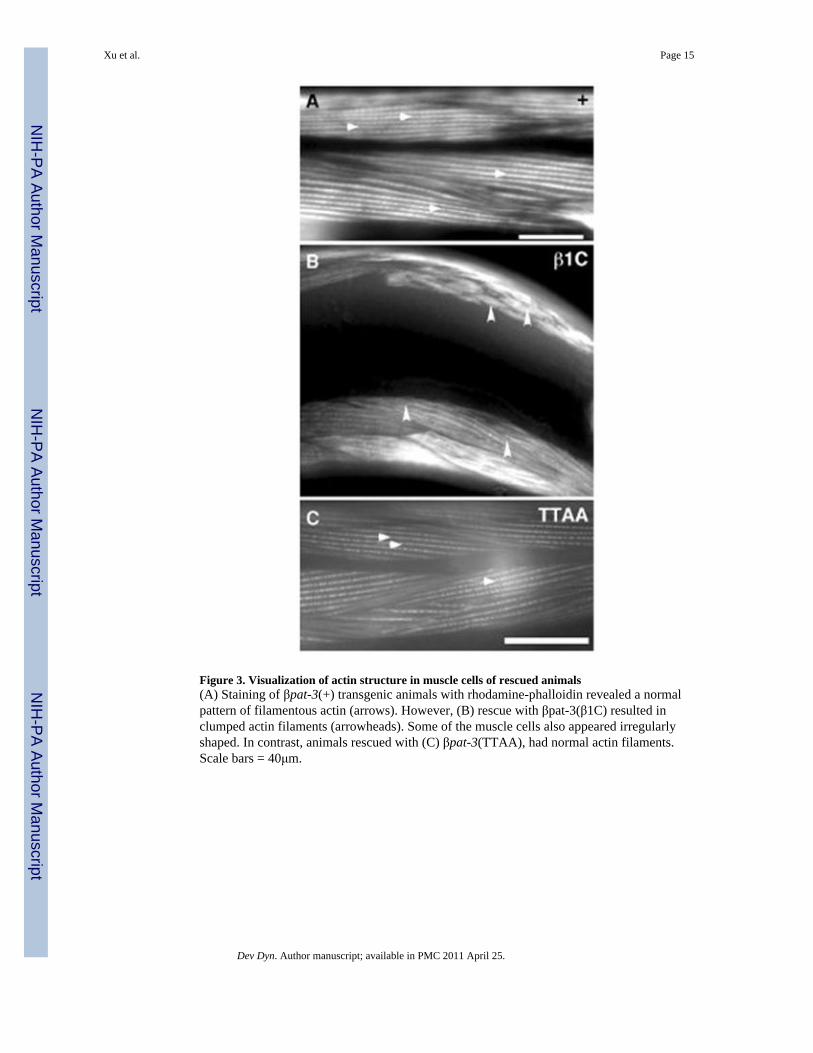
Figure 3. Visualization of actin structure in muscle cells of rescued animals(A) Staining of βpat-3(+) transgenic animals with rhodamine-phalloidin revealed a normalpattern of filamentous actin (arrows). However, (B) rescue with βpat-3(β1C) resulted inclumped actin filaments (arrowheads). Some of the muscle cells also appeared irregularlyshaped. In contrast, animals rescued with (C) βpat-3(TTAA), had normal actin filaments.Scale bars = 40μm.
Xu et al. Page 15
Dev Dyn. Author manuscript; available in PMC 2011 April 25.
NIH
-PA Author Manuscript
NIH
-PA Author Manuscript
NIH
-PA Author Manuscript
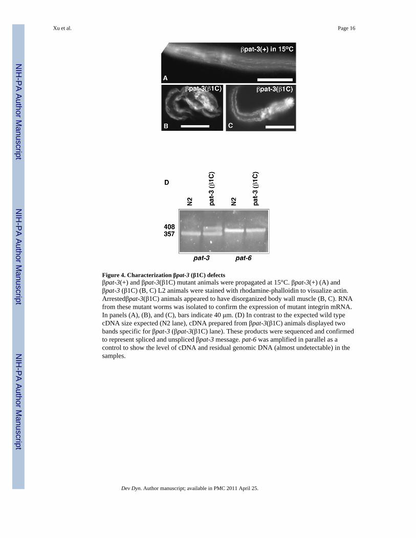
Figure 4. Characterization βpat-3 (β1C) defectsβpat-3(+) and βpat-3(β1C) mutant animals were propagated at 15°C. βpat-3(+) (A) andβpat-3 (β1C) (B, C) L2 animals were stained with rhodamine-phalloidin to visualize actin.Arrestedβpat-3(β1C) animals appeared to have disorganized body wall muscle (B, C). RNAfrom these mutant worms was isolated to confirm the expression of mutant integrin mRNA.In panels (A), (B), and (C), bars indicate 40 μm. (D) In contrast to the expected wild typecDNA size expected (N2 lane), cDNA prepared from βpat-3(β1C) animals displayed twobands specific for βpat-3 (βpat-3(β1C) lane). These products were sequenced and confirmedto represent spliced and unspliced βpat-3 message. pat-6 was amplified in parallel as acontrol to show the level of cDNA and residual genomic DNA (almost undetectable) in thesamples.
Xu et al. Page 16
Dev Dyn. Author manuscript; available in PMC 2011 April 25.
NIH
-PA Author Manuscript
NIH
-PA Author Manuscript
NIH
-PA Author Manuscript

Figure 5. βpat-3 transgenic animals display DTC migration defectsNormal DTC migration in a (A) βpat-3 (+) rescued animal in contrast to the (B)βpat-3(Y972F), (C) βpat-3(β1C), and (D) βpat-3(TTAA) rescued lines, in which the DTC(arrowhead) follows an incorrect migratory path. The path taken by the DTC is indicated bythe arrow diagrams. In all panels, vulva is located on the bottom right corner. Scale bars =40 μm.
Xu et al. Page 17
Dev Dyn. Author manuscript; available in PMC 2011 April 25.
NIH
-PA Author Manuscript
NIH
-PA Author Manuscript
NIH
-PA Author Manuscript

Figure 6. Proximal gonad morphology of βpat-3 transgenic animals(A) βpat- (+) line possessed normally developing oocytes arranged in a typical linearpattern. In the (B) βpat-3(β1C), (C) βpat-3(TTAA), and (D) βpat-3(Y792F) lines, oocytesfailed to ovulate and piled on top of each other in the proximal gonad, a phenotype typical ofendomitotic (Emo) oocytes. Arrowheads indicate the oocyte nuclei. Scale bar = 40 μm.
Xu et al. Page 18
Dev Dyn. Author manuscript; available in PMC 2011 April 25.
NIH
-PA Author Manuscript
NIH
-PA Author Manuscript
NIH
-PA Author Manuscript

Figure 7. Abnormal tail (Abt) phenotype of βpat-3 transgenic animals(A) A typical wild-type, whiplike hermaphrodite tail of βpat-3(+) hermaphrodites. (B)βpat-3(TTAA) animals displayed severely abnormal tail morphology. Arrowheads indicatethe location of the anus. Scale bar = 40 μm.
Xu et al. Page 19
Dev Dyn. Author manuscript; available in PMC 2011 April 25.
NIH
-PA Author Manuscript
NIH
-PA Author Manuscript
NIH
-PA Author Manuscript

NIH
-PA Author Manuscript
NIH
-PA Author Manuscript
NIH
-PA Author Manuscript
Xu et al. Page 20
Tabl
e 1
βPA
T-3
cyt
opla
smic
tail
resi
dues
are
req
uire
d fo
r fe
rtili
ty, g
onad
mor
phog
enes
is a
nd fu
nctio
n, a
nd ta
il m
orph
ogen
esis
You
ng a
dult
anim
als w
ere
scor
ed fo
r fer
tility
(% o
f ani
mal
s bea
ring
live
prog
eny)
, DTC
mig
ratio
n de
fect
s (%
of i
ncor
rect
ly fo
rmed
gon
ad a
rms)
, Em
o (%
of a
nim
als w
ith e
ndom
itotic
ooc
ytes
) and
Abt
(% o
f ani
mal
s with
abn
orm
al ta
il m
orph
olog
y). S
train
s sho
win
g si
gnifi
cant
pai
r-w
ise
diff
eren
ces f
rom
wild
type
(+) f
or e
ach
phen
otyp
e, b
ased
on
the
95%
con
fiden
ce in
terv
al fo
r pro
porti
ons,
are
show
n sh
aded
in g
ray.
Tra
it+
β1C
23°
β1C
15°
TT
AA
Y79
2FY
804F
YY
FFFK
VV
W78
4A
Fert
ility
86%
N=5
954
%N
=56
1%N
=100
72%
N=1
4284
%N
=44
93%
N=5
586
%N
=59
83%
N=4
810
0%N
=60
DT
C m
igra
tion
11%
N=1
2224
%N
=58
50%
N=2
645
%N
=101
22%
N=1
3438
%N
=202
27%
N=1
4819
%N
=88
21%
N=1
00
Em
o1%
N=1
1218
%N
=129
N.D
.14
%N
=59
12%
N=1
2619
%N
=104
19%
N=1
002%
N=1
003%
N=1
46
Abt
14%
N=5
923
%N
=56
N.D
.68
%N
=133
18%
N=4
45% N=5
522
%N
=59
4% N=4
84% N=9
8
Dev Dyn. Author manuscript; available in PMC 2011 April 25.