Host-Parasite Interaction in Sarcoptes scabiei Infestation in ...
Upload
independentCategory
view
2download
0
INFECTION AND IMMUNITY, Feb. 2006, p. 1313–1322 Vol. 74, No. 20019-9567/06/$08.00�0 doi:10.1128/IAI.74.2.1313–1322.2006Copyright © 2006, American Society for Microbiology. All Rights Reserved.
Analysis of Antibodies Directed against Merozoite Surface Protein 1 ofthe Human Malaria Parasite Plasmodium falciparum
Ute Woehlbier,1 Christian Epp,1 Christian W. Kauth,1 Rolf Lutz,1 Carole A. Long,2
Boubacar Coulibaly,3 Bocar Kouyate,3 Myriam Arevalo-Herrera,4
Socrates Herrera,4 and Hermann Bujard1*Zentrum fuer Molekulare Biologie (ZMBH), Universitaet Heidelberg, Im Neuenheimer Feld 282, D-69120 Heidelberg, Germany1;
Malaria Vaccine Development Branch, NIAID, National Institutes of Health, Rockville, Maryland 208522; Centre deRecherche en Sante de Nouna (CRSN), Nouna, Burkina Faso3; and Centro Internacional de Vacunas,
Carrera 35 No. 4A-53, Cali, Colombia4
Received 1 July 2005/Returned for modification 31 August 2005/Accepted 27 October 2005
The 190-kDa merozoite surface protein 1 (MSP-1) of Plasmodium falciparum, an essential component in theparasite’s life cycle, is a primary candidate for a malaria vaccine. Rabbit antibodies elicited by the heterolo-gously produced MSP-1 processing products p83, p30, p38, and p42, derived from strain 3D7, were analyzedfor the potential to inhibit in vitro erythrocyte invasion by the parasite and parasite growth. Our data show that(i) epitopes recognized by antibodies, which inhibit parasite replication, are distributed throughout the entireMSP-1 molecule; (ii) when combined, antibodies specific for different regions of MSP-1 inhibit in a strictlyadditive manner; (iii) anti-MSP-1 antibodies interfere with erythrocyte invasion as well as with the intraeryth-rocytic growth of the parasite; and (iv) antibodies raised against MSP-1 of strain 3D7 strongly cross-inhibitreplication of the heterologous strain FCB-1. Accordingly, anti-MSP-1 antibodies appear to be capable ofinterfering with parasite multiplication at more than one level. Since the overall immunogenicity profile ofMSP-1 in rabbits closely resembles that found in sera of Aotus monkeys immunized with parasite-derivedMSP-1 and of humans semi-immune to malaria from whom highly inhibiting antigen-specific antibodies wererecovered, we consider the findings reported here to be relevant for the development of MSP-1-based vaccinesagainst malaria.
The merozoite surface protein 1 (MSP-1) complex of Plas-modium falciparum constitutes a major component at the sur-face of the erythrocyte (RBC)-invading form of the parasite(21). It originates from an approximately 190-kDa glyco-sylphosphatidylinositol (GPI)-anchored precursor, which isproteolytically processed during merozoite maturation, yield-ing in a first step four major fragments, p83, p30, p38, and p42,that remain, however, noncovalently associated (31). This com-plex interacts with further surface proteins of the parasite suchas—again processed forms of—MSP-6 (48) and MSP-7 (36,44). Before reinvasion of new RBCs, a secondary proteolyticevent cleaves p42 into p33 and the approximately 10-kDa GPI-anchored C terminus, designated p19 (4). This portion ofMSP-1, which contains two epidermal growth factor (EGF)-like domains, is transferred into the newly infected RBC, whilethe rest of the complex is shed from the parasite’s surface (3).Analyses of the primary structure of MSP-1 from differentclones of P. falciparum have revealed that several regions arehighly conserved, whereas others appear to be dimorphic, per-mitting classification of strains into the K1 or MAD20 family.In addition, there are two small blocks of higher sequencevariation (32, 46) (Fig. 1).
There is good evidence that MSP-1 plays an essential role inthe parasite’s life cycle and that it is crucially involved in the
RBC invasion process. For example, preventing the proteolyticcleavage that generates p19 inhibits invasion of RBCs in vitro(5). Moreover, results indicating direct interactions betweenMSP-1 and the RBC surface have been reported (14, 34),suggesting that MSP-1 may play a role in early interactionsbetween the parasite and RBCs, thus being possibly involved inthe RBC invasion process at more than one level. Finally,attempts to genetically inactivate the msp-1 gene failed (35),underlining its essential role. All these findings make MSP-1 amost interesting target for interfering with the infectious cycleof the parasite, and there is ample evidence in support ofMSP-1 as a prime candidate for a vaccine against malaria.Indeed, MSP-1 is a target of the human immune response, andnumerous seroepidemiological studies have revealed associa-tions between reduced susceptibility to clinical malaria andhumoral responses against various regions of the molecule (6,8, 11, 38–40, 47). Furthermore, immunization of Aotus mon-keys with MSP-1 isolated from parasites induces high levels ofprotection against lethal challenges with parasites (42; H. Bu-jard et al., unpublished data), and partial (17, 43) or full (7)protection in the primate model was also reported for variousMSP-1-derived recombinant protein preparations. Importantinformation was collected from the mouse Plasmodium yoeliimodel, in which immunization with native MSP-1 (13) and withrecombinant protein (24) conferred not only protection butalso passive transfer of a monoclonal antibody (30).
Some studies revealed a particularly interesting role forepitopes located within the two EGF-like domains of the p19processing fragment at the C terminus of MSP-1 (Fig. 1), as
* Corresponding author. Mailing address: Zentrum fuer MolekulareBiologie (ZMBH), Universitaet Heidelberg, Im Neuenheimer Feld282, D-69120 Heidelberg, Germany. Phone: 49-6221 54 8214. Fax:49-6221 54 5892. E-mail: [email protected].
1313
on February 23, 2016 by guest
http://iai.asm.org/
Dow
nloaded from

recombinant proteins containing these domains, when used asvaccines, were protective in mice and in primates (1, 7, 9, 10,18–20, 29, 38). Moreover, monoclonal antibodies targetingspecific conformational epitopes within these domains wereshown to inhibit not only in vitro RBC invasion by the parasite(2) but also processing of p42 into p33 and p19 (5), therebyindicating that this proteolytic cleavage is an essential step inthe infectious cycle of blood stage parasites. These findingshave moved the C-terminal portion (p42 and p19) of MSP-1 tothe center of interest, also as a candidate for a malaria vaccine.Interestingly, Guevara Patino et al. (15) have also identifiedso-called blocking antibodies that can prevent the interactionof inhibiting antibodies with their respective epitopes, thusallowing cleavage of p42 and consequently invasion of RBCs toproceed. Blocking antibodies, which were also identified insome human sera, were shown to bind not only within p19 butalso in other regions of MSP-1, such as conserved domains ofp83 (15). Clearly, as proposed, the induction of blocking anti-bodies would represent a novel mechanism of immune evasion;with respect to the development of an MSP-1-based vaccine, itwould therefore seem advisable to restrict the effective antigento p19, preferably in a modified version that induces exclusivelyinhibiting but not blocking antibodies (49).
On the other hand, considering the situation in vivo, theeffect of blocking antibodies depends on how efficiently theycompete with inhibitory antibodies, which in turn is a functionof a number of thermodynamic and kinetic parameters that aredifficult to quantitatively assess. In this context, it is interestingto note that the successful immunization of rodents (23) andprimates (7) with recombinant p19 or p42 preparations indi-cates an effective competition of invasion-inhibitory antibodieswith at least p19-specific blocking activities. The same appearsto hold true for protective immunizations with full-size MSP-1of mice (22) and primates (42; Bujard et al., unpublished data),again suggesting that blocking antibodies, possibly elicited byother parts of MSP-1, are not sufficiently effective at leastunder these conditions of immunization. Finally, it is not clearto what extent invasion-inhibiting antibodies specifically elic-ited by the p19 portion of MSP-1 contribute to the overallprotective humoral response in semi-immune individuals andwhich increment of protection would be lost by the antagonismof respective blocking antibodies.
Interestingly, seroepidemiological studies aimed at uncover-
ing associations between a reduced risk for clinical malaria andantibody responses towards various regions of MSP-1 have notled to a coherent picture, even when differences in the variousexperimental approaches are reconciled. They suggest ratherthat MSP-1 harbors, in addition to p19, further regions capableof eliciting protective immune responses.
In following up the latter question, we have examined anti-bodies specific for different regions of MSP-1 for the potentialto interfere with the multiplication of P. falciparum in vitro. Wehave raised in rabbits antibodies against the four major pro-cessing products of MSP-1 of P. falciparum strain 3D7, hence-forth referred to as MSP-1D, and tested their potential toinhibit in vitro parasite growth and RBC invasion. We showthat epitopes eliciting highly inhibitory antibodies are distrib-uted throughout the MSP-1 molecule and that combinations ofsuch antibodies interfere with parasite multiplication in astrictly additive mode. Furthermore, antibodies raised againstprocessing fragments of MSP-1D effectively cross-inhibit par-asites of the FCB-1 strain, a representative of the K1 proto-type. As the overall antigenicity profiles of MSP-1 in rabbits,monkeys, and humans closely resemble each other, the out-come of this study should be of interest for the development ofmalaria vaccines based on MSP-1.
MATERIALS AND METHODS
MSP-1 and MSP-1 processing fragments. All MSP-1D-derived proteins wereproduced in Escherichia coli from coding sequences synthesized analogously asdescribed previously (37). They contain N-terminal hexahistidine fusions andwere purified as described by Kauth et al. (26). Processing fragment p42, whichcontains six disulfide bridges within its two EGF-like domains, was prepared andcharacterized as described in detail by Epp et al. (12). Full-size MSP-1 wasassembled from its two “halves,” p83/30 and p38/42 (26). MSP-6 (48) and MSP-7(36) were heterologously produced in E. coli as glutathione S-transferase fusions.All MSP-1D fragments, as well as MSP-6 and MSP-7, used in this study are morethan 90% pure (26) and contain comparable amounts of endotoxins, rangingfrom 250 to 750 endotoxin units per 100 �g of protein, according to the Limulusamoebocyte lysate test (data not shown). MSP-1F from merozoites was isolatedfrom synchronized late-schizont cultures of P. falciparum strain FCB-1, essen-tially as described by Siddiqui et al. (42).
Generation of MSP-1-specific antibodies. Groups of three rabbits were im-munized at Charles River Laboratories, Kisslegg, Germany, according to thestandard procedure of the company. Accordingly, 100 �g of each of the fourpurified MSP-1D fragments, p83, p30, p38 and p42, was administered in Freund’scomplete adjuvant (CFA) for priming, followed by three boosts—on days 28, 42,and 56—with the same amount of protein in Freund’s incomplete adjuvant.Serum samples were withdrawn prior to each immunization, and total sera werecollected 2 weeks after the last boost.
Aotus lemurinus griseimembra monkeys were immunized with MSP-1F. Be-tween 40 and 50 �g of protein was administered in 50% CFA for priming,followed by boosts containing the same amount of protein in 25% and 10% CFA.Serum samples were withdrawn prior to each immunization (day 0, priming; day28, first boost; day 56, second boost) and 2 weeks after the last immunization.The full Aotus study will be reported elsewhere.
Purification of rabbit serum. Sera from rabbits of each group were pooled andpurified by Squarix, Marl, Germany, according to the standard procedures of thecompany. The same protocol was used in our laboratory to purify immunoglobu-lins of further rabbit sera and of human sera. In brief, sera were heat inactivatedfor 30 min at 56°C and centrifuged for 30 min at 5,000 rpm. Immunoglobulinswere precipitated by slow addition of 29.2 g ammonium sulfate per 100 ml serum,and the mixture was kept for 24 h at 4°C. The precipitate was pelleted bycentrifugation (60 min, 4000 � g, 4°C); redissolved in 60% of the original volumeof phosphate-buffered saline (PBS), pH 7.4, containing 0.05% sodium azide;dialyzed five times against 5 liters of the same buffer; and kept for 12 to 16 h at4°C. The resulting solution was cleared by centrifugation (60 min, 4000 � g, 4°C)and by subsequent filtration through a 0.45-�m membrane (Millipore) before itwas stored at 4°C. Some anti-p30 and anti-p38 preparations were redissolved inPBS, reconstituting the original serum concentration.
FIG. 1. Schematic outline of MSP-1D. The precursor of MSP-1D isa protein comprising 1,720 amino acids, including a 20-amino-acidsignal sequence (SS) and a signal for anchoring the protein at thecellular surface via a GPI moiety (GA). The precursor is processedinto four major products, p83, p30, p38, and p42, as indicated byarrows. In a second proteolytic step, p42 is cleaved into p33 and p19.Primary sequences of MSP-1 from different strains follow distinctpatterns of conservation. As indicated, there are regions of high con-servation (white) and regions that are essentially dimorphic (grey),classifying the molecule as a member of either the K1 or the MAD20family (32, 46). In addition, there are two short oligomorphic areas,block II and block IV (46). MSP-1D of strain 3D7 belongs to theMAD20 family, with the exception of block II, which is K1-like.
1314 WOEHLBIER ET AL. INFECT. IMMUN.
on February 23, 2016 by guest
http://iai.asm.org/
Dow
nloaded from
Purification of antibodies by antigen affinity chromatography. The antigensp83, p30, p38, and p42 were immobilized on HYDRA-R activated affinity resin(Squarix GmbH, Marl, Germany), according to the standard procedures of thecompany. In brief, the recombinant proteins were concentrated by a 10-kDaultrafiltration unit (Millipore) and exhaustively dialyzed against coupling buffer(4 M urea, 0.05% sodium borate, pH 9.0) before they were incubated with theresin for 48 h at room temperature. After the addition of blocking buffer (4 Murea, 0.2 M ethanolamine, pH 9.2), the suspension was incubated for 12 h furtherat room temperature and then transferred to a chromatography system (AKTA;Amersham Pharmacia) and processed at 4°C. The column was washed exten-sively with coupling buffer, distilled water, PBS-sodium azide, and all of thebuffers that were used in the following chromatography procedures.
For purification of human antibodies, p83 (5 mg/ml) was dialyzed againstcoupling buffer (6 M urea, 50 mM sodium borate, pH 8.5) and subsequentlyincubated with HYDRA-R activated affinity resin for 48 h at room temperature.The resin was washed with the same buffer, blocked with 6 M urea–0.2 Methanolamine (pH 9.2) for 24 h at room temperature, and transferred to thechromatography system. Proteins, covalently coupled to the resin, were refoldedby a linear gradient replacing the blocking buffer with 1 M arginine–50 mM Tris(pH 8.5). A second linear gradient exchanged the refolding buffer for PBS, pH7.4. For both gradients, a flow rate of 0.2 column volumes per minute over 24 hwas applied. The resin was thoroughly washed with all buffers used in thesubsequent chromatography procedures. The serum was loaded onto the columnwith a flow rate of 0.5 cm/min. After being washed with PBS, pH 7.4, theantibodies were eluted by 75 mM glycine (pH 2.8), 0.5 M NaCl, and PBS (pH 7.4)plus 1 M NaCl. The acidic eluates were immediately neutralized by 0.1 volumesof 1 M Tris, pH 8.0. All fractions were pooled, and the antibodies were precip-itated by ammonium sulfate as described above. The pellet was dissolved in PBS,pH 7.4, and extensively dialyzed against the same buffer. All affinity-purifiedantibody preparations were examined in Western blots, as shown for the humananti-p83 preparation (see Fig. 7A).
Immunological methods. Sodium dodecyl sulfate-polyacrylamide gel electro-phoresis and Western blotting were performed as described previously (37).
An enzyme-linked immunosorbent assay was performed as follows. Microtiterplates (96 well) were coated with recombinant protein by overnight incubationwith 0.1 ml of 100 nM protein in PBS at 4°C. Plates were washed once with TBST(10 mM Tris-HCl [pH 8.0], 150 mM NaCl, 0.05% Tween 20) before they wereincubated for 1 h at room temperature with blocking buffer (TBST containing1% milk powder). Sera to be examined were diluted serially with blocking bufferand incubated for 2 h at room temperature. After three washes with TBST, theappropriate secondary antibodies (Sigma, Munich, Germany), diluted as recom-mended by the supplier, were added and the mixture was incubated for 1 h atroom temperature. The substrate p-nitrophenyl-phosphate (1 mg/ml in 0.96%[vol/vol] diethanolamine [pH 9.5] and 1 mM MgCl2) was added, and the mixturewas incubated for 1 h at room temperature in the dark. The reaction was stoppedwith 0.1 ml of 2 M NaOH, and the absorbance was measured at 405 nm.
Human and Aotus antibodies were detected with a goat anti-human immuno-globulin G (IgG)–alkaline phosphatase conjugate (Promega) diluted at 1:3,000,whereas rabbit antibody titers were measured with a goat anti-rabbit IgG–alka-line phosphatase conjugate (Sigma) diluted at 1:2,500.
Antibody titers were calculated at the serum dilutions that produced an ab-sorbance (optical density at 405 nm [OD405]) of 1.0 for human, rabbit, andmonkey sera.
Culturing of parasites. P. falciparum parasites were grown under standardconditions (16) at 37°C, 5% CO2, 3% O2, and 95% humidity, until most of theparasites reached schizont stage. Magnetic cell separation columns (MACS;Miltenyi Biotech) were used to purify schizonts. After the column was placed inthe magnetic field, the matrix was blocked by washing with preheated buffer A(PBS [pH 7.4]–0.5% bovine serum albumin, 37°C). Plasmodium cultures weresuspended and applied to the column. After being washed with preheated bufferA, the column was removed from the magnetic field and schizonts were elutedwith culture medium. Centrifugation (5 min, 1,500 rpm) yielded small blackpellets consisting of infected RBCs (iRBCs), of which 90% were in the schizontstage. The pellets were dissolved in culture medium having the desired hemat-ocrit (1%), and parasitemia was adjusted to 0.3%.
Assays for inhibition of parasite replication. The potential of sera and anti-body preparations to inhibit parasite multiplication was monitored (i) by mea-suring parasite-specific lactate dehydrogenase (LDH) activity and (ii) by moni-toring the development of cultured parasites via flow cytometry (16).
Prior to use in inhibition assays, sera and antibody preparations, including allpreparations used as references and as controls, were preadsorbed on type Ahuman RBCs, whereby 1 ml of antibody solution was incubated with 50 �l of
RBCs (�90% hematocrit) for 1 h. After RBCs were pelleted by centrifugation,the supernatants were dialyzed together against 100 volumes of RPMI medium.
LDH assay. Sera were examined for their potency in inhibiting P. falciparumreplication by measuring LDH levels in late-trophozoite/early-schizont-stageparasites, according to a protocol developed by C. A. Long (33). Plasmodiumstrains 3D7 and FCB-1 were synchronized by magnetic cell separation as de-scribed above. Cultures were adjusted to 0.3% parasitemia with human type ARBCs at a final hematocrit of 1%. The final assay volume of cultures was 100 �l,containing between 5 and 40% (vol/vol) serum or antigen affinity-purified anti-body preparations. In standard assays, sera or antigen affinity-purified antibodieswere added in a volume of 20 �l. For monitoring of the concentration depen-dencies of sera or antibodies or of the additive effects of sera specific for differentfragments, sera or antibody preparations were supplied to the assay mixture in atotal volume of 40 �l, which contained the respective amount of antibody prepara-tion (5 to 40 �l) adjusted to a final volume of 40 �l with medium whenever required.Corresponding volumes of preimmune rabbit sera or immunoglobulins, as well assera from rabbits immunized with the ClpB protein from E. coli in Freund’s adju-vant, served as controls. P. falciparum 3D7 and FCB-1 were assayed after 40 and48 h, respectively. Inhibition was measured at 650 nm and calculated as follows:percent inhibition � 100% � (ODimmune serum � ODRBC/ODpreimmune serum �ODRBC) � 100.
Flow cytometric assay. P. falciparum 3D7 was synchronized by magnetic cellseparation columns as described above. Cultures were adjusted to 0.3% para-sitemia with human type A RBCs at a final hematocrit of 1% containing 20%(vol/vol) serum or immunoglobulin preparation. Negative controls were as de-scribed above. For following the time course of parasite multiplication, indepen-dently incubated samples were harvested at 4-h intervals up to 44 h and assayedas follows. Cells were resuspended in 0.1 ml of PBS, pH 7.4, containing 0.05%glutaraldehyde and incubated for 20 h at 4°C. For DNA staining, 10 �g/ml ofpropidium iodide in PBS, pH 7.4, was added to the fixed preparation, which waskept for another hour in the dark. Parasitemia was detected via flow cytometry(FACScan; Becton-Dickinson) according to DNA content. iRBCs were visual-ized via “density dot blotting” with FL3 as the x axis (red fluorescence) and FL2as the y axis (orange fluorescence). Inhibition was calculated as follows: percentinhibition � 100% � (Pimmune serum � PRBC)/(Ppreimmune serum � PRBC) � 100,where P is parasitemia. In all inhibition experiments, antibody preparations to beexamined for their inhibitory potential were assayed together with reference andcontrol antibodies prepared under identical biochemical and temporal condi-tions.
Microscopic analysis of parasite cultures. Samples of parasite cultures wereprepared as thin films on microscopic glass slides before they were fixed for 2 minin methanol and stained for 10 min in Giemsa stain (Sigma), air dried, mountedwith Mowiol, and covered with a coverslip. The preparations were examined bylight microscopy with a 100� oil immersion objective lens.
RESULTS
Inhibition of parasite replication in vitro by MSP-1D-spe-cific rabbit antibodies. All rabbit sera used in this study weresubjected to ammonium sulfate precipitation, and the precip-itate was redissolved in PBS as described in Materials andMethods. For simplicity, the latter preparations are designatedserum or sera in contrast to antigen affinity-purified antibodies.
Rabbits immunized with purified p83, p30, p38, and p42yielded sera which reproducibly differed in fragment-specificantibody titers, indicating regions of significantly differing im-munogenicity within MSP-1D. Accordingly, p42 appears to bethe most immunogenic protein, followed by p83, p30, and p38(Fig. 2A). When these sera were examined for the potential toinhibit growth of P. falciparum 3D7 in vitro, as monitored bythe LDH assay, they all inhibited significantly parasite replica-tion (Fig. 2A), and when inhibition is related to fragment-specific antibody titers, sera obtained by immunization withp30 and p38 are particularly effective. As expected, the ob-served inhibition was concentration dependent, as shown inFig. 2B. By contrast, no inhibition was found with serum ob-tained from rabbits immunized with purified ClpB proteinfrom E. coli using the same immunization protocol. To un-
VOL. 74, 2006 ANALYSIS OF ANTIBODIES AGAINST MSP-1 1315
on February 23, 2016 by guest
http://iai.asm.org/
Dow
nloaded from
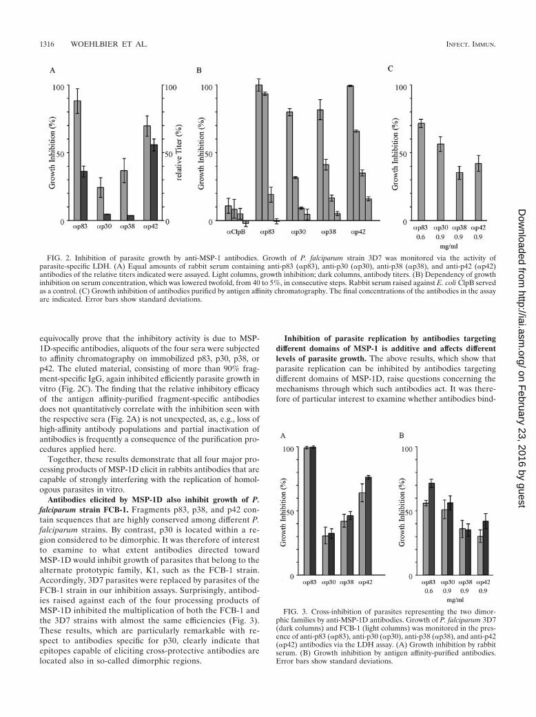
equivocally prove that the inhibitory activity is due to MSP-1D-specific antibodies, aliquots of the four sera were subjectedto affinity chromatography on immobilized p83, p30, p38, orp42. The eluted material, consisting of more than 90% frag-ment-specific IgG, again inhibited efficiently parasite growth invitro (Fig. 2C). The finding that the relative inhibitory efficacyof the antigen affinity-purified fragment-specific antibodiesdoes not quantitatively correlate with the inhibition seen withthe respective sera (Fig. 2A) is not unexpected, as, e.g., loss ofhigh-affinity antibody populations and partial inactivation ofantibodies is frequently a consequence of the purification pro-cedures applied here.
Together, these results demonstrate that all four major pro-cessing products of MSP-1D elicit in rabbits antibodies that arecapable of strongly interfering with the replication of homol-ogous parasites in vitro.
Antibodies elicited by MSP-1D also inhibit growth of P.falciparum strain FCB-1. Fragments p83, p38, and p42 con-tain sequences that are highly conserved among different P.falciparum strains. By contrast, p30 is located within a re-gion considered to be dimorphic. It was therefore of interestto examine to what extent antibodies directed towardMSP-1D would inhibit growth of parasites that belong to thealternate prototypic family, K1, such as the FCB-1 strain.Accordingly, 3D7 parasites were replaced by parasites of theFCB-1 strain in our inhibition assays. Surprisingly, antibod-ies raised against each of the four processing products ofMSP-1D inhibited the multiplication of both the FCB-1 andthe 3D7 strains with almost the same efficiencies (Fig. 3).These results, which are particularly remarkable with re-spect to antibodies specific for p30, clearly indicate thatepitopes capable of eliciting cross-protective antibodies arelocated also in so-called dimorphic regions.
Inhibition of parasite replication by antibodies targetingdifferent domains of MSP-1 is additive and affects differentlevels of parasite growth. The above results, which show thatparasite replication can be inhibited by antibodies targetingdifferent domains of MSP-1D, raise questions concerning themechanisms through which such antibodies act. It was there-fore of particular interest to examine whether antibodies bind-
FIG. 2. Inhibition of parasite growth by anti-MSP-1 antibodies. Growth of P. falciparum strain 3D7 was monitored via the activity ofparasite-specific LDH. (A) Equal amounts of rabbit serum containing anti-p83 (�p83), anti-p30 (�p30), anti-p38 (�p38), and anti-p42 (�p42)antibodies of the relative titers indicated were assayed. Light columns, growth inhibition; dark columns, antibody titers. (B) Dependency of growthinhibition on serum concentration, which was lowered twofold, from 40 to 5%, in consecutive steps. Rabbit serum raised against E. coli ClpB servedas a control. (C) Growth inhibition of antibodies purified by antigen affinity chromatography. The final concentrations of the antibodies in the assayare indicated. Error bars show standard deviations.
FIG. 3. Cross-inhibition of parasites representing the two dimor-phic families by anti-MSP-1D antibodies. Growth of P. falciparum 3D7(dark columns) and FCB-1 (light columns) was monitored in the pres-ence of anti-p83 (�p83), anti-p30 (�p30), anti-p38 (�p38), and anti-p42(�p42) antibodies via the LDH assay. (A) Growth inhibition by rabbitserum. (B) Growth inhibition by antigen affinity-purified antibodies.Error bars show standard deviations.
1316 WOEHLBIER ET AL. INFECT. IMMUN.
on February 23, 2016 by guest
http://iai.asm.org/
Dow
nloaded from

ing to different parts of MSP-1 would, when combined, have anadditive, possibly synergistic effect on inhibition or, consideringthe hypothesis of immune evasion via blocking antibodies (15),would interfere with inhibitory activities. As a blocking mech-anism was described in the context of inhibitory antibodiesdirected toward the p19 processing fragment, we first com-bined sera raised against p42 with p83, p30, or p38. As shownin Fig. 4, all three combinations inhibit parasite replication, aswould be expected by simple addition of the individual inhib-itory activities. Corresponding results were obtained with all ofthe other assayed combinations (Fig. 4). These results excludea significant role of blocking antibodies under our experimen-tal conditions.
To gain an initial insight into how antibodies targeted todifferent regions of MSP-1D may inhibit parasite replication,we monitored the time course of parasite multiplication insynchronized cultures in the presence and absence of anti-MSP-1 antibodies by flow cytometric measurement of theDNA content of iRBCs, as well as by microscopy. As thedisappearance of iRBCs from such cultures is a measure ofinvasion inhibition, our data demonstrate that all of our serumpreparations specific for p83, p30, p38, and p42 are effective atthis level (Fig. 5A and B). Thus, invasion of RBCs by theparasite can be efficiently prevented by antibodies binding tovarious domains of MSP-1. Analyzing the flow cytometric pat-tern in more detail reveals yet another intriguing feature. Wefound a substantial fraction of RBCs which had been success-
fully invaded by the parasite in the presence of inhibitinganti-MSP-1 antibodies. In these RBCs, however, the develop-ment of the parasite is severely hampered: in controls, thenumbers of ring and trophozoite stage iRBCs decrease overtime due to the transition into the schizont stage, but thepopulation of iRBCs in the presence of inhibiting antibodiesremains constant over the same period, indicating blocked orhighly retarded development of the intraerythrocytic parasite(Fig. 5B and C). This is most clearly seen for anti-p83 andanti-p42 antibodies which, in the particular experimentsshown, inhibit overall parasite multiplication by 80 to 95%.The same phenomenon is observed also with anti-p30 andanti-p38 antibodies even under conditions of lower overallinhibition (Fig. 5C). This intracellular inhibition has been ver-ified also with antigen affinity-purified anti-p83 and anti-p42antibodies (unpublished data). Microscopic inspection of suchparasite cultures reveals iRBCs containing parasites which re-semble previously described nonviable crisis forms (45), asshown in Fig. 5D.
Together, these results demonstrate that invasion of RBCsby parasites can be inhibited by antibodies targeting epitopeslocated in different regions of MSP-1D. They indicate further-more that MSP-1-specific antibodies are capable of interferingnot only with the RBC invasion process but also with theintraerythrocytic development of the parasite.
Immunogenicity profile of MSP-1. Rabbits immunized withthe individual processing fragments of MSP-1D yield sera thatdiffer largely in antibody titers, revealing a characteristic im-munogenicity profile for MSP-1D. Consistently, p42 was foundto be the most immunogenic compound, followed by p83, p30,and p38, whereas the titers for the two latter proteins usuallydo not differ significantly (Fig. 2A and 6A). Actual titers werebetween 9 � 105 for anti-p42 and 5 � 104 for anti-p38 anti-bodies under the immunization conditions used. This basicimmunogenicity profile was observed irrespective of whetherrabbits were immunized with individual fragments of MSP-1D(Fig. 2A) or with the full-size MSP-1D assembled from its twohalves, i.e., p83/30 and p38/42 (Fig. 6A). The same hierarchy ofantibody titers is found also in sera from Aotus monkeys im-munized with MSP-1 isolated from merozoites of P. falciparumstrain FCB-1 (Bujard et al., unpublished data) and in sera ofsemi-immune individuals living in an area of Burkina Faso,West Africa, where malaria is hyperendemic (Fig. 6B and C).
This obvious similarity between the immunogenicity profilesof MSP-1 in different mammalian systems raises the questionof whether MSP-1-specific antibodies from semi-immune indi-viduals would inhibit parasite replication in vitro as do thecorresponding rabbit antibodies. Accordingly, in a first exper-iment we have purified by affinity chromatography anti-p83antibodies from the serum of a semi-immune individual (Fig.7A). These human antibodies inhibit parasite replication invitro as efficiently as corresponding preparations from rabbitserum (Fig. 7B).
DISCUSSION
Clinical symptoms caused by P. falciparum infections areconsequences of the asexual erythrocytic stages of the malariaparasite, during which merozoites invade and—upon multipli-cation—emerge from RBCs in repetitive cycles. Immune re-
FIG. 4. Inhibition of parasite growth by antibodies (�) directedtowards different regions of MSP-1 (p83, p30, p38, and p42). Rabbitsera specific for the four processing fragments were diluted to givesuboptimal inhibition of parasite growth before the indicated combi-nations were composed and assayed for growth inhibition by the LDHassay. Antibodies were added to the assay mixtures in a volume of 40�l containing 10 �l of each serum, as indicated, and whenever neces-sary adjusted to 40 �l with medium. Light columns, inhibition calcu-lated from the expected contribution of the individual antibody prep-arations; dark columns, inhibition measured. Error bars show standarddeviations.
VOL. 74, 2006 ANALYSIS OF ANTIBODIES AGAINST MSP-1 1317
on February 23, 2016 by guest
http://iai.asm.org/
Dow
nloaded from
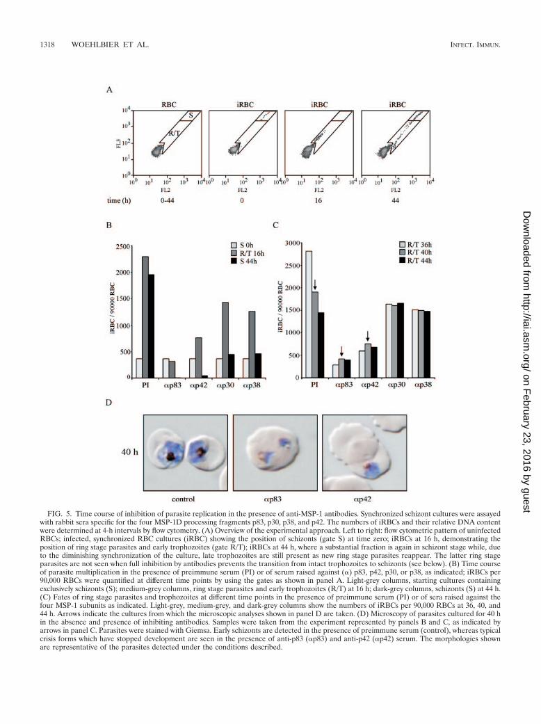
FIG. 5. Time course of inhibition of parasite replication in the presence of anti-MSP-1 antibodies. Synchronized schizont cultures were assayedwith rabbit sera specific for the four MSP-1D processing fragments p83, p30, p38, and p42. The numbers of iRBCs and their relative DNA contentwere determined at 4-h intervals by flow cytometry. (A) Overview of the experimental approach. Left to right: flow cytometric pattern of uninfectedRBCs; infected, synchronized RBC cultures (iRBC) showing the position of schizonts (gate S) at time zero; iRBCs at 16 h, demonstrating theposition of ring stage parasites and early trophozoites (gate R/T); iRBCs at 44 h, where a substantial fraction is again in schizont stage while, dueto the diminishing synchronization of the culture, late trophozoites are still present as new ring stage parasites reappear. The latter ring stageparasites are not seen when full inhibition by antibodies prevents the transition from intact trophozoites to schizonts (see below). (B) Time courseof parasite multiplication in the presence of preimmune serum (PI) or of serum raised against (�) p83, p42, p30, or p38, as indicated; iRBCs per90,000 RBCs were quantified at different time points by using the gates as shown in panel A. Light-grey columns, starting cultures containingexclusively schizonts (S); medium-grey columns, ring stage parasites and early trophozoites (R/T) at 16 h; dark-grey columns, schizonts (S) at 44 h.(C) Fates of ring stage parasites and trophozoites at different time points in the presence of preimmune serum (PI) or of sera raised against thefour MSP-1 subunits as indicated. Light-grey, medium-grey, and dark-grey columns show the numbers of iRBCs per 90,000 RBCs at 36, 40, and44 h. Arrows indicate the cultures from which the microscopic analyses shown in panel D are taken. (D) Microscopy of parasites cultured for 40 hin the absence and presence of inhibiting antibodies. Samples were taken from the experiment represented by panels B and C, as indicated byarrows in panel C. Parasites were stained with Giemsa. Early schizonts are detected in the presence of preimmune serum (control), whereas typicalcrisis forms which have stopped development are seen in the presence of anti-p83 (�p83) and anti-p42 (�p42) serum. The morphologies shownare representative of the parasites detected under the conditions described.
1318 WOEHLBIER ET AL. INFECT. IMMUN.
on February 23, 2016 by guest
http://iai.asm.org/
Dow
nloaded from

sponses directed toward antigens of the blood stage parasite,which interfere with this infectious cycle, are believed to playan essential role in the control of parasitemia, leading to re-duction or abrogation of clinical manifestations. Accordingly,MSP-1, which is crucially involved in the invasion process of
RBCs by merozoites, is considered a prime candidate for avaccine directed toward blood stage malaria.
With respect to MSP-1-based vaccine development, majorattention has been paid in the past to the C-terminal part ofthe molecule, as delineated by p42 and p19, for reasons alludedto above. At the same time, we know little about the immu-nogenicity of other parts of MSP-1, particularly about thepotentially protective nature of antibodies elicited by epitopeslocated outside the well-studied C-terminal portion of the mol-ecule. In the work described here, we have examined the invitro neutralizing potential of antibodies specific for the fourmajor processing fragments of MSP-1D by monitoring inhibi-tion of parasite growth and RBC invasion. If one accepts thatthe in vitro assays, as used here, describe valid correlates forthe in vivo functions of antibodies, our results strongly suggestthat protective antibodies are elicited by epitopes distributedthroughout the MSP-1 molecule.
Antibodies specific for the major subunits of the MSP-1Dcomplex were raised in rabbits using highly purified prepara-tions of p83, p30, p38, and p42 capable of assembling into thefull-size complex, as described previously (26). The resultingsera were first examined for the potential to inhibit growth of3D7 parasites in vitro, as monitored by the LDH assay. Inter-estingly, all sera clearly interfered with parasite multiplication,as did antibodies purified from such sera via antigen affinitychromatography. Obviously, MSP-1 encodes numerousepitopes which elicit antibodies that, based on the criteria ofour experimental systems, efficiently inhibit parasite replica-tion in vitro. Remarkably, antibodies elicited by the apparentlyleast-immunogenic regions of MSP-1, as delineated by p30 andp38, were highly effective.
Major regions of MSP-1, and in particular the area coveredby p30, are dimorphic in nature (32, 46). It therefore appearedlikely that a sizeable fraction of the inhibitory effect observedwith our rabbit sera would be strain specific. We thus examinedthe extent to which antibodies raised against subunits ofMSP-1D would cross-inhibit growth of the FCB-1 strain,which, with the exception of block II (Fig. 1) (46), is a typical
FIG. 6. Immunogenicity of MSP-1 in rabbits, Aotus monkeys, and humans semi-immune to malaria. Antibodies specific for p83, p30, p38, andp42 were quantified in the various sera via an enzyme-linked immunosorbent assay yielding the fragment-specific titers shown. (A) Rabbits (n �2) immunized with full-size MSP-1D assembled from p83/30 plus p38/42. The animals were primed with 100 �g of MSP-1D in CFA followed bytwo 100-�g boosts in Freund’s incomplete adjuvant. (B) Aotus lemurinus griseimembra monkeys (n � 10) were immunized with 50 �g of MSP-1Fisolated from parasites in CFA followed by two boosts of 50 �g of MSP-1F in Freund’s incomplete adjuvant. (C) Human sera were obtained from11 blood donors of the District Hospital of Nouna, Burkina Faso.
FIG. 7. Inhibition of parasite multiplication by human anti-p83 an-tibodies. Antibodies specific for p83 were isolated via antigen affinitychromatography from serum of a semi-immune blood donor fromNouna, Burkina Faso. (A) Sodium dodecyl sulfate-polyacrylamide gelelectrophoresis (12% polyacrylamide) followed by Western blot anal-ysis of serum (top) and of purified anti-p83 antibodies (bottom). Se-rum and antibodies were probed against recombinant p83, p30, p38,p42, MSP-7, and MSP-6. MW, molecular weight (in thousands).(B) Inhibition of parasite replication by anti-p83 (�p83) antibodies atthe concentrations indicated (measured by the LDH assay); humanIgG served as a control. Error bars show standard deviations.
VOL. 74, 2006 ANALYSIS OF ANTIBODIES AGAINST MSP-1 1319
on February 23, 2016 by guest
http://iai.asm.org/
Dow
nloaded from
representative of the K1 parasite family. Interestingly, a highdegree of cross-inhibition was observed, indicating that a con-siderable portion of the effective epitopes is encoded in con-served regions. This result was particularly unexpected for an-tibodies elicited by p30. However, a more detailed comparativeanalysis between the amino acid sequences of the 3D7 and theFCB-1 strains reveals numerous conserved short runs of aminoacids that could well serve as linear epitopes eliciting cross-reactive antibodies. In addition, data to be reported elsewhereindicate that p30 of MSP-1D and MSP-1F can form tertiarystructures which are functionally homologous and thus couldpossibly also give rise to shared conformational epitopes. Fi-nally, as shown by an immunofluorescence assay, our p30-specific antibodies stain both 3D7 and FCB-1 parasites (un-published data).
We next examined the effect of combining antibodies tar-geting different regions of MSP-1 on growth inhibition. As itwas of particular interest to see whether sera obtained byimmunization with p83, p30, or p38 would contain so-calledblocking antibodies, which would interfere with the inhibitorycapacity of p42-specific antibodies, respective combinationswere assayed first, followed by various other mixtures. Allcombinations of antibodies examined showed additive inhibi-tory effects on parasite growth, leaving no room for a net effectof blocking antibodies, which nevertheless may be containedwithin the various sera.
With the exception of invasion-inhibiting antibodies thatfunction through the prevention of p42 processing (5), we haveno information on how antibodies interfere with merozoitereplication in vitro. To gain initial insights, we studied, via flowcytometry and microscopy, the time course of parasite replica-tion in vitro in the presence and absence of MSP-1D-specificantibodies. It is evident from our data that all four antibodypreparations efficiently prevent RBC invasion, demonstratingthat antibodies which associate with MSP-1D outside of thep19 domain are capable of incapacitating merozoites for inva-sion. It will be interesting to explore whether some of theseantibodies also act via prevention of p42 processing or whethertheir inhibitory effect is based on a different mechanism. Suchmechanisms are conceivable, considering that MSP-1 interactswith further surface proteins of the parasite and that it has alsobeen implicated in initial associations between the parasite andthe RBC surface. Accordingly, antibodies associating with dif-ferent domains of this large and abundant surface compoundmay be expected to affect the parasite’s invasion process atseveral levels.
Our findings that anti-MSP-1D antibodies interfere alsowith the intraerythrocytic development of the parasite are par-ticularly intriguing, as they may signal an inhibitory effect onparasites that have escaped antibody-mediated surveillance ofinvasion. The intraerythrocytic parasites that develop, e.g., inthe presence of anti-MSP-1 antibodies resemble crisis forms inmorphology (45). The generation of such nonviable intraeryth-rocytic forms of the parasite in the context of protective anti-bodies was discussed previously (25). As this interesting phe-nomenon may have important implications for vaccinedevelopment, presently we are attempting to quantify the con-tribution of the intracellular inhibition of parasite develop-ment by antibodies to overall interference with parasite mul-
tiplication, particularly with respect to antibody preparationstargeting different regions of MSP-1.
One might argue that results obtained with antibodies raisedin rabbits and using Freund’s adjuvant do not reflect suffi-ciently the properties of antibody populations induced in hu-mans living in areas in which malaria is endemic. However,when the immunogenicity profiles of MSP-1, i.e., the relativetiters of antibodies specific for the four major processing frag-ments of MSP-1, were determined in rabbit sera as describedhere, in sera from Aotus monkeys immunized with MSP-1Fisolated from parasites and administered with CFA, or in serafrom semi-immune adults of West Africa exposed to naturalinfections, they closely resembled each other. Obviously, thisrather coarse correlation requires a more refined analysis. Wehave therefore begun to isolate MSP-1D fragment-specific an-tibodies from the sera of individuals who are semi-immune tomalaria by antigen-specific affinity chromatography, and in-deed an initial preparation of human anti-p83 antibodies in-hibits efficiently parasite replication in vitro, corroborating therelevance of our study in the rabbit system.
The results reported here raise questions and permit thedrawing of some conclusions. For example, it will be interest-ing to examine whether invasion-inhibiting antibodies bindingoutside of p42 also prevent p42 processing or whether they actvia a different mechanism. Furthermore, several modes of ac-tion can be conceived as to how MSP-1-specific antibodies mayretard or block intraerythrocytic development of the parasiteand apparently induce nonviable crisis forms, of which someare experimentally amenable in a straightforward manner.
On the other hand, our data strongly support the view that,with respect to MSP-1-based malaria vaccines, the entire mol-ecule should be taken into account. It appears evident thatMSP-1 is a carrier of a large number of B-cell epitopes capableof eliciting antibodies that efficiently inhibit parasite multipli-cation. Among these epitopes, a sufficient number is appar-ently encoded in conserved regions of the protein to providepotential cross-protection among parasite strains belonging toeither of the two prototypic families, K1 and MAD20. More-over, as we have to assume that under natural conditions RBCinvasion by merozoites is a rapid process (41), it appears ad-vantageous to induce, via vaccination, sets of antibodies thatinterfere with invasion at more than one level and which, inaddition, may retard or block intraerythrocytic parasite devel-opment.
Finally, as MSP-1 is initially synthesized in the infected he-patocyte, where its gene is apparently transcribed within thefirst 24 h (50), a presentation of MSP-1 epitopes via majorhistocompatibility complex class I complexes may make in-fected hepatocytes vulnerable to cytotoxic T-cell attack. Thisreasoning is supported by the findings of Krzych and coworkers(28), who showed that lymphocytes from human volunteersimmunized with radiation-attenuated sporozoites proliferateupon stimulation with MSP-1-derived peptides. Moreover,Kawabata et al. (27) have reported that adoptive transfer ofMSP-1-specified CD8� T cells results in protection in themouse P. yoelii system. Should an MSP-1-specified cytotoxicT-cell response indeed be effective at the liver stage of theparasite, it can be safely assumed that full-size MSP-1 harborsnumerous epitopes that will be presented by the various majorhistocompatibility complex class I complexes.
1320 WOEHLBIER ET AL. INFECT. IMMUN.
on February 23, 2016 by guest
http://iai.asm.org/
Dow
nloaded from

A vaccine based on the full-size MSP-1 may thus be consid-ered multivalent, which, due to the complexity of the approx-imately 190-kDa protein, provides a multitude of epitopes forhumoral and cellular responses, parameters that are also fa-vorable for overcoming the possible genetic restrictions of vac-cinees and for preventing the development of resistance.
ACKNOWLEDGMENTS
We are grateful to David Haynes for advice in setting up the inva-sion inhibition assays and flow cytometric monitoring, to John M.Gonzalez for help in the Aotus studies, and to Bernd Bukau for kindlyproviding anti-ClpB rabbit serum. We gratefully acknowledge the ex-cellent cooperation of blood donors of the District Hospital of Nouna,Burkina Faso, in particular, A. Barry, J. Bassole, G. Coulibaly, N.Dakuyo, F. Dembele, P. Dembele, R. Dehoun, D. Konate, F. Ouarme,M. Oui, and A. Wibiga. We thank Nestor Dembele (CRSN), AdamaCompaore (CRSN), and Silke Druffel-Augustin for diligent technicalassistance; Christian Mengede (Squarix) for advice; and Sibylle Reinigfor help in preparing the manuscript.
This work was supported by special research funds from the State ofBaden-Wuerttemberg, by the Deutsche Forschungsgemeinschaft (SFB544), and by Vakzine Projekt Management GmbH, Braunschweig,Germany.
We have no conflicting financial interest.
REFERENCES
1. Ahlborg, N., I. T. Ling, W. Howard, A. A. Holder, and E. M. Riley. 2002.Protective immune responses to the 42-kilodalton (kDa) region of Plasmo-dium yoelii merozoite surface protein 1 are induced by the C-terminal 19-kDa region but not by the adjacent 33-kDa region. Infect. Immun. 70:820–825.
2. Blackman, M. J., H. G. Heidrich, S. Donachie, J. S. McBride, and A. A.Holder. 1990. A single fragment of a malaria merozoite surface proteinremains on the parasite during red cell invasion and is the target of invasion-inhibiting antibodies. J. Exp. Med. 172:379–382.
3. Blackman, M. J., and A. A. Holder. 1992. Secondary processing of thePlasmodium falciparum merozoite surface protein-1 (MSP1) by a calcium-dependent membrane-bound serine protease: shedding of MSP133 as anoncovalently associated complex with other fragments of the MSP1. Mol.Biochem. Parasitol. 50:307–315.
4. Blackman, M. J., I. T. Ling, S. C. Nicholls, and A. A. Holder. 1991. Proteo-lytic processing of the Plasmodium falciparum merozoite surface protein-1produces a membrane-bound fragment containing two epidermal growthfactor-like domains. Mol. Biochem. Parasitol. 49:29–33.
5. Blackman, M. J., T. J. Scott-Finnigan, S. Shai, and A. A. Holder. 1994.Antibodies inhibit the protease-mediated processing of a malaria merozoitesurface protein. J. Exp. Med. 180:389–393.
6. Cavanagh, D. R., D. Dodoo, L. Hviid, J. A. L. Kurtzhals, T. G. Theander,B. D. Akanmori, S. Polley, D. J. Conway, K. Koram, and J. S. McBride. 2004.Antibodies to the N-terminal block 2 of Plasmodium falciparum merozoitesurface protein 1 are associated with protection against clinical malaria.Infect. Immun. 72:6492–6502.
7. Chang, S. P., S. E. Case, W. L. Gosnell, A. Hashimoto, K. J. Kramer, L. Q.Tam, C. Q. Hashiro, C. M. Nikaido, H. L. Gibson, C. T. Lee-Ng, P. J. Barr,B. T. Yokota, and G. S. Hut. 1996. A recombinant baculovirus 42-kilodaltonC-terminal fragment of Plasmodium falciparum merozoite surface protein 1protects Aotus monkeys against malaria. Infect. Immun. 64:253–261.
8. Conway, D. J., D. R. Cavanagh, K. Tanabe, C. Roper, Z. S. Mikes, N.Sakihama, K. A. Bojang, A. M. Oduola, P. G. Kremsner, D. E. Arnot, B. M.Greenwood, and J. S. McBride. 2000. A principal target of human immunityto malaria identified by molecular population genetic and immunologicalanalyses. Nat. Med. 6:689–692.
9. Darko, C. A., E. Angov, W. E. Collins, E. S. Bergmann-Leitner, A. S. Gir-ouard, S. L. Hitt, J. S. McBride, C. L. Diggs, A. A. Holder, C. A. Long, J. W.Barnwell, and J. A. Lyon. 2005. The clinical-grade 42-kilodalton fragment ofmerozoite surface protein 1 of Plasmodium falciparum strain FVO expressedin Escherichia coli protects Aotus nancymai against challenge with homolo-gous erythrocytic-stage parasites. Infect. Immun. 73:287–297.
10. Egan, A. F., M. J. Blackman, and D. C. Kaslow. 2000. Vaccine efficacy ofrecombinant Plasmodium falciparum merozoite surface protein 1 in malaria-naive, -exposed, and/or -rechallenged Aotus vociferans monkeys. Infect. Im-mun. 68:1418–1427.
11. Egan, A. F., J. A. Chappel, P. A. Burghaus, J. S. Morris, J. S. McBride, A. A.Holder, D. C. Kaslow, and E. M. Riley. 1995. Serum antibodies from malaria-exposed people recognize conserved epitopes formed by the two epidermalgrowth factor motifs of MSP1(19), the carboxy-terminal fragment of the
major merozoite surface protein of Plasmodium falciparum. Infect. Immun.63:456–466.
12. Epp, C., C. W. Kauth, H. Bujard, and R. Lutz. 2003. Expression and puri-fication of Plasmodium falciparum MSP-1(42): a malaria vaccine candidate.J. Chromatogr. B 786:61–72.
13. Freeman, R. R., and A. A. Holder. 1983. Characteristics of the protectiveresponse of BALB/c mice immunized with a purified Plasmodium yoeliischizont antigen. Clin. Exp. Immunol. 54:609–616.
14. Goel, V. K., X. Li, H. Chen, S. C. Liu, A. H. Chishti, and S. S. Oh. 2003. Band3 is a host receptor binding merozoite surface protein 1 during the Plasmo-dium falciparum invasion of erythrocytes. Proc. Natl. Acad. Sci. USA 100:5164–5169.
15. Guevara Patino, J. A., A. A. Holder, J. S. McBride, and M. J. Blackman.1997. Antibodies that inhibit malaria merozoite surface protein-1 processingand erythrocyte invasion are blocked by naturally acquired human antibod-ies. J. Exp. Med. 186:1689–1699.
16. Haynes, J. D., J. K. Moch, and D. S. Smoot. 2002. Erythrocytic malariagrowth or invasion inhibition assays with emphasis on suspension cultureGIA. Methods Mol. Med. 72:535–554.
17. Herrera, S., M. A. Herrera, B. L. Perlaza, Y. Burki, P. Caspers, H. Dobeli,D. Rotmann, and U. Certa. 1990. Immunization of Aotus monkeys withPlasmodium falciparum blood-stage recombinant proteins. Proc. Natl. Acad.Sci. USA 87:4017–4021.
18. Hirunpetcharat, C., D. Stanisic, X. Q. Liu, J. Vadolas, R. A. Strugnell, R.Lee, L. H. Miller, D. C. Kaslow, and M. F. Good. 1998. Intranasal immuni-zation with yeast-expressed 19 kDa carboxyl-terminal fragment of Plasmo-dium yoelii merozoite surface protein-1 (yMSP119) induces protective im-munity to blood stage malaria infection in mice. Parasite Immunol. 20:413–420.
19. Hirunpetcharat, C., J. H. Tian, D. C. Kaslow, N. van Rooijen, S. Kumar, J. A.Berzofsky, L. H. Miller, and M. F. Good. 1997. Complete protective immu-nity induced in mice by immunization with the 19-kilodalton carboxyl-ter-minal fragment of the merozoite surface protein-1 (MSP1[19]) of Plasmo-dium yoelii expressed in Saccharomyces cerevisiae: correlation of protectionwith antigen-specific antibody titer, but not with effector CD4� T cells.J. Immunol. 159:3400–3411.
20. Hirunpetcharat, C., P. Vukovic, X. Q. Liu, D. C. Kaslow, L. H. Miller, andM. F. Good. 1999. Absolute requirement for an active immune responseinvolving B cells and Th cells in immunity to Plasmodium yoelii passivelyacquired with antibodies to the 19-kDa carboxyl-terminal fragment of mer-ozoite surface protein-1. J. Immunol. 162:7309–7314.
21. Holder, A. A. 1988. The precursor to major merozoite surface antigens:structure and role in immunity. Prog. Allergy 41:72–97.
22. Holder, A. A., and R. R. Freeman. 1981. Immunization against blood-stagerodent malaria using purified parasite antigens. Nature 294:361–364.
23. Holder, A. A., R. R. Freeman, and C. I. Newbold. 1983. Serological cross-reaction between high molecular weight proteins synthesized in blood schi-zonts of Plasmodium yoelii, Plasmodium chabaudi and Plasmodium falcipa-rum. Mol. Biochem. Parasitol. 9:191–196.
24. Holder, A. A., R. R. Freeman, and S. C. Nicholls. 1988. Immunization againstPlasmodium falciparum with recombinant polypeptides produced in Esche-richia coli. Parasite Immunol. 10:607–617.
25. Jensen, J. B., M. T. Boland, J. S. Allan, J. M. Carlin, J. A. Vande Waa, A. A.Divo, and M. A. Akood. 1983. Association between human serum-inducedcrisis forms in cultured Plasmodium falciparum and clinical immunity tomalaria in Sudan. Infect. Immun. 41:1302–1311.
26. Kauth, C. W., C. Epp, H. Bujard, and R. Lutz. 2003. The merozoite surfaceprotein 1 complex of human malaria parasite Plasmodium falciparum: in-teractions and arrangements of subunits. J. Biol. Chem. 278:22257–22264.
27. Kawabata, Y., H. Udono, K. Honma, M. Ueda, H. Mukae, J. Kadota, S.Kohno, and K. Yui. 2002. Merozoite surface protein 1-specific immuneresponse is protective against exoerythrocytic forms of Plasmodium yoelii.Infect. Immun. 70:6075–6082.
28. Krzych, U., J. A. Lyon, T. Jareed, I. Schneider, M. R. Hollingdale, D. M.Gordon, and W. R. Ballou. 1995. T lymphocytes from volunteers immunizedwith irradiated Plasmodium falciparum sporozoites recognize liver andblood stage malaria antigens. J. Immunol. 155:4072–4077.
29. Kumar, S., W. Collins, A. Egan, A. Yadava, O. Garraud, M. J. Blackman,J. A. Guevara Patino, C. Diggs, and D. C. Kaslow. 2000. Immunogenicity andefficacy in Aotus monkeys of four recombinant Plasmodium falciparum vac-cines in multiple adjuvant formulations based on the 19-kilodalton C termi-nus of merozoite surface protein 1. Infect. Immun. 68:2215–2223.
30. Majarian, W. R., T. M. Daly, W. P. Weidanz, and C. A. Long. 1984. Passiveimmunization against murine malaria with an IgG3 monoclonal antibody.J. Immunol. 132:3131–3137.
31. McBride, J. S., and H. G. Heidrich. 1987. Fragments of the polymorphic Mr185,000 glycoprotein from the surface of isolated Plasmodium falciparummerozoites form an antigenic complex. Mol. Biochem. Parasitol. 23:71–84.
32. Miller, L. H., T. Roberts, M. Shahabuddin, and T. F. McCutchan. 1993.Analysis of sequence diversity in the Plasmodium falciparum merozoitesurface protein-1 (MSP-1). Mol. Biochem. Parasitol. 59:1–14.
33. Miura, K., H. Zhou, O. V. Muratova, A. Miles, L. H. Miller, A. Saul, and
VOL. 74, 2006 ANALYSIS OF ANTIBODIES AGAINST MSP-1 1321
on February 23, 2016 by guest
http://iai.asm.org/
Dow
nloaded from
C. A. Long. Development and standardization of an in vitro Plasmodiumfalciparum growth inhibition assay utilizing measurement of lactate dehy-drogenase (LDH) activity. Submitted for publication.
34. Nikodem, D., and E. Davidson. 2000. Identification of a novel antigenicdomain of Plasmodium falciparum merozoite surface protein-1 that specif-ically binds to human erythrocytes and inhibits parasite invasion, in vitro.Mol. Biochem. Parasitol. 108:79–91.
35. O’Donnell, R. A., A. Saul, A. F. Cowman, and B. S. Crabb. 2000. Functionalconservation of the malaria vaccine antigen MSP-119 across distantly relatedPlasmodium species. Nat. Med. 6:91–95.
36. Pachebat, J. A., I. T. Ling, M. Grainger, C. Trucco, S. Howell, D. Fernandez-Reyes, R. Gunaratne, and A. A. Holder. 2001. The 22 kDa component of theprotein complex on the surface of Plasmodium falciparum merozoites isderived from a larger precursor, merozoite surface protein 7. Mol. Biochem.Parasitol. 117:83–89.
37. Pan, W., E. Ravot, R. Tolle, R. Frank, R. Mosbach, I. Turbachova, and H.Bujard. 1999. Vaccine candidate MSP-1 from Plasmodium falciparum: aredesigned 4917 bp polynucleotide enables synthesis and isolation of full-length protein from Escherichia coli and mammalian cells. Nucleic AcidsRes. 27:1094–1103.
38. Perraut, R., L. Marrama, B. Diouf, C. Sokhna, A. Tall, P. Nabeth, J. F.Trape, S. Longacre, and O. Mercereau-Puijalon. 2005. Antibodies to theconserved C-terminal domain of the Plasmodium falciparum merozoite sur-face protein 1 and to the merozoite extract and their relationship with invitro inhibitory antibodies and protection against clinical malaria in aSenegalese village. J. Infect. Dis. 191:264–271.
39. Riley, E. M., S. J. Allen, J. G. Wheeler, M. J. Blackman, S. Bennett, B.Takacs, H. J. Schonfeld, A. A. Holder, and B. M. Greenwood. 1992. Naturallyacquired cellular and humoral immune responses to the major merozoitesurface antigen (PfMSP1) of Plasmodium falciparum are associated withreduced malaria morbidity. Parasite Immunol. 14:321–337.
40. Riley, E. M., S. Morris-Jones, M. J. Blackman, B. M. Greenwood, and A. A.Holder. 1993. A longitudinal study of naturally acquired cellular and hu-moral immune responses to a merozoite surface protein (MSP1) of Plasmo-dium falciparum in an area of seasonal malaria transmission. Parasite Im-munol. 15:513–524.
41. Saul, A. 1987. Kinetic constraints on the development of a malaria vaccine.Parasite Immunol. 9:1–9.
42. Siddiqui, W. A., L. Q. Tam, K. J. Kramer, G. S. Hui, S. E. Case, K. M.Yamaga, S. P. Chang, E. B. Chan, and S. C. Kan. 1987. Merozoite surfacecoat precursor protein completely protects Aotus monkeys against Plasmo-dium falciparum malaria. Proc. Natl. Acad. Sci. USA 84:3014–3018.
43. Singh, S., M. C. Kennedy, C. A. Long, A. J. Saul, L. H. Miller, and A. W.Stowers. 2003. Biochemical and immunological characterization of bacteri-ally expressed and refolded Plasmodium falciparum 42-kilodalton C-terminalmerozoite surface protein 1. Infect. Immun. 71:6766–6774.
44. Stafford, W. H., B. Gunder, A. Harris, H. G. Heidrich, A. A. Holder, andM. J. Blackman. 1996. A 22 kDa protein associated with the Plasmodiumfalciparum merozoite surface protein-1 complex. Mol. Biochem. Parasitol.80:159–169.
45. Taliaferro, W. H., and L. G. Taliaferro. 1944. The effect of immunity on theasexual reproduction of Plasmodium brasilianum. J. Infect. Dis. 75:1–32.
46. Tanabe, K., M. Mackay, M. Goman, and J. G. Scaife. 1987. Allelic dimor-phism in a surface antigen gene of the malaria parasite Plasmodium falci-parum. J. Mol. Biol. 195:273–287.
47. Tolle, R., K. Fruh, O. Doumbo, O. Koita, M. N�Diaye, A. Fischer, K. Dietz,and H. Bujard. 1993. A prospective study of the association between thehuman humoral immune response to Plasmodium falciparum blood stageantigen gp190 and control of malarial infections. Infect. Immun. 61:40–47.
48. Trucco, C., D. Fernandez-Reyes, S. Howell, W. H. Stafford, T. J. Scott-Finnigan, M. Grainger, S. A. Ogun, W. R. Taylor, and A. A. Holder. 2001.The merozoite surface protein 6 gene codes for a 36 kDa protein associatedwith the Plasmodium falciparum merozoite surface protein-1 complex. Mol.Biochem. Parasitol. 112:91–101.
49. Uthaipibull, C., B. Aufiero, S. E. Syed, B. Hansen, J. A. Guevara Patino, E.Angov, I. T. Ling, K. Fegeding, W. D. Morgan, C. Ockenhouse, B. Birdsall,J. Feeney, J. A. Lyon, and A. A. Holder. 2001. Inhibitory and blockingmonoclonal antibody epitopes on merozoite surface protein 1 of the malariaparasite Plasmodium falciparum. J. Mol. Biol. 307:1381–1394.
50. Wang, Q., S. Brown, D. S. Roos, V. Nussenzweig, and P. Bhanot. 2004.Transcriptome of axenic liver stages of Plasmodium yoelii. Mol. Biochem.Parasitol. 137:161–168.
Editor: J. L. Flynn
1322 WOEHLBIER ET AL. INFECT. IMMUN.
on February 23, 2016 by guest
http://iai.asm.org/
Dow
nloaded from
Copyright © 2022 FDOKUMEN