Can Interventions Rescue Glennan's Mechanistic Account of Causality?
Upload
newmexicoconsortiumCategory
view
0download
0
Anaerobic Sulfatase-Maturating Enzyme: A Mechanistic Linkwith Glycyl Radical Activating Enzymes?
Alhosna Benjdia‡, Sowmya Subramanian†, Jérôme Leprince§, Hubert Vaudry§, Michael K.Johnson†, and Olivier Berteau‡,*
‡ INRA, UMR1319 MICALIS, Domaine de Vilvert, F-78352 Jouy-en-Josas, France§ INSERM U413, IFRMP23, UA CNRS, Université de Rouen, 76821 Mont-Saint-Aignan, France† Department of Chemistry and Center for Metalloenzyme Studies, University of Georgia, Athens,Georgia 30602, USA
AbstractSulfatases form a major group of enzymes present in prokaryotes and eukaryotes. This class ofhydrolases is unique in requiring an essential post-translational modification of a critical active-site cysteinyl or seryl residue to Cα-formylglycine (FGly). Herein, we report mechanisticinvestigations of a unique class of radical-AdoMet enzymes, anSMEs (anaerobic sulfatase-maturating enzymes) which catalyze the oxidation of Cys-type and Ser-type sulfatases and possessthree [4Fe-4S]2+,+ clusters. We were able to develop a reliable quantitative enzymatic assay whichallowed the direct measurement of FGly production and AdoMet cleavage. The resultsdemonstrate stoichiometric coupling of AdoMet cleavage and FGly formation using peptidesubstrates with cysteinyl or seryl active-site residues. Analytical and EPR studies of thereconstituted wild-type enzyme and cysteinyl cluster mutants indicate the presence of three almostisopotential [4Fe-4S]2+,+ clusters, each of which is required for in vitro FGly generation. Moresurprisingly, our data indicate that the two additional [4Fe-4S]2+,+ clusters are required to obtainefficient reductive cleavage of AdoMet suggesting their involvement in the reduction of theradical AdoMet [4Fe-4S]2+,+ center. These results, in addition to the recent demonstration ofdirect abstraction by anSMEs of the Cβ H-atom from the active site cysteinyl or seryl residueusing a 5′-deoxyadenosyl radical, provide new insights into the mechanism of this new class ofradical-AdoMet enzymes.
KeywordsSulfatase; Radical AdoMet Enzyme; Radical SAM enzyme; Iron sulfur center; S-adenosyl-L-methionine
Sulfatases belong to at least three mechanistically distinct groups, namely the Fe(II) α-ketoglutarate-dependent dioxygenases [1], the recently identified group of Zn-dependentalkylsulfatase [2] and the broad family of arylsulfatases [3]. This latter family of enzymes,termed “sulfatases” in this article, is certainly the most widespread among bacteria withsome of them possessing more than one hundred sulfatase genes in their genomes [4].Nevertheless, their biological function has almost never been investigated despite reports ontheir potential involvement in pathogenic processes [5,6].
Address correspondence to: Olivier Berteau at: INRA, UMR1319 MICALIS, Bât 440, Domaine de Vilvert, F-78352 Jouy-en-Josas,France. [email protected].
NIH Public AccessAuthor ManuscriptFEBS J. Author manuscript; available in PMC 2011 April 1.
Published in final edited form as:FEBS J. 2010 April ; 277(8): 1906–1920. doi:10.1111/j.1742-4658.2010.07613.x.
NIH
-PA Author Manuscript
NIH
-PA Author Manuscript
NIH
-PA Author Manuscript
Among hydrolases, sulfatases are unique in requiring an essential catalytic residue, a 3-oxoalanine usually called Cα-formylglycine (FGly) [7]. In sulfatases, it has been proposedthat this modified amino acid is hydrated as a geminal diol in order to perform anucleophilic attack on the sulfur atom of the substrate. This leads to the release of thedesulfated product and the formation of a covalent sulfate-enzyme intermediate. The secondhydroxyl group on the gem-diol is essential for the release of the inorganic sulfate asdemonstrated by the inactivation of sulfatase bearing a seryl residue instead of the FGlyresidue [8].
This essential FGly residue results from the post-translational modification of a criticalactive-site cysteinyl or seryl residue (Fig. 1A). This has led to the classification of sulfatasesinto two sub-types, i.e. Cys-type and Ser-type sulfatases. In eukaryotes, only Cys-typesulfatases have been identified so far, while in bacteria, both types of sulfatases exist.Nevertheless, eukaryotic and prokaryotic sulfatases undergo identical post-translationalmodification involving the oxidation of a critical cysteinyl or a seryl residue into 3-oxoalanine.
In prokaryotes, 3-oxoalanine formation is catalyzed by at least three enzymatic systems butto date only two have been identified [9]. The first one, termed formylglycine-generatingenzyme (FGE), uses molecular oxygen and an unidentified reducing agent in order tocatalyze the aerobic conversion of the cysteinyl residue into FGly [10]. The second one,termed anaerobic sulfatase maturating enzyme (anSME), is a member of the S-adenosyl-L-methionine (AdoMet)-dependent superfamily of radical enzymes [11–13].
We have recently demonstrated that anSMEs are dual substrate enzymes able to catalyze theoxidation of cysteinyl or seryl residues making these enzymes responsible for the activationof both types sulfatase under anaerobic conditions [12]. Nevertheless, the mechanism bywhich these enzymes catalyze the anaerobic oxidation of cysteinyl or seryl residues is stillobscure. Furthermore, in addition to the Cx3Cx2C motif that binds the [4Fe-4S]2+,+ clustercommon to all radical AdoMet superfamily enzymes, anSMEs have two additionalconserved cysteinyl clusters with unknown functions.
In the present study, we have carried out mutagenesis studies to investigate the involvementof the conserved cysteinyl clusters in the anSME mechanism. Our data demonstrate that theadditional conserved cysteinyl clusters bind two additional [4Fe-4S]2+,+ centers which arerequired for FGly generation and efficient reductive cleavage of AdoMet suggesting that oneor both of the additional [4Fe-4S]2+,+ play a role in mediating the reduction of the radical-AdoMet [4Fe-4S]2+,+ cluster.
RESULTSFormylglycine and 5′-deoxyadenosine kinetics
The first step of the reaction catalyzed by all radical AdoMet enzymes investigated thus faris the reductive cleavage of AdoMet, via one-electron transfer from the enzyme [4Fe-4S]+
center to AdoMet, to yield methionine and a 5′-deoxyadenosyl radical [14,15]. AdoMet isgenerally used as an oxidizing substrate with the noticeable exception of enzymes such aslysine 2,3-aminomutase [15,16] and spore photoproduct lyase [17–20] which use AdoMetcatalytically. In other radical AdoMet enzymes, AdoMet is a co-substrate and as such oneequivalent of AdoMet is used to oxidize one molecule of substrate. The only knownexceptions are coproporphyrinogen III oxidase (HemN) which uses two AdoMet moleculesper turnover for the decarboxylation of two propioniate side chains [21,22] and the radicalAdoMet enzymes which catalyze sulfur insertion such as lipoyl synthase, biotin synthaseand MiaB [14,15].
Benjdia et al. Page 2
FEBS J. Author manuscript; available in PMC 2011 April 1.
NIH
-PA Author Manuscript
NIH
-PA Author Manuscript
NIH
-PA Author Manuscript

Recently Grove et al. characterized the K. pneumoniae anSME (anSMEkp) and investigatedthe maturation of a 18mer peptide, derived from the K. pneumoniae sulfatase sequence,containing the seryl residue target of the modification [23]. Quantitative data were extractedfrom HPLC and MALDI-TOF mass spectrometry analysis of the products. With the 18merpeptide substrate, three uncharacterized products and 5′-deoxyadenosine were observed byHPLC analysis and two peptide products were identified by mass spectrometry analysis. Theexpected FGly product (i.e. a 2-Da mass decrease, see Fig. 1A) was found to be a minorproduct in the mass spectrometry analysis, while the major product exhibited a 20-Da massdecrease and was tentatively attributed to the loss of a water molecule from the FGlyproduct as a result of formation of a Schiff base via interaction between the aldehydecarbonyl of FGly and the N-terminal amino group. The three products observed in the HPLCanalysis were not further characterized and it is not currently possible to state whether or notthey are FGly-containing peptides, side reaction products or reaction intermediates.Nevertheless, based on the assumption that all three products observed by HPLCcorresponded to or were derived from the FGly product, the authors concluded that anSMEsuse one mole of AdoMet to produce one mole of FGly-containing peptide. While this is themost likely scenario based on mechanistic studies of other radical AdoMet enzymes, thisresult must be viewed as preliminary in light of the undetermined nature of the multiplepeptide products.
Intrigued by the possibility that some of the peptides produced could be reactionintermediates, we have performed similar experiments with the C. perfringens enzyme(anSMEcpe) that was recently characterized in our laboratory [11,12]. In our previousstudies, we used 23mer peptides as substrates [11,12]. Although these substrates proved tobe satisfactory to demonstrate that anSMEs are able to catalyze the anaerobic oxidation ofcysteinyl or seryl residues, the instability of these peptides prevented accuratequantifications of the enzymatic reaction. We thus investigated several peptides in order tofind a more stable substrate and finally chose a 17mer peptide closer in size to the 18mersubstrates used by Grove et al [23]. The substrate peptides used were Ac-TAVPSCIPSRASILTGM-NH2 (17C peptide) ([M+H]+ = 1745) and Ac-TAVPSSIPSRASILTGM-NH2 (17S peptide) ([M+H]+ = 1729). Upon incubation withanSMEcpe, both peptides were converted into a new species with a mass [M+H]+ of 1727Da (Fig. 1B, 1C & Fig. S1). This molecular mass was precisely the one expected for theconversion of the cysteinyl or the seryl residue into FGly. To further ascertain the nature ofthe modification, labeling experiments with 2,4-dinitrophenyl-hydrazine (DNPH) wereperformed [24]. A hydrazone derivative with a mass increment of 180 Da was formeddemonstrating the presence of an aldehyde functional group in the newly formed peptide(supplementary data Fig. S2). Thus, in our experiments, only the substrate and the expectedproduct were evident in the mass spectra and no other species appeared even after extendedincubation (i.e. 12 hours with peptide 17S) (Fig. 1& Fig. S1&S2).
We then developed an HPLC-based assay that could provide reliable and direct quantitativedata regarding the anSME activity. During incubation with each peptide, one new peptideappeared with a retention time of 20.4 min (Fig. 2A&B). The purification of this productand its MALDI-TOF MS analysis confirmed the nature of the product formed and kineticexperiments demonstrated that in both cases (i.e. with a cysteinyl or seryl containingpeptide) a strict 1:1 coupling between AdoMet cleavage and FGly production occurred (Fig.2C&D). AnSMEcpe exhibited a specific activity of 0.07 nmol.min−1.mg−1 with the 17Ssubstrate, while the specific activity increased more than 15-fold (1.09 nmol.min−1.mg−1)for the 17C substrate.
Peptide 17A was initially included as a control to demonstrate that FGly productionoccurred on the target cysteinyl or seryl residue. As expected, in presence of enzyme, no
Benjdia et al. Page 3
FEBS J. Author manuscript; available in PMC 2011 April 1.
NIH
-PA Author Manuscript
NIH
-PA Author Manuscript
NIH
-PA Author Manuscript
modification of the peptide 17A occurred (Fig. 2&S1C). Interestingly, AdoMet cleavageanalysis in presence of this peptide showed that no 5′-dA was produced (Fig. 2D). Thisresult is surprising because we previously showed that anSMEcpe alone, is able, underreducing conditions using sodium dithionite as electron donor, to produce 5′-dA fromAdoMet [11]. This result suggests that non-productive peptides such as 17A bind near theactive site and prevent either direct reduction of the [4Fe-4S]2+,+ center or interaction withnew AdoMet molecules.
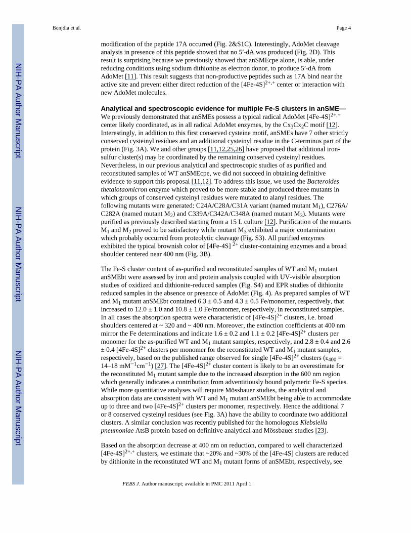
Analytical and spectroscopic evidence for multiple Fe-S clusters in anSME—We previously demonstrated that anSMEs possess a typical radical AdoMet [4Fe-4S]2+,+
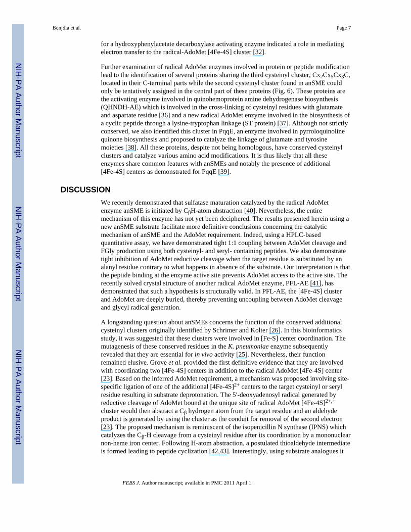
center likely coordinated, as in all radical AdoMet enzymes, by the Cx3Cx2C motif [12].Interestingly, in addition to this first conserved cysteine motif, anSMEs have 7 other strictlyconserved cysteinyl residues and an additional cysteinyl residue in the C-terminus part of theprotein (Fig. 3A). We and other groups [11,12,25,26] have proposed that additional iron-sulfur cluster(s) may be coordinated by the remaining conserved cysteinyl residues.Nevertheless, in our previous analytical and spectroscopic studies of as purified andreconstituted samples of WT anSMEcpe, we did not succeed in obtaining definitiveevidence to support this proposal [11,12]. To address this issue, we used the Bacteroidesthetaiotaomicron enzyme which proved to be more stable and produced three mutants inwhich groups of conserved cysteinyl residues were mutated to alanyl residues. Thefollowing mutants were generated: C24A/C28A/C31A variant (named mutant M1), C276A/C282A (named mutant M2) and C339A/C342A/C348A (named mutant M3). Mutants werepurified as previously described starting from a 15 L culture [12]. Purification of the mutantsM1 and M2 proved to be satisfactory while mutant M3 exhibited a major contaminationwhich probably occurred from proteolytic cleavage (Fig. S3). All purified enzymesexhibited the typical brownish color of [4Fe-4S] 2+ cluster-containing enzymes and a broadshoulder centered near 400 nm (Fig. 3B).
The Fe-S cluster content of as-purified and reconstituted samples of WT and M1 mutantanSMEbt were assessed by iron and protein analysis coupled with UV-visible absorptionstudies of oxidized and dithionite-reduced samples (Fig. S4) and EPR studies of dithionitereduced samples in the absence or presence of AdoMet (Fig. 4). As prepared samples of WTand M1 mutant anSMEbt contained 6.3 ± 0.5 and 4.3 ± 0.5 Fe/monomer, respectively, thatincreased to 12.0 ± 1.0 and 10.8 ± 1.0 Fe/monomer, respectively, in reconstituted samples.In all cases the absorption spectra were characteristic of [4Fe-4S]2+ clusters, i.e. broadshoulders centered at ~ 320 and ~ 400 nm. Moreover, the extinction coefficients at 400 nmmirror the Fe determinations and indicate 1.6 ± 0.2 and 1.1 ± 0.2 [4Fe-4S]2+ clusters permonomer for the as-purified WT and M1 mutant samples, respectively, and 2.8 ± 0.4 and 2.6± 0.4 [4Fe-4S]2+ clusters per monomer for the reconstituted WT and M1 mutant samples,respectively, based on the published range observed for single [4Fe-4S]2+ clusters (ε400 =14–18 mM−1cm−1) [27]. The [4Fe-4S]2+ cluster content is likely to be an overestimate forthe reconstituted M1 mutant sample due to the increased absorption in the 600 nm regionwhich generally indicates a contribution from adventitiously bound polymeric Fe-S species.While more quantitative analyses will require Mössbauer studies, the analytical andabsorption data are consistent with WT and M1 mutant anSMEbt being able to accommodateup to three and two [4Fe-4S]2+ clusters per monomer, respectively. Hence the additional 7or 8 conserved cysteinyl residues (see Fig. 3A) have the ability to coordinate two additionalclusters. A similar conclusion was recently published for the homologous Klebsiellapneumoniae AtsB protein based on definitive analytical and Mössbauer studies [23].
Based on the absorption decrease at 400 nm on reduction, compared to well characterized[4Fe-4S]2+,+ clusters, we estimate that ~20% and ~30% of the [4Fe-4S] clusters are reducedby dithionite in the reconstituted WT and M1 mutant forms of anSMEbt, respectively, see
Benjdia et al. Page 4
FEBS J. Author manuscript; available in PMC 2011 April 1.
NIH
-PA Author Manuscript
NIH
-PA Author Manuscript
NIH
-PA Author Manuscript
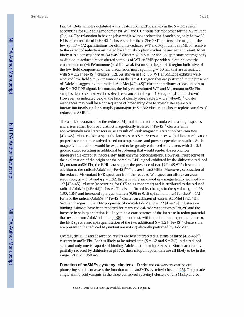
Fig. S4. Both samples exhibited weak, fast-relaxing EPR signals in the S = 1/2 regionaccounting for 0.12 spins/monomer for WT and 0.07 spins per monomer for the M1 mutant(Fig. 4). The relaxation behavior (observable without relaxation broadening only below 30K) is characteristic of [4Fe-4S]+ clusters rather than [2Fe-2S]+ clusters. The origin of thelow spin S = 1/2 quantitations for dithionite-reduced WT and M1 mutant anSMEbt, relativeto the extent of reduction estimated based on absorption studies, is unclear at present. Mostlikely it is a consequence of [4Fe-4S]+ clusters with S = 1/2 and 3/2 spin state heterogeneityas dithionite-reduced reconstituted samples of WT anSMEcpe with sub-stoichiometriccluster content (~6 Fe/monomer) exhibit weak features in the g = 4–6 region indicative ofthe low field components of the broad resonances spanning ~400 mT that are associatedwith S = 3/2 [4Fe-4S]+ clusters [12]. As shown in Fig. S5, WT anSMEcpe exhibits well-resolved low-field S = 3/2 resonances in the g = 4–6 region that are perturbed in the presenceof AdoMet suggesting that radical-AdoMet [4Fe-4S]+ cluster contributes at least in part tothe S = 3/2 EPR signal. In contrast, the fully reconstituted WT and M1 mutant anSMEbtsamples do not exhibit well-resolved resonances in the g = 4–6 region (data not shown).However, as indicated below, the lack of clearly observable S = 3/2 [4Fe-4S]+ clusterresonances may well be a consequence of broadening due to intercluster spin-spininteraction involving the strongly paramagnetic S = 3/2 clusters in cluster replete samples ofreduced anSMEbt.
The S = 1/2 resonance for the reduced M1 mutant cannot be simulated as a single speciesand arises either from two distinct magnetically isolated [4Fe-4S]+ clusters withapproximately axial g tensors or as a result of weak magnetic interaction between two[4Fe-4S]+ clusters. We suspect the latter, as two S = 1/2 resonances with different relaxationproperties cannot be resolved based on temperature- and power-dependence studies. Suchmagnetic interactions would be expected to be greatly enhanced for clusters with S = 3/2ground states resulting in additional broadening that would render the resonancesunobservable except at inaccessibly high enzyme concentrations. However, irrespective ofthe explanation of the origin for the complex EPR signal exhibited by the dithionite-reducedM1 mutant anSMEbt, the EPR data support the presence of two [4Fe-4S]2+,+ clusters inaddition to the radical-AdoMet [4Fe-4S]2+,+ cluster in anSMEbt. Moreover, subtraction ofthe reduced M1-mutant EPR spectrum from the reduced WT spectrum affords an axialresonance, g|| = 2.04 and g⊥ = 1.92, that is readily simulated as a magnetically isolated S =1/2 [4Fe-4S]+ cluster (accounting for 0.05 spins/monomer) and is attributed to the reducedradical-AdoMet [4Fe-4S]+ cluster. This is confirmed by changes in the g values (g = 1.98,1.90, 1.84) and increased spin quantitation (0.05 to 0.15 spins/monomer) for the S = 1/2form of the radical-AdoMet [4Fe-4S]+ cluster on addition of excess AdoMet (Fig. 4B).Similar changes in the EPR properties of radical-AdoMet S = 1/2 [4Fe-4S]+ clusters onbinding AdoMet have been reported for many radical-AdoMet enzymes [28,29] and theincrease in spin quantitation is likely to be a consequence of the increase in redox potentialthat results from AdoMet binding [30]. In contrast, within the limits of experimental error,the EPR spectra and spin quantitation of the two additional S = 1/2 [4Fe-4S]+ clusters thatare present in the reduced M1 mutant are not significantly perturbed by AdoMet.
Overall, the EPR and absorption results are best interpreted in terms of three [4Fe-4S]2+,+
clusters in anSMEbt. Each is likely to be mixed spin (S = 1/2 and S = 3/2) in the reducedstate and only one is capable of binding AdoMet at the unique Fe site. Since each is onlypartially reduced by dithionite at pH 7.5, their midpoint potentials are all likely to be in therange −400 to −450 mV.
Function of anSMEs cysteinyl clusters—Dierks and co-workers carried outpioneering studies to assess the function of the anSMEs cysteinyl clusters [25]. They madesingle amino acid variants in the three conserved cysteinyl clusters of anSMEkp and co-
Benjdia et al. Page 5
FEBS J. Author manuscript; available in PMC 2011 April 1.
NIH
-PA Author Manuscript
NIH
-PA Author Manuscript
NIH
-PA Author Manuscript
expressed these mutants in E. coli, along with the sulfatase from K. pneumonia. All mutantsfailed to mature the co-expressed sulfatase as no sulfatase activity could be measured.Nevertheless, it was not possible to conclude whether the mutated enzymes were unable tocatalyze any reaction or led to the formation of reaction intermediates like sporephotoproduct lyase (SPL), another radical AdoMet enzyme, for which it has been elegantlydemonstrated that a cysteinyl mutant, while inactive in vivo [31], efficiently catalyzes invitro AdoMet cleavage with substrate H-atom abstraction leading to the formation of a sideproduct [18].
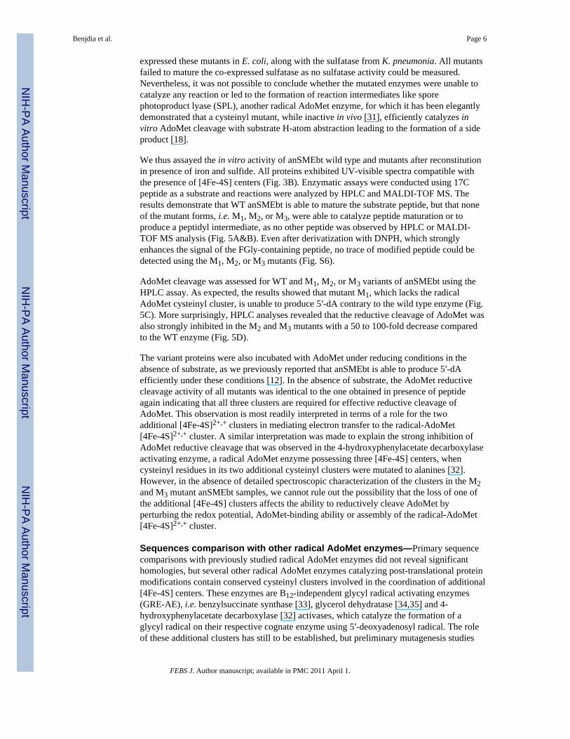
We thus assayed the in vitro activity of anSMEbt wild type and mutants after reconstitutionin presence of iron and sulfide. All proteins exhibited UV-visible spectra compatible withthe presence of [4Fe-4S] centers (Fig. 3B). Enzymatic assays were conducted using 17Cpeptide as a substrate and reactions were analyzed by HPLC and MALDI-TOF MS. Theresults demonstrate that WT anSMEbt is able to mature the substrate peptide, but that noneof the mutant forms, i.e. M1, M2, or M3, were able to catalyze peptide maturation or toproduce a peptidyl intermediate, as no other peptide was observed by HPLC or MALDI-TOF MS analysis (Fig. 5A&B). Even after derivatization with DNPH, which stronglyenhances the signal of the FGly-containing peptide, no trace of modified peptide could bedetected using the M1, M2, or M3 mutants (Fig. S6).
AdoMet cleavage was assessed for WT and M1, M2, or M3 variants of anSMEbt using theHPLC assay. As expected, the results showed that mutant M1, which lacks the radicalAdoMet cysteinyl cluster, is unable to produce 5′-dA contrary to the wild type enzyme (Fig.5C). More surprisingly, HPLC analyses revealed that the reductive cleavage of AdoMet wasalso strongly inhibited in the M2 and M3 mutants with a 50 to 100-fold decrease comparedto the WT enzyme (Fig. 5D).
The variant proteins were also incubated with AdoMet under reducing conditions in theabsence of substrate, as we previously reported that anSMEbt is able to produce 5′-dAefficiently under these conditions [12]. In the absence of substrate, the AdoMet reductivecleavage activity of all mutants was identical to the one obtained in presence of peptideagain indicating that all three clusters are required for effective reductive cleavage ofAdoMet. This observation is most readily interpreted in terms of a role for the twoadditional [4Fe-4S]2+,+ clusters in mediating electron transfer to the radical-AdoMet[4Fe-4S]2+,+ cluster. A similar interpretation was made to explain the strong inhibition ofAdoMet reductive cleavage that was observed in the 4-hydroxyphenylacetate decarboxylaseactivating enzyme, a radical AdoMet enzyme possessing three [4Fe-4S] centers, whencysteinyl residues in its two additional cysteinyl clusters were mutated to alanines [32].However, in the absence of detailed spectroscopic characterization of the clusters in the M2and M3 mutant anSMEbt samples, we cannot rule out the possibility that the loss of one ofthe additional [4Fe-4S] clusters affects the ability to reductively cleave AdoMet byperturbing the redox potential, AdoMet-binding ability or assembly of the radical-AdoMet[4Fe-4S]2+,+ cluster.
Sequences comparison with other radical AdoMet enzymes—Primary sequencecomparisons with previously studied radical AdoMet enzymes did not reveal significanthomologies, but several other radical AdoMet enzymes catalyzing post-translational proteinmodifications contain conserved cysteinyl clusters involved in the coordination of additional[4Fe-4S] centers. These enzymes are B12-independent glycyl radical activating enzymes(GRE-AE), i.e. benzylsuccinate synthase [33], glycerol dehydratase [34,35] and 4-hydroxyphenylacetate decarboxylase [32] activases, which catalyze the formation of aglycyl radical on their respective cognate enzyme using 5′-deoxyadenosyl radical. The roleof these additional clusters has still to be established, but preliminary mutagenesis studies
Benjdia et al. Page 6
FEBS J. Author manuscript; available in PMC 2011 April 1.
NIH
-PA Author Manuscript
NIH
-PA Author Manuscript
NIH
-PA Author Manuscript
for a hydroxyphenylacetate decarboxylase activating enzyme indicated a role in mediatingelectron transfer to the radical-AdoMet [4Fe-4S] cluster [32].
Further examination of radical AdoMet enzymes involved in protein or peptide modificationlead to the identification of several proteins sharing the third cysteinyl cluster, Cx2Cx5Cx3C,located in their C-terminal parts while the second cysteinyl cluster found in anSME couldonly be tentatively assigned in the central part of these proteins (Fig. 6). These proteins arethe activating enzyme involved in quinohemoprotein amine dehydrogenase biosynthesis(QHNDH-AE) which is involved in the cross-linking of cysteinyl residues with glutamateand aspartate residue [36] and a new radical AdoMet enzyme involved in the biosynthesis ofa cyclic peptide through a lysine-tryptophan linkage (ST protein) [37]. Although not strictlyconserved, we also identified this cluster in PqqE, an enzyme involved in pyrroloquinolinequinone biosynthesis and proposed to catalyze the linkage of glutamate and tyrosinemoieties [38]. All these proteins, despite not being homologous, have conserved cysteinylclusters and catalyze various amino acid modifications. It is thus likely that all theseenzymes share common features with anSMEs and notably the presence of additional[4Fe-4S] centers as demonstrated for PqqE [39].
DISCUSSIONWe recently demonstrated that sulfatase maturation catalyzed by the radical AdoMetenzyme anSME is initiated by CβH-atom abstraction [40]. Nevertheless, the entiremechanism of this enzyme has not yet been deciphered. The results presented herein using anew anSME substrate facilitate more definitive conclusions concerning the catalyticmechanism of anSME and the AdoMet requirement. Indeed, using a HPLC-basedquantitative assay, we have demonstrated tight 1:1 coupling between AdoMet cleavage andFGly production using both cysteinyl- and seryl- containing peptides. We also demonstratetight inhibition of AdoMet reductive cleavage when the target residue is substituted by analanyl residue contrary to what happens in absence of the substrate. Our interpretation is thatthe peptide binding at the enzyme active site prevents AdoMet access to the active site. Therecently solved crystal structure of another radical AdoMet enzyme, PFL-AE [41], hasdemonstrated that such a hypothesis is structurally valid. In PFL-AE, the [4Fe-4S] clusterand AdoMet are deeply buried, thereby preventing uncoupling between AdoMet cleavageand glycyl radical generation.
A longstanding question about anSMEs concerns the function of the conserved additionalcysteinyl clusters originally identified by Schrimer and Kolter [26]. In this bioinformaticsstudy, it was suggested that these clusters were involved in [Fe-S] center coordination. Themutagenesis of these conserved residues in the K. pneumoniae enzyme subsequentlyrevealed that they are essential for in vivo activity [25]. Nevertheless, their functionremained elusive. Grove et al. provided the first definitive evidence that they are involvedwith coordinating two [4Fe-4S] centers in addition to the radical AdoMet [4Fe-4S] center[23]. Based on the inferred AdoMet requirement, a mechanism was proposed involving site-specific ligation of one of the additional [4Fe-4S]2+ centers to the target cysteinyl or serylresidue resulting in substrate deprotonation. The 5′-deoxyadenosyl radical generated byreductive cleavage of AdoMet bound at the unique site of radical AdoMet [4Fe-4S]2+,+
cluster would then abstract a Cβ hydrogen atom from the target residue and an aldehydeproduct is generated by using the cluster as the conduit for removal of the second electron[23]. The proposed mechanism is reminiscent of the isopenicillin N synthase (IPNS) whichcatalyzes the Cβ-H cleavage from a cysteinyl residue after its coordination by a mononuclearnon-heme iron center. Following H-atom abstraction, a postulated thioaldehyde intermediateis formed leading to peptide cyclization [42,43]. Interestingly, using substrate analogues it
Benjdia et al. Page 7
FEBS J. Author manuscript; available in PMC 2011 April 1.
NIH
-PA Author Manuscript
NIH
-PA Author Manuscript
NIH
-PA Author Manuscript
has been reported that IPNS can oxidize its target cysteinyl residue into a hydrated aldehydewhich is virtually the same as the reaction catalyzed by anSME [44].
Thus, it is conceivable that one of the two additional clusters binds and deprotonates thetarget cysteinyl or seryl residues and provides a conduit for removal of the second electron[23]. If such mechanism is correct, our recent demonstration that the 5′-deoxyadenosylradical produced by anSME directly abstracts one of the cysteinyl Cβ hydrogen atoms [40],coupled with the results reported herein, indicate that deprotonation occurs prior to orsimultaneously with AdoMet cleavage. Indeed, using an alanyl containing peptide weobserved complete inhibition of AdoMet cleavage.
Although the mutagenesis studies reported herein suggest that both of the two additional[4Fe-4S] clusters are required for AdoMet cleavage using dithionite as an electron donor, wecannot rule out the possibility that this is a consequence of perturbation of the redox orAdoMet-binding properties of the radical-AdoMet [4Fe-4S]2+,+ center that are induced byloss of either the two additional clusters. Hence it is possible that one of the additional[4Fe-4S] clusters (Cluster II) is involved with binding the peptide substrate and providing aconduit for removal of the second electron. The other [4Fe-4S] cluster (Cluster III) couldfunction in mediating electron transfer from the physiological electron donor to the radical-AdoMet [4Fe-4S] cluster or from Cluster II to the physiological electron acceptor, see Fig.7A. The former mechanism is analogous to that recently proposed by Grove et al [23].
Nevertheless, the data presented herein suggests an alternative mechanism. Indeed, theprimary sequence analyses discussed above indicate that the two additional clusters arelikely to be ligated by the 8 conserved cysteinyl residues and hence both [4Fe-4S] clustersmay have complete cysteinyl ligation, one cysteinyl residue from the last motif beinginvolved in the coordination of the second cluster (Fig. 3A). Furthermore, the preliminaryobservation that these clusters are almost isopotential with the radical-AdoMet cluster andthe mutagenesis studies reported herein which indicate that both additional [4Fe-4S] clustersare required for productive reductive cleavage of AdoMet, both suggest that the additional[4Fe-4S]2+,+ clusters play a role in facilitating electron transfer to the radical-AdoMetcluster, as appears to be the case in some GRE-AEs [32]. Finally, sequence analysis revealedthat these cysteinyl clusters are also found in other radical AdoMet enzymes involved inprotein or peptide modification. These enzymes catalyze the modification of amino acidssuch as glutamate or tyrosine which are not known to bind [Fe-S] centers. Moreover,another radical AdoMet enzyme, BtrN, has recently been demonstrated to use AdoMetstoichiometrically to catalyze the two electron oxidation of a hydroxyl group to a ketonewithout additional Fe-S centers, a reaction formally analogous to the one catalyzed byanSME [45]. However, the absence of additional Fe-S clusters in BtrN clearly requiresconfirmation using Mössbauer spectroscopy.
Based on the above considerations, we propose an alternate mechanism for anSME (Fig.7B). In our proposed mechanism, the initial step is the reduction of the radical-AdoMet[4Fe-4S]2+ cluster via electron transfer from the two additional [4Fe-4S]2+,+ clusters.Following this reduction, the CβH-atom of the substrate is abstracted by the 5′-deoxyadenosyl radical generated by the reductive cleavage of AdoMet bound at the radical-AdoMet [4Fe-4S]2+,+ cluster as recently demonstrated [40]. Simultaneously, deprotonationof the thiol or hydroxyl group occurs catalyzed by an amino acid side chain. The substrateradical intermediate formed by CβH-atom abstraction is then further oxidized to yield analdehyde or a thioaldehyde. In this scenario, the radical would be transferred back to theradical-AdoMet [4Fe-4S]2+ cluster by outer-sphere electron transfer. The implication is thatthe reaction would have a substrate radical intermediate, as recently demonstrated for BtrN[45], and would be self-sustaining once the initial electron has been supplied by an
Benjdia et al. Page 8
FEBS J. Author manuscript; available in PMC 2011 April 1.
NIH
-PA Author Manuscript
NIH
-PA Author Manuscript
NIH
-PA Author Manuscript
exogenous electron donor. Both possibilities are currently under investigation in ourlaboratories.
For Cys-type sulfatases, both mechanisms shown in Fig. 7 result in the formation of athioaldehyde intermediate as is also the case in IPNS [42] and cysteine decarboyxlases[46,47]. Hydrolysis of the thioaldehyde by a water molecule is the likely next step. In accordwith this hypothesis 18O incorporation into the FGly-residue is observed when the reactionis carried out in H2
18O buffer, see Fig. S7.
If more work needs to be done to clarify the catalytic mechanism of anSMEs and the role ofthe two additional [4Fe-4S] clusters, the present report suggests that anSMEs possesscommon features with some glycyl radical activating enzymes and that radical AdoMetenzymes possessing additional [4Fe-4S] clusters are likely to be found notably in enzymescatalyzing protein post-translational modifications. It remains to be seen if the function ofthese additional clusters involves mediating electron transfer and/or binding and activatingthe peptidyl substrates.
EXPERIMENTAL PROCEDURESChemicals
All chemicals and reagents were obtained from commercial sources and were of analyticalgrade. S,S-Adenosyl-L-methionine (AdoMet) was synthesized enzymatically and purified asdescribed previously [17].
anSMEcpe and anSMEbt protein expression and purificationProtein expression and purification were performed as previously described [12]. Briefly, E.coli BL21 (DE3) transformed with a plasmid bearing the anSMEcpe or the anSMEbt gene(pET-6His-anSMEcpe or pET-6His-anSMEbt) were grown aerobically overnight at 37°C inLB medium (100 mL) supplemented with kanamycin (50 μg.mL−1). An overnight culturewas then used to inoculate fresh LB medium (15 L) supplemented with the same antibiotic.After overnight growth at 25°C in the presence of IPTG, cells were collected and suspendedin Tris-buffer (50 mM Tris, 150 mM KCl, 10% glycerol pH 7.5). The cells were thendisrupted by sonication and centrifuged at 220,000 ×g at 4°C for 1 hour. The solution wasthen loaded onto a Ni-NTA Sepharose column equilibrated with Tris-buffer, pH 7.5. Thecolumn was washed extensively with the same buffer. Some of the adsorbed proteins wereeluted by a washing step with 25 and 100 mM imidazole and the over-expressed protein waseluted by applying 500 mM imidazole. Imidazole was removed by gel filtrationchromatography PD-10 columns (GE Healthcare) and fractions containing the anSMEcpe oranSMEbt proteins were immediately concentrated using Ultrafree cells (Millipore) with amolecular cut-off of 10 kDa.
Construction of cysteinyl cluster mutantsAnSMEbt mutants were obtained using the QuikChange site-directed mutagenesis kit(Stratagene). For each mutant a two-step PCR method was used [48]. The following primerswere used for C24A/C28A/C31A mutant: 5′-GCC GTA GCC AAC CTC GCA GCC GAATAC GCC TAT TAT-3′ and 5′-ATA ATA GGC GTA TTC GGC TGC GAG GTT GGCTAC GGC-3′ for C276A/C282A mutant: 5′-GGC GTA GCT ACA ATG GCG AAG CATGCC GGA CAT-3′ and 5′-ATG TCC GGC ATG CTT CGC CAT TGT AGC TAC GCC-3′and for C339A/C342A/C348A mutant: 5′-ACC CAA GCC AAG GAG GCC GAC TTTCTA TTT GCC GCC AAC GGA-3′ and 5′-TCC GTT GGC GGC AAA TAG AAA GTCGGC CTC CTT GGC TTG GGT-3′ (in bold are indicated the changed codons). Afterverification of the correct mutation by sequencing, the plasmid obtained were transformed
Benjdia et al. Page 9
FEBS J. Author manuscript; available in PMC 2011 April 1.
NIH
-PA Author Manuscript
NIH
-PA Author Manuscript
NIH
-PA Author Manuscript
into E. coli BL21 (DE3) and the mutated proteins produced using the same protocol as thewild-type enzyme.
Reconstitution of Fe-S clusters on anSMEbt and anSMEcpeReconstitution was carried out anaerobically in a glove box (Bactron IV). Anaerobicallypurified anSMEs (200 μM monomer) were treated with 5 mM DTT (SIGMA, St Louis, MO,USA) and incubated overnight with a 10-fold molar excess of both Na2S (SIGMA, St Louis,MO, USA). and (NH4)2Fe(SO4)2 (SIGMA, St Louis, MO, USA) at 12°C. The proteinwasdesalted using a Sephadex G25 column (GE Healthcare, WI, USA) and thecolored fractionswere concentrated on Amicon Ultra-4 (Millipore, Billerica, MA, USA). Proteinconcentrations were determined by the Bradford protein assay (SIGMA, St Louis, MO,USA), using BSA as a standard. Iron concentrations were determined colorimetrically usingbathophenanthroline (SIGMA, St Louis, MO, USA) under reducing conditions, afterdigestion of the protein in 0.8% KMnO4/0.2 M HCl.
Peptide synthesisThe following 17-mer peptides (with the critical residue in bold): Ac-TAVPSCIPSRASILTGM-NH2, Ac-TAVPSSIPSRASILTGM-NH2 and Ac-TAVPSAIPSRASILTGM-NH2 were synthesized (0.1-mmol scale) by the solid phasemethodology on a Rink amide 4-methylbenzhydrylamine resin (VWR, Fontenay-sous-Bois,France) by using a 433A Applied Biosystems peptide synthesizer (Applera-France,Courtaboeuf, France) and the standard Fmoc manufacturer’s procedure. The syntheticpeptides were purified by reversed-phase HPLC on a 2.2 × 25-cm Vydac 218TP1022 C18column (Alltech, Templemars, France) by using a linear gradient (10–50% over 45 min) ofacetonitrile/trifluoroacetic acid (TFA) (99.9: 0.1; v/v) at a flow rate of 10 mL/min.Analytical HPLC, performed on a 0.46 × 25-cm Vydac 218TP54 C18 column (Alltech),showed that the purity of the peptides was >99.1%. The purified peptides were characterizedby MALDI-TOF mass spectrometry on a Voyager DE PRO (Applera, France) in thereflector mode with α-cyano-4-hydroxycinnamic acid as a matrix.
Peptide maturationSamples containing 6 mM dithiothreitol, 3 mM sodium dithionite, 500 μM peptides and 1mM AdoMet in Tris-buffer, pH 7.5 were incubated with reconstituted proteins. Thereactions were performed in an anaerobic glovebox (Bactron IV). The oxygen concentrationwas monitored with a gas analyzer (Coy Laboratory). After incubation at 25°C, sampleswere divided in half, one part was used to test the maturation activity by mass spectrometrywhile the other half was used to quantify the reductive cleavage of AdoMet and FGlyformation. Control samples were prepared without enzyme to verify peptide and AdoMetstability over time. Experiments performed in H2
18O were made exactly as described aboveexcept that the Tris-buffer was made in H2
18O and the enzyme was exchanged twice withthis buffer before the experiments.
Peptide maturation analysis by MALDI-TOF MSThe α-cyano-4-hydroxycinnamic acid matrix (CHCA) (SIGMA, St Louis, MO, USA) wasprepared at 4 mg.mL−1 in 0.15% TFA, 50% acetonitrile. The 2,4-dinitrophenylhydrazoneacid matrix (DNPH) was prepared at 100 mg.mL−1 in 0.15% TFA, 50% acetonitrile. Equalvolumes (1 μL) of matrix and sample were spotted onto the MALDI-TOF target plate.MALDI-TOF analysis was then performed on a Voyager DE STR Instrument (AppliedBiosystems, Framingham, CA). Spectra were acquired in the reflector mode with: 20 kVaccelerating voltage, 62% grid voltage and a 120 ns delay.
Benjdia et al. Page 10
FEBS J. Author manuscript; available in PMC 2011 April 1.
NIH
-PA Author Manuscript
NIH
-PA Author Manuscript
NIH
-PA Author Manuscript
Peptide maturation and 5′-deoxyadenosine production quantification by HPLCPeptide modification and 5′-deoxyadenosine production was measured by high-performanceliquid chromatography using a C18 column (LicroSphere, 5-μm, 4.6 × 150-mm) equilibratedin solvent A (0.1% trifluoroacetic acid). A linear gradient from 0 to 80% acetonitrile wasapplied at a constant flow rate of 1 mL/min. Detection was carried out at 260 nm for S-adenosylmethionine and its derivative and at 215 nm to follow peptide modification.
Electron paramagnetic resonance (EPR)X-band EPR spectra were recorded on a Bruker Instruments ESP 300D spectrometerequipped with an Oxford Instruments ESR 900 flow cryostat (4.2–300 K). Spectra werequantified under non-saturating conditions by double integration against a 1 mM CuEDTAstandard.
Supplementary MaterialRefer to Web version on PubMed Central for supplementary material.
AcknowledgmentsThis work was supported by grants from Agence Nationale de la Recherche (Grant ANR-08-BLAN-0224-02) andthe NIH to M.K.J. (GM62524). Mass spectrometry experiments were performed at PAPSSO, INRA, Jouy-en-Josas.
References1. Muller I, Kahnert A, Pape T, Sheldrick GM, Meyer-Klaucke W, Dierks T, Kertesz M, Uson I.
Crystal structure of the alkylsulfatase AtsK: insights into the catalytic mechanism of the Fe(II)alpha-ketoglutarate-dependent dioxygenase superfamily. Biochemistry. 2004; 43:3075–3088.[PubMed: 15023059]
2. Hagelueken G, Adams TM, Wiehlmann L, Widow U, Kolmar H, Tummler B, Heinz DW, SchubertWD. The crystal structure of SdsA1, an alkylsulfatase from Pseudomonas aeruginosa, defines athird class of sulfatases. Proc Natl Acad Sci U S A. 2006; 103:7631–7636. [PubMed: 16684886]
3. Hanson SR, Best MD, Wong CH. Sulfatases: structure, mechanism, biological activity, inhibition,and synthetic utility. Angew Chem Int Ed Engl. 2004; 43:5736–5763. [PubMed: 15493058]
4. Glockner FO, Kube M, Bauer M, Teeling H, Lombardot T, Ludwig W, Gade D, Beck A, Borzym K,Heitmann K, Rabus R, Schlesner H, Amann R, Reinhardt R. Complete genome sequence of themarine planctomycete Pirellula sp. strain 1. Proc Natl Acad Sci U S A. 2003; 100:8298–8303.[PubMed: 12835416]
5. Hoffman JA, Badger JL, Zhang Y, Huang SH, Kim KS. Escherichia coli K1 aslA contributes toinvasion of brain microvascular endothelial cells in vitro and in vivo. Infect Immun. 2000; 68:5062–5067. [PubMed: 10948126]
6. Tralau T, Vuilleumier S, Thibault C, Campbell BJ, Hart CA, Kertesz MA. Transcriptomic analysisof the sulfate starvation response of Pseudomonas aeruginosa. J Bacteriol. 2007; 189:6743–6750.[PubMed: 17675390]
7. Schmidt B, Selmer T, Ingendoh A, von Figura K. A novel amino-acid modification in sulfatases thatis defective in multiple sulfatase deficiency. Cell. 1995; 82:271–278. [PubMed: 7628016]
8. Recksiek M, Selmer T, Dierks T, Schmidt B, von Figura K. Sulfatases, trapping of the sulfatedenzyme intermediate by substituting the active site formylglycine. J Biol Chem. 1998; 273:6096–6103. [PubMed: 9497327]
9. Benjdia A, Deho G, Rabot S, Berteau O. First evidences for a third sulfatase maturation system inprokaryotes from E. coli aslB and ydeM deletion mutants. FEBS Lett. 2007; 581:1009–1014.[PubMed: 17303125]
10. Carlson BL, Ballister ER, Skordalakes E, King DS, Breidenbach MA, Gilmore SA, Berger JM,Bertozzi CR. Function and structure of a prokaryotic formylglycine-generating enzyme. J BiolChem. 2008; 283:20117–20125. [PubMed: 18390551]
Benjdia et al. Page 11
FEBS J. Author manuscript; available in PMC 2011 April 1.
NIH
-PA Author Manuscript
NIH
-PA Author Manuscript
NIH
-PA Author Manuscript
11. Benjdia A, Leprince J, Guillot A, Vaudry H, Rabot S, Berteau O. Anaerobic sulfatase-maturatingenzymes: radical SAM enzymes able to catalyze in vitro sulfatase post-translational modification.J Am Chem Soc. 2007; 129:3462–3463. [PubMed: 17335281]
12. Benjdia A, Subramanian S, Leprince J, Vaudry H, Johnson MK, Berteau O. Anaerobic sulfatase-maturating enzymes - first dual substrate radical S-adenosylmethionine enzymes. J Biol Chem.2008; 283:17815–17826. [PubMed: 18408004]
13. Berteau O, Guillot A, Benjdia A, Rabot S. A new type of bacterial sulfatase reveals a novelmaturation pathway in prokaryotes. J Biol Chem. 2006; 281:22464–22470. [PubMed: 16766528]
14. Fontecave M, Atta M, Mulliez E. S-adenosylmethionine: nothing goes to waste. Trends BiochemSci. 2004; 29:243–249. [PubMed: 15130560]
15. Frey PA, Hegeman AD, Ruzicka FJ. The Radical SAM Superfamily. Crit Rev Biochem Mol Biol.2008; 43:63–88. [PubMed: 18307109]
16. Chen D, Walsby C, Hoffman BM, Frey PA. Coordination and mechanism of reversible cleavage ofS-adenosylmethionine by the [4Fe-4S] center in lysine 2,3-aminomutase. J Am Chem Soc. 2003;125:11788–11789. [PubMed: 14505379]
17. Chandor A, Berteau O, Douki T, Gasparutto D, Sanakis Y, Ollagnier-de-Choudens S, Atta M,Fontecave M. Dinucleotide spore photoproduct, a minimal substrate of the DNA repair sporephotoproduct lyase enzyme from Bacillus subtilis. J Biol Chem. 2006; 281:26922–26931.[PubMed: 16829676]
18. Chandor-Proust A, Berteau O, Douki T, Gasparutto D, Ollagnier-de-Choudens S, Fontecave M,Atta M. DNA repair and free radicals, new insights into the mechanism of spore photoproductlyase revealed by single amino acid substitution. J Biol Chem. 2008; 283:36361–36368. [PubMed:18957420]
19. Cheek J, Broderick JB. Direct H atom abstraction from spore photoproduct C-6 initiates DNArepair in the reaction catalyzed by spore photoproduct lyase: evidence for a reversibly generatedadenosyl radical intermediate. J Am Chem Soc. 2002; 124:2860–2861. [PubMed: 11902862]
20. Friedel MG, Berteau O, Pieck JC, Atta M, Ollagnier-de-Choudens S, Fontecave M, Carell T. Thespore photoproduct lyase repairs the 5S- and not the 5R-configured spore photoproduct DNAlesion. Chem Commun (Camb). 2006:445–447. [PubMed: 16493831]
21. Layer G, Kervio E, Morlock G, Heinz DW, Jahn D, Retey J, Schubert WD. Structural andfunctional comparison of HemN to other radical SAM enzymes. J Biol Chem. 2005; 386:971–980.
22. Layer G, Moser J, Heinz DW, Jahn D, Schubert WD. Crystal structure of coproporphyrinogen IIIoxidase reveals cofactor geometry of Radical SAM enzymes. EMBO J. 2003; 22:6214–6224.[PubMed: 14633981]
23. Grove TL, Lee KH, St Clair J, Krebs C, Booker SJ. In vitro characterization of AtsB, a radicalSAM formylglycine-generating enzyme that contains three [4Fe-4S] clusters. Biochemistry. 2008;47:7523–7538. [PubMed: 18558715]
24. Peng J, Schmidt B, von Figura K, Dierks T. Identification of formylglycine in sulfatases by matrix-assisted laser desorption/ionization time-of-flight mass spectrometry. J Mass Spectrom. 2003;38:80–86. [PubMed: 12526009]
25. Fang Q, Peng J, Dierks T. Post-translational formylglycine modification of bacterial sulfatases bythe radical S-adenosylmethionine protein AtsB. J Biol Chem. 2004; 279:14570–14578. [PubMed:14749327]
26. Schirmer A, Kolter R. Computational analysis of bacterial sulfatases and their modifying enzymes.Chem Biol. 1998; 5:R181–186. [PubMed: 9710560]
27. Agar JN, Krebs C, Frazzon J, Huynh BH, Dean DR, Johnson MK. IscU as a scaffold for iron-sulfur cluster biosynthesis: sequential assembly of [2Fe-2S] and [4Fe-4S] clusters in IscU.Biochemistry. 2000; 39:7856–7862. [PubMed: 10891064]
28. Fontecave M. Iron-sulfur clusters: ever-expanding roles. Nat Chem Biol. 2006; 2:171–174.[PubMed: 16547473]
29. Wang SC, Frey PA. S-adenosylmethionine as an oxidant: the radical SAM superfamily. TrendsBiochem Sci. 2007; 32:101–110. [PubMed: 17291766]
Benjdia et al. Page 12
FEBS J. Author manuscript; available in PMC 2011 April 1.
NIH
-PA Author Manuscript
NIH
-PA Author Manuscript
NIH
-PA Author Manuscript
30. Wang SC, Frey PA. Binding energy in the one-electron reductive cleavage of S-adenosylmethionine in lysine 2,3-aminomutase, a radical SAM enzyme. Biochemistry. 2007;46:12889–12895. [PubMed: 17944492]
31. Fajardo-Cavazos P, Rebeil R, Nicholson WL. Essential cysteine residues in Bacillus subtilis sporephotoproduct lyase identified by alanine scanning mutagenesis. Curr Microbiol. 2005; 51:331–335. [PubMed: 16163454]
32. Yu L, Blaser M, Andrei PI, Pierik AJ, Selmer T. 4-Hydroxyphenylacetate decarboxylases:properties of a novel subclass of glycyl radical enzyme systems. Biochemistry. 2006; 45:9584–9592. [PubMed: 16878993]
33. Leuthner B, Leutwein C, Schulz H, Horth P, Haehnel W, Schiltz E, Schagger H, Heider J.Biochemical and genetic characterization of benzylsuccinate synthase from Thauera aromatica: anew glycyl radical enzyme catalysing the first step in anaerobic toluene metabolism. MolMicrobiol. 1998; 28:615–628. [PubMed: 9632263]
34. O’Brien JR, Raynaud C, Croux C, Girbal L, Soucaille P, Lanzilotta WN. Insight into themechanism of the B12-independent glycerol dehydratase from Clostridium butyricum: preliminarybiochemical and structural characterization. Biochemistry. 2004; 43:4635–4645. [PubMed:15096031]
35. Raynaud C, Sarcabal P, Meynial-Salles I, Croux C, Soucaille P. Molecular characterization of the1,3-propanediol (1,3-PD) operon of Clostridium butyricum. Proc Natl Acad Sci U S A. 2003;100:5010–5015. [PubMed: 12704244]
36. Ono K, Okajima T, Tani M, Kuroda S, Sun D, Davidson VL, Tanizawa K. Involvement of aputative [Fe-S]-cluster-binding protein in the biogenesis of quinohemoprotein aminedehydrogenase. J Biol Chem. 2006; 281:13672–13684. [PubMed: 16546999]
37. Ibrahim M, Guillot A, Wessner F, Algaron F, Besset C, Courtin P, Gardan R, Monnet V. Controlof the transcription of a short gene encoding a cyclic peptide in Streptococcus thermophilus: a newquorum-sensing system? J Bacteriol. 2007; 189:8844–8854. [PubMed: 17921293]
38. Puehringer S, Metlitzky M, Schwarzenbacher R. The pyrroloquinoline quinone biosynthesispathway revisited: a structural approach. BMC Biochem. 2008; 9:8. [PubMed: 18371220]
39. Wecksler SR, Stoll S, Tran H, Magnusson OT, Wu SP, King D, Britt RD, Klinman JP.Pyrroloquinoline quinone biogenesis: demonstration that PqqE from Klebsiella pneumoniae is aradical S-adenosyl-L-methionine enzyme. Biochemistry. 2009; 48:10151–10161. [PubMed:19746930]
40. Benjdia A, Leprince J, Sandstrom C, Vaudry H, Berteau O. Mechanistic investigations ofanaerobic sulfatase-maturating enzyme: direct Cbeta H-atom abstraction catalyzed by a radicalAdoMet enzyme. J Am Chem Soc. 2009; 131:8348–8349. [PubMed: 19489556]
41. Vey JL, Yang J, Li M, Broderick WE, Broderick JB, Drennan CL. Structural basis for glycylradical formation by pyruvate formate-lyase activating enzyme. Proc Natl Acad Sci U S A. 2008;105:16137–16141. [PubMed: 18852451]
42. Bollinger JM Jr, Krebs C. Enzymatic C-H activation by metal-superoxo intermediates. Curr OpinChem Biol. 2007; 11:151–158. [PubMed: 17374503]
43. Burzlaff NI, Rutledge PJ, Clifton IJ, Hensgens CM, Pickford M, Adlington RM, Roach PL,Baldwin JE. The reaction cycle of isopenicillin N synthase observed by X-ray diffraction. Nature.1999; 401:721–724. [PubMed: 10537113]
44. Long AJ, Clifton IJ, Roach PL, Baldwin JE, Rutledge PJ, Schofield CJ. Structural studies on thereaction of isopenicillin N synthase with the truncated substrate analogues delta-(L-alpha-aminoadipoyl)-L-cysteinyl-glycine and delta-(L-alpha-aminoadipoyl)-L-cysteinyl-D-alanine.Biochemistry. 2005; 44:6619–6628. [PubMed: 15850395]
45. Yokoyama K, Ohmori D, Kudo F, Eguchi T. Mechanistic study on the reaction of a radical SAMdehydrogenase BtrN by electron paramagnetic resonance spectroscopy. Biochemistry. 2008;47:8950–8960. [PubMed: 18672902]
46. Strauss E, Zhai H, Brand LA, McLafferty FW, Begley TP. Mechanistic studies onphosphopantothenoylcysteine decarboxylase: trapping of an enethiolate intermediate with amechanism-based inactivating agent. Biochemistry. 2004; 43:15520–15533. [PubMed: 15581364]
Benjdia et al. Page 13
FEBS J. Author manuscript; available in PMC 2011 April 1.
NIH
-PA Author Manuscript
NIH
-PA Author Manuscript
NIH
-PA Author Manuscript
47. Blaesse M, Kupke T, Huber R, Steinbacher S. Crystal structure of the peptidyl-cysteinedecarboxylase EpiD complexed with a pentapeptide substrate. EMBO J. 2000; 19:6299–6310.[PubMed: 11101502]
48. Wang W, Malcolm BA. Two-stage PCR protocol allowing introduction of multiple mutations,deletions and insertions using QuikChange Site-Directed Mutagenesis. Biotechniques. 1999;26:680–682. [PubMed: 10343905]
Benjdia et al. Page 14
FEBS J. Author manuscript; available in PMC 2011 April 1.
NIH
-PA Author Manuscript
NIH
-PA Author Manuscript
NIH
-PA Author Manuscript
Figure 1. Sulfatase maturation scheme leading from a cysteinyl or seryl residue to a Cα-formylglycine residue (FGly) in sulfatase active site (A) – MALDI-TOF MS analysis ofmaturation of peptide 17C (B) and 17S (C) incubated 2 and 12 hours with anSMEcperespectivelyAnSMEcpe was incubated with each peptide (500 μM) under reducing conditions in thepresence of AdoMet (1 mM).
Benjdia et al. Page 15
FEBS J. Author manuscript; available in PMC 2011 April 1.
NIH
-PA Author Manuscript
NIH
-PA Author Manuscript
NIH
-PA Author Manuscript
Figure 2. HPLC analysis of incubation reactions with peptide 17C (A) or 17S (B) and time-dependant formation of FGly-containing peptide (C) and 5′-deoxyadenosine (D) by anSMEcpeAnSMEcpe was incubated with 17C (◆), 17S (▲) or 17A (●) peptide (500 μM) underreducing conditions in the presence of AdoMet (1 mM), DTT (6 mM) and dithionite (3mM).
Benjdia et al. Page 16
FEBS J. Author manuscript; available in PMC 2011 April 1.
NIH
-PA Author Manuscript
NIH
-PA Author Manuscript
NIH
-PA Author Manuscript

Figure 3. Sequence alignment of the three anSMEs putative clusters(A) anSMEcpe (CPF_0616 from Clostridium perfringens), anSMEbt (BT_0238 fromBacteroides thetaiotaomicron) and anSMEkp (AtsB from Klebsiella pneumoniae). Sequencepositions in the proteins are in brackets. The conserved cysteinyl residues are indicated inblack boxes and the other conserved residues are shadowed – UV visible absorption spectaof reconstituted wild-type (WT) and M1, M2 and M3 variants of anSMEbt (B).
Benjdia et al. Page 17
FEBS J. Author manuscript; available in PMC 2011 April 1.
NIH
-PA Author Manuscript
NIH
-PA Author Manuscript
NIH
-PA Author Manuscript

Figure 4. X-band EPR spectra of dithionite-reduced reconstituted samples of WT and M1mutant anSMEbt in the absence (A) and presence (B) of a 20-fold stoichiometric excess ofAdoMetThe WT minus M1 mutant difference spectrum at the bottom of each panel corresponds tothe EPR spectrum of the S = 1/2 [4Fe-4S]+ radical-AdoMet cluster with (B) and without (A)AdoMet bound at the unique Fe site. EPR spectra were recorded at 10 K with 20 mWmicrowave power, 0.65 mT modulation amplitude and a microwave frequency of 9.603GHz. The spectrometer gain was 2-fold higher for the samples prepared without AdoMet.Samples of WT and M1 mutant anSMEbt (each 0.4 mM) in Tris-HCl buffer, pH 7.5, wereanaerobically reduced with a 10-fold stoichiometric excess of sodium dithionite.
Benjdia et al. Page 18
FEBS J. Author manuscript; available in PMC 2011 April 1.
NIH
-PA Author Manuscript
NIH
-PA Author Manuscript
NIH
-PA Author Manuscript
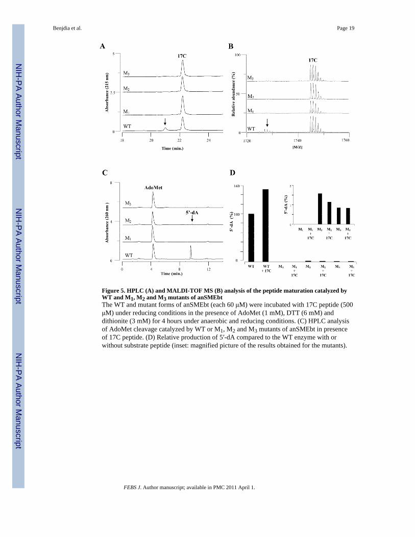
Figure 5. HPLC (A) and MALDI-TOF MS (B) analysis of the peptide maturation catalyzed byWT and M1, M2 and M3 mutants of anSMEbtThe WT and mutant forms of anSMEbt (each 60 μM) were incubated with 17C peptide (500μM) under reducing conditions in the presence of AdoMet (1 mM), DTT (6 mM) anddithionite (3 mM) for 4 hours under anaerobic and reducing conditions. (C) HPLC analysisof AdoMet cleavage catalyzed by WT or M1, M2 and M3 mutants of anSMEbt in presenceof 17C peptide. (D) Relative production of 5′-dA compared to the WT enzyme with orwithout substrate peptide (inset: magnified picture of the results obtained for the mutants).
Benjdia et al. Page 19
FEBS J. Author manuscript; available in PMC 2011 April 1.
NIH
-PA Author Manuscript
NIH
-PA Author Manuscript
NIH
-PA Author Manuscript
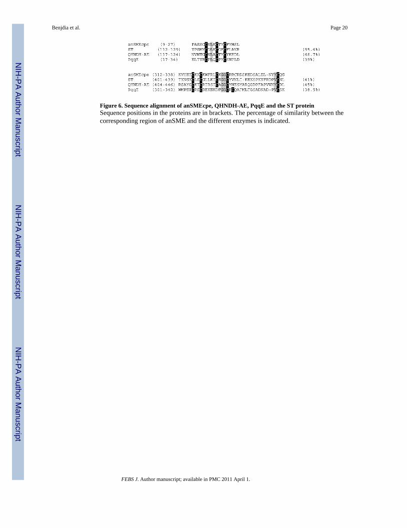
Figure 6. Sequence alignment of anSMEcpe, QHNDH-AE, PqqE and the ST proteinSequence positions in the proteins are in brackets. The percentage of similarity between thecorresponding region of anSME and the different enzymes is indicated.
Benjdia et al. Page 20
FEBS J. Author manuscript; available in PMC 2011 April 1.
NIH
-PA Author Manuscript
NIH
-PA Author Manuscript
NIH
-PA Author Manuscript
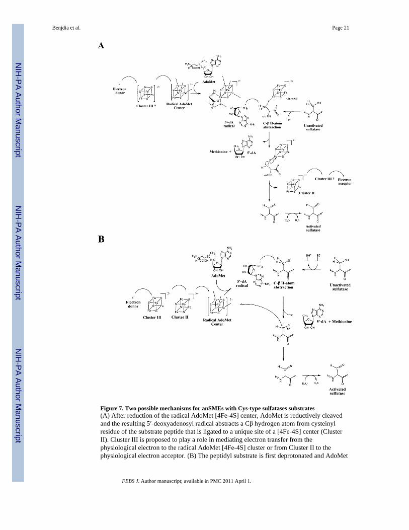
Figure 7. Two possible mechanisms for anSMEs with Cys-type sulfatases substrates(A) After reduction of the radical AdoMet [4Fe-4S] center, AdoMet is reductively cleavedand the resulting 5′-deoxyadenosyl radical abstracts a Cβ hydrogen atom from cysteinylresidue of the substrate peptide that is ligated to a unique site of a [4Fe-4S] center (ClusterII). Cluster III is proposed to play a role in mediating electron transfer from thephysiological electron to the radical AdoMet [4Fe-4S] cluster or from Cluster II to thephysiological electron acceptor. (B) The peptidyl substrate is first deprotonated and AdoMet
Benjdia et al. Page 21
FEBS J. Author manuscript; available in PMC 2011 April 1.
NIH
-PA Author Manuscript
NIH
-PA Author Manuscript
NIH
-PA Author Manuscript
is reductively cleaved. The resulting 5′-deoxyadenosyl radical abstracts a Cβ hydrogen atomfrom cysteinyl residue to generate a substrate radical that is converted to the thioaldehydeintermediate via outer-sphere electron transfer to the radical AdoMet cluster. In this scheme,the additional [4Fe-4S] centers, Clusters II and III, have a key role in mediating the initialelectron transfer from the physiological electron to the radical AdoMet [4Fe-4S] cluster. Inboth mechanisms, a thioaldehyde intermediate is formed and further hydrolyzed to form theFGly residue with the release of hydrogen disulfide.
Benjdia et al. Page 22
FEBS J. Author manuscript; available in PMC 2011 April 1.
NIH
-PA Author Manuscript
NIH
-PA Author Manuscript
NIH
-PA Author Manuscript
Copyright © 2022 FDOKUMEN