An Unexpectedly Complex Mitoribosome in Andalucia godoyi ...
17
An Unexpectedly Complex Mitoribosome in Andalucia godoyi, a Protist with the Most Bacteria-like Mitochondrial Genome Matus Valach ,* ,1 Jos e Angel Gonzalez Alcazar, 1 Matt Sarrasin , 1 B. Franz Lang , 1 Michael W. Gray , 2 and Gertraud Burger 1 1 Department of Biochemistry and Molecular Medicine, Robert-Cedergren Centre for Bioinformatics and Genomics, Universit e de Montr eal, Montreal, Quebec, Canada 2 Department of Biochemistry and Molecular Biology, Centre for Comparative Genomics and Evolutionary Bioinformatics, Dalhousie University, Halifax, Nova Scotia, Canada *Corresponding author: E-mail: [email protected]. Associate editor: Banu Ozkan Abstract The mitoribosome, as known from studies in model organisms, deviates considerably from its ancestor, the bacterial ribosome. Deviations include substantial reduction of the mitochondrial ribosomal RNA (mt-rRNA) structure and ac- quisition of numerous mitochondrion-specific (M) mitoribosomal proteins (mtRPs). A broadly accepted view assumes that M-mtRPs compensate for structural destabilization of mt-rRNA resulting from its evolutionary remodeling. Since most experimental information on mitoribosome makeup comes from eukaryotes having derived mitochondrial genomes and mt-rRNAs, we tested this assumption by investigating the mitochondrial translation machinery of jakobids, a lineage of unicellular protists with the most bacteria-like mitochondrial genomes. We report here proteomics analyses of the Andalucia godoyi small mitoribosomal subunit and in silico transcriptomic and comparative genome analyses of four additional jakobids. Jakobids have mt-rRNA structures that minimally differ from their bacterial counterparts. Yet, with at least 31 small subunit and 44 large subunit mtRPs, the mitoriboproteome of Andalucia is essentially as complex as that in animals or fungi. Furthermore, the relatively high conservation of jakobid sequences has helped to clarify the identity of several mtRPs, previously considered to be lineage-specific, as divergent homologs of conserved M-mtRPs, notably mS22 and mL61. The coexistence of bacteria-like mt-rRNAs and a complex mitoriboproteome refutes the view that M-mtRPs were ancestrally recruited to stabilize deviations of mt-rRNA structural elements. We postulate instead that the numerous M-mtRPs acquired in the last eukaryotic common ancestor allowed mt-rRNAs to pursue a broad range of evolutionary trajectories across lineages: from dramatic reduction to acquisition of novel elements to structural conservatism. Key words: jakobids, ribosome, proteomics, LECA, ribosomal protein, mitochondria. Introduction During the pregenomic era, the most convincing evidence for the endosymbiotic origin of mitochondria was the organelle’s translation system, as it shares numerous features, from rRNA structure to protein constituents, with those of bacteria (Gray and Doolittle 1982). The most detailed knowledge about the bacterial ribosome came initially from crystal structure deter- mination in Thermus thermophilus (Yusupov et al. 2001) and Escherichia coli (Schuwirth et al. 2005) and concurrent sem- inal works from the groups of Steitz, Yonath, and Ramakrishnan (Rodnina and Wintermeyer 2010; Ramakrishnan 2014). Typically, the bacterial ribosome con- tains three ribosomal RNA (rRNA) species, the small subunit (SSU) or 16S rRNA, the large subunit (LSU) or 23S rRNA, and the 5S rRNA (also associated with the LSU), in addition to 54 ribosomal proteins (RPs) of which 21 are associated with the SSU and 33 with the LSU. Only minor deviations in bacterial RP content have been reported (Yutin et al. 2012; Grosjean et al. 2014; Brown et al. 2015). For example, the SSU of T. thermophilus lacks the otherwise broadly distributed S21 RP and instead contains THX (Tsiboli et al. 1994; Wimberly et al. 2000). In turn, a putative new RP was recently postulated in Bacteroidetes (Sberro et al. 2019). Although bacterial ribosomes are close to invariable, their descendants—present-day mitochondrial ribosomes (mitor- ibosomes)—display an extraordinarily large array of conspic- uous differences, both in RNA structure and in protein composition. Mitochondrial ribosomal RNAs (mt-rRNAs) have diverged in various organisms in sequence and under- gone both loss of ancestral secondary structure elements and, to a lesser extent, gain of structural extensions. Deviations from bacterial RNA structures range from moderate, as in plants and several protist lineages (see Gray et al. 2004; Valach, Burger, et al. 2014) to extreme, for example, in bilat- erian animals, kinetoplastids, diplonemids, and apicomplex- ans (de la Cruz, Lake, et al. 1985; de la Cruz, Simpson, et al. Article ß The Author(s) 2020. Published by Oxford University Press on behalf of the Society for Molecular Biology and Evolution. This is an Open Access article distributed under the terms of the Creative Commons Attribution Non-Commercial License (http:// creativecommons.org/licenses/by-nc/4.0/), which permits non-commercial re-use, distribution, and reproduction in any medium, provided the original work is properly cited. For commercial re-use, please contact [email protected] Open Access 788 Mol. Biol. Evol. 38(3):788–804 doi:10.1093/molbev/msaa223 Advance Access publication September 4, 2020 Downloaded from https://academic.oup.com/mbe/article/38/3/788/5901548 by guest on 02 June 2022
-
Upload
khangminh22 -
Category
Documents
-
view
1 -
download
0
Transcript of An Unexpectedly Complex Mitoribosome in Andalucia godoyi ...
An Unexpectedly Complex Mitoribosome in Andalucia godoyi,a Protist with the Most Bacteria-like Mitochondrial Genome
Matus Valach ,*,1 Jos�e Angel Gonzalez Alcazar,1 Matt Sarrasin ,1 B. Franz Lang ,1
Michael W. Gray ,2 and Gertraud Burger 1
1Department of Biochemistry and Molecular Medicine, Robert-Cedergren Centre for Bioinformatics and Genomics, Universit�e deMontr�eal, Montreal, Quebec, Canada2Department of Biochemistry and Molecular Biology, Centre for Comparative Genomics and Evolutionary Bioinformatics, DalhousieUniversity, Halifax, Nova Scotia, Canada
*Corresponding author: E-mail: [email protected].
Associate editor: Banu Ozkan
Abstract
The mitoribosome, as known from studies in model organisms, deviates considerably from its ancestor, the bacterialribosome. Deviations include substantial reduction of the mitochondrial ribosomal RNA (mt-rRNA) structure and ac-quisition of numerous mitochondrion-specific (M) mitoribosomal proteins (mtRPs). A broadly accepted view assumesthat M-mtRPs compensate for structural destabilization of mt-rRNA resulting from its evolutionary remodeling. Sincemost experimental information on mitoribosome makeup comes from eukaryotes having derived mitochondrialgenomes and mt-rRNAs, we tested this assumption by investigating the mitochondrial translation machinery of jakobids,a lineage of unicellular protists with the most bacteria-like mitochondrial genomes. We report here proteomics analysesof the Andalucia godoyi small mitoribosomal subunit and in silico transcriptomic and comparative genome analyses offour additional jakobids. Jakobids have mt-rRNA structures that minimally differ from their bacterial counterparts. Yet,with at least 31 small subunit and 44 large subunit mtRPs, the mitoriboproteome of Andalucia is essentially as complex asthat in animals or fungi. Furthermore, the relatively high conservation of jakobid sequences has helped to clarify theidentity of several mtRPs, previously considered to be lineage-specific, as divergent homologs of conserved M-mtRPs,notably mS22 and mL61. The coexistence of bacteria-like mt-rRNAs and a complex mitoriboproteome refutes the viewthat M-mtRPs were ancestrally recruited to stabilize deviations of mt-rRNA structural elements. We postulate insteadthat the numerous M-mtRPs acquired in the last eukaryotic common ancestor allowed mt-rRNAs to pursue a broadrange of evolutionary trajectories across lineages: from dramatic reduction to acquisition of novel elements to structuralconservatism.
Key words: jakobids, ribosome, proteomics, LECA, ribosomal protein, mitochondria.
IntroductionDuring the pregenomic era, the most convincing evidence forthe endosymbiotic origin of mitochondria was the organelle’stranslation system, as it shares numerous features, from rRNAstructure to protein constituents, with those of bacteria (Grayand Doolittle 1982). The most detailed knowledge about thebacterial ribosome came initially from crystal structure deter-mination in Thermus thermophilus (Yusupov et al. 2001) andEscherichia coli (Schuwirth et al. 2005) and concurrent sem-inal works from the groups of Steitz, Yonath, andRamakrishnan (Rodnina and Wintermeyer 2010;Ramakrishnan 2014). Typically, the bacterial ribosome con-tains three ribosomal RNA (rRNA) species, the small subunit(SSU) or 16S rRNA, the large subunit (LSU) or 23S rRNA, andthe 5S rRNA (also associated with the LSU), in addition to 54ribosomal proteins (RPs) of which 21 are associated with theSSU and 33 with the LSU. Only minor deviations in bacterialRP content have been reported (Yutin et al. 2012; Grosjean
et al. 2014; Brown et al. 2015). For example, the SSU ofT. thermophilus lacks the otherwise broadly distributed S21RP and instead contains THX (Tsiboli et al. 1994; Wimberlyet al. 2000). In turn, a putative new RP was recently postulatedin Bacteroidetes (Sberro et al. 2019).
Although bacterial ribosomes are close to invariable, theirdescendants—present-day mitochondrial ribosomes (mitor-ibosomes)—display an extraordinarily large array of conspic-uous differences, both in RNA structure and in proteincomposition. Mitochondrial ribosomal RNAs (mt-rRNAs)have diverged in various organisms in sequence and under-gone both loss of ancestral secondary structure elements and,to a lesser extent, gain of structural extensions. Deviationsfrom bacterial RNA structures range from moderate, as inplants and several protist lineages (see Gray et al. 2004;Valach, Burger, et al. 2014) to extreme, for example, in bilat-erian animals, kinetoplastids, diplonemids, and apicomplex-ans (de la Cruz, Lake, et al. 1985; de la Cruz, Simpson, et al.
Article
� The Author(s) 2020. Published by Oxford University Press on behalf of the Society for Molecular Biology and Evolution.This is an Open Access article distributed under the terms of the Creative Commons Attribution Non-Commercial License (http://creativecommons.org/licenses/by-nc/4.0/), which permits non-commercial re-use, distribution, and reproduction in any medium,provided the original work is properly cited. For commercial re-use, please contact [email protected] Open Access788 Mol. Biol. Evol. 38(3):788–804 doi:10.1093/molbev/msaa223 Advance Access publication September 4, 2020
Dow
nloaded from https://academ
ic.oup.com/m
be/article/38/3/788/5901548 by guest on 02 June 2022
1985; Klimov and Knowles 2011; Feagin et al. 2012; Valach,Moreira, et al. 2014). For example, whereas bacteria-like 5Smt-rRNAs are present in various protists (Valach, Burger, et al.2014) and plants (Waltz et al. 2020), this rRNA species hasbeen lost in other lineages like fungi and metazoans. With fewexceptions, this loss has been functionally compensated indifferent ways such as by a repurposed tRNA in mammals,expansions of LSU mt-rRNA in yeast, and new mitoribosomalproteins (mtRPs) in trypanosomes (Amunts et al. 2014;Brown et al. 2014; Greber et al. 2014; Ramrath et al. 2018;Tobiasson and Amunts 2020).
The difference in protein components between bacterialand mitochondrial ribosomes is even more conspicuous.According to comparative phylogenomic analyses (Smitset al. 2007; Desmond et al. 2011), the mitoribosome of thelast eukaryotic common ancestor (LECA) has maintained allbacterial RPs except S20. Mitochondrial RPs of bacterial origin(B-mtRPs) are on average almost twice as large as their bac-terial counterparts, due both to additional conserved proteindomains such as the RNA recognition motif (Smits et al.2007) and to nonglobular extensions (Melnikov et al. 2018).More importantly, LECA’s mitoribosome has apparentlyrecruited numerous novel proteins—here referred to asmitochondrion-specific mitoribosomal proteins (M-mtRPs)—whose most recent count has been established at18 (Gray et al. 2020). Subsequent divergence into present-dayeukaryotic clades was accompanied by the differential loss ofLECA mtRPs and gain of new lineage-specific proteins, leadingto a striking diversity of mitoribosome architecture acrosseukaryotes (Waltz and Gieg�e 2020).
The genes that specify mitoribosomal components residein both the nuclear and mitochondrial genome. In all eukar-yotes examined, mt-rRNA genes have remained in mitochon-drial DNA (mtDNA). In contrast, the 53 B-mtRPs havemigrated to the nucleus to various extents (100% in mam-mals but only �50% in jakobids [Burger et al. 2013]). Theunicellular protist clade jakobids (supergroup Discoba), and inparticular Andalucia godoyi, possess the most bacteria-likemitochondrial genomes known, not only with regard to thenumber of genes retained, but also other features such asbacteria-like LSU, SSU, and 5S mt-rRNAs, RNA polymerase,and putative Shine–Dalgarno motifs (Lang et al. 1997; Burgeret al. 2013; Valach, Burger, et al. 2014). Indeed, our recent insilico mitoproteome analysis of Andalucia characterizes thisspecies as “the eukaryote whose mitochondrion likely resem-bles the LECA mitochondrion more closely than does themitochondrion in any other eukaryote studied to date”(Gray et al. 2020). Therefore, Andalucia is the system of choicefor testing hypotheses regarding the transition from LECA’smitoribosome to contemporary mitochondrial translationmachineries.
Our previous in silico analyses predicted that theAndalucia mitoribosome includes as many as 70 mtRPs. Ofthese, 28 were predicted to be associated with the SSU and 42with the LSU, of which 12 and 16, respectively, are mtDNA-encoded (Gray et al. 2020). However, since these predictionswere based on sequence similarity (and across large evolu-tionary distances), a protein in Andalucia that is a homolog of
a yeast or human mtRP might not necessarily be associatedwith the same mitoribosomal subunit, or may not even be aconstituent of the mitoribosome. The in silico assignment forproteins with dual function is particularly tricky, as in the caseof yeast mS47, which appears to be a catalytically active 3-hydroxyisobutyryl-CoA hydrolase, HIBCH (Ehd3), involved inamino acid degradation (Hiltunen et al. 2003; Desai et al.2017).
Therefore, we set out to examine experimentally the mi-tochondrial translation machinery of A. godoyi. In addition,we identified in silico mtRPs from four other jakobids—Jakoba bahamiensis, J. libera, Reclinomonas americana, andSeculamonas ecuadoriensis—as well as broadly across eukar-yotes. This work addressed several questions: 1) Are the mtRPcandidates predicted for Andalucia conserved across jako-bids? 2) Are these proteins indeed associated with theAndalucia mitoribosome? 3) Does the ancestral structure ofAndalucia mt-rRNAs correlate with the absence of mtRPsthat have been proposed to compensate for and stabilizedeviant RNA structure elements in model organisms?
In the discussion that follows, we will use the now widelyaccepted new unifying nomenclature of RPs that reflects thephylogenetic distribution and subcellular localization of theseproteins (Ban et al. 2014). For instance, uL1m designates themitoribosomal protein L1 of the LSU that occurs universallyin all domains of life, whereas bS1m designates the mitoribo-somal protein S1 of the SSU that occurs also in bacteria, withRPs specific to mitoribosomes assigned the prefix “m” (e.g.,mS22 and mL61) (Amunts et al. 2014; Brown et al. 2014).
Results
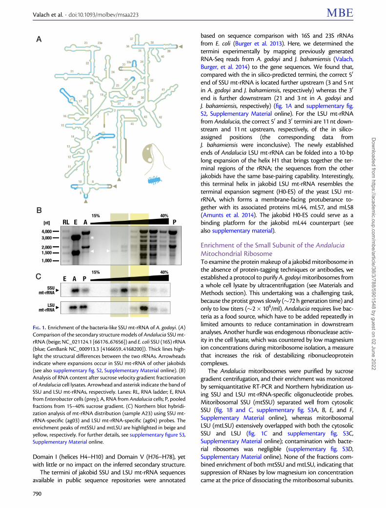
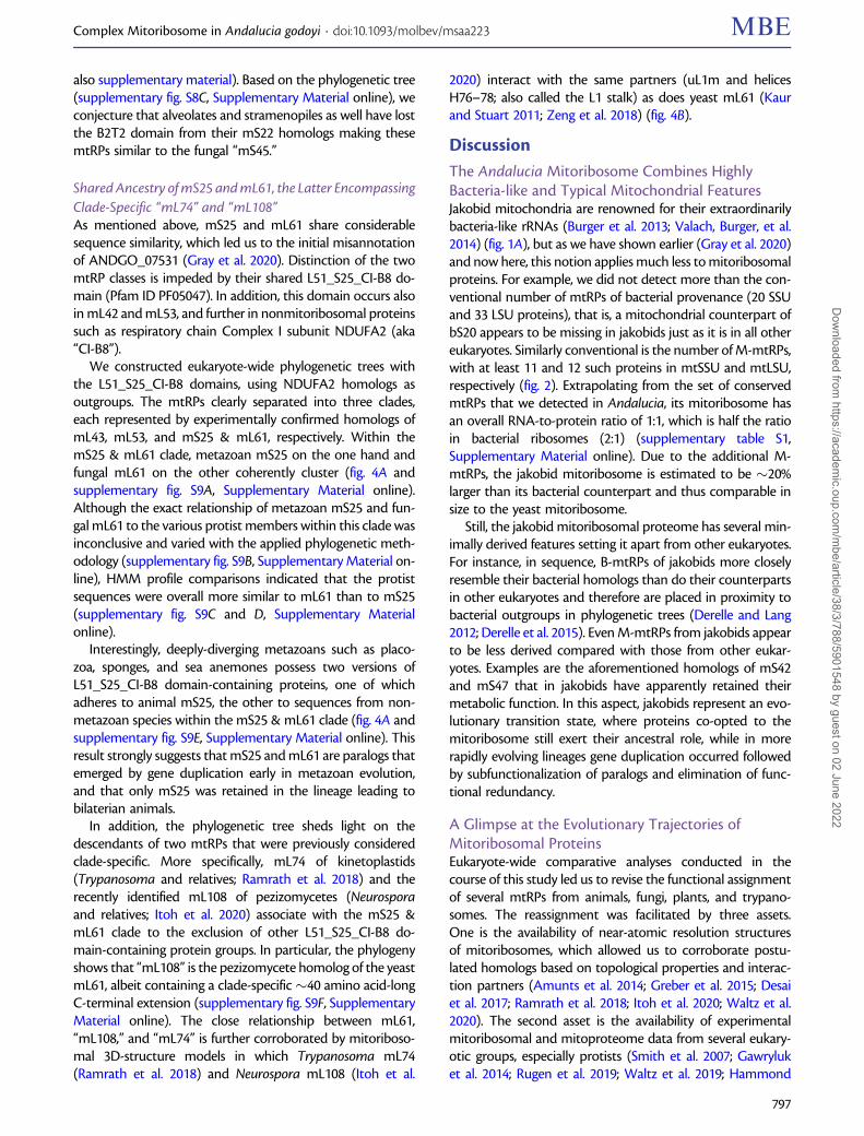
Secondary Structures of Jakobid Mitochondrial rRNAsThe mitochondrial LSU and SSU rRNAs from jakobids aremuch more similar to their bacterial homologs than thosefrom other eukaryotes (Burger et al. 2013). Among examinedjakobids, Andalucia has the most bacteria-like mt-rRNAs.Specifically, sequence identity of Andalucia SSU mt-rRNAwith the E. coli 16S rRNA is upwards of 70% (supplementaryfig. S1, Supplementary Material online). Comparison with thebacterial 16S rRNA secondary structure model indicates lossof only two terminal helices in Andalucia SSU mt-rRNA, aswell as minor apical truncations in three helices. Nucleotidesubstitutions occur mostly in the central and 30 majordomains, and are compensated by covariational changes ofthe base-pairing partners (fig. 1A; for sequence–structurealignments of jakobid SSU mt-rRNAs, see supplementaryfig. S2, Supplementary Material online).
Compared with jakobid SSU mt-rRNA, the secondarystructure model of LSU mt-rRNA deviates more noticeablyfrom the E. coli 23S rRNA model (for a secondary structurediagram of R. americana LSU mt-rRNA, see [Petrov et al.2019]; the only difference in Andalucia is a slightly reducedH63). In Andalucia (and the other jakobids), several heliceshave been lost and others truncated in Domain III (i.e., H54 toH59). Furthermore, helices H63 and H68 are apically trun-cated, and H98 is absent. Most sequence variation occurs in
Complex Mitoribosome in Andalucia godoyi . doi:10.1093/molbev/msaa223 MBE
789
Dow
nloaded from https://academ
ic.oup.com/m
be/article/38/3/788/5901548 by guest on 02 June 2022
Domain I (helices H4–H10) and Domain V (H76–H78), yetwith little or no impact on the inferred secondary structure.
The termini of jakobid SSU and LSU mt-rRNA sequencesavailable in public sequence repositories were annotated
based on sequence comparison with 16S and 23S rRNAsfrom E. coli (Burger et al. 2013). Here, we determined thetermini experimentally by mapping previously generatedRNA-Seq reads from A. godoyi and J. bahamiensis (Valach,Burger, et al. 2014) to the gene sequences. We found that,compared with the in silico-predicted termini, the correct 50
end of SSU mt-rRNA is located further upstream (3 and 5 ntin A. godoyi and J. bahamiensis, respectively) whereas the 30
end is further downstream (21 and 3 nt in A. godoyi andJ. bahamiensis, respectively) (fig. 1A and supplementary fig.S2, Supplementary Material online). For the LSU mt-rRNAfrom Andalucia, the correct 50 and 30 termini are 11 nt down-stream and 11 nt upstream, respectively, of the in silico-assigned positions (the corresponding data fromJ. bahamiensis were inconclusive). The newly establishedends of Andalucia LSU mt-rRNA can be folded into a 10-bplong expansion of the helix H1 that brings together the ter-minal regions of the rRNA; the sequences from the otherjakobids have the same base-pairing capability. Interestingly,this terminal helix in jakobid LSU mt-rRNA resembles theterminal expansion segment (H0-ES) of the yeast LSU mt-rRNA, which forms a membrane-facing protuberance to-gether with its associated proteins mL44, mL57, and mL58(Amunts et al. 2014). The jakobid H0-ES could serve as abinding platform for the jakobid mL44 counterpart (seealso supplementary material).
Enrichment of the Small Subunit of the AndaluciaMitochondrial RibosomeTo examine the protein makeup of a jakobid mitoribosome inthe absence of protein-tagging techniques or antibodies, weestablished a protocol to purify A. godoyi mitoribosomes froma whole cell lysate by ultracentrifugation (see Materials andMethods section). This undertaking was a challenging task,because the protist grows slowly (�72 h generation time) andonly to low titers (�2� 106/ml). Andalucia requires live bac-teria as a food source, which have to be added repeatedly inlimited amounts to reduce contamination in downstreamanalyses. Another hurdle was endogenous ribonuclease activ-ity in the cell lysate, which was countered by low magnesiumion concentrations during mitoribosome isolation, a measurethat increases the risk of destabilizing ribonucleoproteincomplexes.
The Andalucia mitoribosomes were purified by sucrosegradient centrifugation, and their enrichment was monitoredby semiquantitative RT-PCR and Northern hybridization us-ing SSU and LSU mt-rRNA-specific oligonucleotide probes.Mitoribosomal SSU (mtSSU) separated well from cytosolicSSU (fig. 1B and C, supplementary fig. S3A, B, E, and F,Supplementary Material online), whereas mitoribosomalLSU (mtLSU) extensively overlapped with both the cytosolicSSU and LSU (fig. 1C and supplementary fig. S3C,Supplementary Material online); contamination with bacte-rial ribosomes was negligible (supplementary fig. S3D,Supplementary Material online). None of the fractions com-bined enrichment of both mtSSU and mtLSU, indicating thatsuppression of RNases by low magnesium ion concentrationcame at the price of dissociating the mitoribosomal subunits.
FIG. 1. Enrichment of the bacteria-like SSU mt-rRNA of A. godoyi. (A)Comparison of the secondary structure models of Andalucia SSU mt-rRNA (beige; NC_021124.1 [66176..67656]) and E. coli SSU (16S) rRNA(blue; GenBank NC_000913.3 [4166659..4168200]). Thick lines high-light the structural differences between the two rRNAs. Arrowheadsindicate where expansions occur in SSU mt-rRNA of other jakobids(see also supplementary fig. S2, Supplementary Material online). (B)Analysis of RNA content after sucrose velocity gradient fractionationof Andalucia cell lysates. Arrowhead and asterisk indicate the band ofSSU and LSU mt-rRNAs, respectively. Lanes: RL, RNA ladder; E, RNAfrom Enterobacter cells (prey); A, RNA from Andalucia cells; P, pooledfractions from 15–40% sucrose gradient. (C) Northern blot hybridi-zation analysis of mt-rRNA distribution (sample A23) using SSU mt-rRNA-specific (ag03) and LSU mt-rRNA-specific (ag04) probes. Theenrichment peaks of mtSSU and mtLSU are highlighted in beige andyellow, respectively. For further details, see supplementary figure S3,Supplementary Material online.
Valach et al. . doi:10.1093/molbev/msaa223 MBE
790
Dow
nloaded from https://academ
ic.oup.com/m
be/article/38/3/788/5901548 by guest on 02 June 2022
Therefore, proteomics validation of Andalucia mtRPs wasperformed exclusively on mtSSU, whereas in silico approacheswere applied to the complete mitoribosome.
Composition of the Small Subunit of the AndaluciaMitochondrial RibosomeThe proteins of the Andalucia mtSSU preparation (supple-mentary fig. S3H, Supplementary Material online) wereextracted and analyzed by tandem mass spectrometry (MS/MS). In parallel, we examined the protein composition ofwhole cell lysates to gauge the relative enrichment of individ-ual proteins in mtSSU samples (supplementary fig. S3H,Supplementary Material online).
According to the composition of the Andalucia mitoribo-some inferred in silico by sequence similarity (Gray et al.2020), SSU and LSU contain 28 and 40 proteins, respectively.By MS/MS analysis, we detected all of the expected SSUmtRPs with the exception of two (mS25 and mS38). In con-trast, whole cell-lysate controls contained only about 30% ofthe SSU mtRPs, testifying to a significant enrichment ofmtSSU by our purification protocol (table 1 and supplemen-tary tables S1–S3, Supplementary Material online).
Retrospectively, it is not surprising that mS25(ANDGO_07531) and mS38 (ANDGO_00506) (Gray et al.2020) were not retrieved by MS/MS of mtSSU-enriched sam-ples. For ANDGO_07531, we now have evidence that despitesequence similarity this protein is not a homolog of mS25, butrather of mL61, and thus absent from the mtSSU. The mS25and mL61 proteins have been confounded because of theirshared L51_S25_CI-B8 domain (Pfam ID PF05047), which isalso present in additional mtRPs and other non-RPs.However, as we dissect in detail below, the two proteins aredistinguishable by phylogenetic analysis, which places mS25and mL61 into separate clades and clearly affiliatesANDGO_07531 with experimentally confirmed mL61sequences from model organisms.
The absence of the second protein, mS38, from MS/MSdata most probably reflects an experimental issue, that is,the protease digestion procedure applied. The Andaluciaprotein is likely processed in the same way as the mam-malian and yeast homologs (Greber et al. 2015; Desaiet al. 2017), yielding only a 29-residue long mature proteinthat represents the highly conserved C-terminal portion ofthe precursor protein. Due to the high lysine and argininecontent of the mature Andalucia mS38 protein, trypsindigestion will generate peptides too short for conventionalMS/MS detection.
Newly Identified Small Subunit MitoribosomalProteins of AndaluciaTo identify potentially unrecognized components of theAndalucia mtSSU, we examined which of the MS/MS-detected proteins have an enrichment profile (whole cell vs.mtSSU preparations) that resembles that of the trustedmtRPs. For details about the procedure applied, see the flow-chart in supplementary fig. S4, Supplementary Material on-line. We found 104 proteins with SSU mtRP-like distribution(supplementary fig. S5, Supplementary Material online). From
these 104 proteins, we filtered out 63, which according to thein silico functional annotation were evidently copurified for-tuitously, for example, components of the cytoskeleton orendoplasmatic reticulum. In this step, we also removed pro-teins predicted to contain trans-membrane and membrane-interacting domains, since in all systems so far examined,anchoring of the ribosome to the mitochondrial inner mem-brane is achieved by the LSU and not the SSU (Ott andHerrmann 2010; Pfeffer et al. 2015; Englmeier et al. 2017).Among the remaining 41 proteins, 25 were conserved SSUmtRPs, leaving 16 candidates of unrecognized SSU mtRPs,which we then ranked by their propensity of residing in mi-tochondria and conservation across jakobids (supplementarytable S4, Supplementary Material online). The six highestranking candidates included proteins annotated as a hypo-thetical protein, various dehydrogenases, a hydrolase, and asuperoxide dismutase. These six proteins are referred to in thefollowing as the top SSU mtRP candidates.
All candidates were examined by highly sensitive profileHidden Markov Model (HMM) searches and phylogeneticanalyses, and working hypotheses were tested by inspectingmitoribosomal structures from model organisms. These in-depth studies allowed us to identify four mitoribosomal pro-teins (mS22, mS31, mS42, and mS47), which had remainedunrecognized by our previous sequence similarity-based anal-yses (Gray et al. 2020) (table 1).
Identification of Andalucia mS22The top SSU mtRP candidate ANDGO_06241 is prominentlyenriched in Andalucia mtSSU preparations and, lacking obvi-ous sequence similarity with functionally defined proteins,was initially annotated as a mitochondrion-targeted hypo-thetical protein. HMM searches with profiles from the Pfamdatabase (El-Gebali et al. 2019) returned a weak match to theMRP-S22 domain (PF10245; E� 8.7e�03), which is character-istic for animal and trypanosome mS22. However,ANDGO_06241 matches also, and with a somewhat strongersignal (E� 1.5e�07), the MRP-L20 protein domain (PF12824),which is typical for fungal mL58 (previously designated MRP-L20). In ANDGO_06241, the MRP-S22 domain is located C-terminally and the MRP-L20 domain N-terminally. Homologswith the precisely same domain arrangement are present inthe inferred proteomes of all other jakobids examined, sug-gesting that this domain arrangement plays a vital role (sup-plementary table S1, Supplementary Material online).Inspection of the experimentally validated mS22 fromTrypanosoma and homologs from other kinetoplastidsrevealed that these mtRPs are also composed of a C-terminalMRP-S22 and an N-terminal mL58-like domain, which sup-ports that ANDGO_0624 is an mS22 homolog.
Identification of Andalucia mS47 with Dual FunctionANDGO_00091 is another top SSU mtRP candidate (supple-mentary table S4, Supplementary Material online). However,Blast searches of this sequence against NCBI’s nonredundantprotein sequences (nr) retrieved the mitochondrial matrixenzymes HIBCHs with convincingly strong support (E�
Complex Mitoribosome in Andalucia godoyi . doi:10.1093/molbev/msaa223 MBE
791
Dow
nloaded from https://academ
ic.oup.com/m
be/article/38/3/788/5901548 by guest on 02 June 2022
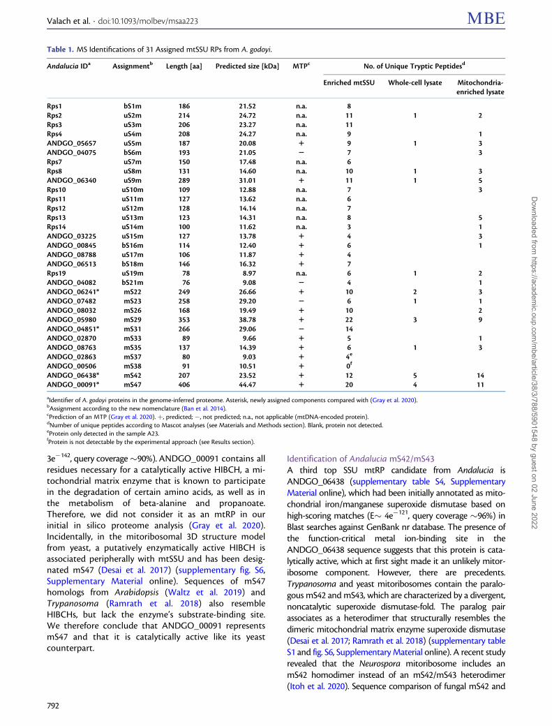
3e�142, query coverage�90%). ANDGO_00091 contains allresidues necessary for a catalytically active HIBCH, a mi-tochondrial matrix enzyme that is known to participatein the degradation of certain amino acids, as well as inthe metabolism of beta-alanine and propanoate.Therefore, we did not consider it as an mtRP in ourinitial in silico proteome analysis (Gray et al. 2020).Incidentally, in the mitoribosomal 3D structure modelfrom yeast, a putatively enzymatically active HIBCH isassociated peripherally with mtSSU and has been desig-nated mS47 (Desai et al. 2017) (supplementary fig. S6,Supplementary Material online). Sequences of mS47homologs from Arabidopsis (Waltz et al. 2019) andTrypanosoma (Ramrath et al. 2018) also resembleHIBCHs, but lack the enzyme’s substrate-binding site.We therefore conclude that ANDGO_00091 representsmS47 and that it is catalytically active like its yeastcounterpart.
Identification of Andalucia mS42/mS43A third top SSU mtRP candidate from Andalucia isANDGO_06438 (supplementary table S4, SupplementaryMaterial online), which had been initially annotated as mito-chondrial iron/manganese superoxide dismutase based onhigh-scoring matches (E� 4e�121, query coverage �96%) inBlast searches against GenBank nr database. The presence ofthe function-critical metal ion-binding site in theANDGO_06438 sequence suggests that this protein is cata-lytically active, which at first sight made it an unlikely mitor-ibosome component. However, there are precedents.Trypanosoma and yeast mitoribosomes contain the paralo-gous mS42 and mS43, which are characterized by a divergent,noncatalytic superoxide dismutase-fold. The paralog pairassociates as a heterodimer that structurally resembles thedimeric mitochondrial matrix enzyme superoxide dismutase(Desai et al. 2017; Ramrath et al. 2018) (supplementary tableS1 and fig. S6, Supplementary Material online). A recent studyrevealed that the Neurospora mitoribosome includes anmS42 homodimer instead of an mS42/mS43 heterodimer(Itoh et al. 2020). Sequence comparison of fungal mS42 and
Table 1. MS Identifications of 31 Assigned mtSSU RPs from A. godoyi.
Andalucia IDa Assignmentb Length [aa] Predicted size [kDa] MTPc No. of Unique Tryptic Peptidesd
Enriched mtSSU Whole-cell lysate Mitochondria-enriched lysate
Rps1 bS1m 186 21.52 n.a. 8Rps2 uS2m 214 24.72 n.a. 11 1 2Rps3 uS3m 206 23.27 n.a. 11Rps4 uS4m 208 24.27 n.a. 9 1ANDGO_05657 uS5m 187 20.08 1 9 1 3ANDGO_04075 bS6m 193 21.05 2 7 3Rps7 uS7m 150 17.48 n.a. 6Rps8 uS8m 131 14.60 n.a. 10 1 3ANDGO_06340 uS9m 289 31.01 1 11 1 5Rps10 uS10m 109 12.88 n.a. 7 3Rps11 uS11m 127 13.62 n.a. 6Rps12 uS12m 128 14.14 n.a. 7Rps13 uS13m 123 14.31 n.a. 8 5Rps14 uS14m 100 11.62 n.a. 3 1ANDGO_03225 uS15m 127 13.78 1 4 3ANDGO_00845 bS16m 114 12.40 1 6 1ANDGO_08788 uS17m 106 11.87 1 4ANDGO_06513 bS18m 146 16.32 1 7Rps19 uS19m 78 8.97 n.a. 6 1 2ANDGO_04082 bS21m 76 9.08 2 4 1ANDGO_06241* mS22 249 26.66 1 10 2 3ANDGO_07482 mS23 258 29.20 2 6 1 1ANDGO_08032 mS26 168 19.49 1 10 2ANDGO_05980 mS29 353 38.78 1 22 3 9ANDGO_04851* mS31 266 29.06 2 14ANDGO_02870 mS33 89 9.66 1 5 1ANDGO_08763 mS35 137 14.39 1 6 1 3ANDGO_02863 mS37 80 9.03 1 4e
ANDGO_00506 mS38 91 10.51 1 0f
ANDGO_06438* mS42 207 23.52 1 12 5 14ANDGO_00091* mS47 406 44.47 1 20 4 11
aIdentifier of A. godoyi proteins in the genome-inferred proteome. Asterisk, newly assigned components compared with (Gray et al. 2020).bAssignment according to the new nomenclature (Ban et al. 2014).cPrediction of an MTP (Gray et al. 2020). þ, predicted; �, not predicted; n.a., not applicable (mtDNA-encoded protein).dNumber of unique peptides according to Mascot analyses (see Materials and Methods section). Blank, protein not detected.eProtein only detected in the sample A23.fProtein is not detectable by the experimental approach (see Results section).
Valach et al. . doi:10.1093/molbev/msaa223 MBE
792
Dow
nloaded from https://academ
ic.oup.com/m
be/article/38/3/788/5901548 by guest on 02 June 2022
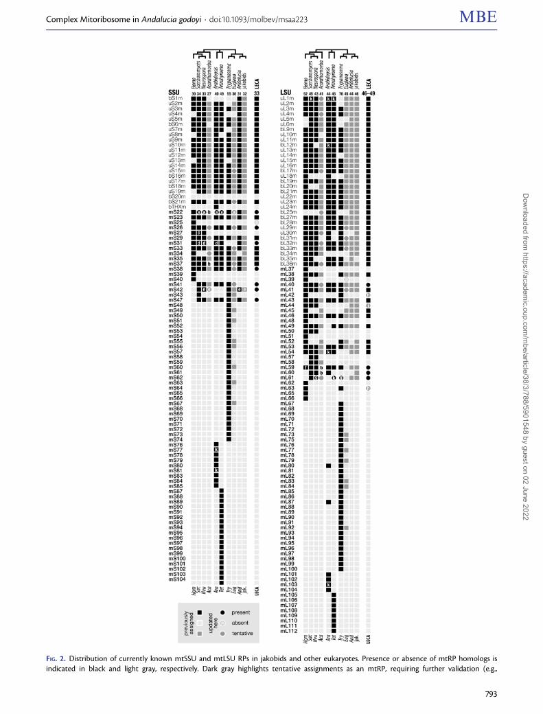
FIG. 2. Distribution of currently known mtSSU and mtLSU RPs in jakobids and other eukaryotes. Presence or absence of mtRP homologs isindicated in black and light gray, respectively. Dark gray highlights tentative assignments as an mtRP, requiring further validation (e.g.,
Complex Mitoribosome in Andalucia godoyi . doi:10.1093/molbev/msaa223 MBE
793
Dow
nloaded from https://academ
ic.oup.com/m
be/article/38/3/788/5901548 by guest on 02 June 2022
mS43 indicates that the Neurospora protein is actually moreclosely related to the yeast mS43 than mS42; still, similarly toits yeast counterparts, the Neurospora mtRP also lacks thecatalytic Fe ion-binding site and is therefore considered anenzyme paralog that lost its activity upon recruitment to themitoribosome.
The sequence resemblance to Fe/Mn superoxide dismut-ase strongly suggests that ANDGO_06438 is an mS42/mS43homolog. In contrast to its counterparts from fungi andTrypanosoma, the Andalucia mS42/mS43 has apparentlyretained its catalytic activity as a radical-scavenging enzyme.Evolutionary relationships between mS42 and mS43 fromvarious organisms are difficult to resolve, which could indicateeither that superoxide dismutases or superoxide dismutase-like proteins have been recruited to the mitoribosome atmultiple occasions, or that the original mtRPs were replacedat different times in different lineages by paralogs. For sim-plicity, we refer to the Andalucia protein as mS42 (table 1 andfig. 2).
Identification of Andalucia mS31Akin to ANDGO_06241 (Andalucia mS22, see above),ANDGO_04851 was initially annotated as a hypothetical pro-tein. While lacking a recognizable conserved protein domain,a closer inspection revealed that the C-terminal moiety ofANDGO_04851 contains a region with moderate similarityto the �40 amino acid-long core of mS31 from ciliates andmetazoans. Across eukaryotes, the core mS31 region can beconsiderably degenerate, which makes it sometimes difficultto retrieve homologs even from closely related species (seealso supplementary fig. S7, Supplementary Material online).This may explain why we did not identify homologs in threeout of the other four jakobids (supplementary table S4,Supplementary Material online). Nevertheless, the enrich-ment of ANDGO_04851 in MS/MS experiments supportsthe proposed functional assignment (supplementary tableS2 and fig. S5, Supplementary Material online), even if theprotein does not carry a recognizable mitochondrial targetingpeptide (MTP), as in three other undisputed instances (table 1and supplementary table S4, Supplementary Material online).
Tentative Clade-Specific Components of JakobidmtSSUThe three remaining top SSU mtRP candidates fromAndalucia are ANDGO_08608 with high sequence similarityto a 3-hydroxybutyryl-CoA dehydrogenase (Blast E� 3e�170,query coverage�94%), an enzyme involved in the mitochon-drial isoleucine metabolism; ANDGO_06552, which resem-bles mitochondrial glutamate dehydrogenases (E� 0, querycoverage�93%); and ANDGO_06037, which retrieves malatedehydrogenases (E� 0, query coverage �96%) (supplemen-tary table S4, Supplementary Material online). All threeAndalucia proteins have sequence features of functionalenzymes, with homologs also present in the inferred pro-teomes from the other jakobids examined here. We speculatethat these three proteins are jakobid-specific recruits of mi-tochondrial metabolic matrix proteins to the mitoribosomeand have retained their enzymatic activity. It would be mostinteresting to validate our inferences by structural analyses ofthe jakobid mitoribosome.
Broadly Distributed mS34 and mS41 Are Missing fromthe Andalucia mtSSUProteome and 3D-structure analyses confirmed that mS34 isan integral constituent of mitoribosomes in mammals,Arabidopsis and Trypanosoma, but not in Saccharomycesor Neurospora. Andalucia also appears to lack the mS34gene as do Jakoba spp., whereas unambiguous homologsare present in the R. americana and S. ecuadoriensis nucleargenomes (supplementary table S1, Supplementary Materialonline). Previously it seemed that the presence of mS34 wascorrelated with the absence or truncation of the mt-rRNAstructure elements h6, h8–h10, and h12 in human andTrypanosoma SSU mt-rRNA (PDB 5aj3-Aj and 6hiw-Cj, re-spectively; Greber et al. 2015; Ramrath et al. 2018), and viceversa, that the presence of these rRNA elements is associ-ated with the absence of mS34 in yeast. However, recentdata from the Arabidopsis and Tetrahymena mitoribosomesshow that the retention of these rRNA elements does notpreclude mS34 being part of the mitoribosome (see PDB6xyw-By and 6z1p-Bz, respectively; Tobiasson and Amunts2020; Waltz et al. 2020) (supplementary fig. S6,
Fig. 2. Continuedmitoribosome isolation). Circles, updated assignments compared with previous publications (Desmond et al. 2011; Gray et al. 2020; Hammondet al. 2020; Tobiasson and Amunts 2020; Waltz and Gieg�e 2020). All RPs with the prefix “b” or “u” (bS/bL, uS/uL) are present in bacteria. Notes toboxes and circles labeled a–k: a, mS22 and Saccharomyces/Neurospora/Arabidopsis mS45 were recognized as homologs, and Tetrahymena andEuglena also contain an A3T2-domain-only mS22 (see Results section). b, mS22, mS37, mL59, and mL60 were not retrieved from Acanthamoeba,but homologs are present in some amoebozoans (for mS22, see supplementary fig. S8B, Supplementary Material online). c, mS27 and mS44 havebeen recognized as homologs (Itoh et al. 2020). d, mS31 and mS46 have been recognized as homologs (see Waltz et al. 2020; supplementary fig. S7,Supplementary Material online). e, mS42/mS43 is a homodimer, assuming the role of distinct paralogous mS42 and mS43, in Neurospora (Itoh et al.2020), most likely in jakobids (see the Results section), and possibly also in Acanthamoeba. f, mL59 and mL64 have been recognized as homologs(Waltz and Gieg�e 2020). g, mL74 of Trypanosoma and mL108 of Neurospora represent mL61 (see the Results). h, presence of mL42 and mL63 inLECA is uncertain because due to an extremely short and weak sequence signature of the proposed conserved domains between animal andtrypanosome counterparts, it is currently impossible to retrieve homologs in other lineages by sequence similarity searches and verify that theseRPs are indeed orthologs, that is, not a result of convergent evolution. i, presence of mL44 in LECA is uncertain because the mL44 homolog injakobids has not been confirmed experimentally as an mtRP. k, mtRPs missing from mitoribosome cryo-EM 3D reconstructions. The followingmtRP assignments have become obsolete: mS45, mS46, mL74, and mL108 (this work), mS44 (Itoh et al. 2020), and mL64 (Waltz and Gieg�e 2020)(see also supplementary material).
Valach et al. . doi:10.1093/molbev/msaa223 MBE
794
Dow
nloaded from https://academ
ic.oup.com/m
be/article/38/3/788/5901548 by guest on 02 June 2022
Supplementary Material online). In these species with mt-rRNA structure more similar to that of bacteria and jako-bids, mS34 binds in between h6 and h8–h10, a region thatmost likely corresponds to the ancestral binding site of theprotein.
Another protein apparently absent from Andalucia andthe other examined jakobids is mS41. This mtRP has beenexperimentally confirmed in yeast (Desai et al. 2017), try-panosome (Ramrath et al. 2018), ciliate (Tobiasson andAmunts 2020), and Arabidopsis (Waltz et al. 2020), but isapparently not part of the metazoan mitoribosomes (sup-plementary fig. S6, Supplementary Material online).Otherwise, this mtRP appears widespread throughouteukaryotes as seen in sequence similarity searches acrossprotist genomes (fig. 2). In the structural model of the yeastand Arabidopsis mitoribosomes, mS41 interacts almost
exclusively with the SSU mt-rRNA helices h6, h15, andh17, but the corresponding region is completely protein-free in bacterial ribosomes. The same situation most likelyapplies to jakobid SSU mt-rRNAs, given their particularlybacteria-like structures.
Previously Unrecognized or Incorrectly AnnotatedMitoribosomal Proteins in Other EukaryotesIn the course of this study, we realized that certain mtRPclasses are difficult to distinguish because of shared sequencedomain signatures. One easily confounded pair is mS22/“mS45” & mL58 and the other is mS25 & mL61, the distinc-tion between which we attempted to resolve by eukaryote-wide comparative analyses, as detailed in the following twosections.
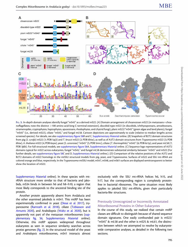
FIG. 3. In-depth domain analyses identify fungal “mS45” as a derived mS22. (A) Domain arrangement of choanozoan mS22 (in metazoansþchoa-noflagellates; note the distinct >100 amino acid-long C-terminal extension), discobid-type mS22 (in discobids, ichthyosporeans, amoebozoans,stramenopiles, cryptophytes, haptophytes, apusozoans, rhodophytes, and chytrid fungi), plant mS22/“mS45” (green algae and land plants), fungal“mS45” (i.e., derived mS22), ciliate “mS45,” and fungal mL58. Cartoon depictions are approximately to scale (relative to median lengths acrossscreened species). For details, see also supplementary figure S8B and C, Supplementary Material online. (B) Snapshots of B2T2 domain structuresfrom pig (S. scrofa) mS22 (1; PDB 5aj3) and T. brucei mS22 (2; PDB 6hiw), as well as of A3T2 domain structures from Trypanosoma mS22 (3; PDB6hiw), A. thaliana mS22 (4; PDB 6xyw), yeast (S. cerevisiae) “mS45” (5; PDB 5mrc), ciliate (T. thermophila) “mS45” (6; PDB 6z1p), and yeast mL58 (7;PDB 3j6b). For full structural models, see supplementary figure S8A, Supplementary Material online. (C) Sequence logo representations of A3T2domains typical for mS22 across eukaryotes, fungal “mS45,” and fungal mL58 demonstrate substantial similarity between “mS45” and mS22 (Forfurther details, see supplementary figure S8C and D, Supplementary Material online.). (D) Comparison of the relative positions of the A3T2 andB2T2 domains of mS22 homologs in the mtSSU structural models from pig, yeast, and Trypanosoma. Surfaces of mS22 and SSU mt-rRNA arecolored orange and blue, respectively. In the Trypanosoma mtSSU model, mS47, mS48, and mS61 surfaces are displayed semitransparent to bettershow the location of mS22.
Complex Mitoribosome in Andalucia godoyi . doi:10.1093/molbev/msaa223 MBE
795
Dow
nloaded from https://academ
ic.oup.com/m
be/article/38/3/788/5901548 by guest on 02 June 2022
Fungal “mS45” Is a Derived mS22 HomologThe identification of Andalucia’s mS22 was complicated bythe fact that homologs from animals include a single distinc-tive protein domain MRP-S22, whereas those from trypano-somes include an additional domain otherwise typical forfungal mL58. To understand the domain composition ofmS22 across eukaryotes, we created specific profile HMMsfor each of these domains, first based on metazoan, trypano-some, and jakobid homologs, and then iteratively includingnewly identified additional homologs from other eukaryotes.The profile HMM so generated and covering the MRP-S22(PF10245) domain will be referred to as B2T2 (�40 residues),alluding to the secondary structure of the corresponding pro-tein region that consists of two beta strands and two turns.Similarly, the profile HMM including the mL58 domain (MRP-L40 or PF12824) will be referred to as A3T2 (�50 residues)because it contains three alpha helices and two turns (fig. 3Aand B, supplementary fig. S8A, Supplementary Material on-line). With these profile HMMs in hand, we performed HMMsearches to identify in eukaryotic proteomes all sequencescarrying B2T2, and in independent searches those containingA3T2 (for the collection of inferred eukaryotic proteomesexamined here, see Materials and Methods section). Bothsets of retrieved sequences were then inspected for additionalprotein domains.
Sequences retrieved with the B2T2 profile fall into twodistinct groups (fig. 3A). The first contains proteins carryingexclusively a B2T2 domain, found in choanozoans, that is,
metazoans and their closest protozoan relatives, the choano-flagellates (supplementary fig. S8B, Supplementary Materialonline). The second group contains proteins that carry notonly B2T2, but also a second domain, A3T2. The correspond-ing sequences come from essentially all nonchoanozoan eu-karyotic groups including jakobids (supplementary fig. S8B,Supplementary Material online).
Sequences retrieved with the profile A3T2 were highly di-verse. Phylogenetic analysis based on this domain showedmoderate resolution due to the limited length of the proteinregion, but still revealed an informative pattern. Among thefour clades is one that combines the above-identified two-domain mS22 RPs, together with several proteins lacking theB2T2 domain. The latter proteins are mostly from fungi (butalso from alveolates), annotated as homologs of the yeastmS45, which has been considered to be a clade-specificmtRP (fig. 3C and supplementary fig. S8C and D,Supplementary Material online).
Our finding that the fungal and alveolate mS45—whichlack the mS22-typical B2T2 domain—affiliate in this tree withthe two-domain mS22 suggests that “mS45” is not a distinctmtRP but rather a variant mS22 that has lost its B2T2 domain.The view that mS45 is an mS22 homolog is corroborated bythe fact that in the 3D-structure model of yeast mtSSU, thisprotein interacts with a similar set of conserved mtRPs andoccupies a similar location as do animal and trypanosomemS22, in particular the A3T2 domain of the latter (Greberet al. 2015; Desai et al. 2017; Ramrath et al. 2018) (fig. 3D; see
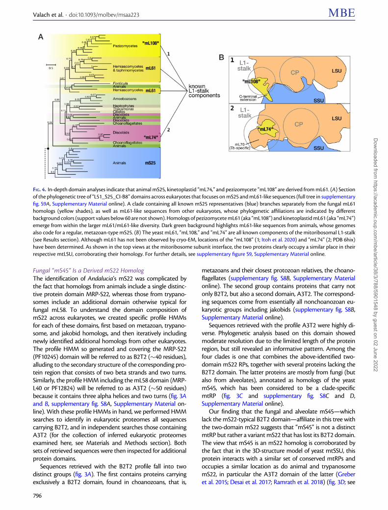
FIG. 4. In-depth domain analyses indicate that animal mS25, kinetoplastid “mL74,” and pezizomycete “mL108” are derived from mL61. (A) Sectionof the phylogenetic tree of “L51_S25_CI-B8” domains across eukaryotes that focuses on mS25 and mL61-like sequences (full tree in supplementaryfig. S9A, Supplementary Material online). A clade containing all known mS25 representatives (blue) branches separately from the fungal mL61homologs (yellow shades), as well as mL61-like sequences from other eukaryotes, whose phylogenetic affiliations are indicated by differentbackground colors (support values below 60 are not shown). Homologs of pezizomycete mL61 (aka “mL108”) and kinetoplastid mL61 (aka “mL74”)emerge from within the larger mL61/mL61-like diversity. Dark green background highlights mL61-like sequences from animals, whose genomesalso code for a regular, metazoan-type mS25. (B) The yeast mL61, “mL108,” and “mL74” are all known components of the mitoribosomal L1-stalk(see Results section). Although mL61 has not been observed by cryo-EM, locations of the “mL108” (1; Itoh et al. 2020) and “mL74” (2; PDB 6hix)have been determined. As shown in the top views at the mitoribosome subunit interface, the two proteins clearly occupy a similar place in theirrespective mtLSU, corroborating their homology. For further details, see supplementary figure S9, Supplementary Material online.
Valach et al. . doi:10.1093/molbev/msaa223 MBE
796
Dow
nloaded from https://academ
ic.oup.com/m
be/article/38/3/788/5901548 by guest on 02 June 2022
also supplementary material). Based on the phylogenetic tree(supplementary fig. S8C, Supplementary Material online), weconjecture that alveolates and stramenopiles as well have lostthe B2T2 domain from their mS22 homologs making thesemtRPs similar to the fungal “mS45.”
Shared Ancestry of mS25 and mL61, the Latter Encompassing
Clade-Specific “mL74” and “mL108”As mentioned above, mS25 and mL61 share considerablesequence similarity, which led us to the initial misannotationof ANDGO_07531 (Gray et al. 2020). Distinction of the twomtRP classes is impeded by their shared L51_S25_CI-B8 do-main (Pfam ID PF05047). In addition, this domain occurs alsoin mL42 and mL53, and further in nonmitoribosomal proteinssuch as respiratory chain Complex I subunit NDUFA2 (aka“CI-B8”).
We constructed eukaryote-wide phylogenetic trees withthe L51_S25_CI-B8 domains, using NDUFA2 homologs asoutgroups. The mtRPs clearly separated into three clades,each represented by experimentally confirmed homologs ofmL43, mL53, and mS25 & mL61, respectively. Within themS25 & mL61 clade, metazoan mS25 on the one hand andfungal mL61 on the other coherently cluster (fig. 4A andsupplementary fig. S9A, Supplementary Material online).Although the exact relationship of metazoan mS25 and fun-gal mL61 to the various protist members within this clade wasinconclusive and varied with the applied phylogenetic meth-odology (supplementary fig. S9B, Supplementary Material on-line), HMM profile comparisons indicated that the protistsequences were overall more similar to mL61 than to mS25(supplementary fig. S9C and D, Supplementary Materialonline).
Interestingly, deeply-diverging metazoans such as placo-zoa, sponges, and sea anemones possess two versions ofL51_S25_CI-B8 domain-containing proteins, one of whichadheres to animal mS25, the other to sequences from non-metazoan species within the mS25 & mL61 clade (fig. 4A andsupplementary fig. S9E, Supplementary Material online). Thisresult strongly suggests that mS25 and mL61 are paralogs thatemerged by gene duplication early in metazoan evolution,and that only mS25 was retained in the lineage leading tobilaterian animals.
In addition, the phylogenetic tree sheds light on thedescendants of two mtRPs that were previously consideredclade-specific. More specifically, mL74 of kinetoplastids(Trypanosoma and relatives; Ramrath et al. 2018) and therecently identified mL108 of pezizomycetes (Neurosporaand relatives; Itoh et al. 2020) associate with the mS25 &mL61 clade to the exclusion of other L51_S25_CI-B8 do-main-containing protein groups. In particular, the phylogenyshows that “mL108” is the pezizomycete homolog of the yeastmL61, albeit containing a clade-specific�40 amino acid-longC-terminal extension (supplementary fig. S9F, SupplementaryMaterial online). The close relationship between mL61,“mL108,” and “mL74” is further corroborated by mitoriboso-mal 3D-structure models in which Trypanosoma mL74(Ramrath et al. 2018) and Neurospora mL108 (Itoh et al.
2020) interact with the same partners (uL1m and helicesH76–78; also called the L1 stalk) as does yeast mL61 (Kaurand Stuart 2011; Zeng et al. 2018) (fig. 4B).
Discussion
The Andalucia Mitoribosome Combines HighlyBacteria-like and Typical Mitochondrial FeaturesJakobid mitochondria are renowned for their extraordinarilybacteria-like rRNAs (Burger et al. 2013; Valach, Burger, et al.2014) (fig. 1A), but as we have shown earlier (Gray et al. 2020)and now here, this notion applies much less to mitoribosomalproteins. For example, we did not detect more than the con-ventional number of mtRPs of bacterial provenance (20 SSUand 33 LSU proteins), that is, a mitochondrial counterpart ofbS20 appears to be missing in jakobids just as it is in all othereukaryotes. Similarly conventional is the number of M-mtRPs,with at least 11 and 12 such proteins in mtSSU and mtLSU,respectively (fig. 2). Extrapolating from the set of conservedmtRPs that we detected in Andalucia, its mitoribosome hasan overall RNA-to-protein ratio of 1:1, which is half the ratioin bacterial ribosomes (2:1) (supplementary table S1,Supplementary Material online). Due to the additional M-mtRPs, the jakobid mitoribosome is estimated to be �20%larger than its bacterial counterpart and thus comparable insize to the yeast mitoribosome.
Still, the jakobid mitoribosomal proteome has several min-imally derived features setting it apart from other eukaryotes.For instance, in sequence, B-mtRPs of jakobids more closelyresemble their bacterial homologs than do their counterpartsin other eukaryotes and therefore are placed in proximity tobacterial outgroups in phylogenetic trees (Derelle and Lang2012; Derelle et al. 2015). Even M-mtRPs from jakobids appearto be less derived compared with those from other eukar-yotes. Examples are the aforementioned homologs of mS42and mS47 that in jakobids have apparently retained theirmetabolic function. In this aspect, jakobids represent an evo-lutionary transition state, where proteins co-opted to themitoribosome still exert their ancestral role, while in morerapidly evolving lineages gene duplication occurred followedby subfunctionalization of paralogs and elimination of func-tional redundancy.
A Glimpse at the Evolutionary Trajectories ofMitoribosomal ProteinsEukaryote-wide comparative analyses conducted in thecourse of this study led us to revise the functional assignmentof several mtRPs from animals, fungi, plants, and trypano-somes. The reassignment was facilitated by three assets.One is the availability of near-atomic resolution structuresof mitoribosomes, which allowed us to corroborate postu-lated homologs based on topological properties and interac-tion partners (Amunts et al. 2014; Greber et al. 2015; Desaiet al. 2017; Ramrath et al. 2018; Itoh et al. 2020; Waltz et al.2020). The second asset is the availability of experimentalmitoribosomal and mitoproteome data from several eukary-otic groups, especially protists (Smith et al. 2007; Gawryluket al. 2014; Rugen et al. 2019; Waltz et al. 2019; Hammond
Complex Mitoribosome in Andalucia godoyi . doi:10.1093/molbev/msaa223 MBE
797
Dow
nloaded from https://academ
ic.oup.com/m
be/article/38/3/788/5901548 by guest on 02 June 2022
et al. 2020). Third, the moderate divergence of jakobid pro-teins allowed us to build highly specific profile HMMs forsensitive homology assignment and to reconstruct phyloge-netic trees stabilized by short branches.
For example, we provide evidence that mL74 and mL108,previously considered clade-specific to the kinetoplastids andpezizomycete fungi, respectively, are in fact derived versionsof the otherwise widely distributed mL61 (fig. 4). In addition,we disentangled the assignment of several mitoribosomalproteins that are easily confused when relying on sequenceinformation and conserved-protein content alone. The firstpair of easily confounded mtRPs is mL61 & mS25.Phylogenetic analyses indicate that the ancestral form is ac-tually mL61, from which mS25 arose by gene duplication inthe metazoan lineage. In this instance, evolution took an ad-ditional step: in bilaterian animals, the orthologous mL61 waslost, leaving behind the paralogous mS25 (fig. 4 and supple-mentary fig. S8, Supplementary Material online). This is atypical case of hidden paralogy, a well-known problem inphylogenetic reconstruction leading to erroneous phyloge-netic inferences.
The second confounding pair is mS22/mS45 & mL58.According to our in silico analyses, mS22, which is presentin most eukaryotes including jakobids (fig. 3 and supplemen-tary fig. S8, Supplementary Material online), contains twoconserved protein domains (B2T2 and A3T2, see Results).The corresponding orthologs from metazoans and choano-flagellates have lost the N-terminal domain, and thus onlypossess B2T2, whereas those in fungi have retained just theA3T2 domain and have therefore not been previously recog-nized as mS22, but incorrectly assigned as a novel mtRP,mS45. Further complicating matters, fungal mitoribosomescontain the LSU mtRP mL58, which is characterized by asingle A3T2 domain similarly to “mS45,” that is, the actualfungal mS22. The most parsimonious scenario is that thefungal mL58 originated from mS22 by gene duplication (likelyvia another paralog, Rrg9; supplementary fig. S8C and D,Supplementary Material online) and subsequently lost theB2T2 protein domain.
The mtRP Complement of Andalucia Implies anExtended Set of Mitoribosomal Proteins in LECAThe phylogenetic distribution of paralogs not only allows usto formulate hypotheses about the timing of gene duplica-tions, subfunctionalization, and losses of one of the two paral-ogs, but most importantly, about the ancestral gene at theorigin of these events. For two paralogous pairs of mtRP-encoding genes, we provide reasonable ancestry evidence,notably, we posit that mS22 gave rise to mL58 and mL61to mS25.
A related longstanding question pertains to the set of an-cestral genes present in LECA. The array of postulated mitor-ibosomal components in LECA was first established over adecade ago (Smits et al. 2007). Through the present and ear-lier studies by us (Gray et al. 2020) and work by others(Desmond et al. 2011; Waltz and Gieg�e 2020), this list hasnow expanded from initially 30 SSU and 38 LSU mt-RPs to 33and 46 (possibly 49), respectively (see fig. 2). Thus, LECA’s
mitoribosome, with about 50% more RPs than its bacterialpredecessor, possessed a much more complex mitochondrialtranslation machinery than generally assumed.
Was Recruitment of Mitochondrion-SpecificRibosomal Proteins Driven by mt-rRNA Defects?The observation that in mammals and fungi streamlined mt-rRNAs are associated with high mitoriboproteome complex-ity led to the common assumption that M-mtRPs structurallyreplace evolutionarily deleted rRNA elements. High-resolution analyses of mitoribosomal architecture showedthat this notion fully applies only to certain mitoribosomes(e.g., from Trypanosoma; Ramrath et al. 2018), whereas inothers (e.g., from pig; Greber et al. 2015), the volume of certainmissing rRNA segments has remained void. A variation of theRNA-replacement-by-protein view is that evolutionarychanges to mt-rRNA structure resulted in its structural insta-bility, which led to the incorporation of stabilizing M-mtRPs(Petrov et al. 2019).
As we show here, the hypothesis of “structural patching” isat odds with our findings in Andalucia and other jakobids, inwhich a remarkably bacteria-like rRNA architecture coexistswith numerous M-mtRPs. Although many M-mtRPs or evenB-mtRPs have evolved to compensate for absence of certainrRNA elements (Hosseini et al. 2018), this does not appear tohave been their ancestral role. For instance, the mtSSU pro-teins mS42/mS43 and mS47 nowadays compensate for rRNAhelix truncations in Trypanosoma (Ramrath et al. 2018).However, as illustrated here by Andalucia, these M-mtRPswere recruited early on into a mitoribosome with virtuallyno rRNA structural deviations, which implies that their initialincorporation did not serve to counter rRNA lability. Similarly,five M-mtRPs that compensate for helix truncations andlosses of certain rRNA helices in mammalian mtSSU(Hosseini et al. 2018) are conserved in other lineages thatdo contain full-length versions of these helices with the cor-responding proteins binding close by. This strongly suggeststhat the compensatory role of these proteins could arise be-cause they already resided in the corresponding location (sup-plementary table S5, Supplementary Material online). Theseand other examples (see supplementary material) illustratethat deviation of mt-rRNAs and acquisition of most, if not allancestral M-mtRPs were independent evolutionary eventsand presumably the result of very different evolutionaryforces.
Although we argue about the “purpose” of protein acquis-itions to the ancestral mitoribosome, rRNA-stabilizing M-mtRPs may well have been recruited in lineages such as plantsand fungi, whose mitoribosomes have undergone a relativelyrecent constructive phase. In the Arabidopsis mitoribosome,for example, numerous extended mt-rRNA segments are sta-bilized by dedicated proteins of the pentatricopeptide repeat(PPR) family (Waltz et al. 2020), which has undergone a recentexpansion in land plants (Gutmann et al. 2020). Likewise, M-mtRPs interacting with rRNA expansions in theSaccharomyces mitoribosome are recent acquisitions in thefungal lineage, many having nucleic acid-interaction domains(Amunts et al. 2014) (The case of mL44 is discussed in more
Valach et al. . doi:10.1093/molbev/msaa223 MBE
798
Dow
nloaded from https://academ
ic.oup.com/m
be/article/38/3/788/5901548 by guest on 02 June 2022
detail in supplementary material.). It will be interesting toretrace the order of evolutionary events once more informa-tion is available about mitoribosomes of early-diverging plantsand fungi.
What Then Drives the Recruitment of Organelle-Specific Proteins?The mitoribosome is not the only mitochondrial complexthat is considerably inflated compared with its bacterial an-cestor. The same phenomenon applies to mitochondrial bio-energetic complexes where numerous accessory proteinswere added to the ancient bacterial core (Acestor et al.2011; Gawryluk et al. 2012; Hirst 2013; Jett and Leary 2018).It is widely assumed that the newly acquired proteins servemitochondrion-specific functions, and thus are a manifesta-tion of adaptive evolutionary forces (Greber and Ban [2016]for mitoribosomes and Hirst [2011] for respiratory ComplexI). Up to now, special biological functions in mitochondrialtranslation have only been documented for three mtRPs. ThemtRP mL45 (and, in yeast, its evolutionarily unrelated coun-terpart Mba1) mediates the binding of the mitoribosome tothe inner mitochondrial membrane (Greber et al. 2014;Englmeier et al. 2017), which is necessary for the cotransla-tional insertion of the many highly hydrophobicmitochondrion-encoded proteins synthesized on the mitor-ibosome (Ott and Herrmann 2010). The other two examplesare lineage-specific mtRPs of the PPR family: mS39, whichhelps to usher leader-less mt-mRNAs to the mammalianmitoribosome (Kummer et al. 2018), and mS83, which formsthe mt-mRNA channel in the Arabidopsis mitoribosome(Waltz et al. 2020). Actually, the majority of eukaryote-wideconserved M-mtRPs are of lesser importance in mitoribo-some biogenesis, since they are incorporated at intermediateor late stages of assembly, and two particularly broadly con-served RPs, mL53 and mL61, are even nonessential in yeast(Zeng et al. 2018).
As outlined earlier (van der Sluis et al. 2015), the fact that agiven protein plays a specific functional role does not implythat the initial recruitment was driven by adaptation tomitochondrion-specific demands. Instead, the expansion ofmitochondrial protein content is more likely due to neutral(constructive) evolution, which invokes neutrally fixed com-plexity and only gradual increasing dependency from thenewly recruited components (Stoltzfus 1999; Gray et al.2010; Luke�s et al. 2011).
It is noteworthy that the evolutionary trajectory of com-plexes and pathways in the much younger “sister” organelle,the plastid, is quite similar, but the number of accessoryproteins is apparently less. For instance the plastoribosomefrom land plants comprises only six such accessory proteins(Graf et al. 2017) and the plastid NAD(P)H dehydrogenasecomplex only 10 (compared with �30 in the mitochondrialComplex I) (Peng et al. 2011;), indicating that protein acqui-sition is intimately linked to the establishment of organelles inthe eukaryotic cell. However, available plastid proteome dataare limited in number and taxonomic breadth, with conspic-uous lack of information from slowly evolving plastids thatretain more (cyano)bacterial features (similar to
mitochondria in jakobids), precluding us from drawing a pic-ture of the evolutionary trajectory of the plastid as detailed asthe one we currently have for mitochondria.
Current Limitations and OutlookAs a cautionary note, we feel it is important to emphasize thedifficulty in recognizing homologs of mitoribosomal proteins,especially in rapidly evolving systems. Extreme cases are themitoribosomes of Tetrahymena and Trypanosoma, whichappeared to have lost nearly half of the mtRPs predicted tohave been present in LECA based on sequence informationalone. However, many of the seemingly lost mtRPs have infact remained unrecognized due to extreme sequence diver-gence (this study; Ramrath et al. 2018; Tobiasson and Amunts2020). Although proteins with low sequence conservation arebest recognized by HMM searches, this approach fails whenmoderately derived and taxonomically broad homologoussequences are in short supply for profile-HMM constructionas most aptly illustrated by the case of mS31 (see supplemen-tary material).
A second issue is that our view of mitoribosome compo-sition and architecture is shaped by the biochemical proce-dures by which ribonucleoprotein complexes are currentlyisolated. In contrast to bacterial or cytosolic ribosomes, whichare distinct subcellular entities, mitoribosomes are tightlybound to the mitochondrial membrane (Pfeffer et al. 2015;Englmeier et al. 2017), explaining why the isolation of freemitoribosomes has been a challenge in whichever system ithas been attempted. Procedures for isolating mitoribosomesrely on detergents, potentially disrupting critical protein-protein or protein-RNA interactions. In particular, if a proteinof poor sequence conservation and short length (e.g., mL42 ormL63) is absent from the 3D-structure inference, it was notnecessarily lost during evolution, but potentially during ribo-some isolation. Noteworthy examples are components of theL1 stalk, mL61 and uL1m, which have not been observed incryo-EM-based structures either due to fragility or high mo-bility of the L1 stalk (Amunts et al. 2014; Desai et al. 2017).Similarly, the absence of this structural element in a recentmodel of the Tetrahymena mitoribosome (Tobiasson andAmunts 2020) is unlikely a consequence of genuinely missingL1 stalk components because candidate uL1m and mL61(identified here) were previously detected by proteomics ofthe organism’s purified mitochondria (Smith et al. 2007) (seealso supplementary fig. S9A and table S1, SupplementaryMaterial online). Conversely, copurification of extra proteinswith the mitoribosome does not provide unequivocal evi-dence that such proteins participate in translation (seeKehrein et al. 2015). The mitoribosome may just serve as afoothold for certain proteins such as the matrix enzymeHIBCH in yeast and Andalucia.
The last and most important point is that it is currentlynot feasible to prepare a minimal functional mitoribosomecapable of in vitro translation, so it is difficult to argue thatisolated mitoribosomes are pure and intact. Therefore, key forfuture 3D and proteomics studies will be the availability of anin vitro translation assay, which will pinpoint, in conjunctionwith gene-knockout or gene-editing, the components that
Complex Mitoribosome in Andalucia godoyi . doi:10.1093/molbev/msaa223 MBE
799
Dow
nloaded from https://academ
ic.oup.com/m
be/article/38/3/788/5901548 by guest on 02 June 2022
are necessary and sufficient for translation. Once a functionaltest is available, our view of the mitoribosome as we see ittoday may undergo further unexpected transformation.
Materials and Methods
Strains and Culture ConditionsAndalucia godoyi (strain PRA-185) was kindly provided byAlastair Simpson (Dalhousie University, Halifax, Canada).Andalucia godoyi starter cultures were grown at 18–20 �Cfor 2 weeks in 25 cm2 horizontal plastic culture flasks contain-ing 15 ml of WCL medium (http://megasun.bch.umontreal.ca/People/lang/FMGP/methods/wcl.html, last accessed June11, 2020), feeding on live Klebsiella (Enterobacter) aerogenes(ATCC 13048). To scale up, the starter culture was first trans-ferred into a 225 cm2 horizontal plastic culture flask contain-ing 100 ml fresh medium, grown for a further two weeks, andthen transferred into a 700 cm2 sterile glass container with400 ml additional medium. The culture was regularlyinspected under a microscope and new live bacteria wereadded once most had been consumed by the protist. Assoon as the late exponential phase was reached at�2� 106 cells/ml (usually after two weeks), Andalucia cellswere harvested by centrifugation at 8,000 � g for 20 min at4 �C. The supernatant was discarded and the pellet resus-pended in WCL. The suspension was transferred into 1.5-mltubes and centrifuged at 8,000� g for 3 min. This WCL washwas repeated twice. The pelleted cells were either directlyfrozen in liquid nitrogen or resuspended in 1% DMSO andstored at �70 �C until further use.
RNA Extraction, Reverse Transcription, PCR, andNorthern Blot HybridizationRNA was extracted with a “home-made” Trizol substitute(Rodr�ıguez-Ezpeleta et al. 2009). Residual DNA in RNA prep-arations was removed by digestion with TURBO DNase(Invitrogen) followed by extraction with the Trizol substitute.Reverse transcription was performed with AMV reverse tran-scriptase (Roche) using target-specific primers or withSuperScript IV Reverse Transcriptase (Thermo) using randomhexamers. DNA was amplified with Q5 High-Fidelity DNAPolymerase (New England BioLabs). Radio-labeling of oligo-nucleotide probes employed T4 polynucleotide kinase(Thermo). Northern blotting and hybridization were per-formed essentially as described previously (Valach, Moreira,et al. 2014), except that agarose gel electrophoresis was per-formed in the TT buffer system (1� TT: 30 mM tricine,30 mM triethanolamine) with 0.4 M formaldehyde in thegel to improve the separation of long, highly structuredRNAs (Mansour and Pestov 2013). Each RNA sample wasmixed with an equal volume of the loading buffer (50% form-amide, 30 mM tricine, 30 mM triethanolamine, 0.5 mMEDTA, 0.02% bromophenol blue), denatured (70 �C, 5 min),chilled on ice, and supplemented with formaldehyde to a finalconcentration of 0.4 M. Electrophoretic separation of thesamples was carried out at 6 V/cm in a 1% or 2% agarosegel in 1� TT buffer. Gels were stained with ethidium bromideand RNA was visualized under UV light. Oligonucleotides
(Integrated DNA Technologies) used as primers and probesare listed in supplementary table S6, Supplementary Materialonline.
Mapping of mt-rRNA EndsTranscriptome sequencing (RNA-Seq) data from a previousstudy (Valach, Burger, et al. 2014) were used to determine the50 and 30 termini of mature mt-rRNAs of A. godoyi andJ. bahamiensis. Briefly, total RNA including small RNAs wasextracted from whole cells. RNA-Seq libraries were con-structed using the TruSeq Small RNA Sample Prep kit(Illumina) without size-fractionation and sequenced onIllumina HiSeq2500 (A. godoyi) and MiSeq (J. bahamiensis)platforms at the Genome Quebec Innovation Center(Montreal, Canada). After quality trimming and adaptor clip-ping with cutadapt v1.2.1 (Martin 2011), reads were mappedonto mitochondrial genome sequences (GenBank Acc. NosKC353352 and KC353354, respectively) by bowtie2 in thelocal mode (Langmead and Salzberg 2012). The bowtie2-output SAM file was then parsed to locate the mt-rRNAtermini at single nucleotide resolution. The same procedureallowed us earlier to precisely map 5S mt-rRNA termini(Valach, Burger, et al. 2014).
Enrichment of MitochondriaMitochondrial preparations were obtained by the nitrogencavitation approach described earlier (Valach et al. 2018).Briefly, cells were grown until late exponential phase as de-tailed above, harvested by centrifugation (8,000 � g, 4 �C,20 min), washed twice in WCL medium, once in ice-coldSoTE buffer (0.6 M sorbitol, 10 mM Tris-HCl pH7.5, 5 mMEDTA pH8.0), and then resuspended in the same buffer.Cells were lysed in a nitrogen cavitation chamber (ParrInstrument Company) under 90-bar nitrogen pressure for20 min. The lysate was centrifuged (1,000 � g, 4 �C, 20 min)and the mitochondria-containing supernatant was trans-ferred to a new tube. After an additional centrifugation(20,000 � g, 4 �C, 20 min), the mitochondria-enriched pelletwas washed once in ice-cold SoTE buffer (20,000 � g, 4 �C,10 min). The mitochondrial material was either directly usedor frozen in liquid nitrogen and stored at �70 �C.Mitochondrial enrichment was assessed by measuring therelative proportion of mitochondrial to cytosolic rRNAs usingNorthern blotting.
Sucrose Gradient, Protein Extraction, and SamplePreparation for Mass SpectrometryAndalucia godoyi mtSSU was enriched by sucrose gradientcentrifugation. Andalucia cells harvested from a 4–6 l culturewere resuspended in one volume homogenization buffer(5 mM Tris-HCl pH7.5, 1 mM MgCl2, 1 mM DTT, 0.25 mMEDTA; the buffer composition was optimized to minimizemt-rRNA degradation). Cells were then lysed with two vol-umes homogenization buffer supplemented with 2% TritonX-100, 1� EDTA-free protease inhibitor (Roche), and 4 U/ll SUPERase�In (Invitrogen). The lysate was incubated onice for 5 min followed by centrifugation (18,000 � g,10 min, 4 �C). From the resulting supernatant of �250ml,
Valach et al. . doi:10.1093/molbev/msaa223 MBE
800
Dow
nloaded from https://academ
ic.oup.com/m
be/article/38/3/788/5901548 by guest on 02 June 2022
�1/10 (�100mg proteins) was set aside to be later used asthe “input reference,” while the remaining 9/10 portion wasloaded on top of a 5-ml 15–40% sucrose gradient (containingthe homogenization buffer and 0.05% Triton X-100) and cen-trifuged in an AH-650 swinging-bucket rotor (�250,000� g,3 h, 4 �C); alternatively, we used a 10-ml 10–40% sucrosegradient in an TH-641 swinging-bucket rotor (�250,000 �g, 5 h, 4 �C). After centrifugation, gradient fractions of 250 llwere collected from the top, snap-frozen in liquid nitrogen,and stored at �70 �C until further use. To determine themigration of the mtSSU, RNA was extracted from every sec-ond gradient fraction, and the mt-rRNA content was mea-sured by Northern blotting and RT-PCR as described above.
Fractions enriched in the mtSSU were concentrated to avolume of 50–65ml on a 10-kDa MWCO Amicon Ultra-0.5(Millipore Sigma) centrifugal filter (14,000 � g, 30–120 min,4 �C) following the manufacturer’s instructions. The concen-trate was mixed with a 20-fold larger volume of UT buffer(6 M urea, 100 mM Tris pH8.5) and concentrated to�120mlusing a 3-kDa MWCO filter device, essentially following apreviously published procedure (Wi�sniewski et al. 2009) toremove detergents and sucrose. A small aliquot (�1/5) wasanalyzed by Tricine SDS–PAGE (Sch€agger 2006). After elec-trophoresis, protein bands were visualized by staining withSYPRO Ruby fluorescent dye (Invitrogen). Similarly, the ali-quot of the sucrose-gradient input reference was diluted 200times in UT buffer and then concentrated to �250ml bypassage through a 3-kDa MWCO filter (Amicon Ultra-15)to remove Triton X-100 prior to MS/MS analysis. Samplesenriched in mitochondrial material (see above) were heat-denaturated in the presence of 2% SDS and concentratedby electrophoresis in a SDS–PAGE stacking gel.Subsequently, the protein-containing zone was cut out andthe proteins were fixed by methanol and acetic acid ( seeValach et al. 2018). Replicates were prepared for each sample,the mtSSU-enriched material (samples A23 and A26), whole-cell lysate reference (A31 and A32), and mitochondria-enriched fraction (A10 and A11) (supplementary tables S2,S4, and S6, Supplementary Material online).
Protein Identification, Quantification, and RankingDetergent-free protein samples (�2 to 4mg each) were sub-mitted to proteomics technology platforms at the Institut deRecherches Cliniques de Montr�eal (IRCM) and the Institut deRecherche en Immunologie et en Canc�erologie (IRIC) inMontreal, Canada, for in-solution trypsin digestion and liquidchromatography-tandem mass spectrometry (LC-MS/MS)analysis using an Orbitrap Fusion (Thermo) instrument.Peptide searches in the raw MS/MS data set were performedby the platforms using Mascot (Proteome Discoverer, MatrixScience), whereas we used MaxQuant v1.6.1.0 (Cox andMann 2008; Tyanova et al. 2016) (supplementary table S7,Supplementary Material online).
In both Mascot and MaxQuant analyses, we searched forpeptide-spectrum matches (PSMs) in a custom database ofA. godoyi proteins inferred from mitochondrial and nucleargenome sequences (Burger et al. 2013; Gray et al. 2020).Trypsin digestion parameters were set allowing up to two
missed cleavage sites per protein. As a fixed modification,we specified carbamidomethylation of cysteine; as variablemodifications (up to four per peptide), we specified methio-nine oxidation, asparagine and glutamine deamidation, serineand threonine phosphorylation, and conversion of glutamineand glutamate at peptide N-termini to pyrrolidone-carboxylicacid. Minimum and maximum peptide lengths were set to 7and 50 amino acid residues, respectively (up to 6,000 Da).False discovery rates for PSMs and protein identification prob-ability were determined by the target-decoy approach and setto 1%.
Proteins were quantified essentially as in an earlier publi-cation (Valach et al. 2018) by calculating PAI (ProteinAbundance Index) (Rappsilber et al. 2002) and iBAQ (inten-sity-Based Absolute Quantification) values (Schwanh€ausseret al. 2011). Briefly, spectral (MS/MS) counts and identifiedpeptide intensities (for PAI and iBAQ, respectively) of eachprotein were normalized by the theoretical number of pep-tides to which the protein could give rise using the MS-Digesttool from the ProteinProspector v5.23.2 tool suite (availablefrom: http://prospector.ucsf.edu/prospector/cgi-bin/msform.cgi? form¼msdigest, last accessed April 15, 2020). We usedthe following parameters: trypsin digest; no missed cleavage;carbamidomethyl at Cys residues as fixed modification; novariable modification; minimal length of 7 amino acids; andpeptide mass range from 720 to 3,000 Da. The selected massrange thus covered >95% of all identified peptides.
The enrichment of a protein in mtSSU samples (A23, A26)was calculated as the ratio of the protein quantity in thissample versus its average abundance in the reference samples(A31, A32). Threshold values for proteins to be consideredcandidates for new SSU mtRPs were determined based on theabundance and enrichment of the identified mtSSU RPs (sup-plementary table S2 and fig. S5, Supplementary Material on-line). This was done separately for the results of Mascot andMaxQuant, as well as the particular quantification scheme(PAI, iBAQ), that is, with four different metrics. Proteins wereanalyzed further (see below) if in both mtSSU-enriched sam-ples they displayed an SSU mtRP-like distribution for at leastone metric (supplementary fig. S4, Supplementary Materialonline).
Protein identification in the whole-cell lysate replicates wasconsistent, with a correlation factor �0.89 for both quanti-tative measures (iBAQ and PAI) and both search tools(Mascot and MaxQuant). The composition of mtSSU-enriched samples across different preparations was more var-iable (correlation �0.63).
Analyses of Protein and RNA Sequences and StructureModelingThe Andalucia-specific SSU mtRP candidates identified byquantitative analyses of MS/MS data had been mostly anno-tated as hypothetical proteins (Gray et al. 2020). For thesecandidates, we analyzed their protein-domain compositionwith SMART (Letunic and Bork 2018), Pfam (Finn et al. 2016),and NCBI CDD (Marchler-Bauer et al. 2015). Potential trans-membrane helices (THM) were predicted using TMHMM2.0(webservice at http://www.cbs.dtu.dk/services/TMHMM/,
Complex Mitoribosome in Andalucia godoyi . doi:10.1093/molbev/msaa223 MBE
801
Dow
nloaded from https://academ
ic.oup.com/m
be/article/38/3/788/5901548 by guest on 02 June 2022
last accessed January 28, 2020) and PolyPhobius (http://pho-bius.sbc.su.se/poly.html, last accessed January 28, 2020)employing default parameters. Mitochondrial protein target-ing was predicted previously (Gray et al. 2020).
We searched by local Blast (Altschul et al. 1997) for homo-logs of Andalucia SSU and LSU mtRPs in the genome-inferredproteomes of four additional jakobid species (J. bahamiensis,J. libera, R. americana, and S. ecuadoriensis). The correspond-ing proteome data sets were constructed as described previ-ously for A. godoyi (Gray et al. 2020). Procedures described forjakobids in the following were also applied to reference pro-teome data sets from a wide collection of eukaryotes down-loaded from NCBI RefSeq or from UniProtKB (supplementarytable S8, Supplementary Material online). For comparativepurposes, we identified homologs of SSU and LSU mtRPs inthe five jakobid species by local Blast searches (Altschul et al.1997) in custom databases of predicted jakobid proteins witha collection of previously characterized RPs as queries. WhenBlast failed to retrieve a homolog, the proteomes were que-ried with HMMER3 (Eddy 2011) using in-house generatedprofile HMMs. For building these profiles, as well as for con-structing phylogenetic trees, multiple sequence alignmentswere generated by Clustal Omega (Sievers et al. 2011) orMAFFT v7.388 (Katoh and Standley 2013). Trees were com-puted by FastTree v2.1.11 (Price et al. 2010) or IQ-TREEv1.6.10 (Nguyen et al. 2015). Sequence logos were generatedusing WebLogo 3 (Crooks et al. 2004). HHsuite v3.0.3(Remmert et al. 2012) was then used to generate profileHMMs from the alignments and to compare pairs of profileHMMs. Alignment annotations and peptide-to-protein map-ping were done in Geneious Prime 2020.1.1 (Biomatters).
Multiple nucleotide-sequence alignments of SSU and LSUmt-rRNAs from seven jakobid species (A. godoyi,R. americana, S. ecuadoriensis, J. bahamiensis, J. libera,Histiona aroides, Ophirina amphinema; available fromGenBank) and E. coli 16S and 23S rRNAs were also generatedusing MAFFT and were manually adjusted based on second-ary structure conservation. Secondary structures were mod-eled following known structural elements of the E. coli rRNAs(Yusupov et al. 2001) (see also the Comparative RNA websiteat http://www.rna.ccbb.utexas.edu/, last accessed February 29,2020). Folding of divergent and additional elements in thejakobid sequences was predicted by thermodynamics- andcovariation-based tools implemented in the Vienna RNApackage 2.0 (Lorenz et al. 2011). Models were drawn usingXRNA v1.1.12 (http://rna.ucsc.edu/rnacenter/xrna/, lastaccessed February 29, 2020) and Affinity Designer v1.8 (SerifEurope).
3D models of the E. coli ribosome (Noeske et al. 2015) andmitochondrial ribosomes from Sus scrofa (Greber et al. 2015),Saccharomyces cerevisiae (Amunts et al. 2014; Desai et al.2017), Trypanosoma brucei (Ramrath et al. 2018),Arabidopsis thaliana (Waltz et al. 2020), and Tetrahymenathermophila (Tobiasson and Amunts 2020) were visuallyinspected in the UCSF Chimera (Pettersen et al. 2004) andUCSF ChimeraX 0.91/1.0 (Goddard et al. 2018) environments.
Supplementary MaterialSupplementary data are available at Molecular Biology andEvolution online.
AcknowledgmentsWe thank Mohamed Aoulad Aissa (Universit�e de Montr�eal,Montreal, Canada) for excellent technical assistance duringprotist cultivation and help with the initial experiments. Thiswork was supported by grants from the Natural Sciences andEngineering Research Council of Canada (NSERC) (grantsRGPIN-2014-05286 and RGPIN-2019-04024 to G.B.; andRGPIN-2017-05411 to B.F.L.) and from the Fonds deRecherche du Quebec—Nature et Technologies (FRQNT)(grant 2018-PR-206806 to B.F.L., G.B.). M.W.G. acknowledgespast support of research in this area by CIHR (grant MOP-4124).
Author ContributionsG.B., M.V. conceptualization; M.V., M.S., M.W.G. data cura-tion; M.V., J.A.G.A., M.W.G., G.B. investigation; M.V. formalanalysis; J.A.G.A., M.V. methodology; G.B. project administra-tion, supervision; G.B., B.F.L., M.S. resources; G.B., B.F.L. fundingacquisition; M.V. visualization; M.V., G.B. writing—originaldraft; M.V., G.B., M.W.G., B.F.L., J.A.G.A., M.S. writing—reviewand editing.
Data AvailabilityNew data reported in this article are available in the onlinesupplementary material.
ReferencesAcestor N, Z�ıkov�a A, Dalley RA, Anupama A, Panigrahi AK, Stuart KD.
2011. Trypanosoma brucei mitochondrial respiratome: compositionand organization in procyclic form. Mol Cell Proteomics10(9):M110.006908.
Altschul SF, Madden TL, Sch€affer AA, Zhang J, Zhang Z, Miller W,Lipman DJ. 1997. Gapped Blast and PSI-Blast: a new generation ofprotein database search programs. Nucleic Acids Res.25(17):3389–3402.
Amunts A, Brown A, Bai X-C, Ll�acer JL, Hussain T, Emsley P, Long F,Murshudov G, Scheres SHW, Ramakrishnan V. 2014. Structure of theyeast mitochondrial large ribosomal subunit. Science343(6178):1485–1489.
Ban N, Beckmann R, Cate JHD, Dinman JD, Dragon F, Ellis SR, LafontaineDLJ, Lindahl L, Liljas A, Lipton JM, et al. 2014. A new system fornaming ribosomal proteins. Curr Opin Struct Biol. 24:165–169.
Brown A, Amunts A, Bai X-C, Sugimoto Y, Edwards PC, Murshudov G,Scheres SHW, Ramakrishnan V. 2014. Structure of the large ribo-somal subunit from human mitochondria. Science346(6210):718–722.
Brown CT, Hug LA, Thomas BC, Sharon I, Castelle CJ, Singh A, WilkinsMJ, Wrighton KC, Williams KH, Banfield JF. 2015. Unusual biologyacross a group comprising more than 15% of domain Bacteria.Nature 523(7559):208–211.
Burger G, Gray MW, Forget L, Lang BF. 2013. Strikingly bacteria-like andgene-rich mitochondrial genomes throughout jakobid protists.Genome Biol Evol. 5(2):418–438.
Cox J, Mann M. 2008. MaxQuant enables high peptide identificationrates, individualized p.p.b.-range mass accuracies and proteome-wide protein quantification. Nat Biotechnol. 26(12):1367–1372.
Valach et al. . doi:10.1093/molbev/msaa223 MBE
802
Dow
nloaded from https://academ
ic.oup.com/m
be/article/38/3/788/5901548 by guest on 02 June 2022
Crooks GE, Hon G, Chandonia J-M, Brenner SE. 2004. WebLogo: a se-quence logo generator. Genome Res. 14(6):1188–1190.
de la Cruz VF, Lake JA, Simpson AM, Simpson L. 1985. A minimal ribo-somal RNA: sequence and secondary structure of the 9S kinetoplastribosomal RNA from Leishmania tarentolae. Proc Natl Acad Sci U SA. 82(5):1401–1405.
de la Cruz VF, Simpson AM, Lake JA, Simpson L. 1985. Primary sequenceand partial secondary structure of the 12S kinetoplast (mitochon-drial) ribosomal RNA from Leishmania tarentolae: conservation ofpeptidyl-transferase structural elements. Nucleic Acids Res.13(7):2337–2356.
Derelle R, Lang BF. 2012. Rooting the eukaryotic tree with mitochondrialand bacterial proteins. Mol Biol Evol. 29(4):1277–1289.
Derelle R, Torruella G, Klime�s V, Brinkmann H, Kim E, Vl�cek �C, Lang BF,Eli�a�s M. 2015. Bacterial proteins pinpoint a single eukaryotic root.Proc Natl Acad Sci U S A. 112(7):E693–699.
Desai N, Brown A, Amunts A, Ramakrishnan V. 2017. The structure ofthe yeast mitochondrial ribosome. Science 355(6324):528–531.
Desmond E, Brochier-Armanet C, Forterre P, Gribaldo S. 2011. On thelast common ancestor and early evolution of eukaryotes: recon-structing the history of mitochondrial ribosomes. Res Microbiol.162(1):53–70.
Eddy SR. 2011. Accelerated profile HMM searches. PLoS Comput Biol.7(10):e1002195.
El-Gebali S, Mistry J, Bateman A, Eddy SR, Luciani A, Potter SC, QureshiM, Richardson LJ, Salazar GA, Smart A, et al. 2019. The Pfam proteinfamilies database in 2019. Nucleic Acids Res. 47(D1):D427–432.
Englmeier R, Pfeffer S, Forster F. 2017. Structure of the human mito-chondrial ribosome studied in situ by cryoelectron tomography.Structure 25(10):1574–1581.e2.
Feagin JE, Harrell MI, Lee JC, Coe KJ, Sands BH, Cannone JJ, Tami G,Schnare MN, Gutell RR. 2012. The fragmented mitochondrial ribo-somal RNAs of Plasmodium falciparum. PLoS One 7(6):e38320.
Finn RD, Coggill P, Eberhardt RY, Eddy SR, Mistry J, Mitchell AL, PotterSC, Punta M, Qureshi M, Sangrador-Vegas A, et al. 2016. The Pfamprotein families database: towards a more sustainable future. NucleicAcids Res. 44(D1):D279–285.
Gawryluk RMR, Chisholm KA, Pinto DM, Gray MW. 2012. Compositionof the mitochondrial electron transport chain in Acanthamoebacastellanii: structural and evolutionary insights. Biochim BiophysActa 1817(11):2027–2037.
Gawryluk RMR, Chisholm KA, Pinto DM, Gray MW. 2014.Compositional complexity of the mitochondrial proteome of a uni-cellular eukaryote (Acanthamoeba castellanii, supergroupAmoebozoa) rivals that of animals, fungi, and plants. J Proteomics109:400–416.
Goddard TD, Huang CC, Meng EC, Pettersen EF, Couch GS, Morris JH,Ferrin TE. 2018. UCSF ChimeraX: meeting modern challenges invisualization and analysis. Protein Sci. 27(1):14–25.
Graf M, Arenz S, Huter P, Donhofer A, Nov�acek J, Wilson DN. 2017. Cryo-EM structure of the spinach chloroplast ribosome reveals the loca-tion of plastid-specific ribosomal proteins and extensions. NucleicAcids Res. 45(5):2887–2896.
Gray MW, Burger G, Derelle R, Klime�s V, Leger MM, Sarrasin M, Vl�cek �C,Roger AJ, Eli�a�s M, Lang BF. 2020. The draft nuclear genome sequenceand predicted mitochondrial proteome of Andalucia godoyi, a pro-tist with the most gene-rich and bacteria-like mitochondrial ge-nome. BMC Biol. 18(1):22.
Gray MW, Doolittle WF. 1982. Has the endosymbiont hypothesis beenproven? Microbiol Rev. 46(1):1–42.
Gray MW, Lang BF, Burger G. 2004. Mitochondria of protists. Annu RevGenet. 38(1):477–524.
Gray MW, Luke�s J, Archibald JM, Keeling PJ, Doolittle WF. 2010.Irremediable complexity? Science 330(6006):920–921.
Greber BJ, Ban N. 2016. Structure and function of the mitochondrialribosome. Annu Rev Biochem. 85(1):103–132.
Greber BJ, Bieri P, Leibundgut M, Leitner A, Aebersold R, Boehringer D,Ban N. 2015. The complete structure of the 55S mammalian mito-chondrial ribosome. Science 348(6232):303–308.
Greber BJ, Boehringer D, Leitner A, Bieri P, Voigts-Hoffmann F, ErzbergerJP, Leibundgut M, Aebersold R, Ban N. 2014. Architecture of the largesubunit of the mammalian mitochondrial ribosome. Nature505(7484):515–519.
Grosjean H, Breton M, Sirand-Pugnet P, Tardy F, Thiaucourt F, Citti C,Barr�e A, Yoshizawa S, Fourmy D, de Cr�ecy-Lagard V, et al. 2014.Predicting the minimal translation apparatus: lessons from the re-ductive evolution of Mollicutes. PLoS Genet. 10(5):e1004363.
Gutmann B, Royan S, Schallenberg-Rudinger M, Lenz H, Castleden IR,McDowell R, Vacher MA, Tonti-Filippini J, Bond CS, Knoop V, et al.2020. The expansion and diversification of pentatricopeptide repeatRNA-editing factors in plants. Mol Plant 13(2):215–230.
Hammond MJ, Nenarokova A, Butenko A, Zoltner M, Dob�akov�a EL,Field MC, Luke�s J. 2020. A uniquely complex mitochondrial prote-ome from Euglena gracilis. �Avila-Arcos MC, editor. Mol Biol Evol.37:2173–2191.
Hiltunen JK, Mursula AM, Rottensteiner H, Wierenga RK, Kastaniotis AJ,Gurvitz A. 2003. The biochemistry of peroxisomal beta-oxidation inthe yeast Saccharomyces cerevisiae. FEMS Microbiol Rev. 27(1):35–64.
Hirst J. 2011. Why does mitochondrial complex I have so many subunits?Biochem J. 437(2):e1–3.
Hirst J. 2013. Mitochondrial complex I. Annu Rev Biochem.82(1):551–575.
Hosseini M, Roy P, Sissler M, Zirbel CL, Westhof E, Leontis N. 2018. Howto fold and protect mitochondrial ribosomal RNA with fewer gua-nines. Nucleic Acids Res. 46(20):10946–10968.
Itoh Y, Naschberger A, Mortezaei N, Herrmann J, Amunts A. 2020.Analysis of translating mitoribosome reveals functional character-istics of protein synthesis in mitochondria of fungi. Nat Commun 11,5187. https://doi.org/10.1038/s41467-020-18830-w.
Jett KA, Leary SC. 2018. Building the CuA site of cytochrome c oxidase: acomplicated, redox-dependent process driven by a surprisingly largecomplement of accessory proteins. J Biol Chem. 293(13):4644–4652.
Katoh K, Standley DM. 2013. MAFFT multiple sequence alignment soft-ware version 7: improvements in performance and usability. Mol BiolEvol. 30(4):772–780.
Kaur J, Stuart RA. 2011. Truncation of the Mrp20 protein reveals newribosome-assembly subcomplex in mitochondria. EMBO Rep.12(9):950–955.
Kehrein K, Schilling R, Moller-Hergt BV, Wurm CA, Jakobs S,Lamkemeyer T, Langer T, Ott M. 2015. Organization of mitochon-drial gene expression in two distinct ribosome-containing assem-blies. Cell Rep. 10(6):843–853.
Klimov PB, Knowles LL. 2011. Repeated parallel evolution of minimalrRNAs revealed from detailed comparative analysis. J Hered.102(3):283–293.
Kummer E, Leibundgut M, Rackham O, Lee RG, Boehringer D, FilipovskaA, Ban N. 2018. Unique features of mammalian mitochondrial trans-lation initiation revealed by cryo-EM. Nature 560(7717):263–267.
Lang BF, Burger G, O’Kelly CJ, Cedergren R, Golding GB, Lemieux C,Sankoff D, Turmel M, Gray MW. 1997. An ancestral mitochondrialDNA resembling a eubacterial genome in miniature. Nature387(6632):493–497.
Langmead B, Salzberg SL. 2012. Fast gapped-read alignment with Bowtie2. Nat Methods 9(4):357–359.
Letunic I, Bork P. 2018. 20 years of the SMART protein domain annota-tion resource. Nucleic Acids Res. 46(D1):D493–496.
Lorenz R, Bernhart SH, Hone Zu Siederdissen C, Tafer H, Flamm C,Stadler PF, Hofacker IL. 2011. ViennaRNA Package 2.0. AlgorithmsMol Biol. 6(1):26.
Luke�s J, Archibald JM, Keeling PJ, Doolittle WF, Gray MW. 2011. How aneutral evolutionary ratchet can build cellular complexity. IUBMBLife 63(7):528–537.
Mansour FH, Pestov DG. 2013. Separation of long RNA by agarose-formaldehyde gel electrophoresis. Anal Biochem. 441(1):18–20.
Marchler-Bauer A, Derbyshire MK, Gonzales NR, Lu S, Chitsaz F, Geer LY,Geer RC, He J, Gwadz M, Hurwitz DI, et al. 2015. CDD: NCBI’s con-served domain database. Nucleic Acids Res. 43(D1):D222–226.
Complex Mitoribosome in Andalucia godoyi . doi:10.1093/molbev/msaa223 MBE
803
Dow
nloaded from https://academ
ic.oup.com/m
be/article/38/3/788/5901548 by guest on 02 June 2022
Martin M. 2011. Cutadapt removes adapter sequences from high-throughput sequencing reads. EMBnet J. 17(1):10–12.
Melnikov S, Manakongtreecheep K, Soll D. 2018. Revising the structuraldiversity of ribosomal proteins across the three domains of life. MolBiol Evol. 35(7):1588–1598.
Nguyen L-T, Schmidt HA, Haeseler von A, Minh BQ. 2015. IQ-TREE: a fastand effective stochastic algorithm for estimating maximum-likelihood phylogenies. Mol Biol Evol. 32(1):268–274.
Noeske J, Wasserman MR, Terry DS, Altman RB, Blanchard SC, Cate JHD.2015. High-resolution structure of the Escherichia coli ribosome. NatStruct Mol Biol. 22(4):336–341.
Ott M, Herrmann JM. 2010. Co-translational membrane insertion ofmitochondrially encoded proteins. Biochim Biophys Acta1803(6):767–775.
Peng L, Yamamoto H, Shikanai T. 2011. Structure and biogenesis of thechloroplast NAD(P)H dehydrogenase complex. Biochim BiophysActa 1807(8):945–953.
Petrov AS, Wood EC, Bernier CR, Norris AM, Brown A, Amunts A. 2019.Structural patching fosters divergence of mitochondrial ribosomes.Mol Biol Evol. 36(2):207–219.
Pettersen EF, Goddard TD, Huang CC, Couch GS, Greenblatt DM, MengEC, Ferrin TE. 2004. UCSF Chimera—a visualization system for ex-ploratory research and analysis. J Comput Chem. 25(13):1605–1612.
Pfeffer S, Woellhaf MW, Herrmann JM, Forster F. 2015. Organization ofthe mitochondrial translation machinery studied in situ by cryoelec-tron tomography. Nat Commun. 6(1):110–118.
Price MN, Dehal PS, Arkin AP. 2010. FastTree 2—approximatelymaximum-likelihood trees for large alignments. Poon AFY, editor.PLoS One 5:e9490.
Ramakrishnan V. 2014. The ribosome emerges from a black box. Cell159(5):979–984.
Ramrath DJ, Niemann M, Leibundgut M, Bieri P, Prange C, Horn EK,Leitner A, Boehringer D, Schneider A, Ban N. 2018. Evolutionary shifttoward protein-based architecture in trypanosomal mitochondrialribosomes. Science 362(6413):eaau7735.
Rappsilber J, Ryder U, Lamond AI, Mann M. 2002. Large-scale proteomicanalysis of the human spliceosome. Genome Res. 12(8):1231–1245.
Remmert M, Biegert A, Hauser A, Soding J. 2012. HHblits: lightning-fastiterative protein sequence searching by HMM–HMM alignment.Nat Methods 9(2):173–175.
Rodnina MV, Wintermeyer W. 2010. The ribosome goes Nobel. TrendsBiochem Sci. 35(1):1–5.
Rodr�ıguez-Ezpeleta N, Teijeiro S, Forget L, Burger G, Lang BF. 2009.Construction of cDNA libraries: focus on protists and fungi.Methods Mol Biol. 533:33–47.
Rugen N, Straube H, Franken LE, Braun H-P, Eubel H. 2019. Complexomeprofiling reveals association of PPR proteins with ribosomes in themitochondria of plants. Mol Cell Proteomics 18(7):1345–1362.
Sberro H, Fremin BJ, Zlitni S, Edfors F, Greenfield N, Snyder MP,Pavlopoulos GA, Kyrpides NC, Bhatt AS. 2019. Large-scale analysesof human microbiomes reveal thousands of small, novel genes. Cell178(5):1245–1259.e14.
Sch€agger H. 2006. Tricine-SDS–PAGE. Nat Protoc. 1(1):16–22.Schuwirth BS, Borovinskaya MA, Hau CW, Zhang W, Vila-Sanjurjo A,
Holton JM, Cate JHD. 2005. Structures of the bacterial ribosome at3.5 A resolution. Science 310(5749):827–834.
Schwanh€ausser B, Busse D, Li N, Dittmar G, Schuchhardt J, Wolf J, ChenW, Selbach M. 2011. Global quantification of mammalian gene ex-pression control. Nature 473(7347):337–342.
Sievers F, Wilm A, Dineen D, Gibson TJ, Karplus K, Li W, Lopez R,McWilliam H, Remmert M, Soding J, et al. 2011. Fast, scalable gen-eration of high-quality protein multiple sequence alignments usingClustal Omega. Mol Syst Biol. 7(1):539.
Smith DGS, Gawryluk RMR, Spencer DF, Pearlman RE, Siu KWM, GrayMW. 2007. Exploring the mitochondrial proteome of the ciliateprotozoon Tetrahymena thermophila: direct analysis by tandemmass spectrometry. J Mol Biol. 374(3):837–863.
Smits P, Smeitink JAM, van den Heuvel LP, Huynen MA, Ettema TJG.2007. Reconstructing the evolution of the mitochondrial ribosomalproteome. Nucleic Acids Res. 35(14):4686–4703.
Stoltzfus A. 1999. On the possibility of constructive neutral evolution. JMol Evol. 49(2):169–181.
Tobiasson V, Amunts A. 2020. Ciliate mitoribosome illuminates evolu-tionary steps of mitochondrial translation. eLife 9:e59264.
Tsiboli P, Herfurth E, Choli T. 1994. Purification and characterization ofthe 30s ribosomal proteins from the bacterium Thermus thermophi-lus. Eur J Biochem. 226(1):169–177.
Tyanova S, Temu T, Cox J. 2016. The MaxQuant computational platformfor mass spectrometry-based shotgun proteomics. Nat Protoc.11(12):2301–2319.
Valach M, Burger G, Gray MW, Lang BF. 2014. Widespread occurrence oforganelle genome-encoded 5S rRNAs including permuted mole-cules. Nucleic Acids Res. 42(22):13764–13777.
Valach M, L�eveill�e-Kunst A, Gray MW, Burger G. 2018. Respiratory chaincomplex I of unparalleled divergence in diplonemids. J Biol Chem.293(41):16043–16056.
Valach M, Moreira S, Kiethega GN, Burger G. 2014. Trans-splicing andRNA editing of LSU rRNA in Diplonema mitochondria. Nucleic AcidsRes. 42(4):2660–2672.
van der Sluis EO, Bauerschmitt H, Becker T, Mielke T, Frauenfeld J,Berninghausen O, Neupert W, Herrmann JM, Beckmann R. 2015.Parallel structural evolution of mitochondrial ribosomes andOXPHOS complexes. Genome Biol Evol. 7(5):1235–1251.
Waltz F, Gieg�e P. 2020. Striking diversity of mitochondria-specific trans-lation processes across eukaryotes. Trends Biochem Sci.45(2):149–162.
Waltz F, Nguyen T-T, Arriv�e M, Bochler A, Chicher J, Hammann P, KuhnL, Quadrado M, Mireau H, Hashem Y, et al. 2019. Small is big inArabidopsis mitochondrial ribosome. Nat Plants 5(1):106–117.
Waltz F, Soufari H, Bochler A, Gieg�e P, Hashem Y. 2020. Cryo-EM struc-ture of the RNA-rich plant mitochondrial ribosome. Nat Plants20:745–747.
Wimberly BT, Brodersen DE, Clemons WM, Morgan-Warren RJ, CarterAP, Vonrhein C, Hartsch T, Ramakrishnan V. 2000. Structure of the30S ribosomal subunit. Nature 407(6802):327–339.
Wi�sniewski JR, Zougman A, Nagaraj N, Mann M. 2009. Universal samplepreparation method for proteome analysis. Nat Methods6(5):359–362.
Yusupov MM, Yusupova GZ, Baucom A, Lieberman K, Earnest TN, CateJHD, Noller HF. 2001. Crystal structure of the ribosome at 5.5 Aresolution. Science 292(5518):883–896.
Yutin N, Puigb�o P, Koonin EV, Wolf YI. 2012. Phylogenomics of prokary-otic ribosomal proteins. PLoS One 7(5):e36972.
Zeng R, Smith E, Barrientos A. 2018. Yeast mitoribosome large subunitassembly proceeds by hierarchical incorporation of protein clustersand modules on the inner membrane. Cell Metab.27(3):645–656.e647.
Valach et al. . doi:10.1093/molbev/msaa223 MBE
804
Dow
nloaded from https://academ
ic.oup.com/m
be/article/38/3/788/5901548 by guest on 02 June 2022