An investigation of the origin of the colour of the Lycurgus Cup by analytical transmission electron...
13
Archoeomerr? 32, 1 (1990), 33-45. Printed in Great Britain RESEARCH NOTES AND APPLICATION REPORTS AN INVESTIGATION OF THE ORIGIN OF THE COLOUR OF THE LYCURGUS CUP BY ANALYTICAL TRANSMISSION ELECTRON MICROSCOPY D. J. BARBER Department of Physics, University of Essex, Colchesrer, Essex, C 0 4 3SQ. U.K. and I. C. FREESTONE British Museum Research Laboratory, London, WCIB 3DG, U.K. INTRODUCTION The Lycurgus Cup (Figure 1) is an outstanding example of a late Roman cut glass cage cup. It is decorated in openwork with a frieze showing scenes of the myth of King Lycurgus. The cup, which is 165mm high, was produced from a blown glass blank about 15 mm thick. The figures were cut and ground and are attached to the wall of the vessel by small glass bridges left by the cutter. It is dated to the fourth century A.D. on stylistic grounds, but is first recorded in 1845 and was acquired by the Rothschild family shortly after this date; its findspot is unknown (Harden and Toynbee 1959; Harden et al. 1987). In addition to the exceptional workmanship demonstrated by the cut decoration. the Lycurgus Cup is also noted for the unusual optical effects displayed by the glass. In transmitted light, the glass appears a deep wine-red, while in reflected light it is an opaque pea-green. Less than ten other examples of Roman glass showing this so-called ‘dichroic’ effect are known, but these are fragments. Furthermore, these other pieces show less intense colours than the Lycurgus Cup, or are brownish or amber in colour (Brill 1965; Harden et al. 1987). Chemical analyses of the glass of the cup were reported by Chirnside and Profitt (1 963 and 1965), Chirnside (1965) and Brill (1965). A composite of these analyses (mainly from Brill 1965) is given in Table 1. Brill also measured optical spectra of the glass and had a replica composition prepared. On the basis of these studies, Brill (1965) was able to conclude that the optical properties of the glass were caused by the presence of finely dispersed particles of gold, probably alloyed with silver. At the time of Brill’s study, it was necessary to infer the presence of the metallic particles by indirect means, since particles of the size postulated were then below the resolution limits for techniques such as scanning electron microscopy (SEM). However, modern trans- mission electron microscopy (TEM) can resolve and determine the structure of metal particles with diameters N 10nm (e.g. Marks and Smith 1982). Compositional information on slightly larger particles is readily obtained by energy dispersive X-ray (EDX) spectrome- try in the TEM. Thus analytical TEM offers a more positive and detailed characterization of the phases responsible for the dichroic properties of the glass of the Lycurgus Cup. In view of the interest generated by this unique piece of glass, it was decided to use about 33
Transcript of An investigation of the origin of the colour of the Lycurgus Cup by analytical transmission electron...

Archoeomerr? 32, 1 (1990), 33-45. Printed in Great Britain
RESEARCH NOTES AND APPLICATION REPORTS
AN INVESTIGATION OF THE ORIGIN OF THE COLOUR OF THE LYCURGUS CUP BY ANALYTICAL TRANSMISSION ELECTRON
MICROSCOPY
D. J. BARBER
Department of Physics, University of Essex, Colchesrer, Essex, C 0 4 3SQ. U.K.
and I. C . FREESTONE
British Museum Research Laboratory, London, W C I B 3DG, U.K.
INTRODUCTION
The Lycurgus Cup (Figure 1) is an outstanding example of a late Roman cut glass cage cup. It is decorated in openwork with a frieze showing scenes of the myth of King Lycurgus. The cup, which is 165 mm high, was produced from a blown glass blank about 15 mm thick. The figures were cut and ground and are attached to the wall of the vessel by small glass bridges left by the cutter. It is dated to the fourth century A.D. on stylistic grounds, but is first recorded in 1845 and was acquired by the Rothschild family shortly after this date; its findspot is unknown (Harden and Toynbee 1959; Harden et al. 1987).
In addition to the exceptional workmanship demonstrated by the cut decoration. the Lycurgus Cup is also noted for the unusual optical effects displayed by the glass. In transmitted light, the glass appears a deep wine-red, while in reflected light it is an opaque pea-green. Less than ten other examples of Roman glass showing this so-called ‘dichroic’ effect are known, but these are fragments. Furthermore, these other pieces show less intense colours than the Lycurgus Cup, or are brownish or amber in colour (Brill 1965; Harden et al. 1987).
Chemical analyses of the glass of the cup were reported by Chirnside and Profitt ( 1 963 and 1965), Chirnside (1965) and Brill (1965). A composite of these analyses (mainly from Brill 1965) is given in Table 1. Brill also measured optical spectra of the glass and had a replica composition prepared. On the basis of these studies, Brill (1965) was able to conclude that the optical properties of the glass were caused by the presence of finely dispersed particles of gold, probably alloyed with silver.
At the time of Brill’s study, it was necessary to infer the presence of the metallic particles by indirect means, since particles of the size postulated were then below the resolution limits for techniques such as scanning electron microscopy (SEM). However, modern trans- mission electron microscopy (TEM) can resolve and determine the structure of metal particles with diameters N 10 nm (e.g. Marks and Smith 1982). Compositional information on slightly larger particles is readily obtained by energy dispersive X-ray (EDX) spectrome- try in the TEM. Thus analytical TEM offers a more positive and detailed characterization of the phases responsible for the dichroic properties of the glass of the Lycurgus Cup. In view of the interest generated by this unique piece of glass, it was decided to use about
33
34 D. J . Bmher titid I . C. Freesione
50; 73.5 Fcl 0: 1 5 SnO: < 0.01 Na, 0 13-15 PI 0, 0.' B 2 0 , 0.1
K, 0 0.9 Sb,O, 0.3 4 0.03
AIZO, 2.5 PbO 0.'
C'aO 6.5 MI10 0.45 TiO, 0.07
M p O 0.5-0.6 cuo 0.04 Au 0.004
Investigation of the origin of the colour of the Lycurgus Cup by analytical TEM 35
2 mm3 of the glass remaining from the original analytical studies to conduct an investigation of its microstructure.
E X P E R l M E N T A L P R O C E D U R E
Small pieces were broken from the two fragments available and were used to make specimens for study by TEM. Two types of specimen were prepared: (a) sputter-thinned sections and (b) dispersions of finely crushed glass particles.
Type (a) specimens were prepared by polishing two flat and parallel faces on a piece of the Roman glass, cementing it to a type ‘7-HEX’ electron microscope grid and sputter-thin- ning to perforation in a fast-atom-beam thinner. Type (b) specimens were made by first crushing a piece of the glass between two clean glass microscope slides, transferring the crush to a small agate mortar, and further reducing its size by grinding with an agate pestle. The ground-up particles of the Roman glass were then dispersed on a holey carbon film supported by a fine mesh copper electron microscope grid by placing a drop of ethyl alcohol in the mortar and wiping the grid in the fluid. A thin layer of carbon was evaporated on to both types of specimen in order to prevent charging in the electron beam of the microscope and to give additional mechanical stability to the small glass particles on the type (b) holey film. During the work a problem arose because a small copper Ka peak was found in the EDX spectra from the metallic particles that were found in the glass. To overcome the ambiguity inadvertently introduced by using holey films on copper grids, new dispersions of glass fragments were prepared on continuous carbon films supported on nickel grids.
The specimens were examined using two transmission microscopes, a JEOL 200-CX and a Philips CM12, both of which were equipped with EDX facilities and spectrum processing. routines based on the Cliff and Lorimer (1 975) ratio method of determining concentrations. Cliff-Lorimer k-factors were determined for each electron microscope.
Because it proved difficult to obtain a good assessment of population density of the metallic particles seen by TEM methods, several imaging methods were also attempted in both TEM and SEM instruments. For these purposes crushed fragments were again used. For SEM work they were dispersed on to ‘carbon-dagged’ solid supports and coated with evaporated carbon to avoid electrical charging without causing difficulties in interpretation. The SEM used was a Cambridge S-360.
R E S U L T S
Non-metallic particles Transmission images of the glass, using type (a) specimens, at once revealed that it was neither homogeneous nor completely amorphous, but contained numerous small crystalline particles, in the size range 15-100 nm, which were readily visible because of their diffraction contrast. Investigation of these precipitates at higher magnifications showed some of them to have polygonal outlines which included both square and hexagonal forms. Electron diffraction patterns were recorded in both selected area diffraction and microdiffraction modes from a number of the precipitates. Microdiffraction requires the formation of a small intense electron probe and it was noted that the effect of any such intense irradiation was to cause spotty contrast to appear in the particles and, in some cases, more severe degrada-
36 D, J . Barber ond I . C . Freestone
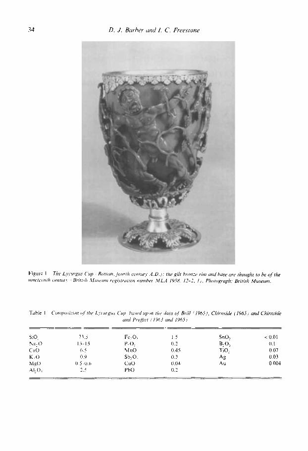
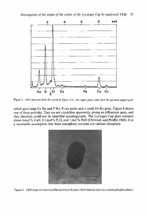
tion and loss of material. The reason for this behaviour became clear when X-ray spectra were collected from some of the same particles. These spectra contained strong peaks corresponding to the Na KCI and C1 Kz lines, in addition to the Si Kcr peak from beam spreading into the surrounding glass. Analysis of the single crystal diffraction patterns obtained from these particles confirmed that they were indeed NaCl crystals. Figure 2(a) shows a transmission image of several NaCl particles, while Figure 2(b) shows a single particle next to a small hole sputtered in the glass, together with, Figure 2(c), its diffraction pattern; its X-ray spectrum is shown in Figure 3 . Subsequent experience showed that the surfaces of the glass specimens underwent a form of alteration if they were not stored in the presence of a drying agent (silica gel was used). This was attributed to efflorescence resulting from the exposure to atmosphere of NaCl crystals in the glass surface, possibly coupled with increased surface reactivity caused by atom irradiation during sputter-thinning.
As explained in the discussion, it was recognized that the NaCl particles, although numerous, could not be responsible for the coloration of the glass. While searching for the real source, larger and more strongly absorbing rounded particles were occasionally found
(a) Transmission elecrron microscop?. / T E M ) image cfsodium chloride particles within the glass of rhe LXcurgu.9 Cup. (b) TEM image o f a solirar?, sodium chloride c r p tal on the edge of a hole produced hy atom rhinning of the glass. (c ) The corresponding relected area elecrron dg- .fraction parrern.
Figure 2
Investigation of the origin of the colour of the Lycurgus Cup by analytical TEM 37
Figure 3 EDX spectrum from rhe crystal in Figure 2(b) ; ihe copper peaks come from the specimen support grid.
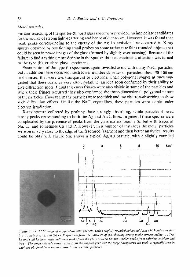
which gave large Ca Ka and P Kcr X-ray peaks and a small Fe Ka peak. Figure 4 shows one of these particles. They are not crystalline apparently, giving no diffraction spots, and they therefore could not be identified unambiguously. The Lycurgus Cup glass contains about 6molY CaO, 0.1 mol YO P,O, and 1 mol YO FeO (Chirnside and Proffitt 1965). It is a reasonable assumption that these amorphous particles are calcium phosphate.
Figure 4 TEM image of a nan-crystalline particle in the glass (EDX indicates that it is a calcium phosphate phase).
38 D . J . Barber iind I . C . Freestone
Metal particles
Further searching of the sputter-thinned glass specimens provided no immediate candidates for the source of strong light-scattering and hence of dichroism. However, it was found that weak peaks corresponding to the energy of the Ag Lr emission line occurred in X-ray spectra obtained by positioning small probes on some rather rare faint rounded objects that could be seen in phase images of the glass (formed by slightly overfocusing). Because of the failure to find anything more definite in the sputter-thinned specimens, attention was turned to the type (b), crushed glass, specimens.
Examination of the type (b) specimens again revealed areas with many NaCl particles, but in addition there occurred much lower number densities of particles, about 50-100 nm in diameter, that were less transparent to electrons. Their polygonal shapes at once sug- gested that these particles were also crystalline, an idea soon confirmed by their ability to give diffraction spots. Equal thickness fringes were also visible in some of the particles and where these fringes occurred they also confirmed the three-dimensional, polygonal nature of the particles. However. many particles were too thick and too electron-absorbing to show such diffraction effects. Unlike the NaCl crystallites, these particles were stable under electron irradiation.
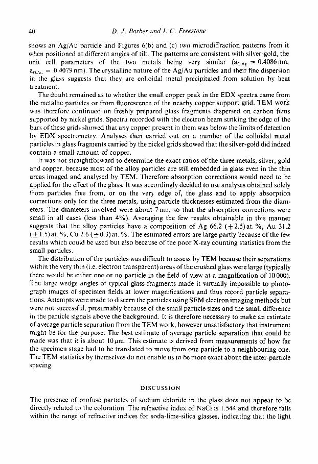
X-ray spectra collected by probing these strongly absorbing, stable particles showed strong peaks corresponding to both the Ag and Au L lines. In general these spectra were complicated by the presence of peaks from the glass matrix, mainly Si, but with traces of Na, C1, and sometimes Ca and P. However, in a number of instances the metal particles were on or very close to the edge of the fractured fragment and then better analytical results could be obtained. Figure 5(a) shows a typical Ag/Au particle, with a slightly rounded
2 4 6 8 10 keV I I I
t
Figure 5 ( a ) TE.11 image 0f.a typical metollic particle with a sligh[I>, roundedpoivgona[forni which indicares thaf ir is a single cr,v.stul. and (b) E D X specrrum.from the particles of (a), showing strong peaks corresponding to silver Lz and gold Lr lines, II i th additional peakr/ronl the glass (silicon Kr mid sn~nller peaksfroni chlorine. calcium und iron). The copper .signal.< mostly arise,from (he support grid. but the large phosphorus Kz peak is t.vpically seen in annlrses obtained from regions close I O the metallic porticles.

Investigation of the origin of the colour of the Lycurgus Cup bl) ana!,>ticul TEM 39
polygonal form. Figure 5(b) shows the EDX spectrum from this particle. In one fortuitous case, the fracture of the glass resulted in a solitary metal particle free from any glass. This particle was analysed and gave a spectrum uncluttered by extraneous peaks and probably the most reliable result for the Ag/Au ratio. Processing the spectra gave the atomic ratio of silver to gold as approximately 68/32. All the EDX spectra exhibited a small peak corresponding to the Cu Kcc line, and there was some evidence to suggest that phosphorus was enhanced close to or within some particles. Since the first crushed specimens of the Lycurgus glass were dispersed on copper support grids, it was not immediately possible to decide whether the copper peak arose from fluorescence effects or from a low concentration of the element in the Ag/Au particles.
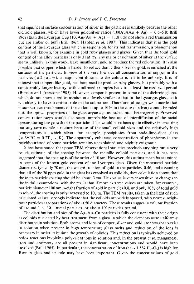
Electron microdiffraction was also used to investigate the metallic particles. Figure 6(a)
(a) TEM image of a silver-gold alloy crysiul. (b) and (c) Two electron microd[lpraction patterns ,from (he crystal of (a), recorded by rilting to obtuin strong Bragg spots (indices of strongly rejecting planes are labelled).
Figure 6
40 D . J . Barber arid I . C . Freestone
shows an Ag/Au particle and Figures 6(b) and (c) two microdiffraction patterns from it when positioned at different angles of tilt. The patterns are consistent with silver-gold. the unit cell parameters of the two metals being very similar (ao,n, = 0.4086nm, ao,Au = 0.4079 nm). The crystalline nature of the Ag/Au particles and their fine dispersion in the glass suggests that they are colloidal metal precipitated from solution by heat treatment .
The doubt remained as to whether the small copper peak in the EDX spectra came from the metallic particles or from fluorescence of the nearby copper support grid. TEM work was therefore continued on freshly prepared glass fragments dispersed on carbon films supported by nickel grids. Spectra recorded with the electron beam striking the edge of the bars of these grids showed that any copper present in them was below the limits of detection by EDX spectrometry. Analyses then carried out on a number of the colloidal metal particles in glass fragments carried by the nickel grids showed that the silver-gold did indeed contain a small amount of copper.
It was not straightforward to determine the exact ratios of the three metals, silver, gold and copper, because most of the alloy particles are still embedded in glass even in the thin areas imaged and analysed by TEM. Therefore absorption corrections would need to be applied for the effect of the glass. It was accordingly decided to use analyses obtained solely from particles free from, or on the very edge of, the glass and to apply absorption corrections only for the three metals, using particle thicknesses estimated from the diam- eters. The diameters involved were about 7nm, so that the absorption corrections were small in all cases (less than 4%). Averaging the few results obtainable in this manner suggests that the alloy particles have a composition of Ag 66.2 (& 2.5) at. YO, Au 3 1.2 ( f 1.5) at. %, Cu 2.6 ( f 0.3) at. YO. The estimated errors are large partly because of the few results which could be used but also because of the poor X-ray counting statistics from the small particles.
The distribution of the particles was difficult to assess by TEM because their separations within the very thin (i.e. electron transparent) areas of the crushed glass were large (typically there would be either one or no particle in the field of view at a magnification of 10000). The large wedge angles of typical glass fragments made i t virtually impossible to photo- graph images of specimen fields at lower magnifications and thus record particle separa- tions. Attempts were made to discern the particles using SEM electron imaging methods but were not successful, presumably because of the small particle sizes and the small difference in the particle signals above the background. It is therefore necessary to make an estimate of average particle separation from the TEM work, however unsatisfactory that instrument might be for the purpose. The best estimate of average particle separation that could be made was that it is about 10pm. This estimate is derived from measurements of how far the specimen stage had to be translated to move from one particle to a neighbouring one. The TEM statistics by themselves do not enable us to be more exact about the inter-particle spacing.
DISCUSSION
The presence of profuse particles of sodium chloride in the glass does not appear to be directly related to the coloration. The refractive index of NaCl is 1.544 and therefore falls within the range of refractive indices for soda-lime-silica glasses, indicating that the light

Investigation of the origin of the colour of the Lycurgus Cup by analytical TEA4 41
scattering capability of the particles is small. Light scattering by itself is of limited usefulness in giving coloration, since the effect is strongest in the ultra-violet, in accordance with the Rayleigh (fourth power of the wavelength) law. The scattering particles must also be highly absorbing, so that absorption predominates over scattering. Metals and some semiconduc- tor particles satisfy this criterion if the particle dimensions are restricted to < 50 nm (Bamford 1977). Sodium chloride particles do not, since their transmission of visible wavelengths is excellent.
The presence of significant amounts of chlorine (up to about 1.2%) in ancient glasses is well known (Geilmann 1955; Velde and Gendron 1980; Bimson and Freestone 1983). In early glassmaking chlorine was inadvertently added via the plant ash or mineral salts used to supply the alkali, causing an immiscible scum to form on the surface of the molten glass (Turner 1956). Removal of the scum left the glass virtually saturated in chlorine and the concentrations in ancient glass often approach the high temperature saturation concentra- tion, measured at 1.42 wt. 'YO C1 for a soda-lime-silica glass by Bateson and Turner (1939). There is a wide miscibility gap between molten NaCl and liquid silicates, which has a marked temperature dependence (Delitsyn and Melent'yev 1968). Reheating of the glass below its melting temperature to precipitate the colourant particles would also have resulted in phase separation of NaCl.
Given the deterioration of the surfaces of the experimental samples as a result of efflorescence of the sodium chloride particles, some brief word of explanation is needed to account for the remarkably fine state of preservation of the cup itself. The SO, content of the cup, at 73.5% (Chirnside and Proffitt 1965), is in the upper part of the range for Roman glass and this, coupled with its relatively low soda concentration ( N 13%: Chirnside and Proffitt 1965) and 2.5% Al,O, will have rendered it very stable. It appears that only the NaCl crystals at the surface undergo hydration and that the high silica glass matrix isolates the internal particles from the atmosphere.
The particles of calcium phosphate probably arose by a similar mechanism to the chloride, as there is also a wide miscibility gap between silicate and calcium-phosphate-rich liquids (Barret and McCaughey 1942). The exact nature of the phosphate phase could not be determined since the particles did not diffract. Although the refractive indices of some possible phosphates differ from that of soda-lime glass by larger amounts than does that of NaCI, it still does not seem that the phosphate particles are relevant to the coloration owing to their lower concentration and the knowledge that glasses containing abundant crystalline phosphate are opaque white (Turner and Rooksby 1959).
The identification of silver-gold alloy particles in the Lycurgus Cup confirms the infer- ence of Brill (1965) that the dichroic effect is caused by colloidal metal. Furthermore, Brill suggested that both silver and gold contributed to the colour, the gold component being mainly responsible for the reddish transmission and the silver for the greenish reflection. However, in the manufacture of gold ruby glasses in recent times, use has been made of the fact that gold is more insoluble than silver, tin or lead, and therefore forms colloidal nuclei on to which atoms of the other elements precipitate. Thus the particles in the Lycurgus glass need not necessarily be homogeneous and silver could be concentrated relative to gold in their outer layers. The small dimensions of the particles and the close match between the lattice parameters of gold and silver preclude the testing of this possibility by a physical method such as lattice imaging. The problem is also beyond the current limits of spatial resolution for EDX and electron energy loss spectrometry. In spite of this, we can reason
42 D . J . Barber m d I. C . Freestone
that significant surface concentration of silver in the particles is unlikely because the other dichroic glasses. which have lower go1d:silver ratios (100Au/(Au + Ag) = 0.6-5.8: Brill 1968) than the Lycurgus Cup (100Au/(Au + Ag) = 1 13), do not show a red transmission but are amber or buff (Brill 1965; Harden et al. 1987). This indicates that it is the gold content of the Lycurgus glass which is responsible for its red transmission, a phenomenon that is well known, for example in gold ruby glasses and glazes. Given that the total gold content of the alloy particles is only 31 at. (YO, any major enrichment of silver at the surface seems unlikely, as this would leave insufficient gold to produce the red coloration. It is also possible that copper, which is harder to reduce than either silver or gold, is enriched at the surfaces of the particles. In view of the very low overall concentration of copper in the particles ( 5 2.5 at. YO), a major contribution to the colour is felt to be unlikely. It is of interest that copper, like gold, has been used to produce ruby glasses, but probably with a considerably longer history, with confirmed examples back to at least the medieval period (Bimson and Freestone 1985). However, copper is present in some of the dichroic glasses which do not show a red transmission at levels similar to that in the Lycurgus glass, so it is unlikely to have a critical role in the coloration. Therefore, although we concede that minor surface enrichments of the colloids (up to 10% in the case of silver) cannot be ruled out. the optical properties of the glass argue against substantial heterogeneity. Preserved concentration steps would also seem improbable because of interdiffusion of the metal species during the growth of the particles. This would have been quite effective in smearing out any core-mantle structure because of the small colloid sizes and the relatively high temperatures at which silver, for example, precipitates from soda-lime-silica glass ( > 580°C = 0.7Tm p.Ag K). The apparently enhanced concentration of phosphorus in the neighbourhood of some particles remains unexplained and slightly enigmatic.
It has been stated that poor TEM observational statistics preclude anything but a very rough estimate of the spacing between the metallic colloid particles, and it has been suggested that the spacing is of the order of 10 pm. However, this estimate can be examined in terms of the known gold content of the Lycurgus glass. Given the measured particle diameters, typically 70 nm, the weight fraction of gold in the particles, 0.46, and assuming that all of the 30 ppm gold in the glass has exsolved as colloids, then calculation shows that the inter-particle spacing should be about 3 pm. This value is very insensitive to changes in the initial assumptions, with the result that if more extreme values are taken, for example, particle diameter 100 nm, weight fraction of gold in particles 0.8, and only 10% of total gold exsolved, the spacing is only increased to 10 pm. The TEM results, taken in the light of such calculated values, strongly indicate that the colloids are widely spaced, with nearest neigh- bour particles at separations of about 50 diameters. These results suggest a volume fraction of around 1 x lO~-’ metal particles, or about lo9 particles per ml.
The distribution and size of the Ag-Au-Cu particles is fully consistent with their origin as colloids nucleated by heat treatment from a glass in which the elements were uniformly distributed in solution. Both atoms and ions of copper, silver and gold are thought to exist in solution when present in high temperature glass melts and reduction of the ions is necessary in order to initiate the growth of colloids. This reduction is typically achieved by redox reactions involving polyvalent ions in solution and, in the present case, manganese, iron and antimony are all present in significant concentrations and would have been involved (Brill 1965). In particular, the concentration of iron (at - 1.5% Fe,O,) is high for Roman glass and its role may have been important. Given the concentrations of gold
Investigation of the origin of the colour of the Lycurgus Cup by analytical TEM 43
(40 ppm), silver (- 0.03%) and copper (- 0.04% CuO) in the glass (Brill 1965; Chirnside 1965), it is clear that in the alloy crystals, as entities, gold is greatly enriched over silver, which in turn is enriched over copper. This is consistent with the relative ease of reduction of the metals.
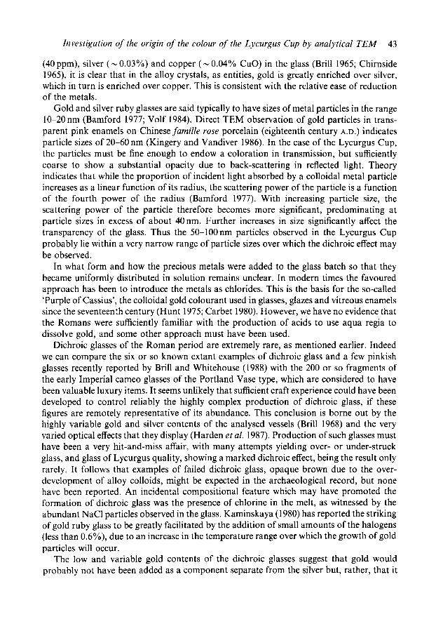
Gold and silver ruby glasses are said typically to have sizes of metal particles in the range 10-20 nm (Bamford 1977; Volf 1984). Direct TEM observation of gold particles in trans- parent pink enamels on Chinese famille rose porcelain (eighteenth century A.D.) indicates particle sizes of 20-60 nm (Kingery and Vandiver 1986). In the case of the Lycurgus Cup, the particles must be fine enough to endow a coloration in transmission, but sufficiently coarse to show a substantial opacity due to back-scattering in reflected light. Theory indicates that while the proportion of incident light absorbed by a colloidal metal particle increases as a linear function of its radius, the scattering power of the particle is a function of the fourth power of the radius (Bamford 1977). With increasing particle size, the scattering power of the particle therefore becomes more significant, predominating at particle sizes in excess of about 40nm. Further increases in size significantly affect the transparency of the glass. Thus the 50-100nm particles observed in the Lycurgus Cup probably lie within a very narrow range of particle sizes over which the dichroic effect may be observed.
In what form and how the precious metals were added to the glass batch so that they became uniformly distributed in solution remains unclear. In modern times the favoured approach has been to introduce the metals as chlorides. This is the basis for the so-called ‘Purple of Cassius’, the colloidal gold colourant used in glasses, glazes and vitreous enamels since the seventeenth century (Hunt 1975; Carbet 1980). However, we have no evidence that the Romans were sufficiently familiar with the production of acids to use aqua regia to dissolve gold, and some other approach must have been used.
Dichroic glasses of the Roman period are extremely rare, as mentioned earlier. Indeed we can compare the six or so known extant examples of dichroic glass and a few pinkish glasses recently reported by Brill and Whitehouse (1988) with the 200 or so fragments of the early Imperial cameo glasses of the Portland Vase type, which are considered to have been valuable luxury items. It seems unlikely that sufficient craft experience could have been developed to control reliably the highly complex production of dichroic glass, if these figures are remotely representative of its abundance. This conclusion is borne out by the highly variable gold and silver contents of the analysed vessels (Brill 1968) and the very varied optical effects that they display (Harden et al. 1987). Production of such glasses must have been a very hit-and-miss affair, with many attempts yielding over- or under-struck glass, and glass of Lycurgus quality, showing a marked dichroic effect, being the result only rarely. It follows that examples of failed dichroic glass, opaque brown due to the over- development of alloy colloids, might be expected in the archaeological record, but none have been reported. An incidental compositional feature which may have promoted the formation of dichroic glass was the presence of chlorine in the melt, as witnessed by the abundant NaCl particles observed in the glass. Kaminskaya (1980) has reported the striking of gold ruby glass to be greatly facilitated by the addition of small amounts of the halogens (less than 0.6%), due to an increase in the temperature range over which the growth of gold particles will occur.
The low and variable gold contents of the dichroic glasses suggest that gold would probably not have been added as a component separate from the silver but, rather, that it
44 D . J . Barber mid I . C . Freestone
was incidental to it. One is led to suggest silver metal or scrap as a source but such was the effectiveness of refining processes that Roman silver analysed in the British Museum Research Laboratory rarely has a gold content approaching even I YO (e.g. Hughes and Hall 1979). We are left to speculate upon other sources, a possibility being the by-products of some specialized metallurgical processes, such as assaying or refining. According to Chirn- side (1965). the Lycurgus glass contains 0.2% PbO as well as 0.04% CuO and these values, which are of the same order as the silver content, may be indicative of the use of some compositionally complex metallurgical source material for the precious metals. Another possibility is that the glassmakers actually attempted to equilibrate their batch in some way with metallic gold, when the more readily oxidizable impurities, such as lead, copper and silver, were preferentially incorporated into the glass. Direct dissolution of metallic gold into molten glass can occur (Newton 1970) but, because of segregation of the metal, repeated stages of crushing, grinding and remelting would be required to produce a homogeneous ruby glass. Such a process, while possible, is considered to have been uneconomic for practical purposes in the post-medieval period (Newton 1970). However, such considerations are unlikely to have constrained the production of luxury glass in the Roman period. Whatever the origin and mode of introduction of the precipitating metals into the dichroic glass, the difficulties in introducing them, coupled with those of controlling nucleation and growth, led such glass to be exceedingly rare, so that the best produced was saved for exceptional pieces of workmanship. as exemplified by the Lycurgus Cup.
A C K N O W L E D G E M E N T S
D. J. Barber wishes to thank Professor W. F. Muller of the Technische Hochschule, Darmstadt, F.R.G., for his kind hospitality during a sabbatical leave. during which much of this work was done, and for access to the CM12 microscope. We both thank Miss M. Bimson, Mr D. Buckton and Professor M. S. Tite for their advice and encouragement.
R E F E R E N C E S
Bamford. C. R.. 1977. Colour generation and control in glass, glass science and technologys Elsevier, Amsterdam. Bateson. H. M. and Turner. W. E. S., 1939. A note on the solubility of sodium chloride in a soda-lime-silica glass,
garret, R. L. and McCaughy, W. J., 1942. The system Ca0-Si0,-P20,, Am. Mineralogist, 27, 680-95. Bimson, M. and Freestone, I. C.. 1983. An analytical study of the relationship between the Portland Vase and
other Roman cameo vessels, J . Glass Studies. 25, 55-64, Bimson. M. and Freestone. I. C., 1985, ‘Rouge Clair’ and other late fourteenth-century enamels on the Royal Gold
Cup of the Kings of England and France, Annales du P’ Congres de I’Associarion lnternationalepour I’Hisroire du Verre. 209-22, Liege.
Brill. R. H., 1965, The chemistry of the Lycurgus Cup, Proceedings of the Seventh International Congress on Glass, compres rendus. 2, paper 223, 1-13, Brussels.
Brill. R. H.. 1968, The scientific investigation of ancient glasses, Proceedings o f f h e Eighth International Congress on G l a s ~ . 47-68. Sheffield.
Brill, R. H. and Whitehouse, D. C . , 1988. The Thomas Panel, J . Glass Srudies, 30, 34-50. Carbet, J . , 1980. Gold-based enamel colours, Gold Bull. 13, 144-50. Chirnside. R. C.. 1965, The Rothschild Lycurgus Cup: an analytical investigation, Proceedings of the Seventh
Chirnside. R. C . and Proffitt. P. M. C.. 1963, The Rothschild Lycurgus Cup: an analytical investigation, J . Glass
J . Soc. Glass Technology. 23, 265-7.
International Congress on Glass, comptes rendus, 2, paper 222, 1-6. Brussels.
Studies. 5 , 18-23.

Investigation of the origin of the colour of the Lycurgus Cup by analytical TEM 45
Chirnside, R. C. and Proffitt, P. M. C., 1965, Letter to the Editor, J . Glass Studies, 7, 139-40. Cliff, G. and Lorimer, G. W., 1975, The quantitative analysis of thin specimens, J. Microscopy, 103, 203-7. Delitsyn, L. M. and Melent’yev, B. N., 1968, Coexistence of two liquid phases at high temperatures: the system
sodium chloride-albite glass, Doklady Akademia Nauk SSSR, Earth Science sections. 180, 208-10 (American Geological Institute).
Geilmann. W., 1955, Die chemische Zusammensetzung einiger alter Glaser, insbesondere deutscher Glaser des 10. his 18. Jahrhunderts, Glastechnische Berichte, 28, 146-56.
Harden, D. B., Hellenkemper, H., Painter, K. and Whitehouse, D., 1987, Glass ofthe Caesars, Olivetti, Milan. Harden, D. B. and Toynbee, J. M. C., 1959, The Rothschild Lycurgus Cup, Archaeologia, 97, 179-212. Hughes, M. J. and Hall, J. A,. 1979, X-ray fluorescence analysis of late Roman and Sassanian silver plate, J .
Hunt, L. B., 1976, The true story of Purple of Cassius, Gold Bull., 9, 134-9. Kaminskaya, N. L., 1980, Initiating the finishing process for goldcontaining glasses, Glass and Ceramics, 37,
402-5 (Consultants Bureau, New York, translation from Steklo i Keramika, 8, 23-4). Kingery, W. D. and Vandiver, P. B.. 1986, The eighteenth-century changes in technology and style from thefamille
verte palette to thefamille rose palette, in Ceramics and Civilization: Vol. II, Technology and Style (ed. W. D. Kingery), 363-8 1, American Ceramic Society, Columbus, Ohio.
Marks, L. D. and Smith, D. J., 1982, HREM and STEM of defects in multiply-twinned particles, J. Microscopy.
Newton. R. G., 1970, Metallic gold and ruby glass, J . Glass Studies, 12, 165-70. Turner, W. E. S. and Rooksby, H. P., 1959, A study of the opalizing agents in ancient opal glasses throughout
Velde, B. and Gendron, C., 1980, Chemical composition of some Gallo-Roman glass fragments from central
Volf, M. B., 1984, Chemical approach to glass, Elsevier, Amsterdam.
Archaeol. Sci., 6, 321-44.
130,249-61.
3400 years, Glastechnische Berichte. 32K, Beiheft 8, 17-28.
western France, Archaeometry, 22, 183-7.