AN INTRACELLULAR POOL OF A CELL-SURFACE LIGAND WHICH INHIBITS LECTIN-INDUCED CAPPING
10
J. Cell Sci. 27, 245-254 (1977) 245 Printed in Great Britain © Company of Biologist! Limited AN INTRACELLULAR POOL OF A CELL-SURFACE LIGAND WHICH INHIBITS LECTIN-INDUCED CAPPING JAMES McDONOUGH, RICHARD RUTZ AND JACK LILIEN Department of Zoology, University of Wisconsin, Madison, Wisconsin 53706, U.S.A. SUMMARY It has previously been established that cells dissociated from, embryonic chick neural retina tissues redistribute their cell surface Concanavalin A receptors into patches and caps upon treatment with the lectin. During 4 h in continuous culture the cell population progressively loses the capacity to cap Con A receptors. In the presence of cycloheximide this decrease occurs for only the first 2 h of incubation. Concomitant with this loss in the ability to redistribute Con A receptors, cells become agglutinable by a tissue-specific component first described in 1975. The results of our experiments assaying inhibition of capping by tissue culture supernatant solutions and I09S of capping ability through cellular repair processes are interpreted in terms of an endogenous pool of one or more species, which, when displaced to the cell surface, inhibit the capping of Con-A receptors and permit tissue-specific cell agglutination. Once present at the cell surface, these species are released into the culture medium, either during continuous culture or during an incubation in fresh medium containing cycloheximide. Subsequent to release of these species, the cells regain their capacity for cap formation and lose agglutinability. In addition, the components released from the cells inhibit capping of Con A receptors when added to freshly dissociated cells. The assay methods were also used to determine parameters governing mobilization of the pool of capping inhibitors to the cell surface. INTRODUCTION Tissue culture medium conditioned by embryonic chick neural retinae contains macromolecular components which inhibit the lectin-induced redistribution of cell surface receptors into caps on freshly dissociated neural retina cells (McDonough & Lilien, 1975a). The capping-inhibition activity is tissue-type specific: medium obtained from neural retina cultures has no effect on capping of lectin receptors on single cerebral lobe cells and vice versa. In addition, for neural retina, the capping inhibition activity is lost upon treatment of the medium with /?-JV-acetyl hexo- saminidase, but not with pronase (Lilien & Rutz, 1977). Capping of plant lectin receptors among single cells is also inhibited through intrinsic cell processes which occur during in vitro culture (McDonough & Lilien, 19756). In this paper, assays for inhibition of capping by tissue culture supernatant solutions and loss of capping ability through cellular repair processes are exploited to establish the existence of an endogenous pool of the tissue-type specific capping inhibitor. The assays are also used to determine the parameters which govern mobilization of the pool to the cell surface.
-
Upload
independent -
Category
Documents
-
view
6 -
download
0
Transcript of AN INTRACELLULAR POOL OF A CELL-SURFACE LIGAND WHICH INHIBITS LECTIN-INDUCED CAPPING

J. Cell Sci. 27, 245-254 (1977) 245Printed in Great Britain © Company of Biologist! Limited
AN INTRACELLULAR POOL OF A
CELL-SURFACE LIGAND WHICH INHIBITS
LECTIN-INDUCED CAPPING
JAMES McDONOUGH, RICHARD RUTZ AND JACK LILIENDepartment of Zoology, University of Wisconsin, Madison,Wisconsin 53706, U.S.A.
SUMMARY
It has previously been established that cells dissociated from, embryonic chick neural retinatissues redistribute their cell surface Concanavalin A receptors into patches and caps upontreatment with the lectin. During 4 h in continuous culture the cell population progressivelyloses the capacity to cap Con A receptors. In the presence of cycloheximide this decrease occursfor only the first 2 h of incubation. Concomitant with this loss in the ability to redistributeCon A receptors, cells become agglutinable by a tissue-specific component first described in 1975.
The results of our experiments assaying inhibition of capping by tissue culture supernatantsolutions and I09S of capping ability through cellular repair processes are interpreted in termsof an endogenous pool of one or more species, which, when displaced to the cell surface,inhibit the capping of Con-A receptors and permit tissue-specific cell agglutination. Oncepresent at the cell surface, these species are released into the culture medium, either duringcontinuous culture or during an incubation in fresh medium containing cycloheximide.Subsequent to release of these species, the cells regain their capacity for cap formation and loseagglutinability. In addition, the components released from the cells inhibit capping of Con Areceptors when added to freshly dissociated cells.
The assay methods were also used to determine parameters governing mobilization of thepool of capping inhibitors to the cell surface.
INTRODUCTION
Tissue culture medium conditioned by embryonic chick neural retinae containsmacromolecular components which inhibit the lectin-induced redistribution of cellsurface receptors into caps on freshly dissociated neural retina cells (McDonough &Lilien, 1975a). The capping-inhibition activity is tissue-type specific: mediumobtained from neural retina cultures has no effect on capping of lectin receptors onsingle cerebral lobe cells and vice versa. In addition, for neural retina, the cappinginhibition activity is lost upon treatment of the medium with /?-JV-acetyl hexo-saminidase, but not with pronase (Lilien & Rutz, 1977).
Capping of plant lectin receptors among single cells is also inhibited throughintrinsic cell processes which occur during in vitro culture (McDonough & Lilien,19756). In this paper, assays for inhibition of capping by tissue culture supernatantsolutions and loss of capping ability through cellular repair processes are exploited toestablish the existence of an endogenous pool of the tissue-type specific cappinginhibitor. The assays are also used to determine the parameters which governmobilization of the pool to the cell surface.
246 J. McDonough, R. Rutz and J. Lilien
Neural retina tissue culture conditioned media also contain a macromolecularspecies which binds at the cell surface and renders the cells competent to be aggluti-nated by a tissue specific component which accumulates in serum free monolayerconditioned media (Lilien & Rutz, 1977; Balsamo & Lilien, 1974a). The acquisitionof agglutinability by cells fixed after various incubation regimes indicate that a poolof the species which renders cells agglutinable also exists, and is indistinguishablefrom that of the capping inhibitor. The data suggest that a single molecular specieswhich inhibits capping and is required for agglutination is found both as a pool incells and free in tissue culture supernatants. We will refer to this species as 'ligand',although it should be kept in mind that both activities need not reside in one molecule.
MATERIALS AND METHODS
Cell preparation
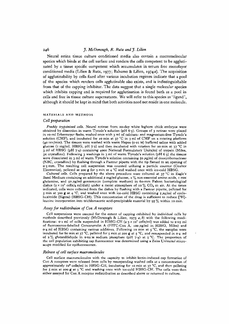
Freshly trypsinised cells. Neural retinae from 10-day white leghorn chick embryos wereobtained by dissection in warm Tyrode's solution (pH 6-5). Groups of 3 retinae were placedin 10-ml Erlenmeyer flasks, washed once with 3 ml of calcium- and magnesium-free Tyrode'ssolution (CMF), and incubated for 10 min at 37 °C in 3 ml of CMF on a rotating platform(40 rev/min). The tissues were washed with warm Hepes (o-oi M) buffered saline with addedglucose (1 mg/ml, HBSG, pH 7-2) and then incubated with rotation for 20 min at 37 °C in3 ml of HBSG (pH 7-4) containing 4000 National Formulatory Units/ml of trypsin (Miles,3X crystalline). Following 3 washings in 3 ml of warm Tyrode's solution (pH 6-5) the tissueswere dissociated in 3 ml of warm Tyrode's solution containing 50 /Jg/ml of deoxyribonuclease(NBC, crystalline) by flushing through a Pasteur pipette with the tip flamed to an opening of0-5 mm. The resulting cell suspension was counted utilizing a particle counter (CoulterElectronics), pelleted at 200 g for 5 min at 4 °C and washed once with ice-cold HBSG.
Cultured cells. Cells prepared by the above procedure were cultured at 37 °C in Eagle'sBasal Medium containing an additional 2 mg/ml glucose, 2 % non-essential amino acids, 1 mMglutamine, and 50/ig/ml gentamycin (complete medium) in 60-mm Falcon bacteriologicaldishes (2 x io7 cells/4 ml/dish) under a moist atmosphere of 10% CO, in air. At the timesindicated, cells were collected from the dishes by flushing with a Pasteur pipette, pelleted for5 min at 300 g at 4 °C, and washed once with ice-cold HBSG containing 5 /Jg/ml of cyclo-heximide (Sigma) (HBSG-CH). This concentration of the drug is sufficient to reduce [3H]-leucine incorporation into trichloroacetic acid-precipitable material by 95 % within 10 min.
Assay for redistribuion of Con A receptors
Cell suspensions were assayed for the extent of capping exhibited by individual cells bymethods described previously (McDonough & Lilien, 1975 a, b) with the following modi-fications: o-i ml of cells suspended in HBSG-CH (25 x io7 cells/ml) was added to 005 mlof fluorescence-labelled Concanavalin A (FITC-Con A, 100 fig/ml in HBSG, Miles) and0-4 ml of HBSG containing various additives. Following 10 min at 5 °C, the samples wereincubated for 60 min at 37 °C, pelleted for 5 min at 200 g at 5 °C, and resuspended in 0-4 mlof 2 % glutaraldehyde in 0-02 M sodium phosphate (pH 7-4) at 5 °C. The proportion ofthe cell population exhibiting cap fluorescence was determined using a Zeiss Universal micro-scope modified for epifluorescence.
Release of cell surface macromolecules
Cell surface macromolecules with the capacity to inhibit lectin-induced cap formation ofCon A receptors were released from cells by resuspending washed cells at a concentration ofapproximately io8 cells/ml in HBSG-CH, incubating for 10 min at 37 °C, and then pelletingfor 5 min at 200 g at 5 °C and washing once with ice-cold HBSG-CH. The cells were theneither assayed for Con A receptor redistribution as described above or returned to culture.
IntracelMar pool of a cell-surface ligand inhibiting lectin-induced capping 247
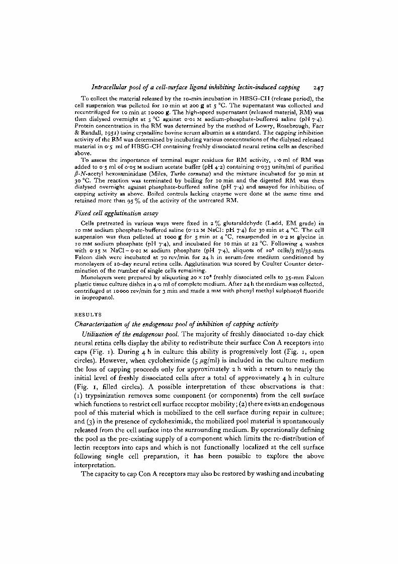
To collect the material released by the 10-min incubation in HBSG-CH (release period), thecell suspension was pelleted for 10 min at 200 g at 5 CC. The supernatant was collected andrecentrifuged for 10 min at 10000 g. The high-speed supernatant (released material, RM) wasthen dialysed overnight at 5 CC against o-oi M sodium-phosphate-buffered saline (pH 7-4).Protein concentration in the RM was determined by the method of Lowry, Rosebrough, Farr& Randall, 1951) using crystalline bovine serum albumin as a standard. The capping inhibitionactivity of the RM was determined by incubating various concentrations of the dialysed releasedmaterial in 05 ml of HBSG-CH containing freshly dissociated neural retina cells as describedabove.
To assess the importance of terminal sugar residues for RM activity, 1 o ml of RM wasadded to 0-5 ml of 0-05 M sodium acetate buffer (pH 4-2) containing 0-033 units/ml of purified/?-iV-acetyl hexosaminidase (Miles, Turbo cornutus) and the mixture incubated for 30 min at30 CC. The reaction was terminated by boiling for 10 min and the digested RM was thendialysed overnight against phosphate-buffered saline (pH 7-4) and assayed for inhibition ofcapping activity as above. Boiled controls lacking enzyme were done at the same time andretained more than 95 % of the activity of the untreated RM.
Fixed cell agglutination assay
Cells pretreated in various ways were fixed in 2 % glutaraldehyde (Ladd, EM grade) in10 mM sodium phosphate-buffered saline (0-12 M NaCl: pH 7-4) for 30 min at 4 °C. The cellsuspension was then pelleted at 1000 g for 5 min at 4 °C, resuspended in 02 M glycine in10 mM sodium phosphate (pH 7-4), and incubated for 10 min at 22 °C. Following 4 washeswith 0-15 M NaCl - 001 M sodium phosphate (pH 7'4), aliquots of io8 cells/3 ml/35-mmFalcon dish were incubated at 70 rev/min for 24 h in serum-free medium conditioned bymonolayers of 10-day neural retina cells. Agglutination was scored by Coulter Counter deter-mination of the number of single cells remaining.
Monolayers were prepared by aliquoting 20 x 10' freshly dissociated cells to 35-mm Falconplastic tissue culture dishes in 40 ml of complete medium. After 24 h the medium was collected,centrifuged at 10000 rev/min for 3 min and made 2 mM with phenyl methyl sulphonyl fluoridein isopropanol.
RESULTS
Characterization of the endogenous pool of inhibition of capping activity
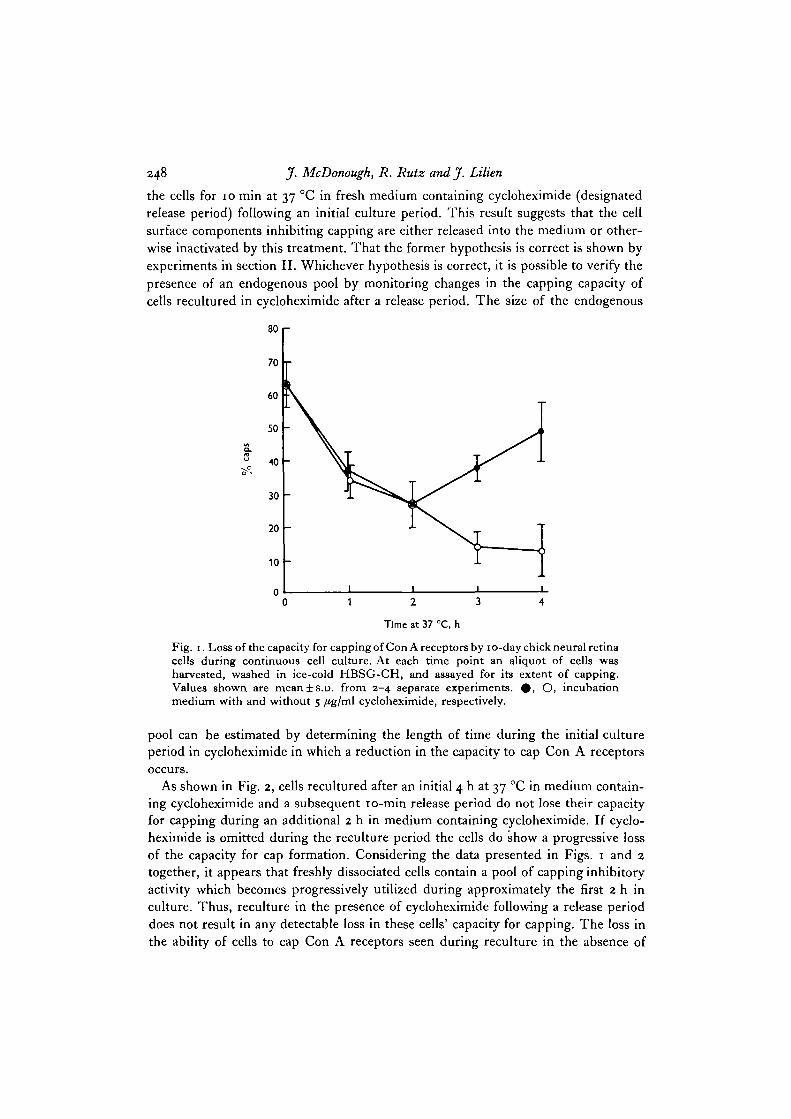
Utilization of the endogenous pool. The majority of freshly dissociated 10-day chickneural retina cells display the ability to redistribute their surface Con A receptors intocaps (Fig. 1). During 4 h in culture this ability is progressively lost (Fig. 1, opencircles). However, when cycloheximide (5 /tg/ml) is included in the culture mediumthe loss of capping proceeds only for approximately 2 h with a return to nearly theinitial level of freshly dissociated cells after a total of approximately 4 h in culture(Fig. 1, filled circles). A possible interpretation of these observations is that:(1) trypsinization removes some component (or components) from the cell surfacewhich functions to restrict cell surface receptor mobility; (2) there exists an endogenouspool of this material which is mobilized to the cell surface during repair in culture;and (3) in the presence of cycloheximide, the mobilized pool material is spontaneouslyreleased from the cell surface into the surrounding medium. By operationally definingthe pool as the pre-existing supply of a component which limits the re-distribution oflectin receptors into caps and which is not functionally localized at the cell surfacefollowing single cell preparation, it has been possible to explore the aboveinterpretation.
The capacity to cap Con A receptors may also be restored by washing and incubating
248 J. McDonough, R. Rutz and J. Lilien
the cells for 10 min at 37 °C in fresh medium containing cycloheximide (designatedrelease period) following an initial culture period. This result suggests that the cellsurface components inhibiting capping are either released into the medium or other-wise inactivated by this treatment. That the former hypothesis is correct is shown byexperiments in section II. Whichever hypothesis is correct, it is possible to verify thepresence of an endogenous pool by monitoring changes in the capping capacity ofcells recultured in cycloheximide after a release period. The size of the endogenous
80r
70
60
50
40
30
20
10
0
Time at 37 °C, h
Fig. 1. Loss of the capacity for capping of Con A receptors by 10-day chick neural retinacells during continuous cell culture. At each time point an aliquot of cells washarvested, washed in ice-cold HBSG-CH, and assayed for its extent of capping.Values shown are mean±s.D. from 2-4 separate experiments. # , O, incubationmedium with and without 5 /tg/ml cycloheximide, respectively.
pool can be estimated by determining the length of time during the initial cultureperiod in cycloheximide in which a reduction in the capacity to cap Con A receptorsoccurs.
As shown in Fig. 2, cells recultured after an initial 4 h at 37 °C in medium contain-ing cycloheximide and a subsequent 10-min release period do not lose their capacityfor capping during an additional 2 h in medium containing cycloheximide. If cyclo-heximide is omitted during the reculture period the cells do show a progressive lossof the capacity for cap formation. Considering the data presented in Figs. 1 and 2together, it appears that freshly dissociated cells contain a pool of capping inhibitoryactivity which becomes progressively utilized during approximately the first 2 h inculture. Thus, reculture in the presence of cycloheximide following a release perioddoes not result in any detectable loss in these cells' capacity for capping. The loss inthe ability of cells to cap Con A receptors seen during reculture in the absence of

Intracellular pool of a cell-surface ligand inhibiting lectin-induced capping 249
cycloheximide must therefore be due to the rapid utilization of newly synthesizedmaterial which inhibits capping.
It is apparent from Fig. 2 that the endogenous pool is depleted during the initial4 h at 37 °C whether or not cycloheximide is included in the medium since reculturein the presence of cycloheximide following a 10-min release period results in no lossin the ability to cap Con A receptors. Thus, it seems that some event during singlecell preparation - perhaps trypsinization or dissociation - triggers the cells to begin
80
70
60
SO
40
30
20
0
10
•4 0 1 2
Time at 37 °C, h
Fig. 2. Capacity of cells to undergo Con A-induced capping during continuous cellculture. After 4 h of continuous culture with (# ) or without (O) cycloheximide(5 /ig/ml) the cells were harvested, washed once with ice-cold HBSG-CH and thenincubated for 10 min at 37 °C in HBSG-CH at a concentration of approximatelyio8 cells/ml (release period). The cells were then pelleted, washed with ice-coldHBSG-CH and returned to culture at 37 °C with (dashed line) or without (solid line)cycloheximide (5 /ig/ml). At each time point an aliquot of cells was harvested, washedin ice-cold HBSG-CH, and assayed for its extent of capping. Values shown aremeans ± s.D. from 2-4 separate experiments.
utilizing their internal pool independently of whether or not de novo synthesis isoccurring (see Discussion). Furthermore, the pool is replaced in culture only overlong time periods. After 24 h in culture approximately 1 h of pool has beenreconstituted.
Effects of temperature on pool utilization. Throughout 4 h at 5 °C the percentage ofthe harvested cells which can cap is essentially identical whether or not cycloheximideis included in the medium. Culture for 4 h at 22 °C results in a progressive loss ofcapping the rate of which is intermediate between that occurring at 37 and 5 °C.
These data suggest that the rate of pool displacement is temperature-dependent andimply that after 4 h at 5 or 22 °C the cells still contain some proportion of their pool

250 J. McDonough, R. Rutz and J. Lilien
material. Therefore, the release period-reculture protocol described above wasutilized to determine the amount of pool material remaining in cells cultured at thetwo temperatures. In spite of the fact that culture at 5 °C results in a slight decrease inthe cells' capacity for capping during the 4-h culture period, following this period theentire endogenous pool still remains in these cells as measured by this assay (Fig. 3).Similarly, while cells cultured at 22 °C appear to mobilize most of their endogenouspool to the cell surface during a 4-h incubation period, they still contain a portion oftheir pool when assayed after the initial incubation (Fig. 3).
80
70
60
50
3 40
30
20
10
- -O
Time, h
Fig. 3. Effects of incubation at different temperatures on loss of capacity for capping ofCon A receptors by 10-day retina cells in continuous cell culture. Conditions are asdescribed in the legend to Fig. 2, except that the initial 4-h incubation was done ateither 5 °C (A) or 22 CC (B). Values shown are means from duplicates from 2 separateexperiments. # , O, incubation medium with and without cycloheximide (5 fig/m\),respectively. Following the release period, cells were incubated with (dashed lines) orwithout (solid lines) cycloheximide (5 fig/ird) at 37 °C.
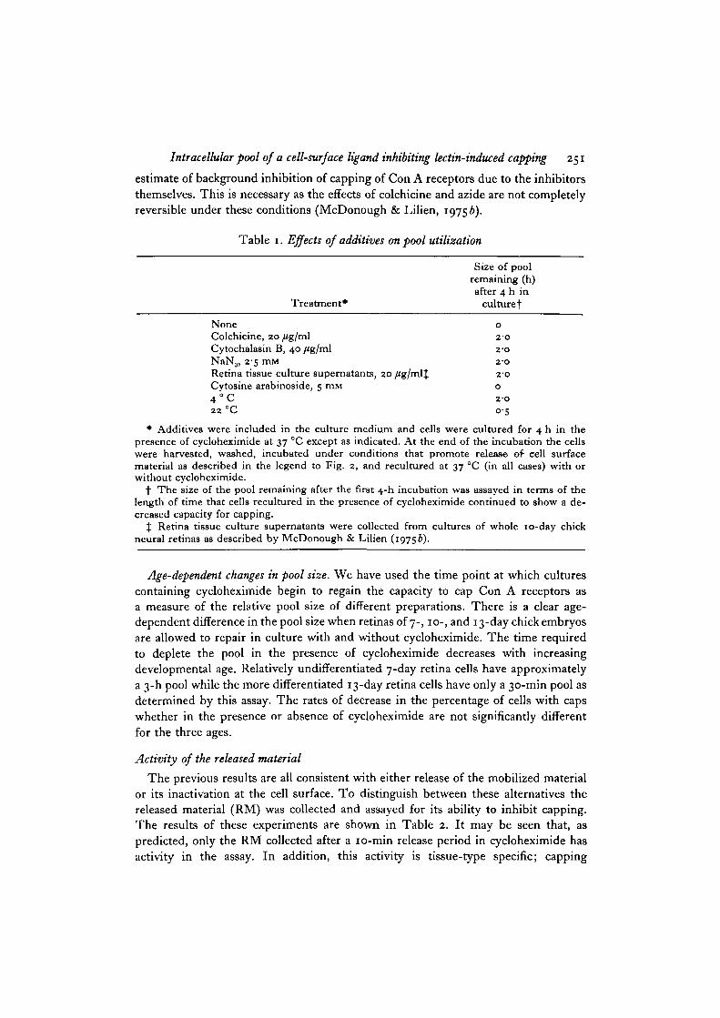
Effects of drugs on pool utilization. The effects of various compounds on poolutilization were tested by including them in the culture medium during the first4-h incubation at 37 °C. The cells were then harvested and recultured followinga release period to determine the amount of pool remaining. The results of theseexperiments are shown in Table 1. Incubation in medium containing cycloheximideand either colchicine, cytochalasin B, sodium azide, or retina tissue culture super-natant solutions (Balsamo & Lilien, 1974^, 1975) results in the cells' retaining theirentire pool, indicating that mobilization of the pool has been completely inhibited.On the other hand, treatment with cytosine arabinoside does not hinder completepool utilization. In all of these experiments controls were done which containedinhibitor but which were preincubated at 4 °C instead of 37 °C. These provided an
Intracelhlar pool of a cell-surface ligand inhibiting lectin-induced capping 251
estimate of background inhibition of capping of Con A receptors due to the inhibitorsthemselves. This is necessary as the effects of colchicine and azide are not completelyreversible under these conditions (McDonough & Lilien, 19756).
Table 1. Effects of additives on pool utilization
Treatment*
NoneColchicine, 20/ig/mlCytochalasin B, 40 /ig/mlNaN3> 2'5 mMRetina tissue culture supernatants, 20 /ig/mljCytosine arabinoside, 5 mM4 ° C22 °C
Size of poolremaining (h)after 4 h in
culture f
0
2 0
2 - 0
2 - 0
2 - 0
0
2-O
0->!
• Additives were included in the culture medium and cells were cultured for 4 h in thepresence of cycloheximide at 37 °C except as indicated. At the end of the incubation the cellswere harvested, washed, incubated under conditions that promote release of cell surfacematerial as described in the legend to Fig. 2, and recultured at 37 °C (in all cases) with orwithout cycloheximide.
f The size of the pool remaining after the first 4-h incubation was assayed in terms of thelength of time that cells recultured in the presence of cycloheximide continued to show a de-creased capacity for capping.
X Retina tissue culture supernatants were collected from cultures of whole 10-day chickneural retinas as described by McDonough & Lilien (19756).
Age-dependent changes in pool size. We have used the time point at which culturescontaining cycloheximide begin to regain the capacity to cap Con A receptors asa measure of the relative pool si2e of different preparations. There is a clear age-dependent difference in the pool size when retinas of 7-, 10-, and 13-day chick embryosare allowed to repair in culture with and without cycloheximide. The time requiredto deplete the pool in the presence of cycloheximide decreases with increasingdevelopmental age. Relatively undifferentiated 7-day retina cells have approximatelya 3-h pool while the more differentiated 13-day retina cells have only a 30-min pool asdetermined by this assay. The rates of decrease in the percentage of cells with capswhether in the presence or absence of cycloheximide are not significantly differentfor the three ages.
Activity of the released material
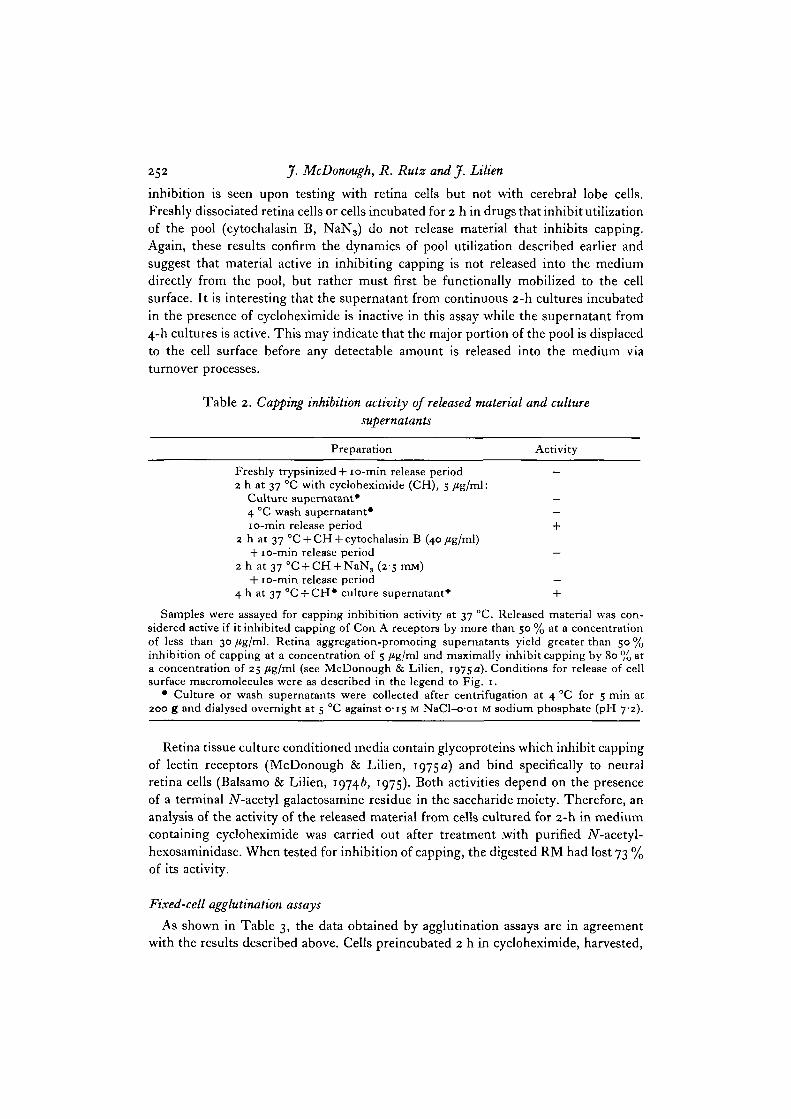
The previous results are all consistent with either release of the mobilized materialor its inactivation at the cell surface. To distinguish between these alternatives thereleased material (RM) was collected and assayed for its ability to inhibit capping.The results of these experiments are shown in Table 2. It may be seen that, aspredicted, only the RM collected after a 10-min release period in cycloheximide hasactivity in the assay. In addition, this activity is tissue-type specific; capping
252 J. McDonough, R. Rutz and J. Lilien
inhibition is seen upon testing with retina cells but not with cerebral lobe cells.Freshly dissociated retina cells or cells incubated for 2 h in drugs that inhibit utilizationof the pool (cytochalasin B, NaN3) do not release material that inhibits capping.Again, these results confirm the dynamics of pool utilization described earlier andsuggest that material active in inhibiting capping is not released into the mediumdirectly from the pool, but rather must first be functionally mobilized to the cellsurface. It is interesting that the supernatant from continuous 2-h cultures incubatedin the presence of cycloheximide is inactive in this assay while the supernatant from4-h cultures is active. This may indicate that the major portion of the pool is displacedto the cell surface before any detectable amount is released into the medium viaturnover processes.
Table 2. Capping inhibition activity of released material and culturesupernatants
Preparation Activity
Freshly trypsinized + 10-min release period —2 h at 37 °C with cycloheximide (CH), 5 /ig/ml:
Culture supernatant* —4 °C wash supernatant* —10-min release period +
2 h at 37 °C + CH + cytochalasin B (40/ig/ml)+ 10-min release period —
2 h at 37 °C + CH + NaN3 (2-5 mw)+ 10-min release period —
4 h at 37 °C + CH* culture supernatant* +
Samples were assayed for capping inhibition activity at 37 °C. Released material was con-sidered active if it inhibited capping of Con A receptors by more than 50 % at a concentrationof less than 30 /tg/ml. Retina aggregation-promoting supernatants yield greater than 50 %inhibition of capping at a concentration of 5 fig/ml and maximally inhibit capping by 80 % ata concentration of 25 fig/ml (see McDonough & Lilien, 1975 a). Conditions for release of cellsurface macromolecules were as described in the legend to Fig. 1.
• Culture or wash supernatants were collected after centrifugation at 4 °C for 5 min at200 g and dialysed overnight at 5 °C against 0-15 M NaCl-001 M sodium phosphate (pH 72).
Retina tissue culture conditioned media contain glycoproteins which inhibit cappingof lectin receptors (McDonough & Lilien, 1975 a) and bind specifically to neuralretina cells (Balsamo & Lilien, 19746, 1975). Both activities depend on the presenceof a terminal JV-acetyl galactosamine residue in the saccharide moiety. Therefore, ananalysis of the activity of the released material from cells cultured for 2-h in mediumcontaining cycloheximide was carried out after treatment .with purified iV-acetyl-hexosaminidase. When tested for inhibition of capping, the digested RM had lost 73 %of its activity.
Fixed-cell agglutination assays
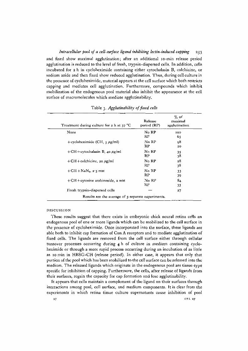
As shown in Table 3, the data obtained by agglutination assays are in agreementwith the results described above. Cells preincubated 2 h in cycloheximide, harvested,
Intracellular pool of a cell-surface ligand inhibiting lectin-induced capping 253
and fixed show maximal agglutination; after an additional 10-min release periodagglutination is reduced to the level of fresh, trypsin-dispersed cells. In addition, cellsincubated for 2 h in cycloheximide containing either cytochalasin B, colchicine, orsodium azide and then fixed show reduced agglutination. Thus, during cell culture inthe presence of cycloheximide, material appears at the cell surface which both restrictscapping and mediates cell agglutination. Furthermore, compounds which inhibitmobilization of the endogenous pool material also inhibit the appearance at the cellsurface of macromolecules which mediate agglutinability.
Table 3. Agglutinability of fixed cells
Treatment during culture for 2 h at
None
+ cycloheximide (CH, 5 /ig/ml)
+ CH +cytochalasin B, 40/ig/ml
+ CH + colchicine, 20 /tg/ml
+ CH + NaN3) 2-s mM
Release37 °C period (RP)
NoRPRP
No RPRP
No RPRP
NoRPRP
No RPRP
+ CH + cytosine arabinoside, 2 mM No RP
Fresh trypsin-dispereed cells
Results are the average
RP
—
of 3 separate experiments.
% ofmaximal
agglutination
1 0 0
6S982 0
35382838
3339
843527
DISCUSSION
These results suggest that there exists in embryonic chick neural retina cells anendogenous pool of one or more ligands which can be mobilized to the cell surface inthe presence of cycloheximide. Once incorporated into the surface, these ligands areable both to inhibit cap formation of Con A receptors and to mediate agglutination offixed cells. The ligands are removed from the cell surface either through cellularturnover processes occurring during 4 h of culture in medium containing cyclo-heximide or through a more rapid process occurring during an incubation of as littleas 10 min in HBSG-CH (release period). In either case, it appears that only thatportion of the pool which has been mobilized to the cell surface can be released into themedium. The released ligands which originate in the endogenous pool are tissue-typespecific for inhibition of capping. Furthermore, the cells, after release of ligands fromtheir surfaces, regain the capacity for cap formation and lose agglutinability.
It appears that cells maintain a complement of the ligand on their surfaces throughinteractions among pool, cell surface, and medium components. It is clear from theexperiments in which retina tissue culture supernatants cause inhibition of pool
I? CEL 27
254 J- McDonough, R. Rutz andj. Lilien
utilization that some feedback from the cell surface to the pool must occur. Thus, theexistence of surface-localized ligand supplied exogenously and possibly endogenouslyrestricts utilization of the pool. Furthermore, synthesis of new cell surface ligand maybe at least partially controlled by the existence of unreleased pool material. As shownin Fig. i, even in the absence of cycloheximide, the pool appears to be utilized (opencircles) without concomitant replacement. This may indicate that in the presence ofthe pool de novo synthesis is inhibited and only when most of the pool has beendepleted and released can synthesis and utilization of new material occur.
We have no direct evidence which pertains to the cellular localization of the pool.While these data are consistent with a model involving insertion into the plasmamembrane and lateral redistribution of an intracellular pool (Baudiun, Stock,Vincent & Grenier, 1975; Redman, Banerjee, Howell & Palade, 1975), they cannotdistinguish between this mechanism and the possibility of rearrangements of a crypticcell surface-localized ligand pool.
Several lines of evidence imply that the capping-inhibitory component and thesurface ligand which mediates agglutinability are identical. As reported herein thedynamics of pool utilization and release appear to be identical. In addition, tissueculture-conditioned media contain both capping inhibitory activity (McDonough &Lilien, 1975 a) and a component which when fixed at the cell surface renders themagglutinable (Lilien & Rutz, 1977; Balsamo & Lilien, 1974a). Both activities aretissue-type specific and labile to /?-iV-acetyl hexosaminidase, as is the released material.Whether or not a single species is found to mediate both activities, the profoundeffects of these molecules on cell behaviour and membrane architecture make themprime candidates for roles as critical determinants of morphogenetic interactions.
This work was supported by grant no. BMS75-02824 from NSF. J.M. was supported byTraining Grant HD00409 from the NICHD.
REFERENCES
BALSAMO, J. & LILIEN, J. (1974a). Functional identification of three components whichmediate tissue-type specific embryonic cell adhesion. Nature, Lond. 251, 522-524.
BALSAMO, J. & LILIEN, J. (19746). Embryonic cell aggregation: kinetics and specificity ofbinding of enhancing factors. Proc. natn. Acad. Set. U.S.A. 71, 727-731.
BALSAMO, J. & LILIEN, J. (1975). The binding of tissue-specific adhesive molecules to the cellsurface. A molecular basis for specificity. Biochemistry, N.Y. 14, 167-171.
BAUDIUN, H., STOCK, C, VINCENT, D. & GRENIER, J. F. (1975). MicrofiJamentous system andsecretion of enzyme in the exocrine pancreas. Effect of cytochalasin B. J. Cell Biol. 66,165-181.
LILIEN, J. & RUTZ, R. (1977). In Cell and Tissue Interactions, Symp. Soc. gen. Physiologists(ed. J. Lash & M. Burger). (In Press.)
LOWRY, O. H., ROSEBROUCH, N. J., FARR, A. L. & RANDALL, R. J. (1951). Protein measurementwith the Folin phenol reagent. J. biol. Chem. 193, 265-275.
MCDONOUGH, J. & LILIEN, J. (1975 a). Inhibition of mobility of cell-surface receptors byfactors which mediate specific cell-cell interactions. Nature, Lond. 256, 416—417.
MCDONOUGH, J. & LILIEN, J. (19756). Spontaneous and lectin-induced redistribution of cellsurface receptors on embryonic chick neural retina cells. J. Cell Sci. 19, 357-368.
REDMAN, C. M., BANERJEE, D., HOWELL, K. & PALADE, G. E. (1975). Colchicine inhibition ofplasma protein release from rat hepatocytes. J. Cell Biol. 66, 42-59.
{Received 21 February 1977)