An Immunohistochemical Study of Retinol-Binding Protein (RBP) in Livers of Free-Living Polar Bears...
7
440 Journal of Zoo and Wildlife Medicine 36(3): 440–446, 2005 Copyright 2005 by American Association of Zoo Veterinarians AN IMMUNOHISTOCHEMICAL STUDY OF RETINOL-BINDING PROTEIN (RBP) IN LIVERS OF FREE-LIVING POLAR BEARS (URSUS MARITIMUS) FROM EAST GREENLAND Annabelle Heier, D.V.M., Christian Sonne, D.V.M., M.V.Sc., Andrea Gro ¨ne, D.V.M., Dipl. A.C.V.P., Dipl. E.C.V.P., Pall S. Leifsson, D.V.M., Ph.D., Rune Dietz, M.Sc., Erik W. Born, M.Sc., and Luca N. Bacciarini, D.V.M., Dipl. E.C.V.P. Abstract: From 1999 to 2002 samples from 114 free-ranging polar bears (Ursus maritimus) were collected in the municipality of Scoresby Sound, East Greenland, to detect levels of organochlorines and potential histopathologic changes. Livers of 16 female polar bears from this group were evaluated histologically and analyzed for hepatic retinol- binding protein by immunohistochemistry. Retinol-binding protein is the main transport protein for retinol, an important vitamin A metabolite in the polar bear. Only mild pathologic changes were noted on histologic evaluation of the livers. Small lymphocytic or lymphohistiocytic infiltrates were present in all the livers. Small lipid granulomas, mild periportal fibrosis, and bile duct proliferation were found in several cases. Immunohistochemistry for retinol-binding protein of hepatic tissue from free-ranging polar bears showed no distinct difference in staining intensity by a number of criteria: age, season (fasting and nonfasting), or lactation status. The staining was diffuse to finely stippled in the cytoplasm and showed very little variation among the animals. Because of the lack of macroscopic changes and the absence of severe histologic liver lesions, these polar bears were assumed to be healthy. The diffuse cytoplasmic retinol-binding protein staining in hepatocytes of free-ranging polar bears varies markedly from the prominent granular, less intense staining of captive polar bears investigated previously. Key words: Polar bear, Ursus maritimus, liver, retinol-binding protein, vitamin A, immunohistochemistry. INTRODUCTION Livers from free-ranging polar bears (Ursus mar- itimus) contain very high levels of vitamin A. 1,21,22 The high vitamin A levels in polar bear liver are toxic, so it is not consumed by Inuit living in the Arctic. Wild polar bears feed mostly on ringed seal (Phoca hispida) and secondarily on bearded seals (Erignathus barbatus), but they are known to con- sume other species. 4 Independent of the source, the diet of polar bears is largely composed of fat. Blub- ber, liver, and kidney especially contain high con- centrations of vitamin A, which are stored in the liver. 27,28 Lipid-soluble vitamin A is transported in the blood mainly as retinol. Retinol is bound to a specific transport protein, retinol-binding protein From the Institut fu ¨r Tierpathologie, Zentrum fu ¨r Fisch- und Wildtiermedizin, Universita ¨t Bern, La ¨nggassstrasse 122, CH 3001 Bern, Switzerland (Heier, Gro ¨ne, Bacciar- ini), the Department of Pharmacology and Pathobiology, the Royal Veterinary and Agricultural University, Bu ¨- lowsvej 17, DK 1870 Frederiksberg C, Denmark (Leifs- son), Department of Arctic Environment, National Envi- ronmental Research Institute, Frederiksborgvej 399, DK 4000 Roskilde, Denmark (Sonne, Dietz), and the Green- land Institute of Natural Resources, P.O. Box 570, DK 3900 Nuuk, Greenland (Born). Present address (Bacciar- ini): Ufficio del veterinario cantonale, via Dogana 16, CH 6500 Bellinzona, Switzerland. Correspondence should be directed to Dr. Bacciarini. (RBP), which is synthesized in hepatocytes and whose secretion is tightly regulated by the avail- ability of retinol. Over 90% of the total body vi- tamin A is stored as retinyl esters and retinol in the Ito cells of the liver. 17 Vitamin A plays an important role in differentiation and maintenance of special- ized epithelia, and it is critical for hepatic and vi- sual function. 6 Vitamin A metabolites are potent regulators of gene transcription, and they are im- plicated in regulating reproduction and immune functions, including those of lymphocytes. 12,17 Vi- tamin A deficiency may develop from insufficient intake or disruption of storage (e.g., by persistent organic pollutants). 26 The regulation of levels and storage of vitamin A is complex. In a study of polar bears from two European zoos, 6 of 11 bears had liver changes such as in- flammatory, hyperplastic, and neoplastic lesions. An immunohistochemical study for RBP in these diseased livers showed markedly reduced staining intensities compared to livers without lesions. Available vitamin A levels in affected livers were reduced relative to reference values from literature and collected data from healthy polar bears. 8 Im- munohistochemical demonstration of RBP may be a useful indirect indicator for vitamin A in tissue. The present study investigated whether correlations between liver lesions and RBP immunohistochem- istry could be found in free-ranging, apparently healthy polar bears from East Greenland.
-
Upload
independent -
Category
Documents
-
view
0 -
download
0
Transcript of An Immunohistochemical Study of Retinol-Binding Protein (RBP) in Livers of Free-Living Polar Bears...
440
Journal of Zoo and Wildlife Medicine 36(3): 440–446, 2005Copyright 2005 by American Association of Zoo Veterinarians
AN IMMUNOHISTOCHEMICAL STUDY OF RETINOL-BINDINGPROTEIN (RBP) IN LIVERS OF FREE-LIVING POLAR BEARS(URSUS MARITIMUS) FROM EAST GREENLAND
Annabelle Heier, D.V.M., Christian Sonne, D.V.M., M.V.Sc., Andrea Grone, D.V.M., Dipl.A.C.V.P., Dipl. E.C.V.P., Pall S. Leifsson, D.V.M., Ph.D., Rune Dietz, M.Sc., Erik W. Born,M.Sc., and Luca N. Bacciarini, D.V.M., Dipl. E.C.V.P.
Abstract: From 1999 to 2002 samples from 114 free-ranging polar bears (Ursus maritimus) were collected in themunicipality of Scoresby Sound, East Greenland, to detect levels of organochlorines and potential histopathologicchanges. Livers of 16 female polar bears from this group were evaluated histologically and analyzed for hepatic retinol-binding protein by immunohistochemistry. Retinol-binding protein is the main transport protein for retinol, an importantvitamin A metabolite in the polar bear. Only mild pathologic changes were noted on histologic evaluation of the livers.Small lymphocytic or lymphohistiocytic infiltrates were present in all the livers. Small lipid granulomas, mild periportalfibrosis, and bile duct proliferation were found in several cases. Immunohistochemistry for retinol-binding protein ofhepatic tissue from free-ranging polar bears showed no distinct difference in staining intensity by a number of criteria:age, season (fasting and nonfasting), or lactation status. The staining was diffuse to finely stippled in the cytoplasmand showed very little variation among the animals. Because of the lack of macroscopic changes and the absence ofsevere histologic liver lesions, these polar bears were assumed to be healthy. The diffuse cytoplasmic retinol-bindingprotein staining in hepatocytes of free-ranging polar bears varies markedly from the prominent granular, less intensestaining of captive polar bears investigated previously.
Key words: Polar bear, Ursus maritimus, liver, retinol-binding protein, vitamin A, immunohistochemistry.
INTRODUCTION
Livers from free-ranging polar bears (Ursus mar-itimus) contain very high levels of vitamin A.1,21,22
The high vitamin A levels in polar bear liver aretoxic, so it is not consumed by Inuit living in theArctic. Wild polar bears feed mostly on ringed seal(Phoca hispida) and secondarily on bearded seals(Erignathus barbatus), but they are known to con-sume other species.4 Independent of the source, thediet of polar bears is largely composed of fat. Blub-ber, liver, and kidney especially contain high con-centrations of vitamin A, which are stored in theliver.27,28 Lipid-soluble vitamin A is transported inthe blood mainly as retinol. Retinol is bound to aspecific transport protein, retinol-binding protein
From the Institut fur Tierpathologie, Zentrum fur Fisch-und Wildtiermedizin, Universitat Bern, Langgassstrasse122, CH 3001 Bern, Switzerland (Heier, Grone, Bacciar-ini), the Department of Pharmacology and Pathobiology,the Royal Veterinary and Agricultural University, Bu-lowsvej 17, DK 1870 Frederiksberg C, Denmark (Leifs-son), Department of Arctic Environment, National Envi-ronmental Research Institute, Frederiksborgvej 399, DK4000 Roskilde, Denmark (Sonne, Dietz), and the Green-land Institute of Natural Resources, P.O. Box 570, DK3900 Nuuk, Greenland (Born). Present address (Bacciar-ini): Ufficio del veterinario cantonale, via Dogana 16, CH6500 Bellinzona, Switzerland. Correspondence should bedirected to Dr. Bacciarini.
(RBP), which is synthesized in hepatocytes andwhose secretion is tightly regulated by the avail-ability of retinol. Over 90% of the total body vi-tamin A is stored as retinyl esters and retinol in theIto cells of the liver.17 Vitamin A plays an importantrole in differentiation and maintenance of special-ized epithelia, and it is critical for hepatic and vi-sual function.6 Vitamin A metabolites are potentregulators of gene transcription, and they are im-plicated in regulating reproduction and immunefunctions, including those of lymphocytes.12,17 Vi-tamin A deficiency may develop from insufficientintake or disruption of storage (e.g., by persistentorganic pollutants).26 The regulation of levels andstorage of vitamin A is complex.
In a study of polar bears from two Europeanzoos, 6 of 11 bears had liver changes such as in-flammatory, hyperplastic, and neoplastic lesions.An immunohistochemical study for RBP in thesediseased livers showed markedly reduced stainingintensities compared to livers without lesions.Available vitamin A levels in affected livers werereduced relative to reference values from literatureand collected data from healthy polar bears.8 Im-munohistochemical demonstration of RBP may bea useful indirect indicator for vitamin A in tissue.The present study investigated whether correlationsbetween liver lesions and RBP immunohistochem-istry could be found in free-ranging, apparentlyhealthy polar bears from East Greenland.
441HEIER ET AL.—LIVER RBP IN FREE-RANGING POLAR BEARS
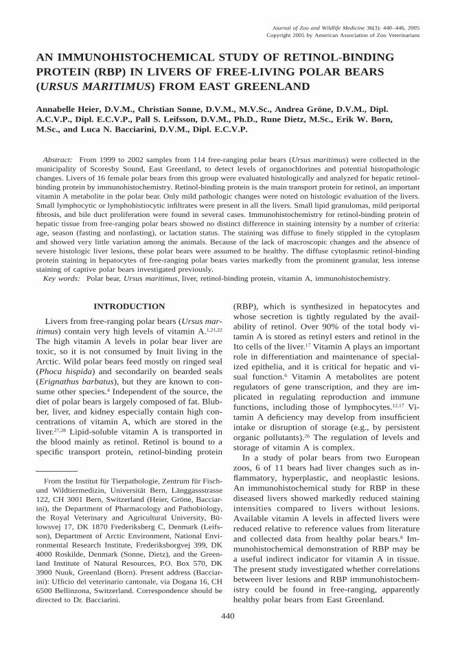
Table 1. Epidemiology, results of retinol-binding protein immunohistochemistry, and histologic findings for thelivers of 16 free-ranging polar bears.
CaseNo.
Age(years)
Date ofdeath Lactating
RBPa
index Histopathology
1 14 b No 1.8 Few randomly distributed small lymphocytic infil-trates; several lipid granulomas
2 22 28 Aug 1999 No 1.9 Few randomly distributed lymphocytic infiltrates;occasional lipid granulomas
3 25 23 Jun 1999 No 2.3 Multiple, mostly periportal lymphohistiocytic infil-trates; mild fibrosis and bile duct hyperplasia;several foci of hepatocellular glycogen storage
4 23 09 Jul 1999 No 2.6 Multiple small lymphohistiocytic infiltrates; moder-ate periportal fibrosis
5 12 13 Feb 2000 Yes 1.7 Multiple small, mostly periportal lymphocytic infil-trates
6 4 27 Mar 2000 No 1.9 Multiple small, mostly periportal lymphocytic infil-trates
7 8 24 Mar 2000 Yes 2.4 Multiple small periportal lymphohistiocytic infil-trates; mild bile duct hyperplasia and periportal fi-brosis
8 3.5 04 Sep 1999 No 2.1 Moderate periportal lymphohistiocytic infiltration9 5.5 21 Mar 2000 No 2.1 Moderate periportal lymphohistiocytic infiltration
10 19 05 Jan 2000 No 1.4 Small random clusters of lymphocytes11 8 21 Mar 2000 No 2.2 Small random clusters of lymphocytes12 20 01 Feb 2001 Yes 2.4 Multiple, mostly periportal moderate infiltrates of
lymphocytes, macrophages and lesser neutrophils;several lipid granulomas; mild bile duct hyperpla-sia
13 14 01 Feb 2001 Yes 1.8 Small, mostly periportal infiltrates of lymphocytes,macrophages and neutrophils; several foci of gly-cogen storage; some lipid granulomas; mild bileduct hyperplasia and periportal fibrosis
14 12 26 Feb 2001 Yes 2.0 Focally extensive infiltration of macrophages andneutrophils with fibrin exudation. Degenerationand necrosis of hepatocytes; multiple, mostlyperiportal lymphohistiocytic infiltrates; mild bileduct hyperplasia and periportal fibrosis; multiplesmall foci of hepatocellular glycogen storage andsome lipid granulomas
15 19 14 Sep 2000 Yes 2.7 Multiple small periportal lymphocytes infiltrates;multiple small lipid granulomas; mild bile ducthyperplasia
16 20 * No 2.5 Several, mostly periportal lymphohistiocytic infil-trates; some foci of hepatocellular glycogen stor-age
a RBP, retinol-binding protein.b No data available from local hunters.
MATERIALS AND METHODS
Liver samples, without macroscopic lesions,from 16 free-ranging female polar bears were ex-amined. The animals constituted a representativesample of the subsistence catch of polar bears byEast Greenland Inuit (1999–2001).23 Tissue sam-ples were fixed immediately in the field in a com-bination of buffered formaldehyde and alcohol (1
part of a 35% formaldehyde solution and 9 parts ofa 96% ethanol solution) to avoid freezing damagebefore shipment. The bears were aged by countingthe annual layers in the cementum of decalcifiedtoluidine-stained incisors (10 mm thick), and theywere found to be 4 to 25 years old.5,10
Fixed tissue samples were embedded in paraffin(Sakura TissueTekt VIP, Sakura Finetek Inc., 1750

442 JOURNAL OF ZOO AND WILDLIFE MEDICINE
Figure 1. Liver of a free-ranging polar bear, case 12. Ito cells are prominent in the periportal zones. H&E, Bar 5320 mm.
West 214th Street, Torrance, California 90501,USA) and sectioned at 2–4 mm. Sections werestained with hematoxylin and eosin (H&E) for his-topathologic assessment. Selected sections werefurther evaluated with a periodic acid-Schiff (PAS)stain for the presence of glycogen. ImmunoreactiveRBP was demonstrated in hepatocytes as previous-ly described in polar bears and other carnivorousspecies.8,20 To summarize, sections were deparaffin-ized and incubated for 45 min in 0.5% hydrogenperoxide in methanol to deactivate endogenous per-oxidases. Nonspecific antibody binding wasblocked by incubation for 10 min in tris-bufferedsaline (TBS) with 20% goat serum. Sections wereincubated overnight in a humidified chamber at 48Cwith rabbit anti-human RBP as the primary anti-body (A 0040, DAKO, P.O. Box 2114, 6302 Zug,Switzerland) diluted at 1:200 in 20% goat serum inTBS. Sections were incubated with biotinylatedanti-rabbit IgG (DAKO) next for 45 min, streptav-idin (DAKO) for 45 min, and finally with 3-amino-9-ethylcarbazole (DAKO) for 15 min. The slideswere coverslipped using Aquamount (Merck,Frankfurter Strasse 250, 64293 Darmstadt, Germa-ny). All incubations except for the primary anti-
body were performed at room temperature. Dog liv-er served as positive control, as the same antibodywas previously shown to be specific with dog tis-sue.20 For the negative control, the primary anti-body was omitted.
Retinol-binding protein staining intensity was as-sessed semiquantitatively and assigned to four clas-ses as strong (3), moderate (2), mild (1), or absent(0). Intracytoplasmic staining in 30 randomly se-lected high-power fields (3400 magnification) wasevaluated by one of the authors (AH), and finallyassessed by three pathologists (AH, AG, and LNB)in a joint session. A mean value was calculated anddesignated the staining index. Areas with severefreezing artifacts were omitted from evaluation.
RESULTS
Data from the 16 free-ranging polar bears arepresented in Table 1.
Histologic evaluation
In all the livers variable numbers of sharply de-marcated vacuoles were found in the cytoplasm ofhepatocytes interpreted as macrovesicular lipidstorage. Hepatic stellate cells (Ito cells), easily rec-

443HEIER ET AL.—LIVER RBP IN FREE-RANGING POLAR BEARS
Figure 2. Liver of a free-ranging polar bear, case 7. Periportal zone with mild bile duct hyperplasia, periportalfibrosis, and lymphocytic infiltration. H&E, Bar 5 80 mm.
ognizable by apparently empty, vacuole-like cyto-plasm with H&E staining, were prominent betweenthe hepatocytes. In four cases increased numbers ofIto cells were observed in the periportal zones (Fig.1). Minimal to mild pathologic changes were de-tected in all the cases and consisted mainly of smallperiportal or random lymphocytic or lymphohistio-cytic infiltrates. Small lipid granulomas (n 5 6),mild bile duct hyperplasia (n 5 6; Fig. 2), periportalfibrosis (n 5 5; Fig. 2), and several foci of PAS-positive cytoplasmic glycogen storage in hepato-cytes (n 5 4) were noted in 10 cases (Table 1).Generally, mild lesions were present in subadults(1–4 yr) as well as old (.15 yr) polar bears, exceptmild bile duct hyperplasia and periportal fibrosiswere not observed in the two subadult animals. Inone case, a small focus of acute inflammation wasfound (Case 14).
Immunohistochemical evaluation of RBP
Virtually all hepatocytes in the 16 cases showedpositive cytoplasmic staining for RBP except forareas affected by freezing artifacts. Generally, thestaining intensity was homogeneous throughout thetissue section. However, stronger RBP staining was
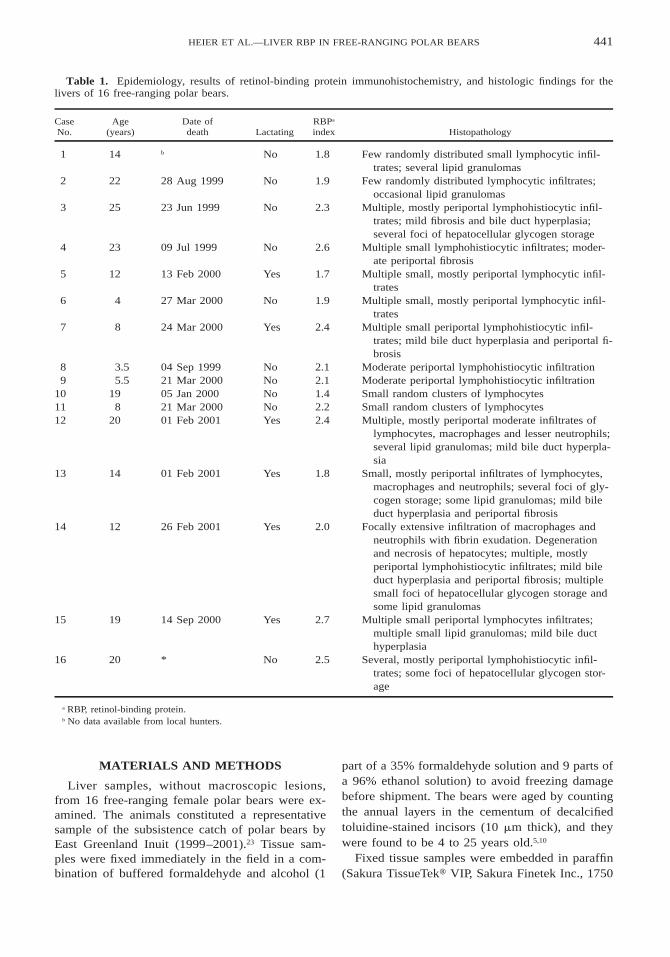
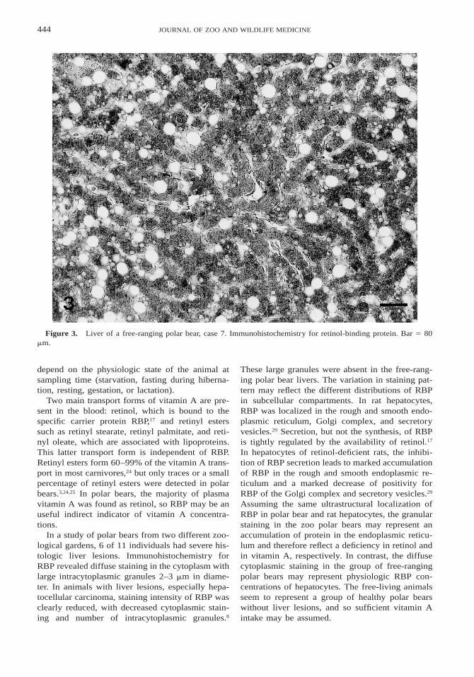
observed in several periportal zones in some indi-viduals. Hepatocyte cytoplasm had diffuse to finelystippled staining for RBP, whereas nuclei were neg-ative (Fig. 3). Ito cells did not stain for RBP. Thestaining index ranged from 1.4 to 2.7 (Table 1). Nocorrelation was found between RBP and age, sea-son, or lactation status (Fig. 4).
DISCUSSION
Only mild histopathologic changes were found inthe livers of all the free-ranging polar bears ex-amined. Changes observed were independent ofage, indicating that they may be present in healthyanimals without clinical significance. Mild bile ducthyperplasia and fibrosis were observed in adult po-lar bears but not in subadults, though only two sub-adult polar bears were examined, so a correlationwith age cannot be determined. Freezing artifactswere attributed to the difficult sampling conditionsin East Greenland.
Staining intensity for RBP in the livers examinedvaried only a little. The findings suggest that the 16free-ranging polar bears constituted a relatively ho-mogeneous group of animals without significanthistologic liver changes. The mild variations might
444 JOURNAL OF ZOO AND WILDLIFE MEDICINE
Figure 3. Liver of a free-ranging polar bear, case 7. Immunohistochemistry for retinol-binding protein. Bar 5 80mm.
depend on the physiologic state of the animal atsampling time (starvation, fasting during hiberna-tion, resting, gestation, or lactation).
Two main transport forms of vitamin A are pre-sent in the blood: retinol, which is bound to thespecific carrier protein RBP,17 and retinyl esterssuch as retinyl stearate, retinyl palmitate, and reti-nyl oleate, which are associated with lipoproteins.This latter transport form is independent of RBP.Retinyl esters form 60–99% of the vitamin A trans-port in most carnivores,24 but only traces or a smallpercentage of retinyl esters were detected in polarbears.3,24,25 In polar bears, the majority of plasmavitamin A was found as retinol, so RBP may be anuseful indirect indicator of vitamin A concentra-tions.
In a study of polar bears from two different zoo-logical gardens, 6 of 11 individuals had severe his-tologic liver lesions. Immunohistochemistry forRBP revealed diffuse staining in the cytoplasm withlarge intracytoplasmic granules 2–3 mm in diame-ter. In animals with liver lesions, especially hepa-tocellular carcinoma, staining intensity of RBP wasclearly reduced, with decreased cytoplasmic stain-ing and number of intracytoplasmic granules.8
These large granules were absent in the free-rang-ing polar bear livers. The variation in staining pat-tern may reflect the different distributions of RBPin subcellular compartments. In rat hepatocytes,RBP was localized in the rough and smooth endo-plasmic reticulum, Golgi complex, and secretoryvesicles.29 Secretion, but not the synthesis, of RBPis tightly regulated by the availability of retinol.17
In hepatocytes of retinol-deficient rats, the inhibi-tion of RBP secretion leads to marked accumulationof RBP in the rough and smooth endoplasmic re-ticulum and a marked decrease of positivity forRBP of the Golgi complex and secretory vesicles.29
Assuming the same ultrastructural localization ofRBP in polar bear and rat hepatocytes, the granularstaining in the zoo polar bears may represent anaccumulation of protein in the endoplasmic reticu-lum and therefore reflect a deficiency in retinol andin vitamin A, respectively. In contrast, the diffusecytoplasmic staining in the group of free-rangingpolar bears may represent physiologic RBP con-centrations of hepatocytes. The free-living animalsseem to represent a group of healthy polar bearswithout liver lesions, and so sufficient vitamin Aintake may be assumed.

445HEIER ET AL.—LIVER RBP IN FREE-RANGING POLAR BEARS
Figure 4. Retinol-binding protein staining index ofpolar bear livers as a function of age and lactation (top)and sampling time (day of the year) (bottom).
Wild polar bears feed mostly on seals, whichprovide very high vitamin A concentrations. Incontrast, the diet of captive polar bears is usuallybased on the nutritional requirements of dogs andcats and contains lower levels of vitamin A.3,30 Se-rum concentrations of vitamin A were lower in cap-tive polar bears than other ursid species in captiv-ity.3 Several studies report a relationship betweenlow serum vitamin A and the development of he-patic tumors in human patients.15,16 In captive mam-mals, hepatic diseases including neoplasia appearto be overrepresented in ursids in general2,9,13,18 andin polar bears in particular.7,11,14,18,19 The absence ofsignificant hepatic lesions in the free-ranging polarbears supports the notion that they had sufficientvitamin A levels. Measuring vitamin A levels offrozen tissue samples of the free-ranging polarbears should confirm or reject this hypothesis.
CONCLUSIONS
In summary, the liver RBP staining of free-rang-ing and captive polar bears differs significantly inmorphology and by the presence of granules in cap-tive polar bear hepatocytes. This might suggest suf-ficient vitamin A intake in free-ranging polar bears
and vitamin A deficiency in captive bears. How-ever, the relationship between vitamin A concentra-tion and RBP staining intensity requires confirma-tion by further studies. Therefore, hepatic vitaminA levels, not currently available, should be deter-mined and correlated to RBP staining. Retinol lev-els were negatively correlated with the amount oforganochlorine contaminant in blood samples fromfree-ranging polar bears at Svalbard.26 The bioac-cumulation of organic pollutants needs to be mea-sured.26 A correlation between RBP staining andage, season, and lactation status was not found. Theanalysis of liver samples from the remaining 98free-ranging polar bears for RBP is recommendedto further investigate its relationship with age, sex,season, and lactation status.
Acknowledgments: The Danish Cooperation forEnvironment in the Arctic (DANCEA) and theCommission for Scientific Research in Greenland(KVUG) are acknowledged for financial support.Jonas Brønlund and local hunters gathered and col-lected the samples, Hanne Tuborg Sandell and Bir-ger Sandell helped with the data coordination, andMaja Kirkegaard helped with the age determinationof the free-ranging polar bears.
LITERATURE CITED
1. Ball, M. D., H. C. Furr, and J. A. Olson. 1986. Acylcoenzyme A: retinol acyltransferase activity and the vi-tamin A content of polar bear (Ursus maritimus) liver.Comp. Biochem. Physiol. B. 84: 513–517.
2. Canfield, P., T. Bellamy, D. Blyde, W. J. Hartley, G.Reddacliff, and D. Spielman. 1990. Pancreatic lesions andhepatobiliary neoplasia in captive bears. J. Zoo Wildl.Med. 21: 471–475.
3. Crissey, S., K. Ange, K. Slifka, P. Bowen, M. Sta-cewicz-Sapuntzakis, C. Langman, W. Sadler, and A. Ward.2001. Serum concentrations of vitamin D metabolites, vi-tamin A and E, and carotenoids in six canid and four ursidspecies at four zoos. Comp. Biochem. Physiol. A: Physiol.128: 155–165.
4. Derocher, A. E., Ø. Wiig, and M. Andersen. 2002.Diet composition of polar bears in Svalbard and the west-ern Barents Sea. Polar Biol. 25: 448–452.
5. Dietz, R., M.-P. Heide-Jørgensen, J. Teilmann, N.Valentin, and T. Harkonen 1991. Age determination in Eu-ropean Harbour seals Phoca vitulina L. Sarsia 76: 17–21.
6. Evarts, R. P., Z. Hu, N. Omori, M. Omori, E. R.Marsden, and S. S. Thorgeirsson. 1995. Effect of vitaminA deficiency on the integrity of hepatocytes after partialhepatectomy. Am. J. Pathol. 147: 699–706.
7. Gosławski, J., and M. Pietrak. 1989. Liver tumors inpolar bears (Thalarctos maritimus). VerhandlungsberichteInternationale Symposium Erkrankungen Zootiere. Dort-mund, Germany. Pp. 295–298.
8. Heier A., A. Grone, J. Vollm, A. Kubber-Heiss, andL. N. Bacciarini. 2003. Immunohistochemical study of ret-
446 JOURNAL OF ZOO AND WILDLIFE MEDICINE
inol-binding protein in livers of polar bears (Thalarctosmaritimus). Vet. Pathol. 40: 196–202.
9. Hellmann, J., R. Hofmeister, and R. Goltenboth.1991. Das Auftreten von Tumoren bei Grossbaren (Ursi-dae)—Eine Literaturubersicht sowie sechs weitere Fall-beschreibungen. Berl. Munch. Tierarztl. Wochenschr. 104:262–268.
10. Hensel, R. J., and F. E. Sorensen. 1980. Age deter-mination of live polar bears. International Conference onBear Research and Management. Madison, Wisconsin. Pp.93–100.
11. Ippen, R., and D. Henne. 1986. Obduktionsbefundebei Baren (Procyonidae, Ailuridae und Ursidae).Verhandlungsberichte Internationale Symposium Erkran-kungen Zootiere. Rostock, Germany. Pp. 89–98.
12. Kane, A. B., and V. Kumar. 1999. Environmentaland nutritional pathology. In: Cotran, R. S., V. Kumar, andT. Collins (eds.). Robbins Pathologic Basis of Disease, 6thed. W. B. Saunders Co., Philadelphia, Pennsylvania. Pp.439–441.
13. Kronberger, H., K.-F. Schuppel, D. Altmann, and S.Seifert. 1975. Ein weiterer Beitrag zu den Geschwulstenbei Zootieren. Verhandlungsberichte Internationale Sym-posium Erkrankungen Zootiere. Tunis, Tunisia. Pp. 361–364.
14. Miller, R. E., W. J. Boever, L. P. Thornburg, andM. Curtis-Velasco. 1985. Hepatic neoplasia in two polarbears. J. Am. Vet. Med. Assoc. 187: 1256–1258.
15. Moriwaki, H., M. Tajika, Y. Miwa, M. Kato, I. Ya-suda, Y. Shiratori, M. Okuno, T. Kato, H. Ohnishi, and Y.Muto. 2000. Nutritional pharmacotherapy of chronic liverdisease: from support of liver failure to prevention of livercancer. J. Gastroenterol. 35: 13–17.
16. Newsome, P. N., I. Beldon, Y. Moussa, T. E. De-lahooke, G. Poulopoulos, P. C. Hayes, and J. N. Plevris.2000. Low serum retinol levels are associated with he-patocellular carcinoma in patients with chronic liver dis-ease. Aliment. Pharmacol. Ther. 14: 1295–1301.
17. Noy, N. 2000. Retinoid-binding proteins: mediatorsof retinoid action. Biochem. J. 348: 481–495.
18. Pettan-Brewer, K. C., and L. J. Lowenstine. 1999.Intrahepatic cysts and hepatic neoplasms in felids, ursids,and other zoo and wild animals. In: Fowler, M. E., andR. E. Miller (eds.). Zoo & Wild Animal Medicine. Current
Therapy, 4th ed. W. B. Saunders Co., Philadelphia, Penn-sylvania. Pp. 426–427.
19. Popplow, U. 1950. Primares Leberzellkarzinombeim Eisbar. Deutsch. Tierarztl. Wochenschr. 57: 194–195.
20. Raila, J., I. Buchholz, H. Aupperle, G. Raila, H.-A. Schoon, and F. J. Schweigert. 2000. The distributionof vitamin A and retinol-binding protein in the blood plas-ma, urine, liver and kidneys of carnivores. Vet. Res. 31:541–551.
21. Rodahl, K., and T. Moore. 1943. The vitamin Acontent and toxicity of polar bear and seal liver. Biochem.J. 37: 166–169.
22. Russell, F. E. 1967. Vitamin A content of polar bearliver. Toxicon 5: 61–62.
23. Sandell, H. T., and B. Sandell. 1996. Polar bearhunting and hunters in Ittoqqortoormiit/Scoresbysund, NEGreenland. Arctic Anthropol. 33: 77–93.
24. Schweigert, F. J., O. A. Ryder, W. A. Rambeck, andH. Zucker. 1990. The majority of vitamin A is transportedas retinyl esters in the blood of most carnivores. Comp.Biochem. Physiol. A. 95: 573–578.
25. Schweigert, F. J., and H. Zucker. 1991. Besonder-heiten im Vitamin A-Stoffwechsel der Ordnung Carnivo-ra—eine Ubersicht. Berl. Munch. Tierarztl. Wschr. 104:89–98.
26. Skaare, J. U., A. Bernhoft, O. Wiig, K. R. Norum,E. Haug, D. M. Eide, and A. E. Derocher. 2001. Rela-tionship between plasma levels of organochlorines, retinoland thyroid hormones from polar bears (Ursus maritimus)at Svalbard. J. Toxicol. Environ. Health A. 62: 227–241.
27. Smith, T. G. 1980. Polar bear predation on ringedseals and bearded seals in the land-fast ice habitat. Can.J. Zool. 58: 2201–2209.
28. Stirling, I., and W. R. Archibald. 1977. Aspects ofpredation of seals by polar bears. J. Fish. Res. Board Can.34: 1126–1129.
29. Suhara, A., M. Kato, and M. Kanai. 1990. Ultra-structural localization of plasma retinol-binding protein inrat liver. J. Lipid Res. 31: 1669–1681.
30. Wallach, J. D., and W. J. Boever. 1983. Diseases ofExotic Animals. Medical and Surgical Management, 1sted. W. B. Saunders Co., Philadelphia, Pennsylvania. Pp.548–573.
Received for publication 25 June 2003