
Genotyping and Evaluation of Pleurotus ostreatus (oyster ...
4480 IEEE SENSORS JOURNAL, VOL. 15, NO. 8, AUGUST 2015
An Algorithm for the Automatic Analysis ofSignals From an Oyster Heart Rate Sensor
Andrew D. Hellicar, Ashfaqur Rahman, Daniel V. Smith, Greg Smith,John McCulloch, Sarah Andrewartha, and Andrea Morash
Abstract— An in situ optical oyster heart rate sensor generatessignals requiring frequency estimation with properties differentto human ECG and speech signals. We discuss the methodof signal generation and highlight a number of these signalproperties. An optimal heart rate estimation approach wasidentified by application of a variety of frequency estimationtechniques and comparing results to manually acquired values.Although a machine learning approach achieved the bestperformance, accurately estimating 96.8% of the heart ratescorrectly, a median filtered autocorrelation approach achieved93.7% with significantly less computational requirement.A method for estimating heart rate variation is also presented.
Index Terms— Biomedical signal processing, frequencyestimation, machine learning.
I. INTRODUCTION
HEART rate and heart rate variability in intertidalinvertebrates are proxies for physiological responses
(such as oxygen transport, neural feedback, stress) andtracking these quantities is highly informative. For examplethey allow the impact of external environmental factors to bequantified and enable optimization of conditions for breedingand consumption stock. Consequently there is a need forheart rate monitoring systems for these animals.
Measurement of heart rate in humans is a well establishedand mature science [1]; ECG systems detect electrical signalsgenerated by the heart. These signals are amenable to analysisand were first analysed by computer in the 1960s [2].Real-time heart rate computer analysis systems were in useby the 1980s [3]. Analysis is based on detecting the repetitionand shape of the ECG signal structure referred to as theQRS complex. Unfortunately such approaches are notapplicable to intertidal invertebrates (such as the oyster)which possess a shell preventing use of externally applied
Manuscript received January 20, 2015; revised March 16, 2015; acceptedMarch 21, 2015. Date of publication April 13, 2015; date of currentversion June 17, 2015. This work was supported in part by the TasmanianGovernment through the Tasmanian Department of Economic Developmentand in part by the CSIRO Food Futures Flagship. The associate editorcoordinating the review of this paper and approving it for publication wasDr. M. R. Yuce.
A. D. Hellicar, A. Rahman, D. V. Smith, G. Smith, and J. McCullochare with Commonwealth Scientific and Industrial Research Organisation,Battery Point, TAS 7004, Australia (e-mail: [email protected];[email protected]; [email protected]; [email protected];[email protected]).
S. Andrewartha and A. Morash are with Commonwealth Scientific andIndustrial Research Organisation, Battery Point, TAS 7004, Australia, andalso with the University of Tasmania, Sandy Bay, TAS 7005, Australia(e-mail: [email protected]; [email protected]).
Color versions of one or more of the figures in this paper are availableonline at http://ieeexplore.ieee.org.
Digital Object Identifier 10.1109/JSEN.2015.2422375
ECG sensors. Early experiments on the bivalve class ofintertidal invertebrates observed heart rate by measuring theheart’s electrical impedance via implanted electrodes [4].However, it is critical that the system measuring heart ratedoes not modify the animal’s heart rate by (for example)increasing animal stress. To mitigate the invasive nature anddegradation of electrical contacts in the impendence approachan alternative optical coupling method was proposed [5].This approach illuminates the heart with an optical signaland detects the intensity of reflected light which is modifiedas a consequence of any motion of the heart’s surface. Forthin shelled intertidal invertebrates a near infrared source anddetector are collocated and positioned on the shell’s outersurface. The NIR signal propagates through the shell withoutneed for drilling into the shell. For thick shelled invertebratessuch as the oyster, the approach is similar with the exceptionof drilling a small hole into the shell [6] which wouldotherwise block the optical signal. The light emitter anddetector are mounted and sealed to ensure the integrity of theshell is maintained. Optical techniques avoid direct contactwith the animal; however, the approach introduces its ownproblems in terms of signal analysis due to signal complexityas pointed out in a recent review paper [7]. Thereforethere is a need to develop robust signal analysis approachesoperating over long installations possible with these sensors.In this paper we analyze the signal from a sensor in situ,and develop algorithms to detect short term average heartrate and variation of heart rate within short (20s) signalsequences.
The oyster biosensor [8] (Fig. 1) provides information onthe animal through both shell gape and heart rate detection.Gape refers to the opening of the two shells which occurswhen the oyster is submerged and feeding and/or taking inoxygen. The gape sensor (based on a Hall effect sensor)detects open and closed states of the oyster shell by measuringchanges in the output voltage of a transducer located onthe upper shell in response to a magnetic field induced bya magnet located on the lower shell. The heart rate sensorilluminates the oyster heart with a fixed intensity 950nm (NIR)optical signal generated by an LED source, and measuresthe reflected optical intensity via an IR diode detector. TheLED source and detector are collocated and mounted within5mm of the heart surface. The sensor responds with a timeconstant of 20μs to reflections within a cone angle 55 degreesfrom the sensor axis out to a range of 5mm. The principal ofoperation of the heart rate sensor is that the heart’s surfacereflects light which is measured by the IR sensor. Any motion
1530-437X © 2015 IEEE. Translations and content mining are permitted for academic research only. Personal use is also permitted,but republication/redistribution requires IEEE permission. See http://www.ieee.org/publications_standards/publications/rights/index.html for more information.

HELLICAR et al.: ALGORITHM FOR THE AUTOMATIC ANALYSIS OF SIGNALS FROM AN OYSTER HEART RATE SENSOR 4481
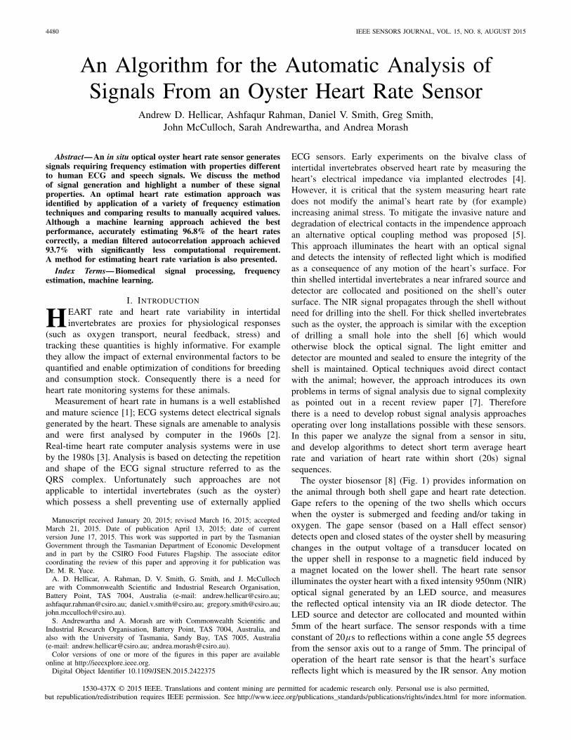
Fig. 1. Oyster attached to biosensor. (a) Photo of sensor and oyster.(b) Schematic of sensor inserted into hole in oyster’s shell. Heart size dependson whether it is in A: contraction phase (systole) or B: expansion phase(diastole). Reflected light rays shown for expanded heart. For contracted heartfewer rays will strike heart with corresponding change in detected signal level.
in the heart causes perturbation in the surface geometry, thusmodifying the intensity of the reflected optical signal. If theheart’s surface geometry varies periodically (as is the case fora beating heart) the measured signal exhibits a period equal tothat of the heart’s motion. Measurement of the signal period isused to infer heartbeat period and consequently heart rate. Thesensor is aligned such that signals are detectable both when theoyster is open and closed. Reflected light intensity is sampledat 100Hz over 20 seconds, resulting in sequences containing2000 samples. This sampling regime is selected to capturemultiple heartbeats per sequence, which are expected to havedurations within the range of 0.5 – 5 seconds [9]. Currentlyone sequence is recorded approximately every six minutesfor the duration of sensor installation, which has beenrunning for nine months. One gape value is recorded for eachsequence. Three classes of sequence occur: saturated (Fig. 2a),aperiodic (Fig. 2b), and periodic (Fig. 3). Any algorithmmust classify a sequence as one of these three classes, anddetermine the heart rate and heart rate variation of sequencesclassified as periodic.
Fig. 2. Examples of saturated (a) and aperiodic (b) sequences.
Fig. 3. Example of periodic time domain signals including 4 heart beats.Note peak value within beat occurs for first pulse in two beats up to 10s,and second pulse for last 2 beats. Sequence motif (described in Section II)shown in red/dashed overlays signal from 9.5 to 14.5 seconds. Best shiftedmotif matches shown from 5.0-9.5 seconds and 14.5-19 seconds. 5-9.5 and15-20 seconds.
Saturated signals likely occur due to the presence ofexternal light sources, or due to sub-optimal biasing ofdetector electronics. Aperiodic signals likely occur in theabsence of strong signal coupled off the heart surface,or absence of a beating heart. Periodic sequences result as aconsequence of the beating heart periodically modifying theintensity of the reflected signal coupled into the detector. Thedual chamber heart comprises a smaller ventricle and larger

4482 IEEE SENSORS JOURNAL, VOL. 15, NO. 8, AUGUST 2015
auricle which each beat once per cycle with a time delaybetween the two chamber contractions. Captured periodicsignals (Fig. 3) can exhibit a strong second-order harmonicdue to the combination of two temporally offset pulses ofdifferent amplitude. Motion of both chambers is detectableeither by direct illumination, or physical coupling through theheart tissue into the illuminated region. The optical reflectionsensing modality is sensitive to a combination of specularand multiple reflections, and is therefore highly sensitive togeometry. Furthermore the geometrical configuration evolvesover time and any technique must be robust against shapeand amplitude modification of the pulse.
Many approaches exist for frequency estimation. Onepopular method is the autocorrelation method (ACM) [10].For a signal sequence S containing L samples the sequencemean is subtracted and a signal S p of length 2L is generatedby zero padding (L = 2000). The period T is estimated byfinding m that maximizes the autocorrelation function:
A (m) = 1
L
2L−1−m∑
t=0
S p (t) S p (t + m) (1)
such that m ∈ [mmin , L/2], where mmin is the lag at whichthe autocorrelation first becomes negative, which removes thesignal coherence time. The sequence is classified as periodic ifpeak of A is larger than a threshold parameter which dependson signal quality and noise level [11]. The ACM is efficientlyimplemented by calculating autocorrelation using the Fouriertransform approach.
An alternative Hanning window (HWE) approachintroduced in [12] weights the sequence S with aHanning window. The method follows the ACM with themodification replacing A(m) by W (m) · A(m) in Equation (1)before calculating the peak and estimating T . W (m) isthe reciprocal of the Hanning window’s autocorrelationfunction. Therefore the HWE approach is weighted towardsselecting the longer lags. Other lag domain methods includecentre clipped autocorrelation (CCA) [13] and normalisedautocorrelation (NAC) [14] approaches. The centre-clippedapproach sets samples with amplitudes less than a fixedthreshold (often 50% of maximum signal amplitude) tozero resulting in a flattening of the spectrum and sharpeningof the lag domain. However, since the low amplitudevalues are removed the method is sensitive to variation inthe high amplitude region, (such as may be the case forspecular reflections off a surface). The NAC divides thelag domain values by the power in the delayed version ofthe signal. This weighting offsets the lag domain taperingcaused by zero padding of the signal; however, it isalso therefore prone to selecting longer period signals.Time domain methods include the average magnitudedifference function (AMD) [15] and the maximum likelihoodmethod (MLH) [16]. The AMD and MLH both optimizemeasures calculated on the time domain signal and delayedversion of the signal directly (difference and sum respectively).They are similar to autocorrelation approach without therelatively computationally expensive use of multiplicationsrequired in the lag domain approaches. Finally a popular
Fig. 4. Flow chart of implemented algorithm including classificationphase (top/green), heart rate estimation phase (bottom/blue) and heart ratevariability phase (right/red).
frequency domain method is the cepstrum method (CEP) [17]which is useful for period estimation where a strongly periodicsignal is convolved with a second signal. CEP recasts thecorresponding product in the frequency domain into a sumvia application of a logarithm. Each technique has specificadvantages and disadvantages, this motivated a machinelearning approach [18] using the output of the six frequencyestimation approaches (ACM, CCA, NAC, AMD, MLH, CEP)as features which can exploit the benefits of each method.
Here we extend the preliminary results reported in [18] byadding the HWE estimator (a total of 7 basic estimators),and add two aggregate estimators: tracking (TR) and medianfiltering (MF) which use information over multiple consecutivesequences. We use these nine estimators as features for amachine learning approach which achieves accuracy superiorto that achieved in [18]. We extend the analysis by presentingthe performance of all approaches and their sensitivity toselecting incorrect period ranges. We also highlight someof the types of signals and sources of error affecting ourapproach. Finally we introduce a method of heart rate varianceestimation, not previously implemented on this class of sensor.As these sensors become more widely deployed the issues wediscuss will need resolving in algorithms embedded in thesensor itself. Therefore computationally efficient and accurateapproaches are required.
II. METHOD
The algorithm is split into three phases: a classificationphase identifies the signal type, a heart rate estimation phasedetermines heart rate for periodic signals, finally heart ratevariability is estimated. A diagram of the algorithm is shownin Fig. 4. The classification phase identifies saturatedsequences (Fig. 2a) by detecting the proportion of time thesignal is saturated. The ACM is used to differentiate betweenperiodic and aperiodic signals. For remaining periodic signalsall seven basic estimators named in Section I were imple-mented generating seven estimates of heart rate. The ACM isbiased towards shorter lags due to the presence of zero paddingand care is needed to ensure the 2nd harmonic due to the

HELLICAR et al.: ALGORITHM FOR THE AUTOMATIC ANALYSIS OF SIGNALS FROM AN OYSTER HEART RATE SENSOR 4483
Fig. 5. Lag domain amplitude(white positive, black negative) from sequencesgathered over a 48 hour time frame November 3–November 4, 2014.
Fig. 6. Tracking estimator’s weighting function for scaling autocorrelationapproaches’ amplitude in the lag domain.
dual chamber beating is not erroneously selected. Alternativemethods such as the NAC and HWE exist to compensatefor this.
Despite the gaps between sequences, the slow change inoyster heart rate results in similar periodic structure whichis apparent when plotting the autocorrelation function forsuccessive samples (Fig. 5). This long term stability and theHWE lag domain weighting approach motivated a simpletracking (TR) estimator which is biased towards selecting heartrates similar to those estimated from the previous sequenceswhere NTR is the number of sequences used in the calculation.The motivation is to reduce the occurrence of erroneous heartrate estimates at half and double the actual rate (typical errorsfor estimators on signals with periodic beats containing doublepeak structure). To achieve this bias the lag space is weightedwith a piece-wise linear window. The window was maximal(and unit valued) at the mean detected period (T̄ ) over theprevious NTR sequences, and linearly decreases for smaller(and larger) lags until reaching a minimum value g < 1 atlags corresponding to half (and double) mean periods. All lagsoutside this range are also weighted by g, the weightingwindow is shown in Fig. 6. Optimal values of g and NTR
were estimated for the tracking estimator.We also applied a median filtering (MF) estimator applied
to the best performing basic estimator:1) The median period was calculated for the N1
MFsequences prior to and including the sequence beinganalysed.
2) Sequences with periods more than 20% different tothe median period were removed from the set ofN1
MF periods.3) From remaining sequences the median period was cal-
culated for the N 2MF sequences prior to and including
the sequence being analsyed.The longer time frame corresponding to N1
MF providesrobust longer term averages, whereas the potentially shortertime frame N2
MF allow accurate estimations of short termtransitions in heart rate. These values were optimally evaluatedfor the median filter approach.
Finally we implemented a machine learning approach usingnine frequency estimators (within dashed block in Fig. 4)as input features. Three WEKA [19] implementations wereassessed including a three layer multilayer perceptron (MLP)with six hidden nodes (larger networks showed no improve-ment in performance). A Linear regression (LR) and a supportvector regression (SMO).
Performance of the techniques was quantified by comparingestimated heart rates with values acquired by manuallyobserving sequence class and heart rate within thefirst 2 months of collected data. After removing saturatedsequences from the data set, sequences were randomly selectedand classified as either periodic or aperiodic based on visualinspection until 100 sequences were labelled for each class(200 in total). Sixteen regions of consecutive periodic signalswere identified and the periods of 9 sequences (evenly spacedthroughout each region) were measured by visual inspectionresulting in a total of 144 measurements. Ground truthperiods were calculated by linearly interpolating the measuredsequence periods across each span of sequences resulting ina total of 1450 sequences with ground truth period data.For the machine learning approach the 1450 sequences weresplit into 10 sets of 145 consecutive sequences and a 10-foldcross-validation used to evaluate performance, trainingon 9 sets and testing on the remaining set.
In addition to heart rate the variation of heart rate isalso of interest as an indicator. Whereas heart rate variationover longer time frames can be calculated as the differencebetween heart rates estimated from sequences with the desiredtemporal separation, short term (intra-sequence) variationrequires an estimate of the duration of each heartbeat. Becausethe structure of the heartbeat signal can change within asequence a motif approach was used to estimate the temporallocations of each beat within the sequence. For each sequencea subsequence of length T was identified as the motif thatis most representative of the multiple beat signals within thesequence. Temporal shifts of the motif within the sequencethat best reconstructed the entire sequence were used toestimate beat delay and consequently estimate heart ratevariance.
A subsequence STt0 (t) of sequence S(t) is uniquely defined
by length T and starting time t0 such that STt0 (t) = S(t + t0).
We zero pad the sequence to length L such that STt0 (t) = 0
for t > T. The sequence motif is the subsequence thatcaptures more of the periodic structure in the signal thanany other subsequence. To quantify this we calculate the

4484 IEEE SENSORS JOURNAL, VOL. 15, NO. 8, AUGUST 2015
cross-correlation C (τ ) of STt0 with S:
C(τ ) = F−1{F{ST
t0 (t)} · F∗{S(t)
}}(2)
Where F is the Fourier operator and ∗ the complex conjugate.The cross-correlation requires normalization to eliminate biastowards selecting motifs in the high amplitude regions of thesignal. Both the power in the sub-sequence and the distributionof power throughout the sequence contribute to this bias.A normalization factor is calculated from the cross correlationof instantaneous signal power S2 with a zero padded stepfunction H of duration T:
N(τ ) = F−1{F{S2(t)
} · F∗{H (t)}}
Where H(t) = 1 for t≤T and H(t)=0 for t>T. ConsequentlyN (t0) is the power in the subsequence and N (τ ) the powerin the target sequence starting at time τ of length T .
A subsequence STt0 can repeat L/T times throughout
the sequence of length L. We evaluate the peaks of thecross-correlation at temporal lag steps of size T correspondingto τi = i T + to modulo T , where i ∈ {1, f loor(L/T )}.To calculate heart rate variation we allow for period driftwithin a sequence and therefore look for maximum valueswithin a range {τi − kT, τi + kT } where k depends on theexpected maximum heart rate variation within a sequence.Finally given the estimated heart rate L the sequence motifis defined by delay tm which corresponds to the subsequencedelay t0 which maximizes the following expression:
tm = argmaxt0
{∑ f loor(
LT
)
i=1maxτ∈{τi−K T ,τi+K T }
[C(τ )
N (t0) N (τ )
]}(3)
The expected maximum heart rate variation within asequence was set to k = 0.1. After detecting the best motifthe temporal shifts of the motif τ that resulted in maximizingexpression (3) are used for estimating beat to beat variationestimation within a sequence.
III. RESULTS
The first 2 months of available data was assessed.Of the 9000 sequences, approximately 14% were saturated,61% periodic, and the remainder aperiodic. Of the approachesthe ACM generated the best classification of the aperiodic/periodic signals achieving 89% accuracy with a detectionthreshold of 1/3 with respect to the 200 sequences forwhich periodic/aperiodic ground truth was available. ThereforeACM was used for the classification stage in Fig. 4.
In terms of the saturated signals, a histogram of saturatedsignals against time of day is shown in Fig. 7 and indi-cates this detection approach is heavily influenced by thepresence of sunlight. An explanation for the generation ofsaturated sequences (Fig 2a) in the presence of sunlight is thatthe IR sensor is detecting bursts of sunlight. The presenceof waves on the water surface directly above the oysterresults in an instantaneous distribution of sunlight below thewater surface. This distribution contains regions of increasedsunlight due to a focusing effect via refraction through the
Fig. 7. Histogram of saturated sequence class against time of day.
TABLE I
ACCURACY RESULTS FOR IMPLEMENTED METHODS. ∗TR USES ACM,
g = 0.6, NTR CORRESPONDS TO 4.5 HOURS. ∗∗MF USES ACM,
N1MF AND N2
MF CORRESPOND TO 4.5 HOURS AND
1 HOUR, RESPECTIVELY
air/water interface. Wave motion then results in temporalvariation of sunlight incident onto the oyster and consequentlycoupled into the detector.
The ultimate aim of heart rate estimation is to findassociations with physiological response of the oysters. Foranalysis by biological scientists a 90% accuracy was targeted.Furthermore due to the interpolated nature of the generatedground truth values, the ground truths were estimated to bewithin 4% of the actual periods. This was based on comparingaccuracy from a coarser interpolation. Consequently a heartrate estimate was deemed accurate if it was within 10% ofthe manually acquired ground truth. The percentages ofheart rates estimated accurately for each approach are shownin Table I. The most accurate methods are the machinelearning approaches followed by the aggregated approaches.The ACM achieves the highest accuracy of the single(lag, time and frequency) estimators (83.2%). MultipleACM heart rate estimates were then aggregated to calculate

HELLICAR et al.: ALGORITHM FOR THE AUTOMATIC ANALYSIS OF SIGNALS FROM AN OYSTER HEART RATE SENSOR 4485
Fig. 8. Plot of estimated heart rates for a selection of approaches.
the Tracking and Median Filtered estimators. Optimal valuesof NTR corresponded to time frames of 4-5 hours withoptimal g = 0.6 giving an accuracy of 85.2%. The medianfilter approach achieved 93.7% with values of NTR and NTR
corresponding to 4.5 hours and 1 hour respectively. Notethese optimal values are sensitive to the level of heart ratevariation over time frames corresponding to hours. Valuesof NTR and NTR corresponding to 2 hours and 20 minutesrespectively still achieved 92% accuracy, but may be moreaccurate for estimating heart rates where variation is morerapid than the current installation. Finally the Machine learningapproach achieved superior results, the best approach (basedon a support vector regression) achieving 96.8% accuracy.
The accuracy of any approach is dependent on two factors:the ability to estimate the period within the sequenceaccurately, and the ability to avoid erroneously selectingdouble and half periods. Each estimate of heartbeat periodwas compared to the ground truth. If the estimated periodwas within the range 2/3 to 3/2 multiples of the groundtruth period the estimate was categorized as being in thecorrect range. For period estimates below and above thisrange, the estimate was categorized as being in the low orhigh range respectively. The percentages of estimated periodscategorized into these ranges by each approach are shownin Table I. For lag based approaches the accuracy is stronglyrelated to the erroneous selection of longer periods. The biasof ACM and CCA to lower periods via lag domain weightingresulting from sequence zero padding is evident, whereas thenormalised approach which removes this weighting sufferedthe most. The two pulse structure of the heart rate signals hasthe potential to generate erroneous half period peaks in the lagdomain. Despite the lower lag bias of the ACM and CCA theACM and CCA were not susceptible to the half period error.The time domain methods exhibited similar performance tothe NAC. The CEP had the highest of estimates in the lowperiod range, as a frequency domain method this correspondsto the CEP selecting the second harmonic, resulting in halfperiod estimates. The MF approach was highly successfulat avoiding half and double period estimates; the averagingeffect over multiple samples did not dramatically reduceaccuracy. Information in the multiple estimators was utilizedby the machine learning approach to improve the accuracybeyond the median filtered results.
Fig. 9. (a)-(b) Two sequences of 20 s duration from early region ofFig. 3 timeframe at ∼4:13AM and 4:25AM (13 minutes apart). (c) Examplesequence including single “beat.”
The results show a wide variation in the ability of themethods to determine heart rate which is strongly influencedby the bias of the various methods towards detection of heartrates at double and half the correct heart rate. An examplerange of sequences is shown in Fig. 8 along with estimated

4486 IEEE SENSORS JOURNAL, VOL. 15, NO. 8, AUGUST 2015
Fig. 10. (a) Original sequence (DC removed). (b) Lag domain of sequence.(c) 2 Hz high-pass filtered sequence. (d) Lag domain of filtered sequence.
heart rates; the ML and MF closely follow the ground truth,whereas ACM and HWE exhibit erroneous readings on somesequences.
For heart rate sensors deployed on cheap, lightweighthardware the MF(ACM) approach is ideal as the more accuratemachine learning approach requires labelled training dataand additional computational complexity. However; for offlineanalysis with labelled data the machine learning approachprovides the most accurate results.
The signals exhibit a number of unique properties. Forexample the signals can switch between regions of periodicand aperiodic signals (observable in lag domain Fig. 5).Although the heart rate varies slowly the signal shape can varyrapidly. This is demonstrated by the sequences in Fig. 9a, bwhich are separated by approximately 13 minutes. Thesesequences are periodic with identical heart rates but exhibitsignificantly different shapes. Sudden signal deviations oftenoccur either in isolation (Fig. 9c) or within a periodicsequence (Fig. 10a). Isolated deviations likely indicate thepresence of a short term geometry variation caused eitherby a single heartbeat or the shell opening and closing.Higher frequency sampling of gape would identify the cause.Deviations within periodic sequences are often ∼2s induration, with a corresponding change in the periodic structureeither side of the deviation. An example of this is seenby comparing the signal waveform in Fig. 10a 0s-5s versusthe 15s-20s signal waveform. We believe these dips occurwhen the oyster rapidly closes and opens the shell, thuschanging the coupling geometry. Furthermore these dips causeproblems for the ACM due to the first peak (2s in Fig. 10b)becoming obscured by the spread of the self coherence peak(0s in Fig. 10b). The spread is introduced as a consequence ofthe sequence mean value being significantly different to theperiodic structure local mean value resulting in temporarilylocalized DC offsets which extend the coherence time. Thetwo sequences recorded adjacent to Fig. 9a were correctlyidentified with 2s period. One solution is to high pass filter thesequence which removes the localized DC offset and exposes
the lag peaks (Fig. 10d). However this lowers the sensitivityof the method to heart rate detection in other sequences.As these errors are present within single sequences, the medianfiltering approach eliminates these errors.
IV. CONCLUSION
We discussed a range of properties of signals generated bya novel oyster heart rate monitor and consequences on signalanalysis. These properties include sudden changes in signallevel, transition from periodic to aperiodic signals, largesaturated signal interference, and evolution of the periodicstructure within the sequences. We implemented a range ofexisting methods for estimating heart rate, along with sometailored approaches aggregating multiple sequences, and amachine learning approach. The approaches were all ableto estimate heart rate. The lag domain approaches biased toshorter lags were the best single sequence simple estimatorsachieving 83.2% accuracy. However none of the existingmethods achieved better than 90% accuracy. This accuracyrequired a machine learning approach which achieved the bestresults (96.8%) using all available estimators as input features.Alternatively an approach utilizing multiple consecutivesequences (the median filtered autocorrelation method) wasrequired which achieved acceptable accuracy (93.7%). Theultimate choice is determined by available computationalresources. Future work would require implementation ofthe algorithms within the sensor platform and requirecomputationally efficient means in calculating heart ratevariation.
ACKNOWLEDGMENT
The authors gratefully acknowledge B. Taylor from theUniversity of Tasmania whose oyster tag design is the basisof the in situ oyster tag design used in this paper, and withoutwhich this work would not be possible.
REFERENCES
[1] E. Braunwald, “Cardiology: The past, the present, and the future,”J. Amer. College Cardiol., vol. 42, no. 12, pp. 2031–2041, 2003.
[2] R. A. Bruce, J. A. Mazzarella, J. W. Jordan, Jr., and E. Green,“Quantitation of QRS and ST segment responses to exercise,” Amer.Heart J., vol. 71, no. 4, pp. 455–466, Apr. 1966.
[3] J. Pan and W. J. Tompkins, “A real-time QRS detection algorithm,” IEEETrans. Biomed. Eng., vol. BME-32, no. 3, pp. 230–236, Mar. 1985.
[4] E. R. Trueman, “Activity and heart rate of bivalve molluscs in theirnatural habitat,” Nature, vol. 214, pp. 832–833, May 1967.
[5] M. H. Depledge and B. B. Andersen, “A computer-aided physiologicalmonitoring system for continuous, long-term recording of cardiac activ-ity in selected invertebrates,” Comparative Biochem. Physiol. A, Physiol.,vol. 96, no. 4, pp. 473–477, 1990.
[6] P. A. Ritto, J. G. Contreras, and J. J. Alvarado-Gil, “Monitoring ofheartbeat by laser beam reflection,” Meas. Sci. Technol., vol. 14, no. 3,p. 317, 2003.
[7] N. P. Burnett et al., “An improved noninvasive method for measur-ing heartbeat of intertidal animals,” Limnol. Oceanography, Methods,vol. 11, no. 2, pp. 91–100, Feb. 2013.
[8] CSIRO. Biosensors for Oysters. [Online]. Available: http://www.csiro.au/ Organisation-Structure/ Flagships/ Food-Futures-Flagship/Breed-Engineering-Theme/Aquaculture-Biosensors-Oysters.aspx, accessedJan. 2014.
[9] K. H. Park, Y.-S. Kim, E.-Y. Chung, S.-N. Choe, and J.-J. Choo,“Cardiac responses of Pacific oyster crassostrea gigas to agentsmodulating cholinergic function,” Comparative Biochem. Physiol. C,vol. 139, no. 4, pp. 303–308, 2004.

HELLICAR et al.: ALGORITHM FOR THE AUTOMATIC ANALYSIS OF SIGNALS FROM AN OYSTER HEART RATE SENSOR 4487
[10] A. V. Oppenheim and R. W. Schafer, Discrete-Time Signal Processing,3rd ed. Englewood Cliffs, NJ, USA: Prentice-Hall, 2009
[11] M. G. Elfeky, W. G. Aref, and A. K. Elmagarmid, “Using convolutionto mine obscure periodic patterns in one pass,” in Proc. EDBT, 2004,pp. 605–620.
[12] P. Boersma, “Accurate short-term analysis of the fundamental frequencyand the harmonics-to-noise ratio of a sampled sound,” in Proc. IFA,vol. 17. 1993 pp. 97–110.
[13] J. J. Dubnowski, R. W. Schafer, and L. Rabiner, “Real-time digitalhardware pitch detector,” IEEE Trans. Acoust., Speech, Signal Process.,vol. ASSP-24, no. 1, pp. 2–8, Feb. 1976.
[14] D. Talkin, “A robust algorithm for pitch tracking (RAPT),” in SpeechCoding and Synthesis. Amsterdam, The Netherlands: Elsevier, 1995,ch. 14, pp. 495–518,
[15] M. J. Ross, H. Shaffer, A. Cohen, R. Freudberg, and H. J. Manley,“Average magnitude difference function pitch extractor,” IEEE Trans.Acoust., Speech, Signal Process., vol. 22, no. 5, pp. 353–362, Oct. 1974.
[16] A. M. Noll, “Pitch determination of human speech by the harmonicproduct spectrum, the harmonic sum spectrum and a maximum like-lihood estimate,” in Proc. Symp. Comput. Process. Commun., vol. 19,1970, pp. 779–797.
[17] A. M. Noll, “Cepstrum pitch determination,” Acoust. Soc. Amer., vol. 41,no. 2, pp. 293–309, Feb. 1967.
[18] A. D. Hellicar, A. Rahman, D. Smith, G. Smith, and J. McCulloch,“A neural network and SOM based approach to analyse periodic signals:Application to oyster heart-rate data,” in Proc. IJCNN, Beijing, China,Jul. 2014, pp. 2211–2217.
[19] M. Hall, E. Frank, G. Holmes, B. Pfahringer, P. Reutemann, andI. H. Witten, “The WEKA data mining software: An update,” SIGKDDExplorations, vol. 11, no. 1, pp. 10–18, Jun. 2009.
Andrew D. Hellicar received the bachelor’s (Hons.)degree in engineering from the University ofTasmania, in 1997, and the Ph.D. degree fromMonash University, in 2007. In 2012, he returned toTasmania, where he conducts research on machinelearning techniques applied to the agriculturedomain with the ICT Centre, Commonwealth Scien-tific and Industrial Research Organisation (CSIRO).Since 2002, he has been with the Radio PhysicsLaboratory in Computational Electromagnetics andDistributed Computing, CSIRO, before leading
projects in terahertz imaging and terrestrial communication systems andundertaking research on antennas for radio astronomy and satellitecommunication.
Ashfaqur Rahman (SM’12) received thePh.D. degree in information technology fromMonash University, Gippsland, VIC, Australia.He worked on specific machine learning problems,including ensemble learning and fusion, featureselection/weighting methods, genetic algorithm-based optimization, and image segmentation andclassification. He is currently a Machine LearningResearcher for more than ten years. He is aResearch Scientist with Commonwealth Scientificand Industrial Research Organisation (CSIRO),
Hobart, TAS, Australia. He is the Leader of the Computational IntelligenceTeam with CSIRO. He has authored around 70 peer-reviewed journal articles,book chapters, and conference papers. He serves as a Reviewer of prestigiousconferences and journals. He was the Program Committee Chair of the2013 International Conference on Digital Image Computing: Techniques andApplications.
Daniel V. Smith received the Ph.D. degree intelecommunication engineering from the Univer-sity of Wollongong, in 2007. From 2008 to 2009,he was a Post-Doctoral Fellow with the Common-wealth Scientific and Industrial Research Organi-sation (CSIRO), Hobart, Australia. He was thenappointed as a Research Scientist with the IntelligentSensing and Systems Laboratory, CSIRO, where hecurrently works. The focus of his work has beenupon the research and development of analyticalmodels for environmental and agricultural appli-
cations using sensors and sensor networks. His research interests includeadaptive filtering, time series modeling, and data uncertainty representations.
Greg Smith received the B.E. (Hons.) degree and the M.Sc. degreein computer science from the University of Technology, Sydney,in 1987 and 1998, respectively, and the Ph.D. degree from the Universityof Sydney, in 2004. He is currently with the Commonwealth Scientific andIndustrial Research Organisation, Hobart, TAS. His current interests are indata mining, software for big data analytics, and agent-based modeling.
John McCulloch received the Degree from theUniversity of Tasmania (UTas), in 1993. He was aComputer Systems Engineer with UTas. He lecturedon experimental design and mechatronics beforejoining Commonwealth Scientific and IndustrialResearch Organisation (CSIRO), where he hasbeen managing biosensor and decision supportprojects in the marine and pond aquaculture space.He is currently a Research Engineer with theAutonomous Systems Program, Digital ProductivityFlagship, CSIRO. His example projects are a data
management and visualization system for the Tasmanian Shellfish QualityAssurance Program, and oyster biosensor development such that real timephysiological data (heart rate and gape) is now being gathered from animalson farms in Southern Tasmania.
Sarah Andrewartha received the Ph.D. (Hons.)degree from Melbourne’s La Trobe University.She focused on understanding how the environmentinfluenced the development and physiology of abroad range of animals. She was a Post-DoctoralFellow with the University of North Texas, whereshe was involved in research on the developmentof acid-base regulation in chicken embryos, andepigenetic inheritance patterns in response tochanges in the environment of Daphnia (tinyfreshwater crustaceans with a transparent body,
often used in biological research). She is currently a Comparative Physiologistand Post-Doctoral Fellow with the Commonwealth Scientific and IndustrialResearch Organisation, Hobart, Australia. She uses biosensors to monitorthe effect of environmental changes on shellfish and fish physiology. In thefuture, this information will be used by aquaculture farmers to assist farmmanagement, based upon measurements of animal fitness. She is examiningbody temperature influences on metabolism, ventilation, and blood acid-baseregulation in developing Tamar wallaby joeys, and the consequences of thenest environment for young reptiles and amphibians.
Andrea Morash received the B.Sc. degreein biology from Mount Allison University,NB, Canada, in 2005, and the Ph.D. degree inbiology from McMaster University, ON, Canada,in 2010. From 2010 to 2012, she completed herfirst post-doctoral fellowship with the Universityof Cambridge funded by an NSERC Post-DoctoralFellowship Award. She joined the Universityof Tasmania and the Commonwealth Scientific andIndustrial Research Organisation as a Post-DoctoralResearcher in 2013. Her research interests focus
on the effects of environmental stress on animal energy production andutilization and how this impacts their physiology and ecology.
Copyright © 2022 FDOKUMEN


![[- 200 [ PROVIDING MODULATED COMMUNICATION SIGNALS ]](https://static.fdokumen.com/doc/165x107/6328adc85c2c3bbfa804c60f/-200-providing-modulated-communication-signals-.jpg)