Amplified fragment length polymorphism assessment of genetic diversity in Pacific lampreys
12
ORIGINAL RESEARCH ARTICLE Erk2 in Ovarian Development of Green Mud Crab Scylla paramamosain Ani Ma, 1 Yilei Wang, 1 Zhihua Zou, 1 Mingjun Fu, 1,2 Peng Lin, 1 and Ziping Zhang 3 We identified extracellular signal-regulated kinase 2 (erk2) from green mud crab, Scylla paramamosain, in this article. It was originally identified from an expressed sequence tag fragment from a normalized gonadal cDNA library. 5¢ Rapid amplification of cDNA end (RACE) technique was used to obtain the 5¢ untranslated region (UTR). The full-length cDNA of Sp-erk2 is 1516 bp, including a 5¢-terminal UTR of 19 bp, an open-reading frame of 1098 bp, and a 3¢-terminal UTR of 399 bp. The translated protein is 365 amino acids in length with a predicted molecular weight of 42 kDa, which is the same as other species. It is the first time that the expression of Sp-erk2 in different stages of ovary development of crustacean was analyzed, and the result showed that the expression of Sp-erk2 increased gradually with ovarian development, with a peak in the mature phase. In situ hybridization histochemistry was used to clarify the detail of expression. Positive signals illustrated that Sp-erk2 mRNA is present in follicular cells when the ovary is in early stages, and in both follicular cells and oocytes when it is in mature phases. All above suggest that Sp-erk2 is important for ovarian development. Introduction G reen mud crab, Scylla paramamosain, is mainly dis- tributed in the Indian Ocean and Western Pacific trop- ical and subtropical waters and is an important commercial species in aquaculture. It has been widely cultured in China, especially in the southern regions such as the Fujian, Guangdong, and Hainan Provinces. Although eyestalk ab- lation is the most common method to induce ovarian mat- uration, it may cause gonadal dysgenesis and lower quality of spawns, which blocks the healthy and sustainable devel- opment of green mud crab aquaculture (Keenan, 1999; Li and Wang, 2001). Therefore, molecular mechanisms and func- tional involvement of reproduction-related genes in ovarian development are necessary for better understanding the re- productive maturation of green mud crab. Green mud crab has been studied since the 20th century. The research on green mud crab ovary was mainly focused on the structure and development (Li and Wang, 2001; Tang et al., 2009; Islam et al., 2010), and study on molecular level was very limited. Three normalized cDNA libraries from ovary, testis, and gonad development-related organs of green mud crab were constructed by our research group (Zou et al., 2007, 2009, 2011), and then a total of 5160 high- quality expressed sequence tags (ESTs) were obtained and a home-made cDNA microarray containing 5664 spots was developed from these libraries. The results from EST anno- tation, cDNA microarray hybridization, and real-time PCR analysis indicated that cell cycle regulatory protein, mitogen- activated protein kinase (MAPK) signal transduction path- way, and ubiquitin proteasome pathway may participate in the ovarian development of green mud crab (Zou et al., 2011). Meanwhile, our group cloned a vasa gene from green mud crab and preliminarily analyzed its expression during gonadal development and gametogenesis, with a result that indicated that vasa gene likely serves as a useful and specific marker for germ cell development (Wang et al., 2011). In addition, some differential expression genes in the thoracic ganglion of green mud crab during ovarian development have been obtained recently from an SSH cDNA library (Zeng et al., 2011). MAPK signal transduction pathway is a conserved signal transduction pathway in eukaryotes that exists from yeast to human and is widely involved in cell growth, differentiation, reproduction, apoptosis, antistress, and other physiological processes (Schaeffer and Weber, 1999; Widmann et al., 1999). It transduces signals from outside the cell to the cytoplasm or nucleus by a conserved three-kinase cascade. This cascade consists of a MAPK, which is activated via phosphorylation by a MAPK kinase, which in turn is phosphorylated by a MAPK kinase kinase, phosphorylating intracellular substrate proteins or transcription factors to regulate physiological processes (Schaeffer and Weber, 1999; Chang and Karin, 2001; Orton et al., 2005). At least four subfamilies of MAPK— 1 Fisheries College, Jimei University, Xiamen, China. 2 Department of Biology, Xiamen University, Xiamen, China. 3 Department of Biological Science, Seton Hall University, South Orange, New Jersey. DNA AND CELL BIOLOGY Volume 00, Number 00, 2012 ª Mary Ann Liebert, Inc. Pp. 1– DOI: 10.1089/dna.2011.1458 1
-
Upload
independent -
Category
Documents
-
view
2 -
download
0
Transcript of Amplified fragment length polymorphism assessment of genetic diversity in Pacific lampreys

ORIGINAL RESEARCH ARTICLE
Erk2 in Ovarian Development of GreenMud Crab Scylla paramamosain
Ani Ma,1 Yilei Wang,1 Zhihua Zou,1 Mingjun Fu,1,2 Peng Lin,1 and Ziping Zhang3
We identified extracellular signal-regulated kinase 2 (erk2) from green mud crab, Scylla paramamosain, in thisarticle. It was originally identified from an expressed sequence tag fragment from a normalized gonadal cDNAlibrary. 5¢ Rapid amplification of cDNA end (RACE) technique was used to obtain the 5¢ untranslated region(UTR). The full-length cDNA of Sp-erk2 is 1516 bp, including a 5¢-terminal UTR of 19 bp, an open-reading frameof 1098 bp, and a 3¢-terminal UTR of 399 bp. The translated protein is 365 amino acids in length with a predictedmolecular weight of 42 kDa, which is the same as other species. It is the first time that the expression of Sp-erk2 indifferent stages of ovary development of crustacean was analyzed, and the result showed that the expression ofSp-erk2 increased gradually with ovarian development, with a peak in the mature phase. In situ hybridizationhistochemistry was used to clarify the detail of expression. Positive signals illustrated that Sp-erk2 mRNA ispresent in follicular cells when the ovary is in early stages, and in both follicular cells and oocytes when it is inmature phases. All above suggest that Sp-erk2 is important for ovarian development.
Introduction
Green mud crab, Scylla paramamosain, is mainly dis-tributed in the Indian Ocean and Western Pacific trop-
ical and subtropical waters and is an important commercialspecies in aquaculture. It has been widely cultured in China,especially in the southern regions such as the Fujian,Guangdong, and Hainan Provinces. Although eyestalk ab-lation is the most common method to induce ovarian mat-uration, it may cause gonadal dysgenesis and lower qualityof spawns, which blocks the healthy and sustainable devel-opment of green mud crab aquaculture (Keenan, 1999; Li andWang, 2001). Therefore, molecular mechanisms and func-tional involvement of reproduction-related genes in ovariandevelopment are necessary for better understanding the re-productive maturation of green mud crab.
Green mud crab has been studied since the 20th century.The research on green mud crab ovary was mainly focusedon the structure and development (Li and Wang, 2001; Tanget al., 2009; Islam et al., 2010), and study on molecular levelwas very limited. Three normalized cDNA libraries fromovary, testis, and gonad development-related organs ofgreen mud crab were constructed by our research group(Zou et al., 2007, 2009, 2011), and then a total of 5160 high-quality expressed sequence tags (ESTs) were obtained and ahome-made cDNA microarray containing 5664 spots wasdeveloped from these libraries. The results from EST anno-
tation, cDNA microarray hybridization, and real-time PCRanalysis indicated that cell cycle regulatory protein, mitogen-activated protein kinase (MAPK) signal transduction path-way, and ubiquitin proteasome pathway may participate inthe ovarian development of green mud crab (Zou et al.,2011). Meanwhile, our group cloned a vasa gene from greenmud crab and preliminarily analyzed its expression duringgonadal development and gametogenesis, with a result thatindicated that vasa gene likely serves as a useful and specificmarker for germ cell development (Wang et al., 2011). Inaddition, some differential expression genes in the thoracicganglion of green mud crab during ovarian developmenthave been obtained recently from an SSH cDNA library(Zeng et al., 2011).
MAPK signal transduction pathway is a conserved signaltransduction pathway in eukaryotes that exists from yeast tohuman and is widely involved in cell growth, differentiation,reproduction, apoptosis, antistress, and other physiologicalprocesses (Schaeffer and Weber, 1999; Widmann et al., 1999).It transduces signals from outside the cell to the cytoplasm ornucleus by a conserved three-kinase cascade. This cascadeconsists of a MAPK, which is activated via phosphorylationby a MAPK kinase, which in turn is phosphorylated by aMAPK kinase kinase, phosphorylating intracellular substrateproteins or transcription factors to regulate physiologicalprocesses (Schaeffer and Weber, 1999; Chang and Karin,2001; Orton et al., 2005). At least four subfamilies of MAPK—
1Fisheries College, Jimei University, Xiamen, China.2Department of Biology, Xiamen University, Xiamen, China.3Department of Biological Science, Seton Hall University, South Orange, New Jersey.
DNA AND CELL BIOLOGYVolume 00, Number 00, 2012ª Mary Ann Liebert, Inc.Pp. 1–DOI: 10.1089/dna.2011.1458
1

extracellular signal-regulated kinase (ERK), c-Jun N-terminalkinases, p38 MAPK (p38), and ERK5—have been defined inmammalian cells (Cowan and Storey, 2003; Qi and Elion,2005). As the first discovered MAPK pathway, ERK signaltransduction pathway is activated by a variety of mitogenicstimuli, such as growth factors, cytokines, and phoebe,which regulate proliferation, differentiation, meiosis, fertil-ization, embryonic development, metamorphosis, learningand memory, cell survival, and so on (Kumano et al., 2001;Lambert and Nagy, 2003; Akanuma and Nishida, 2004;Sharma and Carew, 2004; Feld et al., 2005; Chambon et al.,2007; Junttila et al., 2008).
As is known, meiosis is necessary for producing haploidgametes and ERK pathway is required for this process.Function of ERK on meiosis of oocyte has been studied fordecades in certain species, which include at least four aspectsin starting the meiosis resumption, spindle organization,meiosis I–meiosis II (M I–M II) transition, and M II arrest as acomponent of cytostatic factor (Schmitt and Nebreda, 2002;Fan and Sun, 2004).
ERK2—also termed ERK, MAPK, or MAPK1 in other ar-ticles—is the main molecule in ERK signal transductionpathway. Since the first erk gene was cloned by Boulton et al.in 1991 (Boulton et al., 1991), ERK transduction pathway hasbeen well studied in many species, especially the regulationmechanism during oocyte development and ovarian pa-thology in mammals as a pivotal signal transduction path-way (Fan and Sun, 2004; Steinmetz et al., 2004). Although erkshave been cloned from a great number of vertebrates, theinformation is inadequate in invertebrates. Up to date, inaquatic invertebrates, erks have been cloned from Strongylo-centrotus purpuratus, Ciona intestinalis, Marthasterias glacialis,Aplysia californica, Marsupenaeus japonicus, and partial se-quence from Littorina littorea (Iakovleva et al., 2006). More-over, research on the action of ERK signal transductionpathway in ovarian development in crab is unavailable. Inthis article, the Sp-erk2 full-length cDNA from S. para-mamosain was cloned and its differential expression in de-veloping ovary was also described. These results may allowus to gain more insight into the function of erk2 in ovariandevelopment of crustaceans.
Materials and Methods
Animals’ and materials’ preparation
Green mud crabs were bought mainly from a crab farm inZhangpu, Zhangzhou, P.R. China. Tissues from ovary,muscle, gill, heart, brain, stomach, intestine, and hepato-pancreas were rapidly dissected, snap-frozen in liquid ni-trogen, and stored at - 80�C for later experiments. Inaddition, ovaries for in situ hybridization histochemistrywere immersed into 4% paraformaldehyde in phosphate-buffered saline (pH 7.4) overnight at 4�C, treated with gra-dient methanol, and finally stored in 100% methanol at- 80�C for use. The gonad somatic index (GSI = gonadweight/body weight)—the female crab was classified intosix stages: stage 1 (GSI = 0.7 – 0.3), stage 2 (GSI = 2.0 – 0.1),stage 3 (GSI = 3.5 – 0.2), stage 4 (GSI = 5.2 – 0.2), stage 5(GSI = 7.6 – 0.6), and stage 6 (GSI = 10.3 – 0.3)—described inseveral publications (Lo et al., 2007; Wonglapsuwan et al.,2010; Zhang et al., 2010) was adapted in this study.
RNA isolation and cloning of the full-lengthcDNA of Sp-erk2
RNA isolation and reverse transcription PCR. Total RNAfrom tissues obtained previously was isolated with home-made trizol reagent according to methods described previously(Zhang et al., 2003), and the concentration of the total RNAwas calculated by measuring the absorbance at 260 nm. RQ1RNase-Free DNase I (Promega) was used to eliminate the con-taminating DNA. Three micrograms of total RNA of eachsample was reverse transcribed using random primers and M-MLV reverse transcriptase (Promega). Lastly, the PCR productswere 10-fold diluted in ultrapurified water for final use.
Cloning the 5¢ untranslated region of Sp-erk2 cDNA. Apartial sequence of 1416 bp that was identified as a 3¢ se-quence homologous to erk2 was obtained from a normalizedgonadal cDNA library of green mud crab (Zou et al., 2011). 5¢Rapid amplification of cDNA end (RACE) method was usedto amplify the 5¢ end of Sp-erk2, and it was carried out byusing SMART�-RACE cDNA amplification kit (Clontech)according to the manufacturer’s instructions. Sp-erk2-outand Sp-erk2-inner primers (Table 1) were designed based onthe selected fragments. With Sp-erk2-out and universal pri-mer mixture (UPM), the PCR programs were carried out at94�C for 3 min, followed by 5 cycles of 94�C for 30 s and 72�Cfor 2 min; 5 cycles of 94�C for 30 s, 69�C for 30 s, and 72�C for2 min; 25 cycles of 94�C for 30 s, 67�C for 30 s, and 72�C for 2min; and an extension step at 72�C for 10 min. Then, a nestedPCR was performed using the primer Sp-erk2-inner and thenested universal primer (NUP), using a program at 94�C for3 min, followed by 35 cycles of 94�C for 30 s, 68�C for 30 s,and 72�C for 2 min, and a final extension step at 72�C for10 min. Both UPM and NUP primers were provided by thekit. PCR products were purified with a Qiaquick PCR Pur-ification Kit (Qiagen) and inserted into pMD18-T (TaKaRa)and sequenced. To check the sequence, a pair of full-lengthprimers (Sp-erk2-head and Sp-erk2-toe; Table 1) were
Table 1. Oligonucleotide Primers
Used in This Article
Gene/accession Primers Sequence
Sp-erk2/GQ847862
Sp-erk2-out 5¢ TCTCTCGAAGGGTTCTCTGGCAGT 3¢
Sp-erk2-inner 5¢ CATGCCATAGGCGCCTTCTCC 3¢
Sp-erk2-head 5¢ TGGAGCGGCGGTGTGCGT 3¢
Sp-erk2-toe 5¢ CTTGATGTTGCCTGAACCCCTG 3¢
Sp-erk2-F 5¢ GCATGGTGGTATCTGCTAGTGACA 3¢
Sp-erk2-R 5¢ TTCTATCGTTTGAGCCCGTATTA 3¢
Sp-erk2-ISH-F 5¢ TGCTCAACTCAAAGGGCTACAC 3¢
Sp-erk2-ISH-R 5¢ GTGATGCGTTTATGGGGATTG 3¢
18S rRNA/FJ646616
18S rRNA-F 5¢ ATGATAGGGATTGGGGTTTGC 3¢
18S rRNA-R 5¢ AGTAGCGACGGGCGGTGT 3¢
2 MA ET AL.

designed and head-to-toe PCR was carried out with threedifferent cDNA templates.
Bioinformatic analysis
Nucleotide and predicted amino acid sequence data werecompiled and aligned with sequences in GenBank databasesusing BLASTn and BLASTx with default parameters (http://blast.ncbi.nlm.nih.gov/Blast.cgi) to determine gene identity.The open reading frame (OFR) was analyzed with OFRfinder at www.ncbi.nlm.nih.gov/gorf/gorf.html. Molecular
weight and isoelectric point were predicted at http://cn.expasy.org/tools/pi_tool.html. Phosphorylation sites werepredicted using NetPhos 2.0 server (www.cbs.dtu.dk/services/NetPhos/). SingalP 3.0 (www.cbs.dtu.dk/services/SignalP/) was used to estimate the signal peptide cleavagesites. Conserved domain finder (www.ncbi.nlm.nih.gov/Structure/cdd/wrpsb.cgi) was used to determine functionaldomain of Sp-erk2. Phylogenies of protein sequences werecarried out by neighbor-joining (NJ) method with MEGA 4.0software. BioEdit (www.mbio.ncsu.edu = BioEdit) was usedto perform protein multiple alignments.
FIG. 1. The full-length cDNA ofSp-erk2 and its deduced amino acidsequences. Amino acid residues in ovalsare phosphorylation sites; sequence inshadow is the catalytic domain ofextracellular signal-regulated kinase 1and 2-like serine/threonine kinases;amino acid residues underlined withdotted line are the activation loop(A-loop); the black T and Y are thedual-phosphorylation sites in the TEYmotif. erk2, extracellular signal-regulatedkinase 2.
ERK2 IN OVARIAN DEVELOPMENT OF GREEN MUD CRAB 3

Real-time quantitative PCR
The expression levels of Sp-erk2 in crab tissues and dif-ferent stages of ovaries were analyzed by real-time quanti-tative PCR. Primers for Sp-erk2 (Sp-erk2-F and Sp-erk2-R;Table 1) and 18S rRNA (18S rRNA-F and 18S rRNA-R; Table1) were designed using Primer Premier 5 software and testedto ensure amplification of single discrete bands with noprimer dimers. An aliquot of 3 mg of RNA pretreated withDNase I was used as template for cDNA synthesis in 20 mLreactions with random hexamers according to the M-MLVRT user information (Promega). The 20mL reaction volumeof quantitative real-time PCR contained 8mL of 100-fold di-luted original cDNA, 10mL of 2 · SYBR Green Master Mix(Applied Biosystems), and 1 mL of each primer (10 pM/mL)and analyzed in the ABI 7500 Real-Time System. The originalcDNAs were 20,000-fold diluted for preparing the templatesfor 18S rRNA. The cycling program for both Sp-erk2 and 18SrRNA was as follows: 1 min at 95�C, followed by 40 cycles of15 s at 95�C and 1 min at 60�C. Melting curves were alsoplotted (60�C–90�C) to make sure that a single PCR productwas amplified by each pair of primers. PCR products forSp-erk2 and 18S rRNA were ligated into plasmids andtransformed into Escherichia coli JM109–competent cells.Minipreps of isolated plasmid DNA were then prepared forsequencing to check the sequence of the real-time PCRproducts. The comparative threshold cycle (CT) method(user Bulletin#2, the ABI PrismR 7500 Sequence detector; PEApplied Biosystems) was used to calculate the relative con-centrations. This method involves obtaining CT values forthe Sp-erk2; normalized to the housekeeping gene, 18S rRNA;and compared with the relative expression level. Experi-ments were performed routinely with at least three crabseach stage with values presented as 2 -DDCT for the expres-sion levels of Sp-erk2 normalized with 18S rRNA (DCT = CTof Sp-erk2 - CT of 18S rRNA, DDCT =DCT of challenged
sample -DCT of calibrator sample). Data were expressed asmean and standard error of the mean unless otherwise sta-ted. Statistical analysis of the normalized CT values wasperformed with one-way analysis of variance and Student’st-test using SPSS 18.0 program. Differences were consideredsignificant at p < 0.05 (two-tailed test). In the analysis of geneexpression pattern in crab developing ovary, stage 1 ovarywas chosen as the calibrator.
In situ hybridization histochemistry
Sp-erk2 digoxigenin-labeled RNA probe synthesis. A pairof primers (Sp-erk2-ISH-F and Sp-erk2-ISH-R; Table 1) weredesigned for Sp-erk2 DIG-labeled RNA probe synthesis. Acommon PCR with an annealing temperature at 60�C wasconducted and the products were cloned into the pGEM-TEasy Vector (Promega), and then the ligation mixture wastransformed into E. coli JM109 competent followed by se-quencing. Then, the plasmids were isolated and linearizedwith T7 and SP6 primers. After purification, the cDNA wassubjected to in vitro transcription using SP6/T7 RNA poly-merase and DIG RNA Labeling Mix (Roche Applied Science)following the user’s manual and quantitated via spectro-photometer. The DIG-labeled probe was subpackaged andstored at - 80�C.
Tissue preparation and in situ hybridization. Tissues pre-served in methanol were translocated in turn into ethanol,xylene, and four grades of hot paraffin to complete paraffinembedding. After cooling, the wax block was sliced to 4mmand transferred to slides (pretreated with polylysine). Tissuesections were bathed in xylene and then in gradient ethanolto rehydrate just before hybridization. Prehybridization,hybridization, and posthybridization were performed ac-cording to the general protocol of DIG RNA labeling kit(Roche). The concentration of both sense and antisense DIG-
FIG. 2. Phylogenetic analy-sis of Sp-ERK2. A phyloge-netic tree was structuredbased on the ERK2 aminoacid sequence of several spe-cies from human to aquaticinvertebrates as follows:Scylla paramamosain,ACX32460.1; Marsupenaeusjaponicus, BAH86598.1; Tribo-lium castaneum, XP_966833.1;Strongylocentrotus purpuratus,NP_999813.1; Apis mellifera,XP_393029.1; Bombyx mori,NP_001036921.1; Xenopus lae-vis, NP_001083548.1; Bostaurus, NP_786987.1; Homosapiens, NP_620407.1; Gallusgallus, NP_989481.1; Musmusculus, EDK97436.1; Daniorerio, AAH65868.1; Ciona in-testinalis, NP_001071697.1;Marthasterias glacialis,CAD60453.1. The numbernear node represents boot-strap values.
4 MA ET AL.
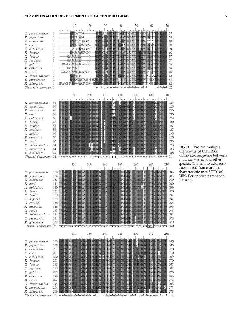
FIG. 3. Protein multiplealignments of the ERK2amino acid sequence betweenS. paramamosain and otherspecies. The amino acid resi-dues in red frame are thecharacteristic motif TEY ofERK. For species names seeFigure 2.
ERK2 IN OVARIAN DEVELOPMENT OF GREEN MUD CRAB 5

labeled RNA probe was 1 ng/mL for hybridization. An anti-DIG antibody was used to react with the DIG-labeled RNAprobe. NBT/BCIP was used to detect the signal by dyeingthe substrates. The hybridized sections were observed andphotographed under an automatic digital microscope (MoticBA600Mot-7.5) and the pictures were retouched with MoticVM V1 Viewer as well as Adobe Photoshop CS5.
Results
Cloning and sequence analysis of Sp-erk2
Full-length cDNA of Sp-erk2 was cloned by 5¢ RACE basedon the EST sequence in the cDNA library and was verifiedthrough head-to-toe PCR. The full-length cDNA of Sp-erk2 is1516 bp, with an 18 bp polyA tail. The ORF contains 1098 bpthat encodes a protein with 365 amino acids. Its 5¢ untrans-lated region (UTR) and 3¢ UTR contain 19 and 399 bp, re-spectively. The GenBank accession number is GQ847862 (Fig.1). The predicted molecular mass of ERK2 is 42 kDa, with atheoretical pI of 5.98. It is a nonsecretary protein since theSingalP 3.0 server showed that the ERK2 lacks an amino-terminal signal peptide. There are 22 predicted phosphory-lation sites in the amino acid sequence with 7 serines, 8threonines, and 7 tyrosines. Conserved domain finder sug-gested that 17–329 amino acid residues are the catalytic do-main of ERK1 and ERK2-like serine/threonine kinases,including an active site, an ATP binding site, a substratebinding site, an activation loop (A-loop), and a kinase in-teraction motif docking site that appears to be conserved inall MAPKs. Moreover, there is a TEY motif located in the A-
loop, in which Thr183 and Tyr185 are dual-phosphorylationsites (Fig. 1).
A phylogenetic tree was constructed by MEGA 4.0 soft-ware using NJ method to investigate the relationship ofERK2 of several species from human to aquatic invertebrates(Fig. 2). The phylogenetic tree shows that mammalian, avian,amphibian, and fish form one cluster; arthropod forms an-other cluster; and these two clusters together finally. Chor-date was relatively distant and echinodermata forms abranch independently. Protein multiple-alignment results(Fig. 3) demonstrate that it is very highly conserved amongERK2 proteins. Sequence of M. japonicus that belongs to crus-tacea shows the highest similarity to Sp-erk2 (94% identity).
Expression of Sp-erk2 in different tissues
Real-time quantitative PCR results (Fig. 4) showed that Sp-erk2 was expressed in all the tissues chosen. However,compared with heart (H), muscle (M), gill (G), brain (B),stomach (ST), hepatopancreas (HP), and intestines (I), ovary(O) had a significant high expression level ( p < 0.05), and itled us to further research on the expression levels of differentdeveloping stages of ovaries.
Expression of Sp-erk2 in different developingstages of ovaries
Sp-erk2 expression pattern in developing ovaries is alsocarried out by real-time quantitative PCR. At least five crabsof each stage were used in the experiment. Data collected(Fig. 5) show that the expression level of Sp-erk2 raised
FIG. 3. (Continued)
6 MA ET AL.

gradually as the ovarian stage increased from stage 1 to stage6 (O1–O6), reaching the highest expression peak at stage 6.Expression level of stage 6 ovary is significantly differentfrom the first four stages ( p < 0.05), while there is no signif-icant difference between stage 6 and stage 5.
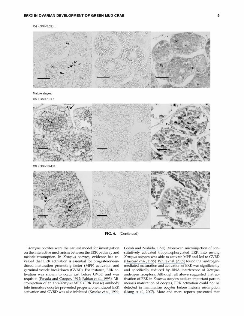
The DIG-labeled RNA probe for Sp-erk2 was obtainedusing Sp-erk2-ISH-F and Sp-erk2-ISH-R. In situ hybridizationhistochemistry was performed to clarify and analyze thedetail of the expression. Positive signals in early developingovary were detected in follicular cells around the oocytes(O1–O4), though there seems to have a subtle expression inoocytes in O3 and O4 stages. In relatively mature and fullymature ovarian tissues of GSI 7.9 and 10.43, Sp-erk2 mRNAwas distributed in both follicular cells and oocytes (Fig. 6).
Discussion
MAPK signal transduction pathway is considered to behighly conserved in all the eukaryotes. The key molecules ofthis pathway show extremely high sequence similarity asevident in the protein multiple alignments. Our resultsdemonstrate that Sp-ERK2 shares 94% identity to the coun-terpart from M. japonicus, which is the only one identified incrustacean, and has above 80% identity to the other ERKsfrom almost all the species examined so far. Our data suggestthat the structure and function of ERK2 may be somewhatdifferent between marine chordate/echinodermata animals
and other species. In addition, ERK signal transductionpathway is famous for the dual-phosphorylation of ERK onthreonine and tyrosine separated by a glutamic acid (Wid-mann et al., 1999), which makes the cascade reaction moreeffective and precise (Farooq and Zhou, 2004). In Sp-ERK2,the dual-phosphorylation sites, Thr183 and Tyr185, are alsofound through bioinformatic analysis.
Although erk2 has been investigated extensively in manydifferent species, the expression patterns of this gene in crabovarian development have not been systematically examinedyet. In this study, the real-time quantitative PCR conse-quences of expression of Sp-erk2 in different green mud crabtissues showed that the expression level in ovaries (relativemature stages) is significantly higher than other tissues. Thisindicated that Sp-erk2 may play a crucial role in the devel-opment and function of crab ovaries. With this hypothesis,we checked the expression pattern in developing ovaries. Asexpected, the expression levels increase along with the de-velopment of ovaries, indicating the function of ERK2 wasrelated to oocyte maturation and ovarian development.Furthermore, the in situ hybridization histochemistry resultsshowed that Sp-erk2 mRNA was detected in crab follicularcells but not in oocytes in early developing stages, while inrelatively mature phases positive signals were found in bothfollicular cells and oocytes, indicating that ERK2 in follicularcells may involve in early steps of oocyte meiosis and ERK2in oocytes may participate in later events.
FIG. 4. Expression of Sp-erk2 in differ-ent tissues. G, gill; H, heart; M, muscle;O, ovary; B, brain; ST, stomach; I, intes-tine; HP, hepatopancreas. Data are pre-sented as means – standard error of themean (SEM) of at least three separateindividuals. Significant differences wereindicated: *p < 0.05.
FIG. 5. Expression of Sp-erk2 in differ-ent stages of developing ovaries. O1,stage 1 (GSI = 0.7 – 0.3); O2, stage 2(GSI = 2.0 – 0.1); O3, stage 3 (GSI = 3.5 –0.2); O4, stage 4 (GSI = 5.2 – 0.2); O5,stage 5 (GSI = 7.6 – 0.6); O6, stage 6(GSI = 10.3 – 0.3). Data are presented asmeans – SEM of at least three separateindividuals. Significant differences wereindicated: a and b were significantlydifferent ( p < 0.05); a/b and ab had nosignificant differences ( p > 0.05). GSI,gonad somatic index.
ERK2 IN OVARIAN DEVELOPMENT OF GREEN MUD CRAB 7

FIG. 6. In situ hybridization results. The first picture of each group is the positive result, the second one is negative control,and the third is HE control. Mature stages’ group showed that Sp-erk2 was distributed in both follicular cells and oocytes,while in follicular cells only, in early stages’ group. The red bars stand for 50mm. The black arrows indicate the positivesignals. OC, oocyte; FC, follicular cell; Nu, nucleus; Yg, yolk globules.
8
Xenopus oocytes were the earliest model for investigationon the interactive mechanism between the ERK pathway andmeiotic resumption. In Xenopus oocytes, evidence has re-vealed that ERK activation is essential for progesterone-in-duced maturation promoting factor (MPF) activation andgerminal vesicle breakdown (GVBD). For instance, ERK ac-tivation was shown to occur just before GVBD and wasrequisite (Posada and Cooper, 1992; Fabian et al., 1993). Mi-croinjection of an anti-Xenopus MEK (ERK kinase) antibodyinto immature oocytes prevented progesterone-induced ERKactivation and GVBD was also inhibited (Kosako et al., 1994;
Gotoh and Nishida, 1995). Moreover, microinjection of con-stitutively activated thiophosphorylated ERK into restingXenopus oocytes was able to activate MPF and led to GVBD(Haccard et al., 1995). White et al. (2005) found that androgen-mediated maturation and activation of ERK was significantlyand specifically reduced by RNA interference of Xenopusandrogen receptors. Although all above suggested that ac-tivation of ERK in Xenopus oocytes took an important part inmeiosis maturation of oocytes, ERK activation could not bedetected in mammalian oocytes before meiosis resumption(Liang et al., 2007). More and more reports presented that
FIG. 6. (Continued)
ERK2 IN OVARIAN DEVELOPMENT OF GREEN MUD CRAB 9
activation of ERK in follicular cells was probably necessary formeiosis resumption in mammalian oocytes (Zhang et al., 2007;Fan et al., 2009). Studies on mouse and porcine oocytes showthat ERK activation in cumulus-enclosed oocytes (CEOs) isinitiated before GVBD and is essential for gonadotropin-induced GVBD (Sun et al., 2002; Li et al., 2008; Kolesarovaet al., 2010). To observe more clearly, Liang et al. (2005) usedultracentrifugation method to move the opaque lipid dropletsaside the porcine oocyte and found that activation of ERK incumulus cells, but not in oocytes, was essential for GVBD inCEOs, while phosphorylation of ERK in oocytes occurred afterGVBD. Similar process was also found in sheep (Cecconi et al.,2008). In our experiments, Sp-erk2 mRNA was found in fol-licular cells only in early stages of ovarian development,which is similar to the research of mammal cited previously.Thus, we conjectured that it may be related to GVBD in a waysimilar to that reported in mammals, although further ex-periments need to be done to prove it.
Gross et al. (2000) found that U0126-treated Xenopus oocytesfail to form metaphase I spindles and appear to enter S-phaseinstead of M II, indicating that ERK2 activation is required forsuppression of S-phase and entry into M II. In mammalianoocytes, it is reported that ERK is indispensable for the post-GVBD events, for instance, spindle organization; M I–M IItransition; and M II arrest (Fan and Sun, 2004; Liang et al.,2007). Mature mammalian oocytes are arrested at metaphase ofM II and ovulated for fertilization (Liang et al., 2007; Dugga-vathi and Murphy, 2009). However, there are some differencesin several invertebrates such as crab where the fertilizationhappens at M I arrest when oocytes are ovulated, which meansthat crab oocytes are ovulated just at M I arrest (Chen et al.,2004). In starfish, which also spawn M I arrest oocytes, M Ipause was maintained by ERK as well as low intracellular pH(*7.0) as recorded by Usui et al. (2008). Moreover, the ERKtransduction pathway was reported for involving in M I arrest(Usui et al., 2008), M I–M II transition (Tachibana et al., 2000),pronucleus stage arrest, and the eventual fate decision ofstarfish oocytes (Kishimoto, 2004; Sadler et al., 2004; Mori et al.,2006). Based on the information just outlined and on our ex-periments, we deduce that ERK in crab oocytes may participatein events before or around M I since the Sp-erk2 mRNA wasstrongly distributed in oocytes in relatively mature and matureovarian tissues of GSI 7.9 and 10.43. To obtain thorough in-formation on the role of ERK signal transduction pathwayduring crab ovarian development, an ERK knockdown or in-hibition experiment should be taken to see whether the de-velopment of ovary is blocked or influenced.
To summarize, we have cloned and characterized the Sp-erk2 from S. paramamosain, which is the first such cloning incrustaceans to our knowledge. Our experiments indicatedthat ERK2 may play a crucial role on crab ovary develop-ment. According to our experiments, we concluded that thefunction of ERK in crab ovarian development is similar tomammalian oocytes rather than to Xenopus oocytes. The re-sults could provide essential information on the regulatorymechanism of crab or even crustacean ovarian development.
Acknowledgments
This work was supported by the National Natural ScienceFoundation of China (Grant No. 30571430 and 31072200) andInnovation Team foundation of Jimei University (2010A001).
The authors thank Mr. Ion Beldorth (Department of Chem-istry & Biochemistry, Texas State University, San Marcos,TX) for critical reading of the article.
Disclosure Statement
No competing financial interests exist.
References
Akanuma, T., and Nishida, H. (2004). Ets-mediated brain in-duction in embryos of the ascidian Halocynthia roretzi. DevGenes Evol 214, 1–9.
Boulton, T.G., Nye, S.H., Robbins, D.J., Ip, N.Y., Radziejewska, E.,Morgenbesser, S.D., Depinho, R.A., Panayotatos, N., Cobb,M.H., and Yancopoulos, G.D. (1991). ERKs: a family of protein-serine/threonine kinases that are activated and tyrosine phos-phorylated in response to insulin and NGF. Cell 65, 663–675.
Cecconi, S., Mauro, A., Capacchietti, G., Berardinelli, P., Berna-bo, N., Di Vincenzo, A., Mattioli, M., and Barboni, B. (2008).Meiotic maturation of incompetent prepubertal sheep oocytesis induced by paracrine factor (s) released by gonadotropin-stimulated oocyte-cumulus cell complexes and involves mi-togen-activated protein kinase activation. Endocrinology 149,
100–107.Chambon, J.P., Nakayama, A., Takamura, K., Mcdougall, A.,
and Satoh, N. (2007). ERK-and JNK-signalling regulate genenetworks that stimulate metamorphosis and apoptosis in tailtissues of ascidian tadpoles. Development 134, 1203–1219.
Chang, L., and Karin, M. (2001). Mammalian MAP kinase sig-nalling cascades. Nature 410, 37–40.
Chen, J., Kang, X., Li, S., Wang, G., and Ye, H. (2004). Nucleichanges during fertilization in Scylla serrata. Xiamen Da XueXue Bao Zi Ran Ke Xue Ban Xiamen Chin Ed 43, 688–692.
Cowan, K.J., and Storey, K.B. (2003). Mitogen-activated proteinkinases: new signaling pathways functioning in cellular re-sponses to environmental stress. J Exp Biol 206, 1107–1115.
Duggavathi, R., and Murphy, B.D. (2009). Ovulation signals.Science 324, 890–891.
Fabian, J.R., Morrison, D.K., and Daar, I.O. (1993). Requirementfor Raf and MAP kinase function during the meiotic matura-tion of Xenopus oocytes. J Cell Biol 122, 645–652.
Fan, H.Y., Liu, Z., Shimada, M., Sterneck, E., Johnson, P.F.,Hedrick, S.M., and Richards, J. (2009). MAPK3/1 (ERK1/2) inovarian granulosa cells are essential for female fertility. Sci-ence 324, 938–941.
Fan, H.Y., and Sun, Q.Y. (2004). Involvement of mitogen-acti-vated protein kinase cascade during oocyte maturation andfertilization in mammals. Biol Reprod 70, 535–547.
Farooq, A., and Zhou, M.M. (2004). Structure and regulation ofMAPK phosphatases. Cell Signal 16, 769–779.
Feld, M., Dimant, B., Delorenzi, A., Coso, O., and Romano, A.(2005). Phosphorylation of extra-nuclear ERK/MAPK is re-quired for long-term memory consolidation in the crab Chas-magnathus. Behav Brain Res 158, 251–261.
Gotoh, Y., and Nishida, E. (1995). Activation mechanism andfunction of the MAP kinase cascade. Mol Reprod Dev 42, 486–492.
Gross, S.D., Schwab, M.S., Taieb, F.E., Lewellyn, A.L., Qian,Y.W., and Maller, J.L. (2000). The critical role of the MAPkinase pathway in meiosis II in Xenopus oocytes is mediatedby p90Rsk. Curr Biol 10, 430–438.
Haccard, O., Lewellyn, A., Hartley, R.S., Erikson, E., and Maller,J.L. (1995). Induction of Xenopus oocyte meiotic maturation byMAP kinase. Dev Biol 168, 677–682.
10 MA ET AL.

Iakovleva, N.V., Gorbushin, A.M., and Zelck, U.E. (2006). Partialcharacterization of mitogen-activated protein kinases (MAPK)from haemocytes of the common periwinkle, Littorina littorea(Gastropoda: Prosobranchia). Fish Shellfish Immun 20, 665–668.
Islam, M., Kodama, K., and Kurokora, H. (2010). Ovarian de-velopment of the mud crab Scylla paramamosain in a tropicalMangrove Swamps, Thailand. J Sci Res 2, 380–389.
Junttila, M.R., Li, S.P., and Westermarck, J. (2008). Phosphatase-mediated crosstalk between MAPK signaling pathways in theregulation of cell survival. FASEB J 22, 954–965.
Keenan, C.P. (1999). Aquaculture of the mud crab, genus Scylla-past, present and future. In Mud Crab Aquaculture and Biology,vol 78. C.P. Keenan and A. Blackshaw, eds. (ACIAR Pro-ceedings, China), pp. 9–13.
Kishimoto, T. (2004). More than G1 or G2 arrest: useful starfishoocyte system for investigating skillful MAP kinase. Biol Cell96, 241–244.
Kolesarova, A., Sirotkin, A., Roychoudhury, S., and Massanyi, P.(2010). Effect of dbcAMP, IGF-I and oxytocin treatment onprotein kinase A, transcription factor CREB-1 and ERK 1, 2related MAP kinase: role in porcine sexual maturation. AssamUniv J Sci Technol 4, 1–8.
Kosako, H., Gotoh, Y., and Nishida, E. (1994). Requirement forthe MAP kinase kinase/MAP kinase cascade in Xenopus oo-cyte maturation. EMBO J 13, 2131–2138.
Kumano, M., Carroll, D.J., Denu, J.M., and Foltz, K.R. (2001).Calcium-mediated inactivation of the MAP kinase pathway insea urchin eggs at fertilization. Dev Biol 236, 244–257.
Lambert, J., and Nagy, L. (2003). The MAPK cascade in equallycleaving spiralian embryos. Dev Biol 263, 231–241.
Li, M., Liang, C.G., Xiong, B., Xu, B.Z., Lin, S.L., Hou, Y., Chen,D.Y., Schatten, H., and Sun, Q.Y. (2008). PI3-kinase and mi-togen-activated protein kinase in cumulus cells mediate EGF-induced meiotic resumption of porcine oocyte. Domest AnimEndocrin 34, 360–371.
Li, S.J., and Wang, G.Z. (2001). Studies on reproductive biologyand artificial culture of mud crab, Scylla Serrata. Xiamen DaXue Xue Bao Zi Ran Ke Xue Ban Xiamen Chin Ed 40, 552–565.
Liang, C.G., Huo, L.J., Zhong, Z.S., Chen, D.Y., Schatten, H., andSun, Q.Y. (2005). Cyclic adenosine 3¢, 5¢-monophosphate-dependent activation of mitogen-activated protein kinase incumulus cells is essential for germinal vesicle breakdown ofporcine cumulus-enclosed oocytes. Endocrinology 146, 4437–4444.
Liang, C.G., Su, Y.Q., Fan, H.Y., Schatten, H., and Sun, Q.Y.(2007). Mechanisms regulating oocyte meiotic resumption:roles of mitogen-activated protein kinase. Mol Endocrinol 21,
2037–2055.Lo, T.S., Cui, Z., Mong, J.L.Y., Wong, Q.W.L., Chan, S.M., Kwan,
H.S., and Chu, K.H. (2007). Molecular coordinated regulationof gene expression during ovarian development in the penaeidshrimp. Mar Biotechnol 9, 459–468.
Mori, M., Hara, M., Tachibana, K., and Kishimoto, T. (2006).p90Rsk is required for G1 phase arrest in unfertilized starfisheggs. Development 133, 1823–1830.
Orton, R.J., Sturm, O.E., Vyshemirsky, V., Calder, M., Gilbert,D.R., and Kolch, W. (2005). Computational modelling of thereceptor-tyrosine-kinase-activated MAPK pathway. Biochem J392, 249–261.
Posada, J., and Cooper, J.A. (1992). Requirements for phos-phorylation of MAP kinase during meiosis in Xenopus oocytes.Science 255, 212–215.
Qi, M., and Elion, E.A. (2005). MAP kinase pathways. J Cell Sci118, 3569–3572.
Sadler, K.C., Yuce, O., Hamarato, F., Verge, V., Peaucellier, G.,and Picard, A. (2004). MAP kinases regulate unfertilized eggapoptosis and fertilization suppresses death via Ca2 + signal-ing. Mol Reprod Dev 67, 366–383.
Schaeffer, H.J., and Weber, M.J. (1999). Mitogen-activated pro-tein kinases: specific messages from ubiquitous messengers.Mol Cell Biol 19, 2435–2444.
Schmitt, A., and Nebreda, A.R. (2002). Signalling pathways inoocyte meiotic maturation. J Cell Sci 115, 2457–2459.
Sharma, S.K., and Carew, T.J. (2004). The roles of MAPK cas-cades in synaptic plasticity and memory in Aplysia: facilitatoryeffects and inhibitory constraints. Learn Mem 11, 373–378.
Steinmetz, R., Wagoner, H.A., Zeng, P., Hammond, J.R., Hannon,T.S., Meyers, J.L., and Pescovitz, O.H. (2004). Mechanismsregulating the constitutive activation of the extracellular signal-regulated kinase (ERK) signaling pathway in ovarian cancerand the effect of ribonucleic acid interference for ERK1/2 oncancer cell proliferation. Mol Endocrinol 18, 2570–2582.
Sun, Q.Y., Wu, G.M., Lai, L., Bonk, A., Cabot, R., Park, K.W.,Day, B.N., Prather, R.S., and Schatten, H. (2002). Regulation ofmitogen-activated protein kinase phosphorylation, microtu-bule organization, chromatin behavior, and cell cycle pro-gression by protein phosphatases during pig oocytematuration and fertilization in vitro. Biol Reprod 66, 580–588.
Tachibana, K., Tanaka, D., Isobe, T., and Kishimoto, T. (2000). c-Mos forces the mitotic cell cycle to undergo meiosis II toproduce haploid gametes. Proc Natl Acad Sci U S A 97, 14301–14306.
Tang, X., Shen, Q., Hu, C., and Zhan, G. (2009). Histologicalstudies on the early development of the ovaries Scylla para-mamosain. Tran Oceanol Limnol Qingdao Chin Ed 03, 72–78.
Usui, K., Hirohashi, N., and Chiba, K. (2008). Involvement ofmitogen-activating protein kinase and intracellular pH in theduration of the metaphase I (MI) pause of starfish oocytesafter spawning. Dev Growth Differ 50, 357–364.
Wang, Y., Chen, Y., Han, K., Zou, Z., and Zhang, Z. (2011). Avasa gene from green mud crab Scylla paramamosain and itsexpression during gonadal development and gametogenesis.Mol Biol Rep DOI: 10.1007/s11033-011-1220-5.
White, S.N., Jamnongjit, M., Gill, A., Lutz, L.B., and Hammes,S.R. (2005). Specific modulation of nongenomic androgensignaling in the ovary. Steroids 70, 352–360.
Widmann, C., Gibson, S., Jarpe, M.B., and Johnson, G.L. (1999).Mitogen-activated protein kinase: conservation of a three-ki-nase module from yeast to human. Physiol Rev 79, 143–180.
Wonglapsuwan, M., Miyazaki, T., Loongyai, W., and Chotigeat,W. (2010). Characterization and biological activity of the ri-bosomal protein L10a of the white shrimp: Fenneropenaeusmerguiensis De Man during vitellogenesis. Mar Biotechnol 12,
230–240.Zeng, H., Huang, J., Li, W., Huang, H., and Ye, H. (2011).
Identification of differentially expressed genes in the thoracicganglion of the mud crab, Scylla paramamosain during ovarianmaturation. Mar Biol Res 7, 617–622.
Zhang, M., Xia, G., Zhou, B., and Wang, C. (2007). Gonado-tropin-controlled mammal oocyte meiotic resumption. FrontBiosci 12, 282–296.
Zhang, Z., Shen, B., Wang, Y., Chen, Y., Wang, G., Lin, P., andZou, Z. (2010). Molecular cloning of proliferating cell nuclearantigen and its differential expression analysis in the devel-oping ovary and testis of penaeid shrimp Marsupenaeus japo-nicus. DNA Cell Biol 29, 163–170.
Zhang, Z., Wu, R., Mok, H.O., Wang, Y., Poon, W., Cheng, S.H.,and Kong, R. (2003). Isolation, characterization and expression
ERK2 IN OVARIAN DEVELOPMENT OF GREEN MUD CRAB 11

analysis of a hypoxia-responsive glucose transporter genefrom the grass carp, Ctenopharyngodon idellus. Eur J Biochem270, 3010–3017.
Zou, Z., Zhang, Z., Wang, Y., Chen, J., Jia, X., Wang, S., and Lin,P. (2007). Normalized cDNA Library Construction of Ovary ofScylla serrata. J Fujian Fish Xiamen Chin Ed 12, 1–4.
Zou, Z., Zhang, Z., Wang, Y., Han, K., Fu, M., Lin, P., and Xiwei,J. (2011). EST analysis on the gonad development related or-gans and microarray screen for differentially expressed genesin mature ovary and testis of Scylla paramamosain. CompBiochem Phys D 6, 150–157.
Zou, Z.H., Zhang, Z.P., Wang, Y.L., Chen, J.M., Jia, X.W.,Wang, S.H., and Lin, P. (2009). The construction of normal-ized cDNA library and preliminary EST analysis from thegonad development and sexual differentiation related organsof Scylla serrata. J Trop Oceanogr Guangzhou Chin Ed 28,
89–94.
Address correspondence to:Yilei Wang, Ph.D.
Fisheries CollegeJimei UniversityXiamen 361021
China
E-mail: [email protected]
Ziping Zhang, Ph.D.Department of Biological Science
Seton Hall UniversitySouth Orange, NJ 07079
E-mail: [email protected]
Received for publication September 24, 2011; received inrevised form January 31, 2012; accepted February 1, 2012.
12 MA ET AL.