Automatic Resting Tremor Assessment in Parkinson's Disease ...
Upload
independentCategory
view
0download
0
Altered zinc transport disrupts mitochondrialprotein processing/import in fragile X-associatedtremor/ataxia syndrome
Eleonora Napoli1, Catherine Ross-Inta1, Sarah Wong1, Alicja Omanska-Klusek1,
Cedrick Barrow1, Christine Iwahashi2, Dolores Garcia-Arocena2, Danielle Sakaguchi1,
Elizabeth Berry-Kravis5, Randi Hagerman3,4, Paul J. Hagerman2,4 and Cecilia Giulivi1,∗
1Department of Molecular Biosciences, School of Veterinary Medicine, 2Department of Biochemistry
and Molecular Medicine, 3Department of Pediatrics and 4MIND Institute, School of Medicine, University of California
Davis, Davis, CA 95616, USA and 5Department of Pediatrics, Neurological Sciences, and Biochemistry, Rush
University Medical Center, Chicago, IL 60612, USA
Received January 14, 2011; Revised and Accepted May 6, 2011
Fragile X-associated tremor/ataxia syndrome (FXTAS) is a late-onset neurodegenerative disorder that affectsindividuals who are carriers of small CGG premutation expansions in the fragile X mental retardation 1(FMR1) gene. Mitochondrial dysfunction was observed as an incipient pathological process occurring inindividuals who do not display overt features of FXTAS (1). Fibroblasts from premutation carriers hadlower oxidative phosphorylation capacity (35% of controls) and Complex IV activity (45%), and higherprecursor-to-mature ratios (P:M) of nDNA-encoded mitochondrial proteins (3.1-fold). However, fibroblastsfrom carriers with FXTAS symptoms presented higher FMR1 mRNA expression (3-fold) and lower ComplexV (38%) and aconitase activities (43%). Higher P:M of ATPase b-subunit (ATPB) and frataxin were alsoobserved in cortex from patients that died with FXTAS symptoms. Biochemical findings observed inFXTAS cells (lower mature frataxin, lower Complex IV and aconitase activities) along with common phenoty-pic traits shared by Friedreich’s ataxia and FXTAS carriers (e.g. gait ataxia, loss of coordination) are consist-ent with a defective iron homeostasis in both diseases. Higher P:M, and lower ZnT6 and mature frataxinprotein expression suggested defective zinc and iron metabolism arising from altered ZnT proteinexpression, which in turn impairs the activity of mitochondrial Zn-dependent proteases, critical for theimport and processing of cytosolic precursors, such as frataxin. In support of this hypothesis, Zn-treatedfibroblasts showed a significant recovery of ATPB P:M, ATPase activity and doubling time, whereas Znand desferrioxamine extended these recoveries and rescued Complex IV activity.
INTRODUCTION
Fragile X-associated tremor/ataxia syndrome (FXTAS) is alate-onset neurodegenerative disorder (2–4) that affects indi-viduals who are carriers of premutation expansions (55–200CGG repeats) in the 5′untranslated region (5′UTR) of thefragile X mental retardation 1 (FMR1; OMIM ∗309550)gene. FXTAS typically affects carriers (males . females)over 50 years of age, with core features of action tremor and
gait ataxia, but also (more variably) parkinsonism, executivedysfunction, cognitive decline, neuropathy and autonomicdysfunction. The core neuropathologic feature of FXTAS isthe presence of FMR1 mRNA–containing inclusions in thenuclei of neurons and astrocytes of affected individuals(5–7), consistent with the ‘RNA toxicity’ model of pathogen-esis (3). Larger expansions (.200 CGG repeats; fullmutation) generally result in transcriptional silencing andabsence of the FMR1 mRNA and protein product (8–11),
∗To whom correspondence should be addressed at: Department of Molecular Biosciences, University of California Davis, 1120 Haring Hall,One Shields Avenue, Davis, CA 95616, USA. Tel: +1 5307548603; Fax: +1 5307524698; Email: [email protected]
# The Author 2011. Published by Oxford University Press. All rights reserved.For Permissions, please email: [email protected]
Human Molecular Genetics, 2011, Vol. 20, No. 15 3079–3092doi:10.1093/hmg/ddr211Advance Access published on May 10, 2011

leading to fragile X syndrome, the most common heritableform of cognitive impairment and leading known form ofautism. Individuals with the full mutation are not at-risk forFXTAS or the fragile X-associated primary ovarian insuffi-ciency, as in these cases the toxic FMR1 mRNA is absent orpresent at low levels.
Given that several symptoms of FXTAS, including gaitataxia, white matter disease, dysautonomia, peripheral neuro-pathy, weakness/exercise intolerance and neuropsychiatricinvolvement, overlap those of mitochondrial respiratoryenzyme chain enzyme deficiencies, we recently investigatedand identified mitochondrial dysfunction (MD) in both cul-tured dermal fibroblasts and brain samples from individualswith the premutation with FXTAS symptoms (PS) andwithout FXTAS symptoms (premutation asymptomatic orPA) with CGG repeat length at the low end of the premutationrange (1). Our study resulted in several important conclusions:(i) decreased nicotinamide adenine dinucleotide- and flavinadenine dinucleotide-linked oxygen uptake rates and uncou-pling between electron transport and synthesis of ATP; (ii) alower expression of mitochondrial proteins preceded clinicalinvolvement, even in younger carriers with smaller CGGrepeat lengths; (iii) the CGG repeat size required for alteredmitochondrial protein expression was also lower than thelower bound of CGG repeat size associated with intranuclearinclusions observed to date in individuals who died withFXTAS, suggesting that MD is an incipient pathologicalprocess occurring in individuals who do not display overtfeatures of FXTAS; (iv) for a given CGG repeat, MD precededthe increase in oxidative/nitrative stress damage indicatingthat the latter is a late event (1).
Thus, the goals of the present study were to elucidate themechanisms underlying the MD observed in premutation car-riers of comparable age and CGG repeat expansions, and ifthis mechanism would show differences between premutationcarriers with (PS) and without (PA) symptoms of FXTAS thatwould explain the clinical phenotype.
RESULTS
Clinical characteristics of individuals with FXTAS withand without symptoms
The subjects included in this study were all males referred tothe Chicago or UC Davis clinics either because they presentedwith FXTAS symptoms, including tremor and/or ataxia, orwere either premutation carriers ascertained through familieswith a fragile X syndrome proband, or controls in a similarage group range. Individuals contributing skin biopsies wereparticipants in a multicenter study to characterize neurologicalfindings in premutation carriers. Primary cultured skin fibro-blasts from the skin biopsies were obtained from 14 malepremutation carriers: 8 with FXTAS symptoms (identified inTables as PS) and 6 without FXTAS symptoms (or asympto-matic, identified in Tables as PA) and 7 controls(Table 1 and Supplementary Material, Table S1). There wasno statistical difference in age between the control group(64+ 2 years old) and the premutation carriers (PA: 71+ 2and PS: 68+ 3 years old; Table 1). For the fibroblastsamples, there was a significant difference between CGG
repeat numbers between PS and PA (96+ 7 and 69+ 2,mean+SEM, respectively; P ¼ 5 × 10²³; Table 1), andboth groups were significantly different from controls (CGGrepeat for controls 27+ 2 with P ¼ 2 × 1028 to PA andP ¼ 4 × 1027 to PS; Table 1). On average, there was a gapof 9+ 2 years between the onset of FXTAS symptoms andthe time of the skin biopsy.
It could be argued that primary dermal fibroblasts do not con-stitute an appropriate model system to address mitochondrialfunction in FXTAS because the level of FMR1 transcript over-expression in fibroblasts may not necessarily mimic that inneurons. The FMR1 transcript levels in PA and PS cells were97+ 3% (P ¼ 0.97) and 350+ 50% (P ¼ 0.001), respectively,of control values (Table 1). These results indicated that primaryfibroblasts in culture from PS do over-express FMR1 transcriptand that the levels of the expanded CGG-repeat FMR1 mRNAare similar to those observed in FXTAS neurons (from 2- to8-fold in premutation carriers; 12–14). Moreover, there isincreasing evidence that the underlying pathogenesis ofFXTAS also results in additional systemic [non-centralnervous system (CNS)-related] dysregulation, including the for-mation of intranuclear inclusions, in cells of the peripheralnervous system, and in diverse organ tissues (15,16; C.G. andP.J.H., unpublished data).
Lower oxidative phosphorylation capacity in culturedfibroblasts from premutation carriers is linked to lowerComplex IV and/or V activities
The rates of oxygen uptake by primary cultured fibroblastsobtained from PA, PS and controls were obtained in a glucose-containing media supplemented with oligomycin (State 4 ornon-phosphorylating conditions) and subsequently withFCCP, an uncoupler (State 3u). In PA fibroblasts, no statisticaldifference with respect to controls was observed in the oxygenuptake in State 4 (Table 2); however, PS cells exhibited a2.6-fold increase (P ¼ 0.05; Table 2). The rates of oxygen
Table 1. Clinical characteristics of the individuals from which the culturedprimary dermal fibroblasts were obtained and utilized in this study
Group (n) Age(years)
CGGrepeats
Stage FMR1 transcript level(% of control)
Control(n ¼ 7)
64+2 27+2 0 100
PA (n ¼ 6) 71+2 69+2 0 97+3P-value to
controls0.042 2 × 1028 –
PS (n ¼ 8) 68+3 96+7 3.8+0.2 350+50P-value to
controls4 × 1027 0.001
P-value toPA
5 × 10²³ 0.002
Stages were defined as indicated previously (1). The CGG repeats differ fromthose previously published (1) because these were determined from the gDNAfrom dermal fibroblast grown in culture (not from original biopsy) under theconditions indicated under Materials and Methods. All individuals were maleand further clinical data of these individuals were provided underSupplementary Material, Table S1. The level of FMR1 transcript (normalizedto beta-2-microglobulin) was obtained from primary dermal fibroblasts fromeach group of individuals as described under Materials and Methods.
3080 Human Molecular Genetics, 2011, Vol. 20, No. 15

uptake of PA or PS cells in State 3u were half of control values(P ¼ 0.03 and 0.05; Table 2). The respiratory control ratio(RCR) evaluates the coupling between electron transfer andoxidative phosphorylation (OXPHOS). Fully uncoupled mito-chondria have RCR values of 1 (17,18). Control cells werefully coupled with a respiratory control ratio in the presenceof uncoupler (RCRu) of 11+ 2, whereas PA cells had lowerRCRu, but not significantly different from controls(Table 2). The RCRu of PS cells was 3.1+ 0.9 indicatingthat the mitochondria were substantially uncoupled(Table 2). The higher uncoupling (lower RCRu) observed inPS was the result of both an increase in State 4 oxygenuptake and a decrease in State 3u (Table 2). Of note, thehigher uncoupling of electron transport with ATP synthesiswas statistically significant only in cells from PS individuals,suggesting a segregation of the severity of MD with the pres-ence of FXTAS symptoms.
To evaluate whether the decreased mitochondrial functionhad an impact in the glucose consumed by the cells viaOXPHOS, the rate of oligomycin-sensitive oxygen uptake ina glucose-supplemented media was evaluated in control, PSand PA cells. In PA cells, the rate of glucose uptake inOXPHOS was 41% of controls (P ¼ 0.02), whereas in PSwas 30% of controls (P ¼ 0.02). These results indicated thatpremutation cells had a lower rate of glucose oxidation viaOXPHOS, suggesting that a higher proportion of their ATPis derived from anaerobic glycolysis especially in PS cells,consistent with the presence of symptoms in these individuals.
No significant changes in either mtDNA copy number ormitochondrial mass was found between cells from premutationcarriers and controls indicating that the lower oxygen uptakerate in State 3u and glucose consumption via OXPHOSfound in premutation cells could not be explained by lowermitochondrial number (estimated by means of mtDNAcontent or citrate synthase activity; Supplementary Material,Table S2). In fact, the mtDNA copy number in PS cells was1.34-fold of controls (P ¼ 0.008), suggesting a cellularresponse to overcome oxidative stress as described in otherbiological models (19–23). To evaluate increased oxidativestress and considering that mitochondria are the main intra-cellular source of reactive oxygen species (ROS; 24), therate of hydrogen peroxide production was evaluated incontrol and PS fibroblasts using succinate (in the presence of
rotenone and antimycin) and NADH (in the presence of rote-none). This experimental design allowed testing for ROS pro-duction by Complex III and Complex I, respectively. Ourresults indicated that the ROS production was significantlyenhanced [2.4-fold; controls ¼ 0.08+ 0.02 and PS: 0.19+0.05 nmol H2O2 × (min × 107 cells)21; P , 0.05] in PScells at the level of Complex I, whereas no significant differ-ence was obtained at Complex III.
To determine whether the lower OXPHOS capacityaccompanied by a higher ROS rate observed in cells from pre-mutation carriers was the result of a defect at any of the elec-tron transport chain Complexes, the activities of each weretested (Table 3). In premutation cells, the activity of cyto-chrome c oxidase (Complex IV) was 33% (PA) and 56%(PS) of control values (P ¼ 0.01 and 0.05, respectively;Table 3). In addition, in PS cells only, the activity ofATPase (Complex V) was 38% of the controls (P ¼ 0.02;Table 3) and significantly different from PA (53% withP ¼ 0.02). The lower Complex IV activity in cells from pre-mutation carriers was consistent with the lower rates of State
Table 2. Rates of oxygen uptake in State 3u and State 4, RCRu and glucose consumed in OXPHOS by primary PA, PS and control fibroblasts
Group Oxygen uptake rate (nmolO2 × (min × 106 cells)21)
RCRu Glucose consumed in OXPHOS (nmol × (min × 106 cells)21)
Basal State 4 State 3u
Control (n ¼ 5) 4.7+0.6 1.0+0.2 9+1 11+2 0.54+0.09PA (n ¼ 4) 2.8+0.6 1.4+0.7 4.0+0.8 5+1 0.22+0.03
P-value to controls – – 0.03 – 0.02PS (n ¼ 6) 5+1 2.6+0.6 5.1+0.9 3.1+0.9 0.16+0.07
P-value to controls – 0.05 0.05 0.05 0.02P-value to PA – – – – –
Oxygen uptake rates were taken at 228C with cells suspended in high glucose DMEM buffer (25 mM in glucose; basal) supplemented with 5 mg/ml oligomycin(State 4), subsequently supplemented with 5 nM FCCP (State 3u). RCRu was calculated by dividing the State 3u rates by the State 4 rate. Glucose consumption wasobtained as the rate of oxygen consumption sensitive to oligomycin divided by 6 (6 oxygen consumed per glucose). The P-values were obtained from unpairedtwo-tailed t-test. Other experimental details were described under Materials and Methods.
Table 3. Activities of mitochondrial electron transport chain Complexes incontrol, PA and PS primary fibroblasts
Group Complex activities normalized to citrate synthase(mean+SEM)Complex I Complexes
II–IIIComplexIV
ComplexV
NQR NFR SCCR CCO ATPase
Controls(n ¼ 5)
8+2 6.1+0.7 0.19+0.03 1.8+0.3 2.1+0.5
PA (n ¼ 4) 8+2 7+2 0.27+0.07 0.6+0.1 1.5+0.2P-value tocontrols
– – – 0.01 –
PS (n ¼ 6) 6+1 6+2 0.13+0.01 1.0+0.2 0.8+0.2P-value tocontrols
– – – 0.05 0.02
P-value toPA
– – – – 0.02
Individual values represent the mean of duplicate or triplicate experiments. TheP-values were obtained from Student t-test. Other experimental details weredescribed under Materials and Methods.NFR, NADH-ferricyanide oxidoreductase; NQR, NADH-decylubiquinoneoxidoreductase.
Human Molecular Genetics, 2011, Vol. 20, No. 15 3081

3u oxygen uptake and glucose consumption via OXPHOSobserved above (Table 2). More importantly, the lowerATPase activity in PS cells segregated with the presence ofFXTAS symptoms.
No other mean Complex activity in premutation cellsresulted in a statistical significant difference from controls.However, three out of five PS samples showed lowerComplex I (below 95% CI) and two out of four had lowerComplex II–III activities, whereas in PA samples only onehad low Complex I activity and none showed low succinatecytochrome c reductase (SCCR) activity, suggesting that Com-plexes other than IV and V were also affected in premutationcarriers with symptoms than in those without.
Defective import/processing of mitochondrial precursorsin FXTAS fibroblasts
The lower Complex IV (PS and PA) and V (PS only) activitiesobserved in premutation cells could result from one or both ofthe following processes: (i) decreased steady-state levels ofcertain mitochondrial subunits resulting from decreased syn-thesis, increased proteolysis or a combination of both pro-cesses; (ii) a defective processing, assembly and/or import ofnuclear-encoded proteins resulting in lower mitochondrialactivities.
To ascertain if the lower Complex activities were the resultof lower protein expression, western blots were performed toevaluate the levels of several subunits from Complexes I, IVand V (Supplementary Material, Table S3). For all 10 proteinstested, 6 in PA and 7 in PS were significantly lower comparedwith controls (60+ 7 and 57+ 5% of controls for PA and PS,respectively). However, the lack of correlation betweenComplex activities (Table 3) and subunit protein expressionin premutation cells (Supplementary Material, Table S3) indi-cated that the steady-state levels of the individual Complexsubunits did not translate (in most of the cases) into activityloss. The lack of a more consistent correlation seemed toreflect a defective processing of precursor proteins, importor assembly of Complexes resulting in higher instability ofthe assembled Complexes, rather than an increased degra-dation or decreased synthesis of the individual subunits.
Considering that the rates of the import/processing of mito-chondrial precursor proteins (or preproteins) are generally oneorder of magnitude faster than their rates of degradation in thecytosol (25,26), a defect at the processing and/or import levelsmay result in the accumulation of precursor proteins in thecytosol. To this end, we evaluated the mature and precursorprotein expression of ATPB (subunit of Complex V), cyto-chrome c oxidase (CCO4, subunit IV of Complex IV) andNADH dehydrogenase (ubiquinone) Fe-S protein 4, 18 kDa(NDUFS4, subunit of Complex I) in control and premutationcells (Table 4). Our experimental conditions do not allow dis-cerning MW differences of ,5 kDa between precursor andmature mitochondrial proteins, thus from the 10 proteinstested (Supplementary Material, Table S3), only NDUFS4,CCO4 and ATPB fulfilled this requirement.
The ATPB precursor-to-mature ratio (P:M) was signifi-cantly increased in PA (4.6-fold of controls; P ¼ 0.03) andPS (2.5-fold of controls; P ¼ 0.01; Table 4 and Fig. 1A). Con-sistent with the results obtained on ATPB, an altered P:M was
also observed for CCO4 and NDUFS4 in premutation cells.The P:M of CCO4 (Fig. 1A and Table 4) and NDUFS4(Table 4) was significantly increased in both PA (CCO4:3.5-fold of controls; P ¼ 0.04; NDUFS4: 2-fold of controls;P ¼ 0.024) and PS (CCO4: 3.8-folds; P ¼ 0.035; NDUFS4:2.3-fold of controls; P ¼ 0.014) (Table 4). The expressionlevels of CCO2, an mtDNA-encoded subunit, were also exam-ined in dermal fibroblasts from controls and PS carriers. Asexpected for a defect in the import/processing step, no differ-ences were found in the expression levels of this proteinbetween controls and PS (normalized to actin;controls ¼ 0.35+ 0.06; PS ¼ 0.34+ 0.1; SupplementaryMaterial, Fig. S1 and Table S3).
In PS cells, the higher P:M, the lower mature ATPB content(42+ 14% of controls; P ¼ 0.02) and the lower ATPaseactivity (38+ 10% of controls; P ¼ 0.02) suggested that theprocessing of ATPB (as well as other proteins synthesized inthe cytosol, i.e. NDUFS4 and CCO4) was limiting, resultingin dysfunctional mitochondria. Although similar conclusionscould be reached with PA cells, the relatively lower increasein the P:M in conjunction with a relatively higher content ofmature ATPB (62+ 13% of controls; P ¼ 0.04) comparedwith PS cells could explain the lack of ATPase activitydecline in PA cells.
It could be inferred that changes in P:M may be attributed tothe age difference between groups since premutation patientswere older than controls (77+ 3 and 58+ 4; P ¼ 0.002;Table 5); however, P:M as well as the mature and precursorATPB levels decreased significantly with the CGG repeatexpansion [as reported before (1)] but not with age in bothcontrols and PS (Supplementary Material, Fig. S2).
Defective import/processing of ATPB and frataxin inbrains from patients that died with FXTAS symptoms
To confirm the results obtained with fibroblasts, the P:M levelsof ATPB were analyzed in brain samples from patients thatdied with FXTAS symptoms (clinical characteristics ofpatients in Table 5 and Supplementary Material, Table S4).The abnormal ATPB P:M observed in premutation carrierswas also detected in frontal cortex samples (Fig. 1B), mitigat-ing concerns that the foregoing observations may reflect eitheran exclusive property of dermal fibroblasts or as an artifact ofculture conditions.
Table 4. ATPB, NDUFS4 and CCO4 precursor-to-mature ratios in skinfibroblasts from premutation carriers
P:M (mean+SEM in % of controls)NDUFS4 CCO4 ATPB
PA (n ¼ 5) 198+29 348+90 464+136P-value to controls 0.02 0.04 0.03
PS (n ¼ 5) 233+44 376+81 248+50P-value to controls 0.01 0.03 0.01P-value to PA – – –
The ratios of intensities at precursor and mature MW bands were evaluated intotal cell lysates (see Figure 1 for an example of P and M bands). The P:M forcontrol cells was 0.27+0.05 for NDUFS4, 0.66+0.06 for CCO4 and 0.20+0.04 for ATPB.
3082 Human Molecular Genetics, 2011, Vol. 20, No. 15

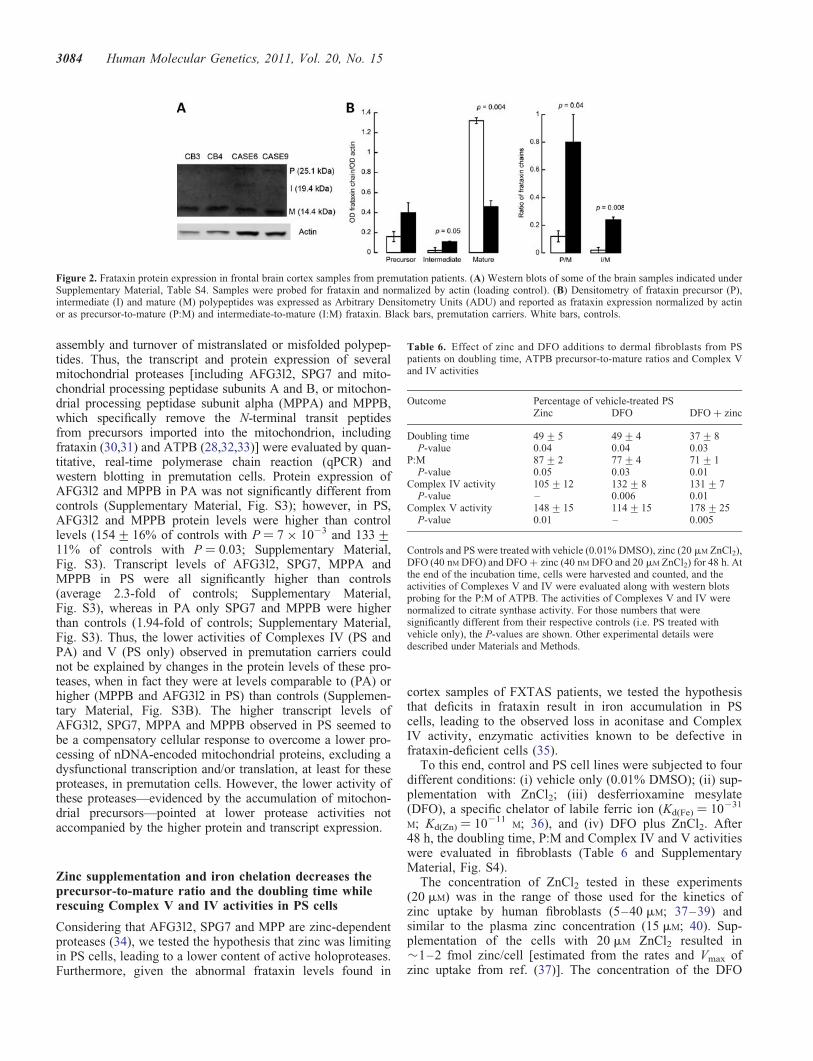
Frataxin, a protein involved in mitochondrial iron homeo-stasis, is a nuclear-encoded mitochondrial protein whose pre-cursor (P, MW ¼ 23.1 kDa) is converted to the mature(M, MW ¼ 14.3 kDa) form through a two-step proteolyticprocessing via formation of an intermediate (I, MW ¼18.8 kDa) polypeptide chain (27,28). To test whether theimport/processing of this protein was also impaired inFXTAS brain samples, western blots were performed to
evaluate the levels of frataxin P, I and M. As observed forATPB, NDUFS4 and CCO4 in PS fibroblasts, and for ATPBin cortex samples from PS, significantly lower mature frataxinlevels (normalized by actin: 2.8-fold; P ¼ 0.004) and signifi-cantly higher P:M and I:M (6.3-fold; P ¼ 0.04 and 15-fold;P ¼ 0.008) were observed in brain samples from FXTASpatients compared with controls (Fig. 2), further confirminga generalized defect in the import/processing machinery ofmitochondrially targeted proteins.
Impaired AFG3l2 and mitochondrial processing peptidaseactivities are not related to lower protein or transcriptexpression
Mice deficient in ATPase family gene 3-like 2 (AFG3l2) andits partner protein, paraplegin (SPG7), show tremor and ataxiaas well as mitochondrial abnormalities (29). In yeast, AFG3and RCA1 constitute the m-AAA protease complex that med-iates the degradation of non-assembled mitochondrial innermembrane proteins. This complex is necessary for the assem-bly of mitochondrial respiratory chain and ATPase Complexesand has an important function in both post-translational
Figure 1. Mitochondrial protein expression in cultured primary dermal fibroblasts and frontal brain cortex samples from premutation patients. (A) Western blotsof fibroblasts from control, PS and PA carriers. Representative western blot of the fibroblast panel probed for ATPB and CCO4 (precursor and mature proteins).Actin was used as a loading control. Samples were identified by following the nomenclature indicated under Supplementary Material, Table S1 with the excep-tion of P7, which was described before (1). (B) Representative western blot of some of the brain samples indicated under Supplementary Material, Table S4. Thedensitometry for all the cortex samples is also shown. The samples were probed for ATPB and normalized by VDAC1 (loading control). Data were expressed asArbitrary Densitometry Units and reported as P:M.
Table 5. Clinical characteristics of the individuals from which the brainsamples were utilized in this study
Group (n) Age (years) CGG repeats
Control (n ¼ 4) 58+4 24+3PS (n ¼ 6) 77+3 83+5
P-value 0.002 2 × 1025
Clinical and molecular characteristics of the panel of postmortem brain samples(frontal cortex) used in this study correspond in case number to those presentedpreviously (1) and further described under Supplementary Material, Table S4.All individuals were male and the brain samples were kept at 2808C until theprotein samples were obtained.
Human Molecular Genetics, 2011, Vol. 20, No. 15 3083
assembly and turnover of mistranslated or misfolded polypep-tides. Thus, the transcript and protein expression of severalmitochondrial proteases [including AFG3l2, SPG7 and mito-chondrial processing peptidase subunits A and B, or mitochon-drial processing peptidase subunit alpha (MPPA) and MPPB,which specifically remove the N-terminal transit peptidesfrom precursors imported into the mitochondrion, includingfrataxin (30,31) and ATPB (28,32,33)] were evaluated by quan-titative, real-time polymerase chain reaction (qPCR) andwestern blotting in premutation cells. Protein expression ofAFG3l2 and MPPB in PA was not significantly different fromcontrols (Supplementary Material, Fig. S3); however, in PS,AFG3l2 and MPPB protein levels were higher than controllevels (154+16% of controls with P ¼ 7 × 10²³ and 133+11% of controls with P ¼ 0.03; Supplementary Material,Fig. S3). Transcript levels of AFG3l2, SPG7, MPPA andMPPB in PS were all significantly higher than controls(average 2.3-fold of controls; Supplementary Material,Fig. S3), whereas in PA only SPG7 and MPPB were higherthan controls (1.94-fold of controls; Supplementary Material,Fig. S3). Thus, the lower activities of Complexes IV (PS andPA) and V (PS only) observed in premutation carriers couldnot be explained by changes in the protein levels of these pro-teases, when in fact they were at levels comparable to (PA) orhigher (MPPB and AFG3l2 in PS) than controls (Supplemen-tary Material, Fig. S3B). The higher transcript levels ofAFG3l2, SPG7, MPPA and MPPB observed in PS seemed tobe a compensatory cellular response to overcome a lower pro-cessing of nDNA-encoded mitochondrial proteins, excluding adysfunctional transcription and/or translation, at least for theseproteases, in premutation cells. However, the lower activity ofthese proteases—evidenced by the accumulation of mitochon-drial precursors—pointed at lower protease activities notaccompanied by the higher protein and transcript expression.
Zinc supplementation and iron chelation decreases theprecursor-to-mature ratio and the doubling time whilerescuing Complex V and IV activities in PS cells
Considering that AFG3l2, SPG7 and MPP are zinc-dependentproteases (34), we tested the hypothesis that zinc was limitingin PS cells, leading to a lower content of active holoproteases.Furthermore, given the abnormal frataxin levels found in
cortex samples of FXTAS patients, we tested the hypothesisthat deficits in frataxin result in iron accumulation in PScells, leading to the observed loss in aconitase and ComplexIV activity, enzymatic activities known to be defective infrataxin-deficient cells (35).
To this end, control and PS cell lines were subjected to fourdifferent conditions: (i) vehicle only (0.01% DMSO); (ii) sup-plementation with ZnCl2; (iii) desferrioxamine mesylate(DFO), a specific chelator of labile ferric ion (Kd(Fe) ¼ 10231
M; Kd(Zn) ¼ 10211M; 36), and (iv) DFO plus ZnCl2. After
48 h, the doubling time, P:M and Complex IV and V activitieswere evaluated in fibroblasts (Table 6 and SupplementaryMaterial, Fig. S4).
The concentration of ZnCl2 tested in these experiments(20 mM) was in the range of those used for the kinetics ofzinc uptake by human fibroblasts (5–40 mM; 37–39) andsimilar to the plasma zinc concentration (15 mM; 40). Sup-plementation of the cells with 20 mM ZnCl2 resulted in�1–2 fmol zinc/cell [estimated from the rates and Vmax ofzinc uptake from ref. (37)]. The concentration of the DFO
Figure 2. Frataxin protein expression in frontal brain cortex samples from premutation patients. (A) Western blots of some of the brain samples indicated underSupplementary Material, Table S4. Samples were probed for frataxin and normalized by actin (loading control). (B) Densitometry of frataxin precursor (P),intermediate (I) and mature (M) polypeptides was expressed as Arbitrary Densitometry Units (ADU) and reported as frataxin expression normalized by actinor as precursor-to-mature (P:M) and intermediate-to-mature (I:M) frataxin. Black bars, premutation carriers. White bars, controls.
Table 6. Effect of zinc and DFO additions to dermal fibroblasts from PSpatients on doubling time, ATPB precursor-to-mature ratios and Complex Vand IV activities
Outcome Percentage of vehicle-treated PSZinc DFO DFO + zinc
Doubling time 49+5 49+4 37+8P-value 0.04 0.04 0.03
P:M 87+2 77+4 71+1P-value 0.05 0.03 0.01
Complex IV activity 105+12 132+8 131+7P-value – 0.006 0.01
Complex V activity 148+15 114+15 178+25P-value 0.01 – 0.005
Controls and PS were treated with vehicle (0.01% DMSO), zinc (20 mM ZnCl2),DFO (40 nM DFO) and DFO + zinc (40 nM DFO and 20 mM ZnCl2) for 48 h. Atthe end of the incubation time, cells were harvested and counted, and theactivities of Complexes V and IV were evaluated along with western blotsprobing for the P:M of ATPB. The activities of Complexes V and IV werenormalized to citrate synthase activity. For those numbers that weresignificantly different from their respective controls (i.e. PS treated withvehicle only), the P-values are shown. Other experimental details weredescribed under Materials and Methods.
3084 Human Molecular Genetics, 2011, Vol. 20, No. 15
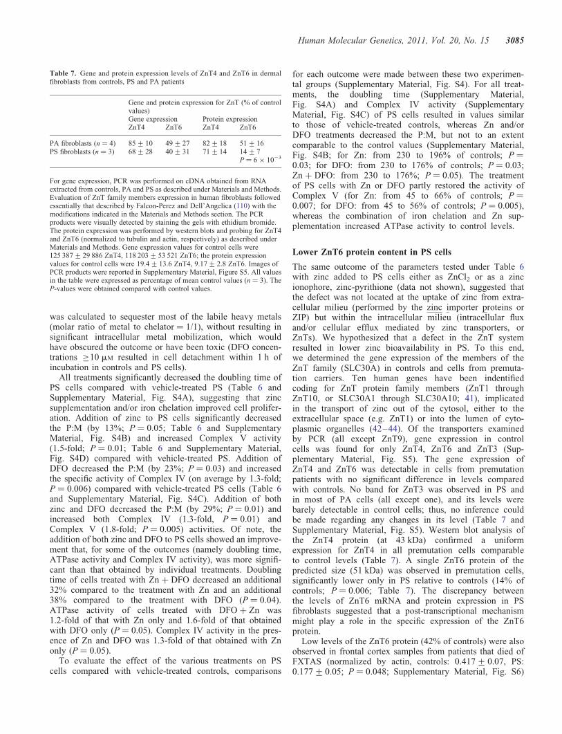
was calculated to sequester most of the labile heavy metals(molar ratio of metal to chelator ¼ 1/1), without resulting insignificant intracellular metal mobilization, which wouldhave obscured the outcome or have been toxic (DFO concen-trations ≥10 mM resulted in cell detachment within 1 h ofincubation in controls and PS cells).
All treatments significantly decreased the doubling time ofPS cells compared with vehicle-treated PS (Table 6 andSupplementary Material, Fig. S4A), suggesting that zincsupplementation and/or iron chelation improved cell prolifer-ation. Addition of zinc to PS cells significantly decreasedthe P:M (by 13%; P ¼ 0.05; Table 6 and SupplementaryMaterial, Fig. S4B) and increased Complex V activity(1.5-fold; P ¼ 0.01; Table 6 and Supplementary Material,Fig. S4D) compared with vehicle-treated PS. Addition ofDFO decreased the P:M (by 23%; P ¼ 0.03) and increasedthe specific activity of Complex IV (on average by 1.3-fold;P ¼ 0.006) compared with vehicle-treated PS cells (Table 6and Supplementary Material, Fig. S4C). Addition of bothzinc and DFO decreased the P:M (by 29%; P ¼ 0.01) andincreased both Complex IV (1.3-fold, P ¼ 0.01) andComplex V (1.8-fold; P ¼ 0.005) activities. Of note, theaddition of both zinc and DFO to PS cells showed an improve-ment that, for some of the outcomes (namely doubling time,ATPase activity and Complex IV activity), was more signifi-cant than that obtained by individual treatments. Doublingtime of cells treated with Zn + DFO decreased an additional32% compared to the treatment with Zn and an additional38% compared to the treatment with DFO (P ¼ 0.04).ATPase activity of cells treated with DFO + Zn was1.2-fold of that with Zn only and 1.6-fold of that obtainedwith DFO only (P ¼ 0.05). Complex IV activity in the pres-ence of Zn and DFO was 1.3-fold of that obtained with Znonly (P ¼ 0.05).
To evaluate the effect of the various treatments on PScells compared with vehicle-treated controls, comparisons
for each outcome were made between these two experimen-tal groups (Supplementary Material, Fig. S4). For all treat-ments, the doubling time (Supplementary Material,Fig. S4A) and Complex IV activity (SupplementaryMaterial, Fig. S4C) of PS cells resulted in values similarto those of vehicle-treated controls, whereas Zn and/orDFO treatments decreased the P:M, but not to an extentcomparable to the control values (Supplementary Material,Fig. S4B; for Zn: from 230 to 196% of controls; P ¼0.03; for DFO: from 230 to 176% of controls; P ¼ 0.03;Zn + DFO: from 230 to 176%; P ¼ 0.05). The treatmentof PS cells with Zn or DFO partly restored the activity ofComplex V (for Zn: from 45 to 66% of controls; P ¼0.007; for DFO: from 45 to 56% of controls; P ¼ 0.005),whereas the combination of iron chelation and Zn sup-plementation increased ATPase activity to control levels.
Lower ZnT6 protein content in PS cells
The same outcome of the parameters tested under Table 6with zinc added to PS cells either as ZnCl2 or as a zincionophore, zinc-pyrithione (data not shown), suggested thatthe defect was not located at the uptake of zinc from extra-cellular milieu (performed by the zinc importer proteins orZIP) but within the intracellular milieu (intracellular fluxand/or cellular efflux mediated by zinc transporters, orZnTs). We hypothesized that a defect in the ZnT systemresulted in lower zinc bioavailability in PS. To this end,we determined the gene expression of the members of theZnT family (SLC30A) in controls and cells from premuta-tion carriers. Ten human genes have been indentifiedcoding for ZnT protein family members (ZnT1 throughZnT10, or SLC30A1 through SLC30A10; 41), implicatedin the transport of zinc out of the cytosol, either to theextracellular space (e.g. ZnT1) or into the lumen of cyto-plasmic organelles (42–44). Of the transporters examinedby PCR (all except ZnT9), gene expression in controlcells was found for only ZnT4, ZnT6 and ZnT3 (Sup-plementary Material, Fig. S5). The gene expression ofZnT4 and ZnT6 was detectable in cells from premutationpatients with no significant difference in levels comparedwith controls. No band for ZnT3 was observed in PS andin most of PA cells (all except one), and its levels werebarely detectable in control cells; thus, no inference couldbe made regarding any changes in its level (Table 7 andSupplementary Material, Fig. S5). Western blot analysis ofthe ZnT4 protein (at 43 kDa) confirmed a uniformexpression for ZnT4 in all premutation cells comparableto control levels (Table 7). A single ZnT6 protein of thepredicted size (51 kDa) was observed in premutation cells,significantly lower only in PS relative to controls (14% ofcontrols; P ¼ 0.006; Table 7). The discrepancy betweenthe levels of ZnT6 mRNA and protein expression in PSfibroblasts suggested that a post-transcriptional mechanismmight play a role in the specific expression of the ZnT6protein.
Low levels of the ZnT6 protein (42% of controls) were alsoobserved in frontal cortex samples from patients that died ofFXTAS (normalized by actin, controls: 0.417+ 0.07, PS:0.177+ 0.05; P ¼ 0.048; Supplementary Material, Fig. S6)
Table 7. Gene and protein expression levels of ZnT4 and ZnT6 in dermalfibroblasts from controls, PS and PA patients
Gene and protein expression for ZnT (% of controlvalues)Gene expression Protein expressionZnT4 ZnT6 ZnT4 ZnT6
PA fibroblasts (n ¼ 4) 85+10 49+27 82+18 51+16PS fibroblasts (n ¼ 3) 68+28 40+31 71+14 14+7
P ¼ 6 × 10²³
For gene expression, PCR was performed on cDNA obtained from RNAextracted from controls, PA and PS as described under Materials and Methods.Evaluation of ZnT family members expression in human fibroblasts followedessentially that described by Falcon-Perez and Dell’Angelica (110) with themodifications indicated in the Materials and Methods section. The PCRproducts were visually detected by staining the gels with ethidium bromide.The protein expression was performed by western blots and probing for ZnT4and ZnT6 (normalized to tubulin and actin, respectively) as described underMaterials and Methods. Gene expression values for control cells were125 387+29 886 ZnT4, 118 203+53 521 ZnT6; the protein expressionvalues for control cells were 19.4+13.6 ZnT4, 9.17+2.8 ZnT6. Images ofPCR products were reported in Supplementary Material, Figure S5. All valuesin the table were expressed as percentage of mean control values (n ¼ 3). TheP-values were obtained compared with control values.
Human Molecular Genetics, 2011, Vol. 20, No. 15 3085

validating the results obtained with dermal fibroblasts(Table 7).
DISCUSSION
In this study, we showed abnormal mitochondrial function inboth CNS and non-CNS (dermal fibroblasts) tissues frompatients with the neurodegenerative disorder, FXTAS. All bio-chemical characteristics tested point to a lower ATP pro-duction, which would particularly affect neurons because oftheir high (sole) dependence on OXPHOS for energy supply(45–50).
Fibroblasts from premutation carriers, independently of thepresence of FXTAS symptoms, when compared with controlshad lower State 3u oxygen uptake rates (PA and PS 44 and57% of age-matched controls, respectively), lower glucoseconsumption via OXPHOS (35% of controls), lowerComplex IV activity (PA and PS 34 and 56% of age-matchedcontrols, respectively) and higher P:M of nDNA-encoded sub-units from Complexes I, IV and V (in average 3.4-fold and2.8-fold for PA and PS, respectively).
It is noteworthy that several of the parameters tested in thebiological samples segregated with the presence of FXTASsymptoms. Fibroblasts from PS when compared with thosefrom PA had higher FMR1 mRNA expression (3-fold;Table 1), lower Complex V activity (38% of controls), loweraconitase activity (43% of controls), higher MPPB andAFG3l2 protein expression (133 and 154% of controls,respectively), increased frataxin gene expression (2.5-fold ofcontrols) and lower ZnT6 protein expression (14% of con-trols), suggesting a segregation of outcomes with the occur-rence of symptoms. Some of these characteristics were alsoobserved in brain samples from patients that died withFXTAS symptoms: abnormal ATPB P:M and relative lowlevels of ZnT6 protein, suggesting a parallel between the phe-notype observed in PS fibroblasts and cortex.
The significant increase in the P:M of severalnDNA-encoded proteins in fibroblasts from premutation car-riers and frontal cortex of PS patients appears to stem froma lower import/processing capacity of precursor proteins syn-thesized in the cytosol. Import/processing of nDNA-encodedproteins to mitochondria is the result of the presence of both(i) energized, polarized mitochondria and (ii) cytoplasmicand mitochondrial trans-acting components of the import/pro-cessing apparatus. The membrane potential promotes theinitial translocation of the positively charged presequencesof preproteins (51), and further translocation of preprotein seg-ments requires the action of the matrix heat shock protein 70,an ATP-dependent molecular chaperone (52,53).Zn-dependent mitochondrial processing peptidase (MPP; 54),the Zn-dependent mitochondrial intermediate peptidase(MIP; 55) and the inner membrane protease (IMP; 56). MPPacts on all or part of the targeting sequence as the initial pro-cessing step, whereas MIP is specifically involved in the sub-sequent cleavage of proteins targeted to the mitochondrialmatrix (i.e. frataxin) or inner membrane (i.e. subunits of theelectron transport chain; 57), among which are ATPasesubunit alpha and sub unit o, MnSOD and CCO4 (32). IMPtargets proteins destined to the intermembrane space (56)
and, therefore, not expected to be involved in the maturationof any of the proteins analyzed in this study.
It is likely that altered zinc bioavailability in premutationcarriers (in particular PS) caused by insufficient ZnT6protein level (Table 7) might result in a lower transport ofcytoplasmic zinc into the trans-Golgi network vesicles,where it may act as the limiting step for incorporating zincinto zinc-dependent proteases. Then it is reasonable topropose that a defect on both Zn-dependent proteases, MPPand MIP, underlies the decreased level of mature mitochon-drial subunits [NADH dehydrogenase (ubiquinone) Fe-Sprotein 1, 75 kDa (NDUFS1), NADH dehydrogenase (ubiqui-none) Fe-S protein 2, 49 kDa (NADUF2), NDUFS4, ComplexII 70 kDa FP, CCO4, ATPB, MnSOD and frataxin) and theaccumulation of precursor polypeptides (ATPB, CCO4,NDUFS4 and frataxin) observed in fibroblasts and brainsamples and fibroblasts from FXTAS patients. In support ofthis hypothesis, supplementation of PS cells with zincdecreased the ATPB P:M, possibly restoring the zinc-dependent activities of proteases. In addition, because theincrease in the ATPase specific activity was higher than thedecrease in P:M, an increase in assembly/folding of subunitsinto Complex V seemed to have been favored (possiblythrough a mechanism involving the chaperone activity of zinc-dependent AFG3l2-SPG7). Compromised processing of mito-chondrial proteins, especially of ETC components, can alsoexplain the lower OXPHOS capacity of FXTAS fibroblastsobserved in this study, which can further exacerbate thisenergy-dependent process.
A defective zinc bioavailability may result in changes inzinc-containing proteins other than those evaluated in thisstudy (58), which could additionally contribute to theFXTAS phenotype. In this regard, it is tempting to proposethat the disrupted lamin A/C structure observed in FXTASneural cells (59) and fibroblasts (60) could result from a defec-tive processing of the farnesylated prelamin A by the zinc-metalloproteinase FACE1 (or STE24; 61,62), suggesting thatzinc-dependent proteases other than mitochondrial onescould also be affected. In addition, cells are endowed withseveral zinc-dependent transcription factors (e.g. TFIIIA andSp1; 63,64), which are affected by zinc availability (65).Zn-dependent transcription factor Sp1 interacts with the pro-moter of several mitochondrial proteins, such as MnSOD,ATPB, CCO4 and CYTC, among others (66–71), thus animbalance in zinc metabolism might also affect the expressionof mitochondrial proteins other than the activity of zinc-dependent proteases and chaperones, as confirmed by thereduced protein expression of MnSOD, ATPB and CCOobserved in fibroblasts and brain samples from premutationcarriers.
Given that frataxin, protein involved in iron metabolism, isa substrate of both MPP and MIP (27,30,31), zinc supplemen-tation of PS fibroblasts, by restoring the activities ofZn-dependent proteases, should have increased maturefrataxin in mitochondria and restored the activity of iron-containing proteins such as aconitase and Complex IV,proteins known to be deficient in frataxin-deficient cells(35). However, this treatment partly restored ATPB P:M andComplex V (iron-independent) but not Complex IV activity(Table 6). To bridge this apparent discrepancy, we reasoned
3086 Human Molecular Genetics, 2011, Vol. 20, No. 15

that lower MPP and MIP activities along with lower content ofmature frataxin resulted in iron accumulation which could notbe immediately mobilized by restoring normal levels ofmature frataxin. Indeed, DFO supplementation appeared criti-cal for the rescue of Complex IV activity (Table 6) but also forATPB P:M. Although not apparent, accumulated iron can havea detrimental role on transport/processing of precursors by anyof the following processes: (i) iron-mediated inhibition of MIPactivity (72), and, as a consequence, inhibition of the proces-sing of nDNA-encoded subunits whose final destination is thematrix and inner membrane; (ii) oxidative stress, which couldresult in oxidative damage and direct inactivation of mito-chondrial proteins (73–75); (iii) oxidative damage of mito-chondrial membrane, leading to lower membrane potentialand low transport/processing activity. Oxidative inactivationof proteins has been reported in pathologies associated withincreased oxidative stress caused by MnSOD deficiency(76), and in several neurodegenerative diseases including pro-gressive supranuclear palsy (77), Friedreich’s ataxia (FA; 78)and Huntington disease (79). In FXTAS, we have observedincreased oxidative and nitrative stress (as judged by increasedtyrosine nitration of proteins) in fibroblasts from premutationcarriers (1), and increased mtDNA copy number (Supplemen-tary Material, Table S2) and ROS production (this study) infibroblasts of premutation carriers with clinical symptoms ofFXTAS.
The lack of complete recovery of some of the outcomes inPS fibroblasts treated with DFO (or Zn + DFO) comparedwith vehicle-treated controls can be explained by the relativelylow concentrations of DFO, and short treatment period used inthis study designed to mobilize prolonged accumulated ironminimizing adverse effects potentially originated fromexcess metal chelation. This is the basis for the therapeuticinterventions used for iron-loading diseases (such asb-thalassemia; 80,81) and neurodegenerative diseases (suchas FA; 81,82) which are based on the daily use of relativelylow doses of chelators over long periods (3 weeks to years;81,83,84).
Based on this study, some overlapping biochemical mech-anisms and symptoms are expected between FXTAS andother diseases with (direct or indirect) defects at the mitochon-drial protein import and/or iron homestasis. (i) Defective mito-chondrial protein import precludes C. elegans from normaldevelopment promoting defective formation of the somaticgonad (85) similar to the premature ovarian insufficiencyobserved in female premutation carriers (86). (ii)Loss-of-function mutations in the human Tim8p homologDDP1 causes the neurodegenerative Mohr–Tranebjaerg syn-drome (87), syndrome with defective mitochondrial importmachinery that presents neurological symptoms, some ofwhich can be observed in FXTAS such as dystonia andmental deterioration. (iii) Spg7– / – Afg3l2Emv66/+ mice,bearing mutations of the genes Spg7 and Afg3l2 (encodingproteins belonging to the mitochondrial m-AAA protease) dis-played an early-onset severe neurological phenotype, charac-terized by loss of balance, tremor and ataxia, unstablemitochondrial Complexes in affected tissues and, at latestages, neurons containing structurally abnormal mitochondriadefective in cytochrome c oxidase-succinate dehydrogenaseactivity (88). In addition, deletion of AFG3 prevented the
growth of yeast on non-fermentable carbon sources and abro-gated the degradation of mitochondrially synthesized proteinsand the assembly of cytochrome c oxidase and F1FO ATPase(89). To note, although FXTAS phenotype and biochemicalfindings reported in this study are similar to those ofSpg7– / – Afg3l2Emv66/+ mice and AFG3-deleted yeast, nodecreases in protein expression of AFG3l2 and MPPB wereobserved. However, a deficiency in the bioavailability ofzinc not accompanied by a lower protein expression mightstill elicit lower protease/assembly activities, resulting in aphenotype similar to that of Spg7– / – Afg3l2Emv66/+ mice.(iv) Age-dependent loss of trans-synaptic zinc movement hasbeen suggested to lead to cognitive loss in Alzheimer’sdisease (90). Extracellular b-amyloid is aggregated by zinc(91,92), trapping this pool of synaptic zinc (90,91) and contri-buting to the sequestration of this metal. As an extension,genetic ablation of ZnT6 (and/or ZnT3) and/or trapping ofzinc by over-expressed FMR1 transcript may represent a phe-nocopy for FXTAS symptoms (2,93). (v) Some premutationcarriers (15–40%) exhibit signs of autistic behavior (94–97)suggesting some overlapping mechanisms. Besides the pres-ence of MD in children with autism and autism spectrum dis-orders (ASD) (98,99), transcriptional profiles from peripheralwhite blood cells from children with autism with a historyof developmental regression presented over-expression of 24different zinc-finger proteins, three ZIP (ZIP6, ZIP8 andZIP10) and two ZnT (ZnT1 and ZnT5) compared with thosewith an early onset of ASD (100), suggesting imbalances inzinc homestasis, and possibly iron metabolism. (vi) FA, a neu-rodegenerative disorder caused by the expansion of a GAA tri-nucleotide repeat in the first intron of the frataxin gene(FRDA), is characterized by lower expression of frataxinprotein, lower aconitase and Complex IV activities, andaccumulation of iron in mitochondria (35,101). The lowerComplex IV (Table 4) and aconitase activities (43% of con-trols; P ¼ 0.05) and the beneficial effect of DFO observed inthe FXTAS fibroblasts utilized in this study were all consistentwith the biochemical findings reported for patients with FA(35,102–104). Furthermore, it is interesting to note that at aphenotypic level, FA and FXTAS share some commontraits: gait ataxia, loss of coordination, difficulty walkingand numbness in the extremities (105,106).
Concluding remarks
This study raises three important issues: (i) MD in carriers ofsmall CGG-repeat expansions may potentiate the appearanceof phenotypes consistent with other disorders (e.g. FA’s,Parkinson’s disease or parkinsonism, and Alzheimer’sdisease) that are likely to involve MD, even when the allelesize is not sufficient to produce FXTAS symptoms. (ii) Sub-clinical MD may also predispose such carriers to environ-mental stressors (e.g. nutritional status), which may in turncontribute to both the penetrance and the severity of clinicalinvolvement in FXTAS, and finally, (iii) our data demonstratethat the appearance of FXTAS symptoms segregate with thelower activity of import/processing proteases (higher P:M)modulated by zinc and iron availability. Moreover, thisstudy implicates impaired mitochondrial proteolysis and pro-cessing as a novel pathway in the development of FXTAS.
Human Molecular Genetics, 2011, Vol. 20, No. 15 3087

A better understanding of this latter issue might help us todefine which carriers are most likely to develop FXTAS andto design targeted, preventative therapies.
MATERIALS AND METHODS
Chemicals and biochemicals
Ethylene diamine tetra-acetic acid (EDTA), ethylene glycoltetraacetic acid, sodium succinate, mannitol, sucrose, DFO(deferoxamine) and 4-2-hydroxyethyl-1-piperazineethanesul-fonic acid (HEPES) were all purchased from Sigma (StLouis, MO, USA). Tris–HCl, glycine, sodium chloride andpotassium chloride were purchased from Fisher (Pittsburg,PA, USA). Bovine serum albumin (fatty-acid free) wasobtained from MP Biomedicals. All other reagents were ofanalytical grade.
Subject samples
All studies of post-mortem and fibroblast (biopsy) tissuesamples were performed with approved protocols andinformed consent in accordance with the Declaration ofHelsinki (107) and the Institutional Review Boards of the Uni-versity of California, Davis, or Rush University MedicalCenter. Clinical and molecular characteristics of the panel ofcultured skin fibroblasts used in this study were presented inTable 1 and under Supplementary Material, Table S1.Samples not bolded in the Supplementary information werecultured as described in ref. (1).
Cell lines and culture conditions
Skin biopsies from individuals were performed with a 3 mmpunch under local anesthesia. The biopsy was diced understerile conditions and then plated in T25 flasks inAmnioMAXTM-C100 Basal Medium (Gibco, Grand Island,NY, USA) containing 15% AmnioMAXTM-C100 Supplement(Gibco) at 378C and 5% CO2 atmosphere. Longer term culturesof cell lines indicated in bold under Supplementary Material,Table S1 were maintained in DMEM high glucose +L-glutamine + 110 mg/ml sodium pyruvate (Gibco)supplemented with 15% fetal bovine serum (HyClone) and 1Xpenicillin–streptomycin (pen–strep) (100 units/ml penicillinG sodium and 100 mg/ml streptomycin; Gibco). Longer termcultures of the rest of the cell lines (not bolded) were maintainedin RPMI-1640 (Gibco) supplemented with 10% fetal bovineserum (Gibco) and 1X pen–strep media (100 units/ml penicillinG sodium and 100 mg/ml streptomycin sulfate; Gibco; 1). Cellswere trypsinized (0.25%) when they reached 90–95% conflu-ence, resuspended in serum-containing media and centrifugedat 200g for 5 min. Following removal of supernatant, cell viabi-lity and cell counts were quantified using Trypan blue exclusionusing a hemocytometer. These intact cells were used for oxygenuptake experiments. Protein extracts (for enzymatic analyses)were obtained by resuspending cell pellets at a concentrationof 5 × 10⁶ cells/ml in a hypotonic buffer (20 mM HEPES, pH7.4) supplemented with kinase, phosphatase and proteolyticinhibitors, incubating on ice for 10–15 min, homogenizingand then freezing immediately in liquid nitrogen. Protein
extracts (for western blots) were obtained by homogenizingthe cells and resuspending them in radio-immunoprecipitationassay (RIPA) buffer (50 mM Tris–HCl, 150 mM NaCl, 2 mM
EDTA, 0.5% CA-630 octylphenoxypolyethoxyethanol(IGEPAL), 0.1% sodium dodecyl sulfate (SDS), 0.012% deox-ycholate, 0.5% Triton X-100, pH 7.4) 4 ml/mg (wet pelletweight) with protease and phosphatase inhibitors.
Brain samples
Clinical and molecular characteristics of the brain samplesutilized in this study are given under Table 5 and underSupplementary Material, Table S4 essentially as describedpreviously (1). Frozen frontal cortex from controls and PScases was powdered in the presence of liquid N2 and hom-ogenized in a Dounce style homogenizer with 20 downwardstrokes of a tight fitting pestle in RIPA buffer (50 mM
Tris–HCl, 150 mM NaCl, 2 mM EDTA, 0.5% IGEPAL, 0.1%SDS, 0.012% deoxycholate, 0.5% triton X-100, pH 7.4) withprotease and phosphatase inhibitors. Homogenates were trans-ferred to centrifuge tubes and rotated overnight at 48Cfollowed by centrifugation at 16 000g at 48C. RIPA-solubleprotein fractions were quantified with BCA Protein AssayKit (#23225, Pierce Biotechnology, Rockford, IL, USA).
Treatment of cells with zinc and DFO
Cells (1.25 × 10⁵) were plated in two T25 and treated for 48 hat 378C. They were incubated with the following reagents: (i)0.01% DMSO or vehicle only; (ii) 20 mM ZnCl2; (iii) 40 nM
DFO; (iv) 40 nM DFO and 20 mM ZnCl2. Cells were allowedto grow for 48 h, and at the end of the incubation period,the flasks were washed three times with PBS prior to harvest-ing them, usually at 80–90% confluence.
Oxygen uptake
The oxygen uptake of intact cell suspensions was measuredusing a Clark-type O2 electrode from Hansatech (King’sLynn, UK) at 228C as described before (1). The respiratorycontrol ratio (RCRu) was obtained by dividing the rate ofoxygen consumption in State 3u [expressed as nmoloxygen × (min × million cells)21] obtained in the presenceof FCCP in high glucose DMEM buffer by that of State 4(in the presence of oligomycin) expressed with the sameunits. States 4 and 3 as well as RCRu are parametersdefined when using isolated mitochondria. However, we willuse the same nomenclature applied to the cell studies per-formed in this project to indicate oxygen uptake under non-phosphorylating conditions (or oligomycin-resistant oxygenuptake, State 4), phosphorylating conditions (glucose-dependent and oligomycin-sensitive oxygen uptake, State 3),maximum phosphorylating capacity (as before with FCCP,an uncoupler, or State 3u) and the ratio of State 3u/State 4to evaluate RCRu.
Enzymatic activities
All mitochondrial enzymatic and Complex activities weredescribed in detail in ref. (98) and Supplementary information.
3088 Human Molecular Genetics, 2011, Vol. 20, No. 15

mtDNA copy number
The mtDNA copy number estimated by evaluating themtDNA/nDNA ratio was determined using qPCR essentiallyas described in ref. (108). The mtDNA copy number in eachcell was expressed as the ratio between a mitochondrialgene (CYTB, ND1 and ND4) and the single-copy nuclearPK. Other experimental details were given in ref. (98) andin the Supplementary information.
Measurement of the rate of hydrogen peroxide production
The rate of H2O2 production in mitochondrial preparationswas followed fluorometrically using 5 U/ml horseradish per-oxidase (HRP) coupled to 40 mM p-hydroxyphenylacetic acidoxidation (109). Succinate (10 mM), in the presence of 5 mM
rotenone and 3.6 mM antimycin, were used as substrates forthis assay. Mitochondrial lysate (10–100 mg/assay) wasadded to start the reaction. Increased fluorescence at 228Cwas monitored by a Shimadzu fluorimeter. Arbitrary fluor-escence units per minute for the reaction were converted toamount of H2O2 by comparing the values to a standardcurve generated over a range of H2O2 concentrations. H2O2
generation was expressed as nmol H2O2 × (min × 107
cells)21. The addition of selective inhibitors of the respiratorychain permitted delineation of sites of mitochondrial ROS pro-duction.
Expression of selected genes in cultured cells
RNA was extracted using Qiagen’s RNEasy Plus extraction kitfollowing the manufacturer’s recommendations. cDNA wasgenerated with Qiagen’s Quantitect cDNA kit according tothe manufacturer’s protocol, quantified using Tecan’s platereader and normalized to the weakest sample of 107 ng/ml.All other experimental details were included in the Sup-plementary information. The approach used to test for theexpression of ZnT family members in human fibroblasts fol-lowed essentially that described by Falcon-Perez andDell’Angelica (110) with the following modifications indi-cated under the Supplementary information.
Western blots
All western blot procedures were performed essentially asdescribed before (1) with the modifications indicated in theSupplementary information (Supplementary Material,Table S5).
Statistical analyses
The number of individuals per group was calculated from an apriori G power analysis (two-tailed t-test, alpha ¼ 0.05,power ¼ (1-beta) ¼ 0.95, and n1 = n2) utilizing data fromRCRu and Complex IV activity. This analysis indicated thatwe needed four to six individuals per group. All our exper-iments were performed with this number of individualsexcept PA data on Table 7. The experiments were run in dupli-cate or triplicates and repeated three times in independentexperiments unless noted otherwise. Data were expressed as
mean+SEM and evaluated by using the t-test (StatSimplev2.0.5; Nidus Technologies, Toronto, Canada) and consideringP ≤ 0.05 as statistically significant.
SUPPLEMENTARY MATERIAL
Supplementary Material is available at HMG online.
ACKNOWLEDGEMENTS
We wish to express our gratitude to the patients and familiesthat participated in this study. We acknowledge the generousgift of the antibody to ZnT4 from Dr Liping Huang (Univer-sity of California Davis). The authors of this study do nothave any competing financial interests in relation to thework described with the exception of Dr Randi Hagerman(who has received research funding for clinical trials fromNeuropharm, Roche, Johnson & Johnson, Novartis, Seasidetherapeutics, Curemark and Forest pharmaceuticals and sheis also a consultant to Roche and Novartis) and Dr PaulHagerman (he is a non-paid collaborator/consultant forAsuragen and has a patent application for a method for detect-ing FMR1 allele size).
Conflict of Interest statement. The funding agencies werenot responsible for the design and conduct of the study; collec-tion, management, analysis and interpretation of the data; andpreparation, review or approval of this manuscript. C.G. hadfull access to all the data in this study and takes responsibilityfor the integrity of the data and the accuracy of the data analy-sis. D.G.-A., C.I., E.B.-K. and P.J.H. developed and providedthe cell lines utilized in this study. E.N. contributed signifi-cantly to the writing of this manuscript, and E.N. and C.R.I.performed all cell cultures and biochemical studies; E.N. eval-uated the effects of Zn and desferal treatments. A.O.-K. andS.W. performed all experiments related to molecularbiology. C.B., E.N. and D.S. performed all western blots inthe brain. E.B.-K. and R.H. recruited the subjects, performedthe skin biopsies and take responsibility for the integrityof the diagnostic and socio-demographic data. The authorsof this publication declare that they have no conflicting finan-cial interest in relation to the work described.
FUNDING
This work was partially supported by funds provided byAutism Speaks Foundation (#58739, C.G.), the National Insti-tute on Aging (AG024488, P.J.H.), the National Institutes ofHealth Interdisciplinary Research Consortium (IRC) grant(RL1 AG032119 and UL1 DE19583, P.J.H.; RL1AG032115, R.H.) and the Spastic Paralysis and Allied Dis-eases of the Central Nervous System Research Foundationof The Illinois-Eastern Iowa District Kiwanis International(E.B.-K.).
REFERENCES
1. Ross-Inta, C., Omanska-Klusek, A., Wong, S., Barrow, C.,Garcia-Arocena, D., Iwahashi, C., Berry-Kravis, E., Hagerman, R.J.,Hagerman, P.J. and Giulivi, C. (2010) Evidence of mitochondrial
Human Molecular Genetics, 2011, Vol. 20, No. 15 3089

dysfunction in fragile X-associated tremor/ataxia syndrome. Biochem. J.,429, 545–552.
2. Berry-Kravis, E., Goetz, C.G., Leehey, M.A., Hagerman, R.J., Zhang, L.,Li, L., Nguyen, D., Hall, D.A., Tartaglia, N., Cogswell, J. et al. (2007)Neuropathic features in fragile X premutation carriers. Am. J. Med.
Genet. A, 143, 19–26.
3. Hagerman, R.J., Leehey, M., Heinrichs, W., Tassone, F., Wilson, R.,Hills, J., Grigsby, J., Gage, B. and Hagerman, P.J. (2001) Intentiontremor, parkinsonism, and generalized brain atrophy in male carriers offragile X. Neurology, 57, 127–130.
4. Jacquemont, S., Hagerman, R.J., Leehey, M.A., Hall, D.A., Levine, R.A.,Brunberg, J.A., Zhang, L., Jardini, T., Gane, L.W., Harris, S.W. et al.
(2004) Penetrance of the fragile X-associated tremor/ataxia syndrome ina premutation carrier population. JAMA, 291, 460–469.
5. Greco, C.M., Berman, R.F., Martin, R.M., Tassone, F., Schwartz, P.H.,Chang, A., Trapp, B.D., Iwahashi, C., Brunberg, J., Grigsby, J. et al.
(2006) Neuropathology of fragile X-associated tremor/ataxia syndrome(FXTAS). Brain, 129, 243–255.
6. Greco, C.M., Hagerman, R.J., Tassone, F., Chudley, A.E., Del Bigio,M.R., Jacquemont, S., Leehey, M. and Hagerman, P.J. (2002) Neuronalintranuclear inclusions in a new cerebellar tremor/ataxia syndromeamong fragile X carriers. Brain, 125, 1760–1771.
7. Tassone, F., Hagerman, R.J., Garcia-Arocena, D., Khandjian, E.W.,Greco, C.M. and Hagerman, P.J. (2004) Intranuclear inclusions in neuralcells with premutation alleles in fragile X associated tremor/ataxiasyndrome. J. Med. Genet., 41, e43.
8. Beckel-Mitchener, A. and Greenough, W.T. (2004) Correlates across thestructural, functional, and molecular phenotypes of fragile X syndrome.Ment. Retard. Dev. Disabil. Res. Rev., 10, 53–59.
9. El-Osta, A. (2002) FMR1 silencing and the signals to chromatin: aunified model of transcriptional regulation. Biochem. Biophys. Res.
Commun., 295, 575–581.
10. Oberle, I., Rousseau, F., Heitz, D., Kretz, C., Devys, D., Hanauer, A.,Boue, J., Bertheas, M. and Mandel, J. (1991) Instability of a 550-basepair DNA segment and abnormal methylation in fragile X syndrome.Science, 252, 1097–1102.
11. Verkerk, A.J., Pieretti, M., Sutcliffe, J.S., Fu, Y.H., Kuhl, D.P., Pizzuti,A., Reiner, O., Richards, S., Victoria, M.F., Zhang, F.P. et al. (1991)Identification of a gene (FMR-1) containing a CGG repeat coincidentwith a breakpoint cluster region exhibiting length variation in fragile Xsyndrome. Cell, 65, 905–914.
12. Tassone, F., Hagerman, R.J., Taylor, A.K., Gane, L.W., Godfrey, T.E.and Hagerman, P.J. (2000) Elevated levels of FMR1 mRNA in carriermales: a new mechanism of involvement in the fragile-X syndrome.Am. J. Hum. Genet., 66, 6–15.
13. Tassone, F., Hagerman, R.J., Taylor, A.K., Mills, J.B., Harris, S.W.,Gane, L.W. and Hagerman, P.J. (2000) Clinical involvement and proteinexpression in individuals with the FMR1 premutation. Am. J. Hum.
Genet., 91, 144–152.14. Kenneson, A., Zhang, F., Hagedorn, C.H. and Warren, S.T. (2001)
Reduced FMRP and increased FMR1 transcription is proportionallyassociated with CGG repeat number in intermediate-length andpremutation carriers. Hum. Mol. Genet., 10, 1449–1454.
15. Gokden, M., Al-Hinti, J.T. and Harik, S.I. (2009) Peripheral nervoussystem pathology in fragile X tremor/ataxia syndrome (FXTAS).Neuropathology, 29, 280–284.
16. Greco, C.M., Soontrapornchai, K., Wirojanan, J., Gould, J.E., Hagerman,P.J. and Hagerman, R.J. (2007) Testicular and pituitary inclusionformation in fragile X associated tremor/ataxia syndrome. J. Urol., 177,1434–1437.
17. Chance, B. and Williams, G.R. (1955) A simple and rapid assay ofoxidative phosphorylation. Nature, 175, 1120–1121.
18. Chance, B. and Williams, G.R. (1956) The respiratory chain andoxidative phosphorylation. Adv. Enzymol. Relat. Subj. Biochem.,17, 65–134.
19. Barrientos, A., Casademont, J., Cardellach, F., Estivill, X.,Urbano-Marquez, A. and Nunes, V. (1997) Reduced steady-state levelsof mitochondrial RNA and increased mitochondrial DNA amount inhuman brain with aging. Brain Res. Mol. Brain Res., 52, 284–289.
20. Lee, H.C., Yin, P.H., Lu, C.Y., Chi, C.W. and Wei, Y.H. (2000) Increaseof mitochondria and mitochondrial DNA in response to oxidative stressin human cells. Biochem. J., 348, 425–432.
21. Masayesva, B.G., Mambo, E., Taylor, R.J., Goloubeva, O.G., Zhou, S.,Cohen, Y., Minhas, K., Koch, W., Sciubba, J., Alberg, A.J. et al. (2006)Mitochondrial DNA content increase in response to cigarette smoking.Cancer Epidemiol. Biomarkers Prev., 15, 19–24.
22. May-Panloup, P., Chretien, M.F., Savagner, F., Vasseur, C., Jean, M.,Malthiery, Y. and Reynier, P. (2003) Increased sperm mitochondrialDNA content in male infertility. Hum. Reprod., 18, 550–556.
23. Noack, H., Bednarek, T., Heidler, J., Ladig, R., Holtz, J. and Szibor, M.(2006) TFAM-dependent and independent dynamics of mtDNA levels inC2C12 myoblasts caused by redox stress. Biochim. Biophys. Acta, 1760,141–150.
24. Giulivi, C., Boveris, A. and Cadenas, E. (1999) In Gilbert, D. andColton, C. (eds), Reactive Oxygen Species in Biological Systems: An
Interdisciplinary Approach. Kluwer Academic/Plenum Publishers,New York, NY, pp. 77–102.
25. Arakane, F., Kallen, C.B., Watari, H., Foster, J.A., Sepuri, N.B., Pain, D.,Stayrook, S.E., Lewis, M., Gerton, G.L. and Strauss, J.F. III (1998) Themechanism of action of steroidogenic acute regulatory protein (StAR).StAR acts on the outside of mitochondria to stimulate steroidogenesis.J. Biol. Chem., 273, 16339–16345.
26. Jaussi, R., Sonderegger, P., Fluckiger, J. and Christen, P. (1982)Biosynthesis and topogenesis of aspartate aminotransferase isoenzymesin chicken embryo fibroblasts. The precursor of the mitochondrialisoenzyme is either imported into mitochondria or degraded in thecytosol. J. Biol. Chem., 257, 13334–13340.
27. Condo, I., Ventura, N., Malisan, F., Rufini, A., Tomassini, B. and Testi,R. (2007) In vivo maturation of human frataxin. Hum. Mol. Genet., 16,1534–1540.
28. Cavadini, P., Adamec, J., Taroni, F., Gakh, O. and Isaya, G. (2000)Two-step processing of human frataxin by mitochondrial processingpeptidase. Precursor and intermediate forms are cleaved at differentrates. J. Biol. Chem., 275, 41469–41475.
29. Martinelli, P., La Mattina, V., Bernacchia, A., Magnoni, R., Cerri, F.,Cox, G., Quattrini, A., Casari, G. and Rugarli, E.I. (2009) Geneticinteraction between the m-AAA protease isoenzymes reveals novel rolesin cerebellar degeneration. Hum. Mol. Genet., 18, 2001–2013.
30. Koutnikova, H., Campuzano, V. and Koenig, M. (1998) Maturation ofwild-type and mutated frataxin by the mitochondrial processingpeptidase. Hum. Mol. Genet., 7, 1485–1489.
31. Gordon, D.M., Shi, Q., Dancis, A. and Pain, D. (1999) Maturation offrataxin within mammalian and yeast mitochondria: one-step processingby matrix processing peptidase. Hum. Mol. Genet., 8, 2255–2262.
32. Hendrick, J.P., Hodges, P.E. and Rosenberg, L.E. (1989) Survey ofamino-terminal proteolytic cleavage sites in mitochondrial precursorproteins: leader peptides cleaved by two matrix proteases share athree-amino acid motif. Proc. Natl Acad. Sci. USA, 86, 4056–4060.
33. Xu, G., Shin, S.B. and Jaffrey, S.R. (2009) Global profiling of proteasecleavage sites by chemoselective labeling of protein N-termini. Proc.Natl Acad. Sci. USA, 106, 19310–19315.
34. Martinelli, P. and Rugarli, E.I. (2010) Emerging roles of mitochondrialproteases in neurodegeneration. Biochim. Biophys. Acta, 1797, 1–10.
35. Napoli, E., Morin, D., Bernhardt, R., Buckpitt, A. and Cortopassi, G.(2007) Hemin rescues adrenodoxin, heme a and cytochrome oxidaseactivity in frataxin-deficient oligodendroglioma cells. Biochim. Biophys.
Acta, 1772, 773–780.36. Keberle, H. (1964) The biochemistry of deferrioxamine and its relation
to iron metabolism. Ann. N. Y. Acad. Sci., 119, 758–768.37. Ackland, M.L., Danks, D.M. and McArdle, H.J. (1988) Studies of the
mechanism of zinc uptake by human fibroblasts. J. Cell Physiol., 135,521–526.
38. Ackland, M.L., Danks, D.M. and McArdle, H.J. (1989) Zinc transport byfibroblasts from patients with acrodermatitis enteropathica. Biol. TraceElem. Res., 22, 257–263.
39. Ackland, M.L. and McArdle, H.J. (1990) Significance of extracellularzinc-binding ligands in the uptake of zinc by human fibroblasts. J. Cell
Physiol., 145, 409–413.40. Forbes, I.J., Zalewski, P.D., Hurst, N.P., Giannakis, C. and Whitehouse,
M.W. (1989) Zinc increases phorbol ester receptors in intact B-cells,neutrophil polymorphs and platelets. FEBS Lett., 247, 445–447.
41. McMahon, R.J. and Cousins, R.J. (1998) Mammalian zinc transporters.J. Nutr., 128, 667–670.
42. Devergnas, S., Chimienti, F., Naud, N., Pennequin, A., Coquerel, Y.,Chantegrel, J., Favier, A. and Seve, M. (2004) Differential regulation of
3090 Human Molecular Genetics, 2011, Vol. 20, No. 15

zinc efflux transporters ZnT-1, ZnT-5 and ZnT-7 gene expression by zinclevels: a real-time RT-PCR study. Biochem. Pharmacol., 68, 699–709.
43. Gaither, L.A. and Eide, D.J. (2001) Eukaryotic zinc transporters andtheir regulation. BioMetals, 14, 251–270.
44. Kelleher, S.L. and Lonnerdal, B. (2002) Zinc transporters in the ratmammary gland respond to marginal zinc and vitamin A intakes duringlactation. J. Nutr., 132, 3280–3285.
45. Davey, G.P., Peuchen, S. and Clark, J.B. (1998) Energy thresholds inbrain mitochondria. Potential involvement in neurodegeneration. J. Biol.
Chem., 273, 12753–12757.
46. Mazzio, E.A., Reams, R.R. and Soliman, K.F. (2004) The role ofoxidative stress, impaired glycolysis and mitochondrial respiratory redoxfailure in the cytotoxic effects of 6-hydroxydopamine in vitro. Brain
Res., 1004, 29–44.
47. Fornuskova, D., Brantova, O., Tesarova, M., Stiburek, L., Honzik, T.,Wenchich, L., Tietzeova, E., Hansikova, H. and Zeman, J. (2008) Theimpact of mitochondrial tRNA mutations on the amount of ATP synthasediffers in the brain compared to other tissues. Biochim. Biophys. Acta,1782, 317–325.
48. Rossignol, R., Letellier, T., Malgat, M., Rocher, C. and Mazat, J.P.(2000) Tissue variation in the control of oxidative phosphorylation:implication for mitochondrial diseases. Biochem. J., 347, 45–53.
49. Chow, C.W. and Thorburn, D.R. (2000) Morphological correlates ofmitochondrial dysfunction in children. Hum. Reprod., 15(Suppl. 2),68–78.
50. Nissenkorn, A., Michelson, M., Ben-Zeev, B. and Lerman-Sagie, T.(2001) Inborn errors of metabolism: a cause of abnormal braindevelopment. Neurology, 56, 1265–1272.
51. Martin, J., Mahlke, K. and Pfanner, N. (1991) Role of an energized innermembrane in mitochondrial protein import. Delta psi drives themovement of presequences. J. Biol. Chem., 266, 18051–18057.
52. Kang, P.J., Ostermann, J., Shilling, J., Neupert, W., Craig, E.A. andPfanner, N. (1990) Requirement for hsp70 in the mitochondrial matrixfor translocation and folding of precursor proteins. Nature, 348,137–143.
53. Scherer, P.E., Krieg, U.C., Hwang, S.T., Vestweber, D. and Schatz, G.(1990) A precursor protein partly translocated into yeast mitochondria isbound to a 70 kd mitochondrial stress protein. EMBO J., 9, 4315–4322.
54. Luciano, P. and Geli, V. (1996) The mitochondrial processing peptidase:function and specificity. Experientia, 52, 1077–1082.
55. Isaya, G., Kalousek, F. and Rosenberg, L.E. (1992) Sequence analysis ofrat mitochondrial intermediate peptidase: similarity to zincmetallopeptidases and to a putative yeast homologue. Proc. Natl Acad.
Sci. USA, 89, 8317–8321.
56. Schneider, A., Behrens, M., Scherer, P., Pratje, E., Michaelis, G. andSchatz, G. (1991) Inner membrane protease I, an enzyme mediatingintramitochondrial protein sorting in yeast. EMBO J., 10, 247–254.
57. Branda, S.S. and Isaya, G. (1995) Prediction and identification of newnatural substrates of the yeast mitochondrial intermediate peptidase.J. Biol. Chem., 270, 27366–27373.
58. Udom, A.O. and Brady, F.O. (1980) Reactivation in vitro ofzinc-requiring apo-enzymes by rat liver zinc-thionein. Biochem. J., 187,329–335.
59. Arocena, D.G., Iwahashi, C.K., Won, N., Beilina, A., Ludwig, A.L.,Tassone, F., Schwartz, P.H. and Hagerman, P.J. (2005) Induction ofinclusion formation and disruption of lamin A/C structure bypremutation CGG-repeat RNA in human cultured neural cells. Hum.
Mol. Genet., 14, 3661–3671.
60. Garcia-Arocena, D., Yang, J.E., Brouwer, J.R., Tassone, F., Iwahashi, C.,Berry-Kravis, E.M., Goetz, C.G., Sumis, A.M., Zhou, L., Nguyen, D.V.et al. (2010) Fibroblast phenotype in male carriers of FMR1 premutationalleles. Hum. Mol. Genet., 19, 299–312.
61. Corrigan, D.P., Kuszczak, D., Rusinol, A.E., Thewke, D.P., Hrycyna,C.A., Michaelis, S. and Sinensky, M.S. (2005) Prelamin Aendoproteolytic processing in vitro by recombinant Zmpste24. Biochem.
J., 387, 129–138.
62. Liu, B. and Zhou, Z. (2008) Lamin A/C, laminopathies and prematureageing. Histol. Histopathol., 23, 747–763.
63. Kadonaga, J.T., Carner, K.R., Masiarz, F.R. and Tjian, R. (1987)Isolation of cDNA encoding transcription factor Sp1 and functionalanalysis of the DNA binding domain. Cell, 51, 1079–1090.
64. Moreland, R.J., Dresser, M.E., Rodgers, J.S., Roe, B.A., Conaway, J.W.,Conaway, R.C. and Hanas, J.S. (2000) Identification of a transcriptionfactor IIIA-interacting protein. Nucleic Acids Res., 28, 1986–1993.
65. Zeng, J., Vallee, B.L. and Kagi, J.H. (1991) Zinc transfer fromtranscription factor IIIA fingers to thionein clusters. Proc. Natl Acad. Sci.
USA, 88, 9984–9988.66. Evans, M.J. and Scarpulla, R.C. (1988) Both upstream and intron
sequence elements are required for elevated expression of the rat somaticcytochrome c gene in COS-1 cells. Mol. Cell Biol., 8, 35–41.
67. Carter, R.S., Bhat, N.K., Basu, A. and Avadhani, N.G. (1992) The basalpromoter elements of murine cytochrome c oxidase subunit IV geneconsist of tandemly duplicated ets motifs that bind to GABP-relatedtranscription factors. J. Biol. Chem., 267, 23418–23426.
68. Basu, A., Park, K., Atchison, M.L., Carter, R.S. and Avadhani, N.G.(1993) Identification of a transcriptional initiator element in thecytochrome c oxidase subunit Vb promoter which binds to transcriptionfactors NF-E1 (YY-1,delta) and Sp1. J. Biol. Chem., 268, 4188–4196.
69. Connor, M.K., Irrcher, I. and Hood, D.A. (2001) Contractileactivity-induced transcriptional activation of cytochrome c involves Sp1and is proportional to mitochondrial ATP synthesis in C2C12 musclecells. J. Biol. Chem., 276, 15898–15904.
70. Zaid, A., Li, R., Luciakova, K., Barath, P., Nery, S. and Nelson, B.D.(1999) On the role of the general transcription factor Sp1 in theactivation and repression of diverse mammalian oxidativephosphorylation genes. J. Bioenerg. Biomembr., 31, 129–135.
71. Ohta, S., Tomura, H., Matsuda, K. and Kagawa, Y. (1988) Genestructure of the human mitochondrial adenosine triphosphate synthasebeta subunit. J. Biol. Chem., 263, 11257–11262.
72. Kalousek, F., Isaya, G. and Rosenberg, L.E. (1992) Rat livermitochondrial intermediate peptidase (MIP): purification and initialcharacterization. EMBO J., 11, 2803–2809.
73. Dhanasekaran, A., Kotamraju, S., Kalivendi, S.V., Matsunaga, T.,Shang, T., Keszler, A., Joseph, J. and Kalyanaraman, B. (2004)Supplementation of endothelial cells with mitochondria-targetedantioxidants inhibit peroxide-induced mitochondrial iron uptake,oxidative damage, and apoptosis. J. Biol. Chem., 279, 37575–37587.
74. Bulteau, A.L., Dancis, A., Gareil, M., Montagne, J.J., Camadro, J.M. andLesuisse, E. (2007) Oxidative stress and protease dysfunction in the yeastmodel of Friedreich ataxia. Free Rad. Biol. Med., 42, 1561–1570.
75. Cantu, D., Schaack, J. and Patel, M. (2009) Oxidative inactivation ofmitochondrial aconitase results in iron and H2O2-mediated neurotoxicityin rat primary mesencephalic cultures. PLoS ONE, 4, e7095.
76. Williams, M.D., Van Remmen, H., Conrad, C.C., Huang, T.T., Epstein,C.J. and Richardson, A. (1998) Increased oxidative damage is correlatedto altered mitochondrial function in heterozygous manganese superoxidedismutase knockout mice. J. Biol. Chem., 273, 28510–28515.
77. Park, L.C., Albers, D.S., Xu, H., Lindsay, J.G., Beal, M.F. and Gibson,G.E. (2001) Mitochondrial impairment in the cerebellum of the patientswith progressive supranuclear palsy. J. Neurosci. Res., 66, 1028–1034.
78. Bradley, J.L., Blake, J.C., Chamberlain, S., Thomas, P.K., Cooper, J.M.and Schapira, A.H. (2000) Clinical, biochemical and molecular geneticcorrelations in Friedreich’s ataxia. Hum. Mol. Genet., 9, 275–282.
79. Tabrizi, S.J., Cleeter, M.W., Xuereb, J., Taanman, J.W., Cooper, J.M.and Schapira, A.H. (1999) Biochemical abnormalities and excitotoxicityin Huntington’s disease brain. Ann. Neurol., 45, 25–32.
80. Rund, D. and Rachmilewitz, E. (2005) Beta-thalassemia. NewEngl. J. Med., 353, 1135–1146.
81. Brittenham, G.M. (2011) Iron-chelating therapy for transfusional ironoverload. New Engl. J. Med., 364, 146–156.
82. Lim, C.K., Kalinowski, D.S. and Richardson, D.R. (2008) Protectionagainst hydrogen peroxide-mediated cytotoxicity in Friedreich’s ataxiafibroblasts using novel iron chelators of the 2-pyridylcarboxaldehydeisonicotinoyl hydrazone class. Mol. Pharmacol., 74, 225–235.
83. Kwiatkowski, J.L. (2010) Oral iron chelators. Hematol. Oncol. Clin.North Am., 24, 229–248.
84. Wong, C.S., Kwok, J.C. and Richardson, D.R. (2004) PCTH: a novelorally active chelator of the aroylhydrazone class that induces ironexcretion from mice. Biochim. Biophys. Acta, 1739, 70–80.
85. Curran, S.P., Leverich, E.P., Koehler, C.M. and Larsen, P.L. (2004)Defective mitochondrial protein translocation precludes normalCaenorhabditis elegans development. J. Biol. Chem., 279, 54655–54662.
86. Allingham-Hawkins, D.J., Babul-Hirji, R., Chitayat, D., Holden, J.J.,Yang, K.T., Lee, C., Hudson, R., Gorwill, H., Nolin, S.L., Glicksman, A.
Human Molecular Genetics, 2011, Vol. 20, No. 15 3091

et al. (1999) Fragile X premutation is a significant risk factor forpremature ovarian failure: the International Collaborative POF in FragileX study—preliminary data. Am. J. Med. Genet., 83, 322–325.
87. Koehler, C.M., Leuenberger, D., Merchant, S., Renold, A., Junne, T. andSchatz, G. (1999) Human deafness dystonia syndrome is a mitochondrialdisease. Proc. Natl Acad. Sci. USA, 96, 2141–2146.
88. Bolot, J.F., Robert, D., Chemorin, B., Fournier, G., Thomas, L.,Buffat, J. and Bertoye, A. (1977) Assisted ventilation at home bytracheotomy in subjects with chronic respiratory insufficiency.Minerva Med., 68, 409–414.
89. Frigerio, B., Ravazola, M., Ito, S., Buffa, R., Capella, C., Solcia, E. andOrci, L. (1977) Histochemical and ultrastructural identification ofneurotensin cells in the dog ileum. Histochemistry, 54, 123–131.
90. Adlard, P.A., Parncutt, J.M., Finkelstein, D.I. and Bush, A.I. (2010)Cognitive loss in zinc transporter-3 knock-out mice: a phenocopy for thesynaptic and memory deficits of Alzheimer’s disease? J. Neurosci., 30,1631–1636.
91. Cherny, R.A., Atwood, C.S., Xilinas, M.E., Gray, D.N., Jones, W.D.,McLean, C.A., Barnham, K.J., Volitakis, I., Fraser, F.W., Kim, Y.S.et al. (2001) Treatment with a copper-zinc chelator markedly and rapidlyinhibits Œ≤-amyloid accumulation in Alzheimer’s disease transgenicmice. Neuron, 30, 665–676.
92. Lee, J.Y., Cole, T.B., Palmiter, R.D., Suh, S.W. and Koh, J.Y. (2002)Contribution by synaptic zinc to the gender-disparate plaque formationin human Swedish mutant APP transgenic mice. Proc. Natl Acad. Sci.
USA, 99, 7705–7710.
93. Bourgeois, J.A., Cogswell, J.B., Hessl, D., Zhang, L., Ono, M.Y.,Tassone, F., Farzin, F., Brunberg, J.A., Grigsby, J. and Hagerman, R.J.(2007) Cognitive, anxiety and mood disorders in the fragile X-associatedtremor/ataxia syndrome. Gen. Hosp. Psychiatry, 29, 349–356.
94. Reiss, A.L. and Freund, L. (1990) Fragile X syndrome, DSM-III-R, andautism. J. Am. Acad. Child. Adolesc. Psychiatry, 29, 885–891.
95. Feinstein, C. and Reiss, A.L. (1998) Autism: the point of view fromfragile X studies. J. Autism Dev. Disord., 28, 393–405.
96. Goodlin-Jones, B.L., Tassone, F., Gane, L.W. and Hagerman, R.J. (2004)
Autistic spectrum disorder and the fragile X premutation. J. Dev. Behav.
Pediatr., 25, 392–398.
97. Farzin, F., Perry, H., Hessl, D., Loesch, D., Cohen, J., Bacalman, S.,Gane, L., Tassone, F., Hagerman, P. and Hagerman, R. (2006) Autism
spectrum disorders and attention-deficit/hyperactivity disorder in boyswith the fragile X premutation. J. Dev. Behav. Pediatr., 27,S137–S144.
98. Giulivi, C., Zhang, Y.-F., Omanska-Klusek, A., Ross-Inta, C.M., Wong,S., Hertz-Picciotto, I., Tassone, F. and Pessah, I.N. (2010) Mitochondrialdysfunction in autism. JAMA, 304, 2389–2396.
99. Oliveira, G., Diogo, L., Grazina, M., Garcia, P., Ataide, A., Marques, C.,Miguel, T., Borges, L., Vicente, A.M. and Oliveira, C.R. (2005)Mitochondrial dysfunction in autism spectrum disorders: apopulation-based study. Dev. Med. Child Neurol., 47, 185–189.
100. Gregg, J.P., Lit, L., Baron, C.A., Hertz-Picciotto, I., Walker, W., Davis,R.A., Croen, L.A., Ozonoff, S., Hansen, R., Pessah, I.N. et al. (2008)Gene expression changes in children with autism. Genomics, 91, 22–29.
101. Campuzano, V., Montermini, L., Molto, M.D., Pianese, L., Cossee, M.,Cavalcanti, F., Monros, E., Rodius, F., Duclos, F., Monticelli, A. et al.(1996) Friedreich’s ataxia: autosomal recessive disease caused by anintronic GAA triplet repeat expansion. Science, 271, 1423–1427.
102. Wong, A., Yang, J., Cavadini, P., Gellera, C., Lonnerdal, B., Taroni, F.and Cortopassi, G. (1999) The Friedreich’s ataxia mutation conferscellular sensitivity to oxidant stress which is rescued by chelators of ironand calcium and inhibitors of apoptosis. Hum. Mol. Genet., 8, 425–430.
103. Kontoghiorghes, G.J., Efstathiou, A., Kleanthous, M., Michaelides, Y.and Kolnagou, A. (2009) Risk/benefit assessment, advantages over otherdrugs and targeting methods in the use of deferiprone as apharmaceutical antioxidant in iron loading and non iron loadingconditions. Hemoglobin, 33, 386–397.
104. Richardson, D.R. (2004) Novel chelators for central nervous systemdisorders that involve alterations in the metabolism of iron and othermetal ions. Ann. N. Y. Acad. Sci., 1012, 326–341.
105. Delatycki, M.B., Williamson, R. and Forrest, S.M. (2000) Friedreichataxia: an overview. J. Med. Genet., 37, 1–8.
106. Leehey, M.A. (2009) Fragile X-associated tremor/ataxia syndrome:clinical phenotype, diagnosis, and treatment. J. Invest. Med., 57,830–836.
107. World Medical Association (2000) Declaration of Helsinki: ethicalprinciples for medical research involving human subjects. JAMA, 284,3043–3045.
108. Ross-Inta, C.M., Zhang, Y.F., Almendares, A. and Giulivi, C. (2009)Threonine-deficient diets induced changes in hepatic bioenergetics.Am. J. Physiol. Gastrointest. Liver Physiol., 296, G1130–G1139.
109. Sarkela, T.M., Berthiaume, J., Elfering, S., Gybina, A.A. and Giulivi, C.(2001) The modulation of oxygen radical production by nitric oxide inmitochondria. J. Biol. Chem., 276, 6945–6949.
110. Falcon-Perez, J.M. and Dell’Angelica, E.C. (2007) Zinc transporter 2(SLC30A2) can suppress the vesicular zinc defect of adaptor protein3-depleted fibroblasts by promoting zinc accumulation in lysosomes.Exp. Cell Res., 313, 1473–1483.
3092 Human Molecular Genetics, 2011, Vol. 20, No. 15
Copyright © 2022 FDOKUMEN