Altered immunohistochemical expression of mast cell tryptase and chymase in the pathogenesis of oral...
13
Altered Immunohistochemical Expression of Mast Cell Tryptase and Chymase in the Pathogenesis of Oral Submucous Fibrosis and Malignant Transformation of the Overlying Epithelium Archana Yadav, Rajiv S. Desai*, Bansari A. Bhuta, Jatinder S. Singh, Reema Mehta, Akash P. Nehete Department of Oral Pathology, Nair Hospital Dental College, Mumbai, India Abstract Mast cells (MCs) expressing serine proteases; tryptase and chymase, are associated with fibrosis in various diseases. However, little is known about their involvement in oral submucous fibrosis (OSF). Our goal was to evaluate the role of MC tryptase and chymase in the pathogenesis of OSF and its malignant transformation. Immunohistochemical expression of MC tryptase and chymase was evaluated in 20 cases of OSF, 10 cases of oral squamous cell carcinoma (OSCC) and 10 cases of healthy controls. Subepithelial zone of Stage 1 and 2 while deep zone of Stage 3 and 4 OSF demonstrated increased tryptase positive MCs. OSCC revealed a proportionate increase in tryptase and chymase positive MCs irrespective of areas of distribution. An altered balance in the subepithelial and deep distribution of tryptase and chymase positive MCs play an important role in the pathogenesis of OSF and its malignant transformation. Citation: Yadav A, Desai RS, Bhuta BA, Singh JS, Mehta R, et al. (2014) Altered Immunohistochemical Expression of Mast Cell Tryptase and Chymase in the Pathogenesis of Oral Submucous Fibrosis and Malignant Transformation of the Overlying Epithelium. PLoS ONE 9(5): e98719. doi:10.1371/journal.pone.0098719 Editor: Muy-Teck Teh, Barts & The London School of Medicine and Dentistry, Queen Mary University of London, United Kingdom Received March 18, 2014; Accepted May 7, 2014; Published May 29, 2014 Copyright: ß 2014 Yadav et al. This is an open-access article distributed under the terms of the Creative Commons Attribution License, which permits unrestricted use, distribution, and reproduction in any medium, provided the original author and source are credited. Data Availability: The authors confirm that all data underlying the findings are fully available without restriction. Not Applicable. Funding: The authors have no support or funding to report. Competing Interests: The authors have declared that no competing interests exist. * E-mail: [email protected] Introduction Oral submucous fibrosis (OSF) is a chronic progressive, areca nut chewing habit related, precancerous condition of the oral mucosa predominantly affecting Indians and South Asians. It is clinically characterized by burning sensation of the oral mucosa accompanied by pallor and progressive, irreversible fibrosis leading to difficulty in opening mouth, speech and swallowing [1]. Characteristic histopathologic features of this disease include epithelial atrophy with loss of rete ridges, reduced vascularity, chronic inflammatory infil- trate and hyalinization of the submucosal tissue. The patho- genic mechanism in OSF begins primarily in the connective tissue and epithelial response secondarily. The characteristic fibro-elastic changes observed in the connective tissue are almost entirely due to abnormal accumulation of collagen. OSF is thus regarded as a collagen related disorder induced by betel nut/betel quid chewing habit often resulting in an overall increased production of collagen with decreased collagen degradation [2]. In normal wound healing fibroblasts are transiently activat- ed into myofibroblast, a particular type of fibroblast to proliferate and deposit the collagen [3]. OSF is similar to a wound where continuous signals for tissue repair are emitted. These continuous signals can lead to abnormal production of cytokines and growth factors, resulting in chronic, sustained long term myofibroblast activation leading to fibrosis. Re- search has shown that fibrosis in OSF is a continuous, scarring process in which the myofibroblast plays an essential role [4]. However, the dynamics of extra cellular matrix remodeling with OSF is largely unknown, since the origin of fibroblast activation in OSF is debated. Chronic inflammatory conditions can evolve a fibrotic phenotype often associated with an increase in the number of MCs, which are the local residents of connective tissue. Several lines of evidence suggest that MC when activated, secrete a large number of fibrogenic factors and have been implicated in the development of various fibrotic conditions affecting the lungs [5], [6], [7], liver [8], [9], skin [10], [11], and kidney [12]. Despite the potential of MCs to mediate fibrosis, limited attention has been given to the role of MCs in OSF. There have been a few studies assessing MC density in OSF using Toluidine blue stain [13], [14], [15], [16], [17], [18] and C-kit [19], which have yielded controversial results. MCs produce and store various profi- brotic cytokines including transforming growth factor-b (TGFb) [20], fibroblast growth factor (FGF), platelet derived growth factor (PDGF), interleukin 1 and 6 (IL-1 & IL-6), and tumor necrosis factor–a (TNF-a) [21]. The significance of these profibrotic cytokines in OSF has been studied extensively in the literature [22], [23], [24], [25]. Human MCs also contain two types of serine protease, tryptase and chymase. Tryptase is a trypsin-like enzyme, and chymase is a chymotrypsin-like enzyme. According to their protease content, human MCs are divided into two pheno- types: Mast cell secreting both tryptase and chymase are termed MC TC , while mast cell secreting only tryptase are PLOS ONE | www.plosone.org 1 May 2014 | Volume 9 | Issue 5 | e98719
Transcript of Altered immunohistochemical expression of mast cell tryptase and chymase in the pathogenesis of oral...

Altered Immunohistochemical Expression of Mast CellTryptase and Chymase in the Pathogenesis of OralSubmucous Fibrosis and Malignant Transformation ofthe Overlying EpitheliumArchana Yadav, Rajiv S. Desai*, Bansari A. Bhuta, Jatinder S. Singh, Reema Mehta, Akash P. Nehete
Department of Oral Pathology, Nair Hospital Dental College, Mumbai, India
Abstract
Mast cells (MCs) expressing serine proteases; tryptase and chymase, are associated with fibrosis in various diseases.However, little is known about their involvement in oral submucous fibrosis (OSF). Our goal was to evaluate the role of MCtryptase and chymase in the pathogenesis of OSF and its malignant transformation. Immunohistochemical expression of MCtryptase and chymase was evaluated in 20 cases of OSF, 10 cases of oral squamous cell carcinoma (OSCC) and 10 cases ofhealthy controls. Subepithelial zone of Stage 1 and 2 while deep zone of Stage 3 and 4 OSF demonstrated increasedtryptase positive MCs. OSCC revealed a proportionate increase in tryptase and chymase positive MCs irrespective of areas ofdistribution. An altered balance in the subepithelial and deep distribution of tryptase and chymase positive MCs play animportant role in the pathogenesis of OSF and its malignant transformation.
Citation: Yadav A, Desai RS, Bhuta BA, Singh JS, Mehta R, et al. (2014) Altered Immunohistochemical Expression of Mast Cell Tryptase and Chymase in thePathogenesis of Oral Submucous Fibrosis and Malignant Transformation of the Overlying Epithelium. PLoS ONE 9(5): e98719. doi:10.1371/journal.pone.0098719
Editor: Muy-Teck Teh, Barts & The London School of Medicine and Dentistry, Queen Mary University of London, United Kingdom
Received March 18, 2014; Accepted May 7, 2014; Published May 29, 2014
Copyright: � 2014 Yadav et al. This is an open-access article distributed under the terms of the Creative Commons Attribution License, which permitsunrestricted use, distribution, and reproduction in any medium, provided the original author and source are credited.
Data Availability: The authors confirm that all data underlying the findings are fully available without restriction. Not Applicable.
Funding: The authors have no support or funding to report.
Competing Interests: The authors have declared that no competing interests exist.
* E-mail: [email protected]
Introduction
Oral submucous fibrosis (OSF) is a chronic progressive,
areca nut chewing habit related, precancerous condition of the
oral mucosa predominantly affecting Indians and South
Asians. It is clinically characterized by burning sensation of
the oral mucosa accompanied by pallor and progressive,
irreversible fibrosis leading to difficulty in opening mouth,
speech and swallowing [1]. Characteristic histopathologic
features of this disease include epithelial atrophy with loss of
rete ridges, reduced vascularity, chronic inflammatory infil-
trate and hyalinization of the submucosal tissue. The patho-
genic mechanism in OSF begins primarily in the connective
tissue and epithelial response secondarily. The characteristic
fibro-elastic changes observed in the connective tissue are
almost entirely due to abnormal accumulation of collagen.
OSF is thus regarded as a collagen related disorder induced by
betel nut/betel quid chewing habit often resulting in an overall
increased production of collagen with decreased collagen
degradation [2].
In normal wound healing fibroblasts are transiently activat-
ed into myofibroblast, a particular type of fibroblast to
proliferate and deposit the collagen [3]. OSF is similar to a
wound where continuous signals for tissue repair are emitted.
These continuous signals can lead to abnormal production of
cytokines and growth factors, resulting in chronic, sustained
long term myofibroblast activation leading to fibrosis. Re-
search has shown that fibrosis in OSF is a continuous, scarring
process in which the myofibroblast plays an essential role [4].
However, the dynamics of extra cellular matrix remodeling
with OSF is largely unknown, since the origin of fibroblast
activation in OSF is debated. Chronic inflammatory conditions
can evolve a fibrotic phenotype often associated with an
increase in the number of MCs, which are the local residents of
connective tissue. Several lines of evidence suggest that MC
when activated, secrete a large number of fibrogenic factors
and have been implicated in the development of various
fibrotic conditions affecting the lungs [5], [6], [7], liver [8],
[9], skin [10], [11], and kidney [12]. Despite the potential of
MCs to mediate fibrosis, limited attention has been given to
the role of MCs in OSF. There have been a few studies
assessing MC density in OSF using Toluidine blue stain [13],
[14], [15], [16], [17], [18] and C-kit [19], which have yielded
controversial results. MCs produce and store various profi-
brotic cytokines including transforming growth factor-b(TGFb) [20], fibroblast growth factor (FGF), platelet derived
growth factor (PDGF), interleukin 1 and 6 (IL-1 & IL-6), and
tumor necrosis factor–a (TNF-a) [21]. The significance of
these profibrotic cytokines in OSF has been studied extensively
in the literature [22], [23], [24], [25].
Human MCs also contain two types of serine protease,
tryptase and chymase. Tryptase is a trypsin-like enzyme, and
chymase is a chymotrypsin-like enzyme. According to their
protease content, human MCs are divided into two pheno-
types: Mast cell secreting both tryptase and chymase are
termed MCTC, while mast cell secreting only tryptase are
PLOS ONE | www.plosone.org 1 May 2014 | Volume 9 | Issue 5 | e98719

termed MCT. Both MCT and MCTC phenotypes are present in
all human tissues. However, the ratio is different in each
anatomical site: while MCTC predominates in the skin, heart,
synovial and small intestinal submucosa, MCT predominates in
the lungs and the small intestinal mucosa [26]. MC tryptase
and chymase, the most abundant profibrotic cytokines of
human MC have been studied in various fibrotic disorders [5],
[8], [10], [12], however, their role in OSF has not been
reported so far in the literature.
It is already known that neoangiogenesis is required for the
growth and spread of tumor [27], [28]. Increased angiogenesis has
been associated with neoplastic progression, metastasis and
outcome in several studies in numbers of malignancies [29],
[30], [31], [32]. Current literature suggests the probable role of
MCs in tumor angiogenesis, thereby favoring the tumor progres-
sion [33], [34]. However, the contribution of proangogenic
cytokines namely MC tryptase and chymase during the malignant
transformation of OSF is not clear yet.
The present study was undertaken to quantify and characterize
the distribution of MC subpopulation in OSF, thereby evaluating
the potential role of MC tryptase and chymase in the pathogenesis
of OSF & its malignant transformation.
Materials and Methods
Human Tissue Specimen CollectionThe study protocol was approved by the Institutional
Review Board and the Local Ethics Committee of Nair
Hospital Dental College and was in compliance with ethical
standards of the Declaration of Helsinki. Written informed
consents were obtained from all the study participants. Twenty
(n = 20) previously untreated cases of OSF and ten (n = 10)
cases of OSCC with no history of OSF, diagnosed on clinical
grounds and confirmed histologically were randomly selected
to form the study groups. Ten (n = 10) age and sex matched
healthy volunteers without habits were included in the control
Figure 1. Clinical picture. A, Healthy control. B, Stage 1 OSF: Blanched palatal mucosa with vesicle. C, Stage 2 OSF: Blanched buccal mucosa. D,Stage 3 OSF: Hockey stick uvula. E, Stage 4 OSF: Blanched labial mucosa with vesicle. F, OSCC: Carcinoma of the left labial mucosa.doi:10.1371/journal.pone.0098719.g001
Mast Cell Tryptase and Chymase in OSMF
PLOS ONE | www.plosone.org 2 May 2014 | Volume 9 | Issue 5 | e98719

group. Patients with mouth opening of more than 40 mm were
considered as normal for healthy controls The staging of the
disease was performed based upon the degree of mouth
opening, Stage 1: mouth opening between 35 and 40 mm;
Stage 2: mouth opening between 30 and 34 mm; Stage 3:
mouth opening between 20 and 29 mm and Stage 4: mouth
opening of less than 20 mm (Figure. 1).
Punch biopsies (5 mm) were performed on OSF patients and
control subjects from identical oral site (right buccal mucosa),
whereas for OSCC study group the formalin fixed paraffin
blocks were retrieved from the department archives and
stained with hematoxylin-eosin for histological examination
(Figure. 2)
ImmunohistochemistryFour-mm-thick formalin fixed paraffin embedded tissue
sections were deparafinized in xylene and rehydrated through
decreasing graded ethanol solution. Endogenous peroxidase
activity was suppressed by incubation for 10 min with 3%
hydrogen peroxidase (Dako). The primary monoclonal anti-
bodies used for MC were anti-MC tryptase (Dako) and anti-
MC chymase (Abcam). Staining was done at room temperature
on automatic staining work station (Dako Autostainer Plus)
using the Dako Envision Flex Plus Visualization System
(Dako). Counterstaining with hematoxylin was the final step.
Normal skin sections were used as positive controls and a
negative control was performed in all cases by omitting the
primary antibody.
Immunohistochemical evaluation of MC tryptase andchymase
Tryptase and chymase positive MCs were counted sepa-
rately in serial sections of healthy controls, OSF and OSCC
using Carl Zeiss, Axiolab microscope. Cytoplasm showing
brown colour was considered positive immunoreactivity. MCs
were counted in two areas of immunostained sections, namely
subepithelial and deep. MC density was assessed in areas
showing the highest concentration of tryptase and chymase
positive MC (hot spots) as determine by an initial scan at 100X
magnification (10X objective and 10X Ocular lens). MCs were
then counted from 5 different fields at 400X magnification
using an ocular grid of 12.5612.5 mm size divided into 100
blocks.
Statistical analysisStatistical analysis was performed using the SPSS software
for Windows S version 16.0 (SPSS Inc., Chicago, IL, USA).
The statistical analysis was carried out using Kruskal-Wallis
test to compare mean values (cases, within cases and controls)
and Mann Whitney U test for individual mean values. For all
Figure 2. Hematoxylin & eosin stained section. A, Healthy control. B, Stage1 OSF. C, Stage 2 OSF. D, Stage 3 OSF. E. Stage 4 OSF. F, OSCC.doi:10.1371/journal.pone.0098719.g002
Mast Cell Tryptase and Chymase in OSMF
PLOS ONE | www.plosone.org 3 May 2014 | Volume 9 | Issue 5 | e98719
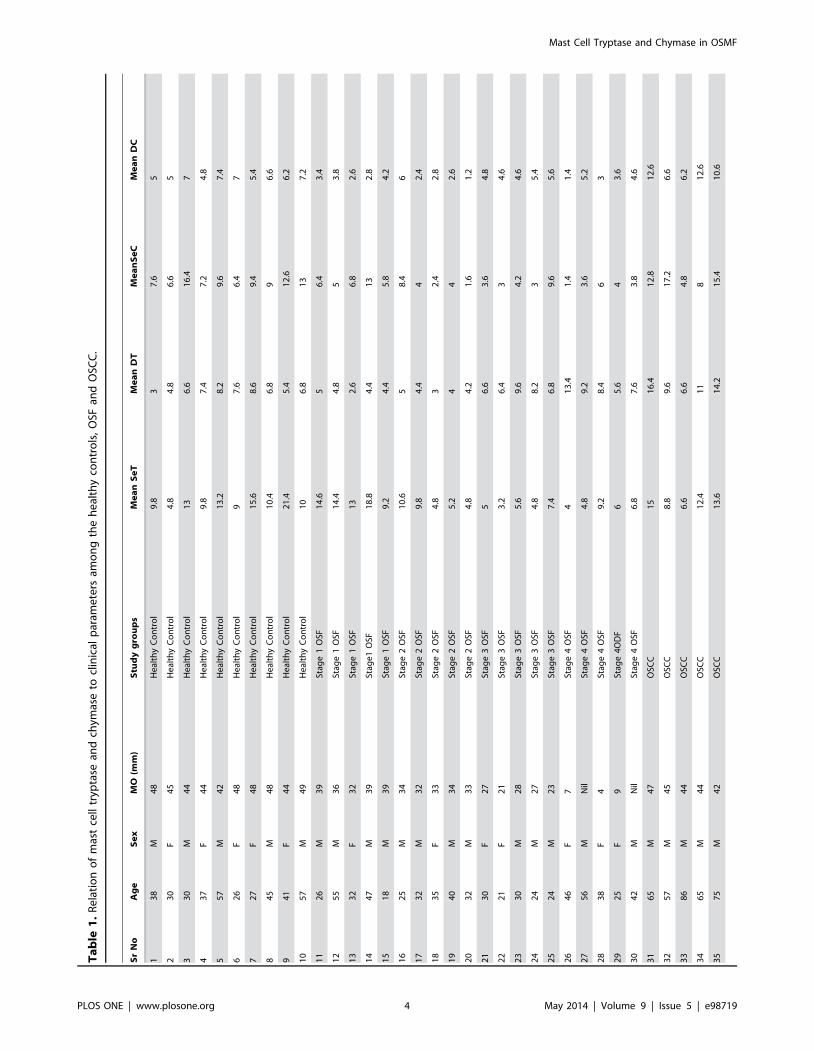
Ta
ble
1.
Re
lati
on
of
mas
tce
lltr
ypta
sean
dch
ymas
eto
clin
ical
par
ame
ters
amo
ng
the
he
alth
yco
ntr
ols
,O
SFan
dO
SCC
.
Sr
No
Ag
eS
ex
MO
(mm
)S
tud
yg
rou
ps
Me
an
Se
TM
ea
nD
TM
ea
nS
eC
Me
an
DC
13
8M
48
He
alth
yC
on
tro
l9
.83
7.6
5
23
0F
45
He
alth
yC
on
tro
l4
.84
.86
.65
33
0M
44
He
alth
yC
on
tro
l1
36
.61
6.4
7
43
7F
44
He
alth
yC
on
tro
l9
.87
.47
.24
.8
55
7M
42
He
alth
yC
on
tro
l1
3.2
8.2
9.6
7.4
62
6F
48
He
alth
yC
on
tro
l9
7.6
6.4
7
72
7F
48
He
alth
yC
on
tro
l1
5.6
8.6
9.4
5.4
84
5M
48
He
alth
yC
on
tro
l1
0.4
6.8
96
.6
94
1F
44
He
alth
yC
on
tro
l2
1.4
5.4
12
.66
.2
10
57
M4
9H
eal
thy
Co
ntr
ol
10
6.8
13
7.2
11
26
M3
9St
age
1O
SF1
4.6
56
.43
.4
12
55
M3
6St
age
1O
SF1
4.4
4.8
53
.8
13
32
F3
2St
age
1O
SF1
32
.66
.82
.6
14
47
M3
9St
age
1O
SF1
8.8
4.4
13
2.8
15
18
M3
9St
age
1O
SF9
.24
.45
.84
.2
16
25
M3
4St
age
2O
SF1
0.6
58
.46
17
32
M3
2St
age
2O
SF9
.84
.44
2.4
18
35
F3
3St
age
2O
SF4
.83
2.4
2.8
19
40
M3
4St
age
2O
SF5
.24
42
.6
20
32
M3
3St
age
2O
SF4
.84
.21
.61
.2
21
30
F2
7St
age
3O
SF5
6.6
3.6
4.8
22
21
F2
1St
age
3O
SF3
.26
.43
4.6
23
30
M2
8St
age
3O
SF5
.69
.64
.24
.6
24
24
M2
7St
age
3O
SF4
.88
.23
5.4
25
24
M2
3St
age
3O
SF7
.46
.89
.65
.6
26
46
F7
Stag
e4
OSF
41
3.4
1.4
1.4
27
56
MN
ilSt
age
4O
SF4
.89
.23
.65
.2
28
38
F4
Stag
e4
OSF
9.2
8.4
63
29
25
F9
Stag
e4
OD
F6
5.6
43
.6
30
42
MN
ilSt
age
4O
SF6
.87
.63
.84
.6
31
65
M4
7O
SCC
15
16
.41
2.8
12
.6
32
57
M4
5O
SCC
8.8
9.6
17
.26
.6
33
86
M4
4O
SCC
6.6
6.6
4.8
6.2
34
65
M4
4O
SCC
12
.41
18
12
.6
35
75
M4
2O
SCC
13
.61
4.2
15
.41
0.6
Mast Cell Tryptase and Chymase in OSMF
PLOS ONE | www.plosone.org 4 May 2014 | Volume 9 | Issue 5 | e98719

tests, p-values ,0.05 were considered to be statistically
significant.
Results
The present study consisted of 20 OSF, 10 OSCC and 10
healthy controls. There were 5 cases in each stage of OSF
(Stage 1 to Stage 4). The patients with OSF were in age group
of 18 to 75 years, with the mean age being 33.9 years with a
marked male predominance (male to female ratio of 13:7)
(Table 1) There are two populations of mast cells, those
containing only tryptase (MCT) and those containing both
tryptase and chymase (MCTC), only chymase containing MCs
are extremely rare, therefore all MCs contain tryptase [21],
[35]. Tryptase positive MCs represented total MCs, while the
number of chymase positive MCs can be considered equal to
number of MCTC. To determine MC subpopulations (MCT,
MCTC) in healthy controls, OSF and OSCC, the formula:
Total MCs = MCT+MCTC was used as previously described
[36]. In all tissues examined, the predominant MC population
was positive for both tryptase and chymase. (Table 2).
Subepithelial and deep distribution of tryptase positiveMCs and chymase positive MCs between healthycontrols, OSF and OSCC group
We found a statistically significant increase in the number of
subepithelial as well as deeper distribution of tryptase positive MCs
in OSCC group when compared to OSF group (p,0.05).
Subepithelial and deeper distribution of tryptase positive MCs in
OSF group demonstrated no statistical significance when com-
pared to healthy controls. From our results, it can be compre-
hended that OSCC group has significantly more number of
tryptase positive MCs as compared to OSF group irrespective of
the area of distribution (Figure. 3).
We observed a statistically significant increase in the number
of subepithelial distribution of chymase positive MCs in OSCC
group when compared to healthy controls and OSF group (p,
0.05). Conversely, a statistically significant decrease in the
number of subepithelial distribution of chymase positive MCs
in OSF group was noted when compared to healthy controls
(p,0.05). Hence, it can be concluded that OSF has the least
number of chymase positive MCs subepithelially. We recorded
a statistically significant decrease in the number of deep
distribution of chymase positive MCs in the OSF group when
compared to healthy controls and OSCC group (p,0.05)
(Figure. 4).
Subepithelial distribution of tryptase positive MCs inhealthy controls, different stages of OSF and OSCC group(Figure. 5.)
Stage I OSF showed a statistically significant increase in the
number of tryptase positive MCs subepithelially as compared to
other stages of OSF, however, no significant difference was noted
between stage 2, 3 and 4 OSF (Figure. 6).
Deep distribution of tryptase positive MCs in healthycontrols, different stages of OSF and OSCC group(Figure. 7)
Deeper distribution of tryptase positive MCs was greater in
healthy controls, OSCC group and advanced stages of OSF (Stage
3 and Stage 4) as compared to early stages of OSF (Stage 1 and
stage 2) (Figure. 6).Ta
ble
1.
Co
nt.
Sr
No
Ag
eS
ex
MO
(mm
)S
tud
yg
rou
ps
Me
an
Se
TM
ea
nD
TM
ea
nS
eC
Me
an
DC
36
55
M4
7O
SCC
10
.61
11
3.6
16
.8
37
50
M4
5O
SCC
12
.21
3.8
13
7.8
38
47
M4
8O
SCC
18
.21
9.2
12
.65
.2
39
62
M4
4O
SCC
14
.41
6.4
16
.62
.6
40
59
M4
9O
SCC
34
.81
92
22
0.8
M,
mal
e;
F,fe
mal
e;
SeT
,su
be
pit
he
lial
tryp
tase
;D
T,
de
ep
tryp
tase
;Se
C,
sub
ep
ith
elia
lch
ymas
e;
DC
,d
ee
pch
ymas
e;
OSF
,o
ral
sub
mu
cou
sfi
bro
sis;
OSC
C,
ora
lsq
uam
ou
sce
llca
rcin
om
a.d
oi:1
0.1
37
1/j
ou
rnal
.po
ne
.00
98
71
9.t
00
1
Mast Cell Tryptase and Chymase in OSMF
PLOS ONE | www.plosone.org 5 May 2014 | Volume 9 | Issue 5 | e98719

Subepithelial distribution of chymase positive MCs inhealthy controls, different stages of OSF and OSCC group(Figure. 8)
No statistically significant difference for the subepithelial
distribution of chymase positive MCs was observed among
different stages of OSF. A statistically significant increase in the
number of subepithelial distribution of chymase positive MCs was
noted in the OSCC group when compared to all the stages of
OSF. Conversely, barring Stage 1 OSF, all stages of OSF showed
a statistically decrease in the subepithelial distribution of chymase
positive MCs when compared to healthy controls (Figure. 9).
Deep distribution of chymase positive MCs in healthycontrols, different stages of OSF and OSCC group(Figure. 10)
A statistically significant decrease in the deeper distribution
of chymase positive MCs was observed in all the stages of OSF
when compared to healthy controls & OSCC group. OSCC
group showed a statistically significant increase in chymase
positive MCs in deeper areas when compared to healthy
controls. However no statistically significant correlation was
found for chymase positive MCs in deeper areas among
different stages of OSF. OSCC group showed maximum
number of chymase positive MCs in the deeper region when
compared to all the other study groups (Figure. 9).
Discussion
OSF is a chronic, inflammatory, premalignant fibrotic condition
characterized by excessive deposition of collagen in the submu-
cosa, leading to restricted mouth opening [19]. MC activation is a
characteristic feature of chronic inflammation that may lead to
fibrosis as a result of increased collagen synthesis by fibroblasts
[37]. To the best of our knowledge this is the first study to
emphasize the possible role of MC tryptase and chymase in OSF
and its malignant transformation.
The enzyme profile of MCs in oral tissues resembles that of skin,
with most MCs expressing the serine proteases, tryptase and
chymase [38], [39]. Total MC distribution was higher in
subepithelial region than the deeper connective tissue in all the
study groups which was in accordance with other findings in
literature [40]. From our results, it can be comprehended that
OSF group showed the least while OSCC group showed the
maximum number of tryptase positive MCs and chymase positive
MCs irrespective of the area of distribution amongst the study
groups. Thus, our findings support the idea of the possible role of
MC tryptase and chymase in the pathogenesis of OSF and their
role in upregulation of tumor angiogenesis during its malignant
transformation.
Role of MC tryptase and chymase in OSFThe fibrogenic cytokines influencing the fibrotic process are
shown to play an important role in regulating fibroblast function,
such as proliferation, migration, matrix synthesis and is likely to
Table 2. Subepithelial and deep distribution of mean MCT and MCTC counts in the healthy controls, OSF and OSCC.
TMC MCTC MCT
Subepithelial
Healthy controls 11.68 9.78 1.9
OSF 8.10 4.98 3.12
OSCC 15.66 13.60 2.06
Deep
Healthy controls 6.52 6.16 0.36
OSF 5.68 3.68 2.00
OSCC 13.72 12.18 1.54
TMC, total mast cells; MCTC, tryptase and chymase positive mast cells; MCT, tryptase positive mast cells.doi:10.1371/journal.pone.0098719.t002
Figure 3. Mean value comparison of subepithelial and deeptryptase positive mast cells in healthy controls, OSF, andOSCC.doi:10.1371/journal.pone.0098719.g003
Figure 4. Mean value comparison of subepithelial and deepchymase positive mast cells in healthy controls, OSF, andOSCC.doi:10.1371/journal.pone.0098719.g004
Mast Cell Tryptase and Chymase in OSMF
PLOS ONE | www.plosone.org 6 May 2014 | Volume 9 | Issue 5 | e98719

play a key role in regulating the initiation and progression of
scarring in any fibrotic disease. The present study demonstrated a
significantly higher subepithelial distribution of tryptase positive
MCs than chymase positive MCs in the early stages of OSF
against the increased distribution of tryptase positive MCs than
chymase positive MCs in deeper zones of the advanced stages of
Figure 5. Subepithelial distribution of tryptase positive mast cells. A, Healthy control;. B, Stage 1 OSF. C, Stage 2 OSF. D, Stage 3 OSF. E,Stage 4 OSF. F, OSCC.doi:10.1371/journal.pone.0098719.g005
Figure 6. Mean value comparison of subepithelial and deep tryptase positive mast cells in healthy controls, different stages of OSF,and OSCC.doi:10.1371/journal.pone.0098719.g006
Mast Cell Tryptase and Chymase in OSMF
PLOS ONE | www.plosone.org 7 May 2014 | Volume 9 | Issue 5 | e98719

OSF, which was in accordance with other findings in the literature
[13], [14], [15], [16], [17], [18], [19]. This trend of initial increase
in number of MC in stage I OSF followed by subsequent decrease
in number of MC in later stages of OSF could be attributed to the
initial inflammatory response of oral mucosa to the exogenous
irritants and carcinogens like areca nut and tobacco. Subsequent-
ly, healing response in the form of fibrosis may be responsible for
the decrease in total MC in Stage 2, 3 and 4 OSF.
MC tryptase is a major protease & produces mitogenic effects
on various types of cultured cells such as smooth muscles and
bronchial epithelial cells. Tryptase has in turn been described to
activate TGF-b and collagenase, induce C3a and collagen mRNA,
cleave type IV collagen, fibronectin, elastase, and proteoglycans,
and induce fibroblast proliferation [41]. Tryptase has also been
described to induce fibroblast procollagen mRNA upregulation.
Studies have revealed that tryptase is a strong mitogen in its own
right, but it synergizes its action with more traditional growth
factors such as PDGF & FGF. Other investigators have shown that
tryptase stimulates fibroblast chemotaxis and production of
collagen [42], [43], however, the mechanism of the signaling
event that mediates the fibrogenic effects of tryptase remains
unclear.
A statistically significant decrease in subepithelial & deep
distribution of chymase positive mast cells in all the stages of
OSF (except in stage 1 OSF subepithelial distribution) in
comparison to healthy controls and OSCC group made us to
propose the possible role of MC chymase in the pathogenesis of
OSF and its malignant transformation. Chymase can activate
collagenase and stromelysin, destruct vitronectin and fibronectin,
and induce fibroblast proliferation, suggesting an important role of
this mast-cell-specific protease in tissue matrix turnover and
renewal. Chymase has been shown to degrade the extra-cellular
matrix (ECM) and basement membrane components, digest
specific neuropeptides, and convert pro-IL-1B to its active
molecule. Furthermore, it can cleave soluble stem cell factor
(SCF) from its membrane form [44] and may thus contribute to
the influx of MC precursors and to their in situ differentiation.
Recent in-vitro studies have shown that MCs chymase also plays
an important role in fibrosis [26]. Kofford et al [45] have reported
that human chymase cleaves type I precollagen to form collagen
fibrils in vitro. In addition, it has been reported that angiotensin II
stimulates fibroblast proliferation through the activation of TGF–
b1 [46]. Normally, several protease inhibitors within the
connective tissue ensure tissue homeostasis by inhibiting excessive
activities of chymase in the immediate MC environment. Such
natural inhibitors are not known for MC-specific tryptase which is
less active than chymase in connective tissue remodelling and
fibroblast proliferation, resulting in shorter active life of chymase
after secretion, thereby MC-derived tryptase can be considered as
Figure 7. Deep distribution of tryptase positive mast cells. A, Healthy control. B, Stage 1 OSF. C, Stage 2 OSF. D, Stage 3 OSF. E, Stage 4 OSF.F, OSCC.doi:10.1371/journal.pone.0098719.g007
Mast Cell Tryptase and Chymase in OSMF
PLOS ONE | www.plosone.org 8 May 2014 | Volume 9 | Issue 5 | e98719

the main mediator stimulating fibroblast proliferation in the event
of fibrosis [37].
MCs are a major source of other pro-fibrotic cytokines like
bFGF [15], TGF-b [20], [22], [23], IL-6 [47], and their
upregulation has been studied extensively in OSF. Thus, we
suggest that a combination of tryptase and chymase along with the
various fibrogenic cytokines seems to be an important factor in the
development of OSF (Figure. 11).
Figure 8. Subepithelial distribution of chymase positive mast cells. A, Healthy control. B, Stage 1 OSF. C, Stage 2 OSF. D, Stage 3 OSF. E,Stage 4 OSF. F, OSCC.doi:10.1371/journal.pone.0098719.g008
Figure 9. Mean value comparison of subepithelial and deep tryptase positive mast cells in healthy controls, different stages of OSF,and OSCC.doi:10.1371/journal.pone.0098719.g009
Mast Cell Tryptase and Chymase in OSMF
PLOS ONE | www.plosone.org 9 May 2014 | Volume 9 | Issue 5 | e98719

From the above results it can be inferred that there is an altered
balance in the subepithelial and deep distribution of tryptase
positive MCs and chymase positive MCs as it progresses from
healthy controls to OSF, thereby playing an important role in the
pathogenesis of OSF. From the present study it can also be
deduced that fibrosis in OSF may be initiating in the subepithelial
zone in early stages of OSF (Stage 1 and stage 2) and gradually
progressing towards the deeper muscle layer in advanced stages of
OSF (Stage 3 and Stage 4). We observed a greater number of MCs
in the muscle bundles of advanced stages than early stages of OSF.
Based on our findings we assume that process of fibrosis in OSF
remains uninterrupted since activated resident MCs continue to
secrete fibrogenic & angiogenic cytokines even after the cessation
of the areca nut chewing habit, resulting in irrevocable fibrosis.
Role of MC tryptase and chymase in tumour progressionThe pathogenesis of OSF and circumstances leading to its
proven pre-cancerous outcome have always aroused curiosity but
remained enigmatic till date. Fibroblast density and activation are
increased in physiological responses, such as wound healing, and
in disease states, such as fibrotic disorders and carcinogenesis [48],
[49], [50], [51]. During epithelial carcinogenesis, fibroblasts have
paracrine and autocrine interactions with resident and immune
cells that include keratinocytes and MCs [52], [53], [54].
Fibroblasts can stimulate keratinocyte migration, proliferation,
and malignant transformation [55], [56]. MCs and transformed
epithelial cells stimulate fibroblasts to acquire an activated
phenotype that favors tumor progression [56], [57], [58], [59].
Increased fibroblast density has been described previously during
the formation of a reactive stroma in several neoplasia, including
intraoral cancer and skin [49], [51], [57]. In the light of finding of
the present study MC-derived tryptase can be considered as an
important mediator of fibroblast activation and proliferation
during malignant transformation of OSF since we observed a
statistically significant increase in tryptase positive MCs in OSCC
group as compared to other study groups [60].
Tumours require a blood supply for their expansive growth.
Solid tumours, in order to outgrow the size of 2 mm3, demand for
oxygen supply, a fact that makes necessary the formation of new
microvasculature [61]. This progressive angiogenesis is the
outcome of an imbalance between positive and negative angio-
genic factors produced by both tumor and host cells. Among the
host cells, which produce and release in a considerable quantity
pro-angiogenic and angiogenic factors are MCs. The cancer
stimulating mechanisms operated by MCs include participation in
immunosuppression, the release of proangiogenic and mitogenic
factors and involvement in the degradation of the extracellular
matrix. MCs contain many angiogenic factors and a variety of
cytokines, such as histamine, heparin, tryptase, chymase, PDGF,
TNF-a, bFGF, TGF-b, IL-6 and vascular endothelial growth
Figure 10. Deep distribution of distribution of chymase positive mast cells. A, Healthy control. B, Stage 1 OSF. C, Stage 2 OSF. D, Stage 3OSF. E, Stage 4 OSF. F, OSCC.doi:10.1371/journal.pone.0098719.g010
Mast Cell Tryptase and Chymase in OSMF
PLOS ONE | www.plosone.org 10 May 2014 | Volume 9 | Issue 5 | e98719

factor (VEGF) [21].Upregulation of the above pro-angiogenic
cytokines in OSF has already been demonstrated in the literature
[22], [23], [24], [25], [62]. The present study demonstrated a
statistically significant increase in subepithelial distribution of
chymase positive MCs in OSCC group when compared to healthy
controls and OSF group. Chymase is known for its ability to
promote extracellular matrix (ECM) degradation and for indirect-
ly stimulating angiogenesis. Chymase activates latent matrix
metalloproteinases (MMPs), including gelatinase B and procolla-
genase, which degrade components of epithelial basement
membranes and ECM, respectively. These responses are essential
for tumor invasion and metastasis [63]. We have already
established the possible role of angiogenesis in malignant
transformation of OSF by demonstrating CD34 positive blood
vessels [63] and VEGF in atrophic epithelium of OSF [62].
As we have observed a significant increase in the tryptase and
chymase positive MCs in OSCC group irrespective of the areas
of distribution when compared to OSF group, it suggests that
upregulation of MCs may play a crucial role in tumour
progression during malignant transformation of atrophic
epithelium in OSF (Figure.7.). A similar increase in the number
of MC tryptase, a potent proangiogenic factor has been
documented in various malignancies including oral cancers
[26], [33], [64]. Hence, we would like to state that the event of
fibrosis in the early OSF is probably a protective mechanism
manifested in the form of excessive collagen deposition,
primarily initiated in the connective tissue of the oral mucosa
in order to prevent the deeper penetration of carcinogenic
substances, while malignant transformation in the advanced
OSF is entirely a different issue principally affecting the atrophic
epithelium of OSF if the carcinogenic insult persists.
As part of the process of oral mucosal carcinogenesis in OSF,
the connective tissue adjacent to the epithelium gets affected by
the topically applied chemical carcinogen and plays a directive
role, not yet understood, in the epithelial changes observed. It
appears likely that extracellular matrix molecules, growth factors,
angiogenic cytokines and proteinases co-operate in influencing
epithelium via an autocrine proliferative effect on the atrophic
epithelium, while paracrine stimulation of the vascular network
may maintain survival and growth [19].
In summary, we present data indicating that MC tryptase and
chymase contribute to the development of OSF and malignant
transformation of the overlying epithelium. Several therapeutic
approaches such as the use of MC stabilizer, blockade of stem cell
Figure 11. Schematic presentation of speculative hypothesis proposing possible role of mast cell tryptase and chymase in thepathogenesis of OSF and their possible role in tumor progression and malignant transformation of the overlying epithelium. IL-1,Interleukin-1. IL-6, Interleukin 6. TNF-a, Tumor necrosis factor a. TGF-b, Transforming growth factor b. PDGF, Platelet derived growth factor. VEGF,Vascular endothelial growth factor. B-FGF, Basic fibroblast growth factor.doi:10.1371/journal.pone.0098719.g011
Mast Cell Tryptase and Chymase in OSMF
PLOS ONE | www.plosone.org 11 May 2014 | Volume 9 | Issue 5 | e98719

factor or inhibitors of tryptase, chymase and TGF-b have already
demonstrated some clinical utility in tissue fibrosis or inflammatory
diseases, by inhibiting MC activation. Considering the important
role of MC tryptase and chymase in the pathogenesis of OSF,
further studies using either administration of a neutralizing
antibody against tryptase and chymase or application of a MC
tryptase knock-out mouse strategy in experimental models of OSF
are required for more definitive determination of the role of
tryptase and chymase in the development of OSF.
Acknowledgments
The authors thank Dr. Anita Borges for her kind support during the study
period, and Dr. Dnyaneshwar Gajbhare for his assistance in statistical
analysis.
Author Contributions
Conceived and designed the experiments: RSD. Performed the experi-
ments: AY BAB. Analyzed the data: AY BAB JSS RM APN. Contributed
reagents/materials/analysis tools: AY RSD. Contributed to the writing of
the manuscript: RSD AY JSS.
References
1. Kumar KK, Saraswathi TR, Ranganathan K, Devi UM, Joshua E (2007) Oral
Submucous fibrosis; A clinicopathological study in Chennai. Ind J Dent
Res.18:106–111.
2. Rajalalitha P, Vali S (2005) Molecular pathogenesis of oral submucous fibrosis –
a Collagen metabolic disorder. J Oral Pathol Med; 34:321–328.
3. Angadi PV, Kale AD, Hallikerimath S (2011) Evaluation of myofibroblasts in
oral submucous fibrosis: correlation with disease severity. J Oral Pathol Med.
40:208–213.
4. Punnya V, Angadi SS (2011) Role Areca nut in pathogenesis of oral submucous
fibrosis: revisited. Oral and Maxillofac Surg. 15:1–9.
5. Cairns JA, Walls AF (1997) Mast cell tryptase stimulates the synthesis of type I
collagen in human lung fibroblasts. J Clin Invset 99:1313–1321.
6. Choi KL, Claman HN (1987) Mast cells, fibroblasts and fibrosis. New clues to
the riddle of mast cells. Immunol Res 6:145–152.
7. Akers IA, Parsons M, Hill MR, Hollenberg MD, Sanjar S (2000) Mast cell
tryptase stimulates human lung fibroblast proliferation via protease-activated
receptor-2. Am J Physiol Lung Cell Mol Physiol. 278:L193–L201.
8. Frungeiri MB, Weidinger S, Meineke V, Kohn FM, Mayerhofer A (2002)
Proliferative action of mast-cell tryptase is mediated by PAR2, COX2,
prostaglandins, and PPAR: Possible relevance to human fibrotic disorders. Proc
Natl Acad Sci USA 99:15072–15077.
9. Farrell DJ, Hines JE, Walls AF, Kelly PJ, Bennett MK, et al. (1995) Intrahepatic
mast cells in chronic liver diseases. Hepatology 22:1175–1181.
10. Akimoto S, Ishikawa O, Igarashi Y, Kurosawa M, Miyachi Y (1998) Dermal
mast cells in scleroderma: their skin density, tryptase/chymase phenotypes and
degranulation.Br J Dermatol. 138:399–406.
11. Wang HW, Tedla N, Hunt JE, Wakefield D, McNeil HP (2005) Mast cell
accumulation and cytokine expression in the tight skin mouse model of
scleroderma. Exp Dermatol 14:295–302.
12. Roberts IS, Brenchley PE (2000) Mast cells: the forgotten cells of renal fibrosis.
J Clin Pathol 53: 858–862.
13. Bhatt AP, Dholakia HM (1997) Mast cell density in oral submucous fibrosis. J
Ind Dent Assoc. 49:187–191.
14. Ankle MR, Kale AD, Nayak R (2007) Mast cells are increased in leukoplakia,
oral submucous fibrosis, oral lichen planus and oral squamous cell carcinoma.
J Oral and Maxillofac Pathol 11:18–22.
15. Bishen KA, Radhakrishnan R, Satyamoorthy K (2008) The role of basic
fibroblast growth factor in oral submucous fibrosis pathogenesis. J Oral Pathol
Med 37:402–411.
16. Sabarinath B, Sriram G, Saraswathi TR, Sivapathasundharam B (2011)
Immunohistochemical evaluation of mast cells and vascular endothelial
proliferation in oral submucous fibrosis. Ind J Dent Res. 22:116–121.
17. Pujari R, Vidya N (2013) Mast cell density in oral submucous fibrosis: a possible
role in pathogenesis. Int J Health Sci, Qassim University. 7:23–29.
18. Chavan S, Deshmukh SR (2013) Quantitative analysis of mast cells in oral
submucous fibrosis. Al Ameen J Med Sci 6:144–149.
19. Khatri MJ, Desai RS, Mamatha GS, Kulkarni M, Khatri J (2013)
Immunohistochemical expression of mast cells using c- kit in various grades of
oral submucous fibrosis. ISRN Pathology Article ID 543976. 5 Pages.
20. Kale AD, Mane DR, Shukla D (2013) Expression of transforming growth factor
b and its correlation with lipodystrophy in oral submucous fibrosis: an
Immunohistochemical study. Med Oral Pathol Oral Cir Bucal. 18:e12–18.
21. Metcalfe DD, Baram D, Mekori Y (1997) Mast cells. Physiol Rev. 77:1033–
1079.
22. Haque MF, Harris M, Meghji S, Speight PM (1997) An immunohistochemical
study of oral submucous fibrosis. J Oral Pathol Med. 26:75–82.
23. Haque MF, Harris M, Meghji S, Barrett AW (1998) Immunohistochemical
localization of cytokines and growth factors in oral submucous fibrosis. Cytokine.
10:713–719.
24. Tsai CC, Chen CC, Lin CC, Chen CH, Lin TS, et al. (1999) Interleukin-1 beta
in oral submucous fibrosis, verrucous hyperplasia and squamous cell carcinoma
tissues. Kaohsiung J Med Sci. 15:513–519.
25. Khan I, Agarawal P, Thangiam GS, Radhesh R, Rao SG, et al. (2011) Role of
TGF-b and BMP 7 in pathogenesis of oral submucous fibrosis. Growth Factor.
29: 119–127.
26. Fukushima H, Ohsawa M, Ikura Y, Naruka T, Sugama Y, et al. (2006) Mastcells in diffuse large B-cell lymphoma: their role in fibrosis. Histopathol 49:498–
505.
27. Folkman J (1985) Tumour angiogenesis. Adv Cancer Res. 43:203.
28. Folkman J, Shing Y (1992) Angiogenesis. J Biol Chem. 267:10931–10934.
29. Yoshiji H, Gomez DE, Shibuya M, Thorgeirsson UP (1996) Expression of
vascular endothelial growth factor, its receptor, and other angiogenic factors in
human breast cancer. Cancer Res. 56:2013–2016.
30. Weidner N, Semple JP, Welch WR, Folkman J (1991) Tumor angiogenesis and
metastasis—correlation in invasive breast carcinoma. N Engl J Med. 324:1–8.
31. Tanigawa N, Amaya H, Matsumura M, Shimomatsuya T (1997) Correlation
between expression of vascular endothelial growth factor and tumor vascularity,
and patient outcome in human gastric carcinoma. J Clin Oncol 15:826–832.
32. Kyzas PA, Stefanou D, Batistatou A, Agnantis NJ (2005) Prognostic significance
of VEGF immunohistochemical expression and tumor angiogenesis in head andneck squamous cell carcinoma. J Cancer Res Clin Oncol 131:624–630.
33. Iamaroon A, Pongsiriwet S, Jittidecharaks S, Pattanaporn K, Prapayasatok S, etal. (2003) Increase of mast cells and tumor angiogenesis in oral squamous cell
carcinoma. J Oral Pathol Med 32:195–199.
34. Elpek GO, Gelen T, Aksoy NH, Erdogan A, Dertsiz L, et al. (2001) The
prognostic relevance of angiogenesis and mast cells in squamous cell carcinoma
of the oesophagus. J Clin Pathol. 54:940–944.
35. Irani AA, Schechter NM, Craig SS, DeBlois G, Schwartz LB (1986) Two types
of human mast cells have distinct neutral protease compositions. Proc Natl AcadSci USA. 83:4464–4468.
36. Rojas IG, Spencer MI, Martinez A, Marurellia MA, Rudolph MI (2005)
Characterization of mast Cell subpopulation in lip Cancer. J Oral Pathol Med.34:268–273.
37. Caughey GH (2007) Mast cell tryptase and chymase in inflammation and hostdefense. Immunol Rev. 217:141–154.
38. Walsh LJ (2003) Mast cells and oral inflammation. Crit Rev in Oral Biol Med.14:188–198.
39. Walsh LJ, Davis MF, Xu LJ, Savage NW (1995) Relationship between mast celldegranulation and inflammation in the oral cavity. J Oral Pathol Med. 24:266–
272.
40. Roukonen H, Hietanen J, Malmstrom M, Sane J, Hayrinen RI, et al. (1993)Peripheral nerves and mast cells in normal buccal mucosa. J Oral Pathol Med.
22:30–34.
41. Algermissen B, Bauer F, Schadendorf, Kroopp JD, Czarnetzki BM (1994)
Analysis of mast cell subpopulations (MCT, MCTC) in cutaneous inflammationusing novel enzyme-histochemical staining techniques. Exp Dermatol. 3:290–
297.
42. Gruber BL, Kew RR, Jelaska A, Marcheses MJ, Garlick J, et al. (1997) Humanmast cells activate fibroblasts: Tryptase is a fibrogenic factor stimulating collagen
messenger ribonucleic acid synthesis and fibroblast chemotaxis. J Immunol158:2310–2317.
43. Ruoss SJ, Thomas H, Caughey GH (1991) Mast cell tryptase is a mitogen for
cultured fibroblasts. J Clin Invest. 88:439–499.
44. Longley BJ, Tyrrell L, Ma Y, Williams DA, Halaban R, et al. (1997) Chymase
cleavage of stem cell factor yields a bioactive, soluble product. Proc Natl AcadSci USA. 94:9017–9021.
45. Kofford MW, Schwartz LB, Schecter NW, Yager DR, Diegelmann RF, et al.(1997) Cleavage of type II precollagen by human mast cell chymase initiates
collagen fibril formation and generates a unique carboxyl-terminal propeptide. J
Biol Chem. 272:7127–7131.
46. Lingstedt KA, Wang Y, Shiota N, Saarinen J, Hytiasnen M, et al. (2001)
Activation of paracrine TGF-b1 signalling upon stimulation and degranulationof rat serosal mast cells: a novel function for chymase. FASEB J. 15:1377–1388.
47. Chen CC, Huang JF, Tsai CC (1995) In vitro production of interleukin-6 byhuman gingival, normal buccal mucosa, and oral submucous fibrosis fibroblasts
treated with betel-nut alkaloids. Kaohsiung J Med Sci. 11:604–614.
48. Martin P (1997) Wound healing-aiming for perfect skin regeneration. Science.
276:75–81.
49. Kalluri R, Zeisberg M (2006) Fibroblasts in cancer. Nat Rev Cancer. 6: 392–401.
50. Wynn TA (2008) Cellular and molecular mechanisms of fibrosis. J Pathol.214:199–210.
Mast Cell Tryptase and Chymase in OSMF
PLOS ONE | www.plosone.org 12 May 2014 | Volume 9 | Issue 5 | e98719

51. Thode C, Jorgensen TG, Dabelsteen E, Mackenzie I, Dabelsteen S (2011)
Significance of myofibroblasts in oral squamous cell carcinoma. J Oral PatholMed. 40: 201–207.
52. Sorrell JM, Caplan AI (2004) Fibroblast heterogeneity: more than skin deep. J
Cell Sci. 117(:667–675.53. Taun TL, Keller LC, Sun D, Nimni ME, Cheung D (1994) Dermal fibroblasts
activate keratinocyte outgrowth on collagen gels. J Cell Sci. 107:2285–2289.54. Trautmann A, Krohne G, Brocker EB, Klein CE (1998) Human Mast cells
augment fibroblast proliferation by heterotypic cell-cell adhesion and action of
IL-4. J Immunol. 160: 5053–5057.55. Krtolica A, Parrinello S, Lockett S, Desprez PY, Campisi J (2001) Senescent
fibroblasts promote epithelial cell growth and tumorigenesis: a link betweencancer and aging. Proc Natl Acad Sci USA. 98:12072–12077.
56. Bhowmick NA, Neilson EG, Moses HL (2004) Stromal fibroblasts in cancerinitiation and progression. Nature 432:332–337.
57. Coussens LM, Raymond WW, Bergers G (1999) Inflammatory mast cells
upregulate angiogenesis during squamous epithelial carcinogenesis. Genes Dev.13: 1382–1397.
58. Artuc M, Steckelings UM, Henz BM (2002) Mast cell-fibroblast interactions:human mast cells as source and inducers of fibroblast and epithelial growth
factors. J Invest Dermatol. 118: 391–395.
59. Mueller MM, Fusenig NE (2004) Friends or foes–bipolar effects of the tumour
stroma in cancer. Nat Rev Cancer. 4:839–849.
60. Rojas IG, Boza YV, Spencer ML, Flores M, Martinez A (2012) Increased
fibroblast density in actinic cheilitis: association with tryptase-positive mast cells,
actinic elastosis and epithelial p53 and COX-2 expression. J Oral Pathol Med.
41:27–33.
61. Folkman J (2000) Tumor Angiogenesis.In: Cancer Medicine, 5th ed, London:
Decker pp 13–52.
62. Desai RS, Mamatha GS, Khatri MJ, Shetty SJ (2012) Immunohistochemical
expression of vascular endothelial growth factor (VEGF) and its possible role in
tumor progression during malignant transformation of atrophic epithelium in
oral submucous fibrosis. Current Angiogenesis 1:347–53.
63. Desai RS, Mamatha GS, Khatri MJ, Shetty SJ (2010) Immunohistochemical
expression of CD34 for characterization and quantification of mucosal
vasculature and its probable role in malignant transformation of atrophic
epithelium in oral submucous fibrosis. Oral Oncol. 46:553–558.
64. Benitez-Bribiesca L, Wong A, Utera D, Castellanos E (2001) The role of mast
cell tryptase in neoangiogenesis of premalignant and malignant lesion of uterine
cervix. J Histochem and Cytochem 49:1061–1062.
Mast Cell Tryptase and Chymase in OSMF
PLOS ONE | www.plosone.org 13 May 2014 | Volume 9 | Issue 5 | e98719