Algal Proteins: Extraction, Application, and Challenges ... - MDPI
34
foods Review Algal Proteins: Extraction, Application, and Challenges Concerning Production Stephen Bleakley 1,2 and Maria Hayes 1, * 1 Food Biosciences Department, Teagasc Ashtown Food Research Centre, Ashtown, Dublin D15 KN3K, Ireland; [email protected] 2 School of Biological Sciences, College of Sciences and Health and Environment, Sustainability and Health Institute, Dublin Institute of Technology, Kevin Street, Dublin D08 NF82, Ireland * Correspondence: [email protected]; Tel.: +353-1-805-9957 Academic Editor: Christopher J. Smith Received: 21 March 2017; Accepted: 20 April 2017; Published: 26 April 2017 Abstract: Population growth combined with increasingly limited resources of arable land and fresh water has resulted in a need for alternative protein sources. Macroalgae (seaweed) and microalgae are examples of under-exploited “crops”. Algae do not compete with traditional food crops for space and resources. This review details the characteristics of commonly consumed algae, as well as their potential for use as a protein source based on their protein quality, amino acid composition, and digestibility. Protein extraction methods applied to algae to date, including enzymatic hydrolysis, physical processes, and chemical extraction and novel methods such as ultrasound-assisted extraction, pulsed electric field, and microwave-assisted extraction are discussed. Moreover, existing protein enrichment methods used in the dairy industry and the potential of these methods to generate high value ingredients from algae, such as bioactive peptides and functional ingredients are discussed. Applications of algae in human nutrition, animal feed, and aquaculture are examined. Keywords: seaweed; microalgae; peptides; phycobiliproteins; biorefinery; bioavailability; extraction methods; legislation 1. Introduction The global population is expected to increase by over a third (2.3 billion people) by 2050, requiring an estimated 70% increase in food production [1]. A combination of improved agricultural food production methods and an increase of average per capita income have led to a decrease in global hunger over the last half-century, despite a doubling of the world’s population [1]. However, worldwide food production is now facing a greater challenge than ever before. Previously utilised methods of intensifying agriculture will soon no longer be an option due to the high impact trade-offs they have on the environment, including fragmenting natural habitats and threatening biodiversity, production of greenhouse gases from land clearing, fertilisers and animal livestock production, and nutrient run-off from fertiliser damaging marine, freshwater and terrestrial ecosystems [2]. In particular, protein is one of the main nutrients that will be in short supply in the future. Alternative protein sources and production methods are required to fulfil the demand of consumers and to meet predicted global protein requirements. Seaweed and microalgae are considered a viable source of protein. Some species of seaweed and microalgae are known to contain protein levels similar to those of traditional protein sources, such as meat, egg, soybean, and milk [3,4]. Algae use for protein production has several benefits over traditional high-protein crop use in terms of productivity and nutritional value. Seaweed and microalgae have higher protein yield per unit area (2.5–7.5 tons/Ha/year and 4–15 tons/Ha/year, respectively) compared to terrestrial crops, such as soybean, pulse legumes, and Foods 2017, 6, 33; doi:10.3390/foods6050033 www.mdpi.com/journal/foods
-
Upload
khangminh22 -
Category
Documents
-
view
0 -
download
0
Transcript of Algal Proteins: Extraction, Application, and Challenges ... - MDPI

foods
Review
Algal Proteins: Extraction, Application,and Challenges Concerning Production
Stephen Bleakley 1,2 and Maria Hayes 1,*1 Food Biosciences Department, Teagasc Ashtown Food Research Centre, Ashtown,
Dublin D15 KN3K, Ireland; [email protected] School of Biological Sciences, College of Sciences and Health and Environment, Sustainability and
Health Institute, Dublin Institute of Technology, Kevin Street, Dublin D08 NF82, Ireland* Correspondence: [email protected]; Tel.: +353-1-805-9957
Academic Editor: Christopher J. SmithReceived: 21 March 2017; Accepted: 20 April 2017; Published: 26 April 2017
Abstract: Population growth combined with increasingly limited resources of arable land and freshwater has resulted in a need for alternative protein sources. Macroalgae (seaweed) and microalgaeare examples of under-exploited “crops”. Algae do not compete with traditional food crops for spaceand resources. This review details the characteristics of commonly consumed algae, as well as theirpotential for use as a protein source based on their protein quality, amino acid composition, anddigestibility. Protein extraction methods applied to algae to date, including enzymatic hydrolysis,physical processes, and chemical extraction and novel methods such as ultrasound-assisted extraction,pulsed electric field, and microwave-assisted extraction are discussed. Moreover, existing proteinenrichment methods used in the dairy industry and the potential of these methods to generate highvalue ingredients from algae, such as bioactive peptides and functional ingredients are discussed.Applications of algae in human nutrition, animal feed, and aquaculture are examined.
Keywords: seaweed; microalgae; peptides; phycobiliproteins; biorefinery; bioavailability; extractionmethods; legislation
1. Introduction
The global population is expected to increase by over a third (2.3 billion people) by 2050,requiring an estimated 70% increase in food production [1]. A combination of improved agriculturalfood production methods and an increase of average per capita income have led to a decrease inglobal hunger over the last half-century, despite a doubling of the world’s population [1]. However,worldwide food production is now facing a greater challenge than ever before. Previously utilisedmethods of intensifying agriculture will soon no longer be an option due to the high impact trade-offsthey have on the environment, including fragmenting natural habitats and threatening biodiversity,production of greenhouse gases from land clearing, fertilisers and animal livestock production, andnutrient run-off from fertiliser damaging marine, freshwater and terrestrial ecosystems [2]. In particular,protein is one of the main nutrients that will be in short supply in the future. Alternative proteinsources and production methods are required to fulfil the demand of consumers and to meet predictedglobal protein requirements.
Seaweed and microalgae are considered a viable source of protein. Some species of seaweedand microalgae are known to contain protein levels similar to those of traditional protein sources,such as meat, egg, soybean, and milk [3,4]. Algae use for protein production has severalbenefits over traditional high-protein crop use in terms of productivity and nutritional value.Seaweed and microalgae have higher protein yield per unit area (2.5–7.5 tons/Ha/year and4–15 tons/Ha/year, respectively) compared to terrestrial crops, such as soybean, pulse legumes, and
Foods 2017, 6, 33; doi:10.3390/foods6050033 www.mdpi.com/journal/foods

Foods 2017, 6, 33 2 of 34
wheat (0.6–1.2 tons/Ha/year, 1–2 tons/Ha/year, and 1.1 tons/Ha/year, respectively) [5]. Terrestrialagriculture already requires approximately 75% of the total global freshwater with animal proteinin particular requiring 100 times more water than if the equivalent amount of protein was producedfrom plant sources [6,7]. Marine algae do not require freshwater or arable land to grow, maximisingresources that can be used for additional food production or other cash crops [5]. Furthermore, due totheir harsh environment and phototropic life, algae are often exposed to high oxidative and free-radicalstresses [8]. This has led to the evolution of natural protective systems, such as the productionof pigments (e.g., carotenes, chlorophylls, and phycobiliproteins) and polyphenols (e.g., catechins,flavonols, and phlorotannins), which can impart health benefits to the consumer when eaten [9,10].
However, widespread use of seaweed and microalgae is limited by a number of factors including;harvesting access and rights, seasonality and geographical location of algae, as well as the availabilityof scalable production methods for protein isolation from algae. Current processes of algal proteinisolation are time-consuming and economically unviable [11]. The objective of this paper is thereforeto discuss the value of algal proteins as a source of human nutrition, functional foods and animal feed,as well as describe current extraction methods and novel processing technologies that are used in dairyprocessing which may be employed to make algae a viable source of protein ingredients.
1.1. Characteristics of Seaweed
Algae are a diverse group of species which can be broadly described as oxygen-producing,photosynthetic, unicellular or multicellular organisms excluding embryophyte terrestrial plants andlichens [12]. Macroalgae can be divided into three main taxonomic groups based on their pigmentation;Phaeophyta (brown algae), Chlorophyta (green algae), and Rhodophyta (red algae) [13].
According the Food Balance sheets published by the Food and Agriculture Organisation of theUnited Nations (FAO), the Republic of Korea is the greatest consumer of seaweed (22.41 kg/capita/yearin 2013), followed by China and Japan [14]. Production of farmed seaweed has more than doubledworldwide since 2000, with particular expansion seen in Indonesia due to their vast areas of shallowsunlight coasts suitable for culture sites [15]. The culture of Japanese kelp, Laminaria japonica, hastraditionally been the most extensively farmed cold-water species. This was surpassed by the tropicalEucheuma seaweeds (Kappaphycus alvarezii and Eucheuma spp.) in 2010. The other most commonlyfarmed seaweed species are Gracilaria spp., Undaria sp., and Porphyra spp. [15].
Brown algae are distinguished by the presence of the pigment fucoxanthin, which is responsiblefor the distinctive olive-brown colour that lends this group its name [13]. Brown algae are alsounique among algae as they are only found in multicellular form [16]. There are approximately1500–2000 species of brown algae worldwide [17]. Some species, such as Macrocystis pyrifera(giant kelp), play an important role in the ecosystem, growing up to 20 m and forming underwaterkelp forests [18]. There are also many other species that have been exploited for human consumption,including Undaria pinnatifida (wakame), Hizikia fusiformis (hijiki), and Laminaria japonica (kombu) [19].Several types of brown algae are used for animal feed, including Laminaria digitata (oarweed),Ascophyllum nodosum (rockweed) and Fucus vesiculosus (bladder wrack) [3].
Green algae are a diverse group of approximately 8000 species, consisting of the divisionsChlorophyta and Charophyta [20]. Chlorophyta are a large group of unicellular and multicellularalgae, while charophyta are a smaller group of exclusively freshwater, multicellular green algae fromwhich it is believed that Embryophyta (terrestrial plants) evolved from [21]. Green algae obtaintheir pigmentation from chlorophyll a and b, as well as other pigments including β-carotene andxanthophylls [13]. The most commonly consumed species of green algae are Ulva spp. includingU. lactuca (sea lettuce), U. intestinalis, and U. compressa.
Red algae are a large group of mostly multicellular macroalgae with approximately6000 species [13]. Red algae are characterised by the presence of phycobilins, which are responsiblefor their red colour [13]. Species of Irish moss, such as Chondrus crispus and Mastocarpus stellatus, areexploited for their production of carrageenan [22]. Other species of red algae, including Porphyra tenera

Foods 2017, 6, 33 3 of 34
(nori) and Palmaria palmata (dulse), are among the highest consumed species of seaweed in Asia, as wellas Western countries, due to their high protein content and their delectable flavour [23]. In particular,Porphyra tenera is used in the production of sushi.
1.2. Characteristics of Microalgae
Microalgae are unicellular, microscopic organisms that are also considered as a viable alternativeprotein source. The most abundant microalgal divisions are Bacillariophyta (diatoms), Chlorophyta(green algae), Chrysophyta (golden algae), and Cyanophyta (blue-green algae). Microalgae are ahugely diverse group containing approximately 200,000 species [24]. Several of these species arecurrently exploited for a variety of biotechnological purposes, including cosmeceuticals, animal feed,fatty acids, alginates, wastewater treatment, and biofuel [9,25,26]. Furthermore, Arthrospira platensis(Spirulina), and Chlorella vulgaris (Chlorella) are also sold as functional foods due to their high vitaminand mineral content and as they are generally regarded as safe (GRAS) by the European Food SafetyAuthority (EFSA) [25]. Despite the relatively low quantity of microalgae produced annually comparedto that of seaweed (5000 tonnes dry matter per year versus 7.5 × 10⁶ tonnes dry matter per year,respectively), whole microalgal biomass and added-value compounds are economically valuable,representing a global turnover of about US $1.25 × 10⁹ per year, compared to annual seaweed turnoverof US $6 × 10⁹ [9].
Arthrospira platensis is a filamentous Cyanobacterium that has among the highest recorded proteincontent of any whole food [27]. Arthrospira sp. was originally referred to as Spirulina sp. untila re-examination showed that they are actually a distinct genus [9]. However, due to its widelypublicised use as a food and dietary supplement, the term Spirulina is often used interchangeablywith Arthrospira.
Chlorella spp. are spherical members of the phylum Chlorophyta (green algae) that have also seenincreased popularity as a food supplement in recent times. Chlorella vulgaris is the most commonlyexploited industry species due to its high protein content (51%–58% dry weight; dw) and favourableessential amino acid composition [28]. Chlorella also contains many other beneficial nutrients, includingβ-1,3-glucan, vitamins (B-complex and ascorbic acid), minerals (potassium, sodium, magnesium, iron,and calcium), β-carotene, chlorophyll, and Chlorella growth factor (CGF) [29].
2. Protein Quality
2.1. Amino Acid Composition
The quality of proteins can vary dramatically, depending on digestibility and the availability ofessential amino acids [30]. Animal sources of protein are generally considered as complete proteins, asthey are a rich source of essential amino acids (EAAs) which the human body is unable to biosynthesise.Alternatively, plant proteins are often considered an incomplete protein source as they commonly lackone or more of the essential amino acids, including histidine, isoleucine, leucine, lysine, methionine,phenylalanine, threonine, tryptophan, and valine [31]. However, the lacking essential amino acid(s) inplant-based proteins can differ, meaning that an individual should be able to obtain a sufficient quantityof all essential amino acids if they consume a varied diet of plant proteins from fruit, vegetables, grains,and legumes [32]. Plant-based proteins are also typically harder to digest than animal proteins, due totheir high concentration of insoluble polysaccharides. Despite this, there are increasing concerns aboutthe high levels of saturated fats and cholesterol found in foods of animal origin, which are linked to thedevelopment of cardiovascular disease and diabetes. This has led to nutritionists and organisations,such as the FAO, recommending a more varied diet rich in plant-based proteins [33].
Algae are generally regarded as a viable protein source, with EAA composition meeting FAOrequirements and they are often on par with other protein sources, such as soybean and egg [3,33].The lack of widespread consumption of marine algae has led to a shortage of in vivo research regardingthe ileal digestion of algae, thus limiting the comparison of algal protein quality between different

Foods 2017, 6, 33 4 of 34
algae species, as well as with other protein sources [34]. Nevertheless, tryptophan and lysine are oftenlimiting amino acids in most algae species [35–37]. Furthermore, leucine and isoleucine are commonlyfound in low concentrations in red species of algae, while methionine, cysteine, and lysine are oftenlimiting in brown algae species [35,38]. Cysteine typically occurs at low levels in many seaweedspecies, and is often not detectable [39]. Aspartic acid and glutamic acid constitute a relatively largeproportion of the total amino acids in many seaweed species, largely contributing to the distinctive‘umami’ taste associated with seaweed [40]. For example, these two amino acids have been reported torepresent 22%–44% of total amino acids in Fucus sp. and 26%–32% in Ulva sp. [3].
2.2. Algal Protein Digestibility
Bioavailability can be described as the fraction of ingested food components that is available atthe target site of action for utilisation in various physiological functions [41]. Bioavailability entailsthe entire process following food element consumption, including digestibility and solubility of thefood element in the gastrointestinal tract, absorption/assimilation of the food element across theintestinal epithelial cells and into the circulatory system, and finally, incorporation into the targetsite of utilisation (Figure 1) [42]. Studies examining the bioavailability of food elements are thereforerequired to incorporate in vivo experiments. One in vivo study evaluated the bioavailability of P. teneraand U. pinnatifida in Wistar rats, which reported that the fibre in seaweed had a negative impact onprotein intake digestibility and food efficiency [43]. Similarly, L. japonica was also reported to decreaseprotein digestibility in rats, although interestingly, digestibility ended up being comparable with thecontrol diet after 3 weeks as the rats appeared to adapt to the high fibre diet [44]. It is thought thatphlorotannins and high polysaccharide content are the main factors which negatively impact thedigestibility of algal proteins [45,46].
Foods 2017, 6, 33 4 of 34
regarding the ileal digestion of algae, thus limiting the comparison of algal protein quality between
different algae species, as well as with other protein sources [34]. Nevertheless, tryptophan and
lysine are often limiting amino acids in most algae species [35–37]. Furthermore, leucine and
isoleucine are commonly found in low concentrations in red species of algae, while methionine,
cysteine, and lysine are often limiting in brown algae species [35,38]. Cysteine typically occurs at low
levels in many seaweed species, and is often not detectable [39]. Aspartic acid and glutamic acid
constitute a relatively large proportion of the total amino acids in many seaweed species, largely
contributing to the distinctive ‘umami’ taste associated with seaweed [40]. For example, these two
amino acids have been reported to represent 22%–44% of total amino acids in Fucus sp. and 26%–
32% in Ulva sp. [3].
2.2. Algal Protein Digestibility
Bioavailability can be described as the fraction of ingested food components that is available at
the target site of action for utilisation in various physiological functions [41]. Bioavailability entails
the entire process following food element consumption, including digestibility and solubility of the
food element in the gastrointestinal tract, absorption/assimilation of the food element across the
intestinal epithelial cells and into the circulatory system, and finally, incorporation into the target
site of utilisation (Figure 1) [42]. Studies examining the bioavailability of food elements are therefore
required to incorporate in vivo experiments. One in vivo study evaluated the bioavailability of P.
tenera and U. pinnatifida in Wistar rats, which reported that the fibre in seaweed had a negative
impact on protein intake digestibility and food efficiency [43]. Similarly, L. japonica was also reported
to decrease protein digestibility in rats, although interestingly, digestibility ended up being
comparable with the control diet after 3 weeks as the rats appeared to adapt to the high fibre diet
[44]. It is thought that phlorotannins and high polysaccharide content are the main factors which
negatively impact the digestibility of algal proteins [45,46].
Figure 1. Schematic representation of digestion and methods that may be used to determine
bioavailability, bio‐accessibility and bioactivity of proteins and other foods. Adapted from
Carbonell‐Capella et al. [47]. TIM: TNOASR’s intestinal model; TNOASR: The Netherlands
Organization for Applied Scientific Research.
Bioavailability can be further divided into two different stages; bioaccessibility and bioactivity.
Bioaccessibility involves examining the fraction of particular components that are released from the
Figure 1. Schematic representation of digestion and methods that may be used to determinebioavailability, bio-accessibility and bioactivity of proteins and other foods. Adapted fromCarbonell-Capella et al. [47]. TIM: TNOASR’s intestinal model; TNOASR: The NetherlandsOrganization for Applied Scientific Research.
Bioavailability can be further divided into two different stages; bioaccessibility and bioactivity.Bioaccessibility involves examining the fraction of particular components that are released from thewhole food matrix within the gastrointestinal tract in order to identify elements that are accessible

Foods 2017, 6, 33 5 of 34
for further absorption [48]. Bioactivity refers to the assimilation of a food elemen14 t across intestinalcells, transport of the element to the target site, interaction of the element with the target site, anynecessary biotransformation of the food element, and the physiological response created as a resultof incorporation of the element with the target site (Figure 1) [49]. There are many factors affectingdigestibility which can make in vitro studies difficult, including the macronutrient composition, enzymespecificity, anti-nutritional factors, fibre, and varying absorptive capacities at different stages within thegastrointestinal tract [30]. Nevertheless, in vitro studies serve as a useful preliminary screening tool toidentify promising food matrices, growing conditions, and processing methods [48,50,51].
Methods for assessing in vitro digestibility are typically divided into four categories, includingsolubility, dialysability, gastrointestinal chambers, and cell models [48]. Only small, soluble moleculescan be absorbed in the small intestine, which can be evaluated by methods such as atomic absorptionspectrophotometry, mass spectrometry, or high-performance liquid chromatography [48]. Dialysabilityis a direct measure of a food components ability to cross a membrane, although dialysability assayshave typically been carried out on micronutrients, including iron, zinc, magnesium and calcium [52].
Bioaccessibility in vitro studies are typically accomplished using either static or dynamicmeasuring systems. Static systems are the more basic of the two, measuring the release of freeamino acids from dietary proteins following hydrolysis from gastrointestinal enzymes under discretepH and temperature [49]. Static systems have the advantage of being easily implemented, low cost,and high throughput, but have the disadvantage of being unrealistic for normal gastrointestinalphysiological processes. Alternatively, dynamic systems use computer systems to tightly regulate pH,temperature, enzyme addition, mixing and residence times within chambers in order to more closelymimic gastrointestinal digestion [48]. These systems model gastric physiology more accurately, buthave the drawbacks of being costly and low throughput, limiting their routine use.
The Netherlands Organization for Applied Scientific Research (TNOASR) has developed twosimilar dynamic gastrointestinal models, called TNOASR’s intestinal model (TIM-1 and TIM-2) [48].The TIM-1 system contains several compartments used to mimic the effect of the stomach and smallintestine (including duodenum, jejunum, and ileum) [53]. TIM-2 focuses on the large intestine, servingas a tool to study the effect of microbial fermentation and nutrient absorption in the colon [54].In both models, aliquots can be taken from any chamber at any given time [48]. Additional in vitrogastrointestinal models have also been described to study digestion and microbiota colonisation [55–58].However, one such issue that plagues digestibility studies is the lack of consistency occurring indiffering methodologies, making the resulting data difficult to compare [59]. INFOGEST is an actiongranted by the European Cooperation in Science and Technology (COST) and was developed to helpovercome this hurdle. INFOGEST is a static in vitro digestion model which aims to harmonise themethods used to assess digestibility, allowing for better comparisons between studies [60,61].
Various in vitro cell culture methods have been utilised to simulate a food component’s ability tobe assimilated within the intestine, the first component within bioactivity (Figure 1). Caco-2 cells, a cellline derived from human colonic adenocarcinoma, are by far the most commonly used [52,62]. HT-29is another human colon carcinoma cell line that has been used to study epithelial transport, although itis rarely used [63]. The co-culture of Caco-2 cells with a human mucous-producing cell line, such asHT29-MTX, has been suggested to more closely resemble in vivo conditions [64].
Similar to the previously described in vivo protein quality studies [43,44], in vitro bioaccessibilitystudies also appear to suggest that unprocessed seaweed proteins have reduced digestibility comparedto that of other protein sources. For example, the seaweed species P. tenera, U. pinnatifida, andUlva pertusa have reported in vitro bioaccessibility of 78%, 87%, and 95%, respectively, expressedas a percentage of casein bioaccessibility (100%) [3]. U. lactuca has been shown to have an in vitrodigestibility of 85.7% ± 1.9%, while the red seaweeds Hypnea charoides and H. japonica have highdigestibility of 88.7% ± 0.7% and 88.9% ± 1.4%, respectively [65]. Comparable results for U. lactucawere found with in vitro simulated ileal digestibility of 82.3% [66]. Tibbetts and colleagues (2016)reported significantly greater in vitro digestibility in red seaweeds (83%–87%) compared to brown

Foods 2017, 6, 33 6 of 34
seaweeds (78.7%–82%) [67]. These results demonstrate that seaweed proteins have comparable in vitrodigestibility compared to that of other commonly consumed plants, including grains (69%–84%),legumes (72%–92%), fruits (72%–92%), and vegetables (68%–80%) [67].
The digestibility of microalgae is poorly examined within the literature, including in vitrobioaccessibility studies. However, microalgae appear to have similar digestibility to that of seaweed,with Scenedesmus obliquus, Spirulina sp., Chlorella sp. having digestibility coefficient values of 88.0%,77.6%, and 76.6%, respectively [28]. This is in comparison to protein sources such as casein and eggwith a digestibility coefficient of 95.1% and 94.2%.
3. Protein Extraction Methods
3.1. Conventional Protein Extraction Methods
Seaweed and microalgae have poor protein digestibility in their raw, unprocessed form and it isfor this reason that great emphasis has been placed on developing improved methods for algalprotein extraction in order to improve their bioavailability. Algal proteins and their extractionis a relatively poorly studied topic compared to proteins from other crops [68]. Algal proteinsare conventionally extracted by means of aqueous, acidic, and alkaline methods, followed byseveral rounds of centrifugation and recovery using techniques such as ultrafiltration, precipitation,or chromatography [69]. Chemical extraction methods, such as two-phase acid and alkali treatments,have been especially efficient for extracting proteins from A. nodosum, Ulva spp. and L. digitata(Table 1) [69–71].
However, the successful extraction of algal proteins can be greatly influenced by the availabilityof the protein molecules, which can be substantially hindered by high viscosity and anioniccell-wall polysaccharides, such as alginates in brown seaweed and carrageenans in red seaweed [72].Cell disruption methods and the inclusion of selected chemical reagents are therefore used in orderto improve the efficiency of algal protein extraction. Some examples of conventional methodsthat are commonly utilised include mechanical grinding, osmotic shock, ultrasonic treatment, andpolysaccharidases-aided hydrolysis (Table 1) [73].
Table 1. Conventional pre-treatment cell disruption methods and extraction methods for precipitatingproteins from seaweed. Dry weight; dw.
ExtractionMethod Species Extraction Name Reagents Protein Yield Reference
Enzymatichydrolysis
Palmaria palmata Polysaccharidasedegradation
Cellulase (Cellucast®) andxylanase (Shearzyme®)
Factor 3.3 compared tocontrol [46]
Chondrus crispus,Gracilaria verrucosa,and Palmaria palmata
Polysaccharidasedegradation
κ-carrageenase, β-agarase,xylanase, cellulase - [74]
Palmaria palmata Polysaccharidasedegradation
Cellulase (Cellucast®),xylanase (Shearzyme®) andUltraflo® (β-glucanase)
11.57 ± 0.08 g/100 g dw(67% yield) [73]
PhysicalProcess
Porphyra acanthophoravar. acanthophora,Sargassum vulgareand Ulva fasciata
Aqueous treatmentand Potter
homogenisationUltra-pure water
8.9 g/100 g dw,6.9 g /100 g dw,7.3 g /100 g dw
[68]
Palmaria palmata Osmotic stress - 6.77 ± 0.22 g/100 g dw(39% yield) [73]
High shear force - 6.92 ± 0.12 g/100 g dw(40% yield)
Chemicalextraction
Ascophyylum nodosum Acid-alkalinetreatment 0.4 M HCl and 0.4 M NaOH 59.76% yield [69]
Ulva rigida Two-phase system NaOH and 2-mercaptoethanol - [70]Ulva rotunda
Laminaria digitata Two-phase system Polyethylene glycol (PEG) andpotassium carbonate - [71]
Palmaria palmata Alkaline andaqueous
NaOH and N-acetyl-L-cysteine (NAC)
4.16 g/100 g dw(24% yield) [73]

Foods 2017, 6, 33 7 of 34
3.1.1. Physical Processes
Barbarino and Lourenço (2005) reported that physical grinding with the use of a Potterhomogeniser significantly increased protein extraction yield from Porphyra acanthophora var.acanthophora, Sargassum vulgare, and Ulva fasciata following immersion in ultra-pure water (Table 1) [68].Alternatively, osmotic stress has also been reported to improve extraction of algal proteinsefficiency [45,75]. Osmotic shock was reported to yield a significantly higher concentration of watersoluble proteins from P. palmata (1.02 ± 0.07 g/100 g) compared to high shear force with anUltra-turrax® T25 Basic tool (IKA®, Staufen, Germany) (0.74 ± 0.02 g/100 g) [73]. However, there wasno significant difference in the amount of total protein extracted between the two methods (6.77 versus6.92 g/100 g). Alternatively, the use of polysaccharidases was reported to be a more promising methodof protein extraction, with a concentration of 11.57 ± 0.08 g/100 g P. palmata, equating to a yield of 67%(Table 1) [73].
3.1.2. Enzymatic Hydrolysis
Seaweed is rich in several types of polysaccharides, including cellulose, galactans, xylans,fucoidan, laminarin, alginates, carrageenans, and floridean starch [22]. These polysaccharides canreduce the availability of algal proteins and decrease protein extraction efficiency [68]. Enzymessuch as polysaccharidases can therefore be applied as a cell disruption treatment prior to proteinextraction in order to increase protein yield (Table 1). Several polysaccharidases (κ-carrageenase,β-agarase, xylanase, cellulase) were used in protein extractions from the red seaweed species C. crispus,Gracilaria verrucosa, and P. palmata as a method of combating the tough cell wall [74]. Hydrolysis ofC. crispus with carrageenase and cellulase increased protein yield ten-fold compared to the enzyme-freeprocedure, while the highest yield from P. palmata was obtained with xylanase. Similarly, hydrolysisof P. palmata with xylanase and cellulase was demonstrated to yield a ten-fold increase in thephycoerythrin pigment protein compared to mechanical extraction [46]. Harnedy and Fitzgerald(2013) also increased protein yield from P. palmata with xylanase, although they reported that the highenzyme:substrate concentration required (48.0 × 10³ units/100 g) may not be commercially feasible atan industrial scale. Combining multiple extraction methods may also help to improve algal proteinextraction. Combining enzymatic hydrolysis with alkaline extraction increased protein yield 1.63-foldcompared to alkaline extraction alone in P. palmata [76].
3.2. Current Protein Extraction Methods
Protein extraction methods used on algae to date are limited for commercial use due to concernswith up-scaling. Conventional mechanical and enzymatic methods for protein extraction may alsoaffect the integrity of extracted algal proteins due to the release of proteases from cytosolic vacuoles [77].Furthermore, these methods are also laborious and time consuming [69]. Improved extraction methodsof cell disruption and extraction are therefore required. Pre-treatment with cell-disruption techniquesaid the breakdown of the tough algal cell wall, increasing the availability of proteins and otherhigh-value components for later protein extraction. Some examples of novel protein extraction methodsinclude ultrasound-assisted extraction, pulsed electric field, and microwave-assisted extraction [13].
3.2.1. Ultrasound-Assisted Extraction
Ultrasound-assisted extraction (UAE) can be applied to food sources for a number of applications,including modification of plant micronutrients to improve bioavailability, simultaneous extraction andencapsulation, quenching radical sonochemistry to avoid degradation of bioactives, and increasingbioactivity of phenolics and carotenoids by targeted hydroxylation [78]. The degradative effect ofradical sonochemistry, which is the most relevant aspect in terms of improving bioavailability of algalproteins, is not produced by the ultrasound waves, but rather by the formation, growth, and implosionof bubbles formed by what is known as acoustic cavitation [79]. The violent implosion of these bubbles
Foods 2017, 6, 33 8 of 34
creates microscopic regions of extreme pressure and temperature, resulting in the chemical excitation ofthe sonicated liquid and its contents, facilitating the particle breakdown and degradation of the targetcompound [80]. The major advantages of UAE are its fast processing time, non-thermal properties,and low solvent consumption, resulting in a higher purity final product with reduced downstreamprocessing required [81].
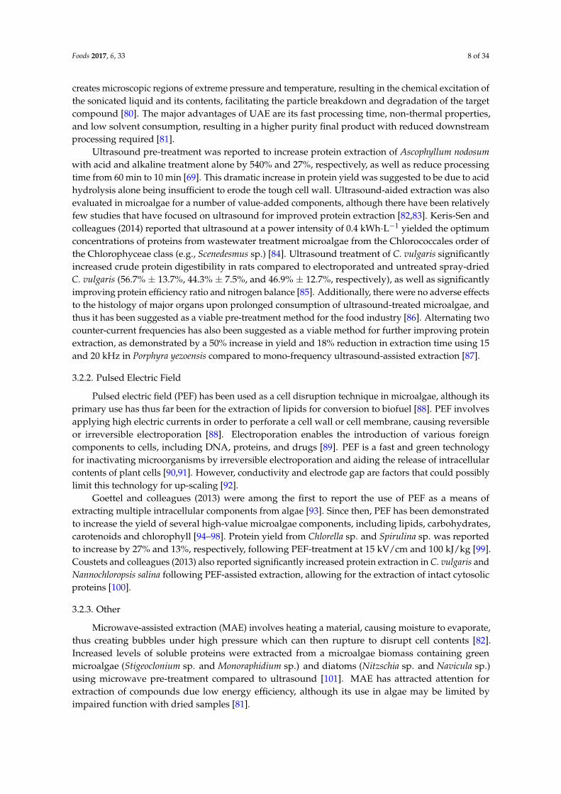
Ultrasound pre-treatment was reported to increase protein extraction of Ascophyllum nodosumwith acid and alkaline treatment alone by 540% and 27%, respectively, as well as reduce processingtime from 60 min to 10 min [69]. This dramatic increase in protein yield was suggested to be due to acidhydrolysis alone being insufficient to erode the tough cell wall. Ultrasound-aided extraction was alsoevaluated in microalgae for a number of value-added components, although there have been relativelyfew studies that have focused on ultrasound for improved protein extraction [82,83]. Keris-Sen andcolleagues (2014) reported that ultrasound at a power intensity of 0.4 kWh·L−1 yielded the optimumconcentrations of proteins from wastewater treatment microalgae from the Chlorococcales order ofthe Chlorophyceae class (e.g., Scenedesmus sp.) [84]. Ultrasound treatment of C. vulgaris significantlyincreased crude protein digestibility in rats compared to electroporated and untreated spray-driedC. vulgaris (56.7% ± 13.7%, 44.3% ± 7.5%, and 46.9% ± 12.7%, respectively), as well as significantlyimproving protein efficiency ratio and nitrogen balance [85]. Additionally, there were no adverse effectsto the histology of major organs upon prolonged consumption of ultrasound-treated microalgae, andthus it has been suggested as a viable pre-treatment method for the food industry [86]. Alternating twocounter-current frequencies has also been suggested as a viable method for further improving proteinextraction, as demonstrated by a 50% increase in yield and 18% reduction in extraction time using 15and 20 kHz in Porphyra yezoensis compared to mono-frequency ultrasound-assisted extraction [87].
3.2.2. Pulsed Electric Field
Pulsed electric field (PEF) has been used as a cell disruption technique in microalgae, although itsprimary use has thus far been for the extraction of lipids for conversion to biofuel [88]. PEF involvesapplying high electric currents in order to perforate a cell wall or cell membrane, causing reversibleor irreversible electroporation [88]. Electroporation enables the introduction of various foreigncomponents to cells, including DNA, proteins, and drugs [89]. PEF is a fast and green technologyfor inactivating microorganisms by irreversible electroporation and aiding the release of intracellularcontents of plant cells [90,91]. However, conductivity and electrode gap are factors that could possiblylimit this technology for up-scaling [92].
Goettel and colleagues (2013) were among the first to report the use of PEF as a means ofextracting multiple intracellular components from algae [93]. Since then, PEF has been demonstratedto increase the yield of several high-value microalgae components, including lipids, carbohydrates,carotenoids and chlorophyll [94–98]. Protein yield from Chlorella sp. and Spirulina sp. was reportedto increase by 27% and 13%, respectively, following PEF-treatment at 15 kV/cm and 100 kJ/kg [99].Coustets and colleagues (2013) also reported significantly increased protein extraction in C. vulgaris andNannochloropsis salina following PEF-assisted extraction, allowing for the extraction of intact cytosolicproteins [100].
3.2.3. Other
Microwave-assisted extraction (MAE) involves heating a material, causing moisture to evaporate,thus creating bubbles under high pressure which can then rupture to disrupt cell contents [82].Increased levels of soluble proteins were extracted from a microalgae biomass containing greenmicroalgae (Stigeoclonium sp. and Monoraphidium sp.) and diatoms (Nitzschia sp. and Navicula sp.)using microwave pre-treatment compared to ultrasound [101]. MAE has attracted attention forextraction of compounds due low energy efficiency, although its use in algae may be limited byimpaired function with dried samples [81].

Foods 2017, 6, 33 9 of 34
Alternatively, sub- and supercritical fluid extraction techniques have gained popularity inrecent decades as extraction methods. Subcritical water extraction (SWE) involves using hot water(100–374 ◦C) under high pressure (~10 bar) to maintain water in a liquid state [102]. Alternatively,supercritical fluid extraction (SFE) is a technique that heats a fluid above its critical point, makingit supercritical. Under supercritical conditions, the properties of the fluid become indistinguishablefrom its gaseous state, with a density similar to a fluid, but viscosity matching that of gas [102]. SFEtypically utilises carbon dioxide (CO2), making it a relatively ‘green’ technology with low solventconsumption [81]. However, SWE and SFE both require high investment costs for equipment and havetypically only been used in algae to date for the extraction of lipids [102].
3.3. Enrichment Methods—Membrane Filtration
Membrane technologies are widely used in the dairy industry to recover whey proteins from milkreleased as a result of the cheese-making process [103]. Membrane technology refers to the use of asemi-permeable membrane to separate a liquid into two different fractions by selectively allowingsome compounds to pass through while impeding other compounds, typically based on molecularweight. Membrane technologies are promising alternative methods of enriching algal proteins, as wellas developing novel techno-functional and bioactive ingredients. They have the advantage of beingnon-thermal and environmentally-friendly [103]. The most commonly used membrane technologiesinclude microfiltration, ultrafiltration, nanofiltration, and reverse osmosis.
Membrane technologies could act as an alternative method for enriching algal proteins whenused in conjunction with a cell disruption technique, such as polysaccharidase hydrolysis, UAE, or PEF.Disruption of the tough cell wall is a critical step required in order to increase the availability of algalproteins for extraction [82]. Membrane technologies are well suited for use with seaweed as part of acascading biorefinery process to maximise valorisation of all components within algae, while avoidingthe presence of heavy metals in the final product [104]. A combination of membrane technologiescould be used to isolate algal proteins using the same principles of molecular weight cut-offs usedin the dairy industry. In the dairy industry, microfiltration (MF) is used to extend the shelf-life ofmilk without any thermal treatment by removing microorganisms, while preserving overall taste andsensory attributes [105]. MF could be used to remove algae cell wall components and bacteria witha molecular weight greater than 200 kDa. Ultrafiltration (UF) could then be used to isolate proteinsand other macromolecules between 1 and 200 kDa, similar to the way it is used in the dairy industryto generate enriched fractions less than 10, 5, 3 and 1 kDa. Nanofiltration (NF) could then be used toremove monovalent salts to minimise osmotic pressure, followed by reverse osmosis (RO) to reducefluid volume [103].
Indeed, membrane technologies have already been used to isolate whole microalgae cellsand several seaweed components. Tangential flow microfiltration was reported as an efficientmethod for recovering 70%–89% of algal biomass from wastewater treatments [106]. UF waspreviously used in conjunction with supercritical CO2 extraction and ultrasound to isolateSargassum pallidum polysaccharides [107]. Polysaccharides with antioxidant activities were isolatedfrom U. fasciata utilising hot water extraction followed by several stages of ultrafiltration withincreasingly smaller pore sizes [108]. Furthermore, UF was used to isolate phycoerythrin protein fromGrateloupia turuturu following cell homogenisation, which was reported to retain 100% of the proteinwithout denaturation [109]. Alternatively, a two-stage ultrafiltration could be applied for algal proteinenrichment, as demonstrated by the separation of polysaccharide components in Tetraselmis suecicausing different sized pore membranes following high-pressure homogenisation [110].

Foods 2017, 6, 33 10 of 34
4. Applications
4.1. Human Nutrition
Protein is an essential nutritional component in the diet of athletes, required to repair and buildmuscle tissue broken down during exercise, with the American College of Sports and Medicinerecommending between 1.2 and 1.7 g protein per kg body weight [111]. Seaweed and microalgae arerich sources of protein and contain all of the essential amino acids at various concentrations [112].Algae could therefore represent a valuable resource for athletes requiring high levels of protein,especially for vegan athletes for whom eggs and dairy whey protein may not be suitable [113].
Seaweed and microalgae have been used as a source of human nutrition for thousands of years insome indigenous populations [114]. One of the main reasons for the high consumption of seaweedand microalgae is due to their significant protein content, which is comparable to, or even greater than,some plant sources [28]. Some species of red seaweeds (Rhodophyta), such as P. palmata and P. tenera,have been reported to contain as much as 33% and 47% dw, respectively [3]. Similarly, some species ofmicroalgae have been reported to contain even higher levels, as high as 63% dw in Spirulina sp. [115].Microalgae are typically consumed as a dietary supplement in the form of powder, pills, or tablets [9].However, they have also been incorporated into a number of functional foods, including noodles,bread, biscuits, drinks, sweets, and beer [116]. Several businesses have been set up for the sale of algalproducts, such as AlgaVia® (www.algavia.com), which produces protein- and lipid-rich algal flourfrom Chlorella protothecoides.
There are several species of seaweed that have traditionally been consumed, largely due to theirhigh protein content. For example, P. tenera (nori) is commonly used as a sushi wrap in severalAsian cultures [22]. Many species of seaweed are particularly high in the amino acids aspartic acidand glutamic acid, which exhibit a unique and interesting flavour that led to the discovery of thetaste sensation referred to as ‘umami’ [40]. The flavour enhancer monosodium glutamate was firstdiscovered in the brown seaweed L. japonica (kombu), which has been found to particularly appeal tothe umami taste sensation [117].
Although the consumption of seaweed in humans is currently underdeveloped, especially inWestern countries, the high protein content and favourable essential amino acid profile makes seaweeda promising source of protein that is ripe for future expansion [40]. Seaweed has been successfullyincorporated as a functional ingredient into several foods at the laboratory scale. U. pinnatifida(wakame) integrated into pasta has antioxidant activity and acceptable sensory attributes at levels upto 10% [118]. Bread containing 4% A. nodosum can significantly reduce energy intake in overweightindividuals in the meal following enriched bread consumption [119]. Bread incorporating similarconcentrations of renin-inhibitory peptides from P. palmata hydrolysates also had acceptable sensoryattributes, with the bioactive properties reported as having survived the baking process [120].
Spirulina is the most highly consumed microalgae due to its high protein content andadded nutritional benefits, including anti-hypertension, renal protective, anti-hyperlipidaemia,and anti-hyperglycaemic [121]. As well as being a rich source of proteins, Spirulina containshigh levels of hypocholesterolemic γ-linoleic acid (GLA), B-vitamins, and free-radical scavengingphycobiliproteins [122]. It has therefore been given the label of a ‘super food’ by the World HealthOrganisation (WHO) and has even been sent to space by the National Aeronautics and SpaceAdministration (NASA) due to its nutrient-dense properties [123]. As a demonstration of this, Spirulinahas 180% more calcium than milk, 670% more protein than tofu, 3100% more β-carotene than carrots,and 5100% more iron than spinach [27]. The world’s largest producer of Spirulina is Hainan SimaiEnterprising Ltd., which is located in the Hainan province of China [121]. This cultivation farmproduces an annual 200 tonnes dried biomass, accounting for 25% of the national output and aconsiderable 10% of the global production. Alternatively, the Earthrise Company has the largestSpirulina production plant, which is located in California, USA, and covers 440,000 m2 [121].

Foods 2017, 6, 33 11 of 34
Chlorella is another widely consumed microalga, with global sales exceeding US $38 billion [124].The largest producer of Chlorella is Taiwan Chlorella Manufacturing and Co. (Taipei, Taiwan), whichproduces 400 tons dried biomass annually [121]. The main substance found in Chlorella that is beneficialfor human health is β-1,3-glucan, which is an active immunostimulant, free-radical scavenger andreducer of blood lipids [125]. Chlorella is also rich in proteins (48% dw), polyunsaturated fatty acids(PUFAs) (39% of total lipids), and phosphorus (1761.5 mg/100g dw) [115].
4.2. Industrial Applications
Lectins and phycobiliproteins are two families of bioactive algal proteins which have beenexploited for several industrial applications. Lectins are most commonly extracted from macroalgalsources, while phycobiliproteins are typically isolated from microalgae [126]. Phycobiliproteins,especially phycoerythrin, can constitute a significant proportion of the overall protein content in redalgae, with levels of 1.2% (total dw) reported for P. palmata [127]. Lectins are found at similar levelsand a yield of 1% lectins was obtained previously from Eucheuma serra (Rhodophyta) [128].
4.2.1. Lectins
Lectins are glycoproteins known for their aggregation and high specificity binding withcarbohydrates without initiating modification through associated enzymatic activity [129]. Lectins areinvolved in several biological processes, including host-pathogen interactions, cell–cell communication,induction of apoptosis, cancer metastasis and antiviral activities [130]. Due to their carbohydratebinding capacity with high specificity, lectins are used in blood grouping, anti-viral (including humanimmunodeficiency virus type 1(HIV-1)), cancer biomarkers, and targets for drug delivery [131].Lectins derived from algae have not received the same level of characterisation compared to otherplant-derived lectins. Nevertheless, some of the bioactivities that have been observed in algal lectinsinclude mitogenic, cytotoxic, antibacterial, anti-nociceptive, anti-inflammatory, anti-viral (HIV-1),platelet aggregation inhibition, and anti-adhesion [132].
4.2.2. Phycobiliproteins
Phycobiliproteins are water-soluble proteins with an important role in photosynthesiswithin cyanobacteria, Rhodophyta, and cryptomonads [133]. Phycobiliproteins are componentsof phycobilisomes, which are large light energy-capturing complexes anchored to thylakoidmembranes [134]. There are four main divisions of phycobiliproteins which are grouped basedon their colour and absorption characteristics, namely, phycoerythrin, phycocyanin, allophycocyanin,and phycoerythrocyanin [135]. The main commercial producers are Spirulina sp. (cyanobacterium)and Porphydrium sp. (Rhodophyta macroalgae) [121].
Phycobiliproteins are used in fluorescent labelling, flow cytometry, fluorescent microscopy, andfluorescent immunohistochemistry [136,137]. However, the primary commercial application of thesephycobiliproteins appears to be as natural dyes, with phycocyanin in particular used as a blue pigmentused in products such as chewing gum, popsicles, confectionary, soft drinks, dairy products, andwasabi, as well as cosmetic products, such as lipstick and eyeliner [121]. Several patents concerninghealth beneficial bioactivities of phycobiliproteins have also already been filed for nutraceuticalapplications such as anti-oxidative, anti-inflammatory, anti-viral, anti-tumour, neuroprotective, andhepatoprotective activities [135].
4.3. Animal Feed
The high protein content of algae can also be beneficial for use as animal feed, includingaquaculture, farm animals, and pets. An estimated 30% of global algal production is estimatedto be used for animal feed, with 50% of Spirulina biomass in particular used as feed supplement due toits excellent nutritional profile [124,138]. Several species of microalgae including Spirulina, Chlorella,and Schizochytrium sp., and seaweed including Laminaria sp. and Ulva sp. can be incorporated as

Foods 2017, 6, 33 12 of 34
protein sources into the diets of poultry, pigs, cattle, sheep, and rabbits [4,139]. Most of the researchon the incorporation of algae as animal feed has been carried out with poultry, likely due to theirpromising prospects for improved commerciality [138].
Tasco® is an example of a proprietary seaweed meal derived from A. nodosum, producedby Acadian Seaplants in Nova Scotia, Canada (http://www.acadianseaplants.com), which hasdemonstrated beneficial properties when included in animal feed [140]. Tasco® has four mainidentified benefits for animal production, including resistance to stressors, improved immune system,increased productivity/quality, and a reduction in pathogenic microorganisms in the final meatproduct [141–144]. These benefits have been observed in several species, including monogastric andruminant species, when at feed inclusion levels of 2% on a daily basis [145].
4.3.1. Poultry
Supplementing poultry feed with microalgae as a protein source can improve their health,productivity, and value. This has been demonstrated using a variety of species, including Chlorellasp., Arthrospira sp., Porphyridium sp., and Haematococcus sp. [86,139,146,147]. Chickens fed withsupplemented Spirulina have been reported to have increased viability, improved overall health andreduced plasma concentrations of cholesterol, triglycerides, and fatty acids [148]. These birds alsoappeared to have an improved immune system as demonstrated by a significant increase in whiteblood cell count and enhanced macrophage phagocytic activity [148,149]. Ross and Dominy (1990)reported that feeding White Leghorn cockerel chicks, Hubbard by Hubbard male broiler chicks, andJapanese quail with varying concentrations of Spirulina in dietary feed slightly delayed growth rates,but did not affect final growth at concentrations less than 10% [150]. Furthermore, this study alsoreported that the inclusion of Spirulina also increased fertility rates, as well as increasing the intensityof the egg-yolk colour [150]. These results have been confirmed by several other studies, indicatingthat the inclusion of Spirulina at a concentration of 2%–2.5% in the feed intensifies the colour of eggyolks to make it more esthetically pleasing for consumers [151,152]. The intensified colouration ofthe yolk is thought to be due to β-carotene [153]. The inclusion of Spirulina can also further valoriseegg products by decreasing their cholesterol and saturated fatty acid content, and replacing it withincreased levels of beneficial omega-3 polyunsaturated fatty acids [146,154].
Incorporation of 3% U. lactuca in broiler chickens increased breast muscle yield compared to birdssolely fed corn diet, as well as decreased serum lipids, cholesterol, and uric acid concentrations [155].The red seaweed Polysiphonia spp. was also demonstrated to improve pellet binding in duck feed atconcentrations of 3%, as well as improve its overall nutrient profile [156]. The incorporation of redseaweeds C. crispus and Sarcodiotheca gaudichaudii was also reported to effectively act as a prebiotic toimprove chicken gut health, productivity, and egg quality [157].
4.3.2. Pigs
The replacement of up to 33% of soy proteins with proteins from Arthrospira maxima, A. platensis,and C. vulgaris in pig feed has been reported as being suitable without any adverse effects [158].The effect of feed processing appears to play a role in the utility of Spirulina in pig’s feed. The additionof Spirulina to pellets was reported to decrease average daily gain, whereas incorporation of Spirulinato meal diets actually increased average daily gain [159]. Addition of Spirulina to the diet has also beensuggested to improve fertility in pigs, increasing sperm motility and storage viability [160].
Supplementation of the brown seaweed L. digitata increased pig body weight gain by 10%, as wellas increase the concentration of iodine in fresh muscle by 45%, thus increasing its valorisation [161].Similarly, A. nodosum has also been reported to increase iodine content in pig tissue, while alsoincreasing the concentrations of beneficial bacteria within the gut [162]. However, these resultsare in contrast to the findings of Reilly and colleagues (2008), who reported that the brownseaweeds Laminaria hyperborea and L. digitata actually decreased the biodiversity of beneficial microbialpopulations within the pig’s gut, although this did not significantly affect the pig’s performance [163].
Foods 2017, 6, 33 13 of 34
4.3.3. Ruminants
Of all the animals evaluated for algae supplementation, ruminants are the most promising in termsof digesting the high fibre content for the greatest extraction efficiency of algal proteins [139]. This is incontrast to mono-gastric animals, for which it has been suggested that some form of prior processingmay be required in order for animals (and humans) to utilise algal proteins more efficiently [164]. Cattlewill preferentially drink water containing 20% suspended Spirulina, increasing their daily water intakeby 24.8 g/kg [165]. Furthermore, this study also reported that 20% of the consumed Spirulina bypassesdegradation within the rumen, allowing for increased digestion and absorption of protein and nutrientswithin the abomasum [165]. Incorporation of 200 g/day Spirulina with cattle feed was reported tobe an economically effective method of increasing animal body weight (8.5%–11%) and daily milkproduction (21%) [166]. As well as increasing milk quantity, Spirulina supplementation has also beendemonstrated to increase milk quality by decreasing saturated fatty acids, while simultaneouslyincreasing monounsaturated fatty acids and polyunsaturated fatty acids [167]. Similar results wereobserved in other studies, as well as with supplementation of Schizochytrium sp. [168,169].
Sheep have also been demonstrated to benefit from microalgae as a protein source, with lambsreported to have increased average daily gains upon consumption of 10 g of Spirulina per day [170].Similarly, Spirulina diet supplementation increased the feed intake of rabbits, as well as improve thequality of rabbit meat with higher levels of GLA [171]. The green seaweed U. lactuca has been reportedas a suitable low-energy, high-protein foodstuff for sheep and goats [172,173]. However, seaweedmay be not be suitable for supplementation in pregnant ewes, having been reported to interfere withpassive immunity in lambs and increasing mortality rate [174].
4.4. Aquaculture
Microalgae are vital for the artificial reproduction of several aquaculture species, especiallymolluscs [138]. Microalgae also play an important role in aquaculture, other than as a food sourcefor zooplanktons by stabilising pH, reducing bacterial growth, and improving the quality of rearingmedium [175]. This leads to improved survival and growth compared to that of clear water fed withartificial diets [176]. Microalgae are the natural base of the entire aquatic food chain. This has ledto their widespread incorporation as an important food source and feed additive in the commercialrearing of many aquatic animals, including molluscs, shrimp, and rotifers [177]. Filtering molluscsare by far the greatest consumer of microalgae in aquaculture, with 10.1 × 10⁶ tonnes produced in1999, compared to shrimp (1.2 × 10⁶ tonnes), and small larvae fish, such as sea breams and flatfish(177,400 tonnes) [178].
Replacements of live microalgae are already commercially available (such as Chaetoceros 1000“Premium Fresh” Instant Algae™ paste, Liqualife™ liquid larval feed, Zeigler™ E-Z Larvae liquidfeed, and Zeigler™ Z-Plus feed), but typically provide inferior growth and survival rates [179].For example, the survival rate of brown larval shrimp (Farfantepenaeus aztecus) significantly decreasedfrom 90.86 ± 3.19% when fed live microalgae, to 14.865 ± 14.35% in shrimp fed with 100% replacementE-Z larvae [179].
Microalgae are also often used as a dietary supplement to refine the products of aquaculture andincrease their valorisation. Carotenoid pigments, such as astaxanthin derived from Haematococcus pluvalis,are incorporated into the diets of salmonoids, shrimp, lobsters, and crayfish, to give them theircharacteristic pink flesh [180]. Similarly, inclusion of astaxanthin at a concentration of 30 ppmsignificantly increases the colour pattern and intensity of ornamental fish, including tetras, cichlids,gouramis, damos, goldfish, and koi, increasing their market value several fold [147].
Red seaweed has been suggested as a promising protein source feed additive. Incorporating 10%Gracilaria chilensis in the diet of Atlantic salmon (Salmo solar) was reported to significantly increasespecific growth rate by 1.51% ± 0.12% compared to the control diet [181]. Including 1.0% and 10%G. chilensis in the diets of S. solar was also suggested to increase antiviral activity against the infectioussalmon anaemia (ISA) virus. Similarly, inclusion of 5% and 15% P. palmata was reported to improve
Foods 2017, 6, 33 14 of 34
hepatic function and have a positive effect on body lipid content in S. salar compared to the controldiets [182]. Wild abalone are opportunistic feeders that consume a variety of macroalgae speciesand typically have increased growth rate in captivity when fed a diet with several species comparedto single species diets [183]. Haliotis tuberculata coccinea fed with a mixed diet including Ulva rigida,Hypnea spinella, and Gracilaria cornea displayed significantly greater growth rates, length, and weightgain than diets consisting of single algal species [184]. Seaweed is also often used in the feed ofsea cucumber culture systems, which are used for human consumption in many Asian countries.L. japonica and U. lactuca have been reported as an economical additive to the diets of sea cucumberApostichopus japonicas with low ammonia–nitrogen production and suitable digestibility [185].
4.5. Bioactive Peptides
Bioactive peptides are particular amino acid sequences that can have additional physiologicalhealth benefits beyond their basic nutritional value [126]. These peptides are typically between 2 and30 amino acids in length and have hormone-like properties. Bioactive peptides are inactive withinthe parent proteins, but can be released through fermentation or hydrolysis. Milk proteins remain themost common source for bioactive peptides [186,187]. However, bioactive peptides have also beenidentified in a number of food sources, including meat, egg, fish, and blood, as well as plant sources,including rice, soybean, wheat, pea, broccoli, garlic, and algae [132,188–197].
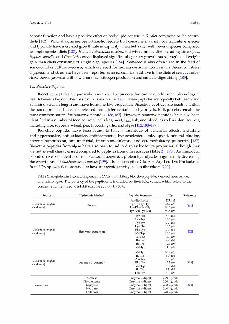
Bioactive peptides have been found to have a multitude of beneficial effects, includinganti-hypertensive, anti-oxidative, antithrombotic, hypocholesterolemic, opioid, mineral binding,appetite suppression, anti-microbial, immunomodulatory, and cytomodulatory properties [187].Bioactive peptides from algae have also been found to display bioactive properties, although theyare not as well characterised compared to peptides from other sources (Table 2) [198]. Antimicrobialpeptides have been identified from Saccharina longicruris protein hydrolysates, significantly decreasingthe growth rate of Staphylococcus aureus [199]. The hexapeptide Glu-Asp-Arg-Leu-Lys-Pro isolatedfrom Ulva sp. was demonstrated to have mitogenic activity in skin fibroblasts [200].
Table 2. Angiotensin-I-converting enzyme (ACE)-I inhibitory bioactive peptides derived from seaweedand microalgae. The potency of the peptides is indicated by their IC50 values, which refers to theconcentration required to inhibit enzyme activity by 50%.
Source Hydrolytic Method Peptide Sequence IC50 Reference
Undaria pinnatifida(wakame)
Pepsin
Ala-Ile-Tyr-Lys 213 µM
[201]Tyr-Lys-Tyr-Tyr 64.2 µMLys-Phe-Tyr-Gly 90.5 µMTyr-Asn-Lys-Leu 90.5 µM
Undaria pinnatifida(wakame) Hot water extraction
Tyr-His 5.1 µM
[202]
Lys-Trp 10.8 µMLys-Tyr 7.7 µMLys-Phe 28.3 µMPhe-Tyr 3.7 µMVal-Trp 10.8 µMVal-Phe 43.7 µMIle-Tyr 2.7 µMIle-Trp 12.4 µMVal-Tyr 11.3 µM
Undaria pinnatifida(wakame) Protease S “Amano”
Val-Tyr 35.2 µM
[203]
Ile-Tyr 6.1 µMAla-Trp 18.8 µMPhe-Tyr 42.3 µMVal-Trp 3.3 µMIle-Trp 1.5 µM
Leu-Trp 23.6 µM
Ecklonia cava
Alcalase Enzymatic digest 2.79 µg/mL
[204]Flavourzyme Enzymatic digest 3.56 µg/mL
Kojizyme Enzymatic digest 2.33 µg/mLNeutrase Enzymatic digest 3.10 µg/mLProtamex Enzymatic digest 3.28 µg/mL

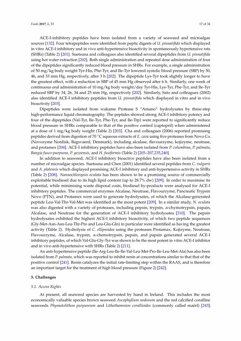
Foods 2017, 6, 33 15 of 34
Table 2. Cont.
Source Hydrolytic Method Peptide Sequence IC50 Reference
Porphyra yezoensis
Ile-Tyr 2.69 µM
[205]Met-Lys-Tyr 7.26 µM
Ala-Lys-Tyr-Ser-Tyr 1.52 µMLeu-Arg-Tyr 5.06 µM
Hizikia fusiformisGly-Lys-Tyr 3.92 µM
[206]Ser-Val-Tyr 8.12 µMSer-Lys-Thr-Tyr 11.07 µM
Palmaria palmata (dulse) Thermolysin
Val-Tyr-Arg-Thr 0.14 µM
[207]Leu-Asp-Tyr 6.1 µMLeu-Arg-Tyr 0.044 µM
Phe-Glu-Gln-Trp-Ala-Ser 2.8 µM
Chlorella vulgaris
Pepsin
Ile-Val-Val-Glu 315.3 µM
[208]
Ala-Phe-Leu 63.8 µMPhe-Ala-Leu 26.3 µMAla-Glu-Leu 57.1 µM
Val-Val-Pro-Pro-Ala 79.5 µM
Arthrospira platensis
Ile-Ala-Glu 34.7 µMPhe-Ala-Leu 11.4 µMAla-Glu-Leu 11.4 µM
Ile-Ala-Pro-Gly 11.4 µMVal-Ala-Phe 35.8 µM
Nannochloropsis oculata Alcalase Leu-Val-Thr-Val-Met 18.0 µM [209]
Nannochloropsis oculata Pepsin Gly-Met-Asn-Asn-Leu-Thr-Pro 123 µM[210]Leu-Glu-Gln 173 µM
Chlorella ellipsoideaProtamex, Kojizyme, Neutrase,Flavourzyme, Alcalase, trypsin,
α-chymotrypsin, pepsin, and papainVal-Glu-Gly-Tyr 128.4 µM [211]
Chlorella vulgaris Flavourzyme, alcalase, papain, and pepsin Val-Glu-Cys-Tyr-Gly-Pro-Asn-Arg-Pro-Gln-Phe 29.6 µM [212]
Owing to the high levels of oxidative stresses and free radicals in their environment, microalgaeand seaweed have developed many defensive systems that can stimulate antioxidant activity whenconsumed [213]. Antioxidant peptides have therefore been isolated from several species of microalgae,including C. vulgaris, Navicula incerta, and Chlorella ellipsoidea [214–217]. Antioxidant peptides have alsobeen isolated from various Irish and Korean brown seaweeds, which indicated that those with higherphenolic content, such as Ecklonia cava and Sargassum coreanum, correlated with increased antioxidantactivity [218–220]. Peptides displaying antioxidant and anticancer bioactivity have also been reportedin Sri Lankan red and green seaweed, of which Caulerpa racemosa demonstrated the most promisingfree radical scavenging and anticancer bioactivity [221].
Microalgae have also been reported to display anticancer peptides, such as Chlorella pyrenoidosaantitumor polypeptide (CPAP) derived from Chlorella pyrenoidosa and polypeptide Y2 derived fromA. platensis [222,223]. Protein hydrolysates from Porphyra columbina phycocolloid extraction by-productswere reported to have immunosuppressive, anti-hypertensive, and antioxidant capacities [224].Anti-inflammatory peptides have been isolated from microalgae, such as Chlorella 11-peptide derivedfrom C. pyrenoidosa [225], as well as peptides derived from A. maxima (Leu-Asp-Ala-Val-Asn-Argand Met-Met-Leu-Asn-Phe), which were additionally reported to display anti-atherosclerosisbioactivity [226,227]. Similarly, anti-atherosclerosis peptides have also been isolated from P. palmataand were shown to be non-toxic in Zebrafish at a concentration of 1 mg/mL [228].
Algal peptides have been reported to display several other bioactivities, includinghepatoprotective, immunomodulatory, ultraviolet (UV) radiation-protective, anti-osteoporosis, andanti-coagulant [229–234]. Finally, it is important to note that several studies have reported that shortalgae-derived peptides are capable of resisting gastrointestinal digestion from enzymes such as trypsin,pepsin, and chymotrypsin [203,207,235]. This is an essential trait for bioactive peptides in order toachieve their physiological effect at their site of action [236].

Foods 2017, 6, 33 16 of 34
Anti-Hypertensive Peptides
Hypertension is the single largest risk factor attributed to deaths worldwide, making it anideal target for bioactive peptides [237]. Angiotensin-I-converting enzyme (ACE-I) is a proteolyticenzyme that affects vasoconstriction in two major blood pressure regulatory systems, namelyrenin-angiotensin–aldosterone system (RAAS) and kinin–kallikrein system, leading on to thedevelopment of hypertension (Figure 2) [126]. ACE-I inhibitors have therefore become one of the mostcommonly studied targets, and with global annual sales exceeding US $6 billion, ACE-I inhibitory drugscan be considered as one of the major protease inhibitor success stories [238]. Synthetic ACE-I inhibitordrugs, such as captopril, enalapril, and alacepril, often come with several side effects, includinghypotension, dry cough, and impaired renal function [19]. Function foods with anti-hypertensivebioactivities have therefore become a popular alternative to synthetic drugs, especially for individualswho are borderline hypertensive and do not warrant the prescription of pharmaceutical drugs [239].
Foods 2017, 6, 33 16 of 34
especially for individuals who are borderline hypertensive and do not warrant the prescription of
pharmaceutical drugs [239].
Figure 2. Schematic representation of the renin–angiotensin–aldosterone system (RAAS) and the
hypertensive effect of angiotensin‐I‐converting enzyme (ACE‐I). Angiotensinogen is converted to the
decapeptide angiotensin‐I by renin. ACE‐I cleaves the C‐terminal dipeptide His‐Leu of angiotensin‐I
to form angiotensin‐II. Binding of angiotensin‐II to its receptor (AT1) stimulates the secretion of
inositol 1,4,5‐triphosphate (IP3) and aldosterone, which induce arteriolar vasoconstriction and
increased intravascular fluid volume, respectively, resulting in increased blood pressure. Within the
kallikrein–kinin system, kallikrein converts kininogen to bradykinin, which induces arteriolar
vasodilation by prostaglandin secretion and binding of bradykinin with its receptor, resulting in
decreased blood pressure. However, the hypotensive effect of bradykinin is largely dependent on the
rate of degradation by ACE‐I, which hydrolyzes bradykinin to form inactive metabolites.
ACE‐I‐inhibitory peptides have been isolated from a variety of seaweed and microalgae sources
[132]. Four tetrapeptides were identified from peptic digests of U. pinnatifida which displayed in
vitro ACE‐I inhibitory and in vivo anti‐hypertensive bioactivity in spontaneously hypertensive rats
(SHRs) (Table 2) [201]. Suetsuna and colleagues also identified several dipeptides from U. pinnatifida
using hot water extraction [202]. Both single administration and repeated dose administration of
four of the dipeptides significantly reduced blood pressure in SHRs. For example, a single
administration of 50 mg/kg body weight Tyr‐His, Phe‐Tyr, and Ile‐Tyr lowered systolic blood
pressure (SBP) by 50, 46, and 33 mm Hg, respectively, after 3 h [202]. The dipeptide Lys‐Tyr took
slightly longer to have the greatest effect, with a reduction in SBP of 45 mm Hg observed after 6 h.
Similarly, one week of continuous oral administration of 10 mg/kg body weight/day Tyr‐His,
Lys‐Tyr, Phe‐Tyr, and Ile‐Tyr reduced SBP by 34, 26, 34 and 25 mm Hg, respectively [202]. Similarly,
Figure 2. Schematic representation of the renin–angiotensin–aldosterone system (RAAS) and thehypertensive effect of angiotensin-I-converting enzyme (ACE-I). Angiotensinogen is converted to thedecapeptide angiotensin-I by renin. ACE-I cleaves the C-terminal dipeptide His-Leu of angiotensin-Ito form angiotensin-II. Binding of angiotensin-II to its receptor (AT1) stimulates the secretionof inositol 1,4,5-triphosphate (IP3) and aldosterone, which induce arteriolar vasoconstriction andincreased intravascular fluid volume, respectively, resulting in increased blood pressure. Withinthe kallikrein–kinin system, kallikrein converts kininogen to bradykinin, which induces arteriolarvasodilation by prostaglandin secretion and binding of bradykinin with its receptor, resulting indecreased blood pressure. However, the hypotensive effect of bradykinin is largely dependent on therate of degradation by ACE-I, which hydrolyzes bradykinin to form inactive metabolites.
Foods 2017, 6, 33 17 of 34
ACE-I-inhibitory peptides have been isolated from a variety of seaweed and microalgaesources [132]. Four tetrapeptides were identified from peptic digests of U. pinnatifida which displayedin vitro ACE-I inhibitory and in vivo anti-hypertensive bioactivity in spontaneously hypertensive rats(SHRs) (Table 2) [201]. Suetsuna and colleagues also identified several dipeptides from U. pinnatifidausing hot water extraction [202]. Both single administration and repeated dose administration of fourof the dipeptides significantly reduced blood pressure in SHRs. For example, a single administrationof 50 mg/kg body weight Tyr-His, Phe-Tyr, and Ile-Tyr lowered systolic blood pressure (SBP) by 50,46, and 33 mm Hg, respectively, after 3 h [202]. The dipeptide Lys-Tyr took slightly longer to havethe greatest effect, with a reduction in SBP of 45 mm Hg observed after 6 h. Similarly, one week ofcontinuous oral administration of 10 mg/kg body weight/day Tyr-His, Lys-Tyr, Phe-Tyr, and Ile-Tyrreduced SBP by 34, 26, 34 and 25 mm Hg, respectively [202]. Similarly, Sato and colleagues (2002)also identified ACE-I inhibitory peptides from U. pinnatifida which displayed in vitro and in vivobioactivity [203].
Dipeptides were isolated from wakame Protease S “Amano” hydrolysates by three-stephigh-performance liquid chromatography. The peptides showed strong ACE-I inhibitory potency andfour of the dipeptides (Val-Tyr, Ile-Tyr, Phe-Tyr, and Ile-Trp) were reported to significantly reduceblood pressure in SHRs comparable to that of the positive control (captopril) when administeredat a dose of 1 mg/kg body weight (Table 2) [203]. Cha and colleagues (2006) reported promisingpeptides derived from digestion of 70 ◦C aqueous extracts of E. cava using five proteases from Novo Co.(Novozyme Nordisk, Bagsvaerd, Denmark), including alcalase, flavourzyme, kojizyme, neutrase,and protamex [204]. ACE-I inhibitory peptides have also been isolated from P. columbina, P. palmata,Bangia fusco-purpurea, P. yezoensis, and H. fusiformis (Table 2) [205–207,235,240].
In addition to seaweed, ACE-I inhibitory bioactive peptides have also been isolated from anumber of microalgae species. Suetsuna and Chen (2001) identified several peptides from C. vulgarisand A. platensis which displayed promising ACE-I inhibitory and anti-hypertensive activity in SHRs(Table 2) [208]. Nannochloropsis oculata has been shown to be a promising source of commerciallyexploitable biodiesel due to its high lipid content (up to 28.7% dw) [209]. In order to maximise itspotential, while minimising waste disposal costs, biodiesel by-products were analysed for ACE-Iinhibitory peptides. The commercial enzymes Alcalase, Neutrase, Flavourzyme, Pancreatic TrypsinNovo (PTN), and Protamex were used to generate hydrolysates, of which the Alcalase-generatedpeptide Leu-Val-Thr-Val-Met was identified as the most potent [209]. In a similar study, N. oculatawas also digested with a variety of proteases, including pepsin, trypsin, α-chymotrypsin, papain,Alcalase, and Neutrase for the generation of ACE-I inhibitory hydrolysates [210]. The pepsinhydrolysates exhibited the highest ACE-I inhibitory bioactivity, of which two peptide sequences(Gly-Met-Asn-Asn-Leu-Thr-Pro and Leu-Glu-Gln) in particular were identified as having the greatestactivity (Table 2). Hydrolysis of C. ellipsoidea using the proteases Protamax, Kojizyme, Neutrase,Flavourzyme, Alcalase, trypsin, α-chemotrypsin, pepsin, and papain generated several ACE-Iinhibitory peptides, of which Val-Glu-Gly-Tyr was shown to be the most potent in vitro ACE-I inhibitorand in vivo anti-hypertensive with SHRs (Table 2) [211].
An anti-hypertensive peptide (Ile-Arg-Leu-Ile-Ile-Val-Leu-Met-Pro-Ile-Leu-Met-Ala) has also beenisolated from P. palmata, which was reported to inhibit renin at concentrations similar to that that of thepositive control [241]. Renin catalyzes the initial rate-limiting step within the RAAS, and is thereforean important target for the treatment of high blood pressure (Figure 2) [242].
5. Challenges
5.1. Access Rights
At present, all seaweed species are harvested by hand in Ireland. This includes the mosteconomically valuable species brown seaweed Ascophyllum nodosum and the red calcified corallineseaweeds Phymatolithon purpureum and Lithothamnion corallioides (commonly called maërl) [243].

Foods 2017, 6, 33 18 of 34
The legislation for harvesting seaweed in Ireland is based on the Foreshore Acts 1933–1998, whichstates that the Department of Communications Marine and Natural Resources is empowered to grantlicences for seaweed harvesting on the seabed out to 12 nautical miles [244]. There are currently norestrictions on harvesting quantities in Ireland, nor are there any restrictions on harvesting times. Theonly exception to this is for maërl due to its extremely slow growth rate (0.6–1.5 mm per annum) [245].However, the future implementation of mechanical harvesting methods will likely require a review ofexisting legislation to ensure appropriate access rights and sustainable harvesting yields are maintained.Mechanical harvesting of Laminaria spp. is already present in various European countries, such asFrance and Norway, providing a valuable resource for making any such revisions [243].
5.2. Variability
An additional challenge, which is particularly relevant in the production of bioactive peptides, arethe high levels of variability in algal proteins. The protein content can vary by season, temperature, andlocation in which the seaweed is harvested [46]. The relative composition of particular proteins withinthe plant can also differ, changing the concentrations of amino acids and therefore altering the yield ofdesired peptides as a consequence [246]. For example, annual monitoring of P. palmata harvested onthe French Atlantic coast showed that protein levels were highest in the winter and spring months,varying from 9 to 25%, and peaking in May [247]. Similarly, Gracilaria cervicornis and S. vulgare variedby season, with protein levels negatively correlating with temperature and salinity [248]. Differentharvest locations of U. pinnatifida in New Zealand also significantly affected the protein content andamino acid composition [249].
5.3. Scalability
The scalability of protein extraction from algae is a further obstacle that needs to be overcomebefore seaweed and microalgae become a viable source. Algal protein extraction is still very much inits infancy, meaning that many of the methods that have been developed are still at small scale [132].PEF and ultrasound have been suggested as being suitable for large scale algal protein extraction [250].Membrane technologies may also be scalable for commercial applications, with ultrafiltration reportedas being suitable for R-phycoerythrin extraction from the seaweed G. turuturu at the industrialscale [109].
5.4. Digestibility
One of the most important challenges for extracting proteins from algae is the high levels of cellwall anionic polysaccharides which can become bound to the proteins and create viscous medium,further increasing the difficulty of protein extraction [46]. The morphology of different seaweed specieshas been suggested to be an important factor in determining protein yield, with tougher thallus formsreported to require increased processing [68]. Several brown algae species (Eisena bicyclis, H. fusiformis,U. pinnatifida) were reported to have reduced in vitro digestibility (51.8%–57.1%) compared to redalgae P. palmata and P. tenera (70.2% and 87.3%, respectively), which was suggested to have been dueto increased levels of dietary fibre [34]. Furthermore, the type of polysaccharide has been shown toinfluence the level of protein digestion. P. palmata and G. verrucosa, both red seaweeds, were shown tohave significantly different levels of in vitro corrected nitrogen digestibility (1.9% and 16.3% after 6 h,respectively) [251]. This was largely attributed to differences in polysaccharide fractions (pentoses andhexoses, respectively).
Treatments to disrupt the cellulosic cell wall could help to overcome this issue to make algalproteins and other cell components more accessible [28]. Heat treatment is one such option that canimprove food taste, texture, safety in terms of allergenicity and microbial load, and preservation, whilealso increasing bioavailability and utilisation of proteins by partial denaturation and breakdown ofproteins into peptides, allowing for easier access by proteolytic enzymes [252]. An example of thiscan be seen with boiling of P. palmata , resulting in a 64%–96% increase in liberated amino acids [253].

Foods 2017, 6, 33 19 of 34
Alternatively, fermentation can also increase protein digestibility due to the degradation of insolublefibres, such as xylan. This can be seen with the fermentation of P. palmata using the fungal mouldTrichoderma pseudokoningii which was found to decrease the xylan content from 53% to 19% dw [75].Washing in distilled water has also been shown to be a simple, yet effective method for improvingin vitro digestibility by solubilising and removing mineral salts [75].
Alternatively, the method of drying seaweed has been shown to influence algal protein digestibility.Compared to freeze-drying, oven-drying was demonstrated to significantly improve (p < 0.05,two-way ANOVA) the protein extractability and in vitro bioaccessibility of brown seaweed speciesSargassum hemiphyllum, Sargassum henslowianum, and Sargassum patens [45]. The increased extractabilityfrom oven-drying were suggested to have been due to the decomposition of phenolic compounds atthe high temperatures, as well as increased disruption of anionic or neutral polysaccharides foundwithin the cell wall of the seaweed [45].
Protein digestibility can also be largely influenced by the presence of phenolic compoundspresent in algae, especially phlorotannins in brown seaweeds [45]. Oxidised phenolic compounds canreact with amino acids to form insoluble complexes, which may inhibit proteolytic enzymes and thusdecrease their nutritive values [65]. The negative correlation between phenolic content and digestibilitywas demonstrated by U. lactuca which was reported to have higher phenolic content (38.8 ± 0.5%)compared to the red algae H. charoides (16.9% ± 1.0%) and H. japonica (16.3% ± 0.03%) and thus, lowerbioaccessibility (85.7% ± 1.9%, 88.7% ± 0.7%, and 88.9% ± 1.4%, respectively) [65]. This is likely to bea greater issue in brown algae, as these are typically higher in phenolics, including catechins, flavanols,and phlorotannins [10,67].
5.5. Food Safety
While algae can be natural accumulators of vitamins and minerals, they can also gather toxicelements, such as heavy metals. There are therefore strict legal limits in Europe for the safemaximum exposure of heavy metals, such as mercury, arsenic, lead, and cadmium, in foods forhuman consumption. These limits are based upon the recommendations of the Joint FAO/WHOExpert Committee on Food Additives (JECFA) and the Panel on Contaminants in the Food Chainof the European Food Safety Authority (CONTAM Panel), and are outlined in the legislation underCommission Regulation (EC) No. 1881/2006 [254].
The tolerable weekly intake (TWI) of methylmercury, arsenic, lead, and cadmium are 1.6, 15, 25,and 25 µg/kg body weight, respectively [255–258]. The CONTAM panel has reported that the currentlead TWI (15 µg/kg body weight) may no longer be appropriate due to low levels found in foodstuffs,while arsenic TWI (1.6 µg/kg body weight) should be lowered due to cancers and adverse effectsobserved at current legal limits [256,257]. Lead and mercury appear to occur in algae at levels that aresafe for human consumption [259,260]. In contrast, arsenic and cadmium have been identified in algaeat levels above the legal limits, with H. fusiformis in particular containing a high content of these toxicheavy metals [259,260]. Microalgae do not appear to exceed legal levels of heavy metals [261].
The lack of regulation for algae as a dietary supplement is of additional concern, leading to largevariations between different batches from the same producer [262]. This has led to concerns beingraised about quality and contamination at the production and processing stages [263]. Downstreamapplications for which the algal proteins are intended can play a role in determining processingmethods. For example, food grade reagents and processes may be required for proteins intended forhuman consumption, whereas this may not be such an issue with proteins intended for commercialapplications or animal feed [264]. Furthermore, any reagents used for the production or extraction ofalgal proteins must also be environmentally friendly with minimum impact on local ecosystems [265].Following protein extraction, greater emphasis must additionally be placed on protein purification,with salt removal and buffer exchange being important steps [266]. Membrane technologies, suchas ultrafiltration and nanofiltration, have already been implemented in this context within the dairy
Foods 2017, 6, 33 20 of 34
industry, although these technologies could also be used for other food applications, including algalproteins [267].
5.6. Price
A critical factor that will determine the commercial viability of microalgae and seaweed istheir competitiveness compared to other sources on the market. For example, a major applicationfor the production of microalgae is the extraction of lipids to be converted to biofuel. However,while they are a greener alternative for the environment, they are still not competitive compared tofossil-based petroleum fuels [266]. Algae competitiveness could be further increased by taking a holisticview and maximising the extraction of all available high-value components by cascading biorefinery.Conventional methods of algal protein extraction are often quite wasteful as they typically dispose ofthe algae by-products following processing [88]. Seaweed is ideally suited for cascading biorefinerybecause it contains many high-value components as well as bulky low-value components that areconsidered raw materials for the bio-based economy, such as xylose and glucose [268]. As severalstudies have shown, the by-products of algae can still have many applications that could be of economicvalue [209,215,224]. Further research is needed to develop new methods that will enable algal proteinproduction, extraction and processing costs to be lowered.
6. Discussions
Seaweed and microalgae are generally considered to be a promising source of nutrition, richin vitamins, minerals, and protein. Some species have even been reported to display protein atconcentrations greater than traditional plant and animal protein sources [28]. Within these proteinsequences, bioactive peptides have been identified with beneficial properties for human health,including antioxidant, anticancer, anti-hypertensive, immunosuppressive, anti-atherosclerosis, andhepatoprotective effects [198]. Algae have also been reported to contain high quality proteins in termsof EAA content [3], as well as displaying in vitro protein digestibility that is comparable to commonlyconsumed plants [67]. However, there are conflicting reports about the bioavailability of algal proteinswhen examined in vivo. Several studies have demonstrated that consumption of raw, unprocessedseaweed can result in reduced growth [43,44]. In contrast, other studies have reported oppositefindings, with seaweed proteins suggested as being a beneficial dietary supplement for improvedanimal growth, meat quality, and valorisation [155,161,162]. Furthermore, microalgae have long beenused as the basal diet in the rearing of animals in aquaculture, especially molluscs [138].
One of the major reasons for reduced protein digestibility was suggested to be due to the high fibrecontent that makes up the algal cell wall [46]. Algae typically contain high levels of polysaccharides,although this can vary significantly in seaweed (4%–76% dw) and microalgae (8%–64% dw) accordingto species and time of harvest [22,28]. Similarly, phenolic compounds can react with amino acids toform insoluble compounds [65]. Great emphasis has therefore been placed on researching methodsfor breaking down of the anionic cell wall in order to allow the release of the valuable intracellularcontents [13].
Conventional protein extraction methods, such as enzymatic hydrolysis and physical processes,are laborious, time-consuming, and may require the use of solvents [13]. Novel protein extractionmethods, such as UAE, PEF, and MAE, have thus been investigated as a means of overcoming theselimitations. UAE is a cost-effective and low-solvent consumption method for extraction of high-valuecompounds. UAE is already widely used at commercial scale in food processing and could thereforealso be implemented for the extraction of algal proteins [81]. PEF for the extraction of intracellularalgal components is still in its infancy, having predominantly been used for lipid extraction [93].Nevertheless, its lack of solvent consumption or heat gives it potential for the future extraction ofalgal proteins [100]. MAE has been poorly studied in algae, although the high temperature involvedwould likely make it unsuitable for extraction of proteins [81]. Alternatively, membrane filtrationtechnologies, such as those used in the dairy industry, could also be considered a gentle method for

Foods 2017, 6, 33 21 of 34
protein enrichment when used in conjunction with cell disruption methods. MF has already beendemonstrated for harvesting whole microalgal cells, while UF was reported for the extraction of algalpolysaccharides [106,107]. However, there remains to be a distinct lack of studies investigating the useof membrane technologies in the enrichment of algal proteins.
In conclusion, algae have been reported as a valuable source of nutrition. However, there areinconclusive reports about the digestibility of algae and the bioavailability of the proteins within. Moreresearch is needed to investigate the bioavailability of algal proteins in vivo. Cell disruption techniqueswill likely play an essential role in the successful extraction and enrichment of algal protein ingredientsat a commercial scale. The most promising methods that have been used in algae to date are UAE andPEF, both of which are non-thermal, require low solvent consumption, and have been implemented atcommercial scale for the extraction of compounds in other food processes. Alternatively, membranetechnologies are a method for isolating compounds which show great promise, but have yet to beinvestigated sufficiently in algae.
Acknowledgments: Stephen Bleakley is in receipt of a Teagasc Walsh Fellowship (Grant No. 2016073). This workforms part of the BioAlgae project funded by the Teagasc (Grant No. NFNY6889-142).
Conflicts of Interest: The authors declare no conflict of interest.
Abbreviations
EAA(s) essential amino acid(s)FAO Food and Agriculture Organisation of the United NationsTNOASR The Netherland Organisation for applied scientific researchTIM TNOASR’s Intestinal ModelUAE ultrasound-assisted extractionPEF pulsed electric fieldMAE microwave-assisted extractionMF microfiltrationUF ultrafiltrationWHO World Health OrganisationACE-I angiotensin-I-converting enzymeRAAS renin-angiotensin-aldosterone systemSHR(s) spontaneously hypertensive rat(s)SBP systolic blood pressure
References
1. Godfray, H.C.J.; Beddington, J.R.; Crute, I.R.; Haddad, L.; Lawrence, D.; Muir, J.F.; Pretty, J.; Robinson, S.;Thomas, S.M.; Toulmin, C. Food security: The challenge of feeding 9 billion people. Science 2010, 327, 812–818.[CrossRef] [PubMed]
2. Tilman, D.; Balzer, C.; Hill, J.; Befort, B.L. Global food demand and the sustainable intensification ofagriculture. Proc. Natl. Acad. Sci. USA 2011, 108, 20260–20264. [CrossRef] [PubMed]
3. Fleurence, J. Seaweed proteins: Biochemical, nutritional aspects and potential uses. Trends Food Sci. Technol.1999, 10, 25–28. [CrossRef]
4. Gouveia, L.; Batista, A.P.; Sousa, I.; Raymundo, A.; Bandarra, N. Microalgae in Novel Food Products. InFood Chemistry Research Development; Konstantinos, N., Papadopoulos, P.P., Eds.; Nova Science Publishers:New York, NY, USA, 2008; pp. 75–112.
5. Van Krimpen, M.; Bikker, P.; Van der Meer, I.; Van der Peet-Schwering, C.; Vereijken, J. Cultivation, Processingand Nutritional Aspects for Pigs and Poultry of European Protein Sources as Alternatives for Imported SoybeanProducts; Wageningen UR Livestock Research: Lelystad, The Netherlands, 2013; p. 48.
6. Wallace, J. Increasing agricultural water use efficiency to meet future food production. Agric. Ecosyst. Environ.2000, 82, 105–119. [CrossRef]
7. Pimentel, D.; Pimentel, M. Sustainability of meat-based and plant-based diets and the environment. Am. J.Clin. Nutr. 2003, 78, 660S–663S. [PubMed]

Foods 2017, 6, 33 22 of 34
8. Sampath-Wiley, P.; Neefus, C.D.; Jahnke, L.S. Seasonal effects of sun exposure and emersion on intertidalseaweed physiology: Fluctuations in antioxidant contents, photosynthetic pigments and photosyntheticefficiency in the red alga Porphyra umbilicalis kützing (Rhodophyta, Bangiales). J. Exp. Mar. Biol. Ecol. 2008,361, 83–91. [CrossRef]
9. Pulz, O.; Gross, W. Valuable products from biotechnology of microalgae. Appl. Microbiol. Biotechnol. 2004, 65,635–648. [CrossRef] [PubMed]
10. Wang, T.; Jonsdottir, R.; Ólafsdóttir, G. Total phenolic compounds, radical scavenging and metal chelation ofextracts from icelandic seaweeds. Food Chem. 2009, 116, 240–248. [CrossRef]
11. Wijffels, R.H.; Barbosa, M.J. An outlook on microalgal biofuels. Science 2010, 329, 796–799. [CrossRef][PubMed]
12. Cavalier-Smith, T. Evolution and relationships. In Unravelling the Algae: The Past, Present, and Future of AlgalSystematics; Brodie, J., Ed.; CRC Press: Boca Raton, FL, USA, 2007; p. 21.
13. Kadam, S.U.; Tiwari, B.K.; O’Donnell, C.P. Application of novel extraction technologies for bioactives frommarine algae. J. Agric. Food Chem. 2013, 61, 4667–4675. [CrossRef] [PubMed]
14. Food and Agriculture Organization. Food Balance Sheets; Food and Agriculture Organisation of the UnitedNations: Rome, Italy, 2013.
15. Food and Agriculture Organization. The State of World Fisheries and Aquaculture: Opportunties and Challenges;Food and Agriculture Organisation of the United Nations: Rome, Italy, 2014.
16. Cock, J.M.; Sterck, L.; Rouzé, P.; Scornet, D.; Allen, A.E.; Amoutzias, G.; Anthouard, V.; Artiguenave, F.;Aury, J.-M.; Badger, J.H. The ectocarpus genome and the independent evolution of multicellularity in brownalgae. Nature 2010, 465, 617–621. [CrossRef] [PubMed]
17. Hoek, C.; Mann, D.; Jahns, H.M. Algae: An Introduction to Phycology; Cambridge University Press: Cambridge,UK, 1995.
18. Foster, M.S.; Schiel, D.R. Ecology of Giant Kelp Forests in California: A Community Profile; San Jose StateUniversity, Moss Landing Marine Labs.: Moss Landing, CA, USA, 1985.
19. Fitzgerald, C.; Gallagher, E.; Tasdemir, D.; Hayes, M. Heart health peptides from macroalgae and theirpotential use in functional foods. J. Agric. Food Chem. 2011, 59, 6829–6836. [CrossRef] [PubMed]
20. Karol, K.G.; McCourt, R.M.; Cimino, M.T.; Delwiche, C.F. The closest living relatives of land plants. Science2001, 294, 2351–2353. [CrossRef] [PubMed]
21. McCourt, R.M.; Delwiche, C.F.; Karol, K.G. Charophyte algae and land plant origins. Trends Ecol. Evol. 2004,19, 661–666. [CrossRef] [PubMed]
22. Holdt, S.L.; Kraan, S. Bioactive compounds in seaweed: Functional food applications and legislation.J. Appl. Phycol. 2011, 23, 543–597. [CrossRef]
23. Mouritsen, O.G.; Dawczynski, C.; Duelund, L.; Jahreis, G.; Vetter, W.; Schröder, M. On the humanconsumption of the red seaweed dulse (Palmaria palmata (L.) weber & amp; mohr). J. Appl. Phycol. 2013, 25,1777–1791.
24. Norton, T.A.; Melkonian, M.; Andersen, R.A. Algal biodiversity. Phycologia 1996, 35, 308–326. [CrossRef]25. Chacón-Lee, T.; González-Mariño, G. Microalgae for “healthy” foods—Possibilities and challenges.
Compr. Rev. Food Sci. Food Saf. 2010, 9, 655–675. [CrossRef]26. Brennan, L.; Owende, P. Biofuels from microalgae—A review of technologies for production, processing,
and extractions of biofuels and co-products. Renew. Sustain. Energy Rev. 2010, 14, 557–577. [CrossRef]27. Capelli, B.; Cysewski, G.R. Potential health benefits of Spirulina microalgae. Nutrafoods 2010, 9, 19–26.
[CrossRef]28. Becker, E. Micro-algae as a source of protein. Biotechnol. Adv. 2007, 25, 207–210. [CrossRef] [PubMed]29. Rodriguez-Garcia, I.; Guil-Guerrero, J.L. Evaluation of the antioxidant activity of three microalgal species for
use as dietary supplements and in the preservation of foods. Food Chem. 2008, 108, 1023–1026. [CrossRef][PubMed]
30. Boisen, S.; Eggum, B. Critical evaluation of in vitro methods for estimating digestibility in simple-stomachanimals. Nutr. Res. Rev. 1991, 4, 141–162. [CrossRef] [PubMed]
31. Young, V.R.; Pellett, P.L. Plant proteins in relation to human protein and amino acid nutrition. Am. J.Clin. Nutr. 1994, 59, 1203S–1212S. [PubMed]
32. Hoffman, J.R.; Falvo, M.J. Protein-which is best. J. Sports Sci. Med. 2004, 3, 118–130. [PubMed]

Foods 2017, 6, 33 23 of 34
33. Food and Agriculture Organization; World Health Organization. Protein Quality Evaluation—Report of JointFAO/WHO Expert Consultation; FAO: Rome, Italy, 1991.
34. Mišurcová, L.; Krácmar, S.; Klejdus, B.; Vacek, J. Nitrogen content, dietary fiber, and digestibility in algalfood products. Czech J. Food Sci. 2010, 28, 27–35.
35. Dawczynski, C.; Schubert, R.; Jahreis, G. Amino acids, fatty acids, and dietary fibre in edible seaweedproducts. Food Chem. 2007, 103, 891–899. [CrossRef]
36. Kolb, N.; Vallorani, L.; Stocchi, V. Chemical composition and evaluation of protein quality by amino acidscore method of edible brown marine algae Arame (Eisenia bicyclis) and Hijiki (Hijikia fusiforme). Acta Aliment.1999, 28, 213–222. [CrossRef]
37. Volkmann, H.; Imianovsky, U.; Oliveira, J.L.; Sant’Anna, E.S. Cultivation of arthrospira (Spirulina) platensisin desalinator wastewater and salinated synthetic medium: Protein content and amino-acid profile.Braz. J. Microbiol. 2008, 39, 98–101. [CrossRef] [PubMed]
38. Mišurcová, L.; Bunka, F.; Ambrožová, J.V.; Machu, L.; Samek, D.; Krácmar, S. Amino acid composition ofalgal products and its contribution to RDI. Food Chem. 2014, 151, 120–125. [CrossRef] [PubMed]
39. Kakinuma, M.; Park, C.S.; Amano, H. Distribution of free L-cysteine and glutathione in seaweeds. Fish. Sci.2001, 67, 194–196. [CrossRef]
40. MacArtain, P.; Gill, C.I.; Brooks, M.; Campbell, R.; Rowland, I.R. Nutritional value of edible seaweeds.Nutr. Rev. 2007, 65, 535–543. [CrossRef] [PubMed]
41. Guerra, A.; Etienne-Mesmin, L.; Livrelli, V.; Denis, S.; Blanquet-Diot, S.; Alric, M. Relevance and challengesin modeling human gastric and small intestinal digestion. Trends Biotechnol. 2012, 30, 591–600. [CrossRef][PubMed]
42. Ekmekcioglu, C. A physiological approach for preparing and conducting intestinal bioavailability studiesusing experimental systems. Food Chem. 2002, 76, 225–230. [CrossRef]
43. Urbano, M.G.; Goñi, I. Bioavailability of nutrients in rats fed on edible seaweeds, nori (Porphyra tenera) andwakame (Undaria pinnatifida), as a source of dietary fibre. Food Chem. 2002, 76, 281–286. [CrossRef]
44. Suzuki, T.; Nakai, K.; Yoshie, Y.; Shirai, T.; Hirano, T. Digestibility of dietary fiber in brown Alga, Kombu, byrats. Bull. Jpn. Soc. Sci. Fish 1993, 59, 879–884. [CrossRef]
45. Wong, K.; Cheung, P.C. Influence of drying treatment on three Sargassum species. J. Appl. Phycol. 2001, 13,43–50. [CrossRef]
46. Joubert, Y.; Fleurence, J. Simultaneous extraction of proteins and DNA by an enzymatic treatment of the cellwall of Palmaria palmata (Rhodophyta). J. Appl. Phycol. 2008, 20, 55–61. [CrossRef]
47. Carbonell-Capella, J.M.; Buniowska, M.; Barba, F.J.; Esteve, M.J.; Frígola, A. Analytical methods fordetermining bioavailability and bioaccessibility of bioactive compounds from fruits and vegetables: A review.Compr. Rev. Food Sci. Food Saf. 2014, 13, 155–171. [CrossRef]
48. Etcheverry, P.; Grusak, M.A.; Fleige, L.E. Application of in vitro bioaccessibility and bioavailability methodsfor calcium, carotenoids, folate, iron, magnesium, polyphenols, zinc, and vitamins B6, B12, D, and E.Front. Physiol. 2012, 3, 317. [CrossRef] [PubMed]
49. Alegría, A.; Garcia-Llatas, G.; Cilla, A. Static digestion models: General introduction. In The Impact of FoodBioactives on Health; Springer: Heidelberg, Germany, 2015; pp. 3–12.
50. Fernández-García, E.; Carvajal-Lérida, I.; Pérez-Gálvez, A. In vitro bioaccessibility assessment as a predictiontool of nutritional efficiency. Nutr. Res. 2009, 29, 751–760. [CrossRef] [PubMed]
51. Butts, C.A.; Monro, J.A.; Moughan, P.J. In vitro determination of dietary protein and amino acid digestibilityfor humans. Br. J. Nutr. 2012, 108, S282. [CrossRef] [PubMed]
52. Hur, S.J.; Lim, B.O.; Decker, E.A.; McClements, D.J. In vitro human digestion models for food applications.Food Chem. 2011, 125, 1–12. [CrossRef]
53. Minekus, M.; Marteau, P.; Havenaar, R. Multicompartmental dynamic computer-controlled model simulatingthe stomach and small intestine. Altern. Lab. Anim. ATLA 1995, 23, 197–209.
54. Minekus, M.; Smeets-Peeters, M.; Bernalier, A.; Marol-Bonnin, S.; Havenaar, R.; Marteau, P.; Alric, M.;Fonty, G. A computer-controlled system to simulate conditions of the large intestine with peristaltic mixing,water absorption and absorption of fermentation products. Appl. Microbiol. Biotechnol. 1999, 53, 108–114.[CrossRef] [PubMed]

Foods 2017, 6, 33 24 of 34
55. Barmpalia-Davis, I.M.; Geornaras, I.; Kendall, P.A.; Sofos, J.N. Differences in survival among 13 listeriamonocytogenes strains in a dynamic model of the stomach and small intestine. Appl. Environ. Microbiol.2008, 74, 5563–5567. [CrossRef] [PubMed]
56. Van den Abbeele, P.; Grootaert, C.; Marzorati, M.; Possemiers, S.; Verstraete, W.; Gérard, P.; Rabot, S.;Bruneau, A.; El Aidy, S.; Derrien, M. Microbial community development in a dynamic gut modelis reproducible, colon region specific, and selective for Bacteroidetes and Clostridium cluster IX.Appl. Environ. Microbiol. 2010, 76, 5237–5246. [CrossRef] [PubMed]
57. Vardakou, M.; Mercuri, A.; Naylor, T.; Rizzo, D.; Butler, J.; Connolly, P.; Wickham, M.; Faulks, R. Predictingthe human in vivo performance of different oral capsule shell types using a novel in vitro dynamic gastricmodel. Int. J. Pharm. 2011, 419, 192–199. [CrossRef] [PubMed]
58. Curto, A.L.; Pitino, I.; Mandalari, G.; Dainty, J.R.; Faulks, R.M.; Wickham, M.S.J. Survival of probioticlactobacilli in the upper gastrointestinal tract using an in vitro gastric model of digestion. Food Microbiol.2011, 28, 1359–1366. [CrossRef] [PubMed]
59. Dupont, D.; Bordoni, A.; Brodkorb, A.; Capozzi, F.; Cirkovic Velickovic, T.; Corredig, M.; Cotter, P.D.;de Noni, I.; Gaudichon, C.; Golding, M. An international network for improving health properties of food bysharing our knowledge on the digestive process. Food Dig. 2011, 2, 23–25. [CrossRef]
60. Minekus, M.; Alminger, M.; Alvito, P.; Ballance, S.; Bohn, T.; Bourlieu, C.; Carriere, F.; Boutrou, R.;Corredig, M.; Dupont, D. A standardised static in vitro digestion method suitable for food—An internationalconsensus. Food Funct. 2014, 5, 1113–1124. [CrossRef] [PubMed]
61. Egger, L.; Menard, O.; Delgado-Andrade, C.; Alvito, P.; Assunção, R.; Balance, S.; Barberá, R.; Brodkorb, A.;Cattenoz, T.; Clemente, A.; et al. The harmonized infogest in vitro digestion method: From knowledge toaction. Food Res. Int. 2016, 88, 217–225. [CrossRef]
62. Glahn, R.P.; Lee, O.A.; Yeung, A.; Goldman, M.I.; Miller, D.D. Caco-2 cell ferritin formation predictsnonradiolabeled food iron availability in an in vitro digestion/Caco-2 cell culture model. J. Nutr. 1998, 128,1555–1561. [PubMed]
63. Rousset, M. The human colon carcinoma cell lines HT-29 and Caco-2: Two in vitro models for the study ofintestinal differentiation. Biochimie 1986, 68, 1035–1040. [CrossRef]
64. Mahler, G.J.; Shuler, M.L.; Glahn, R.P. Characterization of Caco-2 and HT29-MTX cocultures in an in vitrodigestion/cell culture model used to predict iron bioavailability. J. Nutr. Biochem. 2009, 20, 494–502.[CrossRef] [PubMed]
65. Wong, K.; Cheung, P.C. Nutritional evaluation of some subtropical red and green seaweeds part II. In vitroprotein digestibility and amino acid profiles of protein concentrates. Food Chem. 2001, 72, 11–17. [CrossRef]
66. Bikker, P.; Krimpen, M.M.; Wikselaar, P.; Houweling-Tan, B.; Scaccia, N.; Hal, J.W.; Huijgen, W.J.; Cone, J.W.;López-Contreras, A.M. Biorefinery of the green seaweed Ulva lactuca to produce animal feed, chemicals andbiofuels. J. Appl. Phycol. 2016, 28, 3511–3525. [CrossRef] [PubMed]
67. Tibbetts, S.M.; Milley, J.E.; Lall, S.P. Nutritional quality of some wild and cultivated seaweeds: Nutrientcomposition, total phenolic content and in vitro digestibility. J. Appl. Phycol. 2016, 28, 3575–3585. [CrossRef]
68. Barbarino, E.; Lourenço, S.O. An evaluation of methods for extraction and quantification of protein frommarine macro-and microalgae. J. Appl. Phycol. 2005, 17, 447–460. [CrossRef]
69. Kadam, S.U.; Álvarez, C.; Tiwari, B.K.; O’Donnell, C.P. Extraction and characterization of protein from irishbrown seaweed ascophyllum nodosum. Food Res. Int. 2016. [CrossRef]
70. Fleurence, J.; Le Coeur, C.; Mabeau, S.; Maurice, M.; Landrein, A. Comparison of different extractiveprocedures for proteins from the edible seaweeds Ulva rigida and Ulva rotundata. J. Appl. Phycol. 1995, 7,577–582. [CrossRef]
71. Jordan, P.; Vilter, H. Extraction of proteins from material rich in anionic mucilages: Partition and fractionationof vanadate-dependent bromoperoxidases from the brown algae Laminaria digitata and L. Saccharina inaqueous polymer two-phase systems. Biochim. Biophys. Acta (BBA) Gen. Subj. 1991, 1073, 98–106. [CrossRef]
72. Fleurence, J. The enzymatic degradation of algal cell walls: A useful approach for improving proteinaccessibility? J. Appl. Phycol. 1999, 11, 313–314. [CrossRef]
73. Harnedy, P.A.; FitzGerald, R.J. Extraction of protein from the macroalga Palmaria palmata. LWT FoodSci. Technol. 2013, 51, 375–382. [CrossRef]

Foods 2017, 6, 33 25 of 34
74. Fleurence, J.; Massiani, L.; Guyader, O.; Mabeau, S. Use of enzymatic cell wall degradation for improvementof protein extraction from chondrus crispus, gracilaria verrucosa and Palmaria palmata. J. Appl. Phycol. 1995,7, 393–397. [CrossRef]
75. Marrion, O.; Schwertz, A.; Fleurence, J.; Gueant, J.L.; Villaume, C. Improvement of the digestibility of theproteins of the red alga Palmaria palmata by physical processes and fermentation. Mol. Nutr. Food Res. 2003,47, 339–344.
76. Maehre, H.K.; Jensen, I.-J.; Eilertsen, K.-E. Enzymatic pre-treatment increases the protein bioaccessibility andextractability in Dulse (Palmaria palmata). Mar. Drugs 2016, 14, 196. [CrossRef] [PubMed]
77. Ganeva, V.; Galutzov, B.; Teissié, J. High yield electroextraction of proteins from yeast by a flow process.Anal. Biochem. 2003, 315, 77–84. [CrossRef]
78. Vilkhu, K.; Mawson, R.; Simons, L.; Bates, D. Applications and opportunities for ultrasound assistedextraction in the food industry—A review. Innov. Food Sci. Emerg. Technol. 2008, 9, 161–169. [CrossRef]
79. Ashokkumar, M.; Sunartio, D.; Kentish, S.; Mawson, R.; Simons, L.; Vilkhu, K.; Versteeg, C.K. Modification offood ingredients by ultrasound to improve functionality: A preliminary study on a model system. Innov. FoodSci. Emerg. Technol. 2008, 9, 155–160. [CrossRef]
80. Mason, T.; Paniwnyk, L.; Lorimer, J. The uses of ultrasound in food technology. Ultrason. Sonochem. 1996, 3,S253–S260. [CrossRef]
81. Chemat, F.; Khan, M.K. Applications of ultrasound in food technology: Processing, preservation andextraction. Ultrason. Sonochem. 2011, 18, 813–835. [CrossRef] [PubMed]
82. Barba, F.J.; Grimi, N.; Vorobiev, E. New approaches for the use of non-conventional cell disruptiontechnologies to extract potential food additives and nutraceuticals from microalgae. Food Eng. Rev. 2015, 7,45–62. [CrossRef]
83. Parniakov, O.; Apicella, E.; Koubaa, M.; Barba, F.; Grimi, N.; Lebovka, N.; Pataro, G.; Ferrari, G.;Vorobiev, E. Ultrasound-assisted green solvent extraction of high-added value compounds from microalgaeNannochloropsis spp. Bioresour. Technol. 2015, 198, 262–267. [CrossRef] [PubMed]
84. Keris-Sen, U.D.; Sen, U.; Soydemir, G.; Gurol, M.D. An investigation of ultrasound effect on microalgal cellintegrity and lipid extraction efficiency. Bioresour. Technol. 2014, 152, 407–413. [CrossRef] [PubMed]
85. Janczyk, P.; Wolf, C.; Souffrant, W.B. Evaluation of nutritional value and safety of the green microalgaeChlorella vulgaris treated with novel processing methods. Arch Zootech 2005, 8, 132–147.
86. Janczyk, P.; Halle, B.; Souffrant, W. Microbial community composition of the crop and ceca contents of layinghens fed diets supplemented with Chlorella vulgaris. Poult. Sci. 2009, 88, 2324–2332. [CrossRef] [PubMed]
87. Qu, W.; Ma, H.; Wang, T.; Zheng, H. Alternating two-frequency countercurrent ultrasonic-assisted extractionof protein and polysaccharide from Porphyra yezoensis. Trans. Chin. Soc. Agric. Eng. 2013, 29, 285–292.
88. Vanthoor-Koopmans, M.; Wijffels, R.H.; Barbosa, M.J.; Eppink, M.H. Biorefinery of microalgae for food andfuel. Bioresour. Technol. 2013, 135, 142–149. [CrossRef] [PubMed]
89. Fox, M.; Esveld, D.; Valero, A.; Luttge, R.; Mastwijk, H.; Bartels, P.; Van Den Berg, A.; Boom, R. Electroporationof cells in microfluidic devices: A review. Anal. Bioanal. Chem. 2006, 385, 474–485. [CrossRef] [PubMed]
90. Grahl, T.; Märkl, H. Killing of microorganisms by pulsed electric fields. Appl. Microbiol. Biotechnol. 1996, 45,148–157. [CrossRef] [PubMed]
91. Corrales, M.; Toepfl, S.; Butz, P.; Knorr, D.; Tauscher, B. Extraction of anthocyanins from grape by-productsassisted by ultrasonics, high hydrostatic pressure or pulsed electric fields: A comparison. Innov. Food Sci.Emerg. Technol. 2008, 9, 85–91. [CrossRef]
92. Joannes, C.; Sipaut, C.S.; Dayou, J.; Yasir, S.M.; Mansa, R.F. The potential of using pulsed electric field (pef)technology as the cell disruption method to extract lipid from microalgae for biodiesel production. Int. J.Renew. Energy Res. 2015, 5, 598–621.
93. Goettel, M.; Eing, C.; Gusbeth, C.; Straessner, R.; Frey, W. Pulsed electric field assisted extraction ofintracellular valuables from microalgae. Algal Res. 2013, 2, 401–408. [CrossRef]
94. Zbinden, M.D.A.; Sturm, B.S.; Nord, R.D.; Carey, W.J.; Moore, D.; Shinogle, H.; Stagg-Williams, S.M. Pulsedelectric field (PEF) as an intensification pretreatment for greener solvent lipid extraction from microalgae.Biotechnol. Bioeng. 2013, 110, 1605–1615. [CrossRef] [PubMed]
95. Lai, Y.S.; Parameswaran, P.; Li, A.; Baez, M.; Rittmann, B.E. Effects of pulsed electric field treatment onenhancing lipid recovery from the microalga, scenedesmus. Bioresour. Technol. 2014, 173, 457–461. [CrossRef][PubMed]

Foods 2017, 6, 33 26 of 34
96. Postma, P.; Pataro, G.; Capitoli, M.; Barbosa, M.; Wijffels, R.H.; Eppink, M.; Olivieri, G.; Ferrari, G. Selectiveextraction of intracellular components from the microalga Chlorella vulgaris by combined pulsed electricfield–temperature treatment. Bioresour. Technol. 2016, 203, 80–88. [CrossRef] [PubMed]
97. Luengo, E.; Martínez, J.M.; Coustets, M.; Álvarez, I.; Teissié, J.; Rols, M.-P.; Raso, J. A comparative studyon the effects of millisecond-and microsecond-pulsed electric field treatments on the permeabilization andextraction of pigments from Chlorella vulgaris. J. Membr. Biol. 2015, 248, 883–891. [CrossRef] [PubMed]
98. Parniakov, O.; Barba, F.J.; Grimi, N.; Marchal, L.; Jubeau, S.; Lebovka, N.; Vorobiev, E. Pulsed electric fieldassisted extraction of nutritionally valuable compounds from microalgae Nannochloropsis spp. Using thebinary mixture of organic solvents and water. Innov. Food Sci. Emerg. Technol. 2015, 27, 79–85. [CrossRef]
99. Töpfl, S. Pulsed Electric Fields (Pef) for Permeabilization of Cell Membranes in Food-and Bioprocessing–Applications,Process and Equipment Design and Cost Analysis; Berlin University of Technology: Berlin, Germany, 2006.
100. Coustets, M.; Al-Karablieh, N.; Thomsen, C.; Teissié, J. Flow process for electroextraction of total proteinsfrom microalgae. J. Membr. Biol. 2013, 246, 751–760. [CrossRef] [PubMed]
101. Passos, F.; Carretero, J.; Ferrer, I. Comparing pretreatment methods for improving microalgae anaerobicdigestion: Thermal, hydrothermal, microwave and ultrasound. Chem. Eng. J. 2015, 279, 667–672. [CrossRef]
102. Herrero, M.; Cifuentes, A.; Ibanez, E. Sub-and supercritical fluid extraction of functional ingredients fromdifferent natural sources: Plants, food-by-products, algae and microalgae: A review. Food Chem. 2006, 98,136–148. [CrossRef]
103. Kumar, P.; Sharma, N.; Ranjan, R.; Kumar, S.; Bhat, Z.; Jeong, D.K. Perspective of membrane technology indairy industry: A review. Asian-Australas. J. Anim. Sci. 2013, 26, 1347. [CrossRef] [PubMed]
104. Yaich, H.; Garna, H.; Besbes, S.; Paquot, M.; Blecker, C.; Attia, H. Chemical composition and functionalproperties of Ulva lactuca seaweed collected in Tunisia. Food Chem. 2011, 128, 895–901. [CrossRef]
105. Pafylias, I.; Cheryan, M.; Mehaia, M.; Saglam, N. Microfiltration of milk with ceramic membranes.Food Res. Int. 1996, 29, 141–146. [CrossRef]
106. Petrusevski, B.; Bolier, G.; Van Breemen, A.; Alaerts, G. Tangential flow filtration: A method to concentratefreshwater algae. Water Res. 1995, 29, 1419–1424. [CrossRef]
107. Ye, H.; Wang, K.; Zhou, C.; Liu, J.; Zeng, X. Purification, antitumor and antioxidant activities in vitro ofpolysaccharides from the brown seaweed Sargassum pallidum. Food Chem. 2008, 111, 428–432. [CrossRef][PubMed]
108. Shao, P.; Chen, M.; Pei, Y.; Sun, P. In intro antioxidant activities of different sulfated polysaccharides fromchlorophytan seaweeds Ulva fasciata. Int. J. Biol. Macromol. 2013, 59, 295–300. [CrossRef] [PubMed]
109. Denis, C.; Massé, A.; Fleurence, J.; Jaouen, P. Concentration and pre-purification with ultrafiltration of ar-phycoerythrin solution extracted from macro-algae grateloupia turuturu: Process definition and up-scaling.Sep. Purif. Technol. 2009, 69, 37–42. [CrossRef]
110. Safi, C.; Liu, D.Z.; Yap, B.H.; Martin, G.J.; Vaca-Garcia, C.; Pontalier, P.-Y. A two-stage ultrafiltration processfor separating multiple components of Tetraselmis suecica after cell disruption. J. Appl. Phycol. 2014, 26,2379–2387. [CrossRef]
111. American College of Sports Medicine; American Dietetic Association; Dietitians of Canada. Joint PositionStatement: Nutrition and athletic performance. American College of Sports Medicine, American DieteticAssociation, and Dietitians of Canada. Med. Sci. Sports Exerc. 2000, 32, 2130–2145.
112. Fleurence, J.; Morançais, M.; Dumay, J.; Decottignies, P.; Turpin, V.; Munier, M.; Garcia-Bueno, N.; Jaouen, P.What are the prospects for using seaweed in human nutrition and for marine animals raised throughaquaculture? Trends Food Sci. Technol. 2012, 27, 57–61. [CrossRef]
113. Du, J. Research on functionality sports nutrition and health food security issues based on Circulation.Open Cybern. System. J. 2015, 9, 1945–1949. [CrossRef]
114. Chakdar, H.; Jadhav, S.D.; Dhar, D.W.; Pabbi, S. Potential applications of blue green algae. J. Sci. Ind. Res.2012, 71, 13–20.
115. Tokusoglu, Ö.; Üunal, M. Biomass nutrient profiles of three microalgae: Spirulina platensis, Chlorella vulgaris,and Isochrisis galbana. J. Food Sci. 2003, 68, 1144–1148. [CrossRef]
116. Liang, S.; Liu, X.; Chen, F.; Chen, Z. Current Microalgal Health Food R & D Activities in China. In AsianPacific Phycology in the 21st Century: Prospects and Challenges; Springer: Heidelberg, Germany, 2004; pp. 45–48.
117. Marcus, J.B. Enhancing umami taste in foods. In Modifying Flavour in Food; Taylor, A., Hort, J., Eds.; CRC Press:Cambridge, UK, 2007; pp. 202–220.
Foods 2017, 6, 33 27 of 34
118. Prabhasankar, P.; Ganesan, P.; Bhaskar, N.; Hirose, A.; Stephen, N.; Gowda, L.R.; Hosokawa, M.; Miyashita, K.Edible Japanese seaweed, wakame (Undaria pinnatifida) as an ingredient in pasta: Chemical, functional andstructural evaluation. Food Chem. 2009, 115, 501–508. [CrossRef]
119. Hall, A.; Fairclough, A.; Mahadevan, K.; Paxman, J. Ascophyllum nodosum enriched bread reducessubsequent energy intake with no effect on post-prandial glucose and cholesterol in healthy, overweightmales. A pilot study. Appetite 2012, 58, 379–386. [CrossRef] [PubMed]
120. Fitzgerald, C.; Gallagher, E.; Doran, L.; Auty, M.; Prieto, J.; Hayes, M. Increasing the health benefits ofbread: Assessment of the physical and sensory qualities of bread formulated using a renin inhibitoryPalmaria palmata protein hydrolysate. LWT-Food Sci. Technol. 2014, 56, 398–405. [CrossRef]
121. Spolaore, P.; Joannis-Cassan, C.; Duran, E.; Isambert, A. Commercial applications of microalgae.J. Biosci. Bioeng. 2006, 101, 87–96. [CrossRef] [PubMed]
122. Sajilata, M.; Singhal, R.; Kamat, M. Fractionation of lipids and purification of γ-linolenic acid (GLA) fromSpirulina platensis. Food Chem. 2008, 109, 580–586. [CrossRef]
123. Khan, Z.; Bhadouria, P.; Bisen, P. Nutritional and therapeutic potential of Spirulina. Curr. Pharm. Biotechnol.2005, 6, 373–379. [CrossRef] [PubMed]
124. Yamaguchi, K. Recent advances in microalgal bioscience in Japan, with special reference to utilization ofbiomass and metabolites: A review. J. Appl. Phycol. 1996, 8, 487–502. [CrossRef]
125. Yaakob, Z.; Ali, E.; Zainal, A.; Mohamad, M.; Takriff, M.S. An overview: Biomolecules from microalgae foranimal feed and aquaculture. J. Biol. Res. Thessalon. 2014, 21, 1. [CrossRef] [PubMed]
126. Hayes, M. Biological Activities of Proteins and Marine-Derived Peptides from Byproducts and Seaweeds.In Marine Proteins and Peptides: Biological Activities and Applications; Kim, S.-K., Ed.; John Wiley & Sons, Ltd.:Chichester, UK, 2013; pp. 139–165.
127. Wang, G.; Sun, H.; Fan, X.; Tseng, C. Large-scale isolation and purification of R-phycoerythrin from red algaPalmaria palmata using the expanded bed adsorption method. Acta Bot. Sin. 2001, 44, 541–546.
128. Kawakubo, A.; Makino, H.; Ohnishi, J.-I.; Hirohara, H.; Hori, K. The marine red alga Eucheuma serra J.Agardh, a high yielding source of two isolectins. J. Appl. Phycol. 1997, 9, 331–338. [CrossRef]
129. Weis, W.I.; Drickamer, K. Structural basis of lectin-carbohydrate recognition. Annu. Rev. Biochem. 1996, 65,441–473. [CrossRef] [PubMed]
130. Ziółkowska, N.E.; Wlodawer, A. Structural studies of algal lectins with anti-HIV activity. Acta Biochim. Pol.2006, 53, 617–626. [PubMed]
131. Naeem, A.; Saleemuddin, M.; Hasan Khan, R. Glycoprotein targeting and other applications of lectins inbiotechnology. Curr. Protein Pept. Sci. 2007, 8, 261–271. [CrossRef] [PubMed]
132. Harnedy, P.A.; FitzGerald, R.J. Bioactive Proteins, Peptides, and Amino Acids from macroalgae. J. Phycol. 2011,47, 218–232. [CrossRef] [PubMed]
133. Glazer, A.N. Phycobiliproteins—A family of valuable, widely used fluorophores. J. Appl. Phycol. 1994, 6,105–112. [CrossRef]
134. De Marsac, N.T. Phycobiliproteins and phycobilisomes: The early observations. Photosynth. Res. 2003, 76,193–205. [CrossRef] [PubMed]
135. Sekar, S.; Chandramohan, M. Phycobiliproteins as a commodity: Trends in applied research, patents andcommercialization. J. Appl. Phycol. 2008, 20, 113–136. [CrossRef]
136. Kronick, M.; Grossman, P.D. Immunoassay techniques with fluorescent phycobiliprotein conjugates.Clin. Chem. 1983, 29, 1582–1586. [PubMed]
137. Aneiros, A.; Garateix, A. Bioactive peptides from marine sources: Pharmacological properties and isolationprocedures. J. Chromatogr. B 2004, 803, 41–53. [CrossRef] [PubMed]
138. Becker, W. Microalgae in Human and Animal Nutrition. In Handbook of Microalgal Culture: Biotechnology andApplied Phycology; Richmond, A., Ed.; Wiley-Blackwell: Hoboken, NJ, USA, 2004; p. 312.
139. Holman, B.; Malau-Aduli, A. Spirulina as a livestock supplement and animal feed. J. Anim. Physiol. Anim.Nutr. 2013, 97, 615–623. [CrossRef] [PubMed]
140. Evans, F.; Critchley, A. Seaweeds for animal production use. J. Appl. Phycol. 2014, 26, 891–899. [CrossRef]141. Allen, V.; Pond, K.; Saker, K.; Fontenot, J.; Bagley, C.; Ivy, R.; Evans, R.; Brown, C.; Miller, M.; Montgomery, J.
Tasco-Forage: III. Influence of a seaweed extract on performance, monocyte immune cell response, andcarcass characteristics in feedlot-finished steers. J. Anim. Sci. 2001, 79, 1032–1040. [CrossRef] [PubMed]

Foods 2017, 6, 33 28 of 34
142. Saker, K.; Allen, V.; Fontenot, J.; Bagley, C.; Ivy, R.; Evans, R.; Wester, D. Tasco-Forage: II. Monocyte immunecell response and performance of beef steers grazing tall fescue treated with a seaweed extract. J. Anim. Sci.2001, 79, 1022–1031. [CrossRef] [PubMed]
143. Montgomery, J.; Allen, V.; Pond, K.; Miller, M.; Wester, D.; Brown, C.; Evans, R.; Bagley, C.; Ivy, R.; Fontenot, J.Tasco-Forage: IV. Influence of a seaweed extract applied to tall fescue pastures on sensory characteristics,shelf-life, and vitamin e status in feedlot-finished steers. J. Anim. Sci. 2001, 79, 884–894. [CrossRef] [PubMed]
144. Braden, K.; Blanton, J.; Allen, V.; Pond, K.; Miller, M. Ascophyllum nodosum supplementation: A preharvestintervention for reducing Escherichia Coli O157: H7 and Salmonella spp. In feedlot steers. J. Food Prot. 2004,67, 1824–1828. [CrossRef]
145. Allen, V.; Pond, K.; Saker, K.; Fontenot, J.; Bagley, C.; Ivy, R.; Evans, R.; Schmidt, R.; Fike, J.; Zhang, X. Tasco:Influence of a brown seaweed on antioxidants in forages and livestock—A review. J. Anim. Sci. 2001, 79,E21–E31. [CrossRef]
146. Ginzberg, A.; Cohen, M.; Sod-Moriah, U.A.; Shany, S.; Rosenshtrauch, A.; Arad, S.M. Chickens fed withbiomass of the red microalga Porphyridium sp. Have reduced blood cholesterol level and modified fatty acidcomposition in egg yolk. J. Appl. Phycol. 2000, 12, 325–330. [CrossRef]
147. Lorenz, R.T.; Cysewski, G.R. Commercial potential for Haematococcus microalgae as a natural source ofastaxanthin. Trends Biotechnol. 2000, 18, 160–167. [CrossRef]
148. Mariey, Y.; Samak, H.; Abou-Khashba, H.; Sayed, M.; Abou-Zeid, A. Effect of using Spirulina platensis algaeas a feed additives for poultry diets: 2 productive performance of broiler. Egypt. Poult. Sci. 2014, 34, 245–258.
149. Al-Batshan, H.A.; Al-Mufarrej, S.I.; Al-Homaidan, A.A.; Qureshi, M.A. Enhancement of chicken macrophagephagocytic function and nitrite production by dietary Spirulina platensis. Immunopharmacol. Immunotoxicol.2001, 23, 281–289. [CrossRef] [PubMed]
150. Ross, E.; Dominy, W. The nutritional value of dehydrated, blue-green algae (Spirulina plantensis) for poultry.Poult. Sci. 1990, 69, 794–800. [CrossRef] [PubMed]
151. Zahroojian, N.; Moravej, H.; Shivazad, M. Effects of dietary marine algae (Spirulina platensis) on egg qualityand production performance of laying hens. J. Agric. Sci. Technol. 2013, 15, 1353–1360.
152. Mariey, Y.; Samak, H.; Ibrahem, M. Effect of using Spirulina platensis algae as a feed additive for poultrydiets. 1. Productive and reproductive performances of local laying hens. Egypt. Poult. Sci. 2012, 32, 201–215.
153. Anderson, D.W.; Tang, C.S.; Ross, E. The xanthophylls of Spirulina and their effect on egg-yolkpigmentationthe xanthophylls of Spirulina and their effect on egg-yolk pigmentation. Poult. Sci. 1991,70, 115–119. [CrossRef]
154. Sujatha, T.; Narahari, D. Effect of designer diets on egg yolk composition of ‘white leghorn’ hens. J. FoodSci. Technol. 2011, 48, 494–497. [CrossRef] [PubMed]
155. Abudabos, A.M.; Okab, A.B.; Aljumaah, R.S.; Samara, E.M.; Abdoun, K.A.; Al-Haidary, A.A. Nutritionalvalue of green seaweed (Ulva lactuca) for broiler chickens. Ital. J. Anim. Sci. 2013, 12, e28. [CrossRef]
156. El-Deek, A.; Brikaa, M.A. Nutritional and biological evaluation of marine seaweed as a feedstuff and as apellet binder in poultry diet. Int. J. Poult. Sci. 2009, 8, 875–881. [CrossRef]
157. Kulshreshtha, G.; Rathgeber, B.; Stratton, G.; Thomas, N.; Evans, F.; Critchley, A.; Hafting, J.; Prithiviraj, B.Feed supplementation with red seaweeds, Chondrus crispus and Sarcodiotheca gaudichaudii, affectsperformance, egg quality, and gut microbiota of layer hens. Poult. Sci. 2014, 93, 2991–3001. [CrossRef][PubMed]
158. Gatrell, S.; Lum, K.; Kim, J.; Lei, X. Nonruminant Nutrition Symposium: Potential of defatted microalgaefrom the biofuel industry as an ingredient to replace corn and soybean meal in swine and poultry diets.J. Anim. Sci. 2014, 92, 1306–1314. [CrossRef] [PubMed]
159. Grinstead, G.; Tokach, M.; Dritz, S.; Goodband, R.; Nelssen, J. Effects of Spirulina platensis on growthperformance of weanling pigs. Anim. Feed Sci. Technol. 2000, 83, 237–247. [CrossRef]
160. Granaci, V. Achievements in the Artificial Insemination of Swine; University of Agricultural Sciences andVeterinary Medicine: Cluj-Napoca, Romania, 2007.
161. He, M.; Hollwich, W.; Rambeck, W. Supplementation of algae to the diet of pigs: A new possibility toimprove the iodine content in the meat. J. Anim. Physiol. Anim. Nutr. 2002, 86, 97–104. [CrossRef]
162. Dierick, N.; Ovyn, A.; De Smet, S. Effect of feeding intact brown seaweed Ascophyllum nodosum on somedigestive parameters and on iodine content in edible tissues in pigs. J. Sci. Food Agric. 2009, 89, 584–594.[CrossRef]

Foods 2017, 6, 33 29 of 34
163. Reilly, P.; O’doherty, J.; Pierce, K.; Callan, J.; O’sullivan, J.; Sweeney, T. The effects of seaweed extract inclusionon gut morphology, selected intestinal microbiota, nutrient digestibility, volatile fatty acid concentrationsand the immune status of the weaned pig. Animal 2008, 2, 1465–1473. [CrossRef] [PubMed]
164. Angell, A.R.; Angell, S.F.; de Nys, R.; Paul, N.A. Seaweed as a protein source for mono-gastric livestock.Trends Food Sci. Technol. 2016, 54, 74–84. [CrossRef]
165. Panjaitan, T.; Quigley, S.; McLennan, S.; Poppi, D. Effect of the concentration of Spirulina (Spirulina platensis)algae in the drinking water on water intake by cattle and the proportion of algae bypassing the rumen.Anim. Prod. Sci. 2010, 50, 405–409. [CrossRef]
166. Kulpys, J.; Paulauskas, E.; Pilipavicius, V.; Stankevicius, R. Influence of cyanobacteria arthrospira (Spirulina)platensis biomass additive towards the body condition of lactation cows and biochemical milk indexes.Agron. Res 2009, 7, 823–835.
167. Christaki, E.; Karatzia, M.; Bonos, E.; Florou-Paneri, P.; Karatzias, C. Effect of dietary Spirulina Platensis onmilk fatty acid profile of dairy cows. Asian J. Anim. Vet. Adv. 2012, 7, 597–604. [CrossRef]
168. Šimkus, A.; Oberauskas, V.; Laugalis, J.; Želvyte, R.; Monkeviciene, I.; Sederevicius, A.; Šimkiene, A.;Pauliukas, K. The effect of weed Spirulina platensis on the milk production in cows. Vet. Zootech. 2007, 38,74–77.
169. Boeckaert, C.; Vlaeminck, B.; Dijkstra, J.; Issa-Zacharia, A.; Van Nespen, T.; Van Straalen, W.; Fievez, V. Effectof dietary starch or micro algae supplementation on rumen fermentation and milk fatty acid composition ofdairy cows. J. Dairy Sci. 2008, 91, 4714–4727. [CrossRef] [PubMed]
170. Bezerra, L.; Silva, A.; Azevedo, S.; Mendes, R.; Mangueira, J.; Gomes, A. Performance of santa inês lambssubmitted to the use of artificial milk enriched with Spirulina Platensis. Ciênc. Anim. Bras. 2010, 11, 258–263.
171. Peiretti, P.; Meineri, G. Effects of diets with increasing levels of Spirulina Platensis on the performance andapparent digestibility in growing rabbits. Livest. Sci. 2008, 118, 173–177. [CrossRef]
172. Arieli, A.; Sklan, D.; Kissil, G. A note on the nutritive value of Ulva lactuca for ruminants. Anim. Sci. 1993,57, 329–331. [CrossRef]
173. Ventura, M.; Castañón, J. The nutritive value of seaweed (Ulva lactuca) for goats. Small Rumin. Res. 1998, 29,325–327. [CrossRef]
174. Novoa-Garrido, M.; Aanensen, L.; Lind, V.; Larsen, H.J.S.; Jensen, S.K.; Govasmark, E.; Steinshamn, H.Immunological effects of feeding macroalgae and various vitamin e supplements in Norwegian whitesheep-ewes and their offspring. Livest. Sci. 2014, 167, 126–136. [CrossRef]
175. Lio-Po, G.D.; Leaño, E.M.; Peñaranda, M.M.D.; Villa-Franco, A.U.; Sombito, C.D.; Guanzon, N.G.Anti-luminous vibrio factors associated with the ‘green water’grow-out culture of the tiger shrimp Penaeusmonodon. Aquaculture 2005, 250, 1–7. [CrossRef]
176. Chuntapa, B.; Powtongsook, S.; Menasveta, P. Water quality control using Spirulina Platensis in shrimpculture tanks. Aquaculture 2003, 220, 355–366. [CrossRef]
177. Borowitzka, M.A. Microalgae for aquaculture: Opportunities and constraints. J. Appl. Phycol. 1997, 9,393–401. [CrossRef]
178. Müller-Fuega, A. Microalgae for Aquaculture: The current global situation and future trends. In Handbook ofMicroalgal C Ulture: Applied Phycology and Biotechnology, 2nd ed.; Richmond, A., Hu, Q., Eds.; John WileySons, Ltd.: Oxford, UK, 2013; pp. 352–364.
179. Robinson, C.; Samocha, T.; Fox, J.; Gandy, R.; McKee, D. The use of inert artificial commercial food sources asreplacements of traditional live food items in the culture of larval shrimp, Farfantepenaeus aztecus. Aquaculture2005, 245, 135–147. [CrossRef]
180. Muller-Feuga, A. The role of microalgae in aquaculture: Situation and trends. J. Appl. Phycol. 2000, 12,527–534. [CrossRef]
181. Lozano, I.; Wacyk, J.M.; Carrasco, J.; Cortez-San Martín, M.A. Red macroalgae Pyropia columbina andGracilaria chilensis: Sustainable feed additive in the Salmo salar diet and the evaluation of potential antiviralactivity against infectious salmon anemia virus. J. Appl. Phycol. 2016, 28, 1343–1351. [CrossRef]
182. Wan, A.H.; Soler-Vila, A.; O’Keeffe, D.; Casburn, P.; Fitzgerald, R.; Johnson, M.P. The inclusion ofPalmaria palmata macroalgae in Atlantic salmon (Ulva lactuca) diets: Effects on growth, haematology,immunity and liver function. J. Appl. Phycol. 2016, 28, 3091–3100. [CrossRef]
183. Bansemer, M.S.; Qin, J.G.; Harris, J.O.; Howarth, G.S.; Stone, D.A. Nutritional requirements and use ofmacroalgae as ingredients in abalone feed. Rev. Aquac. 2016, 8, 121–135. [CrossRef]
Foods 2017, 6, 33 30 of 34
184. Viera, M.; de Vicose, G.C.; Gómez-Pinchetti, J.; Bilbao, A.; Fernandez-Palacios, H.; Izquierdo, M. Comparativeperformances of juvenile abalone (Haliotis tuberculata coccinea Reeve) fed enriched vs non-enrichedmacroalgae: Effect on growth and body composition. Aquaculture 2011, 319, 423–429. [CrossRef]
185. Xia, S.; Yang, H.; Li, Y.; Liu, S.; Zhou, Y.; Zhang, L. Effects of different seaweed diets on growth, digestibility,and ammonia-nitrogen production of the sea cucumber Apostichopus japonicus (selenka). Aquaculture 2012,338, 304–308. [CrossRef]
186. Saito, T. Antihypertensive peptides derived from Bovine Casein and Whey Proteins. In Bioactive Components ofMilk; Bösze, Z., Ed.; Springer: New York, NY, USA, 2008; pp. 295–317.
187. Korhonen, H.; Pihlanto, A. Bioactive peptides: Production and functionality. Int. Dairy J. 2006, 16, 945–960.[CrossRef]
188. Vercruysse, L.; Van Camp, J.; Smagghe, G. ACE inhibitory peptides derived from enzymatic hydrolysates ofanimal muscle protein: A review. J. Agric. Food Chem. 2005, 53, 8106–8115. [CrossRef] [PubMed]
189. Miguel, M.; Aleixandre, A. Antihypertensive peptides derived from egg proteins. J. Nutr. 2006, 136,1457–1460. [PubMed]
190. Jung, W.K.; Mendis, E.; Je, J.Y.; Park, P.J.; Son, B.W.; Kim, H.C.; Kim, S.K. Angiotensin I-converting enzymeinhibitory peptide from yellowfin sole (Limanda aspera) frame protein and its antihypertensive effect inspontaneously hypertensive ratsangiotensin I-converting enzyme inhibitory peptide from yellowfin sole(Limanda aspera) frame protein and its antihypertensive effect in spontaneously hypertensive rats. Food Chem2006, 94, 26.
191. Yu, Y.; Hu, J.; Miyaguchi, Y.; Bai, X.; Du, Y.; Lin, B. Isolation and characterization of angiotensin I-convertingenzyme inhibitory peptides derived from porcine hemoglobin. Peptides 2006, 27, 2950–2956. [CrossRef][PubMed]
192. Li, G.H.; Qu, M.R.; Wan, J.Z.; You, J.M. Antihypertensive effect of rice protein hydrolysate with in vitroangiotensin I-converting enzyme inhibitory activity in spontaneously hypertensive rats. Asia. Pac. J. Clin.Nutr. 2007, 16, 275–280. [PubMed]
193. Rho, S.J.; Lee, J.S.; Chung, Y.I.; Kim, Y.W.; Lee, H.G. Purification and identification of an angiotensinI-converting enzyme inhibitory peptide from fermented soybean extract. Process Biochem. 2009, 44, 490.[CrossRef]
194. Motoi, H.; Kodama, T. Isolation and characterization of angiotensin I-converting enzyme inhibitory peptidesfrom wheat gliadin hydrolysate. Nahrung 2003, 47, 354–358. [CrossRef] [PubMed]
195. Aluko, R.E. Determination of nutritional and bioactive properties of peptides in enzymatic pea, chickpea,and mung bean protein hydrolysates. J. AOAC Int. 2008, 91, 947–956. [PubMed]
196. Lee, J.-E.; Bae, I.Y.; Lee, H.G.; Yang, C.-B. Tyr-Pro-Lys, an angiotensin I-converting enzyme inhibitory peptidederived from broccoli (Brassica oleracea Italica). Food Chem. 2006, 99, 143–148. [CrossRef]
197. Suetsuna, K. Isolation and characterization of angiotensin I-converting enzyme inhibitor dipeptides derivedfrom Allium sativum L. (garlic) Isolation and characterization of angiotensin I-converting enzyme inhibitordipeptides derived from. J. Nutr. Biochem. 1998, 9, 415. [CrossRef]
198. Fan, X.; Bai, L.; Zhu, L.; Yang, L.; Zhang, X. Marine algae-derived bioactive peptides for human nutritionand health. J. Agric. Food Chem. 2014, 62, 9211–9222. [CrossRef] [PubMed]
199. Beaulieu, L.; Bondu, S.; Doiron, K.; Rioux, L.-E.; Turgeon, S.L. Characterization of antibacterial activityfrom protein hydrolysates of the macroalga Saccharina longicruris and identification of peptides implied inbioactivity. J. Funct. Foods 2015, 17, 685–697. [CrossRef]
200. Ennamany, R.; Saboureau, D.; Mekideche, N.; Creppy, E. Secma 1®, a mitogenic hexapeptide from Ulvaalgeae modulates the production of proteoglycans and glycosaminoglycans in human foreskin fibroblast.Hum. Exp. Toxicol. 1998, 17, 18–22. [CrossRef] [PubMed]
201. Suetsuna, K.; Nakano, T. Identification of an antihypertensive peptide from peptic digest of wakame(Undaria pinnatifida). J. Nutr. Biochem. 2000, 11, 450–454. [CrossRef]
202. Suetsuna, K.; Maekawa, K.; Chen, J.-R. Antihypertensive effects of Undaria pinnatifida (wakame) peptide onblood pressure in spontaneously hypertensive rats. J. Nutr. Biochem. 2004, 15, 267–272. [CrossRef] [PubMed]
203. Sato, M.; Hosokawa, T.; Yamaguchi, T.; Nakano, T.; Muramoto, K.; Kahara, T.; Funayama, K.; Kobayashi, A.;Nakano, T. Angiotensin I-converting enzyme inhibitory peptides derived from wakame (Undaria pinnatifida)and their antihypertensive effect in spontaneously hypertensive rats. J. Agric. Food Chem. 2002, 50, 6245–6252.[CrossRef] [PubMed]
Foods 2017, 6, 33 31 of 34
204. Cha, S.-H.; Ahn, G.-N.; Heo, S.-J.; Kim, K.-N.; Lee, K.-W.; Song, C.-B.; Jeon, Y.-J. Screening of extracts frommarine green and brown algae in Jeju for potential marine angiotensin-I converting enzyme (ACE) inhibitoryactivity. J. Korean Soc. Food Sci. Nutr. 2006, 35, 307–314.
205. Suetsuna, K. Purification and identification of angiotensin I-converting enzyme inhibitors from the red algaPorphyra yezoensis. J. Mar. Biotechnol. 1998, 6, 163–167. [PubMed]
206. Suetsuna, K. Separation and identification of angiotensin I-converting enzyme inhibitory peptides frompeptic digest of Hizikia fusiformis protein. Nippon. Suisan. Gakkaishi. 1998, 64, 862–866. [CrossRef]
207. Furuta, T.; Miyabe, Y.; Yasui, H.; Kinoshita, Y.; Kishimura, H. Angiotensin I Converting Enzyme InhibitoryPeptides Derived from Phycobiliproteins of Dulse Palmaria palmata. Mar. Drugs 2016, 14, 32. [CrossRef] [PubMed]
208. Suetsuna, K.; Chen, J.-R. Identification of antihypertensive peptides from peptic digest of two microalgae,Chlorella vulgaris and Spirulina Platensis. Mar. Biotechnol. 2001, 3, 305–309. [CrossRef] [PubMed]
209. Qian, Z.-J.; Heo, S.-J.; Oh, C.H.; Kang, D.-H.; Jeong, S.H.; Park, W.S.; Choi, I.-W.; Jeon, Y.-J.; Jung, W.-K.Angiotensin I-Converting Enzyme (ACE) Inhibitory Peptide Isolated from Biodiesel Byproducts of MarineMicroalgae, Nannochloropsis oculata. J. Biobased Mater. Bioenergy 2013, 7, 135–142. [CrossRef]
210. Samarakoon, K.W.; Kwon, O.-N.; Ko, J.-Y.; Lee, J.-H.; Kang, M.-C.; Kim, D.; Lee, J.B.; Lee, J.-S.; Jeon, Y.-J.Purification and identification of novel angiotensin-I converting enzyme (ACE) inhibitory peptides fromcultured marine microalgae (Nannochloropsis oculata) protein hydrolysate. J. Appl. Phycol. 2013, 25, 1595–1606.[CrossRef]
211. Ko, S.-C.; Kang, N.; Kim, E.-A.; Kang, M.C.; Lee, S.-H.; Kang, S.-M.; Lee, J.-B.; Jeon, B.-T.; Kim, S.-K.; Park, S.-J.A novel angiotensin I-converting enzyme (ACE) inhibitory peptide from a marine Chlorella ellipsoidea and itsantihypertensive effect in spontaneously hypertensive rats. Process Biochem. 2012, 47, 2005–2011. [CrossRef]
212. Sheih, I.-C.; Fang, T.J.; Wu, T.-K. Isolation and characterisation of a novel angiotensin I-converting enzyme(ACE) inhibitory peptide from the algae protein waste. Food Chem. 2009, 115, 279–284. [CrossRef]
213. Tierney, M.S.; Croft, A.K.; Hayes, M. A review of antihypertensive and antioxidant activities in macroalgae.Bot. Mar. 2010, 53, 387–408. [CrossRef]
214. Sheih, I.C.; Fang, T.J.; Wu, T.K.; Lin, P.H. Anticancer and antioxidant activities of the peptide fraction fromalgae protein waste. J. Agric. Food Chem. 2010, 58, 1202–1207. [CrossRef] [PubMed]
215. Sheih, I.C.; Wu, T.K.; Fang, T.J. Antioxidant properties of a new antioxidative peptide from algae proteinwaste hydrolysate in different oxidation systems. Bioresour. Technol. 2009, 100, 3419–3425. [CrossRef][PubMed]
216. Kang, K.H.; Qian, Z.J.; Ryu, B.; Karadeniz, F.; Kim, D.; Kim, S.K. Antioxidant peptides from proteinhydrolysate of microalgae Navicula incerta and their protective effects in HepG2/CYP2E1 cells induced byethanol. Phytother. Res. 2012, 26, 1555–1563. [CrossRef] [PubMed]
217. Ko, S.-C.; Kim, D.; Jeon, Y.-J. Protective effect of a novel antioxidative peptide purified from a marine Chlorellaellipsoidea protein against free radical-induced oxidative stress. Food Chem. Toxicol. 2012, 50, 2294–2302.[CrossRef] [PubMed]
218. O’Sullivan, A.; O’Callaghan, Y.; O’Grady, M.; Queguineur, B.; Hanniffy, D.; Troy, D.; Kerry, J.; O’Brien, N.In vitro and cellular antioxidant activities of seaweed extracts prepared from five brown seaweeds harvestedin spring from the west coast of Ireland. Food Chem. 2011, 126, 1064–1070. [CrossRef]
219. Heo, S.J.; Lee, G.W.; Song, C.B.; Jeon, Y.J. Antioxidant activity of enzymatic extracts from brown seaweeds.Algae 2003, 18, 71–81. [CrossRef]
220. Heo, S.-J.; Park, E.-J.; Lee, K.-W.; Jeon, Y.-J. Antioxidant activities of enzymatic extracts from brown seaweeds.Bioresour. Technol. 2005, 96, 1613–1623. [CrossRef] [PubMed]
221. Lakmal, H.C.; Samarakoon, K.W.; Lee, W.; Lee, J.-H.; Abeytunga, D.; Lee, H.-S.; Jeon, Y.-J. Anticancer andantioxidant effects of selected Sri Lankan marine algae. J. Natl. Sci. Found. Sri Lanka 2014, 42, 315–323.[CrossRef]
222. Wang, X.; Zhang, X. Separation, antitumor activities, and encapsulation of polypeptide from Chlorellapyrenoidosa. Biotechnol. Progress 2013, 29, 681–687. [CrossRef] [PubMed]
223. Zhang, B.; Zhang, X. Separation and nanoencapsulation of antitumor polypeptide from Spirulina Platensis.Biotechnol. Progress 2013, 29, 1230–1238. [CrossRef] [PubMed]
224. Cian, R.E.; Martínez-Augustin, O.; Drago, S.R. Bioactive properties of peptides obtained by enzymatichydrolysis from protein byproducts of Porphyra columbina. Food Res. Int. 2012, 49, 364–372. [CrossRef]

Foods 2017, 6, 33 32 of 34
225. Shih, M.F.; Chen, L.C.; Cherng, J.Y. Chlorella 11-peptide inhibits the production of macrophage-inducedadhesion molecules and reduces endothelin-1 expression and endothelial permeability. Mar. Drugs 2013, 11,3861–3874. [CrossRef] [PubMed]
226. Vo, T.-S.; Kim, S.-K. Down-regulation of histamine-induced endothelial cell activation as potentialanti-atherosclerotic activity of peptides from Spirulina maxima. Eur. J. Pharm. Sci. 2013, 50, 198–207.[CrossRef] [PubMed]
227. Vo, T.-S.; Ryu, B.; Kim, S.-K. Purification of novel anti-inflammatory peptides from enzymatic hydrolysate ofthe edible microalgal Spirulina maxima. J. Funct. Foods 2013, 5, 1336–1346. [CrossRef]
228. Fitzgerald, C.; Gallagher, E.; O’Connor, P.; Prieto, J.; Mora-Soler, L.; Grealy, M.; Hayes, M. Development ofa seaweed derived platelet activating factor acetylhydrolase (PAF-AH) inhibitory hydrolysate, synthesisof inhibitory peptides and assessment of their toxicity using the zebrafish larvae assay. Peptides 2013, 50,119–124. [CrossRef] [PubMed]
229. Kang, K.-H.; Qian, Z.-J.; Ryu, B.; Karadeniz, F.; Kim, D.; Kim, S.-K. Hepatic fibrosis inhibitory effect ofpeptides isolated from navicula incerta on TGF-β1 Induced activation of LX-2 human hepatic stellate cells.Prev. Nutr. Food Sci. 2013, 18, 124–132. [CrossRef] [PubMed]
230. Ahn, G.; Hwang, I.; Park, E.; Kim, J.; Jeon, Y.-J.; Lee, J.; Park, J.W.; Jee, Y. Immunomodulatory effects of anenzymatic extract from Ecklonia cava on murine splenocytes. Mar. Biotechnol. 2008, 10, 278–289. [CrossRef][PubMed]
231. Chen, C.-L.; Liou, S.-F.; Chen, S.-J.; Shih, M.-F. Protective effects of Chlorella-derived peptide on uvb-inducedproduction of MMP-1 and degradation of procollagen genes in human skin fibroblasts. Regul. Toxicol. Pharm.2011, 60, 112–119. [CrossRef] [PubMed]
232. Shih, M.-F.; Cherng, J.-Y. Potential protective effect of fresh grown unicellular green algae component(resilient factor) against PMA-and UVB-induced MMP1 expression in skin fibroblasts. Eur. J. Dermatol. 2008,18, 303–307. [PubMed]
233. Nguyen, M.H.T.; Qian, Z.-J.; Nguyen, V.-T.; Choi, I.-W.; Heo, S.-J.; Oh, C.H.; Kang, D.-H.; Kim, G.H.;Jung, W.-K. Tetrameric peptide purified from hydrolysates of biodiesel byproducts of Nannochloropsisoculata induces osteoblastic differentiation through MAPK and SMAD pathway on MG-63 and D1 cells.Process Biochem. 2013, 48, 1387–1394. [CrossRef]
234. Athukorala, Y.; Lee, K.-W.; Kim, S.-K.; Jeon, Y.-J. Anticoagulant activity of marine green and brown algaecollected from Jeju Island in Korea. Bioresour. Technol. 2007, 98, 1711–1716. [CrossRef] [PubMed]
235. Cian, R.E.; Garzón, A.G.; Ancona, D.B.; Guerrero, L.C.; Drago, S.R. Hydrolyzates from Pyropia columbinaseaweed have antiplatelet aggregation, antioxidant and ACE I inhibitory peptides which maintain bioactivityafter simulated gastrointestinal digestion. LWT-Food Sci. Technol. 2015, 64, 881–888. [CrossRef]
236. Vermeirssen, V.; Van Camp, J.; Verstraete, W. Bioavailability of angiotensin I converting enzyme inhibitorypeptides. Br. J. Nutr. 2004, 92, 357–366. [CrossRef] [PubMed]
237. World Heath Organization. Global Health Risks: Mortality and Burden of Disease Attributable to Selected MajorRisks; World Health Organization: Geneva, Switzerland, 2009.
238. Turk, B. Targeting proteases: Successes, failures and future prospects. Nature Rev. Drug Discov. 2006, 5,785–799. [CrossRef] [PubMed]
239. Chen, Z.-Y.; Peng, C.; Jiao, R.; Wong, Y.M.; Yang, N.; Huang, Y. Anti-hypertensive nutraceuticals andfunctional foods. J. Agric. Food Chem. 2009, 57, 4485–4499. [CrossRef] [PubMed]
240. Wu, Q.; Cai, Q.-F.; Yoshida, A.; Sun, L.-C.; Liu, Y.-X.; Liu, G.-M.; Su, W.-J.; Cao, M.-J. Purificationand characterization of two novel angiotensin I-converting enzyme inhibitory peptides derived fromr-phycoerythrin of red algae (Bangia fusco-purpurea). Eur. Food Res. Technol. 2017, 243, 779–789. [CrossRef]
241. Fitzgerald, C.N.; Mora-Soler, L.; Gallagher, E.; O’Connor, P.; Prieto, J.; Soler-Vila, A.; Hayes, M. Isolationand characterization of bioactive pro-peptides with in vitro renin inhibitory activities from the macroalgaPalmaria palmata. J. Agric. Food Chem. 2012, 60, 7421–7427. [CrossRef] [PubMed]
242. Verdecchia, P.; Angeli, F.; Mazzotta, G.; Gentile, G.; Reboldi, G. The renin angiotensin system in thedevelopment of cardiovascular disease: Role of aliskiren in risk reduction. Vasc. Health Risk Manag. 2008, 4,971–981. [CrossRef] [PubMed]
243. Werner, A.; Kraan, S. Review of the Potential Mechanisation of Kelp Harvesting in Ireland; Marine Institute:Dublin, Ireland, 2004; p. 52.

Foods 2017, 6, 33 33 of 34
244. Werner, A.; Clarke, D.; Kraan, S. Strategic Review and the Feasibility of Seaweed Aquaculture in Ireland;Irish Seaweed Centre, Martin Ryan Institute, National University of Ireland: Galway, Ireland, 2004; p. 120.
245. De Grave, S.; Fazakerley, H.; Kelly, L.; Guiry, M.; Ryan, M.; Walshe, J. A Study of Selected MaëRl Beds in IrishWaters and Their Potential for Sustainable Extraction; Marine Institute: Dublin, Ireland, 2000.
246. Stengel, D.B.; Connan, S.; Popper, Z.A. Algal chemodiversity and bioactivity: Sources of natural variabilityand implications for commercial application. Biotechnol. Adv. 2011, 29, 483–501. [CrossRef] [PubMed]
247. Galland-Irmouli, A.-V.; Fleurence, J.; Lamghari, R.; Luçon, M.; Rouxel, C.; Barbaroux, O.; Bronowicki, J.-P.;Villaume, C.; Guéant, J.-L. Nutritional value of proteins from edible seaweed Palmaria palmata (dulse).J. Nutr. Biochem. 1999, 10, 353–359. [CrossRef]
248. Marinho-Soriano, E.; Fonseca, P.; Carneiro, M.; Moreira, W. Seasonal variation in the chemical compositionof two tropical seaweeds. Bioresour. Technol. 2006, 97, 2402–2406. [CrossRef] [PubMed]
249. Zhou, A.Y.; Robertson, J.; Hamid, N.; Ma, Q.; Lu, J. Changes in total nitrogen and amino acid compositionof New Zealand Undaria pinnatifida with growth, location and plant parts. Food Chem. 2015, 186, 319–325.[CrossRef] [PubMed]
250. Sheng, J.; Vannela, R.; Rittmann, B. Disruption of Synechocystis PCC 6803 for lipid extraction. Water Sci.Technol. 2012, 65, 567–573. [CrossRef] [PubMed]
251. Marrion, O.; Fleurence, J.; Schwertz, A.; Guéant, J.-L.; Mamelouk, L.; Ksouri, J.; Villaume, C. Evaluation ofprotein in vitro digestibility of Palmaria palmata and Gracilaria verrucosa. J. Appl. Phycol. 2005, 17, 99–102.[CrossRef]
252. Meade, S.J.; Reid, E.A.; Gerrard, J.A. The impact of processing on the nutritional quality of food proteins.J. AOAC Int. 2005, 88, 904–922. [PubMed]
253. Maehre, H.K.; Edvinsen, G.K.; Eilertsen, K.-E.; Elvevoll, E.O. Heat treatment increases the proteinbioaccessibility in the red seaweed dulse (Palmaria palmata), but not in the brown seaweed winged kelp(Alaria esculenta). J. Appl. Phycol. 2016, 28, 581–590. [CrossRef]
254. Setting Maximum Levels for Certain Contaminants in Foodstuffs. 2006. Available online: https://www.fsai.ie/uploadedFiles/Consol_Reg1881_2006.pdf (accessed on 20 April 2017).
255. Opinion of the Scientific Panel on Contaminants in the Food Chain on a request from the CommissionRelated to Mercury and Methylmercury in Food. Available online: http://www.efsa.europa.eu/sites/default/files/scientific_output/files/main_documents/34.pdf (accessed on 20 April 2017).
256. European Food Safety Authority. Scientific Opinion on Arsenic in Food. EFSA Panel on Contaminants in theFood Chain. Available online: http://www.iss.it/binary/meta/cont/AsSummary2009en.pdf (accessed on20 April 2017).
257. European Food Safety Authority. Scientific Opinion on Lead in Food. EFSA Panel on Contaminants in theFood Chain. Available online: http://www.iss.it/binary/meta/cont/Pb_Opinion2010.pdf (accessed on20 April 2017).
258. European Food Safety Authority. Statement on Tolerable Weekly Intake for Cadmium. EFSA Panel onContaminants in the Food Chain. Available online: http://www.megapesca.com/megashop/FH201102_tgf/EFSA_Scientific_Opinion_Cadmium.pdf (accessed on 20 April 2017).
259. Almela, C.; Algora, S.; Benito, V.; Clemente, M.; Devesa, V.; Suner, M.; Velez, D.; Montoro, R. Heavy metal,total arsenic, and inorganic arsenic contents of algae food products. J. Agric. Food Chem. 2002, 50, 918–923.[CrossRef] [PubMed]
260. Besada, V.; Andrade, J.M.; Schultze, F.; González, J.J. Heavy metals in edible seaweeds commercialised forhuman consumption. J. Mar. Syst. 2009, 75, 305–313. [CrossRef]
261. Van der Spiegel, M.; Noordam, M.; Fels-Klerx, H. Safety of novel protein sources (insects, microalgae,seaweed, duckweed, and rapeseed) and legislative aspects for their application in food and feed production.Compr. Rev. Food Sci. Food Saf. 2013, 12, 662–678. [CrossRef]
262. Grobbelaar, J.U. Quality control and assurance: Crucial for the sustainability of the applied phycologyindustry. J. Appl. Phycol. 2003, 15, 209–215. [CrossRef]
263. Görs, M.; Schumann, R.; Hepperle, D.; Karsten, U. Quality analysis of commercial Chlorella products used asdietary supplement in human nutrition. J. Appl. Phycol. 2010, 22, 265–276. [CrossRef]
264. Harnedy, P.A.; FitzGerald, R.J. Extraction and Enrichment of Protein from red and green Macroalgae. Nat. Prod.Mar. Algae Methods Protoc. 2015, 1308, 103–108.
Foods 2017, 6, 33 34 of 34
265. Machmudah, S.; Shotipruk, A.; Goto, M.; Sasaki, M.; Hirose, T. Extraction of Astaxanthin from Haematococcuspluvialis Using Supercritical CO2 and Ethanol as Entrainer. Ind. Eng. Chem. Res. 2006, 45, 3652–3657.[CrossRef]
266. Cuellar-Bermudez, S.P.; Aguilar-Hernandez, I.; Cardenas-Chavez, D.L.; Ornelas-Soto, N.;Romero-Ogawa, M.A.; Parra-Saldivar, R. Extraction and purification of high-value metabolitesfrom microalgae: Essential lipids, astaxanthin and phycobiliproteins. Microb. Biotechnol. 2015, 8, 190–209.[CrossRef] [PubMed]
267. Marella, C.; Muthukumarappan, K.; Metzger, L. Application of membrane separation technology fordeveloping novel dairy food ingredients. J. Food Process. Technol. 2013, 4. [CrossRef]
268. Van Hal, J.W.; Huijgen, W.; López-Contreras, A. Opportunities and challenges for seaweed in the biobasedeconomy. Trends Biotechnol. 2014, 32, 231–233. [CrossRef] [PubMed]
© 2017 by the authors. Licensee MDPI, Basel, Switzerland. This article is an open accessarticle distributed under the terms and conditions of the Creative Commons Attribution(CC BY) license (http://creativecommons.org/licenses/by/4.0/).